Suspension Matrices for Improved Schwann-Cell Survival after Implantation into the Injured Rat...
14
Suspension Matrices for Improved Schwann-Cell Survival after Implantation into the Injured Rat Spinal Cord Vivek Patel, 1 Gravil Joseph, 1 Amit Patel, 1 Samik Patel, 1 Devin Bustin, 1 David Mawson, 1 Luis M. Tuesta, 1 Rocio Puentes, 1 Mousumi Ghosh, 1 and Damien D. Pearse 1–3 Abstract Trauma to the spinal cord produces endogenously irreversible tissue and functional loss, requiring the appli- cation of therapeutic approaches to achieve meaningful restoration. Cellular strategies, in particular Schwann- cell implantation, have shown promise in overcoming many of the obstacles facing successful repair of the injured spinal cord. Here, we show that the implantation of Schwann cells as cell suspensions with in-situ gelling laminin:collagen matrices after spinal-cord contusion significantly enhances long-term cell survival but not proliferation, as well as improves graft vascularization and the degree of axonal in-growth over the standard implantation vehicle, minimal media. The use of a matrix to suspend cells prior to implantation should be an important consideration for achieving improved survival and effectiveness of cellular therapies for future clinical application. Key words: collagen; contusion; laminin; Matrigel; matrix; methylcellulose Introduction S pinal-cord trauma produces deficits in motor, sensory, and autonomic function as a result of extensive cell death, axonal disruption, and demyelination (Profyris et al., 2004). Following injury, the limited inherent capacity for repair possessed by the central nervous system (CNS) means that therapeutic measures must be undertaken to compensate for this deficiency (Pearse and Barakat, 2006; Pearse and Bunge, 2006). The implantation of exogenous cells, derived from a variety of sources, has been demonstrated to be an effective approach for overcoming many of the barriers to successful spinal-cord repair, as well as promoting improved functional outcome (Franssen et al., 2007; Louro and Pearse, 2008; Nandoe Tewarie et al., 2007; Pearse and Barakat, 2006) The implantation of Schwann cells (SCs), the myelinating glia of the peripheral nervous system (PNS) important for facilitating regeneration after nerve injury, has been shown to provide neuroprotection (Pearse et al., 2004; Schaal et al., 2007; Takami et al., 2002), support axonal regrowth and re- myelination, (Bunge and Pearse, 2003; Pearse et al., 2004, 2007), as well as improve functional outcome in experimental models of both acute (Pearse et al., 2004, 2007) and chronic spinal cord injury (SCI) (Barakat et al., 2005). Their reparative efficacy, however, may be limited by their survival rate post implantation; approximately 20% of grafted SCs survive within the subacutely (Hill et al., 2007; Pearse et al., 2007) or chronically (Barakat et al., 2005) contused spinal cord. The dramatic loss of implanted SCs via necrotic and apoptotic cell death occurs largely during the first 3 weeks following im- plantation (Hill et al., 2007; Pearse et al., 2007), and may be attributed to both the implantation environment, with low oxygen levels, oxidative metabolites, inflammatory cytokines, and a cell-mediated immune response (Hill et al., 2007; Pearse et al., 2007), and=or to the withdrawal of trophic=mitogenic factors and adequate extracellular matrix adherence, termed anoikis, when taken from their pre-implantation culture conditions (Frisch and Screaton, 2001). In the current study, we investigated whether implantation of SCs within various injectable, in-situ gelling matrix for- mulations as suspension grafts would improve their long- term survival within the contused, adult thoracic spinal cord, as opposed to a previously employed DMEM=F12 media suspension. Although the use of SC-seeded matrices has been a fundamental component of bridging strategies for periph- eral nerve injury ( Johnson et al., 2005) and following complete spinal-cord transection (Fouad et al., 2005; Meijs et al., 2004; Xu et al., 1997), their use in more clinically relevant SCI 1 The Miami Project to Cure Paralysis, 2 The Neuroscience Program, 3 Department of Neurological Surgery, University of Miami Miller School of Medicine, Miami, Florida. JOURNAL OF NEUROTRAUMA 27:789–801 (May 2010) ª Mary Ann Liebert, Inc. DOI: 10.1089=neu.2008.0809 789
Transcript of Suspension Matrices for Improved Schwann-Cell Survival after Implantation into the Injured Rat...

Suspension Matrices for Improved Schwann-CellSurvival after Implantation into the Injured
Rat Spinal Cord
Vivek Patel,1 Gravil Joseph,1 Amit Patel,1 Samik Patel,1 Devin Bustin,1 David Mawson,1 Luis M. Tuesta,1
Rocio Puentes,1 Mousumi Ghosh,1 and Damien D. Pearse1–3
Abstract
Trauma to the spinal cord produces endogenously irreversible tissue and functional loss, requiring the appli-cation of therapeutic approaches to achieve meaningful restoration. Cellular strategies, in particular Schwann-cell implantation, have shown promise in overcoming many of the obstacles facing successful repair of theinjured spinal cord. Here, we show that the implantation of Schwann cells as cell suspensions with in-situ gellinglaminin:collagen matrices after spinal-cord contusion significantly enhances long-term cell survival but notproliferation, as well as improves graft vascularization and the degree of axonal in-growth over the standardimplantation vehicle, minimal media. The use of a matrix to suspend cells prior to implantation should be animportant consideration for achieving improved survival and effectiveness of cellular therapies for future clinicalapplication.
Key words: collagen; contusion; laminin; Matrigel; matrix; methylcellulose
Introduction
Spinal-cord trauma produces deficits in motor, sensory,and autonomic function as a result of extensive cell death,
axonal disruption, and demyelination (Profyris et al., 2004).Following injury, the limited inherent capacity for repairpossessed by the central nervous system (CNS) means thattherapeutic measures must be undertaken to compensate forthis deficiency (Pearse and Barakat, 2006; Pearse and Bunge,2006). The implantation of exogenous cells, derived from avariety of sources, has been demonstrated to be an effectiveapproach for overcoming many of the barriers to successfulspinal-cord repair, as well as promoting improved functionaloutcome (Franssen et al., 2007; Louro and Pearse, 2008;Nandoe Tewarie et al., 2007; Pearse and Barakat, 2006)
The implantation of Schwann cells (SCs), the myelinatingglia of the peripheral nervous system (PNS) important forfacilitating regeneration after nerve injury, has been shown toprovide neuroprotection (Pearse et al., 2004; Schaal et al.,2007; Takami et al., 2002), support axonal regrowth and re-myelination, (Bunge and Pearse, 2003; Pearse et al., 2004,2007), as well as improve functional outcome in experimentalmodels of both acute (Pearse et al., 2004, 2007) and chronicspinal cord injury (SCI) (Barakat et al., 2005). Their reparative
efficacy, however, may be limited by their survival rate postimplantation; approximately 20% of grafted SCs survivewithin the subacutely (Hill et al., 2007; Pearse et al., 2007) orchronically (Barakat et al., 2005) contused spinal cord. Thedramatic loss of implanted SCs via necrotic and apoptotic celldeath occurs largely during the first 3 weeks following im-plantation (Hill et al., 2007; Pearse et al., 2007), and may beattributed to both the implantation environment, with lowoxygen levels, oxidative metabolites, inflammatory cytokines,and a cell-mediated immune response (Hill et al., 2007; Pearseet al., 2007), and=or to the withdrawal of trophic=mitogenicfactors and adequate extracellular matrix adherence, termedanoikis, when taken from their pre-implantation cultureconditions (Frisch and Screaton, 2001).
In the current study, we investigated whether implantationof SCs within various injectable, in-situ gelling matrix for-mulations as suspension grafts would improve their long-term survival within the contused, adult thoracic spinal cord,as opposed to a previously employed DMEM=F12 mediasuspension. Although the use of SC-seeded matrices has beena fundamental component of bridging strategies for periph-eral nerve injury ( Johnson et al., 2005) and following completespinal-cord transection (Fouad et al., 2005; Meijs et al., 2004;Xu et al., 1997), their use in more clinically relevant SCI
1The Miami Project to Cure Paralysis, 2The Neuroscience Program, 3Department of Neurological Surgery, University of Miami MillerSchool of Medicine, Miami, Florida.
JOURNAL OF NEUROTRAUMA 27:789–801 (May 2010)ª Mary Ann Liebert, Inc.DOI: 10.1089=neu.2008.0809
789

models has been largely unexplored. In addition, studies tocompare the utility of different matrices in supporting im-planted cell survival and=or enhancing their ability to act as asubstrate for axon growth or for promoting revascularizationfollowing contusive SCI, to our knowledge, has not beenundertaken.
The matrices used for these investigations were chosenbased upon: (a) their successful and independent use asbridging scaffolds within nervous-system injury models(Bunge and Pearse, 2003; Iannotti et al., 2003); (b) knownphysical properties that allow them to provide a fluid sus-pension with cells for controlled injection directly into theinjured CNS (Feng et al., 2005; Iannotti et al., 2003; Stabenfeldtet al., 2006); and (c) their adhesive properties for SCs(Armstrong et al., 2007). Fulfilling these criteria were the fol-lowing matrices: (a) methylcellulose (Sigma-Aldrich), a bio-polymer that is a methyl ether of cellulose, which has beenshown to be an effective material for neural tissue-engineeringtherapies, mainly in the area of drug or molecular, rather thancellular, delivery (Tate et al., 2001); (b) extracellular matrix(ECM; Sigma-Aldrich) gel composed of laminin and collagentype IV, both of which have been demonstrated to be sup-portive matrices for SCs (Kassar-Duchossoy et al., 2001; Pier-ucci et al., 2008; Stabenfeldt et al., 2006); and lastly (c)Matrigel� (BD), another matrix composition of laminin andcollagen type IV, with molecules including heparan sulfateproteoglycans, entacin, and growth factors. BD Matrigel isderived from a gelatinous protein mixture secreted by mousetumor cells, and has been employed conjunctively with SCgrafting in models of spinal-cord hemisection (Iannotti et al.,2003) and complete transection (Fouad et al., 2005; Meijs et al.,2004; Xu et al., 1997). In these studies, the SC:BD Matrigel mixwas placed within a biopolymer channel and used to bridge theinjury site so as to provide a substrate for axon regeneration. Inthe current study, the three matrices or DMEM-F12 media weremixed with a defined number of enhanced green fluorescentprotein(EGFP)-labeled SCs (2�106 cells), and implanted di-rectly into the epicenter of the injured adult rat thoracic spinalcord at 1 week following a moderate contusion.
Methods
Cultures
Schwann cells. SCs were obtained from the sciatic nervesof adult female Fischer rats, as previously described(Morrissey et al., 1991). SCs were then purified and expandedin accordance with published methods (Meijs et al., 2004). Thecells were grown to confluency, and passaged to new dishesthree times (P3) prior to transplantation. Their purity forgrafting, using a method described earlier (Takami et al.,2002), was determined to be 95–98%.
Lentiviral vectors
Lentiviral-vector preparation. A lentiviral vector encod-ing enhanced GFP (EGFP) was used to transduce the SCs atpassage one, so as to enable their long-term in-vivo tracking(Pearse et al., 2007). Viral-vector preparation was performedas previously described by Follenzi and colleagues (2000).Briefly, the gene coding for EGFP was subcloned intoa lentiviral-vector plasmid. This plasmid contained thecytomegalovirus (CMV) promoter, which drives transgene
expression, and the Woodchuck post-transcriptional regu-latory element (WPRE), which improves mRNA transport.Cultured 293T cells were used for transfection of plasmidsand viral harvesting. Prior to being resuspended in phos-phate buffered saline (PBS), the virus was concentrated byultracentrifugation at 20,000g. The viral vectors were thentitered for transducing units either on 293T cells or by usingan enzyme-linked immunosorbent assay (ELISA; PerkinElmer) for quantifying p24 core protein concentrations, ac-cording to the manufacturer’s instructions. Purified viralvector stocks were stored at �808C until SC infection.
Lentiviral-vector transduction of Schwann cells to expressenhanced green fluorescent protein. Lentiviral vector en-coding EGFP was used to infect SCs at passage one. Cultureswere infected by the addition of a predetermined volume ofthe concentrated virus in DMEM with 10% FBS and mitogens.A multiplicity of infection (MOI) of 50 was used to ensurehigh transduction efficiency (>95%) with an absence oftoxicity.
Animals
Adult female Fischer rats (n¼ 51, 180–200 g) were housedin accordance with NIH guidelines and The Guide for theCare and Use of Animals. The Institutional Animal Careand Use Committee of the University of Miami approvedall animal procedures. Prior to spinal-cord surgery, the ratswere anesthetized (45 mg=kg ketamine, 5 mg=kg xylazine) byintraperitoneal injection.
Moderate thoracic contusion injury. A moderate thoracic(T8) contusion injury was induced using the MASCIS weight-drop device developed at New York University (Gruner,1992). Prior to injury, a laminectomy was performed at tho-racic vertebra T8. The laminectomy exposed the dorsal surfaceof the spinal cord underneath (T9) without disrupting thedura mater. A moderate injury to the spinal cord was enactedby dropping a 10-g weight from a height of either 12.5 mm(histological studies; n¼ 37) or 25.0 mm (behavioral studies;n¼ 14). Animals (n¼ 7) were excluded immediately when theheight or velocity errors exceeded 7% or if the compressiondistance was not within range (Barakat et al., 2005; Pearseet al., 2007). Following injury, the overlying musculature wassutured and the skin closed using wound clips. The rats wereallowed to recover in a warmed cage with water and foodeasily accessible. The rats received post-operative treatmentand care that included the administration of Gentamicin(5 mg=kg, intramuscular; Abbott Laboratories), Buprenex(0.3 mg=kg, subcutaneous; Reckitt Benckiser), and lactatedringers, as described elsewhere (Pearse et al., 2007).
Schwann-cell implantation. Prior to implantation, EGFPtransduced SCs were harvested from culture via trypsini-zation, then centrifuged, resuspended, and counted. Sub-sequent to counting, SCs were resuspended as aliquots inDMEM=F12 medium and were kept on ice prior to surgery.For SC implantation, 1 week post injury, rats were re-anesthetized with 2% halothane, and the injury site wasexposed. For the histological studies, animals were ran-domly assigned to one of four treatment groups, receivingthe EGFP-SC suspensions of 2�106 cells within DMEM=F12
790 PATEL ET AL.

medium (n¼ 9, Hyclone; 8ml total volume) or a specific fluidmatrix: methylcellulose (n¼ 8, Sigma-Aldrich), ECM gel(n¼ 9, Sigma-Aldrich), or Matrigel (n¼ 9, BD). For the non-medium groups, DMEM=F12 media was removed immedi-ately prior to implantation, and SCs were resuspended in thespecific matrix to be tested. Cells were implanted into theinjury epicenter at a depth of 1 mm using a 10-ml siliconizedHamilton syringe with a pulled, beveled glass-pipette tip(100 mm diameter) held in a micromanipulator with a mi-croinjector at a rate of 2 ml=min (World Precision Instru-ments). The injection pipette was kept in place for anadditional 3 min to minimize leakage upon withdrawal. Forthe behavioral cohort, animals received EGFP-SCs withineither DMEM=F12 medium (n¼ 6) or Matrigel (n¼ 8) inaccordance with these same procedures. Following SC im-plantation, the muscle layers and the skin were closed sep-arately, and animals received the aforementioned post-operative care. ECM gel and BD Matrigel were used withoutany additional modification of the supplier’s product, andwere kept on ice until SC suspension and injection. For themethylcellulose group, a 5% methylcellulose in D-PBS so-lution was prepared under aseptic conditions, in accordancewith previous methodology (Tate et al., 2001), and kept atroom temperature until use.
Histology
At 10 or 13 weeks post injury (9 or 12 weeks post trans-plantation; behavior or histology groups respectively), therats were anesthetized (70 mg=kg ketamine, 10 mg=kg xyla-zine) and transcardially perfused with 4% paraformaldehyde(PFA, 0.1M, pH 7.4) (Pearse et al., 2001). The brain and spinalcord were removed and post-fixed overnight in 4% PFAat 48C and then cryoprotected via transfer to phosphate-buffered 30% sucrose at 48C. The T7-11 thoracic spinal cords(20-mm-long piece), encompassing the lesion and graft site,were then sectioned on a cryostat at 20mm, and mounted infive series onto Snowcoat X-tra slides (Surgipath MedicalInd., Inc.).
Immunohistochemistry. Four series were immunochemi-cally stained for fluorescent microscopy with specific anti-bodies, as previously described (Pearse et al., 2007), whilekeeping the spontaneous fluorescence of the EGFP labeledSCs intact. The first series was used for staining host astro-cytes using a GFAP antibody (1:1000; DAKO) to define thegraft:host border and permit stereological quantification of SCsurvival. The second, third, and fourth series were used forstaining axons, blood vessels, or proliferating cells usingpolyclonal pan-neurofilament (NF-H and NF-M and NF-L,1:500; Encor Biotechnology), laminin (1:1000; Sigma-Aldrich),or Ki-67 (1:100; Abcam) antibodies respectively. After theprimary antibodies were added, the tissue was incubatedwith an appropriate fluorescent secondary antibody, Alexa594-conjugated goat anti-rabbit (1:200; Molecular Probes).Sections were then cover-slipped with Vectashield mountingmedium (Vector Laboratories) containing the nuclear dyeHoechst and kept at 48C.
Stereological quantification
Stereological quantification of EGFP-SC survival andproliferation (double labeling with Ki-67) was conducted
throughout the volume of the 20-mm length of the T7-T11spinal cord using unbiased computer-assisted microscopyand StereoInvestigator 3.0 software (MicroBrightField). Thearea of interest was first traced with the 20�objective andlogged into the StereoInvestigator software via the serialsection manager. The optical fractionator then divided thearea into 50�50mm2 counting frames and automaticallydetermined the appropriate grid size to allow 35 samplingsites within this area for each section. In each sampling site,the number of SCs was marked and recorded using a dis-sector probe under a 63�objective. Algorithms for esti-mating the total counts within the area and then volumewere performed according to the StereoInvestigator soft-ware (Pearse et al., 2007). Approximately 10 to 12 sectionsacross the entire width of the spinal cord (200-mm intervals)were used for this quantitative assessment of cell numbersstereologically. Axon and blood-vessel areas were measuredwithin EGFP-SC grafts using Metamorph Imaging software(Molecular Devices), as previously described (Pearse et al.,2004). In brief, three sections from the center of the SC graftfrom each animal were imaged on an Olympus BX51 micro-scope (Olympus America Inc.), the fluorescent intensity ofeach image after conversion to black and white was thenthresholded to remove the background and obtain only fil-amentous immunoreactivity. The thresholded area of positivestaining was then calculated and averaged across the threesections using Metamorph (Molecular Devices).
Behavioral testing
Evaluation of gross locomotor performance in the openfield was determined according to the Basso, Beattie, andBresnahan (BBB) scale developed by Basso and colleagues(1995). Testing occurred weekly from 1 to 9 weeks post im-plantation by two observers blinded to the studies’ groupallocation. In addition, a BBB subscore was employed toprovide a separate assessment of hindlimb and tail position-ing, as described elsewhere (Pearse et al., 2007). Deficits indescending motor control were evaluated by testing the ani-mals on an irregularly spaced grid-walk apparatus pre-injury(baseline) and at 2, 4, 6, and 8 weeks post injury, as previouslydescribed (Pearse et al., 2004).
Statistical analysis
For statistical analysis of data sets, a one-way analysis ofvariance (ANOVA) followed by the Tukey post-test were usedto compare the rates of SC survival and proliferation, as well asthe degree of axon and blood-vessel support provided by thedifferent cell-seeded matrices versus DMEM=F12 mediumcontrols. A repeated-measures ANOVA with a Bonferronipost-test was used to analyze temporal differences in func-tional recovery between medium and matrix EGFP-SC groupsfrom 1 to 8 weeks post implantation. Significant differenceswere accepted at p< 0.05, p< 0.01, and p< 0.001. All errors areprovided as standard errors of the mean. For independentlinear correlation analyses between SC survival rates and al-tered axonal or vascular components of the graft by expressionof Pearson correlation coefficients (R), standard R tables wereemployed to calculate statistical significance from degrees offreedom (n� 2) (Pearse et al., 2005). Significant differenceswere accepted at the values listed previously.
SUSPENSION MATRICES FOR SC GRAFT SURVIVAL AFTER SCI 791

Results
Schwann-cell morphology and migration were largelyunaffected by matrix suspension
The implanted SCs, as described previously (Pearse et al.,2007), remained confined to the lesion site, encased stronglyby GFAP immunoreactive host astrocytes; the host cord re-striction of exogenous SC migration was not relieved by theirgrafting in different matrices (Fig. 1A–D). Evidence of thematrices into which the SCs were grafted was not apparent ineither the ECM gel or BD Matrigel groups, although thepresence of clear regions, devoid of cellular infiltrate withinonly those animals receiving methylcellulose, may have in-dicated that this material had not fully degraded followingimplantation. The morphology of EGFP-SCs within the lesionenvironment was largely similar among the grafted groups,with SCs exhibiting a characteristic spindle shape with anarrow cell body and very long processes (Barakat et al., 2005;Pearse et al., 2007). Lastly, those animals receiving SCs im-planted within laminin:collagen matrices displayed a verypronounced swirling pattern within the implant site (Fig.1B,C) that was not evident in the other groups and was similarto what is observed when they are in culture (Pearse et al.,2007). This could indicate that the cells were mitotically active,although no distinction in the number of proliferating SCswas observed among the groups (see below).
Schwann-cell numbers were enhancedwithin laminin: Collagen matrices
Stereological quantification of EGFP-SC numbers at 12weeks post implantation revealed that, of the original 2 mil-lion cells injected into the injured spinal cord, 279,200� 44,300(or 14%) were present following their suspension in DMEM-F12 medium (Fig. 1A,E). The survival rate is in accordancewith earlier reports (Hill et al., 2007; Pearse et al., 2007). Asmall percentage of the cells may have also been lost duringthe ex-vivo preparation and injection procedures (Hill et al.,2007). There was a 190% increase in SC numbers with ECMgel (531,300� 44,100; an overall survival rate of 27%,t34¼ 5.478, p< 0.01; Fig. 1B,E), and a 256% increase with BDMatrigel (715,900� 64,800; an overall survival rate of 36%,t34¼ 9.491, p< 0.001; Fig. 1C,E). In contrast, suspension inmethylcellulose not only failed to enhance SC survival butactual significantly reduced numbers by 85.4% compared tomedium suspension (43,500� 11,300; an overall survival rateof 2%, t34¼ 4.970, p< 0.01; Fig. 1D,E). BD Matrigel alsoproved to improve SC numbers significantly compared toECM gel (t34¼ 4.013, p< 0.05).
To identify proliferating EGFP SCs within the lesion=implant site, we looked for co-labeling of the cell proliferationmarker Ki-67, a protein present only during active phases ofthe cell cycle (Scholzen and Gerdes, 2000), within GFP fluo-rescing SCs (Fig. 2A–C) at the study endpoint. We found that,irrespective of implant group, approximately 3–5% of im-planted SCs were Ki-67 positive (Fig. 2D). These cells werelocated predominately on the outer fringes of the implant.
Laminin: Collagen matrices containing Schwann cellsexhibit greater revascularization and axonal in-growth
Evaluation of the SC graft=lesion site also demonstratedsignificant disparity among the groups in the amount of
revascularization and axonal in-growth. To determine thedegree of graft vascularization, areas of intensely immuno-reactive, filamentous laminin staining, indicative of bloodvessels, was quantified. The average area covered by bloodvessels within SC grafts containing DMEM-F12 media was0.65� 0.13 mm2 (Fig. 3A,E). Suspension grafts composed ofmethylcellulose displayed few blood vessels (Fig. 3D), an 85%reduction compared to medium controls (t25¼ 4.623, p< 0.05;Fig 3E), due to the dramatically smaller implant sizes. Vas-cularization of the ECM gel and Matrigel SC suspension grafts(Fig. 3B,D) was significantly greater than that of DMEMcontrols, with increases of 131% and 134%, (t25¼ 7.370,t25¼ 7.261, p< 0.001 for both matrices respectively; Fig. 3E).
The degree of axonal in-growth into the SC grafts, com-posed of different matrices, mirrored that of their vasculari-zation, indicating a close relationship between these events.In SC:DMEM-F12 medium suspension implants (Fig. 4A),the average area of neurofilament axon immunoreactivity,as a measure of axonal in-growth, was determined to be1.03� 0.18 mm2 (Fig. 3E). With poor support for SC survival,grafts containing methylcellulose exhibited a significant re-duction (92%) in axonal growth compared to medium con-trols (t25¼ 7.205, p< 0.001; Fig. 4D,E). On the other hand, axonin-growth was significantly enhanced within suspension im-plants of ECM gel (79%, t25¼ 6.406, p< 0.001; Fig. 4B,E) or BDMatrigel (97%, t25¼ 7.589, p< 0.001; Fig. 4C,E).
Revascularization and axonal in-growthwithin Schwann-cell implants strongly correlatesto Schwann-cell numbers
To examine the relationship between SC numbers and thedegree of graft vascularization or axonal in-growth, we per-formed linear correlation analyses using the expression ofPearson correlation coefficients (R). These coefficients wereobtained from data plots of these variables, SC numbersversus either blood vessel (Fig. 5A) or axon areas (Fig. 5B),from individual animals across implant cohorts. We foundthat a significant relationship existed between both theamount of graft vascularization (R¼ 0.687, p< 0.01) or axonalin-growth (R¼ 0.712, p< 0.01) and the numbers of EGFP-SCspresent within the implant=lesion site. This implies that SCsurvival and=or proliferation required good vascularizationof the graft and axonal ingress, which had been supported bythe matrix into which the SCs were suspended prior to im-plantation.
Suspension of Schwann cells in Matrigel leadsto improved locomotor recovery after spinalcord injury compared to when theyare implanted in medium
No significant difference in locomotor performance in theopen field, as measured using the BBB score, was observedbetween SC-Matrigel and SC-medium groups during the first4 weeks post injury (4 weeks: 9.9� 0.3 vs. 9.8� 0.5 respec-tively, p> 0.05; Fig. 6A). However, beginning at 5 weeks postSCI (4 weeks post implantation) and continuing through to theendpoint, animals receiving SC-Matrigel implants performedsignificantly better in the open field than their SC-mediumcounterparts (9 weeks: 11.1� 0.3 vs. 9.8� 0.4 respectively,t12¼ 3.161, p< 0.01; Fig. 6A). Subsequent analysis of BBBsubscores, however, revealed no differences in hindpaw
792 PATEL ET AL.

FIG. 1. BD Matrigel and ECM gel, but not methylcellulose, improve GFP-SC numbers over cell:medium suspensions (A–D).A significant improvement in SC numbers at 12 weeks following implantation into the injured spinal cord is achieved overDMEM-F12 medium suspensions (A) when the cells are injected within ECM gel (B) or BD Matrigel (C) but not methyl-cellulose (D). Sections are 20-mm thick sagittal sections taken from the center of the SC graft from an animal with the overallSC survival rate closest to that of the mean for that group. SCs are labeled with GFP (green) and host astrocytes are stainedfor GFAP (red). Scale bar: 500 mm. (E) Stereological quantification of SC numbers post implantation shows a significantincrease when laminin:collagen matrices are employed; a significant decrease was observed compared to medium controlswhen methylcellulose was used. Statistical significance indicated at *p< 0.05, **p< 0.01, or ***p< 0.001. Color image isavailable online at www.liebertonline.com=neu.
SUSPENSION MATRICES FOR SC GRAFT SURVIVAL AFTER SCI 793

positioning between the two SC-implanted groups during thepost-injury period (9 weeks: 1.1� 0.3 vs. 0.7� 0.2 respectively,p> 0.05; Fig. 6B). Evaluation of hindpaw placement on an ir-regularly spaced grid-walk test revealed a significant deficit(number of footfall errors) at 1 week post SCI compared touninjured baseline values (8.5� 1.1 vs. 0.3� 0.1 errors re-spectively, t12¼ 3.992, p< 0.001). Although no significantdifference in grid-walk ability between SC-medium andSC-Matrigel groups was observed at 2 weeks post injury, at 4weeks, animals receiving SC-Matrigel implants exhibitedsignificantly fewer footfall errors than those treated with SCmedium (5.8� 0.5 vs. 7.8� 0.6 errors respectively, t12¼ 2.682,p¼ 0.015). At 8 weeks after SCI, however, no difference infootfall errors between the SC-implanted groups was observed.
Discussion
The current study evaluated several in-situ gelling matrices(synthetic as well as ECM) for their ability to support im-planted SC survival after SCI and to enhance anatomical andfunctional outcomes over standard SC-medium suspensions.Although previous work has demonstrated that SC-mediumimplantation leads to spinal-cord repair and behavioral re-covery in acute and chronic experimental SCI models (Barakatet al., 2005; Pearse et al., 2004, 2007; Takami et al., 2002), sig-nificant cell loss (*80%) may limit their effectiveness (Barakatet al., 2005; Hill et al., 2007; Pearse et al., 2007). We have shownin this investigation that the suspension of SCs in matricescomposed of laminin and collagen significantly increases SCsurvival, graft vascularization, and axonal in-growth overmedium controls. Through the use of independent linear
correlation analyses, both blood vessel and axonal densitywere demonstrated to correlate strongly with SC numbers,indicating that improved implant vascularization and axonalsupport within the matrix may have provided a more sup-portive environment for the implanted SCs. Moreover, thesuspension of SCs in Matrigel significantly enhanced grosslocomotor function, as assessed using the BBB score, overanimals that received SC medium. In contrast, the use of asynthetic matrix, methylcellulose, reduced SC numbers, aswell as blood-vessel and axonal density within the implants,compared to SC medium, indicating that specific propertiesare required of the in-situ gelling matrices for these beneficialeffects.
Previous studies by our group and others have shown that*15–20% of SCs, when suspended within minimal culturemedium, survive following their implantation into the injuredspinal cord (Barakat et al., 2005; Hill et al., 2007; Pearse et al.,2007). This dramatic cell loss could be caused by a lack ofadequate support, including serum-mitogen withdrawal,oxygen starvation, and the disruption of cell adhesions, aswell as a hostile implant environment composed of oxida-tive metabolites, proinflammatory cytokines, and tissue-degrading proteases (Bao and Liu, 2002; Brundin et al., 2000;Hall and Braughler, 1986; Pearse et al., 2003). In the presentstudy, the use of laminin:collagen matrices, Matrigel or ECMgel, improved implanted SC numbers approximately twofold.ECM proteins like collagen and laminin can regulate SC pro-liferation and survival (Armstrong et al., 2007), as well as pro-mote effective cellular adhesion and spreading by modulatingtheir actin cytoskeleton dynamics (Sugrue and Hay, 1986).These ECM proteins possess an arginine–glycine–aspartic acid
FIG. 2. Proliferation of GFP-SCs are unchanged by suspension vehicle (A–B). Immunochemical staining with the cell pro-liferation marker Ki-67 (A) of GFP-SC implants (B) shows that at 12 weeks post implantation only a small percentage of cellsare undergoing cell division; these cells are localized predominantly to the outer fringes of the implant (C, white arrowsindicate co-localization). Scale bar: 20 mm. (D) No significant difference in the percentage of GFP-SCs undergoing proliferationwas observed among matrix and medium groups. Color image is available online at www.liebertonline.com=neu.
794 PATEL ET AL.
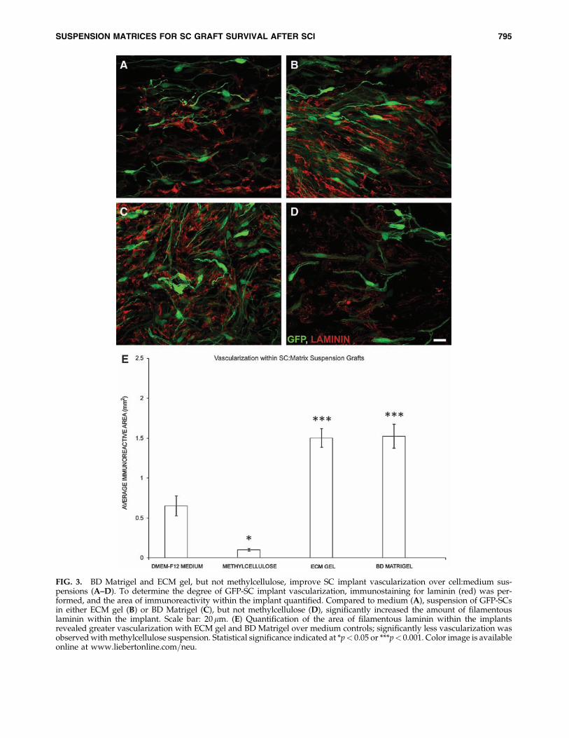
FIG. 3. BD Matrigel and ECM gel, but not methylcellulose, improve SC implant vascularization over cell:medium sus-pensions (A–D). To determine the degree of GFP-SC implant vascularization, immunostaining for laminin (red) was per-formed, and the area of immunoreactivity within the implant quantified. Compared to medium (A), suspension of GFP-SCsin either ECM gel (B) or BD Matrigel (C), but not methylcellulose (D), significantly increased the amount of filamentouslaminin within the implant. Scale bar: 20mm. (E) Quantification of the area of filamentous laminin within the implantsrevealed greater vascularization with ECM gel and BD Matrigel over medium controls; significantly less vascularization wasobserved with methylcellulose suspension. Statistical significance indicated at *p< 0.05 or ***p< 0.001. Color image is availableonline at www.liebertonline.com=neu.
SUSPENSION MATRICES FOR SC GRAFT SURVIVAL AFTER SCI 795

FIG. 4. BD Matrigel and ECM gel, but not methylcellulose, improve axonal in-growth within the SC implant overcell:medium suspensions (A–D). To determine the degree of axonal in-growth within the GFP-SC implants, we employedimmunostaining for neurofilaments (red) and subsequent quantification of the area of immunoreactivity within the implant.Compared to medium (A), suspension of GFP-SCs in either ECM gel (B) or BD Matrigel (C), but not methylcellulose (D),significantly increased the degree of axonal in-growth within the implant. Scale bar: 20 mm. (E) Quantification of the area ofneurofilament immunoreactivity within the implants revealed greater axonal in-growth with ECM gel and BD Matrigel overmedium controls; significantly less axonal in-growth was observed with methylcellulose suspension. Statistical significanceindicated at ***p< 0.001. Color image is available online at www.liebertonline.com=neu.
796 PATEL ET AL.

sequence (RGD) that is recognized by integrins, a family of cellsurface receptors that mediate cell–cell and cell–ECM adhesion(McKerracher et al., 1996). ECM binding thus allows integrinsto regulate, through altered GTPase activity (Relvas et al., 2001),a broad range of cellular processes (Kariya et al., 2004; Kim,2000; Stupack, 2005). In the case of SCs, ECM binding alsoserves as a prerequisite to myelin formation (Podratz et al.,2001).
Laminin, an integral part of the basement membrane,governs many important SC functions (Chen and Strickland,2003), including their migration, differentiation, and axonassociation, and is the major ECM produced by SCs (Corn-brooks and Bunge, 1983). The environment provided byscaffolds composed of laminin have been demonstrated notonly to increase SC proliferation in culture but have also beenshown to be critical to basement membrane formation fol-lowing SC implantation into the injured peripheral nervoussystem (Armstrong et al., 2007), providing an adherent sub-strate for both SCs and in-growing axons. SC adherence tolaminin within the hostile environment of the injured spinalcord may not only activate important cell survival and pro-liferation pathways and provide a protective niche for theimplanted cells, but laminin could also prevent anoikis, aprocess whereby the cells undergo apoptosis due to a lack ofadequate adhesion (Frisch and Screaton, 2001). Many celltypes require anchorage to ECM within tissues to maintaintheir growth and survival (Frisch and Ruoslahti, 1997).
Likewise, collagen has proven to be an effective ECM for SCadhesion because of its ability to promote SC process exten-sion and their association with axons for ensuring myelination(Chernousov et al., 2008). Collagen’s properties give it highmechanical strength, significant permeability and water-uptake properties, exceptional biocompatibility and biode-gradability, as well as low antigenicity (Han et al., 2009;Tabesh et al., 2009). Collagen therefore has served as an at-tractive matrix for tissue engineering because of these prop-erties and its functional role in connective-tissue formation,as well as its critical importance in wound healing, being apotent substrate for angiogenesis and the revascularization ofinjured tissues (Serini et al., 2006). Collagen can also bind
various molecules, including neurotrophic factors that are inaddition supportive for cell migration and axon extension(Han et al., 2009).
The use of Matrigel, which is known to promote survivaland the differentiation of many different cell types, exhibited atrend toward a greater improvement in SC numbers, includ-ing early angiogenesis and axonal in-growth, over ECM gel.The mechanism(s) underlying the superior effectiveness ofMatrigel-suspended implants is not clear. Additional biolog-ically active components in Matrigel, such as the growthfactors EGF, bFGF, NGF, PDGF, IGF-1, and TGF-B (Davis andStroobant, 1990; Iannotti et al., 2003), could have further en-hanced the angiogenic, axon growth, and cell survival pro-moting proprieties of laminin and collagen. NGF plays crucialroles in both axon growth and angiogenesis (Lykissas et al.,2007; Nico et al., 2008). NGF stimulates axon growth by me-diating the polymerization and accumulation of F-actin ingrowth cones and axon shafts (Lykissas et al., 2007), and canpromote angiogenesis though its proliferation effects on en-dothelial cells, acting in concert with FGF and TGF to supporttissue vascularization (Ohta et al., 1991) under physiologicaland pathological conditions. NGF can also promote cell sur-vival through ERK and Akt activation, and downstream anti-apoptotic signaling (Perron and Bixby, 1999; Zhou et al.,2004). These different growth factors within Matrigel wouldbe bound by the ECM and slowly released to the implantedcells and in-growing axons or other cellular components of therevascularization machinery, providing sustained effects onthese processes post implantation.
In contrast, methylcellulose had less-promising resultscompared to the medium or laminin:collagen matrices. Thepoor capacity of methylcellulose to act as a supportive sub-strate for cell suspension grafts could be explained by inferiorcell adherence and=or its inability to provide an adequatesubstrate for angiogenesis and axon growth. Althoughprior reports have indicated that methylcellulose, when usedat a final concentration of 2% to bridge the injured PNS, issupportive of axon regeneration and improves nerve con-ductance compared to other matrices, such as laminin orcollagen (Wells et al., 1997), the current study shows that
FIG. 5. Numbers of GFP-SCs within the injured spinal cord correlates with the degree of implant vascularization and theamount of axonal in-growth (A–B). Linear correlation analysis of animals from all implant groups indicated a significantrelationship between SC numbers and both graft vascularization (A) and axonal in-growth (B). Statistical significanceindicated at **p< 0.01.
SUSPENSION MATRICES FOR SC GRAFT SURVIVAL AFTER SCI 797

methylcellulose was significantly worse than the standardpractice of cell suspension in medium when employed at aslightly higher concentration of 5%; for the purpose of en-hancing the mechanical strength of the in-situ gelling matrix.This increased concentration and viscosity may have re-stricted cell migration, the diffusion of growth factors, andaxon penetrance (Labrador et al., 1995). This work demon-
strates that, with synthetic matrices, defining their optimalformulations and viscosity is critical to whether they behaveas an advantageous or disadvantageous suspension mediumfor cell implantation into the injured CNS.
In all implant groups, a small degree of cell proliferation(3–5% of the total GFP-SCs) was observed at the endpoint,9 weeks post implantation through Ki-67 immunoreactivity(Gerdes et al., 1991); Ki-67 staining was largely confined tothose cells located on the outer fringe of the implants. Al-though no significant difference in GFP-SC proliferation wasnoted, more acute differences in proliferation rather than animproved survival with laminin:collagen matrices may haveaccounted for the significant increase in GFP-SC numbers atendpoint. Future work looking at acute survival times fol-lowing SC implantation within different suspension mediamay shed light on the relative contribution of enhanced GFP-SC survival versus proliferation to the significant increase inSC numbers with laminin:collagen compared to medium-only controls.
Correlation data suggested a strong association betweenthe degree of SC survival and the amount of vascularizationand axon penetrance of the injury-implant site. It appearedthat the implantation of collagen:laminin matrices and theirassociated growth factors encouraged greater angiogenesiswithin the injury-implant site than medium, providing ameans to support the survival of exogenous SCs through animproved supply of nutrients and oxygen. Conversely,methylcellulose suspended implants were significantly lessvascularized and also exhibited the fewest number of SCspresent within the injured cord. Increased axonal growth intocollagen:laminin suspended implants could also have in-creased SC numbers through the release of known mitogenicfactors for SCs; SCs do proliferate upon their exposure toaxons (Wood and Bunge, 1975). Although we were unable todetect a difference in SC proliferation among implant cohorts,changes in proliferation may have been detectable at a muchearlier time post implantation when axons first grew into theimplants. A temporal investigation of SC numbers and theirproliferation rate would identify whether such a mechanismmay have played a role in the enhancement of SC numbersfollowing collagen:laminin suspension.
DMEM-F12 medium suspension has been shown to beadequate for SC implantation, supporting cell survival, axongrowth, and functional recovery (Firouzi et al., 2006; Pearseet al., 2004, 2005, 2007). Here, we demonstrate, in a clinicallyrelevant contusion-injury model, that SC suspension withinin-situ gelling laminin:collagen matrices enhances SC survivalcompared to medium and further improves their capacity foraxon growth support, injured cord revascularization, as wellas functional recovery. With cellular therapies progressingtoward clinical trials for a variety of CNS diseases and trauma,as well as the known issues of obtaining adequate cell survivalwithin the injured CNS environment (Parr et al., 2008; Pearseand Barakat, 2006; Pearse et al., 2007), these studies highlightthe importance of identifying clinically applicable, in-situgelling matrices for achieving greater survival and effective-ness of cellular implants.
Acknowledgments
We thank Raisa Puzis and David Barakat for performingimmunohistochemistry; Beata Frydel and Bridgette Shaw for
FIG. 6. Suspension of GFP-SCs in BD Matrigel led to im-proved hindlimb function compared to SC-medium controls.BBB locomotor testing revealed a significant improvementwith SC-BD Matrigel over SC-medium implants from 4weeks post implantation (A), although no improvement wasobserved in subsequent BBB subscore analysis (B). Foot po-sitioning on the grid-walk test showed a transient improve-ment with BD Matrigel at 4 weeks post implantationcompared to medium controls, but this did not persistthrough 6–8 weeks (C).
798 PATEL ET AL.
aid with image analysis; Yelena Pressman for her cell-culturework; Neil Masters for tissue embedding and sectioning, andslide preparation; Rosa Abril, Denise Koivisto, Monica Stagg,Maritza Garcia, Ileana Oropesa, and Andres Maldonado forhelp with animal care; the Viral Vector Core of The MiamiProject for supplying lentiviral vectors encoding EGFP; andAlexander Marcillo, Paulo Diaz, and Michael Lynch for in-ducing contusion injuries.
This work was supported by: NIH NINDS Grant NumberR01NS056281; The Miami Project to Cure Paralysis; TheBuoniconti Fund; and The Fa Bene, Schumann, Craig H.Neilsen, and Bryon Riesch Paralysis Foundations.
Author Disclosure Statement
No competing financial interests exist.
References
Armstrong, S.J., Wiberg, M., Terenghi, G., and Kingham, P.J.(2007). ECM molecules mediate both Schwann-cell prolifera-tion and activation to enhance neurite outgrowth. Tissue Eng.12, 2863–2870.
Bao, F., and Liu, D. (2002). Peroxynitrite generated in the ratspinal cord induces neuron death and neurological deficits.Neuroscience 115, 839–849.
Barakat, D.J., Gaglani, S.M., Neravetla, S.R., Sanchez, A.R., An-drade, C.M., Pressman, Y., Bunge, M.B., and Pearse, D.D.(2005). Survival, integration and axon growth support of gliatransplanted into the chronically contused spinal cord. CellTransplant. 14, 225–240.
Brundin, P., Karlsson, J., Emgard, M., Schierle, G.S., Hansson,O., Petersen, A., and Castilho, R.F. (2000). Improving thesurvival of grafted dopaminergic neurons: A review overcurrent approaches. Cell Transplant. 9, 179–195.
Bunge, M.B., and Pearse, D.D. (2003). Transplantation strategiesto promote repair of the injured spinal cord. J. Rehabil. 40,55–62.
Chen, Z.L., and Strickland, S. (2003). Laminin gamma1 is criticalfor Schwann cell differentiation, axon myelination, and re-generation in the peripheral nerve. J. Cell. Biol. 163, 889–899.
Chernousov, M.A., Yu, W.M., Chen, Z.L., Carey, D.J., andStrickland, S. (2008). Regulation of Schwann cell function bythe extracellular matrix. Glia 56, 1498–1507.
Cornbrooks, C.J., Carey, D.J., McDonald, J.A., Timpl, R., andBunge, R.P. (1983). In vivo and in vitro observations on la-minin production by Schwann cells. Proc. Natl. Acad. Sci. 80,3850–3854.
Davis, J.B., and Stroobant, P. (1990). Platelet-derived growthfactors and fibroblast growth factors are mitogens for ratSchwann cells. J. Cell Biol. 110, 1353–1360.
Feng, S.Q., Kong, X.H., Guo, S.F., Wang, P., Li, L., Zhong, J.H.,and Zhou, X.F. (2005). Treatment of spinal cord injury withco-grafts of genetically modified Schwann cells and fetalspinal cord cell suspension in the rat. Neurotox. Res. 7, 169–177.
Firouzi, M., Moshayedi, P., Saberi, H., Mobasheri, H., Abol-hassani, F., Jahanzad, I., and Raza, M. (2006). Transplantationof Schwann cells to subarachnoid space induces repair incontused rat spinal cord. Neurosci. Lett. 402, 66–70.
Follenzi, A., Ailles, L.E., Bakovic, S., Geuna, M., and Naldini, L.(2000). Gene transfer by lentiviral vectors is limited by nucleartranslocation and rescued by HIV-1 pol sequences. NatureGenetics 25, 217–222.
Fouad, K., Schnell, L., Bunge, M.B., Schwab, M.E., Liebscher, T.,and Pearse, D.D. (2005). Combining Schwann cell bridges andolfactory-ensheathing glia grafts with chondroitinase pro-motes locomotor recovery after complete transection of thespinal cord. J. Neurosci. 25, 1169–1178.
Franssen, E.H., de Bree, F.M., and Verhaagen, J. (2007). Olfactoryensheathing glia: Their contribution to primary olfactorynervous system regeneration and their regenerative potentialfollowing transplantation into the injured spinal cord. BrainRes. Rev. 56, 236–258.
Frisch, S.M., and Ruoslahti, E. (1997). Integrins and anoikis. Curr.Opin. Cell Biol. 9, 701–706.
Frisch, S.M., and Screaton, R.A. (2001). Anoikis mechanisms.Curr. Opin. Cell Biol. 13, 555–562.
Gerdes, J., Li, L., Schliiter, C., Duchrow, M., Wohlenberg, C.,Gerlach, C., Stahmer, I, Kloth, S., Brandi, E., and Flad, H.D.(1991). Immunobiochemical and molecular biologic charac-terization of the cell proliferation-associated nuclear antigenthat is defined by monoclonal antibody Ki-67. Am. J. Pathol.138, 867–873.
Gess, B., Halfter, H., Kleffner, I., Monje, P., Athauda, G., Wood,P.M., Young, P., and Wanner, I.B. (2008). Inhibition ofN-cadherin and beta-catenin function reduces axon-inducedSchwann cell proliferation. J. Neurosci. Res. 86, 797–812.
Gruner, J.A. (1992). A monitored contusion model of spinal cordinjury in the rat. J. Neurotrauma 9, 123–126.
Hall, E.D., and Braughler, J.M. (1986). Role of lipid peroxidationin post-traumatic spinal cord degeneration: A review. Cent.Nerv. Syst. Trauma 3, 281–294.
Han, Q., Sun, W., Lin, H., Zhao, W., Gao, Y., Zhao, Y., Chen,B., Xiao, Z., Hu, W., Li, Y., Yang, B., and Dai, J. (2009).Linear ordered collagen scaffolds loaded with collagen-binding brain-derived neurotrophic factor improve the re-covery of spinal cord injury in rats. Tissue Eng. Part A. 15,2927–2935.
Hill, C.E., Hurtado, A., Blits, B., Bahr, B.A., Wood, P.M., BartlettBunge, M., and Oudega, M. (2007). Early necrosis and apo-ptosis of Schwann cells transplanted into the injured rat spinalcord. Eur. J. Neurosci. 26, 1433–1445.
Iannotti, C., Li, H., Yan, P., Lu, X., Wirthlin, L., and Xu, X.M.(2003). Glial cell line-derived neurotrophic factor-enrichedbridging transplants promote propriospinal axonal regenera-tion and enhance myelination after spinal cord injury. Exp.Neurol. 183, 379–393.
Johnson, E.O., Zoubos, A.B., and Soucacos, P.N. (2005). Re-generation and repair of peripheral nerves. Injury 36, 24–29.
Kassar-Duchossoy, L., Duchossoy, Y., Rhrich-Haddout, F., andHorvat, J.C. (2001). Reinnervation of a denervated skeletalmuscle by spinal axons regenerating through a collagenchannel directly implanted into the rat spinal cord. Brain Res.908, 25–34.
Kim, J.E., Kim, S.J., Lee, B.H., Park, R.W., Kim, K.S., and Kim,I.S. (2000). Identification of motifs for cell adhesion withinthe repeated domains of transforming growth factor-beta-induced gene, betaig-h3. J. Boil. Chem. 275, 30907–30915.
Labrador, R.O., Buti, M., and Navarro, X. (1995). Peripheralnerve repair: Role of agarose matrix density on functionalrecovery. Neuroreport 6, 2022–2026.
Louro, J., and Pearse, D.D. (2008). Stem and progenitor celltherapies: Recent progress for spinal cord injury repair. Neu-rol. Res. 30, 5–16.
Lykissas, M.G., Batistatou, A.K., Charalabopoulos, K.A., andBeris, A.E. (2007). The role of neurotrophins in axonal
SUSPENSION MATRICES FOR SC GRAFT SURVIVAL AFTER SCI 799

growth, guidance, and regeneration. Curr. Neurovasc. Res. 4,143–151.
Masuda-Nakagawa, L.M., Muller, K.J., and Nicholls, J.G. (1990).Accumulation of laminin and microglial cells at sites of injuryand regeneration in the central nervous system of the leech.Proc. Biol. Sci. 241, 201–206.
McKerracher, L., Chamoux, M., and Arregui, C.O. (1996). Role oflaminin and integrin interactions in growth cone guidance.Mol. Neurobiol. 12, 95–116.
Meijs, M.F., Timmers, L., Pearse, D.D., Tresco, P.A., Bates, M.L.,Joosten, E.A., Bunge, M.B., and Oudega, M. (2004). Basic fi-broblast growth factor promotes neuronal survival but notbehavioral recovery in the transected and Schwann cellimplanted rat thoracic spinal cord. J. Neurotrauma 21, 1415–1430.
Morrissey, T.K., Kleitman, N., and Bunge, R.P. (1991). Isolationand functional characterization of Schwann cells derived fromadult peripheral nerve. J. Neurosci. 11, 2433–2442.
Nandoe Tewarie, R.D., Hurtado, A., Levi, A.D., Grotenhuis, J.A.,and Oudega, M. (2007). Bone marrow stromal cells for repairof the spinal cord: Towards clinical application. Cell Trans-plant. 16, 183.
Nico, B., Mangieri, D., Benagiano, V., Crivellato, E., and Ribatti,D. (2008). Nerve growth factor as an angiogenic factor.Microvasc. Res. 75, 135–141.
Ohta, H., Ishiyama, J., Saito, H., and Nishiyama, N. (1991). Ef-fects of pretreatment with basic fibroblast growth factor, epi-dermal growth factor and nerve growth factor on neuronsurvival and neovascularization of superior cervical gangliontransplanted into the third ventricle in rats. Japan. J. Phar-macol. 55, 255–262.
Parr, A.M., Kulbatski, I., Zahir, T., Wang, X., Yue, C., Keating,A., and Tator, C.H. (2008). Transplanted adult spinal cord-derived neural stem=progenitor cells promote early functionalrecovery after rat spinal cord injury. Neuroscience 155, 760–770.
Pearse, D.D., and Barakat, D.J. (2006). Cellular repair strategiesfor spinal cord injury. Expert Opin. Biol. Ther. 6, 639–652.
Pearse, D.D., and Bunge, M.B. (2006). Designing cell- and gene-based regeneration strategies to repair the injured spinal cord.J. Neurotrauma 23, 438–452.
Pearse, D.D., Bushell, G., and Leah, J.D. (2001). Jun, Fos andKrox in the thalamus after C-fiber stimulation: Coincident-input-dependent expression, expression across somatotropicboundaries, and nucleolar translocation. Neuroscience 107,143–159.
Pearse, D.D., Chatzipanteli, K., Marcillo, A.E., Bunge, M.B., andDietrich, W.D. (2003). Comparison of iNOS inhibition by an-tisense and pharmacological inhibitors after spinal cord injury.J. Neuropathol. Exp. Neurol. 62, 1096–1107.
Pearse, D.D., Lo, T.P., Jr., Cho, K.S., Lynch, M.P., Garg, M.S.,Marcillo, A.E., Sanchez, A.R., Cruz, Y., and Dietrich, W.D.(2005). Histopathological and behavioral characterization of anovel cervical spinal cord displacement contusion injury in therat. J. Neurotrauma 22, 680–702.
Pearse, D.D., Marcillo, A.E., Oudega, M., Lynch, M.P., Wood,P.M., and Bunge, M.B. (2004). Transplantation of Schwanncells and olfactory ensheathing glia after spinal cord injury:Does pretreatment with methylprednisolone and interleukin-10 enhance recovery? J. Neurotrauma 21, 1223–1239.
Pearse, D.D., Sanchez, A.R., Pereira, F.C., Andrade, C.M., Puzis,R., Pressman, Y., Golden, K., Kitay, B.M., Blits, B., Wood, P.M.,and Bunge, M.B. (2007). Transplantation of Schwann cellsand=or olfactory ensheathing glia into the contused spinal
cord: Survival, migration, axon association, and functionalrecovery. Glia 55, 976–1000.
Perron, J.C., and Bixby, J.L. (1999). Distinct neurite outgrowthsignaling pathways converge on ERK activation. Mol. Cell.Neurosci. 13, 362–378.
Pierucci, A., de Duek, E.A., and de Oliveira, A.L. (2008). Per-ipheral nerve regeneration through biodegradable conduitsprepared using solvent evaporation. Tissue Eng. Part A 14,595–606.
Podratz, J.L., Rodriguez, E., and Windebank, A.J. (2001). Role ofthe extracellular matrix in myelination of peripheral nerve.Glia 35, 35–40.
Profyris, C., Cheema, S.S., Zang, D., Azari, M.F., Boyle, K., andPetratos, S. (2004). Degenerative and regenerative mecha-nisms governing spinal cord injury. Neurobiol. Dis. 15,415–436.
Properzi, F., Lin, R., Kwok, J., Naidu, M., van Kuppevelt, T.H.,Ten Dam, G.B., Camargo, L.M., Raha-Chowdhury, R., Fur-ukawa, Y., Mikami, T., Sugahara, K., and Fawcett, J.W. (2008).Heparan sulphate proteoglycans in glia and in the normal andinjured CNS: Expression of sulphotransferases and changes insulphation. Eur. J. Neurosci. 27, 593–604.
Relvas, J.B., Setzu, A., Baron, W., Buttery, P.C., LaFlamme, S.E.,Franklin, R.J., and French-Constant, C. (2001). Expression ofdominant-negative and chimeric subunits reveals an essentialrole for beta1 integrin during myelination. Curr. Biol. 10, 11,1039–1043.
Schaal, S.M., Kitay, B.M., Cho, K.S., Lo, T.P., Jr., Barakat, D.J.,Marcillo, A.E., Sanchez, A.R., Andrade, C.M., and Pearse, D.D.(2007). Schwann cell transplantation improves reticulospinalaxon growth and forelimb strength after severe cervical spinalcord contusion. Cell Transplant. 16, 207–228.
Scholzen, T., and Gerdes, J. (2000). The Ki-67 protein: From theknown and the unknown. J. Cell Physiol. 182, 311–322.
Serini, G., Valdembri, D., and Bussolino, F. (2006). Integrinsand angiogenesis: A sticky business. Exp. Cell Res. 312, 651–658.
Stabenfeldt, S.E., Garcı́a, A.J., and LaPlaca M.C. (2006). Ther-moreversible laminin-functionalized hydrogel for neural tis-sue engineering. J. Biomed. Mater. Res. A 77, 718–725.
Stupack, D.G. (2005). Integrins as a distinct subtype of depen-dence receptors. Cell Death Differ. 12, 1021–1030.
Sugrue, S.P., and Hay, E.D. (1986). The identification of extra-cellular matrix (ECM) binding sites on the basal surface ofembryonic corneal epithelium and the effect of ECM bindingon epithelial collagen production. J. Cell Biol. 102, 1907–1916.
Tabesh, H., Amoabediny, G., Nik, N.S., Heydari, M., Yosefifard,M., Siadat, S.O., and Mottaghy, K. (2009). The role of biode-gradable engineered scaffolds seeded with Schwann cells forspinal cord regeneration. Neurochem. Int. 54, 73–83.
Takami, T., Oudega, M., Bates, M.L., Wood, P.M., Kleitman, N.,and Bunge, M.B. (2002). Schwann cell but not olfactory en-sheathing glia transplants improve hind limb locomotor per-formance in the moderately contused adult rat thoracic spinalcord. J. Neurosci. 22, 6670–6681.
Tate, M.C., Shear, D.A., Hoffman, S.W., Stein, D.G., and LaPlaca,M.C. (2001). Biocompatibility of methylcellulose-based con-structs designed for intracerebral gelation following experi-mental traumatic brain injury. Biomaterials 22, 1113–1123.
Wells, M.R., Kraus, K., Batter, D.K., Blunt, D.G., Weremowitz, J.,Lynch, S.E., Antoniades, H.N., and Hansson, H.A. (1997). Gelmatrix vehicles for growth factor application in nerve gapinjuries repaired with tubes: A comparison of biomatrix, col-lagen, and methylcellulose. Exp. Neurol. 146, 395–402.
800 PATEL ET AL.

Wood, P.M., and Bunge, R.P. (1975). Evidence that sen-sory axons are mitogenic for Schwann cells. Nature 256, 662–664.
Xu, X.M., Chen, A., Guenard, V., Kleitman, N., and Bunge, M.B.(1997). Bridging Schwann cell transplants promote axonal re-generation from both the rostral and caudal stumps of trans-ected adult rat spinal cord. J. Neurocytol. 26, 1–16.
Yoshii, S., Ito, S., Shima, M., Taniguchi, A., and Akagi, M. (2009).Functional restoration of rabbit spinal cord using collagen-filament scaffold. J. Tissue Eng. Regen. Med. 3, 19–25.
Zhou, F.Q., Zhou, J., Dedhar, S., Wu, Y.H., and Snider, W.D.(2004). NGF-induced axon growth is mediated by localized
inactivation of GSK-3beta and functions of the microtubuleplus end binding protein APC. Neuron 42, 897–912.
Address correspondence to:Damien D. Pearse, Ph.D.
The Miami Project to Cure ParalysisUniversity of Miami Miller School of MedicineThe Lois Pope Life Center, Locator Code (R-48)
P.O. Box 016960Miami, FL 33101
E-mail: [email protected]
SUSPENSION MATRICES FOR SC GRAFT SURVIVAL AFTER SCI 801