Surface residues dynamically organize water bridges to enhance electron transfer between proteins
8
Surface residues dynamically organize water bridges to enhance electron transfer between proteins Aurélien de la Lande a,b,2 , Nathan S. Babcock c,d , Jan Řezáč a,b,3 , Barry C. Sanders c,d , and Dennis R. Salahub a,b,e,1 a Department of Chemistry, b Institute for Biocomplexity and Informatics, c Department of Physics and Astronomy, d Institute for Quantum Information Science, and e Institute for Sustainable Energy, Environment and Economy, University of Calgary, 2500 University Drive NW, Calgary, Alberta, Canada, T2N 1N4 Edited by David N. Beratan, Duke University, Durham, NC, and accepted by the Editorial Board May 17, 2010 (received for review December 15, 2009) Cellular energy production depends on electron transfer (ET) be- tween proteins. In this theoretical study, we investigate the impact of structural and conformational variations on the electronic cou- pling between the redox proteins methylamine dehydrogenase and amicyanin from Paracoccus denitrificans. We used molecular dynamics simulations to generate configurations over a duration of 40 ns (sampled at 100-fs intervals) in conjunction with an ET pathway analysis to estimate the ET coupling strength of each configuration. In the wild-type complex, we find that the most frequently occurring molecular configurations afford superior elec- tronic coupling due to the consistent presence of a water molecule hydrogen-bonded between the donor and acceptor sites. We attri- bute the persistence of this water bridge to a “molecular break- water” composed of several hydrophobic residues surrounding the acceptor site. The breakwater supports the function of nearby solvent-organizing residues by limiting the exchange of water mo- lecules between the sterically constrained ET region and the more turbulent surrounding bulk. When the breakwater is affected by a mutation, bulk solvent molecules disrupt the water bridge, result- ing in reduced electronic coupling that is consistent with recent experimental findings. Our analysis suggests that, in addition to enabling the association and docking of the proteins, surface residues stabilize and control interprotein solvent dynamics in a concerted way. respiratory chain ∣ Marcus theory ∣ pathway model ∣ dynamic docking ∣ blue copper proteins T he electron transport chain is the cornerstone of biological energy transduction. All known life-forms use membrane- bound chains of redox proteins to convert energy from food or sunlight into chemical energy stored in adenosine triphosphate (1). Biological electron transfer (ET) often occurs over long dis- tances (>1 nm) between protein-encapsulated redox cofactors separated by intervening protein or solvent molecules. Over the last two decades, there has been increasing interest in water as an “active constituent in cellular biology” (2). Today there is a growing body of evidence suggesting that water plays an impor- tant role mediating long-range ET and that conformational fluctuations are critical to protein-solvent interactions at ET interfaces (3–10). Notably, previous authors have suggested that “ordered water molecules in the protein-protein interface may considerably influence electronic coupling between redox cen- ters” (4) and that “water may be a particularly strong tunneling mediator when it occupies a sterically constrained space between redox cofactors with strong organizing forces that favor construc- tively interfering coupling pathways” (7). In general, however, the degree of sophistication of solvent- organizing effects at aqueous ET interfaces remains unknown. In this study, we show that a pair of solvent-organizing residues in direct contact with a bridging water molecule may be aided by surrounding residues that help stabilize local solvent dynamics by mediating contact with the bulk. We predict that surface residues at protein-protein interfaces can act collectively to organize and stabilize solvent structures and dynamics during long-range ET. The timely passage of electrons from protein to protein is crucial for proper metabolic regulation (11) and relies on all the physical and chemical phenomena (i.e., diffusion, protein docking, and ET reaction steps) that participate in overall elec- tron transmission. Here we investigate the redox reaction be- tween methylamine dehydrogenase (MADH) and amicyanin taken from Paracoccus denitrificans. This redox pair is represen- tative of a broad class of interprotein ET reactions involving blue copper proteins (12). Under methylotrophic growth conditions, MADH supplies electrons to amicyanin, a blue copper protein that in turn shuttles electrons to various c-type cytochromes (13). In vitro, the transfer occurs between the reduced tryptophan tryptophylquinone (TTQ) group on the MADH β-subunit and a cupric complex buried just under the amicyanin surface (Fig. 1). The oxidation of MADH by amicyanin is a “true” ET reaction, limited by the ET reaction rate k ET (14). Ma et al. recently reported a series of site-directed mutations performed on the amicyanin methionine 51 residue by alanine, lysine, and leucine (Fig. 1) (15). Kinetic measurements revealed nearly a tenfold de- crease in k ET without significant changes to the proteins’ overall structural, binding, or redox potentials. Ma et al. concluded that “surface residues of redox proteins may not only dictate specifi- city for their redox protein partners but also be critical to opti- mize the orientations of the redox centers and intervening media within the protein complex for the ET event” (15). To analyze an interprotein ETreaction such as the reduction of amicyanin by MADH, it is necessary to consider large ensembles of protein-protein complex configurations that contribute to the average ET rate (16). For a true ET reaction, the ET rate k ET can be estimated by nonadiabatic Marcus–Hush–Levich theory (17, 18), k ET ¼ 2π ℏ 1 ffiffiffiffiffiffiffiffiffiffiffiffiffiffiffi 4πλk B T p jT DA j 2 exp −ΔG ‡ k B T ¼ 2π ℏ 1 ffiffiffiffiffiffiffiffiffiffiffiffiffiffiffi 4πλk B T p jT DA j 2 exp −ðΔG ∘ þ λÞ 2 4λk B T ; [1] where ℏ is the reduced Planck’s constant, k B is Boltzmann’s con- stant, T is the temperature, ΔG ‡ is the Gibbs free energy of activa- Author contributions: A.d.l.L. and N.S.B. designed research; A.d.l.L. and N.S.B. performed research; J.R. contributed new reagents/analytic tools; A.d.l.L., N.S.B., B.C.S., and D.R.S. analyzed data; and A.d.l.L., N.S.B., B.C.S., and D.R.S. wrote the paper. The authors declare no conflict of interest. This article is a PNAS Direct Submission. D.N.B. is a guest editor invited by the Editorial Board. 1 To whom correspondence should be addressed: [email protected]. 2 Present address: Laboratoire de Chimie Physique—Centre National de la Recherche Scientifique -Unité Mixte de Recherche 8000, Université Paris-Sud 11, Bâtiment 349, Campus d’Orsay. 15, Rue Georges Clémenceau, 91405 Orsay Cedex, France. 3 Present address: Institute of Organic Chemistry and Biochemistry, Academy of Sciences of the Czech Republic and Center for Biomolecules and Complex Molecular Systems, Flemingovo náměstí 2, 166 10 Prague 6, Czech Republic. This article contains supporting information online at www.pnas.org/lookup/suppl/ doi:10.1073/pnas.0914457107/-/DCSupplemental. www.pnas.org/cgi/doi/10.1073/pnas.0914457107 PNAS ∣ June 29, 2010 ∣ vol. 107 ∣ no. 26 ∣ 11799–11804 BIOPHYSICS AND COMPUTATIONAL BIOLOGY
-
Upload
independent -
Category
Documents
-
view
0 -
download
0
Transcript of Surface residues dynamically organize water bridges to enhance electron transfer between proteins

Surface residues dynamically organize water bridgesto enhance electron transfer between proteinsAurélien de la Landea,b,2, Nathan S. Babcockc,d, Jan Řezáča,b,3, Barry C. Sandersc,d, and Dennis R. Salahuba,b,e,1
aDepartment of Chemistry, bInstitute for Biocomplexity and Informatics, cDepartment of Physics and Astronomy, dInstitute for Quantum InformationScience, and eInstitute for Sustainable Energy, Environment and Economy, University of Calgary, 2500 University Drive NW, Calgary, Alberta,Canada, T2N 1N4
Edited by David N. Beratan, Duke University, Durham, NC, and accepted by the Editorial Board May 17, 2010 (received for review December 15, 2009)
Cellular energy production depends on electron transfer (ET) be-tween proteins. In this theoretical study, we investigate the impactof structural and conformational variations on the electronic cou-pling between the redox proteins methylamine dehydrogenaseand amicyanin from Paracoccus denitrificans. We used moleculardynamics simulations to generate configurations over a durationof 40 ns (sampled at 100-fs intervals) in conjunction with an ETpathway analysis to estimate the ET coupling strength of eachconfiguration. In the wild-type complex, we find that the mostfrequently occurring molecular configurations afford superior elec-tronic coupling due to the consistent presence of a water moleculehydrogen-bonded between the donor and acceptor sites. We attri-bute the persistence of this water bridge to a “molecular break-water” composed of several hydrophobic residues surroundingthe acceptor site. The breakwater supports the function of nearbysolvent-organizing residues by limiting the exchange of water mo-lecules between the sterically constrained ET region and the moreturbulent surrounding bulk. When the breakwater is affected by amutation, bulk solvent molecules disrupt the water bridge, result-ing in reduced electronic coupling that is consistent with recentexperimental findings. Our analysis suggests that, in addition toenabling the association and docking of the proteins, surfaceresidues stabilize and control interprotein solvent dynamics in aconcerted way.
respiratory chain ∣ Marcus theory ∣ pathway model ∣ dynamic docking ∣blue copper proteins
The electron transport chain is the cornerstone of biologicalenergy transduction. All known life-forms use membrane-
bound chains of redox proteins to convert energy from food orsunlight into chemical energy stored in adenosine triphosphate(1). Biological electron transfer (ET) often occurs over long dis-tances (>1 nm) between protein-encapsulated redox cofactorsseparated by intervening protein or solvent molecules. Overthe last two decades, there has been increasing interest in wateras an “active constituent in cellular biology” (2). Today there is agrowing body of evidence suggesting that water plays an impor-tant role mediating long-range ET and that conformationalfluctuations are critical to protein-solvent interactions at ETinterfaces (3–10). Notably, previous authors have suggested that“ordered water molecules in the protein-protein interface mayconsiderably influence electronic coupling between redox cen-ters” (4) and that “water may be a particularly strong tunnelingmediator when it occupies a sterically constrained space betweenredox cofactors with strong organizing forces that favor construc-tively interfering coupling pathways” (7).
In general, however, the degree of sophistication of solvent-organizing effects at aqueous ET interfaces remains unknown.In this study, we show that a pair of solvent-organizing residuesin direct contact with a bridging water molecule may be aided bysurrounding residues that help stabilize local solvent dynamics bymediating contact with the bulk. We predict that surface residuesat protein-protein interfaces can act collectively to organize andstabilize solvent structures and dynamics during long-range ET.
The timely passage of electrons from protein to protein iscrucial for proper metabolic regulation (11) and relies on allthe physical and chemical phenomena (i.e., diffusion, proteindocking, and ET reaction steps) that participate in overall elec-tron transmission. Here we investigate the redox reaction be-tween methylamine dehydrogenase (MADH) and amicyanintaken from Paracoccus denitrificans. This redox pair is represen-tative of a broad class of interprotein ETreactions involving bluecopper proteins (12). Under methylotrophic growth conditions,MADH supplies electrons to amicyanin, a blue copper proteinthat in turn shuttles electrons to various c-type cytochromes(13). In vitro, the transfer occurs between the reduced tryptophantryptophylquinone (TTQ) group on the MADH β-subunit and acupric complex buried just under the amicyanin surface (Fig. 1).The oxidation of MADH by amicyanin is a “true” ET reaction,limited by the ET reaction rate kET (14). Ma et al. recentlyreported a series of site-directed mutations performed on theamicyanin methionine 51 residue by alanine, lysine, and leucine(Fig. 1) (15). Kinetic measurements revealed nearly a tenfold de-crease in kET without significant changes to the proteins’ overallstructural, binding, or redox potentials. Ma et al. concluded that“surface residues of redox proteins may not only dictate specifi-city for their redox protein partners but also be critical to opti-mize the orientations of the redox centers and intervening mediawithin the protein complex for the ET event” (15).
To analyze an interprotein ETreaction such as the reduction ofamicyanin by MADH, it is necessary to consider large ensemblesof protein-protein complex configurations that contribute to theaverage ET rate (16). For a true ET reaction, the ET rate kETcan be estimated by nonadiabatic Marcus–Hush–Levich theory(17, 18),
kET ¼ 2π
ℏ1ffiffiffiffiffiffiffiffiffiffiffiffiffiffiffiffi
4πλkBTp jTDAj2 exp
�−ΔG‡
kBT
�
¼ 2π
ℏ1ffiffiffiffiffiffiffiffiffiffiffiffiffiffiffiffi
4πλkBTp jTDAj2 exp
�−ðΔG∘ þ λÞ2
4λkBT
�; [1]
where ℏ is the reduced Planck’s constant, kB is Boltzmann’s con-stant, T is the temperature,ΔG‡ is the Gibbs free energy of activa-
Author contributions: A.d.l.L. and N.S.B. designed research; A.d.l.L. and N.S.B. performedresearch; J.R. contributed new reagents/analytic tools; A.d.l.L., N.S.B., B.C.S., and D.R.S.analyzed data; and A.d.l.L., N.S.B., B.C.S., and D.R.S. wrote the paper.
The authors declare no conflict of interest.
This article is a PNAS Direct Submission. D.N.B. is a guest editor invited by theEditorial Board.1To whom correspondence should be addressed: [email protected] address: Laboratoire de Chimie Physique—Centre National de la RechercheScientifique -Unité Mixte de Recherche 8000, Université Paris-Sud 11, Bâtiment 349,Campus d’Orsay. 15, Rue Georges Clémenceau, 91405 Orsay Cedex, France.
3Present address: Institute of Organic Chemistry and Biochemistry, Academy of Sciencesof the Czech Republic and Center for Biomolecules and Complex Molecular Systems,Flemingovo náměstí 2, 166 10 Prague 6, Czech Republic.
This article contains supporting information online at www.pnas.org/lookup/suppl/doi:10.1073/pnas.0914457107/-/DCSupplemental.
www.pnas.org/cgi/doi/10.1073/pnas.0914457107 PNAS ∣ June 29, 2010 ∣ vol. 107 ∣ no. 26 ∣ 11799–11804
BIOPH
YSICSAND
COMPU
TATIONALBIOLO
GY

tion, ΔG∘ is the Gibbs free energy of reaction, λ is the reorganiza-tion energy, and TDA is the superexchange matrix elementthat couples the donor and acceptor electronic states quantummechanically.
The experimental trends reported by Ma et al. (15) stronglysuggest the existence of a correlation between protein motionand ET activity in the MADH-amicyanin complex. While mostnumerical studies have addressed nanosecond time scales orshorter, we investigated longer time scales (40 ns) that are lesswell understood, thereby obtaining statistics for vibrationalmodes spanning several temporal orders of magnitude. Inaddition to the wild-type complex, we consider four amicyaninmutants: M51A, M51L, M51K, and M51C. The first three muta-tions correspond to those reported by Ma et al. (15), whereasM51C was added to investigate the impact of a thiol replacingthe original thioether. We employed molecular dynamics (MD)simulations to generate the configurations within the transientET complex along with an ET pathway analysis to characterizeeach configuration’s intrinsic ET activity (see Methods). Thiscomputationally intensive investigation has allowed us to identifyimportant solvent-stabilizing functions of interprotein surfaceresidues. Because of this solvent-stabilizing effect, the mostET-active wild-type conformations are also the most statisticallyfavored ones, an effect that is lost in the mutant complexes.
ResultsWe obtained 400,000 molecular configurations from 40 ns of MDsimulations of the wild-type complex and each mutant. We em-ployed the semiempirical pathway model originally developed byBeratan, Onuchic, and Hopfield (19) to determine the tunnelingpathway with the largest electronic coupling matrix element TDAfor each configuration. Although this model does not provide anabsolute value for the superexchange coupling matrix elementTDA and does not account for interferences between multiplepathways, it can be used to estimate the total electronic couplingdecay factor εtot, where TDA ≈HDA · εtot and HDA is the theore-tical “close-contact” coupling matrix element (20, 21). Despiteits simplicity, the pathway model has previously demonstratedexcellent predictive power when comparing different molecularconfigurations (8, 22, 23). We recorded the pathway with the
largest decay factor εtot for each configuration and labeled itby the surface residue through which the electron exits theMADH. We found that in the vast majority of pathways(>99%), the electron leaves the MADH through one of Ser β56, Trp β 57, Val β 58, or Trp β 108 before tunneling throughone or more water molecules and entering the amicyanin throughHis 95. Adopting the terminology already in use (21), it is con-venient to define four distinct collections of similar pathways, or“pathway tubes,” labeled by the letters A (Ser β 56), B (Trp β 57),C (Val β 58), and D (Trp β 108) (Fig. 2 and Fig. S1). All the re-maining excess pathways (labeled E) afford comparatively weakelectronic coupling (Table S1).
Pathway tubeA is of particular interest due to its large couplingstrength and high frequency of occurrence (∼60%, Table 1). Forthis reason, we further divided it into three subcategories: A1 re-presents a single pathway with a hydrogen-bonded bridge fromthe Ser β 56 O carbonyl oxygen through a single water moleculeto the His 95 HE2 proton, A2 represents all the remaining com-pletely hydrogen-bonded water bridges between Ser β 56 and His95, and A3 represents all the partially broken (i.e., van der Waalscoupled) pathways from Ser β 56 to His 95. In a previous pathwayanalysis performed on the crystal structure of the MADH-ami-cyanin dimer (24), Brooks et al. found that the strongest pathwayrequired a through-vacuum jump from MADH Trp β 108 to ami-cyanin Pro 94 (25). In contrast, in our solvated system we find thatpathways involving a direct jump from Trp 108 to Pro 94 make upless than 0.01% of our data and are on average one-tenth asstrong as the completely hydrogen-bonded A1 pathway. In an-other analysis performed on the MADH-amicyanin-cytochrometernary crystal structure, Chen et al. concluded that the strongestpathway involved a trapped interfacial water molecule, eventhough its efficiency depended “critically on the presence ofthe water molecule which may not always be occupied” (26).Chen et al.’s water-mediated pathway is identifiable as our A1
pathway, assuming a hydrogen-bonded arrangement for the hy-drogen atoms that were not resolved. Thus, our computationalstudy strongly supports Chen et al.’s water-bridge hypothesisand moreover stresses the importance of the dynamical behaviorof the ET pathways; very frequent switches between the pathwaysare obtained in the course of the simulations (Fig. S2) but path-way A1 was favored only in the wild-type complex.
Configurations associated with pathway tube A1 depend criti-cally on the probability Phb of a water molecule forming twosimultaneous hydrogen bonds with atoms Ser β 56 O and His95 HE2. The wild-type complex’s statistical affinity for pathwayA1 depends on this high probability (Phb > 50%) compared withthose of the mutants (Phb < 20%) (Table 1). In turn, the presenceof this water bridge is linked to the discrepancy in the averagenumber of water molecules found at the interface between theproteins. The consistent presence of the water molecule joiningSer β 56 and His 95 in the wild type, its corresponding absence inthe mutants, and the resulting reduction in the mutant couplingstrength indicate that solvent organization is vital to this reaction.Any destabilization of the A1 water bridge results in a statisticalshift toward less efficient pathways (Table 1).
The A1 pathway is disrupted when other water molecules jostleor compete with the bridging water molecule (Fig. 2 A1). Todetermine the impact of surrounding water molecules on theA1 bridge, we computed the number of water molecules withinthe “ETregion,” which we define to be a sphere of radius R cen-tered between the MADH Ser β 56 O and amicyanin His 95 HE2atoms. Even for large radii (R ¼ 5 Å), an average of only 2.5water molecules are present within the wild-type ET region,compared with mutant averages ranging from 4.7 to 6.3 (Fig. 3).Comparatively few water molecules are exchanged between thewild-type ET region and the surrounding bath. The mutant ETregions are much more turbulent, as evidenced by the largernumber of water molecules present, the lower probability of
Fig. 1. (A) Solvated amicyanin (blue) in contact with MADH subunit β (red).Residues of interest to ET are represented as liquorice. Redox cofactors areshown in purple and surface methionine residues in orange. The dominantpathway A1 is shown as a transparent blue tube connecting the redox cofac-tors. (B) Direct (“head on”) view of interfacial residues represented by theirchemical nature: hydrophobic (white), hydrophilic (yellow), positivelycharged acidic (blue), and negatively charged basic (red). The dotted greencircles indicate the ET region on the surface of each protein. The molecularbreakwater is visible as a white ring of hydrophobic residues surrounding theamicyanin His 95.
11800 ∣ www.pnas.org/cgi/doi/10.1073/pnas.0914457107 de la Lande et al.

hydrogen-bonded bridge formation Phb, and the higher rate ofturnover τ between the individual water molecules involved informing the A1 bridge (Table 1). These results indicate thatthe dynamical organization of the intervening solvent is crucialto the formation of the most efficient ET configurations.
A comparison of mutant-to-wild-type ratios of the calculateddecay factors rmut
ε ¼ hε 2totimut∕hε 2
totiwt and the experimental ratesrmutk ¼ kmut
ET ∕kwtET provides insight about the impact of the Met 51mutation on experimental rates through modifications of theelectronic coupling term. Our results are in overall qualitativeagreement with experiment, as the decreases in the mutant decayfactors are of comparable size to the experimentally observeddecreases in the ET rate constants (Table 1). There is, however,a discrepancy between the ordering of the experimentally deter-mined mutant rates (1 > rM51L
k > rM51Kk > rM51A
k ) and the pathwaymodel decay factors (1 > rM51A
ε > rM51Kε > rM51L
ε ), discussed be-low. We find that the packing density model (27) produces decayfactors similar to those of the pathway model for the M51L andM51K mutants, but predicts the M51A and M51C mutants to beat least as kinetically competent as the wild type.
DiscussionIt is remarkable that a single strongly coupled pathway shoulddominate the thermal statistics of the wild-type complex alone(>50%, Fig. 3A). There is no a priori correlation between thebinding affinity of the complex and ET activity: Maxwell-Boltz-mann statistics govern the probability of occurrence of a givenconfiguration, whereas the nonadiabatic Marcus expression(Eq. 1) independently determines the ET rate constant kET forthat configuration. It is apparent that the protein structure atthe wild-type interface is specifically suited to favor conforma-tions most amenable to ET. On the other hand, configurationsassociated with lower efficiency pathways become more statisti-cally prominent in the mutant distributions, thereby decreasingthe average decay factor hεtoti (Table 1). This statistical changealso carries implications for the overall reaction kinetics.
In order to relate solvent dynamics directly to the mutation, itis necessary to consider interactions between the amicyanin 51residue and other protein or solvent molecules. Previously, whenperforming a P52G mutation upon amicyanin, Ma et al. attribu-ted the resulting reduction in kET to a loss of interactions betweenthe amicyanin Met 51 residue and the MADH Val 58 (28). This
Fig. 2. Representative pathways for each pathway tube in colors corresponding to those in Fig. 3. Hydrogen bonds are represented by dotted lines andthrough-space jumps by solid ovals. The arrows on A1 illustrate perturbations to the hydrogen-bond network caused by another nearby water molecule.
Table 1. Expectation values for hεtoti and the ratios rmutε ¼ hε 2
totimut∕hε 2totiwt and rmut
k ¼ kmutET ∕kwt
ET obtained from packing density andpathway analyses*
Wild type M51L M51K M51A M51C
rmutk (Experiment) 1.0 0.68 0.49 0.13 —hεtoti × 103 (Pathway analysis) 0.90 ± 0.03 0.47 ± 0.03 0.61 ± 0.02 0.65 ± 0.02 0.73 ± 0.02rmutε (Pathway analysis) 1.0 0.36 ± 0.04 0.52 ± 0.04 0.57 ± 0.04 0.76 ± 0.05εtot × 103 (Packing density) 0.70 ± 0.03 0.42 ± 0.04 0.51 ± 0.03 0.62 ± 0.05 1.03 ± 0.05rmutε (Packing density) 1.0 0.56 ± 0.09 0.76 ± 0.07 0.89 ± 0.15 2.29 ± 0.26Phb 0.53 0.15 0.19 0.18 0.16τ (ns−1) 0.40 0.57 0.60 0.65 0.68
*The uncertainties account for the sampling errors of the computational protocol (see SI Text). Experimental rates kET were obtained from k3 (at 30 °C) intable 3 of ref. 15 (M51C was not reported). Phb is the unit-normalized likelihood that a water molecule is simultaneously hydrogen bonded to both theMADH Ser β 56 O and amicyanin His 95 HE2 atoms during our simulations. The turnover τ of the bridging water molecule is defined as the number ofdifferent water molecules that participate in pathway A1 divided by the length of the simulation in nanoseconds.
de la Lande et al. PNAS ∣ June 29, 2010 ∣ vol. 107 ∣ no. 26 ∣ 11801
BIOPH
YSICSAND
COMPU
TATIONALBIOLO
GY

conclusion is compatible with our simulation results, which showthat conformational variations in the amicyanin M51K and M51Lresidues allow an increased number of water molecules to“sneak” into the ET region (Fig. S3). Intuitively, one expectsthe replacement of Met 51 by a smaller (alanine) or hydrophilic(lysine) residue to allow more water molecules to enter the ETregion. The cases of cysteine and leucine are less straightforwardto analyze, as both residues are hydrophobic and similar in size tomethionine. Many complex interactions influence the dynamicsof these residues, and a future analysis will have to examine avariety of chemical effects (e.g., methionine is a Lewis basewhereas leucine is more acidic). In this regard, our study high-lights the importance of subtle interprotein surface dynamicsto the formation of efficient ET pathways.
Ma et al. (28) used the “true, gated, and coupled ET” (17) fra-mework to rationalize the decrease in the ET rates in terms ofprotein motion at the interface. Based on large increases inthe inferred values of TDA and λ, as well as observed changesto the rate-limiting reaction kinetics for the N-quinol TTQ formof MADH (15, 28), Ma et al. inferred that the rate constants kETmeasured for the M51A andM51K mutants were not those of thetrue ET reaction, as is the case for the wild-type system. Rather,they proposed that the M51A and M51K reactions were “gated”by an unidentified, separate, slower pre-ETstep x that imposed itsrate kx over that of the actual ETevent (15). On the other hand,experimental data for the M51L mutant is similar to that of thetrue wild-type reaction and is not consistent with conformation-ally gated ET for which kET > kx. For the M51L mutant, Ma et al.concluded that either kx ∼ kET or that the ET reaction is kineti-cally “coupled” to a rapid but unfavorable conformational rear-rangement with equilibrium constant Kx, so that the observedrate is actually kET × Kx (15). This kinetically coupled picture(29) is compatible with the “dynamic docking” framework inwhich “a large ensemble of weakly bound protein-protein config-urations contribute to binding, but only a few are reactive” (30). Itis not clear why the mutation of theMADHMet 51 residue wouldlead to gated ET (kET > kx) in the M51A and M51K mutants, butcoupled ET (kET < kx) in the M51L mutant.
Our numerical analysis is consistent with the viewpoint that ETis modulated by rapid interconversion within an ensemble of con-figurations of varying ETreactivity. The configurations producedby our simulations exhibit a continuum of ETaffinities, whereas
the kinetically coupled and dynamic docking models assume asimple active/inactive model of ET activity (29, 30). This ac-tive/inactive dichotomy fails to capture the variation in intermedi-ate coupling strengths revealed by our pathway analysis (Fig. 3).ET rate reductions comparable to the experimental ones areobtained by summing the contributions to TDA arising fromthe various accessible molecular configurations within the tran-sient ET complex, without assuming a distinct preorganizationstep. Because each configuration is associated with an intrinsicETcoupling strength, it is enough to modulate the ETrate simplyby modifying each configuration’s respective statistical weight.We propose the hypothesis that the increased amount of waterat the ET interface dynamically modulates the ET rate in themutants, akin to kinetically coupled ET as described above.
Further work will be required to reproduce the exact experi-mental trend in the mutant rate constants (Table 1). Variations inrelevant parameters such as ΔG∘ and λ contribute to the experi-mental rate kET, but given that the wild-type ET rate iskET ¼ 10 s−1, 40 ns of MD simulation may not fully accountfor these parameters. Furthermore, although the use of Langevindynamics improves the sampling of the configuration space, theartificial noise inherent to this method can also become a sourceof error. The pathway model itself is limited by its inability toaccount for complex-valued interferences between tunnelingpathways. It successfully estimates the electronic coupling for mo-lecular configurations with a single dominant tunneling-pathwayor a few constructively interfering pathways, but its accuracy islimited for configurations with multiple destructively interferingpathways (7). Because pathway “tube” A1 represents only onestrongly coupled pathway and because very few water moleculesare present at the wild-type interface, the pathway model is ex-pected to provide a good coupling estimate for the statisticallyfavored wild-type A1 configurations. The increased number ofwater molecules at the mutant interfaces makes inter- and intra-tube destructive interference more likely in the mutant com-plexes, and the pathway model may overestimate the couplingstrengths for these configurations. The question of multiple inter-ferences accentuates the potential importance of solvent controlto create one dominant strongly coupled pathway at the proteininterface.
Fig. 3. Normalized distributions of sampled pathway tubes (A–E) and the average number of water molecules (F) within the ET region defined by a sphere ofradius R (sphere shown in Fig. 4).
11802 ∣ www.pnas.org/cgi/doi/10.1073/pnas.0914457107 de la Lande et al.

ConclusionsEarlier studies on both inter- and intraprotein ET revealedthe possibility of water-mediated ET pathways in biological ET(6, 31), as well as the specific role of protein residues stabilizingwell-defined ET pathways (8, 32). Our study, however, providescompelling evidence that several protein surface residues can acttogether in concert to organize bridging water molecules, enhan-cing electron transfer between proteins. Our numerical simula-tions indicate that MADH Ser β 56 and amicyanin His 95work together to form a solvent-linked bridge between donorand acceptor, while the surrounding hydrophobic residues actas a “molecular breakwater” to support the stability of this bridge(Fig. 1B). Comparisons of the solvent organization in the wild-type and mutant complexes show that the amicyanin Met 51 re-sidue plays an essential role, repelling bulk water molecules fromthe ET region (Fig. 4). Any modification of the steric or electro-static interactions at the Met 51 site—by either replacement (15)or repositioning (28)—may disrupt this solvent-repelling mechan-ism. In this respect, we believe that site-directed mutagenesis stu-dies of the nearby amicyanin Met 28 and Met 71 residues (Fig. 1)would also be of great interest. If Met 28 and Met 71 function inthe same manner as Met 51, mutations to these residues willproduce reductions in kET similar to those found for Met 51.
More generally, our proposed solvent-repelling mechanismdepends on a patch of hydrophobic surface residues surroundingthe acceptor site, a characteristic shared by other blue copperproteins (12). So far, this surface characteristic was believed toensure a weak binding affinity of the redox partners, allowing fastassociation and dissociation processes. Our study reveals anotherpossible role for a blue copper protein’s hydrophobic surface(Fig. 1B). It may enhance the ET activity of the redox complex,controlling solvent dynamics to significantly improve the strengthand stability of water-mediated ET pathways.
MethodsMolecular Dynamics Simulations. We carried out molecular mechanicscomputations using the CHARMM 33a package (33). We selected the ternaryMADH-amicyanin-cytochrome-c551i complex resolved to 1.9 Å by X-ray crys-tallography (Protein Data Bank ID code 2GC4). This is a reasonable startingstructure for simulations because crystalline MADH has been demonstratedto be catalytically competent to transfer electrons to amicyanin (34, 35). Afterdeleting the cytochrome-c551i from the ternary complex, hydrogen atomswere added with the HBUILD routine (as implemented in CHARMM), andthe proteins were solvated in a TIP3P (36) water box of dimensions115 × 80 × 80 Å3. Approximately 40 Naþ ions were added to ensure electricalneutrality (depending on the mutant). Histidine residues, including His 53and His 95 were monoprotonated consistent with the experimental pH of7.5 (15). The mutant complexes were generated from this structure in silicousing Molden (37). The amicyanin cupric center was treated using the forcefield parameters developed by Comba and Remeny for blue copper proteins(38). The Lennard-Jones parameters for the copper ion were ε ¼ 0.05 kJ∕mol
and σ ¼ 2.13 Å (39). The wild-type and mutant structures were first geome-trically optimized by 500 steps of steepest descent algorithm and subsequent1,500 steps of adopted basis Newton–Raphson optimizer. This was followedby 1 ns of Langevin dynamics to ensure equilibration and a further 40 ns fromwhich conformations were sampled every 100 fs. The Shake algorithm wasemployed to constrain hydrogenated bonds at their equilibrium bondlengths. A friction coefficient of 15 ps−1 and a bath temperature of 298 Kwere used to propagate the equations of motion within the Langevin ap-proach. Periodic boundary conditions were applied to simulate a continuousmedium. Finally, a shift function was used to compute electrostatic interac-tions between distant pairs of atoms, with a 12-Å cutoff. A switch functionwas applied for van der Waals interactions (starting from 10 Å and set to zeroat 12 Å). This is the recommended (default) scheme in CHARMM to computenonbonded terms.
Choice of Donor and Acceptor. We defined the donor based on the densityfunctional theory (DFT) highest occupied molecular orbital of the TTQ cofac-tor. For these computations we used the deMon2k code (40) with the Per-dew–Burke–Ernzerhof functional (41) and the double-zeta-valence pluspolarization–generalized gradient approximation basis sets. The catecholatering represents 63% of the donor molecular orbital, whereas the full MADHTrp β 57 aromatic ring represents almost 73%. There is very little orbital de-localization onto the MADH Trp β 108 residue (less than 15% spread over itsaromatic ring), and as such it cannot be considered part of the donor group.We therefore restrict our definition of the donor to the Trp β 57 catecholatering, assigning a decay factor of 1 between the Trp β 57 atoms. Consequently,the best pathway for a given configuration does not depend on the choice ofthe starting atom within the MADH Trp β 57 catecholate ring. We note thatour DFT-based definition of the donor orbital is different from the one cho-sen in a previous pathway analysis where the electron density was assumed tobe delocalized across both cycles of the TTQ group and the Trp β 108 residuewas therefore taken as part of the donor group (25). The copper atom wastaken as the acceptor because the beta lowest unoccupied molecular orbitalessentially consists of the copper dxy orbital (some contributions are found onthe Cys 92 residue but do not extend farther than the sulfur atom 3p orbital).
ET Analysis. We chose the empirical pathway model originally developed byBeratan et al. (20) to estimate εtot for the huge number of sampled molecularconfigurations. The pathway model allowed us to classify the configurationsin terms of distinct geometric motifs, directly relating conformational fluc-tuations to variations in the coupling strength. The pathway model assumesthat the electron can tunnel from atom to atom along a given pathway,each interatomic step i contributing a coupling decay factor εi. Individualcovalently bonded, hydrogen-bonded, and through-vacuum decay factors(denoted εc , εhb, εv , respectively) were calculated based on semiempirical for-mulae (Eqs. 3–6). TDA is the product of the first order close-contact matrixcoupling element HDA and the total semiempirical decay factor εtot (Eq. 3),where εtot is the product of N individual decay factors (N ¼ Nc þ Nhb þ Nv ,respectively). To improve the accuracy of εtot, we used refined parametersderived recently from constrained DFT (42, 43) for the εhb term, whichdepends on the hydrogen-bond angle ϕ and the atom-to-atom distance R.We employed Dijkstra’s algorithm (44) to find the pathway with the largestcoupling for each configuration. To make the search tractable, each proteincomplex was pruned to about 300 atoms at the ET interface belonging to thefollowing residues: amicyanin Met 28, Met 51, Pro 52, His 53, Met 71, Cys 92,Pro 94, His 95, Met 98, Cu(II); MADH Ala β 55, Ser β 56, Trp β 57, Val β 58, Pro β100, Glu β 101, Trp β 108; and all water molecules within 7 Å of the amicyanin95 HE2 or MADH 108 CD2 atoms.
TDA ¼ HDA · εtot; [2]
εtot ¼YNc
i¼1
εic ·YNhb
j¼1
εihb ·YNv
k¼1
εkv ½pathway model�; [3]
εc ¼ 0.6; [4]
εhb ¼ 0.36 · e−0.64ðR−2.01Þ · e−2.23ðcosϕþ1Þ · e−1.83ðR−2.01Þðcosϕþ1Þ; [5]
εv ¼ 0.6 · e−1.7ðR−1.4Þ: [6]
For comparison with the pathway model, the packing density (27)approach was also tested. In this case, εtot is written as a product of two
Fig. 4. Representative snapshots of the wild-type (Left) and M51A (Right)interfaces, revealing the breach in the molecular breakwater due to the mu-tation (other mutants shown in Fig. S3). The gray sphere represents the ETregion for radius R ¼ 5.5 Å. Water molecules are shown in the van der Waalsrepresentation.
de la Lande et al. PNAS ∣ June 29, 2010 ∣ vol. 107 ∣ no. 26 ∣ 11803
BIOPH
YSICSAND
COMPU
TATIONALBIOLO
GY
exponential decay factors that involve the fraction of filled space (i.e., spacewithin the atoms’ van der Waals radii), the complementary fraction of va-cuum space, and the donor-acceptor separation RDA (Eq. 7). The associateddecay factor parameters βfill (0.45 Å−1) and βvac (1.4 Å−1) were taken fromPage et al. (27). To evaluate the fraction of filled space f fill we defined200 points regularly spaced along the donor-acceptor axis and determinedfor each of them the presence of any surrounding atom within their vander Waals radius.
εtot ¼ e−f fillβfillRDA · e−ð1−f fillÞβvacRDA ½packing density model�: [7]
Figures. Molecular graphics were prepared with VMD (version 1.8.6) (45).
ACKNOWLEDGMENTS. The authors wish to thank Sergei Noskov (University ofCalgary, Canada) and Pascal Permot (Université Paris-Sud 11—CNRS, France)for helpful discussions. This work was supported by the Natural Sciencesand Engineering Research Council of Canada, the Canadian Institute forAdvanced Research (CIFAR), the Informatics Circle of Research Excellence,and the Alberta Ingenuity Fund. Computational resources were providedby WestGrid. B.C.S. is a CIFAR Fellow.
1. Lane N (2006) Power, Sex, Suicide: Mitochondria and the Meaning of Life (Oxford UnivPress, New York).
2. Ball P (2008) Water as an active constituent in cell biology. Chem Rev 108:74–108.3. Tezcan FA, Crane BR, Winkler JR, Gray HB (2001) Electron tunneling in protein crystals.
Proc Natl Acad Sci USA 98:5002–5006.4. van Amsterdam IMC, et al. (2002) Dramatic modulation of electron transfer in protein
complexes by crosslinking. Nat Struct Biol 9:48–52.5. Francisco WA, Wille G, Smith AJ, Merkler DJ, Klinman JP (2004) Investigation of
the pathway for inter-copper electron transfer in peptidyclycine α-amidatingmonooxygenase. J Am Chem Soc 126:13168–13169.
6. Miyashita O, Okamura MY, Onuchic JN (2005) Interprotein electron transfer fromcytochrome c2 to photosynthetic reaction center: Tunneling across an aqueousinterface. Proc Natl Acad Sci 102:3558–3563.
7. Lin J, Balabin IA, Beratan DN (2005) The nature of aqueous tunneling pathwaysbetween electron-transfer proteins. Science 310:1311–1313.
8. de la Lande A, Martí S, Parisel O, Moliner V (2007) Long distance electron-transfermechanism in peptidylglycine α-hydroxylating monooxygenase: A perfect fittingfor a water bridge. J Am Chem Soc 129:11700–11707.
9. Balabin IA, Beratan DN, Skourtis SS (2008) Persistence of structure over fluctuations inbiological electron-transfer reactions. Phys Rev Lett 101:158102.
10. Beratan DN, et al. (2009) Steering electrons on moving pathways. Acc Chem Res42:1669–1678.
11. de Rienzo F, Gadboulline RR, Wade RC, Sola M, Menziani MC (2004) Computationalapproaches to structural and functional analysis of plastocyanin and other bluecopper proteins. Cell Mol Life Sci 61:1123–1142.
12. de Rienzo F, Gabdoulline RR, Menziani MC, Wade RC (2000) Blue copper proteins:A comparative analysis of their molecular interaction properties. Protein Sci9:1439–1454.
13. Harms N, van Spanning RJM (1991) C1 Metabolism in Paracoccus denitrificans:Genetics of Paracoccus denitrificans. J Bioenerg Biomembr 23:187–210.
14. Brooks HB, Davidson VL (1994) Free energy dependence of the electron transferreaction between methylamine dehydrogenase and amicyanin. J Am Chem Soc116:11201–11202.
15. Ma JK, Wang Y, Carrell CJ, Mathews FS, Davidson VL (2007) A single methionineresidue dictates the kinetic mechanism of interprotein electron transfer from methy-lamine dehydrogenase to amicyanin. Biochemistry 46:11137–11146.
16. Leys D, Basran J, Talfournier F, Sutcliffe MJ, Scrutton NS (2003) Extensive conforma-tional sampling in a ternary electron transfer complex. Nat Struct Biol 10:219–225.
17. Davidson VL (2008) Protein control of true, gated, and coupled electron transferreactions. Acc Chem Res 41:730–738.
18. Marcus RA, Sutin N (1985) Electron transfers in chemistry and biology. Biochim BiophysActa 811:265–322.
19. Beratan DN, Onuchic JN, Hopfield JJ (1987) Electronic coupling through covalent andnoncovaltent pathways in proteins. J Chem Phys 86:4488–4498.
20. Beratan DN, Betts JN, Onuchic JN (1991) Protein electron transfer rates set by thebridging secondary and tertiary structure. Science 252:1285–1288.
21. Curry WB, et al. (1995) Pathways, pathway tubes, pathway docking, and propagatorsin electron transfer proteins. J Bioener Biomembr 27:285–293.
22. Gray HB, Winkler JR (2003) Electron tunnelling through proteins. Q Rev Biophys36:341–372.
23. Jasaitis A, Johansson MP, Wikström M, Vos MH, Verkhovsky M (2007) Nanosecondelectron tunneling between the hemes in cytochrome bo3. Proc Natl Acad Sci USA104:20811–20814.
24. Chen L, et al. (1992) Crystal structure of an electron-transfer complex betweenmethylamine dehydrogenase and amicyanin. Biochemistry 31:4959–4964.
25. Brooks HB, Davidson VL (1994) Kinetic and thermodynamic analysis of a physiologicintermolecular electron-transfer reaction between methylamine dehydrogenase andamicyanid. Biochemistry 33:5696–5701.
26. Chen L, Durley RCE, Mathews FS, Davidson VL (1994) Structure of an electron transfercomplex: Methylamine dehydrogenase, amicyanin, and cytochrome c551i. Science264:86–89.
27. Page CC, Moser MC, Chen X, Dutton PL (1999) Natural engineering principles ofelectron tunnelling in biological oxidation-reduction. Nature 402:47–52.
28. Ma JK, Carrell CJ, Mathews FS, Davidson VL (2006) Site-directedmutagenesis of proline52 to glycine in amicyanin converts a true electron transfer reaction into one that isconformationally gated. Biochemistry 45:8284–8293.
29. Davidson VL (2000) Effects of kinetic coupling on experimentally determined electrontransfer parameters. Biochemistry 39:4924–4928.
30. Liang Z-X, et al. (2004) Dynamic docking and electron-transfer between cytochromeb5 and a suite of myoglobin surface-charge mutants. Introduction of a functional-docking algorithm for protein-protein complexes. J Am Chem Soc 126:2785–2798.
31. Bizzarri AR, Brunori E, Bonanni B, Cannistraro S (2007) Docking and moleculardynamics simulation of the Azurin-Cytochrome c551 electron transfer complex.J Mol Recognit 20:122–131.
32. Prytkova TR, Kurnikov IV, Beratan DN (2007) Sensitivity in protein electron transfercoupling coherence distinguishes structure. Science 315:622–625.
33. Brooks BR, et al. (1983) CHARMM: A program for macromolecular energy, minimiza-tion, and dynamics calculations. J Comput Chem 4:187–217.
34. Merli A, et al. (1996) Enzymatic and electron transfer activities in crystalline proteincomplexes. J Biol Chem 271:9177–9180.
35. Ferrari D, et al. (2004) Electron transfer in crystals of the binary and ternary complexesof methylamine dehydrogenase with amicyanin and cytochrome c551i as detected byEPR spectroscopy. J Biol Inorg Chem 9:231–237.
36. Jorgensen WL, Chandrasekhar J, Madura JD, Impey RW, Klein ML (1983) Comparisonof simple potential functions for simulating liquid water. J Chem Phys 79:926–935.
37. Schaftenaar G, Noordik JH (2000) Molden: A pre- and post-processing program formolecular and electronic structures. J Comput Aided Mol Design 14:123–124.
38. Comba P, Remeny R (2002) A new molecular mechanics force field for the oxidizedform of blue copper proteins. J Comput Chem 23:697–705.
39. Buning C, Comba P (2000) protonation of the copper(I) form of the blue copperproteins plastocyanin and amicyanin—A molecular dynamics study. Eur J Inorg Chem2000:1267–1273.
40. Köster AM, et al. (2006) deMon developers. http://www.demon-software.com/public_html/index.html.
41. Perdew JP, Burke K, Ernzerhof M (1996) Generalized gradient approximation madesimple. Phys Rev Lett 77:3865–3868.
42. Wu Q, van Voorhis T (2006) Extracting electron transfer coupling elements fromconstrained density functional theory. J Chem Phys 125:164105–8.
43. de la Lande A, Salahub DR (2010) Derivation of interpretative models for long rangeelectron transfer from constrained density functional theory. J Mol Struct Theochem943:115–120.
44. Dijkstra EW (1959) A note on two problems with connections with graphs. NumMath1:269–271.
45. Humphrey W, Dalke A, Schulten K (1996) VMD: Visual molecular dynamics. J MolGraphics 14:33–38.
11804 ∣ www.pnas.org/cgi/doi/10.1073/pnas.0914457107 de la Lande et al.
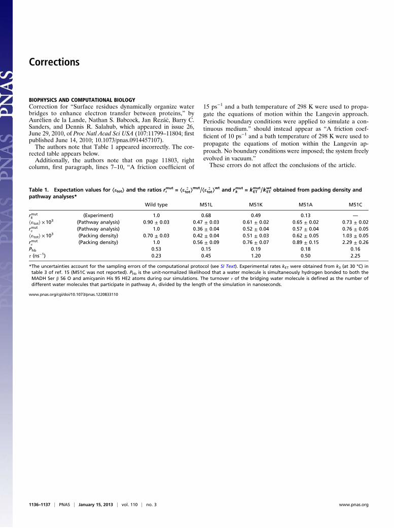
Corrections
BIOPHYSICS AND COMPUTATIONAL BIOLOGYCorrection for “Surface residues dynamically organize waterbridges to enhance electron transfer between proteins,” byAurélien de la Lande, Nathan S. Babcock, Jan �Rezá�c, Barry C.Sanders, and Dennis R. Salahub, which appeared in issue 26,June 29, 2010, of Proc Natl Acad Sci USA (107:11799–11804; firstpublished June 14, 2010; 10.1073/pnas.0914457107).The authors note that Table 1 appeared incorrectly. The cor-
rected table appears below.Additionally, the authors note that on page 11803, right
column, first paragraph, lines 7–10, “A friction coefficient of
15 ps−1 and a bath temperature of 298 K were used to propa-gate the equations of motion within the Langevin approach.Periodic boundary conditions were applied to simulate a con-tinuous medium.” should instead appear as “A friction coef-ficient of 10 ps−1 and a bath temperature of 298 K were used topropagate the equations of motion within the Langevin ap-proach. No boundary conditions were imposed; the system freelyevolved in vacuum.”These errors do not affect the conclusions of the article.
www.pnas.org/cgi/doi/10.1073/pnas.1220833110
Table 1. Expectation values for ⟨etot⟩ and the ratios remut = ⟨e 2
tot⟩mut/⟨e 2
tot⟩wt and rk
mut = kETmut/kET
wt obtained from packing density andpathway analyses*
Wild type M51L M51K M51A M51C
rmutk (Experiment) 1.0 0.68 0.49 0.13 —
⟨«tot⟩ ×103 (Pathway analysis) 0.90 ± 0.03 0.47 ± 0.03 0.61 ± 0.02 0.65 ± 0.02 0.73 ± 0.02rmut« (Pathway analysis) 1.0 0.36 ± 0.04 0.52 ± 0.04 0.57 ± 0.04 0.76 ± 0.05⟨«tot⟩ ×103 (Packing density) 0.70 ± 0.03 0.42 ± 0.04 0.51 ± 0.03 0.62 ± 0.05 1.03 ± 0.05rmut« (Packing density) 1.0 0.56 ± 0.09 0.76 ± 0.07 0.89 ± 0.15 2.29 ± 0.26Phb 0.53 0.15 0.19 0.18 0.16τ (ns−1) 0.23 0.45 1.20 0.50 2.25
*The uncertainties account for the sampling errors of the computational protocol (see SI Text). Experimental rates kET were obtained from k3 (at 30 °C) intable 3 of ref. 15 (M51C was not reported). Phb is the unit-normalized likelihood that a water molecule is simultaneously hydrogen bonded to both theMADH Ser β 56 O and amicyanin His 95 HE2 atoms during our simulations. The turnover τ of the bridging water molecule is defined as the number ofdifferent water molecules that participate in pathway A1 divided by the length of the simulation in nanoseconds.
1136–1137 | PNAS | January 15, 2013 | vol. 110 | no. 3 www.pnas.org
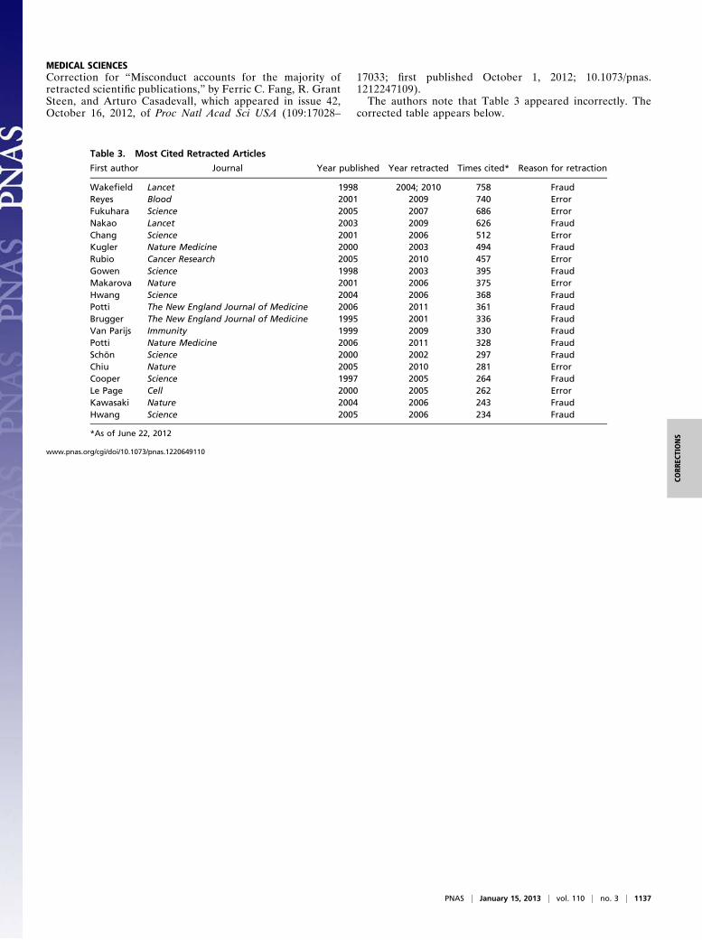
MEDICAL SCIENCESCorrection for “Misconduct accounts for the majority ofretracted scientific publications,” by Ferric C. Fang, R. GrantSteen, and Arturo Casadevall, which appeared in issue 42,October 16, 2012, of Proc Natl Acad Sci USA (109:17028–
17033; first published October 1, 2012; 10.1073/pnas.1212247109).The authors note that Table 3 appeared incorrectly. The
corrected table appears below.
www.pnas.org/cgi/doi/10.1073/pnas.1220649110
Table 3. Most Cited Retracted Articles
First author Journal Year published Year retracted Times cited* Reason for retraction
Wakefield Lancet 1998 2004; 2010 758 FraudReyes Blood 2001 2009 740 ErrorFukuhara Science 2005 2007 686 ErrorNakao Lancet 2003 2009 626 FraudChang Science 2001 2006 512 ErrorKugler Nature Medicine 2000 2003 494 FraudRubio Cancer Research 2005 2010 457 ErrorGowen Science 1998 2003 395 FraudMakarova Nature 2001 2006 375 ErrorHwang Science 2004 2006 368 FraudPotti The New England Journal of Medicine 2006 2011 361 FraudBrugger The New England Journal of Medicine 1995 2001 336 FraudVan Parijs Immunity 1999 2009 330 FraudPotti Nature Medicine 2006 2011 328 FraudSchön Science 2000 2002 297 FraudChiu Nature 2005 2010 281 ErrorCooper Science 1997 2005 264 FraudLe Page Cell 2000 2005 262 ErrorKawasaki Nature 2004 2006 243 FraudHwang Science 2005 2006 234 Fraud
*As of June 22, 2012
PNAS | January 15, 2013 | vol. 110 | no. 3 | 1137
CORR
ECTIONS