Sunrui Chen 陈荪睿著 - RePub, Erasmus University Repository
201
Facing the resurgence of rotavirus: Development of novel antiviral agents Sunrui Chen 陈荪睿 著
-
Upload
khangminh22 -
Category
Documents
-
view
7 -
download
0
Transcript of Sunrui Chen 陈荪睿著 - RePub, Erasmus University Repository

Facing the resurgence of rotavirus:
Development of novel antiviral agents
Sunrui Chen
陈荪睿 著

The studies presented in this thesis were performed at the Laboratory of Gastroenterology and Hepatology, Erasmus MC-University Medical Center Rotterdam, the Netherlands. The research was funded by:
• China Scholarship Council © Copyright by Sunrui Chen. All rights reserved. No part of the thesis may be reproduced or transmitted, in any form, by any means, without express written permission of the author. Cover design: Qun Pan and the author of this thesis. Layout design: The author of this thesis. Printed by: ISBN:

Facing the resurgence of rotavirus:
Development of novel antiviral agents
Het gevecht tegen de wederopstanding van het
rotavirus: ontwikkeling van nieuwe middelen
Thesis
to obtain the degree of Doctor from the
Erasmus University Rotterdam
by command of the
rector magnificus
Prof. dr. R.C.M.E. Engels
and in accordance with the decision of the Doctorate Board
The public defense shall be held on
Tuesday 15th December 2020 at 11:30
by
Sunrui Chen
born in Wuhan, Hubei Province, China

Doctoral Committee:
Promoter:
Prof. dr. M.P. Peppelenbosch
Inner Committee:
Prof. dr. C.C. Baan
Prof. dr. L.J.W. van der Laan
Prof. dr. J.C.H. Hardwick
Copromoter:
Dr. Q. Pan

CONTENTS
Chapter 1 .............................................................................................................................. 1
General Introduction and Outline of This Thesis
Chapter 2 ............................................................................................................................ 15
Rotavirus infection and cytopathogenesis in human biliary organoids potentially recapitulate
biliary atresia development
mBio. 2020, 11(4):e01968.
Chapter 3 ............................................................................................................................ 35
The eukaryotic translation initiation factor 4F complex restricts rotavirus infection via
regulating the expression of IRF1 and IRF7
International journal of molecular sciences, 2019, 20(7): 1580.
Chapter 4 ............................................................................................................................ 59
Basal interferon signaling and therapeutic use of interferons in controlling rotavirus infection
in human intestinal cells and organoids
Scientific Reports, 2018; 8(1): 8341.
Chapter 5 ............................................................................................................................ 93
Suppression of Pyrimidine biosynthesis by targeting DHODH enzyme robustly inhibits
rotavirus replication
Antiviral research, 2019, 167: 35-44.
Chapter 6 .......................................................................................................................... 121
6-thioguanine inhibits rotavirus replication through suppression of Rac1 GDP/GTP cycling
Antiviral research, 2018, 156: 92-101.
Chapter 7 .......................................................................................................................... 149
Drug screening identifies gemcitabine inhibiting rotavirus through alteration of pyrimidine
nucleotide synthesis pathway
Antiviral Research, 2020: 104823.
Chapter 8 .......................................................................................................................... 171

Summary and Discussion
Chapter 9 .......................................................................................................................... 179
Dutch Summary
Appendix ........................................................................................................................... 185
Acknowledgements
Publications
PhD Portfolio
Curriculum Vitae

1 | P a g e
Chapter 1
General Introduction and Outline of
This Thesis


Chapter 1
3 | P a g e
GENERAL INTRODUCTION
Pathogenesis and virology of rotavirus
In this thesis I shall focus on rotavirus. Rotavirus is a member of the so-called Reoviridae family
and was discovered as a novel pathogen by Dr. Ruth Bishop together with colleagues at the
Royal Children’s Hospital (RCH) in 1973(1). The name rotavirus was first coined one year later
by Thomas Henry Flewett and in 1978 rotavirus was officially defined(2). Since, it has become
clear that it constitutes a major health concern.
Indeed data from 2015 show that diarrheal disease is the fourth leading cause of death among
children under 5 years old, responsible for almost 500 000 deaths in that year (3). As one of
the leading causal factors for pediatric diarrhea globally, it is estimated that rotavirus
infection develops in 25% of cases in moderate-to-severe illnesses (4) and associates with
approximately 125,000-200,000 deaths each year(3, 5), contributing to 30% of all death due
to diarrhea for the first two years after birth (6).
Though among young children, diarrhea-associated mortality number has decreased since
1990 by approximately 65% (7), due to limited resources and poor hygiene practices,
situations are still unsatisfactory in many developing countries and especially India, Nigeria,
Pakistan, and the Democratic Republic of the Congo need to be named in this respect as they
together account for half of the global mortally due to pediatric diarrhea (8).
Although primarily a health concern in infants, it is important to realize that rotavirus infection
is not restricted by age. Especially orthotopic organ transplantation patients are effected, the
average incidence of rotavirus infection is 3.03% among them, with intestinal transplant
recipients, liver transplant recipients and hematopoietic stem cell transplant patients being
under the highest risk for suffering from rotavirus infection-caused chronic diarrhea (9).
Improved treatment options are called for.
Rational novel avenues for the treatment of rotavirus may be designed following an
understanding of rotavirus biology. Rotavirus is a non-enveloped virus which has a double-
stranded RNA genome containing 11 segments and is surrounded by multilayered icosahedral
protein capsid. Its genome encodes 12 proteins including 6 viral proteins and 6 non-structural

Chapter 1
4 | P a g e
proteins (10). VP1, VP2 and VP3 proteins make up the rotavirus core particle. VP1 protein is
the RNA-dependent RNA polymerase (RdRP) of rotavirus catalyzing viral RNA synthesis (11).
VP2 protein consists of 120 molecules and VP3 is a guanylyl transferase (12, 13). There is also
a VP4 protein, with a length of 775 or 776 amino acids and this constitutes an 84 KDa spike
protein, part of the outer capsid. VP4 needs to be cleaved into VP5* and VP8* before the virus
is infectious. This process will activate the virus, and it helps the virus to penetrate the target
cell’s interior (14). VP6 forms the bulk of the capsid. It is highly antigenic and constitutes the
intermediate shell of the virions (15). VP7 forms as trimers the icosahedral lattice of the
rotavirus outer capsid, in addition it also plays an important role in the virus’ uncoating
process once it enters host cells (16). Although there is a lot of information on the biology of
the virus, this has still to be translated into improved therapeutic strategies.
Vaccination and treatment against rotavirus infection
Up to 2019, there were four rotavirus vaccines that had been prequalified by the WHO for
global use: Rotarix (GlaxoSmithKline Biologicals SA, Rixensart, Belgium; prequalified in 2009),
RotaTeq (Merck & Co., Inc. , West point, PA, USA; prequalified in 2008), Rotavac (Bharat
Biotech, Hyderabad, India; prequalified in 2018), and ROTASIIL (Serum Institute of India PVT.
LTD., Pune, India; prequalified in 2018) (17). Among these vaccines, Rotarix and RotaTeq were
used in 97 countries in the end of 2018, while Rotavac and ROTASIIL were only used in India
and the Palestine district. Besides these vaccines, which are used on international scale, two
additional rotavirus vaccines have come available for nationwide-restricted use only: Rotavin-
M1 (Center of Research and Production of Vaccines and Biologicals, Hanoi, Vietnam) is
available in Vietnam only and the Lanzhou Lamb Rotavirus (LLR) vaccine (Lanzhou Institute of
Biological Products Co., Ltd., Lanzhou, China), which now serves the private market in China
(17). More widespread use of the latter two vaccines is hampered by the unique
immunological profiles elicited, In past decade, rotavirus vaccines have made a remarkable
impact on the global burden of disease (18). Nevertheless, initial optimism has recently
subsided.
Though rotavirus vaccination programs have generated a huge breakthrough in preventing
young children to contract rotavirus-related diarrhea, for patients who have already been
infected by rotavirus, vaccination is not effective and specific medical treatment

Chapter 1
5 | P a g e
counteracting ongoing rotavirus-mediated disease is still lacking. Indeed, even in developed
countries, treatment is largely confined to supportive care and mainly involves rehydration
and maintenance of fluid and electrolyte balance (19, 20). Even for many developing countries,
the lack of coverage of rotavirus vaccination precludes efficient protection of the population
at large. In addition, in severe cases, which include immunocompromised patients subject to
rotavirus infection, virus-specific treatment is needed. Therefore, further steps should be
taken to reduce the burden of mortality and morbidity and especially more research on the
development of novel antiviral therapies is required and might even be considered urgent (21).
Furthermore, as rotavirus is especially a scourge for resource-limited regions, cost efficacy
should also be taken into account. An obvious solution is to investigate the potential for
effective treatment based on the existing approved reagents, especially broad-spectrum
antiviral agents come to mind in this respect.
Viral-host interaction
Evolutionary pressure and especially the need for efficient production of novel viral particles
as well as the fight with the cell-autonomous innate immune system has shaped viral biology.
Many viral mRNAs have acquired a variety of sophisticated strategies to compete with cellular
mRNAs that are already present in the cytoplasm. This allows the selective translation of viral
mRNAs, since they circumvent the need to possess the required components to initiate the
translation of mRNA (22, 23). In this the eukaryotic translation initiation factor 4F (eIF4F) is
important to consider. eIF4F is a protein complex containing three constituent proteins: a
eukaryotic translation initiation factor 4A (eIF4A), a eukaryotic translation initiation factor 4E
(eIF4E), and a eukaryotic translation initiation factor 4G (eIF4G)(24). This complex plays a
pivotal role in cap-dependent mRNA protein translation initiated by recruiting mRNA to a
ribosome (25). Moreover, it is essential in the regulation of interferon (INF) signaling(26),
which closely relates to anti-viral immunity.
Innate immune responses are at the front line of this battle, playing a critical role against
rotavirus infection and the critical role of INF signaling in this fight is well-established (27).
Cytokine production, a broad term that includes the production of IFNs , are induced following
the recognition of rotavirus viral proteins and/or RNA by the infected host cell (28). IFNs
constitute a group of related humoral factors that exert potent antiviral activities and are

Chapter 1
6 | P a g e
further subclassified into three groups: type I INFs (IFN-α, IFN-β, IFN-δ and others), type II INFs
(IFN-γ) and type Ⅲ INFs (IFN-λ1, IFN-λ2 and IFN-λ3)(29, 30). They bind to distinct cognate but
evolutionary related receptors and all signal through the Janus kinase signal transducer and
activator of transcription (JAK-STAT) pathway (30, 31). Following engagement of IFNs with
their receptors, STAT1 and STAT2 are activated through phosphorylation and subsequently
form complexes of STAT proteins that will translocate to the nucleus and bind to IFN
regulatory factor 9 (IRF9) to form the IFN-stimulated gene factor 3 complex (ISGF3). ISGF3
induces transcription of numerous IFN-stimulated genes (ISGs) which cooperatively exert
antiviral effect against various kinds of viruses (32). Moreover, recognition of rotavirus and
IFN induction are indispensable to promote the development of adaptive B-cell mediated
immune response and thus links innate immunity to adaptive immunity (33). The resulting
evolutionary pressure on rotavirus has fostered the pathogen in developing its own strategies
to evade such host immune responses. For instance, rotavirus can inhibit the production of
IFNs in infected cells by blocking the activation of STAT1 and STAT2 proteins (34). Although
many details are still unclear, one of the viral nonstructural proteins, NSP1, was reported to
mediate this inhibition of IFN signaling in rotavirus infected primary mouse cells (35). Further
understanding of the interaction of rotavirus with the host innate immune machinery may
well be essential for developing novel rational avenues for managing rotavirus-provoked
disease.
Nucleotide biosynthesis and viral infection
Cellular nucleotides, which are classified as either purines or pyrimidines, are the building
blocks of RNA and DNA. Cellular demand for nucleotides is met either through the activity of
the de novo synthesis pathway or through the salvage pathway (36) and also viruses require
these pathways to obtain the nucleotides essential for their replication. Therefore, host
enzymes or other molecules involved in nucleoside biosynthesis pathways represent potential
targets for antiviral strategies.
Pyrimidine biosynthesis is a relatively linear sequential process in which the enzyme
dihydroorotate dehydrogenase (DHODH) represents the fourth and, importantly, rate-limiting
step. The enzyme is located in the inner membrane of mitochondria where it catalyzes the
conversion of dihydroorotate to orotate (37). The thus produced orotate is then a substrate

Chapter 1
7 | P a g e
for uridine monophosphate (UMP) synthase and the UMP resulting from the reaction serves
as an essential precursor for synthesis of all other pyrimidine nucleotides. Several studies have
reported that the inhibition of DHODH enzyme suppresses a range of different viruses’
replication (6, 38-40). Apparently viral replication is more dependent on pyrimidine
biosynthesis relative to host demands for these nucleotides.
Gemcitabine is a cytidine analogue, it has been reported to inhibit pyrimidine biosynthesis
resulting in depletion of nucleotide pool (43) and might thus be useful for combating viral
replication. By inference from the dependency of viral replication on pyrimidine biosynthesis,
also purine biosynthesis might be target for anti-viral therapy. A common strategy for
interfering with purine biosynthesis is the use of poorly-metabolizable analogues of
intermediate products its biosynthetic pathway. 6-thioguanine (6-TG), a thio analogue of the
naturally occurring purine base guanine, has been used clinically since 1950s (41). Metabolized
6-TG can bind to Rac1 to form a 6-TGNP•Rac1 complex that inhibits the activation of Rac1, an
important regulator of cell physiology (42). The usefulness for interfering with nucleotide
biosynthesis for combating rotavirus infection, however, remains largely obscure at best,
prompting studies in this respect.
Organoid models for studying rotavirus infection
Lack of insight into the biology of rotavirus was also due to the absence of appropriate cellular
model systems that truly recapitulated the infection process in vivo. An exciting development
in this respect is advent of organoid technology. In 2009, the group of Prof. dr. Hans Clevers
from Hubrecht institute (Utrecht, Netherlands) established primary intestinal organoids from
intestinal stem cells (ISCs) (44). Subsequently the same research team generated liver
organoids as well. Organoid technology is largely possible through exploiting the properties of
the Lgr5-positive (Lgr5+) stem cells (45, 46). Compared to conventional 2D cultured
immortalized cell lines, organoids model systems are superior in various respects: 1. 2D
cultured immortalized cells are cancer cells, which by definition involves genetic alterations
and thus never truly mimic the situation in healthy cells in vivo; although there are certainly
differences between organoids and real body structures, at least the genetic dimension is
absent in organoid models. 2. As organoids have the capacity to form different types of stem
cell-derived progeny they allow study of the interactions between these different cell types,

Chapter 1
8 | P a g e
at least more so as compared to conventional model systems. 3. Organoids are 3D structures,
hence studying spatial effects is greatly facilitated compared to 2D cultures. 4. Establishing
immortalized cell lines is cumbersome and does not really allow personalized medicine.
Indeed, much of the knowledge on intestinal cell biology generated in the last century
depended on the use of a handful of cell lines. Organoids models perform much better in this
respect and might allow precision medicine, also potentially with respect to rotavirus-
mediated disease. Overall, for rotavirus research the organoid model appears highly
promising and I decided in the course of this thesis research to grab the momentum and
opportunity involved.
Biliary atresia and rotavirus infection
Effective research into rotavirus-mediated disease requires full understanding of the entire
spectrum of clinical manifestations associated with rotavirus infection. In this case biliary
atresia (BA) has been overlooked. BA is a neonatal disease of the liver and bile duct and is
characterized by a progressive fibro-inflammatory obliteration of both the intrahepatic and
extrahepatic bile duct. If untreated, patients have a high risk for developing chronic cholestasis
and biliary cirrhosis. Also, in infants BA is one of the leading causes of end-stage liver disease
requiring liver transplantation (47, 48). The pathogenesis of BA remains unknown, however
the evidence points to multiple pathogenic mechanisms being involved in the development of
BA, including gene mutation (49), exposure to environmental toxins (50), dysregulation of the
immune system, and most importantly, viral factors (51-56).
Various lines of evidence led me to speculate that rotavirus is one of the viral factors causing
biliary atresia. In rodents experimental rotavirus infection provokes aspects of neonate biliary
atresia. Remarkable findings in respect are that both blood-borne viral antigens (antigenemia)
and infectious virus (viremia) have been detected in this mouse model while other data
indicates as well that rotavirus infection is not limited to the intestinal mucosa but may escape
into the circulation, thus potentially reaching bile duct structures (57-61). If rotavirus indeed
causes biliary atresia in children, this will constitute an important extra-intestinal clinical
manifestation of rotavirus-mediated pathology and would have important consequences with
respect to our thinking on how rotavirus infection should be managed and prevented.
Aim of this thesis

Chapter 1
9 | P a g e
Following the introduction of rotavirus vaccines on a global scale the burden provoked by this
virus to mankind has been decreasing. Nevertheless, especially in developing countries, the
rotavirus-associated misery is still depressingly common, in particular with respect to infants.
In addition, persistent rotavirus infection commonly occurs in immunocompromised patients,
irrespective whether this concerns pediatric or adult patients and whether they hail from
developing or developed countries. Finally, the full amount of clinical manifestations caused
by rotavirus infections may be underestimated, for instance rotavirus may be one of the
causative agents of biliary atresia in neonates. Overall, there is clear need for obtaining better
insight into the role of rotavirus in pathophysiology and to define novel ways of dealing with
rotavirus-induced disease, especially for patients in which disease has already manifested
itself and thus vaccination is no longer an option. Excitingly, the advent of the 3D organoid
model also opens new possibilities to perform research into rotavirus, previously impossible.
In conjunction, I considered the case for investigating rotavirus biology imperative.
Outline of this thesis
Although it is well-recognized that rotavirus infection causes thousands of deaths among
children under 5 years old, the full spectrum of rotavirus-mediated disease may still be
underestimated. There is substantial evidence indicating the possible involvement of rotavirus
in BA development, at least in a subset of patients, but in lieu of concrete proof this issue
remains largely ignored both by medical professionals as well as by policy decision makers.
Therefore, in Chapter 2, exploiting the novel possibilities offered by the recently developed
organoid technology, I investigated the role of rotavirus in BA development and I
demonstrated that human biliary organoids are susceptible to rotavirus infection, and this
leads to active virus-host interactions and causes severe cytopathogenesis. Furthermore, I
demonstrated that antiviral drugs and neutralizing antibodies can counteract the infection
and BA-like morphological changes, suggesting their potential for mitigating BA in patients.
Thus, encouraged to further identify novel avenues for treating rotavirus infection, I
investigated the viral mRNA translation process. In Chapter 3, I investigated the function of
eukaryotic translation initiation factor 4F (eIF4F) complex in rotavirus replication. I dissect the
effects of loss-of-function of the three components of eIF4F, including eIF4A, eIF4E and eIF4G,
and establish the resulting consequences on the levels of rotavirus genomic RNA and viral

Chapter 1
10 | P a g e
protein VP4. Consistent with the observations made, I observe that knockdown of the
negative regulator of eIF4F and programmed cell death protein 4 (PDCD4) inhibits the
expression of viral mRNA and the VP4 protein. More importantly, I established that eIF4F
complex can regulate the IFN signaling pathway in the context of rotavirus infection. In
Chapter 4 I further extend these observations. Host immune responses determine the
outcome of viral infections, and IFNs are produced as the first and the main anti-viral cytokines
to combat the virus infection. In this chapter I show that rotavirus predominantly induces type
III IFNs (IFN-λ1), and to a lesser degree type I IFNs (IFN-α and IFN-β) in human intestinal cells
on the RNA level. However, I was not able to detect IFN protein and thus this interferon
response is not sufficient to effectively inhibit rotavirus replication. In contrast, however, I
establish a significant role of constitutive IFN signaling for limiting rotavirus replication, as for
instance evident from experiments involving the silencing of STAT1, STAT2 and IRF9 genes. In
addition, exogenous IFN treatment impaired rotavirus replication irrespective of IFN type used.
Again making this observation was also facilitated by the use of organoid technology. IFNs
significantly upregulated a panel of well-known anti-viral ISGs and inhibition of the JAK-STAT
cascade abrogated ISG induction and the anti-rotavirus effects of IFNs. Thus overall, I was able
to substantially advance our understanding of the interaction between rotavirus infection and
intestinal epithelial innate immunity.
Viral replication heavily relies on the host to supply nucleosides. So, limiting nucleoside
availability might inhibit rotavirus replication. In Chapter 5, I demonstrate that two specific
DHODH enzyme inhibitors, brequinar (BQR) and leflunomide (LFM) robustly inhibit rotavirus
replication in the conventional human intestinal Caco2 cell line model as well as in human
primary intestinal organoids. These antiviral effects are evident both when using the
laboratory strain SA11 as well as when using the rotavirus strain 2011K which was isolated
from clinical sample in my host institution. Mechanistic studies indicated that BQR and LFM
exerted their anti-rotavirus effect through targeting DHODH and by depleting the pyrimidine
nucleotide pool. Therefore, targeting pyrimidine biosynthesis represents a potential approach
for developing antiviral strategies against rotavirus. In Chapter 6, I aimed at testing on the
other side of the nucleoside coin, in casu purine biosynthesis, by testing another
immunosuppressive drug 6-TG, a purine biosynthesis inhibitor. I observed, however, that 6-
TG although significantly inhibits rotavirus replication, gene knockdown or knockout of Rac1,
Chapter 1
11 | P a g e
an alternative cellular target of 6-TG, also significantly inhibited rotavirus replication,
indicating a previously not-suspected accessory role for Rac1 in rotavirus infection. I further
demonstrated that 6-Thioguanine (6-TG) can effectively inhibit the active form of Rac1 (GTP-
Rac1) and essentially mediates the anti-rotavirus effect of 6-TG. Consistently, ectopic over-
expression of GTP-Rac1 facilitates but an inactive Rac1 (N17) or a specific Rac1 inhibitor
(NSC23766) inhibits rotavirus replication. In conclusion, I identified 6-TG as an effective
inhibitor of rotavirus replication but that unexpectedly acts through inhibition of Rac1
activation. In Chapter 7, I screened a library of safe-in-man broad-spectrum antivirals and
identified gemcitabine, a widely used anticancer drug, as a potent inhibitor of rotavirus
infection. I confirmed this effect in 2D cell cultures and 3D cultured human intestinal organoids
with both laboratory-adapted rotavirus strains and five clinical isolates. Supplementation of
UTP or uridine largely abolished the anti-rotavirus activity of gemcitabine, suggesting its
function through inhibition of pyrimidine biosynthesis pathway.
Overall, I feel I have been able to significantly advance our understanding of rotavirus disease
and it potential management and I hope these studies will allow better treatment of disease.

Chapter 1
12 | P a g e
References
1. Bishop RJJog, hepatology. 2009. Discovery of rotavirus: Implications for child health. 24:S81-S85.
2. Flewett T, Bryden A, Davies H, Woode G, Bridger J, Derrick JJTL. 1974. Relation between viruses from acute gastroenteritis of children and newborn calves. 304:61-63.
3. Troeger C, Forouzanfar M, Rao PC, Khalil I, Brown A, Reiner Jr RC, Fullman N, Thompson RL, Abajobir A, Ahmed MJTLID. 2017. Estimates of global, regional, and national morbidity, mortality, and aetiologies of diarrhoeal diseases: a systematic analysis for the Global Burden of Disease Study 2015. 17:909-948.
4. Kotloff KL, Nataro JP, Blackwelder WC, Nasrin D, Farag TH, Panchalingam S, Wu Y, Sow SO, Sur D, Breiman RFJTL. 2013. Burden and aetiology of diarrhoeal disease in infants and young children in developing countries (the Global Enteric Multicenter Study, GEMS): a prospective, case-control study. 382:209-222.
5. Troeger C, Khalil IA, Rao PC, Cao S, Blacker BF, Ahmed T, Armah G, Bines JE, Brewer TG, Colombara DVJJp. 2018. Rotavirus vaccination and the global burden of rotavirus diarrhea among children younger than 5 years. 172:958-965.
6. Wang Y, Wang W, Xu L, Zhou X, Shokrollahi E, Felczak K, Van Der Laan LJ, Pankiewicz KW, Sprengers D, Raat NJJAa, chemotherapy. 2016. Cross talk between nucleotide synthesis pathways with cellular immunity in constraining hepatitis E virus replication. 60:2834-2848.
7. Wang H, Naghavi M, Allen C, Barber RM, Bhutta ZA, Carter A, Casey DC, Charlson FJ, Chen AZ, Coates MMJTl. 2016. Global, regional, and national life expectancy, all-cause mortality, and cause-specific mortality for 249 causes of death, 1980–2015: a systematic analysis for the Global Burden of Disease Study 2015. 388:1459-1544.
8. Tate JE, Burton AH, Boschi-Pinto C, Parashar UJCID. 2016. World Health Organization Coordinated Global Rotavirus Surveillance Network. Global, regional, and national estimates of rotavirus mortality in children< 5 years of age, 2000–2013. 62:S96-S105.
9. Yin Y, Metselaar HJ, Sprengers D, Peppelenbosch MP, Pan QJAJoT. 2015. Rotavirus in organ
transplantation: drug‐virus‐host interactions. 15:585-593.
10. Estes MK, Cohen JJM, Reviews MB. 1989. Rotavirus gene structure and function. 53:410-449. 11. Lu X, McDonald SM, Tortorici MA, Tao YJ, Vasquez-Del Carpio R, Nibert ML, Patton JT, Harrison
SCJS. 2008. Mechanism for coordinated RNA packaging and genome replication by rotavirus polymerase VP1. 16:1678-1688.
12. Zeng CQ-Y, Labbé M, Cohen J, Prasad BV, Chen D, Ramig RF, Estes MKJV. 1994. Characterization of rotavirus VP2 particles. 201:55-65.
13. Vásquez M, Sandino AM, Pizarro JM, Fernández J, Valenzuela S, Spencer EJJogv. 1993. Function of rotavirus VP3 polypeptide in viral morphogenesis. 74:937-941.
14. Ludert JE, Krishnaney AA, Burns JW, Vo PT, Greenberg HBJJogv. 1996. Cleavage of rotavirus VP4 in vivo. 77:391-395.
15. Beards G, Campbell A, Cottrell N, Peiris J, Rees N, Sanders R, Shirley J, Wood H, Flewett TJJocm. 1984. Enzyme-linked immunosorbent assays based on polyclonal and monoclonal antibodies for rotavirus detection. 19:248-254.
16. Dormitzer PR, Greenberg HB, Harrison SCJV. 2000. Purified recombinant rotavirus VP7 forms soluble, calcium-dependent trimers. 277:420-428.
17. Burke RM, Tate JE, Kirkwood CD, Steele AD, Parashar UDJCoiid. 2019. Current and new rotavirus vaccines. 32:435.
18. Burnett E, Parashar UD, Tate JEJTJoID. 2020. Global impact of rotavirus vaccination on diarrhea hospitalizations and deaths among children< 5 years old: 2006–2019.
19. Yin Y, Bijvelds M, Dang W, Xu L, van der Eijk AA, Knipping K, Tuysuz N, Dekkers JF, Wang Y, de Jonge JJAr. 2015. Modeling rotavirus infection and antiviral therapy using primary intestinal organoids. 123:120-131.

Chapter 1
13 | P a g e
20. Rossignol J-F, Abu-Zekry M, Hussein A, Santoro MGJTL. 2006. Effect of nitazoxanide for treatment of severe rotavirus diarrhoea: randomised double-blind placebo-controlled trial. 368:124-129.
21. Chen S, Ding S, Yin Y, Xu L, Li P, Peppelenbosch MP, Pan Q, Wang WJAr. 2019. Suppression of pyrimidine biosynthesis by targeting DHODH enzyme robustly inhibits rotavirus replication. 167:35-44.
22. Chaudhry Y, Nayak A, Bordeleau M-E, Tanaka J, Pelletier J, Belsham GJ, Roberts LO, Goodfellow IGJJoBC. 2006. Caliciviruses differ in their functional requirements for eIF4F components. 281:25315-25325.
23. Burgui I, Yángüez E, Sonenberg N, Nieto AJJov. 2007. Influenza virus mRNA translation revisited: is the eIF4E cap-binding factor required for viral mRNA translation? 81:12427-12438.
24. Montero H, García-Román R, Mora SIJV. 2015. eIF4E as a control target for viruses. 7:739-750. 25. Gingras A-C, Raught B, Sonenberg NJArob. 1999. eIF4 initiation factors: effectors of mRNA
recruitment to ribosomes and regulators of translation. 68:913-963. 26. Colina R, Costa-Mattioli M, Dowling RJ, Jaramillo M, Tai L-H, Breitbach CJ, Martineau Y, Larsson
O, Rong L, Svitkin YVJN. 2008. Translational control of the innate immune response through IRF-7. 452:323-328.
27. Holloway G, Coulson BSJJoGV. 2013. Innate cellular responses to rotavirus infection. 94:1151-1160.
28. Deal EM, Jaimes MC, Crawford SE, Estes MK, Greenberg HBJPP. 2010. Rotavirus structural proteins and dsRNA are required for the human primary plasmacytoid dendritic cell IFNα response. 6:e1000931.
29. Randall RE, Goodbourn SJJogv. 2008. Interferons and viruses: an interplay between induction, signalling, antiviral responses and virus countermeasures. 89:1-47.
30. Donnelly RP, Kotenko SVJJoI, Research C. 2010. Interferon-lambda: a new addition to an old family. 30:555-564.
31. Platanias LCJNRI. 2005. Mechanisms of type-I-and type-II-interferon-mediated signalling. 5:375-386.
32. Schneider WM, Chevillotte MD, Rice CMJAroi. 2014. Interferon-stimulated genes: a complex web of host defenses. 32:513-545.
33. Deal EM, Lahl K, Narváez CF, Butcher EC, Greenberg HBJTJoci. 2013. Plasmacytoid dendritic cells promote rotavirus-induced human and murine B cell responses. 123:2464-2474.
34. Arnold MM, Sen A, Greenberg HB, Patton JTJPP. 2013. The battle between rotavirus and its host for control of the interferon signaling pathway. 9:e1003064.
35. Feng N, Sen A, Nguyen H, Vo P, Hoshino Y, Deal E, Greenberg HJJov. 2009. Variation in antagonism of the interferon response to rotavirus NSP1 results in differential infectivity in mouse embryonic fibroblasts. 83:6987-6994.
36. Evans DR, Guy HIJJoBC. 2004. Mammalian pyrimidine biosynthesis: fresh insights into an ancient pathway. 279:33035-33038.
37. Munier-Lehmann Hln, Vidalain P-O, Tangy Fdr, Janin YLJJomc. 2013. On dihydroorotate dehydrogenases and their inhibitors and uses. 56:3148-3167.
38. Hoffmann H-H, Kunz A, Simon VA, Palese P, Shaw MLJPotNAoS. 2011. Broad-spectrum antiviral that interferes with de novo pyrimidine biosynthesis. 108:5777-5782.
39. Luthra P, Naidoo J, Pietzsch CA, De S, Khadka S, Anantpadma M, Williams CG, Edwards MR, Davey RA, Bukreyev AJAr. 2018. Inhibiting pyrimidine biosynthesis impairs Ebola virus replication through depletion of nucleoside pools and activation of innate immune responses. 158:288-302.
40. Tan YH, Driscoll JS, Mui SM. 2005. Dihydroorotate dehydrogenase inhibitors for the treatment of viral-mediated diseases. Google Patents.
41. Munshi PN, Lubin M, Bertino JRJTo. 2014. 6-thioguanine: a drug with unrealized potential for cancer therapy. 19:760.

Chapter 1
14 | P a g e
42. Shin J-Y, Wey M, Umutesi HG, Sun X, Simecka J, Heo JJJoBC. 2016. Thiopurine prodrugs mediate immunosuppressive effects by interfering with Rac1 protein function. 291:13699-13714.
43. Lee K, Kim D-E, Jang K-S, Kim S-J, Cho S, Kim CJO. 2017. Gemcitabine, a broad-spectrum antiviral drug, suppresses enterovirus infections through innate immunity induced by the inhibition of pyrimidine biosynthesis and nucleotide depletion. 8:115315.
44. Sato T, Vries RG, Snippert HJ, Van De Wetering M, Barker N, Stange DE, Van Es JH, Abo A, Kujala P, Peters PJJN. 2009. Single Lgr5 stem cells build crypt-villus structures in vitro without a mesenchymal niche. 459:262-265.
45. Huch M, Gehart H, Van Boxtel R, Hamer K, Blokzijl F, Verstegen MM, Ellis E, Van Wenum M, Fuchs SA, de Ligt JJC. 2015. Long-term culture of genome-stable bipotent stem cells from adult human liver. 160:299-312.
46. Huch M, Dorrell C, Boj SF, Van Es JH, Li VS, Van De Wetering M, Sato T, Hamer K, Sasaki N, Finegold MJJN. 2013. In vitro expansion of single Lgr5+ liver stem cells induced by Wnt-driven regeneration. 494:247-250.
47. Balistreri WF, Grand R, Hoofnagle JH, Suchy FJ, Ryckman FC, Perlmutter DH, Sokol RJJH. 1996. Biliary atresia: current concepts and research directions. Summary of a symposium. 23:1682-1692.
48. Hartley JL, Davenport M, Kelly DAJTL. 2009. Biliary atresia. 374:1704-1713. 49. Cheng G, Tang CS-M, Wong EH-M, Cheng WW-C, So M-T, Miao X, Zhang R, Cui L, Liu X, Ngan ES-
WJJoh. 2013. Common genetic variants regulating ADD3 gene expression alter biliary atresia risk. 59:1285-1291.
50. Walesky C, Goessling WJH. 2016. Nature and nurture: Environmental toxins and biliary atresia. 64:717-719.
51. Mahjoub F, Shahsiah R, Ardalan FA, Iravanloo G, Sani MN, Zarei A, Monajemzadeh M, Farahmand F, Mamishi SJDp. 2008. Detection of Epstein Barr Virus by Chromogenic In Situ Hybridization in cases of extra-hepatic biliary atresia. 3:1-4.
52. Riepenhoff-Talty M, Gouvea V, Evans M, Svensson L, Hoffenberg E, Sokol R, Uhnoo I, Greenberg S, Schäkel K, Zhaori GJJoID. 1996. Detection of group C rotavirus in infants with extrahepatic biliary atresia. 174:8-15.
53. Morecki R, Glaser JH, Cho S, Balistreri WF, Horwitz MSJNEJoM. 1982. Biliary atresia and reovirus type 3 infection. 307:481-484.
54. Fischler B, Ehrnst A, Forsgren M, Ö rvell C, Nemeth AJJopg, nutrition. 1998. The viral association of neonatal cholestasis in Sweden: a possible link between cytomegalovirus infection and extrahepatic biliary atresia. 27:57-64.
55. Fischler B, Woxenius S, Nemeth A, Papadogiannakis NJJops. 2005. Immunoglobulin deposits in liver tissue from infants with biliary atresia and the correlation to cytomegalovirus infection. 40:541-546.
56. Greenberg HB, Estes MKJG. 2009. Rotaviruses: from pathogenesis to vaccination. 136:1939-1951.
57. Blutt SE, Kirkwood CD, Parreño V, Warfield KL, Ciarlet M, Estes MK, Bok K, Bishop RF, Conner MEJTl. 2003. Rotavirus antigenaemia and viraemia: a common event? 362:1445-1449.
58. Blutt SE, Conner MEJCoig. 2007. Rotavirus: to the gut and beyond! 23:39-43. 59. Blutt SE, Matson DO, Crawford SE, Staat MA, Azimi P, Bennett BL, Piedra PA, Conner MEJPM.
2007. Rotavirus antigenemia in children is associated with viremia. 4:e121. 60. Patel M, Rench MA, Boom JA, Tate JE, Sahni LC, Hull JA, Gentsch JR, Parashar UD, Baker CJJTPidj.
2010. Detection of rotavirus antigenemia in routinely obtained serum specimens to augment surveillance and vaccine effectiveness evaluations. 29:836-839.
61. Ramani S, Paul A, Saravanabavan A, Menon VK, Arumugam R, Sowmyanarayanan TV, Samuel P, Gagandeep KJCid. 2010. Rotavirus antigenemia in Indian children with rotavirus gastroenteritis and asymptomatic infections. 51:1284-1289.

15 | P a g e
Chapter 2
Rotavirus infection and cytopathogenesis in human biliary
organoids potentially recapitulate biliary atresia
development
Sunrui Chen, Pengfei Li, Yining Wang, Yuebang Yin, Petra E. de Ruiter, Monique M. A.
Verstegen, Maikel P. Peppelenbosch, Luc J. W. van der Laan, Qiuwei Pan
mBio. 2020; 11: e01968-20.


Chapter 2
17 | P a g e
Abstract
Biliary atresia (BA) is a neonatal liver disease characterized by progressive fibro-inflammatory
obliteration of both intrahepatic and extrahepatic bile duct. The etiologies of BA remain
largely unknown, but rotavirus infection has been implicated at least for a subset of patients
and this causal relation has been well-demonstrated in mouse models. In this study, we aim
to further consolidate this evidence in human biliary organoids. We obtained seven batches
of human biliary organoids cultured from fetal liver, adult liver and bile duct tissues. We found
that these organoids are highly susceptible and support the full life cycle of rotavirus infection
in 3D culture. The robust infection triggers active virus-host interactions, including interferon-
based host defense mechanisms and injury response. We have observed direct
cytopathogenesis in organoids upon rotavirus infection, which may partially recapitulate the
development of BA. Importantly, we have demonstrated the efficacy of mycophenolic acid
and interferon-alpha but not ribavirin in inhibiting rotavirus in biliary organoids. Furthermore,
a neutralizing antibody targeting rotavirus VP7 protein effectively inhibits the infection in
organoids. Thus, we have substantiated the causal evidence of rotavirus inducing BA in
humans and provided potential strategies to combat the disease.
Importance
There are substantial evidence indicating the possible involvement of rotavirus in biliary
atresia (BA) development at least in a subset of patients, but concrete proof remains lacking.
In mouse model, it has been well-demonstrated that rotavirus can infect the biliary epithelium
to cause biliary inflammation and obstruction, representing the pathogenesis of BA in
humans. By using the recently developed organoids technology, we now have demonstrated
that human biliary organoids are susceptible to rotavirus infection, and this provokes active
virus-host interactions and causes severe cytopathogenesis. Thus, our model recapitulates
some essential aspects of BA development. Furthermore, we have demonstrated that antiviral
drugs and neutralizing antibodies are capable of counteracting the infection and BA-like
morphological changes, suggesting their potential for mitigating BA in patients.
Keywords

Chapter 2
18 | P a g e
Biliary atresia, Rotavirus infection, Human organoids
Observation
Biliary atresia (BA) is characterized by progressive fibro-inflammatory obliteration of the bile
ducts, resulting in chronic cholestasis and biliary cirrhosis. It is one of the leading causes for
liver transplantation in infants (1, 2). Exposure to rotavirus in mice has demonstrated the
infection in biliary epithelium, resulting in BA-like biliary inflammation and obstruction (3).
Nevertheless, whether rotavirus is a causal agent for BA in patients remains controversial, also
because of a paucity of preclinical models. Organoid technology provides an excellent way
forward here. These 3D cultured organoids are superior in recapitulating the architecture,
composition, diversity, organization and functionality of cell types of the tissue/organ of
origin. Human organoids have been increasingly explored to advance the research in disease
modeling (4, 5). Although it is feasible to culture hepatocyte-like organoids from liver tissue,
it remains technically challenging with stringent requirement of experimental protocols (6). In
contrast, organoids resembling cholangiocyte-phenotype are relatively easy to be cultured
from the hepatic and extrahepatic bile duct compartments (7-9). In this study, we explored
the feasibility of employing human biliary organoids cultured from fetal liver, adult liver and
bile duct for recapitulating BA development.
The canonical compartment for rotavirus infection is the small intestinal enterocyte. We have
previously shown that human intestinal organoids (HIOs) sustain rotavirus infection (4) and
we now again confirmed these results (see Fig. S1A-C in the supplemental material). BA is a
disorder that typically first manifests itself during mid-gestation and murine experimentation
has demonstrated rotavirus-induced BA development. Hence, we first tested if biliary fetal
liver organoids (FLOs) support rotavirus infection. Inoculation of FLOs with rotavirus resulted
in an increase of cellular viral RNA by a factor of 103-105 at 24 hours and 104-106 fold at 48
hours post-inoculation (Fig. 1A) with a concomitant increase in levels of rotavirus VP4 protein
(Fig. 1B). Thus the human fetal biliary epithelium is highly permissive for rotavirus infection,
comparable to the level in intestinal epithelium (Fig. S1A). In apparent agreement,
supernatant harvested from infected FLOs effectively infected and replicated in Caco2
intestinal epithelial model as shown by qRT-PCR quantification of viral RNA (Fig 1C). Next, we
performed TCID50 assay to compare the level of infectious viral particles between the baseline
Chapter 2
19 | P a g e
of inoculation and five batches of organoids at 48 hours post-inoculation. We harvested
rotavirus from the organoids through repeated freezing and thawing and demonstrated 102-
103 times increase of infectious virus titers (Fig. 1D). This was further confirmed by cytopathic
effects in Caco2 cells at 48 hours post-inoculation with rotavirus harvested from these five
batches of organoids and the control (see Fig. S2 in the supplemental material). Collectively,
these results convincingly showed effective replication and production of infectious viral
particles by infected fetal biliary organoids. Similar results were obtained in biliary organoids
derived from adult human liver and bile duct (Fig. 1A-C). Thus, the human biliary epithelium
is highly susceptible to rotavirus infection and supports its full life cycle.
To better understand the consequences of rotavirus infection in biliary epithelium, we
performed a genome-wide transcriptomic analysis of FLOs upon infection. Volcano plots of
the results showed significant down-regulation of 103 and up-regulation of 512 genes in
response to rotavirus, compared to uninfected organoids (Fig. 1E). Most of the highly up-
regulated genes, including IFIT2, IFITM3, OASL, DDX58, MX2, IFI35, HERC5 and BST2, are
interferon-stimulated genes (ISGs). Other genes, such as CXCL11 and NLRC5 are related to
inflammatory response. Gene ontology (GO) enrichment analysis of these differentially
expressed genes confirmed the essential involvement of “immune system process” (Fig. 1F).
Interestingly, “response to stress”, “cell death” and “extracellular space” were also identified
as the top regulated processes, with obvious relations to the development and pathogenesis
of BA (Fig. 1E).

Chapter 2
20 | P a g e
Figure 1. Characterizing rotavirus infection in human biliary organoids. (A) The dynamics of cellular viral
RNA levels upon inoculation of SA11 rotavirus at different time points of post-inoculation. The level at post-
inoculation 1 hour (hr) was set as 1. Three batches of human fetal liver organoids (FLOs), two adult liver
(LiOs) and two bile duct organoids (BDOs) were tested. (B) Expression of rotavirus VP4 protein in the
organoids determined by Western blotting. (C) Inoculation of human intestinal Caco2 cell line with
supernatant from rotavirus infected organoids for 48 hrs. Relative cellular viral RNA levels were quantified.
(D) TCID50 of five batches of rotavirus infected organoids at 48 hrs post-inoculation compared with the basal
level at incubation. Organoids after inoculation were thoroughly washed to remove free viruses and

Chapter 2
21 | P a g e
subjected to repeated freezing and thawing to harvest the attached and entered rotaviruses. The total
amount of rotaviruses in organoids incubated for 48 hrs were harvested by repeated freezing and thawing
of entire well. (E) Volcano plots of differentially expressed genes in rotavirus infected (for 48 hrs) compared
to un-infected fetal liver organoids. (F) Gene ontology (GO) enrichment analysis of differentially expressed
genes. All the data represent as means ± SEM. For each organoids batch, experiments were repeated 3-6
times. Mann-Whitney test; *P < 0.05, **P < 0.01.
This is in line with the observations that naïve organoids grow and become hyaline in a
spheroidal shape, whereas rotavirus-infected organoids are opaque, shriveled and
disorganized (Fig. 2A; upper panel). Propidium iodide (PI) staining marked the wide-spread of
dead cells in infected organoids (Fig. 2A; middle panel). Confocal analysis after
immunostaining of viral VP6 protein further visualized the disruption of infected organoid cells
(Fig. 2A; lower panel). Quantitative analysis demonstrated significant increase of the
percentage of deteriorated biliary organoids at 12-, 24- and 48-hours post-infection of
rotavirus (Fig. 2B). Thus, rotavirus infection causes severe cytopathogenesis in human biliary
organoids.
Next, we evaluated a monoclonal neutralizing antibody targeting rotavirus VP7 protein (10)
using three representative batches of biliary organoids. It effectively inhibited rotavirus
infection in a dose-dependent manner (Fig. 2C). Finally, the effects of the known broad
antiviral drugs were tested in all bathes of organoids. Similar as in HIOs (see Fig. S1D in the
supplemental material), mycophenolic acid (MPA) and interferon-alpha (IFN-α) potently
inhibited rotavirus in all batches of biliary organoids (Fig. 2D). Surprisingly, ribavirin is effective
in intestinal (Fig. S1D) but not in biliary organoids (Fig. 2D). Therefore, antiviral drugs and
neutralizing antibodies are potential therapeutics to combat rotavirus infection in the human
biliary epithelium compartment.

Chapter 2
22 | P a g e
Figure 2. Cytopathogenesis of rotavirus infected human biliary organoids, and efficacy of antiviral
treatment/neutralizing antibody. (A) Organoids from 50 µm -150 µm diameter were selected to capture
images. Optical microscopy images of infected and un-infected organoids (upper). Fluorescence staining of
dead cells (PI; red), live cells (Calcein; green) and nuclei (Hoechst; blue) (middle). Confocal immunostaining
of rotavirus structural protein VP6 (red), Epcam (green) and nuclei (blue) (lower). These are representative
images of one FLO batch from the tested seven biliary organoids batches. (B) Quantitative analysis of the
percentage of deteriorated organoids with or without rotavirus infection at indicated time points and
calculated based on the live/dead cell staining (A, middle). (C) The inhibitory activities of neutralizing
monoclonal antibody HS-1 against rotavirus infection in three representative batches of biliary organoids.
(D) The effects of the broad-spectrum antiviral drugs on rotavirus in biliary organoids. Ribavirin: Rib;
mycophenolic acid: MPA; interferon alpha: IFN-α. All the data represent as means ± SEM. For each
organoids batch, experiments were repeated 3-6 times. Mann-Whitney test; *P < 0.05, **P < 0.01.

Chapter 2
23 | P a g e
Discussion and conclusions
Although the etiologies and pathogenesis of BA remain largely unknown, multiple pathogenic
mechanisms are likely involved, including genetic mutations (11), exposure of environmental
toxins (12), dysregulation of immune system, and most intriguingly, viral factors in particular
rotavirus (3, 13-18). Previous studies have attempted to detect rotavirus in liver or biliary
tissues and the antibody in serum of BA patients, but results are inconclusive (19). Since the
wide implementation of vaccines that have substantially counteracted rotavirus-mediated
diarrheal disease, more direct investigation on causality of rotavirus infection for BA has
become possible. A survey of the national registry system in Taiwan found decreased
incidence of BA from 2004 to 2009 mirroring the increased uptake of rotavirus vaccination
(20). A nationwide population-based study in Korea has shown that rotavirus infection in
neonates is a risk factor for BA, although vaccination did not impact disease incidence (21).
Unfortunately, detection of rotavirus in tissue is often not feasible, as advanced disease is
usually diagnosed in children of 4-6 weeks old and the virus likely has been cleared by that
time. Here we show, however, using organoid technology that the human biliary epithelium
supports the full life cycle of rotavirus infection and results in cellular and morphological
changes consistent with BA development, even in the absence of immune cell components in
our model. Furthermore, we identify therapeutic strategies potentially useful for combating
rotavirus infection in the biliary epithelium.
Interestingly, a study in mice has demonstrated that maternal vaccination can prevent
rotavirus-induced BA in newborn pups (22). This is in line with our findings that neutralizing
antibodies inhibit rotavirus infection in organoids. Thus, we have substantiated the causal
evidence of rotavirus inducing BA in humans and provided potential strategies to combat the
disease.
Materials and methods
Human fetal liver organoids (FLOs, n=3 batches) were initiated from 17-week-old human fetal
livers collected at abortion, from adult liver (LiOs, n=2 batches), and from adult bile duct

Chapter 2
24 | P a g e
(BDOs, n=2 batches). Human intestinal organoids (HIOs, n=1 batch) were cultured to serve as
standard model for rotavirus infection. Detailed methods are described in the Text S1 of
supplemental materials and methods.
Data availability. Details about data availability can be found in Text S1 in the supplemental
material.

Chapter 2
25 | P a g e
References
1. Balistreri WF, Grand R, Hoofnagle JH, Suchy FJ, Ryckman FC, Perlmutter DH, Sokol RJ. 1996.
Biliary atresia: current concepts and research directions. Summary of a symposium. Hepatology 23:1682-1692.
2. Hartley JL, Davenport M, Kelly DA. 2009. Biliary atresia. The Lancet 374:1704-1713. 3. Riepenhoff-Talty M, Gouvea V, Evans MJ, Svensson L, Hoffenberg E, Sokol RJ, Uhnoo I,
Greenberg SJ, Schäkel K, Zhaori G. 1996. Detection of group C rotavirus in infants with extrahepatic biliary atresia. Journal of Infectious Diseases 174:8-15.
4. Yin Y, Bijvelds M, Dang W, Xu L, van der Eijk AA, Knipping K, Tuysuz N, Dekkers JF, Wang Y, de Jonge J. 2015. Modeling rotavirus infection and antiviral therapy using primary intestinal organoids. Antiviral research 123:120-131.
5. Clevers H. 2016. Modeling development and disease with organoids. Cell 165:1586-1597. 6. Hu H, Gehart H, Artegiani B, LÖ pez-Iglesias C, Dekkers F, Basak O, van Es J, de Sousa Lopes
SMC, Begthel H, Korving J. 2018. Long-term expansion of functional mouse and human hepatocytes as 3D organoids. Cell 175:1591-1606. e1519.
7. Broutier L, Andersson-Rolf A, Hindley CJ, Boj SF, Clevers H, Koo B-K, Huch M. 2016. Culture and establishment of self-renewing human and mouse adult liver and pancreas 3D organoids and their genetic manipulation. Nature protocols 11:1724.
8. Shiota J, Zaki NHM, Merchant JL, Samuelson LC, Razumilava N. 2019. Generation of Organoids from Mouse Extrahepatic Bile Ducts. JoVE (Journal of Visualized Experiments):e59544.
9. Cao W, Chen K, Bolkestein M, Yin Y, Verstegen MMA, Bijvelds MJC, Wang W, Tuysuz N, ten Berge D, Sprengers D. 2017. Dynamics of proliferative and quiescent stem cells in liver homeostasis and injury. Gastroenterology 153:1133-1147.
10. Ruggeri FM, Greenberg HB. 1991. Antibodies to the trypsin cleavage peptide VP8 neutralize rotavirus by inhibiting binding of virions to target cells in culture. Journal of virology 65:2211-2219.
11. Cheng G, Tang CS-M, Wong EH-M, Cheng WW-C, So M-T, Miao X, Zhang R, Cui L, Liu X, Ngan ES-W. 2013. Common genetic variants regulating ADD3 gene expression alter biliary atresia risk. Journal of hepatology 59:1285-1291.
12. Walesky C, Goessling W. 2016. Nature and nurture: Environmental toxins and biliary atresia. Hepatology 64:717-719.
13. Mahjoub F, Shahsiah R, Ardalan FA, Iravanloo G, Sani MN, Zarei A, Monajemzadeh M, Farahmand F, Mamishi S. 2008. Detection of Epstein Barr Virus by Chromogenic In Situ Hybridization in cases of extra-hepatic biliary atresia. Diagnostic pathology 3:19.
14. Morecki R, Glaser JH, Cho S, Balistreri WF, Horwitz MS. 1982. Biliary atresia and reovirus type 3 infection. New England Journal of Medicine 307:481-484.
15. Tyler KL, Sokol RJ, Oberhaus SM, Le M, Karrer FM, Narkewicz MR, Tyson RW, Murphy JR, Low R, Brown WR. 1998. Detection of reovirus RNA in hepatobiliary tissues from patients with extrahepatic biliary atresia and choledochal cysts. Hepatology 27:1475-1482.
16. Fischler B, Ehrnst A, Forsgren M, Ö rvell C, Nemeth A. 1998. The viral association of neonatal cholestasis in Sweden: a possible link between cytomegalovirus infection and extrahepatic biliary atresia. Journal of pediatric gastroenterology and nutrition 27:57-64.
17. Fischler B, Woxenius S, Nemeth A, Papadogiannakis N. 2005. Immunoglobulin deposits in liver tissue from infants with biliary atresia and the correlation to cytomegalovirus infection. Journal of pediatric surgery 40:541-546.
18. Brindley SM, Lanham AM, Karrer FM, Tucker RM, Fontenot AP, Mack CL. 2012. Cytomegalovirus‐specific T‐cell reactivity in biliary atresia at the time of diagnosis is associated with deficits in regulatory T cells. Hepatology 55:1130-1138.
19. Hertel PM, Estes MK. 2012. Rotavirus and biliary atresia: can causation be proven? Current opinion in gastroenterology 28:10-17.

Chapter 2
26 | P a g e
20. Lin Y-C, Chang M-H, Liao S-F, Wu J-F, Ni Y-H, Tiao M-M, Lai M-W, Lee H-C, Lin C-C, Wu T-C. 2011. Decreasing rate of biliary atresia in Taiwan: a survey, 2004–2009. Pediatrics 128:e530-e536.
21. Lee JH, Ahn HS, Han S, Swan HS, Lee Y, Kim HJ. 2019. Nationwide population‐based study showed that the rotavirus vaccination had no impact on the incidence of biliary atresia in Korea. Acta Paediatrica.
22. Bondoc AJ, Jafri MA, Donnelly B, Mohanty SK, McNeal MM, Ward RL, Tiao GM. 2009. Prevention of the murine model of biliary atresia after live rotavirus vaccination of dams. Journal of pediatric surgery 44:1479-1490.

Chapter 2
27 | P a g e
Text S1 – Supplemental Materials and Methods
Reagents
Propidium iodide (PI) (Method Detection Limit [MDL] no. MFCD00011921), calcein-AM (MDL
no. MFCD05861516), ribavirin (MDL no. MFCD00058564) and mycophenolic acid (MPA)
(MDL no. MFCD00036814) were purchased from Sigma. All the reagents above were
dissolved in dimethyl sulfoxide (DMSO). Hoechst 33342 (Catalog number: H3570) and Type I
human recombinant IFN alpha 2a (IFN-α) were purchased from Thermo Fisher, and IFN-α
was dissolved in culture medium.
Viruses
Simian rotavirus SA11, a widely used laboratory strain(1), was gifted by Karen Knipping from
Nutricia Research Utrecht, The Netherland. Rotavirus SA11 was prepared as previously
described(2).
Cell lines and human organoids
Human colon cancer cell line Caco2 was cultured as previous study(3). Cells were analyzed
by genotyping and confirmed to be mycoplasma negative.
Human primary small intestinal organoids (HIOs) were cultured as described previously(4).
three batches of human fetal liver organoids (FLO P, FLO 521 and FLO 528), two adult bile
duct organoids (BDO S, DD 1125) and two adult liver organoids (LiO K, DL 1125) were
cultured as previously described(5). The use of human organoids was approved by the
Medisch Ethische Toetsings Commissie Erasmus MC (Medical Ethical Committee of the
Erasmus medical center).
Virus inoculation assay
Virus, cell line and organoids were treated as previously described (4). Briefly, the stock of
SA11 rotavirus (4.5*108 TCID50/ml) was used. MA104 cell line was inoculated with the
diluted stock virus at MOI of 0.7 at 37 ̊C with 5 µg/mL of trypsin (Gibco, Paisley, UK) and 5%
CO2 for 15 min.

Chapter 2
28 | P a g e
For inoculating organoids, collected organoids were incubated with trypsin pre-activated
rotavirus at concentration of 4.5*105 TCID50/ml for 1.5 h followed by 4 times wash with PBS.
Afterwards, for RNA and protein detection assay, organoids with no Matrigel remain were
spun down at 500 g for 10 min to adhered to the bottom of 24-well or 48-well plate coated
with Collagen R solution (SERVA, Heidelberg, Germany). Culture medium was added gently
and organoids were incubated at 37 ̊C with 5% CO2. Supernatant was harvested for
secondary infection assay after 48 h incubation. Supernatant and organoids were harvested
at different time points for RNA isolation or Western blot assay. Infected organoids for
observation or staining were mixed with Matrigel and seeded back to 24-well plate for
continues culturing.
For secondary infection assay, Caco2 cells were washed, suspended in T75 flask and
subsequently seeded into a 48-well plate (5×10⁴ cells/well). Culture medium was discarded
when cell confluence was approximately 80%, and cell monolayers were washed twice with
PBS. 100 µL of serum-free DMEM medium, then pretreated supernatant were added and
incubated at 37 ̊C with 5% CO2 for 60 min for infection, followed by 4 times washing with
PBS to remove un-attached viruses. Then, cells were incubated with maintenance medium
with 1 µg/mL of trypsin at 37 ̊C with 5% CO2.
TCID50 assay
The titers of rotaviruses produced by organoids were determined by calculating the
log10TCID50/mL in Ma104 cells using the method developing by Reed and Muench in 1938(6).
RNA isolation and sequencing, cDNA synthesis and qRT-PCR
Total RNA was isolated using Macherey-Nagel NucleoSpin® RNA II kit (Bioke, Leiden,
Netherlands) and quantified using a Nanodrop ND-1000 (Wilmington, DE, USA). The quality
of RNA was measured by Bioanalyzer RNA 6000 Picochip as quality-control step, followed by
RNA sequencing performed by Novogene with paired-end 150 bp (PE 150) sequencing
strategy. qRT-PCR assay were performed and glyceraldehyde 3-phosphate dehydrogenase
(GAPDH) gene was used as housekeeping gene. Relative gene expression was normalized to
GAPDH using the formula 2-∆∆CT (∆∆CT = ∆CTsample - ∆CTcontrol). Template control and
reverse transcriptase control were included in all qRT-PCR experiments. Primers for SA11

Chapter 2
29 | P a g e
rotavirus are TGGTTAAACGCAGGATCGGA as sense and AACCTTTCCGCGTCTGGTAG as
antisense primer. The primers for the reference gene GAPDH are
GTCTCCTCTGACTTCAACAGCG and ACCACCCTGTTGCTGTAGTAGCCAA as sense and antisense
primer, respectively.
Determination of organoids cell death
The staining and scoring process was performed as previously(3). In short, minimum of 100
organoids were counted after 3 days of passaging. After 1.5 h incubation with SA11
rotavirus, organoids were cultured in Matrigel for 48 h followed by staining with propidium
iodide (PI) (red, dead cells), Hoechst (blue, nuclear), and Calcein (green, live cells). Images
were detected using EVOS FL cell imaging system (Thermo Fisher). Under fluorescence
version, organoids in which red signal is more than green signal and also more than 50% of
blue signal was counted as positive. On the contrary, organoids in which green signal is more
than red signal and also more than 50% of blue signal was treated as negative or viable.
Three random visions in each well have been chosen and organoids have been counted by
total number and viable number. The proportion of deteriorated organoids was calculated
as (viable/ total).
Western blot assay
Lysed cells were subjected to SDS-PAGE, and proteins were transferred to PVDF membrane
(Immobilon-FL). SA11 rotavirus VP4 (1:1000, HS-2, mouse monoclonal; provided by professor
Harry Greenberg, Stanford University School of Medicine, USA) was detected by western
blot analysis and β-actin protein was detected as loading control (sc-47778, 1:1000, mouse
monoclonal; Santa Cruz). The intensity of the immunoreactive bands of blotted protein was
quantified by the Odyssey V3.0 software.
Immunofluorescence analysis
After rotavirus infection, organoids were harvested and fixed in 4% paraformaldehyde in PBS
at 4 ̊C for 10 min. Fixed organoids were added into the CytoSpin II Cytocentrifuge (Shandon
Scientifi Ltd, Runcorn, England), then spun down at 1000 rpm for 2 min. The slides
containing organoids were rinsed 3 times with PBS for 5 min each, followed by treatment

Chapter 2
30 | P a g e
with 0.1% (vol/vol) Tritonx100 for 4 min. Subsequently, the slides were twice rinsed with PBS
for 5 min, followed by incubation with milk-tween-glycine medium (0.05% tween, 0.5% skim
milk and 0.15% glycine) to block background staining for 30 min. Slides were incubated in a
humidity chamber with anti-rotavirus antibody (1:250, mouse monoclonal; Abcam) and anti-
EpCAM antibody (1:250, rabbit polyclonal; Abcam) diluted in milk-tween-glycine medium at
4 ºC overnight. Slides were washed 3 times for 5 min each in PBS prior to 1 h incubation with
1:1000 dilutions of the anti-mouse IgG (H+L, Alexa Fluor® 594) and the anti-rabbit IgG (H+L,
Alexa Fluor® 488) secondary antibodies. Nuclei were stained with DAPI (4, 6-diamidino-2-
phenylindole; Invitrogen). Images were detected using Leica SP5 cell imaging system.
Neutralization assay
Neutralizing monoclonal antibody (MAb) HS-1 was gifted by Professor Harry Greenberg,
Stanford University School of Medicine, USA. Neutralization assay was performed as
previous study(7). Briefly, rotavirus were activated by 5 µg/mL trypsin, followed by adding
Mab in series of dilution (1:1000, 1:250, 1:100), then kept neutralizing for 2 h at 37 ºC and
overnight at 4 ºC. After 48 h inoculation with organoids, RNA was isolated and detected by
qRT-PCR.
Statistics
The statistical significance of differences between means was assessed with the Mann-
Whitney test (GraphPad Prism 5; GraphPad Software Inc., La Jolla, CA). The threshold for
statistical significance was defined as P ≤ 0.05.

Chapter 2
31 | P a g e
Supplementary Figures
Figure S1. Characterizing rotavirus infection in human intestinal organoids (HIOs). (A) Rotavirus RNA
quantified by qRT-PCR post-inoculation. (B) Optical microscopy images of infected and un-infected
organoids (upper). Fluorescence staining of dead cells (PI; red), live cells (Calcein; green) and nuclei
(Hoechst; blue) (middle). Confocal immunostaining of rotavirus structural protein VP6 (red), Epcam (green)
and nuclei (blue) (lower). (C) Quantitative analysis of the percentage of deteriorated organoids with or
without rotavirus infection at indicated time points and calculated based on the live/dead cell staining (F,
middle). (D) The effects of the broad-spectrum antiviral drugs on rotavirus in intestinal organoids. Ribavirin:
Rib; mycophenolic acid: MPA; interferon alpha: IFN-α. All the data represent means ± SEM. Experiments
were repeated 3-6 times. Mann-Whitney test; *P < 0.05, **P < 0.01.

Chapter 2
32 | P a g e
Figure S2. Optical microscopy images of Caco2 cell infected with 104 times diluted rotavirus stocks
harvested from five batches of organoids infected with rotavirus at 48 hours post-infection with the
control at baseline inoculation (See Fig. 2D in details). In mock group, cells were all hyaline and

Chapter 2
33 | P a g e
polygonous, while the other infected groups, except the control group, cells were spindle-shaped or
crimpled with large number of exfoliated cells, indicating cytopathogenesis.

Chapter 2
34 | P a g e
References
1. Cecílio AB, de Faria DB, de Carvalho Oliveira P, Caldas S, de Oliveira DA, Sobral MEG, Duarte MGR, de Souza Moreira CP, Silva CG, de Almeida VL. 2012. Screening of Brazilian medicinal plants for antiviral activity against rotavirus. Journal of ethnopharmacology 141:975-981.
2. Knipping K, Garssen J, van’t Land B. 2012. An evaluation of the inhibitory effects against rotavirus infection of edible plant extracts. Virology journal 9:137.
3. Chen S, Ding S, Yin Y, Xu L, Li P, Peppelenbosch MP, Pan Q, Wang W. 2019. Suppression of pyrimidine biosynthesis by targeting DHODH enzyme robustly inhibits rotavirus replication. Antiviral research 167:35-44.
4. Yin Y, Bijvelds M, Dang W, Xu L, van der Eijk AA, Knipping K, Tuysuz N, Dekkers JF, Wang Y, de Jonge J. 2015. Modeling rotavirus infection and antiviral therapy using primary intestinal organoids. Antiviral research 123:120-131.
5. Huch M, Gehart H, van Boxtel R, Hamer K, Blokzijl F, Verstegen MMA, Ellis E, van Wenum M, Fuchs SA, de Ligt J. 2015. Long-term culture of genome-stable bipotent stem cells from adult human liver. Cell 160:299-312.
6. Reed LJ, Muench H. 1938. A simple method of estimating fifty per cent endpoints. American journal of epidemiology 27:493-497.
7. Ruggeri FM, Greenberg HB. 1991. Antibodies to the trypsin cleavage peptide VP8 neutralize rotavirus by inhibiting binding of virions to target cells in culture. Journal of virology 65:2211-2219.

35 | P a g e
Chapter 3
The eukaryotic translation initiation factor 4F
complex restricts rotavirus infection via regulating
the expression of IRF1 and IRF7
Sunrui Chen‡, Cui Feng‡, Yan Fang‡ Xinying Zhou, Lei Xu, Wenshi Wang, Xiangdong Kong,
Maikel P. Peppelenbosch, Qiuwei Pan and Yuebang Yin
‡These authors contributed equally to this work.
International journal of molecular sciences, 2019, 20(7): 1580.


Chapter 3
37 | P a g e
ABSTRACT
The eIF4F complex is a translation initiation factor that closely regulates translation in
response to a multitude of environmental conditions including viral infection. How translation
initiation factors regulate rotavirus infection remains poorly understood. In this study, the
knockdown of the components of the eIF4F complex using shRNA and CRISPR/Cas9 was
performed. We demonstrate that loss-of-function of the three components of eIF4F, including
eIF4A, eIF4E and eIF4G, remarkably promotes the levels of rotavirus genomic RNA and viral
protein VP4. Consistently, knockdown of the negative regulator of eIF4F and programmed cell
death protein 4 (PDCD4) inhibits the expression of viral mRNA and the VP4 protein.
Mechanically, we confirmed that the silence of the eIF4F complex suppressed the protein level
of IRF1 and IRF7 that exert potent antiviral effects against rotavirus infection. Thus, these
results demonstrate that the eIF4F complex is an essential host factor restricting rotavirus
replication, revealing new targets for the development of new antiviral strategies against
rotavirus infection.
KEYWORDS: Rotavirus; eIF4F complex; Translation initiation factors; IRF7; IRF7.

Chapter 3
38 | P a g e
INTRODUCTION
Rotavirus is considered to be one of leading causative agent of severe diarrhea in infants
younger than five years old [1], and it causes estimated 215,000 deaths in children each year
globally [2]. Although rotavirus infection mainly occurs in low-income countries [3], it also
inflicts a heavy burden in industrialized countries. For example, in the European Union,
rotavirus infection causes more than 200 deaths, over 87,000 hospital admissions, and almost
700,000 outpatient visits in children younger than five years of age annually [4]. Emerging
evidence indicates that rotavirus infection causes severe complications in organ transplant
patients irrespective to their ages [5]. Although vaccines have been developed, no approved
antiviral treatment is available.
The genome of rotavirus contains 11 segments encoding 12 proteins including six structural
(VP1-4, VP6, and VP7) and six non-structural proteins (NSP1-6) [6]. Among the structural
proteins, as a spike protein, rotavirus VP4 plays an essential role in both viral entry and exit
[7]. VP4 was also demonstrated to be of importance in viral attachment and internalization
[8], which is often used for the development of rotavirus vaccines [8]. VP4 contains two
subunits including a C-terminal subunit VP5* and a N-terminal subunit VP8*, and both VP5*
and VP8* help virus entry by interacting with several putative partners and cell surface
receptors [7]. The rotavirus genome is a double-strand RNA containing a cap at 5’ untranslated
regions (UTR) synthesized by the viral transcriptase but lacks a polyadenylated tail instead
having a consensus sequence at 3’ UTR [9]. It has been reported that rotavirus NSP3 is able to
bind to the 3’ consensus sequence of viral mRNA and interact with eIF4G to aid translation of
viral mRNA [10]. Rotavirus NSP3 stabilizes the eIF4E–eIF4G interaction to exert an enhanced
effect on the translation of both poly (A)- and non-poly (A)-tailed mRNAs [11].
There are three phases in protein synthesis including initiation, elongation, and termination
[11]. Initiation determines translation rates [12]. Most of the eukaryotic mRNAs are
characterized by a m7GpppX structure (where m7Gppp is the 7-methyl-guanosine-containing
and X is any nucleotide), termed as a cap, at the 5’ ends and the poly (A) tail at the 3’ end [13].
The mRNA containing a cap at the 5’ ends is able to be more efficiently translated than that
lacking this structure [14], and these mRNAs are translated in a cap-dependent manner [12].
The cap structure is bound by the eukaryotic translation initiation factor 4F (eIF4F) which is a

Chapter 3
39 | P a g e
protein complex containing three constituent proteins: a eukaryotic translation initiation
factor 4A (eIF4A), a eukaryotic translation initiation factor 4E (eIF4E), and a eukaryotic
translation initiation factor 4G (eIF4G) [15]. The eIF4F complex plays a pivotal role in cap-
dependent mRNA protein translation initiated by recruiting mRNA to a ribosome [13]. As an
RNA helicase, eIF4A makes use of ATP hydrolysis to unwind and resolve the RNA secondary
structure [16]. The eIF4E recognizes and binds to the 7-methylguanosine (m7G) cap located
at the 5’-UTRs of mRNA to mediate the mRNA recruitment on ribosomes, thus initiating the
translation together with other initiation factors [17]. As a scaffold protein, eIF4G plays a
central role in translation initiation by assembling eIF4E and eIF4A to further form the eIF4F
complex [18]. The programmed cell death protein 4 (PDCD4) is a translation suppressor of
mRNAs by interacting with eIF4A to suppress its helicase activity [19]. As a downstream target
of mechanistic target of the rapamycin (mTOR) pathway, the phosphorylation of protein
kinase S6K1 is able to phosphorylate PDCD4 to release it from eIF4A, thus allowing eIF4A to
interact with eIF4G to form the eIF4F complex, followed by initiating translation [20].
Viruses require components from the host cell to replicate, assemble viral components, and
release their new synthesized virions [14]. Viral protein synthesis completely relies on the
translational machinery of the host due to viruses lacking this machinery themselves [21].
Viruses are able to exploit the translational machinery, including eIF4F, to support the
translation of their transcripts [15]. Certain mammalian viruses are involved in targeting the
eIF4F complex to regulate both viral and host mRNA translation; while many RNA viruses
inhibit host protein synthesis to initiate the translation of their own mRNAs by inhibiting eIF4F
[22]. Feline calicivirus (FCV) and mouse norovirus (MNV) have been reported to be capable of
directly interacting with eIF4E, and viral RNA translation requires the eIF4A, indicating that
the replication of the two viruses requires eIF4F [23]. Blocking eIF4E–eIF4G interaction has
been demonstrated to cause inhibition of coronavirus replication, indicating that eIF4F has a
promoting effect on the virus replication [24]. Furthermore, certain members of the eIF4G
family were thought to be able restrict the infection of Rice yellow mottle virus (RYMV) [25].
Thus, the eIF4F complex has different effects on distinct viruses.
The eIF4F complex plays an essential role in regulating interferon signaling which is considered
to be the first line of antiviral defense [26]. The phosphorylation of eIF4E was reported to
exert antiviral effect via regulating the production of type I interferon [27]. It was found that

Chapter 3
40 | P a g e
the eIF4F complex could tightly regulate the translation of the signal transducer and activator
of transcription 1 (STAT1) mRNA that plays a crucial role in type I interferon signaling [28].
Thus, in this study, we have dissected the effects of the cellular translation machinery, the
eIF4F complex, on rotavirus infection, and we found that the eIF4F complex is an essential
host factor in counteracting rotavirus infection through regulating the antiviral protein IRF1
and IRF7.
2.RESULTS
2.1. The eIF4A inhibits rotavirus infection
We first examined the effects of rotavirus infection on the expression of the components of
the eIF4F complex, including eIF4A and eIF4E, at indicated time points (1, 2, 4, 6, 24, and 48
h). Although rotavirus infection did not affect the protein expression of the eIF4F complex
(Supplementary Figure S1), it is interesting to investigate whether the components of the
eIF4F complex regulate the course of the rotavirus infection. To this aim, we first detected the
effects of eIF4A on rotavirus infection, by performing a lentiviral RNAi-mediated loss-of-
function assay to silence the eIF4A gene. All five short hairpin RNA (shRNA) vectors showed
successful knockdown (Figure 1A, B). Importantly, two shRNA vectors (No 1 and 2) resulted in
a 121.7 ± 60.6 (n = 6, P < 0.01)- and 56.7 ± 29.9 (n = 6, P < 0.01)- fold increase in SA11 rotavirus
mRNA, respectively (Figure 1C). All shRNA vectors showed no clear cytotoxicity determined
by 3-(4,5-dimethyl-2-thiazolyl)-2,5-diphenyl-2-H-tetrazolium bromide (MTT) assay
(Supplementary Figure S2A). Western blot indicated that all five shRNA vectors increased the
rotavirus VP4 protein level (Figure 1D). To further verify these findings, we used CRISPR/Cas9
to knockout the eIF4A gene in Caco2 cells (Figure 1E). After eIF4A knockout, the viral mRNA
and protein levels were remarkably reduced (Figure 1F, G).

Chapter 3
41 | P a g e
Figure 1. Knockdown of eIF4A supports rotavirus infection. (A) Western blot assay detected eIF4A in Caco2
cells transduced with lentiviral RNAi vectors against eIF4A. (B) Successful knockdown by shRNA vectors
measured by RT-qPCR (n = 6, **P< 0.01, Mann-Whitney test). (C) Silence of eIF4A increased rotavirus
genomic RNA level (n = 6, *P< 0.05, **P< 0.01, Mann-Whitney test). (D) Silence of eIF4A increased viral VP4
protein level in SA11 rotavirus infected Caco2 cells. (E) Western blot detected eIF4A protein level in Caco2
cells transduced with CRIPSRs against eIF4A, suggesting a successful knockout. (F) Silence of eIF4A by
CRISPR/Cas9 increased rotavirus genomic RNA level (n = 6, *P< 0.05, Mann-Whitney test). (G) Silence of
eIF4A by CRISPR/Cas9 increased viral VP4 protein level in SA11 rotavirus infected Caco2 cells.
2.2. Blocking eIF4E promotes rotavirus infection
Another component of the eIF4F complex, eIF4E, is responsible for binding to the cap of
mRNA. Similarly, a lentiviral RNAi-mediated loss-of-function assay was performed to silence
the eIF4E gene to assess its effect on rotavirus infection. One out of the four shRNAs exerted
a potent knockdown effect on eIF4E (Figure 2A). Consistently, this shRNA vector resulted in a

Chapter 3
42 | P a g e
14.5 ± 5.8 (n = 6, P < 0.01)-fold increase in SA11 rotavirus mRNA (Figure 2B). All shRNA vectors
showed no clear cytotoxicity measured by MTT assay (Supplementary Figure S2B). This shRNA
vector resulted in the increase in the SA11 rotavirus VP4 protein level (Figure 2C). To further
verify this, we used CRISPR/Cas9 to knockout eIF4E in Caco2 cells (Figure 2D). After eIF4E gene
knockout, the viral RNA and protein levels were restricted (Figure 2E, F).
Figure 2. The eIF4E suppresses rotavirus infection. (A) Western blot assay measured eIF4E protein in Caco2
cells transduced lentiviral RNAi vectors against eIF4E, indicating that one out of three shRNA vectors
showed potent knockdown. (B) The knockdown of eIF4E significantly promoted rotavirus genomic RNA (n
= 6, *P< 0.05, Mann-Whitney test). (C) Western blot assay confirmed eIF4E knockdown increased rotavirus
protein level. (D) The eIF4E was knockout by crispr/cas9 assay. (E) Silence of eIF4E by crispr/cas9 increased
rotavirus RNA level (n = 6, *P< 0.05, Mann-Whitney test). (F) Silence of eIF4E by crispr/cas9 increased
rotavirus VP4 protein level.
2.3. The eIF4G suppresses rotavirus infection
As a scaffolding protein, eIF4G protein is able to physically link mRNA and the small ribosomal
subunit via protein-protein interactions. To investigate the effects of eIF4G protein on
rotavirus infection, a lentiviral shRNA-mediated loss-of-function assay was conducted, which
resulted in gene silence of eIF4G by all five shRNA vectors at both the mRNA and protein level

Chapter 3
43 | P a g e
(Figure 3A,B). Importantly, all these shRNA vectors resulted in a potent increase in the viral
RNA and protein level (Figure 3C, D). The vitality of cells was not affected by these shRNA
vectors (Supplementary Figure S2C). To further verify these findings, we performed a
Crispr/cas9 assay to knockout eIF4G in Caco2 cells (Figure 3E). After gene knockout, the viral
RNA and protein levels were inhibited (Figure 3F, G).
To further verify the effect of eIF4A, eIF4E, and eIF4G on rotavirus infection, CRIPRs against
eIF4A, eIF4E, and eIF4G were co-transfected in Caco2 cells. After puromycin selection, the
knockout of eIF4A, eIF4E, and eIF4G was detected by western blot, indicating successful
knockout of three genes (Figure 4A). The effect of eIF4A, eIF4E, and eIF4G on rotavirus
infection was further detected by RT-qPCR and western blot, demonstrating that both viral
genomic RNA and viral protein VP4 synthesis were promoted (Figure 4B, C).

Chapter 3
44 | P a g e
Figure 3. Silence of eIF4G promotes rotavirus infection. (A) Western blot assay detected eIF4G in Caco2
cells transduced with lentiviral RNAi vectors against eIF4G. (B) Successful knockdown by shRNA vectors
detected by RT-qPCR assay (n = 6, **P< 0.01, Mann-Whitney test). (C) Silence of eIF4G showed increased
rotavirus genomic RNA level (n = 6, *P< 0.05, **P< 0.01, Mann-Whitney test). (D) Silence of eIF4G promoted
viral VP4 protein level in SA11 rotavirus infected Caco2 cells. (E) Western blot assay detected eIF4G in Caco2
cells transduced with CRIPSRs against eIF4G. (F) Silence of eIF4A by CRISPR/Cas9 increased rotavirus
genomic RNA level (n = 6, *P< 0.05, Mann-Whitney test). (G) Silence of eIF4G by CRISPR/Cas9 increased
viral VP4 protein level in SA11 rotavirus infected Caco2 cells.
Figure 4. Silence of eIF4A/E/G promotes rotavirus infection. (A) Western blot assay detected eIF4 A/E/G
in Caco2 cells transduced CRIPRs against eIF4A, eIF4E and eIF4G. (B) Silence of eIF4A/E/G promoted
rotavirus genomic RNA level detected by qPCR assay (n = 6, **P< 0.01, Mann-Whitney test). (C) Silence of
eIF4A/E/G showed increased rotavirus protein VP4.
2.4. Programmed cell death protein 4 (PDCD4) promotes rotavirus infection
PDCD4 functions by inhibiting the translation by directly inhibiting the helicase activity of
eIF4A to competitively bind to the scaffold protein eIF4G. To further verify the effects of the
eIF4F complex on rotavirus infection, a lentiviral shRNA-mediated loss-of-function assay was
performed to silence PDCD4, which indicated that one out of three shRNA vectors (No 1)
exerted a potent knockdown effect (Figure 5A, B). Consistently, the shRNA vector resulted in
a 50 ± 12 % (n= 8, P < 0.05)-fold reduction in SA11 rotavirus mRNA (Figure 5C), and remarkable
reduction in viral VP4 protein levels (Figure 5D). All shRNA vectors have no significant
cytotoxicity (supplementary Figure S2D).

Chapter 3
45 | P a g e
Figure 5. PDCD4 sustains rotavirus infection. (A) Western blot assay detected PDCD4 in Caco2 cells
transduced lentiviral RNAi vectors against PDCD4. (B) One out of three shRNA vectors showed successful
knockdown detected by RT-qPCR (n = 6, **P< 0.01, Mann-Whitney test). (C) Silence of PDCD4 showed
inhibition of rotavirus genomic RNA level (n = 8, *P< 0.05, Mann-Whitney test). (D) Silence of PDCD4
inhibited viral VP4 protein level in SA11 rotavirus infected Caco2 cells.
2.5. The eIF4F complex inhibits the level of antiviral proteins to exert anti-
rotavirus effect
To further explore the underlying molecular mechanism of the antiviral effects of the eIF4F
complex on rotavirus infection, we questioned the effect of the eIF4F complex on several
antiviral proteins including interferon regulatory factor 1 (IRF1), IRF7, and retinoic acid-
inducible-I (RIG-I), which indicated that the knockdown of eIF4A had no obvious effect on
these antiviral proteins. While the knockdown of eIF4E (shRNA No 2) was able to inhibit
protein level of IRF1 and IRF7 (Figure 6B). Knockdown of eIF4G was also capable of suppressing
the protein level of IRF1 and IRF7 (Figure 6C). To further verify this, the effect of IRF7 and IRF1
on rotavirus replication was detected, indicating that knockout of either IRF7 (Figure 6D) or

Chapter 3
46 | P a g e
IRF1 (Figure 6F) could significantly reduce the genomic mRNA of SA11 rotavirus (Figure 6E, G).
Thus, the antiviral effect against rotavirus of the eIF4F complex may operate through
regulating the expression antiviral proteins in the host.
Figure 6. The eIF4F complex is required for IRF7 that exerts anti-rotavirus effect. (A) Western blot assay
detected the protein level of antiviral factors including IRF1, IRF7, and RIG-I in eIF4A knockdown Caco2 cells.
(B) Western blot assay detected the protein level of antiviral factors including IRF1, IRF7, and RIG-I in eIF4E
knockdown Caco2 cells. (C) Western blot assay detected the protein level of antiviral factors including IRF1,
IRF7, and RIG-I in eIF4G knockdown Caco2 cells. (D) Western blot assay detected the protein level of IRF7
in eIF4G knockout Caco2 cells, and two out of four shRNA vectors showed successful knockdown of IRF7
detected by western blot. (E) Silence of IRF7 increased rotavirus genomic RNA level detected by qPCR assay
(n = 6, **P< 0.01, ***P< 0.001, Mann-Whitney test). (F) Western blot assay detected the protein level of
IRF7 in eIF4G knockout Caco2 cells, indicating successful knockdown of IRF1 detected by western blot. (G)
Silence of IRF1 increased rotavirus genomic RNA level detected by qPCR assay (n = 6, **P< 0.01, Mann-
Whitney test).

Chapter 3
47 | P a g e
3. DISCUSSION
Many viral mRNAs have acquired a variety of sophisticated strategies to compete with cellular
mRNAs that are already present in the cytoplasm. This allows the selective translation of viral
mRNAs, since they do not possess the required components to initiate the translation of
mRNA [23,29]. The eIF4F initiation factor complex plays a vital role in initiating translation by
recognizing the 5’ cap structure in a cap-dependent manner [23]. As the target of numerous
cellular signaling pathways, eIF4F closely controls the translation in response to a multitude
of environmental conditions including viral infection [30]. In contrast, viral infections often
cause dysregulation of eIF4F expression to benefit its replication. Vesicular stomatitis virus
(VSV) was reported to cause modifications of the eIF4F complex to inhibit host protein
synthesis [31]. Human Cytomegalovirus (HCMV) replication was demonstrated to increase the
overall abundance of the eIF4F components and promote assembly of the eIF4F complexes
[32]. The cellular cap-dependent protein level was reported to be rapidly inhibited by foot-
and-mouth disease virus (FMDV) via inducing the cleavage of eIF4G [33]. However, we did not
find remarkably altered expression levels of the components of eIF4F, including eIF4A and
eIF4E, upon rotavirus infection (Supplementary Figure S1).
The eIF4F is a tripartite complex composed of three components including a cap-binding
subunit (eIF4E), an RNA helicase (eIF4A), and a large molecular scaffold (eIF4G) that is
associated with eIF3-bound 40S ribosome subunits [30]. These three components of the eIF4F
complex were reported to closely regulate the life cycle of various viruses either promoting or
restricting the infection. By treating influenza virus infected cells with Hippuristanol, a sterol,
which is able to suppress RNA binding, ATPase, and helicase activities to block eIF4A-
dependent translation, which demonstrated that eIF4A fully determines the translation of
influenza virus mRNAs [34]; however, eIF4E does not affect influenza virus protein synthesis
[29]. Hepatitis E virus (HEV) requires all three components of eIF4F to replicate in the host
[35]. The protein synthesis of Junin virus (JUNV), Tacaribe virus (TCRV), and Pichinde virus
(PICV) requires the participation of eIF4A and eIF4G but not eIF4E [36]. The eIF4F complex is
required for maintaining translation efficiency of HCMV at the start of infection; while viral
protein synthesis becomes increasingly insensitive to inhibition of eIF4F complex at late stage
of virus infection [37]. Cencic et al. demonstrated that blocking the interaction between eIF4E

Chapter 3
48 | P a g e
and eIF4G by a small molecule was capable of remarkably suppressing corovarius replication
[24]. Rotavirus mRNAs are capped but not polyadenylated, and its NSP3 is capable of binding
to eIF4G to stabilize the eIF4E–eIF4G interaction [10]. However, knockdown of NSP3 did not
influence the synthesis of viral proteins, indicating that it is not involved in viral mRNA
translation and virus replication [38]. We have previously confirmed that the PI3K-Akt-mTOR
pathway sustains rotavirus infection via its downstream effector 4E-BP1 [39], which provokes
us to further investigate the effect of eIF4E on rotavirus infection, since mTOR exerts the
function of splitting the 4E-BP1-eIF4E dimer via phosphorylating 4E-BP1 [40]. Unexpectedly,
we found that blocking the three components of the eIF4F complex promotes rotavirus
infection (Figure 1, Figure 2, Figure 3, and Figure 4). This suggests that the inhibitory effect of
eIF4F on rotavirus might be via another scenario other than the regulating of the capping
process of viral mRNA. PDCD4 is capable of directly inhibiting the helicase activity of eIF4A by
competing its binding to eIF4G to suppress the activity of the eIF4F complex [41]. Consistently,
we found that blocking PDCD4 was capable of inhibiting rotavirus infection (Figure 5), which
is in agreement with the effect of the eIF4F complex on rotavirus infection. Herein, our
findings suggest that the eIF4F complex exerts inhibitory effects on rotavirus infection.
Mounting evidence reveals that the eIF4F complex perverts the interferon signaling pathway
that exerts a pivotal role in defensing various viruses. Viruses often trigger a cascade of event
to activate certain transcription factors, such as IRF3, IRF7, and NF- B, after being recognized
by pattern-recognition receptors (e.g., Toll-like receptors), which eventually enhances
interferon-β (IFN-β) to exert antiviral effects [27]. It was confirmed that the
dephosphorylation of eIF4E was able to stimulate the NF-κB pathway to promote the
production of type I interferon [27]. The eIF4F complex was demonstrated to closely regulate
the translation of STAT1 mRNA [28]. In 4E-BP1-/- and 4E-BP2-/- double knockout mouse
embryonic fibroblasts (MEF), it was found that type-I IFN was strongly promoted through
enhancing the expression of IRF7 mRNA [26]. The binding of eIF4E to eIF4G may be involved
in the translation of IRF7 mRNA [42]. Consistently, we demonstrated that knockdown and
knockout of eIF4E and eIF4G were capable of suppressing the protein level of IRF1 and IRF7
(Figure 6). Importantly, we confirmed that IRF1 and IRF7 per se were able to significantly
inhibit rotavirus replication (Figure 6). Thus, we have provided a possible novel insight into
how the eIF4F complex regulates rotavirus replication.

Chapter 3
49 | P a g e
In summary, this study has demonstrated that the three components of the eIF4F complex
can remarkably inhibit rotavirus infection. The negative regulator of the eIF4F complex, PDCD4,
inhibits rotavirus replication. The effect of eIF4F complex on rotavirus replication is through
regulating the protein level of certain antiviral proteins including IRF1 and IRF7 that exerting
remarkable antiviral effect against rotavirus. Hereto, this study revealed new insight in the
regulation of eIF4F on rotavirus replication, and the eIF4F complex may represent as
important targets for the development of new antivirals and antiviral therapies against
rotavirus infection.
4. MATERIALS AND METHODS
4.1. Cell culture and Virus
All cells used in the study were grown in Dulbecco’s Modified Eagle Medium (DMEM, Lonza,
Verviers, Belguim) containing 20% (v/v) heat-inactivated fetal calve serum (FCS) (Hyclone,
Lonan, Utah) and penicillin (100 IU/mL)/ streptomycin (100 mg/mL) (P/S) Invitrogen-Gibco) at
37⁰C in a humidified 5% CO2 incubator.
Simian rotavirus SA11 was used and prepared as described previously [39,43].
4.2. Inoculation of SA11 Rotavirus in cells
The inoculation method of SA11 rotavirus was described previously [6,44]. Briefly, cell
monolayers were incubated with SA11 rotavirus (MOI = 0.7) at 37⁰C with 5% CO2 for 60 min
for infection, followed by removing unbound viral particles by washing with cold PBS three
times, followed by adding culture medium (FCS free) containing 5 μg/mL of trypsin and
indicative drugs. Thereafter, cells were incubated at 37oC in a humidified 5% CO2 incubator.
Viral titer was detected by RT-qPCR after 48 h inoculation.
4.3. Real-time quantitative reverse transcription PCR (RT-qPCR) analyses
Total RNA was isolated using NucleoSpin® RNA kit (MACHEREY-NAGEL, Düren, Germany)
according to the manufacturer’s instructions. The extracted RNA was dissolved in
diethylpyrocarbonate (DEPC)-treated water, and its concentration was measured using a
Chapter 3
50 | P a g e
Nanodrop ND-1000 (Wilmington, DE, USA). The 500 ng of total RNA was used for cDNA
synthesis using the reverse transcription system from TAKARA (TAKARA BIO INC) according to
manufacturer’s instructions. The resultant cDNA was diluted and used for evaluating gene
expression with corresponding primers. All RT-qPCR experiments were performed by SYBR-
Green-based (Applied Biosystems SYBR Green PCR Master Mix; Thermo Fisher Scientific Life
Scienc; Carlsbad, CA, USA) real-time PCR with the StepOnePlus System (Thermo Fisher
Scientific Life Sciences). The expression of target mRNAs was normalized to the
glyceraldehyde 3-phosphate dehydrogenase (GAPDH) mRNA. The relative mRNA levels of
target genes were expressed as 2-ΔΔCt, in which ΔΔCt = (ΔCtTarget - ΔCtGAPDH) treatment -
(ΔCtTarget - ΔCtGAPDH) control. Gene primers for RT-qPCR were listed in supplementary
Table S1.
4.4. Gene knockdown by shRNA
Lentiviral shRNA vectors, targeting eIF4A, eIF4E, eIF4B, and PDCD4 or non-targeted control
lentivirus was prepared in 293T cells in a 6-well-plate using a standard third-generation
packaging system (All shRNA primers were listed in supplementary Table S2).
Cell monolayers were transfected with lentiviral vectors for three days, followed by screening
by puromycin (8 μg/mL) to obtain stable clones, and the knockdown effect was detected by
western blot.
4.5. Gene knockout by clustered regularly interspaced short palindromic
repeats (CRISPR/Cas9) assay
The sgRNAs against eIF4A, eIF4E, eIF4G (primers were listed in supplementary Table S3) were
designed using freely available online tools (https://www.genscript.com/gRNA-
database.html). Oligos were annealed according to the manufacturer’s protocol (37°C for 30
min; 95°C for 5 min and then ramp down to 25°C at 5°C/min). Subsequently, annealed oligos
were ligated into Lenti CRISPR v2, followed by transforming 10 μL of electrocomp™cells
(ThermoFisher SCIENTIFIC, Carlsbad, USA) with 5 μL of annealed oligos, and then incubated at
37°C for overnight. 2–3 colonies were picked up and cultured in LB medium, followed by
performing a sequence verification. The verified colony was expanded in a LB containing

Chapter 3
51 | P a g e
Ampicillin culture (100 μg/mL, SIGMA-ALDRICH, Saint Louis, USA) and DNA was isolated by
using DNA extraction kit (ThermoFisher SCIENTIFIC) according to manufacturer’s protocol.
CRISPR DNA was co-transfected with packaging plasmids including pVSV-G, pMD, and pREV
into HEK293T cells and lentiviral vectors were harvested.
Cell monolayers were transfected with lentiviral vectors for three days, followed by screening
by puromycin (8 μg/mL) to obtain stable clones, and the knockdown effect was detected by
western blot.
4.6. The 3-(4, 5)-dimethylthiahiazo (-z-y1)-3, 5-di- phenytetrazoliumromide
(MTT) assay
MTT assay was used to evaluate cell viability, and the cells were seeded into 96-well plates
(5000 cells/well). Then, the viable cells were quantified at indicated time points by adding 10
μL 5 mg/mL MTT per well, and the cells were incubated for an aditional 3 h in a humidified
incubator. Thereafter, medium containing MTT was removed, and a total of 100 μL dimethyl
sulfoxide (DMSO) was added, followed by measuring the absorbance (490 nm) after 50 min
inoculation.
4.7. Western blot
Western blot was described previously [6]. Briefly, cells were lysed using Laemmli buffer
containing 10% dithiothreitol (DTT, Thermo Fisher Scientific, Carlsbad, USA), and then
denatured by heating at 95⁰C for 5 min. Proteins were detected with antibodies against eIF4A
(Cell Signaling Technology, Danvers, USA), eIF4E (Cell Signaling Technology), eIF4G (Cell
Signaling Technology), PDCD4 (Cell Signaling Technology), and SA11 rotavirus VP4 (provided
by professor Harry Greenberg, Stanford University School of Medicine, USA). The bound
antibodies were visualized with Odyssey (LI-COR Biosciences, USA). The β-actin (Santa Cruz)
was used as a loading control.
4.8. Statistics
All numerical results are reported as Mean ± Standard Error of Mean (SEM). The statistical
significance of differences between means was assessed with the Mann–Whitney test using

Chapter 3
52 | P a g e
GraphPad Prism 5 (stationary-pc/mac version; GraphPad Software Inc., La Jolla, CA, USA). The
statistical significance was defined as P ≤ 0.05.

Chapter 3
53 | P a g e
REFERENCE
1. Tate, J. E.; Burton, A. H.; Boschi-Pinto, C.; Steele, A. D.; Duque, J.; Parashar, U. D., 2008 estimate of worldwide rotavirus-associated mortality in children younger than 5 years before the introduction of universal rotavirus vaccination programmes: a systematic review and meta-analysis. Lancet Infect Dis 2012, 12, (2), 136-41.
2. Tate, J. E.; Burton, A. H.; Boschi-Pinto, C.; Parashar, U. D., Global, Regional, and National Estimates of Rotavirus Mortality in Children <5 Years of Age, 2000-2013. Clin Infect Dis 2016, 62 Suppl 2, S96-S105.
3. Bines, J. E.; Danchin, M.; Jackson, P.; Handley, A.; Watts, E.; Lee, K. J.; West, A.; Cowley, D.; Chen, M. Y.; Barnes, G. L.; Justice, F.; Buttery, J. P.; Carlin, J. B.; Bishop, R. F.; Taylor, B.; Kirkwood, C. D., Safety and immunogenicity of RV3-BB human neonatal rotavirus vaccine administered at birth or in infancy: a randomised, double-blind, placebo-controlled trial. Lancet Infect Dis 2015, 15, (12), 1389-97.
4. Vesikari, T.; Karvonen, A.; Prymula, R.; Schuster, V.; Tejedor, J. C.; Cohen, R.; Meurice, F.; Han, H. H.; Damaso, S.; Bouckenooghe, A., Efficacy of human rotavirus vaccine against rotavirus gastroenteritis during the first 2 years of life in European infants: randomised, double-blind controlled study. Lancet 2007, 370, (9601), 1757-63.
5. Yin, Y.; Metselaar, H. J.; Sprengers, D.; Peppelenbosch, M. P.; Pan, Q., Rotavirus in organ transplantation: drug-virus-host interactions. Am J Transplant 2015, 15, (3), 585-93.
6. Yin, Y.; Bijvelds, M.; Dang, W.; Xu, L.; van der Eijk, A. A.; Knipping, K.; Tuysuz, N.; Dekkers, J. F.; Wang, Y.; de Jonge, J.; Sprengers, D.; van der Laan, L. J.; Beekman, J. M.; Ten Berge, D.; Metselaar, H. J.; de Jonge, H.; Koopmans, M. P.; Peppelenbosch, M. P.; Pan, Q., Modeling rotavirus infection and antiviral therapy using primary intestinal organoids. Antiviral Res 2015, 123, 120-31.
7. Condemine, W.; Eguether, T.; Courousse, N.; Etchebest, C.; Gardet, A.; Trugnan, G.; Chwetzoff, S., The C-terminus of rotavirus VP4 protein contains an actin binding domain which requires co-operation with the coiled-coil domain for actin remodeling. J Virol 2018.
8. Li, Y.; Xue, M.; Yu, L.; Luo, G.; Yang, H.; Jia, L.; Zeng, Y.; Li, T.; Ge, S.; Xia, N., Expression and characterization of a novel truncated rotavirus VP4 for the development of a recombinant rotavirus vaccine. Vaccine 2018, 36, (16), 2086-2092.
9. Imai, M.; Akatani, K.; Ikegami, N.; Furuichi, Y., Capped and conserved terminal structures in human rotavirus genome double-stranded RNA segments. J Virol 1983, 47, (1), 125-36.
10. Vende, P.; Piron, M.; Castagne, N.; Poncet, D., Efficient translation of rotavirus mRNA requires simultaneous interaction of NSP3 with the eukaryotic translation initiation factor eIF4G and the mRNA 3' end. J Virol 2000, 74, (15), 7064-71.
11. Gratia, M.; Sarot, E.; Vende, P.; Charpilienne, A.; Baron, C. H.; Duarte, M.; Pyronnet, S.; Poncet, D., Rotavirus NSP3 Is a Translational Surrogate of the Poly(A) Binding Protein-Poly(A) Complex. J Virol 2015, 89, (17), 8773-82.
12. Mamane, Y.; Petroulakis, E.; LeBacquer, O.; Sonenberg, N., mTOR, translation initiation and cancer. Oncogene 2006, 25, (48), 6416-22.
13. Gingras, A. C.; Raught, B.; Sonenberg, N., eIF4 initiation factors: effectors of mRNA recruitment to ribosomes and regulators of translation. Annu Rev Biochem 1999, 68, 913-63.
14. Zan-Kowalczewska, M.; Bretner, M.; Sierakowska, H.; Szczesna, E.; Filipowicz, W.; Shatkin, A. J., Removal of 5'-terminal m7G from eukaryotic mRNAs by potato nucleotide pyrophosphatase and its effect on translation. Nucleic Acids Res 1977, 4, (9), 3065-81.
15. Montero, H.; Garcia-Roman, R.; Mora, S. I., eIF4E as a control target for viruses. Viruses 2015, 7, (2), 739-50.
16. Malka-Mahieu, H.; Newman, M.; Desaubry, L.; Robert, C.; Vagner, S., Molecular Pathways: The eIF4F Translation Initiation Complex-New Opportunities for Cancer Treatment. Clin Cancer Res 2017, 23, (1), 21-25.

Chapter 3
54 | P a g e
17. Pisera, A.; Campo, A.; Campo, S., Structure and functions of the translation initiation factor eIF4E and its role in cancer development and treatment. J Genet Genomics 2018, 45, (1), 13-24.
18. Chang, Y.; Huh, W. K., Ksp1-dependent phosphorylation of eIF4G modulates post-transcriptional regulation of specific mRNAs under glucose deprivation conditions. Nucleic Acids Res 2018, 46, (6), 3047-3060.
19. Biyanee, A.; Ohnheiser, J.; Singh, P.; Klempnauer, K. H., A novel mechanism for the control of translation of specific mRNAs by tumor suppressor protein Pdcd4: inhibition of translation elongation. Oncogene 2015, 34, (11), 1384-92.
20. Dennis, M. D.; Jefferson, L. S.; Kimball, S. R., Role of p70S6K1-mediated phosphorylation of eIF4B and PDCD4 proteins in the regulation of protein synthesis. J Biol Chem 2012, 287, (51), 42890-9.
21. Gale, M., Jr.; Tan, S. L.; Katze, M. G., Translational control of viral gene expression in eukaryotes. Microbiol Mol Biol Rev 2000, 64, (2), 239-80.
22. Walsh, D., Manipulation of the host translation initiation complex eIF4F by DNA viruses. Biochem Soc Trans 2010, 38, (6), 1511-6.
23. Chaudhry, Y.; Nayak, A.; Bordeleau, M. E.; Tanaka, J.; Pelletier, J.; Belsham, G. J.; Roberts, L. O.; Goodfellow, I. G., Caliciviruses differ in their functional requirements for eIF4F components. J Biol Chem 2006, 281, (35), 25315-25.
24. Cencic, R.; Desforges, M.; Hall, D. R.; Kozakov, D.; Du, Y.; Min, J.; Dingledine, R.; Fu, H.; Vajda, S.; Talbot, P. J.; Pelletier, J., Blocking eIF4E-eIF4G interaction as a strategy to impair coronavirus replication. J Virol 2011, 85, (13), 6381-9.
25. Boisnard, A.; Albar, L.; Thiemele, D.; Rondeau, M.; Ghesquiere, A., Evaluation of genes from eIF4E and eIF4G multigenic families as potential candidates for partial resistance QTLs to Rice yellow mottle virus in rice. Theor Appl Genet 2007, 116, (1), 53-62.
26. Colina, R.; Costa-Mattioli, M.; Dowling, R. J.; Jaramillo, M.; Tai, L. H.; Breitbach, C. J.; Martineau, Y.; Larsson, O.; Rong, L.; Svitkin, Y. V.; Makrigiannis, A. P.; Bell, J. C.; Sonenberg, N., Translational control of the innate immune response through IRF-7. Nature 2008, 452, (7185), 323-8.
27. Herdy, B.; Jaramillo, M.; Svitkin, Y. V.; Rosenfeld, A. B.; Kobayashi, M.; Walsh, D.; Alain, T.; Sean, P.; Robichaud, N.; Topisirovic, I.; Furic, L.; Dowling, R. J. O.; Sylvestre, A.; Rong, L.; Colina, R.; Costa-Mattioli, M.; Fritz, J. H.; Olivier, M.; Brown, E.; Mohr, I.; Sonenberg, N., Translational control of the activation of transcription factor NF-kappaB and production of type I interferon by phosphorylation of the translation factor eIF4E. Nat Immunol 2012, 13, (6), 543-550.
28. Cerezo, M.; Guemiri, R.; Druillennec, S.; Girault, I.; Malka-Mahieu, H.; Shen, S.; Allard, D.; Martineau, S.; Welsch, C.; Agoussi, S.; Estrada, C.; Adam, J.; Libenciuc, C.; Routier, E.; Roy, S.; Desaubry, L.; Eggermont, A. M.; Sonenberg, N.; Scoazec, J. Y.; Eychene, A.; Vagner, S.; Robert, C., Translational control of tumor immune escape via the eIF4F-STAT1-PD-L1 axis in melanoma. Nat Med 2018.
29. Burgui, I.; Yanguez, E.; Sonenberg, N.; Nieto, A., Influenza virus mRNA translation revisited: is the eIF4E cap-binding factor required for viral mRNA translation? J Virol 2007, 81, (22), 12427-38.
30. Walsh, D.; Arias, C.; Perez, C.; Halladin, D.; Escandon, M.; Ueda, T.; Watanabe-Fukunaga, R.; Fukunaga, R.; Mohr, I., Eukaryotic translation initiation factor 4F architectural alterations accompany translation initiation factor redistribution in poxvirus-infected cells. Mol Cell Biol 2008, 28, (8), 2648-58.
31. Connor, J. H.; Lyles, D. S., Vesicular stomatitis virus infection alters the eIF4F translation initiation complex and causes dephosphorylation of the eIF4E binding protein 4E-BP1. J Virol 2002, 76, (20), 10177-87.
32. Walsh, D.; Perez, C.; Notary, J.; Mohr, I., Regulation of the translation initiation factor eIF4F by multiple mechanisms in human cytomegalovirus-infected cells. J Virol 2005, 79, (13), 8057-64.

Chapter 3
55 | P a g e
33. Strong, R.; Belsham, G. J., Sequential modification of translation initiation factor eIF4GI by two different foot-and-mouth disease virus proteases within infected baby hamster kidney cells: identification of the 3Cpro cleavage site. J Gen Virol 2004, 85, (Pt 10), 2953-62.
34. Yanguez, E.; Castello, A.; Welnowska, E.; Carrasco, L.; Goodfellow, I.; Nieto, A., Functional impairment of eIF4A and eIF4G factors correlates with inhibition of influenza virus mRNA translation. Virology 2011, 413, (1), 93-102.
35. Zhou, X.; Xu, L.; Wang, Y.; Wang, W.; Sprengers, D.; Metselaar, H. J.; Peppelenbosch, M. P.; Pan, Q., Requirement of the eukaryotic translation initiation factor 4F complex in hepatitis E virus replication. Antiviral Res 2015, 124, 11-9.
36. Linero, F.; Welnowska, E.; Carrasco, L.; Scolaro, L., Participation of eIF4F complex in Junin virus infection: blockage of eIF4E does not impair virus replication. Cell Microbiol 2013, 15, (10), 1766-82.
37. Lenarcic, E. M.; Ziehr, B.; De Leon, G.; Mitchell, D.; Moorman, N. J., Differential role for host translation factors in host and viral protein synthesis during human cytomegalovirus infection. J Virol 2014, 88, (3), 1473-83.
38. Montero, H.; Arias, C. F.; Lopez, S., Rotavirus Nonstructural Protein NSP3 is not required for viral protein synthesis. J Virol 2006, 80, (18), 9031-8.
39. Yin, Y.; Dang, W.; Zhou, X.; Xu, L.; Wang, W.; Cao, W.; Chen, S.; Su, J.; Cai, X.; Xiao, S.; Peppelenbosch, M. P.; Pan, Q., PI3K-Akt-mTOR axis sustains rotavirus infection via the 4E-BP1 mediated autophagy pathway and represents an antiviral target. Virulence 2018, 9, (1), 83-98.
40. Ruoff, R.; Katsara, O.; Kolupaeva, V., Cell type-specific control of protein synthesis and proliferation by FGF-dependent signaling to the translation repressor 4E-BP. Proc Natl Acad Sci U S A 2016, 113, (27), 7545-50.
41. Wei, N. A.; Liu, S. S.; Leung, T. H.; Tam, K. F.; Liao, X. Y.; Cheung, A. N.; Chan, K. K.; Ngan, H. Y., Loss of Programmed cell death 4 (Pdcd4) associates with the progression of ovarian cancer. Mol Cancer 2009, 8, 70.
42. Petersson, J.; Ageberg, M.; Sanden, C.; Olofsson, T.; Gullberg, U.; Drott, K., The p53 target gene TRIM22 directly or indirectly interacts with the translation initiation factor eIF4E and inhibits the binding of eIF4E to eIF4G. Biol Cell 2012, 104, (8), 462-75.
43. Yin, Y.; Chen, S.; Hakim, M. S.; Wang, W.; Xu, L.; Dang, W.; Qu, C.; Verhaar, A. P.; Su, J.; Fuhler, G. M.; Peppelenbosch, M. P.; Pan, Q., 6-Thioguanine inhibits rotavirus replication through suppression of Rac1 GDP/GTP cycling. Antiviral Res 2018, 156, 92-101.
44. Yin, Y.; Wang, Y.; Dang, W.; Xu, L.; Su, J.; Zhou, X.; Wang, W.; Felczak, K.; van der Laan, L. J.; Pankiewicz, K. W.; van der Eijk, A. A.; Bijvelds, M.; Sprengers, D.; de Jonge, H.; Koopmans, M. P.; Metselaar, H. J.; Peppelenbosch, M. P.; Pan, Q., Mycophenolic acid potently inhibits rotavirus infection with a high barrier to resistance development. Antiviral Res 2016, 133, 41-9.

Chapter 3
56 | P a g e
Supplementary Information
Supplementary table 1. qRT-PCR primers (human gene) used in the study, from 5' to 3'.
Gene name Sense primer Antisense primer
eIF4A AAGCCGTGGATTCAAGGACCAG CACCTCAAGCACATCAGAAGGC
eIF4E ATGCCTGGCTGTGACTACTCAC GAGGTCACTTCGTCTCTGCTGT
eIF4G GCCATTTCAGAGCCCAACTTCTC CGGAAGTTCACAGTCACTGTTGG
PDCD4 ACTGTGCCAACCAGTCCAAAGG CCTCCACATCATACACCTGTCC
GAPDH TGTCCCCACCCCCAATGTATC CTCCGATGCCTGCTTCACTACCTT
Supplementary table 2. shRNA target sequences.
ShRNA Sequence
sheIF4A-1 CCGGGCCGTGTGTTTGATATGCTTACTCGAGTAAGCATATCAAACACACGGCTTTTTG
sheIF4A-2 CCGGGCCGTAAAGGTGTGGCTATTACTCGAGTAATAGCCACACCTTTACGGCTTTTTG
sheIF4A-3 CCGGCCTTGTATCAAGGGTTATGATCTCGAGATCATAACCCTTGATACAAGGTTTTTG
sheIF4A-4 CCGGCGAAATGTTAAGCCGTGGATTCTCGAGAATCCACGGCTTAACATTTCGTTTTTG
sheIF4E-1 CCGGCCACTCTGTAATAGTTCAGTACTCGAGTACTGAACTATTACAGAGTGGTTTTTG
sheIF4E-2 CCGGCCAAAGATAGTGATTGGTTATCTCGAGATAACCAATCACTATCTTTGGTTTTTG
sheIF4E-3 CCGGCCGACTACAGAAGAGGAGAAACTCGAGTTTCTCCTCTTCTGTAGTCGGTTTTTG
sheIF4E-4 CCGGCGGCTGATCTCCAAGTTTGATCTCGAGATCAAACTTGGAGATCAGCCGTTTTTG
sheIF4G-1 CCGGCCCTACAGAATTTGGGACCTACTCGAGTAGGTCCCAAATTCTGTAGGGTTTTTG
sheIF4G-2 CCGGGCCCTTGTAGTGACCTTAGAACTCGAGTTCTAAGGTCACTACAAGGGCTTTTTG
sheIF4G-3 CCGGGCAGATAGTATCCAACACGTTCTCGAGAACGTGTTGGATACTATCTGCTTTTTG
sheIF4G-4 CCGGCCCAAGTAATGATGATCCCTTCTCGAGAAGGGATCATCATTACTTGGGTTTTTG
shPDCD4-1 CCGGGCGGTTTGTAGAAGAATGTTTCTCGAGAAACATTCTTCTACAAACCGCTTTTTG
shPDCD4-2 CCGGCTGACCTTTGTGGGACAGTAACTCGAGTTACTGTCCCACAAAGGTCAGTTTTTG
shPDCD4-3 CCGGCTACCATTACTGTAGACCAAACTCGAGTTTGGTCTACAGTAATGGTAGTTTTTG
Supplementary table 3. Sg RNA Primers (human gene) used in the study, from 5' to 3'.
Sg RNA Sense primer Antisense primer
eIF4A CACCGCCCCCGATACAGGCGTGAC CGGGGGCTATGTCCGCACTGCAAA
eIF4E CACCGGGACGTCCCCACTTGTCCG CCCTGCAGGGGTGAACAGGCCAAA
eIF4G CACCGCTATCCAGTCGAACACCCGC CGATAGGTCAGCTTGTGGGCGCAAA

Chapter 3
57 | P a g e
Supplementary Figures:
Supplementary Fig. 1. The effect of rotavirus and mock infection (1, 2, 4, 6, 24 and 48 hr) on the
expression level of eIF4E, eIF4A and PDCD4. Western blot assay detected the expression of eIF4E, eIF4A
and PDCD4 after 1, 2, 4, 6, 24 and 48 hr post-infection by rotavirus and mock infection in Caco2 cells.
Supplementary Fig. 2. Effect of shRNA against eIF4A (A), eIF4E (B), eIF4G (C) and PDCD4 (D) on host
cell viability determined by MTT assays.


59 | P a g e
Chapter 4
Basal interferon signaling and therapeutic use of
interferons in controlling rotavirus infection in human
intestinal cells and organoids
Mohamad S. Hakim, Sunrui Chen, Shihao Ding, Yuebang Yin, Aqsa Ikram, Xiao-xia Ma, Wenshi
Wang, Maikel P. Peppelenbosch, Qiuwei Pan
Scientific Reports 2018; 8(1): 8341.


Chapter 4
61 | P a g e
Abstract
Rotavirus (RV) primarily infects enterocytes and results in severe diarrhea, particularly in
children. It is known that the host immune responses determine the outcome of viral
infections. Following infections, interferons (IFNs) are produced as the first and the main anti-
viral cytokines to combat the virus. Here we showed that RV predominantly induced type III
IFNs (IFN-λ1), and to a less extent, type I IFNs (IFN-α and IFN-β) in human intestinal cells.
However, it did not produce detectable IFN proteins and thus, was not sufficient to inhibit RV
replication. In contrast, we revealed the essential roles of the basal IFN signaling in limiting RV
replication by silencing STAT1, STAT2 and IRF9 genes. In addition, exogenous IFN treatment
demonstrated that RV replication was able to be inhibited by all types of IFNs, both in human
intestinal Caco2 cell line and in primary intestinal organoids. In these models, IFNs significantly
upregulated a panel of well-known anti-viral IFN-stimulated genes (ISGs). Importantly,
inhibition of the JAK-STAT cascade abrogated ISG induction and the anti-RV effects of IFNs.
Thus, our study shall contribute to better understanding of the complex RV-host interactions
and provide rationale for therapeutic development of IFN-based treatment against RV
infection.
Keywords: immune responses; interferons; interferon-stimulated genes; JAK-STAT pathway;
rotavirus

Chapter 4
62 | P a g e
Introduction
Rotavirus (RV) is a member of the Reoviridae family that primarily infects mature enterocytes
of the small intestinal villi. However, it can spread systemically to cause viremia and infection
of multiple organs1. RV is the most frequent agent of severe dehydrating diarrhea episodes in
children under five years of age2. Before introduction of RV vaccines, RV caused 9.8 billion of
severe diarrhea episodes and 1.9 billion diarrhea-related deaths worldwide, with the highest
burden in southeast Asian and African countries3. The incidence is lower especially in countries
that have introduced oral RV vaccination4.
Intestinal innate immune responses are the first line of defense to battle RV infection5.
Recognition of RV viral proteins and double-stranded RNA by the host induces the production
of cytokines, including interferons (IFNs)6. IFNs are potent anti-viral cytokines classified into
three different groups, type I (IFN-α, IFN-β, IFN-δ, and others), type II (IFN-γ) and type III (IFN-
λ1, IFN-λ2 and IFN-λ3) IFNs7,8. Some members are widely used in the clinic for treating viral
infections or malignancy, whereas others are at stages of clinical development. Even though
they bind to distinct receptors, they signal through a common, classical Janus kinase signal
transducer and activator of transcription (JAK-STAT) pathway8,9.
Once activated, STAT1 and STAT2 are phosphorylated and bind IFN regulatory factor 9 (IRF9)
to form IFN stimulated gene factor 3 complex (ISGF3). ISGF3 subsequently translocates to the
nuclues, inducing transcription of hundreds IFN-stimulated genes (ISGs) which cooperatively
establish an anti-viral state that protects against various types of viruses10. Furthermore, IFN
induction following RV recognition is essential to promote the development of adaptive, B-
cell-mediated immune responses11. On the other hand, however, RV has developed effective
strategies to evade the host immune response12. RV can inhibit IFN production in the infected
cells13 and also block the action of STAT1 and STAT2 proteins14. Viral nonstructural protein
NSP1-mediated IFN inhibition has been shown to be associated with different levels of RV
replication in primary mouse cells15.
Detectable levels of IFN-α16 and IFN-γ17,18 were documented in chidlren with acute RV diarrhea,
suggesting a role for these INFs in disease pathogenesis. Indeed, early in vitro19-21 and animal
studies in calves22 and piglets23 demonstrated anti-RV effects of both type I and II IFNs.

Chapter 4
63 | P a g e
However, in the murine models of homologous RV infection, administration of type I (IFN-α
and IFN-β) and II (IFN-γ) IFNs failed to protect the mice against RV infection24. In addition, mice
deficient in type I or II IFN receptor signaling controlled RV infection as well as wild-type
mice24,25. These results suggest a minor role of type I and II IFNs in controlling RV infection in
mice. Interestingly, a more prominent function of type III IFNs in constraining viral infection
was suggested by experimentation in mouse models25,26. Administration of IFN-λ conferred
better protection against RV infection than IFN-α/β25.
Because animal models do not always recapitulate the responsiveness in human, we therefore
comprehensively assessed the role of endogenous and the therapeutic IFNs on RV infection in
human intestinal cell line and primary intestinal organoids. We found that the basal JAK-STAT
cascade is effective in restraining RV infection. Furthermore, RV is sensitive to inhibition by all
three types of IFNs in both models. Our results strengthen the evidence of essential roles of
IFN pathway in protecting the host against viral infection.
Results
RV infection modulates IFN gene expression
First, we investigated whether RV SA11 modulates the expression of the three types of IFN
genes. Human intestinal Caco2 cells were infected with RV SA11 for 48 hours. An effective
replication was shown by an increase in intracellular RNA level as well as secreted rotavirus in
culture medium (Supplementary Fig. S1). In addition, immunofluorescence staining showed
VP6-positive Caco2 cells at 48 hours after infection, indicating productive replications
(Supplementary Fig. S2).
Relative RNA levels of ifna, ifnb, ifng, il29 (IFN-λ1) and il28 (IFN-λ2/IFN-λ3) genes were
examined and compared to uninfected cells at 6, 24, 36 and 48 hours post infection. As shown
in Fig. 1, RV infection had no major effect on the gene expression at 6 and 24 hours post-
infection. At 36 hours after infection, only il29 gene expression was notably increased by 3.4
± 1.0 (P < 0.05) fold. Importantly, at 48 hours after infection, the expression of ifna and ifnb
genes were significantly increased by 2.8 ± 0.6 (P < 0.001) and 2.8 ± 0.5 (P < 0.01) fold,
respectively. A profound upregulation was observed on il29 by 29.6 ± 10.7 fold (P < 0.001). No
difference was found on il28 gene expression. The expression level of ifng gene was

Chapter 4
64 | P a g e
undetectable (data not shown). Together, our findings showed that RV SA11 infection
preferentially induced il29 (IFN-λ1) gene expression in Caco2 cells.
Figure 1. RV infection modulates IFN gene expression in Caco2 cells. Caco2 cells were infected with RV
SA11. Relative RNA levels of ifna, ifnb, il29 (IFN-λ1) and il28 (IFN-λ2 and IFN-λ3) genes were examined at 6,
24, 36 and 48 hours post infection as compared to uninfected cells. Data were normalized to GAPDH and
presented as means ± SEM. (n = 3 independent experiments with each of 3-4 replicates; **P < 0.01; ***P <
0.0001)
The increased expression of IFN genes does not result in production of
detectable IFN protein and was not sufficient to limit RV SA11 replication
To examine whether the increased expression of IFN genes result in IFN production in our cell
culture system, we collected the conditioned medium (supernatant) derived from control and
SA11-infected Caco2 cells at 48 hours post-infection. Then, we performed IFN production
bioassay by adding the conditioned medium into two highly IFN sensitive cell lines, Huh7-
based ISRE-luciferase and HCV-luciferase reporter cell lines. As shown in Fig. 2A, the
supernatant from SA11-infected Caco2 cells was not capable of stimulating ISRE reporter,
while as low as 1 U/mL of IFN-α significantly induced ISRE-luc by 1.8 ± 0.2 fold (P < 0.001).
Consistently, it was not able to diminish HCV replication, although as low as 0.1 IU/mL of IFN-
α considerably reduced HCV-luc by 40 ± 7% fold (P < 0.001) (Fig. 2B).

Chapter 4
65 | P a g e
The induced expression of IFN-α and IFN-λ1 proteins can signal via autocrine and paracrine
manner to stimulate ISGs expression. Therefore, to confirm our previous findings from ISRE-
luciferase cell lines, we also examined ISG expression in SA11-infected Caco2 cells. Despite a
clear induction of IFN genes, we observed no upregulation of ISGs, both at 24 and 48 hours
post-infection. At 48 hours, some ISGs were significantly downregulated by SA11 infection,
including IRF1, IRF9, MX1 and IFIT3 (Fig. 2C).
To rule out the possibility that endogenous IFN produced (if any) following RV infection is
sufficient to restrict RV replication, we investigated whether the inhibition of JAK proteins, the
downstream elements of IFN receptor, influences RV replication. Treatment of JAK I inhibitor
at 5 and 10 μM had no effect on RV replication (Fig. 2D). At those concentrations, the drug did
not influence the cell viability as determined by MTT assay (Fig. 2E). Collectively, our results
demonstrate that the increased expression of IFN genes during RV infection does not result in
IFN production and consequently appears relatively unimportant for constraining RV
replication in intestinal epithelial cells.
Basal IFN signaling is necessary to restrict RV replication
As no functional production of IFN is observed following RV infection, we investigated the
possibility that constitutive ligand independent IFN signal transduction defends intestinal
epithelial cells against RV replication. ISGF3 that consists of STAT1, STAT2 and IRF9 is a central
complex in IFN signal transduction. We have previously reported that in the absence of IFN
stimulation, unphosphorylated ISGF3 drives constitutive ISG expression in homeostatic
conditions and is critical to provide immunity against hepatitis C (HCV) and E (HEV) virus
infections27. It is thus possible that constitutive IFN signal transduction is important for
counteracting RV in intestinal cells as well.
To test this possibility, we first transduced Caco2 cells with a lentiviral vector expressing
STAT1- and STAT2-specific shRNA. A successful knockdown is shown in Fig. 3A and 3C.
Supplementary Fig. S3 shows the quantification of knockdown efficiency. Importantly, shRNA-
mediated STAT1 and STAT2 knockdown resulted in an increased RV replication by 3.8 ± 0.6
fold (P < 0.01) (Fig. 3B) and 13 ± 4.6 (P < 0.001) (Fig. 3D), respectively.

Chapter 4
66 | P a g e
Next we investigated the role of IRF9. Two of three tested IRF9-specific shRNA (sh-2 and sh-3)
exert a potent gene silencing capacity (Fig. 3E and Supplementary Fig. S3). Consistently, IRF9
KD led to 4.8 ± 1.0 fold elevation of RV replication as compared to sh-CTR transfected cells (P
< 0.001) (Fig. 3F). Altogether, these results indicate that the integrity of ISGF3 complex is
required to provide basal immunity against RV infections.
Figure 2. The increased expression of IFN genes does not result in production of detectable IFN protein
and was not sufficient to limit RV SA11 replication. IFN production bioassay was performed in ISRE-
luciferase (A) and HCV-luciferase (B) cell lines which are highly sensitive to IFN treatment. Conditioned
medium derived from 48 hours post-RV infection on Caco2 cells was used (n = 3 independent experiments
with each 2-3 replicates). (C) Caco2 cells were infected with RV SA11. Relative RNA levels of IFN-stimulated
genes (ISGs) were examined at 24 and 48 hours post-infection as compared to uninfected cells (n = 3
independent experiments with each 2-3 replicates). (D) Pan-JAK I inhibitor had no effects on RV replication
(n = 3 independent experiments with each 2-3 replicates). (E) Pan-JAK I inhibitor did not affect cell viability

Chapter 4
67 | P a g e
as determined by MTT assay (OD490 value) at 48 hours of treatment (n = 3 independent experiments with
each 2 replicates). Data were presented as means ± SEM., **P < 0.01; ***P < 0.001; ns, not significant.
RV SA11 is sensitive to IFN treatment in human intestinal Caco2 cells and
primary intestinal organoids
Since we did not find a significant role of endogenous IFN in restricting RV replication, we then
investigated whether RV was sensitive to exogenous IFN treatment (Fig. 4A). Treatment of
SA11-infected Caco2 cells with 100 and 1000 IU/mL of IFNα resulted in a potent inhibition of
RV replication by 79 ± 4% (P < 0.001) and 98 ± 0.7% (P < 0.001) as measured in total RNA levels.
Similarly, IFNβ treatment at 100 and 1000 IU/mL dose-dependently inhibited total viral RNA
Figure 3. The key component of ISGF3
complex is necessary to restrict RV
replication. (A) STAT1 knockdown by
lentiviral shRNA vectors. Western blot
analysis confirms a successful knockdown
of total STAT1 protein. (B)
Correspondingly, knockdown of STAT1 led
to a significant increase of RV replication (n
= 3 independent experiments with 3-4
replicates each). (C) Knockdwon of STAT2
by lentiviral shRNA vectors. Western blot
analysis shows a potent decrease of total
STAT2 protein level. (D) Similarly, silencing
of STAT2 resulted in a prominent increase
of RV RNA level (n = 3 independent
experiments with 3-4 replicates each). (E)
IRF9 knockdwon by lentiviral shRNA
vectors. Western blot analysis shows a
potent decrease of IRF9 protein level. (F)
Knockdown of IRF9 led to a notable
increase of RV replication (n = 3
independent experiments with 3-4
replicates each). Data were presented as
means ± SEM., **P < 0.01; ***P < 0.001.

Chapter 4
68 | P a g e
levels by 60 ± 9% (P < 0.01) and 73 ± 7% (P < 0.001). Type II IFNs also strongly reduced RV
replication, although the effects were not dose-dependent. At concentration of 100 and 1000
ng/mL, IFNγ restricted RV replication by 81 ± 6% (P < 0.001) and 68 ± 7% (P < 0.01). Analysis
of intra and extracellular RNA levels also demonstrated the inhibition of RV replication by type
I and II IFNs in Caco2 cells (Supplementary Fig. S4).
Next, we investigated anti-RV effects of type III IFNs. Treatment of SA11-infected Caco2 cells
with 100 and 1000 ng/mL of IFNλ1 resulted in a notable restriction of RV replication by 76 ±
11% (P < 0.01) and 65 ± 6% (P < 0.001). Inhibition of RV replication was also observed with
IFNλ2 treatment. At a concentration of 100 and 1000 ng/mL, IFNλ2 decreased RV replication
by 61 ± 17% (P < 0.01) and 50 ± 7% (P < 0.05), respectively. At a similar concentration, IFNλ3
significantly dimished RV replication by 52 ± 7% (P < 0.05) and 62 ± 6% (P < 0.01). Analysis of
intra and extracellular RNA levels also demonstrated the effects of type III IFNs in limiting
rotavirus replication in Caco2 cells (Supplementary Fig. S4).
To further confirm the effects of various types of IFNs that we observed in the conventional
2D cell culture system, we employed a 3D culture model of primary intestinal organoids
derived from one individual (P1) to more closely mimick the physiological situation in vivo (Fig.
4B). Treatment of SA11-infected organoid with 1000 IU/mL of IFNα and IFNβ led to a
significant reduction of intracellular viral RNA levels by 73.9 ± 7.5% (P < 0.05) and 57.8 ± 7.7%
(P < 0.05), respectively. Surprisingly, the inhibition by IFNγ was more pronounced. Treatment
of SA11-infected organoid with 1000 ng/mL of IFNγ significantly reduced intracellular viral
RNA levels by 99.5 ± 0.1% (P < 0.01) (Fig. 4C). Similarly, treatment with 1000 ng/mL of IFNλ1,
IFNλ2 or IFNλ3 significantly diminished intracellular viral RNA levels by 70.2 ± 7.6% (P < 0.01),
78.2 ± 4.2% (P < 0.01) and 79.6 ± 3.6% (P < 0.01), respectively (Fig. 4C). Quantification of
extracellular (secreted) RNA levels also showed a notable reduction of viral production
(Supplementary Fig. S5). To confirm these findings, we obtained primary intestinal organoids
from the second individual (P2). Similarly, treatment of SA11-infected organoid with IFNα
(1000 IU/mL), IFNγ (1000 ng/mL) and IFNλ1 (1000 ng/mL) significantly reduced total viral RNA
levels by 74.6 ± 5.1% (P < 0.01), 85.5 ± 4.4% (P < 0.01) and 71.4 ± 10.5% (P < 0.01), respectively
(Supplementary Fig. S6A). Collectively, these results suggest that all type of IFNs effectively
inhibit RV replication, both in 2D and 3D culture model system.

Chapter 4
69 | P a g e
Sensitivity of patient-derived RV strains to type I, II and III IFNs
Next, we evaluated the sensitivity of patient-derived RV strains against different types of IFNs.
We treated human RV (G1P[8]) with IFNα 100 IU/mL (as representative of type I IFN), IFNγ 100
ng/mL (type II IFN), IFNλ1 and IFNλ3 100 ng/mL (as representative of type III IFN) for 48 hours.
Three out of four samples were sensitive to the inhibition by IFNα and all samples were
inhibited by IFNγ (Fig. 5). Interestingly, only one sample which is sensitive to type III IFN
treatment. Collectively, our data suggest that type I and II IFN more efficiently inhibit the
replication of human RV strains as compared to type III IFN.
Induction of the known antiviral ISGs by all three types of IFNs
Figure 4. Exogenous treatment
of type I, II and III IFNs inhibits
RV SA11 infection. Antiviral
activity of IFNα, IFNβ, IFNγ,
IFNλ1, IFNλ2 and IFNλ3
treatment against RV SA11
infection on (A) Caco2 cells (n =
2-3 independent experiments
with each of 3-4 replicates) and
(C) organoids (n = 3
independent experiments with
each of 2-3 replicates) at 48
hours after infection. Data were
presented as means ± SEM., *P
< 0.05; **P < 0.01; ***P < 0.001.
(B) A representative picture of
the morphology of human small
intestinal organoid at day 4 post
embedding in Matrigel. The
organoid used in this
experiment was derived from
one individual (P1).

Chapter 4
70 | P a g e
The observed anti-RV activity of type I, II and III IFNs prompted us to investigate whether all
types of IFNs effectively induce the expression of known anti-viral ISGs in Caco2 cells and
organoid. Although there are hundreds of ISGs, only a subset have broad or targeted antiviral
effects28. We have selectively investigated the expression of those known antiviral ISGs.
Indeed, treatment of Caco2 cells with recombinant human IFN-α (1000 IU/mL), IFN-γ (1000
ng/mL) and IFN-λ1 (1000 ng/mL) for 24 hours induced the expression a panel of ISGs (Fig. 6A).
Similarly, they also efficiently induced ISGs in organoids derived from both P1 (Fig. 6B) and P2
(Supplementary Fig. S6B). Interestingly, we observed a variation of the type and extent of ISG
induction with different IFN treatment. For example, IRF1 and RTP4 were more induced by
IFN-γ as compared to IFN-α in Caco2 cells. In contrast, DDX60 and IFI6 were more induced by
IFN-α as compared to IFN-γ (Fig. 6A). In organoid (P1), several ISGs were more efficiently
induced by IFN-λ1 as compared to IFN-α and IFN-γ, including OASL, ISG15 and OAS1 (Fig. 6B).
Figure 5. Sensitivity
of patient-derived RV
strains to type I, II
and III IFNs. Caco2
cells were infected
with four different
patient-derived RV
strains (G1P[8]) and
treated with IFNα 100
IU/mL (as
representative of
type I IFN), IFNγ 100
ng/mL (type II IFN),
IFNλ1 and IFNλ3 100 ng/mL (as representative of type III IFN). Distinct sensitivity was observed among these
patient-derived RV samples. Human RV RNA levels were quantified by qRT-PCR at 48 hours post-infection
and normalized to a reference gene GAPDH. The data are derived from an experiment for multiple patient-
derived RV strains.
Inhibition of JAK-STAT signaling abrogates the anti-RV activity of IFN-α as well
as IFN-γ

Chapter 4
71 | P a g e
The ISG induction by type I and III IFNs is mediated via a similar pathway involving ISGF3
complex. For type II IFN, its signaling pathway involves phosphorylation and dimerization of
STAT1 to form IFNγ activation factor (GAF)9. To investigate the role of JAK-STAT signaling
pathway in the anti-RV effects of IFNs, we used JAK I inhibitor that predominantly inhibit JAK1
protein, the upstream element that is responsible for STAT1 and STAT2 phosphorylation. As
expected, JAK I inhibitor (10 μM) efficiently blocked IFNα- and IFNγ-induced ISG expression in
Caco2 cells (Supplementary Fig. S7 and S8). Consistently, JAK I inhibitor treatment abolished
the anti-RV effects of IFN-α and IFNγ in Caco2 cell lines (Fig. 7A and 7B, repectively). These
data clearly indicate an important role of JAK-STAT pathway in mediating the anti-RV effects
of IFNs.
Figure 6. ISG
induction by type I,
II and III IFNs on
Caco2 cells and
organoids. Caco2
cells (A) and
organoids (B) were
stimulated with
IFNα 1000 IU/mL
(as representative
of type I IFN), IFNγ
1000 ng/mL (type II
IFN) and IFNλ1
1000 ng/mL (as
representative of
type III IFN) for 24
hours. The
expression levels of several ISGs were measured by qRT-PCR. The organoid used in this experiment was
derived from one individual (P1).

Chapter 4
72 | P a g e
Figure 7. JAK I inhibitor blocks the anti-RV effects of IFNα and IFNγ. Caco2 cells were first infected with RV
SA11 for 60 minutes. After four times washing, IFNα or IFNγ and Pan-JAK I inhibitor were added
simultaneously to SA11-infected Caco2 cells and then cultured for 48 hours. Pan-JAK I inhibitor (10 μM) can
block the anti-RV effects of IFNα 100 IU/mL (A) and IFNγ 100 ng/mL (B) in Caco2 cells. (n = 3 independent
experiments with 2-4 replicates each) Data were presented as means ± SEM., *P < 0.05; **P < 0.01; ns, not
significant.
Discussion
The in vitro study of RV biology had mainly been based on the conventional two-dimensional
(2D) cell culture system of intestinal carcinoma-derived cell lines, including Caco2 and HT29
cell lines14,29-31. They are homogenous immortalized cell lines that can functionally reflect the
real biological processes in the humans intestinal epithelium However, they are lacking three-
dimensional (3D) higher order organisation present in the human intestine in vivo32. Recently,
3D models of primary intestinal organoids were developed for studying RV biology which
better recapitulate the architecture and cellular composition of the human intestine33-35.
Intestinal organoids contain heterogenous and non-transformed cell types, including
enterocytes, enteroendocrine cells, goblet cells, Paneth cells and stem cells35. Therefore, they
enable us to investigate individual-specific response associated with histo-blood group
antigen (HBGAs) profiles and microbiome diversity35.
Transcriptional analysis of SA11-infected Caco2 cells revealed that RV predominantly induced
type III (IFNλ1) rather than type I (IFNα and IFNβ) IFN responses. In other intestinal epithelial
cells, such as HT29, RV infection induces type I IFN response (IFNβ) and subsequently regulates
ISG expression29,30,36. However, our findings are consistent with previous studies in human
intestinal enteroids (organoids) infected with human RV strains in which predominant type III

Chapter 4
73 | P a g e
IFN responses (IFNλ1 and IFNλ2) were observed34. The relatively low induction of type I IFNs
in these human epithelial cells is also consistent with studies in murine RV37. Following murine
RV infection, type I IFN response was mainly produced by immune (hematopoietic) cells, not
epithelial cells37. A predominant type III IFN response was also observed in hepatocytes upon
HCV38 and HEV infection39. However, both type I and III IFN were similarly induced following
influenza virus infection in lung epithelial cells40. Thus, all these findings suggest that
preferential induction of type I and/or type III IFN response is virus- and cell type-specific and
reflects the complex regulation of type I and III IFN induction following viral infections.
Despite a clear induction of type III IFN responses, our IFN production bioassay found
undetectable levels of IFN proteins in the (conditioned) culture medium from SA11-infected
Caco2 cells (Fig. 2A and 2B). Consistently, we did not observe ISG upregulation in SA11-
infected Caco2 cells (Fig. 2C), indicating an absence of secreted IFN to stimulate ISG expression
in an autocrine and/or paracrine manner. Further analysis by inhibiting downstream IFN
signaling using JAK inhibitors demonstrated that RV replication levels were not altered. In
human intestinal enteroids (organoids) infected with human RV strains, type III IFN induction
was followed by stimulation of type III-dependent ISGs34. These discrepancies may be due to
different RV strains as well as the different cellular composition of both models. However,
despite this ISG induction, blockade of type I and III IFN receptor had no effects on RV
replication34. Altogether, these findings indicate that endogenously produced IFNs (if any)
were not sufficient to limit RV replication, even though they were able to induce ISGs in
intestinal organoid models. These findings also suggest the ability of RV to subvert innate
immune responses. It is known that RV have multiple ways to blunt innate IFN responses12. It
has been shown that RV nonstructural protein 1 (NSP1) interacts with IRF3 to promote its
degradation, leading to attenuation of IFN induction41.
While previous studies mainly focused on the role of STAT1 in RV replication15,42, here we
highlighted the role of basal IFN signaling in constraining RV infection. Individual knockdown
of ISGF3 component, i.e. STAT1, STAT2 and IRF9, led to an elevated level of RV replication.
Similarly, STAT1 knockout (KO) mice shed a significantly higher titer of RV than wild-type (WT)
controls42. STAT1 is also shown to protect against lethal challenge of murine norovirus
infection in mice43. In dengue virus (DENV)-infected mice, STAT2 was essentially required to
protect against DENV-mediated diseases independently of STAT144. Our findings are also

Chapter 4
74 | P a g e
consistent with our previous in vitro studies demonstrating that unphosphorylated ISGF3
complex is responsible for maintaining basal transcription of ISGs in the absence of IFN
stimulation to provide a “combat-ready” antiviral state in the susceptible host27. Thus,
constitutive IFN signaling, ligand independent but maintained by background expression of its
essential component, including STAT1, STAT2 and IRF9, are pivotal to restrict RV replication in
the intestinal epithelium.
While endogenous IFN response was not able to reduce RV replication, exogenous IFN
treatment was effective to limit RV replication. We demonstrated that all three types of IFNs
have notable antiviral effects against simian RV SA11 both in Caco2 cells and human organoids.
Patient-specific organoid lines have promising implications for personalized medicine.
Noteworthy, our study employed organoids derived from only two individuals. In this aspect,
using organoids derived from several numbers of patients would be much better in
recapitulating inter-individual variations, including HBGAs profiles, microbime diversity and
genetic background32,35.
We have also successfully cultivated four human-derived RV strains from acute diarrhea
patients. Treatment with representative type I (IFNα), II (IFNγ) and III (IFNλ1 and IFNλ3) IFNs
showed various sensitivity of human RV to IFNs, in which more pronounced inhibition was
observed with type I and II rather than type III IFN treatment. In previous studies using human
organoid models, type I IFN was more effective than type III IFN to suppress human RV
replication34. However, conflicting results were found from in vivo studies about the relative
contributions of type I and III IFNs during RV infections25,26,45. Type I IFN response plays a
functional role to limit extra-intestinal spread in the mesenteric lymph node (MLN)45. On the
other hand, RV has the capacity to attenuate the antiviral actions of IFNs14. Our study
therefore suggests that human RV may differentially adapt in homologous host.
While many previous studies mainly focused on the effects of type I and III IFNs, our study
highlights the role of type II IFN (IFNγ) in limiting RV replication. Previous studies showed that
IFNγ level in the serum was significantly higher in children with RV diarrhea than those of
control children17. In our study, transcriptional analysis showed that IFNγ mRNA level was not
detectable from SA11-infected Caco2 cells (data not shown). However, it has been shown that
IFNγ was produced from human peripheral blood mononuclear cells (PBMCs) stimulated with

Chapter 4
75 | P a g e
RV46. Consistently, the level of IFNγ gene expression as well as secreted level in the
supernatant of PBMCs were significantly elevated in children with RV diarrhea as compared
with controls18,47. These findings suggest that immune cells, and not epithelial Caco2 cells,
were responsible for IFNγ production upon RV infection.
RV-specific CD4+ and CD8+ IFNγ+ T cells were detected in the peripheral blood of RV-infected
children and adults48,49. IFNγ producing T cells were also observed following experimental
vaccines in animals and associated with disease protection21,50,51. It was previously shown that
IFNγ inhibit RV entry into Caco2 cells20. In our study, we found that human IFNγ significantly
reduces RV replication, suggesting that IFNγ can inhibit RV infection at various steps of the life
cycle in the infected cells. In apparent agreement, it was suggested that IFNγ responses
critically determine the severity of RV diseases in children52.
ISGs are the ultimate effectors of IFN-mediated antiviral responses. Based on our findings, all
three types of IFNs effectively induced a panel of well-known anti-viral ISGs both in Caco2 cells
and in human organoids. However, further studies are needed to identify specific anti-RV ISGs
to improve our understanding of immunity against RV infections. In conclusions, our study
describes the role of both endogenous and exogenous IFN in RV infection, as well as the role
of both basal and activated IFN signaling in limiting RV infection. These knowledge shall
contribute to the better understanding of RV-host interactions and therapeutic development.

Chapter 4
76 | P a g e
Material and Methods
Reagents
Type I human recombinant IFN alpha 2a (IFNα; Thermo Scientific) and IFN beta 1a (IFNβ;
Sigma-Aldrich; Catalog Number 14151); Type II IFN gamma (IFNγ; BioLegend; Catalog#
570202), and Type III IFNs IL29 (IFNλ1; Abnova), IL28A (IFNλ2; Abnova) and IL28B (IFNλ3;
Abnova) were dissolved in culture medium. Stocks of Jak inhibitor I (Santa Cruz Biotech, CA)
was dissolved in DMSO (Sigma-Aldrich, St Louis, MO, USA) with a final concentration of 5
mg/mL. Anti-STAT1 antibody (#9172) was purchased from Cell Signaling Technology. IRF9
antibody was obtained from LSBio (Life Span BioSciences, Inc). -actin and STAT2 (sc-476)
antibodies were purchased from Santa Cruz Biotechnology. Anti-rabbit or anti-mouse IRDye-
conjugated antibodies were used as secondary antibodies for western blotting (Stressgen,
Victoria, BC, Canada).
Viruses
Simian RV SA11, a broadly used laboratory strain, was employed. SA11 RV used in this study
was prepared as described previously53. RV genome copy numbers were determined by
quantitative real-time polymerase chain reaction (qRT-PCR). A plasmid template was used to
generate a standard curve by plotting the log copy number versus the cycle threshold (CT) as
previously described 33.
Human-derived RV strains (G1P[8]) were obtained from fecal samples of four RV patients and
stored at -80 oC freezer (the Erasmus MC Biobank, Department of Viroscience, Erasmus
Medical Center, Rotterdam). These samples were collected during diarrhea period and tested
negative for enterovirus, parechovirus, norovirus genogroup I and II, adenovirus, astrovirus
and sapovirus by qRT-PCR. The patient characteristics were shown in Supplementary Table S1.
Cell and human primary intestinal organoid culture
Caco2 cell line (human caucasian colon adenocarcinoma ECACC) was cultured in Dulbecco’s
modified Eagles’s medium (DMEM; Lonza, Verviers, Belgium) containing 20% (vol/vol) heat-
inactivated fetal calf serum (FCS; Sigma-Aldrich, St. Louis USA), 100 U/mL penicillin and

Chapter 4
77 | P a g e
streptomycin (Gibco, Grand Island, USA). The cells were maintained in 5% CO2 at 37 oC in a
humidified incubator.
Organoid culture was performed as described previously33. Briefly, intestinal tissues taken
from biopsy were vigorously shaken in 8 mM EDTA for 15 min at 4 oC. The EDTA solution was
then discarded. Loosened crypts were collected by pipetting the solution up and down for 8-
10 times through a 10 mL pipette and transferred into a 50 mL tube (Greiner Bioone, the
Netherlands). The biopsies were repeatedly used for crypts collection (2-3 times). Crypt
suspensions were pooled and centrifuged at 300 g for 5 min. The crypt pellets were
resuspended in 2 mL complete medium containing growth factors CMGF-: advanced
DMEM/F12 supplemented with 1% (vol/vol) GlutaMAX™ Supplement (Gibco, Grand island,
USA), 10 mM HEPES. The crypts were collected by centrifugation at 130g for 5 min at 4 oC,
suspended in matrigel (Corning, Bedford, USA) and placed in the center of a 24-well plate (40
μL per well). After the matrigel had solidified (15 min at 37 OC), organoids were maintained in
culture medium at 37 oC, 5% CO2. Culture medium was refreshed every 2-3 days, and
organoids were passaged every 6-7 days.
Inoculation of SA11 and human-derived RV strains and treatment
Caco2 cells cultured in T75 flask were suspended and subsequently seeded into 48-well plate
(5 * 104 cells/well) in DMEM complemented with 20% (vol/vol) FCS and 100 IU/mL penicillin-
streptomycin. After 2-3 days of culture, culture medium was removed when the cell
confluence was about 80%. The cell layers were then washed twice with 500 μL PBS. Serum-
free DMEM medium (100 μL) containing 5 μg/mL of trypsin (Gibco, Paisley, UK) and SA11 RV
were added and incubated for 60 min at 37 oC with 5% CO2 to allow efficient infection,
followed by four times washing with PBS (500 μL each) to remove free virus particles.
Subsequently, culture medium containing 5 μg/mL of trypsin (and indicated treatments) were
added to the infected cells and incubated for 24 or 48 hours at 37 oC with 5% CO2.
For organoid infection, SA11 RV (contain 5000 genome copies) was first activated with 5
μg/mL of trypsin at 37 oC with 5% CO2 for 30 minutes. Subsequently, organoids were infected
with the activated SA11 for 60 minutes at 37 oC with 5% CO2, followed by four times washing
with PBS to discard the free viruses. Organoids were then aliquoted into 48-well plates that

Chapter 4
78 | P a g e
have been coated with 20% (vol/vol) Collagen R Solution (SERVA, Heidelberg, Germany) and
maintained in culture medium containing indicated treatments at 37 oC with 5% CO2.
Quantitative real-time polymerase chain reaction (qRT-PCR)
Total, intracellular or extracellular (secreted) RNA was isolated by using the Machery-
NucleoSpin RNA II kit (Bioke, Leiden, The Netherlands) and quantified by a Nanodrop ND-1000
spectrophotometer (Wilmington, DE, USA). cDNA was made from total RNA using a cDNA
Synthesis Kit (Takara Bio Inc, Shiga, Japan) with random hexamer primers. qRT-PCR of RV RNA
and genes of interest were performed with a SYBRGreen-based real-time PCR (MJ Research
Opticon, Hercules, CA, USA) according to the manufacturer’s instructions with the
StepOnePlus System (Thermo Fisher Scientific Life Sciences). Glyceraldehyde-3-phosphate
dehydrogenase (GAPDH) gene was used as a housekeeping gene to normalize (relative) gene
expression using the 2-ΔΔCT formula. All primers used in this study are listed in Supplementary
Table S2.
Lentiviral vector production and transfection assays
Lentiviral pLKO knockdown vectors (Sigma–Aldrich) targeting STAT1, STAT2 and IRF9 or
scrambled control, were obtained from the Erasmus Center of Biomics. All shRNA sequences
are listed in Supplementary Table S3. The lentiviral vectors were produced in human
embryonic kidney epithelial cell line HEK 293T cells as previously described54. The shRNA
vectors exerting optimal gene knockdown were selected. To generate the gene knockdown
cells, Caco2 cells were transduced with the lentiviral vectors and subsequently selected by
adding puromycin (8 μg/mL; Sigma) to the culture medium. Knockdown and control Caco2
cells were infected with RV as previously described.
IFN production bioassay
The IFN production bioassay was performed to detect secreted IFN proteins in the culture
medium as described previously55. Briefly, the culture (conditioned) medium derived from
control and SA11-infected Caco2 cells (48 hours) were collected and filtered through 0.45 μm
pore size membrane. Two luciferase reporter models which are extremely sensitive to IFN
treatments were employed. Huh7.5-ET-Luc luciferase model is a hepatitis C virus (HCV)

Chapter 4
79 | P a g e
replicon (I389/NS3-3V/LucUbiNeo-ET) in which the HCV-related firefly luciferase activity (HCV-
luc) can be potently inhibited by a low concentration of IFN-α treatments. Huh7-ISRE-luc is a
luciferase reporter model in which the firefly luciferase gene was driven by multiple IFN-
stimulated response elements (ISRE) promoter. In this model, the firefly luciferase activity can
be potently stimulated by a low concentration of IFN-α treatment. Therefore, these two
luciferase models can be employed to sensitively detect the presence of IFN proteins in the
conditioned medium. Huh7 HCV-luc and ISRE-luc cells were cultured in DMEM supplemented
with 10% FCS (vol/vol), 100 U/mL penicillin and streptomycin. For Huh7 HCV-luc, 250 μg/mL
G418 (Sigma-Aldrich) was added to the culture medium.
Western blot assay
Cultured cells were lysed in Laemmli sample buffer containing 0.1 M dithiothreitol (DTT) and
heated for 5 min at 95°C. Cell lysates were subjected to 10% sodium dodecyl sulfate
polyacrylamide gel electrophoresis (SDS-PAGE) for 100 min running at 110 V. The proteins
were transferred onto polyvinylidene difluoride (PVDF) membrane (Immobilon-FL) for 1.5
hours with an electric current of 250 mA. Subsequently, the membrane was blocked with a
mixture of 2.5 ml of blocking buffer (Odyssey) and 2.5 ml of PBS containing 0.05% Tween 20
for 1 hour at room temperature. This was followed by an overnight incubation with the
indicated primary antibody (1:1000 dilution) at 4°C. The membrane was then washed three
times, followed by incubation for 1 hour with IRDye-conjugated secondary antibody (1:5000
dilution) at room temperature. After washing three times, the protein bands were detected
with the Odyssey 3.0 Infrared Imaging System. The intensity of the immunoreactive bands of
blotted proteins was quantified by the Odyssey V3.0 software.
Immunofluorescence microscope assay
Caco2 cells were seeded on glass coverslips. After SA11 infection for 48 hours, cells were
washed with PBS, fixed in 4% PBS-buffered formalin for 10 mins and blocked with tween-milk-
glycine medium (PBS, 0.05% tween, 5g/L skim milk and 1.5g/L glycine). Samples were
incubated with anti-rotavirus (ab181695) antibody (Abcam) overnight at 4 °C. Subsequently,
samples were incubated with 1:1000 dilutions of Alexa FluorTM 594 goat anti-mouse secondary

Chapter 4
80 | P a g e
antibodies (Invitrogen). Nuclei were stained with DAPI (4,6-diamidino-2-phenylindole;
Invitrogen). Images were detected using immunofluorescence microscope.
MTT assay
10 mM 3-(4,5-dimethylthiazol-2-yl)-2,5-diphenyltetrazolium bromide (MTT) (Sigma) was
added to Caco2 cells seeded in 96-well plates at indicated time points. The cells were
incubated at 37 0C with 5% CO2 for 3 hours. The culture medium was then removed and 100
μl of dimethyl sulfoxide (DMSO) was added to each well. The absorbance of each well was
read on the microplate absorbance readers (BIO-RAD) at wavelength of 490 nm.
Study Approval
Human intestinal tissue were obtained during surgical resection. A written informed consent
was signed by the volunteers or patients who agreed to participate. The study was approved
by the Medical Ethical Committee of the Erasmus Medical Center (Medisch Ethische Toetsings
Commissie Erasmus MC), and all experiments were performed in accordance with relevant
guidelines and regulations.
Statistical Analysis
Statistical analysis was performed using the nonpaired, nonparametric test (Mann-Whitney
test; GraphPad Prism software, GraphPad Software Inc., La Jolla, CA). P values <0.05 were
considered statistically significant.

Chapter 4
81 | P a g e
References
1 Greenberg, H. B. & Estes, M. K. Rotaviruses: from pathogenesis to vaccination. Gastroenterology 136, 1939-1951, doi:10.1053/j.gastro.2009.02.076 (2009).
2 Lanata, C. F. et al. Global causes of diarrheal disease mortality in children <5 years of age: a systematic review. PLoS One 8, e72788, doi:10.1371/journal.pone.0072788 (2013).
3 Walker, C. L. et al. Global burden of childhood pneumonia and diarrhoea. Lancet 381, 1405-1416, doi:10.1016/S0140-6736(13)60222-6 (2013).
4 Desai, R. et al. Reduction in morbidity and mortality from childhood diarrhoeal disease after species A rotavirus vaccine introduction in Latin America - a review. Mem Inst Oswaldo Cruz 106, 907-911, doi:S0074-02762011000800002 (2011).
5 Holloway, G. & Coulson, B. S. Innate cellular responses to rotavirus infection. J Gen Virol 94, 1151-1160, doi:10.1099/vir.0.051276-0 (2013).
6 Deal, E. M., Jaimes, M. C., Crawford, S. E., Estes, M. K. & Greenberg, H. B. Rotavirus structural proteins and dsRNA are required for the human primary plasmacytoid dendritic cell IFNalpha response. PLoS Pathog 6, e1000931, doi:10.1371/journal.ppat.1000931 (2010).
7 Randall, R. E. & Goodbourn, S. Interferons and viruses: an interplay between induction, signalling, antiviral responses and virus countermeasures. J Gen Virol 89, 1-47, doi:10.1099/vir.0.83391-0 (2008).
8 Donnelly, R. P. & Kotenko, S. V. Interferon-lambda: a new addition to an old family. J Interferon Cytokine Res 30, 555-564, doi:10.1089/jir.2010.0078 (2010).
9 Platanias, L. C. Mechanisms of type-I- and type-II-interferon-mediated signalling. Nat Rev Immunol 5, 375-386, doi:10.1038/nri1604 (2005).
10 Schneider, W. M., Chevillotte, M. D. & Rice, C. M. Interferon-stimulated genes: a complex web of host defenses. Annu Rev Immunol 32, 513-545, doi:10.1146/annurev-immunol-032713-120231 (2014).
11 Deal, E. M., Lahl, K., Narvaez, C. F., Butcher, E. C. & Greenberg, H. B. Plasmacytoid dendritic cells promote rotavirus-induced human and murine B cell responses. J Clin Invest 123, 2464-2474, doi:10.1172/JCI60945 (2013).
12 Arnold, M. M., Sen, A., Greenberg, H. B. & Patton, J. T. The battle between rotavirus and its host for control of the interferon signaling pathway. PLoS Pathog 9, e1003064, doi:10.1371/journal.ppat.1003064 (2013).
13 Graff, J. W., Ettayebi, K. & Hardy, M. E. Rotavirus NSP1 inhibits NFkappaB activation by inducing proteasome-dependent degradation of beta-TrCP: a novel mechanism of IFN antagonism. PLoS Pathog 5, e1000280, doi:10.1371/journal.ppat.1000280 (2009).
14 Holloway, G., Truong, T. T. & Coulson, B. S. Rotavirus antagonizes cellular antiviral responses by inhibiting the nuclear accumulation of STAT1, STAT2, and NF-kappaB. J Virol 83, 4942-4951, doi:10.1128/JVI.01450-08 (2009).
15 Feng, N. et al. Variation in antagonism of the interferon response to rotavirus NSP1 results in differential infectivity in mouse embryonic fibroblasts. J Virol 83, 6987-6994, doi:10.1128/JVI.00585-09 (2009).
16 De Boissieu, D., Lebon, P., Badoual, J., Bompard, Y. & Dupont, C. Rotavirus induces alpha-interferon release in children with gastroenteritis. J Pediatr Gastroenterol Nutr 16, 29-32 (1993).
17 Jiang, B. et al. Cytokines as mediators for or effectors against rotavirus disease in children. Clin Diagn Lab Immunol 10, 995-1001 (2003).
18 Azim, T. et al. Rotavirus-specific subclass antibody and cytokine responses in Bangladeshi children with rotavirus diarrhoea. J Med Virol 69, 286-295, doi:10.1002/jmv.10280 (2003).
19 Dagenais, L., Pastoret, P. P., Van den Broecke, C. & Werenne, J. Susceptibility of bovine rotavirus to interferon. Brief report. Arch Virol 70, 377-379 (1981).
20 Bass, D. M. Interferon gamma and interleukin 1, but not interferon alfa, inhibit rotavirus entry into human intestinal cell lines. Gastroenterology 113, 81-89, doi:S0016508597003144 (1997).

Chapter 4
82 | P a g e
21 McNeal, M. M. et al. IFN-gamma is the only anti-rotavirus cytokine found after in vitro stimulation of memory CD4+ T cells from mice immunized with a chimeric VP6 protein. Viral Immunol 20, 571-584, doi:10.1089/vim.2007.0055 (2007).
22 Schwers, A. et al. Experimental rotavirus diarrhoea in colostrum-deprived newborn calves: assay of treatment by administration of bacterially produced human interferon (Hu-IFN alpha 2). Ann Rech Vet 16, 213-218 (1985).
23 Lecce, J. G., Cummins, J. M. & Richards, A. B. Treatment of rotavirus infection in neonate and weanling pigs using natural human interferon alpha. Mol Biother 2, 211-216 (1990).
24 Angel, J., Franco, M. A., Greenberg, H. B. & Bass, D. Lack of a role for type I and type II interferons in the resolution of rotavirus-induced diarrhea and infection in mice. J Interferon Cytokine Res 19, 655-659, doi:10.1089/107999099313802 (1999).
25 Pott, J. et al. IFN-lambda determines the intestinal epithelial antiviral host defense. Proc Natl Acad Sci U S A 108, 7944-7949, doi:10.1073/pnas.1100552108 (2011).
26 Hernandez, P. P. et al. Interferon-lambda and interleukin 22 act synergistically for the induction of interferon-stimulated genes and control of rotavirus infection. Nat Immunol 16, 698-707, doi:10.1038/ni.3180 (2015).
27 Wang, W. et al. Unphosphorylated ISGF3 drives constitutive expression of interferon-stimulated genes to protect against viral infections. Sci Signal 10, doi:10.1126/scisignal.aah4248 (2017).
28 Schoggins, J. W. & Rice, C. M. Interferon-stimulated genes and their antiviral effector functions. Curr Opin Virol 1, 519-525, doi:10.1016/j.coviro.2011.10.008 (2011).
29 Frias, A. H. et al. Intestinal epithelia activate anti-viral signaling via intracellular sensing of rotavirus structural components. Mucosal Immunol 3, 622-632, doi:10.1038/mi.2010.39 (2010).
30 Frias, A. H., Jones, R. M., Fifadara, N. H., Vijay-Kumar, M. & Gewirtz, A. T. Rotavirus-induced IFN-beta promotes anti-viral signaling and apoptosis that modulate viral replication in intestinal epithelial cells. Innate Immun 18, 294-306, doi:10.1177/1753425911401930 (2012).
31 Cuadras, M. A., Feigelstock, D. A., An, S. & Greenberg, H. B. Gene expression pattern in Caco-2 cells following rotavirus infection. J Virol 76, 4467-4482 (2002).
32 Dutta, D. & Clevers, H. Organoid culture systems to study host-pathogen interactions. Curr Opin Immunol 48, 15-22, doi:10.1016/j.coi.2017.07.012 (2017).
33 Yin, Y. et al. Modeling rotavirus infection and antiviral therapy using primary intestinal organoids. Antiviral Res 123, 120-131, doi:10.1016/j.antiviral.2015.09.010 (2015).
34 Saxena, K. et al. A paradox of transcriptional and functional innate interferon responses of human intestinal enteroids to enteric virus infection. Proc Natl Acad Sci U S A 114, E570-E579, doi:10.1073/pnas.1615422114 (2017).
35 Saxena, K. et al. Human Intestinal Enteroids: a New Model To Study Human Rotavirus Infection, Host Restriction, and Pathophysiology. J Virol 90, 43-56, doi:10.1128/JVI.01930-15 (2015).
36 Hirata, Y., Broquet, A. H., Menchen, L. & Kagnoff, M. F. Activation of innate immune defense mechanisms by signaling through RIG-I/IPS-1 in intestinal epithelial cells. J Immunol 179, 5425-5432, doi:179/8/5425 (2007).
37 Sen, A. et al. Innate immune response to homologous rotavirus infection in the small intestinal villous epithelium at single-cell resolution. Proc Natl Acad Sci U S A 109, 20667-20672, doi:10.1073/pnas.1212188109 (2012).
38 Park, H. et al. IL-29 is the dominant type III interferon produced by hepatocytes during acute hepatitis C virus infection. Hepatology 56, 2060-2070, doi:10.1002/hep.25897 (2012).
39 Yin, X. et al. Hepatitis E virus persists in the presence of a type III interferon response. PLoS Pathog 13, e1006417, doi:10.1371/journal.ppat.1006417 (2017).
40 Crotta, S. et al. Type I and type III interferons drive redundant amplification loops to induce a transcriptional signature in influenza-infected airway epithelia. PLoS Pathog 9, e1003773, doi:10.1371/journal.ppat.1003773 (2013).
41 Barro, M. & Patton, J. T. Rotavirus nonstructural protein 1 subverts innate immune response by inducing degradation of IFN regulatory factor 3. Proc Natl Acad Sci U S A 102, 4114-4119, doi:10.1073/pnas.0408376102 (2005).

Chapter 4
83 | P a g e
42 Vancott, J. L., McNeal, M. M., Choi, A. H. & Ward, R. L. The role of interferons in rotavirus infections and protection. J Interferon Cytokine Res 23, 163-170, doi:10.1089/107999003321532501 (2003).
43 Karst, S. M., Wobus, C. E., Lay, M., Davidson, J. & Virgin, H. W. t. STAT1-dependent innate immunity to a Norwalk-like virus. Science 299, 1575-1578, doi:10.1126/science.1077905 (2003).
44 Perry, S. T., Buck, M. D., Lada, S. M., Schindler, C. & Shresta, S. STAT2 mediates innate immunity to Dengue virus in the absence of STAT1 via the type I interferon receptor. PLoS Pathog 7, e1001297, doi:10.1371/journal.ppat.1001297 (2011).
45 Lin, J. D. et al. Distinct Roles of Type I and Type III Interferons in Intestinal Immunity to Homologous and Heterologous Rotavirus Infections. PLoS Pathog 12, e1005600, doi:10.1371/journal.ppat.1005600 (2016).
46 Yasukawa, M., Nakagomi, O. & Kobayashi, Y. Rotavirus induces proliferative response and augments non-specific cytotoxic activity of lymphocytes in humans. Clin Exp Immunol 80, 49-55 (1990).
47 Xu, J. et al. Expression of Toll-like receptors and their association with cytokine responses in peripheral blood mononuclear cells of children with acute rotavirus diarrhoea. Clin Exp Immunol 144, 376-381, doi:10.1111/j.1365-2249.2006.03079.x (2006).
48 Jaimes, M. C. et al. Frequencies of virus-specific CD4(+) and CD8(+) T lymphocytes secreting gamma interferon after acute natural rotavirus infection in children and adults. J Virol 76, 4741-4749 (2002).
49 Rojas, O. L. et al. Human rotavirus specific T cells: quantification by ELISPOT and expression of homing receptors on CD4+ T cells. Virology 314, 671-679, doi:S0042682203005075 (2003).
50 Yuan, L. et al. Virus-specific intestinal IFN-gamma producing T cell responses induced by human rotavirus infection and vaccines are correlated with protection against rotavirus diarrhea in gnotobiotic pigs. Vaccine 26, 3322-3331, doi:10.1016/j.vaccine.2008.03.085 (2008).
51 Smiley, K. L. et al. Association of gamma interferon and interleukin-17 production in intestinal CD4+ T cells with protection against rotavirus shedding in mice intranasally immunized with VP6 and the adjuvant LT(R192G). J Virol 81, 3740-3748, doi:10.1128/JVI.01877-06 (2007).
52 Mormile, R. Severe gastroenteritis and acute pancreatitis following rotavirus infection in children: The age-related failure of IFN-gamma? Immunol Lett 175, 58-59, doi:10.1016/j.imlet.2016.04.017 (2016).
53 Knipping, K., Garssen, J. & van't Land, B. An evaluation of the inhibitory effects against rotavirus infection of edible plant extracts. Virol J 9, 137, doi:10.1186/1743-422X-9-137 (2012).
54 Pan, Q. et al. Combined antiviral activity of interferon-alpha and RNA interference directed against hepatitis C without affecting vector delivery and gene silencing. J Mol Med (Berl) 87, 713-722, doi:10.1007/s00109-009-0470-3 (2009).
55 Xu, L. et al. IFN regulatory factor 1 restricts hepatitis E virus replication by activating STAT1 to induce antiviral IFN-stimulated genes. FASEB J 30, 3352-3367, doi:10.1096/fj.201600356R (2016).

Chapter 4
84 | P a g e
Supplementary Information
Supplementary Figure S1.Efficient replication of rotavirus SA11 in Caco2 cells as demonstrated by
qRT-PCR. Intracellular (A) and secreted (extracellular) levels (B) of rotavirus SA11 were detected and
quantified by qRT-PCR at 0 (before virus inoculation) as well as at 1, 24 and 48 hours after inoculation.
For intracellular RNA quantification (A), RV RNA levels were shown as fold increases relative to 1 hour
post infection. GAPDH was used as the housekeeping gene.
Supplementary Figure S2. Efficient replication of rotavirus SA11 in Caco2 cells as demonstrated by
immunofluorescence stainings. Representrative immunofluorescence stainings of ab181695
(targeting VP6 rotavirus protein) (red) after 48 hours infection of Caco2 cells with rotavirus SA11.
Nuclei were visualized by DAPI (blue).

Chapter 4
85 | P a g e
Supplementary Figure S3. Western blot assays showed a successful knockdown of STAT1 (58%
reduction), STAT2 (69% reduction) and IRF9 proteins (69% and 77% reduction). β-actin served as an
internal reference. (means ± SEM, n = 3).
Supplementary Figure S4.
The effects of exogenous
treatment of type I, II and III
IFNs on RV SA11 replication
in Caco2 cells. Antiviral
activities of IFNα, IFNβ, IFNγ,
IFNλ1, IFNλ2 and IFNλ3
treatments against RV SA11
infection on Caco2 cells were
determined by quantifying
intracellular (A) and
extracellular (secreted) (B)
RNA levels at 48 hours post-
infection. (n = 2-3
independent experiment with
each of 3 replicates) Data
were presented as means ±
SEM., *P < 0.05; **P < 0.01;
***P < 0.001; ns, not
significant.

Chapter 4
86 | P a g e
Supplementary Figure S5. The effects of type I, II and III IFNs on the extracellular level of RV SA11 in
human organoids. Antiviral effects of IFNα, IFNβ, IFNγ, IFNλ1, IFNλ2 or IFNλ3 treatment against RV
SA11 infection in organoids were determined by quantifying extracellular (secreted) RNA levels at 48
hours post-infection. The organoids were derived from one individual (P1). Data were presented as
means ± SEM., *P < 0.05; **P < 0.01; ns, not significant. (n = 3 independent experiments with each of
2-3 replicates).

Chapter 4
87 | P a g e
Supplementary Figure S6. (A) Antiviral effects of representative type I (IFNα, 1000 IU/mL), type II (IFNγ,
1000 ng/mL), and type III (IFNλ1, 1000 ng/mL) treatment against RV SA11 infection in organoids were
determined by quantifying total RV RNA levels at 48 hours post-infection. The organoid was derived
from individual 2 (P2). Data were presented as means ± SEM, **P < 0.01. (n = 3 independent
experiments with each of 1-2 replicates). (B) Organoids (P2) were stimulated with IFNα 1000 IU/mL,
IFNγ 1000 ng/mL and IFNλ1 1000 ng/mL for 24 hours. The expression levels of several ISGs were
measured by qRT-PCR.

Chapter 4
88 | P a g e
Supplementary Figure S7. JAK I inhibitor blocks IFNα-induced ISGs in Caco2 cells. JAK I inhibitor (10
μM) abrogated IFNα- (100 IU/mL) induced ISG expression as measured by qRT-PCR. Data were
presented as means ± SEM., *P < 0.05; **P < 0.01; ns, not significant. (n = 3 independent experiments
with each of 2-3 replicates)

Chapter 4
89 | P a g e
Supplementary Figure S8. JAK I inhibitor abolishes IFNγ-induced expression of ISGs in Caco2 cells.
JAK I inhibitor (10 μM) abrogated IFNγ- (100 ng/mL) induced ISG expression as measured by qRT-PCR.
Data were presented as means ± SEM., *P < 0.05; **P < 0.01; ***P < 0.001; ns, not significant. (n = 3
independent experiments with each of 2-3 replicates)

Chapter 4
90 | P a g e
Supplementary Table S1. Patient characteristics.
Patient Age (years)
Gender Clinical symptoms
Virus Detection
Enterovirus Parechovirus Norovirus genogroups
I
Norovirus genogroups
II
Adenovirus Astrovirus Sapovirus Rotavirus
1 74 Female Congestive heart failure, myocarditis
No No No No No No No Yes
2 27 Female Fever, diarrhea, nausea, vomiting
No No No No No No No Yes
3 67 Male Fever, stomachache, watery diarrhea (Kidney transplant)
No No No No No No No Yes
4 1.5 Female Vomiting, watery diarrhea
No No No No No No Yes Yes

91 | P a g e
Supplementary Table S2. Primers used in this study.
Target Genes Sequences (5’ - 3’)
Viruses RV SA11-F TGGTTAAACGCAGGATCGGA RV SA11-R AACCTTTCCGCGTCTGGTAG Human RV-F ACCATCTACACATGACCCTC Human RV-R CACATAACGCCCCTATAGCC Human Genes IRF1-F GAGGAGGTGAAAGACCAGAGCA IRF1-R TAGCATCTCGGCTGGACTTCGA DDX58-F CACCTCAGTTGCTGATGAAGGC DDX58-R GTCAGAAGGAAGCACTTGCTACC IFIH1(MDA5)-F GCTGAAGTAGGAGTCAAAGCCC IFIH1(MDA5)-R CCACTGTGGTAGCGATAAGCAG IFITM1-F GGCTTCATAGCATTCGCCTACTC IFITM1-R AGATGTTCAGGCACTTGGCGGT IFITM3-F CTGGGCTTCATAGCATTCGCCT IFITM3-R AGATGTTCAGGCACTTGGCGGT IRF2-F TAGAGGTGACCACTGAGAGCGA IRF2-R CTCTTCATCGCTGGGCACACTA IRF7-F CCACGCTATACCATCTACCTGG IRF7-R GCTGCTATCCAGGGAAGACACA IRF9-F CCACCGAAGTTCCAGGTAACAC IRF9-R AGTCTGCTCCAGCAAGTATCGG DDX60-F GGTGTTTTCACCAGGGAGTATCG DDX60-R CCAGTTTTGGCGATGAGGAGCA IFI44L-F TGCACTGAGGCAGATGCTGCG IFI44L-R TCATTGCGGCACACCAGTACAG OASL-F GTGCCTGAAACAGGACTGTTGC OASL-R CCTCTGCTCCACTGTCAAGTGG IFI6-F TGATGAGCTGGTCTGCGATCCT IFI6-R GTAGCCCATCAGGGCACCAATA TREX-F GCATCTGTCAGTGGAGACCACA TREX-R CAGTGGTTGTGACAGCAGATGG RTP4-F GACGCTGAAGTTGGATGGCAAC RTP4-R GTGGCACAGAATCTGCACTTGG ADAR-F TCCGTCTCCTGTCCAAAGAAGG ADAR-R TTCTTGCTGGGAGCACTCACAC FAM46C-F CCTTGAACAGCAGAGGAAGTTGG FAM46C-R GGAGATGAGGTTCAGAGTCTGC LY6E-F GACCAGGACAACTACTGCGTGA LY6E-R AAGCCACACCAACATTGACGCC PKR-F GAAGTGGACCTCTACGCTTTGG PKR-R TGATGCCATCCCGTAGGTCTGT ISG15-F CTCTGAGCATCCTGGTGAGGAA ISG15-R AAGGTCAGCCAGAACAGGTCGT OAS1-F AGGAAAGGTGCTTCCGAGGTAG

Chapter 4
92 | P a g e
OAS1-R GGACTGAGGAAGACAACCAGGT OAS3-F CCTGATTCTGCTGGTGAAGCAC OAS3-R TCCCAGGCAAAGATGGTGAGGA IFI27-F CGTCCTCCATAGCAGCCAAGAT IFI27-R ACCCAATGGAGCCCAGGATGAA IFIT3-F CCTGGAATGCTTACGGCAAGCT IFIT3-R GAGCATCTGAGAGTCTGCCCAA IFNαR1-F CGCCTGTGATCCAGGATTATCC IFNαR1-R TGGTGTGTGCTCTGGCTTTCAC IFNλR1-F CAGCAAGTTCTCTAAGCCCACC IFNλR1-R GTCATTCACGGACTCTGGTCTG IFN-F GACTCCATCTTGGCTGTGA IFN-R TGATTTCTGCTCTGACAACCT IFN1-F CTTGGATTCCTACAAAGAAGCAGC IFN1-R TCCTCCTTCTGGAACTGCTGCA IFN--F GAGTGTGGAGACCATCAAGGAAG IFN--R TGCTTTGCGTTGGACATTCAAGTC IFN1 (IL-29)-F GGAAGACAGGAGAGCTGCAACT IFN1 (IL-29)-R AACTGGGAAGGGCTGCCACATT IFN2/3 (IL-28)-F TCGCTTCTGCTGAAGGACTGCA IFN2/3 (IL-28)-R CCTCCAGAACCTTCAGCGTCAG GAPDH-F TGTCCCCACCCCCAATGTATC GAPDH-R CTCCGATGCCTGCTTCACTACCTT
Supplementary Table S3. Lentiviral sh-RNA sequences used in this study.
Name Oligo Sequences (5’ - 3’)
shSTAT1 CCGGCGACAGTATGATGAACACAGTCTCGAGACTGTGTTCATCATACTGTCGTTTTT
shSTAT2 CCGGGCTGAGCCATAGGTCTAAATACTCGAGTATTTAGACCTATGGCTCAGCTTTTT
shIRF9-1 CCGGTTCAAGGCCTGGGCAATATTTCTCGAGAAATATTGCCCAGGCCTTGAATTTTTG
shIRF9-2 CCGGGAGACTTGGTCAGGTACTTTCCTCGAGGAAAGTACCTGACCAAGTCTCTTTTTG shIRF9-3 CCGGCTCAGTAGTTGTCCGTGATAACTCGAGTTATCACGGACAACTACTGAGTTTTTG

93 | P a g e
Chapter 5
Suppression of Pyrimidine biosynthesis by
targeting DHODH enzyme robustly inhibits
rotavirus replication
Sunrui Chen, Shihao Ding, Yuebang Yin, Lei Xu, Pengfei Li, Maikel P. Peppelenbosch, Qiuwei
Pan and Wenshi Wang
Antiviral research, 2019, 167: 35-44.


Chapter 5
95 | P a g e
Abstract
Rotavirus infection remains a great health burden worldwide, especially in various developing
countries. It causes severe dehydrating diarrhea in infants, young children, as well as in
immunocompromised and organ-transplanted patients. Viral replication heavily relies on the
host to supply nucleosides. Thus, host enzymes involved in nucleotide biosynthesis represent
potential targets for antiviral development. Dihydroorotate dehydrogenase (DHODH) is the
rate-limiting enzyme in the de novo biosynthesis pathway of pyrimidines. In this study, we
demonstrated that two specific DHODH enzyme inhibitors, brequinar (BQR) and leflunomide
(LFM) robustly inhibited rotavirus replication in the conventional human intestinal Caco2 cell
line model as well as in human primary intestinal organoids. The antiviral effect is conserved
between both the laboratory strain SA11 and the rotavirus strain 2011K, isolated from clinical
sample. Mechanistic study indicated that BQR and LFM exerted their anti-rotavirus effect
through targeting DHODH to deplete pyrimidine nucleotide pool. Therefore, targeting
pyrimidine biosynthesis represents a potential approach for developing antiviral strategies
against rotavirus.

Chapter 5
96 | P a g e
1. Introduction
Rotavirus (RV), a member of Reoviridae family, is a non-enveloped virus which has a double-
stranded RNA genome of 11 segments surrounded by three concentric protein layers. It has
been reported to cause estimated 25% of moderate-to-severe illnesses (Kotloff et al., 2013)
and 30% of total diarrheal deaths during the first 2 years from birth (Wang et al., 2016a). The
development and deployment of rotavirus vaccines was a breakthrough in the fight against
diarrheal diseases. Two rotavirus vaccines, RotarixTM and Rotateq, are licensed and widely
available in several countries (Yin et al., 2018). Nevertheless, due to limited resources,
developing countries including India, Nigeria, Pakistan, Ethiopia and the Democratic Republic
of the Congo bear the major burden of mortality (Groome et al., 2014). In addition,
immunocompromised patients are also under the risk of RV infection which causes
remarkable morbidity and mortality (Lee and Ison, 2014; Sugata et al., 2012; Yin et al., 2015c).
For the treatment of rotavirus gastroenteritis, intravenous fluid supply has been used for
treatment of dehydration from diarrhea. However, in the severe case of inpatients and
immunocompromised patients who are suffering from prolonged diarrhea and fever, virus-
specific treatment is urgently required. In fact, there is still no FDA-approved medication
available against rotavirus disease. Therefore, to ensure that the remaining burden of
mortality and morbidity can be fully addressed in the future, research on the development of
novel antiviral strategies is highly needed.
Cellular nucleotides, composed of purines and pyrimidines, play a vital role in constituting
nucleic acids RNA and DNA. De novo synthesis and salvage pathway are the two pathways for
nucleic acid synthesis in vivo (Evans and Guy, 2004). Viral replication heavily relies on host
supply of nucleoside biosynthesis. Therefore, host enzymes involved in nucleoside
biosynthesis represent potential targets for antiviral development. Ribavirin, the most well-
known antiviral drug, is such an inhibitor that suppresses guanine biosynthesis via inhibition
of cellular IMP dehydrogenase (IMPDH). Several studies have indicated that depletion of
cellular GTP pool is the primary mechanism by which ribavirin inhibits virus replication (e.g.
flaviviruses and hepatitis E virus) (García et al., 2018; Nicolini et al., 2018). Along this line, we
aim to investigate whether inhibitors of the pyrimidine biosynthesis pathway could be
targeted for potential antiviral development against rotavirus.

Chapter 5
97 | P a g e
Dihydroorotate dehydrogenase (DHODH) is sequentially the fourth and the rate-limiting
enzyme in the de novo biosynthesis pathway of pyrimidines. It is located in the inner
membrane of mitochondria, where it plays a role of converting dihydroorotate to
orotate(Munier-Lehmann et al., 2013). Then, the multifunctional UMP synthase uses orotate
to produce UMP, one of the essential precursors for synthesis of all other pyrimidine
nucleotides. Several studies have reported that inhibition of DHODH enzyme suppresses a
range of different viruses replication (Hoffmann et al., 2011; Luthra et al., 2018; Tan et al.,
2005; Wang et al., 2011; Wang et al., 2016b).
In this study, we report that brequinar (BQR) and leflunomide (LFM), two DHODH inhibitors,
potently inhibit rotavirus replication in human intestinal cell line as well as three dimensional
(3D) cultured primary intestinal organoids. Mode-of-action studies demonstrate that their
antiviral activity is mainly achieved via the inhibition of DHODH, resulting in the depletion of
intracellular pyrimidine pools. Collectively, our study has stressed the concept that targeting
pyrimidine biosynthesis represents a potential approach for antiviral development against
rotavirus.
2. Materials and methods
2.1 Reagents
Brequinar (BQR) sodium salt hydrate (Method Detection Limit [MDL] no. MFCD21363375),
leflunomide (LFM) (Chemical Abstracts Service [CAS] no. 75706-12-6) ,6-azauracil (6-AU)
(MDL no. MFCD00006456) and uridine (CAS no. 58-96-8), were purchased from Sigma. All the
reagents were dissolved in dimethyl sulfoxide (DMSO).
2.2. Viruses
Simian rotavirus SA11, a broadly used and well-studied laboratory strain (Cecílio et al., 2012),
was a gift from Karen Knipping from Nutricia Research Utrecht, The Netherland. In the study,
rotavirus SA11 was prepared as described (Knipping et al., 2012). Stool sample collected from
rotavirus infected patient was obtained from the Erasmus MC biobank, Department of
Viroscience, Erasmus Medical Center, Rotterdam. This stool sample 2011K was taken during
patient diarrhea period and tested for enterovirus, parechovirus, norovirus genogroups I and

Chapter 5
98 | P a g e
II, rotavirus, adenovirus, astrovirus and sapovirus by PCR. Sample 2011K is only positive to
rotavirus and negative to other virus mentioned above. Sample information was also
described in previous studies (Corless et al., 2002; Hoek et al., 2013; van Maarseveen et al.,
2010).
2.3. Cell lines and human primary intestinal organoids
The human colon cancer cell line Caco2 and the human embryonic kidney cell line 293T (HEK
293T) were cultured in Dulbecco’s modified Eagle’s medium (DMEM; Lonza, Verviers, Belgium)
containing 20% (vol/ vol) heat-inactivated fetal calf serum (FCS, Sigma–Aldrich, St. Louis USA)
and 100 U/ml Penicillin/ Streptomycin (P/S, Gibco, Grand Island, USA) solution. A humidified
incubator was used for cells culturing at 37 ̊C in 5% CO2. Cells were analyzed by genotyping
and confirmed to be mycoplasma negative.
Human primary small intestinal organoids culture was performed as described previously (Yin
et al., 2015a). Fragments of intestinal biopsies or surgically resected intestinal tissues were
treated with complete chelating solution (CCS, MilliQ H2O was supplemented with 1.0 g/L of
Na2HPO4-2H2O, 1.08 g/L of KH2PO4, 5.6 g/L of NaCl, 0.12 g/L of KCl, 15 g/L of Sucrose, 10 g/L
of D-Sorbitol and 80 µg/L of DL-dithiothreitol) followed by 8 mM EDTA. Crypts were finally
collected and suspended in 40 µL growth factor reduced phenol-red free Matrigel (Corning,
Bedford, USA). Then, they were incubated at 37 ̊C with 5% CO2 with culture medium
containing CMGF, 2% (vol/vol) of B-27® Supplements (Gibco, Grand Island, USA), 1% (vol/vol)
of N2® Supplements (Gibco, Grand Island, USA), 500 pg/mL of EGF, 1 mM n-Acetyl Cysteine,
10 mM Nicotinamide, 0.5 µM A83-01 (TGF-b inhibitor), 3 µM SB202190 (p38 inhibitor), 20%
(vol/vol) of R-Spondin 1 (conditioned medium), 10% (vol/vol) of Noggin (conditioned medium)
and 50% (vol/vol) of Wnt3a (conditioned medium). Culture medium was refreshed every 2-3
days, and HIOs were passaged every 6–7 days.
2.4. Inoculation of SA11 rotavirus and clinical derived strain 2011K
Caco2 cells were washed, suspended in a T75 flask and subsequently seeded into a 48-well
plate (5×10⁴ cells/well). Culture medium was discarded when cell confluence was
approximately 80%, and cell monolayers were washed twice with PBS. 100 µL of serum-free
DMEM medium, then rotavirus (MOI=0.7) with 5 µg/mL of trypsin (Gibco, Paisley, UK) were
Chapter 5
99 | P a g e
added and incubated at 37 ̊C with 5% CO2 for 60 min for infection, followed by 4 times washing
with PBS to remove un-attached viruses Then, cells were incubated with culture medium
containing 5 µg/ml of trypsin at 37 ̊C with 5% CO2.
2.5. Virus production assay
Virus, cell line and HIOs were treated as described in previous work (Yin et al., 2015b). In short,
caco2 cells were inoculated with SA11 rotavirus (MOI=0.7) then harvested after 48h
incubation with serum-free medium to perform assays as followed. In parallel, human primary
intestinal organoids were infected with 10 times higher concentration of SA11 rotavirus than
cell infection for 1.5h followed by 4 times washing with PBS. Afterwards, HIOs with no Matrigel
remain were spun down at 500g for 10 min to adhered to the bottom of 24-well plate coated
with Collagen R solution (SERVA, Heidelberg, Germany). Organoids culture medium was added
gently and HIOs were incubated at 37 ̊C with 5% CO2. Culture medium and HIOs were
harvested respectively after 48h to detect and enumerate rotavirus. Virus titers from
supernatants were determined by calculating the log10TCID50/mL in Ma104 cells using the
method developed by Reed and Muench(Reed and Muench, 1938).
2.6. RNA isolation, cDNA synthesis and qRT-PCR
Total RNA was isolated using Macherey-Nagel NucleoSpin® RNA II kit (Bioke, Leiden,
Netherlands) and quantified using a Nanodrop ND-1000 (Wilmington, DE, USA). Total RNA
reverse transcription was performed by using a cDNA Synthesis kit (TAKARA BIO INC.) with
random hexamer primers. Real-time PCR reactions (50 ̊C for 2 min, 95 ̊C for 10 min, followed
by 50 or 60 cycles of 95 ̊C for 15 s and 58 ̊C for 30 s and 72 ̊C for 10 min) were performed with
SYBRGreen-based real-time PCR (Applied Biosystems®, Austin, USA) according to the
manufacturer’s instruction. Glyceraldehyde 3-phosphate dehydrogenase (GAPDH) gene was
used as housekeeping gene. Relative gene expressions were normalized to GAPDH using the
formula 2-∆∆CT (∆∆CT = ∆CTsample - ∆CTcontrol). Template control and reverse transcriptase
control were included in all RT-qPCR experiments. All qRT-PCR primers are listed in
Supplementary table 1.
2.7. Quantification of rotavirus genome copy numbers
Chapter 5
100 | P a g e
An amplicon of the SA11 (a fragment of VP6 gene from 564-718) was cloned into the pCR2.1-
TOPO vector (Invitrogen, San Diego, CA) to generate a template for quantifying rotavirus
genome copy number. The plasmid was extracted by Quick Plasmid Miniprep Kit (Invitrogen,
Lohne, Germany) following manufacturer’s instructions. A series of dilutions (from 10-2 to 10-
10) were prepared and then were amplified and quantified by qRT-PCR to generate a standard
curve. This standard curve was generated by plotting the log copy number versus the cycle
threshold (CT) value (Fig. S1). Copy numbers were calculated by using the following equation:
Copy number (molecules/µl) = [concentration (ng/µl) × 6.022× 10²³ (molecules/mol)]/[length
of amplicon× 640 (g=/mol) × 109 (ng/g).
2.8. Determination of compromised organoids scoring
The scoring process was performed as previously(Grabinger et al., 2014). In short, a minimum
of 100 organoids were counted after 3 days following passaging. After a 1.5h incubation with
SA11 rotavirus, HIOs were cultured in matrigel and treated with different agents for 48h, and
HIOs were counted and defined as viable or dead via their morphological appearance in the
optimal microscope (Axiovert 40 CFL, Zeiss, Oberkochen, Germany) in at least three random
visions in different parallel wells, the description of this scoring system is showed in Fig. S3.
The proportion of deteriorated organoids was calculated as (viable/ total) %.
2.9. Gene knockdown assays
Lentiviral shRNA vectors, targeting dihydroorotate dehydrogenase (DHODH) gene or non-
targeted control lentivirus were produced in 293T cells as previous work (Qu et al., 2018).
Caco2 cells were inoculated with lentivirus to generate stable gene knockdown cells. Cells
were subsequently selected with 5 µg/mL puromycin (Sigma). After pilot study, two shRNA
vectors exerting optimal gene knockdown were selected. Knockdown and control Caco2 cells
were incubated with rotavirus as described above. All shRNA primers were listed in
supplementary table 2.
2.10. Western blot assay
Lysed cells were subjected to SDS-PAGE, and proteins were transferred to PVDF membrane
(Immobilon-FL). DHODH (ab 54621, 1:1000, mouse monoclonal, Abcam), SA11 rotavirus VP4

Chapter 5
101 | P a g e
(1:1000, HS-2, mouse monoclonal; provided by professor Harry Greenberg, Stanford
University School of Medicine, USA) was detected by western blot analysis and β-actin protein
was detected as loading control (sc-47778, 1:1000, mouse monoclonal; Santa Cruz). The
intensity of the immunoreactive bands of blotted protein was quantified by the Odyssey V3.0
software.
2.11. Immunofluorescence analysis
After rotavirus infection and BQR treatment, HIOs were harvested and fixed in 4%
paraformaldehyde in PBS at 4 ̊C for 10 min. Fixed HIOs were added into the CytoSpin II
Cytocentrifuge (Shandon Scientifi Ltd, Runcorn, England), then spined down at 1000 rpm for
2 min. The slides containing HIOs were rinsed 3 times with PBS for 5 min each, followed by
treatment with 0.1% (vol/vol) Tritonx100 for 4 min. Subsequently, the slides were twice rinsed
with PBS for 5 min, followed by incubation with milk-tween-glycine medium (0.05% tween,
0.5% skim milk and 0.15% glycine) to block background staining for 30 min. Slides were
incubated in a humidity chamber with anti-rotavirus antibodies (1:250, mouse monoclonal;
Abcam) diluted in milk-tween-glycine medium at 4 ºC overnight. Slides were washed 3 times
for 5 min each in PBS prior to 1h incubation with 1:1000 dilutions of the anti-mouse IgG (H+L,
Alexa Fluor® 594) secondary antibody. Nuclei were stained with DAPI (4, 6-diamidino-2-
phenylindole; Invitrogen). Images were detected using EVOS FL cell imaging system (Thermo
Fisher).
2.12. MTT assay
The CC50 values of BQR and LFM (1613µM for BQR and 372µM for LFM) were determined by
MTT assay (Fig. 1C) as previous study (Qu et al., 2018). Approximately 1×10⁴ Caco2 cells were
seeded per well in 96-well plate. After 48h, cells were incubated with 10 µL 5 mg/ml MTT for
3 h, then replaced with 100 ml dimethyl sulfoxide (DMSO) medium (Sigma). Absorbance (490
nm) was analyzed accordingly.
2.13. Statistics
All numerical results reported represent at least two technical replicates obtained in two
independent biological replicates and are reported as Mean ± SEM. The statistical significance

Chapter 5
102 | P a g e
of differences between means was assessed with the Mann-Whitney test (GraphPad Prism 5;
GraphPad Software Inc., La Jolla, CA). The threshold for statistical significance was defined as
P ≤ 0.05.

Chapter 5
103 | P a g e
3. Result
3.1. BQR and LFM potently inhibit the replication of SA11 and patient-derived
rotavirus strain 2011K.
BQR is an immunosuppressive and anti-proliferative compound which has been evaluated in
multiple clinical trials as a potential treatment for cancer (Madak et al., 2017; Makowka et al.,
1993). Similarly, LFM is also an immunosuppressive drug used in active moderate-to-severe
rheumatoid arthritis and psoriatic arthritis (Davis et al., 1996). So we tried to investigate
whether these two inhibitors of the pyrimidine biosynthesis pathway have potential anti-
rotavirus effect. After infection by simian rotavirus SA11, Caco2 cells were treated with various
concentrations of BQR and LFM for 48h. BQR and LFM potently inhibited SA11 rotavirus
replication in a dose-dependent manner as measured in both RNA and protein levels (Fig. 1A
and B), while cell viability was not affected (Fig. 1C). The IC50 values of BQR and LFM are
0.04917 µM and 48.98 µM respectively, while the CC50 values are 1613 µM and 372 µM,
indicating a high specificity of these two compounds on rotavirus (Fig. 1C and Fig. S2).
Consistently, SA11 rotavirus titer was also significantly decreased by BQR and LFM treatment
(Fig. 1D). Indirect fluorescence microscopy analysis further confirmed that BQR and LFM could
inhibit rotavirus replication (Fig. 2A and B). 3D cultured primary human intestinal organoids
(HIO) represent a state-of-art model for investigating intestinal physiology and pathology.
Therefore, we further tested the antiviral activity of BQR and LFM in the HIO model.
Importantly, BQR and LFM reduced the levels of cellular viral RNA and inhibited virus
production significantly, while no cytotoxicity effects were observed (Fig. 3A and B, Fig. S3A).
For instance, with 5μM of BQR, cellular viral RNA was decreased by 65% (n=6, p<0.01), and
virus production was inhibited by 76% (n=6, p<0.01). Since rotavirus infection causes
pathological and morphological changes on organoids, we randomly selected three scopes
and quantified the proportion of compromised organoids upon different treatments. Both
BQR and LFM could strongly decrease the proportion of compromised organoids (Fig. 4 A-C).
This notion was further supported by the evidence that BQR and LFM treatment led to
decreased levels of rotavirus VP6 protein (Fig. 4D). Next, we expand our study to patient-
derived rotavirus strain 2011K. Consistently, BQR and LFM profoundly inhibited virus

Chapter 5
104 | P a g e
replication of this rotavirus strain (Fig. 5). Collectively, these results demonstrated that BQR
and LFM could potently inhibit rotavirus replication.
Figure 1. BQR and LFM exert potent antiviral activity against rotavirus SA11 on Caco2 cells. Caco2 cells
were infected with SA11 rotavirus. After infection, cells were treated with various concentrations of BQR
or LFM. (A) qRT-PCR analysis indicated that BQR (n=6) and LFM (n=6) exerted potent antiviral effect against
rotavirus replication. Data were normalized to housekeeping genes GAPDH and presented relative to the
control (CTR) (set as 1). Data represent means ± SEM. *, P < 0.05; **, P < 0.01. (B) Western blot analysis
showed that BQR (n=4, 0.05 to 5 µM) and LFM (n=4, 50 µM) inhibited rotavirus replication. Data represent
means ± SEM. *, P < 0.05. (C) 50% cytotoxic concentration (CC50) curves of BQR (left panel) and LFM (right
panel) were determined by MTT assays. (D) The supernatant of each well was harvested after freeze and

Chapter 5
105 | P a g e
thaw for three times, virus titer from different groups was measured (n=4. Data represent means ± SEM.
***, P < 0.001).
Figure 2. BQR and LFM inhibit the expression of rotavirus VP6 protein. (A)Indirect fluorescence
microscope analysis of viral structural protein VP6 (red) upon treatment with BQR (0.05 µM) and LFM (5
µM). Nuclei were visualized by DAPI (blue). (B) The ratio of VP6 positive cells/ total cell number was
quantified (n=4). Data represent means ± SEM. *, P < 0.05.

Chapter 5
106 | P a g e
Figure 3. BQR and LFM exert potent antiviral activity against rotavirus SA11 on human intestinal
organoids. (A) Representative microscopy of cultured human small intestinal organoids. (B) BQR (n=6) as
well as LFM (n=6) inhibit SA11 replication and virus production after 48h incubation. The relative rotavirus

Chapter 5
107 | P a g e
RNA and genomic copy number were analyzed by qRT-PCR. Data represent means ± SEM. *, P < 0.05; **, P
< 0.01. (Data of standard curve is in Supplementary Fig. S1)

Chapter 5
108 | P a g e
Figure 4. Microscopy image analysis indicated that BQR and LFM exert potent antiviral activity against
rotavirus SA11 on human intestinal organoids (HIOs). (A) Microscopy image analysis of HIOs treated with
5µM BQR and 50µM LFM. (B) Microscopy image analysis of HIOs infected by rotavirus SA11 and treated
with CTR, 5µM BQR or 50µM LFM. (C) Scoring of proportion of deteriorated HIOs in different random visions,
n=6. Data represent means ± SEM. **, P < 0.01. (D) 5µM BQR and 50µM LFM inhibited rotavirus SA11
replication on HIOs. Rotavirus structural protein VP6 (red). Nuclei were visualized by DAPI (blue).
Figure 5. BQR and LFM showed antiviral effect against clinical-derived rotavirus strains. (A) qRT-PCR
analysis indicated that BQR (n=6) and LFM (n=6) exerted strong antiviral effect against rotavirus 2011K.
Data were normalized to housekeeping genes GAPDH and are presented relative to the control (CTR) (set
as 1). Data represent means ± SEM. *, P < 0.05; **, P < 0.01. (B) Virus titer of rotavirus 2011K were measured
upon BQR and LFM treatment respectively (n=4). Data represent means ± SEM. ***, P < 0.001.
3.2 BQR and LFM, two DHODH inhibitors, inhibit rotavirus replication through
depletion of the cellular pyrimidine nucleotide pool
Classically, BQR and LFM are two well-established DHODH inhibitors. Upon inhibition of
DHODH, cellular pyrimidine biosynthesis pathway will be blocked, resulting in the depletion

Chapter 5
109 | P a g e
of pyrimidine nucleotide pool. To investigate whether these two compounds exert a similar
mode-of-action against rotavirus, we employed both BQR and LFM for further mechanistic
study. We first used an RNAi-based loss-of-function assay to study the role of DHODH. Two
successful knock-down clones were confirmed by both qRT-PCR and western blot (Fig. 6A).
Silencing of DHODH resulted in compromised rotavirus replication (Fig. 6B). The specificity of
the effect was further confirmed when we added BQR and LFM to DHODH knockdown or
control cell lines infected with SA11. Knockdown of DHODH abrogated the anti-rotavirus
effect rate of BQR from 53% to 37% and 38% respectively. As for LFM, the effect decreased
from 72% to 50% in both knockdown cell lines (Fig. 6C). To further validate whether the
antiviral effect of BQR and LFM was achieved via the depletion of pyrimidine pool, exogenous
uridine was supplemented to cell culture medium. Of note, supplementation of pyrimidine
dose-dependently abolished the anti-rotavirus effect of BQR and LFM. At the concentration
of 2000μM, the BQR and LFM-provoked anti-rotavirus effect was totally abolished (Fig. 6D).
Taken together, these results indicated that BQR and LFM exerts their anti-rotavirus effect
through targeting DHODH to deplete pyrimidine nucleotide pool. Orotidine 5'-
monophosphate decarboxylase (ODCase) serves as the downstream enzyme of DHODH, which
catalyzes OMP to UMP. 6-azauracil (6-AU), a potent inhibitor of ODCase, was evaluated for its
potential effect on rotavirus replication. Consistently, 6-AU also inhibits rotavirus replication
(Fig. 7A). Conceptually, targeting pyrimidine biosynthesis represents a potential approach for
antiviral development against rotavirus.

Chapter 5
110 | P a g e
Figure 6. BQR and LFM exert its anti-rotavirus effect through targeting DHODH to deplete pyrimidine
nucleotide pool. (A) qRT-PCR and western blot analysis confirmed the knockdown of DHODH in Caco2 cells
(n=9). Data represent means ± SEM. ***, P < 0.001. (B) Knockdown of DHODH significantly inhibits SA11
rotavirus replication in Caco2 cells (n=6). Data represent means ± SEM. **, P < 0.01. (C) Anti-rotavirus effect
of BQR and LFM was partially blocked in two clones of DHODH knockdown cells (n=6). Data represent means
± SEM. *, P < 0.05. (D) Supplementation of exogenous uridine attenuates the anti-rotavirus effect of BQR
and LFM in Caco2 cells (n=6). Data represent means ± SEM. **, P < 0.01.

Chapter 5
111 | P a g e
Figure 7. Depleting pyrimidine nucleotide pool inhibits rotavirus replication. (A) qRT-PCR analysis showed
that 6-AU, a specific inhibitor of ODCase, inhibited rotavirus replication (n=6). Data represent means ± SEM.
*, P < 0.05. (B) Schematic overview of de novo pyrimidine biosynthesis pathway.
4. Discussion
Though rotavirus vaccines have been introduced, and the vaccines remarkably reduced the
burden of rotavirus gastroenteritis in many developed countries, due to prevalence and
diversity of the virus, one third to half of the children is subject to vaccination failure in some
developing countries (Macartney et al., 2000). Antiviral treatment may serve as complement
to vaccination. A drug repurposing approach offers many advantages for the identification of
new antiviral strategies over the development of new drugs, since safety and pharmacology
in humans are already well known. Therefore, if a drug, currently in clinical use, demonstrates
considerable antiviral activity against rotavirus, there should be great potential to expand its
application to combating rotavirus infection.

Chapter 5
112 | P a g e
Of note, BRQ and LFM are two well-known immunosuppressive agents (Batt, 1999). As for
LFM, it was reported as immunosuppressive therapy for bone marrow (Avery et al., 2004) and
renal (Chon and Josephson, 2011) transplantation. Accumulating evidence indicates that
organ transplant patients, irrespective of their age, are more vulnerable to rotavirus infection.
Thus, the potential clinical prospects of BQR and LFM or their analogues may be to achieve a
“one stone two birds” effect: exerting immunosuppressive function and anti-rotavirus effect
at the same time. Of note, BQR was reported to have some side effects, such as
leukocytopenia ,thrombocytopenia, reduced body weight gain or body weight loss, thymic
atrophy, cellular depletion of bone marrow and splenic white pulp, and villous atrophy in
jejunum (Pally et al., 1998). Similarly, LFM also has side effects, including gastrointestinal
symptoms, rash or allergic reactions and alopecia (Emery et al., 2000; Mladenovic et al., 1995;
Smolen et al., 1999; Strand et al., 1999). These kinds of clinical observations should be seen in
the light of that all drugs come with side effects. Importantly, the drug dosages of both drugs
used in our study for inhibition of rotavirus are much lower than their previous clinical trial
dosages (Maroun et al., 1993; Natale et al., 1992; Smolen et al., 1999). This might make BQR
or LFM a preferable option for treating rotavirus infection, achieving high efficacy with less
side effects. In addition, pyrimidine biosynthesis (Figure 7B) represents a potential host
pathway for novel antiviral drug development. These specific inhibitors of this pathway may
represent as a starting point for the development of efficient anti-rotavirus drugs with more
broad applications.
Intestinal carcinoma-derived cell lines such as Caco2 cell line are widely used as in vitro models
to investigate rotavirus infection (Cuadras et al., 2002; Frias et al., 2012). Compared to such
two-dimensional (2D) single cell type culture systems, the three-dimensional (3D) model of
human primary intestinal organoids surpasses 2D models in cell diversity and spatial structure
for mimicking rotavirus infection (Yin et al., 2015a). Specifically, these primary intestinal
organoids consist of many undifferentiated and heterogeneous cell types with similar
functions as in the organ/tissue of origin (Sato and Clevers, 2012, 2013; Sato et al., 2011; Sato
et al., 2009), including enterocytes, enteroendocrine cells, goblet cells, Paneth cells and stem
cells (Saxena et al., 2015). We have validated our results observed in conventional cell culture
model also in the primary human organoids. Moreover, the use of rotavirus strains isolated

Chapter 5
113 | P a g e
from clinical samples leads our study one step closer to eventual improved clinical therapy for
rotavirus infection.
Nucleotides play an important role in host cell metabolism and are essential for virus infection.
Many inhibitors targeting de novo pyrimidine biosynthesis (Fig. 7B) have been well studied on
dengue virus, HBV, HEV and other virus infection models (Beardsley et al., 1989; Greene et al.,
1995; Hoppe‐Seyler et al., 2012; Nelson et al., 1975; Qing et al., 2010; Silva et al., 1997; Wang
et al., 2011; Wang et al., 2016b). In this study, we demonstrated that both BQR and LFM,
inhibitors of DHODH enzyme, have potent antiviral activity against rotavirus infection.
Mechanistic study demonstrated that BQR and LFM exert their anti-rotavirus effect through
targeting DHODH to deplete pyrimidine nucleotide pool. Therefore, targeting pyrimidine
biosynthesis represents a potential approach for developing antivirals against rotavirus.

Chapter 5
114 | P a g e
References
Avery, R.K., Bolwell, B.J., Yen-Lieberman, B., Lurain, N., Waldman, W.J., Longworth, D.L., Taege, A.J., Mossad, S.B., Kohn, D., Long, J.R., 2004. Use of leflunomide in an allogeneic bone marrow transplant recipient with refractory cytomegalovirus infection. Bone marrow transplantation 34, 1071.
Batt, D.G., 1999. Inhibitors of dihydroorotate dehydrogenase. Expert Opinion on Therapeutic Patents 9, 41-54.
Beardsley, G.P., Moroson, B.A., Taylor, E.C., Moran, R.G., 1989. A new folate antimetabolite, 5, 10-dideaza-5, 6, 7, 8-tetrahydrofolate is a potent inhibitor of de novo purine synthesis. Journal of Biological Chemistry 264, 328-333.
Bonavia, A., Franti, M., Keaney, E.P., Kuhen, K., Seepersaud, M., Radetich, B., Shao, J., Honda, A., Dewhurst, J., Balabanis, K., 2011. Identification of broad-spectrum antiviral compounds and assessment of the druggability of their target for efficacy against respiratory syncytial virus (RSV). Proceedings of the National Academy of Sciences 108, 6739-6744.
Cecílio, A.B., de Faria, D.B., de Carvalho Oliveira, P., Caldas, S., de Oliveira, D.A., Sobral, M.E.G., Duarte, M.G.R., de Souza Moreira, C.P., Silva, C.G., de Almeida, V.L., 2012. Screening of Brazilian medicinal plants for antiviral activity against rotavirus. Journal of ethnopharmacology 141, 975-981.
Cheung, N.N., Lai, K.K., Dai, J., Kok, K.H., Chen, H., Chan, K.-H., Yuen, K.-Y., Kao, R.Y.T., 2017. Broad-spectrum inhibition of common respiratory RNA viruses by a pyrimidine synthesis inhibitor with involvement of the host antiviral response. Journal of General Virology 98, 946-954.
Chon, W.J., Josephson, M.A., 2011. Leflunomide in renal transplantation. Expert review of clinical immunology 7, 273-281.
Corless, C.E., Guiver, M., Borrow, R., Edwards‐Jones, V., Fox, A.J., Kaczmarski, E.B., Mutton, K.J., 2002. Development and evaluation of a ‘real‐time’RT‐PCR for the detection of enterovirus and parechovirus RNA in CSF and throat swab samples. Journal of medical virology 67, 555-562.
Cuadras, M.A., Feigelstock, D.A., An, S., Greenberg, H.B., 2002. Gene expression pattern in Caco-2 cells following rotavirus infection. Journal of virology 76, 4467-4482.
Davis, J.P., Cain, G.A., Pitts, W.J., Magolda, R.L., Copeland, R.A., 1996. The immunosuppressive metabolite of leflunomide is a potent inhibitor of human dihydroorotate dehydrogenase. Biochemistry 35, 1270-1273.
Dunn, M.C.C., Knight, D.A., Waldman, W.J., 2011. Inhibition of respiratory syncytial virus in vitro and in vivo by the immunosuppressive agent leflunomide. Antiviral therapy 16, 309.
Emery, P., Breedveld, F., Lemmel, E., Kaltwasser, J., Dawes, P., Gömör, B., Van den Bosch, F., Nordström, D., Bjørneboe, O., Dahl, R.J.R., 2000. A comparison of the efficacy and safety of leflunomide and methotrexate for the treatment of rheumatoid arthritis. 39, 655-665.
Evans, D.R., Guy, H.I., 2004. Mammalian pyrimidine biosynthesis: fresh insights into an ancient pathway. Journal of Biological Chemistry 279, 33035-33038.
Frias, A.H., Jones, R.M., Fifadara, N.H., Vijay-Kumar, M., Gewirtz, A.T., 2012. Rotavirus-induced IFN-β promotes anti-viral signaling and apoptosis that modulate viral replication in intestinal epithelial cells. Innate immunity 18, 294-306.
García, C.C., Quintana, V.M., Castilla, V., Damonte, E.B., 2018. Towards Host Cell-Targeting Therapies to Treat Dengue Virus Infections. Frontiers in Anti-Infective Drug Discovery: Volume 7 7, 45.
Grabinger, T., Luks, L., Kostadinova, F., Zimberlin, C., Medema, J.P., Leist, M., Brunner, T., 2014. Ex vivo culture of intestinal crypt organoids as a model system for assessing cell death induction in intestinal epithelial cells and enteropathy. Cell death & disease 5, e1228.
Grandin, C., Hourani, M.-L., Janin, Y.L., Dauzonne, D., Munier-Lehmann, H., Paturet, A., Taborik, F., Vabret, A., Contamin, H., Tangy, F., 2016. Respiratory syncytial virus infection in macaques is not suppressed by intranasal sprays of pyrimidine biosynthesis inhibitors. Antiviral research 125, 58-62.
Greene, S., Watanabe, K., Braatz-Trulson, J., Lou, L., 1995. Inhibition of dihydroorotate dehydrogenase by the immunosuppressive agent leflunomide. Biochemical pharmacology 50, 861-867.
Groome, M.J., Moon, S.-S., Velasquez, D., Jones, S., Koen, A., Niekerk, N.v., Jiang, B., Parashar, U.D., Madhi, S.A., 2014. Effect of breastfeeding on immunogenicity of oral live-attenuated human

Chapter 5
115 | P a g e
rotavirus vaccine: a randomized trial in HIV-uninfected infants in Soweto, South Africa. Bulletin of the World Health Organization 92, 238-245.
Hoek, R.A.S., Paats, M.S., Pas, S.D., Bakker, M., Hoogsteden, H.C., Boucher, C.A.B., van der Eerden, M.M., 2013. Incidence of viral respiratory pathogens causing exacerbations in adult cystic fibrosis patients. Scandinavian journal of infectious diseases 45, 65-69.
Hoffmann, H.-H., Kunz, A., Simon, V.A., Palese, P., Shaw, M.L., 2011. Broad-spectrum antiviral that interferes with de novo pyrimidine biosynthesis. Proceedings of the National Academy of Sciences 108, 5777-5782.
Hoppe‐Seyler, K., Sauer, P., Lohrey, C., Hoppe‐Seyler, F., 2012. The inhibitors of nucleotide biosynthesis leflunomide, FK778, and mycophenolic acid activate hepatitis B virus replication in vitro. Hepatology 56, 9-16.
Knipping, K., Garssen, J., van’t Land, B., 2012. An evaluation of the inhibitory effects against rotavirus infection of edible plant extracts. Virology journal 9, 1.
Kotloff, K.L., Nataro, J.P., Blackwelder, W.C., Nasrin, D., Farag, T.H., Panchalingam, S., Wu, Y., Sow, S.O., Sur, D., Breiman, R.F., 2013. Burden and aetiology of diarrhoeal disease in infants and young children in developing countries (the Global Enteric Multicenter Study, GEMS): a prospective, case-control study. The Lancet 382, 209-222.
Lee, L.Y., Ison, M.G., 2014. Diarrhea caused by viruses in transplant recipients. Transplant Infectious Disease 16, 347-358.
Luthra, P., Naidoo, J., Pietzsch, C.A., De, S., Khadka, S., Anantpadma, M., Williams, C.G., Edwards, M.R., Davey, R.A., Bukreyev, A., 2018. Inhibiting pyrimidine biosynthesis impairs Ebola virus replication through depletion of nucleoside pools and activation of innate immune responses. Antiviral research 158, 288-302.
Macartney, K.K., Baumgart, D.C., Carding, S.R., Brubaker, J.O., Offit, P.A., 2000. Primary murine small intestinal epithelial cells, maintained in long-term culture, are susceptible to rotavirus infection. Journal of virology 74, 5597-5603.
Madak, J.T., Cuthbertson, C.R., Chen, W., Showalter, H.D., Neamati, N., 2017. Design, synthesis, and characterization of brequinar conjugates as probes to study DHODH inhibition. Chemistry–A European Journal 23, 13875-13878.
Makowka, L., Sher, L.S., Cramer, D.V., 1993. The development of brequinar as an immunosuppressive drug for transplantation. Immunological reviews 136, 51-70.
Maroun, J., Ruckdeschel, J., Natale, R., Morgan, R., Dallaire, B., Sisk, R., Gyves, J.J.C.c., pharmacology, 1993. Multicenter phase II study of brequinar sodium in patients with advanced lung cancer. 32, 64-66.
Marschall, M., Niemann, I., Kosulin, K., Bootz, A., Wagner, S., Dobner, T., Herz, T., Kramer, B., Leban, J., Vitt, D., 2013. Assessment of drug candidates for broad-spectrum antiviral therapy targeting cellular pyrimidine biosynthesis. Antiviral research 100, 640-648.
Mladenovic, V., Domljan, Z., Rozman, B., Jajic, I., Mihajlovic, D., Dordevic, J., Popovic, M., Dimitrijevic, M., Zivkovic, M., Campion, G.J.A., Rheumatology, R.O.J.o.t.A.C.o., 1995. Safety and effectiveness of leflunomide in the treatment of patients with active rheumatoid arthritis. 38, 1595-1603.
Munier-Lehmann, H.l.n., Vidalain, P.-O., Tangy, F.d.r., Janin, Y.L., 2013. On dihydroorotate dehydrogenases and their inhibitors and uses. Journal of medicinal chemistry 56, 3148-3167.
Natale, R., Wheeler, R., Moore, M., Dallaire, B., Lynch, W., Carlson, R., Grillo-Lopez, A., Gyves, L.J.A.o.o., 1992. Multicenter phase II trial of brequinar sodium in patients with advanced melanoma. 3, 659-660.
Nelson, J.A., Carpenter, J.W., Rose, L.M., Adamson, D.J., 1975. Mechanisms of action of 6-thioguanine, 6-mercaptopurine, and 8-azaguanine. Cancer Research 35, 2872-2878.
Nicolini, L.A., Zappulo, E., Viscoli, C., Mikulska, M., 2018. Management of chronic viral hepatitis in the hematological patient. Expert review of anti-infective therapy 16, 227-241.
Chapter 5
116 | P a g e
Pally, C., Smith, D., Jaffee, B., Magolda, R., Zehender, H., Dorobek, B., Donatsch, P., Papageorgiou, C., Schuurman, H.-J.J.T., 1998. Side effects of brequinar and brequinar analogues, in combination with cyclosporine, in the rat. 127, 207-222.
Qing, M., Zou, G., Wang, Q.-Y., Xu, H.Y., Dong, H., Yuan, Z., Shi, P.-Y., 2010. Characterization of dengue virus resistance to brequinar in cell culture. Antimicrobial agents and chemotherapy 54, 3686-3695.
Qu, C., Zhang, S., Wang, W., Li, M., Wang, Y., van der Heijde-Mulder, M., Shokrollahi, E., Hakim, M.S., Raat, N.J., Peppelenbosch, M.P.J.T.F.J., 2018. Mitochondrial electron transport chain complex III sustains hepatitis E virus replication and represents an antiviral target. 33, 1008-1019.
Reed, L.J., Muench, H.J.A.j.o.e., 1938. A simple method of estimating fifty per cent endpoints. 27, 493-497.
Sato, T., Clevers, H., 2012. Primary mouse small intestinal epithelial cell cultures, Epithelial Cell Culture Protocols. Springer, pp. 319-328.
Sato, T., Clevers, H., 2013. Growing self-organizing mini-guts from a single intestinal stem cell: mechanism and applications. Science 340, 1190-1194.
Sato, T., Stange, D.E., Ferrante, M., Vries, R.G.J., Van Es, J.H., Van Den Brink, S., Van Houdt, W.J., Pronk, A., Van Gorp, J., Siersema, P.D., 2011. Long-term expansion of epithelial organoids from human colon, adenoma, adenocarcinoma, and Barrett's epithelium. Gastroenterology 141, 1762-1772.
Sato, T., Vries, R.G., Snippert, H.J., Van De Wetering, M., Barker, N., Stange, D.E., Van Es, J.H., Abo, A., Kujala, P., Peters, P.J., 2009. Single Lgr5 stem cells build crypt–villus structures in vitro without a mesenchymal niche. Nature 459, 262.
Saxena, K., Blutt, S.E., Ettayebi, K., Zeng, X.-L., Broughman, J.R., Crawford, S.E., Karandikar, U., Sastri, N.P., Conner, M.E., Opekun, A., 2015. Human intestinal enteroids: a new model to study human rotavirus infection, host restriction and pathophysiology. Journal of virology, JVI. 01930-01915.
Silva, H.T., Cao, W., Shorthouse, R.A., Loffler, M., Morris, R.E., 1997. In vitro and in vivo effects of leflunomide, brequinar, and cyclosporine on pyrimidine biosynthesis, Transplantation proceedings. Elsevier Science Publishing Company, Inc., pp. 1292-1293.
Smee, D.F., Hurst, B.L., Day, C.W., 2012. D282, a non-nucleoside inhibitor of influenza virus infection that interferes with de novo pyrimidine biosynthesis. Antiviral chemistry and chemotherapy 22, 263-272.
Smolen, J.S., Kalden, J.R., Scott, D.L., Rozman, B., Kvien, T.K., Larsen, A., Loew-Friedrich, I., Oed, C., Rosenburg, R., Lancet, E.L.S.G.J.T., 1999. Efficacy and safety of leflunomide compared with placebo and sulphasalazine in active rheumatoid arthritis: a double-blind, randomised, multicentre trial. 353, 259-266.
Strand, V., Cohen, S., Schiff, M., Weaver, A., Fleischmann, R., Cannon, G., Fox, R., Moreland, L., Olsen, N., Furst, D.J.A.o.i.m., 1999. Treatment of active rheumatoid arthritis with leflunomide compared with placebo and methotrexate. 159, 2542-2550.
Sugata, K., Taniguchi, K., Yui, A., Nakai, H., Asano, Y., Hashimoto, S., Ihira, M., Yagasaki, H., Takahashi, Y., Kojima, S., 2012. Analysis of rotavirus antigenemia in hematopoietic stem cell transplant recipients. Transplant Infectious Disease 14, 49-56.
Tan, Y.H., Driscoll, J.S., Mui, S.M., 2005. Dihydroorotate dehydrogenase inhibitors for the treatment of viral-mediated diseases. Patent No: US 6841561 B1.
van Maarseveen, N.M., Wessels, E., de Brouwer, C.S., Vossen, A.C.T.M., Claas, E.C.J., 2010. Diagnosis of viral gastroenteritis by simultaneous detection of Adenovirus group F, Astrovirus, Rotavirus group A, Norovirus genogroups I and II, and Sapovirus in two internally controlled multiplex real-time PCR assays. Journal of Clinical Virology 49, 205-210.
Waldman, W.J., Knight, D.A., Blinder, L., Shen, J., Lurain, N.S., Miller, D.M., Sedmak, D.D., Williams, J.W., Chong, A.S.F., 1999. Inhibition of cytomegalovirus in vitro and in vivo by the experimental immunosuppressive agent leflunomide. Intervirology 42, 412-418.
Wang, H., Naghavi, M., Allen, C., Barber, R.M., Bhutta, Z.A., Carter, A., Casey, D.C., Charlson, F.J., Chen, A.Z., Coates, M.M., 2016a. Global, regional, and national life expectancy, all-cause mortality,

Chapter 5
117 | P a g e
and cause-specific mortality for 249 causes of death, 1980–2015: a systematic analysis for the Global Burden of Disease Study 2015. The lancet 388, 1459-1544.
Wang, Q.-Y., Bushell, S., Qing, M., Xu, H.Y., Bonavia, A., Nunes, S., Zhou, J., Poh, M.K., de Sessions, P.F., Niyomrattanakit, P., 2011. Inhibition of dengue virus through suppression of host pyrimidine biosynthesis. Journal of virology, JVI. 02510-02510.
Wang, Y., Wang, W., Xu, L., Zhou, X., Shokrollahi, E., Felczak, K., Van Der Laan, L.J.W., Pankiewicz, K.W., Sprengers, D., Raat, N.J.H., 2016b. Crosstalk between nucleotide synthesis pathways with cellular immunity in constraining hepatitis E virus replication. Antimicrobial agents and chemotherapy, AAC. 02700-02715.
Yin, Y., Bijvelds, M., Dang, W., Xu, L., van der Eijk, A.A., Knipping, K., Tuysuz, N., Dekkers, J.F., Wang, Y., de Jonge, J., 2015a. Modeling rotavirus infection and antiviral therapy using primary intestinal organoids. Antiviral research 123, 120-131.
Yin, Y., Bijvelds, M., Dang, W., Xu, L., van der Eijk, A.A., Knipping, K., Tuysuz, N., Dekkers, J.F., Wang, Y., de Jonge, J.J.A.r., 2015b. Modeling rotavirus infection and antiviral therapy using primary intestinal organoids. 123, 120-131.
Yin, Y., Dang, W., Zhou, X., Xu, L., Wang, W., Cao, W., Chen, S., Su, J., Cai, X., Xiao, S., 2018. PI3K-Akt-mTOR axis sustains rotavirus infection via the 4E-BP1 mediated autophagy pathway and represents an antiviral target. Virulence 9, 83-98.
Yin, Y., Metselaar, H.J., Sprengers, D., Peppelenbosch, M.P., Pan, Q., 2015c. Rotavirus in Organ Transplantation: Drug‐Virus‐Host Interactions. American Journal of Transplantation 15, 585-593.

Chapter 5
118 | P a g e
Supplementary information
Supplementary table 2. qRT-PCR primers (human gene) used in the study, from 5' to 3'.
Gene name Sense primer Antisense primer
SA11
Rotavirus
TGGTTAAACGCAGGATCGGA AACCTTTCCGCGTCTGGTAG
Human
Patient
Rotavirus
ACCATCTACACATGACCCTC CACATAACGCCCCTATAGCC
Human
GAPDH
GTCTCCTCTGACTTCAACAGCG ACCACCCTGTTGCTGTAGTAGCCAA
Supplementary table 2. shRNA target sequences.
ShRNA Sequence
shDHODH-1 CCGGCGATGGGCTGATTGTTACGAACTCGAGTTCGTAACAATCAGCCCATCGTTTTT
shDHODH-2 CCGGGTGAGAGTTCTGGGCCATAAACTCGAGTTTATGGCCCAGAACTCTCACTTTTT
y = -0.2993x + 11.39 R2=0.9986 P<0.0001
0 10 20 30 400
5
10
15
CT value
Lo
g10co
pie
s o
f p
lasm
id s
tan
dard
s

Chapter 5
119 | P a g e
Supplementary Fig. S1. Standard curve for quantifying rotavirus genome copy numbers. An amplicon of the
SA11 was cloned into the pCR2.1-TOPO vector. The plasmid was extracted, followed by a series of dilutions,
from 10-2 to 10-10, were prepared and then were amplified and quantified by qRT-PCR. Standard curve was
generated by plotting the cycle threshold (CT) value regarding the log copy number.
Supplementary Fig. S2. 50% cytotoxic concentration (CC50) curves of BQR (A) and LFM (B) determined by
MTT assays. Caco2 cells treated with two agents for 48h respectively.
Supplementary Fig. S3. 50% inhibitory concentration (IC50) curves of BQR (A) and LFM (B) against SA11
rotavirus on Caco2 cell line.

Chapter 5
120 | P a g e
Supplementary Fig. S4. Description of the organoid scoring system. (A) Fluorescence microscopy images
analysis of HIOs treated with BQR, LFM and various concentration of DMSO after 48h incubation and then
stained by PI(red) which represented as dead cell, Hoechst(blue) which represented as nuclear, and
Calcein(green) which represented as live cell. (B) After 48h, in 2% DMSO group, we define the upper right
HIO(which red signal is more than green signal and also more than 50% of blue signal) as positive HIO, on
the contrary, HIOs like lower left one, will be counted as negative HIO, in optimal microscopy image, positive
HIOs were opaque and disorganized while negative HIOs were hyaline and in a spheroidal shape.

Chapter 6
121 | P a g e
Chapter 6
6-thioguanine inhibits rotavirus replication
through suppression of Rac1 GDP/GTP cycling
Yuebang Yin‡, Sunrui Chen‡, Mohamad S. Hakim‡, Wenshi Wang, Lei Xu, Wen Dang, Changbo
Qu, Auke P. Verhaar, Junhong Su, Gwenny M. Fuhler, Maikel P. Peppelenbosch, and Qiuwei
Pan
Antiviral research, 2018, 156: 92-101.


Chapter 6
123 | P a g e
ABSTRACT
Rotavirus infection has emerged as an important cause of complications in organ
transplantation recipients and might play a role in the pathogenesis of inflammatory bowel
disease (IBD). 6-thioguanine (6-TG) has been widely used as an immunosuppressive drug for
organ recipients and treatment of IBD in the clinic. This study aims to investigate the effects
and mode-of-action of 6-TG on rotavirus replication. Human intestinal Caco2 cell line, 3D
model of human primary intestinal organoids, laboratory rotavirus strain (SA11) and patient-
derived rotavirus isolates were used. We have demonstrated that 6-TG significantly inhibits
rotavirus replication in these intestinal epithelium models. Importantly, gene knockdown or
knockout of Rac1, the cellular target of 6-TG, significantly inhibited rotavirus replication,
indicating the supportive role of Rac1 for rotavirus infection. We have further demonstrated
that 6-TG can effectively inhibit the active form of Rac1 (GTP-Rac1), which essentially
mediates the anti-rotavirus effect of 6-TG. Consistently, ectopic over-expression of GTP-Rac1
facilitates but an inactive Rac1 (N17) or a specific Rac1 inhibitor (NSC23766) inhibits rotavirus
replication. In conclusion, we have identified 6-TG as an effective inhibitor of rotavirus
replication via the inhibition of Rac1 activation. Thus, for transplantation patients or IBD
patients infected with rotavirus or at risk of rotavirus infection, the choice of 6-TG as a
treatment appears rational.
Keywords: Rotavirus; 6‐thioguanine; Rac1; intestinal organoids

Chapter 6
124 | P a g e
INTRODUCTION
Rotavirus, a double stranded RNA (dsRNA) virus in the Reoviridae family, is a major cause of
gastroenteritis, particularly in children younger than 5 years of age. As a major global health
problem, this virus causes 114 million diarrhea episodes, 2.4 million hospitalizations, and an
estimated 215,000 deaths worldwide annually (Grimwood and Buttery, 2007; Tate et al., 2016).
Although rotavirus infection mainly occurs in developing countries, it also results in over 200
deaths and more than 87,000 hospital admissions in infants in European Union (Vesikari et al.,
2007). Besides young children, organ transplantation patients are also susceptible to rotavirus
infection irrespective of their age, causing long-term diarrhea and even death due to graft failure
(Yin et al., 2015b). Although two global licensed rotavirus vaccines have been launched, no
specific antiviral treatment is available.
A thio analogue of the naturally occurring purine base guanine, 6-thioguanine (6-TG), has been
used in the clinic since the early 1950s (Munshi et al., 2014). 6-TG was initially developed to treat
cancer; whereas currently it is widely used as an immunosuppressive agent in organ
transplantation. It is also used as treatment for acute lymphoblastic leukemia in children and for
autoimmune diseases (Bourgine et al., 2011). In particular, 6-TG is often used to treat
inflammatory bowel disease (IBD) (de Boer et al., 2006). IBD including Crohn’s disease (CD),
ulcerative colitis (UC) and indeterminate colitis (IC) represent a heavy burden in Western
countries (Kolho et al., 2012). Although the causes of exacerbations of IBD remain poorly
characterized, gastrointestinal infections including rotavirus might induce flares in IBD (Masclee
et al., 2013). Thus, preventing or treating rotavirus infection in these patients is of importance.
Upon ingestion, 6-TG is first metabolized into 6-thioguanosine monophosphate (6-TGMP), and
subsequently into 6-thioguanosine diphosphate (6-TGDP), and finally into 6-thioguanosine
triphosphate (6-TGTP) (Chouchana et al., 2012). Among these metabolites, 6-TGDP and 6-TGTP
are able to compete with endogenous guanosine phosphates for Rac1 binding and to form 6-
TGNP•Rac1 complexes. These complexes are in turn incapable to support the formation of the
active configuration of Rac1, a process that Rac1 interacts with GTP. Thus, 6-TG indirectly

Chapter 6
125 | P a g e
provokes inhibition of Rac1-dependent signaling (Shin et al., 2016), which has substantial
consequences for cellular physiology. As a member of the Ras superfamily of Rho GTPases, GTP-
bound Rac1 mediates a myriad of cellular processes including actin reorganization and gene
transcription. Intriguingly, IBD is characterized by hyperactivation of Rac1 in the phagocyte
compartment. This is associated with reduced effector function of Rac1, which is sensitive to 6-
TG treatment (Parikh et al., 2014). The inhibition of Rac1 resulting from 6-TG treatment restores
innate immune functionality of phagocytes in IBD patients, contributing to disease remission
(Parikh et al., 2014). Thus, Rac1 hyperactivation appears an important immunosuppressive
effector in human pathophysiology, at least in the phagocyte compartment.
Given the clinical relevance and the potential in revealing mechanistic insight, we have
investigated the effects and mechanism-of-action of 6-TG on rotavirus replication. To this end,
we have demonstrated that 6-TG effectively combats rotavirus replication through inhibition of
Rac1 activation.
MATERIALS AND METHODS
Viruses and reagents
Simian rotavirus SA11 strain and patient-derived rotavirus isolates (G1P[8]) were prepared as
previously described (Yin et al., 2015a; Yin et al., 2016).
Stocks (0.1 mg/mL) of 6-TG (Sigma-Aldrich) were dissolved in alkali solution (1 M NaOH, 50 mg/ml),
and NSC23766 (Merck Millipore) was dissolved in H2O (2 mM). All chemicals were stored in 25 µL
aliquots and frozen at -80 ºC.
Conventional enterocyte culture and human primary intestinal organoid culture
Human colon cancer cell line Caco2 and human embryonic kidney cell line 293T (HEK 293T) cells
were grown in Dulbecco's modified Eagle medium (DMEM) (Invitrogen-Gibco, Breda, The
Netherlands) supplemented with 20% (vol/vol) heat-inactivated fetal calve serum (FCS) (Hyclone,
Lonan, Utah) and Penicillin (100 IU/mL)/streptomycin (100 mg/mL) (Invitrogen-Gibco) at 37°C in
Chapter 6
126 | P a g e
a humidified 5% CO2 incubator. Rac1 knockout mouse embryonic fibroblast (MEF) cells were
cultured in DMEM supplemented with 10% FCS, Penicillin (100 IU/mL)/streptomycin (100 mg/mL),
L-Glutamine (Gibco® by Life Technologies), non-essential amino acids (Gibco® by Life Technologies)
and sodium-pyruvate (Gibco® by Life Technologies). Caco2 cells with stable knockdown of Rac1
were generated by transduction of lentiviral shRNA (produced in HEK293T cells) targeting Rac1
and selected with puromycin (6 µg/mL) as described previously (Yin et al., 2016). The shRNA
targeting sequences used in this study were listed in Table S1.
Rotavirus inoculation and drug treatment
Inoculation and treatment of Caco2 cells and human intestinal organoids with SA11 and patient-
derived rotavirus were performed as previously described (Yin et al., 2016).
Viability assay of cells or organoids
The viability of cells or organoids was determined by 3-(4, 5-Dimethylthiazol-2-yl)-2,5-diphenyl-
tetrazolium bromide (MTT) assay. Briefly, Caco2 cells (1x10⁴ cells/well) or organoids were seeded
into a 96-well culture plate and incubated with various concentrations of 6-TG or NSC23766 for
48 h, followed by adding 500 µg/mL of MTT (solution to each well and incubation at 37 °C for 3
hrs. Subsequently, the medium was removed, and replaced with 100 µL DMSO and incubated at
37 °C for 50 min. Then, the absorbance was measured at 490 nm in an enzyme-linked
immunosorbent assay reader (BIO-RAD). The effects of 6-TG, NSC294002, IFNα and ribavirin on
host cell viability were determined by MTT assay (Fig. S1).
Transfection of plasmids
A constitutively active Rac1V12 and dominant inactive Rac1N17 plasmids were prepared as
previously described (Apolloni et al., 2013). HEK 293 cells were seeded into 6-well plates for Rac
activation assay or 48-well plates for infection assay with ~70% confluence. The cells were washed
with PBS, followed by adding 500 µL of Opti-MEM® reduced serum medium with 2 µg Rac1V12 or
Rac1N17 plasmids and 10 µg polyethylenimine (PEI) per well of a 6-well plate, or 100 µL of Opti-
MEM® reduced serum medium (Thermo Fisher Scientific) with 0.25 µg Rac1V12 or Rac1N17

Chapter 6
127 | P a g e
plasmids and 1.25 µg polyethylenimine (PEI) (Sigma-Aldrich) per well of a 48-well plate. After 4 to
5 hrs of incubation, 2 mL or 0.5 mL of DMEM containing 10% FCS was added to each well of 6-
well plate or 48-well plate, respectively. Transfected cells were infected with rotavirus for 24 hrs.
Quantitative real-time PCR (qRT-PCR)
Total cellular RNA was isolated using a NucleoSpin® RNA kit (MACHEREY-NAGEL, Düren, Germany)
following the manufacturer’s protocol, and quantified with a Nanodrop ND-1000 (Wilmington,
DE, USA). cDNA was synthesized using the reverse transcription system from TAKARA according
to manufacturer’s instructions (TAKARA BIO INC). The resulting cDNA was diluted 1:10, and 2 µL
of the diluted cDNA was used for qRT-PCR with primers listed in Table S1. All qRT-PCR experiments
were performed by SYBR-Green-based (Applied Biosystems SYBR Green PCR Master Mix; Thermo
Fisher Scientific Life Science) real-time PCR with the StepOnePlus System (Thermo Fisher Scientific
Life Sciences). The expression of target mRNA was normalized to the glyceraldehyde 3-phosphate
dehydrogenase (GAPDH) mRNA. Gene expression analysis was performed by the ΔΔCT method
(Yin et al., 2015a; Yin et al., 2016).
Rac activation assay
The levels of Rac1-bound GTP were detected using Rac interactive binding (CRIB) domain of PAK
(aa56-272) as described previously (Fuhler et al., 2008). In brief, GST-PAKcrib protein was pre-
coupled to glutathione-Sepharose beads (Sigma-Aldrich) for 45 min at 4 °C. Caco2 cells treated
with various concentrations of 6-TG or NSC23766 (48 h, seeded in 6-well-plate) were lysed for 10
min in lysis buffer (50 mM Tris, pH 7.4, 10% glycerol, 200mM NaCl, 1% NP-40, 2mM MgCl2, 2 mM
sodium orthovanadate, and protease inhibitors). Cell lysates were incubated with pre-coupled
beads for 45 min. Then, agarose beads were washed 3 times with 1x lysis buffer, followed by
boiling in Laemmli buffer, and sodium dodecyl sulfate-polyacrylamide gel electrophoresis (SDS-
PAGE, 15%) analysis.
Western blotting

Chapter 6
128 | P a g e
Cell lysates were subjected to SDS-PAGE, and proteins were transferred to PVDF membrane
(Immobilon-FL). Rac1 monoclonal antibody (1:1000, rabbit; cell signaling) and SA11 rotavirus VP4
[1:1000, HS-2, a mouse monoclonal antibody (provided by Professor Harry Greenberg, Stanford
University School of Medicine, USA)] were detected by Western blotting analysis. Detection of β-
actin was served as loading control (1:1000, mouse monoclonal; Santa Cruz) as previously
described (Versteeg et al., 2002).
Serial passaging of rotavirus with 6-TG treatment
To investigate whether rotavirus can develop resistance to 6-TG treatment, viruses were
passaged in both MA104 and Caco2 cells in the absence of drug (vehicle control) or in the
presence of gradually increasing concentrations of the drug (1000 ng/mL of 6-TG for passage 1-
10 and 2000 ng/mL of 6-TG for passage 11-20). In brief, MA104 or Caco2 cells in 24-well plate
were inoculated with 200 µL virus (MOI=0.7) at 37 °C for 1 hr, followed by adding 6-TG or without
drug (as control). After 48 hrs, both cells and supernatant were harvested, subsequently frozen,
thawed once, and centrifuged. The supernatant containing passaged viruses was stored at -80 °C
until used for the next passage. Viruses were serially passaged by using 1 aliquot of viral stock
from the preceding passage to infect fresh MA104 or Caco2 cells. The effect of each passage of
virus (same titer) was quantified by qRT-PCR.
IC50 and CC50 calculation
IC50 and CC50 calculation were described previously (Yin et al., 2016).
Statistics
Data are presented as means, and statistical comparison between different groups was
performed by Mann-Whitney test (two-tailed) using GraphPad Prism 5. Error bars represent the
SEM, and P value <0.05 was considered statistically significant.
RESULTS
6-TG remarkably inhibits rotavirus replication

Chapter 6
129 | P a g e
To assess the effects of 6-TG on rotavirus replication, we treated SA11 rotavirus-infected Caco2
cells with various concentrations (0.001 – 10000 ng/mL) of 6-TG for 48 hrs. This resulted in dose-
dependent inhibition of rotavirus RNA replication (Fig. 1A, n = 3-17, *P<0.05, ***P<0.001). The
IC50 value of 6-TG against SA11 rotavirus was 3.0×10⁻¹³ M, which is substantially below the
concentrations reached in patients undergoing 6-TG or azathioprine therapy. CC50 of 6-TG to
Caco2 cells was 9.8×10⁻⁶ M and selectivity index (SI, CC50/IC50) was 3.3×10⁷ (Table S2). This was
further confirmed at the infectious virus production levels and protein levels of the rotavirus VP4
protein (Fig. 1B and 1C). To further verify the effect of 6-TG on rotavirus replication, SA11
rotavirus-infected Caco2 cells were treated with 6-TG at different time points (6, 12, 24 and 48
hrs), indicating that 6-TG significantly inhibited rotavirus RNA replication in time-dependent
Manner (Fig. 1D, n = 4-8, *P<0.05, ***P<0.01). These results were substantiated by experiments
in human primary intestinal organoids, a model system that recapitulates many aspects of the
intestinal epithelium, including the presence of a villus domain and a crypt domain (Fig. 1E) (Sato
and Clevers, 2013). 6-TG significantly inhibited SA11 rotavirus replication and virus production in
these organoids (Fig. 1F and G, n = 6, *P<0.05, **P<0.01). Our results were essentially repeated
using five patient-derived rotavirus isolates in Caco2 cells but also in human intestinal organoids
(Table S3). Treatment with 100 ng/mL 6-TG inhibited patient-derived isolates in both Caco2 cells
(Fig.1H) and human intestinal organoids (Fig.1I). Hence, 6-TG significantly counteracts rotavirus
replication at clinically relevant concentrations.
Development of resistance to 6-TG is uncommon for rotavirus
Rotavirus was serially passaged in the presence of escalating concentrations of 6-TG to assess the
potential of drug resistance development. As a control, wild-type SA11 rotavirus was passaged in
the absence of the drug. During the initial ten passages, infected cultures were exposed to 1000
ng/mL of 6-TG; whereas the drug concentration was increased to 2000 ng/mL at the subsequent
later passages. After 20 passages, the effects of 6-TG (100 ng/mL) on each representative passage
(both drug treated and untreated) of rotavirus was quantified by qRT-PCR. Our results indicate
that rotavirus remains sensitive to 6-TG treatment in MA104 cells (Fig. 2A) and Caco2 cells (Fig.

Chapter 6
130 | P a g e
2B). Therefore, rotavirus does not easily develop 6-TG resistance, suggesting that the antiviral
effects of 6-TG likely depend on a cellular target but not a direct interaction with the virus itself.
Fig. 1. 6-TG effectively inhibits rotavirus replication. (A) Treatment with 6-TG (48 hrs) significantly inhibited
viral genomic RNA in SA11 rotavirus infected Caco2 cells (MOI = 0.7) in a dose-dependent manner (n = 6-17,
means ± SEM, *P < 0.05, ***P < 0.001, Mann-Whitney test, A.U. denotes artificial unit). (B) Effects of 6-TG on
the production of infectious viral particles determined by TCID50 method. Each bar represents the TCID50/mL
(mean ± SEM) (n = 3, P < 0.05, Mann-Whitney test). (C) Treatment with 6-TG (48 h) significantly inhibited viral
VP4 protein in SA11 rotavirus infected Caco2 cells in a dose-dependent manner (the densitometric analysis of
immunoreactive bans in western bots was quantified by ImageJ software, and the ratio of VP4/β-actin was
expressed in arbitrary units). (D) Treatment with 6-TG (6 - 48 hrs) significantly inhibited viral genomic RNA in
SA11 rotavirus infected Caco2 cells (MOI = 0.7) in a time-dependent manner (n = 4-8, means ± SEM, *P < 0.05,
***P < 0.01, Mann-Whitney test, p.i.h denotes hour of post infection). (E) The morphology of human primary

Chapter 6
131 | P a g e
small intestinal organoids with clear villus domain and crypt domain. (F) Treatment with 6-TG (48 hrs)
significantly inhibited genomic RNA in SA11 rotavirus (MOI = 7) infected human intestinal organoids (n = 6,
means ± SEM, *P < 0.05, Mann-Whitney test). (G) Treatment with 6-TG (48 hrs) significantly inhibited viral
production in SA11 rotavirus infected human intestinal organoids (n = 6, means ± SEM, **P < 0.01, Mann-
Whitney test). (H) Treatment with 100 ng/mL of 6-TG (48 hrs) inhibited viral RNA of patient-derived rotavirus
isolates in Caco2 cells. (I) Treatment with 100 ng/mL of 6-TG (48 hrs) inhibited viral RNA of patient-derived
rotavirus isolates in human intestinal organoids.
Rac1, the cellular target of 6-TG, sustains rotavirus replication
Although initially characterized as a nucleotide synthesis inhibitor, in the last decade it has
become clear that many of the effects of 6-TG, at least in gastrointestinal pathophysiology,
depend on its potency to inhibit Rac1 (Shin et al., 2016). Thus, we investigated the potential role
of Rac1 on rotavirus replication. To this end, we performed a lentiviral RNAi-mediated loss-of-
function assay to silence the Rac1 gene. Two (no. 9689 and 9691) out of five RNAi vectors showed
potent knockdown of the target gene (Fig. 3A). Importantly, these two RNAi vectors resulted in
65.0 ± 0.1% (n = 10, P < 0.001) and 61.8 ± 0.2% (n = 6, P < 0.05) reduction of SA11 rotavirus viral
RNA, respectively (Fig. 3B). In apparent agreement, we obtained Rac1 knockout mouse embryonic
fibroblasts cells (MEFs), and their status as a bona fide knockout was confirmed by western blot
assay (Fig. 3C). Rotavirus replication in Rac1 knockout MEFs (-/-) was significantly less efficient
(62.4 ± 0.2%; n = 4; P < 0.05), compared with the replication in wild type MEF (lf/lf) (Fig. 3D). Thus,
decreased Rac1 levels correlate with increased resistance towards rotavirus replication.
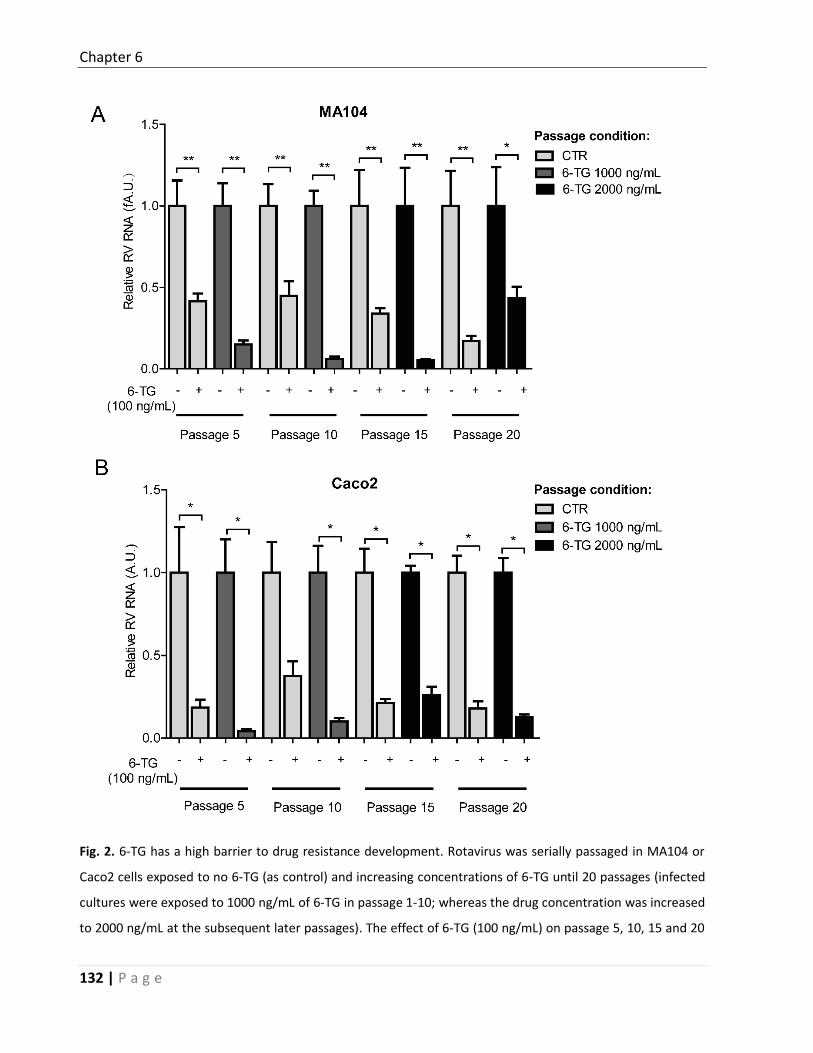
Chapter 6
132 | P a g e
Fig. 2. 6-TG has a high barrier to drug resistance development. Rotavirus was serially passaged in MA104 or
Caco2 cells exposed to no 6-TG (as control) and increasing concentrations of 6-TG until 20 passages (infected
cultures were exposed to 1000 ng/mL of 6-TG in passage 1-10; whereas the drug concentration was increased
to 2000 ng/mL at the subsequent later passages). The effect of 6-TG (100 ng/mL) on passage 5, 10, 15 and 20

Chapter 6
133 | P a g e
(the drug treated and untreated) of rotavirus was quantified using qRT-PCR. 6-TG retained its anti-rotavirus
activity even with continuous exposure to 6-TG for 20 passages in MA104 cells (A) and Caco2 cells (B).
Efficient rotavirus replication requires Rac1 activation
Rac1 acts as binary switch in cellular biochemistry, and it is only capable of provoking signaling in
the active GTP-bound form (Shin et al., 2016). NSC23766, a specific GTP-Rac1 inhibitor was able
to effectively inhibit GTP-Rac1 accumulation in Caco2 cells, as evident when tested in GTP-Rac1
specific pull-down assay (Fig. 4A and B). Importantly, treatment with either 5 or 25 µM NSC23766
for 48 hrs resulted in 58.8 ± 0.1% (n = 8; P < 0.05) and 77.48 ± 0.1% (n = 10; P < 0.001) reduction
on viral RNA levels, respectively (Fig. 4C). The IC50 value of NSC23766 against SA11 rotavirus was
1.1×10⁻⁸ M, the CC50 of NSC23766 to Caco2 cells was 3.1×10⁻⁴ M, and the SI was 2.8 ×10⁴ (Table.
S2). The inhibitory effect of NSC23766 on the rotavirus infectious was further verified by a
western blot assay, revealing that treatment with either 1, 5 or 25 µM NSC23766 significantly
inhibits rotavirus VP4 protein synthesis in Caco2 cells (Fig. 4D). The effect of this drug was further
confirmed by experiments in human primary intestinal organoids, which indicated 91.1 ± 0.1% (n
= 6; P < 0.05) reduction of viral RNA in the organoids (Fig. 4E) following treatment with NSC23766.
Next, we tested the effects of constitutively active and dominant negative forms of Rac1 on
rotavirus replication. This was done by transfection of the active Rac1V12 plasmid or the
dominant negative Rac1N17 plasmid. Expression of both plasmids was successful when tested by
flow cytometry (Fig. 4F). Accordingly, Rac1V12-transfected cells displayed abundant GTP-Rac1,
which in contrast is low in Rac1N17-transfected cells, as determined in a Rac pull-down assay (Fig.
4G). These results were confirmed by analyzing abundance of phospho-PAK2 status (Fig. 4H).
Importantly, Rac1V12 promoted, but Rac1N17 inhibited rotavirus replication (Fig. 4I). Taken
together, activation of Rac1 supports rotavirus replication.

Chapter 6
134 | P a g e
Fig. 3. Rac1, the drug target of 6-TG, sustains rotavirus replication. (A) Western blot assay detected Rac1 in
transduced Caco2 cells transduced lentiviral RNAi vectors against Rac1 (The ratio of Rac1/β-actin was expressed
in arbitrary units). (B) Three (No. 9687, 9689 and 9690) out of five lentiviral shRNA vectors inhibited rotavirus
genomic RNA (n = 6-10, means ± SEM, *P < 0.05, ***P < 0.001, Mann-Whitney test). (C) Western blot assay
confirmed knockout of Rac1 in Rac1 knockout (-/-) MEF cells. (D) SA11 rotavirus replication was significantly
attenuated in Rac1 knockout (-/-) MEF cells (n = 4, means ± SEM, *P < 0.05, Mann-Whitney test).

Chapter 6
135 | P a g e
Fig. 4. The activation form of Rac1 is required for supporting rotavirus replication. (A) Schematic depicting the
pull-down assay. (B) NSC23766 inhibited GTP-Rac1 detected by pulldown assay (The ratio of GTP-Rac1/Rac was
expressed in arbitrary units). (C) Treatment with NSC23766 (48 hrs) significantly inhibited viral genomic RNA in
SA11 rotavirus infected Caco2 cells in a dose-dependent manner (n = 8-10, means ± SEM, *P < 0.05, ***P <
0.001, Mann-Whitney test). (D) Treatment with NSC23766 (48 hrs) significantly inhibited viral VP4 protein in
SA11 rotavirus infected Caco2 cells in a dose-dependent manner (The ratio of VP4/β-actin was expressed in
arbitrary units). (E) Treatment with NSC23766 (48 hrs) significantly inhibited viral RNA in SA11 rotavirus infected

Chapter 6
136 | P a g e
human intestinal organoids (n = 6, means ± SEM, *P < 0.05, Mann-Whitney test). (F) Flow cytometric analysis
of green fluorescence indicated the percentages of transduced cells with Rac1V12 and Rac1N17 plasmids.
Median fluorescence identity (MFIs) of control, N17 and V12 are 3.43, 35.5 and 84.3, respectively. (G) Pull-down
and western blot assays showed higher level of GTP-Rac1 with transduction of Rac1V12 but lower level of GTP-
Rac1 with Rac1N17 plasmids (The ratio of GTP-Rac1/Rac was expressed in arbitrary units). (H) Detection of
phospho-PAK2 indicated successful transduction of Rac1V12 and Rac1N17 plasmids. (I) Rac1V12 transduction
facilitates but Rac1N17 inhibits rotavirus replication (n = 10, means ± SEM, *P < 0.05, Mann-Whitney test).
6-TG inhibits rotavirus via suppression of Rac1 GDP/GTP cycling
The inhibition of the activation of Rac1 by 6-TG was reported in many cell types (Fuhler et al.,
2008; Shin et al., 2016; Tiede et al., 2003). Employing the Rac1 pull-down assay (Fig. 4A), we
observed that 6-TG potently inhibited GTP-Rac1 accumulation; whereas corresponding western
blots did not show reduced overall Rac1 levels in Caco2 cells following 6-TG treatment (Fig. 5A).
Functional studies using Rac1 knockdown Caco2 cells (Fig. 5B and 5C) were performed to
demonstrate that both 6-TG and NSC23766 require Rac1 to combat rotavirus replication. In
agreement, pharmacological Rac1 inhibitors did not inhibit rotavirus replication in Rac1 knockout
MEF cells (Fig. 5D). Thus 6-TG inhibits rotavirus via suppression of Rac1 activation.
6-TG has no combination effect with IFNα, but moderately antagonistic effect with
ribavirin on rotavirus replication
Interferon-alpha (IFNα) and ribavirin are widely used as general antivirals, being confirmed to
significantly inhibit rotavirus replication in vitro (Yin et al., 2015a). Consistently, we again
demonstrated the inhibitory effects of IFNα and ribavirin on rotavirus RNA (Fig. 6A, and 6E) and
viral protein (Fig. 6B and 6F) in a dose-dependent manner. The IC50 value of IFNα against SA11
rotavirus was 3.1×10⁻⁵ IU, CC50 of IFNα to Caco2 cells was 18706 IU, and SI was 6.0×10⁸ (Table S2).
The IC50 value of ribavirin against SA11 rotavirus was 1.6×10⁻⁷ M, CC50 of ribavirin to Caco2 cells
was 3.02×10⁻² M and SI was 1.9×10⁵ (Table S2). Next, we assessed the combinatory antiviral
effects of 6-TG with IFNα or ribavirin. The combination of 6-TG and IFNα resulted in no
combination (no synergy or antagonism) antiviral effect, with a synergy volume of -2.8 µM2% (Fig.

Chapter 6
137 | P a g e
6C and 6D). However, the combination of 6-TG and ribavirin resulted in moderately antagonistic
antiviral effect, with a synergy volume of -26.02 µM2% (Fig. 6G and 6H).
Fig. 5. 6-TG inhibits rotavirus via suppression of Rac1 activation. (A) Anti-rotavirus effect of 6-TG (100 ng/mL)
was attenuated in Rac1 knockdown Caco2 cells (The ratio of VP4/β-actin was expressed in arbitrary units). (B)
6-TG inhibited GTP-Rac1 detected by pull-down assay. (C) Anti-rotavirus effect of NSC23766 (25 μg ng/mL) was
attenuated in Rac1 knockdown Caco2 cells. (D) The anti-rotavirus effect of 6-TG (100 ng/mL) was attenuated in
Rac1 knockout (-/-) MEF cells (n = 6-8, means ± SEM, *P < 0.05, **P < 0.01, Mann-Whitney test).

Chapter 6
138 | P a g e

Chapter 6
139 | P a g e
Fig. 6. The effects of the combination of 6-TG with IFNα, or ribavirin on rotavirus replication. (A) Treatment
with IFNα (48 hrs) significantly inhibited viral genomic RNA in SA11 rotavirus infected Caco2 cells in a dose-
dependent manner (n = 4, means ± SEM, **P < 0.01, Mann-Whitney test). (B) Treatment with IFNα (48 hrs)
remarkably inhibited viral VP4 protein in SA11 rotavirus infected Caco2 cells in a dose-dependent manner (The
ratio of VP4/β-actin was expressed in arbitrary units). (C) Effect of the combination of various concentrations
of 6-TG and IFNα on rotavirus replication in Caco2 cells. (D) Synergy plot representing the percentage of antiviral
activity above/below the expected activity for the 6-TG-IFNα combination based on the data shown in C. (E)
Treatment with ribavirin (48 hrs) significantly inhibited viral genomic RNA in SA11 rotavirus infected Caco2 cells
in a dose-dependent manner (n = 4-7, means ± SEM, *P < 0.05, **P < 0.01, Mann-Whitney test). (F) Treatment
with ribavirin (48 hrs) remarkably inhibited viral VP4 protein in SA11 rotavirus infected Caco2 cells in a dose-
dependent manner (The ratio of VP4/β-actin was expressed in arbitrary units). (G) Effect of the combination of
various concentrations of 6-TG and ribavirin on rotavirus replication in Caco2 cells. (H) Synergy plot
representing the percentage of antiviral activity above/below the expected activity for the 6-TG-ribavirin
combination based on the data shown in G.
DISCUSSION
In this study, we have demonstrated that 6-TG effectively inhibits rotavirus replication via
inhibition of the Rac1 activity. This is particularly interesting in view that rotavirus replication is
especially an issue in organ transplantation recipients and in patients with IBD. 6-TG is a
therapeutic option for both groups of patients. Based on the findings presented in the current
study, the choice of 6-TG for these patients appears rational, in particular when they are at risk
of rotavirus infection (Yin et al., 2015b).
Our results fit well with the momentum of studies that document antiviral activity of 6-TG. For
instance, it has been reported that 6-TG can combat Middle East respiratory syndrome
coronavirus infection by augmenting interferon responses (Cheng et al., 2015) and it has also
reported that Simian virus 40 DNA replication is antagonized by 6-TG (Maybaum et al., 1987).
Interestingly, it was reported that vaccinated IBD patients had lower titers of hepatitis B surface
antibody (HBsAb), which might be influenced by the use of immunosuppressants including 6-TG
(Watts et al., 2017).
As a 6-thiopurine (6-TP) prodrug, 6-TG is converted into pharmacologically active deoxy-6-
thioguanosine phosphate (also called 6-thioguanine nucleotide) and 6-thioguanosine phosphate

Chapter 6
140 | P a g e
(6-TGNP). 6-TGNP can bind to Rac1 to form the 6-TGNP•Rac1 complex inactivating Rac1 (Shin et
al., 2016). As a major player of the Rho family of small GTPases, Rac1 plays a vital role in various
cellular signaling pathways to regulate a wide variety of cell functions including gene transcription,
cell proliferation, apoptosis, motility, and redox signaling (D'Ambrosi et al., 2014). The expression
of Rac1 is ubiquitous, but it has two conformational states including an inactive GDP-bound form
and an active GTP-bound form (Bosco et al., 2009). It exerts biological functions mainly through
activation of Rac1 (i.e. GTP-bound form) (Bosco et al., 2009). Many viruses interfere with or
employ the conformational states of Rac1 to regulate their infection. At early stages of African
swine fever virus (ASFV) infection, Rac1 is activated, and inhibition of Rac1 is able to suppress
production of this virus (Quetglas et al., 2012). Rac1 is found to be activated during intracellular
mature virus (MV) of Vaccinia virus entry (Mercer and Helenius, 2008).
Although rotavirus replication per se does not affect the activation of Rac1 (Fig. S4), we have
demonstrated that the loss-of-function of Rac1 by gene knockdown or knockout significantly
impairs rotavirus replication, which is in line with the previous finding that knockdown Rac1 could
significantly inhibit Enterovirus 1 (EV1) infection (Karjalainen et al., 2008). More specifically, the
activation of Rac1 is required as shown by the opposing effects of ectopic over-expression of the
active or inactive forms of Rac1 on rotavirus replication. This mechanistically explains the potent
anti-rotavirus effects of the GTP-Rac1 inhibitors, 6-TG and NSC23766. Of note, NSC23766 has
been shown to inhibit the replication of several influenza viruses including a human virus strain
from the 2009 pandemic and highly pathogenic avian virus strains (Dierkes et al., 2014).
Despite the absence of approved medications for treating rotavirus, the widely used general
antivirals including ribavirin and IFNα have been studied on rotavirus in experimental models (Yin
et al., 2015a). Here, we have evaluated the combinatory effects of 6-TG with IFNα or ribavirin.
Consistently, we confirmed that ribavirin and IFNα inhibit rotavirus replication at both RNA and
protein levels (Fig. 6A and 6B). We found increased potency of IFNα in the presence of 6-TG.
However, whether the combination of IFNα and 6-TG could be used to treat rotavirus infected
patients remains to be further investigated.
In conclusion, this study has demonstrated that 6-TG effectively inhibits rotavirus replication with
a high barrier to drug resistance development. We further identified the active form of Rac1 as

Chapter 6
141 | P a g e
an important host factor supporting rotavirus replication. 6-TG exerts its anti-rotavirus effects via
the specific inhibition of Rac1 activation. Herein, this study provided important references for
clinicians to optimize medications for organ recipients or IBD patients who are infected with
rotavirus or at risk of rotavirus replication. These results may also help the development of new
anti-rotavirus therapies.
Chapter 6
142 | P a g e
REFERENCE Apolloni, S., Parisi, C., Pesaresi, M.G., Rossi, S., Carri, M.T., Cozzolino, M., Volonte, C., D'Ambrosi, N., 2013.
The NADPH oxidase pathway is dysregulated by the P2X7 receptor in the SOD1-G93A microglia model of amyotrophic lateral sclerosis. J Immunol 190, 5187-5195.
Bosco, E.E., Mulloy, J.C., Zheng, Y., 2009. Rac1 GTPase: a "Rac" of all trades. Cell Mol Life Sci 66, 370-374. Bourgine, J., Garat, A., Allorge, D., Crunelle-Thibaut, A., Lo-Guidice, J.M., Colombel, J.F., Broly, F., Billaut-
Laden, I., 2011. Evidence for a functional genetic polymorphism of the Rho-GTPase Rac1. Implication in azathioprine response? Pharmacogenet Genomics 21, 313-324.
Cheng, K.W., Cheng, S.C., Chen, W.Y., Lin, M.H., Chuang, S.J., Cheng, I.H., Sun, C.Y., Chou, C.Y., 2015. Thiopurine analogs and mycophenolic acid synergistically inhibit the papain-like protease of Middle East respiratory syndrome coronavirus. Antiviral Res 115, 9-16.
Chouchana, L., Narjoz, C., Beaune, P., Loriot, M.A., Roblin, X., 2012. Review article: the benefits of pharmacogenetics for improving thiopurine therapy in inflammatory bowel disease. Aliment Pharmacol Ther 35, 15-36.
D'Ambrosi, N., Rossi, S., Gerbino, V., Cozzolino, M., 2014. Rac1 at the crossroad of actin dynamics and neuroinflammation in Amyotrophic Lateral Sclerosis. Front Cell Neurosci 8, 279.
de Boer, N.K., Reinisch, W., Teml, A., van Bodegraven, A.A., Schwab, M., Lukas, M., Ochsenkuhn, T., Petritsch, W., Knoflach, P., Almer, S., van der Merwe, S.W., Herrlinger, K.R., Seiderer, J., Vogelsang, H., Mulder, C.J., Dutch, T.G.w.g., 2006. 6-Thioguanine treatment in inflammatory bowel disease: a critical appraisal by a European 6-TG working party. Digestion 73, 25-31.
Dierkes, R., Warnking, K., Liedmann, S., Seyer, R., Ludwig, S., Ehrhardt, C., 2014. The Rac1 inhibitor NSC23766 exerts anti-influenza virus properties by affecting the viral polymerase complex activity. PLoS One 9, e88520.
Fuhler, G.M., Drayer, A.L., Olthof, S.G., Schuringa, J.J., Coffer, P.J., Vellenga, E., 2008. Reduced activation of protein kinase B, Rac, and F-actin polymerization contributes to an impairment of stromal cell derived factor-1 induced migration of CD34+ cells from patients with myelodysplasia. Blood 111, 359-368.
Grimwood, K., Buttery, J.P., 2007. Clinical update: rotavirus gastroenteritis and its prevention. Lancet 370, 302-304.
Karjalainen, M., Kakkonen, E., Upla, P., Paloranta, H., Kankaanpaa, P., Liberali, P., Renkema, G.H., Hyypia, T., Heino, J., Marjomaki, V., 2008. A Raft-derived, Pak1-regulated entry participates in alpha2beta1 integrin-dependent sorting to caveosomes. Molecular biology of the cell 19, 2857-2869.
Kolho, K.L., Klemola, P., Simonen-Tikka, M.L., Ollonen, M.L., Roivainen, M., 2012. Enteric viral pathogens in children with inflammatory bowel disease. J Med Virol 84, 345-347.
Masclee, G.M., Penders, J., Pierik, M., Wolffs, P., Jonkers, D., 2013. Enteropathogenic viruses: triggers for exacerbation in IBD? A prospective cohort study using real-time quantitative polymerase chain reaction. Inflamm Bowel Dis 19, 124-131.
Maybaum, J., Bainnson, A.N., Roethel, W.M., Ajmera, S., Iwaniec, L.M., TerBush, D.R., Kroll, J.J., 1987. Effects of incorporation of 6-thioguanine into SV40 DNA. Mol Pharmacol 32, 606-614.
Mercer, J., Helenius, A., 2008. Vaccinia virus uses macropinocytosis and apoptotic mimicry to enter host cells. Science 320, 531-535.
Munshi, P.N., Lubin, M., Bertino, J.R., 2014. 6-thioguanine: a drug with unrealized potential for cancer therapy. Oncologist 19, 760-765.
Parikh, K., Zhou, L., Somasundaram, R., Fuhler, G.M., Deuring, J.J., Blokzijl, T., Regeling, A., Kuipers, E.J., Weersma, R.K., Nuij, V.J., Alves, M., Vogelaar, L., Visser, L., de Haar, C., Krishnadath, K.K., van der Woude, C.J., Dijkstra, G., Faber, K.N., Peppelenbosch, M.P., 2014. Suppression of p21Rac signaling and increased innate immunity mediate remission in Crohn's disease. Sci Transl Med 6, 233ra253.
Chapter 6
143 | P a g e
Quetglas, J.I., Hernaez, B., Galindo, I., Munoz-Moreno, R., Cuesta-Geijo, M.A., Alonso, C., 2012. Small rho GTPases and cholesterol biosynthetic pathway intermediates in African swine fever virus infection. J Virol 86, 1758-1767.
Sato, T., Clevers, H., 2013. Growing self-organizing mini-guts from a single intestinal stem cell: mechanism and applications. Science 340, 1190-1194.
Shin, J.Y., Wey, M., Umutesi, H.G., Sun, X., Simecka, J., Heo, J., 2016. Thiopurine Prodrugs Mediate Immunosuppressive Effects by Interfering with Rac1 Protein Function. J Biol Chem 291, 13699-13714.
Tate, J.E., Burton, A.H., Boschi-Pinto, C., Parashar, U.D., 2016. Global, Regional, and National Estimates of Rotavirus Mortality in Children <5 Years of Age, 2000-2013. Clin Infect Dis 62 Suppl 2, S96-S105.
Tiede, I., Fritz, G., Strand, S., Poppe, D., Dvorsky, R., Strand, D., Lehr, H.A., Wirtz, S., Becker, C., Atreya, R., Mudter, J., Hildner, K., Bartsch, B., Holtmann, M., Blumberg, R., Walczak, H., Iven, H., Galle, P.R., Ahmadian, M.R., Neurath, M.F., 2003. CD28-dependent Rac1 activation is the molecular target of azathioprine in primary human CD4+ T lymphocytes. J Clin Invest 111, 1133-1145.
Versteeg, H.H., Sorensen, B.B., Slofstra, S.H., Van den Brande, J.H., Stam, J.C., van Bergen en Henegouwen, P.M., Richel, D.J., Petersen, L.C., Peppelenbosch, M.P., 2002. VIIa/tissue factor interaction results in a tissue factor cytoplasmic domain-independent activation of protein synthesis, p70, and p90 S6 kinase phosphorylation. J Biol Chem 277, 27065-27072.
Vesikari, T., Karvonen, A., Prymula, R., Schuster, V., Tejedor, J.C., Cohen, R., Meurice, F., Han, H.H., Damaso, S., Bouckenooghe, A., 2007. Efficacy of human rotavirus vaccine against rotavirus gastroenteritis during the first 2 years of life in European infants: randomised, double-blind controlled study. Lancet 370, 1757-1763.
Watts, A., Bennett, W.E., Molleston, J.P., Gupta, S.K., Croffie, J.M., Waseem, S., McFerron, B.A., Steiner, S.J., Kumar, S., Vanderpool, C.P., Hon, E.C., Bozic, M.A., Subbarao, G.C., Pfefferkorn, M.D., 2017. The Incidence of Low Seroimmunity to Hepatitis B Virus in Children with Inflammatory Bowel Disease. J Pediatr Gastroenterol Nutr.
Yin, Y., Bijvelds, M., Dang, W., Xu, L., van der Eijk, A.A., Knipping, K., Tuysuz, N., Dekkers, J.F., Wang, Y., de Jonge, J., Sprengers, D., van der Laan, L.J., Beekman, J.M., Ten Berge, D., Metselaar, H.J., de Jonge, H., Koopmans, M.P., Peppelenbosch, M.P., Pan, Q., 2015a. Modeling rotavirus infection and antiviral therapy using primary intestinal organoids. Antiviral Res 123, 120-131.
Yin, Y., Metselaar, H.J., Sprengers, D., Peppelenbosch, M.P., Pan, Q., 2015b. Rotavirus in organ transplantation: drug-virus-host interactions. Am J Transplant 15, 585-593.
Yin, Y., Wang, Y., Dang, W., Xu, L., Su, J., Zhou, X., Wang, W., Felczak, K., van der Laan, L.J., Pankiewicz, K.W., van der Eijk, A.A., Bijvelds, M., Sprengers, D., de Jonge, H., Koopmans, M.P., Metselaar, H.J., Peppelenbosch, M.P., Pan, Q., 2016. Mycophenolic acid potently inhibits rotavirus infection with a high barrier to resistance development. Antiviral Res 133, 41-49.

Chapter 6
144 | P a g e
Supplementary Information
Table S1. Primers and shRNA sequences used in the study
Sequence of rotavirus primers
SA11 Rotavirus Human Patient Rotavirus Sense TGGTTAAACGCAGGATCGGA ACCATCTACACATGACCCTC Anti-sense AACCTTTCCGCGTCTGGTAG CACATAACGCCCCTATAGCC
Primer sequences of GAPDH
Primers Human Mouse
GAPDH-F GTCTCCTCTGACTTCAACAGCG TTCCAGTATGACTCCACTCACGG
GAPDH-R ACCACCCTGTTGCTGTAGTAGCCAA TGAAGACACCAGTAGACTCCACGAC
shRNA targeting sequences of Rac1
No. Sequence
9687 CCTTCTTAACATCACTGTCTT
9688 GCTAAGGAGATTGGTGCTGTA
9689 CGCAAACAGATGTGTTCTTAA
9690 CGTGAAGAAGAGGAAGAGAAA
9691 CCCTACTGTCTTTGACAATTA
Table S2. 50% inhibition concentration (IC50) of 6-TG, NSC23766, IFNα and ribavirin against SA11 rotavirus, 50% cytotoxic (CC50) of against Caco2 cells, and corresponding selectivity index (SI, CC50/IC50).
Drugs IC50 against SA11 rotavirus (M)
CC50 against Caco2 cells (M)
selectivity index (SI, CC50/IC50)
6-TG 3.0×10⁻¹³ M 9.8×10⁻⁶ M 3.3×10⁷
NSC23766 1.1×10⁻⁸ M 3.1×10⁻⁴ M 2.8 ×10⁴
IFNα 3.1×10⁻⁵ IU 18706 IU 6.0×10⁸
Ribavirin 1.6×10⁻⁷ M 3.02×10⁻² M 1.9×10⁵

145 | P a g e
Table S3. Patient characteristics.
Patient Age (yrs)
Gender Symptoms Virus Detection
Enterovirus Parechovirus Norovirus genegroups
I
Norovirus genegroups
II
Adenovirus Astrovirus Sapovirus Rotavirus
1 3.5 Female Fever No No No No No No No Yes
2 74 Female Congestive heart failure, myocarditis
No No No No No No No Yes
3 27 Female Fever, diarrhea, nausea, vomiting
No No No No No No No Yes
4 67 Male Fever, stomachache, watery diarrhea (Kidney transplant)
No No No No No No No Yes
5 28 Female Nausea, stomachache, watery diarrhea, fever, headache, vomiting
No No No No Yes No No Yes

Chapter 6
146 | P a g e
Supplementary Figures
Fig. S1. Effect of 6-TG (A), NSC23766 (B), IFNα (C) and ribavirin (D) on host cell viability
determined by MTT assays. Caco2 cells treated with all four drugs for 48 h.

Chapter 6
147 | P a g e
Fig. S2. The effect of inactive rotavirus by UV treatment, rotavirus and mock infection (1 and 8 h)
on the expression of GTP-Rac1. (A) Western blot assay detected the expression of GTP-Rac1 after
1 and 8 h post-infection by inactive rotavirus (UV treatment), rotavirus and mock infection in Caco2
cells. (B) Quantification of the intensity of the immunoreactive bands of Rac1 (n = 4, means ± SEM,
Mann-Whitney test) using Odyssey V 3.0 software.


149 | P a g e
Chapter 7
Drug screening identifies gemcitabine as
inhibiting rotavirus through alteration of
pyrimidine nucleotide synthesis pathway
Sunrui Chen, Yining Wang, Pengfei Li, Yuebang Yin, Marcel Bijvelds, Hugo de Jonge, Maikel P.
Peppelenbosch, Denis Kainov, Qiuwei Pan
Antiviral Research, 2020: 104823.


Chapter 7
151 | P a g e
ABSTRACT
Although rotavirus infection is usually acute and self-limiting, it can cause chronic infection
with severe disease phenotypes in immunocompromised patients, including organ
transplantation recipients and cancer patients and irrespective of whether this concerns
pediatric or adult patients. Clinical management is complicated by the absence of approved
medication against rotavirus infection. Here we screened a library of safe-in-man broad-
spectrum antivirals. We identified gemcitabine, a widely used anti-cancer drug, as a potent
inhibitor of rotavirus infection. We confirmed this effect in 2D cell cultures and 3D cultured
human intestinal organoids with both laboratory-adapted rotavirus strains and five clinical
isolates. Supplementation of UTP or uridine largely abolished the anti-rotavirus activity of
gemcitabine, suggesting its function through inhibition of pyrimidine biosynthesis pathway.
Our results support repurposing gemcitabine for treating cancer patients at risk for rotavirus
infection.

Chapter 7
152 | P a g e
Rotavirus infection is the leading cause of severe dehydrating gastroenteritis among children
under five-year-old (Greenberg and Estes, 2009). Although rotavirus infection is usually acute
and self-limiting, it can cause chronic infection with severe diseases in immunocompromised
patients, in particular organ transplantation recipients irrespective of whether these are
pediatric or adult patients (Yin et al., 2015b). In addition, cancer patients have
compromised immune system especially when undergoing chemotherapy or
radiotherapy treatment, which make them prone to infections with worse outcomes
(Hotchkiss and Moldawer, 2014). Rotavirus infections have been widely reported in
pediatric or adult cancer patients causing prolonged diarrhea (Akhtar et al., 2018; Ghosh et
al., 2017). Therefore, specific and effective antiviral treatment is urgently needed for these
special populations when infected with rotavirus. Unfortunately no FDA-approved
medications against rotavirus infection are available.
Developing new drugs usually takes more than ten years with enormous investment and high
risk of failure. Given that only the specific population with rotavirus infection require antiviral
treatment, the pharmaceutical industry will likely not develop new anti-rotavirus drugs. We
propose that repurposing existing drugs represents a cost-effective approach to identify
antiviral treatment that can readily benefit patients (Qu et al., 2019). In this study, we
screened a library of safe-in-man broad-spectrum antiviral agents (BSAAs,
https://drugvirus.info) (Andersen et al., 2020; Ianevski et al., 2018) on rotavirus infection in
experimental models. These compounds are known to target viruses belonging to two or more
viral families and have passed phase 1 clinical trials. This greatly enhances the probability of
identifying novel activities of some of these agents against rotavirus infection and facilitates
their clinical translation.
We first screened 94 BSAAs in the human intestinal Caco2 cell line infected with simian
rotavirus SA11 strain (Table S1). To minimize non-specific effects on host cells, we used low
concentration of 1 µM and treated for 48 hours. By qRT-PCR (primers listed in Table S2)
quantification of rotavirus genomic RNA, we identified 43 candidates exerting over 50%
inhibitory effects, and 17 with inhibition over 70% (Fig. 1A). Among these, gemcitabine was
one of the most effective candidates (Fig. 1A). It is a cytidine analog that has been widely used
for cancer treatment (Cerqueira et al., 2007; Zhang et al., 2019). It has been shown to inhibit
a broad range of RNA viruses including severe acute respiratory syndrome coronavirus (SARS-

Chapter 7
153 | P a g e
CoV), Middle East respiratory syndrome coronavirus (MERS-CoV), Zika virus and hepatitis C
virus (HCV) in experimental models (Beran et al., 2012; Dyall et al., 2014; Kuivanen et al., 2017).
Nucleotide and nucleoside analogues are excellent examples of BSAAs that have been widely
used in the clinic for treating infections of RNA and DNA viruses (Ianevski et al., 2018). We
have previously demonstrated that nucleotides, including purine and pyrimidine, biosynthesis
pathways are essential in the rotavirus infection process and can be pharmacologically
targeted (Chen et al., 2019; Yin et al., 2018a; Yin et al., 2016). As a cytidine analog, gemcitabine
has been reported to inhibit pyrimidine biosynthesis, resulting in nucleotide depletion (Lee et
al., 2017). Therefore, we focused on the effects and mode-of-action of gemcitabine on
rotavirus infection in this study.
We next tested a series of gemcitabine concentrations (0.01-10 µM) in the Caco2 cell model
to assess both antiviral and cytotoxic effects. We confirmed the potent anti-rotavirus effect
and observed a large therapeutic window between cytotoxic and antiviral activities, as shown
the CC50 value of 13.58 mM and IC50 value of 0.12 µM (Fig. 1B, 1C). We performed same
experiments in the monkey MA104 cell line that is widely used for propagating rotavirus in
laboratory. Similar trends were observed with CC50 value of 0.36 mM and IC50 value of 3.98
µM (Supplementary Fig. S1). By harvesting supernatant of Caco2 cells at 48-hour post-
treatment, we performed TCID50 assay to determine the titers of secreted viruses.
Consistently, the titers of produced rotavirus with infectivity were significantly reduced by
gemcitabine treatment (Fig. 1D). At the protein level, we found potent inhibition of viral
protein 4 (VP4) expression determined by Western blotting (Fig. 1E). Furthermore,
immunofluorescent staining of viral capsid protein (VP6) showed significant reduction of the
number of infected Caco2 cells by gemcitabine treatment (Fig. 1F, 1G, Supplementary Fig. S2).

Chapter 7
154 | P a g e
Figure 1. Screening BSAAs identified gemcitabine as a potent inhibitor of rotavirus infection. (A) Human
intestinal Caco2 cells were infected with SA11 rotavirus. After infection, cells were treated with 94 BSAAs
at 1 µM for 48 hours. qRT-PCR analysis of viral RNA revealed that 43 reagents exerted ≥ 50% inhibitory
activity, and 17 of them exerted ≥ 70% inhibitory activity against rotavirus replication. Gemcitabine (GCT)
inhibited rotavirus replication by 70%. (B) 50% cytotoxic concentration (CC50) curves of GCT were
determined by MTT assays on Caco2 cell line. (C) Dose-dependent inhibitory activity of GCT on rotavirus
infected Caco2 cell line. qRT-PCR data were normalized to housekeeping gene GAPDH and presented
relative to the control (CTR) (set as 1) (n = 6). (D) The supernatant of each well under GCT treatment was
harvested after freezing and thawing for three times, virus titer from different groups was measured by
TCID50 assay (n = 6). (E) Rotavirus infected Caco2 cells were treated by different concentrations of GCT, and

Chapter 7
155 | P a g e
protein samples were harvested after 48 hours. The expression of viral structural protein VP4 was stained
and quantified by western blot assay (n = 4). (F) Indirect fluorescence microscope analysis of viral structural
protein VP6 (red) upon treatment with GCT. Nuclei were visualized by DAPI (blue). (G) The ratio of VP6
positive cells/ total cell number was quantified under 40* vision field (Supplementary Fig. S2) (n = 6). Data
represent means ± SEM. *P < 0.05; **P < 0.01; ***P < 0.001.
The responsiveness to antiviral therapy can vary dramatically in patients. This mainly
attributes to host and viral factors. The nucleoside analog ribavirin has been used for treating
HCV infection for decades. Reduced cellular uptake by the host and mutagenesis of the viral
genome have been linked to treatment resistance in chronic HCV patients (Ibarra et al., 2011).
As recently repositioned for treating chronic hepatitis E virus (HEV) infection (Kamar et al.,
2017), viral mutagenesis during ribavirin treatment or pre-deposition of resistance mutations
are thought to contribute to treatment failure (Ikram et al., 2018). Our previous studies have
evaluated the effects of the nucleoside analog ribavirin and mycophenolic acid (MPA), and the
antiviral cytokine interferon alpha (IFN-α) on rotavirus in cell culture models. We found the
responsiveness to these agents dramatically vary among different clinical isolates from potent,
moderate, minimal to even pro-viral effects (Yin et al., 2015a; Yin et al., 2016). This imposes
major challenges for clinical application as how to personalize the selection of potential
responders. Thus, we extended our evaluation of gemcitabine to different rotavirus
strains/isolates. We found that gemcitabine significantly inhibits the replication of rhesus
rotavirus RRV strain (Fig. 2A) and five clinical isolates (Fig. 2B-2F). Based on these results, we
postulate that the response to gemcitabine in rotavirus patients could be universally effective.

Chapter 7
156 | P a g e
Figure 2. Antiviral effect of gemcitabine against RRV rotavirus strain and clinical isolates. qRT-PCR analysis
of rotavirus RNA upon treatment of gemcitabine (GCT) at 1 µM for 48 hours in laboratory rotavirus strain
RRV (A), clinically isolated rotavirus strain 026k (B), 1758k (C), 1934 (D), 664k (E), and 2011k (F). Data were
normalized to housekeeping gene GAPDH and are presented relative to the control (CTR) (set as 1). Data
represent means ± SEM; n = 6; *P < 0.05; **P < 0.01.
We have previously established that modeling of rotavirus infection in intestinal organoids
allows the study of virus-host interactions and assessment of antiviral drugs efficacy (Yin et al.,
2015a; Yin et al., 2018a; Yin et al., 2018b; Yin et al., 2016). Intestinal organoids, also called
mini-guts, are stem cell-derived epithelial 3D cultures. These organoids are much better in
recapitulating the architecture, composition, diversity, organization and functionality of cell
types of the intestine when compared to more conventional model systems. Treatment with
gemcitabine in rotavirus inoculated human intestinal organoids potently inhibited viral RNA
synthesis and secretion of rotaviruses with approximately 80% inhibitory effects for both at 1
µM concentration treated for 48 hours (Fig. 3A; Supplementary Fig. S3). This effect was
further confirmed at the VP4 protein level by western blot assay (Fig. 3B). Importantly,
rotavirus infection led to morphological and pathological changes in organoids. Based on
optical imaging, we observed that rotavirus infected organoids without gemcitabine
treatment showed opaque, wizened and disorganized morphology. In contrast, gemcitabine
treated groups, in particular with 1 µM concentration, most of the organoids were hyaline and
in a spheroidal shape (Fig. 3C upper panel). In confocal immunostaining, VP6 protein was
detected in all groups, but the intensity and frequency of the viral protein were lower in
treatment groups (Fig. 3C middle panel). Fluorescence staining of cell viability by Propidium
Iodide (PI) showed that cell death (determined as described in Supplementary Fig. S4) in un-
treated group is more obvious, and dead cells were diffused in almost all organoids (Fig. 3C
lower panel). This is consistent with the percentage of organoids in a deteriorated condition
(Fig. 3D). Thus, inhibition of rotavirus replication by gemcitabine can protect organoids from
rotavirus induced cytopathogenesis.

Chapter 7
157 | P a g e

Chapter 7
158 | P a g e
Figure 3. Antiviral activity of gemcitabine against rotavirus SA11 in human intestinal organoids . (A)
Human intestinal organoids (HIOs) were inoculated with SA11 rotavirus strain and treated with gemcitabine
(GCT) for 48 hours. The relative cellular rotavirus RNA and secreted viruses calculated as genomic copy
number were analyzed by qRT-PCR (n = 6). Standard curve for calculation of genomic copy number is
included in Supplementary Fig. S3. (B) The expression of viral structural protein VP4 was stained and
quantified by western blot assay (n = 4). (C) Optical microscopy images, confocal images and indirect
fluorescence microscope images of HIOs from mock group and GCT treated groups. In confocal images, VP6
protein was stained in red, green signal represents Epcam, and nuclei were visualized by DAPI (blue). In
fluorescence microscope images, HIOs were stained by PI (red) indicating dead cells, Hoechst (blue) for
nuclear, and Calcein (green) as live cells. (D) Rate of deteriorated HIOs was calculated as dead organoids/
total organoids (n = 6). Data represent means ± SEM; *P < 0.05; **, P < 0.01.
The antiviral activity of gemcitabine has been linked to the inhibition of pyrimidine
biosynthesis pathway, especially the salvage pathway (Lee et al., 2017). We thus
supplemented UTP or uridine to rotavirus infected Caco2 cell model when treated with
gemcitabine. We found that exogenous supplementation of pyrimidine dose-dependently
abolished the anti-rotavirus effect of gemcitabine. The anti-rotavirus effect was almost
completely abolished by addition of 1000 μM UTP or uridine (Fig. 4A, 4B). Hence, the anti-
rotavirus activity of gemcitabine was largely dependent on the salvage pyrimidine
biosynthesis pathway. Combination approaches are often used in clinic to achieve optimal
antiviral efficacy and avoid resistance development. We finally assessed the combinatory
effects of gemcitabine with ribavirin, MPA or IFN-α, as we previously have demonstrated the
anti-rotavirus effects of these three agents (Yin et al., 2015a; Yin et al., 2016). We found
enhanced anti-rotavirus activity when combined with ribavirin or IFN-α, but not MPA (Fig. 4C).

Chapter 7
159 | P a g e
Figure 4. The effects of nucleotide supplementation and combination with other antivirals on the anti-
rotavirus activity of gemcitabine. Supplementation of exogenous uridine (A) and UTP (B) attenuates the
anti-rotavirus effect of gemcitabine (GCT) in Caco2 cell model (n = 6). (C) The combinatory effects of 1 µM
GCT with 1000 U IFN-α, 1 µg/ml ribavirin (Rib) or 0.1 µg/ml mycophenolic acid (MPA), respectively (n = 6).
Data represent means ± SEM; *P < 0.05; **P < 0.01.
In summary, we have identified gemcitabine as a potent inhibitor against rotavirus infection
through screening of a BSAA library. The antiviral activity was largely dependent on the
pyrimidine biosynthesis pathway. Because gemcitabine has been widely used as
chemotherapy for cancer patients, and these patients are at risk of infections (Hotchkiss and
Moldawer, 2014), the use of gemcitabine in cancer patients as chemotherapeutic drug
probably mitigates the risk of viral infections in general. Our results support a repositioning of
gemcitabine for treating rotavirus infection, especially in infected cancer patients.
Furthermore, the approach of discovering new antiviral therapy from BSAAs bears essential
implications in combating emerging viral pathogens, such as the ongoing coronavirus disease
COVID-19 pandemic (Mahase, 2020).

Chapter 7
160 | P a g e
References Akhtar, T., Cargill, J., Gerrard, C., Shaw, F., Cunliffe, N.A., Cooke, R.P.D., Pizer, B., 2018. Detection of
rotavirus in paediatric oncology patients with diarrhoea: the impact of rotavirus vaccine. J Hosp Infect 99, 185-187.
Andersen, P.I., Ianevski, A., Lysvand, H., Vitkauskiene, A., Oksenych, V., Bjoras, M., Telling, K., Lutsar, I., Dampis, U., Irie, Y., Tenson, T., Kantele, A., Kainov, D.E., 2020. Discovery and development of safe-in-man broad-spectrum antiviral agents. Int J Infect Dis.
Beran, R.K., Sharma, R., Corsa, A.C., Tian, Y., Golde, J., Lundgaard, G., Delaney, W.E.t., Zhong, W., Greenstein, A.E., 2012. Cellular growth kinetics distinguish a cyclophilin inhibitor from an HSP90 inhibitor as a selective inhibitor of hepatitis C virus. PLoS One 7, e30286.
Cerqueira, N.M., Fernandes, P.A., Ramos, M.J., 2007. Understanding ribonucleotide reductase inactivation by gemcitabine. Chemistry 13, 8507-8515.
Chen, S., Ding, S., Yin, Y., Xu, L., Li, P., Peppelenbosch, M.P., Pan, Q., Wang, W., 2019. Suppression of pyrimidine biosynthesis by targeting DHODH enzyme robustly inhibits rotavirus replication. Antiviral Res 167, 35-44.
Dyall, J., Coleman, C.M., Hart, B.J., Venkataraman, T., Holbrook, M.R., Kindrachuk, J., Johnson, R.F., Olinger, G.G., Jr., Jahrling, P.B., Laidlaw, M., Johansen, L.M., Lear-Rooney, C.M., Glass, P.J., Hensley, L.E., Frieman, M.B., 2014. Repurposing of clinically developed drugs for treatment of Middle East respiratory syndrome coronavirus infection. Antimicrob Agents Chemother 58, 4885-4893.
Ghosh, N., Malik, F.A., Daver, R.G., Vanichanan, J., Okhuysen, P.C., 2017. Viral associated diarrhea in immunocompromised and cancer patients at a large comprehensive cancer center: a 10-year retrospective study. Infect Dis (Lond) 49, 113-119.
Greenberg, H.B., Estes, M.K., 2009. Rotaviruses: from pathogenesis to vaccination. Gastroenterology 136, 1939-1951.
Hotchkiss, R.S., Moldawer, L.L., 2014. Parallels between cancer and infectious disease. N Engl J Med 371, 380-383.
Ianevski, A., Zusinaite, E., Kuivanen, S., Strand, M., Lysvand, H., Teppor, M., Kakkola, L., Paavilainen, H., Laajala, M., Kallio-Kokko, H., Valkonen, M., Kantele, A., Telling, K., Lutsar, I., Letjuka, P., Metelitsa, N., Oksenych, V., Bjoras, M., Nordbo, S.A., Dumpis, U., Vitkauskiene, A., Ohrmalm, C., Bondeson, K., Bergqvist, A., Aittokallio, T., Cox, R.J., Evander, M., Hukkanen, V., Marjomaki, V., Julkunen, I., Vapalahti, O., Tenson, T., Merits, A., Kainov, D., 2018. Novel activities of safe-in-human broad-spectrum antiviral agents. Antiviral Res 154, 174-182.
Ibarra, K.D., Jain, M.K., Pfeiffer, J.K., 2011. Host-based ribavirin resistance influences hepatitis C virus replication and treatment response. J Virol 85, 7273-7283.
Ikram, A., Hakim, M.S., Zhou, J.H., Wang, W., Peppelenbosch, M.P., Pan, Q., 2018. Genotype-specific acquisition, evolution and adaptation of characteristic mutations in hepatitis E virus. Virulence 9, 121-132.
Kamar, N., Wang, W., Dalton, H.R., Pan, Q., 2017. Direct-acting antiviral therapy for hepatitis E virus? Lancet Gastroenterol Hepatol 2, 154-155.
Kuivanen, S., Bespalov, M.M., Nandania, J., Ianevski, A., Velagapudi, V., De Brabander, J.K., Kainov, D.E., Vapalahti, O., 2017. Obatoclax, saliphenylhalamide and gemcitabine inhibit Zika virus infection in vitro and differentially affect cellular signaling, transcription and metabolism. Antiviral Res 139, 117-128.

Chapter 7
161 | P a g e
Lee, K., Kim, D.E., Jang, K.S., Kim, S.J., Cho, S., Kim, C., 2017. Gemcitabine, a broad-spectrum antiviral drug, suppresses enterovirus infections through innate immunity induced by the inhibition of pyrimidine biosynthesis and nucleotide depletion. Oncotarget 8, 115315-115325.
Mahase, E., 2020. Covid-19: WHO declares pandemic because of "alarming levels" of spread, severity, and inaction. BMJ 368, m1036.
Qu, C., Li, Y., Li, Y., Yu, P., Li, P., Donkers, J.M., van de Graaf, S.F.J., de Man, R.A., Peppelenbosch, M.P., Pan, Q., 2019. FDA-drug screening identifies deptropine inhibiting hepatitis E virus involving the NF-kappaB-RIPK1-caspase axis. Antiviral Res 170, 104588.
Yin, Y., Bijvelds, M., Dang, W., Xu, L., van der Eijk, A.A., Knipping, K., Tuysuz, N., Dekkers, J.F., Wang, Y., de Jonge, J., Sprengers, D., van der Laan, L.J., Beekman, J.M., Ten Berge, D., Metselaar, H.J., de Jonge, H., Koopmans, M.P., Peppelenbosch, M.P., Pan, Q., 2015a. Modeling rotavirus infection and antiviral therapy using primary intestinal organoids. Antiviral Res 123, 120-131.
Yin, Y., Chen, S., Hakim, M.S., Wang, W., Xu, L., Dang, W., Qu, C., Verhaar, A.P., Su, J., Fuhler, G.M., Peppelenbosch, M.P., Pan, Q., 2018a. 6-Thioguanine inhibits rotavirus replication through suppression of Rac1 GDP/GTP cycling. Antiviral Res 156, 92-101.
Yin, Y., Dang, W., Zhou, X., Xu, L., Wang, W., Cao, W., Chen, S., Su, J., Cai, X., Xiao, S., Peppelenbosch, M.P., Pan, Q., 2018b. PI3K-Akt-mTOR axis sustains rotavirus infection via the 4E-BP1 mediated autophagy pathway and represents an antiviral target. Virulence 9, 83-98.
Yin, Y., Metselaar, H.J., Sprengers, D., Peppelenbosch, M.P., Pan, Q., 2015b. Rotavirus in organ transplantation: drug-virus-host interactions. Am J Transplant 15, 585-593.
Yin, Y., Wang, Y., Dang, W., Xu, L., Su, J., Zhou, X., Wang, W., Felczak, K., van der Laan, L.J., Pankiewicz, K.W., van der Eijk, A.A., Bijvelds, M., Sprengers, D., de Jonge, H., Koopmans, M.P., Metselaar, H.J., Peppelenbosch, M.P., Pan, Q., 2016. Mycophenolic acid potently inhibits rotavirus infection with a high barrier to resistance development. Antiviral Res 133, 41-49.
Zhang, Y., Chen, L., Hu, G.Q., Zhang, N., Zhu, X.D., Yang, K.Y., Jin, F., Shi, M., Chen, Y.P., Hu, W.H., Cheng, Z.B., Wang, S.Y., Tian, Y., Wang, X.C., Sun, Y., Li, J.G., Li, W.F., Li, Y.H., Tang, L.L., Mao, Y.P., Zhou, G.Q., Sun, R., Liu, X., Guo, R., Long, G.X., Liang, S.Q., Li, L., Huang, J., Long, J.H., Zang, J., Liu, Q.D., Zou, L., Su, Q.F., Zheng, B.M., Xiao, Y., Guo, Y., Han, F., Mo, H.Y., Lv, J.W., Du, X.J., Xu, C., Liu, N., Li, Y.Q., Chua, M.L.K., Xie, F.Y., Sun, Y., Ma, J., 2019. Gemcitabine and Cisplatin Induction Chemotherapy in Nasopharyngeal Carcinoma. N Engl J Med 381, 1124-1135.

Chapter 7
162 | P a g e
Supplementary information
Materials and methods
Reagents
All the compounds used in this study were from broad-spectrum antiviral agents (BSAs) library.
Names of all BSAs are listed in Supplementary table 1. All the reagents were dissolved in
dimethyl sulfoxide (DMSO) or milli-Q water.
Viruses
Simian rotavirus SA11 and rhesus rotavirus RRV were gifted by Karen Knipping from Nutricia
Research Utrecht, The Netherland. Stool samples collected from rotavirus infected patient
were obtained from the Erasmus MC biobank, Department of Viroscience, Erasmus Medical
Center, Rotterdam.
Cell lines and human primary intestinal organoids
Human colon cancer cell line Caco2 and African green monkey kidney cell line MA104 were
cultured in Dulbecco’s modified Eagle’s medium (DMEM; Lonza, Verviers, Belgium) containing
20% or 10% (vol/ vol) heat-inactivated fetal calf serum (FCS, Sigma–Aldrich, St. Louis USA)
respectively and 100 U/ml Penicillin/ Streptomycin (P/S, Gibco, Grand Island, USA) solution. A
humidified incubator was used for cells culturing at 37 ̊C in 5% CO2. Cells were analyzed by
genotyping and confirmed to be mycoplasma negative.
Human intestinal organoids culture was performed as described previously (Yin et al., 2015).
Culture medium was refreshed every 2-3 days, and HIOs were passaged every 5–7 days.
Virus inoculation and production assay
Virus, cell line and human intestinal organoids (HIOs) were maintained and treated as in
previous work (Chen et al., 2019; Yin et al., 2015; Yin et al., 2018a; Yin et al., 2018b; Yin et al.,
2016). In short, Caco2 cells and MA104 cells seeded into a multi-well plate (5×10⁴ cells/well).
Culture medium was discarded when cell confluence was approximately 80%, followed by

Chapter 7
163 | P a g e
twice PBS washing. Then 100 µL of serum-free DMEM medium, rotavirus (MOI=0.7) with 5
µg/mL of trypsin (Gibco, Paisley, UK) were added and incubated at 37 ̊C with 5% CO2 for 60
min for infection, followed by 3 times washing with PBS to remove un-attached viruses. Then,
cells were incubated with culture medium containing 5 µg/ml of trypsin at 37 ̊C with 5% CO2.
In parallel, human primary intestinal organoids were infected with 10 times higher
concentration of SA11 rotavirus than cell infection for 1.5 h followed by 4 times washing with
PBS. Afterwards, HIOs with no Matrigel remain were spun down at 500g for 10 min to adhered
to the bottom of 24-well plate coated with Collagen R solution (SERVA, Heidelberg, Germany).
Organoids culture medium was added gently and HIOs were incubated at 37 ̊C with 5% CO2.
Culture medium and HIOs were harvested respectively after 48h to detect and enumerate
rotavirus. Virus titers from supernatants were determined by calculating the log10TCID50/mL
method.
RNA isolation, cDNA synthesis and qRT-PCR
Total RNA was isolated using Macherey-Nagel NucleoSpin® RNA II kit (Bioke, Leiden,
Netherlands) and quantified using a Nanodrop ND-1000 (Wilmington, DE, USA). Total RNA
reverse transcription was performed by using a cDNA Synthesis kit (TAKARA BIO INC.) with
random hexamer primers. Real-time PCR reactions (50 ̊C for 2 min, 95 ̊C for 10 min, followed
by 50 or 60 cycles of 95 ̊C for 15 s and 58 ̊C for 30 s and 72 ̊C for 10 min) were performed with
SYBRGreen-based real-time PCR (Applied Biosystems®, Austin, USA) according to the
manufacturer’s instruction. Glyceraldehyde 3-phosphate dehydrogenase (GAPDH) gene was
used as housekeeping gene. Relative gene expressions were normalized to GAPDH using the
formula 2-∆∆CT (∆∆CT = ∆CTsample - ∆CTcontrol). Template control and reverse transcriptase
control were included in all RT-qPCR experiments. All qRT-PCR primers are listed in
Supplementary table 2.
Quantification of rotavirus genome copy numbers
An amplicon of the SA11 (a fragment of VP6 gene from 564-718) was cloned into the pCR2.1-
TOPO vector (Invitrogen, San Diego, CA) to generate a template for quantifying rotavirus
genome copy number. The plasmid was extracted by Quick Plasmid Miniprep Kit (Invitrogen,
Lohne, Germany) following manufacturer’s instructions. A series of dilutions (from 10-2 to 10-

Chapter 7
164 | P a g e
10) were prepared and then were amplified and quantified by qRT-PCR to generate a standard
curve. This standard curve was generated by plotting the log copy number versus the cycle
threshold (CT) value (Supplementary figure. S2). Copy numbers were calculated by using the
following equation: Copy number (molecules/µl) = [concentration (ng/µl) × 6.022× 10²³
(molecules/mol)]/[length of amplicon× 640 (g=/mol) × 109 (ng/g).
Determination of organoids cell death
The scoring process was performed as previously (Chen et al., 2019; Grabinger et al., 2014). In
short, a minimum of 100 organoids were counted after 3 days of passaging. After 1.5 h
incubation with SA11 rotavirus, HIOs were cultured in matrigel and treated with different
agents for 48 h, then HIOs were stained by PI (red) which represented as dead cell,
Hoechst(blue) which represented as nuclear, and Calcein (green) which represented as live
cell. We define the HIOs with red signal more than green signal and also more than 50% of
blue signal as positive, otherwise HIOs will be counted as negative. Detailed example is shown
in Supplementary figure. S4. In optical microscopy imaging, positive HIOs were opaque and
disorganized while negative HIOs were hyaline and in a spheroidal shape. The proportion of
organoids apparently deteriorated was calculated as (viable/ total) %.
Western blot assay
Lysed cells were subjected to SDS-PAGE, and proteins were transferred to PVDF membrane
(Immobilon-FL). DHODH (ab 54621, 1:1000, mouse monoclonal, Abcam), SA11 rotavirus VP4
(1:1000, HS-2, mouse monoclonal; provided by professor Harry Greenberg, Stanford
University School of Medicine, USA) was detected by western blot analysis and β-actin protein
was detected as loading control (sc-47778, 1:1000, mouse monoclonal; Santa Cruz). The
intensity of the immunoreactive bands of blotted protein was quantified by the Odyssey V3.0
software.
Immunofluorescence analysis
After rotavirus infection, organoids were harvested and fixed in 4% paraformaldehyde in PBS
at 4 ̊C for 10 min. Fixed organoids were added into the CytoSpin II Cytocentrifuge (Shandon
Scientifi Ltd, Runcorn, England), then spun down at 1000 rpm for 2 min. The slides containing

Chapter 7
165 | P a g e
organoids were rinsed 3 times with PBS for 5 min each, followed by treatment with 0.1%
(vol/vol) Tritonx100 for 4 min. Subsequently, the slides were twice rinsed with PBS for 5 min,
followed by incubation with milk-tween-glycine medium (0.05% tween, 0.5% skim milk and
0.15% glycine) to block background staining for 30 min. Slides were incubated in a humidity
chamber with anti-rotavirus antibody (1:250, mouse monoclonal; Abcam) and anti-EpCAM
antibody (1:250, rabbit polyclonal; Abcam) diluted in milk-tween-glycine medium at 4 ºC
overnight. Slides were washed 3 times for 5 min each in PBS prior to 1h incubation with 1:1000
dilutions of the anti-mouse IgG (H+L, Alexa Fluor® 594) and the anti-rabbit IgG (H+L, Alexa
Fluor® 488) secondary antibodies. Nuclei were stained with DAPI (4, 6-diamidino-2-
phenylindole; Invitrogen). Images were detected using Leica SP5 cell imaging system.
MTT assay
The CC50 values of GCT were determined by MTT assay (Fig. 1B, 1D) as previous study {Qu,
2018 #156}. Approximately 1×10⁴ Caco2 cells were seeded per well in 96-well plate. After 48h,
cells were incubated with 10 µL 5 mg/ml MTT for 3 h, then replaced with 100 ml dimethyl
sulfoxide (DMSO) medium (Sigma). Absorbance (490 nm) was analyzed accordingly.
Statistics
All numerical results are reported as Mean ± SEM. The statistical significance of differences
between means was assessed with the Mann-Whitney test (GraphPad Prism 5; GraphPad
Software Inc., La Jolla, CA). The threshold for statistical significance was defined as P ≤ 0.05.

Chapter 7
166 | P a g e
Supplementary table 3. List of broad-spectrum antiviral agents (BSAs) library.
1 Abamectin 25 Cyclosporine 49 Hydroxychloroquine 73 Novobiocin
2 ABT-263 26 Dasatinib 50 Imatinib 74 Obatoclax
3 Acetylsalicylic acid 27 Dibucaine 51 Indomethacin 75 Omeprazole
4 Aciclovir 28 Diphyllin 52 Itraconazole 76 Pentosan polysulfate
5 Amiloride 29 Doxycycline 53 Ivermectin 77 Pirlindole
6 Amiodarone 30 DFMO 54 Kasugamycin 78 Quinine
7 Amodiaquine 31 Emetine 55 Lamivudine 79 Raloxifene
8 Apilimod 32 Emodine 56 Leflunomide 80 Rapamycin
9 Arbidol 33 Erlotinib 57 Lanatoside C (Isolanid) 81 Ribavirin
10 Artesunate 34 Esomeprazole 58 Lobucavir 82 Ritonavir
11 Azacitidine 35 Ezetimibe 59 Lopinavir 83 Regorafenib
12 Azithromycin 36 Famciclovir 60 Lovastatin 84 Simvastatin
13 BDA-366 37 Favipiravir 61 Luteolin 85 Sofosbuvir (PSI-7977)
14 Bepridil 38 Fenretinide (4-HPR) 62 Manidipine 86 Suramin
15 Berberine 39 Flavopiridol 63 Maribavir 87 Tamoxifen
16 Bortezomib 40 Fluoxetine 64 Memantine 88 Teicoplanin
17 Brequinar 41 Fluvastatin 65 Metformin 89 Tenofovir
18 Bromocriptine 42 Formoterol 66 Minocycline 90 Topotecan
19 Caffeine 43 Foscarnet 67 Mitoxantrone 91 Trifluridine
20 Camostat 44 Ganciclovir 68 Mycophenolic acid 92 Valacyclovir
21 Camptothecin 45 Gefitinib 69 Nafamostat 93 Verapamil
22 Chloroquine 46 Gemcitabine 70 Nelfinavir 94 Vidarabine
23 Cidofovir 47 Glycyrrhizin 71 Nitazoxanide
24 Clofarabine 48 Homoharringtonine 72 Niclosamide
Supplementary table 2. qRT-PCR primers (human gene) used in the study, from 5' to 3'.
Gene name Sense primer Antisense primer
SA11
Rotavirus
TGGTTAAACGCAGGATCGGA AACCTTTCCGCGTCTGGTAG
RRV
Rotavirus
GCTATTTGG AACGCAAGAG TAGTGGTCACATTCGGAGTT
Human
Patient
Rotavirus
ACCATCTACACATGACCCTC CACATAACGCCCCTATAGCC
Human
GAPDH
GTCTCCTCTGACTTCAACAGCG ACCACCCTGTTGCTGTAGTAGCCAA

Chapter 7
167 | P a g e
Supplementary Fig. S1. Antiviral effect of gemcitabine (GCT) on MA104 cell line and its 50% inhibitory
concentration (IC50) curves. (A) 50% cytotoxic concentration (CC50) curves of GCT on MA104 cell line.
(B) Dos-dependent inhibitory activity of GCT on MA104 cell line. (C) On Caco2 cell line, IC50 is 3.977 µM.
(D) On MA104 cell line, IC50 is 0.1161 µM.
Supplementary Fig. S2. Indirect fluorescence microscopy for Caco2 cell line under 40* vision field.
The ratio of VP6 positive cells/ total cell number was quantified under these visions of field (n=6).

Chapter 7
168 | P a g e
Supplementary Fig. S3. Standard curve for quantifying rotavirus genome copy numbers. An amplicon
of the SA11 was cloned into the pCR2.1-TOPO vector. The plasmid was extracted, followed by a series
of dilutions, from 10-2 to 10-10, were prepared and then were amplified and quantified by qRT-PCR.
Standard curve was generated by plotting the cycle threshold (CT) value regarding the log copy number.
Supplementary Fig. S4. Determination of organoids cell death. Fluorescence microscopy images
analysis of human intestinal organoids (HIOs) treated with gemcitabine (GCT) then stained by PI (red)
which represented as dead cell, Hoechst (blue) which represented as nuclear, and Calcein (green)
which represented as live cell. We define the lower left HIO (with red signal more than green signal
and also more than 50% of blue signal) as positive HIO; on the contrary, HIOs like upper right one, will
be counted as negative HIO. The proportion of deteriorated organoids was calculated as (viable/
total) %.

Chapter 7
169 | P a g e
References
Chen, S., Ding, S., Yin, Y., Xu, L., Li, P., Peppelenbosch, M.P., Pan, Q., Wang, W., 2019. Suppression of pyrimidine biosynthesis by targeting DHODH enzyme robustly inhibits rotavirus replication. Antiviral Res 167, 35-44.
Grabinger, T., Luks, L., Kostadinova, F., Zimberlin, C., Medema, J.P., Leist, M., Brunner, T., 2014. Ex vivo culture of intestinal crypt organoids as a model system for assessing cell death induction in intestinal epithelial cells and enteropathy. Cell Death Dis 5, e1228.
Yin, Y., Bijvelds, M., Dang, W., Xu, L., van der Eijk, A.A., Knipping, K., Tuysuz, N., Dekkers, J.F., Wang, Y., de Jonge, J., Sprengers, D., van der Laan, L.J., Beekman, J.M., Ten Berge, D., Metselaar, H.J., de Jonge, H., Koopmans, M.P., Peppelenbosch, M.P., Pan, Q., 2015. Modeling rotavirus infection and antiviral therapy using primary intestinal organoids. Antiviral Res 123, 120-131.
Yin, Y., Chen, S., Hakim, M.S., Wang, W., Xu, L., Dang, W., Qu, C., Verhaar, A.P., Su, J., Fuhler, G.M., Peppelenbosch, M.P., Pan, Q., 2018a. 6-Thioguanine inhibits rotavirus replication through suppression of Rac1 GDP/GTP cycling. Antiviral Res 156, 92-101.
Yin, Y., Dang, W., Zhou, X., Xu, L., Wang, W., Cao, W., Chen, S., Su, J., Cai, X., Xiao, S., Peppelenbosch, M.P., Pan, Q., 2018b. PI3K-Akt-mTOR axis sustains rotavirus infection via the 4E-BP1 mediated autophagy pathway and represents an antiviral target. Virulence 9, 83-98.
Yin, Y., Wang, Y., Dang, W., Xu, L., Su, J., Zhou, X., Wang, W., Felczak, K., van der Laan, L.J., Pankiewicz, K.W., van der Eijk, A.A., Bijvelds, M., Sprengers, D., de Jonge, H., Koopmans, M.P., Metselaar, H.J., Peppelenbosch, M.P., Pan, Q., 2016. Mycophenolic acid potently inhibits rotavirus infection with a high barrier to resistance development. Antiviral Res 133, 41-49.


171 | P a g e
Chapter 8
Summary and Discussion


Chapter 8
173 | P a g e
The development of rotavirus vaccines has made huge impact on global burden of rotavirus
infection (1). This positive development has however diminished attention to quest for
successful combat against rotavirus infection. However, the mortality of rotavirus disease
remains considerably high in developing countries, especially in sub-Saharan Africa and south-
east Asia (2). Additionally, some organ transplanted patients and IBD patients are still under
the risk of contracting rotavirus infection (3). Moreover, recent research and case reports
showed that rotavirus might be one of the viral causes of biliary atresia (4). So, investigation
on rotavirus and focusing on antiviral treatment is still needed. This consideration led me to
start research described this thesis, and here I want to summarize the result and outline my
comprehension as what these results tell us.
Rotavirus infection and cytopathogenesis in development of biliary atresia
One important issue with respect to rotavirus is that the full extent of clinical complications
provoked by the virus may have been underestimated. Biliary atresia (BA) is a neonatal liver
disease characterized by progressive fibro-inflammatory obliteration of both intrahepatic and
extrahepatic bile duct. The etiologies of BA remain largely unknown, and although some
hypotheses as to how BA may come about, there is little actual data. Intriguingly, rotavirus
infection might be a viral factor provoking this condition as a rotavirus-induced biliary atresia
mouse model is a wildly established model for research on BA. In the present work I exploit
liver organoids culture platforms to obtain more insight (5).
Results of my study indeed bolstered the case of a connection between rotavirus and BA. In
total seven batches of biliary organoids from 3 different origins allowed productive infection
with rotavirus, as evidenced by the generation of infectious viral particles, strongly suggesting
that at least some type of cell in liver or bile duct is susceptible to rotavirus. Although I think
this is an important step in making the case that rotavirus causes BA, I feel much more work
is necessary and more research is now warranted: firstly, more rotavirus strains, and
especially clinical isolated should be tested in more batches of organoids from different origins.
Secondly, we should apply more differentiated organoids to test the infection, so that we can
find out which type of cells are susceptible to rotavirus infection. Thirdly, mouse
experimentation and infection of mouse biliary organoids may help us to get better
understanding of the mechanism rotavirus-induced BA in vivo. Last but not the least, more

Chapter 8
174 | P a g e
clinical evidence will help to reveal how rotavirus gets into neonatal bile duct. Does occur
during gestation and can the virus overcome the placenta barrier or does infection occur in
the birth canal?
Interferon mediated antiviral strategies
The Eukaryotic Translation Initiation Factor 4F (eIF4F) complex, containing a eukaryotic
translation initiation factor 4A (eIF4A), a eukaryotic translation initiation factor 4E (eIF4E), and
a eukaryotic translation initiation factor 4G (eIF4G), is a translation initiation factor that tightly
regulates translation in response to a multitude of environmental conditions including viral
infection (6). In our study, we found that blocking PDCD4 (a negative regulator of the eIF4F
complex) elevates the activity of the three components of eIF4F complex and thereby inhibits
rotavirus replication in host cells. Mounting evidence indicates that such inhibitory activity is
the result of more efficient expression of antiviral proteins from the interferon signaling
pathway. Thus, the eIF4F complex may become a promising target for development of new
antiviral agents.
In line with this notion are especially the results of my studies in cell lines and intestinal
organoids, which show that the interferon (IFNs) production, which follows rotavirus infection,
can upregulate the expression of Interferon-stimulated genes (ISGs), but that cell autonomous
endogenously produced IFNs are not able to limit rotavirus replication. This is mainly due to
the ability of rotavirus to subvert the host innate immune response. However, exogenous
treatment all three existing types of IFN treatment are effective in inhibiting rotavirus
infection, and I also observed that combining IFNs with antiviral drugs also has a remarkable
performance in limiting rotavirus infection, both in conventional cell lines and in the organoid
model. Although further study should be done to identify which specific ISGs mediate the anti-
rotavirus effect to complement the understanding of immunity against rotavirus, I feel my
results indicate that IFN-mediated or IFN combined antiviral medication therapy should now
be considered for managing problematic cases of rotavirus infection.
Nucleotides based anti-rotavirus strategies
Nucleotides play an important role in host cell metabolism but are obviously also essential for
virus replication. Thus, targeting on purine or pyrimidine biosynthesis pathway might

Chapter 8
175 | P a g e
effectively inhibit rotavirus replication and thus constitute a novel approach for potential
novel anti-rotavirus therapy. Intriguingly, especially for organ transplanted patients, who are
under the risk of rotavirus infection after surgery (3), a need for taking immunosuppressive
drug exist and these drugs can be inhibitors of enzymes located in nucleotide producing
pathways or be poorly metabolizible analogues of some intermediate products. Indeed such
drugs have been described to exert potent antiviral activity against various types of virus (7-
10). Therefore, taking this type of drugs would be a “kill two birds with one stone” solution,
limiting rejection as well as virus infection at the same time. However, mechanisms of antiviral
activity should be evaluated before clinical use, take 6-thioguanine (6-TG) and leflunomide
(LFM) for instance, in my study, 6-TG exerts its antiviral activity through impairing Rac1
activation, and LFM can inhibit activity of the DHODH enzyme to limit rotavirus replication.
However, in a previous HEV study, those two drugs have pro-viral effects on HEV infection in
the Huh7 cell line (11). So, when considering using nucleotides based antiviral therapy, more
comprehensive tests are required for specific drugs before clinical use is appropriate.
Broad-spectrum antiviral reagents screening strategy
Developing new drugs usually takes a long period of time require massive investments while
risk of failure is high. Specifically, for rotavirus drug development, the potentially low return
on investment ratio discourages widespread investment in the development of novel antiviral
drugs. Faced with this problem, I proposed that repurposing existing drugs represents a cost-
effective approach to identify antiviral treatment (12). In this line of thinking, screening of
already clinically approved drugs screening should be a good way to reduce development
costs. Among all kinds of drugs, broad-spectrum antiviral agents should be in the first round
of the screening process. To be noticed in this respect, many antiviral drugs my host laboratory
investigated are from the broad-spectrum antiviral agents (BSAAs) library. Setting drug
libraries like BSAAs library is highly needed for humankind in order to have broader range of
candidate drug options when viral pandemic (like the ongoing global COVID-19 pandemic)
occurs.
Overall conclusions and future perspectives
In this thesis, I first established that rotavirus may be one of the causal factors with respect to
biliary atresia, which also emphasized the importance of investigating rotavirus infection and

Chapter 8
176 | P a g e
further illustrates the desperate need of novel antiviral treatment. Consequentially, we aimed
at discovering anti-rotavirus strategies through targeting the interferon pathway, by inhibition
of the nucleotide synthesis pathways and through broad-spectrum antiviral agents screening,
respectively. These strategies provided insight to new antiviral drug development and provide
a window to better treatment.

Chapter 8
177 | P a g e
REFERENCES
1. Kirkwood CD, Ma L-F, Carey ME, Steele ADJV. 2019. The rotavirus vaccine development pipeline. 37:7328-7335.
2. Tate JE, Burton AH, Boschi-Pinto C, Parashar UD, Network WHOCGRS, Agocs M, Serhan F, de Oliveira L, Mwenda JM, Mihigo RJCID. 2016. Global, regional, and national estimates of rotavirus mortality in children< 5 years of age, 2000–2013. 62:S96-S105.
3. Yin Y, Metselaar HJ, Sprengers D, Peppelenbosch MP, Pan QJAJoT. 2015. Rotavirus in organ transplantation: drug‐virus‐host interactions. 15:585-593.
4. Hertel PM, Estes MKJCoig. 2012. Rotavirus and biliary atresia: can causation be proven? 28:10-17.
5. Broutier L, Andersson-Rolf A, Hindley CJ, Boj SF, Clevers H, Koo B-K, Huch MJNp. 2016. Culture and establishment of self-renewing human and mouse adult liver and pancreas 3D organoids and their genetic manipulation. 11:1724.
6. Montero H, García-Román R, Mora SIJV. 2015. eIF4E as a control target for viruses. 7:739-750.
7. Farasati NA, Shapiro R, Vats A, Randhawa PJT. 2005. Effect of leflunomide and cidofovir on replication of BK virus in an in vitro culture system. 79:116-118.
8. Takhampunya R, Ubol S, Houng H-S, Cameron CE, Padmanabhan RJJogv. 2006. Inhibition of dengue virus replication by mycophenolic acid and ribavirin. 87:1947-1952.
9. Wang Q-Y, Bushell S, Qing M, Xu HY, Bonavia A, Nunes S, Zhou J, Poh MK, de Sessions PF, Niyomrattanakit PJJov. 2011. Inhibition of dengue virus through suppression of host pyrimidine biosynthesis. 85:6548-6556.
10. Pan Q, de Ruiter PE, Metselaar HJ, Kwekkeboom J, de Jonge J, Tilanus HW, Janssen HL, van der Laan LJJH. 2012. Mycophenolic acid augments interferon‐stimulated gene expression and inhibits hepatitis C Virus infection in vitro and in vivo. 55:1673-1683.
11. Wang Y, Wang W, Xu L, Zhou X, Shokrollahi E, Felczak K, Van Der Laan LJ, Pankiewicz KW, Sprengers D, Raat NJJAa, chemotherapy. 2016. Cross talk between nucleotide synthesis pathways with cellular immunity in constraining hepatitis E virus replication. 60:2834-2848.
12. Qu C, Li Y, Li Y, Yu P, Li P, Donkers JM, van de Graaf SF, Robert A, Peppelenbosch MP, Pan QJAr. 2019. FDA-drug screening identifies deptropine inhibiting hepatitis E virus involving the NF-κB-RIPK1-caspase axis. 170:104588.


179 | P a g e
Chapter 9
Nederlandse Samenvatting
Dutch Summary


Chapter 9
181 | P a g e
Het rotavirus vormt een gesel die de mensheid hard treft. Deze ziekteverwekker infecteert
onze darmen en veroorzaakt daar diarree, met als gevolg ziekte, uitdroging en kindersterfte.
Ofschoon, eerst door verbeterde hygiëne en daarna door de introductie van vaccins de
situatie sterk verbeterde, blijft rotavirusinfectie een groot probleem. In grote delen van de
Derde Wereld is het nog steeds één van de belangrijkste oorzaken van kindersterfte.
Daarnaast is het rotavirus ook gevaarlijk voor kwetsbare bevolkingsgroepen zoals
transplantatiepatiënten en patiënten die immuunsysteemonderdrukkende medicijnen
gebruiken. Tenslotte zou het zo kunnen zijn dat er klinische beelden bestaan die wel
veroorzaakt worden door rotavirusinfectie, maar die niet als zodanig herkend worden. Zoals
ik in hoofdstuk 1 uitleg dreven deze overwegingen mij het onderzoek zoals beschreven in dit
proefschrift uit te voeren, waarbij ik mij met name heb geconcentreerd op het geven van een
aanzet met betrekking tot het ontwikkelen van nieuwe geneesmiddelen voor de behandeling
van rotavirus infectie. Zulke geneesmiddelen bestaan namelijk momenteel niet en artsen zijn
dus gedwongen om bij de behandeling van rotavirusinfectie zich grotendeels te beperken tot
geven van vocht en zout, dit om uitdroging van de slachtoffer(tje)s tegen te gaan, terwijl er
gehoopt wordt op spontaan herstel.
In hoofdstuk 2 kijk naar afsluitingen van de galgang (zogenaamde biliaire atresie), een
zeldzame ziekte bij pasgeborenen. Verlittekening van de galwegen veroorzaakt het ophopen
van gal en indien er niet geopereerd wordt leidt dit tot steeds verder voortschrijdend
leverfalen en uiteindelijk tot overlijden op jonge leeftijd. De oorzaak is onbekend, maar in dit
hoofdstuk laat ik zien dat de cellen die galwegen bekleden geïnfecteerd kunnen worden met
rotavirus (dit was nog niet bekend) en daar effecten veroorzaken die overeenstemming zijn
met een rol voor het rotavirus bij het ontstaan van biliaire atresie. Ik kon deze resultaten
publiceren in het vooraanstaande vaktijdschrift mBio. Ik laat dus zien dat problematiek
geassocieerd met het rotavirusinfectie zelfs nog groter is dan gedacht.
In Hoofdstuk 3 en Hoofdstuk 4 kijk ik naar hoe ons aangeboren immuunsysteem
rotavirusinfectie onder de duim kan houden. In hoofdstuk 3 doe ik daarbij een interessante
observatie. Rotavirus heeft de eiwit-producerende machinerie van de cel nodig om zichzelf te
kunnen vermenigvuldigen. Ik zie echter dat ons aangeboren immuunsysteem dezelfde eiwit-
producerende machinerie nog harder nodig heeft om het virus effectief te kunnen bestrijden.
Het remmen van de eiwit-producerende machinerie is dus een heilloze weg bij het ontwerpen
van nieuwe therapie. Deze resultaten heb ik gepubliceerd in het vooraanstaande

Chapter 9
182 | P a g e
wetenschappelijke tijdschrift Nature Scientific Reports. Vervolgens heb ik verder gekeken naar
hoe het aangeboren immuunsysteem nog geholpen zou kunnen worden bij effectieve
remming van virusvermenigvuldiging. Ik zie daarbij dat het geven van antivirale cytokinen
(cytokinen zijn enigszins vergelijkbaar met hormonen) het aangeboren immuunsysteem zo
sterk aanspoort dat het nu effectief het virus gaat bestrijden. Omdat dergelijke antivirale
cytokinen ook echt aan mensen kunnen worden gegeven, geeft dit een duidelijk handvat bij
het ontwerpen van nieuwe mogelijke therapie, gebaseerd op het aanjagen van de activiteit
van ons eigen immuun systeem in haar gevecht met het rotavirus. Deze resultaten werden
gepubliceerd in het vooraanstaande vaktijdschrift Antiviral Research.
Ofschoon dus bleek dat het remmen van de eiwitproductie niet de oplossing is bij het
bestrijden van het rotavirus, bleef de mogelijkheid openliggen dat remming op RNA niveau
wel effectief zou kunnen zijn. Rotavirussen bevatten hun genetische informatie in een dubbele
RNA streng en als de aanmaak van de betrokken nucleotiden geremd wordt, zou dat heel wel
een oplossing kunnen bieden bij indectie. Nucleotiden komen in twee smaken, pyrimidines en
purines. Inderdaad zag ik dat remmers van pyrimidine biosynthese een sterk effect hadden
tegen rotavirus (Hoofdstuk 5). Ook een remmer van purines (6-thioguanine) was effectief
tegen rotavirus, maar dit effect bleek niet via purine biosynthese te lopen, maar via de
immuunsysteemregulator p21Rac (Hoofdstuk 6). Onder voorwaarden kunnen zowel
pyrimidine synthese inhibitoren alsook 6-thioguanine veilig aan patiënten worden gegeven en
dus twee middelen zijn ook nieuwe mogelijkheden voor de behandeling van rotavirus infectie.
Ik wist beide studies dan ook te publiceren als twee artikelen in het vooraanstaande
vaktijdschrift Antiviral Research.
Belangrijke overwegingen bij de ontwikkeling van medicijnen zijn de kosten en moeite die
men zich moet getroosten om vast te stellen dat de betreffende medicijnen veilig zijn. Bij
bestaande medicatie, die reeds getest is, speelt dit uiteraard niet. Medicatie heeft vaak
meerdere werkingen in het lichaam en het zou zo maar kunnen dat er bestaande medicatie is
die toevallig ook effectief rotavirusinfectie kan bestrijden. Daarom besloot ik een grote
bibliotheek van bestaande en reeds geteste medicijnen te onderzoeken op hun vermogen
rotavirusinfectie te remmen. In Hoofdstuk 7 beschrijf ik de resultaten en laat ik zien dat het
antikanker medicijn gemcitabine heel krachtig is tegen rotavirus. Vooral voor kankerpatiënten
die veel risico lopen op rotavirusinfectie zou dit wel eens een belangrijke overweging kunnen

Chapter 9
183 | P a g e
vormen in de beslissing met welke medicatie zij behandeld moeten worden. Een publicatie is
in voorbereiding.
Al met al heb ik het gevoel dat ik met dit proefschrift een aantal belangrijke nieuwe
handvatten heb gevonden met over wanneer en hoe behandeling van het rotavirus
uitgevoerd moet gaan worden. Ik hoop dan ook met dit proefschrift een bijdrage te hebben
geleverd in het gevecht van de mensheid tegen dit virus.


185 | P a g e
Appendix
Acknowledgements
Publications
PhD Portfolio
Curriculum Vitae


Appendix
187 | P a g e
Acknowledgements
After these four years of effort, I finally made it to summarize all achievements from my
research and to compile them into this thesis. This work would never be possible without all
your help from every aspect of my life. During these periods of time, after all the loss and gain,
after all the pain and growth, I always had your support. Here, I would like to give all my best
gratitude to all of you, my family, my promotor, co-promotor, colleagues, and friends in the
Netherlands as well as in China.
To Dr. Qiuwei (Abdullah) Pan, you are a brilliant scientist with keen sense smell on research
field, and you are so hard-working with humble attitude. I always admire your character
toward to scientific research. During these four years of study, you have given me plenty of
time and space to process my research, at the same time, you are always helpful for guiding
me through all the difficulties. I have learned a lot from you, and it is an honor to be in our
group under the supervision by you.
To Prof. Maikel P. Peppelenbosch, I treasure every group discussion with you, because it
seems that you every time you can perfectly solve our problems, and come up with new
inspiring ideas on our projects like a lighthouse leading the ship in a storm. Thanks for your
support throughout all my PhD study.
To Prof. Luc van der Laan and Dr. Monique Verstegen, both of you are so kind to me, thanks
for introducing project and sharing research resources to me even I am not a member of your
group, I really enjoy attending the weekly organoids meeting you two held, and indeed I
learned a lot from both of you.
To Prof. Hugo de Jonge and Dr. Marcel Bijvelds, thanks both of you for guiding me the
technology of intestinal organoids culturing, you two are always very friendly and ready to
help.
To Dr. Ron Smits, thanks for your tips while working in the lab, that helps me to form a good
experimental habit.

Appendix
188 | P a g e
To Dr. Gwenny Fuhler, you are a friendly and intelligent scientist, thanks for all the excellent
questions you raised and all the good ideas you suggested, they helped me to enrich my
project to a higher level, your optimistic attitude toward life always impressed me.
To Dr. Yuebang Yin, Dr. Lei Xu and Dr. Wenshi Wang, three of my respectful elder fellows, you
taught me lot of skills on experiment and writing, moreover, you also helped me
understanding the truth of life, I am so glad to be friend of you all, wish you all get big
achievements in your present work.
To Dr. Wanlu Cao, Dr. Guoying Zhou, Dr. Wenhui Wang, Dr. Shan Li and, Dr. Pengyu Liu, thanks
for guiding me with experimental skills as well as showing me the meaning of family, hope all
of you and your family healthy and happy.
To Dr. Wen Dang and Dr. M. S. Hakim, I will never forget the moment when I was the
paranimfen for both of your PhD defense in the same day! Thanks for your helping me with
my experiment, I wish both of you have a bright future in your research field.
To Dr Meng Li, Dr. Changbo Qu and Dr. Buyun Ma, having discussion with all of you is
extremely helpful for my project, it is so nice to have all of you as my colleagues as well as
friends. Also, filming the defense of Changbo and Buyun was my honor to make the moment
everlasting.
To Jiaye Liu and Zhouhong Ge, we came to Rotterdam in the same year with CSC scholarship,
and we have been supported each other for four years, wish you both live a happy life as you
expected. Special thanks to Jiaye for taking care of me when we were sharing the same
apartment, I am so lucky to have you as my friend.
To Yang Li, Peifa Yu, Zhijiang Miao, Yunlong Li, Ruyi Zhang, Ling Wang, Shaojun Shi, Bingting
Yu, Xiaopei Guo and Shanshan Li, all my younger fellows, you are all very helpful and friendly,
hope all of you can get wonderful achievements in your research. Do not be shy, express
yourself and enjoy the life in Netherland!
To Pengfei Li and Yining Wang, two paranimfen of my defense, both of you are hard-working
and promising researchers, thanks for helping me with my experiment and my defense, wish
both of you success!

Appendix
189 | P a g e
To Suk yee Lam and Ketaryna Nesteruk, even we are not from the same group or working on
the same project, we always can find some connection to make us friends. I will treasure all
the moment we spend together.
To my former roommates: Rachid, Jorker, Ruby, Vincent, Thijmen, Yingying and Pauline,
thanks for the help when we were sharing the office, all remember all the happiness we have
experienced.
To my “trip” friends: Glϋce, Anthonie, Lisanne, Floris, Michiel, Monique and Patrick, I am so
happy to go for a short “trip” with all of you, you are so friendly and warm-hearted, we could
be better friends if we had more time to spend. I wish you all the best.
To other MDL colleagues who helped me a lot: Petra, thanks for the experimental help, wish
you can always enjoy your life. Jan, thanks for your management to the lab, you are so kind
and warm-hearted. Leonie, thanks for your outstanding secretary work so that I can focus on
my experimental work. Natasha and Kelly, you two just like tween sisters and always bring
energy to our lab. Auke, also thank you for the management for our lab creating a perfect
research environment for our study. And, many thanks to those colleagues not mentioned yet.
Then out of my department, there are also many friends in Rotterdam I should appreciated:
Lico Lee, my performance partner, the Chinese singing star in Netherland, thanks for
discovering my talent beyond doing research. Quan, Dan and Jialiang, thanks for offering me
the part time job for performing and video shooting, I am incredibly happy to work with you.
Mandy, you just like a big sister for me, and you always console me when I suffered. Then
Yuke and Cong, you two are so lovely and always bring joy to me, thanks for staying with me.
To my master supervisor Prof. Meilin Jin, thanks for guiding me into this research field, and
teaching me a lot of skills as well as the valuable quality for doing research, may all your wishes
come true.
To my bachelor head teacher Prof. Xueying Hu, thank you for the help during my bachelor
period, you found my potential. Wish you and your family well.
To my life-long friends in Wuhan, China: Yu Wang, Ge Liang, Pengcheng Liu Kailun Zhang, Xuan
Chen and Xin Yin, thanks for keeping me company before I went to Netherland, and when I

Appendix
190 | P a g e
went back China, you were the first group of friends I met, whatever I suffered, you always
got my back, wish all of you have a better future and a happy family.
Finally, it is time to thanks my family: to my parents and grandparents, you are the greatest
people in the world, you raised me up with all your love and unconditionally supporting me
about every decision I made. To all my relatives in Wuhan and Liuzhou, thanks for your support
helping me and my family for all time.

Appendix
191 | P a g e
Publications list
1. Chen, S., Li, P.,Wang, Y., Yin, Y., de Ruiter, P., Verstegen, M. M., Peppelenbosch,
M. P., Pan, Q. 2020. Rotavirus infection and cytopathogenesis in human biliary organoids
potentially recapitulate biliary atresia development. mBio.
2. Chen, S., Wang, Y., Li, P., Yin, Y., Bijvelds, M. J., de Jonge, H. R., Peppelenbosch,
M. P., Kainov, D. E., Pan, Q. 2020. Drug screening identifies gemcitabine inhibiting rotavirus
through alteration of pyrimidine nucleotide synthesis pathway. Antiviral Research.
3. Deuring, J. J., Li, M., Cao, W., Chen, S., Wang, W., de Haar, C., van der Woude,
C. J., Peppelenbosch, M. P. 2019. Pregnane X receptor activation constrains mucosal NF-κB
activity in active inflammatory bowel disease. PloS one.
4. Chen, S., Ding, S., Yin, Y., Xu, L., Li, P., Peppelenbosch, M. P., Pan, Q. , Wang, W.
2019. Suppression of pyrimidine biosynthesis by targeting DHODH enzyme robustly inhibits
rotavirus replication. Antiviral research.
5. Chen, S., Feng, C., Fang, Y., Zhou, X., Xu, L., Wang, W., Kong, X., Peppelenbosch,
M. P., Pan, Q., Yin, Y. 2019. The eukaryotic translation initiation factor 4F complex restricts
rotavirus infection via regulating the expression of IRF1 and IRF7. International journal of
molecular sciences.
6. Dang, W., Xu, L., Ma, B., Chen, S., Yin, Y., Chang, K. O., Peppelenbosch, M. P.,
Pan, Q. 2018. Nitazoxanide inhibits human norovirus replication and synergizes with ribavirin
by activation of cellular antiviral response. Antimicrobial agents and chemotherapy.
7. Yin, Y., Chen, S., Hakim, M. S., Wang, W., Xu, L., Dang, W., Qu, C., Verhaar, A. P.,
Su, J., Fuhler, G. M., Peppelenbosch, M. P., Pan, Q. 2018. 6-Thioguanine inhibits rotavirus
replication through suppression of Rac1 GDP/GTP cycling. Antiviral research.
8. Hakim, M. S., Ding, S., Chen, S., Yin, Y., Su, J., van der Woude, C. J., Fuhler, G.
M., Peppelenbosch, M. P., Pan, Q., Wang, W. 2018. TNF-α exerts potent anti-rotavirus effects
via the activation of classical NF-κB pathway. Virus research.
9. Hakim, M. S., Chen, S., Ding, S., Yin, Y., Ikram, A., Ma, X., Wang, W.,
Peppelenbosch, M. P., Pan, Q. 2018. Basal interferon signaling and therapeutic use of
interferons in controlling rotavirus infection in human intestinal cells and organoids. Scientific
reports.

Appendix
192 | P a g e
10. Dang, W., Xu, L., Yin, Y., Chen, S., Wang, W., Hakim, M. S., Chang, K. O.,
Peppelenbosch, M. P., Pan, Q. 2018. IRF-1, RIG-I and MDA5 display potent antiviral activities
against norovirus coordinately induced by different types of interferons. Antiviral research.
11. Yin, Y., Dang, W., Zhou, X., Xu, L., Wang, W., Cao, W., Chen, S., Su, J., Cai, X.,
Xiao, S., Peppelenbosch, M. P., Pan, Q. 2018. PI3K-Akt-mTOR axis sustains rotavirus infection
via the 4E-BP1 mediated autophagy pathway and represents an antiviral target. Virulence.
12. Cai, W., Chen, S., Li, Y., Zhang, A., Zhou, H., Chen, H., Jin, M. 2016. 14-Deoxy-11,
12-didehydroandrographolide attenuates excessive inflammatory responses and protects
mice lethally challenged with highly pathogenic A (H5N1) influenza viruses. Antiviral Research.
13. Zou, Z., Chen, S., Liu, Z., Jin, M. 2016. Identification and genetic analysis of H3N8
subtype influenza viruses isolated from domestic pigeons in Central China. Virus genes.
14. Cai, W., Li, Y., Chen, S., Wang, M., Zhang, A., Zhou, H., Chen, H., Jin, M. 2015.
14-Deoxy-11, 12-dehydroandrographolide exerts anti-influenza A virus activity and inhibits
replication of H5N1 virus by restraining nuclear export of viral ribonucleoprotein complexes.
Antiviral Research.
Appendix
193 | P a g e
PhD Portfolio
Name of PhD student Sunrui Chen
Department Gastroenterology and Hepatology, Erasmus MC-
University Medical Center, Rotterdam
PhD Period October 2016 – October 2020
Promotor Prof. dr. Maikel P. Peppelenbosch
Copromotor Dr. Qiuwei Pan
PhD training
Seminars
• 2016-2020, Weekly MDL seminar program in experimental gastroenterology and hepatology (attending); (42 weeks/year; @1.5 h) (ECTS, 9.0).
• 2016-2020, Weekly MDL seminar program in experimental gastroenterology and hepatology (presenting); (preparation time 16 h; 2 times/year) (ECTS, 4.6).
• 2016-2020, Biweekly research group education (attending); (20 times/year; @1.5 h) (ECTS, 4.3).
• 2016-2020, Biweekly research group education (presenting); (preparation time 8 h; 4 times/year) (ECTS, 4.6).
General Courses and Workshops
• 2016, Basic and Translational Oncology 2016 (ECTS, 1.8).
• 2017, Course on Biomedical English Writing Course for MSc and PhD-students 2017 (ECTS, 2.0).
• 2018, Course in Virology 2018 (ECTS, 1.4).
• 2018, Course Scientific Integrity 2018 (ECTS, 0.3)
Academic Awards
• CSC Scholarship 2016, awarded by China Scholarship Council (CSC) (File No.
201606760056)

Appendix
194 | P a g e
• Outstanding graduate 2014-2016, awarded by Huazhong Agricultural University
• DBN Encouragement Grant 2014-2015, awarded by Huazhong Agricultural
University and DaBeiNong Sci-tech Group
• Student of Merit 2015-2016, awarded by Huazhong Agricultural University
• Student of Merit 2014-2015, awarded by Huazhong Agricultural University
• Student of Merit 2009-2012, awarded by Huazhong Agricultural University
• Grand Prize of The Second National Animal Medicine Skill Competition for College
Students 2012, awarded by the China Veterinary Association
• Top ten singers in HZAU 2010 & 2015, awarded by Huazhong Agricultural University

Appendix
195 | P a g e
Curriculum Vitae
Sunrui Chen was born on March 18th, 1991 in Wuhan, Hubei province, China. He was raised
and finished his primary, secondary, and high school in his hometown.
In 2009, he started his bachelor study in veterinary medicine at the Huazhong
Agricultural University, Wuhan, China (5 years). In 2014, he finished his study with a thesis
entitled “The initial study of dehydroandrographolide anti-H5N1 influenza virus mechanism in
vivo”. And in the same year, he initiated his master research at the same university, in
preventive veterinary medicine and under the supervision of Prof. Meilin Jin. He finished his
master with a thesis book named “Detection of Tembusu virus antibody and the construction
of Duck Enteritis Virus -Tembusu virus genetically engineered bivalent vaccine”.
In 2016, with the support of China Scholarship Council (CSC) scholarship, he started his
PhD research at the Department of Gastroenterology and Hepatology, Erasmus University
Medical Center, Rotterdam, the Netherlands. Under the supervision of Prof. Maikel P.
Peppelenbosch and Dr. Qiuwei Pan, he focuses on the development of novel antiviral
strategies against rotavirus.