Studies on the role of vitamin K1 and K2 in bone metabolism ...
177
Studies on the role of vitamin K1 and K2 in bone metabolism and cardiovascular disease : structural differences determine different metabolic pathways Citation for published version (APA): Schurgers, L. J. (2002). Studies on the role of vitamin K1 and K2 in bone metabolism and cardiovascular disease : structural differences determine different metabolic pathways. Universiteit Maastricht. https://doi.org/10.26481/dis.20020621ls Document status and date: Published: 01/01/2002 DOI: 10.26481/dis.20020621ls Document Version: Publisher's PDF, also known as Version of record Please check the document version of this publication: • A submitted manuscript is the version of the article upon submission and before peer-review. There can be important differences between the submitted version and the official published version of record. People interested in the research are advised to contact the author for the final version of the publication, or visit the DOI to the publisher's website. • The final author version and the galley proof are versions of the publication after peer review. • The final published version features the final layout of the paper including the volume, issue and page numbers. Link to publication General rights Copyright and moral rights for the publications made accessible in the public portal are retained by the authors and/or other copyright owners and it is a condition of accessing publications that users recognise and abide by the legal requirements associated with these rights. • Users may download and print one copy of any publication from the public portal for the purpose of private study or research. • You may not further distribute the material or use it for any profit-making activity or commercial gain • You may freely distribute the URL identifying the publication in the public portal. If the publication is distributed under the terms of Article 25fa of the Dutch Copyright Act, indicated by the “Taverne” license above, please follow below link for the End User Agreement: www.umlib.nl/taverne-license Take down policy If you believe that this document breaches copyright please contact us at: [email protected] providing details and we will investigate your claim. Download date: 06 Jul. 2022
-
Upload
khangminh22 -
Category
Documents
-
view
1 -
download
0
Transcript of Studies on the role of vitamin K1 and K2 in bone metabolism ...

Studies on the role of vitamin K1 and K2 in bonemetabolism and cardiovascular disease : structuraldifferences determine different metabolic pathwaysCitation for published version (APA):
Schurgers, L. J. (2002). Studies on the role of vitamin K1 and K2 in bone metabolism and cardiovasculardisease : structural differences determine different metabolic pathways. Universiteit Maastricht.https://doi.org/10.26481/dis.20020621ls
Document status and date:Published: 01/01/2002
DOI:10.26481/dis.20020621ls
Document Version:Publisher's PDF, also known as Version of record
Please check the document version of this publication:
• A submitted manuscript is the version of the article upon submission and before peer-review. There canbe important differences between the submitted version and the official published version of record.People interested in the research are advised to contact the author for the final version of the publication,or visit the DOI to the publisher's website.• The final author version and the galley proof are versions of the publication after peer review.• The final published version features the final layout of the paper including the volume, issue and pagenumbers.Link to publication
General rightsCopyright and moral rights for the publications made accessible in the public portal are retained by the authors and/or other copyrightowners and it is a condition of accessing publications that users recognise and abide by the legal requirements associated with theserights.
• Users may download and print one copy of any publication from the public portal for the purpose of private study or research.• You may not further distribute the material or use it for any profit-making activity or commercial gain• You may freely distribute the URL identifying the publication in the public portal.
If the publication is distributed under the terms of Article 25fa of the Dutch Copyright Act, indicated by the “Taverne” license above,please follow below link for the End User Agreement:
www.umlib.nl/taverne-license
Take down policyIf you believe that this document breaches copyright please contact us at:
providing details and we will investigate your claim.
Download date: 06 Jul. 2022

STUDIES ON THE ROLE OF VITAMIN K1 AND K2 IN BONE METABOLISM AND CARDIOVASCULAR DISEASE
STRUCTURAL DIFFERENCES DETERMINE DIFFERENT METABOLIC PATHWAYS

Cover: Ralph hendrikx / Leon Schurgers Printed by: Unigraphic, Universiteit Maastricht ISBN: 90-5681-138-X 8 Leon J. Schurgers, Sittard 2002 Thesis University Maastricht - with a summary in Dutch No part of this book may be reproduced, stored in a retrieval system or transmitted in any form or by any means, without permission of the author, or, when appropriate, of the publishers of the publications. Financial support by the Dutch Heart Foundation for the publication of this thesis is gratefully acknowledged. Additional financial support for this thesis by Roche Vitamins Ltd., Unilever Bestfoods Europe, and Greiner Bio-One is gratefully acknowledged.

STUDIES ON THE ROLE OF VITAMIN K1 AND K2 IN BONE METABOLISM AND CARDIOVASCULAR DISEASE
STRUCTURAL DIFFERENCES DETERMINE DIFFERENT METABOLIC PATHWAYS
PROEFSCHRIFT
ter verkrijging van de graad van doctor aan de Universiteit Maastricht,
op gezag van de Rector Magnificus, Prof. Dr. A.C. Nieuwenhuijzen Kruseman
volgens het besluit van het College van Decanen, in het openbaar te verdedigen
op vrijdag 21 juni 2002 om 12.00 uur
door
Leon Johannes Schürgers
Geboren 17 februari 1970 te Geilenkirchen

Promotor: Prof. Dr. J. Rosing Co-promotor: Dr. C. Vermeer Beoordelingscommisie: Prof. Dr. J.G.R De Mey (voorzitter)
Prof. Dr. M.A.J.P. Daemen Prof. Dr. H.F.P. Hillen Prof. Dr. W. Jahnen-Dechent (University Hospital Aachen) Prof. Dr. Ir. W.H.M. Saris

En este mundo traidor Nada es verdad ni mentira Sino que todo es del color Del cristal con que se mira
(Luis de Góngora 1561-1627)
Op deze verraderlijke wereld is er niets waar of onwaar, alles heeft slechts de kleur van de bril waardoor men er naar kijkt
Aan mijn ouders
voor Romy Juul en Sam


CONTENTS
1 Introduction 1 1.1 General introduction 3 1.2 Function of vitamin K 9 1.3 Physiological roles of vitamin K-dependent proteins 11 1.4 Vitamin K requirement 14
1.5 Introduction to this thesis 19 2 Measurement of phylloquinone and menaquinones: absorption, bioavailibility and transport of dietary vitamin K 29
2.1 Determination of phylloquinone and menaquinones in food. Effect of food matrix on circulating vitamin K concentrations. 31 2.2 Intestinal, hepatic, and circulating vitamin K at low and high intake of vitamin K in rats. 41 2.3 Differential lipoprotein transport pathways of K-vitamins in healthy subjects. 51
3 Dietary factors that influence vitamin K absorption 61
3.1 Corn oil-induced decrease of arterial thrombosis tendency may be related to altered plasma vitamin K transport. 65 3.2 Novel effects of diets enriched with corn oil or an olive oil/ sunflower oil mixture on vitamin K metabolism and vitamin K-dependent proteins in young men. 73
4 Vitamin K metabolism in subjects with low vitamin K-status 91
4.1 Patients with congenital vitamin K deficiency and patients with malabsorption of fat-soluble vitamins have low vitamin K status 93 4.2 Effect of dietary vitamin K on stability of oral anticoagulant therapy: dose response relationships in healthy subjects 105
5 Role of K-vitamins and vitamin K-dependent proteins in ectopic calcification 121
5 Role of vitamin K and vitamin K-dependent proteins in vascular calcification 123
6 General discussion / Samenvatting 139 6.1 General discussion 141
6.2 Samenvatting 149 Acknowledgements / Dankwoord 157 Curriculum vitae 161 List of publications 163

Papers and manuscripts based on the studies described in this thesis Chapter 1 Vermeer, C., Knapen, M.H.J., Schurgers, L.J. (1998). Vitamin K and metabolic bone disease. J.Clin. Pathol. 51, 424-426. Vermeer, C., Gijsbers, B.L.M.G., Schurgers, L.J., Soute, B.A.M. (1998). Role of vitamin K in vascular mineralization and in reduction of arterial thrombosis. In: Vitamin K and aging. (Ed.: H. Orimo) Intermed Inc., Tokyo, pp 33-45. Vermeer, C., Schurgers, L.J. (2000). A comprehensive review on vitamin K and vitamin K antagonists. Hematol. Oncol. Clin. North Am. 14, 339-353. Chapter 2 Schurgers, L.J., Geleijnse, J.M., Grobbee, D.E., Pols, H.A.P., Hofman, A., Witteman, J.C.M., Vermeer, C. (1999). Nutritional intake of vitamins K-1 (phylloquinone) and K-2 (menaquinone) in The Netherlands. J. Nutr. Environm. Med. 9, 115-122. Schurgers, L.J., and Vermeer, C. (2000). Determination of phylloquinone and menaquinones in food. Effect of food matrix on circulating vitamin K concentrations. Haemostasis 30, 298-307. Koivu-Tikkanen, T.J., Schurgers, L.J., Thijssen, H.H.W., Vermeer, C. (2000). Intestinal, hepatic, and circulating vitamin K at low and high intake of vitamin K in rats. Brit. J. Nutr. 83, 185-190. Schurgers, L.J., Vermeer, C. Lipoprotein distribution of K-vitamins. (2002). Biochim Biophys Acta, 1570, 27-32 Chapter 3 Schurgers L.J., Vermeer, C. (2001). Corn oil-induced decrease of arterial thrombosis tendency may be related to altered plasma vitamin K transport. J Lipid Res. 42, 1120-1124. Schurgers, L.J., Shearer, M.J., Soute, B.A.M., Elmadfa, I., Harvey, J., Wagner, K-H., Tomasch, R., Vermeer, C. Novel Effects of Diets enriched with corn oil or an olive oil/sunflower oil mixture on vitamin K metabolism and vitamin K-dependent proteins in young men. J Lipid Res, In press

Chapter 4 Oldenburg, J., Brederlow, B. von, Fregin, A., Rost, S., Wolz, W., Eberl, W., Eber, S., Lenz, E., Schwaab, R., Brackmann, H.H., Effenberger, W., Harbrecht, U., Schurgers, L.J., Vermeer, C., Müller, C.R. (2000). Congenital deficiency of vitamin K dependent coagulation factors in two families is caused likely by a defect of the vitamin K-epoxide-reductase complex. Thromb. Haemostas. 84, 937-941. Schoon, E.J., Müller, M.A.C., Vermeer, C., Schurgers, L.J., Stockbrügger, R.W., Brummer, R.-J. (2001). Low serum and bone vitamin K status in patients with longstanding Crohn=s disease: another pathogenic factor of osteoporosis in Crohn=s disease? Gut. 48, 473-477.
Schurgers, L.J., Shearer, M.J., Hamulyak, K., Stoecklin, E., Vermeer, C. Effect of dietary vitamin K on stability of oral anticoagulant therapy: dose response relationships in healthy subjects. Submitted. Chapter 5 Schurgers, L.J., Dissel, P.E.P., Spronk, H.M.H., Soute, B.A.M., Cleutjens, J.A.P., Dhore, C., Vermeer, C. (2001). Role of vitamin K and vitamin K-dependent proteins in vascular calcification. Z Kardiologie. Suppl 3, III/57 -III/63.


1
INTRODUCTION


| 3
1
REVIEW ON VITAMIN K
1.1 GENERAL INTRODUCTION Historical background
Vitamin K was discovered more than 50 years ago as an anti-hemorrhagic factor capable of correcting diet-induced bleeding disorders in chickens 1, as well as clotting defects caused by obstructive jaundice and other biliary diseases in humans 2. Its name stems from the German word 'Koagulationsvitamin' which means clotting vitamin. Until HPLC-based detection techniques were developed in the 1980's 3, the chick-bioassay in which the extent to which nutritional vitamin K-deficiency could be compensated was the main technique for vitamin K quantitation in nutrients. Because circulating vitamin K concentrations are extremely low (200-800 pg/mL), sensitive detection methods had to be developed for its quantification in human serum or plasma 4.
The bleeding symptoms, provoked by a vitamin K-deficient diet, were similar to those described in the 1920s when a hemorrhagic disease was noticed among cattle in Canada and the USA. The underlying cause of the disease was found to be improperly cured sweet clover hay 5. It appeared that in the hay a compound had been liberated which acted as an antagonist of vitamin K6. This active compound, 3,3'-methylene-bis-(4-hydroxycoumarin), is now synthesized in a number of derivatives, all exhibiting anticoagulant activity. Vitamin K-antagonists are used in the production of rodenticides (warfarin, brodifacoum, flocoumafen)7 as well as for the treatment and prophylaxis of thrombotic disease (acenocoumarol, phenprocoumon, warfarin) 8. Various forms of vitamin K
Vitamin K is a group name for a number of related compounds, which have a methylated naphthoquinone ring structure in common, and which vary in the aliphatic side chain attached at the 3-position (Figure 1). Two natural occurring forms can be distinguished, namely phylloquinone (also known as vitamin K1) and the menaquinones (vitamin K2). Phylloquinone (2-methyl-3-phytyl-1,4-naphtaquinone) is found in plants and has the same phytyl side chain as chlorophyll 9. The menaquinones (2-methyl-3-multi-prenyl-1, 4-naphtoquinone) are generally denoted as MK-n, where n stands for the number of unsaturated isoprenyl residues which may vary between 1 and 14. The forms most common in food are

4 |
menaquinone-4 (MK-4, containing 4 isoprenoid residues) and the long-chain menaquinones MK7, MK-8 and MK-9. Vitamin K3 also called menadione, is another form of vitamin K, however it does not occur naturally and has no vitamin K activity by itself. This form is a synthetic homologue which does not contain a side chain. Menadione is called a provitamin and is converted in the body to MK–4 10,11. Menadione is often used as a supplement in animal food. In humans, it is not used anymore because of its toxic side effects 12,13.
Figure 1.
Chemical structures of menadione (vitamin K3), phylloquinone (vitamin K1), and menaquinone (vitamin K2). The (n) stands for number of isoprene residues.
It is generally accepted that in all K-vitamins the naphthoquinone is the functional group, so that the mechanism of action is similar for all K-vitamins. Substantial differences may be expected, however, with respect to intestinal absorption, transport, tissue distribution, and bioavailability. These differences which are caused by the different lipophilicity of the various side chains will be discussed below.
| 5
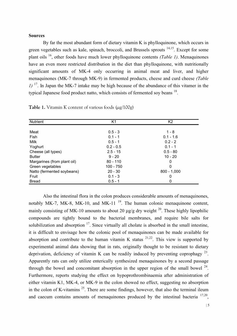
Sources By far the most abundant form of dietary vitamin K is phylloquinone, which occurs in
green vegetables such as kale, spinach, broccoli, and Brussels sprouts 14,15. Except for some plant oils 16, other foods have much lower phylloquinone contents (Table 1). Menaquinones have an even more restricted distribution in the diet than phylloquinone, with nutritionally significant amounts of MK-4 only occurring in animal meat and liver, and higher menaquinones (MK-7 through MK-9) in fermented products, cheese and curd cheese (Table 1) 17. In Japan the MK-7 intake may be high because of the abundance of this vitamer in the typical Japanese food product natto, which consists of fermented soy beans 18.
Table 1. Vitamin K content of various foods (µg/100g)
Nutrient K1 K2
Meat 0.5 - 3 1 - 8Fish 0.1 - 1 0.1 - 1.6Milk 0.5 - 1 0.2 - 2Yoghurt 0.2 - 0.5 0.1 - 1Cheese (all types) 2.5 - 15 0.5 - 80Butter 9 - 20 10 - 20Margarines (from plant oil) 80 - 110 0Green vegetables 100 - 750 0Natto (fermented soybeans) 20 - 30 800 - 1,000Fruit 0.1 - 3 0Bread 0.5 - 1 0
Also the intestinal flora in the colon produces considerable amounts of menaquinones, notably MK-7, MK-8, MK-10, and MK-11 19. The human colonic menaquinone content,
mainly consisting of MK-10 amounts to about 20 µg/g dry weight 20. These highly lipophilic compounds are tightly bound to the bacterial membranes, and require bile salts for solubilization and absorption 17. Since virtually all cholate is absorbed in the small intestine, it is difficult to envisage how the colonic pool of menaquinones can be made available for absorption and contribute to the human vitamin K status 21,22. This view is supported by experimental animal data showing that in rats, originally thought to be resistant to dietary deprivation, deficiency of vitamin K can be readily induced by preventing coprophagy 23. Apparently rats can only utilize enterically synthesized menaquinones by a second passage through the bowel and concomitant absorption in the upper region of the small bowel 24. Furthermore, reports studying the effect on hypoprothrombinaemia after administration of either vitamin K1, MK-4, or MK-9 in the colon showed no effect, suggesting no absorption in the colon of K-vitamins 25. There are some findings, however, that also the terminal ileum and caecum contains amounts of menaquinones produced by the intestinal bacteria 17,20,

6 |
although the inter-individual variation was large. In rats, a positive correlation was found between the menaquinone content in the caecum and the liver after various dietary interventions 26. The menaquinones present in this part of the gastro-intestinal tract could explain the presence of the long-chain menaquinones in the liver, and may therefore contribute to the vitamin K-status.
Synthetic forms of both phylloquinone and MK-4 are commercially available and used in humans a) for the prevention of the haemorrhagic disease of the newborn (HDN), b) to correct an overshoot of coumarin drugs during oral anticoagulant treatment, and c) to maintain normal haemostasis during poisoning with vitamin K-antagonists 27,28. Moreover, an increasing number of vitamin supplements containing synthetic vitamin K (generally
phylloquinone) in dosages between 100 and 500 µg per tablet are presently sold as over the counter products. In Japan MK-4 (menatetrenone) is registered as a therapeutic agent for postmenopausal osteoporosis, in which form it is widely prescribed in a dosage of 45 mg/day for continuous treatment periods of many years. Plasma levels of vitamin K
The extent to which plasma vitamin K reflects tissue vitamin K status is still a matter of debate. The very low levels of menaquinones in fasting plasma are beyond the limit of detection of current methodologies. Most of the data on plasma vitamin K are therefore limited to phylloquinone. The use of plasma phylloquinone to estimate tissue phylloquinone status is limited by the marked influence of various lipids. High plasma values vitamin K were found in hyperlipidaemia and hyperlipoproteinaemia 29-31. Low vitamin K plasma values were found in osteoporosis, coeliac disease, parenteral feeding, and severe pancreatic insufficiency or biliary obstruction 4,31,32. Fasting plasma concentrations of vitamin K were positively correlated with dietary K-intake 29,33. Because the ratio plasma phylloquinone / triglycerides lead to a decrease in intra- and inter individual variations, some authors use this ratio rather than plasma phylloquinone alone 22,34. Furthermore, fasting plasma concentrations of phylloquinone are strongly influenced by the common genetic polymorphism of apoE, being higher in patients with the apoE2 variant, intermediate in apoE3, and lower in apoE4. This is in line with the known link between apoE phenotype and the rate of hepatic clearance of chylomicron remnants. The length of time that phylloquinone remains in the circulation may have implications for the transport of phylloquinone to extra hepatic tissues such as bone 35. Absorption, bioavailability and tissue distribution
Like other fat-soluble vitamins, K vitamins taken in the diet are absorbed in the intestine by the bile salt-mediated pathway. After solubilization into lipid droplets, vitamin K

| 7
is incorporated in micelles consisting of triacylglycerol, phospholipids, and chlolesterol esters. After passing the enterocytes, vitamin K is released into the blood stream, via the lymph, in so-called chylomicrons. These lipoproteins are catabolized in the circulation by the action of lipoprotein lipase (LPL) resulting in chylomicron remnants 36. These chylomicron remnants are then cleared mainly by the liver via an apoE receptor-specific uptake 37,38 (Figure 2).
Figure 2.
Schematic overview of vitamin absorption and distribution in humans. Vitamin K is absorbed in the
intestines with the aid of bile salts. The so-called mixed micelles enter the bloodstream via the
lymphatic system. In the blood stream vitamin K is transported in lipoprotein particles. These
lipoproteins deliver the vitamin K to their target cells.
Consistent with this model is the striking impairment of absorption of vitamin K in patients with extra-hepatic cholestasis and severe pancreatic insufficiency 31. It has been estimated that healthy adults absorb ~80% of an oral dose of phylloquinone given in a detergent-solubilized form 31. The efficacy with which vitamin K is liberated from the food matrix is still a matter of debate and the reported bioavailablility ranges from <10-70% 39-41.

8 |
Factors affecting uptake from the diet include the amount and type of fat in the diet, and matrix in which the vitamin is dissolved. Therefore, considerable differences may be expected between phylloquinone and the menaquinones. Extraction of phylloquinone from vegetables, where it is tightly bound to the chloroplast membrane, is thought to be less efficient than from dairy produce 22.
In plasma, phylloquinone is mainly associated with the triacylglycerol rich lipoprotein (TGRLP) fraction 22,34,42 and only a small part of vitamin K is associated with the LDL and HDL fraction. Although the liver clears most of the postprandial phylloquinone, only 10% of the total liver store comprises of phylloquinone whereas the other 90% are menaquinones 4,43. That the liver stores of phylloquinone are subject to rapid changes was shown by Usui et al. 43, who demonstrated that after only 3 days of severe dietary depletion hepatic phylloquinone had decreased to ~25% of original concentration. Although the liver contains large amounts of menaquinones, plasma menaquinones are difficult to measure and thus far only MK-7 and MK-8 could be detected 4,43,44. Extra-hepatic tissues hardly contain menaquinones except MK-4, which accumulates in testis, aorta, pancreas, brain, and salivary glands 45,46. Thijssen et al. showed that after feeding rats a vitamin K deficient diet for 9 days, hepatic stores were almost depleted whereas other tissues like pancreas, brain and sternum still contained considerable amounts of vitamin K, mainly in the form of MK-4 47. This suggests differences in tissue uptake and metabolism. The fact that there is a tissue-specific vitamin K accumulation had been already demonstrated in 1960 by Billeter and Martius. The authors found that after an oral dose of phylloquinone to rats, MK-4 was found in skeletal muscle, heart, and kidney but hardly in the liver 48. Later, this observation was confirmed by Thijssen et al. and Yamamoto et al. who found that many rat tissues, including pancreas, sternum, brain and heart, accumulated MK-4 after oral phylloquinone intake 47,49.The question remained whether the conversion of phylloquinone into MK-4 is exerted by the bacteria in the gut or in the tissues. In 1996, Ronden et al. found that in germ free rats fed exclusively a phylloquinone rich diet, many tissues accumulated MK-4 50. Since germ-free rats lack an intestinal flora, these experiments were the first direct proof that the conversion of phylloquinone into MK-4 takes place in some well-defined tissues. This observation was confirmed by Davidson et al. 51. Biological activity of K-vitamins
The functional group in vitamin K is the methylated naphtoquinone ring found in both phylloquinone and menaquinones. In vitro, the cofactor activity of vitamin K can be
measured directly in specific assays for γ-glutamyl carboxylase and KO-reductase 52,53. The
cofacor activity was found to be dependent on the side chain of the vitamin. Based on the Vsat/Km ratios, the long chain menaquinones (n > 5) were most active 52. In vivo, the
| 9
biological acitivity can be assessed by measuring the potency in counteracting hypoprothrombinaemia. Matschiner and Taggart used rats to investigate the biological activity of various K-vitamins by injecting them intracardially (to exclude absorption effects), and found that 18 hours after injection the activity of phylloquinone in reversing hypoprothrombinaemia was 8 times higher than that of MK-4, but that long chain menaquinones (MK-7 to MK-10) were up to 25 times more active than phylloquinone 54. Also Groenen et al. found a 2-5 times better efficacy for phylloquinone than for MK-4 in reversing hypoprothombinaemia 55. In another study Will and Suttie fed rats 1-3 weeks with either phylloquinone or MK-9 and found that MK-9 was less active than phylloquinone 56. This result is inconsistent with that of Matschiner and Taggart 54, but the protocols are difficult to compare. First, the administration routes were different and second, the time point measured after injection was different. Reedstrom and Suttie measured the sub-cellular distribution and the utilization of K-vitamins after blocking the K-cycle with warfarin 57. It was found that both MK-9 and MK-9 epoxide were localized in the mitochondria rather than in the microsomes whereas phylloquinone was equally distributed over the two sub-cellular fractions. Since virtually all enzymes of the K-cycle are present in the microsomes rather then
in the mitochondria, the K-vitamins present in the microsomes are readily available for γ-glutamyl carboxylase. In the same report the utilization of K-vitamins was also deduced from the KO/K ratio, which was found to be much higher for phylloquinone than for MK-9. 1.2 FUNCTION OF VITAMIN K Protein carboxylation
In mammals the only known function of vitamin K is that it serves as a
cofactor for γ-glutamylcarboxylase, an endoplasmic enzyme involved in the posttranslational
carboxylation of glutamate residues into γ-carboxyglutamate (Gla). Hence, the vitamin K-
dependent step is a carboxylation reaction taking place during the later stages of protein biosynthesis (Figure 3). Vitamin K hydroquinone (KH2) is the active coenzyme, the oxidation of which to vitamin K 2,3 epoxide (KO) provides the energy to drive the carboxylation reaction. The resulting Gla-residues are found in a limited number of proteins, and in these proteins only at certain well-defined positions 58,59. Using specific staining 60 (western blot) or HPLC detection techniques 15 (after hydrolysis) these so-called Gla-proteins can be identified as unique products of vitamin K action. In vitamin K-deficiency the carboxylation reaction cannot proceed, hence the Gla-proteins are released in an undercarboxylated form. These so-called descarboxy proteins (formerly designated as PIVKA's, proteins induced by vitamin K absence) were shown to be inactive in all cases in which the function of the corresponding

10 |
Gla-protein is known 59. Gla-residues form calcium-binding groups in proteins, so the main physico-chemical difference between normal and descarboxy proteins is their large difference in both binding of calcium from solution and the adsorption of these proteins to insoluble calcium salts.
Figure 3.
The vitamin K-cycle. Vitamin K-quinone (K) is first reduced to vitamin K-hydroquinone (KH2), and
then oxidized to vitamin K-epoxide (KO). During the oxidation step glutamic acid (Glu) is reduced to
γ-carboxyglutamic acid (Gla). Finally KO is reduced to K. 1. = NAD (H)-dependent reductase, 2. =
γ-glutamylcarboxylase enzyme, 3. = extra introduced carboxy group, 4. = dithiol-dependent KO-
reductase, 5. = dithiol-dependent K-reductase Vitamin K-recycling
After being formed as part of a polypeptide chain, free Gla will enter the circulation after protein degradation. Unlike the usual amino acids, Gla cannot be re-used and will be
| 11
excreted via the urine. So there is a 1:1 stoichiometric relation between the conversion of KH2 into KO and the number of protein-bound Gla-residues formed. On the basis of nutritional vitamin K intake and urinary Gla excretion it may be calculated that in mammals the number of carboxylation events exceeds the number of available vitamin K molecules several thousand fold. Therefore, the KO formed must be recycled, which is accomplished by an enzyme known as KO-reductase (Figure 3). Two reduction steps are required to recycle one molecule of KO into KH2 and many independent observations favour the assumption that both steps are exerted by the same enzyme 61. When KO-reductase is blocked (see below), a second enzyme (NADPH-dependent K-reductase, also known as DT-diaphorase) may take over the generation of KH2. This pathway can convert only vitamin K quinone, and not the epoxide into the hydroquinone. When the KO-reductase is blocked vitamin K cannot be recycled, which increases its nutritional requirement to values well above the normal dietary intake. Substrate recognition by carboxylase
All Gla-containing proteins are synthesized in the endoplasmic reticulum as precursor proteins containing a signal sequence for translocation over/thru the endoplasmic membrane, and a carboxylase recognition site, which is usually found within the leader-sequence of the precursor protein14,59. An exception in this respect is matrix Gla-protein (MGP) in which the carboxylase recognition site forms part of the mature protein. Among all Gla-proteins presently known the amino acid residues Phe-16 and Ala-10 in their respective carboxylase recognition sites have been remarkably conserved 14, and in vitro studies in which synthetic peptides were used as substrates for carboxylase have shown that mutations at these positions lead to a dramatic loss of enzyme-substrate affinity 62. In vivo such mutations are expected to lead to the cellular secretion of undercarboxylated and thus inactive proteins. 1.3 PHYSIOLOGICAL ROLES OF VITAMIN K-DEPENDENT PROTEINS
Gla-containing proteins have been detected in many different species including snake 63,64, snails 65, and birds 66. This thesis will focus, however, on mammalian Gla-proteins and mammalian vitamin K-metabolism. Vitamin K-dependent carboxylase has been found in nearly all mammalian tissues investigated, but it seems that - besides those mentioned below - the corresponding Gla-proteins are produced in relatively low quantities. Incompletely characterized Gla-proteins have been reported to occur in spermatozoa 67, urine 68, and extracts of calcified atherosclerotic plaques 69. Those characterized to the level of aminoacid sequence play regulating roles in either of three important physiological processes: blood coagulation, bone metabolism, and vascular biology (Table 2).

12 |
Table 2. Tissue distribution and roles of vitamin K-dependent (Gla) proteins
Gla Protein Tissue Role
Prothrombin (FII), Factors VII, IX, and X Liver (then plasma) ProcoagulantsProtein C, Z Liver (then plasma) AnticoagulantsProtein S Liver (then plasma), endothelium, bone Anticoagulant role as cofactor for protein COsteocalcin (bone Gla-protein) Bone, dentin Regulator of orderly crystallizationMatrix Gla-protein (MGP) Bone, cartilage, and most soft tissues Inhibitor of ectopic mineralizationGas-6 (growth arrest specific gene-6 protein) Most soft tissues Regulator of cell growthProline rich Gla-proteins (PRGP) Most soft tissues UnknownTransmembrane Gla-proteins (TMG) Most soft tissues Unknown Blood coagulation
Involvement in blood coagulation is the classical role of vitamin K, and the major function of Gla-residues in the coagulation factors is that they facilitate the binding of these proteins to the negatively charged phospholipids on the surface of activated blood platelets 70. In this way the reaction rate of thrombin formation is accelerated by several orders of magnitude. Gla-containing proteins involved in blood coagulation are prothrombin, the factors VII, IX, and X, as well as the anticoagulant protein C and S. Protein Z is also involved in coagulation; one of its functions was reported to be the fixation of thrombin to the site of injury 71. In 1998 Han et al. 72 showed that protein Z inhibited factor X activity dose-dependently in the presence of phospholipids and calcium by forming a complex via a protein called protein Z-inhibitor (pZI). Recently, this was confirmed in protein Z-deficient mice 73.
With the exception of protein S (which is produced in a variety of tissues) all the vitamin K-dependent coagulation factors are synthesized in the liver. Their Gla-content ranges between 10 (prothrombin) to 12 (factor IX) residues per molecule and the respective Gla-domains are essential for these proteins in their binding to negatively charged phospholipids such as those found in the outer membranes of activated blood platelets. The negatively charged Gla-residues have a high affinity for calcium ions. It has been shown by X-ray crystal structure analysis and 2D NMR spectroscopy that when bound to calcium ions the coagulation factors undergo structural changes leading to internalization of the Gla-Ca++ complex in the core of the protein, and to exposure of the phospholipid-binding domain 14,74,75. Bone metabolism
All three bone Gla-proteins presently known are synthesized by the osteoblasts (the bone forming cells), but only osteocalcin is a unique product of bone tissue. Protein S and MGP are also synthesized in a number of soft tissues including the vessel wall (see below).

| 13
Osteocalcin represents about 20% of the non-collagenous proteins in bone and is one of the most abundant proteins in humans 76. The precise function of the bone Gla-proteins is not yet clear. Osteocalcin-deficient mice turned out to grow more rapidly than the wild types, showing that osteocalcin is a negative regulator of bone formation. Furthermore, histomorphometric studies revealed that the absence of osteocalcin leads to an increased bone formation and improved quality without impairing bone resporption. 77.
Knock-out mice have also been generated for MGP. These animals showed substantial growth retardation, with excessive calcifications of their growth plates, osteopenia and bone fractures 78. Since all animals died from cardiovascular disease within 2 months after birth (see below) long term effects of MGP-deficiency on bone are still unknown. Recently, it was reported that in three independent cases the human Keutel syndrome is associated with a DNA mutation leading to the expression of non-functional MGP 79. Keutel syndrome is an autosomal recessive disorder characterized by abnormal cartilage calcification, peripheral pulmonary stenosis, short terminal phalanges, abnormal cartilage calcification of the auricles, nose, larynx, trachea and ribs, and by mid-facial hypoplasia with a depressed nasal bridge. Recently, it was reported that upon post mortem examination of KS patients also revealed concentric calcification of pulmonary, coronary, hepatic, renal, meningeal, and cerebral arteries 80. Hence the phenotypes of the MGP-deficient mouse and KS patients are similar, although the human defect does not lead to early cardiovascular mortality. Further research to elucidate this apparent contradiction remains to be carried out. Vascular biology
In the arterial vessel wall the functions of Gla-proteins are probably associated with: local inhibition of thrombosis (protein S) 81, inhibition of mineralization (MGP) 82, and stimulation of normal cell growth and prevention of apoptosis in growth arrested cells (Gas6) 83. In the circulation protein S is known to inhibit blood coagulation and thrombus formation by acting as a cofactor for activated protein C; there are no arguments suggesting that the function of the vascular synthesised protein S would be different. As was discussed above, MGP-deficient mice were born to term but died before the 8th week of life due to massive arterial calcification and rupture of the thoracic or abdominal aorta 78. These experiments established that MGP is a strong inhibitor of soft tissue calcification including cartilage and vessel wall 84. Excessive aorta mineralization was also observed in rats after 4-6 weeks of treatment with vitamin K-antagonists 85, showing that the Gla-residues in MGP are essential for its function. Gla-proteins have been extracted from human atheromatous plaques and although they were only partially characterized, the poor solubility of the human vascular Gla-proteins suggests that the major fraction may be similar to MGP 69. The function of Gas6 (which stands for: growth arrest specific gene 6 protein) has been investigated only in cell

14 |
culture systems thus far. In response to conditions leading to cell death, for instance during serum starvation, cells are induced to produce growth arrest specific proteins. Most of these proteins promote cell death (apoptosis), but Gas6 is an exception in this respect: it was shown to prevent serum starvation-induced death of fibroblasts and smooth muscle cells, and also may act as a growth-potentiating factor which acts synergistically with other known growth factors in these cells 83,86,87. Gla-residues were shown to be essential for its functions in cell culture 86. These data suggest that in humans Gas6 may play a key role in preventing the degeneration of atherosclerotic vessels. The recent observation that Gas6 is produced in both spinal motor neurons and large neurons of the dorsal root ganglia, and that Gas6 is a potent stimulator of human Schwann-cell growth suggests that this protein is involved in growth regulation in other tissues as well, but this needs further confirmation 88. 1.4 VITAMIN K REQUIREMENT
After an overnight fasting period, circulating vitamin K levels in healthy adults range between 200-800 pg/mL, however, they decrease rapidly after prolonged low vitamin K intake. In non-fasting individuals serum vitamin K concentrations may fluctuate as a result of the consumption of various foods 4,29,33. Serum vitamin K and triglyceride levels seem to be firmly correlated and presently it is not clear whether plasma concentrations (either as absolute values or as the vitamin K / triglyceride ratio) are an accurate indicator of tissue reserves. Experimental animal studies have demonstrated that the distribution of vitamin K throughout the various tissues depends on the daily dose of vitamin K, and on the type of vitamin K 45. It is believed, therefore, that a subject's vitamin K status cannot be assessed from overall variables like serum vitamin K or urinary Gla-excretion. A more reliable method to determine tissue vitamin K-status seems to be measurement of the Gla-content of Gla-containing proteins. The occurrence of undercarboxylated proteins in the circulation has enabled us to develop biochemical tests with which vitamin K deficiency can be assessed by measuring the quality (= active proteins) of the products formed in specific tissues. Vitamin K deficiency
A: Blood coagulation: Although in the absence of precipitating causes vitamin K deficiency is rarely seen in adults (measured by the overall coagulation tests), deficiency in the newborn or haemorrhagic disease of the newborn (HDN) remains a significant worldwide cause of infant morbidity and mortality 89. The disease can be subdivided into three clinical patterns: early HDN (within 24 hours after birth), classical HDN (days 2 - 7 after birth), and late onset HDN (week 2 - 6 months after birth). There are several hypotheses for the poor vitamin K status of the newborn. First, the placental barrier for maternal vitamin K leads to a
| 15
calculated ratio of vitamin K in cord blood and maternal blood of 1:30 90 and second, the vitamin K content of human milk is low 91. Therefore, breast fed babies have a higher incidence of classical or late HDN 89,92. The use of medication during pregnancy is another reason for inducing a poor fetal vitamin K status. It is known that anticonvulsants and tuberculostatics interfere with vitamin K metabolism and increase the incidence of early HDN 93,94. As prophylaxis for HDN, vitamin K is given routinely to newborns in all developed countries 95. The route of administration is either orally or intramuscularly. The latter administration was reported to have an association with childhood cancer in two British hospitals in a retrospective study 96,97. A number of prospective epidemiological studies did not confirm this association 98-100. However, the finding of Golding et al. resulted in the change of recommendation in many countries in favor of oral administration.
B: bone metabolism. In 1991 it had been already found in population-based studies that circulating osteocalcin is far more sensitive to poor vitamin K status than other Gla-proteins 101. There are two mechanisms underlying this observation. First, the affinity of the
endoplasmic osteocalcin precursor proteins (pro-osteocalcin) for γ-glutamylcarboxylase is distinctly lower than that of other Gla-proteins. It was demonstrated that this poor affinity is
due to an Ala-10 → Gly mutation in the leader sequence: during periods of low vitamin K supply, precursors of a variety of vitamin K-dependent proteins will accumulate, and the competition for the carboxylase binding site will inevitably be lost by pro-osteocalcin 102.
Later, it was demonstrated by Houben et al. that osteocalcin has a γ-carboxylase binding site distinct form that of other vitamin K-dependent proteins 103. Second, after its synthesis by the osteoblasts, osteocalcin is set free amidst the hydroxyapatite matrix of bone, which will preferentially adsorb the normally carboxylated osteocalcin, so that undercarboxylated osteocalcin will be selectively set free in the circulation. Optimal carboxylation of the bone Gla-proteins requires a higher vitamin K intake, and in recent years it has been demonstrated that undercarboxylation of osteocalcin is quite common, notably in the elderly 104,105. It is important, therefore, to realize that vitamin K-deficiency is a state which should be defined per tissue, not for a whole organism. As early as 1985 it had been reported that low circulating phylloquinone levels were associated with an increased risk for osteoporotic hip fractures 32,106. More recently these data were confirmed 107, and it was found that plasma menaquinone concentrations were also substantially reduced in osteoporotic women 108. The question remained whether the observed low circulating vitamin K levels were associated with a detectable vitamin K deficiency of bone tissue. Knapen et al. developed an assay for the assessment of the ratio between normal and undercarboxylated osteocalcin, and it was found that the fraction of undercarboxylated osteocalcin is increased in postmenopausal women 104,105. Szulc et al. reported that undercarboxylated osteocalcin is inversely correlated with bone mass 109, and that undercarboxylated osteocalcin is an independent predictive

16 |
marker for hip fracture risk 110,111.These data where confirmed in a group of women who were between 1 and 10 years postmenopausal, and it was found that age, body weight and undercarboxylated osteocalcin are three independent variables from which the bone mineral density may be calculated with an accuracy of over 70% 112.
C: Vascular biology. Atherosclerosis is the major cause of mortality in the Western world 113. It is characterized by the presence of atherosclerotic lesions in the arterial intima. Atherosclerosis is thought to progress from fatty streak to fibrous plaques, and finally to calcified atheromata 114. Vascular calcification does not remain restricted to the intima, but also occurs in the media were it is known as Mönckeberg sclerosis 115. In contrast to intimal calcification, medial calcification occurs in the absence of inflammatory cell infiltration and lipid deposition 116,117. Although both processes are different in many ways, it is generally accepted that vascular calcification is an active process rather than a passive process resulting from the accumulation of calcium released from dying cells. With the generation of the MGP knock-out mouse 78, which exhibits extensive and lethal calcification and cartilaginous metaplasia of the media of all elastic arteries, the role of vitamin K and vitamin K-dependent proteins in vascular calcification has gained substantial attention. Several Gla-proteins have been isolated from the calcified atherosclerotic plaques 69,118-120. The finding of different proteins containing Gla in bone and ectopic calcification including hardened plaque suggests that certain calcium-binding proteins containing this amino acid may be of importance in the mineralization process. Indeed, on a level of gene expression, MGP was found to be up-regulated in vivo and in vitro in association with calcification 82,117,120. The role of vitamin K in atherosclerosis was recently investigated. In these studies the authors 121,122 screened for the occurrence of calcifications in the abdominal aorta in more then hundred of apparently healthy volunteers between 60 and 80 years of age. They also recorded the nutritional habits and preferences with the aid of food frequency questionnaires. The subjects were classified into four categories according to the degree of aortic calcification, ranging from no visible calcification to severe calcifications. It turned out that subjects with severe atherosclerotic calcifications had a significantly lower long-term vitamin K intake, a higher amount of undercarboxylated bone Gla-protein (osteocalcin), and lower bone mass. These data suggest that a biochemical vitamin K-deficiency is common in atherosclerotic subjects.
On the basis of these data we propose to re-define vitamin K-deficiency as follows. Mild vitamin K deficiency is a condition in which the circulating osteocalcin is significantly undercarboxylated, whereas the blood coagulation factor prothrombin still contains a functional Gla domain 123,124. Severe vitamin K deficiency may than be regarded as a condition in which descarboxyprothrombin is detectable in the circulation.
| 17
Vitamin K and coumarins Nearly all known inhibitors of the vitamin K cycle interfere with KO-reductase, and
to a far lesser extend with carboxylase and DT-diaphorase 61,125 (Figure3). Well known inhibitors are those derived from 4-hydroxycoumarin. Because these coumarin derivatives effectively block the recycling of vitamin-KO, their action results in an exhaustion of the KH2 stores and thus in an apparent vitamin K-deficiency. During the last 50 years vitamin K-antagonists (coumarin derivatives) have been used on a large scale for the treatment of patients at risk of thrombo-embolic events 8. The drugs are generally administered for periods of several years or longer, a major area of clinical application is the prevention of myocardial re-infarction. Most of these patients are well over 50 years of age, although during recent years oral anticoagulant therapy at younger ages has become more common.
In the early 1970's it became usual to treat relatively young patients with coumarin derivatives, for instance those how had received an artificial heart valve. Because of compelling medical indications the treatment of women was continued even after they had become pregnant. It appeared, however, that in about 30% of the cases the newborns had developed serious bone defects 126. Subsequent experiments in animals (rats, lambs) have shown that notably in rapidly growing (young) bone tissue coumarin derivatives interfere with the regulation of calcium deposition resulting in excessive and irregular precipitation of calcium salts in areas which are normally not calcified (e.g. the growth plates). This leads to bone deformations and growth reduction 127-129, phenomena which must be attributed to strongly impaired activities of the bone Gla-proteins. Remarkably, coumarin derivatives appear to have less effect on adult bone 130-133. Congenital vitamin K deficiencies
Carboxylase and reductase deficiency. Combined deficiencies of all clotting factors are rare and obviously one has first to exclude unequivocally the occurrence of vitamin K-deficiency or unsuspected intake of coumarins. Since the first suggestion of a defect in the carboxylase/reductase system 134, several other cases have been reported some of which also mentioned the occurrence of bone abnormalities comparable with those seen in warfarin-induced embryopathy 135. Such combined defects of bone development before birth and of the blood clotting system during childhood strongly suggest a decreased carboxylation of all Gla-proteins, and may be due to mutations in either the carboxylase or KO-reductase. After the carboxylase cDNA had been sequenced and cloned 136, some of these cases could indeed be associated with a mutation in the carboxylase gene 137,138.
Mutations in the clotting factor Pro-domain. These mutations may lead either to poor recognition of the precursor protein by carboxylase 102,139, or to impaired cleavage of the pro-peptide during protein maturation 140. Reported cases of impaired carboxylase recognition

18 |
refer to factor IX and were discovered by the clinical symptoms (acquired haemophilia B) after mild coumarin treatment. Impaired pro-peptide cleavage leads to fully carboxylated clotting factors which lack procoagulant activity because the extension at their amino terminus strongly interferes with their binding to negatively charged phospholipid vesicles 140.
Mutations in the clotting factor Gla-domain. Besides frame-shift mutations and the introduction of stop codons, direct mutations in the Gla-domain may result in replacement of regular amino acids and of Gla-residues. Whereas all mutations may give rise to altered conformations, mutations leading to a decreased number of Gla-residues will inevitably decrease the protein's charge and calcium binding capacity. In the case of the four vitamin K-dependent clotting factors this will result in a decreased haemostatic capacity and a risk of bleeding, whereas in the case of the coagulation inhibiting protein C and protein S the expected result is an increased thrombosis risk. Indeed several of such mutations have been reported during recent years, and the clinical symptoms matched the expectations 141,142. Whereas in the case of protein C and protein S mutations the thrombosis risk could be successfully normalized by oral anticoagulant therapy, the symptoms of mutations in the clotting factors cannot be relieved by vitamin K.

| 19
1.5 INTRODUCTION TO THIS THESIS Vitamin K is a fat-soluble vitamin which has a well-defined role in the process of
blood coagulation. For over half a century, this has been its only known function. During the last two decades, however, the discovery of extra-hepatic Gla-proteins and their functions, has renewed the interest in vitamin K. These so-called extra-hepatic Gla-proteins are now known to occur in a wide variety of tissues including bone, arterial vessel wall, testis, lung, pancreas and spleen. All recommendations for dietary vitamin K intake are based on the requirements for the synthesis of blood coagulation factors. Extra-hepatic vitamin K requirement has remained unknown thus far. Moreover, thus far the K2 vitamins have received little attention, and their contribution to the human vitamin K status was unknown before we started the work reported here.
In this thesis we have investigated the metabolic pathways of K vitamins following ingestion either in the form of a standardized diet or as supplements. First, we have established an up-to-date food composition table for vitamins K1 and K2 in a wide range of foods purchased in the Netherlands. With the aid of this data base we have investigated in experimental animals and human volunteers the absorption of K-vitamins, their plasma transport by lipoproteins, and the effect of other dietary factors on vitamin K absorption and metabolism. Next, we have demonstrated the occurrence of vitamin K-deficiency in cases of increased expenditure or impaired intestinal uptake, and we have addressed the question of whether vitamin K-containing food supplements may be safely marketed as OTC products. Finally, we have studied the accumulation of the vitamin K-dependent protein MGP in calcifying human arteries. The results from the latter study form the basis for our hypothesis that vitamin K intake at levels above the present RDA are required for optimal protection against arterial calcification.

20 |
REFERENCES 1. Dam H. The antihemorrhagic vitamin of the chick: occurrence and chemical nature. Nature.
1935;135:652-653. 2. Almquist HJ. Vitamin K. Physiol Rev. 1941;21:194-216. 3. Haroon Y, Shearer MJ, Rahim S, Gunn WG, McEnery G, Barkhan P. The content of
phylloquinone (vitamin K1) in human milk, cows' milk and infant formula foods determined by high-performance liquid chromatography. J Nutr. 1982;112:1105-1117.
4. Shearer MJ, McCarthy PT, Crampton OE, Mattock MB. The assessment of human vitamin K status from tissue measurement. Current advances in vitamin K research (ed. JW Suttie). 1988;Elsevier:437-452.
5. Shofield FW. A brief account of a disease in cattle simulating hemorrhagic septicemia due to feeding sweet clover. Can Vet Rec. 1922;3:74-79.
6. Campbell MA. Haemorrhagic disease in cattle. J Biol Chem. 1941;18:21-33. 7. Hadler MR, Shadbolt RS. Novel 4-hydroxycoumarin anticoagulants active against resistant rats.
Nature. 1975;253:275-277. 8. Hirsh J, Fuster V. Guide to anticoagulant therapy. Part 2: Oral anticoagulants. American Heart
Association. Circulation. 1994;89:1469-1480. 9. Lichtenthaler HK. The plant prenyllipids, including cartenoids, chlorophylls and
prenylquinones. Plant Lipids (ed. Moore, T.). 1993;CRC Press, Boca Raton:421-464. 10. Dialameh GH, Taggart WV, Matschiner JT, Olson RE. Isolation and characterization of
menaquinone-4 as a product of menadione metabolism in chicks and rats. Int J Vitam Nutr Res. 1971;41:391-400.
11. Ritzl VF. Biosynthetische veranderungen von tritium-markiertem vitamin K3 in menschen. Atomkernenergie. 1970;15:162-166.
12. Allison AC. Danger of vitamin K to newborn. Lancet. 1955;I:669. 13. Meyer TC, Angus J. The effect of large doses of 'synkavit' in the newborn. Arch Dis Child.
1956;31:213-215. 14. Furie B, Bouchard BA, Furie BC. Vitamin K-dependent biosynthesis of gamma-
carboxyglutamic acid. Blood. 1999;93:1798-808. 15. Kuwada M, Katayama K. An improved method for the determination of gamma-
carboxyglutamic acid in proteins, bone, and urine. Anal Biochem. 1983;131:173-9. 16. Ferland G, Sadowski JA. Vitamin K1 (phylloquinone) content of edible oils: effects of heating
and light exposure. J Agricult Food Chem. 1992;40:1869-1873. 17. Shearer MJ, Kries RV, Saupe J. Comparative aspects of human vitamin K metabolism and
nutriture. J Nutr Sci Vitaminol Tokyo. 1992:413-6. 18. Sakano T, Nagaoka T, Morimoto A, Hirauchi K. Measurement of K vitamins in human and
animal feces by high-performance liquid chromatography with fluorometric detection. Chem Pharm Bull (Tokyo). 1986;34:4322-4326.
19. Shearer MJ. Vitamin K. Lancet. 1995;345:229-34. 20. Conly JM, Stein K. Quantitative and qualitative measurements of K vitamins in human intestinal
contents. Am J Gastroenterol. 1992;87:311-6. 21. Conly JM, Stein K. The production of menaquinones (vitamin K2) by intestinal bacteria and
their role in maintaining coagulation homeostasis. Prog Food Nutr Sci. 1992;16:307-43. 22. Shearer MJ. Vitamin K metabolism and nutriture. Blood-Rev. 1992;6:92-104. 23. Barnes RH, Fiala G. Effects of the prevention of coprophagey in the rat. J Nutr.
1959;68:603-614. 24. Ichihashi T, Takagishi Y, Uchida K, Yamada H. Colonic absorption of menaquinone-4 and

| 21
menaquinone-9 in rats. J Nutr. 1992;122:506-512. 25. Groenen van Dooren MM, Ronden JE, Soute BA, Vermeer C. Bioavailability of phylloquinone
and menaquinones after oral and colorectal administration in vitamin K-deficient rats. Biochem Pharmacol. 1995;50:797-801.
26. Ramotar K, Krulicki W, Gray G, Louie TJ. Studies on intestinal and hepatic concentrations of menaquinone and hypoprothrombinaemia in vitamin K1 deficiency in rats. Elsevier, New York. 1988;Current advances in vitamin K research (ed. Suttie, J.W.):493-498.
27. Chong LL, Chau WK, Ho CH. A case of 'superwarfarin' poisoning. Scand J Haematol. 1986;36:314-315.
28. Lipton RA, Klass EM. Human ingestion of a 'superwarfarin' rodenticide resulting in a prolonged anticoagulant effect. Jama. 1984;252:3004-3005.
29. Booth SL, Tucker KL, McKeown NM, Davidson KW, Dallal GE, Sadowski JA. Relationships between dietary intakes and fasting plasma concentrations of fat-soluble vitamins in humans. J Nutr. 1997;127:587-592.
30. Mattock MB, Shearer MJ, Rahim S, Redmond S, El-Gohari R, Barkhan P. The plasma transport of vitamin K1 (phylloquinone) in hyperlipoprotaemia. Clin Sci. 1983;64:63p.
31. Shearer MJ, McBurney A, Barkhan P. Studies on the absorption and metabolism of phylloquinone (vitamin K1) in man. Vitam Horm. 1974;32:513-42.
32. Hart JP, Shearer MJ, Klenerman L, Catterall A, Reeve J, Sambrook PN, Dodds RA, Bitensky L, Chayen J. Electrochemical detection of depressed circulating levels of vitamin K1 in osteoporosis. J Clin Endocrinol Metab. 1985;60:1268-9.
33. Booth SL, Sokoll LJ, O'Brien ME, Tucker K, Dawson-Hughes B, Sadowski JA. Assessment of dietary phylloquinone intake and vitamin K status in postmenopausal women. Eur J Clin Nutr. 1995;49:832-841.
34. Sadowski JA, Hood SJ, Dallal GE, Garry PJ. Phylloquinone in plasma from elderly and young adults: factors influencing its concentration. Am-J-Clin-Nutr. 1989;50:100-8.
35. Kohlmeier M, Salomon A, Saupe J, Shearer MJ. Transport of vitamin K to bone in humans. J Nutr. 1996;126:1192s-6s.
36. Havel RJ, Kane JP. Introduction: structure and metabolism of plasma lipoproteins. The metabolic and molecular basis of inherited disease (ed. Scriver, C.R.). 1995;7th ed. New York: McGraw-Hill:1841-1851.
37. Sherrill BC, Innerarity TL, Mahley RW. Rapid hepatic clearance of the canine lipoproteins containing only the E apoprotein by a high affinity receptor. Identity with the chylomicron remnant transport process. J Biol Chem. 1980;255:1804-7.
38. Brown MS, Herz J, Kowal RC, Goldstein JL. The low-density lipoprotein receptor-related protein: double agent or decoy? Curr Opin Lipodol. 1991;2:65-72.
39. Gijsbers BL, Jie KS, Vermeer C. Effect of food composition on vitamin K absorption in human volunteers. Br J Nutr. 1996;76:223-9.
40. Garber AK, Binkley NC, Krueger DC, Suttie JW. Comparison of phylloquinone bioavailability from food sources or a supplement in human subjects. J Nutr. 1999;129:1201-3.
41. Olson RE. The function and metabolism of vitamin K. Annu Rev Nutr. 1984;4:281-337. 42. Lamon-Fava S, Sadowski JA, Davidson KW, O'Brien ME, McNamara JR, Schaefer EJ. Plasma
lipoproteins as carriers of phylloquinone (vitamin K1) in humans. Am J Clin Nutr. 1998;67:1226-1231.
43. Usui Y, Tanimura H, Nishimura N, Kobayashi N, Okanoue T, Ozawa K. Vitamin K concentrations in the plasma and liver of surgical patients. Am J Clin Nutr. 1990;51:846-52.
44. Hodges SJ, Pilkington MJ, Shearer MJ, Bitensky L, Chayen J. Age-related changes in the circulating levels of congeners of vitamin K2, menaquinone-7 and menaquinone-8.

22 |
Clin Sci Colch. 1990;78:63-6. 45. Ronden JE, Thijssen HH, Vermeer C. Tissue distribution of K-vitamers under different
nutritional regimens in the rat. Biochim Biophys Acta. 1998;1379:16-22. 46. Thijssen HHW, Drittij-Reijnders MJ, Fischer MA. Phylloquinone and menaquinone-4
distribution in rats: synthesis rather than uptake determines menaquinone-4 organ concentrations. J Nutr. 1996;126:537-543.
47. Thijssen HH, Drittij Reijnders MJ. Vitamin K distribution in rat tissues: dietary phylloquinone is a source of tissue menaquinone-4. Br J Nutr. 1994;72:415-25.
48. Billeter M, Martius C. Über die umwandlung von phyllochinon (vitamin K1) in vitamin K2(20) im tierkörper. Biochem Z. 1960;333:430-439.
49. Thijssen HH, Drittij Reijnders MJ, Fischer MA. Phylloquinone and menaquinone-4 distribution in rats: synthesis rather than uptake determines menaquinone-4 organ concentrations. J Nutr. 1996;126:537-43.
50. Ronden JE, Drittij-Reijnders MJ, Vermeer C, Thijssen HH. Intestinal flora is not an intermediate in the phylloquinone- menaquinone-4 conversion in the rat. Biochim Biophys Acta. 1998;1379:69-75.
51. Davidson RT, Foley AL, Engelke JA, Suttie JW. Conversion of dietary phylloquinone to tissue menaquinone-4 in rats is not dependent on gut bacteria. J Nutr. 1998;128:220-223.
52. Buitenhuis HC, Soute BA, Vermeer C. Comparison of the vitamins K1, K2 and K3 as cofactors for the hepatic vitamin K-dependent carboxylase. Biochim Biophys Acta. 1990;1034:170-5.
53. Yen CS, Mack DO. Solubilized rat liver vitamin K carboxylase demonstrates little selectivity between vitamin K1 and the menaquinones. Proc Soc Exp Biol Med. 1980;165:306-308.
54. Matschiner JT, Taggart WV. Bioassay of vitamin K by intracardial injection in deficient adult male rats. J Nutr. 1968;94:57-9.
55. Groenen-van Dooren MM, Soute BA, Jie KS, Thijssen HH, Vermeer C. The relative effects of phylloquinone and menaquinone-4 on the blood coagulation factor synthesis in vitamin K-deficient rats. Biochem Pharmacol. 1993;46:433-437.
56. Will BH, Suttie JW. Comparative metabolism of phylloquinone and menaquinone-9 in rat liver. J Nutr. 1992;122:953-8.
57. Reedstrom CK, Suttie JW. Comparative distribution, metabolism, and utilization of phylloquinone and menaquinone-9 in rat liver. Proc-Soc-Exp-Biol-Med. 1995;209:403-9.
58. Furie B, Furie BC. Molecular and cellular biology of blood coagulation. N Engl J Med. 1992;326:800-6.
59. Vermeer C. Gamma-carboxyglutamate-containing proteins and the vitamin K-dependent carboxylase. Biochem J. 1990;266:625-36.
60. Jie KS, Gijsbers BL, Vermeer C. A specific colorimetric staining method for gamma-carboxyglutamic acid-containing proteins in polyacrylamide gels. Anal Bioch. 1995;224:163-5.
61. Fasco MJ, Hildebrandt EF, Suttie JW. Evidence that warfarin anticoagulant action involves two distinct reductase activities. J Biol Chem. 1982;257:11210-2.
62. Stanley TB, Wu SM, Houben RJ, Mutucumarana VP, Stafford DW. Role of the propeptide and gamma-glutamic acid domain of factor IX for in vitro carboxylation by the vitamin K-dependent carboxylase. Biochemistry. 1998;37:13262-8.
63. Tans G, Govers Riemslag JW, van Rijn JL, Rosing J. Purification and properties of a prothrombin activator from the venom of Notechis scutatus scutatus. J Biol Chem. 1985;260:9366-72.
64. Speijer H, Govers-Riemslag JW, Zwaal RF, Rosing J. Prothrombin activation by an activator from the venom of Oxyuranus scutellatus (Taipan snake). J Biol Chem. 1986;261:13258-13267.
65. McIntosh JM, Olivera BM, Cruz LJ, Gray WR. Gamma-carboxyglutamate in a neuroactive

| 23
toxin. J Biol Chem. 1984;259:14343-6. 66. Lian JB, Hauschka PV, Gallop PM. Properties and biosynthesis of a vitamin K-dependent
calcium binding protein in bone. Fed Proc. 1978;37:2615-2620. 67. Soute BA, Muller Esterl W, de Boer van den Berg MA, Ulrich M, Vermeer C. Discovery of a
gamma-carboxyglutamic acid-containing protein in human spermatozoa. FEBS-Lett. 1985;190:137-41.
68. Nakagawa Y, Abram V, Coe FL. Isolation of calcium oxalate crystal growth inhibitor from rat kidney and urine. Am J Physiol. 1984;247:F765-F772.
69. Gijsbers BL, van Haarlem LJ, Soute BA, Ebberink RH, Vermeer C. Characterization of a Gla-containing protein from calcified human atherosclerotic plaques. Arteriosclerosis. 1990;10:991-5.
70. Davie EW. Biochemical and molecular aspects of the coagulation cascade. Thromb Haemost. 1995;74:1-6.
71. Hogg PJ, Stenflo J. Interaction of vitamin K-dependent protein Z with thrombin. Consequences for the amidolytic activity of thrombin and the interaction of thrombin with phospholipid vesicles. J Biol Chem. 1991;266:10953-8.
72. Han X, Fiehler R, Broze GJ, Jr. Isolation of a protein Z-dependent plasma protease inhibitor. Proc Natl Acad Sci U S A. 1998;95:9250-5.
73. Yin ZF, Huang ZF, Cui J, Fiehler R, Lasky N, Ginsburg D, Broze GJ, Jr. Prothrombotic phenotype of protein Z deficiency. Proc Natl Acad Sci U S A. 2000;97:6734-8.
74. Li L, Darden TA, Freedman SJ, Furie BC, Furie B, Baleja JD, Smith H, Hiskey RG, Pedersen LG. Refinement of the NMR solution structure of the gamma-carboxyglutamic acid domain of coagulation factor IX using molecular dynamics simulation with initial Ca2+ positions determined by a genetic algorithm. Biochemistry. 1997;36:2132-8.
75. Soriano-Garcia M, Padmanabhan K, de Vos AM, Tulinsky A. The Ca2+ ion and membrane binding structure of the Gla domain of Ca- prothrombin fragment 1. Biochemistry. 1992;31:2554-66.
76. Hauschka PV, Lian JB, Cole DE, Gundberg CM. Osteocalcin and matrix Gla protein: vitamin K-dependent proteins in bone. Physiol rev. 1989;69:990-1047.
77. Ducy P, Desbois C, Boyce B, Pinero G, Story B, Dunstan C, Smith E, Bonadio J, Goldstein S, Gundberg C, Bradley A, Karsenty G. Increased bone formation in osteocalcin-deficient mice. Nature. 1996;382:448-52.
78. Luo G, Ducy P, McKee MD, Pinero GJ, Loyer E, Behringer RR, Karsenty G. Spontaneous calcification of arteries and cartilage in mice lacking matrix GLA protein. Nature. 1997;385:78-81.
79. Munroe PB, Olgunturk RO, Fryns JP, Van Maldergem L, Ziereisen F, Yuksel B, Gardiner RM, Chung E. Mutations in the gene encoding the human matrix Gla protein cause Keutel syndrome. Nat Genet. 1999;21:142-4.
80. Meier M, Weng LP, Alexandrakis E, Ruschoff J, Goeckenjan G. Tracheobronchial stenosis in Keutel syndrome. Eur Respir J. 2001;17:566-9.
81. Fair DS, Marlar RA, Levin EG. Human endothelial cells synthesize protein S. Blood. 1986;67:1168-71.
82. Proudfoot D, Shanahan CM, Weissberg PL. Vascular calcification: new insights into an old problem [editorial; comment]. J Pathol. 1998;185:1-3.
83. Goruppi S, Ruaro E, Schneider C. Gas6, the ligand of Axl tyrosine kinase receptor, has mitogenic and survival activities for serum starved NIH3T3 fibroblasts. Oncogene. 1996;12:471-80.
84. Schinke T, McKee MD, Karsenty G. Extracellular matrix calcification: where is the action? [news; comment]. Nat Genet. 1999;21:150-1.
85. Price PA, Faus SA, Williamson MK. Warfarin causes rapid calcification of the elastic lamellae

24 |
in rat arteries and heart valves. Arterioscler Thromb Vasc Biol. 1998;18:1400-7. 86. Nakano T, Kawamoto K, Kishino J, Nomura K, Higashino K, Arita H. Requirement of gamma-
carboxyglutamic acid residues for the biological activity of Gas6: contribution of endogenous Gas6 to the proliferation of vascular smooth muscle cells. Biochem J. 1997;323:387-92.
87. Varnum BC, Young C, Elliott G, Garcia A, Bartley TD, Fridell YW, Hunt RW, Trail G, Clogston C, Toso RJ, et al. Axl receptor tyrosine kinase stimulated by the vitamin K-dependent protein encoded by growth-arrest-specific gene 6. Nature. 1995;373:623-6.
88. Li R, Chen J, Hammonds G, Phillips H, Armanini M, Wood P, Bunge R, Godowski PJ, Sliwkowski MX, Mather JP. Identification of Gas6 as a growth factor for human Schwann cells. J Neurosci. 1996;16:2012-9.
89. Lane PA, Hathaway WE. Vitamin K in infancy. J Pediatr. 1985;106:351-9. 90. Shearer MJ, Crampton OE, McCarthy PT, Mattock MB. Vitamin K1 in plasma: relationship to
vitamin K status, age, pregnancy, diet, and disease. Haemostasis. 1986;16:83. 91. Greer FR, Marshall S, Cherry J, Suttie JW. Vitamin K status of lactating mothers, human milk,
and breast-feeding infants. Pediatrics. 1991;88:751-6. 92. von Kries R, Shearer MJ, Gobel U. Vitamin K in infancy. Eur J Pediatr. 1988;147:106-12. 93. Moslet U, Hansen ES. A review of vitamin K, epilepsy and pregnancy. Acta Neurol Scand.
1992;85:39-43. 94. Cornelissen M, Steegers-Theunissen R, Kollee L, Eskes T, Vogels-Mentink G, Motohara K, De
Abreu R, Monnens L. Increased incidence of neonatal vitamin K deficiency resulting from maternal anticonvulsant therapy. Am J Obstet Gynecol. 1993;168:923-8.
95. Brousson MA, Klein MC. Controversies surrounding the administration of vitamin K to newborns: a review [see comments]. Can Med Assoc J. 1996;154:307-15.
96. Golding J, Paterson M, Kinlen LJ. Factors associated with childhood cancer in a national cohort study. Br J Cancer. 1990;62:304-8.
97. Golding J, Greenwood R, Birmingham K, Mott M. Childhood cancer, intramuscular vitamin K, and pethidine given during labour [see comments]. BMJ. 1992;305:341-6.
98. Ekelund H, Finnstrom O, Gunnarskog J, Kallen B, Larsson Y. Administration of vitamin K to newborn infants and childhood cancer. BMJ. 1993;307:89-91.
99. Olsen JH, Hertz H, Blinkenberg K, Verder H. Vitamin K regimens and incidence of childhood cancer in Denmark [see comments]. BMJ. 1994;308:895-6.
100. Klebanoff MA, Read JS, Mills JL, Shiono PH. The risk of childhood cancer after neonatal exposure to vitamin K [see comments]. N Engl J Med. 1993;329:905-8.
101. Knapen MH, Eisenwiener HG, Vermeer C. Osteocalcin detection in aging serum and whole blood: stability of different osteocalcin fractions. Clin Chim Acta. 1996;256:151-64.
102. Chu K, Wu SM, Stanley T, Stafford DW, High KA. A mutation in the propeptide of Factor IX leads to warfarin sensitivity by a novel mechanism [see comments]. J Clin Invest. 1996;98:1619-25.
103. Houben RJ, Jin D, Stafford DW, Proost P, Ebberink RH, Vermeer C, Soute BA. Osteocalcin binds tightly to the gamma-glutamylcarboxylase at a site distinct from that of the other known vitamin K-dependent proteins. Biochem J. 1999;341:265-269.
104. Knapen MH, Jie KS, Hamulyak K, Vermeer C. Vitamin K-induced changes in markers for osteoblast activity and urinary calcium loss. Calcif Tissue Int. 1993;53:81-5.
105. Knapen MH, Hamulyak K, Vermeer C. The effect of vitamin K supplementation on circulating osteocalcin (bone Gla protein) and urinary calcium excretion. Ann Intern Med. 1989;111:1001-5.
106. Hart JP, Catterall A, Dodds RA, Klenerman L, Shearer MJ, Bitensky L, Chayen J. Circulating vitamin K1 levels in fractured neck of femur [letter]. Lancet. 1984;2:283.
107. Roberts NB, Holding JD, Walsh HP, Klenerman L, Helliwell T, King D, Shearer M. Serial
| 25
changes in serum vitamin K1, triglyceride, cholesterol, osteocalcin and 25-hydroxyvitamin D3 in patients after hip replacement for fractured neck of femur or osteoarthritis. Eur J Clin Invest. 1996;26:24-9.
108. Hodges SJ, Bejui J, Leclercq M, Delmas PD. Detection and measurement of vitamins K1 and K2 in human cortical and trabecular bone. J Bone Miner Res. 1993;8:1005-8.
109. Szulc P, Arlot M, Chapuy MC, Duboeuf F, Meunier PJ, Delmas PD. Serum undercarboxylated osteocalcin correlates with hip bone mineral density in elderly women. J Bone Miner Res. 1994;9:1591-5.
110. Szulc P, Chapuy MC, Meunier PJ, Delmas PD. Serum undercarboxylated osteocalcin is a marker of the risk of hip fracture in elderly women [see comments]. J Clin Invest. 1993;91:1769-74.
111. Szulc P, Chapuy MC, Meunier PJ, Delmas PD. Serum undercarboxylated osteocalcin is a marker of the risk of hip fracture: a three year follow-up study [see comments]. Bone. 1996;18:487-8.
112. Knapen MH, Nieuwenhuijzen Kruseman AC, Wouters RS, Vermeer C. Correlation of serum osteocalcin fractions with bone mineral density in women during the first 10 years after menopause. Calcif Tissue Int. 1998;63:375-9.
113. Ross R. The pathogenesis of atherosclerosis: a perspective for the 1990s. Nature. 1993;362: 801-9.
114. Wexler L, Brundage B, Crouse J, Detrano R, Fuster V, Maddahi J, Rumberger J, Stanford W, White R, Taubert K. Coronary artery calcification: pathophysiology, epidemiology, imaging methods, and clinical implications. A statement for health professionals from the American Heart Association. Writing Group. Circulation. 1996;94:1175-92.
115. Mönckeberg JG. Uber die reine mediaverkalkung der extremitätenarterien und ihr verhalten zur arteriosklerose. Virchows Arch Pathol Anat. 1902;171:141-167.
116. Jeziorska M, McCollum C, Woolley DE. Mast cell distribution, activation, and phenotype in atherosclerotic lesions of human carotid arteries [published erratum appears in J Pathol 1997 Oct;183(2):248]. J Pathol. 1997;182:115-22.
117. Shanahan CM, Cary NR, Salisbury JR, Proudfoot D, Weissberg PL, Edmonds ME. Medial Localization of Mineralization-Regulating Proteins in Association With Monckeberg's Sclerosis: Evidence for Smooth Muscle Cell-Mediated Vascular Calcification. Circulation. 1999;100:2168-2176.
118. Levy RJ, Lian JB, Gallop P. Atherocalcin, a gamma-carboxyglutamic acid containing protein from atherosclerotic plaque. Biochem Biophys Res Commun. 1979;91:41-9.
119. Levy RJ, Gundberg C, Scheinman R. The identification of the vitamin K-dependent bone protein osteocalcin as one of the gamma-carboxyglutamic acid containing proteins present in calcified atherosclerotic plaque and mineralized heart valves. Atherosclerosis. 1983;46:49-56.
120. Shanahan CM, Cary NR, Metcalfe JC, Weissberg PL. High expression of genes for calcification-regulating proteins in human atherosclerotic plaques. J Clin Invest. 1994;93:2393-2402.
121. Jie KS, Bots ML, Vermeer C, Witteman JC, Grobbee DE. Vitamin K intake and osteocalcin levels in women with and without aortic atherosclerosis: a population-based study. Atherosclerosis. 1995;116:117-23.
122. Jie KG, Bots ML, Vermeer C, Witteman JC, Grobbee DE. Vitamin K status and bone mass in women with and without aortic atherosclerosis: a population-based study. Calcif Tissue Int. 1996;59:352-6.
123. Booth SL, Suttie JW. Dietary intake and adequacy of vitamin K. J Nutr. 1998;128:785-8. 124. Knapen MHJ, Jie K-G, Hamulyak K, Vermeer C. Vitamin K-deficiency redefined. Thromb
26 |
haemostas. 1991;65:671. 125. Vermeer C, Hamulyak K. Pathophysiology of vitamin K-deficiency and oral anticoagulants.
Thromb Haemost. 1991;66:153-9. 126. Pettifor JM, Benson R. Congenital malformations associated with the administration of oral
anticoagulants during pregnancy. J Pediatr. 1975;86:459-62. 127. Howe AM, Webster WS. The warfarin embryopathy: a rat model showing maxillonasal
hypoplasia and other skeletal disturbances. Teratology. 1992;46:379-90. 128. Pastoureau P, Vergnaud P, Meunier PJ, Delmas PD. Osteopenia and bone-remodeling
abnormalities in warfarin-treated lambs. J Bone Miner Res. 1993;8:1417-26. 129. Price PA, Williamson MK, Haba T, Dell RB, Jee WS. Excessive mineralization with growth
plate closure in rats on chronic warfarin treatment. Proc Natl Acad Sci U S A. 1982;79:7734-8. 130. Fiore CE, Tamburino C, Foti R, Grimaldi D. Reduced axial bone mineral content in patients
taking an oral anticoagulant. South Med J. 1990;83:538-42. 131. Resch H, Pietschmann P, Krexner E, Willvonseder R. Decreased peripheral bone mineral content
in patients under anticoagulant therapy with phenprocoumon. Eur Heart J. 1991;12:439-41. 132. Rosen HN, Maitland LA, Suttie JW, Manning WJ, Glynn RJ, Greenspan SL. Vitamin K and
maintenance of skeletal integrity in adults. Am J Med. 1993;94:62-8. 133. Sato Y, Honda Y, Kunoh H, Oizumi K. Long-term oral anticoagulation reduces bone mass in
patients with previous hemispheric infarction and nonrheumatic atrial fibrillation. Stroke. 1997;28:2390-4.
134. Chung KS, Bezeaud A, Goldsmith JC, McMillan CW, Menache D, Roberts HR. Congenital deficiency of blood clotting factors II, VII, IX, and X. Blood. 1979;53:776-87.
135. Pauli RM, Lian JB, Mosher DF, Suttie JW. Association of congenital deficiency of multiple vitamin K-dependent coagulation factors and the phenotype of the warfarin embryopathy: clues to the mechanism of teratogenicity of coumarin derivatives. Am J Hum Genet. 1987;41:566-83.
136. Wu SM, Cheung WF, Frazier D, Stafford DW. Cloning and expression of the cDNA for human gamma-glutamyl carboxylase. Science. 1991;254:1634-6.
137. Brenner B. Hereditary deficiency of vitamin K-dependent coagulation factors. Thromb Haemost. 2000;84:935-6.
138. Spronk HM, Farah RA, Buchanan GR, Vermeer C, Soute BA. Novel mutation in the gamma-glutamyl carboxylase gene resulting in congenital combined deficiency of all vitamin K-dependent blood coagulation factors. Blood. 2000;96:3650-3652.
139. Oldenburg J, Quenzel EM, Harbrecht U, Fregin A, Kress W, Muller CR, Hertfelder HJ, Schwaab R, Brackmann HH, Hanfland P. Missense mutations at ALA-10 in the factor IX propeptide: an insignificant variant in normal life but a decisive cause of bleeding during oral anticoagulant therapy [see comments]. Br J Haematol. 1997;98:240-4.
140. Bristol JA, Freedman SJ, Furie BC, Furie B. Profactor IX: the propeptide inhibits binding to membrane surfaces and activation by factor XIa. Biochemistry. 1994;33:14136-43.
141. Gaussem P, Gandrille S, Duchemin J, Emmerich J, Alhenc-Gelas M, Aillaud MF, Aiach M. Influence of six mutations of the protein C gene on the Gla domain conformation and calcium affinity. Thromb Haemost. 1994;71:748-54.
142. Millar DS, Grundy CB, Bignell P, Moffat EH, Martin R, Kakkar VV, Cooper DN. A Gla domain mutation (Arg 15-->Trp) in the protein C (PROC) gene causing type 2 protein C deficiency and recurrent venous thrombosis. Blood Coagul Fibrinolysis. 1993;4:345-7.


2
MEASUREMENT OF PHYLLOQUINONE AND MENAQUINONES IN FOOD AND SERUM

| 31
2.1
DETERMINATION OF PHYLLOQUINONE AND MENAQUINONES IN FOOD. EFFECT OF FOOD MATRIX ON CIRCULATING VITAMIN K CONCENTRATIONS
Abstract Fluctuations in INR-values are often ascribed to dietary changes in vitamin K intake.
Here we present a database with vitamin K1 and K2 contents of a wide variety of food items. K1 was mainly present in green vegetables and plant margarines, K2 in meat, liver, butter, egg yolk, natto, cheese and curd cheese. To investigate the effect of the food matrix on vitamin K bioavailability, six healthy male volunteers consumed either a detergent solubilized
K1 (3.5 µmoles) or a meal consisting 400 grams of spinach (3.5 µmoles K1) and 200 grams of
natto (3.1 µmoles K2). The absorption of pure K1 was faster than that of food-bound K-vitamins (serum peak values at 4 hours vs 6 hours after ingestion). Moreover, circulating K2 concentrations after the consumption of natto were about 10 times higher than than those of K1 after eating spinach. It is concluded that the contribution of K2 vitamins (menaquinones) to the human vitamin K status is presently underestimated, and that their potential interference with oral anticoagulant treatment needs to be investigated. Introduction
Vitamin K is an essential dietary micro-nutrient that facilitates the synthesis of specific blood coagulation factors and of proteins involved in bone metabolism and vascular
biology 1,2. It serves as a cofactor for the membrane-bound microsomal enzyme γ-glutamylcarboxylase 3. Dietary vitamin K is absorbed and transported in blood in its most stable form, i.e. as a quinone. Vitamin K occurs in two biologically active forms namely phylloquinone (also known as vitamin K1) and the menaquinones (known by their group name vitamin K2) 2-4. All K-vitamins have 2-methyl-1,4-naphthoquinone (also known as menadione) as a common ring structure, but differ from each other in the length and saturation degree of the polyisoprenoid side chain attached to the 3-position. Phylloquinone is produced by green plants, where it is tightly associated with the thylakoid membranes of the chloroplasts. It is a single compound containing 4 isoprenoid residues (one of which is unsaturated) in its aliphatic side chain. Menaquinones contain side chains of varying length; they are designated as MK-n where n denotes the number of isoprenoid residues, all of which are unsaturated. Long chain menaquinones (MK-7 through MK-10) are exclusively

32 |
synthesized by bacteria 5,6. Menadione is often added to fortified animal food and must be
converted in the liver into MK-4 before being active as a cofactor for γ-glutamylcarboxylase 7,8. In addition, a number of other tissues (notably pancreas, testis and vessel wall) are capable of converting phylloquinone into MK-4 9,10. For these reasons animal products (meat, dairy, and eggs) may contain relatively high concentrations of MK-4. It is well known that the bacterial flora in the colon produces large amounts of higher menaquinones (notably MK-10) 11, but since absorption seems to be unlikely at the site of synthesis, the question of whether and to what extent the intestinal flora contributes to the human vitamin K status is still unclear.
Warfarin and other 4-hydroxycoumarin-derivatives are antagonists of vitamin K-action and are effective antithrombotic agents (the so-called oral anticoagulants). They block the conversion of KO into K by inhibiting the enzyme KO-reductase, thus hampering the recycling of vitamin K 12. Under these conditions there is a 1:1 stoichiometric relation between KO formation and the number of Gla-residues synthesized. The INR of some 25% of patients on oral anticoagulant treatment may be outside their therapeutic range. 13. Besides interfering drugs, age, poor compliance, and concurrent diseases 14-18, unstable levels of anticoagulation are often ascribed to dietary influences, mainly fluctuating vitamin K-intake 19-23.
In absolute amounts K1 forms well over 80% of the total amount of vitamin K in the human diet, and most of our present knowledge on vitamin K concerns K1. It is known, however, that the absorption form green vegetables is poor and that only 10-15% of the vitamin is bioavailable, whereas for K2 vitamins this may be higher 24,25. Here we present a data base on both dietary forms of vitamin K, phylloquinone and the menaquinones in a wide range of foods available on the Dutch market. Since the specimens selected formed a representative sample from the common Dutch foods the data presented here can be used in nutritional studies in The Netherlands. Furthermore, we have compared the efficacy of absorption of phylloquinone and menaquinones as deduced from their serum profiles following oral ingestion. Materials and Methods Materials
Phylloquinone was obtained form Sigma (St. Louis, MO). The menaquinones (MK-4 through MK-10) and 2,3,-dihydrophylloquinone were kind gifts from Hoffmann La Roche (Basel, Switzerland). All common foods were obtained at local supermarkets. Konakion® (detergent-solubilized vitamin K1 pharmaceutical product) was obtained from Hoffmann La Roche. For the nutrition experiment we used creamed cooked spinach from Iglo Ola (Utrecht,

| 33
Netherlands), and natto, which was bought as a ready-to-use product at a local oriental store. Silica Sep-pak cartridges were purchased from Millipore (Milford, MA, USA). All other chemicals used were of the highest analytical grade. Extraction of food
The procedure for extraction and purification of vitamin K from beverages and dairy produce (except butter and cheese) was performed as described earlier 25 using 2,3,-dihydrophylloquinone as an internal standard. Vegetables were bought as pre-cooked deep-frozen products. Cooked vegetables and raw fruits were homogenized in a blender (Ultra Turrax; Janke & Kunkel, Staufen, Germany), and processed as described for cooked spinach 25. Aliquots of 1 g of cheese, butter, or margarine were extracted with 4 mL of 2-propanol, 20 ng internal standard (MK-6 for margarine, 2,3-dihydrophylloquinone for other products) and 2 mL of distilled water. The mixture was homogenized with a blender, warmed to a
temperature of 60 °C and extracted with 8 mL of hexane. Raw meat and fish were cut into pieces, 1 g of which was supplemented with 2 mL of distilled water, 5 ng of internal standard (2,3,-dihydrophylloquinone), and 4 mL of ethanol. Homogenization took place with a blender at room temperature, and 8 mL of hexane were used for extraction. Bread was dried and ground to powder in a mortar, 1 gram aliquots were supplemented with 5 ng internal standard (2,3,-dihydrophylloquinone) and 4 mL of ethanol. After homogenization in a blender extraction took place with 8 mL of hexane. In all cases, the hexane phase was evaporated and re-dissolved in two mL of hexane. After pre-purification over silica Sep-Pak cartridges the samples were ready measure on reversed-phase HPLC. All samples were measured in duplicate. Vitamin K detection
Vitamin K was analysed by HPLC using a C-18 reversed phase column and fluorometric detection after post-column electro-chemical reduction as described previously 25. Phylloquinone and the menaquinones were recorded in the same run. Because of the long retention times for the long-chain menaquinones the flow was increased from 0.5 to 1.0 ml/min at 11 minutes after injection. The inter-day variation was 6-8%. Human volunteer study
A panel of 6 male volunteers took part in this protocol. Their mean age was 33.5 years, and their body mass index was 24.3 kg/m2 (Table 1). All participants were apparently healthy, and their serum lipid profiles were in the normal range. Neither medications nor vitamin supplements (other than the experimental supplements) were taken. The experimental
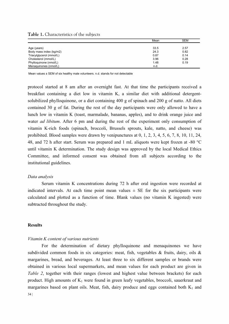
34 |
Table 1. Characteristics of the subjects Mean SEM
Age (years) 33.5 2.57Body mass index (kg/m2) 24.3 0.82Triacylglycerol (mmol/L) 0.87 0.14Cholesterol (mmol/L) 3.96 0.28Phylloquinone (nmol/L) 1.48 0.19Menaquinones (nmol/L) n.d.
Mean values ± SEM of six healthy male volunteers. n.d. stands for not detectable
protocol started at 8 am after an overnight fast. At that time the participants received a breakfast containing a diet low in vitamin K, a similar diet with additional detergent-solubilized phylloquinone, or a diet containing 400 g of spinach and 200 g of natto. All diets contained 30 g of fat. During the rest of the day participants were only allowed to have a lunch low in vitamin K (toast, marmalade, bananas, apples), and to drink orange juice and water ad libitum. After 6 pm and during the rest of the experiment only consumption of vitamin K-rich foods (spinach, broccoli, Brussels sprouts, kale, natto, and cheese) was prohibited. Blood samples were drawn by venipunctures at 0, 1, 2, 3, 4, 5, 6, 7, 8, 10, 11, 24, 48, and 72 h after start. Serum was prepared and 1 mL aliquots were kept frozen at -80 °C until vitamin K determination. The study design was approved by the local Medical Ethics Committee, and informed consent was obtained from all subjects according to the institutional guidelines. Data analysis
Serum vitamin K concentrations during 72 h after oral ingestion were recorded at indicated intervals. At each time point mean values ± SE for the six participants were calculated and plotted as a function of time. Blank values (no vitamin K ingested) were subtracted throughout the study. Results Vitamin K content of various nutrients
For the determination of dietary phylloquinone and menaquinones we have subdivided common foods in six categories: meat, fish, vegetables & fruits, dairy, oils & margarines, bread, and beverages. At least three to six different samples or brands were obtained in various local supermarkets, and mean values for each product are given in Table 2, together with their ranges (lowest and highest value between brackets) for each product. High amounts of K1 were found in green leafy vegetables, broccoli, sauerkraut and margarines based on plant oils. Meat, fish, dairy produce and eggs contained both K1 and
| 35
MK-4 with relatively high MK-4 concentrations in goose meat and liver, butter, and egg yolk. Long-chain menaquinones were mainly found in curd cheese, hard (Dutch) and soft (French) cheeses, probably derived from the bacterial starters fermentation. Very rich in menaquinones was the Japanese food natto, which consists of fermented soy beans. No substantial differences were found between free-range products (eggs, chicken, and meat) and those from factory farms. The fact that fermented beverages like beer and wines did not contain detectable amounts of menaquinones is probably due to the fact that moulds do not synthesise menaquinones 26. Bioavailability of K-vitamers from food
To examine the blank values (serum vitamin K at low vitamin K intake) 6 male volunteers received a vitamin K-poor breakfast with blood sampling (up to 72 h) as indicated. These blank values (data not shown) were subtracted from those obtained after controlled vitamin K intake. Based on the analyses summarized in Table 2 we have prepared meals
consisting of 400 g cooked spinach (equivalent to 3.5 µmoles of K1), 200 g natto (3.1 µmoles of MK-7), supplemented with corn oil to a total fat content of 30 g. Postprandial serum vitamin K concentrations are given in Figure 1. One week later the volunteers received a
vitamin K-poor breakfast supplemented with 3.5 µmoles of Konakion®. Peak values for serum
vitamin K (both K1 and MK-7) were found at 6 h following the meal and at 4 h after intake of the pure compound. The very poor absorption from green vegetables becomes clear by comparing the difference between the curves for K1 pure compound and the similar amount of K1 from spinach. Remarkably, MK-7 from natto was absorbed extremely well with peak values even higher than those for detergent-solubilized K1. After having reached their peak levels a rapid disappearance of both K1 and MK-7 was observed, but MK-7 showed complex pharmacokinetics, with slow disappearance during the second part of the curve, while it remained detectable for at least 72 h. The half-life times for both K1 and MK-7 between 6 and 8 h postprandially were about 1.5 h, whereas during the later phases of MK-7 disappearance the half-life time was about 50 h. To exclude mutual interference of absorption (e.g. by competition for the same binding protein), the above experiment was repeated in a design in which spinach and natto were given in two separate meals with a one-week interval. The serum curves are shown in Figure 2 and are comparable to those obtained after the combined meal. The above absorption curves were repeated for other foods: broccoli as source for K1 and curd cheese and egg yolk as sources for higher menaquinones (MK-8 and MK-9) and MK-4, respectively (L.J. Schurgers, unpublished data). In all cases it was found that K1 absorption from vegetables was very poor (5-10% without concomitant fat intake and 10-15% if taken together with 30 g fat), whereas menaquinone absorption from dairy produce and natto was much better, probably almost complete.

36 |
0
25
50
75
100
0 24 48 72Time after start [hours]
Vit
amin
K c
on
cen
trat
ion
[n
mo
l/L]
Figure 1.
Serum vitamin K following the oral intake of either Konakion® or a meal containing spinach and
natto. The ingested Konakion contained 3.5 µM K1, the mixed meal contained 3.5 µM of K1 and 3.1
µM of MK-7. Points are mean values from 6 volunteers, error bars represent SEM. Symbols: O, K1
after Konakion®; n, K1 after mixed meal; l, MK-7 after mixed meal.
0
25
50
75
100
0 24 48 72Time after start [hours]
Vita
min
K c
on
cen
trat
ion
[nm
ol/L
]
Figure 2.
Serum vitamin K1 and MK-7 following the separate intake of either spinach (3.5 µM K1) or natto (3.1
µM MK-7). Points are mean values from 6 volunteers, error bars represent SEM. Symbols: n, K1; l,
MK-7.

| 37
Table 2. Mean of K-vitamins (µg/100 g or µg/100 ml) in various foods Type of food n K1 MK-4 MK-5 MK-6 MK-7 MK-8 MK-9
Meat Beef 7 0.6 [0.6 - 0.7] 1.1 [ 0.7 - 1.3] - - - - - Chicken breast 7 - 8.9 [ 6.4 - 11.3] - - - - - Chicken leg 7 - 8.5 [ 5.8 - 10.5] - - - - - Pork-steak 7 0.3 [0.2 - 0.4] 2.1 [ 1.7 - 2.4] - - - - - Pork liver 7 0.2 [0.1 - 0.3] 0.3 [ 0.3 - 0.4] - - 0.5 [0.4 - 0.7] 1.1 [0.9 - 1.2] - Minced meat 7 2.4 [2.2 - 2.5] 6.7 [ 6.5 - 6.7] - - - - - Salami 7 2.3 [2.1 - 2.5] 9.0 [ 8.2 - 10.1] - - - - - Luncheon meat 7 3.9 [3.8 - 4.2] 7.7 [ 7.4 - 9.1] - - - - - Hare leg 7 4.8 [4.5 - 5.3] 0.1 [ 0.0 - 0.2] - - - - - Deer back 7 2.0 [1.9 - 2.2] 0.7 [ 0.6 - 0.7] - - - - - Goose leg 5 4.1 [3.5 - 4.8] 31.0 [ 28.2 - 33.1] - - - - - Goose liver paste 5 10.9 [9.3-12.1] 369 [317 - 419 ] - - - - - Duck breast 7 1.9 [1.7 - 2.2] 3.6 [ 3.3 - 3.9] - - - - -
Fish Prawn 7 0.1 [0.0 - 0.1] - - - - - - Mackerel 7 2.2 [1.8 - 2.6] 0.4 [ 0.3 - 0.5] - - - - - Herring 7 0.1 [0.0 - 0.2] - - - - - - Plaice 7 - 0.2 [0.1 - 0.3] - 0.3 [0.2 - 0.3] 0.1 [0.0 - 0.1] 1.6 [1.3 - 1.8] - Eel 7 0.3 [0.2 - 0.5] 1.7 [ 1.4 - 2.1] - 0.1 [0.0 - 0.2] 0.4 [0.2 - 0.6] - - Salmon 7 0.1 [0.1 - 0.2] 0.5 [ 0.4 - 0.6] - - - - -
Fruits & vegetables Kale 4 817 [752 - 881 ] - - - - - - Spinach 6 387 [299 - 429 ] - - - - - - Broccoli 5 156 [139 - 189 ] - - - - - - Green peas 4 36.0 [31.2 - 39.4] - - - - - - Sauerkraut 7 25.1 [23.8 - 27.5] 0.4 [0.3 - 0.5] 0.8 [0.6 - 1.0] 1.5 [1.4 - 1.6] 0.2 [0.1 - 0.3] 0.8 [0.6 - 0.9] 1.1 [0.9 -1.3] Natto 5 34.7 [31.2 - 36.7] - 7.5 [7.1 - 7.8] 13.8 [12.7 -14.8] 998 [882-1034] 84.1 [78.3 -89.8] - Banana 4 0.3 [0.2 - 0.4] - - - - - - Apple 4 3.0 [2.7 - 3.4] - - - - - - Orange 4 0.1 [0.1 - 0.2] - - - - - -
Dairy produce Whole milk 6 0.5 [ 0.4 - 0.6] 0.8 [ 0.7 - 0.9] 0.1 [0.0 - 0.1] - - - - Skimmed milk 6 - - - - - - - Buttermilk 6 - 0.2 [ 0.2 - 0.3] 0.1 [0.1 - 0.2] 0.1 [0.0 - 0.2] 0.1 [0.1 - 0.3] 0.6 [0.5 - 0.6] 1.4 [1.2 - 1.6] Whole yoghurt 6 0.4 [ 0.3 - 0.5] 0.6 [ 0.5 - 0.7] 0.1 [0.0 - 0.2] - - 0.2 [0.2 - 0.3] - Skimmed yoghurt 6 - - - - - 0.1 [0.0 - 0.2] - Whipping cream 6 5.1 [ 4.9 - 5.5] 5.4 [ 5.2 - 5.6] - - - - - Chocolate 6 6.6 [ 6.4 - 6.7] 1.5 [ 1.4 - 1.6] - - - - - Hard cheeses 15 10.4 [ 9.4 - 12.1] 4.7 [ 4.2 - 6.6] 1.5 [1.3 - 1.7] 0.8 [0.6 - 1.0] 1.3 [1.1 - 1.5] 16.9 [14.9-18.2] 51.1 [45.3-54.9] Soft cheeses 15 2.6 [ 2.4 - 2.9] 3.7 [ 3.3 - 3.9] 0.3 [0.2 - 0.4] 0.5 [0.6 - 0.7] 1.0 [0.9 - 1.1] 11.4 [10.7-12.2] 39.6 [35.1-42.7] Curd cheese 12 0.3 [ 0.2 - 0.4] 0.4 [ 0.3 - 0.6] 0.1 [0.0 - 0.2] 0.2 [0.1 - 0.3] 0.3 [0.2 - 0.5] 5.1 [ 4.8 - 5.4] 18.7 [18.1 19.2] Egg yolk 8 2.1 [1.9 - 2.3] 31.4 [29.1 - 33.5] - 0.7 [0.6 - 0.8] - - - Egg albumen 8 - 0.9 [0.8 - 1.0] - - - - -
Oils and margarines Margarine 6 93.2 [85.6 - 98.3] - - - - - Butter 6 14.9 [13.2 - 15.9] 15.0 [13.5 - 15.9] - - - - Corn oil 6 2.9 [ 2.7 - 3.1] - - - - - Sunflower oil 6 5.7 [ 5.5 - 5.9] - - - - - Olive oil 6 53.7 [49.9 - 57.2] - - - - -
Bread Rue bread 6 0.7 [ 0.5 - 0.9] - - - - - Wheaten bread 6 1.1 [1.0 - 1.2] - - - - - Sourdough bread 6 1.0 [0.9 - 1.1] - - - - - Buckwheat bread 6 3.0 [2.8 - 3.4] - - - - 1.1 [1.0 -1.2] Beverages Tea 4 0.3 [0.2 - 0.4] - - - - - - Coffee 4 - - - - - - - Orange juice 4 - - - - - - - All samples were assessed in duplicate, n stands for the number of different samples tested, a hyphen
for not detectable. Values are mean values. Highest and lowest value between brackets. Foods were
bought from shops around Maastricht. MK-10 was not detectable in any of the foods.

38 |
Discussion In this paper we describe the phylloquinone and menaquinone content of various
foods available on the Dutch market. All K-vitamins were quantified in the same run after a slight modification of our previously reported procedure 25. It was confirmed that phylloquinone is mainly present in green vegetables, margarines, and some plant oils such as olive oil. Since these data are similar to those reported by others 27-29 we have focussed on the menaquinones in food. MK-4 was present in nearly all animal products (meat, dairy produce, eggs), but the fact that there were no substantial differences between game (hare, deer), free-range animals and those from factory farms suggests that conversion of menadione from fortified animal food (used at factory farms) does not contribute substantially to the total tissue MK-4 stores. Rather, it seems that the major part of MK-4 in animal products originates from conversion of K1 as was also reported to occur in rats 30. Relatively high concentrations of long-chain menaquinones were found in all cheeses. As was suggested by Shearer, they probably originate from bacteria present in the starter cultures used to induce fermentation 26. On the basis of food frequency questionnaires and the data in Table 2 it has been calculated that phylloquinone forms almost 90% of the total dietary vitamin K intake in the Dutch population, whereas menaquinones account for less than 12 % 6. Phylloquinone, however, is tightly bound to the thylakoid membranes of plant chloroplasts, and the efficacy of its liberation there from in the digestive tract is poor 24,25. This was confirmed in an experiment in which we compared the serum concentration vitamin K profiles after ingestion of similar amounts of K1 from spinach and from a detergent-solubilized pharmaceutical product. To compare the efficacy of absorption of phylloquinone and menaquinone we have chosen a design in which K1 was obtained from spinach and MK-7 from natto. In this way the molar concentrations of both K-vitamers could be kept similar. As is shown in Figure 1, the postprandial serum concentrations of MK-7 were much higher than those of K1, with a peak height difference of more than 10 fold. Both absorption peaks occurred 2 h later than that for the detergent-solubilized product. From the curves obtained, it may be concluded that the contribution of MK-7 from natto to the total bioavailable pool of vitamin K is much higher than estimated on the basis of intake. Menaquinones from other sources (cheeses, egg yolk) were absorbed with comparable efficacy as was MK-7 (L.J. Schurgers, unpublished data), suggesting that the contribution of menaquinones to total human vitamin K status is much higher than generally assumed, and may equal that of K1.
Another remarkable difference between K1 and menaquinones was that the former had a disappearance curve with an apparent half-life time of 1½ h, whereas the long chain menaquinones (not MK-4) had more complex disappearance curves, with very long half-life time. Rapid clearance is consistent with the previously reported uptake and transport of K-vitamins in chylomicrons, from where they are cleared by the liver during the first 8

| 39
postprandial hours. The very long half-life times of the higher menaquinones suggests that these vitamers (and not K1 and MK-4) are redistributed by the liver and set free in the circulation in low and high density lipoproteins. It is well known that LDL may be present in the circulation for several days. The long residence times of higher menaquinones in the circulation implies that they are available for extra-hepatic tissue uptake for much longer periods than is phylloquinone. Both because of their high postprandial serum concentration and their slow clearance, the importance of higher menaquinones for extra-hepatic tissues such as bone and arterial vessel wall may be underestimated if only dietary intake is regarded. Since vitamin K-dependent proteins have been reported to be involved in the regulation of calcium deposition in bone 31 and in the prevention of arterial calcification 32, intake of higher menaquinones may be important for functions of vitamin K not related with blood coagulation.
The high efficacy of menaquinone absorption may also have consequences for subjects on oral anticoagulant treatment. In attempts to identify potential causes of unstable anticoagulation, menaquinone intake has been ignored thus far. Our data demonstrate that this is not justified. Their efficient absorption combined with long serum and tissue half life times 33 suggest that menaquinones from curd and cheese may accumulate at repeated intake and are a potential cause of disturbance of anticoagulant therapy. This is even more so for subjects consuming natto. Although in general natto is rarely eaten by Caucasians, dietary habits may survive after migration of subjects from Asiatic countries so that haematologists in Western countries may be confronted with this unsuspected source of highly bio-available vitamin K.

| 41
2.2
INTESTINAL, HEPATIC, AND CIRCULATING VITAMIN K AT LOW AND HIGH INTAKE OF VITAMIN K IN RATS
Abstract The aim of this study was to assess how high doses of dietary vitamin K influence the
intestinal profile of K-vitamins in vitamin K-deficient rats, and whether the induced changes are reflected in the hepatic vitamin K store. Vitamin K-deficient rats were fed for 10 days with diets containing different forms of vitamin K, and analyses carried out to see how these diets influenced the vitamin K concentration at various sites of the intestine, serum, and the liver. It was found that the absorption of phylloquinone from standard food is not more than 10%, whereas the absorption of pharmacological doses of oil-solubilized phylloquinone and menaquinone-4 was 18 and 55%, respectively. High intakes of phylloquinone suppress the colonic production of all higher menaquinones. High menaquinone-4 intake induces very high menaquinone-8 concentrations, both in the colonic contents as well as in the liver. These data suggest that menaquinone-4 may be converted into menaquinone-8 (but not into other menaquinones) via a metabolic pathway which has not been reported previously. Introduction
Vitamin K serves as a cofactor during the posttranslational synthesis of γ-
carboxyglutamic acid (Gla) in several proteins. Since its discovery in the early 1930's, vitamin K was believed to play an exclusive role in the process of blood coagulation; only recently it has become clear that Gla-containing proteins are synthesized in most mammalian tissues, and that the functions of these proteins include regulation of bone growth (osteocalcin 34), prevention of vascular mineralization (matrix Gla-protein 32), and stimulation of cell growth (growth-arrest-specific gene protein 6, Gas6 35). Natural forms of vitamin K are phylloquinone (also known as vitamin K1) and the group of the menaquinones (K2 vitamins) 2. Phylloquinone is synthesized by plants and the highest levels are found in plant oils and green vegetables 36-38. Menaquinones are of microbial origin: they are found mainly in cheese and fermented foods 6,39. Also the intestinal microflora produces substantial amounts of menaquinones, but the extent to which these products are absorbed has remained a matter of debate 40. In this paper the various menaquinones will be designated as MK-n, where n stands for the number of isoprenoid residues in the aliphatic side chain. Menadione (vitamin K3) is a

42 |
synthetic form of vitamin K which is generally used in commercial animal food (rodents, poultry, and cows). Its vitamin K activity results from its in vivo transformation into menaquinone-4 8.
The vitamin K status of an organism depends on the phylloquinone and menaquinone contents of its diet, on the extent to which these vitamins are absorbed in the intestines, as well as on the colonic absorption of menaquinones produced by intestinal microflora. It is generally assumed that absorption of vitamin K mainly takes place in the jejunum and ileum in the form of mixed micelle complexes with bile salts 41. Since the absorption of bile salts is virtually completed in the distal ileum, uptake of K-vitamins from the colon is less likely 40,42,43.
In the present study we have fed vitamin K-deficient rats for 10 days with either standard diets or with diets containing pharmacological doses of oil-solubilized, purified vitamin K and we have investigated how the vitamin K content of the diet affects the vitamin K concentration at various sites in the intestine and in the faeces. Moreover, we have investigated the uptake and metabolism of K-vitamers by recording their respective concentrations in plasma and in the liver. Materials and Methods Chemicals and animal food
MK-4 and phylloquinone were purchased from Sigma (St. Louis, MO, USA), a set of higher menaquinones (MK-5 through MK-10) as well as 2’,3’-dihydrophylloquinone were
kind gifts from Hoffmann-La Roche (Basel, Switzerland). Powdered vitamin K-deficient, γ-
irradiated (0.9 Mrad) food and standard animal food were purchased from Hope Farms (Woerden, The Netherlands), the detailed food contents have been described previously 44. Upon analysis, the vitamin K-deficient food did not contain detectable amounts of either phylloquinone or menaquinone (< 20 pg/g of diet), the standard food contained
phylloquinone (1.95 µg/g of diet) and menadione (6.23 µg/g of diet). All solvents and chemicals were of analytical quality or HPLC grade purity. Animals and preparation of diets
This study was performed in 15 male rats of Lewis strain, which were 12 weeks old and had a body weight of 303 ± 49.7 g (mean ± SD) at the start of the experiment. The animals were housed individually in flat bottom cages under conditions of a 12-h light-dark cycle, controlled temperature (20 ± 2 °C) and humidity (50 ± 10%). One group of three animals was kept on standard food throughout the experiment and served as the normal

| 43
reference group. The remaining 12 animals were divided into 4 groups (3 rats/group), all of which received the vitamin K-deficient diet for 7 days before the start of the experiment. After that period three groups of three rats received vitamin K-deficient food supplemented
with 20 µg/g of phylloquinone, MK-4, or menadione. The last group served as the zero-intake reference and received vitamin K-deficient food throughout the experiment. The various diets were prepared by dissolving the respective vitamins in 10 ml of corn oil, which was added to 750 g of vitamin K-deficient food; the same amount of corn oil (without vitamin K added) was supplemented to the food of animals receiving either the standard food or the vitamin K-deficient food. Faeces of the rats were collected on each day of the experiment and lyophilized before analysis. After the animals had received these diets for 10 days, they were killed by aortic exsanguinations under light ether anaesthesia. Blood was collected, diluted 9:1 (v/v) with 0.1 M trisodium citrate and centrifuged for 15 min (2,000 x g) for the preparation of plasma. The body was perfused with saline via the abdominal aorta, and the liver and intestine was removed for further analysis. Vitamin K analysis
The spectra of K-vitamers (phylloquinone and menaquinones 4-10) were analysed in 9 parts of the intestinal tract, faeces, livers and plasma. In all cases the intestines were similarly divided as follows: duodenum in total (segment 1), first 10 cm of jejunum (segment 2), middle part of jejunum and ileum in three equal parts (segments 3, 4, and 5), last 10 cm of ileum (segment 6), caecum in total (segment 7), and colon in two equal parts (segments 8 and 9). The content of each part was collected with careful rinsing of the lumen and freeze-dried; dry intestinal contents were weighted and 0.1 g was used for analysis. Faecal vitamin K contents were analysed in 0.2 g (dry weight) of samples collected at days 5, 7, and 9 of the experiment. In plasma, only the data for phylloquinone and MK-4 are provided. Hepatic and circulating vitamin K were measured in 1 g of liver (wet weight) and in 0.5 mL of plasma. Extraction and detection of vitamin K were performed as described earlier 25,45, 2’, 3’-dihydrophylloquinone served as an internal standard. In short, samples were supplemented with 2.0 mL of distilled water (for plasma 1.5 mL), 4 mL of ethanol and internal standard (1-40 ng, as required) and extracted with 8 mL of hexane. Liver and faeces were homogenized using an Ultra Turrax homogenizer (Janke and Kunkel, Staufen, Germany) prior to extraction. Extracts were centrifuged (2,000 g for 5 minutes), evaporated under a stream of nitrogen gas, and re-dissolved in 2 mL hexane. Pre-purification of the samples was performed with silica Sep-pack cartridges (Millipore, Milford, MA, USA) as described previously 25. Quantitative analysis of the samples was performed by reversed-phase HPLC using a C-18 reversed phase column and fluorescence detection 45. Because of the long retention times for the long-chain menaquinones (MK-7 through MK-10) the flow was increased from 0.5 to 1.0

44 |
ml/min at 11 min after injection. K-vitamers in the eluate were reduced electrochemically using a Coulochem 5010 analytical cell (ESA, Bedford, MA, USA) maintained at potential of -1.5 V. Phylloquinone and menaquinones were recorded in the same run. Reference curves were prepared from purified phylloquinone and each of the menaquinones, and a linear dose-response was obtained for the entire range in which the sample concentrations were measured. The vitamin K content of the samples was quantified with the internal standard method based on the ratio of peak heights. Detection limits were 0.015 ng/ml for phylloquinone, MK-4 and MK-5, 0.04 ng/mL for MK-6 and MK-7, 0.10 ng/mL for MK-8, and 0.12 ng/mL for MK-9 and MK-10. Intestinal absorption was calculated by expressing the difference between dietary intake and faecal excretion as a percentage of the intake for each animal separately. Data analysis
All groups consisted of three animals, and mean values for each group are given throughout this paper. Differences between groups were compared using the Mann-Whitney U-test, and were regarded to be significant at p = 0.05. Results Intestinal absorption of nutritional vitamin K
In the experiments described in this paper the experimental animals had been on a constant diet for 10 days before their intestinal contents were analysed. During the entire experiment the individual daily food intake was recorded, and was found to be 19.0 ± 1.9 g (average for all groups). The average faeces production was 3.2 ± 1.3 g (wet weight), and the weight gain was comparable in all groups. To demonstrate that the animals were in a steady state condition at the time of sacrifice, we have assessed the amounts of K-vitamins in the faeces at days 5, 7, and 9 (see Table 1). No significant differences between these three days were found. From this Table 1 it can also be seen that the mean absorption (as calculated from the difference between dietary intake and faecal excretion) of phylloquinone from the
standard diet was 4 µg/day (about 10% of the food content), whereas from the enriched diets
the absorption of phylloquinone and menaquinone was substantially higher, both in absolute
amounts (75 µg/day and 216 µg/day) and as a percentage of the daily intake (18% and 55%,
respectively).

| 45
Table 1. Dietary intake and faecal excretion of K1 and MK-4 on various days Type of dietDay number: 5 7 9 5 7 9 5 7 9 5 7 9 5 7 9
Dietary intake (µg/day): vitamin K1 0 0 0 38 42 40 392 401 397 0 0 0 0 0 0 menaquinone-4 0 0 0 0 0 0 0 0 0 405 389 398 0 0 0 menadione 0 0 0 109 120 114 0 0 0 0 0 0 377 393 412
Faecal excretion (µg/day): vitamin K1 0.5 0.7 0.5 35* 37* 36* 316# 332# 317#
0.2 0.7 0.8 0.4 0.6 0.5
menaquinone-4 0.5 0.1 0.2 2.1* 2.6* 1.9* 6.1# 5.0# 3.2 178 187 180 4.9# 7.2# 5.1# Absorption (%) vitamin K1 - - - 8 12 10 19# 17# 20#
- - - - - - menaquinone-4 - - - - - - - - - 57 54 55 - - -
MenadionVitamin K-deficient Standard diet High vitamin K1 High MK-4
Each group consisted of three animals, and all data provided in this Table are mean values for three
different animals, standard deviations were < 15%. Explanation of symbols: *, significantly different
from the vitamin K-deficient group; #, significantly different from the group receiving standard diet.
Figure 1.
Vitamin K profile in rats on standard diet. The intestinal segments which were analysed were: 1 = duodenum, 2 = proximal jejunum, 3 = distal jejunum, 4 = proximal ileum, 5 = middle ileum, 6 =
distal ileum, 7 = caecum, 8 = proximal colon, 9 = distal colon. Bars represent: MK-4 (closed), K1
(narrowly hatched), MK-8 (cross-hatched), MK-9 (open), MK-10 (widely hatched). Error bars
represent SD.

46 |
Effect of diet on intestinal vitamin K profile The intestinal vitamin K profile in rats receiving the standard food is shown in Figure 1.
K-vitamers recovered were mainly the higher menaquinones (MK-8, MK-9, and MK-10), which were almost exclusively present in the cecum and colon. Other menaquinones were barely detectable or absent in this group, as well as in the other groups described below.
In animals receiving the vitamin K deficient diet, phylloquinone and MK-4 were low
(<0.05 µg/g) in all segments of the intestinal tract (Figure 2A). Long-chain menaquinones were not found in the duodenum, jejunum or ileum, whereas segments 7-9 (caecum and colon) contained considerable amounts of MK-8, MK-9, and MK-10, the concentrations were comparable or even slightly higher than those in rats on standard food (cf. Figures 1 and 2A). Because the rats in the vitamin K-deficient group had not received any vitamin K during 17 days the menaquinone profile shown in Figure 2A must be the result of menaquinone synthesis by the intestinal flora. Supplementation with menadione did not increase the bacterial menaquinone production (Figure 2B). As compared to the vitamin K-deficient animals, the mean values of MK-4 were about 10-fold elevated in the upper part of the intestinal tract.
Figure 2. Effect of vitamin K-deficiency and menadione on intestinal vitamin K content.
A: vitamin K-deficiency.
B: high menadione intake. Further details are as described in the legend to Figure 1.

| 47
In the group receiving a diet rich in phylloquinone (Figure 3A), substantial amounts of this vitamin were found in all parts of the intestinal tract, but the concentration increased from segment 1 to segment 6-9. These result suggested that the absorption of the dietary phylloquinone was far from complete, and that its concentration increased during the passage through the upper part of the intestinal tract because other components of the diet were absorbed more readily. Remarkably, the long chain menaquinones were extremely low in all parts of the intestine (Figure 3B). Also in the group receiving MK-4 the dietary vitamin was recovered from the intestinal contents, but in general the concentrations were lower than in the phylloquinone-group (Figure 3A). So it seems that MK-4 is absorbed somewhat better than phylloquinone. Like in the phylloquinone group, the intestinal production of MK-9 and MK-10 was decreased in animals receiving the menaquinone-rich food. Surprisingly, however, relatively high amounts of MK-8 were found in animals on the MK-4 diet
Figure 3. Effect of dietary phylloquinone and menaquinone-4 on intestinal vitamin K content. A: Phylloquinone after phylloquinone-rich diet (hatched) and MK-4 after MK-4 rich diet (cross-hatched).
B: Higher menaquinones after phylloquinone-rich diet: MK-8 (cross-hatched), MK-9 (open), MK-10 (hatched).
C: Higher menaquinones after MK-4-rich diet: MK-8 (cross-hatched), MK-9 (open), and MK-10 (hatched).
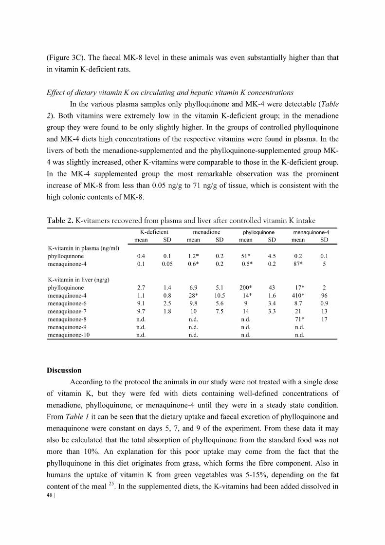
48 |
(Figure 3C). The faecal MK-8 level in these animals was even substantially higher than that in vitamin K-deficient rats. Effect of dietary vitamin K on circulating and hepatic vitamin K concentrations
In the various plasma samples only phylloquinone and MK-4 were detectable (Table 2). Both vitamins were extremely low in the vitamin K-deficient group; in the menadione group they were found to be only slightly higher. In the groups of controlled phylloquinone and MK-4 diets high concentrations of the respective vitamins were found in plasma. In the livers of both the menadione-supplemented and the phylloquinone-supplemented group MK-4 was slightly increased, other K-vitamins were comparable to those in the K-deficient group. In the MK-4 supplemented group the most remarkable observation was the prominent increase of MK-8 from less than 0.05 ng/g to 71 ng/g of tissue, which is consistent with the high colonic contents of MK-8.
Table 2. K-vitamers recovered from plasma and liver after controlled vitamin K intake
mean SD mean SD mean SD mean SDK-vitamin in plasma (ng/ml)phylloquinone 0.4 0.1 1.2* 0.2 51* 4.5 0.2 0.1menaquinone-4 0.1 0.05 0.6* 0.2 0.5* 0.2 87* 5
K-vitamin in liver (ng/g)phylloquinone 2.7 1.4 6.9 5.1 200* 43 17* 2menaquinone-4 1.1 0.8 28* 10.5 14* 1.6 410* 96menaquinone-6 9.1 2.5 9.8 5.6 9 3.4 8.7 0.9menaquinone-7 9.7 1.8 10 7.5 14 3.3 21 13menaquinone-8 n.d. n.d. n.d. 71* 17menaquinone-9 n.d. n.d. n.d. n.d.menaquinone-10 n.d. n.d. n.d. n.d.
K-deficient phylloquinone menaquinone-4menadione
Discussion
According to the protocol the animals in our study were not treated with a single dose of vitamin K, but they were fed with diets containing well-defined concentrations of menadione, phylloquinone, or menaquinone-4 until they were in a steady state condition. From Table 1 it can be seen that the dietary uptake and faecal excretion of phylloquinone and menaquinone were constant on days 5, 7, and 9 of the experiment. From these data it may also be calculated that the total absorption of phylloquinone from the standard food was not more than 10%. An explanation for this poor uptake may come from the fact that the phylloquinone in this diet originates from grass, which forms the fibre component. Also in humans the uptake of vitamin K from green vegetables was 5-15%, depending on the fat content of the meal 25. In the supplemented diets, the K-vitamins had been added dissolved in

| 49
corn oil, from which they are more readily extracted by the action of bile salts. Here a marked difference was observed between phylloquinone and the slightly more hydrophylic menaquinone-4, the latter one being absorbed substantially better than phylloquinone. In this respect it is noteworthy that Ichihashi et al. reported some colonic menaquinone-4 absorption even in the absence of bile salts 43.
After 17 days of vitamin K-deficiency, long chain menaquinones (notably MK-10) were abundantly present in the cecum and the colon, and definitely not lower than in animals on the standard food (cf. Figures 1 and 2). Apparently, the vitamers are not absorbed from this part of the tract, because two animals in the vitamin K-deficient group were bleeding at the end of the experiment. Also, the higher menaquinones were undetectable in the liver (see Table 2). A menadione-rich diet induced an increase of MK-4 in the liver, but not in the colon. Probably because of the entero-hepatic cycle, MK-4 was slightly increased in the upper part of the digestive tract in this group. These data are consistent with the view that menadione may be converted into MK-4 in the liver 8, but they also demonstrate clearly that this conversion is absent in the colonic bacteria. Whether menadione may be used as a source for the intestinal production of higher menaquinones remains to be seen, although it is clear that menadione does not increase the menaquinone concentrations in the gut.
A major finding was that high phylloquinone intake suppressed the intestinal menaquinone production. Apparently the flora is capable of using this vitamin, thereby shutting down its own menaquinone production. It is also clear that - despite the high phylloquinone concentration in the food - the phylloquinone concentration was low in the upper part of the digestive tract. A possible explanation may be found from the fact that in all animals (also in the other groups) the ileum was virtually empty, so that mucosal material and not digested food were the main components of what was assembled from its contents. This effect is negligible in the caecum and colon, where a high degree of intestinal filling was observed. The gradual increase of phylloquinone downstream the digestive tract as well as its relatively high concentration in colon and faeces suggests that its absorption is far from complete. In the group receiving a diet rich in MK-4 the recovery of the corresponding vitamer from the intestinal contents was substantially lower than in the phylloquinone group, which is consistent with a better absorption of MK-4 than of phylloquinone (cf. Table 1). This seems to be in contrast to previous data from Groenen-van Dooren, who showed that the utilization for prothrombin synthesis of nutritional phylloquinone is better than that of MK-4 44,46. Differences between the previous protocols and the present experiment are that Groenen-van Dooren used a single-dose of vitamin K, and that the vitamers were dissolved in detergent (HCO-60). When used in a steady state situation and dissolved in a natural compound (corn oil) it seems that MK-4 and phylloquinone accumulate in the liver to comparable levels.
50 |
As it was found in the group receiving the phylloquinone-diet, animals receiving MK-4 had low intestinal concentrations of MK-9 and MK-10. Surprisingly, however, the diet containing MK-4 induced a remarkable increase of colonic MK-8. These data suggest that either the intestinal flora or the liver is capable of using MK-4 for the exclusive synthesis of MK-8, which is a hitherto unknown metabolic pathway. A second striking observation was that in this group the high MK-8 levels were reflected in the liver vitamin K concentrations (Table 2). Since this seems to be in contrast to the poor absorption of higher menaquinones in the vitamin K-deficient animals (see above), we believe that the conversion of MK-4 into MK-8 may take place in the liver, and reach the digestive tract via the entero-hepatic pathway. This hypothesis implies that re-absorption of MK-8 from the intestines is poor, so that it accumulates to the high concentrations in the colon as observed in our study. Investigations in germ-free animals 9 may help to verify this hypothesis. The data presented here confirm that the absorption of phylloquinone from its natural food matrix in rats is extremely poor, as has also been reported in a limited number of human volunteers 25. Absorption from oil-solubilized preparations is substantially better but still far from complete. This supports the very high efficacy of hepatic vitamin K recycling during the blood clotting factor synthesis, and raises the question of whether a similar efficacy is obtained in other tissues, for instance in bone and vessel wall during the production of osteocalcin and matrix Gla-protein.

| 51
2.3
DIFFERENTIAL LIPOPROTEIN TRANSPORT PATHWAYS OF K-VITAMINS IN HEALTHY SUBJECTS
Abstract Vitamin K is a group name for K1 (phylloquinone) and K2 (menaquinones). Both
forms contribute to the tissue vitamin K status. Following intestinal absorption, the serum transport of these lipophilic compounds to their target tissues takes place via lipoproteins. In previous studies we have found that K1 is preferentially accumulated in the liver, whereas menaquinones have a more widespread distribution pattern. Here we have tested whether these differences may be explained by the different liposolubility and or metabolism of the various K-vitamers, resulting in their association with different lipoprotein particles. Six
healthy male volunteers received a mixture containing 2µM of each of three K vitamers (K1, MK-4, and MK-9) dissolved in corn oil. Blood was obtained at baseline and at different time intervals after intake for the measurement of vitamin K in serum and in the lipoprotein fractions. K1 was found to be associated predominantly with the triacylglycerol-rich lipoprotein (TGRLP) fraction, suggesting that it is mainly cleared by the liver. Both menaquinones investigated were found in TGRLP and LDL, MK-4 was also present in HDL. This may explains why menaquinones have a different tissue distribution profile and suggests relatively large impact of menaquinones on extra-hepatic vitamin K status than generally assumed. Moreover, the very long half-life time of MK-9 in the circulation indicates that it may form a more constant source of vitamin K than either K1 or MK-4. Introduction
Vitamin K is a group name for a class of compounds which share a 2-methyl-naphthoquinone ring system, substituted at the 3-position with a variable aliphatic side chain 2,4. Most common forms found in the human diet are phylloquinone (vitamin K1) and menaquinones (vitamin K2). Whereas phylloquinone is a single compound containing a mono-unsaturated side chain of four isoprenoid residues, menaquinones may be subdivided further on the basis of the number of isoprene residues (all of which are unsaturated) in the aliphatic side chain. According to the general nomenclature menaquinones are denominated as MK-n, where n stands for the number of isoprene residues present. Menaquinones most commonly found in food are MK-4, which is regarded as a short chain menaquinone, and the

52 |
long chain menaquinones MK-7, MK-8, and MK-9. Although all K-vitamins are fat-soluble, their lipophylic character increases substantially at increasing chain length; this may have consequences for their uptake, plasma transport, tissue storage, and biological half-life time 47.
The function of all K-vitamins in mammals is that they serve as a cofactor for the
enzyme γ-glutamylcarboxylase during the biosynthesis of a special class of proteins, the so-called Gla-proteins 3. Well known examples of Gla-proteins are a number of clotting factors produced by the liver, including prothrombin and factors VII, IX, and X 1. During the last two
decades, however, γ-glutamylcarboxylase has been detected in a wide range of non-hepatic
tissues, and an increasing number of Gla-proteins of extra-hepatic origin have been characterized 1,2. Most of these proteins have regulatory functions, for instance in tissue mineralization (osteocalcin and matrix Gla-protein) 32,34 and cell growth (growth-arrest-specific gene-6 protein, Gas6) 48.
It is well known that intestinal absorption of K1 takes place via the enterocytes, which facilitate its incorporation into chylomicrons. These chylomicrons enter the circulation via the lymphatic system, and most of the vitamin is delivered to the liver through receptor-mediated uptake of chylomicron remnants by hepatic parenchymal cells 4,49. It seems probable that the other K-vitamins are taken up via a similar process, but limited data for menaquinone metabolism is available at this time. Moreover, it is not known how the extra-hepatic tissues are provided with vitamin K. Apart from possible uptake from chylomicron remnants another route would be that part of the K-vitamins are re-packed into the low-density lipoproteins (LDL) which form a major transport system from the liver to peripheral tissues. From animal experiments is has become clear that the liver is the main target for uptake and accumulation of vitamin K1, whereas most extra-hepatic tissues (e.g. vessel wall, bone, testis, pancreas, kidney, and lung) preferentially accumulated menaquinone 30,50.
Recently, it has been shown that postprandially, vitamin K1 is predominantly carried in the triacylglycerol-rich lipoprotein fraction (TGRLP), and little is carried by the LDL or HDL fraction 51. In the study described in this paper we have hypothesized that the vitamin K transport in the blood stream to various tissues will take place via circulating lipoproteins, and that the lipophilicity of the various K-vitamers is an important factor in their distribution over the different lipoprotein fractions. To test this hypothesis, human volunteers received a single oral dose of a mixture of K1, MK-4, and MK-9, and we have measured the appearance and disappearance of these vitamins in serum. Subsequently, we have recorded their distribution over the various lipoprotein fractions as a result of changes in postprandial lipoprotein concentration of vitamin K.

| 53
Materials and Methods Subjects and study protocol
This study was carried out in six healthy male volunteers recruited at the University of Maastricht. Their mean age was 33.5 ± 6.3 years, and their body mass index was 24.3 ± 2.0 kg/m2. All participants were apparently healthy, and their serum lipid profiles were within the normal range. Neither medications nor vitamin supplements (other than the experimental ones) were taken throughout the study. The experimental protocol started at 8 am after an overnight fast. At that time the participants received a breakfast containing vitamin K (2
µmol of K1, MK-4 and MK-9) together with 30 g of fat (corn oil), and toast with marmalade. The blank meal (baseline curve) consisted 30 g of fat without vitamin K given at breakfast and values obtained during this control phase were subtracted from those obtained during the experimental design. To exclude influences in vitamin K metabolism due to additional K-intake by the diet, participants were only allowed to eat food low in vitamin K (toast, marmalade, bananas, apples) at lunch, and to drink orange juice and water ad libitum. After 6 pm and during the rest of the week consumption of vitamin K-rich foods (spinach, broccoli, Brussels sprouts and kale) was not allowed. Unless indicated otherwise, blood was drawn by venipunctures at 0, 2, 4, 6, 8, 24, and 48 after intake. Serum was prepared and 1 mL aliquots were kept frozen until serial vitamin K determination. The study design was approved by the local Medical Ethics Committee, and informed consent was obtained from all subjects according to the institutional guidelines. Materials
MK-4 was a kind gift from Eisai (Tokyo, Japan), K1 and MK-9 were obtained from Hoffmann-La Roche (Basel, Switzerland). A set of purified menaquinones (MK-4 through MK-10) was supplied by Roche and served as reference compounds for the identification of menaquinones. Corn oil was from CPC Bestfoods (Heilbronn, Germany) and contained 60 pmol/g of vitamin K1 and no detectable amounts of menaquinones. Analytical techniques
Triacylglycerol and cholesterol were determined by standard enzymatic techniques (Boehringer Mannheim, Germany) using a Beckmann Synchron CX 7-2 autoanalyser (Beckmann, Fullerton, CA). Vitamin K concentrations were assessed by high performance liquid chromatography, with on-line electrochemical reduction of the effluent and fluorescence detection 47. The lipoproteins in serum were separated on a 1,006 - 1,250 g/L KBr density gradient 52. Serum samples (3.5 mL) were adjusted to a density of 1,250 g/L with solid KBr in polyallomer tubes and stacked up with three 3 mL layers of KBr/NaCl solutions

54 |
in 1mM EDTA (pH 7.4) of 1,063, 1,019 and 1,006 g/L, respectively. The gradients were centrifuged for 22 h at 36,000 rpm in a Beckman SW 40 rotor. In this way the following lipoproteins were obtained in separate fractions: TGRLP (including the chylomicrons, the chylomicron remnants, and the VLDL fraction), the IDL, LDL, HDL, as well as the lipoprotein-free (LPF) fraction. Data analysis
Serum vitamin K concentrations during 48 h after oral ingestion were recorded at indicated intervals. At each time point mean values ± SE for the six participants were calculated and plotted as a function of time. Blank values (no vitamin K ingested) were subtracted throughout this study. Results
Serum profiles for lipid components were prepared after the vitamin K-rich meal only. Cholesterol and triacylglycerol concentrations in total serum at baseline level were within the normal range for all subjects. Lamon Fava et al. showed that vitamin K did not affect the plasma lipoprotein profile and that serum vitamin K and cholesterol is not correlated 51. Therefore, cholesterol in the lipoprotein subfractions was not analysed. Peak values for serum triacylglycerol concentrations were observed around 2 h postprandially, and were within the normal range. They remained elevated until 8 h after start and at 24 h all values had returned to baseline. The profile of serum triacylglycerol in the TGRLP fraction was comparable with that in total serum. The triacylglycerol content of the LDL and HDL fraction remained constant in the experiment.
In the same samples we have measured the concentrations of K1, MK-4, and MK-9 (Figure 1). Maximal concentrations in serum were reached at 2.5, 4, and 5 h postprandial for MK-4, K1, and MK-9. All three vitamins showed complex pharmacokinetics with multiphasic disappearance curves, but their clearance rates differed substantially. Whereas K1 and MK-4 had virtually disappeared from the circulation after the overnight period, the clearance of MK-9 was remarkably slower: after 24 h its serum concentration was still about 25% of the peak value, and it remained detectable until the last measurement at 48 h after intake. The estimated half-life time for MK-9 during the latter phase was about 60 h.
In Figure 2 we have plotted the postprandial distribution of K1, MK-4 and MK-9 over the lipoprotein fractions as a function of time. The clearance from the blood stream of K1 in the TGRLP fraction was nearly complete after 8 h, and we have measured whether other lipoprotein fractions contributed to the vitamin K transport. As is shown in Figure 2A, the K1 concentration in the LDL and HDL fractions was elevated during the first 24 h after K1

| 55
intake, but had returned to baseline at 48 h. If expressed as a percentage of total serum vitamin K1, LDL never contained more than 17% and HDL less than 18% of total, whereas the remaining K1 was recovered in the TRLG fraction (Figure 2B). MK-4, on the other hand, was equally distributed over the TGRLP, LDL, and HDL fractions (Figure 2C). The clearance of MK-4 present in the TGRLP fraction followed that of triacylglycerol. Expressed as a percentage, 19% of total serum MK-4 was present in the LDL fraction at 2 h, and up to 81% at 8 h after ingestion (Figure 2D). The pharmacokinetic behaviour of circulating MK-9 was different from that of K1 and MK-4: its appearance in the circulation and incorporation into lipoproteins was slower than for both other vitamins (Figure 2E). MK-9 appeared in the LDL fraction only at 8 h after intake, and remained detectable therein until at least 48 h. At that time the MK-9 in LDL was 55% of the total serum concentration (Figure 2F). At no time point MK-9 was found in the HDL fraction.
0.0
10.0
20.0
30.0
40.0
50.0
0 6 12 18 24
Time after start (h)
Vita
min
K c
once
ntra
tion
in s
erum
(nm
ol/L
)
0.40
0.80
1.20
1.60
2.00
Tria
cylg
lyce
rol c
once
ntra
tion
in s
erum
(m
mol
/L)
Figure 1.
Serum vitamin K following the oral intake of 2 µmol of oil-solubilized preparations. Also the
triacylglycerol concentration of the TGRLP fraction is drawn in the Figure to show the correlation
between the K-vitamin and triacylglycerol. Points are mean values from 6 volunteers, error bars
represent SEM. Symbols: l, K1; m, MK-4; n, MK-9; p, TG-TGRLP.
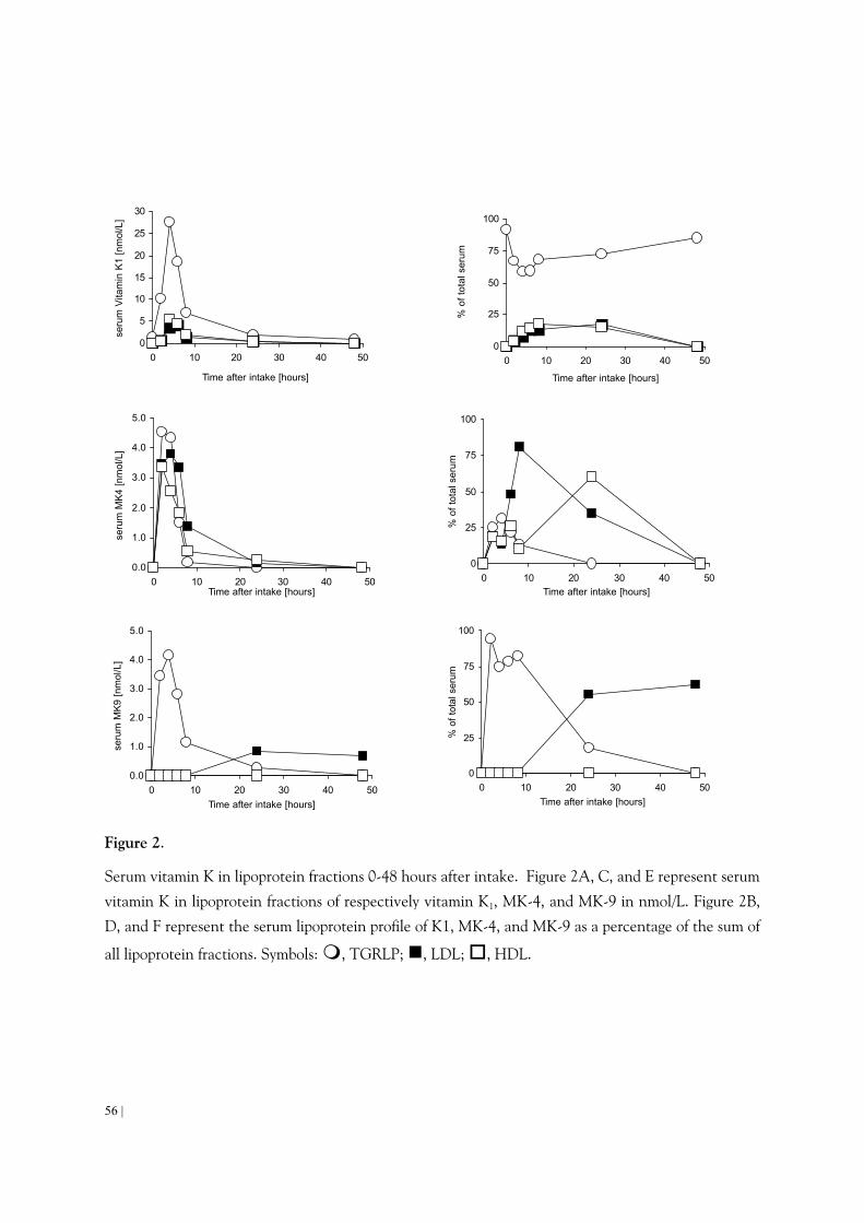
56 |
0
5
10
15
20
25
30
0 10 20 30 40 50
Time after intake [hours]
seru
m V
itam
in K
1 [n
mol
/L]
0
25
50
75
100
0 10 20 30 40 50
Time after intake [hours]
% o
f tot
al s
erum
0.0
1.0
2.0
3.0
4.0
5.0
0 10 20 30 40 50Time after intake [hours]
seru
m M
K4
[nm
ol/L
]
0
25
50
75
100
0 10 20 30 40 50Time after intake [hours]
% o
f tot
al s
erum
0.0
1.0
2.0
3.0
4.0
5.0
0 10 20 30 40 50Time after intake [hours]
seru
m M
K9
[nm
ol/L
]
0
25
50
75
100
0 10 20 30 40 50Time after intake [hours]
% o
f tot
al s
erum
Figure 2.
Serum vitamin K in lipoprotein fractions 0-48 hours after intake. Figure 2A, C, and E represent serum
vitamin K in lipoprotein fractions of respectively vitamin K1, MK-4, and MK-9 in nmol/L. Figure 2B,
D, and F represent the serum lipoprotein profile of K1, MK-4, and MK-9 as a percentage of the sum of
all lipoprotein fractions. Symbols: m, TGRLP; n, LDL; o, HDL.
| 57
Discussion Unlike the fat-soluble vitamins A and D, vitamin K has no specific plasma carrier
protein, but is transported in plasma mainly by lipoproteins 49,51. Following dietary uptake, vitamin K is absorbed in the intestine and enters the circulation via the lymphatic system as part of the chylomicrons 49. In the circulation chylomicrons are catabolized by the action of lipoprotein lipase (LPL) resulting in chylomicron remnants 53, which are then cleared mainly by the liver via an apoE receptor-specific uptake 54,55.
In our study it appeared that during the first 8 hours following intake of a mixture of equal amounts of the three K-vitamins, the serum contained much higher concentrations of K1 than MK-4 or MK-9. Several explanations may be given for this observation: they include poor intestinal absorption of menaquinones or rapid uptake of menaquinones from chylomicrons by the tissues. In previous studies in which we compared the absorption of orally ingested K1 and menaquinones, it was found that both in experimental animals and in human volunteers the absorption of menaquinones was far more complete than that of K1 47,56. This is not consistent with a low efficacy of intestinal uptake of the menaquinones. The fact that in all experiments the serum concentration of MK-4 reached its maximum well before that of K1 and MK-9 suggests a quicker tissue uptake and clearance of MK-4 from the blood stream, whereas the very long half-life time for MK-9 points to a relatively slow tissue uptake of this vitamer. To explain these observations we hypothesized that the different pharmacokinetic behaviour of the three vitamers is related to their different hydrophobicity: whereas the most water-soluble MK-4 may be packed relatively loosely, the highy liposoluble MK-9 may be present in the core of the lipoprotein particles, with an intermediate position for K1. This hypothesis was tested by measuring the vitamin K content of the various lipoprotein fractions.
As early as 1974 Shearer et al. showed that the major transport of vitamin K in plasma takes place via the TGRLP fraction 49; recently this was confirmed by Lamon-Fava et al., who hypothesized that during the hydrolysis of triacylglycerol by lipoprotein lipase, K1 remains bound to the CR 51. It is well known that after hydrolysis, the resulting fatty acids are readily taken up by endothelial cells, and the fact that the serum profile of MK-4 closely follows that of triacylglycerol in the TGRLP fraction suggests that MK-4 is taken up by the endothelium in parallel with the fatty acids. A similar principle has been reported for tocopherol 57. During the formation of CR by LPL, excess surface components are transferred to HDL. The fact that - in contrast to the more hydrophobic K1 and MK-9 - at 2 h after ingestion MK-4 is already present in the HDL fraction further supports the hypothesis that MK-4 is mainly associated with the outer side of the lipoprotein particle, where it is available for substitution. HDL on its turn can transfer such hydrophobic components to all other lipoproteins 58,59. A second pathway for lipoprotein uptake of MK-4 would be the direct

58 |
exchange from TGRLP to LDL, such as has been demonstrated for retinyl esters 60. In fasting serum low amounts of K1 were found in the LDL and HDL fractions, with
only a slight increase during the postprandial period and peak values between 4-8 h after ingestion. It is at least plausible that some of the ingested K1 reaches these fractions by hepatic redistribution via the VLDL pathway 61. These data suggest that the liver is the most important target tissue for vitamin K1, whereas MK-4 is more readily available for other tissues as well. This is consistent with previous data from our group demonstrating that K1 was 2-5 fold more efficient than MK-4 in counteracting vitamin K-deficiency induced hypoprothrombinaemia in rats 44, and that at equal intakes of K1 and MK-4 the former mainly accumulates in the liver, whereas MK-4 is preferentially absorbed by extra-hepatic tissues such as testis, pancreas, and kidney 50. Also, the preferential utilization of K1 in the liver and MK-4 in the vessel wall supports the hypothesis of tissue specific uptake of K-vitamins 62.
As observed for K1, serum MK-9 concentrations reached a maximum at 4 h after intake. During the first 8 h MK-9 was exclusively found in the TGRLP fraction, suggesting that the liver is the main target tissue. A major difference with both short chain K-vitamers, however, was that MK-9 remained present in the circulation until the end of the experiment, whereas K1 and MK-4 had returned to baseline levels after 24 h. In a similar experiment (data not shown) MK-9 was even detectable at 72 h after intake. During this phase MK-9 was mainly found in LDL, which can survive in the circulation for several days. These data are compatible with an initial uptake of MK-9 by the liver, from where it is released slowly via LDL and remains available for all kind of tissues possessing LDL-receptors 63.
Taken together our results show that the different lipophilicity of the various K-vitamers may result in substantial differences in their plasma transport and delivery to target tissues. The data provide an explanation for the previously observed preferential accumulation and utilization of K1 in liver and MK-4 in extra-hepatic tissues. Also, they provide an explanation for the data from a recent population-based study in which it was shown that long term intake of menaquinones (notably the long-chain ones) is inversely correlated with arterial calcification, whereas for K1 intake the effect was much weaker 64.

| 59
REFERENCES 1. Furie B, Furie BC. Molecular and cellular biology of blood coagulation. N Engl J Med. 1992;326:800-6. 2. Shearer MJ. Vitamin K. Lancet. 1995;345:229-34. 3. Vermeer C. Gamma-carboxyglutamate-containing proteins and the vitamin K-dependent carboxylase.
Biochem J. 1990;266:625-36. 4. Shearer MJ. Vitamin K metabolism and nutriture. Blood Rev. 1992;6:92-104. 5. Shearer MJ, Bach A, Kohlmeier M. Chemistry, nutritional sources, tissue distribution and metabolism of
vitamin K with special reference to bone health. J Nutr. 1996;126:1181s-6s. 6. Schurgers LJ, Geleijnse JM, Grobbee DE, Pols HAP, Hofman A, Witteman JCM, C. Vermeer C.
Nutritional intake of vitamins K-1 (phylloquinone) and K-2 (menaquinone) in The Netherlands. J Nutr Environm Med. 1999;9:115-122.
7. Martius C, Esser HO. Über die konstition des im tierkörper aus methylnaphtochinon gebildeten K-vitamines. Biochem Z. 1958;331:S1-S9.
8. Dialameh GH, Taggart WV, Matschiner JT, Olson RE. Isolation and characterization of menaquinone-4 as a product of menadione metabolism in chicks and rats. Int J Vitam Nutr Res. 1971;41:391-400.
9. Ronden JE, Drittij-Reijnders MJ, Vermeer C, Thijssen HH. Intestinal flora is not an intermediate in the phylloquinone- menaquinone-4 conversion in the rat. Biochim Biophys Acta. 1998;1379:69-75.
10. Thijssen HHW, Drittij-Reijnders MJ, Fischer MA. Phylloquinone and menaquinone-4 distribution in rats: synthesis rather than uptake determines menaquinone-4 organ concentrations. J Nutr. 1996;126:537-543.
11. Conly JM, Stein K, Worobetz L, Rutledge-Harding S. The contribution of vitamin K2 (menaquinones) produced by the intestinal microflora to human nutritional requirements for vitamin K. Am J Gastroenterol. 1994;89:915-923.
12. Vermeer C, Hamulyak K. Pathophysiology of vitamin K-deficiency and oral anticoagulants. Thromb Haemost. 1991;66:153-9.
13. Duxbury BM. Therapeutic control of anticoagulant treatment. Br Med J (Clin Res Ed). 1982;284:702-704.
14. Kumar S, Haigh JR, Rhodes LE, Peaker S, Davies JA, Roberts BE, Feely MP. Poor compliance is a major factor in unstable outpatient control of anticoagulant therapy. Thromb Haemost. 1989;62:729-732.
15. James AH, Britt RP, Raskino CL, Thompson SG. Factors affecting the maintenance dose of warfarin. J Clin Pathol. 1992;45:704-706.
16. Harris JE. Interaction of dietary factors with oral anticoagulants: review and applications. J Am Diet Assoc. 1995;95:580-584.
17. Beyth RJ, Landefeld CS. Anticoagulants in older patients. A safety perspective [published erratum appears in Drugs Aging 1995 Apr;6(4):343]. Drugs Aging. 1995;6:45-54.
18. Wynne H, Cope L, Kelly P, Whittingham T, Edwards C, Kamali F. The influence of age, liver size and enantiomer concentrations on warfarin requirements. Br J Clin Pharmacol. 1995;40:203-207.
19. Qureshi GD, Reinders TP, Swint JJ, Slate MB. Acquired warfarin resistance and weight-reducing diet. Arch Intern Med. 1981;141:507-509.
20. Kempin SJ. Warfarin resistance caused by broccoli [letter]. N Engl J Med. 1983;308:1229-1230. 21. Karlson B, Leijd B, Hellstrom K. On the influence of vitamin K-rich vegetables and wine on the
effectiveness of warfarin treatment. Acta Med Scand. 1986;220:347-350. 22. Kalra PA, Cooklin M, Wood G, O'Shea GM, Holmes AM. Dietary modification as cause of
anticoagulation instability [letter]. Lancet. 1988;2:803. 23. Pedersen FM, Hamberg O, Hess K, Ovesen L. The effect of dietary vitamin K on warfarin-induced
anticoagulation. J Intern Med. 1991;229:517-520. 24. Garber AK, Binkley NC, Krueger DC, Suttie JW. Comparison of phylloquinone bioavailability from food
sources or a supplement in human subjects. J Nutr. 1999;129:1201-3.
60 |
25. Gijsbers BL, Jie KS, Vermeer C. Effect of food composition on vitamin K absorption in human volunteers. Br J Nutr. 1996;76:223-9.
26. Shearer MJ. The roles of vitamins D and K in bone health and osteoporosis prevention. Proc Nutr Soc. 1997;56:915-37.
27. Ferland G, Sadowski JA. Vitamin K1 (phylloquinone) content of edible oils: effects of heating and light exposure. J Agricult Food Chem. 1992;40:1869-1873.
28. Booth SL, Sadowski JA, Weihrauch JL, Ferland G. Vitamin K1 (phylloquinone) content of foods: a provisional table. J Food Comp Anal. 1993;6:109-120.
29. Bolton-Smith C, Price RJ, Fenton ST, Harrington DJ, Shearer MJ. Compilation of a provisional UK database for the phylloquinone (vitamin K1) content of foods. Br J Nutr. 2000;83:389-399.
30. Thijssen HH, Drittij Reijnders MJ, Fischer MA. Phylloquinone and menaquinone-4 distribution in rats: synthesis rather than uptake determines menaquinone-4 organ concentrations. J Nutr. 1996;126:537-43.
31. Vermeer C, Knapen MH, Schurgers LJ. Vitamin K and metabolic bone disease [see comments]. J Clin Pathol. 1998;51:424-426.
32. Luo G, Ducy P, McKee MD, Pinero GJ, Loyer E, Behringer RR, Karsenty G. Spontaneous calcification of arteries and cartilage in mice lacking matrix GLA protein. Nature. 1997;385:78-81.
33. Will BH, Suttie JW. Comparative metabolism of phylloquinone and menaquinone-9 in rat liver. J Nutr. 1992;122:953-8.
34. Ducy P, Desbois C, Boyce B, Pinero G, Story B, Dunstan C, Smith E, Bonadio J, Goldstein S, Gundberg C, Bradley A, Karsenty G. Increased bone formation in osteocalcin-deficient mice. Nature. 1996;382:448-52.
35. Nakano T, Kawamoto K, Kishino J, Nomura K, Higashino K, Arita H. Requirement of gamma-carboxyglutamic acid residues for the biological activity of Gas6: contribution of endogenous Gas6 to the proliferation of vascular smooth muscle cells. Biochem J. 1997;323:387-92.
36. Booth SL, Sadowski JA, Pennington JAT. Phylloquinone (vitamin K1) content of foods in the U.S. Food abd Drug Administration's total diet study. J Agric Food Chem. 1995;43:1574-1579.
37. Koivu TJ, Piironen VI, Henttonen SK, Mattila PH. Determination of phylloquinone in vegetables, fruits and berries by high-performance liquid chromatography with electrochemical detection. J Agric Food Chem. 1997;45:4644-4649.
38. Piironen VI, Koivu TJ, Tammisalo O, Mattila PH. Determination of phylloquinone in oils, margarins and butter by high-performance liquid chromatography with electrochemical detection. Food Chem. 1997;59:473-480.
39. Hirauchi K, Sakano T, Notsumoto S, Nagaoka T, Morimoto A, Fujimoto K, Masuda S, Suzuki Y. Measurement of K-vitamins in food by high-performance liquid chromatography with fluorimetric detection. Vitamins. 1989;63:147-151.
40. Suttie JW. The importance of menaquinones in human nutrition. Annu Rev Nutr. 1995;15:399-417. 41. Hollander D. Intestinal absorption of vitamins A, E, D, and K. J Lab Clin Med. 1981;97:449-62. 42. Uchida K, Okuno I, Takase H, Nomura Y, Kadowaki M, Takeuchi N. Distribution of bile acids in rats.
Lipids. 1978;13:42-8. 43. Ichihashi T, Takagishi Y, Uchida K, Yamada H. Colonic absorption of menaquinone-4 and
menaquinone-9 in rats. J Nutr. 1992;122:506-512. 44. Groenen-van Dooren MM, Soute BA, Jie KS, Thijssen HH, Vermeer C. The relative effects of
phylloquinone and menaquinone-4 on the blood coagulation factor synthesis in vitamin K-deficient rats. Biochem Pharmacol. 1993;46:433-437.
45. Thijssen HH, Drittij-Reijnders MJ. Vitamin K metabolism and vitamin K1 status in human liver samples: a search for inter-individual differences in warfarin sensitivity. Br J Haematol. 1993;84:681-685.
46. Groenen van Dooren MM, Ronden JE, Soute BA, Vermeer C. Bioavailability of phylloquinone and menaquinones after oral and colorectal administration in vitamin K-deficient rats. Biochem Pharmacol.

| 61
1995;50:797-801. 47. Schurgers LJ, Vermeer C. Determination of Phylloquinone and Menaquinones in Food. effect of food
matrix on circulating vitamin K concentrations. Haemostasis. 2000;30:298-307. 48. Nagata K, Ohashi K, Nakano T, Arita H, Zong C, Hanafusa H, Mizuno K. Identification of the product of
growth arrest-specific gene 6 as a common ligand for Axl, Sky, and Mer receptor tyrosine kinases. J Biol Chem. 1996;271:30022-7.
49. Shearer MJ, McBurney A, Barkhan P. Studies on the absorption and metabolism of phylloquinone (vitamin K1) in man. Vitam Horm. 1974;32:513-42.
50. Ronden JE, Thijssen HH, Vermeer C. Tissue distribution of K-vitamers under different nutritional regimens in the rat. Biochim Biophys Acta. 1998;1379:16-22.
51. Lamon-Fava S, Sadowski JA, Davidson KW, O'Brien ME, McNamara JR, Schaefer EJ. Plasma lipoproteins as carriers of phylloquinone (vitamin K1) in humans. Am J Clin Nutr. 1998;67:1226-1231.
52. Redgrave TG, Roberts DC, West CE. Separation of plasma lipoproteins by density-gradient ultracentrifugation. Anal Biochem. 1975;65:42-49.
53. Havel RJ. Chylomicron remnants: hepatic receptors and metabolism. Curr Opin Lipidol. 1995;6:312-316. 54. Sherrill BC, Innerarity TL, Mahley RW. Rapid hepatic clearance of the canine lipoproteins containing
only the E apoprotein by a high affinity receptor. Identity with the chylomicron remnant transport process. J Biol Chem. 1980;255:1804-7.
55. Brown MS, Herz J, Kowal RC, Goldstein JL. The low-density lipoprotein receptor-related protein: double agent or decoy? Curr Opin Lipodol. 1991;2:65-72.
56. Koivu-Tikkanen TJ, Schurgers LJ, Thijssen HH, Vermeer C. Intestinal, hepatic, and circulating vitamin K levels at low and high intakes of vitamin K in rats. Br J Nutr. 2000;83:185-90.
57. Traber MG, Olivecrona T, Kayden HJ. Bovine milk lipoprotein lipase transfers tocopherol to human fibroblasts during triglyceride hydrolysis in vitro. J Clin Invest. 1985;75:1729-1734.
58. Bjornson LK, Gniewkowski C, Kayden HJ. Comparison of exchange of alpha-tocopherol and free cholesterol between rat plasma lipoproteins and erythrocytes. J Lipid Res. 1975;16:39-53.
59. Traber MG, Lane JC, Lagmay NR, Kayden HJ. Studies on the transfer of tocopherol between lipoproteins. Lipids. 1992;27:657-663.
60. Zilversmit DB, Morton RE, Hughes LB, Thompson KH. Exchange of retinyl and cholesteryl esters between lipoproteins of rabbit plasma. Biochim Biophys Acta. 1982;712:88-93.
61. Havel RJ, Kane JP. Introduction: structure and metabolism of plasma lipoproteins. The metabolic and molecular basis of inherited disease (ed. Scriver, C.R.). 1995;7th ed. New York: McGraw-Hill:1841-1851.
62. Schurgers LJ, Dissel PEP, Spronk HMH, Soute BAM, Dhore CR, Cleutjens JP, Vermeer C. Role of vitamin K and vitamin K-dependent proteins in vascular calcification. Z Kardiol. 2001;90 Suppl 3:57-63.
63. Brown MS, Goldstein JL. A receptor-mediated pathway for cholesterol homeostasis. Science. 1986;232:34-47.
64. Geleijnse JM, Vermeer C, Schurgers LJ, Grobbee DE, Pols HAP, Witteman JCM. Inverse association of dietary vitamin K-2 intake with cardiac events and aortic atherosclerosis: The Rotterdam study. Thrombosis and Heamostasis. 2001;Supplement ISTH XVIII:P473.


3
DIETARY FACTORS INFLUENCING VITAMIN K ABSORPTION

| 65
3.1
CORN OIL-INDUCED DECREASE OF ARTERIAL THROMBOSIS TENDENCY MAY BE RELATED TO
ALTERED PLASMA VITAMIN K TRANSPORT
Abstract In this article we report the effects of low and high fat diets on the arterial thrombosis
tendency in rats. The animal system used was the aorta loop model, in which we compared the effect of saturated (hardened coconut oil, HCO) and unsaturated (sunflower seed oil, SSO; corn oil, CO) on the arterial thrombosis tendency at high fat intake (50 energy%, 45% of which was either HCO, SSO, or CO). Under these conditions both SSO and CO had a beneficial effect (relative to HCO) on the arterial thrombosis tendency. In a subsequent study we have compared these high fat diets with a low fat diet (5 energy%). As compared to the low fat diet only CO significantly decreased the thrombosis risk. Serum vitamin K and triglycerides had decreased substantially after the CO diet, and to a much lesser extent after the SSO diet. It is concluded that corn oil may have a mildly anticoagulant effect, the potential benefit of which is discussed. Introduction
It is well known that a diet rich in saturated fatty acids (SAFA) forms a risk factor for atherosclerosis in humans 1,2, and that their inclusion in the diet increases the arterial thrombosis tendency (ATT) in rats 3. At our institute we have an animal model for arterial thrombosis tendency, known as the aorta loop model 4. In this model, thrombogenic diets such as those rich in hardened coconut oil (HCO) induce an increase of the ATT. Diets rich in mono- (MUFA) and poly-unsaturated fatty acids (PUFA) such as found in corn oil (CO), sunflower seed oil (SSO), linseed oil, or safflower oil, exhibit this effect to a much lesser extend, but most of these oils will only show a beneficial effect if they are used to replace SAFA in a high fat diet 1. In humans, several studies have shown that fasting factor VIIc, which is regarded as an independent risk factor for cardiovascular disease, is decreased by low-fat diets. A similar effect has been reported for diets in which the SAFA composition was substituted by MUFA or PUFA 5. Exceptions in this respect may be fish oil (rich in n-3 PUFA) and corn oil (n-6 PUFA). Several studies have shown that both fish oil and corn oil consumption leads to a reduction of blood cholesterol and triglycerides 6-9. The mechanism underlying such beneficial effect is still a matter of debate 10,11, but one hypothesis is that the
66 |
protective effect on cardiovascular disease is due to a decrease of the serum cholesterol and triglyceride levels 12, and to a mild reduction of the circulating vitamin K-dependent blood coagulation factor concentrations 12,13. The latter phenomenon may be mediated by a lipid-lowering effect which affected the lipoprotein transport and delivery of vitamin K to the liver, thereby reducing the synthesis of active vitamin K-dependent coagulation proteins. The credence of such a hypothesis is reinforced by findings that plasma concentrations of phylloquinone (vitamin K1), the predominant circulating and dietary form of vitamin K, show a strong positive correlation with plasma triglycerides 14-16 reflecting the fact that triglyceride-rich lipoproteins (TGRLP) are the major transporters of vitamin K1 in both the postprandial and fasting states 17,18. This suggests that factors which influence the metabolism of TGRLP may also affect the transport, tissue distribution and bioavailability of vitamin K 17-19. To test this hypothesis we have compared whether lipid-lowering diets based on n-6 PUFA may affect the arterial thrombosis tendency and vitamin K metabolism in rats. CO and SSO were the experimental fats selected for this study because - in contrast to many other plant oils - their vitamin K content is low. Moreover, these oils are commonly used for food preparation in The Netherlands. We have compared in experimental animal studies the effects of CO, SSO and HCO on the arterial thrombosis tendency, and we have tried to find an explanation for the observed differences from a number of biomarkers in serum. Materials and Methods Materials.
Phylloquinone was purchased from Sigma (St. Louis, MO, USA). The hydrogenated coconut oil (HCO) and sunflower seed oil (SSO) were purchased from Chempri Inc. (Raamsdonksveer, The Netherlands). Corn oil (CO) was a kind gift from CPC-Bestfoods (Heilbronn, Germany). All other chemicals and reagents were of analytical or HPLC grade. Powdered stock rat food was from Hope Farms (Woerden, The Netherlands), and had been made vitamin K deficient by irradiation (0.9 mrad). Stocks for preparing high (25% w/w) fat contained 23% of cerelose/glucose, whereas the low (5% w/w) fat stocks contained 54% of cerelose/glucose. In this way the caloric values for all diets had been made similar. Other dietary components (protein, fibre and trace elements) were present as described previously 20. Prior to each study the fats (as indicated) were mixed with the stocks by our experimental animal department. The fatty acid composition of the oils is shown in Table 1.

| 67
Table 1. Fatty acid compositions of the different oils
Type of diet
Type of diet: Low fat HCO SSO CO Caloric intake additional to standard diet (en.%) 0 45 45 45 SAFA (% of total fat) 12 100 12 13 MUFA (% of total fat) 25 0 25 33 PUFA (% of total fat) 63 0 63 54 Vitamin E (mg/day)
α-tocopherol 0.48 0 4.35 1.23 γ-tocopherol 0.97 0 0.45 6.31
Vitamin K (µg/day) 0.09 0.05 0.79 0.14 Ubiquinone-9 (µg/day) 0.05 0 0.42 3.72
The standard diet (low fat) for rats contained 5 energy% of SSO. In the high fat diets (HCO, SSO, and CO) 45 energy% of either HCO, SSO or CO was added to the standard diet to a final 50 energy%. Animals and diets
Male Wistar rats were housed individually in wired-bottom cages with a 12-h light-dark cycle and controlled temperature (20 ± 2 °C) and humidity (50 ± 10%). The vitamin K-
deficient food was supplemented with 2 µg/g of vitamin K1 as an oil solution, whereas the added oils provided less than 5 ng of K1 per g of food. Based on these data the vitamin K
intake was estimated to be 39 µg/day. Food for the low fat regimen contained 5 energy% of
SSO as the only fat compound, food for the high fat regimen contained 5 energy% of SSO + 45 energy% of either HCO, SSO or CO (as indicated). To prevent the foods from lipid peroxidation, the diets were prepared freshly each week and stored at -20 °C under nitrogen until use. During the experiment the animals were allowed to eat ad libitum. No significant differences between the various groups were found with respect to their mean (± SD) daily food intake (19 ± 5 g) and weight gain (17 ± 7 g/week). The protocol for this experiment was approved by the Maastricht University Committee for Animal Experiments. Arterial thrombosis tendency in rats
The ATT was measured using the rat aorta loop model. This model is based on the surgical insertion of a loop-shaped polyethylene cannula in the abdominal aorta of the animals 3. The wound is closed in such a way that the loop protrudes outside the abdomen and remains available for visual inspection. At places where the cannula is in permanent

68 |
contact with the vessel wall endothelial damage and flow disturbances result in the formation of a thrombus, which is accompanied by a colour change of the loop. Eventually, a thrombus will develop in all cases, and the time required for complete obstruction of the loop is called the obstruction time (OT). It has been well documented that in rats the OT is inversely correlated with the ATT, which may depend on either diet or drugs 4,21. In the experimental design groups of 24 rats each entered the study at the age of 5 weeks, and received the indicated diets until completion of the experiment. After a feeding period of 8 weeks the loops were implanted, and the obstruction times were recorded. Citrated blood was collected shortly before implantation of the loops Various analytical techniques
Prothrombin concentrations were assessed with the one-stage coagulation assay using Thromborel S and human clotting prothrombin-deficient plasma from Behringwerke (Marburg, Germany). Plasma vitamin K concentrations were assessed by high performance liquid chromatography, with on-line electrochemical detection of the effluent and fluorescence detection 22,23. Triglycerides and cholesterol were determined by standard enzymatic techniques (Boehringer Mannheim, Germany) on a Beckmann Synchron CX 7-2 autoanalyser (Fullerton, CA). Statistics
Because obstruction times show a log-normal distribution pattern, logarithmic transformation of these values was performed before the differences between the groups were tested by the one-way ANOVA using the Bonferroni post-hoc procedure for multiple group analysis. Mean obstruction times were calculated from the mean log-OT values. Effects on blood coagulation were tested non-parametrically for each group using the Mann-Whitney U test. All statistics were performed with the statistical program StatView 4.1, (Abdacus Concepts, Cary. NC). Results Arterial thrombosis tendency in rats at high intake of different oils
The effects of high intake of three different oils were compared in three groups of 24 rats each. The data are shown in Figure 1 and demonstrate that at high fat intake both unsaturated oils (SO and CO) induced a significantly decreased arterial thrombosis risk as compared to the saturated oil. Remarkably, there was a significant difference between both unsaturated oils as well, with the lowest arterial thrombosis risk for the corn oil group.
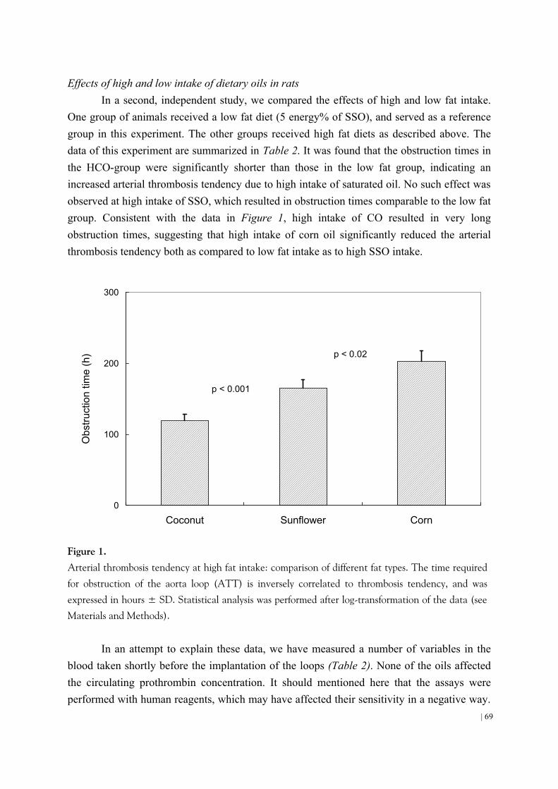
| 69
Effects of high and low intake of dietary oils in rats In a second, independent study, we compared the effects of high and low fat intake.
One group of animals received a low fat diet (5 energy% of SSO), and served as a reference group in this experiment. The other groups received high fat diets as described above. The data of this experiment are summarized in Table 2. It was found that the obstruction times in the HCO-group were significantly shorter than those in the low fat group, indicating an increased arterial thrombosis tendency due to high intake of saturated oil. No such effect was observed at high intake of SSO, which resulted in obstruction times comparable to the low fat group. Consistent with the data in Figure 1, high intake of CO resulted in very long obstruction times, suggesting that high intake of corn oil significantly reduced the arterial thrombosis tendency both as compared to low fat intake as to high SSO intake.
0
100
200
300
Coconut Sunflower Corn
Obs
truc
tion
time
(h)
p < 0.001
p < 0.02
Figure 1.
Arterial thrombosis tendency at high fat intake: comparison of different fat types. The time required
for obstruction of the aorta loop (ATT) is inversely correlated to thrombosis tendency, and was
expressed in hours ± SD. Statistical analysis was performed after log-transformation of the data (see Materials and Methods).
In an attempt to explain these data, we have measured a number of variables in the blood taken shortly before the implantation of the loops (Table 2). None of the oils affected the circulating prothrombin concentration. It should mentioned here that the assays were performed with human reagents, which may have affected their sensitivity in a negative way.

70 |
The HCO diet resulted in relatively high circulating levels of triglycerides and vitamin K, whereas in the CO and SSO groups the triglycerides were significantly lower than in the low fat group. This effect was stronger in the CO group than in the SSO group, but whether this difference is sufficient to explain the strongly reduced arterial thrombosis tendency in the CO group is not clear. Also circulating vitamin K was reduced during high intake of unsaturated oil, but the effect was only statistically significant in the CO group.
Table 2. Comparison of high and low dietary fat in the rat
Rats were fed with various diets during 8 weeks before blood was taken and aorta loops were
implanted. Determinations in plasma and serum were performed in duplicate, mean values for each individual animal were used to calculate the mean of means (± SD) given in this Table. The values
obtained in the three high fat groups were compared with the low fat one, differences were regarded to
be significant at p < 0.05 (*).
Discussion
This paper is based on the initial discovery that in an animal model system the arterial thrombosis tendency is decreased by replacing HCO in the diet by either SSO or CO. Whereas these first experiments were performed exclusively in animals on a high-fat regimen (50 energy%), later studies demonstrated that the beneficial effect of SSO is lost if the high-fat diet is compared with low-fat diets (5 energy%). For high intake of CO, however, a substantial and significant protective effect remained even when the diet was compared with a low-fat regimen (Table 2). The difference between CO and HCO was comparable to that obtained in rats treated with low doses of vitamin K-antagonist (G. Hornstra, unpublished data) or with high doses of aspirin 24. The difference between HCO and both plant oils may be explained by the fact that the former is fully saturated, whereas both CO and SSO contain
Low fat HCO SSO CO
Number of rats: 24 48 48 48 Dietary fat (energy%): 5 50 50 50
Obstruction time (h) 120 ± 8 91 ± 11* 123 ± 10 144 ± 13* Plasma prothrombin (% of ref) 96 ± 7.6 99 ± 9.2 102 ± 9.1 101 ± 5.5 Serum vitamin K 1 ( µ g/L) 0.8 ± 0.2 1.9 ± 0.3* 0.8 ± 0.27 0.7 ± 0.2* Serum triglycerides (mmol/L) 1.0 ± 0.1 1.8 ± 0.1* 0.9 ± 03* 0.6 ± 0.05* K 1 / triglyceride ( µ g/mmol) 0.8 ± 0.2 1.1 ± 0.2 0.9 ± 0.2 1.1 ± 0.2

| 71
a large fraction of unsaturated fatty acids. Because the fatty acid composition of CO and SSO is remarkably similar, other explanations have to be found for their different effect on the arterial thrombosis tendency. Substantial differences between CO and SSO were reported with respect to their non-fat fraction. CO, for instance, was reported to contain 10-20 fold
more ubiquinone and γ-tocopherol than did SSO 25. Since these compounds have a marked structural analogy with vitamin K, our group and others have tested whether either ubiquinone 26 or tocopherol 27 may act as competitive inhibitors for the vitamin K-dependent carboxylase, thus inhibiting the biosynthesis of blood coagulation factors. Although in vitro some inhibition was found, the effect was weak and it is unlikely that it can explain the marked in vivo differences between both oils.
We have tested this hypothesis in rats as well as in a human nutritional study. In rats we found no significant effect of any of the oils on the circulating prothrombin concentration. It should be kept in mind, however, that the prothrombin assay is not a very sensitive one to detect small fluctuations. A direct assay for descarboxy-prothrombin would be far more accurate, but this test is not available for rats. As compared to the low fat group, serum vitamin K had remained constant in rats on SSO, but had significantly decreased in the CO group. Circulating triglycerides, which play a major role in vitamin K transport, had decreased in both plant oils, but to a much larger extent in the CO than in the SSO group. Hence it is at least feasible that the type of oil affects the bioavailability and tissue distribution of vitamin K.
When trying to find an explanation for the observed effects of corn oil it is important to realise that after intestinal absorption, vitamin K is transported in the blood via lipoproteins and that it is liberated therefrom during the hydrolysis of the chylomicron triglycerides in a variety of tissues (including the liver) before the chylomicron remnants are cleared by the liver 28. Only little vitamin K is found in the LDL and HDL fractions 18. Obviously, the postprandial fatty acid content of the chylomicrons will depend on the composition of the diet. Animal and some human studies suggest an increased activity of endothelial lipoprotein lipase towards the hydrolysis of PUFA-rich compared to SAFA-rich chylomicrons 8,29. In the context of vitamin K and vitamin K-dependent coagulation proteins, a recent study in rats showed that the PUFA-rich fish oil not only decreased serum cholesterol and triglyceride levels, but also led to decreased levels of the vitamin K-dependent coagulation factors prothrombin and factor VII 12. These authors postulated that this hypocoagulable effect may be mediated by limited delivery of vitamin K to the liver and thereby the synthesis of vitamin K-dependent coagulation proteins. Our data support the view that circulating levels of vitamin K-dependent coagulation proteins are linked to lipid metabolism in normal physiology 30 with the intriguing possibility that part of this interaction with lipids may mediated via an effect on vitamin K metabolism 12. An explanation for such an effect could

72 |
be that corn oil induces a different transport for vitamin K with more extra-hepatic vitamin K uptake rather than accumulation in the liver, but this is speculative at this time.
We and others have demonstrated that subclinical vitamin K-deficiency is common in the healthy adult population and may be associated with low bone mass 31-33, increased fracture rate in postmenopausal osteoporosis 34, and increased vascular calcification 35. These phenomena may be related to the synthesis of incompletely carboxylated Gla-proteins such as osteocalcin in bones 36, and matrix Gla-protein (MGP) in the arterial vessel wall 37. Therefore, nutrients capable of shifting the balance between hepatic and extra-hepatic vitamin K uptake may have a dual effect. On one hand they might induce a mildly anticoagulant effect by decreasing the hepatic vitamin K status, thus lowering the arterial thrombosis tendency. On the other hand they might improve vascular MGP carboxylation and thus contribute to the prevention of arterial calcification.
| 73
3.2
NOVEL EFFECTS OF DIETS ENRICHED WITH CORN OIL OR AN OLIVE OIL / SUNFLOWER OIL
MIXTURE ON VITAMIN K METABOLISM AND VITAMIN K-DEPENDENT PROTEINS IN YOUNG MEN
Abstract Little is known of how the fat components of diets influence the absorption, and
metabolism of vitamin K and the possible consequences to the synthesis of vitamin K-dependent (VKD) proteins in different target organs. We have evaluated the effects of two diets on circulating phylloquinone (K1) and triacylglycerols (TAG). One diet was enriched
with corn oil (CO) (also rich in γ-tocopherol) and the other with an olive/sunflower (O/SO)
mixture (rich in α-tocopherol). Effects on γ-carboxylation were assessed from coagulation assays and sensitive assays for undercarboxylated prothrombin (ucFII) and osteocalcin (ucOC). Total plasma matrix Gla-protein (MGP) was also measured. After an initial adjustment diet, 26 healthy young men were fed, in a crossover design, the O/SO or CO diet for 2 weeks. Mean intakes of K1 during consumption of adjustment, O/SO, and CO diets were
225 µg/day, 291µg/day, and 291 µg/day, respectively. Mean fasting levels of TAG and K1 were both significantly reduced by the CO diet, but not by the O/SO diet. Neither diet reduced FII activity but ucFII became detectable in 9 subjects, 8 of whom showed this abnormality with both diets. The CO diet induced a rise in ucOC (P<0.05) which was negatively correlated to ucFII (r=-0.71, P<0.03) suggesting differences between subjects in the relative bioavailability of vitamin K between liver and bone. The CO but not O/SO diet induced a decrease of total circulating MGP. We conclude that both oils, notably CO, affected vitamin
K absorption and/or metabolism which may increase the requirements for γ-carboxylation.
The mechanism is unclear but may result from interactions of vitamin K with PUFA and/or other lipid components such as vitamin E. Introduction
The nutritional need for vitamin K is most commonly associated with the four vitamin K-dependent (VKD) coagulation factors (II, VII, IX and X) that are all synthesized in the liver 38. Other VKD proteins are synthesized in extra-hepatic tissues. They include the bone protein OC, and MGP whose mRNA is expressed by various tissues and cell types. There is good evidence that OC plays a regulatory role in bone turnover 39 and that MGP is essential
74 |
to prevent calcification of arteries 40. All these proteins require vitamin K for a post-
translational modification in which selective glutamate residues are transformed to γ-carboxyglutamate (Gla) residues. The necessity of Gla residues for functional activity is known for the VKD coagulation proteins 38,41 and is probable for others. When the supply of vitamin K is insufficient or when there is a metabolic blockade (e.g. by oral anticoagulants), undercarboxylated species of Gla-proteins are released into the circulation and provide a measure of the vitamin K status at their site of synthesis 42.
The major dietary and circulating form of vitamin K is phylloquinone (vitamin K1) 38. After intestinal absorption, phylloquinone, like other fat-soluble vitamins is carried via the chylomicron pathway. However, a major difference from other fat-soluble vitamins is that triacylglycerol-rich (TAG-rich) lipoproteins continue to be the major carriers of phylloquinone in both the postprandial 18 and fasting 17 states. This association with TAG-rich lipoproteins is reflected in a positive correlation of plasma phylloquinone and triacylglycerol concentrations 14-16. This suggests that dietary and other factors that influence the metabolism of TAG-rich lipoproteins may also affect the transport, tissue distribution and
metabolism of vitamin K possibly affecting γ-carboxylation of the target Gla proteins. Indeed
it was recently suggested that decreases in FII and FVII in rats produced by diets enriched with n-3 PUFA may have resulted from a lipid-lowering effect which affected the lipoprotein transport and delivery of vitamin K to the liver 12.
As far as we are aware there have been no human studies that have directly examined whether dietary manipulation of the fat components of diets can influence the intestinal
absorption and/or metabolism of vitamin K and thereby the γ-carboxylation of VKD proteins. The present study was facilitated by our access to a dietary cross-over study in healthy young
men that had initially been designed to compare the effects of γ-tocopherol-rich corn oil (CO)
and α-tocopherol-rich olive/sunflower (O/SO) oil on DNA damage in healthy young men 43. This study was also of interest to us because high dietary intakes of vitamin E have been associated with an inhibitory effect on vitamin K action 44. Apart from measurements of plasma phylloquinone, vitamin K status was assessed from functional assays of VKD coagulation proteins, sensitive assays for undercarboxylated prothrombin 45 and undercarboxylated osteocalcin 46 and a new assay for total MGP 47. Materials and Methods Subjects and Study Design
Twenty-six healthy men (aged 19-31 years) were recruited from the population of Vienna. None were taking medications or vitamin supplements for at least 2 weeks before

| 75
entering the study. During an randomly assigned initial 2-week period all subjects consumed the same adjustment diet followed by diets enriched with either an olive/sunflower oil mixture (O/SO diet) or corn oil (CO diet) in a cross-over design of 2 weeks per diet. Weighed dietary records were kept for each participant for the entire study period to calculate nutrient intakes. Venous blood samples were collected in trisodium citrate (after an overnight fast) at 2-weekly intervals coinciding with the end of each dietary regime. Plasma samples were stored in aliquots at -80°C until analysis. The study protocol was approved by the Medical Ethics Committee of the University of Vienna and all subjects gave their informed consent in writing. Dietary Oils and Experimental Diets
The sources of the oils used for the diets were: corn oil, CPC-Bestfoods (Heilbronn, Germany); olive oil, Linea Natura (Milan, Italy); sunflower oil, Nestle (Karlsruhe, Germany). Their SAFA/MUFA/PUFA compositions were: olive oil 14:77:9, sunflower oil 12:24:64 and
corn oil 13:33:54. Their vitamin E contents per 100g oil were: olive oil 20 mg α-tocopherol
and 1.7 mg γ-tocopherol, sunflower oil 85.3 mg α-tocopherol and 8.8 mg γ-tocopherol, corn
oil 24.6 mg α-tocopherol and 126.2 mg γ-tocopherol 43. The K1 contents of olive, sunflower
and corn oil were 54.8, 5.7, and 2.7µg/100g, respectively. All food was prepared at the Institute of Nutritional Sciences of the University of
Vienna as three daily meals and offered to the subjects in the presence of one of the investigators. On weekdays all subjects ate their mid-day meal in the Institute. Breakfast and evening meals were pre-packed and given to the subjects on a daily basis. Weekend meals were also prepared beforehand and given to the subjects on Fridays. Typical foodstuffs for breakfasts comprised rye and wheat bread, muesli, buttermilk and occasionally yogurt and fruit. Lunches comprised soup, fish or meat with vegetables and usually a fresh salad. Common food items for evening meals were bread, yogurt, fruit and cheese.
The composition of the diets is shown in Table 1. The adjustment diet provided the daily intakes of 35 g olive oil, 4.4 g sunflower oil and 13.8 g of butter. The O/SO and CO test diets were identical apart from their different fat contents. The O/SO diet provided the daily intakes of 68 g olive oil and 12 g sunflower oil (SAFA/MUFA/PUFA = 28/49/23) and the CO diet the equivalent intake of 80 g corn oil (SAFA/MUFA/PUFA = 29/33/38). The 80g total daily intakes of dietary oils represented 73% of the total daily fat intake. Both O/SO and
CO diets provided similar intakes of α-tocopherol but the CO diet provided a high intake of
γ-tocopherol (100 mg/day) compared to the O/SO diet (2.4 mg/day) (Table 1). Dietary intakes of phylloquinone (K1) were calculated from the weighed dietary records of the participants and the K1 content of foods taken from newly available data bases from The Netherlands and the U.K 48-50. Based solely on the K1 values for their component oils the O/SO and CO diets

76 |
provided daily intakes of 15.4 µg and 2.8 µg K1 respectively. However, this difference was
negligible compared to the intakes of K1 from other food components, especially green leafy vegetables. With the cross-over design the average daily intakes of vitamin K were closely
similar for both groups, but there was considerable day to day variation (mean: 291 µg, S.D.
± 213) which was due to the variability of consumption of green vegetables. The average
daily intakes of K1 calculated for each phase of the study were 225 µg (adjustment phase),
238 µg (cross-over phase I) and 343 µg (cross-over phase II).
Table 1. Composition of the diets
Diet content Adjustment diet Olive/sunflower oil diet Corn oil diet
Mean energy (MJ) 11.6 11.6 11.6Carbohydrate (energy%) 50 - 55 50 - 55 50 - 55Protein (energy%) 15 15 15Fat (energy%) 30 - 35 30 - 35 30 - 35SAFA (energy%) `11 - 13 `8 - 10 `9 - 10MUFA (energy%) `13 - 15 `15 - 17 `10 - 12PUFA (energy%) `6 - 7 `7 - 8 `11 - 13α-Tocopherol (mg/day) 12 24 20γ-Tocopherol (mg/day) - 2.4 100Phylloquinone (µg/day) 225 291 291 Table shows the range of daily consumption of macronutrients and the mean daily consumption of
tocopherol and phylloquinone (vitamin K1) micronutrients
Various Assays
All biochemical analyses were performed in duplicate and mean values are given throughout this paper. Prothrombin times (PT) were determined by automated assay using Thromborel S (Behringwerke, Marburg, Germany) as thromboplastin reagent. Factor II (FII) concentrations were assessed assay using Thromborel S and human clotting factor II-deficient plasma from Behringwerke. Species of undercarboxylated FII (ucFII) were measured using a conformation-specific monoclonal antibody in an ELISA-based assay 45. Results are expressed as Arbitrary Units per L (AU/L) because in states of vitamin K deficiency circulating ucFII may comprise multiple forms of partially carboxylated FII and neither their relative abundance in plasma nor their relative affinity for the antibody is
known. Using electrophoretic techniques 1 AU is equivalent to 1 µg of purified ucFII 45. The
detection limit was 150 AU/L plasma. Total immunoreactive OC was measured using the two-site N-mid hOsteocalcin
ELISA (Osteometer A/S, Copenhagen, Denmark). Undercarboxylated osteocalcin (ucOC)
| 77
was determined with the Glu-OC kit from Takara Shuzo (Tokyo, Japan). MGP was determined with an ELISA-based assay recently developed at the Maastricht Biochemistry Department 47. Serum MGP concentrations were calculated with the aid of a reference curve from pooled normal serum, and expressed as arbitrary units per liter (AU/L).
Plasma K1 concentrations were measured by high performance liquid chromatography (HPLC) and fluorescence detection after on-line, post column electrochemical reduction of the effluent which converted the quinone forms of vitamin K compounds to their fluorescent quinol forms 22. Plasma TAG concentrations were determined by an automated enzymatic procedure using commercial reagents (Boehringer Mannheim, Germany) and a Beckmann Synchron CX 7-2 autoanalyser (Fullerton, CA). Data analysis
Blood samples taken at the end of the adjustment phase, cross-over phase I and cross-over phase II and were analysed as repeated measurements in the cross-over design to determine whether there were any significant trends. The analysis was used to examine statistical differences between both the oil-regimens and the adjustment phase or between both oil-regimens themselves. The statistical significance of differences between the oil-regimens and the adjustment phase were examined using the SPSS 9.0 Students paired t-tests (2-tailed). Differences between the regimens in either cross-over phase I or cross-over phase II were examined with the Wilcoxon matched pairs test. Differences were considered to be statistically significant at P<0.05. Results
The results of coagulation assays together with plasma concentrations of TAG, K1 and VKD proteins at the end of each dietary period are shown in Table 2. Compared to the adjustment phase diet, plasma levels of both TAG and K1 were significantly lowered by the CO diet (P<0.01 for TAG, P<0.05 for K1) but not by the O/SO diet. The effect of both diets on hepatic synthesis of VKD procoagulants was assessed by three different assays with varying sensitivities: the PT (an overall coagulation assay which is insensitive to large decreases in the Gla-content of VKD coagulation factors 12), the FII assay (specific for functional Gla forms of prothombin), and an in-house ELISA to detect low concentrations of ucFII. No diet-induced change in hepatic VKD coagulation proteins was detected by either the PT or FII assay. After the adjustment diet, ucFII was below the limit of detection in all subjects but elevated levels were found in 8 subjects (also defined as ucFII positive) after the O/SO diet, and in 9 subjects after the CO diet (Figure 1). Eight of these had an elevated ucFII after both diets which first presented at the end of the first dietary cross-over phase (phase I)

78 |
with the O/SO diet and which continued to remain elevated during phase II while taking the CO diet (subject group B in Table 2). One subject showed an elevated ucFII at the end of cross-over phase I while taking the CO diet which then dropped below the detectable limit after phase II with the OSO diet. In the remaining 17 subjects ucFII remained undetectable throughout the study. In the ucFII positive subjects, there was no significant difference in the magnitude of ucFII after O/SO or CO diets (Table 2) although in 6/8 subjects who had an elevated ucFII after phase I (O/SO diet), the levels had increased slightly by the end of phase II (CO diet) (Figure 1). There was no correlation between ucFII and plasma K1 concentrations: for the O/SO diet the mean K1 concentrations in subjects with undetectable or detectable ucFII were 0.53 nmol/L (n = 18) and 0.63 nmol/L (n = 8) respectively. Similarly, for the CO diet, the mean plasma K1 concentrations were 0.39 nmol/L in ucFII negative subjects (n = 17) and 0.48 nmol/L in ucFII positive subjects (n = 9).
Table 2. Effects of OSO and CO tests diets on vitamin K-dependent proteins, vitamin K and
triglycerides
Measurement
all subjects group A group B all subjects group A group B all subjects group A group B
Prothrombin time (s) 14.4±0.8 14.3±1.0 14.4±0.7 14.2±0.9 14.0±1.0 14.5±0.9 14.5±0.8 14.8±0.9 14.1±0.4Factor II (% normal) 88.7±7.5 88.7±7.6 88.8±7.7 92.1±9.7 89.6±10.0 94.6±9.0** 90.9±11.8 89.2±13.5 92.7±10.1*ucF II (AU/L) <150 <150 150 220 ±120** <150 280 ±150** 240 ±160** 180 ±90 310 ±180ucF II (prevalence) 0/26 0/13 0/13 `8/26 0/13 `8/13 `9/26 `1/13 `9/13
Total osteocalcin (µg/L) 22.3±12.9 23.5±13.7 21.2±12.5 19.1±9.1 18.6±8.7* 19.5±9.9 18.6±10.5* 16.4±8.4**‡ 20.8±12.3ucOC (µg/L) 5.9±2.4 6.0±2.4 5.8±2.5 6.3±2.4 6.8±2.8 5.9±1.9 6.3±2.3* 6.3±2.3 6.3±2.3
Matrix Gla-protein (U/L) 93±8 92±9 94±9 98±10 94±11 101±9 86±11**‡‡ 88±12 83±10*‡‡
Vitamin K1 (nmol/L) 0.59±0.37 0.64±0.35 0.54±0.40 0.56±0.29 0.51±0.18 0.61±0.37 0.42±0.23*‡0.43±0.19 0.41±0.27‡
Triglycerides (mmol/L) 0.85±0.46 0.91±0.44 0.79±0.49 0.79±0.44 0.72±0.33 0.86±0.53 0.64±0.34**‡ 0.57±0.30**‡ 0.71±0.38
Adjustment diet OSO test diet CO test diet
The results (mean ± SD) are shown for measurements made at the end of each 2-week dietary phase
after the subjects had received the adjustment, the olive/sunflower oil (OSO) and corn oil (CO) diets respectively. Values for each diet are shown for all subjects after completion of the cross-over study
and under groups A and B according to the sequence in which the 2 randomized groups of 13 subjects
received the OSO and CO test diets after the adjustment diet. Subjects in group A received the CO
diet in phase I and the OSO diet in phase II while subjects in group B received the OSO diet in phase
I and the CO diet in phase II of the cross-over design. * denotes significant differences between
adjustment diet and either OSO or CO diets with *: P< 0.05, and **: P<0.01. ‡ denotes significant
differences between OSO and CO diets with ‡: P<0.05, and ‡‡: P<0.01.

| 79
0
250
500
750
0.5 2.5
ucF
II (A
U/L
)
Olive/sunflower oil diet Corn oil diet
Figure 1.
Diet induced increase of undercarboxylated factor II (ucFII). Explanation of symbols: n = positive
responders after the O/SO diet; l = positive responders (paired samples) after the CO diet (connecting lines indicate same subjects); o = subject only positive in the CO diet. The shaded area
shows the lower detection limit (150 AU/L).
The effect of both diets on the synthesis of extra-hepatic VKD proteins was assessed for OC and MGP. Neither diet affected circulating concentrations of total immunoreactive OC. However, differences were seen with a monoclonal-based assay specific for ucOC. Both CO and O/SO diets induced an increase in plasma ucOC but this only became statistically significant for the CO diet. Suggestive for a differential carboxylation of FII in the liver and OC in bone was the observation that for both O/SO and CO diets, the mean ucOC concentrations in ucFII positive subjects were 20% lower than in ucFII negative subjects
(ucOC 5.4 µg/L vs 6.8 µg/L in positive and negative subjects respectively). This relatively
small difference did not attain statistical significance, however. A test of the degree of correlation between ucOC and ucFII concentrations was only possible in about a third of the subjects with detectable ucFII, and as shown in Figure 2 for the CO diet there was a negative correlation between ucOC and ucFII (r = -0.71, P = 0.03). In the O/SO group this correlation was also negative (r = -0.35) but not statistically significant.

80 |
0.0
2.5
5.0
7.5
10.0
0 250 500 750 1000
ucFII (AU/L)
ucO
C (
µg/L
)
Figure 2.
Inverse correlation between ucFII and ucOC during the CO diet. The Pearson correlation coefficient
(r) was -0.71 and level of significance (P) was 0.03.
We also measured the circulating levels of MGP, which were significantly lower after the CO diet than after either the adjustment or O/SO diets (Figure 3). These measurements represent total MGP, assays discriminating between MGP and ucMGP are presently not available.
Since previous studies have reported that circulating VKD coagulation factors may be positively related to plasma lipids, we examined the relationship between plasma TAG and the VKD proteins. FII was positively correlated with TAG during all diets (pooled data: R=0.385; P<0.01) and also separately within each dietary phase with the strongest correlation for the CO diet (R =0.536; P<0.01). The extra-hepatic VKD proteins OC and MGP did not correlate with TAG.

| 81
50
75
100
125S
erum
MG
P (U
/L)
Adjustment diet OSO diet CO diet
Figure 3.
Serum MGP concentrations during the three different dietary regimes. The mean value during the
CO diet was significantly lower than the values during the adjustment phase and the OSO diet.
Discussion
The present study provides evidence that altering the lipid component of the diet has
the potential to reduce plasma levels of K1 and/or impair the γ-carboxylation of representative hepatic (FII) and extra-hepatic (OC) Gla-proteins. It was noteworthy that the PUFA-rich CO diet induced a significant reduction in plasma K1 and TAG compared to both adjustment and O/SO diets. This is consistent with the carriage of K1 by TRL 18 and with the well-established capacity of diets rich in PUFA to reduce post-absorptive concentrations of plasma TAG 8, probably by reducing the post-prandial accumulation of TRL 29. The accompanying evidence of slight increases in undercarboxylation of FII and osteocalcin by the CO diet imply a lowered vitamin K status that may, however, be unrelated to the lowering of plasma TAG and K1. Possible explanations include a reduced intestinal absorption of vitamin K, a reduced extra cellular or intracellular delivery of vitamin K to target proteins, an increased turnover
rate of the vitamin, or an inhibitory effect on VKD γ-carboxylation. The possibility that the CO diet reduced the intestinal absorption of vitamin K is
supported by in vivo studies in rats which showed that the PUFA linoleic acid (18:2) caused a

82 |
marked decrease in the intestinal absorption rate of vitamin K1 51. This inhibition was not seen with the MUFA oleic acid (18:1). The same authors briefly commented that linoleic acid also inhibited the absorption of vitamin A. Other in vivo studies have shown that feeding diets with increasing linoleic acid contents to rats depressed the intestinal lymphatic absorption of vitamin E 52. Taken together, these studies in rats suggest a general inhibitory effect of PUFAs on the absorption of fat soluble vitamins which share a similar pathway of absorption and lymphatic transport.
Previous studies in healthy adults have indicated that the levels of VKD FVII are more influenced by the total fat content of the diet rather than the SAFA/MUFA/PUFA composition 53. In our study we also found no effects of the O/SO or CO diets on the PT or FII concentrations but we did find a significant correlation of FII levels with plasma TAG. This is in agreement with previous studies showing associations of one or more VKD coagulation factors with TAG in patients with hyperlipidemia 54,55 and in healthy young men 30. The reason for this association of VKD proteins with lipids is still unclear, but the recent finding of the in vivo binding of all VKD coagulation proteins to TRL 56 provides one explanation. Our data support the view that circulating levels of VKD coagulation proteins are linked to lipid metabolism in normal physiology 30 with the possibility that part of this interaction with lipids may mediated via an effect on vitamin K metabolism 12.
To assess possible effects of the diets on the γ-carboxylation of FII, we used a sensitive immunoassay specific to ucFII. Hitherto, plasma ucFII concentrations in freeliving, healthy adults with this assay have always been below the detection limit. The same was true in the present study for all 26 subjects after the adjustment phase but both test diets induced detectable ucFII. That this was a genuine effect of the diets is supported by the fact that the analyses of ucFII were blind and carried out in random order. It should be emphasised that the absolute increases in ucFII were relatively low and that the presence of such low concentrations (200-700 AU/L) did not affect the PT or FII assay. In patients on stable anticoagulant therapy ucFII values with this assay range from 6,900-99,500 AU/L (mean 40,000 AU/L) (M.J. Shearer, unpublished results). It seems unlikely, therefore, that this small increment in ucFII, and presumably also in ucFVII, ucIX and ucX, would lead to a hypocoagulable state. On the other hand, evidence that minor reductions in VKD coagulation factors may result in a clinically significant decreased coagulability comes from studies of low dose warfarin regimes. Thus, daily doses of 1 mg warfarin reduced the incidence of deep vein thrombosis after surgery with no appreciable prolongation of the PT 57, and prevented thrombosis in central venous catheters with no changes in overall coagulation assays or decreases in specific VKD factors 58. This suggests that a hypocoagulable state can be induced by relatively minor perturbations of the carboxylation status of VKD coagulation proteins.

| 83
In contrast to the hepatic Gla-proteins, there is evidence that the bone protein OC is not fully carboxylated and is readily responsive to changes in dietary intakes of vitamin K 59. Compared to the adjustment diet, ucOC was significantly reduced only by the CO diet, but with the concomitant fall in total OC this effect was significant for both diets when ucOC was expressed as a percent of total OC. Total circulating OC is also a marker of osteoblastic activity, reflecting the rate of bone formation, but as this reduction of total OC was unexpected, further studies would be needed to address such a relationship. Unlike FII, there was no correlation of either OC or ucOC with TAG levels and there are no reports in the literature indicating such a relationship.
A direct inhibition of the VKD γ-glutamyl carboxylase or other enzymes of the vitamin K-epoxide cycle by non-TAG components also cannot be excluded. Compared to the
adjustment diet, both the O/SO and CO diets provided about double the intake of α-
tocopherol while the CO diet provided a high intake of γ-tocopherol. The high γ-tocopherol content of corn oil has already been forwarded as an explanation of the reduced frequency of sister chromatid exchange seen in the same subjects after the CO diet compared with the O/SO diet 43. Corn oil is also a rich source of ubiquinone-9. Vitamin E and ubiquinone compounds are structurally related to vitamin K, and have been reported to inhibit VKD carboxylase in vitro 26,27.
The literature contains several reports that vitamin E compounds may interfere with vitamin K action in animals or humans but the mechanism remains unclear 60-63. An early
study showed that the daily oral administration of large (100 mg) doses of α-
tocopherolquinone to pregnant mice caused bleeding in the reproductive system which was reversible with vitamin K 60 and a similar vitamin K-reversible coagulopathy was observed in chicks with large doses of vitamin E 62. Helson et al. 63 showed in patients with neuroblastoma that large doses of vitamin E caused a bleeding diathesis and that this was a systemic effect on vitamin K metabolism rather than an effect on absorption. More recently, Alexander & Suttie 64 have provided further evidence for cellular and whole animal interactions of vitamin E with vitamin K. Firstly, cultured H-35 cells showed significant
decrease in FII production in the presence of both α-tocopherol and α-tocopherolquinone.
Secondly, rats fed a vitamin K deficient diet and increasing amounts of α-tocopherol in
combination with a constant amount of phylloquinone showed a dose-dependent reduction in plasma FII and concomitant reductions in both plasma and liver phylloquinone. The effects on vitamin K and VKD proteins observed in our study were minor in comparison but they raise the possibility of an interaction with the increased dietary intakes of vitamin E.
The strong inverse relationship between ucFII and ucOC seen in those subjects who had detectable ucFII after the CO diet is suggestive of some inter-subject variability in the relative bioavailability of vitamin K between the liver and bone. This could be mediated by

84 |
difference(s) in lipoprotein metabolism that causes a differential organ/tissue uptake of TRL. One candidate is apolipoprotein E whose common variants are known to influence post-prandial TG metabolism and to mediate the uptake of TRL remnant particles 65. From this and other studies, Kohlmeier et al. 17 generated the hypothesis that the liver and bone are competing organs for vitamin K, a hypothesis that may explain the inverse correlation of ucFII and ucOC.
Measurements of total plasma MGP with a recently available assay showed that the CO diet but not the O/SO diet caused a decrease in circulating MGP compared to the adjustment diet. This fall in MGP after the CO diet mirrored that in total OC but the significance of this finding is unclear since little is yet known of the determinants of plasma MGP. There is strong interest in this VKD protein because recent studies show that vascular MGP synthesis is upregulated at sites of atherosclerotic calcification in the vessel wall 66 and that circulating MGP is increased during some stages of atherosclerosis 47.
| 85
REFERENCES 1. Hornstra G, Kester AD. Effect of the dietary fat type on arterial thrombosis tendency: systematic
studies with a rat model. Atherosclerosis. 1997;131:25-33. 2. Gurr MI. Dietary lipids and coronary heart disease: old evidence, new perspective. Prog Lipid
Res. 1992;31:195-243. 3. Hornstra G, Vendelmans-Starrenburg A. Induction of experimental arterial occlusive thrombi in
rats. Atherosclerosis. 1973;17:369-82. 4. Hornstra G, Lussenburg RN. Relationship between the type of dietary fatty acid and arterial
thrombosis tendency in rats. Atherosclerosis. 1975;22:499-516. 5. Mennen LI, Schouten EG, Grobbee DE, Kluft C. Coagulation factor VII, dietary fat and blood
lipids: a review. Thromb Haemost. 1996;76:492-9. 6. Kohlmeier M, Riesen W, Schlierf G. Metabolic changes in healthy men using fat-modified diets.
I. Disposition of serum cholesterol. Ann Nutr Metab. 1988;32:1-9. 7. Wardlaw GM, Snook JT. Effect of diets high in butter, corn oil, or high-oleic acid sunflower oil
on serum lipids and apolipoproteins in men. Am J Clin Nutr. 1990;51:815-21. 8. Weintraub MS, Zechner R, Brown A, Eisenberg S, Breslow JL. Dietary polyunsaturated fats of
the W-6 and W-3 series reduce postprandial lipoprotein levels. Chronic and acute effects of fat saturation on postprandial lipoprotein metabolism. J Clin Invest. 1988;82:1884-93.
9. Dupont J, White PJ, Carpenter MP, Schaefer EJ, Meydani SN, Elson CE, Woods M, Gorbach SL. Food uses and health effects of corn oil. J Am Coll Nutr. 1990;9:438-70.
10. Goto Y, Tamachi H, Moriguchi EH. Eicosapentaenoic acid and atherosclerosis. Prostaglandins Leukot Essent Fatty Acids. 1993;48:337-42.
11. Chin JP. Marine oils and cardiovascular reactivity. Prostaglandins Leukot Essent Fatty Acids. 1994;50:211-22.
12. Nieuwenhuys CM, Beguin S, Offermans RF, Emeis JJ, Hornstra G, Heemskerk JW. Hypocoagulant and lipid-lowering effects of dietary n-3 polyunsaturated fatty acids with unchanged platelet activation in rats. Arterioscler Thromb Vasc Biol. 1998;18:1480-9.
13. Shahar E, Folsom AR, Wu KK, Dennis BH, Shimakawa T, Conlan MG, Davis CE, Williams OD. Associations of fish intake and dietary n-3 polyunsaturated fatty acids with a hypocoagulable profile. The Atherosclerosis Risk in Communities (ARIC) Study. Arterioscler Thromb. 1993;13:1205-12.
14. Sadowski JA, Hood SJ, Dallal GE, Garry PJ. Phylloquinone in plasma from elderly and young adults: factors influencing its concentration. Am J Clin Nutr. 1989;50:100-8.
15. Saupe J, Shearer MJ, Kohlmeier M. Phylloquinone transport and its influence on gamma-carboxyglutamate residues of osteocalcin in patients on maintenance hemodialysis. Am J Clin Nutr. 1993;58:204-8.
16. Kohlmeier M, Saupe J, Drossel HJ, Shearer MJ. Variation of phylloquinone (vitamin K1) concentrations in hemodialysis patients. Thromb Haemost. 1995;74:1252-4.
17. Kohlmeier M, Salomon A, Saupe J, Shearer MJ. Transport of vitamin K to bone in humans. J-Nutr. 1996;126:1192s-6s.
18. Lamon-Fava S, Sadowski JA, Davidson KW, O'Brien ME, McNamara JR, Schaefer EJ. Plasma lipoproteins as carriers of phylloquinone (vitamin K1) in humans. Am J Clin Nutr. 1998;67:1226-1231.
19. Shearer MJ, Barkhan P, Webster GR. Absorption and excretion of an oral dose of tritiated vitamin K1 in man. Br J Haematol. 1970;18:297-308.
20. Groenen-van Dooren MM, Soute BA, Jie KS, Thijssen HH, Vermeer C. The relative effects of phylloquinone and menaquinone-4 on the blood coagulation factor synthesis in vitamin K-

86 |
deficient rats. Biochem Pharmacol. 1993;46:433-437. 21. Roncaglioni MC, di Minno G, Pangrazzi J, Reyers I, Mussoni L, de Gaetano G, Donati MB.
Plasmatic and vascular factors of the hemostatic system in rats receiving an estrogen-progestogen combination. Contraception. 1980;22:249-57.
22. Gijsbers BL, Jie KS, Vermeer C. Effect of food composition on vitamin K absorption in human volunteers. Br J Nutr. 1996;76:223-9.
23. Thijssen HH, Drittij-Reijnders MJ. Vitamin K metabolism and vitamin K1 status in human liver samples: a search for inter-individual differences in warfarin sensitivity. Br J Haematol. 1993;84:681-685.
24. Reyers I, Hennissen A, Donati MB, Hornstra G, de Gaetano G. Aspirin and the prevention of experimental arterial thrombosis: difficulty in establishing unequivocal effectiveness. Thromb Haemost. 1985;54:619-21.
25. Kohlmeier M, Drossel HJ, Sinha P, Kottgen E. Rapid and simple method for the identification of apolipoprotein E isomorphic phenotypes from whole serum. Electrophoresis. 1992;13:258-61.
26. Ronden JE, Soute BA, Thijssen HH, Saupe J, Vermeer C. Natural prenylquinones inhibit the enzymes of the vitamin K cycle in vitro. Biochim Biophys Acta. 1996;1298:87-94.
27. Uotila L. Inhibition of vitamin K dependent carboxylase by vitamin E and its derivatives. Current Advances in Vitamin K Research (ed: Suttie JW). 1988;Elsevier, New York:59-64.
28. Havel RJ, Kane JP. Introduction: structure and metabolism of plasma lipoproteins. The metabolic and molecular basis of inherited disease (ed. Scriver, C.R.). 1995;7th ed. New York: McGraw-Hill:1841-1851.
29. Williams CM. Postprandial lipid metabolism: effects of dietary fatty acids. Proc Nutr Soc. 1997;56:679-92.
30. Hoffman CJ, Lawson WE, Miller RH, Hultin MB. Correlation of vitamin K-dependent clotting factors with cholesterol and triglycerides in healthy young adults. Arterioscler Thromb. 1994;14:1737-40.
31. Szulc P, Arlot M, Chapuy MC, Duboeuf F, Meunier PJ, Delmas PD. Serum undercarboxylated osteocalcin correlates with hip bone mineral density in elderly women. J Bone Miner Res. 1994;9:1591-5.
32. Knapen MH, Nieuwenhuijzen Kruseman AC, Wouters RS, Vermeer C. Correlation of serum osteocalcin fractions with bone mineral density in women during the first 10 years after menopause. Calcif Tissue Int. 1998;63:375-9.
33. Väänänen HK, Luukinen H, Käkönen S-M, Pettersson K, Koski K, Laippala P, Lövgren T, Kivelä S-L. Strong prediction of fractures among the elderly by the ratio of carboxylated and total serum osteocalcin. Calcif Tissue Int. 1999;64:S79.
34. Szulc P, Chapuy MC, Meunier PJ, Delmas PD. Serum undercarboxylated osteocalcin is a marker of the risk of hip fracture: a three year follow-up study. Bone. 1996;18:487-8.
35. Jie KS, Bots ML, Vermeer C, Witteman JC, Grobbee DE. Vitamin K intake and osteocalcin levels in women with and without aortic atherosclerosis: a population-based study. Atherosclerosis. 1995;116:117-23.
36. Cairns JR, Williamson MK, Price PA. Direct identification of gamma-carboxyglutamic acid in the sequencing of vitamin K-dependent proteins. Anal Biochem. 1991;199:93-7.
37. Price PA, Faus SA, Williamson MK. Warfarin causes rapid calcification of the elastic lamellae in rat arteries and heart valves. Arterioscler Thromb Vasc Biol. 1998;18:1400-7.
38. Shearer MJ. Vitamin K. Lancet. 1995;345:229-34. 39. Shearer MJ. The roles of vitamins D and K in bone health and osteoporosis prevention. Proc
Nutr Soc. 1997;56:915-37. 40. Luo G, Ducy P, McKee MD, Pinero GJ, Loyer E, Behringer RR, Karsenty G. Spontaneous

| 87
calcification of arteries and cartilage in mice lacking matrix GLA protein. Nature. 1997;385:78-81.
41. Vermeer C. Gamma-carboxyglutamate-containing proteins and the vitamin K-dependent carboxylase. Biochem J. 1990;266:625-36.
42. Vermeer C, Hamulyák K. Pathophysiology of vitamin K-deficiency and oral anticoagulants. Thromb Haemostas. 1991;66:153-159.
43. Elmadfa I, Park E. Impact of diets with corn oil or olive/sunflower oils on DNA damage in healthy young men. Eur J Nutr. 1999;38:286-92.
44. Olson RE. The function and metabolism of vitamin K. Annu Rev Nutr. 1984;4:281-337. 45. Belle M, Brebant R, Guinet R, Leclercq M. Production of a new monoclonal antibody specific to
human des-gamma- carboxyprothrombin in the presence of calcium ions. Application to the development of a sensitive ELISA-test. J Immunoassay. 1995;16:213-29.
46. Gundberg CM, Nieman SD, Abrams S, Rosen H. Vitamin K status and bone health: an analysis of methods for determination of undercarboxylated osteocalcin. J Clin Endocrinol Metab. 1998;83:3258-66.
47. Braam LA, Dissel P, Gijsbers BL, Spronk HM, Hamulyak K, Soute BA, Debie W, Vermeer C. Assay for Human Matrix Gla Protein in Serum : Potential Applications in the Cardiovascular Field. Arterioscler Thromb Vasc Biol. 2000;20:1257-1261.
48. Bolton-Smith C, Price RJ, Fenton ST, Harrington DJ, Shearer MJ. Compilation of a provisional UK database for the phylloquinone (vitamin K1) content of foods. Br J Nutr. 2000;83:389-399.
49. Ferland G, Sadowski JA. Vitamin K1 (phylloquinone) content of edible oils: effects of heating and light exposure. J Agricult Food Chem. 1992;40:1869-1873.
50. Schurgers LJ, Vermeer C. Determination of Phylloquinone and Menaquinones in Food. effect of food matrix on circulating vitamin K concentrations. Haemostasis. 2000;30:298-307.
51. Hollander D, Rim E, Muralidhara KS. Vitamin K1 intestinal absorption in vivo: influence of luminal contents on transport. Am J Physiol. 1977;232:E69-74.
52. Gallo-Torres HE, Weber F, Wiss O. The effect of different dietary lipids on the lymphatic appearance of vitamin E. Int J Vitam Nutr Res. 1971;41:504-15.
53. Marckmann P, Sandstrom B, Jespersen J. Effects of total fat content and fatty acid composition in diet on factor VII coagulant activity and blood lipids. Atherosclerosis. 1990;80:227-33.
54. Burns P, Hoffman CJ, Katz JP, Miller RH, Lawson WE, Hultin MB. Vitamin K-dependent clotting factors are elevated in young adults who have close relatives with ischemic heart disease. J Lab Clin Med. 1993;122:720-7.
55. Constantino M, Merskey C, Kudzma DJ, Zucker MB. Increased activity of vitamin K-dependent clotting factors in human hyperlipoproteinaemia - association with cholesterol and triglyceride levels. Thromb Haemost. 1977;38:465-74.
56. Xu N, Dahlback B, Ohlin AK, Nilsson A. Association of vitamin K-dependent coagulation proteins and C4b binding protein with triglyceride-rich lipoproteins of human plasma. Arterioscler Thromb Vasc Biol. 1998;18:33-9.
57. Poller L, McKernan A, Thomson JM, Elstein M, Hirsch PJ, Jones JB. Fixed minidose warfarin: a new approach to prophylaxis against venous thrombosis after major surgery. Br Med J (Clin Res Ed). 1987;295:1309-12.
58. Bern MM, Lokich JJ, Wallach SR, Bothe A, Benotti PN, Arkin CF, Greco FA, Huberman M, Moore C. Very low doses of warfarin can prevent thrombosis in central venous catheters. A randomized prospective trial. Ann Intern Med. 1990;112:423-8.
59. Sokoll LJ, Booth SL, O'Brien ME, Davidson KW, Tsaioun KI, Sadowski JA. Changes in serum osteocalcin, plasma phylloquinone, and urinary gamma- carboxyglutamic acid in response to altered intakes of dietary phylloquinone in human subjects. Am J Clin Nutr. 1997;65:779-84.
88 |
60. Woolley DW. Some biological effects produced by a-tocopherol quinone. J Biol Chem. 1945;159:59-66.
61. Corrigan JJ, Jr., Marcus FI. Coagulopathy associated with vitamin E ingestion. Jama. 1974;230:1300-1.
62. March BE, Wong E, Seier L, Sim J, Biely J. Hypervitaminosis E in the chick. J Nutr. 1973;103:371-7.
63. Helson L. The effect of intravenous vitamin E and menadiol sodium diphosphate on vitamin K dependent clotting factors. Thromb Res. 1984;35:11-8.
64. Alexander GD, Suttie JW. The effect of vitamin E on vitamin K activity. Faseb (abstract). 1999:430.1.
65. Mahley RW. Apolipoprotein E: cholesterol transport protein with expanding role in cell biology. Science. 1988;240:622-30.
66. Proudfoot D, Skepper JN, Shanahan CM, Weissberg PL. Calcification of human vascular cells in vitro is correlated with high levels of matrix Gla protein and low levels of osteopontin expression. Arterioscler Thromb Vasc Biol. 1998;18:379-88.


4
VITAMIN K METABOLISM IN SUBJECTS WITH LOW VITAMIN K-STATUS


| 93
4.1
LOW VITAMIN K-STATUS IN PATIENTS WITH EITHER CONGENITAL DEFICIENCY OR LOW
ABSORPTION OF VITAMIN K
Abstract In this chapter we describe studies in two different groups of people who have a low
vitamin K status or resistance to its effect, arising from either acquired or hereditary causes. Firstly we report on a hereditary combined deficiency of all vitamin K-dependent coagulation factors. Secondly we investigated low vitamin K-status due to Crohn’s disease. Although the etiology of both cases is totally different, they both result in a state in which intestinal absorption is too low to meet the tissue requirement for vitamin K.
Patients with an autosomal recessive deficiency of all vitamin K dependent coagulation factors may experience severe or even fatal perinatal intra-cerebral hemorrhage. In the family investigated here, the deficiency of the coagulation factors could
be corrected by oral vitamin K substitution. A defect in the γ-glutamyl carboxylase gene was excluded by sequencing of the gene. Furthermore, the use of coumarins was excluded. The finding of very high amounts of vitamin K-epoxide suggests a defect in the vitamin K2,3-epoxide-reductase (VKOR) - complex, which explains why a high vitamin K status is needed for normal synthesis of all vitamin K-dependent proteins.
Patients with impaired absorption of fat-soluble vitamins, such as found in Crohn’s disease, also have a low vitamin K status. Serum vitamin K concentrations were significantly lower in patients with Crohn’s disease than in healthy age- and sex-matched subjects. The tissue vitamin K status was measured using the carboxylation degree of osteocalcin, one of the vitamin K-dependent proteins most sensitive to vitamin K status. It was found that in Crohn’s disease the bone tissue is sub-clinically vitamin K deficient. Moreover, these patients had a lower bone mineral density compared to controls.
It is concluded that in certain types of patients increased vitamin K intake may result in a significant increase of Gla-protein carboxylation, and thus in improved functionality of these proteins. This was demonstrated in the case of a defective VKOR-complex. More intervention studies are required to demonstrate whether vitamin K-supplements may similarly lead to improved bone health in chronic diseases characterized by impaired intestinal absorption such as Crohn’s desease.

94 |
Introduction Vitamin K is a fat-soluble vitamin and serves as a cofactor for the endoplasmic
enzyme γ-glutamyl-carboxylase. It provides the energy to drive the γ-glutamate-carboxylation reaction and catalyses the incorporation of an extra carboxyl group in the protein bound
glutamate 1,2. This new, unusual amino acid is called γ-carboxy glutamate (Gla) and renders vitamin K-dependent proteins their functionality. Vitamin K-dependent proteins play a critical role in three physiological processes: blood coagulation (prothrombin, and factors VII, IX, X and protein S, C, and Z), bone metabolism (osteocalcin, matrix Gla-protein (MGP), and protein S) and vascular biology (MGP, growth arrest specific gene-6 protein (gas-6) and protein S).
Bleeding problems due to vitamin K-deficiency are extremely rare in the healthy adult population. Only in newborns a low vitamin K status is common because of poor transfer of maternal vitamin K to the fetus 3,4. It is therefore, that newborns receive i.m. or oral vitamin K to prevent them from vitamin K-deficiency bleeding (VKDB). In some cases, however, also higher intake of vitamin K in the adult population is required to ensure full carboxylation of vitamin K-dependent proteins.
Vitamin K deficiency may also be caused by malabsorption of fat soluble vitamins, especially in patients with ileal involvement or cystic fibrosis 5,6. Low serum concentrations of vitamin K have been reported in patients with osteoporotic fractures 7. Undercarboxylated OC (ucOC) has been shown to be the protein most sensitive to vitamin K-deficiency. Higher concentrations of ucOC were demonstrated to be related to advanced age, low BMD, and the risk of hip fracture 7-9.
Besides low intake and poor vitamin K absorption, there are also several other explanations for under-carboxylation of Gla-proteins. First, the recycling of vitamin K may be impaired by a blockade of the enzyme KO-reductase as a result of coumarin ingestion, which leads to exhaustion of the available vitamin K stores. Under normal conditions one molecule of vitamin K may catalyze the synthesis of up to 500 Gla-residues; however, in case of coumarin intoxication one molecule of vitamin K can only induce the formation of one Gla-residue. Second, malfunction of the precursor protein caused by mutation(s) in the propeptide region can result in poor recognition by carboxylase or impairment of propeptide
cleavage from the mature vitamin K-dependent protein. Third, a mutation in the γ-glutamyl
carboxylase gene or the vitamin K2,3-epoxide-reductase (VKOR)-complex can be the cause of increased vitamin K-requirement.
A hereditary deficiency of all vitamin K dependent proteins is likely to result from
functional deficiency of either the γ-glutamyl carboxylase or the VKOR-complex. So far, the molecular defect has been described in two families, in whom a missense mutation in the
gene coding for the γ-glutamyl carboxylase enzyme has been identified 10,11. In a third family
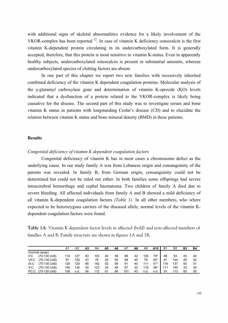
| 95
with additional signs of skeletal abnormalities evidence for a likely involvement of the VKOR-complex has been reported 12. In case of vitamin K deficiency osteocalcin is the first vitamin K-dependent protein circulating in its undercarboxylated form. It is generally accepted, therefore, that this protein is most sensitive to vitamin K-status. Even in apparently healthy subjects, undercarboxylated osteocalcin is present in substantial amounts, whereas undercarboxylated species of clotting factors are absent.
In one part of this chapter we report two new families with recessively inherited combined deficiency of the vitamin K dependent coagulation proteins. Molecular analysis of
the γ-glutamyl carboxylase gene and determination of vitamin K-epoxide (KO) levels indicated that a dysfunction of a protein related to the VKOR-complex is likely being causative for the disease. The second part of this study was to investigate serum and bone vitamin K status in patients with longstanding Crohn’s disease (CD) and to elucidate the relation between vitamin K status and bone mineral density (BMD) in these patients. Results Congenital deficiency of vitamin K dependent coagulation factors
Congenital deficiency of vitamin K has in most cases a chromosome defect as the underlying cause. In our study family A was from Lebanese origin and consanguinity of the parents was revealed. In family B, from German origin, consanguinity could not be determined but could not be ruled out either. In both families some offsprings had severe intracerebral hemorrhage and cephal haematoma. Two children of family A died due to severe bleeding. All affected individuals from family A and B showed a mild deficiency of all vitamin K-dependent coagulation factors (Table 1). In all other members, who where expected to be heterozygous carriers of the diseased allele, normal levels of the vitamin K-dependent coagulation factors were found.
Table 1A: Vitamin K dependent factor levels in affected (bold) and non-affected members of
families A and B. Family structure are shown in figures 1A and 1B.
A1 A2 A3 A4 A5 A6 A7 A8 A9 A10 B1 B2 B3 B4(normal range)II:C (70-130 U/dl) 114 127 63 103 40 59 88 42 105 79* 88 93 45 44VII:C (70-130 U/dl) 97 102 47 76 20 55 98 45 79 62* 81 144 29 42IX:C (70-130 U/dl) 125 120 66 142 52 99 91 40 111 51* 116 137 55 51X:C (70-130 U/dl) 140 135 40 123 24 49 97 32 116 56* 111 140 33 30PC:C (70-130 U/dl) 109 n.d. 56 112 61 80 101 43 n.d. n.d. 91 113 60 55

96 |
Table 1B. Vitamin K dependent factor levels in affected individuals 1 day after oral
substitution of 5 mg (A5, A6, A8) and 1 mg (B3) Vitamin K, respectively. A5 A6 A8 B3
(normal range)II:C (70-130 U/dl) 86 110 96 104VII:C (70-130 U/dl) 77 107 101 90IX:C (70-130 U/dl) 135 140 110 87X:C (70-130 U/dl) 91 138 99 85PC:C (70-130 U/dl) n.d. n.d. n.d. 66
The molecular analysis of all 15 exons of the γ-glutamyl carboxylase gene showed no
mutation in the affected probands. Since a genetic defect in the non-coding region of the γ-
glutamyl carboxylase gene could not be excluded, haplotypes of the γ-glutamyl carboxylase gene were constructed to ascertain whether the phenotype in the families is segregating with
one allele of the γ-glutamyl carboxylase gene. However, there was no single haplotype
segregating with the normal or the disease phenotype, thus arguing against the gene locus of
the γ-glutamyl carboxylase being causative for the disorder in both families.
Table 2. Serum concentrations of Vitamin K (VK) and Vitamin K epoxide (VKO) before
and 5 h after oral substitution of a 10 mg vitamin K dragee. Affected individuals are
indicated with bold letters. The detection limit of VKO is 0.05 ng/ml.
A4 0.4 < 0.05 201.8 < 0.05 > 1000A5 0.4 < 0.05 84.6 19.5 4.3A6 0.6 < 0.05 219.6 52.6 4.2A7 0.4 < 0.05 262.5 < 0.05 > 1000A8 n.d. n.d. 147 59.7 2.5A9 n.d. n.d. 607.3 < 0.05 > 1000B1 0.5 < 0.05 112.5 < 0.05 > 1000B2 0.5 < 0.05 310.3 2.6 119.3B3 0.7 < 0.05 490.4 66.2 7.4B4 0.4 < 0.05 410.7 38.6 10.6
serum concentrations (ng/ml) 5 h after oral substitution of 10 mg vitamin K
VK VKO VK / VKO
basal serum concentrations (ng/ml)
VK VKO
The finding of a normal γ-glutamyl carboxylase gene in both families points to a defect in a protein of the VKOR-complex. To prove this hypothesis, affected and normal subjects of both families were supplemented with 10 mg phylloquinone. Blood was taken

| 97
before and 5 h after oral drug intake at the expected maximum of plasma vitamin K levels. The results are shown in Table 2. While vitamin K-epoxide levels are normally undetectable in serum, all tested individuals of both families with the diseased phenotype showed highly increased vitamin KO levels ranging from 19.5 ng/mL to 59 ng/mL in family A and from 38.6 to 66.2 ng/mL in family B. Vitamin KO could not be detected in the non-affected individuals of family A and B with the exception of one individual in family B who exhibited a very low vitamin KO level (ratio K/KO of 119.3). These findings indicate that a defect of the VKOR-complex is likely to be causative for the apparent vitamin K deficiency and the undercarboxylation of all vitamin K dependent proteins in both families. Low vitamin K-status in patients with longstanding Crohn’s disease (CD)
In the second case of apparent vitamin K deficiency, a malabsorption of all fat-soluble vitamins is the underlying cause. Patients with CD exhibit a malabsorption of fat-soluble vitamins. None of the patients had a history of bleeding problems. Because vitamin K can affect bone metabolism via osteocalcin, we also measured in this population all kind of markers involved in bone metabolism. Mean BMD, expressed as absolute values in g/cm2, of the femoral neck, lumbar spine, and total body were 0.89 (013), 1.10 (0.16), and 1.11 (0.09), respectively. Mean T-scores were -0.61 (1.29), -0.98 (1.16), and -0.66 (1.13), respectively. Mean z-scores were -0.49 (1.07), -0.45 (1.25), and -0.28 (1.10), respectively.
Total OC was measured using the Osteometer test kit (Biotech, Herlev, Denmark). Since carboxylated OC and undercarboxylated OC differ substantially in their affinity for insoluble calcium salts, these fractions can be quantified by measuring serum OC before and after extraction with a standard amount of hydroxyapatite. The fraction which does not bind to hydroxyapatite consists of undercarboxylated “free” osteocalcin (OCfree). To measure the OCfree fraction, 45 mg of hydroxyapatite were added to 300 µl of serum. The fraction of carboxylated OC (OCbound) is calculated from the difference between the total immunoreactive OC (OCtotal) and OCfree. OCbound expressed as a percentage of total OC is known as the hydroxyapatite binding capacity (HBC) of osteocalcin.
Serum vitamin K concentrations in patients with CD were significantly lower than a reference population of 384 healthy subjects (p<0.001). Patients with CD had higher serum OCfree (p<0.05) and lower hydroxyapatite bindings capacity (HBC) values (P<0.001) compared with values obtained from the age and sex matched control group of 34 healthy subject randomly selected from the community registry (Figures 1 and 2). Although the mean OCfree was generally lower and mean serum vitamin K was higher in patients with normal BMD than in patients with a T score < -1 this difference was not statistically significant. In patients, a negative correlation was found between lumbar spine BMD and OCfree (r = -0.381, p<0.05) and between z score and OCfree (r=-0.381, p<0.01) (Figures 3 and 4).

98 |
-5
0
5
10
15
20
25
30
"fre
e" o
steo
calc
in (n
g/m
l)
Controls Crohn's disease
Figure 1.
Serum concentrations of “free” (undercarboxylated) osteocalcin in patients with longstanding Crohn’s disease (n=32) compared with age and sex matched healthy controls (n=34). Lines indicate
median values. Medain for patients with Crohn’s disese is 3.98 ng/mL; median for healthy controls is
1.51 ng/mL.
0
20
40
60
80
100
Hyd
roxy
apat
ite B
indi
ng C
apac
ity (%
)
Controls Crohn's disease
Figure 2.
Hydroxyapatite binding capacity in patients with longstanding Crohn’s disease (n=32) compared
with age and sex matched healthy controls (n=34). Lines indicate median values. Median for
patients with Crohn’s disease is 61.7%; median for healthy controls is 76.8%.

| 99
Mean serum level of vitamin D in patients was 28.4 (10.5) nmol/L; 11 patients (34%) were vitamin D deficient (serum 25-hydroxy vitamin D <25 nmol/L). A significant correlation was found between serum concentrations of vitamin D and K (r= 0.681, p<0.01). No correlation was was found between serum concentrations of vitamin D and vitamin K status of bone (either HBC (%) or OCfree). Mean serum triglyceride concentration was 1.5 (1.1) mmol/L. A positive correlation was found between serum concentrations of vitamin D and triglycerides (r=0.707, p<0.01) and vitamin K and triglycerides (r=0.789, p<0.01), respectively. In the patient group, the variables sex, age, body mass index, serum vitamin K, OCfree, and vitamin D were included in a multiple linear regression. No correlations were found between serum vitamin K levels and BMD or T and z scores at any measurement site. We found that OCfree correlated significantly with BMD of the lumbar spine (p=0.05) and with the lumbar spine z score (p=0.035). The correlation between OCfree and the T score of the lumbar spine did not reach significance (p=0.091). No correlations were found between BMD, T and z scores of the femoral neck and total body, and Ocfree. No correlations were found between serum vitamin D levels and BMD, or T and z scores at any measurement site.
0.5
0.6
0.7
0.8
0.9
1.0
1.1
1.2
1.3
1.4
1.5
0 5 10 15 20 25 30 35
"free" osteocalcin (ng/ml)
lum
bar
spin
e B
MD
(g/
cm2 )
Figure 3.
Correlation between “free” (undercarboxylated) osteocalcin and bone mineral density (BMD) of the lumbar spine in patients with longstanding Crohn’s disease (n=32) Correlation coefficient (Pearson’s) r=-0.375; p<0.05.

100 |
-4.0
-3.0
-2.0
-1.0
0.0
1.0
2.0
0 5 10 15 20 25 30 35
"free" osteocalcin (ng/ml)
lum
bar
spin
e Z
sco
re
Figure 4.
Correlation between “free” (undercarboxylated) osteocalcin and bone mineral density z score of the lumbar spine in patients with longstanding Crohn’s disease (n=32). Correlation coefficient (Pearson’s) r=-0.381; p<0.01.
Discussion Congenital deficiency of vitamin K dependent coagulation factors
Vitamin K-deficiency and treatment with coumarin derivatives are the common acquired causes for a decrease in the vitamin K-dependent coagulation factors. Inherited combined deficiency of these coagulation factors is unlikely because it would require at least four simultaneous mutations. The underlying cause is more likely to be a defect of one of the enzymes responsible for normal carboxylation of the vitamin K-dependent proteins. In our study a defect of one of these enzymes, the VKOR-complex, was the most probable cause of the bleeding problems. Bleeding problems are most severe in childhood when vitamin K levels are low. The combination of a low vitamin K status and a defect in the recycling of vitamin K makes that newborns are at high risk for spontanuous bleeding, for instance resulting in haemarthrosis or mucocutaneous bleeding. So far only twelve kindreds with a combined deficiency of all vitamin K-dependent coagulation factors have been reported in
| 101
the literature. Such rare disorders with suggested autosomal recessive inheritance occur predominantly in families with consanguineous marriage. The degree of deficiency of vitamin K-dependent coagulation proteins varies considerably from very severe forms with activity levels less than 5% to mild with activity levels ranging form 20 - 60%. Only in three of the previously published families some details of the pathophysiological mechanism were discussed 10-12. Pauli et al. found increased plasma levels of vitamin K-epoxide in the affected proband that point to a defect in the VKOR-complex. Brenner et al. and Spronk et al. showed
that a missense mutation within the γ-glutamyl carboxylase gene was causative for the
diseased phenotype. In our study, molecular analysis of the γ-glutamyl carboxylase gene and the increased vitamin K-epoxide levels in the affected probands in both families, suggests a defect in the VKOR-complex causing the mild deficiency of all vitamin K-dependent coagulation factors. Thus, the families reported herein represent the second and third families with a suggested defect in the VKOR-complex. Purification and isolation of the VKOR enzyme has been attempted by several groups without success for many years. It now seems that the VKOR represents a multi-enzyme complex and that purification procedures destroy the protein integrity of this complex and subsequently the VKOR enzyme activity. According to a hypothetical model, the VKOR-complex consists at least of the transmembraneous microsomal epoxide-hydrolase and a further protein originating from the glutathion-S-transferase super family 13,14.
In line with this, we expected in the two families reported herein also a molecular defect in one of these proteins. The genetic disorder in both of our families became clinically apparent by severe or even fatal perinatal intra cerebral bleeding. Severe bleeding at birth is known to be associated with acquired conditions of combined deficiency of vitamin K dependent coagulation factors, but may be amplified by genetic mutations. The clinical symptoms may be masked if vitamin K substitution leads to a temporary normalization of coagulation factors. Mild reduction of vitamin K dependent coagulation factors, as it was present in heterozygous members of our families, may also complicate diagnosis. So far, in three of the families with a moderate to severe deficiency of coagulation factors 12,15,16 skeletal abnormalities were associated with the combined decrease of vitamin K dependent proteins (Table 4). These skeletal abnormalities comprised irregular ossification, nasal hypoplasia, distal digital hypoplasia and mild conductive hearing loss, thus resembling the phenotype of warfarin embryopathy. In contrast, none of the four families with a milder form of the disease showed skeletal abnormalities. Although the warfarin embryopathy phenotype may result from a primary pharmacological effect of coumarin derivates, the report of Pauli et al. provided evidence that the disease is secondary to the pharmacological effect of warfarin, e. g. inhibition of the VKOR-complex.

102 |
Low vitamin K-status in patients with longstanding Crohn’s disease All three bone Gla-proteins are synthesized by the osteoblasts (the bone forming
cells), but only osteocalcin is a unique product of bone tissue. After its synthesis it is secreted by the osteoblasts and a major part is directly bound to the hydroxyapatite matrix of the bone 17. About 25% of the de novo synthesized osteocalcin, however, is set free in the circulation where it can be detected with commercially available test kits 18,19. So the serum concentration of osteocalcin forms a reflection of the osteoblast activity, which makes it an important and widely used marker for bone formation. Its precise function is not known, but transgenic osteocalcin-deficient animals have shown that it is a negative regulator of bone formation 20, and that it has a function in the orderly alignment of hydroxyapatite crystals in bone 21. In patients with CD, cystic fibrosis, and ileopathy, bile and fat malabsorption causes steatorrhoea and malabsorption of fat soluble vitamins. In these patients, the majority had raised PIVKA-II levels in their plasma, indicating a vitamin K deficiency in the liver 6. However, none of the patients had bleeding problems, which is consistent with only a sub-clinical vitamin K deficiency.
Serum vitamin K concentrations are influenced by plasma triglyceride concentrations and reflect recent dietary intake in healthy subjects. 22,23 While there are several publications on the role of vitamin D in metabolic bone disease in CD,24-29 only few data are available with regard to vitamin K status in CD. In a study in which uncarboxylated prothrombin was used as a marker, vitamin K deficiency was identified in 46% of CD patients with ileal involvement but not in patients with Crohn’s colitis 30. It has also been reported that OCfree, as used in the present study, is a much more sensitive marker for vitamin K status than prothrombin 31. The fact that vitamin K serves as a cofactor for carboxylation of bone Gla proteins and is of potential importance for bone formation is reflected by a significant independent and inverse correlation between OCfree and BMD of the lumbar spine (BMD and z score). The trabecular bone of the spine is metabolically more active than the cortical bone of the hip and may thus be more susceptible to vitamin K deficiency. This may explain the lack of correlation between OCfree and BMD of the femoral neck and total body. Bone mineral loss in CD is a multi-factorial process and vitamin K deficiency is only one factor in this process. In the present study there are indications that in patients with longstanding CD, vitamin K deficiency of bone has a greater influence on BMD than serum 25-hydroxy vitamin D levels. The body vitamin D stores are reflected by serum concentrations of 25- hydroxy vitamin D. In accordance with our expectations, significant correlations between serum vitamin D and K concentrations and triglycerides were found. In the present study, however, patients who were vitamin D deficient did not have significantly decreased BMD. Published data are equivocal in this respect: a significant correlation between vitamin D and BMD of the forearm was found in one of three large studies on vitamin D deficiency and

| 103
BMD in patients with CD 26-28. Vitamin D deficiency may cause secondary hyperparathyroidism which predominantly affects cortical bone. The questions remaining are whether bone that is vitamin K deficient is rendered sensitive to other pathogenetic factors of osteoporosis, such as corticosteroids, and whether vitamin K supplementation can improve bone mineralisation in CD patients, as has been demonstrated in other conditions. In prednisolone treated rats, vitamin K supplementation inhibited bone loss 32. Treatment with vitamin K (menatetrenone) has been reported to increase BMD in disused bones in vitamin K and D deficient hemiplegic patients 33. In a study in cystic fibrosis patients, a significantly elevated level of OCfree was found in patients who were not supplemented with vitamin K (phylloquinone) versus supplemented patients 34. The questions addressed above require further study. It has recently been demonstrated that vitamin K treatment effectively prevents the occurrence of new vertebral fractures in patients with osteoporosis 35. Potential implications of long-term low vitamin K-status
Both the fetal warfarin syndrome and mutations in one of the enzymes of the vitamin K cycle may lead to bone pathology characterized by excessive calcification in the rapidly growing parts of bone and strong impairment of bone growth. Similar defects may be induced by warfarin treatment of young animals. These phenomena are caused by undercarboxylation of the bone Gla-proteins osteocalcin and MGP, both of which are known to be involved in the regulation of calcium phosphate precipitation in growing bone and cartilage. Both proteins are strong inhibitors of calcium precipitation, and their function is based on the presence of the Gla-residues. Remarkably, the effects are most pronounced in young, rapidly growing bone. In CD and other malabsorption diseases, vitamin K-deficiency leading to bleeding is rarely seen. The question remains whether long-term poor vitamin K-status may affect the quantity and/or quality of adult bone, even after growth has been completed. Several observational studies supporting this possibility have been published. During recent years there have been two population-based studies in which the relationship between dietary vitamin K intakes and hip fractures or bone mineral density were investigated. The first study was among participants in the Nurses Health Study 36, using a food frequency questionnaire to assess dietary vitamin K intake at baseline, and recording hip fractures during the 10-year follow-up of this cohort. The results suggest an increased fracture risk for subjects with low vitamin K intake. A comparable study was carried out among both men and women of the Framingham Heart Study, and again an association was found between low vitamin K intakes and hip fracture risk 37. The underlying mechanism in both cases is a low vitamin K intake resulting in increased undercarboxylated osteocalcin levels. Thus far, no biochemical mechanism has been established yet. Serum ucOC is a biochemical marker for poor vitamin K status of bone, and during the last decade a number of studies have shown that increased
104 |
serum ucOC is associated with low bone mass 38 and increased fracture rate 8,9. Moreover, in a recent meta-analysis Caraballo et al. concluded that oral anticoagulant treatment is a risk factor for low bone mass and osteoporosis development at later age 39. An intriguing question is whether a similar mechanism underlies the fetal warfarin syndrome and vitamin K-deficiency related osteoporosis. After maturation, bone is not a static tissue, but it is renewed in a constant process known as bone turnover. In this process, cells called osteoclasts resorb bone and produce cavities known as the osteoclastic pits, which are filled up again by bone forming cells (osteoblasts). We would like to put forward the hypothesis that the new bone formation in the osteoclastic bone pits resembles the rapid growth of bone in fetuses and young children. Hence it seems probable that within these pits vitamin K-deficiency induces excessive calcium phosphate precipitation resulting in preliminary termination of the bone renewal and incomplete refilling of the osteoclastic bone pits. In this way it may be understood that vitamin K deficiency is the underlying mechanism in both warfarin- and vitamin K-deficiency-induced bone malformations in foetuses and young individuals as well as low bone mass and osteoporotic fractures in CD and other malabsorption diseases.

| 105
4.2
EFFECT OF DIETARY VITAMIN K ON STABILITY OF ORAL ANTICOAGULANT THERAPY: DOSE RESPONSE RELATIONSHIPS IN HEALTHY SUBJECTS
Abstract This study was performed in young healthy volunteers to measure the effect of dietary
vitamin K on the stability of oral anticoagulant (OAC) treatment. In the first instance we have assessed the effect of daily gradied doses of vitamin K1 given in pure tablet form. We than carried out a second study to asses the effect of a single meal containing a very high content of vitamin K1 or menaquinones. We have investigated dose-response relationships of vitamin K1 given as a vitamin K supplement and of vitamin K-rich foods containing either vitamin K1 (K1; spinach, broccoli), menaquinone-7 (MK-7; natto) or menaquinone-9 (MK-9; curd cheese). The effect on coagulation was monitored 4 times (INR) or twice weekly (FIIc, FVIIc, PIVKA-II (ucFII)). The effect of vitamin K intake on extra-hepatic metabolism was recorded by measuring carboxylated and undercarboxylated osteocalcin (cOC and ucOC, respectively). In addition, we determined the plasma K1, MK-7 and MK-9 levels after intake. Daily supplementation with vitamin K1 resulted in decreases of INR from baseline of 11.5%, 23.4%, 25.6 and 31.5% for 100, 200, 300 and 500 µg K1 respectively. Parallel changes were seen for FIIc, FVIIc and PIVKA-II, the latter showing the fastest response. Plasma peak K1 (4h) and fasting 24-h post K1 levels both increased linearly with vitamin K-intake. Single meals of 400g spinach (1500 µg K1) or broccoli (625 µg K1) induced a similar, but short-lived (24h), decline in INR (14%) and increase in FIIc and FVIIc. In contrast, a single meal of 100g natto (1000 µg MK-7) induced a decrease in INR of 24% that lasted for up to 4 d that was accompanied by complete normalisation of FVIIc. Plasma levels of MK-7 peaked at 5 h but unlike K1 remained at a high level for 4 d. Curd cheese had no effect on OAC therapy, probably because of the low amount of MK-9 present in a single meal. In conclusion: (1) The effects of taking vitamin K1 supplements on the stability of OAC therapy measured by the INR became statistically significant at doses of > 150 µg/d. The interference became clinically relevant at intakes above 150µg/d. (2) A high intake of K1 from a single meal of green vegetables is not likely do destabilize OAC therapy. However, the relative bioavailability compared to pure K1 preparations may vary; the estimated uptake from spinach and broccoli was about 10% and 35%, respectively. (3) A single meal containing a naturally high content of MK-7 had a greater and more sustained effect in reducing OAC response which is probably related to its much longer retention as reflected by the prolonged plasma half-life. This finding suggests that common foods containing long chain MKs may have a greater effect in destabilizing OAC therapy than previously suspected.
106 |
Introduction Oral anticoagulants (OAC, also known as vitamin K antagonists) are used in subjects
at risk for thrombo-embolic events. Their mode of action is based on the inhibition of the enzyme vitamin K epoxide reductase, an enzyme of the vitamin K cycle. Thus, OAC block the re-use of vitamin K and in this way they induce a relative vitamin K-deficiency in liver as well as in other tissues 40.
Vitamin K acts as a cofactor in the posttranslational carboxylation by which a number of specified glutamate residues in proteins are converted into gamma-carboxyglutamate (Gla). Gla-residues are found in a number of blood coagulation factors as well as in some bone proteins including osteocalcin and matrix Gla-protein 1,2 and are essential for their biological activity. Both vitamin K-deficiency and ingestion of OAC result in the synthesis of undercarboxylated Gla-proteins (also known as PIVKA’s), which may be quantified in the circulation with assays based on conformation-specific antibodies 41,42.
There are many factors that may lead to instability of OAC therapy. They include age, alcohol consumption, a number of drugs 43-46, as well as the irregular consumption of vitamin K-rich foods or vitamin K-containing food supplements 47-50. There are little data to which extent dietary vitamin K intake influence the daily dose of OAC treatment. Imbalance may lead to either an increased thrombotic tendency (high vitamin K intake) or to the risk of bleeding (overdose of OAC), and frequent control is required. The stability of OAC therapy is monitored by measuring the International Normalized Ratio (INR), which was used in the present study as the gold standard for assessment of the level of anticoagulation. Other tests we used to monitor OAC therapy include quantification of the plasma levels of the coagulation factors II, VII, and undercarboxylated factor II (ucFII) as a measure of hepatic vitamin K status, and carboxylated bone Gla-protein (osteocalcin, OC) and undercarboxylated osteocalcin (ucOC) as a measure for the vitamin K status in bone.
The experiments reported represent the first systematic study of the dose-response relationships of vitamin K-containing supplements and vitamin K-rich foods on the stability of anticoagulation and on the carboxylation of a number of hepatic and extra-hepatic Gla-proteins in healthy subjects. In our experiments we have chosen for an anticoagulant with a short half-life time (acenocoumarol, t½ = 10 h) and aimed at a relatively low INR value because it may be expected that under these conditions the effects of the various vitamin K sources will be maximal 51,52. Since prothrombin is uniquely synthesized in the liver and osteocalcin in bone, we have compared circulating undercarboxylated forms of both proteins (ucFII and ucOC) to compare the efficacy of vitamin K uptake by these tissues.
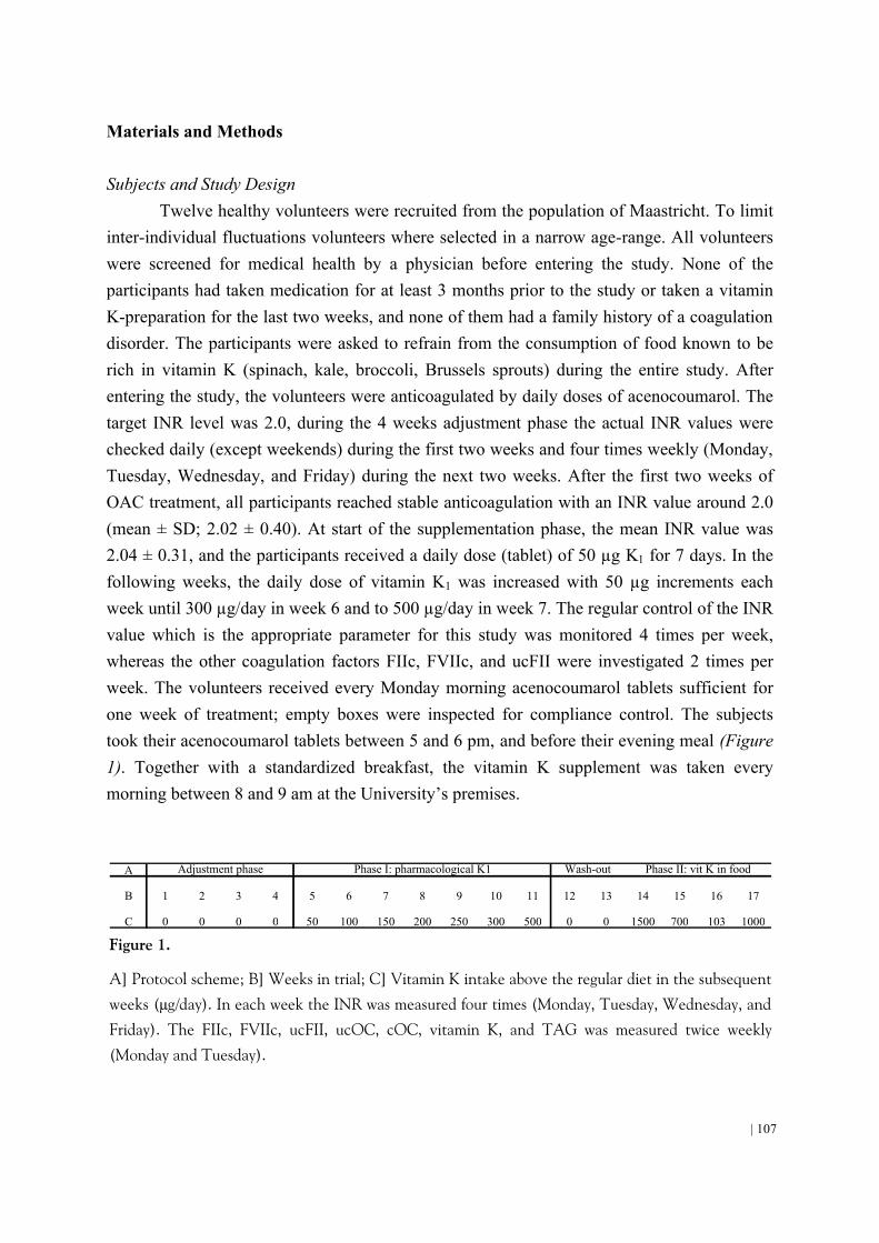
| 107
Materials and Methods Subjects and Study Design
Twelve healthy volunteers were recruited from the population of Maastricht. To limit inter-individual fluctuations volunteers where selected in a narrow age-range. All volunteers were screened for medical health by a physician before entering the study. None of the participants had taken medication for at least 3 months prior to the study or taken a vitamin K-preparation for the last two weeks, and none of them had a family history of a coagulation disorder. The participants were asked to refrain from the consumption of food known to be rich in vitamin K (spinach, kale, broccoli, Brussels sprouts) during the entire study. After entering the study, the volunteers were anticoagulated by daily doses of acenocoumarol. The target INR level was 2.0, during the 4 weeks adjustment phase the actual INR values were checked daily (except weekends) during the first two weeks and four times weekly (Monday, Tuesday, Wednesday, and Friday) during the next two weeks. After the first two weeks of OAC treatment, all participants reached stable anticoagulation with an INR value around 2.0 (mean ± SD; 2.02 ± 0.40). At start of the supplementation phase, the mean INR value was 2.04 ± 0.31, and the participants received a daily dose (tablet) of 50 µg K1 for 7 days. In the following weeks, the daily dose of vitamin K1 was increased with 50 µg increments each week until 300 µg/day in week 6 and to 500 µg/day in week 7. The regular control of the INR value which is the appropriate parameter for this study was monitored 4 times per week, whereas the other coagulation factors FIIc, FVIIc, and ucFII were investigated 2 times per week. The volunteers received every Monday morning acenocoumarol tablets sufficient for one week of treatment; empty boxes were inspected for compliance control. The subjects took their acenocoumarol tablets between 5 and 6 pm, and before their evening meal (Figure 1). Together with a standardized breakfast, the vitamin K supplement was taken every morning between 8 and 9 am at the University’s premises.
A
B 1 2 3 4 5 6 7 8 9 10 11 12 13 14 15 16 17
C 0 0 0 0 50 100 150 200 250 300 500 0 0 1500 700 103 1000
Phase II: vit K in foodWash-outPhase I: pharmacological K1Adjustment phase
Figure 1.
A] Protocol scheme; B] Weeks in trial; C] Vitamin K intake above the regular diet in the subsequent
weeks (µg/day). In each week the INR was measured four times (Monday, Tuesday, Wednesday, and
Friday). The FIIc, FVIIc, ucFII, ucOC, cOC, vitamin K, and TAG was measured twice weekly
(Monday and Tuesday).

108 |
Blood samples were taken by venipuncture at the times indicated and collected in trisodium citrate (Greiner Bio-one, Germany). For all coagulation assays blood collection took place after an overnight fast. On Mondays (after the start of each new vitamin K regimen) also at t= 4 h a blood sample was taken. During the weekends the volunteers had to take their vitamin K supplement at home. After a 2-week washout period, the effect of different, single vitamin K-rich meals on INR value was assessed with subsequent 6-day washouts between meals. The meals were all prepared and consumed at the University of Maastricht. Plasma vitamin K levels were measured at 0 and 4 h postprandially. Plasma samples were stored in aliquots at -80 °C until serial analysis. The study protocol was approved by the Medical Ethics Committee of the University of Maastricht and all subjects gave a written informed consent. Materials
Reference compounds (for analysis) used were phylloquinone (vitamin K1), from Sigma (St. Louis, MO), as well as a series of K2 vitamins (menaquinones, MK-4 through MK-10) and 2,3,-dihydrophylloquinone which were kindly provided by Hoffmann- La Roche (Basel, Switzerland). The supplements of vitamin K1 were provided in tablets of 100 µg/tablet (Roche Vitamins Ltd). Each tablet was scored for easy breaking into two equal parts for the intake of a 50 µg dose. For the nutrition experiment we used deep-frozen ready-to-use cooked spinach from Iglo Ola (Utrecht, Netherlands). Fresh broccoli and curd cheese (Mona®) were bought in a local supermarket; natto was bought as a ready-to-use product at a local oriental store. To facilitate absorption of the fat-soluble vitamin K, 30 g of corn oil (CPC-Bestfoods, Heilbronn, Germany) was added to each meal. The vitamin K content of the corn oil was negligible (K1: 2.7 µg/100g; K2: not detectable). Silica Sep-pak cartridges were purchased from Millipore (Milford, MA, USA). All other chemicals used were of the highest analytical grade. Various biochemical assays
All biochemical analyses were performed in duplicate and mean values are given throughout this paper. Prothrombin times (PT) were determined with an automated analyser (STA, Diagnostica Stago, Asnière, France) using Thromborel S (Behringwerke, Marburg, Germany) as a thromboplastin reagent. The data were expressed as International Normalized Ratio (INR) values. The INR was calculated by using the formula INR = patients PT/mean normal PT)ISI. In this formula ISI is the international sensitivity index that compares each thromboplastin’s sensitivity to an international reference thromboplastin of 1.00 53. Factor II (FIIc) and factor VII (FVIIc) procoagulant activities were measured in a coagulometer, ACL 300 Research (Instrumentation Laboratory, Milan, Italy) using Thromborel S and human

| 109
coagulation factor II and VII-deficient plasma (Behringwerke AG, Marburg, Germany). Species of undercarboxylated FII (ucFII) were measured using a conformation-specific monoclonal antibody in an ELISA-based assay 41. Results are expressed as Arbitrary Units per L (AU/L) because in cases of vitamin K deficiency circulating ucFII may comprise multiple forms of partially carboxylated FII and neither their relative abundance in plasma nor their relative affinity for the antibody is known. Using electrophoretic techniques 1 AU is equivalent to 1 µg of purified ucFII 41. The lower detection limit was 150 AU/L plasma. Undercarboxylated osteocalcin (ucOC) and carboxylated osteocalcin (cOC) were determined with the Glu-OC and Gla-OC kits from Takara Shuzo (Tokyo, Japan), respectively. Plasma vitamin K1 concentrations were measured by high performance liquid chromatography (HPLC) and fluorescence detection after on-line, post column electrochemical reduction of the effluent which converted the quinone forms of vitamin K compounds to their fluorescent quinol forms 54. Plasma triglyceride concentrations were determined by an automated enzymatic procedure using commercial reagents (Boehringer Mannheim, Germany) and a Beckmann Synchron CX 7-2 auto-analyzer (Fullerton, CA). Dietary vitamin K assessment
During the third week of the adjustment phase, the volunteers had to record their food intake during 7 consecutive days using a dietary diary. In this way the daily vitamin K intake was calculated. The weighed dietary record was repeated in the supplementation phase (200 µg supplementation phase). Using previously published databases 54-56, the daily vitamin K (µg/day) intake was calculated. Data analysis
For all biochemical markers, fasting blood samples from four days per week were analysed for significant trends as repeated measurements and as means per week. For each marker the differences between supplementation phase and baseline were analyzed using the SPSS 10.0 Wilcoxon matched pairs test. Differences were considered to be statistically significant at P<0.05. Additionally, the postprandial increase of plasma vitamin K was measured by comparing the early (fasting) sample and a sample taken 4 h later on Mondays.

110 |
Results The adjustment phase
The baseline characteristics of the participants are given in Table 1. There was a substantial difference in body mass index between men and women, which may be the reason why men required slightly more acenocoumarol than women to reach the target INR value. All had FIIc and FVIIc in the normal range, whereas the ucFII was below the detection level. As it is usual in healthy adults, part of the circulating OC circulated in the undercarboxylated form (mean 28%). Vitamin K1 was within the normal range (400-2000 pmol/L), K2 vitamins were undetectable (< 0.50 pmol/L) and triacylglycerol concentrations were normal.
Twelve healthy subjects were enrolled in the study and all received their individual
dose of OAC. The target INR value of 2.0 was reached after about 2 weeks of treatment. During the subsequent two weeks the INR remained stable (mean value: 2.04 ± 0.31). The recorded food intake data were used to calculate the daily vitamin K intake. No significant differences in vitamin K intake were found between the two weeks analyzed (week 3 and 8, respectively; see also Figure 1), and also not between the volunteers within one week. Hence refraining from food known to be rich in vitamin K1 resulted in a satisfactorily constant dietary vitamin K intake. Effects of vitamin K supplements
In Table 2 we have summarized the effects of increasing vitamin K intake on the variables measured. The baselinedata representing the effect of no extra supplementation are those of week 4 (last week of the adjustment phase), and the Monday morning value was
Table 1: Characteristics of the subjects. All Male Female
n= 12 6 6Age (yrs) 27.8 ± 1.8 28.3 ± 1.51 27.3 ± 2.07weight (Kg) 75.5 ± 14.8 85.8 ± 9.4 65.2 ± 11.8BMI (Kg/M2) 24.2 ± 2.6 25.0 ± 2.8 23.4 ± 2.4OAC intake (mg) 3.1 ± 0.7 3.3 ± 0.7 2.8 ± 0.7baseline valuesINR 1.02 ± 0.07 1.04 ± 0.06 1.01 ± 0.08FIIc (%) 103 ± 11 103 ± 7 103 ± 15FVIIc (%) 109 ± 22 108 ± 23 110 ± 23ucFII (AU/L) <150 <150 <150ucOC (ng/mL) 3.4 ± 1.7 3.9 ± 1.6 3.0 ± 1.9cOC (ng/mL) 9.4 ± 3.4 10.7 ± 3.7 8.2 ± 2.8Vitamin K1 (nmol/L) 1.27 ± 0.4 1.36 ± 0.5 1.18 ± 0.5Vitamin K2 (nmol/L) n.d. n.d. n.d.Triacylglycerol (mmol/L) 1.02 ± 0.48 1.18 ± 0.59 0.87 ± 0.33
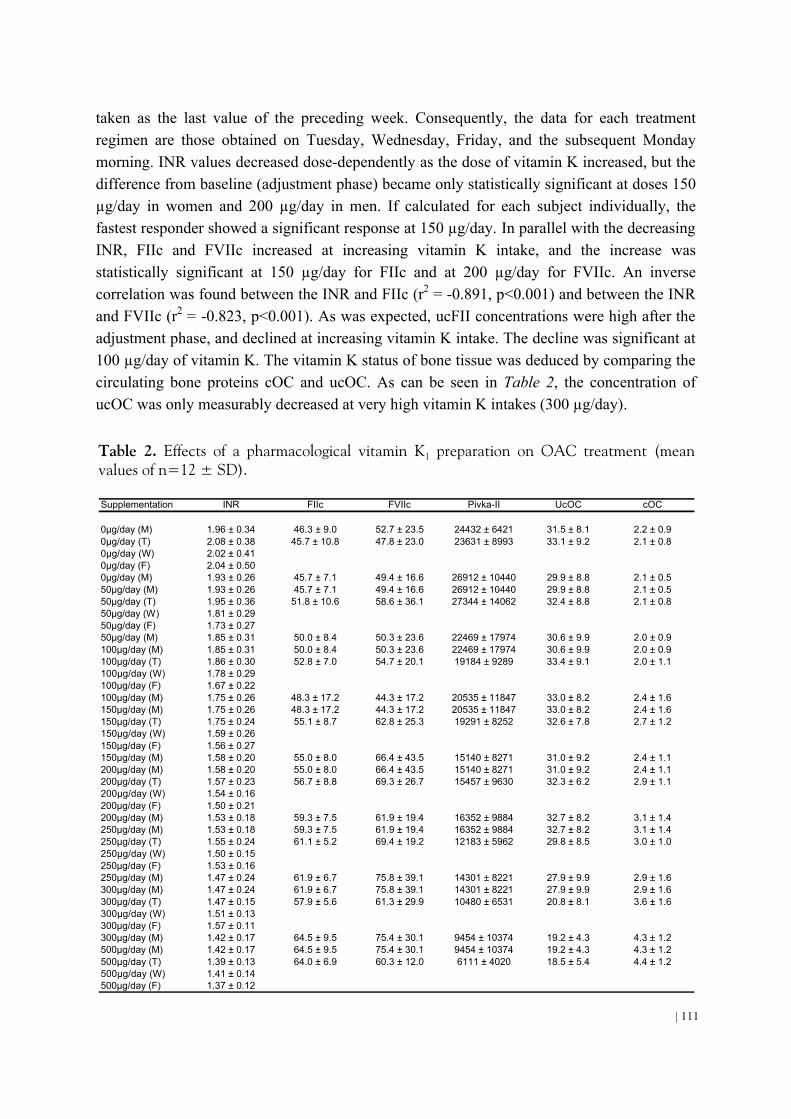
| 111
taken as the last value of the preceding week. Consequently, the data for each treatment regimen are those obtained on Tuesday, Wednesday, Friday, and the subsequent Monday morning. INR values decreased dose-dependently as the dose of vitamin K increased, but the difference from baseline (adjustment phase) became only statistically significant at doses 150 µg/day in women and 200 µg/day in men. If calculated for each subject individually, the fastest responder showed a significant response at 150 µg/day. In parallel with the decreasing INR, FIIc and FVIIc increased at increasing vitamin K intake, and the increase was statistically significant at 150 µg/day for FIIc and at 200 µg/day for FVIIc. An inverse correlation was found between the INR and FIIc (r2 = -0.891, p<0.001) and between the INR and FVIIc (r2 = -0.823, p<0.001). As was expected, ucFII concentrations were high after the adjustment phase, and declined at increasing vitamin K intake. The decline was significant at 100 µg/day of vitamin K. The vitamin K status of bone tissue was deduced by comparing the circulating bone proteins cOC and ucOC. As can be seen in Table 2, the concentration of ucOC was only measurably decreased at very high vitamin K intakes (300 µg/day).
Table 2. Effects of a pharmacological vitamin K1 preparation on OAC treatment (mean values of n=12 ± SD). Supplementation INR FIIc FVIIc Pivka-II UcOC cOC
0µg/day (M) 1.96 ± 0.34 46.3 ± 9.0 52.7 ± 23.5 24432 ± 6421 31.5 ± 8.1 2.2 ± 0.90µg/day (T) 2.08 ± 0.38 45.7 ± 10.8 47.8 ± 23.0 23631 ± 8993 33.1 ± 9.2 2.1 ± 0.80µg/day (W) 2.02 ± 0.410µg/day (F) 2.04 ± 0.500µg/day (M) 1.93 ± 0.26 45.7 ± 7.1 49.4 ± 16.6 26912 ± 10440 29.9 ± 8.8 2.1 ± 0.550µg/day (M) 1.93 ± 0.26 45.7 ± 7.1 49.4 ± 16.6 26912 ± 10440 29.9 ± 8.8 2.1 ± 0.550µg/day (T) 1.95 ± 0.36 51.8 ± 10.6 58.6 ± 36.1 27344 ± 14062 32.4 ± 8.8 2.1 ± 0.850µg/day (W) 1.81 ± 0.2950µg/day (F) 1.73 ± 0.2750µg/day (M) 1.85 ± 0.31 50.0 ± 8.4 50.3 ± 23.6 22469 ± 17974 30.6 ± 9.9 2.0 ± 0.9100µg/day (M) 1.85 ± 0.31 50.0 ± 8.4 50.3 ± 23.6 22469 ± 17974 30.6 ± 9.9 2.0 ± 0.9100µg/day (T) 1.86 ± 0.30 52.8 ± 7.0 54.7 ± 20.1 19184 ± 9289 33.4 ± 9.1 2.0 ± 1.1100µg/day (W) 1.78 ± 0.29100µg/day (F) 1.67 ± 0.22100µg/day (M) 1.75 ± 0.26 48.3 ± 17.2 44.3 ± 17.2 20535 ± 11847 33.0 ± 8.2 2.4 ± 1.6150µg/day (M) 1.75 ± 0.26 48.3 ± 17.2 44.3 ± 17.2 20535 ± 11847 33.0 ± 8.2 2.4 ± 1.6150µg/day (T) 1.75 ± 0.24 55.1 ± 8.7 62.8 ± 25.3 19291 ± 8252 32.6 ± 7.8 2.7 ± 1.2150µg/day (W) 1.59 ± 0.26150µg/day (F) 1.56 ± 0.27150µg/day (M) 1.58 ± 0.20 55.0 ± 8.0 66.4 ± 43.5 15140 ± 8271 31.0 ± 9.2 2.4 ± 1.1200µg/day (M) 1.58 ± 0.20 55.0 ± 8.0 66.4 ± 43.5 15140 ± 8271 31.0 ± 9.2 2.4 ± 1.1200µg/day (T) 1.57 ± 0.23 56.7 ± 8.8 69.3 ± 26.7 15457 ± 9630 32.3 ± 6.2 2.9 ± 1.1200µg/day (W) 1.54 ± 0.16200µg/day (F) 1.50 ± 0.21200µg/day (M) 1.53 ± 0.18 59.3 ± 7.5 61.9 ± 19.4 16352 ± 9884 32.7 ± 8.2 3.1 ± 1.4250µg/day (M) 1.53 ± 0.18 59.3 ± 7.5 61.9 ± 19.4 16352 ± 9884 32.7 ± 8.2 3.1 ± 1.4250µg/day (T) 1.55 ± 0.24 61.1 ± 5.2 69.4 ± 19.2 12183 ± 5962 29.8 ± 8.5 3.0 ± 1.0250µg/day (W) 1.50 ± 0.15250µg/day (F) 1.53 ± 0.16250µg/day (M) 1.47 ± 0.24 61.9 ± 6.7 75.8 ± 39.1 14301 ± 8221 27.9 ± 9.9 2.9 ± 1.6300µg/day (M) 1.47 ± 0.24 61.9 ± 6.7 75.8 ± 39.1 14301 ± 8221 27.9 ± 9.9 2.9 ± 1.6300µg/day (T) 1.47 ± 0.15 57.9 ± 5.6 61.3 ± 29.9 10480 ± 6531 20.8 ± 8.1 3.6 ± 1.6300µg/day (W) 1.51 ± 0.13300µg/day (F) 1.57 ± 0.11300µg/day (M) 1.42 ± 0.17 64.5 ± 9.5 75.4 ± 30.1 9454 ± 10374 19.2 ± 4.3 4.3 ± 1.2500µg/day (M) 1.42 ± 0.17 64.5 ± 9.5 75.4 ± 30.1 9454 ± 10374 19.2 ± 4.3 4.3 ± 1.2500µg/day (T) 1.39 ± 0.13 64.0 ± 6.9 60.3 ± 12.0 6111 ± 4020 18.5 ± 5.4 4.4 ± 1.2500µg/day (W) 1.41 ± 0.14500µg/day (F) 1.37 ± 0.12

112 |
Circulating vitamin K concentrations were measured at t = 0, 4, and 24 h after intake
of each new vitamin K regimen. The 4 h points represent the peak of the absorption curves, and show a linear dose-response relation (figure 2). At 24 h after intake the values had almost completely returned to the baseline level, which is consistent with the rapid plasma clearance of vitamin K. Effects of vitamin K from food
Vitamin K-rich meals were prepared and administered on Monday morning. In this way the effect of a single meal was studied. The meals differed with respect to their vitamin K content as well as the type of vitamin K present (see table 3). Although spinach contains more vitamin K1 than broccoli, the circulating (absorbed) vitamin K1 after the spinach meal was lower than after the broccoli meal; in addition the effects of broccoli on the INR and the individual coagulation factors were larger than that of spinach. This observation is consistent with the much better absorption of vitamin K1 from broccoli than from spinach. The effect of K1 from both spinach and broccoli on the INR lasted only for 24 hours and 48 hours after food intake the INR values had returned to their baseline values. No significant effects of a spinach or broccoli meal were observed on ucOC and cOC. Curd cheese (rich in MK-9) had a less pronounced effect and did not result in significant effects on any of the markers assayed. The circulating MK-9 concentration at 4 h after intake was 3.23 nmol/L and returned to baseline at 24 h. Natto is known to be a rich source of vitamin K2 (mainly MK-7). At 4 h after intake, circulating MK-7 concentrations were high (13.25 nmol/L), and remained elevated until 144 h after intake. The long plasma half-life time was reflected in a significant decrease in INR over the whole week. Consistently, FIIc and FVIIc were increased significantly until four days postprandially. Despite the effect on the coagulation system, the consumption of natto did not affect the Gla-content of circulating osteocalcin which reflects the extra-hepatic metabolism.

| 113
Table 3. Effects of a food items rich in vitamin K on the OAC treatment. Mean values α SD
of n=12 volunteers. * In the natto supplementation week we added an additional measurement on Thursday
because the effect of natto on vitamin K dependent proteins was so pronounced and lasted
for the whole week.
Supplementation INR FIIc FVIIc Pivka-II UcOC cOC
Wash-out (M) 2.02 ± 0.40 47.3 ± 8.0 52.7 ± 23.5 26200 ± 15000 20.2 ± 7.5 3.7 ± 1.4Wash-out (T) 2.05 ± 0.29Washout (W) 1.97 ± 0.26Wash-out (F) 2.19 ± 0.44Washout (M) 2.11 ± 0.34 49.9 ± 8.2 47.3 ± 11.3 27371 ± 20906 19.0 ± 7.1 3.8 ± 1.3
Spinach (M) 2.11 ± 0.34 49.9 ± 8.2 47.3 ± 11.3 27371 ± 20906 19.0 ± 7.1 3.8 ± 1.3Spinach (T) 1.70 ± 0.16 56.5 ± 6.3 71.1 ± 22.6 25000 ± 12782 18.0 ± 7.9 4.3 ± 1.4Spinach (W) 1.91 ± 0.38Spinach (F) 1.86 ± 0.34Spinach (M) 2.03 ± 0.27 47.7 ± 10.1 57.0 ± 18.9 22092 ± 16227 20.2 ± 7.6 4.3 ± 1.1
Broccoli (M) 2.03 ± 0.27 47.7 ± 10.1 57.0 ± 18.9 22092 ± 16227 20.2 ± 7.6 4.3 ± 1.1Broccoli (T) 1.62 ± 0.20 61.6 ± 9.8 76.5 ± 23.1 21000 ± 13000 17.4 ± 7.4 4.2 ± 1.4Broccoli (W) 1.87 ± .28Broccoli (F) 1.89 ± 0.28Broccoli (M) 1.98 ± 0.31 52.0 ± 7.0 47.0 ± 15.8 26873 ± 15736 18.4 ± 5.7 4.3 ± 1.1
Curd cheese (M) 1.98 ± 0.31 52.0 ± 7.0 47.0 ± 15.8 26873 ± 15736 18.4 ± 5.7 4.3 ± 1.1Curd cheese (T) 1.84 ± 0.23 53.0 ± 9.4 54.4 ± 20.6 25000 ± 9221 22.4 ± 7.6 3.7 ± 1.4Curd cheese (W) 1.81 ± 0.31Curd cheese (F) 1.88 ± 0.28Curd cheese (M) 2.01 ± 0.57 50.5 ± 11.3 51.3 ± 23.3 21019 ± 21354 23.1 ± 7.7 3.6 ± 1.2
Natto (M) 2.11 ± 0.34 49.9 ± 8.2 47.3 ± 11.3 27371 ± 20906 19.0 ± 7.1 3.8 ± 1.3Natto (T) 1.50 ± 0.21 55.2 ± 9.0 87.9 ± 30.2 24820 ± 16671 26.0 ± 8.5 3.7 ± 1.1Natto (W) 1.49 ± 0.26 56.2 ± 9.4 81.2 ± 27.8 22970 ± 12460 22.8 ± 7.4 4.3 ± 1.7Natto (Th)* 1.42 ± 0.13 54.6 ± 8.4 73.7 ± 19.3 11446 ± 7085 21.3 ± 6.7 4.3 ± 2.2Natto (F) 1.44 ± 0.21 58.0 ± 8.6 78.1 ± 28.2 25378 ± 16557 21.5 ± 8.0 3.8 ± 1.7Natto (M) 1.75 ± 0.38 52.3 ± 10.6 54.0 ± 27.6 24903 ± 17988 22.9 ± 8.4 3.8 ± 1.9
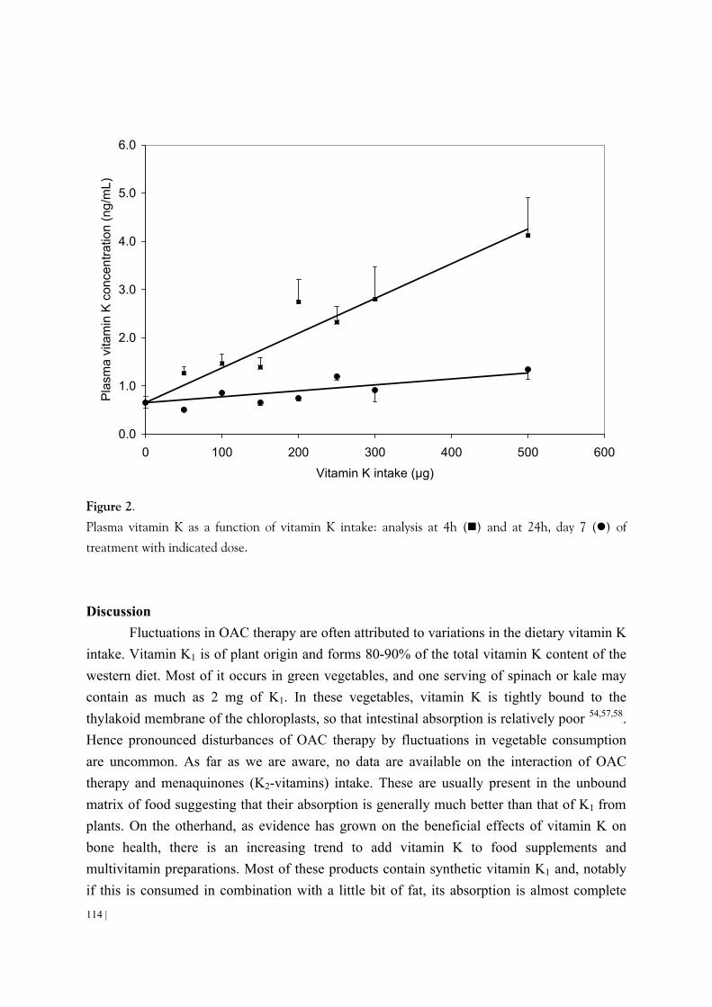
114 |
Discussion
Fluctuations in OAC therapy are often attributed to variations in the dietary vitamin K intake. Vitamin K1 is of plant origin and forms 80-90% of the total vitamin K content of the western diet. Most of it occurs in green vegetables, and one serving of spinach or kale may contain as much as 2 mg of K1. In these vegetables, vitamin K is tightly bound to the thylakoid membrane of the chloroplasts, so that intestinal absorption is relatively poor 54,57,58. Hence pronounced disturbances of OAC therapy by fluctuations in vegetable consumption are uncommon. As far as we are aware, no data are available on the interaction of OAC therapy and menaquinones (K2-vitamins) intake. These are usually present in the unbound matrix of food suggesting that their absorption is generally much better than that of K1 from plants. On the otherhand, as evidence has grown on the beneficial effects of vitamin K on bone health, there is an increasing trend to add vitamin K to food supplements and multivitamin preparations. Most of these products contain synthetic vitamin K1 and, notably if this is consumed in combination with a little bit of fat, its absorption is almost complete
0.0
1.0
2.0
3.0
4.0
5.0
6.0
0 100 200 300 400 500 600
Vitamin K intake (µg)
Pla
sma
vita
min
K c
once
ntra
tion
(ng/
mL)
Figure 2.
Plasma vitamin K as a function of vitamin K intake: analysis at 4h (n) and at 24h, day 7 (l) of
treatment with indicated dose.
| 115
57,58. Here we carried out a systematic analysis of the effect of a K1 supplement as well as various food items known to be rich in either K1 or K2 on the level of anticoagulation established by measuring their effect on the INR.
The INR is relatively insensitive for small fluctuations of the individual plasma coagulation factors, but it is used world wide to control the desired degree of anticoagulation. A clinically relevant disturbance was defined as the value which would require adjustment of the OAC dose (15% of the target INR value), which is close to the value for statistical significance (2 x SD). Overall, our group analysis suggested that pure vitamin K supplements only affected the INR value at a daily dose of 150-200 µg/day. Measurement of the procoagulant activities of FIIc and FVIIc was slightly more sensitive, and at 150 µg/day significant effects were found. Although small variations in its plasma concentration are not clinically relevant, ucFII was the most sensitive coagulation parameter in our protocol. However, even for this marker a decrease was only found at intakes = 100 µg/day. Interindividual differences with respect to the sensitivity of the administered vitamin K were substantial, with the lowest effective dose for effect ranging between 100 µg/day (in one individual) and 500 µg/day (two subjects). From these data we conclude that vitamin K supplements in a daily dose of < 100 µg/day are not interfering with OAC treatment, even at irregular use during OAC treatment. Both hematologists and patients should be aware, however, that some of these preparations (available as over-the-counter products) contain as much as 500µg K1, and that such products may strongly affect OAC treatment.
Although the concept of the assay for ucOC is comparable to that for ucFII, an effect on ucOC was only observed at a high vitamin K intake (300 µg/day). This is consistent with previous work that has suggested that the vitamin K requirement of extra-hepatic tissues such as bone is substantially higher than that of liver, and that at low tissue vitamin K-status the liver has a high efficacy of vitamin K absorption from the blood stream.
When analysing the data of the effect of a single dose of different vitamin K rich foods, these comparisons were made with the first day. It is obvious that after the consumption of vitamin K rich vegetables, the circulating vitamin K concentrations are far lower than after intake of a comparable amount of K from supplements. This is consistent with previous publications 54,57,58, and suggests a poor absorption from vegetables, notably from spinach. Additionally, vitamin K1 rapidly disappears from the circulation, and also from liver: even at high intakes the plasma concentrations had returned to nearly baseline and the effects on coagulation factors (having half-life times of 8 (FVIIc) and 56 (FIIc) h) were only apparent at 24 h postprandially. It cannot be excluded that vitamin K from food containing vitamin K rich vegetable oils (not tested in our study) has a much higher impact on the INR values, because of the likelihood of a better absorption. Those forms of K2 with long sidechaions have long plasma half-life times. However, the number food items rich in K2 are

116 |
restricted. In the western diet the highest menaquinone concentrations (notably MK-9 and MK-10) are found in cheese and curd cheese. However, the concentrations are well below those of K1 in vegetables and a large, single dose had no effect on the INR value. Because of their long biological half-life time the higher menaquinones may accumulate when eaten regularly, but the extent to which this may influence OAC therapy cannot be deduced from our data. Natto is an Asian product which is very rich in MK-7, and which is probably unknown among western hematologists. A single portion of natto had a profound effect on the INR, which was detectable even after several days. Also MK-7 itself remained in the circulation for at least 4 days, so regular consumption of natto may lead to significant accumulation of MK-7 and may substantially interfere with OAC treatment.
In conclusion, we have demonstrated in this study that food supplements providing < 150 µg/day of vitamin K1 do not significantly interfere with oral anticoagulant therapy. Furthermore, irregular consumption of green vegetables in normal amounts will only contribute marginally to fluctuations of the INR value. However, foods rich in menaquinones may form a source of fluctuations. Only a limited number of the more common foods in the western diet have been analyzed for their vitamin K2 content. This paper demonstrates the importance of a more systematic analysis of also more uncommon and exotic foods, which may form an unsuspected source of variable INR values or apparent warfarin resistance.

| 117
REFERENCES 1. Vermeer C. Gamma-carboxyglutamate-containing proteins and the vitamin K-dependent
carboxylase. Biochem J. 1990;266:625-36. 2. Furie B, Bouchard BA, Furie BC. Vitamin K-dependent biosynthesis of gamma-
carboxyglutamic acid. Blood. 1999;93:1798-808. 3. von Kries R, Greer FR, Suttie JW. Assessment of vitamin K status of the newborn infant. J
Pediatr Gastroenterol Nutr. 1993;16:231-8. 4. Shearer MJ. Assay of coumarin antagonists of vitamin K in blood by high-performance liquid
chromatography. Methods Enzymol. 1986;123:223-34. 5. Sokol RJ, Reardon MC, Accurso FJ, Stall C, Narkewicz M, Abman SH, Hammond KB. Fat-
soluble-vitamin status during the first year of life in infants with cystic fibrosis identified by screening of newborns. Am J Clin Nutr. 1989;50:1064-71.
6. Rashid M, Durie P, Andrew M, Kalnins D, Shin J, Corey M, Tullis E, Pencharz PB. Prevalence of vitamin K deficiency in cystic fibrosis. Am J Clin Nutr. 1999;70:378-82.
7. Binkley NC, Suttie JW. Vitamin K nutrition and osteoporosis. J Nutr. 1995;125:1812-21. 8. Szulc P, Chapuy MC, Meunier PJ, Delmas PD. Serum undercarboxylated osteocalcin is a
marker of the risk of hip fracture in elderly women. J Clin Invest. 1993;91:1769-74. 9. Väänänen HK, Luukinen H, Käkönen S-M, Pettersson K, Koski K, Laippala P, Lövgren T,
Kivelä S-L. Strong prediction of fractures among the elderly by the ratio of carboxylated and total serum osteocalcin. Calcif Tissue Int. 1999;64:S79.
10. Brenner B, Sanchez-Vega B, Wu SM, Lanir N, Stafford DW, Solera J. A missense mutation in gamma-glutamyl carboxylase gene causes combined deficiency of all vitamin K-dependent blood coagulation factors. Blood. 1998;92:4554-9.
11. Spronk HM, Farah RA, Buchanan GR, Vermeer C, Soute BA. Novel mutation in the gamma-glutamyl carboxylase gene resulting in congenital combined deficiency of all vitamin K-dependent blood coagulation factors. Blood. 2000;96:3650-3652.
12. Pauli RM, Lian JB, Mosher DF, Suttie JW. Association of congenital deficiency of multiple vitamin K-dependent coagulation factors and the phenotype of the warfarin embryopathy: clues to the mechanism of teratogenicity of coumarin derivatives. Am J Hum Genet. 1987;41:566-83.
13. Cain D, Hutson SM, Wallin R. Assembly of the warfarin-sensitive vitamin K 2,3-epoxide reductase enzyme complex in the endoplasmic reticulum membrane. J Biol Chem. 1997;272:29068-75.
14. Guenthner TM, Cai D, Wallin R. Co-purification of microsomal epoxide hydrolase with the warfarin-sensitive vitamin K1 oxide reductase of the vitamin K cycle. Biochem-Pharmacol. 1998;55:169-75.
15. Leonard CO. Vitamin K responsive bleeding disorder. A genocopy of the warfarin embryopathy. Proceedings of the Greenwood Genetic Center. 1988;7:165-166.
16. Boneh A, Bar-Ziv J. Hereditary deficiency of vitamin K-dependent coagulation factors with skeletal abnormalities. Am J Med Genet. 1996;65:241-3.
17. Hauschka PV, Lian JB, Cole DE, Gundberg CM. Osteocalcin and matrix Gla protein: vitamin K-dependent proteins in bone. Physiol Rev. 1989;69:990-1047.
18. Knapen MH, Eisenwiener HG, Vermeer C. Osteocalcin detection in aging serum and whole blood: stability of different osteocalcin fractions. Clin Chim Acta. 1996;256:151-64.
19. Power MJ, Fottrell PF. Osteocalcin: diagnostic methods and clinical applications. Crit Rev Clin Lab Sci. 1991;28:287-335.
20. Ducy P, Desbois C, Boyce B, Pinero G, Story B, Dunstan C, Smith E, Bonadio J, Goldstein S, Gundberg C, Bradley A, Karsenty G. Increased bone formation in osteocalcin-deficient mice.

118 |
Nature. 1996;382:448-52. 21. Schinke T, McKee MD, Karsenty G. Extracellular matrix calcification: where is the action?
[news; comment]. Nat Genet. 1999;21:150-1. 22. Vermeer C, Jie KS, Knapen MH. Role of vitamin K in bone metabolism. Annu Rev Nutr.
1995;15:1-22. 23. Ferland G, Sadowski JA, ME OB. Dietary induced subclinical vitamin K deficiency in normal
human subjects. J Clin Invest. 1993;91:1761-8. 24. Abitbol V, Roux C, Chaussade S, Guillemant S, Kolta S, Dougados M, Couturier D, Amor B.
Metabolic bone assessment in patients with inflammatory bowel disease. Gastroenterology. 1995;108:417-22.
25. Driscoll RH, Jr., Meredith SC, Sitrin M, Rosenberg IH. Vitamin D deficiency and bone disease in patients with Crohn's disease. Gastroenterology. 1982;83:1252-8.
26. Andreassen H, Rix M, Brot C, Eskildsen P. Regulators of calcium homeostasis and bone mineral density in patients with Crohn's disease. Scand J Gastroenterol. 1998;33:1087-93.
27. Hessov I, Mosekilde L, Melsen F, Fasth S, Hulten L, Lund B, Sorensen OH. Osteopenia with normal vitamin D metabolites after small-bowel resection for Crohn's disease. Scand J Gastroenterol. 1984;19:691-6.
28. Silvennoinen J. Relationships between vitamin D, parathyroid hormone and bone mineral density in inflammatory bowel disease. J Intern Med. 1996;239:131-7.
29. Vogelsang H, Ferenci P, Woloszczuk W, Resch H, Herold C, Frotz S, Gangl A. Bone disease in vitamin D-deficient patients with Crohn's disease. Dig Dis Sci. 1989;34:1094-9.
30. Krasinski SD, Russell RM, Furie BC, Kruger SF, Jacques PF, Furie B. The prevalence of vitamin K deficiency in chronic gastrointestinal disorders. Am J Clin Nutr. 1985;41:639-43.
31. Rucker RB. Improved functional endpoints for use in vitamin K assessment: important implications for bone disease. Am J Clin Nutr. 1997;65:883-4.
32. Hara K, Akiyama Y, Ohkawa I, Tajima T. Effects of menatetrenone on prednisolone-induced bone loss in rats. Bone. 1993;14:813-8.
33. Sato Y, Honda Y, Kuno H, Oizumi K. Menatetrenone ameliorates osteopenia in disuse-affected limbs of vitamin D- and K-deficient stroke patients. Bone. 1998;23:291-6.
34. Beker LT, Ahrens RA, Fink RJ, O'Brien ME, Davidson KW, Sokoll LJ, Sadowski JA. Effect of vitamin K1 supplementation on vitamin K status in cystic fibrosis patients. J Pediatr Gastroenterol Nutr. 1997;24:512-7.
35. Shiraki M, Shiraki Y, Aoki C, Miura M. Vitamin K2 (menatetrenone) effectively prevents fractures and sustains lumbar bone mineral density in osteoporosis. J Bone Miner Res. 2000;15:515-21.
36. Feskanich D, Weber P, Willett WC, Rockett H, Booth SL, Colditz GA. Vitamin K intake and hip fractures in women: a prospective study. Am J Clin Nutr. 1999;69:74-9.
37. Booth SL, Tucker KL, Chen H, Hannan MT, Gagnon DR, Cupples LA, Wilson PW, Ordovas J, Schaefer EJ, Dawson-Hughes B, Kiel DP. Dietary vitamin K intakes are associated with hip fracture but not with bone mineral density in elderly men and women. Am J Clin Nutr. 2000;71:1201-8.
38. Szulc P, Arlot M, Chapuy MC, Duboeuf F, Meunier PJ, Delmas PD. Serum undercarboxylated osteocalcin correlates with hip bone mineral density in elderly women. J Bone Miner Res. 1994;9:1591-5.
39. Caraballo PJ, Heit JA, Atkinson EJ, Silverstein MD, O'Fallon WM, Castro MR, Melton LJ, 3rd. Long-term use of oral anticoagulants and the risk of fracture. Arch Intern Med. 1999;159:1750-6.
40. Vermeer C, Hamulyak K. Pathophysiology of vitamin K-deficiency and oral anticoagulants. Thromb Haemost. 1991;66:153-9.

| 119
41. Belle M, Brebant R, Guinet R, Leclercq M. Production of a new monoclonal antibody specific to human des-gamma- carboxyprothrombin in the presence of calcium ions. Application to the development of a sensitive ELISA-test. J Immunoassay. 1995;16:213-29.
42. Vergnaud P, Garnero P, Meunier PJ, Breart G, Kamihagi K, Delmas PD. Undercarboxylated osteocalcin measured with a specific immunoassay predicts hip fracture in elderly women: the EPIDOS Study. J Clin Endocrinol Metab. 1997;82:719-24.
43. Kumar S, Haigh JR, Rhodes LE, Peaker S, Davies JA, Roberts BE, Feely MP. Poor compliance is a major factor in unstable outpatient control of anticoagulant therapy. Thromb Haemost. 1989;62:729-732.
44. James AH, Britt RP, Raskino CL, Thompson SG. Factors affecting the maintenance dose of warfarin. J Clin Pathol. 1992;45:704-706.
45. Harris JE. Interaction of dietary factors with oral anticoagulants: review and applications. J Am Diet Assoc. 1995;95:580-584.
46. Beyth RJ, Landefeld CS. Anticoagulants in older patients. A safety perspective [published erratum appears in Drugs Aging 1995 Apr;6(4):343]. Drugs Aging. 1995;6:45-54.
47. Kempin SJ. Warfarin resistance caused by broccoli [letter]. N Engl J Med. 1983;308:1229-1230. 48. Karlson B, Leijd B, Hellstrom K. On the influence of vitamin K-rich vegetables and wine on the
effectiveness of warfarin treatment. Acta Med Scand. 1986;220:347-350. 49. Kalra PA, Cooklin M, Wood G, O'Shea GM, Holmes AM. Dietary modification as cause of
anticoagulation instability [letter]. Lancet. 1988;2:803. 50. Pedersen FM, Hamberg O, Hess K, Ovesen L. The effect of dietary vitamin K on warfarin-
induced anticoagulation. J Intern Med. 1991;229:517-520. 51. Fekkes N, de Jonge H, Veltkamp JJ, Bieger R, Loeliger EA. Comparative study of the clinical
effect of acenocoumarol (Sintrom) and phenprocoumon (Marcoumar) in myocardial infarction and angina pectoris. Acta Med Scand. 1971;190:535-40.
52. Breed WP, van Hooff JP, Haanen C. A comparative study concerning the stability of the anticoagulant effect of acenocoumarol and phenprocoumon. Acta Med Scand. 1969;186:283-8.
53. Hirsch J, Dalen J. Oral anticoagulants. Mechanism of action, clinical effectiveness and optimal therapeutic range. Chest. 1992;104:312s-326s.
54. Schurgers LJ, Vermeer C. Determination of Phylloquinone and Menaquinones in Food. effect of food matrix on circulating vitamin K concentrations. Haemostasis. 2000;30:298-307.
55. Bolton-Smith C, Price RJ, Fenton ST, Harrington DJ, Shearer MJ. Compilation of a provisional UK database for the phylloquinone (vitamin K1) content of foods. Br J Nutr. 2000;83:389-399.
56. Booth SL, Sadowski JA, Weihrauch JL, Ferland G. Vitamin K1 (phylloquinone) content of foods: a provisional table. J Food Comp Anal. 1993;6:109-120.
57. Garber AK, Binkley NC, Krueger DC, Suttie JW. Comparison of phylloquinone bioavailability from food sources or a supplement in human subjects. J Nutr. 1999;129:1201-3.
58. Gijsbers BL, Jie KS, Vermeer C. Effect of food composition on vitamin K absorption in human volunteers. Br J Nutr. 1996;76:223-9.


5
ROLE OF K-VITAMINS AND VITAMIN K-DEPENDENT PROTEINS IN VASCULAR CALCIFICATION


| 123
5
ROLE OF VITAMIN K AND VITAMIN K-DEPENDENT PROTEINS IN VASCULAR CALCIFICATION
Abstract To provide a rational basis for recommended daily allowances (RDA) of dietary
phylloquinone (vitamin K1) and menaquinone (vitamin K2) intake that adequately supply extra-hepatic (notably vascular) tissue requirements. Vitamin K has a key function in the synthesis of at least two proteins involved in calcium and bone metabolism, namely osteocalcin and matrix Gla-protein (MGP). MGP was shown to be a strong inhibitor of vascular calcification. Present RDA values for vitamin K are based on the hepatic phylloquinone requirement for coagulation factor synthesis. Accumulating data suggest that extra-hepatic tissues such as bone and vessel wall require higher dietary intakes and have a preference for menaquinone rather than for phylloquinone. Tissue-specific vitamin K consumption under controlled intake was determined in warfarin-treated rats using the vitamin K-quinone/epoxide ratio as a measure for vitamin K consumption. Immunohistochemical analysis of human vascular material was performed using a monoclonal antibody against MGP. The same antibody was used for quantification of MGP levels in serum. At least some extra-hepatic tissues including the arterial vessel wall have a high preference for accumulating and using menaquinone rather than phylloquinone. Both intima and media sclerosis are associated with high tissue concentrations of MGP, with the most prominent accumulation at the interface between vascular tissue and calcified material. This was consistent with increased concentrations of circulating MGP in subjects with atherosclerosis and diabetes mellitus. This is the first report demonstrating the association between MGP and vascular calcification. The hypothesis is put forward that under-carboxylation of MGP is a risk factor for vascular calcification and that the present RDA values are too low to ensure full carboxylation of MGP.
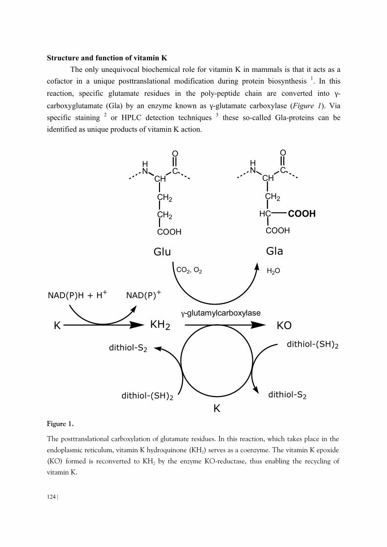
124 |
Structure and function of vitamin K The only unequivocal biochemical role for vitamin K in mammals is that it acts as a
cofactor in a unique posttranslational modification during protein biosynthesis 1. In this
reaction, specific glutamate residues in the poly-peptide chain are converted into γ-
carboxyglutamate (Gla) by an enzyme known as γ-glutamate carboxylase (Figure 1). Via specific staining 2 or HPLC detection techniques 3 these so-called Gla-proteins can be identified as unique products of vitamin K action.
K KH2 KO
K
HN
CHC
O
CH2
CH2
COOH
HN
CHC
O
CH2
HC
COOH
COOH
Glu
CO2, O2 H2O
NAD(P)H + H+ NAD(P)+
dithiol-(SH)2
dithiol-S2
dithiol-S2
dithiol-(SH)2
γ-glutamylcarboxylase
Gla
Figure 1.
The posttranslational carboxylation of glutamate residues. In this reaction, which takes place in the
endoplasmic reticulum, vitamin K hydroquinone (KH2) serves as a coenzyme. The vitamin K epoxide
(KO) formed is reconverted to KH2 by the enzyme KO-reductase, thus enabling the recycling of vitamin K.

| 125
The vitamin K-dependent step is a carboxylation reaction that selectively takes place at a number of well-defined glutamate residues in only a small number of proteins 1,4. Vitamin K is transported in its most stable form, i.e. as a quinone, but before being active as a coenzyme vitamin K quinone must be reduced into its hydroquinone form (KH2). Oxidation
of KH2 provides the energy for the abstraction of a proton from the γ-carbon in the glutamate residue to generate a carbanion that undergoes carboxylation resulting in a Gla-residue and vitamin K 2,3-epoxide (KO). The latter can be re-converted into K and KH2 by the action of an enzyme called KO-reductase, and in this way vitamin K is recycled several thousand fold before it is metabolized into more polar, inactive products. This explains why the requirement of vitamin K is much lower than that of most other vitamins.
The common part of the structure of all K-vitamins is 2-methyl-1,4-naphthoquinone, also known as menadione. Natural K-vitamers are menadione-derivatives differing from each other in a polyisoprenoid side chain attached to the 3-position of the ring structure. Phylloquinone, also known as vitamin K1, is produced by green plants, where it is tightly associated with the thylakoid membranes of the chloroplasts. It is a single compound containing 4 isoprenoid residues (one of which is unsaturated) in its aliphatic side chain. Menaquinones, also known by their group name vitamin K2, contain side chains of varying length; they are designated as MK-n where n stands for the number of isoprenoid residues, all of which are unsaturated. Long chain menaquinones (MK-7 through MK-10) are exclusively synthesized by bacteria, but some mammalian tissues (notably testis, pancreas and arterial vessel wall) possess the remarkable ability to convert phylloquinone into MK-4 5. The fact that even at very high phylloquinone intake the accumulated vitamin K in these tissues is almost exclusively in the form of MK-4 suggests that menaquinones are more important for these tissues than is phylloquinone 6. Also the bacterial flora in the intestinal tract produces large amounts of menaquinones, but since at the site of synthesis (the colon) absorption seems to be unlikely, the question of whether and to which extent the intestinal flora contributes to the human vitamin K status is still unclear 7,8. From these data it appears that both phylloquinone and menaquinones are present in the circulation, and that the ratio in which these K-vitamers are taken up is tissue dependent. We have addressed, therefore, the question of whether experimental animals (rats) exhibit a tissue-specific use of either phylloquinone or menaquinones. In these experiments, in which we have used MK-4 as a representative of the group of menaquinones, reduction of KO was blocked by warfarin, so that the contribution of each K-vitamer to Gla-formation could be assessed from the K/KO ratio. This technique was previously reported for liver by Reedstrom and Suttie 9. In a group
of 5 rats, K/KO ratios (mean ± SEM) for hepatic phylloquinone (0.25 ± 0.05) were much
lower than for MK-4 (1.91 ± 0.59), suggesting a preferential use of phylloquinone by the liver. In arterial vessel wall, on the other hand, substantial MK-4 epoxide formation was
126 |
observed with 10 fold lower K/KO ratios for MK-4 (0.39 ± 0.16) than for phylloquinone
(3.78 ± 1.15). These data point in the direction of a tissue specific utilization of K1 and K2.
Functions of Gla-proteins Gamma-glutamate carboxylase has been found in a wide variety of mammalian
tissues, but it seems that - besides those mentioned below - the corresponding Gla-proteins are produced in relatively low quantities. Incompletely characterized Gla-proteins have been reported to occur in spermatozoa, urine, and extracts of calcified atherosclerotic plaques 1,10. Those characterized to the level of aminoacid sequence have regulatory functions in either of three important physiological processes: blood coagulation 11, bone metabolism 12,13, and vascular biology 14. Here we will restrict ourselves to the latter group. In the arterial vessel wall the functions of Gla-proteins are probably associated with: local inhibition of thrombosis (protein S 15), inhibition of mineralization (MGP 16), and stimulation of normal cell growth and prevention of apoptosis in growth arrested cells (Gas6 17,18). Circulating protein S is known to inhibit blood coagulation and thrombus formation by acting as a cofactor for activated protein C; there are no arguments indicating that the function of vascular protein S is different. The function of growth arrest specific gene 6 protein (Gas6) has been investigated only in cell culture systems thus far, in which it was shown that Gas6 prevents starvation-induced death of fibroblasts and smooth muscle cells, and also may act as a growth-potentiating factor which acts synergistically with other known growth factors in these cells. These data suggest that in humans Gas6 plays a role in preventing the degeneration of the atherosclerotic vessel wall. Gla-residues were shown to be essential for Gas6 functions in cell culture 17.
MGP is a 10-kD secretory protein containing 5 Gla-residues. It was discovered as a component in demineralized bone extracts 19 but it is now known that it is also expressed in a wide variety of soft tissues such as cartilage, heart, lung, kidney, and arterial vessel wall 16,20,21. Cultured cells in which MGP synthesis has been detected include osteoblasts, chondrocytes, vascular smooth muscle cells, pneumocytes, kidney cells, and fibroblasts 21-23. Although several soft tissues express more MGP mRNA than does bone, significant levels of the protein itself have only been found in bone and cartilage 19,20. Massive cartilage and vascular calcification in transgenic MGP-deficient mice demonstrated that MGP is a strong inhibitor of soft tissue calcification 24. Excessive aorta mineralization was also observed in rats after 4-6 weeks of treatment with vitamin K-antagonists, showing that the Gla-residues are essential for the function of MGP 25.

| 127
Pathophysiology of vitamin K-deficiency and oral anticoagulants The discovery that Gla-proteins are synthesized in widely varying amounts of various
tissues, has led to the concept of tissue-specific vitamin K-requirement. Only in extreme cases low dietary vitamin K intake leads to the hepatic synthesis of undercarboxylated clotting factors 26. Apparently, the liver is capable of extracting vitamin K from the circulation in a highly efficient way. Since accumulating evidence suggests that the present
RDA value (1 µg/day per kg body weight) is too low to allow adequate carboxylation of all extra-hepatic Gla-proteins, it is at least questionable, therefore, that recommended dietary allowances for vitamin K are exclusively based on blood coagulation data (i.e.: the hepatic synthesis of blood clotting factors). Moreover, this value is exclusively based on vitamin K1 intake. It has been shown, however, that extra-hepatic tissues preferentially accumulate and use menaquinones; hence it is not very meaningful to apply RDA values defined on the basis of hepatic vitamin K1 requirement.
In the light of the arguments summarized above it is not surprising that in a large majority of the investigated healthy human adults, extra-hepatic Gla-proteins such as osteocalcin and MGP were found in a partly carboxylated form 19,27. The question whether the occurrence of undercarboxylated osteocalcin and MGP are associated with certain diseases has not been answered unequivocally at this time. Low serum levels of vitamin K were found in subjects with postmenopausal osteoporosis and femur neck fractures 28,29, and in a population-based study low dietary vitamin K1 intake was found to be associated with low bone mass 30. Moreover, independent studies have demonstrated that circulating undercarboxylated osteocalcin is inversely correlated with bone mass 31, and directly correlated with fracture risk 32,33, whereas increased intake of vitamin K resulted in improved osteocalcin carboxylation 34,35. A number of Japanese studies have also shown a significant higher bone mass and decreased fracture risk after long term supplementation with vitamin K2, but these studies require confirmation in other populations and geographic areas 36,37.
Oral anticoagulants such as warfarin are vitamin K-antagonists, and have more pronounced effects than vitamin K deficiency, notably at young age. This was discovered in women who were anticoagulated during the first trimester of pregnancy, and who gave birth to babies with serious bone defects in 30% of the cases (the 'fetal warfarin syndrome' 38). Similar bone defects could be provoked by subjecting young animals to a regimen of vitamin K + warfarin 39. Only after the discovery of the major importance of MGP for the prevention of arterial calcification 24, also the effects of warfarin treatment on the vessel wall were investigated 25. It appeared that the rat arterial vessel wall, notably the tunica media, already contained substantial calcifications after 2-3 weeks of warfarin treatment. At later age, both in experimental animals and in humans, the effects of oral anticoagulant treatment seem to be less severe, but it should be emphasized that in many cases this therapy is continued for
128 |
several years, sometimes lifelong. Accumulating literature data demonstrate that in adults the long-term use of oral anticoagulants is associated with decreased bone mass. No data have been published about effects of warfarin on the human vascular system, but it may be expected that oral anticoagulant treatment forms a risk factor for arterial calcification, notably for calcium deposition around the elastin fibers of the tunica media.
Development of a monoclonal antibody against MGP Recently, we have reported the production of a monoclonal antibody raised against a
synthetic peptide homologous to the amino acid sequence 3-15 in human MGP 40. Selection of the clones was performed by specific recognition of recombinant MGP which had been expressed in E.coli as a chimeric protein linked to murine dihydrofolate reductase. On Western blots prepared after polyacrylamide gel electrophoresis under reducing conditions the antibody specifically recognized full length synthetic MGP (84 amino acid residues), as well as a single protein band in a homogenate prepared from human cartilage (kind gift from Dr. R. Wallin). The apparent molecular weights of synthetic and native MGP were 10 kD. The antibody will be designated as mAb3-15 and is used in a number of studies described below. Association of arterial MGP with vascular sclerosis
MGP was visualized by immuno-histochemical techniques in healthy and atherosclerotic coronary arteries. Figure 2A shows the typical low but distinct constitutive MGP expression as is generally seen in the smooth muscle cells of healthy and early atherosclerotic arteries. Accumulation of MGP was found at sites of intima sclerosis, starting at type II but was increasingly present at types III, IV (AHA classification, Figure 2B). The highest concentrations of MGP were found at the interface of tissue and precipitated calcium salts in type Vb atherosclerotic lesions (Figures 2C and D). Although other explanations are possible, these data suggest that as a response to intima sclerosis de novo MGP synthesis is induced, possibly as a defence mechanism against the intrusion of calcium salt crystals. In another series of experiments we have analysed coronary arteries from patients with severe diabetes. As is shown in Figure 2E, calcium precipitates were mainly found in the tunica media, and also in this case MGP was mainly associated with the sites of calcification (Figure 2F). This is noteworthy because the aetiology of media calcification might be different from that of calcium deposition in the intima and in the atherosclerotic plaque. Plaque formation is generally thought to be the result of an inflammatory process, associated with apoptosis, accretion of lipids and macrophage infiltration. Media sclerosis, also known

| 131
Figure 2.
Immunohistochemical localization of MGP-related antigen in intimal (A-D) and medial (E-H)
sclerosis. First antibody was mAb3-15, second antibody was biotin-conjugated swine anti-mouse total
IgG and staining was performed with avidine-conjugated alkaline phosphatase. A: early atherosclerosis
of a coronary artery with intima (In) and media (M), the arrowheads indicate the internal elastic lamina. B: similar staining of atherosclerosis type IV with accumulation of MGP in the lipid core of the
plaque. C: Von Kossa staining of calcium (black) of type Vb atherosclerosis with calcification. D: same
site showing strong accumulation of MGP-related antigen, typically found at the boundaries between
arterial tissue and calcified material (arrow). E: Von Kossa staining of calcium in Mönckeberg
sclerosis. Most of the heavily calcified tissue has been lost from the section. F: staining for MGP at the
same site. G: Von Kossa staining of calcium of a peripheral artery in early Mönckeberg sclerosis.
Arrows indicate parts of the elastica with little calcification. H: staining for MGP-related antigen of the same site, with most prominent MGP accumulation in areas of low calcification.
0
50
100
150
200
250
300
Ser
um M
GP
(U/m
l)
Severe atherosclerosisHealthy men Diabetes mellitus
Figure 3. Serum MGP in patients and matched controls (mean ± SE; 99.3 ± 2.32). All subjects were 55-65
years of age. Serum MGP is expressed as arbitrary units per ml. Mean values ± SE for severe atherosclerosis 145.8 ± 6.6 and type 1 diabetes mellitus 134.5 ± 8.6. Both groups were significantly
different from the controls (p < 0.05).

132 |
Discussion Data presented in this paper demonstrate that vascular degeneration is associated with
increased expression of MGP, starting well before visible calcification, but most prominently present at the boundaries between precipitated calcium salts and adjacent tissue. Since the generally accepted function of MGP is the prevention of calcification, our data lead to an apparent contradiction: in calcifying tissue there is a strong increase of MGP expression, but calcification is not prevented. These observations can be explained as follows. First, most body fluids are supersaturated with calcium and phosphate, and precipitation of calcium salts is actively prevented by a complex mechanism of crystallization inhibition 14. MGP forms part of this mechanism, but apparently pathological conditions leading to calcium deposition may be stronger than the calcification inhibitory activity of MGP is, even at increased MGP expression. This assumption is supported by the fact that at sites of most intense MGP staining calcification seemed to be less pronounced (cf. Figures 2C and D, and 2G and H). Second, we wish to put forward the hypothesis that in a substantial part of the adult population the arterial vitamin K supply is marginal, so that at increased MGP synthesis (as a response to tissue calcification) the vitamin K status of the arterial vessel wall is inadequate to support full MGP carboxylation. This means that mainly undercarboxylated, non-functional MGP is produced which is incapable of retarding the calcification process. Unfortunately, this hypothesis cannot be confirmed before conformation-specific antibodies become available allowing the separate visualization of carboxylated and undercarboxylated MGP.
The hypothesis of vascular vitamin K-deficiency in subjects with normal haemostasis implies the concept of tissue-defined vitamin K requirement: whereas the hepatic vitamin K supply is sufficient to support full carboxylation of the blood coagulation factors, other tissues may produce Gla-proteins in an undercarboxylated form. Several independent studies have demonstrated that undercarboxylation of the bone Gla-protein osteocalcin is common in healthy adults, and it has been suggested that undercarboxylated osteocalcin is a sensitive marker for the vitamin K status of extra-hepatic tissues including the vessel wall 41. It is not hard to envision that in cases in which the vascular vitamin K status is insufficient to support normal MGP carboxylation, up-regulation of MGP expression will only result in more undercarboxylated (non-functional) MGP. Hence the remaining questions are: how much vitamin K is required for an optimal vascular function, and is there a specific role for menaquinones. Human nutrition data have demonstrated that K2 vitamins are preferentially found in the LDL fraction of serum lipoproteins (L.J. Schurgers et al., unpublished data). This suggests a specific transport to and uptake of menaquinones by extra-hepatic tissues. Specific uptake of MK-4 rather than K1 was also found in experimental animals, with K1/MK-4 ratios differing 2-3 orders of magnitude for liver and aorta 6. Finally, our data on
| 133
menaquinone content of common foods were recently added to the Rotterdam Study data base, and showed a strong and increasing protection against cardiovascular disease from the lowest to the highest quartile of menaquinone (and not phylloquinone) intake. (J.M. Geleijnse et al., unpublished data). Taken together these data strongly suggest that for the vessel wall menaquinones are more important than is phylloquinone.
Since no RDA value for dietary menaquinone intake has been defined in any country in the world a starting point for recommendations could be the Rotterdam Study. Mean
values of quartiles for vitamin K2 (all menaquinones) intake were: 10, 23, 33, and 45 µg/day 42. Therefore, we recommend that menaquinone intake should be increased to 45 µg/day, which is comparable to the amount present in 60 g of cheese or curd cheese or 4 grams of natto. If a similar definition would be applied for vitamin K1 the RDA value would increase
from 60 to 375 µg/day, which is comparable to 100-200 grams of green vegetables per day. In fact it is remarkable that only 25% of the Dutch population meets this recommendation at this time.

134 |
REFERENCES 1. Vermeer C. Gamma-carboxyglutamate-containing proteins and the vitamin K-dependent
carboxylase. Biochem J. 1990;266:625-36. 2. Jie KS, Gijsbers BL, Vermeer C. A specific colorimetric staining method for gamma-
carboxyglutamic acid-containing proteins in polyacrylamide gels. Anal Biochem. 1995;224:163-5.
3. Kuwada M, Katayama K. An improved method for the determination of gamma-carboxyglutamic acid in proteins, bone, and urine. Anal Biochem. 1983;131:173-9.
4. Furie B, Furie BC. Molecular basis of vitamin K-dependent gamma-carboxylation. Blood. 1990;75:1753-1762.
5. Ronden JE, Drittij-Reijnders MJ, Vermeer C, Thijssen HH. Intestinal flora is not an intermediate in the phylloquinone- menaquinone-4 conversion in the rat. Biochim Biophys Acta. 1998;1379:69-75.
6. Ronden JE, Thijssen HH, Vermeer C. Tissue distribution of K-vitamers under different nutritional regimens in the rat. Biochim Biophys Acta. 1998;1379:16-22.
7. Conly JM, Stein K, Worobetz L, Rutledge-Harding S. The contribution of vitamin K2 (menaquinones) produced by the intestinal microflora to human nutritional requirements for vitamin K. Am J Gastroenterol. 1994;89:915-923.
8. Shearer MJ. Vitamin K metabolism and nutriture. Blood Rev. 1992;6:92-104. 9. Reedstrom CK, Suttie JW. Comparative distribution, metabolism, and utilization of
phylloquinone and menaquinone-9 in rat liver. Proc Soc Exp Biol Med. 1995;209:403-9. 10. Gijsbers BL, van Haarlem LJ, Soute BA, Ebberink RH, Vermeer C. Characterization of a Gla-
containing protein from calcified human atherosclerotic plaques. Arteriosclerosis. 1990;10:991-5.
11. Davie EW. Biochemical and molecular aspects of the coagulation cascade. Thromb Haemost. 1995;74:1-6.
12. Hauschka PV, Lian JB, Cole DE, Gundberg CM. Osteocalcin and matrix Gla protein: vitamin K-dependent proteins in bone. Physiol-Rev. 1989;69:990-1047.
13. Vermeer C, Jie KS, Knapen MH. Role of vitamin K in bone metabolism. Annu Rev Nutr. 1995;15:1-22.
14. Shanahan CM, Proudfoot D, Farzaneh-Far A, Weissberg PL. The role of Gla proteins in vascular calcification. Crit Rev Eukaryot Gene Expr. 1998;8:357-75.
15. Fair DS, Marlar RA, Levin EG. Human endothelial cells synthesize protein S. Blood. 1986;67:1168-71.
16. Fraser JD, Price PA. Lung, heart, and kidney express high levels of mRNA for the vitamin K-dependent matrix Gla protein. Implications for the possible functions of matrix Gla protein and for the tissue distribution of the gamma-carboxylase. J Biol Chem. 1988;263:11033-6.
17. Nakano T, Kawamoto K, Kishino J, Nomura K, Higashino K, Arita H. Requirement of gamma-carboxyglutamic acid residues for the biological activity of Gas6: contribution of endogenous Gas6 to the proliferation of vascular smooth muscle cells. Biochem J. 1997;323:387-92.
18. Nakano T, Kawamoto K, Higashino K, Arita H. Prevention of growth arrest-induced cell death of vascular smooth muscle cells by a product of growth arrest-specific gene, gas6. FEBS Lett. 1996;387:78-80.
19. Price PA, Williamson MK. Primary structure of bovine matrix Gla protein, a new vitamin K-dependent bone protein. J Biol Chem. 1985;260:14971-5.
20. Hale JE, Fraser JD, Price PA. The identification of matrix Gla protein in cartilage. J Biol Chem. 1988;263:5820-4.

| 135
21. Shanahan CM, Weissberg PL, Metcalfe JC. Isolation of gene markers of differentiated and proliferating vascular smooth muscle cells. Circ Res. 1993;73:193-204.
22. Rannels SR, Cancela ML, Wolpert EB, Price PA. Matrix Gla protein mRNA expression in cultured type II pneumocytes. Am J Physiol. 1993;265:L270-8.
23. Price PA, Rice JS, Williamson MK. Conserved phosphorylation of serines in the Ser-X-Glu/Ser(P) sequences of the vitamin K-dependent matrix Gla protein from shark, lamb, rat, cow, and human. Protein Sci. 1994;3:822-30.
24. Luo G, Ducy P, McKee MD, Pinero GJ, Loyer E, Behringer RR, Karsenty G. Spontaneous calcification of arteries and cartilage in mice lacking matrix GLA protein. Nature. 1997;385:78-81.
25. Price PA, Faus SA, Williamson MK. Warfarin-induced artery calcification is accelerated by growth and vitamin D. Arterioscler Thromb Vasc Biol. 2000;20:317-27.
26. Suttie JW, Mummah Schendel LL, Shah DV, Lyle BJ, Greger JL. Vitamin K deficiency from dietary vitamin K restriction in humans. Am J Clin Nutr. 1988;47:475-80.
27. Cairns JR, Williamson MK, Price PA. Direct identification of gamma-carboxyglutamic acid in the sequencing of vitamin K-dependent proteins. Anal Biochem. 1991;199:93-7.
28. Hart JP, Shearer MJ, Klenerman L, Catterall A, Reeve J, Sambrook PN, Dodds RA, Bitensky L, Chayen J. Electrochemical detection of depressed circulating levels of vitamin K1 in osteoporosis. J Clin Endocrinol Metab. 1985;60:1268-9.
29. Hodges SJ, Akesson K, Vergnaud P, Obrant K, Delmas PD. Circulating levels of vitamins K1 and K2 decreased in elderly women with hip fracture. J Bone Miner Res. 1993;8:1241-5.
30. Feskanich D, Weber P, Willett WC, Rockett H, Booth SL, Colditz GA. Vitamin K intake and hip fractures in women: a prospective study. Am J Clin Nutr. 1999;69:74-9.
31. Knapen MH, Nieuwenhuijzen Kruseman AC, Wouters RS, Vermeer C. Correlation of serum osteocalcin fractions with bone mineral density in women during the first 10 years after menopause. Calcif Tissue Int. 1998;63:375-9.
32. Szulc P, Chapuy MC, Meunier PJ, Delmas PD. Serum undercarboxylated osteocalcin is a marker of the risk of hip fracture in elderly women. J Clin Invest. 1993;91:1769-74.
33. Väänänen HK, Luukinen H, Käkönen S-M, Pettersson K, Koski K, Laippala P, Lövgren T, Kivelä S-L. Strong prediction of fractures among the elderly by the ratio of carboxylated and total serum osteocalcin. Calcif Tissue Int. 1999;64:S79.
34. Knapen MH, Hamulyak K, Vermeer C. The effect of vitamin K supplementation on circulating osteocalcin (bone Gla protein) and urinary calcium excretion. Ann Intern Med. 1989;111:1001-5.
35. Planatalech LC, Chapuy MC, Guillaumont M, Chapuy P, Leclercq M, Delmas PD. Impaired carboxylation of serum osteocalcin in elderly women: effect of vitamin K1 treatment. In: Christiansen, C., Overgaard, K. (eds) Osteoporosis 1990. Osteopress. 1990.
36. Sato Y, Honda Y, Kuno H, Oizumi K. Menatetrenone ameliorates osteopenia in disuse-affected limbs of vitamin D- and K-deficient stroke patients. Bone. 1998;23:291-6.
37. Shiraki M, Shiraki Y, Aoki C, Miura M. Vitamin K2 (menatetrenone) effectively prevents fractures and sustains lumbar bone mineral density in osteoporosis. J Bone Miner Res. 2000;15:515-21.
38. Pettifor JM, Benson R. Congenital malformations associated with the administration of oral anticoagulants during pregnancy. J Pediatr. 1975;86:459-62.
39. Price PA, Williamson MK, Haba T, Dell RB, Jee WS. Excessive mineralization with growth plate closure in rats on chronic warfarin treatment. Proc Natl Acad Sci U S A. 1982;79:7734-8.
40. Braam LA, Dissel P, Gijsbers BL, Spronk HM, Hamulyak K, Soute BA, Debie W, Vermeer C. Assay for Human Matrix Gla Protein in Serum : Potential Applications in the Cardiovascular Field. Arterioscler Thromb Vasc Biol. 2000;20:1257-1261.

136 |
41. Sokoll LJ, Sadowski JA. Comparison of biochemical indexes for assessing vitamin K nutritional status in a healthy adult population. Am J Clin Nutr. 1996;63:566-73.
42. Schurgers LJ, Geleijnse JM, Grobbee DE, Pols HAP, Hofman A, Witteman JCM, C. Vermeer C. Nutritional intake of vitamins K-1 (phylloquinone) and K-2 (menaquinone) in The Netherlands. J Nutr Environm Med. 1999;9:115-122.


6
GENERAL DISCUSSION / SAMENVATTING


| 141
6.1
GENERAL DISCUSSION
Summary and General Discussion Until now only in the USA 1 and in the UK 2 a limited number of data bases have
been published on the vitamin K1 content of foods. Except an analysis of Japanese foods, little information is available on K2 vitamins 34. Because the vitamin K1 content of vegetables may depend on factors such as sunlight exposure, soil composition, and climate and the K2 content of animal products may depend on food fortification and housing, it is important that accurate data bases are produced in different countries. In this thesis we present the first combined data base on vitamin K1 + K2 contents of common foods in the western society. The foods that were analysed are those typically obtained in shops and supermarkets in the Netherlands, and only one exotic product (natto) was included for comparison. Natto is available in the so-called oriental shops found in most Dutch cities. The availability of the vitamin K data base enabled our group to initiate population-based studies in which a clear inverse correlation could be demonstrated between vitamin K2 intake and arterial calcification 5. K2, however, forms less than 20 % of the total vitamin K intake in the Dutch population. The fact that no such correlation was found for K1 suggested different metabolic pathways for both types of vitamin K, a hypothesis which formed the basis of the nutrition studies in experimental animals and humans described in this thesis.
It appeared that the food matrix is of crucial importance for the efficacy of vitamin K-uptake from the diet: whereas oil-solubilized forms of vitamin K (such as K1 in plant oils and K2 in dairy produce) are absorbed relatively well, the K1 present in green leafy vegetables (about 80% of our total dietary vitamin K intake) is tightly bound to the mitochondrial membranes, and is solubilized there from with great difficulty by bile salts. The concomitant intake of fat had a positive effect on vitamin K1 absorption from vegetables, probably because the fat stimulated bile secretion. Remarkably, there was also a more than two-fold difference between uptake from spinach and broccoli. From these data we conclude that the calculation of vitamin K intake from food frequency questionnaires and food composition tables will merely provide a rough estimate of the amounts of vitamin K that will reach the circulation.

142 |
Accounting for less than 20% of the total vitamin K intake, vitamin K2 is less abundant in our daily food than K1. However, because K2 mainly occurs in the matrix of cheese, eggs and meat, the efficacy of uptake will be higher than that of K1, which may lead to underestimation of its importance and contribution to the human vitamin K status. Among the different K2 vitamins there are probably substantial differences in the rate with which they are metabolized. It is well known that MK-4 has a relatively short half-life time both in tissues and in the circulation. Animal studies have demonstrated that MK-9 may remain in the circulation much longer 6, but the extent to which these long-chain menaquinones are absorbed in the gastro-intestinal tract remains questionable 77. From our studies it appeared that MK-7 (from natto) was taken up efficiently in human volunteers, and that it had a much longer plasma and tissue half-life time than either vitamin K1 or MK-4. This was shown by direct analysis in plasma and by its effect on the INR in anticoagulated subjects. Thus, the length of the side chain of menaquinones may be important with regards to the extent to which these vitamers are absorbed, with probably a cut-off between MK-7 (well absorbed) on one hand and higher menaquinones such as MK-8 or MK-9 (poorly absorbed) on the other hand.
From our studies it became also clear that following absorption, the aliphatic side chain in vitamin K also plays a prominent role in its plasma transport to the various target tissues. During the first postprandial hours, the increasing plasma vitamin K concentrations are primarily the result of transport via triglyceride-rich lipoproteins (TGRLP), which are rapidly cleared by the liver. MK-4 is the first vitamer to disappear from this fraction and it is not quite clear whether this is because of a more widespread uptake (in liver as well as in various extra-hepatic tissues), or the result of a more rapid hepatic metabolism. The long-chain menaquinones, on the other hand, were detectable in the circulation for several days, and during the later postprandial stages they were mainly present in the LDL fraction. Since LDL forms a major transport system for fat soluble substances from the liver to extra-hepatic tissues, the liver may have a key function in selecting and re-packing menaquinones in LDL particles, whereas most of K1 is utilized by the liver itself. When hypothesizing on the metabolic pathways of K-vitamins, it should be kept in mind that long chain menaquinones form the largest fraction of the total vitamin K store in the human liver 89. It is questionable, however, whether these menaquinones are also utilized by the liver. Rather, it seems that the liver serves as a store tissue for long-chain menaquinones, and re-distributes them into LDL for transport and utilization in extra-hepatic tissues. This would explain the reported protective effect of K2 (and not of K1) against arterial calcification, myocardial infarction and cardiac death 5, and also gives an explanation for recent data from Spronk et al., who demonstrated that K2 (and not K1) effectively inhibits warfarin-induced arterial calcification. Finally, also the type of dietary fat (mono- or poly-unsaturated) present in the vitamin K-

| 143
containing test meal was shown to influence the vitamin K status of liver and bone (Chapter 3). In chapter 4 we describe two widely different diseases both leading to a substantially
increased vitamin K requirement. The first case is a family with a non-functional vitamin K epoxide-reductase, resulting in a blockade of the vitamin K-cycle. Theoretically, a full blockade will lead to a 500 - 1,000 fold increase of the vitamin K requirement. Since for the majority of the healthy adult population the nutritional vitamin K intake is marginal, it is not surprising that such high requirements are not met by the daily diet, and affected members of the family showed impaired blood coagulation. These subjects will require vitamin K supplements life-long to maintain normal haemostasis. The second example of poor vitamin K status investigated was a group of patients with Crohn’s disease. This disease is associated with malabsorption of fats and fat-soluble vitamins and the substantial undercarboxylation of the bone Gla-protein osteocalcin indicates that their bone vitamin K status (and probably that of other extra-hepatic tissues as well) is sub-optimal. It has been demonstrated by several groups that in postmenopausal women, increased levels of circulating undercarboxylated osteocalcin are associated with low bone mass and with an increased risk for hip fractures 10111213. Although a similar association remains to be demonstrated in patients with Crohn’s disease, our data suggest that vitamin K-supplementation may contribute to improving bone mass in this group. Assuming that undercarboxylated osteocalcin is a marker for extra-hepatic vitamin K status, we propose that most apparently healthy subjects are subclinical vitamin K-deficient. The observations described above have lead to a recent 1.5 fold increase of the recommendations for vitamin K intake by the US Food and Nutrition Board 14.
In the last part of this thesis we have addressed the question of whether the recommended daily allowance for vitamin K intake should be increased further, and whether this may lead to unwanted side-effects, notably interference with oral anticoagulant therapy. It was shown that interference by diet is unlikely, and that even at extreme intakes such as a meal of 400 g of spinach, the effect on the INR-value is minimal. Our data show that varying intakes of green vegetables up to 250 g per day will not interfere with oral anticoagulant treatment. Only natto leads to a long-lasting disturbance, which is consistent with both the efficient absorption of vitamin K (mainly MK-7) there from, and with the long half-life time of this vitamer. Natto, however, is not a common food in the western society. Food supplements are characterized by a far more complete uptake of vitamin K, and at relatively low doses (150 µg/day) a significant effect on (low) INR values was observed. This observation, however, was noticed in healthy volunteers set to a low target INR value, using a coumarin-derivative with a short half-life time, all factors contributing to an “easy” disturbance of INR by vitamin K interference. Furthermore, the critical dose of vitamin K for a significant disturbance of the on INR value had a high variation; 150 µg K1/d (1 volunteer), 200 µg K1/d (2), 250 µg K1/d (1), 300 µg K1/d (1), and >300 µg/day K1/d (5). Therefore,

144 |
these results cannot be extrapolated to patients, who are often set on higher INR target value and using coumarins with long half-life times (e.g. warfarin (t½ = 50h) or phenprocoumon (t½ = 100h)).
Using a monoclonal antibody made in our group, we have analysed the occurrence of the vitamin K-dependent protein MGP in the arterial vessel wall during various stages of atherosclerosis and Mönckeberg’s sclerosis. Substantial accumulation of MGP was observed in tissue prone to calcification (elastin fibres) as well as in areas surrounding calcium precipitates. This is not compatible with the known calcification inhibitory function of MGP.
The fact that MGP must be in its carboxylated (= active) form to inhibit calcification was shown in rats by Price et al. 15 and in hypertrophic chondrocytes by Yagami et al. 16. In both experiments the action of vitamin K was counteracted by warfarin and calcification was triggered. In the hypertrophic chondrocytes the calcification was reversed by over expression MGP in the presence of vitamin K1.
Our hypothesis is that in non-calcified arteries the vitamin K concentration is low and hardly sufficient for complete carboxylation of MGP. The accumulated MGP around calcifications may be the result of strongly increased expression, possibly triggered by the precipitated calcium salts 17, resulting in a parallelly increased vitamin K-requirement. Since it is unlikely that the vascular vitamin K stores are sufficient to meet the increased requirement, most of the MGP will be produced in an undercarboxylated (read: inactive) form. This would explain the fact that vascular calcium precipitates are generally surrounded by MGP deposits. The basis for this hypothesis is the assumption that the extra-hepatic vitamin K status is marginal in the majority of the healthy population. There are several arguments from the literature that support this view. First, both extra-hepatic Gla-proteins in which the Gla-content has been assessed by amino acid analysis (osteocalcin and MGP) were shown to occur as a mixture of carboxylated and undercarboxylated species 1819, which is suggestive for a sub-optimal extra-hepatic vitamin K status. Second, the present recommendations for vitamin K intake are not met by large parts of the population 20. Third, undercarboxylated osteocalcin correlates inversely with bone mass, and vitamin K2 intake is inversely associated with cardiovascular disease. Finally, the first clinical trials with vitamin K-supplements have demonstrated a beneficial effect on the rate of postmenopausal bone loss 2122 (Braam et al, Submitted unpublished observations). Since the dosages used in these studies vary from 45 mg/day to 0.2 mg/day, the optimal dose for supplementation is not yet quite clear. As yet several studies suggest, however, that the present recommendations for vitamin K intake are too low to meet the extrahepatic requirements. Therefore, we propose that until more adequate data are available, the recommendations for dietary vitamin K intake are the medians for the upper quartile of intake in the Rotterdam Study cohort, as they were assessed on the basis of our food composition table. Furthermore, we propose that separate

| 145
recommendations are made for K1 and K2, since these vitamers may have different target tissues. In case these recommendations are not met, supplementation or food fortification may be an option. For vitamin K no Tolerable Upper Intake Level (UL) has been established because no adverse effects have been reported. Therefore, only patients receiving oral anticoagulant treatment should be aware of the interaction between high doses of vitamin K and its antagonists.
146 |
REFERENCES 1. Booth SL, Sadowski JA, Weihrauch JL, Ferland G. Vitamin K1 (phylloquinone) content of
foods: a provisional table. J Food Comp Anal. 1993;6:109-120. 2. Bolton-Smith C, Price RJ, Fenton ST, Harrington DJ, Shearer MJ. Compilation of a provisional
UK database for the phylloquinone (vitamin K1) content of foods. Br J Nutr. 2000;83:389-399. 3. Hirauchi K, Sakano T, Notsumoto S, Nagaoka T, Morimoto A, Fujimoto K, Masuda S, Suzuki
Y. Measurement of K vitamins in animal tissues by high-performance liquid chromatography with fluorimetric detection. J Chromatogr. 1989;497:131-7.
4. Hirauchi K, Sakano T, Notsumoto S, Nagaoka T, Morimoto A, Fujimoto K, Masuda S, Suzuki Y. Measurement of K-vitamins in food by high-performance liquid chromatography with fluorimetric detection. Vitamins. 1989;63:147-151.
5. Geleijnse JM, Vermeer C, Schurgers LJ, Grobbee DE, Pols HAP, Witteman JCM. Inverse association of dietary vitamin K-2 intake with cardiac events and aortic atherosclerosis: The Rotterdam study. Thrombosis and Heamostasis. 2001;Supplement ISTH XVIII:P473.
6. Groenen van Dooren MM, Ronden JE, Soute BA, Vermeer C. Bioavailability of phylloquinone and menaquinones after oral and colorectal administration in vitamin K-deficient rats. Biochem Pharmacol. 1995;50:797-801.
7. Craciun AM, Groenen-van Dooren MM, Thijssen HH, Vermeer C. Induction of prothrombin synthesis by K-vitamins compared in vitamin K- deficient and in brodifacoum-treated rats. Biochim Biophys Acta. 1998;1380:75-81.
8. Shearer MJ. The roles of vitamins D and K in bone health and osteoporosis prevention. Proc Nutr Soc. 1997;56:915-37.
9. Thijssen HH, Drittij Reijnders MJ. Vitamin K status in human tissues: tissue-specific accumulation of phylloquinone and menaquinone-4. Br J Nutr. 1996;75:121-7.
10. Szulc P, Arlot M, Chapuy MC, Duboeuf F, Meunier PJ, Delmas PD. Serum undercarboxylated osteocalcin correlates with hip bone mineral density in elderly women. J Bone Miner Res. 1994;9:1591-5.
11. Knapen MH, Nieuwenhuijzen Kruseman AC, Wouters RS, Vermeer C. Correlation of serum osteocalcin fractions with bone mineral density in women during the first 10 years after menopause. Calcif Tissue Int. 1998;63:375-9.
12. Szulc P, Chapuy MC, Meunier PJ, Delmas PD. Serum undercarboxylated osteocalcin is a marker of the risk of hip fracture in elderly women. J Clin Invest. 1993;91:1769-74.
13. Väänänen HK, Luukinen H, Käkönen S-M, Pettersson K, Koski K, Laippala P, Lövgren T, Kivelä S-L. Strong prediction of fractures among the elderly by the ratio of carboxylated and total serum osteocalcin. Calcif Tissue Int. 1999;64:S79.
14. Vitamin K. In: Dietary reference intakes for vitamin A, vitamin K, arsenic, boron, chromium, copper, iodine, iron, manganese, molybdenum, nickel, silicon, vanadium, and zinc. National Academic Press. 2001; Institute of Medicine, Washington DC.
15. Price PA, Faus SA, Williamson MK. Warfarin causes rapid calcification of the elastic lamellae in rat arteries and heart valves. Arterioscler Thromb Vasc Biol. 1998;18:1400-7.
16. Yagami K, Suh JY, Enomoto-Iwamoto M, Koyama E, Abrams WR, Shapiro IM, Pacifici M, Iwamoto M. Matrix GLA protein is a developmental regulator of chondrocyte mineralization and, when constitutively expressed, blocks endochondral and intramembranous ossification in the limb. J Cell Biol. 1999;147:1097-108.
17. Shanahan CM, Cary NR, Metcalfe JC, Weissberg PL. High expression of genes for calcification-regulating proteins in human atherosclerotic plaques. J Clin Invest. 1994;93:2393-2402.

| 147
18. Price PA, Otsuka AA, Poser JW, Kristaponis J, Raman N. Characterization of a gamma-carboxyglutamic acid-containing protein from bone. Proc Natl Acad Sci USA. 1976;73:1447-51.
19. Price PA, Williamson MK. Primary structure of bovine matrix Gla protein, a new vitamin K-dependent bone protein. J Biol Chem. 1985;260:14971-5.
20. Booth SL, Pennington JA, Sadowski JA. Food sources and dietary intakes of vitamin K-1 (phylloquinone) in the American diet: data from the FDA Total Diet Study. J Am Diet Assoc. 1996;96:149-54.
21. Orimo H, Shiraki M, Tomita A, Morii H, Fujita T, Ohata M. Effects of menatetrenone on the bone and calcium metabolism in osteoporosis: A double-blind placebo-controlled study. Journal of Bone and Mineral Metabolism. 1998;16:106-112.
22. Bolton-Smith C, Mole PA, McMurdo MET, Paterson CR, Shearer MJ. Two-Year intervention with phylloquinone (vitamin K1), vitamin D and calcium effect on bone mineral content of older women. Ann Nutr Metab. 2001;45 (suppl 1):46.


| 149
6.2
SAMENVATTING
Behalve bij de bloedstolling, is vitamine K betrokken bij de aanmaak van eiwitten in bot en in de arteriële vaatwand. Hoewel we de preciese functie van de vitamine K-afhankelijke eiwitten in deze weefsels nog niet kennen, wordt steeds duidelijker dat ze bijdragen aan de bescherming tegen osteoporotisch botverlies en tegen arteriële verkalking. Deze processen spelen in onze (vergrijzende) samenleving een steeds belangrijker rol. Hart- en vaatziekten vormen nog steeds de voornaamste doodsoorzaak in de westerse samenleving. Osteoporose is het gevolg van langdurig botverlies, dat met name vaak bij ouderen wordt gezien, en dat kan leiden tot inzakkende wervels en heupfracturen. Met name heupfracturen dragen substantieel bij aan de morbiditeit en mortaliteit van ouderen aangezien 25% van de patiënten binnen één jaar na de fractuur ten gevolge van complicaties overlijdt, terwijl van de resterende groep de helft nooit meer zonder hulpmiddelen zal kunnen lopen. Dit proefschrift beschrijft de hoeveelheid vitamine K in diverse voedingsmiddelen, de mate waarin deze in de darm worden opgenomen, het plasma transport van diverse K-vitamines, en de opname in de weefsels. Wij hebben ons de vraag gesteld hoeveel vitamine K nodig is per dag om optimaal in de behoefte van bot en vaatwand te voorzien, en tevens of deze inname ongewenste bijwerkingen kan hebben. Tevens werd onderzocht hoe het vitamine K-afhankelijke eiwit MGP zich ophoopt in de gezonde en in de verkalkende vaatwand.
Vitamine K is een essentiële cofactor in de aanmaak van het ongewone aminozuur γ-
carboxyglutaminezuur (afgekort: Gla) dat op specifieke plaatsen in een beperkt aantal eiwitten voorkomt. In alle tot nog toe bekende zogenaamde “vitamine K-afhankelijke” of Gla eiwitten zorgen deze Gla-residuen voor de functionaliteit (activiteit) van het eiwit. Deze Gla residuen zijn calcium bindende groepen, die ervoor zorgen dat de eiwitten kunnen binden aan negatieve fosfolipide membranen (bloedstollingsfactoren FII, FVII, FIX, en FX) of aan een calcium houdende matrix (osteocalcine en MGP). Indien de vitamine K status ontoereikend is zullen de Gla-eiwitten in een niet actieve, ondergecarboxyleerde vorm worden gesynthetiseerd. Deze eiwitten bevatten dan geen Gla maar glutaminezuur (Glu) residuen. Hoofdstuk 1 geeft een literatuuroverzicht van onze huidige kennis op het gebied van vitamine K.
Vitamine K is een vet-oplosbaar vitamine dat via de voeding wordt opgenomen. In de natuur worden twee vormen van vitamine K gevonden: vitamine K1 (fyllochinon) en vitamine K2 (menachinonen). De laatste groep is een verzamelnaam voor een reeks K-vitamines die verschillen wat betreft de lengte van de alifatische zijketen. K1 en K2 hebben een identieke aromatische groep (2-methyl-naftochinon), maar verschillen wat betreft de mate van

150 |
verzadiging van de zijketen. Voor de menachinonen geldt hoe langer de zijketen, hoe meer vet-oplosbaar het vitamine is. Mondiaal gezien heeft het onderzoek zich tot nog toe voornamelijk gericht op vitamine K1. Alleen in Japan bestaat ook veel aandacht voor K2. In hoofdstuk 2 wordt beschreven welke (voornamelijk Nederlandse) voedingsmiddelen rijk zijn aan vitamines K1 en K2. Deze database maakt het mogelijk om populatie-gericht onderzoek te verrichten in welke de rol van vitamine K wordt onderzocht m.b.t. vasculaire aandoeningen. Vitamine K1 komt met name voor in groene bladgroenten en zorgt voor ongeveer 85% van de vitamine K inname in het Nederlandse dieet. De overige 15 % is vitamine K2, dat vooral aanwezig is in gefermenteerde voedingsproducten zoals kaas, kwark, en zuurkool, maar ook in lever en vleesproducten. Echter, bij de inschatting van de bijdrage van individuele voedingsmiddelen aan de vitamine K-status telt niet alleen de concentratie vitamine K in de diverse producten, maar ook de mate waarin het vitamine daaruit wordt vrijgemaakt en geabsorbeerd in de dunne darm. De zgn. biologische beschikbaarheid van vitamine K is o.a. afhankelijk van het soort vitamine K (K1 of K2), de matrix in welke het zit verpakt, en de hoeveelheid vet die tijdens de maaltijd wordt genuttigd. Uit onderzoek beschreven in hoofdstuk 2.1 bleek dat de opname van de verschillende soorten vitamines K sterk kan verschillen, en dat de voedingsmatirx een belangrijke factor is in de biologische beschikbaarheid. Vitamine K1, dat aan de chloroplast membraan van groene bladgroenten zit, wordt daaruit slechts voor een zeer beperkt deel opgelost en geabsorbeerd. Vitamine K2, daarentegen, zit meestal in de vetfractie van het voedsel, waaruit het gemakkelijk wordt vrijgemaakt. In hoofdstuk 2.2, waarin de opname van hoge orale doses vitamine K wordt gemeten, blijkt dat MK-4 (een vorm van vitamine K2) veel beter wordt opgenomen dan K1 en dat dit laatste maar voor 10% wordt opgenomen uit de voeding! Uit deze experimenten kan worden geconcludeerd dat de inname van vitamine K via de voeding slechts een grove indicatie geeft van wat er werkelijk door het lichaam wordt opgenomen en daadwerkelijk de bloedstroom bereikt. Naast de biologische beschikbaarheid is er ook het transport van vitamine K in de bloedstroom naar de verschillende weefsels toe dat bepaald hoeveel van vitamine K de doelorganen bereikt. Omdat vitamine K een vet-oplosbaar vitamine is wordt het in de bloedstroom getransporteerd door lipoproteïne deeltjes. Het onderzoek beschreven in hoofdstuk 2.3 maakt duidelijk dat de zijketen uitermate belangrijk is voor het transport naar de doel-organen. Na absorptie van een mengsel zuivere stof K1, MK-4 (K2-vitamine met een korte zijketen) en MK-9 (K2-vitamine met een lange zijketen), zien we de drie K-vitamines in de eerste uren na absorptie voornamelijk terug in de triglyceride-rijke lipoproteine (TRL) fractie. MK-4 dat het minst vetoplosbaar is, verdwijnt als eerst uit deze fractie en het is niet duidelijk of dit door weefsel absorptie komt of door een snelle metabolisatie in de lever. Vitamine K1, dat qua lipofiliciteit tussen MK-4 en MK-9 zit, wordt zolang het in plasma aantoonbaar is voornamelijk in de TRP fractie teruggevonden, en verschijnt maar nauwelijks

| 151
in de LDL en HDL fractie. Dit duidt erop dat vitamine K1 als belangrijkste doelorgaan de lever heeft. MK-4 echter is 2 uur na inname reeds gelijkelijk verdeeld over de TRL fractie en de LDL en HDL fractie. Het verschil in verdeling over de verschillende lipoproteine fracties duidt erop dat de diverse K-vitamines verschillende doel-organen hebben. K2 met een lange zijketen vertoonde een heel ander patroon in het bloed: het verscheen later in de bloedstroom dan de andere twee K-vitamines, en het bleef veel langer aantoonbaar, tot meer dan vier dagen na inname. Lipoproteine fractionering toonde aan dat het voornamelijk in de LDL fractie aanwezig was. Deze fractie is verantwoordelijk voor het transport van vetoplosbare stoffen (bv. cholesterol) van de lever naar de perifere weefsels. Echter, in de literatuur is aangetoond dat de K2-vitamines 90% van de totale vitamine K-voorraad in de lever vertegenwoordigen. De vraag is of deze K2 vitamines ook in de lever gebruikt worden of dat ze er tijdelijk worden opgeslagen voor de herverdeling in de LDL fractie. Deze laatst genoemde hypothese past in het beeld dat vooral vitamine K2 (en in veel mindere mate K1) een beschermende werking heeft tegen arteriële verkalking en daaruit voorvloeiende hart- en vaatziekten. Verder is dit ook in lijn met gevonden resultaten uit onze groep (Spronk et al.) waarin aangetoond werd dat vitamine K2 (en niet K1) de warfarine geïnduceerde arteriële verkalking voorkwam.
In hoofdstuk 3 werd de invloed van het dieet (lees: vet) op de absorptie en het metabolisme van vitamine K onderzocht. Dat gelijktijdige vet-inname een belangrijke factor is bij de absorptie van vet-oplosbare vitamines was reeds bekend. Ook is bekend dat sommige oliën een verlagend effect hebben op de serum cholesterol en triglyderide spiegels. Daarmee daalt ook het trombose-risico. Het onderliggende mechanisme is nog niet precies bekend, maar één van de hypotheses is dat het verlaagde trombose-risico te verklaren is door de simultane milde reductie van de vitamine K afhankelijke bloedstollingsfactoren, welke deels geassocieerd zijn met de triglyceriden. In hoofdstuk 3 is gekeken naar de relatie tussen de inname van vet en parameters voor de bloedstolling. Hoofdstuk 3.1 beschrijft het effect van verschillende oliën op de arteriële trombose-neiging in ratten gemeten met een aorta-loop model. Uit dit onderzoek bleek dat oliën met veel niet-verzadigde vetzuren de triglyceriden en het cholesterol in bloed verlaagden, maar ook het serum vitamine K. Verder had voornamelijk maïsolie (rijk in n-6 vetzuren) een gunstig effect op de arteriële stollingsneiging. In hoofdstuk 3.2 werd het effect van verschillende oliën op het vitamine K-metabolisme gemeten in de (gezonde) mens onderzocht. Naast de reguliere metingen van de bloedstolling (INR, FIIc en FVIIc) werd ook de ondergecarboxyleerde fractie van FII (ucFII) gemeten. Dit is een veel gevoeligere marker voor de vitamine K status in de lever. Naast de vitamine K afhankelijk eiwitten gesynthetiseerd in de lever werd ook het vitamine K afhankelijke eiwit osteocalcine gemeten. Dit eiwit wordt uitsluitend gemaakt in bot, en de ondergecarboxyleerde fractie van dit eiwit geeft een weerspiegeling van de vitamine K status

152 |
van bot. Uit het onderzoek beschreven in dit hoofdstuk blijkt dat olie niet alleen een invloed op de absorptie heeft maar ook op het metabolisme van vitamine K.
n hoofdstuk 4.1 worden twee totaal verschillende ziektes beschreven die beide een verhoogde vitamine K inname nodig hebben. De eerste ziekte is een genetisch defect in één van de enzymen verantwoordelijk voor het recyclen van vitamine K. Theoretisch leidt dit tot een 500 - 1000 keer verhoogde vitamine K behoefte. Gezien het feit dat een groot deel van de gezonde populatie maar een marginale vitamine K-inname heeft is het niet vreemd dat deze patiënten symptomen (bijvoorbeeld verhoogde bloedingsneiging) van een sterke vitamine K-deficiëntie vertoonden. Om het risico op spontane bloedingen te verminderen is levenslange vitamine K supplementatie nodig. Het tweede voorbeeld van een lage vitamine K-status is in patiënten met de ziekte van Crohn. Deze ziekte wordt gekenmerkt door een slechte absorptie van vet en vetoplosbare vitamines. Wij vonden dat in deze patiënten een aanzienlijk deel van het serum osteocalcine voorkomt in een ondergecarboxyleerde vorm, hetgeen duidt op een slechte vitamine K status van het bot. In post-menopauzale vrouwen is dit circulerend ondergecarboxyleerd osteocalcine geassocieerd met een lage botmassa en met een verhoogd risico op heup-fracturen. Alhoewel een dergelijke associatie nog moet worden aangetoond in Crohn patiënten, suggereren onze data dat extra vitamine K supplementatie in deze groep de bot status zou kunnen verbeteren. Ervan uitgaand dat ondergecarboxyleerd osteocalcine een marker is voor de extra-hepatische vitamine K status, kunnen we veronderstellen dat de vitamine K-inname van een groot deel van de ogenschijnlijk gezonde populatie niet optimaal is. Deze observaties hebben er toe geleidt dat de US Food and Nutrition Board recent de aanbevelingen voor de dagelijkse vitamine K-behoefte met 50% heeft verhoogd. Onze groep heeft actief bijgedragen aan de besprekingen welke tot deze nieuwe aanbevelingen hebben geleid.
In hoofdstuk 4.2 hebben we ons zelf de vraag gesteld of de aanbevolen dagelijkse hoeveelheid vitamine K verder verhoogd moet worden en of er dan ongewenste bijwerkingen kunnen optreden, met name voor mensen die behandeld worden met orale antistollingsmiddelen (de zgn. bloedverdunners). De mogelijke invloed van voeding op de INR, een meting die de mate van antistolling bepaald, was tot voor kort omstreden. Ons onderzoek toont aan dat een eenmalige inname van een voeding rijk in vitamine K1 (spinazie of broccoli) de INR wél significant beïnvloed, maar slechts kortstondig en niet klinisch relevant. Echter een maaltijd rijk aan K2 (natto; MK-7) zorgt voor een veel sterkere daling van de INR, en deze is wel degelijk klinisch relevant. In het laatste geval bleef de verlaging van de INR zelfs een hele week duidelijk meetbaar. Dit is in overeenstemming met de goede absorptie van K2 en de lange half-waarde tijd in het bloed. Natto komt echter niet frequent voor in het westerse dieet. Vitamine K1 supplementen worden gekarakteriseerd door een veel betere opname dan vitamine K1 uit voeding. Dit is terug te vinden in de response op de INR:

| 153
relatief lage doseringen vitamine K1 hebben al een significant effect op de INR. Gegeven deze dosis-response van vitamine K op de antistolling kan worden geconcludeerd dat
vitamine K supplementen tot maximaal 100 µg/dag veilig zijn voor mensen op antistolling. Voor mensen die niet op antistolling staan zijn ook van zeer hoge doseringen (bijvoorbeeld 45 mg/dag) geen nadelige bijwerkingen bekend.
In het laatste onderdeel van dit proefschrift, hoofdstuk 5, hebben we gekeken naar een vitamine K afhankelijk eiwit dat o.a. gemaakt wordt in de vaatwand. Onze groep heeft een zeer belangrijk “gereedschap” gemaakt voor de detectie van matrix Gla-eiwit (MGP), een monoclonaal antilichaam. Met behulp van dit antilichaam hebben we in de arteriële vaatwand de MGP aanmaak in verschillende stadia van atherosclerose en Mönckeberg’s sclerose bekeken. Er werd een substantiële accumulatie van MGP gevonden op plaatsen die gemakkelijk verkalken (elastine vezels) en rond plaatsen waar al calciumzouten geprecipiteerd waren. Dit komt echter niet overeen met de bekende remmende functie die MGP uitoefent. Het feit dat MGP in de gecarboxyleerde (= actieve) vorm moet voorkomen om calcificatie te remmen is aangetoond door Price et al. in ratten en door Yagami et al. in hypertrofe kraakbeen culturen. In beide experimenten werd de werking van (vitamine K afhankelijk) MGP volledig geblokkeerd door warfarine toediening, resulterend in buitensporige verkalking van weefsel, resp. celculturen. In de hypertrofe kraakbeen culturen kon de calcificatie worden opgeheven door overexpressie van MGP in de aanwezigheid van vitamine K.
Onze hypothese is dat niet verkalkte arteriën marginaal zijn w.b. de hoeveelheid aanwezige vitamine K, en maar net voldoende vitamine K aangevoerd krijgen voor een adequate carboxylering van o.a. MGP. De accumulatie van MGP rond verkalking is waarschijnlijk het resultaat van de locaal sterk verhoogde MGP expressie, mogelijk geïnduceerd door de aanwezigheid van de calciumzout precipitaten. De verhoogde synthese van het vitamine K-afhankelijke MGP zal zonder twijfel resulteren in een verhoogde vitamine K behoefte. Omdat het echter zeer onaannemelijk is dat zich in de vaatwand voldoende vitamine K bevindt om bij deze verhoogde aanmaak alle MGP te carboxyeren tot de actieve vorm, zal het meeste MGP in een ondergecarboxyleerde (niet actieve) vorm geproduceerd worden. Dit zou kunnen verklaren waarom vasculaire calcium precipitaten vaak omgeven worden door MGP deposities. De basis voor deze hypothese is de aanname dat de extra-hepatische vitamine K status marginaal is in de meerderheid van de gezonde populatie. Hiervoor zijn meerdere argumenten aan te dragen. In de eerste plaats lieten Gla-analyses van beide extra-hepatische Gla-eiwitten (osteocalcine en MGP) die tot op heden in hun natieve vorm gezuiverd zijn, een mengsel zien van gecarboxyleerde en ondergecarboxyleerde vormen; dit is zeer suggestief is voor een sub-optimale vitamine K status van de weefsels waarin deze eiwitten gemaakt zijn. In de tweede plaats wordt de

154 |
huidige dagelijkse aanbeveling voor vitamine K inname niet gehaald door een groot deel van de bevolking; daarbij steekt Nederland nog erg gunstig af t.o.v. bijvoorbeeld Engeland en de USA. In de derde plaats correleren ondergecarboxyleerde vormen van osteocalcine negatief met de bot massa en het fractuur risico, terwijl hoge vitamine K inname beschermend werkt tegen hart- en vaatziekten. Tenslotte laten de eerste (pas recent gepubliceerde) klinische trials met vitamine K supplementen een gunstig effect op de mate van bot verlies in post-menopauzale vrouwen zien. De vitamine K hoeveelheden gegeven in deze studies variëren echter van 0.2 mg/dag tot 45 mg/dag, en de minimale dosis nodig voor een optimaal effect is nog steeds niet bekend. Daarom menen wij dat, totdat er meer adequate data beschikbaar zijn, de aanbevelingen voor vitamine K inname in opwaartse richting bijgeesteld dienen te worden tot die in het hoogste kwartiel van de Rotterdamse ouderen (ERGO-populatie). Verder stellen wij voor dat er onafhankelijke aanbevelingen moeten komen voor K1 en K2, omdat deze vitamines andere metabole routes volgen. In het geval dat de aanbevelingen voor vitamine K niet worden gehaald via de voeding, kunnen vitamine K supplementen een optie vormen. In
dat geval zou (vanwege de betere absorptie) een dosering van 100 µg/dag voldoende zijn en geen risico inhouden op beïnvloeding van het niveau van antistolling in patiënten die behandeld worden met bloedverdunnende medicijnen.


| 157
DANKWOORD
Eindelijk ben ik beland bij het laatste stuk van dit proefschrift; het dankwoord! Dit is ook het enige stuk dat ik helemaal alleen heb uitgevoerd, de rest van dit proefschrift kwam tot stand met medewerking van heel veel mensen.
Allereerst wil ik mijn promotor Prof. Dr. J. Rosing bedanken; beste Jan, bedankt voor
de mogelijkheid om bij jou te promoveren. Je was op de achtergrond aanwezig, maar had wel altijd tijd voor me. Ik wil je bedanken voor het be-discussieren van mijn artikelen, en natuurlijk voor het kritisch doornemen van dit proefschrift.
Dr. C. Vermeer, Kees. Jou ben ik wel de meeste dank verschuldigd! Ergens begin 1995 kwam ik als stagiair bij jou binnen. Na dat je me had “gedwongen” te trouwen ben ik als analist bij je aan de slag gegaan, om in de zomer van 1998 als promovendus mijn carriere bij jou te vervolgen. Jij was degene die mij alle vrijheid gaf en altijd in mij geloofde. Je liet me zelfs je presentaties overnemen als je zelf geen tijd kon (of wilde) vrijmaken. Ook onze reizen samen waren een genot. Je zwarte humor en vrolijke manier van vertellen maakten dat het reizen een stuk prettiger verliep. Eigenlijk hoef ik dit stuk niet in de verleden tijd te schrijven want ook de tegenwoordige tijd blijven we bij samenwerken. Je pas opgerichte bedrijf VitaK geeft aan dat je naast wetenschapper ook manager en zakenman bent. Van stagiair, analist, promovendus, nu op naar post-doc binnen je groep. Ik weet zeker dat we ook in deze nieuwe fase plezierig zullen samenwerken.
Dr. B. Soute, beste Berry. Toen ik begon binnen de vitamine K groep was je nog regelmatig achter de lab-tafel te vinden, maar inmiddels heb je dat verruild voor je bureau. Je bent behalve een wandelende bibliotheek (niet alleen op biochemische- en computer-gebied) ook het luisterende oor van de groep. Het congres bezoek in Toulouse samen zal ik nooit meer vergeten. Ook de trip naar Vermont was meer dan een wetenschappelijke meeting, en jij was het die altijd zei dat de belangrijkste discussies aan de bar werden gevoerd. Alleen denk ik dat we samen beter maar niet meer in een kano kunnen gaan zitten (of was het Martins schuld?). Ook de gastvrijheid bij jou en Anneke thuis; de vele etentjes (en BBQ-tjes), de wijntjes, en de wintersport samen maakten je meer dan alleen een collega.
Dear Dr. Shearer, Martin, I would like to use this opportunity to thank you for helping me with writing parts of this thesis. It was a privilege for me to work with one of the “god-fathers” in the field of vitamin K-research. I loved the discussions (especially in the bar, or not?) when we met on scientific meetings or when I was over in London. Besides an excellent scientist (with a real scientific view) you are a perfect host. I hope we can continue our collaboration, and work and publish together in the field of vitamin K in the near future.

158 |
Also many thanks for accepting the position in the corona. Furthermore I would like to thank Dom, Julia, Neha, Yatin, and Adrian for hospitality, discussions and beer.
Henri, we hebben beide ongeveer de zelfde achtergrond: Limburgers die via de Mavo naar het promoveren zijn geklommen (waarschijnlijk de kortste omweg). Jij hebt echter nog een tussenstation genomen in de vorm van de universiteit. Alhoewel je zelf soms denkt dat dit niets extra’s brengt, weet ik zeker dat het in je verdere carriere zeker geen windeieren leggen. Je bent iemand met een goed stel hersens, niet alleen op het gebied van wetenschap. Ook computer-software hoort tot jou expertise; of het nu word, excel, chromeleon, mp3’s, een homepage maken of een proefschrift lay-outen. Bedankt voor al je hulp en tijd. Je bent oprecht en eerlijk, iets wat in Limburg vaak op weerstand stuit. Blijf echter zoals je bent!
Beste Lavienja, Fienneke. De epidemiologe en statistische strateeg van onze groep. Ondanks dat je weinig op hebt met de echte biochemie heb jij laten zien dat dat niet nodig is om goed te intergreren in onze groep en ook nog eens een toegevoegde waarde te zijn. Het omzetten van massa’s getallen in kleine tabellen en dan ook nog eens de juiste statistiek er op toepassen. Je bent een gweldige meid, en samen met je vriend Cyriel (met zijn geweldig gevoel voor humor) een perfect koppel. Dat was te merken aan onze talloze gezamelijke etentjes, bezoekjes, en op stap gaan. Succes met het afronden van je proefschrift (jij ook Cyriel) en gelukkig blijven we collega’s.
Beste Marjo. In mijn eerste dagen bij de groep van Kees was jij mijn stage begeleidster. Je ging echter snel met zwangerschapsverlof en jou full-time baan veranderde in een part-time aanstelling. Toch ging je door met het verzorgen van grote klinische trials en werd er op het werk weinig hinder ondervonden van je part-time aanstelling. Bedankt voor de gezellige uurtjes op de kamer. Je hebt zelf nu ook besloten om te promoveren en ik wens je alle succes toe.
Kirsten, jij bent de benjamin van de groep. Niet alleen de jongste maar ook als laatste bij onze groep gekomen. In deze korte tijd heb ik je leren kennen als een spontane, vlotte meid. Doordat ik het laatste jaar veel tijd achter mijn PC heb gezeten hebben we nog niet echt veel samen op het lab gestaan, maar daar komt verandering in! Ik hoop dat we in de nabije toekomst veel zullen samenwerken.
Pauline Versteeg wil ik bedanken voor de gezellige uurtjes op de kamer, het bemoederen, en de goede gesprekken.
Ik wil de mensen bedanken met wie ik heb samengewerkt maar die inmiddels zijn vertrokken. Paul, samen hebben we veel tijd doorgebracht, op het lab en in de trein. Ik mis de goede gesprekken die we hadden. Roger, met jou samen op een kamer, met het zelfde gevoel voor humor werkte aanstekelijk. Ook de reisjes die je organiseerde waren altijd perfect. Birgit, Nicole, Monique, Alexandra, en Jacintha bedankt voor de plezierige samenwerking.

| 159
De “proef”- personen uit hoofdstuk 4.2 wil ik hier graag bij naam noemen. Bedankt voor het perfect laten verlopen van deze proef! 72 keer bloedgeven in drie maanden is niet niets, en dan ook nog eens natto moeten eten.... Het feit dat we nog regelmatig samen borrelen laat zien dat het een hechte groep was. Cyriel, Fien, Henri, Joyce, Natasja, Nicole, Paul, Ron, Sven, Wendy, en Wieke bedankt!
Henk Thijssen wil ik bedanken voor de vele nuttige discussies op het gebied van vitamine K, lipoproteines, carboxyleren, en coumarines. Ik sta altijd versteld van de hoeveelheid literatuur die jij je eigen bent.
Verder bedank ik: Alle (oud) medewerkers van biochemie. Mariet en Trees voor de secretatiele ondersteuning, de gastvrijheid en gezelligheid. De jongens van de centrale glas en sterillidatieafdeling voor het verwerken van al onze “troep”.
Erik Schoon, wil ik bedanken voor de prettige samenwerking van de resultaten beschreven in hoofdstuk 4.1. De combinatie van fundamentele wetenschap en de kliniek werkte uitstekend. Ik hoop dat we ook in de toekomst nog veel zullen samenwerken.
De mensen van de afdeling patologie, (met name het “kalk-clubje”; Mat, Jack, en Cherida) wil ik bedanken voor het introduceren in de “coupe-wereld”, hun medewerking, gastvrijheid, en gezelligheid.
De leden van de beoordelingscommissie, Prof. Dr. De Mey, Prof. Dr. Daemen, Prof. Dr. Hillen, Prof. Dr. Jahnen-Dechent, en Prof. Dr. Ir. Saris wil ik bedanken voor het beoordelen van dit proefschrift.
De paranimphen Simone Hamacher en Tjeu Heykant bedank ik voor de bereidheid deze taak op zich te nemen. Ralph Hendrikx wil ik bedanken voor het helpen bij het ontwerpen van de cover (en ons huis!), waar heel wat engelengeduld nodig voor is.
Ook buiten de universiteit ben ik een aantal mensen veel dank verschuldigd.
Teneerste mijn ouders, die in belangrijke mate mij hebben gemaakt tot wat ik nu ben. Ook bedank ik hen voor het altijd klaar staan zonder er overigens ooit iets voor terug te vragen (zeker nu met het bouwen van ons huis).
Tjeu en Yvon wil ik bedanken voor de vriendschap, vele discussies, en het altijd klaar staan. Zo zie je maar, vriendschap kent geen (leef) tijd!
Tenslotte wil ik Romy bedanken. Samen zitten we in een drukke periode.
Promoveren, een huis zelf bouwen en twee kinderen opvoeden. Toch gaat het ons samen van een leie dakje. Romy, bedankt voor je steun, liefde, tijd en zorgzaamheid. Juul en Sam, omdat jullie zulke geweldige kinderen zijn.


| 161
CURRICULUM VITAE
De auteur van dit proefschrift werd op 17 februari 1970 geboren te Geilenkirchen, Duitsland. Na een kortstondig verblijf in Hillensberg (D) verhuisde hij naar Doenrade (NL). Na de lagere school met goed gevolg te hebben afgerond werd de Nic Beckers MAVO te Sittard als eerste hindernis genomen. Daarna volgde de HAVO (Serviam lyceum) te Sittard waarna er werd gestart met het Hoger Laboratorium Onderwijs, eveneens te Sittard. De afstudeerperiode werd gevolgd aan de toenmalige Rijksuniversiteit Limburg, vakgroep Biochemie, bij Dr. C. Vermeer. Hier werd na een periode van drie jaar als analist, besloten tot een promotie onderzoek getart in augustus 1998. Dit promotie-onderzoek werd verricht binnen de onderzoeksschool hart en vaatziekten (CARIM) aan de Universiteit Maastricht, hetgeen heeft geleidt tot dit proefschrift. Voor een deel van zijn onderzoek kreeg de auteur in 2001 de “Young Investigators Award” (XVIIIth Congress ISTH, Paris). Ook was hij een van de genodigde panelleden voor “ the workshop on vitamine A and K; US Food and Nutrition Board” in 1999 waar besloten werd tot verhoging van de RDA waarden voor vitamine K. Sinds 1 april 2001 is hij in vaste dienst binnen VitaK, alwaar hij als post-doc vanaf 1 mei 2002 (Nederlandse Hartstichting) zijn onderzoek op het gebied van hart en vaatziekten zal voortzetten.

| 163
LIST OF PUBLICATIONS Vermeer, C., Knapen, M.H.J., Schurgers, L.J. Vitamin K and metabolic bone disease. J. Clin. Pathol. 1998; 51, 424-426. Vermeer, C., Gijsbers, B.L.M.G., Schurgers, L.J., Soute, B.A.M. Role of vitamin K in vascular mineralization and in reduction of arterial thrombosis. In: Vitamin K and aging. (Ed.: H. Orimo) Intermedd Inc., Tokyo, 1998, pp 33-45. Schurgers, L.J., Geleijnse, J.M., Grobbee, D.E., Pols, H.A.P., Hofman, A., Witteman, J.C.M., Vermeer, C. Nutritional intake of vitamins K-1 (phylloquinone) and K-2 (menaquinone) in The Netherlands. J. Nutr. Environm. Med. 1999; 9, 115-122. Koivu-Tikkanen, T.J., Schurgers, L.J., Thijssen, H.H.W., Vermeer, C. Intestinal, hepatic, and circulating vitamin K at low and high intake of vitamin K in rats. Br. J. Nutr. 2000; 83, 185-190 Vermeer, C., Schurgers, L.J. A Comprehensive Review on Vitamin K and Vitamin K Antagonists. Hematol Oncol Clin North Am. 2000 Apr;14(2):339-353. Oldenburg, J., Brederlow, B. von, Fregin, A., Rost, S., Wolz, W., Eberl, W., Eber, S., Lenz, E., Schwaab, R., Brackmann, H.H., Effenberger, W., Harbrecht, U., Schurgers, L.J., Vermeer, C., Muller, C.R. Congenital deficiency of vitamin K dependent coagulation factors in two families is caused by a defect of the vitamin K-epoxide-reductase comples. Thromb. Haemostas. 2000; 84, 937-941 Schurgers, L.J., and Vermeer, C. Determination of phylloquinone and menaquinones in food. Effect of food matrix on circulating vitamin K concentrations. Haemostasis, 2000; 30 (6): 298-307 Schoon, E.J., Müller, M.A.C., Vermeer, C., Schurgers, L.J., Stockbrügger, R.W., Brummer, R.-J. Low serum and bone vitamin K status in patients with longstanding Crohn’s disease: another pathogenic factor of osteoporosis in Crohn’s disease? Gut. 2001;48:473-477 Schurgers, L.J., Dissel, P.E.P., Spronk, H.M.H., Soute, B.A.M., Dhore, C.R., Cleutjens, J.P.M., Vermeer, C. Role of vitamin K and vitamin K-dependent proteins in vascular calcification. Z Kardiologie, 2001, Suppl. 3, III/57-63 Schoon, E.J., Geerling, B.J., van Dooren, I.M.A., Schurgers, L.J., Vermeer, C., Stockbrügger, R.W., Brummer, R-J. Suppressed bone formation, but normal bone resorption in long-standing Crohn’s disease in remission. Element. Pharmacol. Therapeut 2001; 15 (6), 783-792 Schurgers L.J., Vermeer, C. Corn oil-induced decrease of arterial thrombosis tendency may be related to altered plasma vitamin K transport. J Lipid Res 2001; 42 (7): 1120-1124

164 |
Spronk, H.M.H., Soute, B.A.M., Schurgers, L.J., Cleutjens, J.P.M., Thijssen, H.H.W., De Mey, J.G.R., and Vermeer, C. Matrix Gla protein accumulates at the border of regions of calcification and normal tissue in the media of the arterial vessel wall. Biochem Biophys Res Commun (Nov) 30;289 (2):485-490 Celie, P.H.N., Stempvoort, G., Fribourg, C., Schurgers, L.J., Lenting, P.J., Mertens, K. The connecting segment between both epidermal growth factor-like domains in blood coagulation factor IX contributes to stimulation by factor VIIIa and its isolated A2 domain. J Biol Chem. In Press Schurgers, L.J., Vermeer, C. Differential lipoprotein transport pathways of K vitamins in healthy adults. BBA 2002; 1570: 27-32 Schurgers, L.J., M.J. Shearer, B.A.M. Soute, I. Elmadfa, J. Harvey, K.H. Wagner, R. Tomasch, C. Vermeer. Effects of diets rich in mono- or polyunsaturated fatty acids on vitamin K metabolism and vitamin K-dependent proteins in young men. J Lipid Res 2002 in Press Vermeer, C. and Schurgers, L.J.. Detection of Matrix Gla-protein (MGP) in vascular tissue and in serum: new tools to investigate arterial calcification. Z Kardiologie 2002 accepted Geleijnse, J.M., Vermeer, C., Grobbee, D.E., Pols, H.A.P., Schurgers, L.J., Knapen, M.H.J., van der Meer, I.M., Hofman, A., Witteman, J.C.M. Dietary intake of vitamin K-2 reduces the risk of cardiac events and aortic atherosclerosis: The Rotterdam Study. Submitted Schurgers, L.J., Gijsbers, B.L.M.G., Konings, E.J.M, Saris, W.H.M., Vermeer, C. Method for calculating the intestinal absorption of K-vitamins from their serum profiles: Validation in healthy ileostomy volunteers. Submitted. Spronk, H.M.H., Soute, B.A.M., Schurgers, L.J., Thijssen, H.H.W., De Mey, J.G.R., and Vermeer, C. Tissue-specific utilization of menaquinone-4 results in prevention of arterial calcification in warfarin treated rats. Submitted. Schurgers, L.J., Shearer, M.J., Hamulyak, K., Stoecklin, E., Vermeer, C. Effect of dietary vitamin K on stability of oral anticoagulant therapy: Dose response relationships in healthy subjects. Submitted Schurgers L.J., Soute, B.A.M., Cleutjens J.P.M, Daemen, M.J.A.P., Vermeer, C. Potential involvement of vitamin K-dependent matrix Gla-protein (MGP) in ectopic calcification. In preparation.

| 165
ABSTRACTS Soute, B.A.M., Hamulyák, K., Gijsbers, B.L.M.G., Schurgers, L.J., Vermeer, C. Dietary phylloquinone and menaquinones: potential effects on oral anticoagulant treatment. Thromb. Haemostas. 78 Suppl (1997) p 705.
Schurgers, L.J., Ronden, J.E., Elmadfa, I., Vermeer, C. Modulation of vitamin K-status and arterial thrombosis tendency by nutritional intake of corn oil. Helsinki, 1998. Abstr of the Lipidforum Seminar on Minor Lipid Components. Schurgers, L.J., Soute, B.A.M., Elmadfa, I., Vermeer, C. The beneficial properties of corn oil for vascular health may be related to its effect on the arterial vessel wall. Washington DC, Thrombos. Haemostas. Suppl. (1999), 729. Schurgers, L.J. B.L.M.G. Gijsbers, H.H.W. Thijssen, C. Vermeer. Absorption, plasma concentration and lipoprotein association of K-vitamins in healthy men. Fat-soluble vitamin congress. Toulouse, France, March 9-11, 2000. Schurgers, L.J., Vermeer, C. Potential Involvement of Vitamin K-dependent Matrix Gla-Protein (MGP) in Ectopic Calcification. XIVth Aachen Colloquim on biomaterials. March 2001 Schurgers, L.J., Soute, B.A.M., Cleutjens, J.P.M., Daemen M.J.A.P., Vermeer, C. Potential involvement of vitamin K-dependent matrix Gla-protein (MGP) in ectopic calcification. Thromb. Haemostas. Suppl (2001) p 473. Schurgers L.J., Shearer, M.J., Hamulyák, K., Vermeer, C. Effect of dietary vitamin K on stability of oral anticoagulant therapy: Dose response relationships in healthy subjects. Thromb. Haemostas. Suppl (2001) p 769. Geleijnse J.M., Vermeer C., Schurgers L.J., Grobbee D.E., Pols H.A.P., Witteman J.C.M. Inverse Association of Dietary vitamin K–2 intake with cardiac events and aortic atherosclerosis: The Rotterdam Study. Thromb. Haemostas. Suppl (2001) p 2789 . Schurgers,L.J. and C. Vermeer. Oral anticoagulants: Friend or foe in cardiovascular disease. The XVIth ISH meeting Cairo 2002.