student's self-study guidelines for - Кафедра пропедевтичної ...
256
Іgnatenko G.А., Taktashov G.S., Zhitkova R.Sh., Shcherbakov K.S., Pola M.K. STUDENT’S SELF-STUDY GUIDELINES FOR PRACTICE ACTIVITIES Propedeutics of the internal medicine For Secord Year Students of International Medical Faculty (Dentistry) Module 1. Main methods of examination of patients with diseases of internal organs. Symptomes and Syndromes of Internal Diseases Donetsk 2011
-
Upload
khangminh22 -
Category
Documents
-
view
0 -
download
0
Transcript of student's self-study guidelines for - Кафедра пропедевтичної ...

1
Іgnatenko G.А., Taktashov G.S., Zhitkova R.Sh.,
Shcherbakov K.S., Pola M.K.
STUDENT’S SELF-STUDY GUIDELINES FOR
PRACTICE ACTIVITIES
Propedeutics of the internal medicine
For Secord Year Students of International Medical Faculty (Dentistry)
Module 1. Main methods of examination of patients with diseases
of internal organs. Symptomes and Syndromes of Internal
Diseases
Donetsk 2011
2
УДК 616-082.3 (075.8)
ББК 53.5 я 7
S90
Іgnatenko G.А., Taktashov G.S., Zhitkova R.Sh., Shcherbakov K.S., Pola M.K.
Guidelines for practice activities on propedeutics of internal medicine for 2
years students of International Medical Faculty (Dentistry). The main methods
of examination of patient on internal disease course and symptoms and
syndromes of internal disease. - Donetsk: the Donetsk national Gorky‘s medical
university, 2011 – 256 p.
Методичні вказівки з пропедевтики внутрішньої медицини для студентів 2
курсу міжнародного медичного факультету (стоматологічне відділення). –
Донецьк: Донецький національний медичний університет ім. М.Горького, 2011
– 256 с.
In the guideline to practical training of propedeutics of internal medicine for the
second year students of international faculty (Dentistry) the ones are represented
according to a method and dedicate to research and diagnostic of organs and
systems with factors of anatomico-physiological data.
The method is prepared with the modern requirements of high school and
directed on increase of studies efficiency for students of higher medical
institutes.
Reviewers:
Putincev V.G. – professor doctor of medicine, head of department of hospital
therapy of the Lugansk state medical university;
Kolomiec V.І. – professor doctor of medicine, head of department of faculty
therapy of the Lugansk state medical university;
Linevskiy U.V. – professor doctor of medicine of the department of internal
disease №2 DonSMU.
Puzik A.A. – Dr., PhD, head of foreign language department of the Donetsk
national medical university;
It is ratified on the conference DonNMU UMK Committee
© Іgnatenko G.А.,
Taktashov G.S.,
Zhitkova R.Sh.,
Shcherbakov K.S.,
Pola M.K.
3
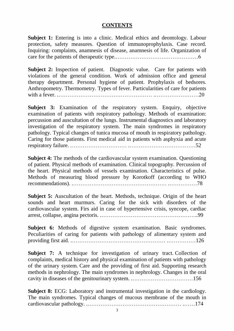
CONTENTS
Subject 1: Entering is into a clinic. Medical ethics and deontology. Labour
protection, safety measures. Question of immunoprophylaxis. Case record.
Inquiring: complaints, anamnesis of disease, anamnesis of life. Organization of
care for the patients of therapeutic type.………………………………………6
Subject 2: Inspection of patient. Diagnostic value. Care for patients with
violations of the general condition. Work of admission office and general
therapy department. Personal hygiene of patient. Prophylaxis of bedsores.
Anthropometry. Thermometry. Types of fever. Particularities of care for patients
with a fever. .…………………………………………… .……………………20
Subject 3: Examination of the respiratory system. Enquiry, objective
examination of patients with respiratory pathology. Methods of examination:
percussion and auscultation of the lungs. Instrumental diagnostics and laboratory
investigation of the respiratory system. The main syndromes in respiratory
pathology. Typical changes of tunica mucosa of mouth in respiratory pathology.
Caring for those patients. First medical aid in patients with asphyxia and acute
respiratory failure. .………………………… .………………………………52
Subject 4: The methods of the cardiovascular system examination. Questioning
of patient. Physical methods of examination. Clinical topography. Percussion of
the heart. Physical methods of vessels examination. Characteristics of pulse.
Methods of measuring blood pressure by Korotkoff (according to WHO
recommendations). .…………………………………………… .……………78
Subject 5: Auscultation of the heart. Methods, technique. Origin of the heart
sounds and heart murmurs. Caring for the sick with disorders of the
cardiovascular system. Firs aid in case of hypertensive crisis, syncope, cardiac
arrest, collapse, angina pectoris. .……………………………………………..99
Subject 6: Methods of digestive system examination. Basic syndromes.
Peculiarities of caring for patients with pathology of alimentary system and
providing first aid. .…………………………………………… .……………126
Subject 7: A technique for investigation of urinary tract. Collection of
complaints, medical history and physical examination of patients with pathology
of the urinary system. Care and the providing of first aid. Supporting research
methods in nephrology. The main syndromes in nephrology. Changes in the oral
cavity in diseases of the genitourinary system. .……………………………156
Subject 8: ECG: Laboratory and instrumental investigation in the cardiology.
The main syndromes. Typical changes of mucous membrane of the mouth in
cardiovascular pathology. .…………………………………………… .……174

4
Subject 9: Examination of the blood system. Enquiry, objective examination of
patients with pathology of blood system. Added methods of investigation in
hematology. Clinical estimation of blood investigation. Changes of tunica
mucosa of mouth in blood pathology. Caring for those patients and first medical
aid. .…………………………………………… .……………………………199
Subject 10: Examination of the endocrine system. Enquiry, objective
examination of patients with pathology of endocrine system. Added methods of
investigation in the endocrinology. The main syndromes in the endocrinology.
Changes of tunica mucosa of mouth in endocrine pathology. .………………223
Subject 11: Examination methods and semiotics of allergy manifestations.
Collection of complaints, anamnesis, objective examination of patients with
allergy. Care for patients. Additional methods of examination in allergology.
Basic syndromes in allergology. Rendering first aid at anaphylactic shock,
asphyxias at the oedema of larynx. Diagnostics of inflammatory and
degenerative diseases of locomotor system. Joint syndromes (arthritic,
osteoarthritic). Features of care for patients. .………………………………237
Practical Skill Checklist.…………………………………………….……..254
Literature.…………………………………………… .……………………..255

5
Module 1 Main methods of examination of patients with diseases of internal organs Symptomes and Syndromes of Internal Diseases Key objectives of the module: 1. To show one‘s ability to possess and follow moral deontological principles of a medical specialist and the principles of a specialist subordination in a hospital. 2. To show one‘s skills in carrying out the procedures of questioning a patient, physical and instrumental examinations of patients and analysis of their results in the course of internal diseases. 3. To master practical skills and analyse the data of main laboratory and instrumental methods of examination. 4. To master practical skills and determine main syndromes and syndromes of pathology. Subject 1: Entering is into a clinic. Medical ethics and deontology. Subject 2: Inspection of patient. Subject 3: Examination of the respiratory system. Percussion and auscultation of the lungs. The main syndromes of pathology. Subject 4: The methods of the cardiovascular system examination. Clinical topography. Percussion of the heart. The main syndromes of pathology. Subject 5: Auscultation of the heart. Origin of the heart sounds and heart murmurs. The main syndromes of pathology. Subject 6: Digestive system examination. The main syndromes of pathology. Subject 7: Investigation of urinary tract. The main syndromes in nephrology. Subject 8: ECG: Laboratory and instrumental investigation in the cardiology. The main syndromes of pathology. Subject 9: Examination of the blood system. The main syndromes of pathology. Subject 10: Examination of the endocrine system. The main syndromes in the endocrinology. Subject 11: Examination methods and semiotics of allergy manifestations. The main syndromes in allergology. Joint syndromes (arthritic, osteoarthritic). Specif ic goals: - learn the main principles of examining a patient in accordance with the traditions of the domestic therapeutic school - To master methods and techniques of a proper questioning and examining patients with diseases of internal organs To interpret correlation between a patient‘s complaints and give a preliminary estimation of the system damaged - To summarize the results of questioning a patient and identify the main syndromes and symptoms on their basis. - To learn how to enquire and examine patients with pathologies blood system, musculoskeletal system, endocrine system. - To identify the main syndromes of pathology of the blood system, musculoskeletal system, endocrine system. - To interpret the received data of laboratory investigation for recognition of pathology of blood system and endocrine system - To choose appropriate methods of investigation for certain blood,endocrine, musculoskeletal diseases
6
The Health Ministry of Ukraine
Donetsk National Medical University
Approved
at the meeting of
Propedeutic and Internal Medicine
Department
Head of department
Associate Member of NAMSc of
Ukraine,
Professor G. A. Ignatenko
« _______ »_____________ 2011 р.
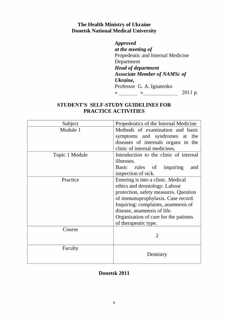
STUDENT’S SELF-STUDY GUIDELINES FOR
PRACTICE ACTIVITIES
Subject Propedeutics of the Internal Medicine
Module 1 Methods of examination and basic
symptoms and syndromes at the
diseases of internals organs in the
clinic of internal medicines.
Topic 1 Module Introduction to the clinic of internal
illnesses.
Basic rules of inquiring and
inspection of sick.
Practice Entering is into a clinic. Medical
ethics and deontology. Labour
protection, safety measures. Question
of immunoprophylaxis. Case record.
Inquiring: complaints, anamnesis of
disease, anamnesis of life.
Organization of care for the patients
of therapeutic type.
Course
2
Faculty
Dentistry
Donetsk 2011

7
Importance of the Subject: getting information from the clinic of internal
illnesses, skills, ethics and deontology allow to formulate a general purpose
from practical medicine and to define ways of study of subsequent themes from
propedeutic of internal medicine. An inquiring has the large value in the
estimation of the general state of patient and allow to find out tactic of care for
patient.
An inquiring is a mean of arrangement of psychological contact between doctor
and patient, information of anamnesis is fundamental principle for the
construction of diagnostic suppositions and in fact an oral cavity is closely
related to the different organs and systems. At some diseases of internal organs
the first symptoms very often appear exactly on the mucus membranes of oral
cavity which make a patient to consult with the dentists.
Key Objective: to find out a concept about the clinic of internal diseases, basic
moral and ethics aspects, to master the rules of labour and accident prevention
protection. Able to distinguish components of anamnesis and know order of
proper sequence each of them.
Specific Goals:
1. To have an idea about the clinic of internal diseases. Development of
domestic therapeutic school.
2. To define the basic methods of examination of patients: physical,
instrumental, laboratory.
3. To gather passport information of patient and write them in a case record.
4. To know the parts of case records and algorithm of inquiring patients with
destabilisation of complaints.
5. To master the method of inquiring according to the functional condition of
organs and systems, history of present disease, anamnesis of life, and also
written down n a case record of the important information.
6. To analyse received data and to done diagnostic suppositions.
7. Psychotherapeutic influences of enquiry on the patient and preventive
iatrogeny.
Level of Knowledge and Skills before the Practice:
1. To estimate basic human physiology. «Physiology».
2. Moral and ethics interrelations in society. «Philosophy».
Questions for Self-Assessment of the Pre-Practice Knowledge (correct
answers gone after last task).
Q1. Under the direction of the nurse the student of the 2nd rate draw a passport
part in admission office of the case record of patient D. 54 years, which
hospitalize in connection with essential hypertension. At this time has hardly
come the man of 65 years old in the office. He was pale, with complaints to

8
dizziness, sharp weakness. Suddenly including patient has lost consciousness,
has fallen, cramps have begun.
What medical and deontological tactics?
A. Nurse should begin immediately external cardiac massage and artificial
breath (on the floor). The student should transfer patient D. in other room and
urgently call a doctor.
B. Firstly he must finish registration of the case record.
C. Student should begin immediately external cardiac massage and artificial
breath (on the floor). The nurse should transfer patient D. in other room and
urgently call a doctor.
D. The nurse should call the doctor immediately and wait his orders.
E. All above variants are allowable
Q2. Determine physical factors which cause diseases.
A. High pressure action
B. Low temperature action (-350С)
C. Electric current action
D. Exposure of electromagnetic field
E. All above-listed
Q3. Determine chemical factors which cause diseases.
A. Contract with water
B. Contract with the concentrated sulfuric acid
C. Contract with physiological solution
D. Contract with a solution of baking soda
Q4. Determine psychogenic factors which cause diseases.
A. Stress
B. Book reading
C. Adequate psychological and emotional loading
D. Joy
E. Physiological sleep
Q5. Determine biological factors which cause diseases.
A. Virus
B. Microbe
S. Protozoa
D. Rickettsia
E. All above-listed
Q6. Determine genetic (hereditary) factors which do not cause diseases.
A. Trysomia on 21-st chromosome
B. Genotype ХО
C. Genotype ХХУ
D. Patau's syndrome

9
E. All above-listed
Q7. Determine the characteristic of the safety measures at work of medical staff
with electrocardiograph.
A. Use of the equipment with past scheduled checking engineering -
metrological control
B. Use of the equipment with obligatory presence of grounding and insulating
conditions
C. Use of the equipment with corresponding ratings to characteristics of an
power grid of an alternating current and a class of power supplies
D. Use of the equipment by persons allowed to work with corresponding
equipment.
E. All above-listed
Key answers: A, B, A, E, C, E
The following printed materials can be of help to improve your pre-practice
knowledge and skills:
1. Test book of Medical Physiology / Arthur C. Guyton, John I. Hall 2001, W.
B. Saunders company Pennsylvania 2001
2. Gray's Anatomy. Edited by T. Pickering Pick, F.R.C.S., 1995
3. M. Prives, N. Lysenkov, V. Bushovich; Human Anatomy
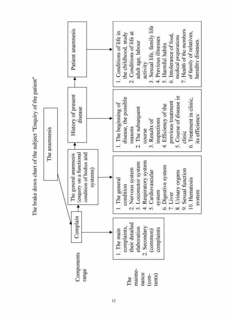
Contents of Practice
Topics of Theory:
1. General biographic information: Surname, name, patronymic, age, sex, home
address, place of work, occupation.
2. Complaints: main, their detailed elaborations, minor (general).
3. The general anamnesis: (Enquiry about a functional condition of all the
organs and systems): the general condition, cardiovascular system,
respiratory system, digestive system, urinary and hematopoietic, nervous
system, the locomotor system.
4. The history of the present disease: the beginning of the disease, the possible
reasons of the disease, the course of the disease, results of inspection,
treatment, current in to clinic etc.
5. Patient anamnesis: conditions of life in the childhood, mature age, labour
activity, family status, previous illness, harmful habits, allergy, health of the
nearest relatives etc.
Practical skills:
Students should be able to demonstrate mastery of the following practical skills
1. Gathering general biographic information of the patient and their
statement in the case record.
2. Revelation of complaints of the patient, their detailed elaboration and

10
gradual statements in the case record.
3. Enquiry about a functional condition of all the organs and systems,
history of the present disease, patient anamnesis and also a statement of
the corresponding data in the case record.
4. The analysis of the revealed data and formations of diagnostic guess.
Suggested Reading List
Required Reading
1. Internal Diseases, an Introductory Course. Edited by V.Vasilenko and
Grebenev, Mir Publishers, Moscow, 1990
2. Clinical Examination. Edited by Jonh Macleod, Jonh Munro, Churchill
Livingsone, 1986
3. A system of case recording and clinical examination of patients on
propaedeutic of internal diseases.
4. Medicine, Edited by K. George Mathew, Praveen Aggarwal, Elsevier, 2004
5. Methodical guideline for students
6. Website of department: www.cardiology.dsmu.edu.ua
7. Flow charts for Practice
Additional Reading
1. Textbook of Physical Diagnosis. History and Examination / Mark H. Swartz
2002, W. B. Saunders Company Pennsylvania
2. History and Physical Examination. Current Clinical Strategies, Edited Paul P.
Chan, Peter, J. Winkle, 2005
3. Davidson‘s Medicine, Nicholas A. Boon, Nicki R. Colledge, Davidson, 2006
Sequence of Actions in the enquiry of the patient
1 STEP: General biographic information
2 STEP: Complaints
3 STEP: The general anamnesis
4 STEP: The history of the present disease
5 STEP: Patient anamnesis
1 step. First gather general biographic information of the patient and their
statement in the case record (better do it in a column).
2 step. Ask the patient: «What do you complain of?» or «What troubles
you?». Give him possible to speak on it and at opportunity moment detail
complaints. At a statement first of all describe the main complaints (pain,

11
vomiting, cough, expectoration of blood, dyspnoea, etc.), in consistent form,
understandable, clear formulations.
3 step. General anamnesis: conduct enquiry about a functional condition of
all the organs and systems consistently and according to the scheme. State in the
name of the patient, in 3 person of a singular (complains, troubles, marks,
specifies, feels etc.) without repetition and medical terms. Don‘t describe only
the revealed symptoms with their details, but also symptoms on which the
patient answers negatively – with the didactic purpose (faster to adopt
pathology‘s displays of different system).
4 step. History of the present disease: enquiry and a statement record
especially carefully, consistently, according to the scheme - initial sing, the
possible reasons, external occasions, the further dynamics of disease, occurrence
of the new symptoms, the lead treatment, its efficiency etc.
5 step. Patient anamnesis: enquiry and a statement conduct as the medical
biography of the patient. Keep to the circuit of the case record - condition of life
in the childhood, at mature age, labour activity, the marital status, previous
illness, harmful habits, a heredity etc.
12
13
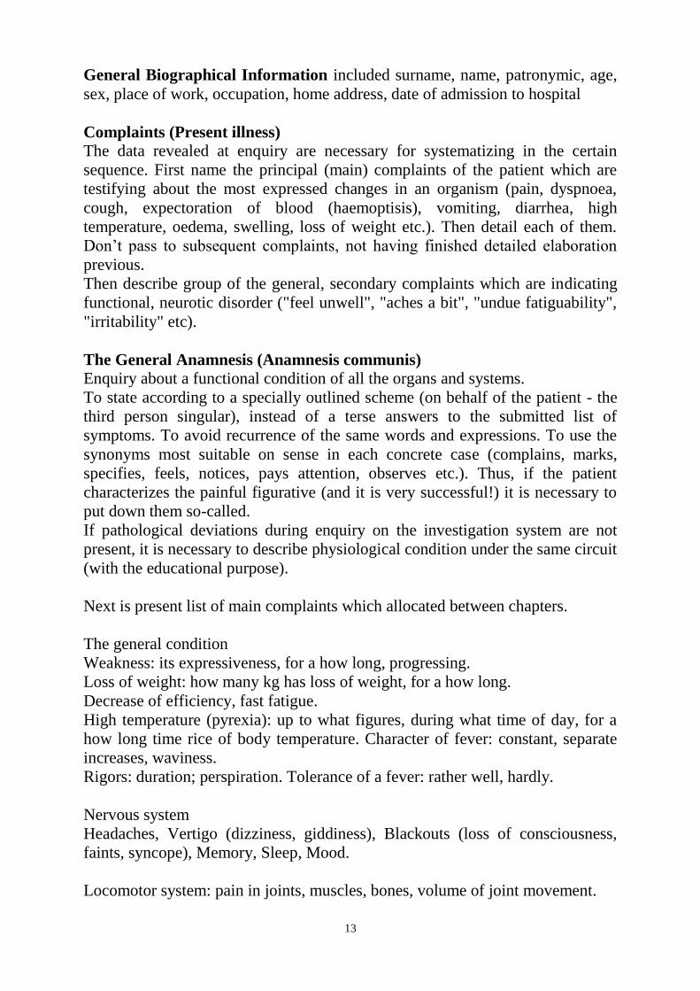
General Biographical Information included surname, name, patronymic, age,
sex, place of work, occupation, home address, date of admission to hospital
Complaints (Present illness)
The data revealed at enquiry are necessary for systematizing in the certain
sequence. First name the principal (main) complaints of the patient which are
testifying about the most expressed changes in an organism (pain, dyspnoea,
cough, expectoration of blood (haemoptisis), vomiting, diarrhea, high
temperature, oedema, swelling, loss of weight etc.). Then detail each of them.
Don‘t pass to subsequent complaints, not having finished detailed elaboration
previous.
Then describe group of the general, secondary complaints which are indicating
functional, neurotic disorder ("feel unwell", "aches a bit", "undue fatiguability",
"irritability" etc).
The General Anamnesis (Anamnesis communis)
Enquiry about a functional condition of all the organs and systems.
To state according to a specially outlined scheme (on behalf of the patient - the
third person singular), instead of a terse answers to the submitted list of
symptoms. To avoid recurrence of the same words and expressions. To use the
synonyms most suitable on sense in each concrete case (complains, marks,
specifies, feels, notices, pays attention, observes etc.). Thus, if the patient
characterizes the painful figurative (and it is very successful!) it is necessary to
put down them so-called.
If pathological deviations during enquiry on the investigation system are not
present, it is necessary to describe physiological condition under the same circuit
(with the educational purpose).
Next is present list of main complaints which allocated between chapters.
The general condition
Weakness: its expressiveness, for a how long, progressing.
Loss of weight: how many kg has loss of weight, for a how long.
Decrease of efficiency, fast fatigue.
High temperature (pyrexia): up to what figures, during what time of day, for a
how long time rice of body temperature. Character of fever: constant, separate
increases, waviness.
Rigors: duration; perspiration. Tolerance of a fever: rather well, hardly.
Nervous system
Headaches, Vertigo (dizziness, giddiness), Blackouts (loss of consciousness,
faints, syncope), Memory, Sleep, Mood.
Locomotor system: pain in joints, muscles, bones, volume of joint movement.
14
Respiratory system
Breathing through nose, Voice, Chest Pain, Cough, Expectoration of blood
(haemoptysis), Breathlessness (shortness of breath, dyspnoea), Attacks of
asthma (onset of breathlessness)
Cardiovascular (blood-circulation) system
Pains, Breathlessness (shortness of breath, dyspnoea), Palpitations,
Intermissions (escaped beats), Filling of a pulsation, Pains in gastrocnemius
muscles, Oedema (swelling)
Alimentary Gastro-intestinal system (DIGESTIVE SYSTEM)
Appetite, Saturability, Taste in a mouth, Thirst, Chewing (mastication),
Salivation (sialism), Swallowing, Abdominal pain, Heartburn (pyrosis), Nausea,
Vomiting, Abdominal swelling, Burning, itch, Stool (evacuation), Constipation,
Diarrhoea, Feces:
Liver
Urinary organs
Pains in lumbar region, Urination, Dysuria, Urine color
Sexual function
Sexual desire (libido), Menstruation
History of the present disease (Anamnesis Morbi) should include next
questions and information about course of the disease:
What time of the onset of the disease?
How began disease? (it is necessary to collect characters of the first symptoms)
What the reason of this disease in patient‘s opinion?
The course of the disease
Course of the disease in clinic
Dynamics of disease development clinical presentations
Patient anamnesis (Anamnesis Vitae) – at this part must be reflected all next
information:
Childhood, Past children's diseases (rickets, dyspepsia, infections), Adolescence,
Maturity
Social history at this time.
Harmful habits
Previous illnesses
Family history
Residence or travel abroad.
Allergological anamnesis (especially drug allergy).

15
Immunoprophylaxis
Immunoprophylaxis against viral illnesses includes the use of vaccines or
antibody-containing preparations to provide a susceptible individual with
immunologic protection against a specific disease. Immunization against viral
illnesses can be either active or passive. With active immunity, protection is
achieved by stimulating the body's immune system to produce its own
antibodies by immunization with a virus preparation. Passive immunity is
conferred by administering antibodies formed in another host. For example, an
antibody-containing gamma globulin preparation may protect a susceptible
individual exposed to a viral illness.
The viral vaccines currently approved for use are of three types:
Attenuated live viral vaccines
Most live vaccines contain viruses that have been attenuated by laboratory
manipulation. These attenuated viruses can infect and replicate in the recipient
and produce a protective immune response without causing disease. Live
attenuated viral vaccines can often confer lifelong immunity after one
immunization series. However, because live viruses can multiply in the body,
there is always the possibility that they may revert to a more pathogenic form.
Adequate laboratory and animal testing and extensive clinical studies must be
performed to assess this possibility. In addition, new recombinant technologies
facilitate direct alteration of viral genetic structure, thus permitting scientists to
produce attenuated viruses in which the genetic regions likely to lead to
pathogenic reversion are modified or deleted.
Killed (inactivated) viral vaccines
Killed viral vaccines contain either whole virus particles, inactivated by
chemical or physical means, or some component(s) of the virus. Completely
inactivated viral vaccines cannot cause infection. However, they do not
generally produce lifelong immunity following one immunization series;
additional doses are usually required. In addition, because killed virus does not
multiply in the host, the inoculum itself must provide a sufficiently large
concentration of viral antigens to induce the desired immune response.
Recombinant-produced antigens
Application of a recombinant DNA strategy to develop new vaccines is
performed by identifying the specific component(s) that can elicit the production
of protective antibodies, and then cloning and expressing the gene encoding that
protein and assembly of a complex in some cases. This approach has made
possible a safe and effective recombinant vaccine against hepatitis B virus,
which has replaced the vaccine derived from the plasma of hepatitis B virus-
infected individuals.

16
Revision Questions
Q1. Under the direction of the nurse second year student drew a passport part in
admission office of the case record of patient D. 54 years, which hospitalize in
connection with essential hypertension. At this time has hardly come the man of
65 years old in the office. He was pale, with complaints of dizziness, sharp
weakness. Suddenly this patient has lost consciousness, has fallen, cramps have
begun. What is medical and deontological tactics in this case?
A. Nurse should begin immediately external cardiac massage and artificial
breath (on the floor). The student should transfer patient D. in other room and
urgently call a doctor.
B. Firstly he must finish registration of the case record.
C. Student should begin immediately external cardiac massage and artificial
breath (on the floor). The nurse should transfer patient D. in other room and
urgently call a doctor.
D. The nurse should call the doctor immediately and wait his orders.
E. All above variants are allowable
Q2. In the resulted list of questions specify those from them which concern to
complaints of the patient:
А. An occupation of the patient now
В. Complaints in the beginning of disease
С. Harmful habits (smoking, the use of alcoholic drinks)
D. Complaints of the patient during admission to hospital
Е. The transferred operative interventions concerning the present disease and
their results
Q3. Patient anamnesis:
А. Labour activity in the past
В. Home address
С. Features of the beginning of disease
D. Results of resort treatment
Е. Results early the carried out treatment
Q4. General biographic information:
А. Intolerance (unusual reactions) medicine (es)
В. Other transferred operations
С. Features of development at children's and youthful age
D. Results early the carried out researches
Е. Transferred diseases during life
Q5. The history of the present disease:
А. The birthplace
В. Dynamics of current disease
С. The family status
D. Domestic conditions
17
Е. Health of close relatives of the patient
Q6. There should not be elements of answers ("help") in the questions, set to the
patient i.e. suggestion as it is easy to run into a mistake – to assume disease
which is not present at the patient actually. In the list of questions resulted below
specify. Correct formulation of question:
A. Do you complain of a pain somewhere or do you haven‘t got any pains?
B. What troubles you?
C. Do you have dyspnoea at walking even on plane surface place?
D. Do you test compressing pains in the precordial area at walking because of
what?
E. Do you have prickle pain in the right hypochondrium?
Q7. Suggestion formulation of question:
A. What do you complain of?
B. Does the compressing pain trouble you at walking at the precordial area
because of what you have to stop?
C. What's brought you along today?
D. What troubles you?
E. Do you have complaints now?
Q8. Patient Н., 28 years, the teacher, during last 5 months complains of a
aching, constricting sometimes pressing pain at the precordial area, with
radiation in the left hand, with duration (from hours to whole day), amplify at
excitement; irritation, insomnia; feels better, without dyspnoea at movement or
physical exertion. Pulse – 84 per 1 minutes, regular, the arterial blood pressure –
120/80 millimeters of mercury column, borders of cardiac dullness normal, heart
sounds are clear, an electrocardiogram – without changes. The diagnosis:
neuroculatory dystonic, cardiac type, "cardioneurosis" In what connection
disease is recognized first of all?
А. Detailed enquiry and absence of objective symptoms
В. Only due to absence of objective symptoms
С. Due to character of complaints
D. Due to subjective symptoms
E. Only due to combination of complaints
Q9. In the list of complaints specify those of them which concern to a category
of the main, prove expressed organic changes in an organism.
А. Compressing pains in precardial area at walking
В. Weakness
С. Fast fatigue
D. Irritation
Е. Depressed mood

18
Q10. In the list of complaints specify those of them which concern not to a
category of the main, don‘t prove expressed organic changes in an organism.
А. Dyspnoea at walking
В. Attacks of an asthma
С. Expectoration of blood
D. Pains in epigastrium area right after meal
Е. Palpitation at excitement
Key answers: 1-A, 2-D, 3-A, 4-A, 5-B, 6-B, 7-B, 8-A, 9-A, 10-E
SUMMARY OF PROCEDURES
The practice lesson conducts in the study room. After the brief repeating
(the structure of the anamnesis, the contents of main parts, technique of enquiry)
checking of the homework and guidelines for students on drawing up of the case
report are distributed to students. Then in an educational room the patient is
invited. One of students is offered to find out general biographic information of
the patient which then writes down in a writing-book (see sequence of actions
about injary of patient). Other student under the offer of the teacher asks
complaints, detail, which it is specified and supplemented with questions of
students of all group. The teacher, leading over work of students, explains
necessity of statement of this or that question, its formulation, corrects admitted
mistakes, briefly stops on diagnostic value of the found out complaints. Then
one is offered to students to formulate complaints, the final variant note down in
a writing-book with the teacher help.
In the same way students conduct enquiry of other sections of the history of
the present disease and patient anamnesis. By the end of lesion the sample of a
fragment case records (anamnesis) for the subsequent independent work is
created at students.
Final tests
Q1. General biographic information:
А. Intolerance (unusual reactions) medicine (es)
В. Other transferred operations
С. Features of development at children's and youthful age
D. Results early the carried out researches
Е. Transferred diseases during life
Q2. There should not be elements of answers ("help") in the questions, set to the
patient i.e. suggestion as it is easy to run into a mistake – to assume disease
which is not present at the patient actually. In the list of questions resulted below
specify incorrect (suggestion) formulation of question:
A. What do you complain of?
19
B. Does the dyspnoea trouble you laying position?
C. What's brought you along today?
D. What troubles you?
Q3. Among the following complaints, choose the main complaints:
А. Weakness
В. Decrease in appetite
С. Nausea
D. Inspiratory dyspnoea
Е. Eructation
Q4. The diseases caused by negative interrelation of medical staff and patients
are called:
A. Social
B. Iatrogenic
C. Somatogenic
D. Professional
E. Psychogenic
Q5. Suggestion formulation of question:
A. What do you complain of?
B. Does stomach pain increase after taking a meal?
C. What's brought you along today?
D. What troubles you?
E. Do you have complaints now?
Q6. Choose correct sequence of parts in case report:
А. General biographic information, anamnesis of life, complaints, history of
present disease, objective examination, inquiring about all organ and system
В. General biographic information, history of present disease, objective
examination, anamnesis of life, inquiring about all organ and system, complaints
С. General biographic information, history of present disease, objective
examination, complaints, anamnesis of life, inquiring about all organ and system
D. General biographic information, anamnesis of life, history of present disease,
inquiring about all organ and system, objective examination, complaints
Е. General biographic information, complaints, inquiring about all organ and
system, history of present disease, anamnesis of life, objective examination,
Q7. In the list of complaints specify those of them which concern to a category
of the main, prove expressed organic changes in an organism.
А. Compressing pains in precardial area at walking
В. Weakness
С. Fast fatigue
D. Irritation
Е. Depressed mood

20
Q8. In the list of complaints specify those of them which concern not to a
category of the main, don‘t prove expressed organic changes in an organism.
А. Dyspnoea at walking
В. Attacks of an asthma
С. Expectoration of blood
D. Pains in epigastrium area right after meal
Е. Palpitation at excitement
Q9. In the list of complaints specify those of them which concern not to a
category of the main, don‘t prove expressed organic changes in an organism.
А. Diarrheas up to 8 times day
В. Bad sleep
С. Sudden occurrence of bloody urine without of pain
D. Fast lost weight (10 kg for 2 months)
Е. Paroxysmal pains in the right hypohondrium, accompanying with a rising of
the temperature up to 37,8 and darkening of urine color (color of beer).
Q10. The diseases caused by negative interrelation of medical staff and patients
are called:
A. Social
B. Iatrogenic
C. Somatogenic
D. Professional
E. Psychogenic
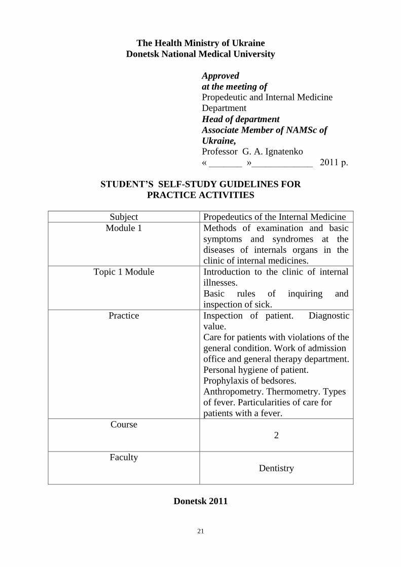
21
The Health Ministry of Ukraine
Donetsk National Medical University
Approved
at the meeting of
Propedeutic and Internal Medicine
Department
Head of department
Associate Member of NAMSc of
Ukraine,
Professor G. A. Ignatenko
« _______ »_____________ 2011 р.
STUDENT’S SELF-STUDY GUIDELINES FOR
PRACTICE ACTIVITIES
Subject Propedeutics of the Internal Medicine
Module 1 Methods of examination and basic
symptoms and syndromes at the
diseases of internals organs in the
clinic of internal medicines.
Topic 1 Module Introduction to the clinic of internal
illnesses.
Basic rules of inquiring and
inspection of sick.
Practice Inspection of patient. Diagnostic
value.
Care for patients with violations of the
general condition. Work of admission
office and general therapy department.
Personal hygiene of patient.
Prophylaxis of bedsores.
Anthropometry. Thermometry. Types
of fever. Particularities of care for
patients with a fever.
Course
2
Faculty
Dentistry
Donetsk 2011

22
Importance of the Subject: the General assessment of the patient has essential
diagnostic value for the internship doctor of any speciality, as allows to assess
the general condition, and in some cases – to distinguish disease "at first sight",
is especial at a pathology endocrine glands (acromegalia, Addison's disease
etc.), nervous system (paresis, paralyses, etc.) to reveal the main syndrome
(heart failure, jaundice, cyanosis, etc.) or other separate symptom which leading
diagnostic search in a true direction. Master by students-medics of ways and
methods of medical care for unhealthy people is an important element of
professional training of doctors.
Key Objective: To be able to lead consistently the general assessment of the
patient with the purpose of revealing of pathological signs and to make
conception about organism generally. To master practical skills in the different
aspects of medical care for patients.
Specific Goals:
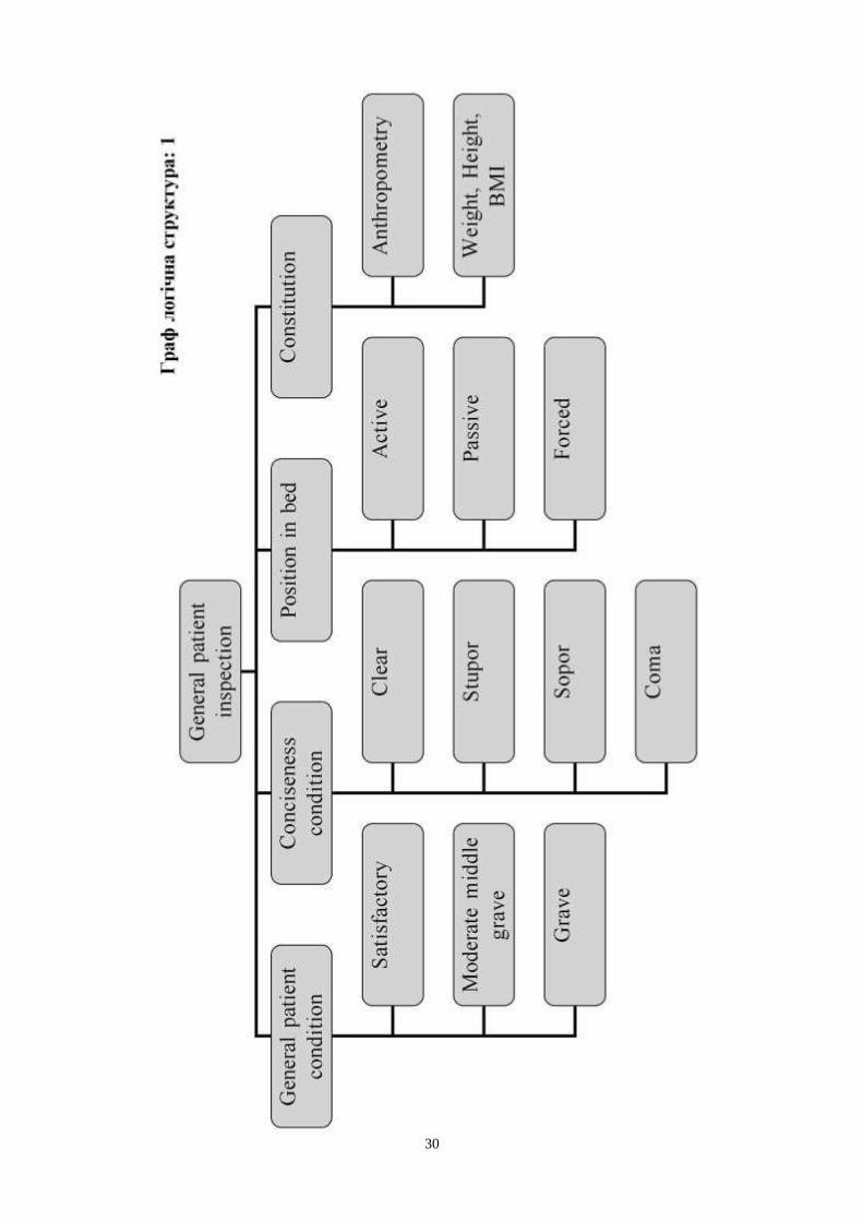
1. To master methodology of conduction of general inspection.
2. Determination of the general state of patient (varieties of the general states of
patient and their criteria).
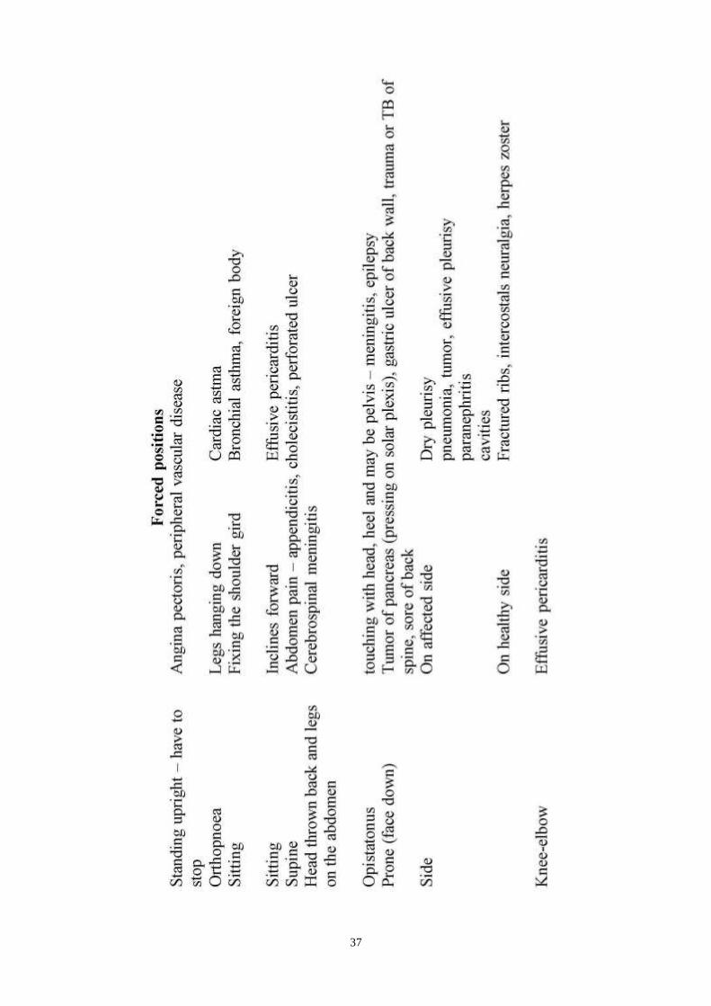
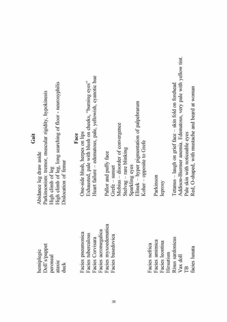
3. To determine posture of the patient in a bed (active, forced, passive, their
kinds), standing, walking (varieties of standing and walking at different
pathology),
4. To determine consciousness (stupor, sopor, coma, delirium).
5. To know habitus (type of constitution), to pay attention on height and body
weight (obesity, emaciation, cachecxia). Basic criteria of normal
constitutional types.
6. To determine oedema (general, local), to assess diagnostic value of available
changes.
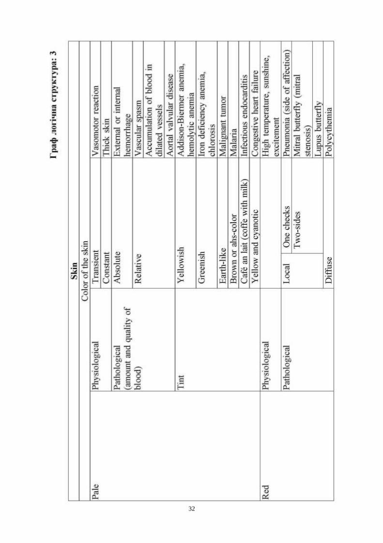
7. To examine mucous membranes (cyanotic colour of lips), skin (color,
elasticity, humidity, temperature, elements of rash, nevus, scars),
pathological changes (pale, acrocyanosis, pigmentation, scars, jaundice,
elasticity, sweating, skin eruptions, etc.), estimation of the condition of hair
and nails, subcutaneous tissue (fattened, distributing, types of obesity).
8. To examine muscles (the level of development, tonus, atrophy), joints
(configuration, deformation, tenderness, active and passive movements,
fluctuation), to assess diagnostic value of available changes
9. Sequence of palpation of lymphatic nods. Properties of lymphatic nods.
10. To explain diagnostic value of symptoms receiving during the general
inspection of patient.
11. To know types of medical institution.
12. To master basic principles of organization and work of admitting office and
general therapeutic department.
13. To master personal hygiene of sick: care of skin, oral cavity, eyes, ears, nose
and hairs.
14. To know concept about the therapeutic regimen.

23
15. To know about prophylaxis of bedsores.
16. To change of linen.
17. Serve of urinal.
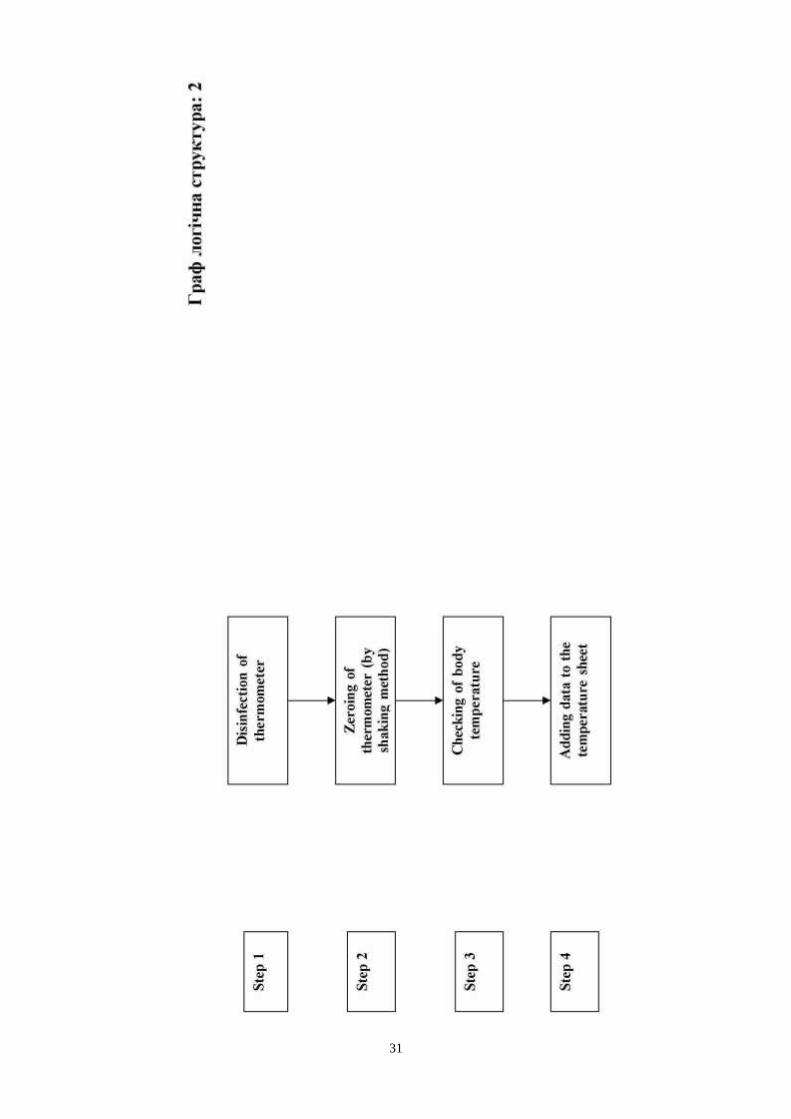
18. To done anthropometry.
19. To know principles and methods of thermometry.
20. To know types of fever.
21. To know peculiarity of care for patients with a fever.
Level of Knowledge and Skills before the Practice:
1. Clear knows physiology of visceral systems of an organism. («Normal
physiology»).
2. The anatomic characteristic of visually determined sings at injury. («Faculty
of Anatomy»).
Questions for Self-Assessment of the Pre-Practice Knowledge (correct
answers gone after last task).
Q1. Determine the characteristic of clear consciousness at the healthy person
from the physiological point of view.
A. Correct display of the reality in a brain of the person, adequate reaction on
external irritant and signals.
B. Twilling state.
C. Correct display of the reality in a brain of the person, the slowed answer on
external irritant and signals.
D. Absence of reaction
E. Any listed variants
Q2. Determine the characteristic of stupor.
A. Correct display of the reality in a brain of the person, adequate reaction on
external irritant and signals.
B. Twilling state.
C. Correct display of the reality in a brain of the person, the slowed answer on
external irritant and signals.
D. Absence of reaction
Q3. Determine characteristic of normosthenic habitus from the physiological
point of view.
A. Significant prevalence of the longitudinal sizes of a body above transversal
B. Significant prevalence ratio of extremities to a trunk
C. Increase anteroposterior sizes of a thorax
D. Right epigastric angle
E. Significant prevalence ratio of a thorax to abdomen

24
Q4. Determine characteristic of asthenic habitus from the physiological point of
view.
A. Prevalence of the transversal sizes of a thorax
B. Right epigastric angle
C. Significant prevalence of the transversal sizes of a body above longitudinal
D. Significant prevalence ratio of extremities to trunk
E. Significant prevalence ratio of abdomen to thorax
Q5. Determine characteristic of hypersthenic habitus from the physiological
point of view.
A. Increase anteroposterior sizes of a thorax
B. Right epigastric angle
C. Significant prevalence ratio of a thorax to abdomen
D. Significant prevalence ratio of extremities to trunk
E. Significant prevalence of the longitudinal sizes of a body above transversal
Q6. What is bedsore?
A. Affection of tissue, which developed under the pressure
B. Affection of tissue, which developed under the beating
C. Infection of the skin
D. Ulcer after the sunburn
E. Ulcer after the acid affection
Q7. Patient A. 66 years with cardiovascular pathology has specific changes of
fingers and nails- clubbing of the fingers (bulbous swelling of the tip of the
fingers) and nail in form of watch glass. What is pathogenesis of these changes?
A. Chronic hypoxia
B. Reduction in output of adrenocortical hormones
C. Anaemia
D. Increased consentration of bilirubin
E. Myxoedema
Q8. Patient F. 38 years has specific changes of fingers and nails - clubbing of
the fingers (bulbous swelling of the tip of the fingers) and nail in form of watch
glass. What is typical reason of these changes?
A. Endocrine disease
B. Chronic liver disease
C. Chronic cardiac disease
D. Chronic intestinal pathology
E. Haemolytic jaundice
Q9. Patient V. 22 years has specific changes of nails - altered forms as curved
inside. How is called these changes?
A. Watch glass nails
B. Koilonychia

25
C. Symptome of ―thimble‖
D. Striae
E. Hirsutism
Q10. Patient M. 44 years female was admitted to the hospital. During physical
examination decreased turgor of skin was revealed.
Which condition may cause this change?
A. Dehydration
B Thyrotoxicosis
C. Hyperhydration
D. Myxoedema
E. Acromegaly
Q11. Alpinist climb of mountains on height about 3 km became feel worse,
developed weakness, loss of consciousness, tachycardia. What is reason of this
state?
A. Hypoxemia.
B. Alkalosis.
C. Acidosis.
D. Hypercapnia.
E. Hyperglycemia.
Q12. Child with patent ductus arteriosus has low physical grows, frequent
pneumonias. Connection of which vessels is cause violation of hemodynamics?
A. By a pulmonary artery and pulmonary veins.
B. By aorta and pulmonary artery.
C. Vein cava superior and aorta.
D. Vein cava superior and pulmonary artery.
E. Aorta and pulmonary veins.
Key answers: 1-A, 2-C, 3-D, 4-D, 5-A, 6-A, 7-A, 8-C, 9-B, 10-A, 11-A, 12-B.
The following printed materials can be of help to improve your pre-practice
knowledge and skills:
1. Test book of Medical Physiology / Arthur C. Guyton, John I. Hall 2001, W.
B. Saunders company Pennsylvania 2001
2. Gray's Anatomy. Edited by T. Pickering Pick, F.R.C.S., 1995
3. M. Prives, N. Lysenkov, V. Bushovich; Human Anatomy
Contents of Practice
Topics of Theory:
1. Conduction of general inspection.
2. General state of patient.
3. Posture of the patient in a bed (active, forced, passive, their kinds),

26
standing, walking (varieties of standing and walking at different pathology)
4. Consciousness (stupor, sopor, coma, delirium).
5. Habitus (type of constitution), height and body weight (obesity, emaciation,
cachecxia). Basic criteria of normal constitutional types.
6. Oedema (general, local), to assess diagnostic value of available changes.
7. Mucous membranes (cyanotic colour of lips), skin (color, elasticity,
humidity, temperature, elements of rash, nevus, scars), pathological
changes (pale, acrocyanosis, pigmentation, scars, jaundice, elasticity,
sweating, skin eruptions, etc.), estimation of the condition of hair and nails,
subcutaneous tissue (fattened, distributing, types of obesity).
8. Muscles (the level of development, tonus, atrophy), joints (configuration,
deformation, tenderness, active and passive movements, fluctuation).
9. Lymphatic nods. Properties of lymphatic nods.
10. Types of medical institution.
11. Basic principles of organization and work of admitting office and general
therapeutic department.
12. Personal hygiene of sick: care of skin, oral cavity, eyes, ears, nose and
hairs.
13. Concept about the therapeutic regimen.
14. Prophylaxis of bedsores.
15. Changing of linen.
16. Serve of urinal.
17. Anthropometry.
18. Principles and methods of thermometry.
19. Types of fever.
20. Peculiarity of care for patients with a fever.
Practical skills:
Students should be able to demonstrate mastery of the following practical skills
1. To show methodology of conduction of general inspection.
2. To determine of the general state of patient.
3. To determine posture of the patient in a bed.
4. To determine consciousness.
5. To determine constitution.
6. To determine presence of oedema.
7. To examine mucous membranes and skin.
8. Estimate of the condition of hair and nails, subcutaneous tissue.
9. To examine muscles and joints.
10. To palpate lymphatic nods.
11. To explain diagnostic value of symptoms receiving during the general
inspection of patient.
12. To tell all types of medical institution.
13. To perform personal hygiene of sick: care of skin, oral cavity, eyes, ears,
nose and hairs.
14. To perform prophylaxis of bedsores.

27
15. To change of linen.
16. Serve of urinal.
17. To done anthropometry.
18. To take body temperature.
19. To care for patients with a fever.
28
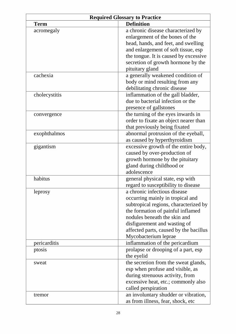
Required Glossary to Practice
Term Definition
acromegaly a chronic disease characterized by
enlargement of the bones of the
head, hands, and feet, and swelling
and enlargement of soft tissue, esp
the tongue. It is caused by excessive
secretion of growth hormone by the
pituitary gland
cachexia a generally weakened condition of
body or mind resulting from any
debilitating chronic disease
cholecystitis inflammation of the gall bladder,
due to bacterial infection or the
presence of gallstones
convergence the turning of the eyes inwards in
order to fixate an object nearer than
that previously being fixated
exophthalmos abnormal protrusion of the eyeball,
as caused by hyperthyroidism
gigantism excessive growth of the entire body,
caused by over-production of
growth hormone by the pituitary
gland during childhood or
adolescence
habitus general physical state, esp with
regard to susceptibility to disease
leprosy a chronic infectious disease
occurring mainly in tropical and
subtropical regions, characterized by
the formation of painful inflamed
nodules beneath the skin and
disfigurement and wasting of
affected parts, caused by the bacillus
Mycobacterium leprae
pericarditis inflammation of the pericardium
ptosis prolapse or drooping of a part, esp
the eyelid
sweat the secretion from the sweat glands,
esp when profuse and visible, as
during strenuous activity, from
excessive heat, etc.; commonly also
called perspiration
tremor an involuntary shudder or vibration,
as from illness, fear, shock, etc

29
uraemia the accumulation of waste products,
normally excreted in the urine, in
the blood
chloasma the appearance on a person's skin,
esp of the face, of patches of darker
colour: associated with hormonal
changes caused by liver disease or
the use of oral contraceptives
chlorosis a disorder, formerly common in
adolescent girls, characterized by
pale greenish-yellow skin,
weakness, and palpitation and
caused by insufficient iron in the
body
endocarditis inflammation of the endocardium
jaundice yellowing of the skin and whites of
the eyes due to the abnormal
presence of bile pigments in the
blood, as in hepatitis
naevus any congenital growth or pigmented
blemish on the skin; birthmark or
mole
thrombosis the formation or presence of a
thrombus
vitiligo or leucoderma any area of skin that is white from
congenital albinism (see albino) or
acquired absence or loss of melanin
pigmentation
thermometry the branch of physics concerned
with the measurement of
temperature and the design and use
of thermometers and pyrometers
30
31
32
33

34

35

36
37
38

39

40

41
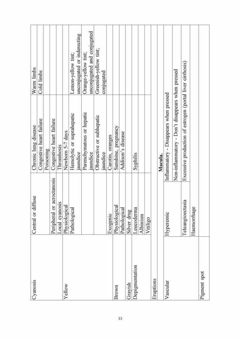
Eruptions on the skin
Herpetic lesions –small vesicles 0.5-1cm in size filled transparent fluid then
after collapse of vesicles crusts are formed (herpes labialis, herpes nasalis)
Pustula- visible accumulation of the pus in the skin
Roseola - rash-like eruption 2-5 mm which disappears when pressed by finger
(typhoid fever, para-typhys, syphilis)
Erythema - develops in hypersensitive to strawberries, eggs, crabs, erythema
nodosum at rheumatic fever, after some drugs
Weals (urticaria) – red round itching lesions elevanted under skin, which appear
as allergic reaction
Purpura – is haemorrhage into skin
Petechia- small pointed haemorrhages, do not disappear during pressing by
finger
Ecchymoses – large black and blue spots, extravasation (Werlhoff`s dsisease,
haemophilia, deficiency vitamin C and K, leukaemia)
Teleangioectasia – visible dilaation of small subcutaneous blood vessels
Rectal temperature is 0.5- 1° higher than in the armpit.
As a rule temperature is taken twice a day (at 7 or 8 a.m. and 5 or 7 P-m.).
Normal temperature of the body (as measured in the armpit) is 36,4-
36,8°C. The temperature undergoes circadian variations. The lowest temperature
is between 3 and 6 a.m. and the maximum between 5 and 9 p.m. The difference
between the morning and evening temperature does not exceed 0.6 °C in normal
persons. The temperature of the body slightly rises after meals and physical
strain, and also at high ambient temperatures.
Subfebrile - from 37° to 38 °C
Moderately high - from 38° to 39 °C
High - from 39° to 40 °C
Very high - over 40 °C
Hyperpyretic - over 41° and 42 °C
Type of fever
1. Continued fever (febris continua). The circadian variation does not
exceed 1 °C. It is observed in patients with acute lobar pheumonia or II stage,
typhoid fever.
2. Remittent fever (febris remittens). The circadian variations exceed 1 °C,
the morning lowest temperature being over 37 °C; it often occurs in
tuberculosis, III stage typhoid fever, purulent diseases, and lobular pneumonia.
3. Intermittent fever (febris intermittens). The daily variations exceed 1 °C,
with complete apyrexia in remissions.
4. Hectic fever (febris hectica). The temperature rises sharply (by 2°-4 °C)
and drops to normal and subnormal level. The fever is often accompanied by
excessive sweating. It usually occurs in grave pulmonary tuberculosis,
suppuration, and sepsis.
5. Inverse fever (typhus inversus). The morning temperature is higher than

42
in the evening; it sometimes occurs is sepsis, tuberculosis, and brucellosis.
6. Irregular fever (febris irregularis). Circadian variations are varied and
irregular. It often occurs in rheumatism, endocarditis, sepsis, tuberculosis, etc.
According to the temperature curve (Fig. 8) recurrent (relapsing) and
undulant (Malta) fevers are distinguished.
Recurrent fever (febris recurrens) is characterized by alternation of fever
and afebrile periods; it occurs in relapsing fever.
Undulant fever (febris undulans) is characterized by periodic elevation of
temperature followed by its drop; it often occurs in brucellosis and
lymphogranulomatosis.
The course of fever (Fig. 9) is characterized by a period of
- elevation of temperature (stadium incrementi), which is followed by the
- period of high temperature
- ending with the period of decreasing temperature (stadium decrementi).
The temperature may decrease gradually, during several days. This
termination of fever is called lysis.
A sudden temperature drop (to norm within 24 hours) is called crisis.
During abatement of fever in some diseases (e.g. in typhoid fever), the daily
variation of temperature exceeds 1 °C (amphibolic period).
Regular alternation of fever attacks (chills, heat, temperature drop with
sweating) and afebrile periods is characteristic of malaria. Attacks may occur
every day (febris quotidiana), every other day (tertian fever, or febris tertiana)
or every third day (quartan fever, or febris quartana). The temperature rise may
be only transient, for few hours (one-day fever, or febris ephemera, febriculara.)
It occurs in mild infection, excess exposure to the sun, after blood transfusion,
sometimes after intravenous injections of medicinal preparations.
Fever lasting up to 15 days is called acute and over 45 days—chronic.
Hypothermia (subnormal temperature) often occurs in the critical fall of
temperature; it persists for 1-2 days at about 35 °C; the pulse is full, slow, the
patient's condition satisfactory. Subnormal temperature may be observed in
grave circulatory collapse; the pulse becomes weak and frequent, respiration
superficial, the skin pallid and covered with sweat. Hypothermia occurs after
profuse bleeding, in starvation and asthenia, during convalescence after
infectious diseases, and in overcooling.
In addition to measuring the body temperature with a thermometer, the
temperature of various parts of the body should be felt by hand. Elevated
temperature of the skin overlying a joint indicates its inflammation; cold
extremities of patients with fever suggest peripheral circulatory failure (collapse,
cardiac insufficiency).
43
The main groups of lymph nodes which should be palpated:
1) submandibular.
2) cervical (anterior, posterior)
3) supraclavicular
4) subclavicular
5) axillary.
6) inguinal lymph node.
Such characteristics of lymph nodes should be determined:
- size.
- mobility
- consistency
- presence or absence of tenderness.
- surface.
- fusing with skin or between each other (fixity).
- changes of skin over lymph nodes.
In the norm we may palpate only submandibilar,axillary inguinal,they must be
painless,soft-elastic consistency,don‘t fuse with skin or between each other,
without changes, skin< 1cm.
In metastasis of tumor – Lymph node is firm, tuberousosis painless or painful it
fuse with skin.
Lymphadenitis – Elastic consistency, painful considerabely movable, skin over
it is red and hot.
Lymphogranulomatosis –Firm, painless; lymph nodes fuse together to form
conglomerates but do not adhere to the skin.
Enlargemant of lymph nodes
Cervical lymph nodes :
- Tonsillitis.
- Pharyngitis.
- Scarlatina (scarlet fever)
- Diptheria
- Lymphangioma
- Tumour of thyroid gland
Submandibular :
- Caries.
- Gingivitis.
- Tumour of larynx.
- Carcinoma of lips.
Supraclavicular :
- Tumour of mammary gland
- Tumour of gastric (virchovis metastasis)-
Inguinal

44
-Paronychia, paraitium.
- Blister foot
- Syphilis
- Gonorrhea
- Bartholinitis
Subclavicular
- Tumour of thyroid gland
- Tumour of the lung
Axillary
- Furunculosis
- Paronychia
- Pararitium (felon,whitlow)
- Tumour of mammary gland
- Tumour of lung
GENERALIZED ENLARGEMENT
-Tbs
-Sarcoidosis
-Syphilis
-Mononucleosis
-Lymphoid leukaemia
-Lymphogranulomatosis
-Lymphosarcoma
-Systemic connective tissue diseases
-HIV
Bedsores - more properly known as pressure ulcers or decubitus ulcers.
Lesions caused by many factors such as:
Unrelieved pressure;
Friction;
Humidity;
Shearing forces;
Temperature;
Age;
Continence and medication;
Sequences of formation of the bedsores is:
Stage I Paleness of skin – first reaction on pressure.
Stage II Redness - is the most superficial, indicated by non blanchable redness
that does not subside after pressure is relieved. This stage is visually similar to
reactive hyperemia seen in skin after prolonged application of pressure. Stage I
pressure ulcers can be distinguished from reactive hyperemia in two ways: a)
reactive hyperemia resolves itself within 3/4 of the time pressure was applied,
and b) reactive hyperemia blanches when pressure is applied, whereas a Stage I
pressure ulcer does not. The skin may be hotter or cooler than normal, have an
45
odd texture, or perhaps be painful to the patient. Although easy to identify on a
light-skinned patient, ulcers on darker-skinned individuals may show up as
shades of purple or blue in comparison to lighter skin tones.
Stage III Edema of skin - is damage to the epidermis extending into, but no
deeper than, the dermis.
Stage IV Skin blistering - the ulcer may be referred to as a blister or abrasion
Stage V initiation of erosion - involves the full thickness of the skin
Stage VI Ulcer - may extend into the subcutaneous tissue layer. This layer has a
relatively poor blood supply and can be difficult to heal. At this stage, there may
be undermining damage that makes the wound much larger than it may seem on
the surface.
Stage VII is the deepest, extending into the muscle, tendon or even bone.
Areas of body where are most common bedsores appear:
The most typical:
Occiput, Elbows, Scapulas, Sacrum, Coccyx, Knees, Ankles, Heels
Less typical:
Other parts of body over prominent parts of bony or cartilaginous areas.
Proper care
The most important care for a patient with bedsores is the relief of pressure.
Once a bedsore is found, pressure should immediately be lifted from the area
and the patient turned at least every two hours to avoid aggravating the wound.
Nursing homes and hospitals usually set programs to avoid the development of
bedsores in bedridden patients such as using a standing frame to reduce pressure
and ensuring dry sheets by using catheters or impermeable dressings. For
individuals with paralysis, pressure shifting on a regular basis and using a
cushion featuring pressure relief components can help prevent pressure wounds.
Camphor spirit (10%) is used for rubbing of the skin, aiming to avoid
appearance of the bedsores.
Suggested Reading List
Required Reading
1. Internal Diseases, an Introductory Course. Edited by V.Vasilenko and
Grebenev, Mir Publishers, Moscow, 1990
2. Clinical Examination. Edited by Jonh Macleod, Jonh Munro, Churchill
Livingsone, 1986
3. A system of case recording and clinical examination of patients on
propaedeutic of internal diseases.
4. Medicine, Edited by K. George Mathew, Praveen Aggarwal, Elsevier, 2004
5. Methodical guideline for students
6. Lecture: Inspection of the patient and its diagnostic value
7. Website of department: www.cardiology.dsmu.edu.ua
46
8. Flow charts for Practice
Additional Reading
1. Textbook of Physical Diagnosis. History and Examination / Mark H. Swartz
2002, W. B. Saunders Company Pennsylvania
2. History and Physical Examination. Current Clinical Strategies, Edited Paul P.
Chan, Peter, J. Winkle, 2005
3. Davidson‘s Medicine, Nicholas A. Boon, Nicki R. Colledge, Davidson, 2006
Sequence of Actions in the enquiry of the patient
1 STEP. Determine consciousness
2 STEP. Determine position in bed; find out their diagnostic value (with the
teacher‘s help).
3 STEP. Determine a habitus (constitution) of the patient.
4 STEP. Ask the patient to undress over the belt, examine skin: color
(pallor, acrocyanosis, pigmentation, scars), sweating, turgor (make fold of skin,
then fast take off fingers), skin eruptions (their difference from hemorrhage by
pressing with a finger).
5 STEP. Determine oedema (appearance of fosse after pressing with a
finger), its symmetry, painfulness, degree; find out diagnostic value of available
symptoms.
6 STEP. Examine lymph nodes (submandibular, parotid, cervical, occipital,
supraclavicular, infraclavicular, axillary, cubital, inguinal, under knee) during
survey and palpation, determine their sizes, surface, mobility, adherence
together and with surrounding tissues, painfulness).
7 STEP. Pass to studying a locomotor system: muscles (level of
development, atrophy, tenderness, tonus), joints (deformation, fluctuation, color
of a skin, active and passive movements). Find out with the teacher diagnostic
value of available symptoms.
8 STEP. Examine head (its form), face (puffy, cyanotic, sharp features,
etc.), necks (pulsation of vessels, increases in a thyroid gland), eye
(exophthalmia, reaction of pupils to light and accommodation, symptoms of the
thyrotoxicosis), mouth (mucous membranes, tongue, teeth, tonsils); determine
diagnostic value of the received symptoms.
Revision Questions
Q1. Patient С., 34 years, observes a constant dispnoea at rest, especially at night,
weakness, palpitation, thirst, oliguria (insignificant amount of urine).
Objectively: sits in armchair, with lean hands in armrests, frequency of breath 36
per 1 minutes, cyanosis of the lips, sharp features of face; significant oedema of
the lower extremities, loin, forward belly wall – they are symmetric, on cruses –
dense, on femurs – soft (doughy).
47
Note true about general condition of the patient:
А. Satisfactory
В. Moderate middle grave
С. Grave
D. Extremely grave
E. Good
Q2. Patient A. 66 years with cardiovascular pathology has specific changes of
fingers and nails - clubbing of the fingers (bulbous swelling of the tip of the
fingers) and nail in form of watch glass. What is pathogenesis of these changes?
A. Chronic hypoxia
B. Reduction in output of adrenocortical hormones
C. Anaemia
D. Increased consentration of bilirubin
E. Myxoedema
Q3. Patient С., 34 years, observes a constant dispnoea at rest, especially at night,
weakness, palpitation, thirst, oliguria (insignificant amount of urine).
Objectively: sits in armchair, with lean hands in armrests, frequency of breath 36
per 1 minutes, cyanosis of the lips, sharp features of face.
Note true about position of the patient
А. Active
В. Passive
С. Forced
Q4. Determine the characteristic of sopor.
A. Unconsciousness.
B. Pathological deep sleep from which patient wake up only for short periods.
C. Twilling state.
D. Correct display of the reality in a brain of the person, the slowed answer on
external irritant and signals.
E. Slow inadequate answers, disorientation in surroundings.
Q5. Determine the characteristic of coma.
A. Unconsciousness.
B. Pathological deep sleep from which patient wake up only for short periods.
C. Twilling state.
D. Correct display of the reality in a brain of the person, the slowed answer on
external irritant and signals.
E. Slow inadequate answers
Q6. Patient O. 54 years complains of severe attack of dispnoea. Objective data:
sits in bed, with lean hands in armrests, frequency of breath 30 per 1 min.,
cyanosis of the lips. Estimate the position of the patient.
А. Active
48
В. Passive
С. Forced
D. Normal
E. Patient does not have certain position
Q7. Patient 3. 45 years female was admitted to the endocrinological department.
During physical examination enlargement of tongue was revealed. Which
disease may cause these changes?
A. Anaemia
B. Thyrotoxicosis
C. Retrobulbar tumours
D. Acromegaly
E. Uraemia
Q8. Patient M. 44 years female was admitted to the hospital. During physical
examination moist skin was revealed.
Which condition may cause this change?
A. Dehydration
B. Myxoedema
C. Vomitting
D. Thyrotoxicosis
E. Acromegaly
Q9. Patient W. 64 years has skin changes- visible dilation of small subcutaneous
blood vessels. Estimate these skin changes:
A. Teleangioectasia
B. Hyperpigmentation
C. Petechia
D. Urticaria
E. Vitiligo
Q10. Patient C. 35 years with herpes labialis – elevanted above mucous
membrane small lesions 0.5-1 cm in size filled transparent fluid. Estimate these
changes:
A. Petechia
B. Urticaria
C. Vesicles
D. Teleangioectasia
E. Vitiligo
Key answers: 1-C, 2-A, 3-C, 4-B, 5-A, 6-C, 7-D, 8-D, 9-A, 10-C.

49
SUMMARY OF PROCEDURES
The practice lesson shall be begun in the study room. Checking of the
homework, the test control is carried out. Then at the study room patient is
invited. Demonstration of methodic of general assessment and examination of
the different parts of body by the teacher: general condition, posture of the
patient, consciousness, habitus, oedema, skin, mucous membranes, the
locomotor system, the lymph nodes, checking of the oral cavitas, examination of
pupils, palpation of the thyroid gland, pay much attention to normal data.Then
lesson is transferred to ward: the teacher of group of students does the doctors
round of patients, showing a different pathology, inviting in turn of each
students on a fragment of research.
At the end of lesson in an educational room diagnostic value of available
symptoms is discussed.
Final tests
Q1. Patient С. 45 years with cardiovascular pathology has cyanosis of the lips;
significant symmetrical oedema of the lower extremities.
What type of oedema does patient have?
А. General
В. Local
C. Hydrothorax
D. Anasarca
E. Ascites
Q2. Patient С., 34 years, observes a constant dispnoea at rest, especially at night,
weakness, palpitation, thirst, oliguria (insignificant amount of urine).
Objectively: significant oedema of the lower extremities, loin, forward belly
wall – they are symmetric, on cruses – dense, on femurs – soft (doughy).
What origin of oedema?
А. Inflammatory
В. Disturbance of outflow blood from veins.
C. Allergy
Q3. Patient V. 56 years with malignant tumour of stomach and metastasises of
malignant tumour has enlargement of lymph nodes. Estimate features of lymph
nodes after metastasis in them:
A. Smooth surface
B. Firm consistency
C. Formation of fistulae
D. Elastic consistency
E. Do not adhere to the skin
Q4. Patient R. 46 years with lymphogranulomatosis has enlargement of lymph
50
nodes. Estimate features of lymph nodes which are characteristic for
lymphogranulomatosis:
A. Fuse with skin
B. Do not fuse with each other
C. Soft consistency
D. Painless
E. Elastic consistency
Q5. Patient J. 35 years has allegic reaction. Inspection of the skin reveals red
round itching lesions elevated above skin. Estimate skin lesions:
A. Petechia
B. Hyperpigmentation
C. Urticaria
D. Teleangioectasia
E. Vitiligo
Q6. Among the following complaints, choose the main ones characteristic of
pathologies of the cardiovascular system:
А. Oedema of the feet occurring in the evenings
В. Oedema that is localized on the face and occurs in the morning
С. Oedema of the feet, legs, fingers and arms
D. Oedema of the face, neck, upper extremities
Е. Oedema localized mainly at the left foot and leg in the evening
Q7. The 24-hour range of temperature for a 41 year-old patient with fever was
4-5°С. For which type of fever is this a characteristic feature?
А. febris hectica (hectic fever)
В. febris undulans (undulant fever)
С. febris remittens (remittent fever)
D. febris recurrent (recurrent fever)
E. febris continua (continued fever)
Q8. The forced position of the patient is that:
А. Position which the doctor recommended for a quicker recovery
В. Position which the patient assumes due to the progressiveness of the disease
С. Position which the patient cannot change independently
D. Position which the patient assumes to reduce the occurrence of the illness
(dyspnoea, cough, pain etc.)
Е.Position with the patient assumes due to immobilization of the
extremities.(The use of skeletal traction, splints etc)
Q9. The passive position of the patient on the hospital bed refers to:
А. When the body and extremities of the patient are located under the influence
of gravity and then the patient cannot independently change it
В When the patient lies on the bed in such a way as to obtain maximum
51
relaxation and rest
С. When the patient is supine and the hands are stretched out around the body
D. When the patient is prone with the hands stretched out around the body
Е. The most comfortable position for the patient.
Q10. During inspection of a 35-year old patient with chronic bronchitis, the
doctor estimated his position in the bed as active. What does this mean?
А. The patient may assume any position he wishes
В. The patient assumes the position which reduces pain, dyspnoea and cough
С. The patient often needs to change his position to alleviate his condition
D. The patient intentionally assumes the position which alleviates expectoration
of sputum
Е. The patient sits with his legs drawn (bended) towards himself

52
The Health Ministry Of Ukraine
Donetsk National Medical University
Approved
at the meeting of
Propedeutic and Internal Medicine
Department
Head of department
Associate Member of NAMSc of
Ukraine,
Professor G. A. Ignatenko
« _______ »_____________ 2011 р.
STUDENT’S SELF-STUDY GUIDELINES FOR
PRACTICE ACTIVITIES
Subject Propedeutics of the Internal Medicine
Module 1 The main methods of examination of
patient and the main symptoms and
syndromes on internal diseases course
Topic 2 Module Methods of examination and the main
symptoms and syndromes in
respiratory pathology
Practice Examination of the respiratory system.
Enquiry, objective examination of
patients with respiratory pathology.
Methods of examination: percussion
and auscultation of the lungs
Instrumental diagnostics and laboratory
investigation of the respiratory system.
The main syndromes in respiratory
pathology. Typical changes of tunica
mucosa of mouth in respiratory
pathology. Caring for those patients.
First medical aid in patients with
asphyxia and acute respiratory failure.
Course 2
Faculty Dentistry
Donetsk 2011

53
Importance of the Subject : Objective examination in addition with instrumental
diagnostics and laboratory investigation of the respiratory system allow discover
and identify a pathology of respiratory system. Some respiratory pathology shows up
itself on mucous membrane of oral cavity and underlines connection oral cavity
pathology and internal organs.
Key Objective: To master knowledge of and skills in enquiring, objective
examination of the patients with pathology of the respiratory system including
inspection, percussion, auscultation, vocal resonance and vocal fremitus, making
an estimate of a diagnostic value of symptoms identifying necessary laboratory
and instrumental investigations for certain syndrome of respiratory disorders and
analyzing laboratory and instrumental investigations in respiratory pathology.
Students should be able to reveal the main syndromes of respiratory pathology
using all mentioned methods, provide caring for patients with respiratory
pathology and render first aid in patients with asphyxia and acute respiratory
failure.
Specific Goals:
1. To learn how to enquire patients with pathologies in respiratory organs about
their most disturbing complaints.
2. To develop skills in carrying out examination of the respiratory system such
as inspection of the chest, percussion and auscultation of the lungs, vocal
resonance and vocal fremitus and in identifying the pathologies discovered.
3. To reveal changes and interpret findings of laboratory and instrumental
investigations for certain respiratory pathology.
4. To be able provide caring for patients with respiratory pathology
5. To develop skills in render first aid in patients with asphyxia and acute
respiratory failure.
6. To reveal changes of mucous membrane of the mouth in respiratory
pathology
Level of Knowledge and Skills before the Practice:
1. To identify main parameters in the act of breathing (i.e. its frequency, depth,
mode and rhythm) and factors which constitute them in normal and abnormal
conditions. (Described in the Physiology course.)
2. To be able to distinguish features (i.e. mass, length, density and/or elasticity)
of a vibrating body by the percussion note physical qualities (Described in the
Biophysics course).
3. To develop understanding of the anatomic features of location of the lungs
(Described in the Human Anatomy course.)
4. To know physiology of respiratory system (Described in the Physiology
course)
5. To connect dependence of acoustical qualities of the percussion note with
physical features of vibrating body (weight, length, density, elasticity)

54
(Described in the Biophysics course.)
Questions for Self-Assessment of the Pre-Practice Knowledge
Q1. A 70-year old patient L. suffers from a severe cardiac failure. During his
inspection the following abnormalities have been discovered: a periodic
respiration, with the amplitude of respiratory movements increasing to reach its
maximal length by cycle 6 or 7; with its further getting smaller to end with an
interruption in breathing for 10-15 seconds, with a similar "wave" to follow
without the patient feeling it.
Which of the following refer to a pathological type of breathing in this case?
A.Кussmaul breathing
B. Biot breathing
C. Cheyne-Stokes breathing
D. Grocco breathing
E. Apnoea
Q2. A 56-year old patient H. Has a periodic respiration, with the amplitude of
respiratory movements increasing to reach its maximal length by cycle 6 or 7;
with its further getting smaller to end with an interruption in breathing for 10-15
seconds, with a similar "wave" to follow without the patient feeling it.
Which of the following refer to a pathological type of breathing in this case?
How are the breathing interruptions between respiratory "waves" called?
A. Tachypnoea
B. Bradypnoea
C. Asthma
D. Apnoea
E. Normopnoea
Q3. A 59-year old patient K. complains of the expiratory dyspnoea. Explain this
term «expiratory dyspnoea»:
A. it is difficult to breathe in
B. it is difficult to breathe out
C. it is difficult to both breath in and out
D. asthma attacks occur
E. a periodic respiration takes place.
Q4. A 59-year old patient K. suffers from dyspnoea. Which of these figures
refer to the respirations-per-minute rate of breathing in a healthy adult when at
rest?
A.10-12
B.16-20
С. 22-28
D.30-32
E. 30-36

55
Q5. A 67-year old patient T. suffers from dyspnoea, cough with purulent
sputum.
What complaint is not typical for pathology of respiratory organs?:
A. Cough
B. Cough out sputum
C. Dyspnoea
D. Vomitting
E. High temperature
Answer Keys: C, D, B, B, D
The following printed materials can be of help to improve your pre-practice
knowledge and skills:
1. Test book of Medical Physiology / Arthur C. Guyton, John I. Hall 2001, W.
B. Saunders company Pennsylvania 2001
2. Gray's Anatomy. Edited by T. Pickering Pick, F.R.C.S., 1995
3. M. Prives, N. Lysenkov, V. Bushovich; Human Anatomy
Contents of Practice
Topics of Theory:
1. A list of the most common complaints of patients suffering from a disease of
respiratory organs. Pathogenesis of them.
2. Static and dynamic types of the chest inspections. Making an estimate of a
diagnostic value of abnormalities in the shape of the chest. An enlargement /
reduction in size of one side of the chest, abnormal changes in the respiratory
frequency, mode and rhythm.
3. Static and topographic percussion of the lungs. Diagnostic value.
4. Auscultation of the lungs. The main breath sounds (vesicular and bronchial
(laryngotracheal) sounds)) and added breath sounds (crepitations, rales, pleural
rub).
5. Vocal fremitus and vocal resonance in identifying the syndromes of
respiratory pathology.
6. Laboratory and instrumental investigations for certain respiratory pathology,
diagnostic value of changes.\
7. The main syndroms of respiratory pathology (syndrome of air and fluid in the
pleural cavity, bronchial obstructive syndrome, syndrome of increased airiness
of the lungs,syndrome of pulmonary consolidation, respiratory failure).
8. Caring for patients with respiratory pathology.
9. First aid in patients with asphyxia and acute respiratory failure.
10. To determine changes of mucous membrane of the mouth in respiratory
pathology
After the above-mentioned topics have been studied, turn to get familiar with
relevant Flow charts for practice given in the Appendices

56
Practical skills:
Students should be able to demonstrate mastery of the following pratical skills
1. To carry out an enquiry of patient with pulmonary pathology. To determine
the main symptoms.
2. To carry out an inspection of patient‘s chest with pulmonary pathology. To
estimate data.
3. To carry out percussion of the lungs and determine clinical value of
symptoms
4. To carry out ausultation of the lungs and give clinical value.
5. To be able to provide first medical aid in patients with asphyxia and acute
respiratory failure
6. To be able to provide caring for patients with respiratory pathology
7. To reveal changes of mucous membrane of the mouth in respiratory
pathology

57
Required Glossary to Practice
Term Definition
Cough It is a reflex action aimed at clearing the air
passages of phlegm, mucus, foreign body or
other irritants
Haemoptysis is expectoration of the blood with sputum
during cough (bloody expectoration)
Bronchial asthma A disease of respiratory system that involves
inflammation of the bronchial tubes associated
with attacks of dyspnoea, cough, wheeze and
varying degrees of reversible airflow
obstruction, typically due to an allergic reaction
causes narrowed lumen of bronchi(spasm),
inflammatory oedema of their mucous
membranes, accumulation of viscous sputum
Static inspection of
the chest
Inspection of the chest is estimated usual
breathing (shape of the chest, symmetry of the
chest)
Dynamic inspection
of the chest
Inspection of the chest is estimated during deep
breathing (involvement of two side of the chest
- absence of lagging of one side, sites of
indrawing or protrusion of intercostals spaces,
involvement of accessory muscles in respiratory
act)
Apnoea Temporary arrest of respiration
Percussion Tapping various parts of the human body
produces sounds (notes) by which one can learn
about the condition of the underlying organs.
Syndrome of
consolidation of
pulmonary tissue
The syndrome of focal consolidation of lung
tissue is caused by filling of the alveoli with the
inflammatory fluid and fibrin (pneumonia -
inflammatory consolidation), blood (lung
infarction), growing connective tissue in the
lung (pneumosclerosis) or developing tumour,
atelectasis (noninflammatory consolidation) .
Topographic
percussion of the
Lungs
This is percussion which includes determination
the upper level of lung apices, width of
pulmonary apices, lower border of the lungs for
each of their topographic lines and the range of
movement of the lower pulmonary borders
(diaphragmatic movement)

58
Flow chart 1, 2, 3, 4.
Funnel chest (pectus
excavatum, funnel- chest
deformity)
Chest has a funnel-shaped depression in the
lower part of the sternum. This deformity can
be regarded as a result of abnormal
development of the sternum or prolonged
compressing effect. I
Foveated chest
It is almost the same as the funnel chest except
that the depression is found mostly in the upper
and the middle parts of the anterior surface of
the chest. This abnormality occurs in
syringomyelia, a rare disease of the spinal cord.
Vocal fremitus Method is used for determining the strength of
voice conduction to the chest surface by
palpation. Examiner puts hands over
symmetrical parts of the chest and asks the
patient to say «one-one-one» or «ninety-nine»
loudly. Vocal fremitus is of about the same
intensity in the symmetrical parts of the chest of
a healthy person. Vocal fremitus is intensified
when a part of the lung or its whole lobe
becomes airless because of a pathological pro-
cess.
Lobar pneumonia Pneumococcal pneumonia with inflammatory
consolidation of whole lobe associated with
pleural affection (dry pleuritis). It has 3 stages.
1 stage- acute hyperaemia of the lung tissue,
exudation. Alvelar wall are impregnated by
exudate, impaired –hyperresonant note is revealed
during percussion. 2 stage- red and grey
hepatization of lung tissue, lung tissue is airless by
consolidation, dull note is revealed during
percussion. 3 stage-resolution stage, impaired-
hyperresonant note is found out during percussion.
Atelectasis Decrease or absence of airness of pulmonary
region caused by obstruction of bronchial lumen
by tumour, foreing body (incomplete or complete
obstructive atelectasis) or compression of lung
tissue from the outside by pleural effusion, air
(compressive atelectasis)
Syndrome of accumulation
of pleural
fluid
occurs in hydrothorax (accumulation of non-
inflammatory effusion, i.e. transudate, for example
in cardiac failure), or in pleurisy with effusion
(inflammation of the pleura).
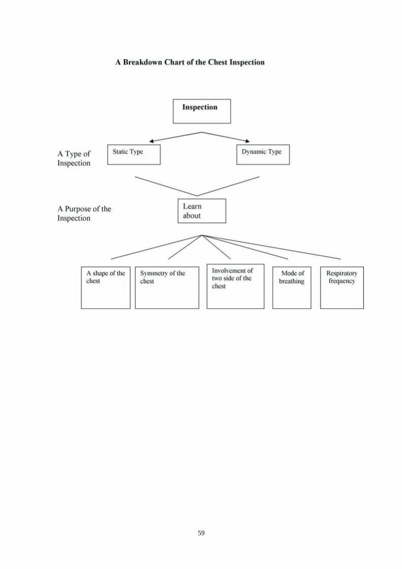
59

60
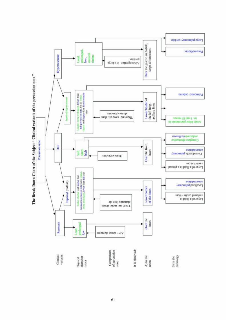
61

62

63
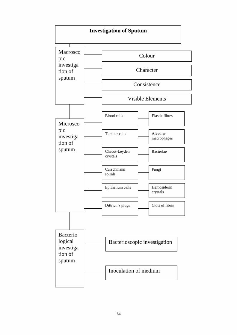
64
Macrosco
pic
investiga
tion of
sputum
Colour
Character
Consistence
Visible Elements
Microsco
pic
investiga
tion of
sputum
Blood cells Elastic fibres
Tumour cells Alveolar
macrophages
Chacot-Leyden
crystals
Bacteriae
Curschmann
spirals
Fungi
Epithelium cells Hemosiderin
crystals
Dittrich‘s plugs Clots of fibrin
Bacterio
logical
investiga
tion of
sputum
Bacterioscopic investigation
Investigation of Sputum
Inoculation of medium

65
Increased airiness
of the lungs
Compression
by air in
pleural cavity
Bronchial
asthma
Pneumonia Tuberculous
infiltration
Syndromes of pathology of lungs and pleura
Obstructive
atelectasis (complete,
incomplete)
Compressive
atelectasis
Obstruction by
tumour
Obstruction by
foreing body
Compression
by fluid in
pleural cavity
Pulmonary
emphysema
Fluid in pleural
cavity
Air in pleural
cavity
Hydrothorax
Haemothorax.
Chylothorax
Exudative
pleurisy
Pneumothorax
Pulmonary cavity
Abscess Bronchiecta
sis
Pulmonary
inflammatory
infiltration
Dry cyst of
lung
Echinococ
cus cyst of
lung
Tuberculous
cavern

66
Suggested Reading List
Required Reading
1. Internal Diseases, an Introductory Course. Edited by V.Vasilenko and
Grebenev, Mir Publishers, Moscow, 1990
2. Clinical Examination, Edited by Jonh Macleod, Jonh Munro, Churchill
Livingsone, 2000
3. Medicine, Edited by K. George Mathew, Praveen Aggarwal, Elsevier,
2004. 4. Caring for the sick by British Red Cross Society
5. First aid manual by British Red Cross Society
6. Davidson‘s Medicine, Edited by Nicholas A. Boon, Nicki R.
Colledge,Davidson, 2008
7. Manual for the Case Record, Edited by Ignatenko G.A., Department of
Propaedeutics of Internal Medicine , Donetsk, 2009
8. Lecture: Examination of the respiratory system
9. Methodical guideline for students
10. Flow charts for Practice
11. Website of department: www.cardiology.dsmu.edu.ua
Additional Reading
1. Textbook of Physical Diagnosis. History and Examination / Mark H. Swartz
2002, W. B. Saunders Company Pennsylvania
2. History and Physical Examination . Current Clinical Strategies, Edited Paul
P. Chan, Peter, J. Winkle, 2005
Sequence of Actions in the Respiratory System Examination
STEP1. Making an enquiry of patient about his complaints in detail and of
functional conditions of his respiratory organs
STEP 2. Inspecting the chest. Stand in a position with your back turned to the
light, 1,5-2 m away from the patient undressed to the waist. Check his chest
for symmetry, presence of supraclavicular and subclavicular fossae on each of
its sides, intercostal spaces, their position, features of the epigastric angle (by
tracing margins of the ribs with your thumbs), closeness of the shoulder
blades to the chest, a ratio between anteroposterior and transverse dimensions
of the chest, identify a type of its shape (by means of static inspection),
involvement of each sides of the chest in the respiratory act (by means of
dynamic inspection). Identify the abnormal changes discovered.
STEP3. Examining the breathing. Measure a rate of breathing in respirations
per minute (by acting as if you take pulse), identify its type (mode of
breathing) and rhythm, and make an assessment of data obtained.
STEP4. Practicing to work out the techniques of the direct and indirect
percussion procedures by Obraztsov‘s method (under instructor‘s guidance),
learning about differences in percussion sounds (resonant, dull, and

67
hyperresonant percussion notes).
STEP5. To perform topographic percussion
To determine:
a) an upper level of lung apices in the front side.
b) an upper level of lung‘s apices in the back side.
c) a width of the Kroenig‘s area.
d)a lower border of the lungs for each topographic line
e)range of movement of the lower pulmonary borders (diaphragmatic
movement)
STEP6. To perform comparative percussion
STEP7. To make palpation of the chest, determine vocal fremitus and vocal
resonance
STEP8. To perform auscultation of the lungs, determine the main breath
sound and presence of the added breath sounds
The main complaints typical for respiratory pathology are dyspnoea,
cough, hemoptysis, pain in the chest Dyspnoea is characterized by changes in the respiratory rate and/or depth,
rhythm, duration of the inspiration or expiration
Base on cause of dyspnoea
1. physiological ( caused by heavy exercise)
2. pathological (associated wityh pathology of the respiratory system,
cardiovascula pathology and haemopoietic pathology, poisoing)
Base on breathing phase
1. Inspiratory dyspnoea
- mechanical obstruction of the upper respiratory tract (tumour, diphtheria)
- affection of the pleura (hydrothorax, pneumothorax, exudative pleuritis,
lobar pneumonia)
- pulmonary embolism
- palmonary cancer
- atelectasis, abscess
- left cardiac failure
2. Expiratory dyspnoea - narrowed lumen in the fine bronchi, bronchioles
due to inflammatory oedema and swelling of their mucosa or else spasm
in the smoth muscle ( bronchial asthma, obstructive bronchitis)
3. Mixed dyspnoea – reduction of the respiratory surface of the lungs
- pneumonia
- pneumosclerosis
- emphysema
- pulmonary infarction
- pulmonary oedema
- left cardiac failure

68
Normal Shapes of the Chest
Features Asthenic chest Normosthenic
Chest
Hypersthenic
Chest
Shape long and narrow conical cylinder like
Anteroposterio
r/
longitudinal
dimensions
< 0,65 0,65-0,75 > 0,75
Ratio limbs>>trunk
thorax>>abdom
en
limbs>trunk
thorax>abdom
en
limbs<trunk
thorax<abdom
en
Epigastric
angle
<90°-acute
angle
90°-right angle >90°- obtuse
angle
Ludowici
angle
levelling moderate expressed
Supraclavicula
r and
subclavicular
fossae
sharply
pronounced
slightly
pronounced
unvisible
Position of
shoulders
sloping horizontal horizontal
Position of
scapulae
separated from
the chest
closely fit to
the chest
closely fit to
the chest
Course of ribs more vertical moderate
horizontal
more
horizontal
Intercostal
spaces
wide narrow
Asymmetrical chest due to enlargement of one side of the chest
- Hydrothorax (exudative pleurisy)
- Pneumothorax
- Enlargement of liver or spleen
- Enlargement of heart
- Hydropericardium
Asymmetrical chest due to diminution of one side of the chest
- Pleural adhesion
- Pneumosclerosis
- Resection of part or entire lung
- Atelectasis (collapse of lung)
69
Added respiratory parametes
The type (mode), frequency, depth and rhythm of respiration can be
determined by carefully observing the chest and the abdomen.
Mode of breathing
Mainly thoracic respiration. Respiratory movements are carried out mainly by the
contraction of the intercostal muscles. The chest markedly broadens and slightly rises
during inspiration, while during expiration it narrows and slightly lowers. This type
of breathing is known as costal and is mostly characteristic of women.
Mainly abdominal respiration. Breathing is mainly accomplished by the
diaphragmatic muscles; during the inspiration phase the diaphragm contracts
and lowers to increase rarefaction in the chest and to suck in air into the lungs.
The intra-abdominal pressure increases accordingly to displace anteriorly the
abdominal wall. During expiration the muscles are relaxed, the diaphragm rises,
and the abdominal wall returns to the initial position.This type of respiration is
also called diaphragmatic and is mostly characteristic of men.
Mixed respiration.
The respiratory movements are carried out sultaneously by the diaphragm and
the intercostal muscles. In physiological conditions this respiration sometimes
occurs in aged persons.
Respiration rate. Respiration rate can be determined "by counting the
movements of the chest or the abdominal wall, with the patient being unaware
of the procedure. The pulse rate should first be taken, with fingers held on
pulse to avoid drawing the patient‘s attention to breathing. The number of
respiratory movements in a healthy adult at rest should be 16 to 20 per
minute.
Respiratory depth
-Normal
-Superficial (shallow)
- Deep (Kussmaul breathing, high temperature,affection of the brain)
Respiratory rhythm
-Rhythmic breathing - equal depth and length of the inspiration and expiration
phases
- Pathological (periodic) rhythm
Changes of Percussion Note Over Lungs in the Pathology
Dull Note (only dense elements, absence of air)
Lobar pneumoniain 2-nd stage
Large pulmonary cavity with fluid (pus)
Tumour
Massive accumulation of fluid in pleural cavity
Complete obsructive atelectasis
70
Dullish (impaired) Note (more dense elements, than air)
Pulmonary fibrosis
Pleural adhesion
Lobular pneumonia
Obstructive atelectasis
Moderate hydrothorax
Pulmonary oedema
Hyperresonant Note (>more air, than dense elements)
Pneumothorax
Large thin-walledpulmonarycavity with air-tbs cavern (more than 6)
Emphysema, attack of bronchial asthma (bandbox note)
Diaphragmatic hernia
Large dry bronchiectasis
Impaired -Hyperresonant Note
Pulmonary oedema (onset)
Lobar pneumonia(1st and 3rd stages)
Compressive and obstructive (incomplete) atelectasis
Features of Vesicular Breath Sounds
Vesicular breath sounds are heard over pulmonary tissue during whole
inspiration and only during first third (1/3) of expiration (decrease in vibration
of alveolar walls)
Ratio between inspiration and expiration – 5 : 1
Soft sound (resemble sound « f »)
Inspiration - 120 Hertz, expiration - 100 Hertz
Transmission to chest is good
Alterations in Vesicular Breath Sounds
A. Quantitative changes
Diminished Vesicular Breath Sounds
physiological diminished vesicular breath sounds
pathological diminished vesicular breath sounds
Increased Vesicular Breath Sounds
physiological increased vesicular breath sounds
pathological increased vesicular breath sounds
B. Qualitative changes vesicular breath sounds with prolonged expiration
harsh breathing
interrupted breathing
Features of Bronchial Breath Sounds Certain places for their auscultation in the norm ((over larynx, thyroid cartilage,
jugular fossa, manubrium of sternum, VII cervical vertebra,

71
II-III thoracic vertebrae (in asthenics). Bronchial breath sounds are not heard
over other parts of the chest because of large masses of pulmonary tissue found
between bronchi and chest wall. When bronchial breath sounds are heard over
other places they are called the pathological bronchial breath sounds.
They are heard during inspiration and whole expiration (sounds are louder and
longer with expiration than with inspiration)
Ratio between inspiration and expiration 4 : 5
Rough sound (resemble sound « ch»)
Inspiration - 300 - 400 Hertz, expiration – 500- 1000 Hertz
Transmission to chest is bad
Types of Pathological Bronchial Breath Sounds consolidated bronchial breath sounds
compressive bronchial breath sounds
amphoric bronchial breath sounds (may be with metallic tone ( loud, high
sound) - thin-walled cavity more than 5-6 cm )
Syndrome of pulmonary cavities - Abscess
- Bronchiectasis
- Kartagener's syndrome – situs viscerus inversus, sinusitis,
bronchiectasis.
- Williamsa-Kempebla – congenital bronchoectasis due to hypoplasia of
bronchial cartilages.
- Tuberculous cavern
- Cystic lung hypoplasia
- Dry cyst
- Echinococcus cyst
- Lung infarction
Pulmonary cavity with communication with bronchus
Inspection - visual changes are absent; percussion- hyperresonant or impaired-
hyperresonant (it depends on size of cavity, presence of inflammatory capsule
which surroundes it, presence fluid in it); auscultation- bronchial amphoric
breath sounds, moist consonating rales, vocal fremitus and vocal resonance are
increased
Bronchiectasis - this is a condition characterized by local dilatation of the
bronchi. Bronchiectatic conditions are divided into primary (congenital, which
are very rare) and secondary (complication of various diseases of the bronchi,
lungs and pleura). Bronchiectases develop in bronchitis only when the
inflammatory process extends onto the muscular layer of the bronchial wall or
onto all its layers.
The main symptom is cough with expectoration of seromucopurulent (three
layers) or purulent sputum (sometimes foul-smelling). The daily amount of
expectorated sputum varies from 50 to 500 ml and more. Blood streaks may be
seen. Cough is paroxysmal in character and occurs mainly in the morning.

72
Haemoptysis, dyspnoea, excess sweating, weakness, headache, dyspepsia,
deranged sleep and appetite, and wasting can also be observed.
The body temperature rises, leucocytosis develops, and ESR increases.
General inspection of the patient reveals acrocyanosis (at later stages of the
disease) and oedematous face; in some cases the terminal phalanges of the
fingers become clubbed (Hippocratic fingers), and the nails resemble the watch
glass.
Bronchial obstructive syndrom
Complaints – dyspnoea with difficulties in exhalation, dyspnoea with
intensification at night and in cold weather, cough,wheezes
Inspection – protrusion of intercostals space, supra- and infraclavicular fossea.
Palpation – deacreased vocal fremitus.
Percussion – hyperresonant( bandbox note), elevation of upper level of
pulmonary (apices), lowering of lower borders. Decrease in movement of
both sides of the chest
Auscultation – harsh, vesicular breath sound with prolonged expiration,
wheezes, rhonchi which increase during forced expiration
Syndrome of increased airiness of the lungs – emphysema
– vicarious- (pulmonectomy, pneumothorax, hydrothorax, so another lung
works more severe)
- primary
- secondary (complication of chronic obstructive pulmonary diseases)
- limited
- diffuse
Inspection – barrel like chest, protrusion of intercostals space, supra- and
infraclavicular fossea.
Palpation – deacreased vocal fremitus.
Percussion – hyperresonant( bandbox note), elevation of upper level of
pulmonary (apices), lowering of lower borders. Decrease in movement of
both sides of the chest
Auscultation – deacreased vesicular breath sound
Syndromes of pneumothorax
- Traumatical pneumothorax ( blunt trauma of the chest, fracture of ribs,
penetrating wound of the chest by knife, bullet)
- Iatrogenic pneumothorax ( after resuscitation -external cardiac massage,
unsuccessful puncture of pleural cavity)
- Spontaneous pneumothorax
– neonatal
– primary (unknown reasons)
– secondary (COPD, BA, pneumonia, infarction, tumor, endometriosis,
tbs, X- Ray examination with contrast, cystic fibrosis, eosinophilic
granuloma, Marfan's syndrome, pregnancy)
73
- Tension pneumothorax (when air can‘t go out from pleural cavity and
new portions come in it again) – it gives dullish note during percussion
Syndrome of atelectasis
Obstructive atelectasis
- complete (collapse) - indrawing of the intercostals spaces, lagging of affected
side, dull note, absence of breath sounds, vocal fremitus and vocal resonance are
absent
- incomplete – normal or small indrawing of the intercostals spaces, impaired-
hyperresonant note, decreased vesicular breath sounds, vocal fremitus and vocal
resonance are decreased
Compressive atelectasis (compression of lung tissue by tumour, hydrothorax,
pneumothorax)
Cause of complicated pulmonary compliance which accompanied with
dyspnoea:
- pneumosclerosis
- pleural adhesion
Revision Questions
Q1. During the inspection of a patient an accelerated respiratory rate of 24 per
minute was observed. What does this mean?
А.Cheyne-Stokes breathing
B. apnoea
C. bradypnoea
D. breathlessness
E. normopnoea
Q2. Choose the pathological rhythm of breathing:
A. laryngotrachealis breathing;
B. vesicular breathing;
C. biot's breathing;
D. saccadic breathing;
E. bronchial breathing.
Q3. It is impossible to define by percussion border between:
A. lung lobes;
B. lung and muscles;
C. lung and liver;
D. lung and heart;
E. intestines and the lower border of liver.
Q4. First percussion stroke is done at comparative percussion over:
A. right side of chest;
B. left side of chest;
C. increased side of chest;
D. decreased side of chest;
74
E. side of chest with good amplitude of breathing.
Q5. Which of these diseases is likely to cause the appearance of the bronchial
breath sounds?:
A. not expressed bronchiectasis;
B. cavernous tuberculosis;
C. complete obstructive atelectasis
D. small adhesive pleuritis;
E. pneumothorax.
Q6. Choose condition which is necessary for occurrence of pleural rub:
A. patency of the bronchi and inflammation of pleural layers;
B. infiltration of pulmonary tissue and inflammation of pleural layers;
C. collapse of pulmonary tissue and inflammation of pleural layers;
D. increased airness of the lungs and inflammation of pleural layers;
E. inflammation of pleural layers.
Q7. What does macroscopic investigation of sputum include:
A. presence of elastic fibres
B. presence of atypical cells
C. colour of sputum
D. inoculation of medium
E. presence of Charcot –Leyden crystals
Q8. A 48-year old patient K., suffers from attack of severe dyspnoea («I can
not exhale»). She is sitting on the bed, with resting on the bed , respiratory
frequency is 24 respirations per minute, with prolonged expiration and a
sound of wheezing heard. Her chest is found emphysematous, symmetric in
shape, with the percussion note being of a hyperresonant type. The cause of
respiratory failure in this is case:
A. accumulation of air in a pleural cavity;
B. accumulation of fluid in a pleural cavity;
C. massive low-lobe right pneumonia;
D. attack of bronchial asthma
E. expressed pleurae adhesions.
Q9. During inspection of patients with syndrome of complete obstructive
atelectasis of lung can be revealed the following:
A. decrease of the affected half of the chest
B. levelling of the intercostals spaces
C. enlargement of the affected half of the chest
D. emphysematous (barrel-like) chest
E. levelling or protrusion of supraclavicular fossae
Q10. Which auscultatory data can be defined in patients with syndrome of
inflammatory pulmonary consolidation (lobar pneumonia 1 and 3 stage):
A. diminished vesicular breath sounds
B. increased vesicular breath sounds
C. bronchial breath sounds
D. dry rhonchi
E. non-consonating moist rales
75
Answer Keys : Q1: D; Q2: C; Q3: A; Q4: E, Q5: B; Q6: E; Q7: C; Q8: D,
Q9: A; Q10: A.
SUMMARY OF PROCEDURES
The practice lesson shall begin in the study room, with the homework
assignment checked and students‘ testing carried out.
Another part of the practice shall be conducted in the hospital wards. Instructor
will demonstrate to the students how to make enquiring, inspection of the chest
and examination of the respiratory system.
Before examination of patients in ward or at the end of practice lesson students
listen to records of added breath sounds in a phonoclass.Then component
elements of the examination of the respiratory system shall be practiced by each
of the students individually.
During it, the students shall enquire patients (one at a time) of their breathing
condition, concentrating on any symptoms of abnormalities or disorders they
noticed. After that, the patient‘s chest shall be inspected, with a rate of breathing
measured, its type (mode of breathing) and rhythm identified,students perform
percussion and auscultation of the lungs and signs of abnormal changes in those
with respiratory pathologies discovered. Diagnostic value of the revealed
changes of breath sounds in examined patients shall be discussed outside of the
ward. Instructor teaches to perceive the organism of man as single unit,
underline connection oral cavity pathology and internal organs, to provide first
aid to the patients before hospitalization and in urgent situations.
At the end of the practice teacher makes short general conclusion concerning
obtained findings and students do final tests
Final Tests
Q1. Which of the following medications is indicated for a patient with
haemoptysis?
А. Cupping glasses
B. Mustard plasters
C. Compress with camphor
D. Ice bag
E. leeches
Q2. Choose disease which is characterised by expiratory dyspnoea:
A. chronic obstructive bronchitis and bronchial asthma;
B. hydrothorax and cardiac asthma;
C. bronchial asthma and pulmonary embolism;
D. pneumothorax and full athelectasis;
E. foreign body in bronchus and bronchial asthma.
Q3.When is impossible to define border with topographical percussion:
A. lung and muscles;
B. lung and liver;
C. lung and heart;
76
D. liver and heart;
E. intestines and the lover edge of liver.
Q4. During recovery of the patient with exudative pleurisy percussion note on
the affected side change from:
A. from dull note to impaired, and then to resonant;
B. from dull note to impaired-hyperresonant, and then to resonant;
C. from dull note to impaired-hyperresonant, and then again to dull;
D. from dull note to resonant, and then to inpaired;
E. all time persistent dull note.
Q5. Choose extra-pulmonary factor which causes diminished vesicular breath
sounds:
A. small amplitude of movements of alveolar walls during their expressed
stretching;
B. small amplitude of movement of alveolar walls during their collapse;
C. increased amplitude of movement of alveolar walls;
D. accumulation of air in the pleural cavity;
E. thick chest wall.
Q6. Which of these diseases is likely to cause the appearance crepitations?
A. lobar pneumonia in 1-st stage;
B. lobar pneumonia in 2-nd stage;
C. cavernous tuberculosis;
D. acute bronchitis;
E. bronchiectasis.
Q7. Choose the pathology which is characterized by presence of hemorragic
exudate:
A. heart failure
B. bronchial asthma
C. pneumothorax
D. pleural tumour
E. connective tussue disease
Q8. The division of types of respiratory failure( acute, chronic) according to
classification is based on:
A. features of dyspnoea
B. features of cough
C. data of anamnesis
D. severity of cyanosis
E. swollen of cervical veins
Q9. The syndrome of inflammatory infiltration of pulmonary tissue is observed
at:
A. pneumonia
B. pneumothorax
C. hydrothorax
D. tuberculous cavern
E. bronchial asthma

77
Q10. Choose the data of percussion which is characteristic of patients with
syndrome of complete obstructive atelectasis of lung:
A. impaired (dullish) percussion note
B. hyperresonant (bandbox) percussion note
C. dull percussion note
D. dullish- hyperresonant (impaired-tympanitic) percussion note
E. resonant percussion note
78
The Health Ministry Of Ukraine
Donetsk National Medical University
Approved
at the meeting of
Propedeutic and Internal Medicine
Department
Head of department
Associate Member of NAMSc of
Ukraine,
Professor G. A. Ignatenko
« _______ »_____________ 2011 р.
STUDENT’S SELF-STUDY GUIDELINES FOR
PRACTICE ACTIVITIES
Subject Propedeutics of the Internal Medicine
Module 1 The Main Methods of Examination of
Patients on Internal Diseases Course.
Subject Module 3 The Methods of Examination and Main
Symptoms and Syndromes of the
Cardiovascular System Pathology.
Topic The methods of the cardiovascular
system examination. Questioning of
patient. Physical methods of
examination. Clinical topography.
Percussion of the heart. Physical
methods of vessels examination.
Characteristics of pulse. Methods of
measuring blood pressure by Korotkoff
(according to WHO recommendations).
Course 2
Faculty Dentistry
Donetsk 2011

79
Importance of the Subject : carrying out of an interview, examination and
palpation of precordial region, properties of cardiac beat with the assessment in
the identified changes plays an important role in the diagnosis of cardiovascular
diseases. Ability to define the heart and vascular bundle borders with percussion
and evaluate the changes of diagnostic value is essential in determining the
nature of the pathology of the cardiovascular system and other organs. Ability to
provide qualified first aid in emergency conditions is preventing their serious
consequences and terminal conditions.
Key Objective: to carrying out an interview of patients with pathology of the
cardiovascular system, examination and palpation of precordial region and
vessels, to identify properties of apex-beat and to interpret revealed symptoms
for the next diagnostic of diseases of the circulation organs. To learn percussion
definition of the borders of absolute and relative heart and vascular bundle
dullness and to estimate normal and pathological parameters that was revealed.
Specific Goals:
1. To interview a patient with sequential specification of patient complaints,
characteristics of disease and life history anamnesis.
2. To describe characteristics of general patient examination with
assessment of bed position, color of skin and mucous membranes,
peripheral edema‘s existence.
3. To conduct consecutive examination of precordial region and neck. To
detect pathological changes (cardiac hump, pulsation in heart area and
neck, varicose veins, etc.) and to estimate theirs diagnostic value.
4. To work for technique and method of palpation of precordial area
following method and technique; to detect clinical properties of apex-beat
(localization, abundance, depth, resistance, dislocation) and to estimate
diagnostic value of revealed changes.
5. To detect and to interpret presystolic and systolic thrill (―cattish purr
symptom‖).
6. Give interpretation of diagnostic value of interview, examination and
palpation information for definition the symptoms of cardiovascular
pathology system.
7. To made heart percussion following method and technique.
8. To describe sonic information of percussion that represents absolute and
relative heart dullness.
9. To shows and estimate the borders (right, upper, left) of absolute and
relative dullness of heart, it changes at pathology.
10. Give interpretation of diagnostic value of physiological conditions that
influence on a size and position of dullness of heart.
11. To definite and estimate width of vascular bundle.
12. Interpret the diagnostic value of changes of borders of relative and
absolute dullness of heart, width of vascular bundle, configuration of
heart for determination of pathology syndromes of the cardiovascular
system of organs of breathing, mediastinum and abdominal region.
80
13. To measure the arterial blood pressure by Korotkoff (according to WHO
recommendations).
14. To render first aid at hypertensic crisis, sudden stop of blood circulation,
heart attack, loss of consciousness, collapse, acute pain at heart region.
15. To render first aid at acute bleeding. To prepare transfusion system and
take part at transfusion of blood or it components.
Level of Knowledge and Skills before the Practice:
1. To clearly know anatomic and physiological bases of appearance of apex
beat. To distinguish main cardiac cycle indexes (described in the
Physiology course).
2. To know topographical anatomy of heart and main vessels (Described in
the Anatomy course).
Questions for Self-Assessment of the Pre-Practice Knowledge
Q1. In healthy human heart rate at rest is in the range of
A.55-80 bpm
B. 60-70 bpm
С. 60-90 bpm
D. 65-95 bpm
Е. 75-85 bpm
Q2. The resistance of blood flow at vessels of human proportionate
depends on a few factors. What are the factors?
A. Radius of vessels and blood viscosity.
B. Radius of vessels and the 2СО saturation of blood.
C. Radius of vessels and erythrocyte sedimentation rate.
D. Radius of vessels and the 2О saturation of blood.
E. Length, radius and pH of blood.
Q3. When pressing on the eye is a reflex:
A. Increase of a heart rate.
B. Decrease of a heart rate.
C. Decrease of respiratory movement‘s rate.
D. Increase of respiratory movement‘s rate.
E. Widening of bronchial tree diameter.
Key answers: 1 –C, 2-A, 3-B.
Suggested Reading List for the initial level of knowledge
1.Human Anatomy. Edited by M.G. Prives., N.K. Lysenkov., V.I.
Bushkevich. Hippocrates publisher. S-Petersburg, 1998 - p. 138-153.
2.Normal physiology. Edited by V.I. Filimonova. – Zdorov‘e., 1994 – p.608
81
3.―Normal human physiology‖. Edited by V.M. Pokrovskiy. M., 1998, 1
volume, p 340-347.
4.Human physiology. Edited by R. Shmidt and G. Tevs. – Mir Publishers,
Moscow, 1986. – P.192-200,205-207,214-216.
Contents of Practice
Theoretical questions to practice.
1.A list of the most common complaints of patients suffering from
cardiovascular pathology. Main indexes of function of cardiovascular system
violations.
2.Characteristics of disease and life anamnesis gathering.
3.Characteristics of general patient examination with pathology of
cardiovascular system: forced position, colour of skin and mucous
membranes, peripheral edema‘s existence.
4.Diagnostic value of examination and palpation of precordial region.
5.Diagnostic value of pathological pulsation at precordial region, neck,
epigastrical region.
6. Physical bases of percussion, rule of percussion of heart.
7. Basic types of cardiac dullness: relative and absolute are features of
determination, formation and difference of acoustic properties of percussion
sound.
8. Method and technique of determination, characteristic of normal borders
(right, upper, left) of relative dullness of heart.
9. Method and technique of determination, characteristic of normal borders
(right, upper, left) of absolute dullness of heart.
10. Diagnostic value of influencing of physiology condition on a size and
position of dullness of heart.
11. Methods and technique of vascular bundle definition and estimating of it
borders.
12. Diagnostic value of changing of borders (right, upper, left) of relative and
absolute dullness of heart and heart configuration.
Practical skills that are performed on lesson.
1. Enquiry (interview) of the patients with cardiovascular pathology (their
most disturbing complaints).
2. Estimation of position in bed, colour of skin and mucous membranes,
peripheral edema‘s existence.
3. Main skills of general and special nursing care of patient with pathology
of circulation system.
4. Variants of rendering of patient‘s comfortable position with pathology of
circulation system, oxygen supply and parenteral introduction of
medicines, application of styptic bandage.
5. Examination and palpation of precordial region and interpretation of the
received information.
6. Definition and characteristics of the borders (right, upper, left) of relative

82
dullness of a heart in health and disease.
7. Definition and characteristics of the borders (right, upper, left) of absolute
dullness of a heart in health and disease
8. To describe a vascular bundle and estimate it borders.
9. To measure the arterial blood pressure by Korotkoff (according to WHO
recommendations).
10. To render first aid at hypertensic crisis, sudden stop of blood circulation,
heart attack, loss of consciousness, collapse, acute pain at heart region.
11. To render first aid at acute bleeding. To prepare transfusion system and
take part at transfusion of blood or it components.
12. Pulse and estimation of its main properties (frequency, rhythm, filling,
tension). Pulse examination on radial, femoral, temporal, carotid arteries.
Required Glossary to Practice
Term Definition
Absolute cardiac dullness The area of the anterior wall of the
heart (right ventricle) which is not
covered by the lungs and directly
contacts with anterior chest wall.
Correspond to dull percussion note.
Aneurysm The dilatation of the lumen of blood
vessels or the cavities of the heart
due to pathological changes in their
walls.
Acrocyanosis Is cyanotic color of the remote parts
of the body (fingers, nose, lips, and
ears) in patients with cardiac failure.
Defect, malformation Is congenital or acquired anatomic
abnormalities of the body structures
(cardiac valve, septum between its
chambers etc)
Relative cardiac dullness The projection of anterior surface of
the heart onto the anterior chest
wall. Correspond to impaired
percussion note.
Exudates Accumulation of liquor in the
cavities of the body.
Hypertrophy
Excessive increase in size of organ,
its part or part of body
Dilation Expansion of a hollow organ
Epigastria pulsation Pulsation of a liver, aorta, right
ventricle.
Megalocardia Increase in heart size

83
Cardiomyopathy Disease is characterized by
noninflammatory and noncoronary
myocardial damage
Cyphosis Curvature of the spine in the sagittal
plane
Coarctation Congenital limited vasoconstriction
Myocarditis Inflammation of the myocardium
with a violation of his
contraction, activation, conduction
Pastose Pallor, reduced skin elasticity
and subcutaneous fat in their little
edema
Pericarditis Inflammation of the pericardium
Dulled Percussion sounds on the border
between distinct and dull
Pulsation of neck vessels Pulasation of carotid arteries
(synchronous to the heart beat),
positive and negative venous
cervical pulse (pulsation of jugular
veins).
Cardiac hump Deformity of the chest due to an
increase of heart from childhood
(congenital and acquired valvular
defect).
Heart beat Thrill of chest area synchronous to
the rhythm of heart contractions
Symptom of Plesh Increase of jugular veins edema at
pressure on the liver. Positive for
right ventricle of heart insufficiency.
Symptom of ―cattish purr‖ Palpation feeling of intracardiac
murmur (aortic and mitral stenosis)
like thrill of chest.
Scoliosis Curvature of the spine in the frontal
plane
Splanchnoptosis Condition characterized by falling of
the internal organs
Stenosis Constriction of the tubular organ,
the external orifice
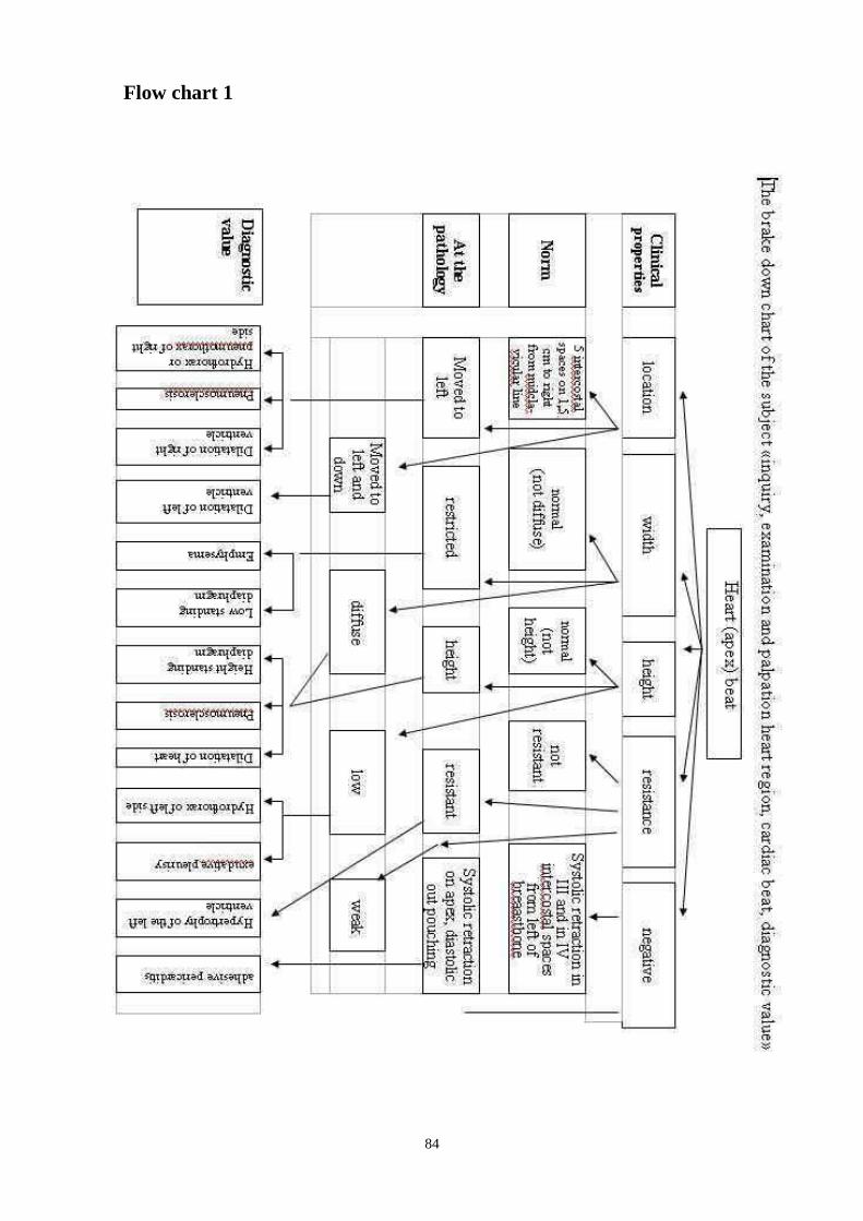
84
Flow chart 1

85
Flow chart 2
Palpation of apical impulse
Conditions of the best perception
The largest inclination of
patient trunk ahead
During deep
exhalation of patient
Localization of apical
beat
Height (amplitude)
of apical beat
1 -1.5 cm inside from a
midclavicular line in V
intercostals space
Low (at the physical
loading a height
increases)
Width (area of beat) Resistance (force) of
beat
Above 2см in a diameter Of moderate
resistance
It is determined at 2/3 people (at other
covered by a rib)
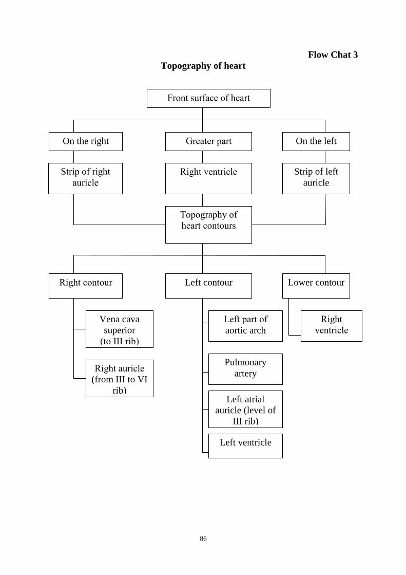
86
Flow Chat 3
Topography of heart
Front surface of heart
On the right Greater part On the left
Right ventricle Strip of left
auricle
Topography of
heart contours
Right contour Left contour Lower contour
Right
ventricle
Vena cava
superior
(to III rib)
Right auricle
(from III to VI
rib)
Left part of
aortic arch
Pulmonary
artery
Left atrial
auricle (level of
III rib)
Left ventricle
Strip of right
auricle

87
Flow Chat 4
Normal borders of heart
Relative dullness of heart
Right border Upper border Left border
0.5 -1 cm ectad
from right
margin of
breastbone
III rib (upper margin
of III rib is more
frequent)
1-1.5cm from
midclaviular line
Absolute
dullness of heart
Right border
Upper border
Left border
IV rib (lower margin
of IV rib)
1.5-2 cm inside
from
midclavicular line
left margin of
breastbone
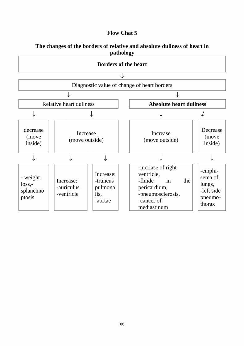
88
Flow Chat 5
The changes of the borders of relative and absolute dullness of heart in
pathology
Borders of the heart
Diagnostic value of change of heart borders
Relative heart dullness Absolute heart dullness
decrease
(move
inside)
Increase
(move outside)
Increase
(move outside)
Decrease
(move
inside)
- weight
loss,-
splanchno
ptosis
Increase:
-auriculus
-ventricle
Increase:
-truncus
pulmona
lis,
-aortae
-incriase of right
ventricle,
-fluide in the
pericardium,
-pneumosclerosis,
-cancer of
mediastinum
-emphi-
sema of
lungs,
-left side
pneumo-
thorax
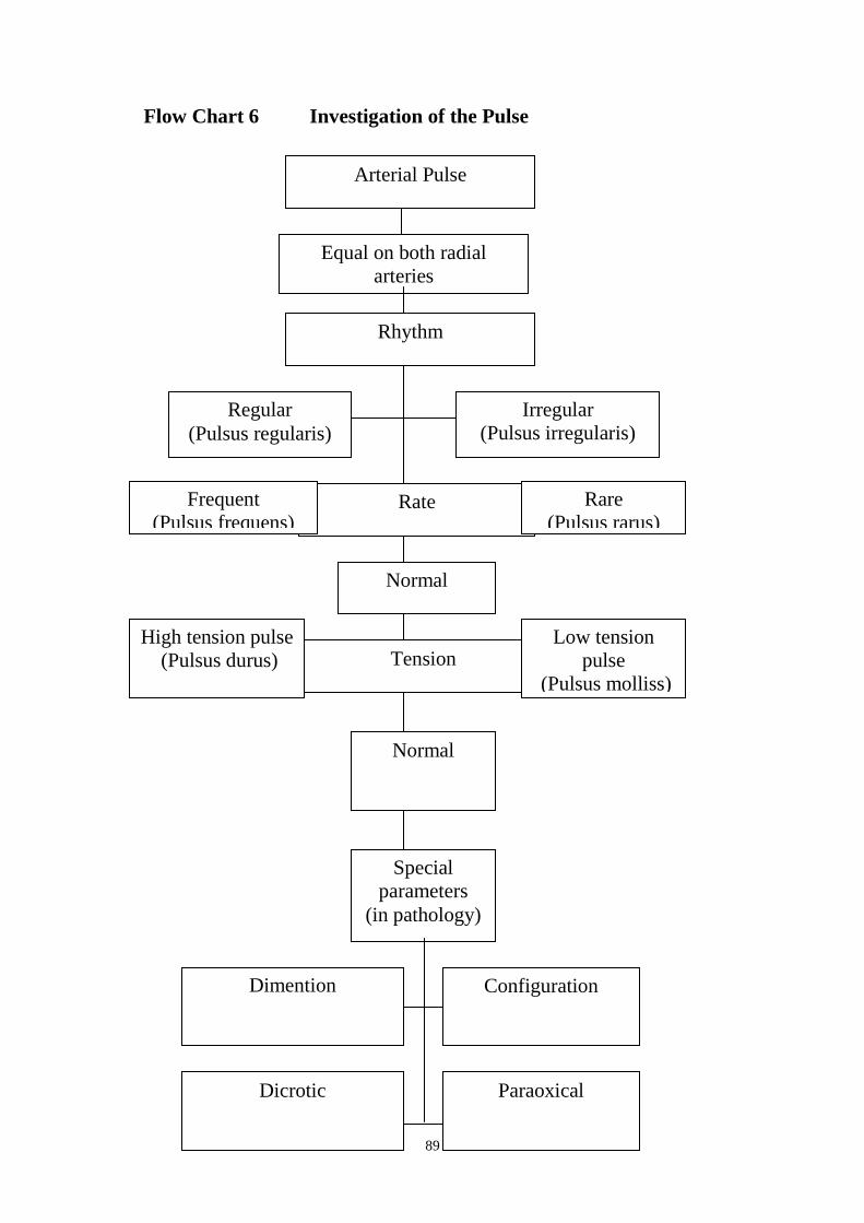
89
Flow Chart 6 Investigation of the Pulse
Arterial Pulse
Equal on both radial
arteries
(Pulsus aequalis)
Rhythm
Regular
(Pulsus regularis)
Irregular
(Pulsus irregularis)
Rate Frequent
(Pulsus frequens)
Normal
Rare
(Pulsus rarus)
Tension High tension pulse
(Pulsus durus)
Normal
Low tension
pulse
(Pulsus molliss)
Special
parameters
(in pathology)
Dimention
Configuration
Dicrotic Paraoxical

90
Suggested Reading List
Required Reading
1. Internal Diseases, an Introductory Course. Edited by V.Vasilenko and
Grebenev, Mir Publishers, Moscow, 1990
2. Clinical Examination, Edited by Jonh Macleod, Jonh Munro, Churchill
Livingsone, 1986
3. Medicine, Edited by K. George Mathew, Praveen Aggarwal, Elsevier, 2004
4. Lecture: Examination of the cardiovascular system: semiotics, inspection of
the heart, percussion: methodic and technique of determining normal borders
of the relative and absolute cardiac dullness, in the norm and pathology
5. Methodical guideline for students
6. Flow charts for Practice
7. Website of department: www.cardiology.dsmu.edu.ua
Additional Reading 1. Textbook of Physical Diagnosis. History and Examination / Mark H. Swartz
2002, W. B. Saunders Company Pennsylvania
2. History and Physical Examination . Current Clinical Strategies, Edited Paul
P. Chan, Peter, J. Winkle, 2005
3. Davidson‘s Medicine, Nicholas A. Boon, Nicki R. Colledge, Davidson,
2006
Estimated base operations (EBO).
Sequence of Actions in the Precordial Area Palpation and Examination
I STEP: Examination of heart region: pay attention in the presence of explosion
in a heart region, pulsations, expansion of skinning veins. Define what places
(on intercostals spaces and lines) the places of pulsations are present. Then it is
necessary to synchronize them with a cardiac beat, for determination of which
inspection is needed to add the palpation. For this purpose attach a right hand
palm on the breast of patient in the area of apex of heart (that hands must be
between parasternal and axillaries anterior lines and occupied the area - limited
from above by the ІІІrd rib and from below IVs rib). Farther with finger-pad of
distal phalanx of index finger setting her strictly athwart find a lowermost and
most lateral point, taking her for the place of cardiac beat.
II STEP: Learn properties of cardiac beat: localization, width, height, resistance.
It is needed to remember about possibility of negative cardiac beat, when during
the systole of ventricles there is no explosion of pectoral wall, and retraction her
(this sign characteristic at adhesion of both layer of pericardium between itself
and external layer of him with a pectoral wall).
III STEP: At presence of visible filing pulsations on the separated areas of heart
region begin their study:
a) define their localization; whether it belong to location of dissecting aneurysm
of aorta, pulmonary artery, atriums, epigastria or livers;
91
b) for differentiation of pulsations of right ventricle from the pulsations of
abdominal aorta it is possible to use a few signs: the pulsation of right ventricle
is visible directly under the xiphoid process, and abdominal aorta a few lower,
greater noticeable retraction, than explosion of epigastric area, at deep breath
pulsation of right ventricle becomes more present (a diaphragm goes down, and
with it and right ventricle too). At palpation of liver it is needed to remember
about 2 it variety: true (systolic), which appears in the intermittent swelling and
diminishments at enlargement liver which is at insufficiency of tricuspid valve
and "transmission" - from an abdominal aorta.
Data of palpation of the apical (cardiac) beat in the normal
Localization of
apical (cardiac)
beat
Topographical location of the heart apex relative to
anatomical landmarks - 0,5-1 cm on the right of the
midclavicular line in normal 5 th intercostal space
Prevalence Width (square) of beat in normal 2 cm in diameter
Altitude Distance to which intercostal space rises during
systole in normal conditions is low
Resistance Power of the heart muscle during systole in normal is
moderate
Sequence of Actions in the Relative and Absolute Heart Dullness
Examination.
1 STEP. To determine the right border of the relative dullness of heart on the
right midclavicular line determine an altitude standing of the diaphragm, to
come up on 2 intercostals space above, turn the plessimeter finger parallel to
searching border and percuss with average force from clear sound to dullness,
mark the border of the clear sound and estimate it relatively to right sternal
line.
2 STEP. To determine the right border of the absolute dullness of heart made
percussion from low to dull sound, mark the border on the external margin of
plessimeter finger and estimate it relatively to left sternal line.
3 STEP. To determine the upper border of the relative dullness of heart place
plessimeter finger parallel to searching border and percuss with average force
from clear sound to dullness, mark the border from the side of clear sound and
estimate it ( intercostals space, upper, lower rib‘s margin ).
4 STEP. To determine the upper border of the absolute dullness of heart from
the border of relative dullness percuss low to dull sound, mark the border on
the external margin of plessimeter finger and estimate it.
5 STEP. To determine the left border of the relative dullness of heart find
apex beat and back up outer from it on 3-4cm and place the plessimeter finger
92
parallel to searching border in this intercostals space, percuss with average
force from clear sound to dullness, mark the border from the side of clear
sound and estimate it relatively to the left midclavicular line.
6 STEP. To determine the left border of the absolute dullness of heart percuss
quite to dull sound from the border of relative dullness, mark the border on
the external margin of plessimeter finger and estimate it.
Sequence of Actions in the Relative and Absolute Heart Dullness
Examination with Pathology.
1 STEP. Determine the borders of the relative dullness of heart.
2 STEP. Determine the borders of the absolute dullness of heart.
3 STEP. Estimate the configuration of the heart.
4 STEP. Determine the width of vascular bundle: place the plessimeter finger at
2nd
intercostals space parallel to searching border and mark the borders from
clear sound by quite percussion from clear sound to dullness from right and left
sides and estimate diameter in centimeters.
5 STEP. Note the diagnostic value of revealed changes of the heart borders,
width of vascular bundle, configuration.
Situational Tasks
Q 1. Patient with acute cardiac infarction has rapid breathing and heartbeat.
What changes of functional bed should be made for relief of his condition?
A. Raise the head end
B. Raise the pedal end
C. Turn the bed with incline on the right side
D. Turn the bed with incline on the left side
E. Pull down the head end
Q2. Patient С., 40 years, a few years suffer on mitral stenosis, the last year often
feels the attacks of difficulty in breathing, expectoration of blood, asthmatic
cough with the expectoration of foamy pink sputum.
Apex beat at IV intercostals space and on 2 cm to the left from midclavicular
line at palpation, with area more than 2 cm², resistant.
On what patient‘s complaints are indicate?
A.Progressive dilatation of mitral orifice
B.Development of severe hypertension of greater circulation
C. Development of severe hypertension of lesser circulation
D.Progressive dilatation of tricuspid orifice
E.Progressive constriction of tricuspid orifice

93
Q3. Patient N., 49 years, has been detected relative dullness by method of
indifferent percussion with sequential determination of the borders of heart
(right, upper, left) by technique of average force of percussion from clear sound
to dullness and after that absolute dullness of heart by quite method to dull
sound has been detected. The borders of heart are normal. Explain the origin and
changes of these dull sounds at determination of the relative and absolute
dullness of heart.
Q4. Student L., 20years, with proper constitution, the borders of the relative and
absolute dullness of heart are corresponding to the norm, increasing of the
borders of relative dullness of heart into the right and left sides was revealed in
the phase of deep exhalation. Estimate these changes from a physiological point
of view.
A.Downshift of diaphragm, as a result of this heart is keeping vertical
position.
B.Downshift of diaphragm, as a result of this heart is keeping horizontal
position.
C.Upward dislocation of diaphragm, as a result of this heart is keeping
vertical position.
D.Upward dislocation of diaphragm, as a result of this heart is keeping
horizontal position.
E.Downshift of diaphragm, as a result of this heart is keeping indefinite
position.
Q5. At patient examination with splanchnoptosis B, 19 years, was revealed next
borders of the relative and absolute dullness of heart: right - by 0,5cm outwards
from right margin of sternum; upper – IV rib, left – on 2,5cm to the left from
midclavicular line. Estimate and explain these changes of the borders of heart.
Q6. At patient examination D, 47 years, asthenic, by the method of indifferent
percussion with sequential determination of the borders of heart (right, upper,
left) by technique of average force of percussion from clear sound to dullness
and after that absolute dullness of heart by quite method to dull sound has been
detected. The borders of heart that was revealed are reduced relatively to
normal. Pathology of internal organs that can change the borders and area of
heart wasn‘t revealed. Explain these changes.
A.Low standing of diaphragm, as a result of this heart is keeping vertical
position.
B.Low standing of diaphragm, as a result of this heart is keeping horizontal
position.
C.High standing of diaphragm, as a result of this heart is keeping vertical
position.
D.High standing of diaphragm, as a result of this heart is keeping horizontal
position.
94
E.Low standing of diaphragm, as a result of this heart is keeping indefinite
position.
Q7. Patient with chronic cardiac insufficiency has evident acrocyanosis. What is
acrocyanosis?
A.Cyanosis of only the lower extremities
B.Cyanosis of only the upper extremities
C.Cyanosis of only the face and neck
D.Cyanosis of only the nasolabial triangle
E.Cyanosis of only the peripheral parts of body
Key answers:
Q1 – A.,
Q3: the relative dullness of heart is covered by margin of lung and made
dullness of pulmonary sound in contrast to the absolute dullness that not covered
by lung and it sounds like absolutely dull sound of airfree organ.
Q4 – D.
Q5. At splanchnoptosis downshift of diaphragm is visualize, as a result of this
heart is keeping vertical position, displacement of upper border of the relative
dullness of heart to down and the right one – to medial.
Q6. – A.
Revision Questions
Q1. Patient M, 18 years, is directed by military registration for consultation with
suspicion on the valvular heart disease. Complaints are not present. On
examination is the expressed pulsation of carotid, clavicles arteries, an apex beat
in VI intercostals spaces on 2 cm to the left of medioclavicular line, its area are
more than 2 сm2, resistant.
What is the name of such an apex beat?
A.Widespread (diffuse)
B.High
C.Arched
D.Restricted
E.Low
Q2. Patient T., 46 years, is suffering by aortal valvular disease during a few
years.
She began often see a pulsating of neck vessels in the last year.
Under the pulsation of the carotid arteries should be understood:
A. Their excessive filling at phase of isometric filling and falling at phase of
expulsion
B.Filling and falling of the carotid arteries at systole phase
C.Filling and falling of the carotid arteries at diastole phase
D.Their excessive filling at systole phase, and falling at diastole phase
E.Their excessive filling at diastole phase, and falling at systole phase
95
Q3. Patient С., 40 years, a few years suffer on mitral stenosis, the last year often
feels the attacks of difficulty in breathing, expectoration of blood, asthmatic
cough with the expectoration of foamy pink sputum.
On what respiration rate dyspnea at heart disease is typify:
A.Not changing
B.Significantly increasing ( to 30-35 per minute)
C.Decreasing to 8-12 per minute
D.Not changing but periods of apnoea are appears
E.All above listed variants of answers.
Q4. Patient C., 20 years, a few years suffers on mitral stenosis, complaints
indicate on progressive constriction of mitral orifice and lesser circuit
hypertension development.
What additional pulsation may occur at examination of precordial region of such
patient?
A.carotid arteries
B.right ventricle of heart
C.liver
D.pulmonary trunk
E.left ventricle of heart
Q5. Patient C., 40 years, a few years suffers on mitral stenosis, complaints
indicate on progressive constriction of mitral orifice and lesser circuit
hypertension development.
What configuration will take heart?
A.aortic
B.mitral
C.drop-shaped
D.trapeziform
E.not changing
Q6. Patient K., 35 years, asthenic, is in the clinic on examination about chronic
gastritis, at detection of heart borders was revealed their changes, particularly –
the absolute dullness of heart: right border – on 0.5cm medially from left
parasternal line, upper – IV intercostals space, left – on 2.5cmmedially from
midclavicullar line. Estimate the borders of absolute dullness of a heart and
heart configuration.
A. Right border increasing
B.Right and upper borders decreasing
C.Upper border decreasing
D.Decreasing of all borders, result of low standing of diaphragm, horizontal
heart configuration
E.Physiological decreasing of heart borders, result of low standing of
diaphragm, vertical heart configuration.
Q7. Patient C., 38 years, asthenic, lower border of right lung on midclavicular
line at 7 intecostals space. At what intercostals space right border of relative
dullness of heart should be percuss?
A.In the 4 intercostals

96
B.In the 5 intercostals
C.In the 6 intercostals
D.In the all intercostals
E.Right border is not percussing
Q8. Patient has absolute atelectasis of superior lobe of left lung. Heart beat at 4-
5 intercostals on 1.5 cm outside from left midclavicular line, resistant. The
borders of relative dullness of heart: right – right margin of sternum, upper – is
not detecting because of merged with pulmonary dullness, left – on 2cm
medially from midclavicular line. Estimate this borders of heart.
A.The border of heart dullness is increase to the right at the expense of heart
parts increasing to the right
B.The border of heart dullness is increase to the left at the expense of heart
displacement to the left
C.The border of heart dullness is increase to the right at the expense of heart
displacement to the right
D.The border of heart dullness is increase to the left at the expense of heart
parts increasing to the left
E.The borders of heart dullness are not changed.
Q9. Patient P., 43 years, a heart beat at 5-6 intercostals, high, on 1,5-2 cm
externally from left midclavicular line. The borders of relative heart dullness:
right – on 2.5cm externally from right margin of sternum, upper – at 2
intercostals, left – on 1, 5 cm externally from left midclavivular line. Estimate
this heart borders.
A.Increasing of right border
B.Increasing of upper border
C.Increasing of left border
D.Increasing of right, upper and left borders
E.Increasing of upper and left borders
Q10. Patient Н., 26 years, combined aortic heart-disease. Cardiac beat diffuse, is
palpated in 5, 6, 7 intercostal spaces. In which intercostal space is it necessary to
percuss left border of relative cardiac dullness?
A. In the 7 intercostal space.
B. In the 6 intercostal space.
С. In the 5 intercostal space.
D. Left border of relative dullness not percuss.
Е. In the 5, 6 and 7 intercostal spaces.
Key answers: 1-А, 2- D, З-В, 4-D, 5-В, 6- Е, 7- В, 8- В, 9- D, 10- А.
SUMMARY OF PROCEDURES
The practice lesson shall begin in the study room, with the homework
assignment checked and students‘ testing carried out.
Then the teacher demonstrates students the technique of examination of
precordial region, definition of heart beat and heart percussion on invited

97
patient. Another part of the practice shall be conducted in the hospital wards.
During it the teacher demonstrates to the students 1-2 patients with precordial
region deformation, occurrence of pathological pulsation areas, changing
properties of heart beat, heart borders, evaluating revealed changes, main
indices of patients vital functions, discussing measures of general and special
nursing care for patients with pathology of circulation organs (discussing of
diagnostic value is holding outside ward from the deontological point of
view). Discussing phases of first help at emergency conditions. During 20-
25 minutes students working off technique of palpation of precordial region,
beat of heart and vessels, percussion of heart borders (on students),
determination of pulse behaviors, measuring of the arterial blood pressure.
Technical details are worked off individually with every student, a teacher
regular checks up student‘s work.
Lesson is continuing in the training room where students are invited to solve
checking tasks to factual topics. At the end of lesson is worked out the totals.
Final test questions
Q1. Patient 58 years, during 20 years suffers from essential hypertension. Against
high arterial pressure left ventricle is dilate and relative aortic insufficiency was
formed. What properties of heart beat at patient you can admit?
A.Displaced to the left and down
B.Displaced to the up
C.Displaced to the left
D.Limited
E.Low
Q2. The apex beat becomes high at the following:
A.subcutaneous emphysema
B.pulmonary emphysema;
C.wide intercostals
D.exudative pericarditis
E.right-side hydrothorax
Q3. Patient L., 18 years, expressed mitral stenosis in the stage of cardiac failure.
In 2-3 intercostal spaces on the left near a breastbone a pulsation is determined,
that is felt also a hand.
At what diseases can be such situation?
A.tricuspid deformity of heart
B.aortal valvular disease
C.congenital heart malformation with hypervolemia of lesser circulation
D.aortic aneurysm
Q4.Patient complaints on breathlessness in horizontal position. On what
respiration rate dyspnea at heart disease typifies:
A.Not changing
B.Significantly increasing ( to 30-35 per minute)
C.Decreasing to 8-12 per minute
D.Not changing but periods of apnoea are appears

98
E.All above listed variants of answers.
Q5. Changes of objective condition were revealed at patient. As a rule
examination is used for determination of:
A. subjective feeling at heartbeat
B.Absolute heart dullness
C.Pulsation of liver
D.Duration of pain syndrome
E.any of the variants offered is correct
Q6. Determination of left cardiac border begins from:
A. installation of plessimeter finger at 5 intercostals
B. installation of plessimeter finger at 6 intercostals
C. determination of an apex beat localization
D. determination of Louis corner
E. determination of standing level of diaphragm
Q7. Position of plessimeter finger at the left heart border percussion:
A. in the 5 intercostals and parallel to rib
B. in the 5 intercostals and perpendicular to rib
C. in the 6 intercostals and parallel to rib
D. in the 6 intercostals and perpendicular to rib
E. in the intercostals space at the place of an apex beat palpation and
perpendicular to rib
Q8. Determination of relative cardiac dullness admits the use:
A. quiet percussion ( the border is the place of appearance of dullness
percussion sound)
B. middle sonority percussion ( the border is the place of appearance of
dullness percussion sound)
C. loud percussion ( the border is the place of appearance of dullness
percussion sound)
D. middle sonority percussion ( the border is the place of appearance of dull
percussion sound)
E. loud percussion( the border is the place of appearance of dull percussion
sound)
Q9. The right border of the absolute cardiac dullness in health is located:
A. on 1 cm to the left from right margin of sternum
B. on 2-3cm to the right from right margin of sternum
C. on 1-1,5cm to the right from right margin of sternum
D. on 1-1,5cm to the left from left margin of sternum
E. on left margin of sternum
Q10. Displacement of absolute cardiac borders medially (that is their
decreasing) typical for:
A. tumour of mediastinum
B. hydropericardium
C. liquid in pericardium cavity
D. pulmonary emphysema;
E. All above listed variants of answers.

99
The Health Ministry Of Ukraine
Donetsk National Medical University
Approved
at the meeting of
Propedeutic and Internal Medicine
Department
Head of department
Associate Member of NAMSc of
Ukraine,
Professor G. A. Ignatenko
« _______ »_____________ 2011 р.
STUDENT’S SELF-STUDY GUIDELINES FOR
PRACTICE ACTIVITIES
Subject Propedeutics of the Internal Medicine
Module 1 The Main Methods of Examination of
Patient on Internal Diseases Course
Subject Module 2 Physical Examination of the
Respiratory System
Practice Auscultation of the heart. Methods,
technique. Origin of the heart sounds
and heart murmurs
Caring for the sick with disorders of
the cardiovascular system. Firs aid in
case of hypertensive crisis, syncope,
cardiac arrest, collapse, angina
pectoris.
Course 2
Faculty Dentistry
Donetsk 2011

100
A topical issue: the ability to auscultate the heart and evaluate the detected heart
sounds and murmurs, has important diagnostic value in interpreting the nature of
the pathology of the cardiovascular system and other organs. The ability to
provide first aid emergency conditions to save the life of patients.
General objectives: Master the methods of examination and identification of
key symptoms and syndromes in diseases of internal organs in the internal
diseases clinic.
Specific objectives:
1. Conduct auscultation of the heart guided methods and techniques.
2. Identify and characterize the heart sounds.
3. Evaluate changes in the volume of the two heart sounds in health and disease.
4. Identify and characterize the cardiac and extracardiac murmurs.
5. Perform medical manipulations of care and provide first aid at the basic
emergency conditions in the pathology of the cardiovascular system.
Level of knowledge and skills before the practice
1. Determine the structure of heart valves and its membranes. "Anatomy"
2. Represent the projection of heart valves on the frontal surface of the thorax
"Anatomy"
3. To call the main parameters of the cardiac cycle, the origin and dependence of
auscultatory data from the physiological properties of the "Physiology"
4. Evaluate the different physical characteristics of the extracardiac factors for
the interpretation of the changes. Physiology "
Questions for self-assessment of the pre-practice knowledge
Task 1.
What are consistently of the heart lining from the outside:
A. myocardium, endocardium, pericardium.
B. pericardium, myocardium, endocardium
S. myocardium, pericardium, endocardium.
D. endocardium, pericardium, myocardium.
E. endocardium, myocardium, pericardium.
Task 2. Call from any of the phases (series) compiled systole and diastole of the
ventricles of the heart.
Task 3.
During the cardiac cycle the two short moment when the wing and the semilunar
heart valves are closed. In what phase of the cycle is observed?
Task 4. In what phase of the cardiac cycle occur main components I, II, III and IV heart
heart sounds?
101
Task 5:
Select the correct statement:
A. Turbulence occurs before narrowing because of increased velocity of blood at
the site of narrowing
B. laminarity arises narrowing because of increased velocity of blood at the site
of narrowing
C. turbulence occurs in the restriction of the increase velocity of blood at the site
of narrowing
D. laminarity occurs before narrowing because of increased velocity of blood at
the site of narrowing
Standards of answers to problems.
Task1:B
Task 2:
I: ventricular systole:
1. Frequency Voltage (0,08-0,09 sec):
a) phase asynchronous reduction (0,05-0,06 sec);
b) phase of isometric contraction (0,03-0,04 sec);
2. The period of exile (0,25-0,28 sec);
II. Diastole
1. Phase of isometric relaxation of the ventricles (0,6-0,07 sec);
2. Phase ventricular filling (the duration depends on the frequency of rhythm):
a) The phase of rapid filling (0,14-0,18 sec);
b) phase of slow filling (depends on the frequency of rhythm);
c) Phase atriosystolic content ( 0,10 sec).
Task 3: Phase of isometric relaxation of the ventricles (0,6-0,07 sec);
Task 4: I heart sound - ventricular systole: a period of tension and exile; II heart
sound - in early diastole (phase of isometric relaxation of the ventricles), III -
heart sound phase of rapid filling; IV - heart sound phase atriosystolic content.
Information for entry-level knowledge can be found in the following
textbooks:
1. Weight gain MG, Lysenkov NK, Bushkevich В.І., Human Anatomy: A
Textbook. - St. Petersburg: Izdatel'stvo "ГиппократГіппократ, 1998. - 755 pp.
1. "Normal physiology" for Ed. V. I. Філімонова, Kyiv: Zdorov'ya, 1994.-608
pp.
2. Remizov KN Medical and biologically physics. M.: Higher School, 1999.-
387s.
102
A list of key terms that students must learn in preparation for the
occupation:
1. "Normal physiology" for Ed. V. I. Філімонова, Kiev: Health, 1994.-608 pp.
2. Remizov K. Medical and biologically physics. M.: Higher School, 1999.-
387s.
Content topics:
Theoretical issues to engage in:
1. Origin of heart sounds (I, II, III, IV).
2. The projections of the valves of the heart on the front chest wall, the place of
their hearing, the sequence of listening.
3. The symptoms I and II heart sounds that allow them to distinguish.
4. Changes in volume (amplification, attenuation) heart sounds, the mechanism
changes, the acoustic properties.
5.Classification, the mechanism of formation of additional heart sounds of the
heart during diastole and systole.
6. The mechanism of murmurs conditions, the laws of their conduct.
7. The ratio of murmurs to the phases of the cardiac cycle, the classification of
murmurs.
8. Provide first aid in emergency conditions.
Practical tasks that are performed on the job: 1. Identification and characterization of heart heart sounds were normal.
2. Determination and interpretation of changes in heart heart sounds in
pathology.
3. Determination and interpretation of organic sounds.
4. Determination and interpretation of functional, organic-functional and
extracardiac murmurs.
5. Provide first aid in emergency conditions.
List of basic skills that students should learn in preparation for the occupation:

103
Required Glossary to Practice
Term Definition
Reduplication of the heart sounds Two short sounds which quickly
follow one another are heard instead
of one. The pause between both
components is 0.04 – 0.08 s The
total duration of the heart sound
becomes increased.
Splitting of the heart sounds The sound starts to reduplicates but
not finishes, we can not clearly
distinguish to sounds. The pause
between both components is not
more than 0.03s The total duration
of the heart sound remains normal.
Opening snap In mitral stenosis, elevated left atrial
pressure causes forceful opening of
the thickened valve leaflets. This
generates an added sound - opening
snap early in diastole that precedes
the mid-diastolic murmur.
Gallop rhythm The presence of an S3 or an S4
creates a cadence resembling the
gallop of a horse.
These sounds are, therefore, called
gallop sounds or rhythms.
Midsystolic click Occurs in the middle of systole. It
may be single or multiple, and it
may change in position during the
cardiac cycle with various
maneuvers that change ventricular
geometry. The most common
condition associated with a
midsystolic click is prolapse of the
mitral or tricuspid valve.
Organic murmurs Occur in anatomical changes in the
structure of the heart valves, atrial
and ventricular septa or/and
morphological changes of aorta and
pulmonary trunk.
Functional murmurs Occur with increased rate of blood
flow, decreased blood viscosity,
decreased heart sound of the
papillary muscles or/and rapid
growth of the hearth in adolescents
and children.
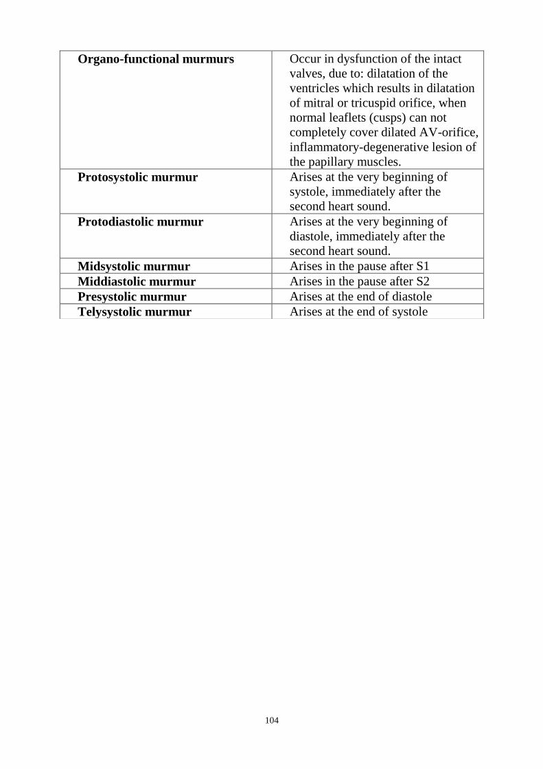
104
Organo-functional murmurs Occur in dysfunction of the intact
valves, due to: dilatation of the
ventricles which results in dilatation
of mitral or tricuspid orifice, when
normal leaflets (cusps) can not
completely cover dilated AV-orifice,
inflammatory-degenerative lesion of
the papillary muscles.
Protosystolic murmur Arises at the very beginning of
systole, immediately after the
second heart sound.
Protodiastolic murmur Arises at the very beginning of
diastole, immediately after the
second heart sound.
Midsystolic murmur Arises in the pause after S1
Middiastolic murmur Arises in the pause after S2
Presystolic murmur Arises at the end of diastole
Telysystolic murmur Arises at the end of systole

105
Pericardial friction murmur
Are connected with the changes in the
visceral and parietal pericardial layers
in which fibrin is deposited (in
pericarditis), cancer nodes develop,
etc. The mechanism by which pericar-
dial friction sounds are generated is
similar to that of the pleural friction
sounds, except that they depend not on
the respiratory movements but on the
movements of the heart during systole
and diastole. It resembles the crisping
sounds of snow, and sometimes they
are very soft, as if produced by rattling
of paper or scratching. The following
signs can be used to differentiate
pericardial friction sounds from
intracardiac sounds: (1) there is no
complete synchronism of pericardial
friction sounds with systole and
diastole; friction sounds are often
continuous, their intensity increasing
during systole or diastole; (2) friction
sounds can be heard for short periods
during various phases of the heart
work, either during systole or during
diastole; (3) pericardial friction sounds
are not permanent and can reappear at
intervals; (4) friction sounds are heard
at sites other than the best auscultative
points; they are best heard in the areas
of absolute cardiac dullness, at the
heart base, at the left edge of the
sternum in the 3rd and 4th intercostal
spaces; their localization is inconstant
and migrates even during the course of
one day; (5) friction sounds are very
poorly transmitted from the site of
their generation; (6) the sounds are
heard nearer the examiner's ear than
endocardial murmurs; (7) friction
sounds are intensified if the
stethoscope is pressed tighter to the
chest and when the patient leans for-
ward, because the pericardium layers
come in closer contact with one
another.
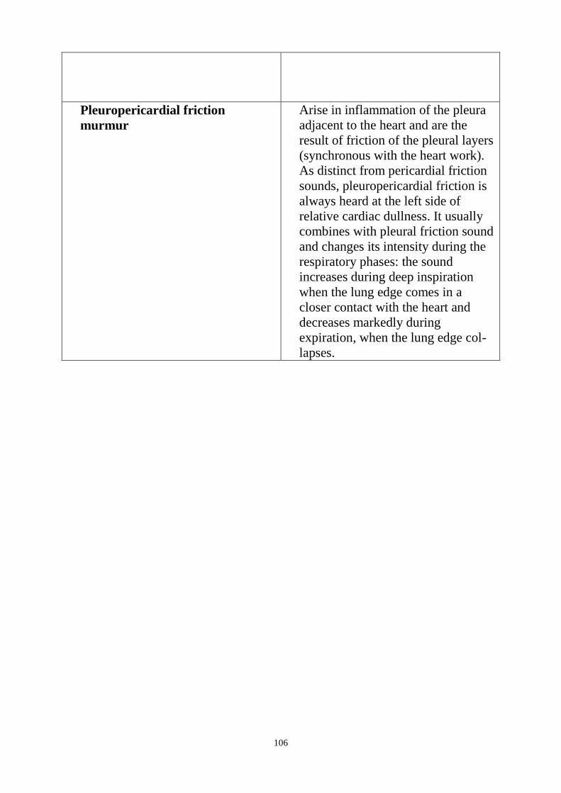
106
Pleuropericardial friction
murmur
Arise in inflammation of the pleura
adjacent to the heart and are the
result of friction of the pleural layers
(synchronous with the heart work).
As distinct from pericardial friction
sounds, pleuropericardial friction is
always heard at the left side of
relative cardiac dullness. It usually
combines with pleural friction sound
and changes its intensity during the
respiratory phases: the sound
increases during deep inspiration
when the lung edge comes in a
closer contact with the heart and
decreases markedly during
expiration, when the lung edge col-
lapses.
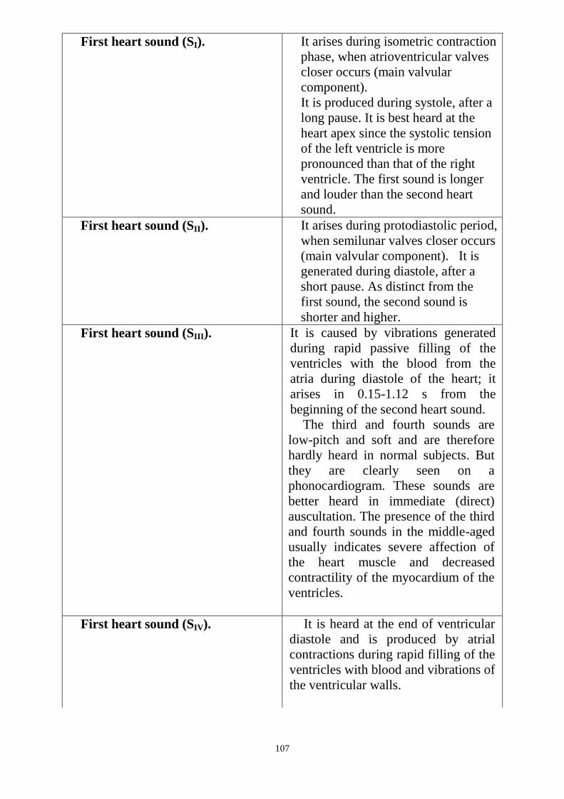
107
First heart sound (SI). It arises during isometric contraction
phase, when atrioventricular valves
closer occurs (main valvular
component).
It is produced during systole, after a
long pause. It is best heard at the
heart apex since the systolic tension
of the left ventricle is more
pronounced than that of the right
ventricle. The first sound is longer
and louder than the second heart
sound.
First heart sound (SII). It arises during protodiastolic period,
when semilunar valves closer occurs
(main valvular component). It is
generated during diastole, after a
short pause. As distinct from the
first sound, the second sound is
shorter and higher.
First heart sound (SIII). It is caused by vibrations generated
during rapid passive filling of the
ventricles with the blood from the
atria during diastole of the heart; it
arises in 0.15-1.12 s from the
beginning of the second heart sound.
The third and fourth sounds are
low-pitch and soft and are therefore
hardly heard in normal subjects. But
they are clearly seen on a
phonocardiogram. These sounds are
better heard in immediate (direct)
auscultation. The presence of the third
and fourth sounds in the middle-aged
usually indicates severe affection of
the heart muscle and decreased
contractility of the myocardium of the
ventricles.
First heart sound (SIV). It is heard at the end of ventricular
diastole and is produced by atrial
contractions during rapid filling of the
ventricles with blood and vibrations of
the ventricular walls.
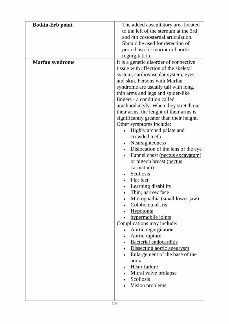
108
Botkin-Erb point The added auscultatory area located
to the left of the sternum at the 3rd
and 4th costosternal articulation.
Should be used for detection of
protodiastolic murmur of aortic
regurgitation.
Marfan syndrome It is a genetic disorder of connective
tissue with affection of the skeletal
system, cardiovascular system, eyes,
and skin. Persons with Marfan
syndrome are usually tall with long,
thin arms and legs and spider-like
fingers - a condition called
arachnodactyly. When they stretch out
their arms, the lenght of their arms is
significantly greater than their height.
Other symptoms include:
Highly arched palate and
crowded teeth
Nearsightedness
Dislocation of the lens of the eye
Funnel chest (pectus excavatum)
or pigeon breast (pectus
carinatum)
Scoliosis
Flat feet
Learning disability
Thin, narrow face
Micrognathia (small lower jaw)
Coloboma of iris
Hypotonia
hypermobile joints
Complications may include:
Aortic regurgitation
Aortic rupture
Bacterial endocarditis
Dissecting aortic aneurysm
Enlargement of the base of the
aorta
Heart failure
Mitral valve prolapse
Scoliosis
Vision problems
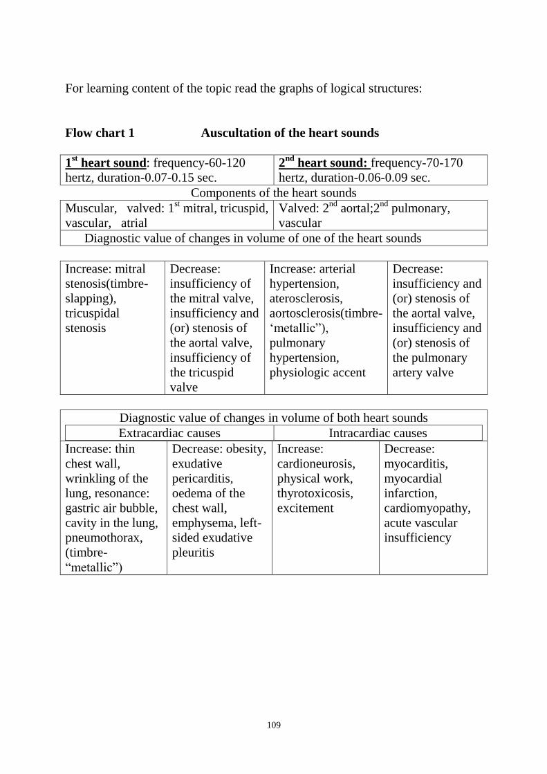
109
For learning content of the topic read the graphs of logical structures:
Flow chart 1 Auscultation of the heart sounds
1st heart sound: frequency-60-120
hertz, duration-0.07-0.15 sec.
2nd
heart sound: frequency-70-170
hertz, duration-0.06-0.09 sec.
Components of the heart sounds
Muscular, valved: 1st mitral, tricuspid,
vascular, atrial
Valved: 2nd
aortal;2nd
pulmonary,
vascular
Diagnostic value of changes in volume of one of the heart sounds
Increase: mitral
stenosis(timbre-
slapping),
tricuspidal
stenosis
Decrease:
insufficiency of
the mitral valve,
insufficiency and
(or) stenosis of
the aortal valve,
insufficiency of
the tricuspid
valve
Increase: arterial
hypertension,
aterosclerosis,
aortosclerosis(timbre-
‗metallic‖),
pulmonary
hypertension,
physiologic accent
Decrease:
insufficiency and
(or) stenosis of
the aortal valve,
insufficiency and
(or) stenosis of
the pulmonary
artery valve
Diagnostic value of changes in volume of both heart sounds
Extracardiac causes Intracardiac causes
Increase: thin
chest wall,
wrinkling of the
lung, resonance:
gastric air bubble,
cavity in the lung,
pneumothorax,
(timbre-
―metallic‖)
Decrease: obesity,
exudative
pericarditis,
oedema of the
chest wall,
emphysema, left-
sided exudative
pleuritis
Increase:
cardioneurosis,
physical work,
thyrotoxicosis,
excitement
Decrease:
myocarditis,
myocardial
infarction,
cardiomyopathy,
acute vascular
insufficiency

110
Flow chart 2 Changes of the heart sounds volume
Reduction on the heart apex
Aortic regurgitation
Aortic stenosis
Strengthening on the heart apex
Mitral stenosis
II sound
Reduction on an aorta
Aortic stenosis
Strengthening
On pulmonary artery On an aorta
Atherosclerosis,
aortosclerosis
Pulmonary
hypertension
Physiological
accent
Aortic regurgitation
Mitral
regurgitation
Arterial
hypertension
I sound
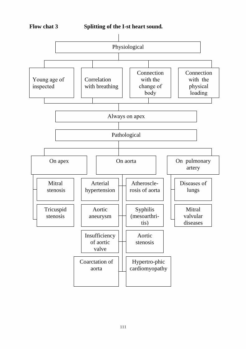
111
Flow chat 3 Splitting of the I-st heart sound.
Always on apex
Pathological
On aorta On pulmonary
artery
Mitral
stenosis
Tricuspid
stenosis
Arterial
hypertension
Atheroscle-
rosis of aorta
Aortic
aneurysm
Syphilis
(mesoarthri-
tis)
Insufficiency
of aortic
valve
Aortic
stenosis
Coarctation of
aorta
Hypertro-phic
cardiomyopathy
Diseases of
lungs
Mitral
valvular
diseases
Physiological
Young age of
inspected
Correlation
with breathing
Connection
with the
change of
body
position
Connection
with the
physical
loading
On apex

112
Flow chat 4 Gallop rhythm
Protodiastolic gallop rhythm Presystolic gallop rhythm
Audible increased IV sound in
telediastole (presystole)
Sharp abatement of
myocardium heart sound
Sharp abatement of
myocardium heart
sound
Strengthening of
auricles reduction
(abatement of
ventricles function)
Cadence ta-TAM-ta
Auscultate in IV -
V intercostals
spaces at the left
edge of
breastbone (at
inhalation)
Prognosis is better than at
the protodiastolical gallop
rhythm
Right ventricular
Arterial hypertension
Myocarditis, cardio-
sclerosis
Aortic heart
deseases
Cardiac
infarction
Myocardi-
tis, cardio-
sclerosis
Arterial
hyperten-
sion
Heart
deseases
Congenital
heart
deseases
Pulmonary
heart
Frequent trinomial rhythm
Audible increased III sound in
ptotodiastole
Cadence TAM-ta-ta
Auscultate on
apex (at
exhalation on left
flank)
Left ventricular

113
Flow Chart 5 Cardiac Murmurs
Intracardiac murmurs
Turbulent (Vortex,
Eddy)
Tissural
Cardiac murmurs
provoking factors
Strength of the
heart contraction
(Velocity of the
bloodflow)
Heart sound of the
myocardium, papillary
and annular muscles
Degree of hydraemia
(haemoglobin and RBC)
content)
Blood viscosity Diameter of
the vessels
(narrowing
degree)
Intracardiac murmurs character
Organic murmurs Organo-functional murmurs
Functional murmurs
Extracardiac murmurs
Pericardial
friction rub
Cardio-pulmonary
murmur
Pleuro-pericardial friction rub
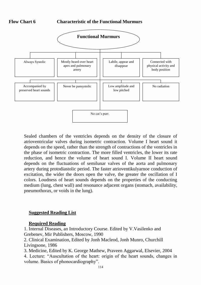
114
Flow Chart 6 Characteristic of the Functional Murmurs
Sealed chambers of the ventricles depends on the density of the closure of
atrioventricular valves during isometric contraction. Volume I heart sound it
depends on the speed, rather than the strength of contractions of the ventricles in
the phase of isometric contraction. The more filled ventricles, the lower its rate
reduction, and hence the volume of heart sound I. Volume II heart sound
depends on the fluctuations of semilunar valves of the aorta and pulmonary
artery during protodiastolic period. The faster atrioventikulyarnoe conduction of
excitation, the wider the doors open the valve, the greater the oscillation of I
colors. Loudness of heart sounds depends on the properties of the conducting
medium (lung, chest wall) and resonance adjacent organs (stomach, availability,
pneumothorax, or voids in the lung).
Suggested Reading List
Required Reading
1. Internal Diseases, an Introductory Course. Edited by V.Vasilenko and
Grebenev, Mir Publishers, Moscow, 1990
2. Clinical Examination, Edited by Jonh Macleod, Jonh Munro, Churchill
Livingsone, 1986
3. Medicine, Edited by K. George Mathew, Praveen Aggarwal, Elsevier, 2004
4. Lecture: ―Auscultation of the heart: origin of the heart sounds, changes in
volume. Basics of phonocardiography‖.
Functional Murmurs
Always Systolic Mostly heard over heart
apex and pulmonary
artery
Labile, appear and
disappear
Connected with
physical activity and
body position
Accompanied by
preserved heart sounds
Never be pansystolic Low amplitude and
low pitched
No radiation
No cat‘s purr.

115
5. Methodical guideline for students
6. Flow charts for practice: ―Auscultation of the heart: the heart sounds‖.
7. Website of department: www.cardiology.dsmu.edu.ua
Additional Reading
1. Textbook of Physical Diagnosis. History and Examination / Mark H. Swartz
2002, W. B. Saunders Company Pennsylvania
2. History and Physical Examination . Current Clinical Strategies, Edited Paul
P. Chan, Peter, J. Winkle, 2005
4. Davidson‘s Medicine, Nicholas A. Boon, Nicki R. Colledge, Davidson,
2006
Estimated base actions.
1. Algorithm actions at listening heart heart sounds
Step1. Check valves in projection chest wall and place of their hearing.
Step2. Spend consistently auscultation of the heart: the top (mitral valve), aortic,
pulmonary, three-leaved, cutting heart sound of I II, estimate the volume of
normal heart sounds depending on the zone listening, heard Create the image in
a scheme PCG.
Step3. Independently conduct in 1.2 STEPS selected patients, evaluate the
changes in volume and timbre heard heart sounds.
Step 4. Indicate the diagnostic value of the detected changes.
Algorithm actions during auscultation of patients
Step1. Under the guidance of a teacher in the chamber in young patients without
cardiovascular pathology of conduct auscultation of the heart. Emphasizing
heart sound I and II, the best places in auscultation, make them "in one
component, uniformity.
Create the image in the form of the scheme heard PCG:
Approximate base action
The algorithm acts when listening heart sounds
STEP 1. Mark projection valves on the chest wall and place of listening.
STEP 2. Spend consistently auscultation of the heart: the top (mitral valve),
aortic, pulmonary, tricuspid, the difference of heart sound I II, estimate the
volume of normal colors, depending on the areas of listening, heard in the form
of sketch schemes PCG.
STEP 3. Self spend STEPS 1,2 have selected patients, evaluate the changes the
volume and timbre listening to heart sounds.
STEP 4. Check the diagnostic value of the identified changes.
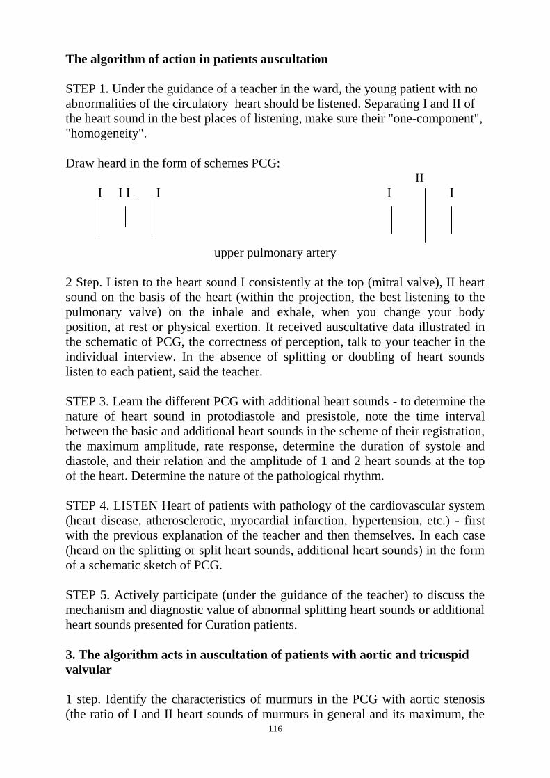
116
The algorithm of action in patients auscultation
STEP 1. Under the guidance of a teacher in the ward, the young patient with no
abnormalities of the circulatory heart should be listened. Separating I and II of
the heart sound in the best places of listening, make sure their "one-component",
"homogeneity".
Draw heard in the form of schemes PCG:
ΙΙ
Ι Ι Ι Ι Ι Ι
upper pulmonary artery
2 Step. Listen to the heart sound I consistently at the top (mitral valve), II heart
sound on the basis of the heart (within the projection, the best listening to the
pulmonary valve) on the inhale and exhale, when you change your body
position, at rest or physical exertion. It received auscultative data illustrated in
the schematic of PCG, the correctness of perception, talk to your teacher in the
individual interview. In the absence of splitting or doubling of heart sounds
listen to each patient, said the teacher.
STEP 3. Learn the different PCG with additional heart sounds - to determine the
nature of heart sound in protodiastole and presistole, note the time interval
between the basic and additional heart sounds in the scheme of their registration,
the maximum amplitude, rate response, determine the duration of systole and
diastole, and their relation and the amplitude of 1 and 2 heart sounds at the top
of the heart. Determine the nature of the pathological rhythm.
STEP 4. LISTEN Heart of patients with pathology of the cardiovascular system
(heart disease, atherosclerotic, myocardial infarction, hypertension, etc.) - first
with the previous explanation of the teacher and then themselves. In each case
(heard on the splitting or split heart sounds, additional heart sounds) in the form
of a schematic sketch of PCG.
STEP 5. Actively participate (under the guidance of the teacher) to discuss the
mechanism and diagnostic value of abnormal splitting heart sounds or additional
heart sounds presented for Curation patients.
3. The algorithm acts in auscultation of patients with aortic and tricuspid
valvular
1 step. Identify the characteristics of murmurs in the PCG with aortic stenosis
(the ratio of I and II heart sounds of murmurs in general and its maximum, the

117
shape), aortic disease (related to the II heart sound, shape, duration), tricuspid
stenosis (the ratio of I hue (presystolic) to II heart sound (middiastolic), shape,
change in amplitude and duration during inspiration), tricuspid insufficiency (the
ratio of murmurs to the heart sound I, II change in the amplitude of heart sound
and inspiratory).
2 Step. Auscultation of patients with aortic stenosis: Pay attention to the
existence of the interval between I heart sound and murmurs, the epicenter,
issued by the holding, the timbre of the murmurs.
STEP 3. Auscultation of patients with aortic insufficiency: Pay attention to the
relationship of murmurs with heart sound II, the epicenter (the aorta, the point
Botkin), directed the holding of the left (top, axilliary region). Listen to the
patient in standing and lying down.
STEP 4. Auscultation of patients with tricuspid stenosis: note the presence of
murmurs before I voice in the zone listening to the tricuspid valve, the murmurs
from the interval after the heart sound II, the dependence of their amplitude and
duration of breath - a symptom River Korvalo), position on the right side.
STEP 5. Auscultation of patients with tricuspid insufficiency: in the zone of the
tricuspid valve, pay attention to the relationship of murmurs with heart sound,
increase its volume to the II-th heart sound of the inspiratory (pp. River
Korvalo) in position on the right side, on the direction of the (left, his sharp
attenuation outside of cardiac dullness).
4. The algorithm of action in the investigation of patients with the presence
of functional, organic-functional and extracardiac murmurs.
1 step. Listen to 2-3 selected patients with functional systolic murmurss on top
of the heart, the pulmonary artery: Define the epicenter of the murmurs, I link it
with the heart sound, timbre. Match the features of the functional murmurs with
organic in another patient.
2 Step. Listen to the patient with a relative insufficiency of the tricuspid valve:
define the nature of phase murmurs, the epicenter, the ratio of I pitch, duration,
changes during inspiration; analyze the PCG of the patient.
STEP 3. Listen to the patient with fibrinous pericarditis: define the nature of
murmurs, listening to his band, the ratio of the phases of the cardiac cycle,
changes during inspiration, expiration, pressing a stethoscope; analyze the PCG
of the patient.
STEP 4. Explain the diagnostic value of these murmurss in the studied patients.
118
5. Hypertensive crisis - high blood pressure to high levels individually, which is
accompanied by the deterioration of the patient and may be complicated
cerebrovascular and coronary circulation.
Algorithm for action in hypertensive crisis
STEP 1 - immediately call a doctor
STEP 2 - to ensure complete relaxation to the patient
STEP 3 - to ensure access to fresh air
STEP 4 - use of hypertensive medications prescribed by a doctor.
Sudden circulatory arrest - the sudden disappearance of the cardiac activity due
to asystole or fibrilation ventricles. It is characteristic signs of clinical death:
loss of consciousness, lack of response to external stimuli, the lack of
photoreaction, respiratory arrest (or single-respiratory movements), lack of pulse
in the radial and carotid arteries, acrocyanosis and marble pallor leather covers.
The algorithm acts in a sudden stop of blood circulation
STEP 1 - Carefully put the patient on a flat and firm surface (eg on the floor.)
STEP 2 - to 2.1 precardiac blows to the sternum (beats the average force applied
fist from a distance of 25-30 cm).
STEP 3 - check and ensure the airway
STEP 4 - to start artificial ventilation of lungs
STEP 5 - start of indirect cardiac massage.
6. Loss of consciousness - (Greek synkope; Syncope) - brief loss of
consciousness caused by insufficient blood circulation of the brain, appeared
suddenly.
Usually occurs when significant neuropsychiatric effects (Fear, severe pain, the
sight of blood), in the stuffy room, with extreme fatigue. Loss of consciousness
is often preceded by tinnitus, vertigo, dimness of the eyes, etc. In case of loss of
consciousness (as distinct from an epileptic seizure), there is no uncontrolled
urination, stops breathing, biting tongue. Loss of consciousness usually occurs
in the upright position, lasts 20-30 seconds, after which the patient regains
consciousness.
7. Sudden circulatory arrest - the sudden disappearance of the cardiac activity
due to asystole or fibrilyatsii ventricles. It is characteristic signs of clinical
death: loss of consciousness, lack of response to external stimuli, the lack of
photoreaction, respiratory arrest (or single-respiratory movements), lack of pulse
in the radial and carotid arteries, acrocyanosis and marble pallor leather covers.
The algorithm acts in a sudden stop of blood circulation
STEP 1 - Carefully put the patient on a flat and firm surface (eg on the floor.)
STEP 2 - to 2.1 precardiac blows to the sternum (beats the average force applied
fist from a distance of 25-30 cm).
STEP 3 - check and ensure the airway
119
STEP 4 - to start artificial ventilation of lungs
STEP 5 - start of indirect cardiac massage.
7. Loss of consciousness - (Greek synkope; Syncope) - brief loss of
consciousness caused by insufficient blood circulation of the brain, appeared
suddenly.
Usually occurs when significant neuropsychiatric effects
(Fear, severe pain, the sight of blood), in the stuffy room, with extreme fatigue.
Loss of consciousness is often preceded by tinnitus, vertigo, dimness of the
eyes, etc. In case of loss of consciousness (as distinct from an epileptic seizure),
there is no uncontrolled urination, stops breathing, biting tongue. Loss of
consciousness usually occurs in the upright position, lasts 20-30 seconds, after
which the patient regains consciousness.
Case problem.
Task 1.
Patient N., aged 30, the boundaries of the heart have not changed, the apex of
the heart is auscultated two heart sounds: one more loud, ends a long pause and
start small, with a quieter heart sound comes at the end of a short pause. Assess
a louder heart sound.
8. Faint - (Greek synkope; syncope camp) - korotkochasova vtrata
consciousness, scho zumovlena failure krovozabezpechennya cerebral mozku
yak vinikla raptovo.
Zazvichay occurs valued at nervovo-mental vplivah (perelyak, strong pain, view
the blood), a stuffy room at fatigue. Loss of consciousness often pereduyut
murmurs in vuhah, dizziness, darkening at the sight of that other. When fainting
(at Unlike epileptic attack) is not going nekontrolovanogo urination, zupinki
dihannya, prikupuvannya Yazikov. Fainting occurs zazvichay a vertical
position, lasts 20-30 seconds, after chogo ailments come up tical.
Algorithm in Action of Faint:
STEP 1 - give a patient ka horizontal position with raised several feet.
STEP 2 - to free the patient from lasting wear.
STEP 3 - to ensure access of fresh air
STEP 4 - Grind the temple and chest of the patient
STEP 5 - pobryzkaty face with cold water
STEP 6 - to smell the patient vatku with liquid ammonia.
9. Collapse (Latin collapsus) - clinical manifestation of acute vascular
insufficiency of the fall of vascular heart sound, decrease myocardial contractile
function, reduction of bcc and fall of AT. It can occur in acute blood loss,
myocardial infarction znevodzhuvanni, poisonings, hypotensive drugs overdose.
120
The clinical picture includes skin pallor, cold sweat tacky, low blood pressure,
rapid pulse, low content. Consciousness can be lost and saved (with the patient
usually retarded) advanced pupil.
Action to collapse algorithm:
Step 1 - remove the cause if possible (eg to stop bleeding).
STEP 2 - horizontal position to provide patient with multiple head lowered.
STEP 3 - to free the patient from lasting wear.
STEP 4 - To provide access fresh air
STEP 5 - for the doctor to enter medication to increase the bcc and / or drugs
that increase vascular heart sound.
Case assignment.
Task 1.
Patient N., aged 30, the boundaries of the heart have not changed, the apex of
the heart vislushivaetsya two-heart sound: The first of these, more than a loud,
ends a long pause, and starts to short, with a quieter heart sound is at the end a
short pause. Enjoy a louder heart sound.
A. I heart sound is the norm.
B. I heart sound this pathology.
S. II heart sound, is the norm.
D. II heart sound, this pathology.
Task 2.
In the recruit, D., 18 years, the boundaries of the heart have not changed,
pathology of the respiratory system is not on the pulmonary artery be heard two
heart sounds: one more loud and occurs at the end of a short pause, and here it is
much louder (on inspiration) than in the aorta ; more quiet heart sound on both
vessels are at the end of a long pause.
Rate II heart sound on the pulmonary artery.
A. Over a loud and a pathology - Accent II heart sound.
B. A loud and this is the norm - a physiological stress II heart sound.
C. A quiet and is the norm.
D. A quiet and a weakening of the P heart sound.
Task 3.
The patient K., 20 years of cardiac-type neurocirculatory dystonia, tachycardia
120 in 1 min. At the apex of the heart is auscultated two heart sounds: one
louder, the other quiet. Since both pause practically the same, on this basis to
determine which of these I-II-nd and the second heart sound is impossible. A
loud heart sound coincides with the apical impulse. Which of these I-th and n-th
heart sound? What it is: a rule or pathology?
A. Over a loud heart sound and the I is the norm.
B. A loud heart sound of I and this pathology (gain).
C. More than a quiet heart sound and the I is the norm.
D. A quiet heart sound II and this pathology (weakening).
E. More than a loud heart sound and II is the norm.
Task 4.
In a patient st., 20 - years in the pulmonary artery listened two heart sounds, of
121
which the one that occurs at the end of a long pause, - bifurcated, with the
second half of his loudest first.
Which component heart sound is late?
A. triple-component 1 - the first pitch
B. mitral component 1 - th heart sound
C. vascular component 1 - th heart sound
D. pulmonary component of the II colors
E. aortic component of the II colors
Task 5.
Physiological third heart sound be heard:
A. in diastole (between 2 nd and 1 st heart sound, but closer to the 1 st heart
sound);
B. in diastole (between 2 nd and 1 st heart sound, but closer to the 2 nd heart
sound);
C. in systole (between 1 st and 2 nd heart sound, but closer to the 1 st heart
sound);
D. in systole (between 1 st and 2 nd heart sound, but closer to the 2 nd heart
sound);
E. in systole (between 1 st and 2 nd heart sound, at an equal distance between
the heart sounds).
Task 6.
F. The patient, 20 years of heart disease - mitral insufficiency.
At the top, in the armpit to the left, along the left edge of the sternum be heard
the murmurs.
What are the characteristics of murmurs in the mitral valve insufficiency?
Specify characteristics of the murmurs.
A. systolic murmur, a place of education - the left ventricle
B. systolic murmur, a place of education - the left atrium
C. place the best possible listening - intercostal space left of the sternum
D. best be heard in an upright position
E. direction of - on the neck vessels
A: Objective 1. A; Task 2. B; Objective 3.A; Task 4. D; Objective 5.A; Task 6.
A
Challenges to verify the achievement of specific learning objectives.
Task 1.
The patient was examined by a dentist suddenly became pale and lost
consciousness.
What position should provide the patient?
A. Vertical.
B. sat.
C. half-sitting.
D. Lying with lowered legs.
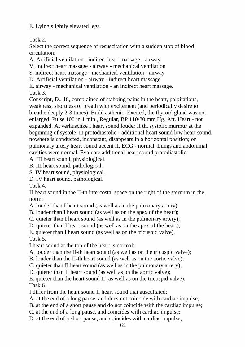
122
E. Lying slightly elevated legs.
Task 2.
Select the correct sequence of resuscitation with a sudden stop of blood
circulation:
A. Artificial ventilation - indirect heart massage - airway
V. indirect heart massage - airway - mechanical ventilation
S. indirect heart massage - mechanical ventilation - airway
D. Artificial ventilation - airway - indirect heart massage
E. airway - mechanical ventilation - an indirect heart massage.
Task 3.
Conscript, D., 18, complained of stabbing pains in the heart, palpitations,
weakness, shortness of breath with excitement (and periodically desire to
breathe deeply 2-3 times). Build asthenic. Excited, the thyroid gland was not
enlarged. Pulse 100 in 1 min., Regular, BP 110/80 mm Hg. Art. Heart - not
expanded. At verhuschke I heart sound louder II th, systolic murmur at the
beginning of systole, in protodiastolic - additional heart sound low heart sound,
nowhere is conducted, inconstant, disappears in a horizontal position; on
pulmonary artery heart sound accent II. ECG - normal. Lungs and abdominal
cavities were normal. Evaluate additional heart sound protodiastolic.
A. III heart sound, physiological.
B. III heart sound, pathological.
S. IV heart sound, physiological.
D. IV heart sound, pathological.
Task 4.
II heart sound in the II-th intercostal space on the right of the sternum in the
norm:
A. louder than I heart sound (as well as in the pulmonary artery);
B. louder than I heart sound (as well as on the apex of the heart);
C. quieter than I heart sound (as well as in the pulmonary artery);
D. quieter than I heart sound (as well as on the apex of the heart);
E. quieter than I heart sound (as well as on the tricuspid valve).
Task 5.
I heart sound at the top of the heart is normal:
A. louder than the II-th heart sound (as well as on the tricuspid valve);
B. louder than the II-th heart sound (as well as on the aortic valve);
C. quieter than II heart sound (as well as in the pulmonary artery);
D. quieter than II heart sound (as well as on the aortic valve);
E. quieter than the heart sound II (as well as on the tricuspid valve);
Task 6.
I differ from the heart sound II heart sound that auscultated:
A. at the end of a long pause, and does not coincide with cardiac impulse;
B. at the end of a short pause and do not coincide with the cardiac impulse;
C. at the end of a long pause, and coincides with cardiac impulse;
D. at the end of a short pause, and coincides with cardiac impulse;
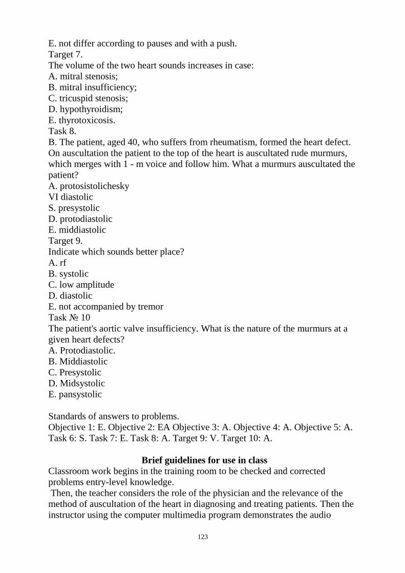
123
E. not differ according to pauses and with a push.
Target 7.
The volume of the two heart sounds increases in case:
A. mitral stenosis;
B. mitral insufficiency;
C. tricuspid stenosis;
D. hypothyroidism;
E. thyrotoxicosis.
Task 8.
B. The patient, aged 40, who suffers from rheumatism, formed the heart defect.
On auscultation the patient to the top of the heart is auscultated rude murmurs,
which merges with 1 - m voice and follow him. What a murmurs auscultated the
patient?
A. protosistolichesky
VI diastolic
S. presystolic
D. protodiastolic
E. middiastolic
Target 9.
Indicate which sounds better place?
A. rf
B. systolic
C. low amplitude
D. diastolic
E. not accompanied by tremor
Task № 10
The patient's aortic valve insufficiency. What is the nature of the murmurs at a
given heart defects?
A. Protodiastolic.
B. Middiastolic
C. Presystolic
D. Midsystolic
E. pansystolic
Standards of answers to problems.
Objective 1: E. Objective 2: EA Objective 3: A. Objective 4: A. Objective 5: A.
Task 6: S. Task 7: E. Task 8: A. Target 9: V. Target 10: A.
Brief guidelines for use in class
Classroom work begins in the training room to be checked and corrected
problems entry-level knowledge.
Then, the teacher considers the role of the physician and the relevance of the
method of auscultation of the heart in diagnosing and treating patients. Then the
instructor using the computer multimedia program demonstrates the audio

124
recording of normal and altered heart sounds with simultaneous images on the
screen.
Classes continued in the wards. Students under the guidance of the teacher
determine the patient's projection of the valves on the anterior chest, and place
them listening. Teacher shows students the method of auscultation, how
differences in heart heart sounds. Next to the House after a preliminary
explanation of the teacher, students listen to a few patients with normal and
altered heart sounds, murmurs. Then listen to each independently selected
patients, checking the correctness of determining the identified changes in
individual conversation with the teacher. Discussion of diagnostic value of the
identified changes were made in the training room.
Classes are continuing in the training room, where students discussed with the
algorithms of action in urgent situations. At the end of classes offered to solve
the control task on factual topics, summarizing the classroom.
Test control tasks
Task 1. The patient at the apex of the heart is auscultated two heart sounds: one
loud, another - a quiet, and loud heart sound comes after a short pause, but still -
long after that, specify colors in this case: A. I heart sound - a loud, II heart
sound - quiet, I>; II, then II heart sound is weak and this pathology.
B. II heart sound - a more quiet, I heart sound loud -, II <I, I mean the heart
sound is reinforced and this pathology.
C. I heart sound - a loud, II heart sound - quiet, I>; II, then II heart sound is
weak and the norm.
D. II heart sound - louder, I heart sound - a quiet, II> I, II means the heart sound
is reinforced and is the norm.
E. I heart sound - a quiet, II heart sound - loud, I <II, I denotes the heart sound
of relaxation and this pathology.
Task 2. The patient listened to the aorta, two heart sounds: one of them loudly,
another quiet, and loud heart sound comes after a long pause, but still - after a
brief that, specify colors in this case:
A. I heart sound - a loud, II heart sound - quiet, I> II, II mean the heart sound is
weak and this pathology.
B. II heart sound - louder, I heart sound - a quiet, II> I, II means the heart sound
is reinforced and this pathology.
C. I heart sound - a loud, II heart sound - quiet, I> II, II mean the heart sound is
weak and this is the norm.
D. II heart sound - louder, I heart sound - a quiet, II> I, II means the heart sound
is reinforced and is the norm.
E. I heart sound - a loud, II heart sound - quiet, I> II, I mean the heart sound is
reinforced and is the norm.
Objective 3. Physiological third heart sound be heard:
A. in diastole (between 2 nd and 1 st heart sound, but closer to the 1 st heart
sound;
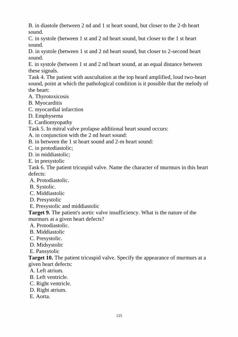
125
B. in diastole (between 2 nd and 1 st heart sound, but closer to the 2-th heart
sound.
C. in systole (between 1 st and 2 nd heart sound, but closer to the 1 st heart
sound.
D. in systole (between 1 st and 2 nd heart sound, but closer to 2-second heart
sound.
E. in systole (between 1 st and 2 nd heart sound, at an equal distance between
these signals.
Task 4. The patient with auscultation at the top heard amplified, loud two-heart
sound, point at which the pathological condition is it possible that the melody of
the heart:
A. Thyrotoxicosis
B. Myocarditis
C. myocardial infarction
D. Emphysema
E. Cardiomyopathy
Task 5. In mitral valve prolapse additional heart sound occurs:
A. in conjunction with the 2 nd heart sound:
B. in between the 1 st heart sound and 2-m heart sound:
C. in protodiastolic;
D. in middiastolic;
E. in presystolic
Task 6. The patient tricuspid valve. Name the character of murmurs in this heart
defects:
A. Protodiastolic.
B. Systolic.
C. Middiastolic
D. Presystolic
E. Presystolic and middiastolic
Target 9. The patient's aortic valve insufficiency. What is the nature of the
murmurs at a given heart defects?
A. Protodiastolic.
B. Middiastolic
C. Presystolic.
D. Midsystolic
E. Pansytolic
Target 10. The patient tricuspid valve. Specify the appearance of murmurs at a
given heart defects:
A. Left atrium.
B. Left ventricle.
C. Right ventricle.
D. Right atrium.
E. Aorta.
126
The Health Ministry Of Ukraine
Donetsk National Medical University
Approved
at the meeting of
Propedeutic and Internal Medicine
Department
Head of department
Associate Member of NAMSc of
Ukraine,
Professor G. A. Ignatenko
« _______ »_____________ 2011 р.
STUDENT’S SELF-STUDY GUIDELINES FOR
PRACTICE ACTIVITIES
Subject
Propedeutics of the Internal Medicine
Module 1
The Main Methods of Examination of
Patient on Internal Diseases Course
Subject Module 4
Methods of examination and main
symptoms and syndromes of the
alimentary and urinary system
disorders.
Practice
Methods of digestive system
examination. Basic syndromes.
Peculiarities of caring for patients
with pathology of alimentary system
and providing first aid.
Course
2
Faculty
Dentistry
Donetsk 2011
127
Importance of the Subject: competent in methodic and technique performance
of questioning of patient, inspection, percussion, auscultation and superficial
palpation of abdomen allows in many cases to diagnose pathological changes of
abdominal viscera, to analyze interconnection between results of questioning
and physical examination of digestive apparatus and identify basic syndromes of
its affection including emergency states. Nursing of patients with pathology of
digestive apparatus and rendering first aid.
Key Objective: to master questioning and physical examination of patients with
pathology of digestive apparatus. To note changes in patient‘s oral cavity at
diseases of digestive apparatus. To conduct auxiliary examination methods of
gastroenterology and use peculiarities of patients‘ nursing and rendering first
aid. To interpret basic syndromes and to give clinical assessment of findings.
Specific Goals:
1. To conduct methodically correct questioning of patient on digestive system,
to assess findings.
2. To make an inspection of oral cavity.
3. To make an inspection, percussion and auscultation of abdomen and assess
findings.
4. To make an inspection of abdomen, to determine clinical significance of
symptoms.
5. To conduct palpation of abdomen, to determine clinical significance of
symptoms.
6. To conduct palpation of the liver, to determine clinical significance of
symptoms.
7. During questioning of patient to pay attention to features of anamnesis and
physical examination at diseases of digestive apparatus and hepatobiliary system.
8. To determine guiding syndromes of digestive apparatus.
9. To be able to interpret laboratory findings at diseases of digestive apparatus.
10. To determine basic and additional methods of gastrointestinal tract
examination.
Level of Knowledge and Skills before the Practice:
1. Thorough inspection of oral cavity (anatomy department)
2. To delimit abdominal region, determine projection of anteroventral muscles
and venous collaterals (anatomy department).
3. To interpret relationship of percussion sound and physical properties of
fluctuating body (physics department).
Questions for Self-Assessment of the Pre-Practice Knowledge
Q1. A 47-year old patient complains of bloating pain in the right part of
abdomen with periodic cramp-like increase, nausea, vomiting. Appendectomy
complicated by circumscribed peritonitis has been made one year ago. During
inspection asymmetric abdominal distension was found. During palpation it was
128
found out that there is painfulness in the right part of abdomen, intestine is
increased in diameter and it‘s dense. Auscultation revealed absence of intestinal
murmurs. Which bowel segments are projected on the right half of iliac region?
A. Ascending segment of large intestine and blind gut.
B. Descending segment of large intestine.
C. Sigmoid colon.
D. Jejunum.
E. Transverse colon.
Q 2. Patient complains of extended moderate abdominal pain, feeling of swelling,
nausea. Which patient‘s complains may be a confirmation of meteorism?
A. Feeling of abdominal swelling.
B. Abdominal pain.
C. Nausea.
D. All of the mentioned complains.
E. None of the mentioned complains.
Q 3. During superficial palpation of patient‘s abdomen there is acute painfulness
in the right iliac region, abdominal wall is hard. After long light stroking and
changing of patient‘s attention fro conversation hardening disappears. Assess
findings of superficial palpation of the right iliac region:
A. Variant of norm.
B. There is resistance of abdominal wall.
C. There is muscular defense.
D. There is hypertrophy of abdominal wall‘s muscles.
E. There is pathological mass of abdominal wall.
Q 4 . 38-year old patient K. complains of intensive pain in epigastrium, feeling
of abdominal swelling, severe general weakness. Complains appeared suddenly.
It is known from the anamnesis that during 10 years patient has been suffering
from duodenal ulcer. On abdominal percussion the percussion sound is loud,
long, low-pitched and ringing throughout including liver region. Doctor
suspected perforation of ulcer into abdominal cavity and it was confirmed after
the further examination. What kind of percussion sound is determined
throughout the abdomen?
A. Dull.
B. Impaired.
C. Tympanic.
D. Impaired -tympanic.
E. Clear.
Q 5. 52-year old patient was admitted to the hospital with complains of the
feeling of heaviness in epigastric zone, low appetite, edema of low extremities,
loss of weight for 6 kg per year. During inspection of patient‘s abdomen
(normosthenic) it was found that: abdomen is expanded, protruding umbilicus,
129
on percussion there is dull sound from the umbilicus level up to the bottom.
Deep palpation of abdomen is complicated. Greater curvature of stomach was
found by auscultatory-affrictional method on 6sm higher than umbilicus.
Assess position of greater curvature of stomach.
A. Norm for asthenic.
B. Norm for normosthenic person.
C. Norm for hypersthenic person.
D. Elevated.
E. Descended.
Q 6. What quantity of human gastric juice is exuded during the day?
A. 1 liter
B. 2 - 2,5 liters
C. 3,5 - 4 liters
D. 0,5 - 1 liter
E. 3 liters
Q 7. What kind of cells produce hydrochloric acid in stomach?
A. Chief cells
B. Accessory cells
C. Goblet cells
D. Accessory cells
E. Columnar cell
Q 8. Select sign of gallbladder inflammation:
A. Presence of leukocytes during the 1st
phase
B. Presence of leukocytes during the 2nd
phase
C. Increase of leukocytes in portion A
D. Increase of leukocytes in portion В
E. Increase of leukocytes in portion С
Q 9. Hippocratic face is typical for:
A. Renal colic
B. Inflammation of parietal peritoneum at appendicitis
C. Meteorism
D. Hyposecretory syndrome
E. Hepatic colic
Q 10. Increase of unconjugated bilirubin is observed at:
A. Pernicious anemia
B. Obstructive jaundice
C. Asiderotic anemia
D. Acholuric jaundice
E. Posthemorrhagic anemia

130
Key answers: 1- А; 2- А; 3- B; 4- С; 5- Е; 6- В; 7- В; 8- Д; 9- В; 10- Д.
Contents of Practice
Topics of the Theory:
1. General rules, technique of patient‘s questioning, specificity of complaints of
patients with digestive apparatus pathology.
2. Scheme of abdominal wall regions.
3. Topographical-anatomical features of organs‘ position in the abdominal
cavity
4. Anatomical features of organs‘ structure.
5. Principles, technique of oral cavity examination
6. Percussion of abdomen, physical bases of percussion.
7. Diagnostic value of findings of oral cavity examination.
8. General rules, technique, stages of superficial and profound palpation,
diagnostic value of findings.
9. Guiding syndromes of digestive organs and hepatobiliary system.
10. Diagnostic value of findings received by basic and additional laboratory
methods.
Practical skills: Students should be able to demonstrate mastery of the following practical skills
1. To conduct questioning of patient with pathology of digestive organs.
2. To assess findings of oral cavity examination.
3. To assess form and size of abdomen.
4. To conduct abdominal percussion.
5. To perform auscultation of abdomen.
6. To fulfill superficial palpation of abdomen.
7. To perform palpation of digestive organs according to Obraztsov-Strazhesko
method.
a) To determine normal parameters of sigmoid colon.
b) To determine normal parameters of blind gut.
c) To determine normal parameters of ileum terminal portion.
d) To determine normal parameters of ascending and descending colon.
e) To determine normal parameters of transverse colon.
8. To master methodic of gastric juice extraction by factional method, to assess
state of secretory function, to determine and assess hourly tension of basal and
stimulated secretion (mg/L), state of evacuation function at ―residue‖ of contents
in 25 minutes after test meal.
9. To master methodic of duodenal intubation, macro- and microscopic
investigation of A, B and C bile and diagnostic value of changes..
10. To master indications and methodic of fiberoptic colonoscopy and
roentgenological methods of gastrointestinal tract examination.
11. To master methodic of stool investigation and to be able to assess findings.

131
12. To be able to use clinical, instrumental and laboratory methods of
examination for detection of syndromes of gastrointestinal tract pathology.
13. To assess diagnostic value of symptoms.
Required Glossary to Practice
Term Definition
Glossitis It‘s superficial tongue inflammation that can be found
in many infectious and noninfectious diseases.
Parodontosis Inflammation of dentogingival recess and denudation
dental cervix
Anorexia Total loss of apetite.
Heartburn Feeling of heat and burning in epigastric and
retrosternal region.
Cachexia Abrupt loss of weight as a result of which
subcutaneous layer of fat disappears and muscles
atrophy.
Distension pain Pain in consequence of distension of intestine by gases
and tension of mesentery. Pain has long term character
and accurate localization.
„ Medusa head ‖ Dilated swollen venous collaterals are located near
umbilicus and radiating from it.
Epigastrium The upper region of abdomen limited by line that
connects the lower edges of both tenth ribs.
Bimanual palpation Palpation by both hands. It‘s used in examination of
ascending and descending colon, kidneys, liver and
spleen
Penetrating palpation It is a kind of profound palpation when the tip of two -
three vertically positioned fingers gradually but strongly
press on the restricted space.
Ascites Accumulation of fluid in the abdominal cavity
Jaundice Staining of skin and visible mucous membranes in
yellow color.
Dysphagy declares itself by complication or impossibility of
swallowing, pains in the moment of swallowing,
ingress of food or liquid into nose, larynx, trachea.
Dyspeptic signs Are typical for nausea, vomiting, constipation and
diarrhea
Meteorism declares itself by abdominal distension, distensing pain
in abdomen; intensive (explosive) discharge of great
amount of flatus is possible.
Telangiectasia venous spiders, venous lakes – it is dilatation of minute
blood vessels (capillaries) that accumulate and appear
132
on the skin.
Alkaline phosphatase
(AP)
Alkaline phosphatase, 1.0-3.0 mmole/L. Index of
cholestasis
GGT Gamma Glutamyl Transpeptidase 0,6-3.9 mmole/L.
Index of cholestasis
Child-Pugh score This score is suggested for assessment of liver cirrhosis
severity. It is based on data of biochemical investigations
(bilirubin, albumin, prothrombin) and clinical signs –
ascitis and encephalopathy.
Hepatargia (hepatargia; hepat- + Greek. argia inactivity; syn.:
hepatargy, hepatic encephalopathy, portal systemic
encephalopathy) – clinical syndrome that develops at
severe hepatic failure or hepatic intoxication and
declares itself in neuropsychic disorders, appearance of
fetor hepaticus, possible development of hepatic coma.
Cytolysis Insult to hepatocytes by virus, toxic agent etc.
Analogous activity at chronic hepatitis and liver
cirrhosis. Declares itself by jaundice, dark urine, light-
colored stool. Transaminase increases more than 2-5
times.
Cholestasis Intrahepatic, develops as a result of impairments of
biliary excretion in bile capillaries. It is observed at
cholestatic hepatitis, biliary liver cirrhosis.
Portal hypertension Impairment of venous outflow from unpaired organs of
abdominal cavity as a result of complication of blood
flow on portal vein, through the cirrotic liver.
Small liver signs Venous spiders, angiomas on chest, back, legs, palmar
and plantar erythema, gynecomastia.
Hypersplenism Increase and distortion of spleen functions on removal
of destroyed blood corpuscles; it is usually associated
with hypersplenism. It is observed at liver cirrhosis with
portal hypertension.
HBsAg Superficial antigen of hepatitis B that appears in 4
weeks after infection by hepatitis B and disappears
after 3-6 months, may stay for years.
HBeAg Antigen of hepatitis B. Indicates viral reproduction.
Test of contagiousness.
Anti HBe Antibodies to antigen Е indicate excretion of virus
from the organism (absence of viral reproduction).
Anti HDV Antibodies to virus of hepatitis D are evidence of delta
infection
Syndrome of
malabsorption
disturbances of absorption and uptake of food.
Irritable colon Impairment of secretory and movement function of the
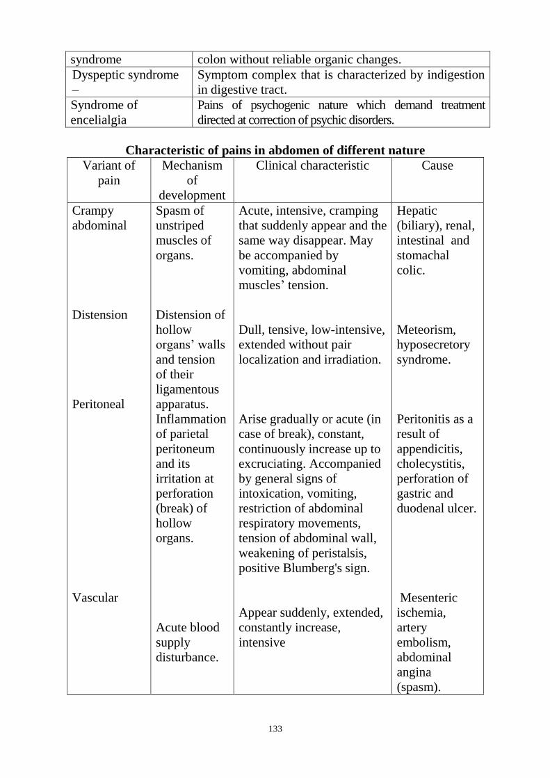
133
syndrome colon without reliable organic changes.
Dyspeptic syndrome
–
Symptom complex that is characterized by indigestion
in digestive tract.
Syndrome of
encelialgia
Pains of psychogenic nature which demand treatment
directed at correction of psychic disorders.
Characteristic of pains in abdomen of different nature
Variant of
pain
Mechanism
of
development
Clinical characteristic Cause
Crampy
abdominal
Distension
Peritoneal
Vascular
Spasm of
unstriped
muscles of
organs.
Distension of
hollow
organs‘ walls
and tension
of their
ligamentous
apparatus.
Inflammation
of parietal
peritoneum
and its
irritation at
perforation
(break) of
hollow
organs.
Acute blood
supply
disturbance.
Acute, intensive, cramping
that suddenly appear and the
same way disappear. May
be accompanied by
vomiting, abdominal
muscles‘ tension.
Dull, tensive, low-intensive,
extended without pair
localization and irradiation.
Arise gradually or acute (in
case of break), constant,
continuously increase up to
excruciating. Accompanied
by general signs of
intoxication, vomiting,
restriction of abdominal
respiratory movements,
tension of abdominal wall,
weakening of peristalsis,
positive Blumberg's sign.
Appear suddenly, extended,
constantly increase,
intensive
Hepatic
(biliary), renal,
intestinal and
stomachal
colic.
Meteorism,
hyposecretory
syndrome.
Peritonitis as a
result of
appendicitis,
cholecystitis,
perforation of
gastric and
duodenal ulcer.
Mesenteric
ischemia,
artery
embolism,
abdominal
angina
(spasm).
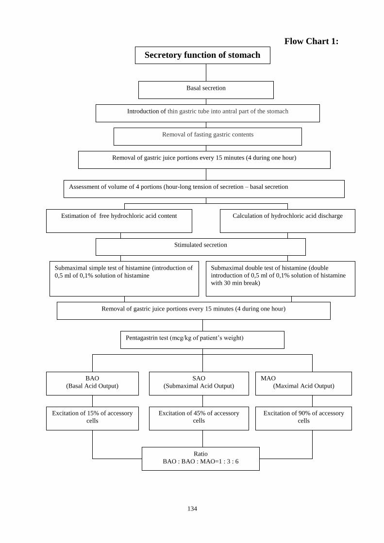
134
Flow Chart 1:
Secretory function of stomach
Basal secretion
Introduction of thin gastric tube into antral part of the stomach
Removal of fasting gastric contents
Removal of gastric juice portions every 15 minutes (4 during one hour)
Assessment of volume of 4 portions (hour-long tension of secretion – basal secretion
Estimation of free hydrochloric acid content Calculation of hydrochloric acid discharge
Stimulated secretion
Submaximal simple test of histamine (introduction of
0,5 ml of 0,1% solution of histamine
Submaximal double test of histamine (double
introduction of 0,5 ml of 0,1% solution of histamine
with 30 min break)
Removal of gastric juice portions every 15 minutes (4 during one hour)
Pentagastrin test (mcg/kg of patient‘s weight)
BAO
(Basal Acid Output)
SAO
(Submaximal Acid Output)
MAO
(Maximal Acid Output)
Excitation of 15% of accessory
cells
Excitation of 45% of accessory
cells
Excitation of 90% of accessory
cells
Ratio
BAO : BAO : MAO=1 : 3 : 6
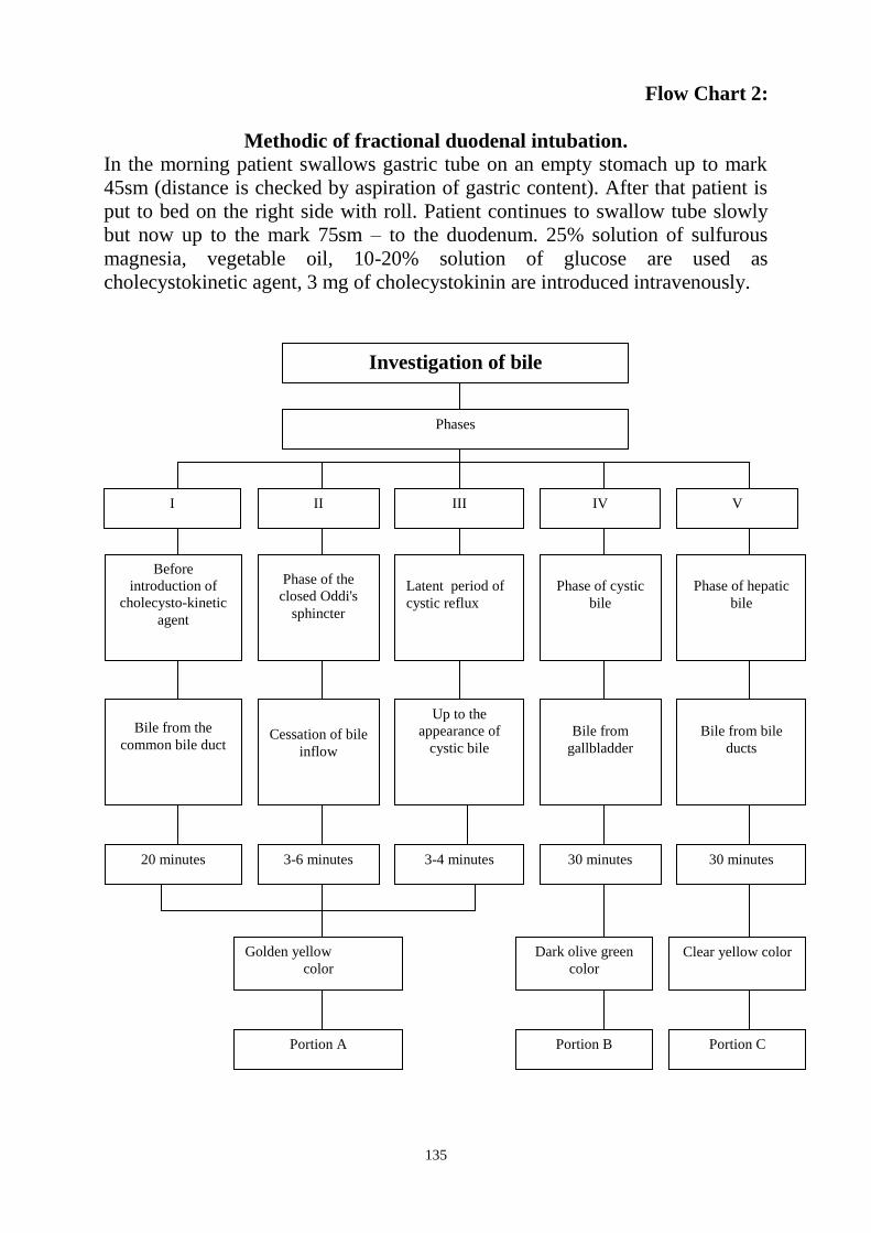
135
Flow Chart 2:
Methodic of fractional duodenal intubation.
In the morning patient swallows gastric tube on an empty stomach up to mark
45sm (distance is checked by aspiration of gastric content). After that patient is
put to bed on the right side with roll. Patient continues to swallow tube slowly
but now up to the mark 75sm – to the duodenum. 25% solution of sulfurous
magnesia, vegetable oil, 10-20% solution of glucose are used as
cholecystokinetic agent, 3 mg of cholecystokinin are introduced intravenously.
Investigation of bile
Phases
Before
introduction of
cholecysto-kinetic
agent
II III IV V
Phase of the
closed Oddi's
sphincter
Phase of hepatic
bile
Latent period of
cystic reflux
Phase of cystic
bile
Bile from the
common bile duct
Cessation of bile
inflow
Up to the
appearance of
cystic bile
Bile from
gallbladder
Bile from bile
ducts
20 minutes 3-6 minutes 3-4 minutes 30 minutes 30 minutes
Golden yellow
color
Portion A
Dark olive green
color
Clear yellow color
Portion B Portion C
I

136
Flow Chart 3:
Syndrome of jaundice
(Pathogenesis)
Jaundice
Obstructive
Hepatocellular
Hemolytic
Lesion of hepatocytes, bile
capillaries, disorders of
capture, conjugation and
excretion of bilirubin, its
regurgitation to blood.
Increase of unconjugated
bilirubin production that
exceeds possibility of liver for
metabolism and discharge.
Disorder of bile outflow on
extrahepatic bile ducts as a
result of their blockade, there is
disturbance of conjugate
bilirubin excretion through
extrahepatic bile ducts and its
regurgitation to blood.

137
Flow chart 4:
Stool analysis
Chemical investigation
pH reaction Reaction on the stercobilin
Alkalescent or neutral
(according to litmus paper)
Positive pink coloration under
sublimate influence
Weber's test with
resin
Gregersen‘s test with
benzidine
Investigation on the hidden
blood
Oxidation of resin in
the presence of
hemoglobin
Oxidation by hydrogen
peroxide and reduction by
benzidine in the presence
of hemoglobin
Diagnostic of
gastrointestinal
disturbances
Diagnostic of
hematologic
disturbances
Coloration of resin in
the blue color
Bacteriologic
study
Coloration in dark blue
or purple
Microscopic
investigation
Eggs and cysts of
parasites
Fatty
acid
Soaps
Non-digested muscle
fibers
Erythrocytes
Non-digested
vegetable cellulose
Macrophages
Neutral
fat

138
Flow chart 5:
Are not absorbed
Malabsorption
Fats, proteins, carbohydrates,
vitamins K and B, iron, calcium, bile
acids
Clinical signs
Complaints: loss of weight, general
weakness, abdominal swelling, diarrhea,
gingival, nasal hemorrhages, ostealgia,
cramps
Inspection: paleness, dryness of skin,
stomatitis, glossitis, bone fractures,
edema
Laboratory diagnostic
Steatorrhea, anemia, hypoalbuminemia
Causes
Chronic pancreatitis,
biliary tract obstruction,
decrease of absorption surface after intestine resection,
gluten enteropathy,
infiltration of mucous membrane (amyloidosis, Crohn's disease),
presence of pathological flora,
exudative enteropathy,
lymphoma of abdominal cavity,
tuberculosis of mesenterial lymph nodes.
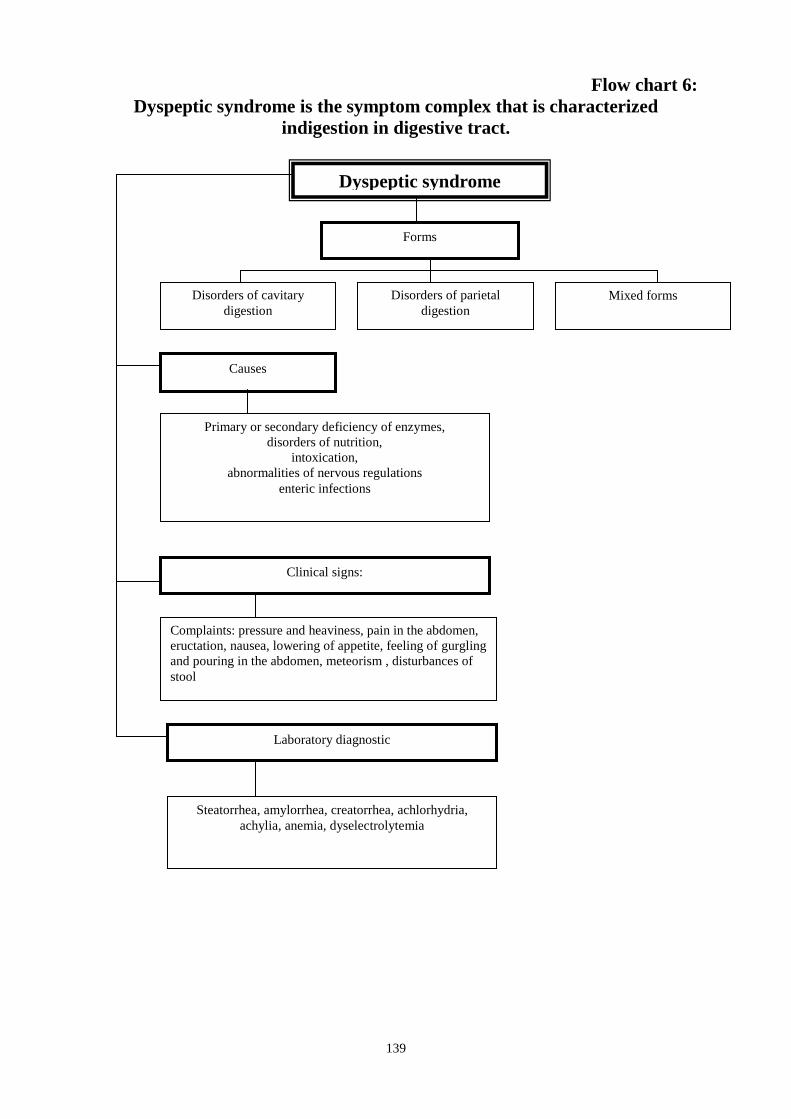
139
Flow chart 6:
Dyspeptic syndrome is the symptom complex that is characterized
indigestion in digestive tract.
Dyspeptic syndrome
Forms
Disorders of parietal
digestion
Mixed forms Disorders of cavitary
digestion
Causes
Primary or secondary deficiency of enzymes,
disorders of nutrition,
intoxication,
abnormalities of nervous regulations
enteric infections
Clinical signs:
Complaints: pressure and heaviness, pain in the abdomen,
eructation, nausea, lowering of appetite, feeling of gurgling
and pouring in the abdomen, meteorism , disturbances of
stool
Laboratory diagnostic
Steatorrhea, amylorrhea, creatorrhea, achlorhydria,
achylia, anemia, dyselectrolytemia
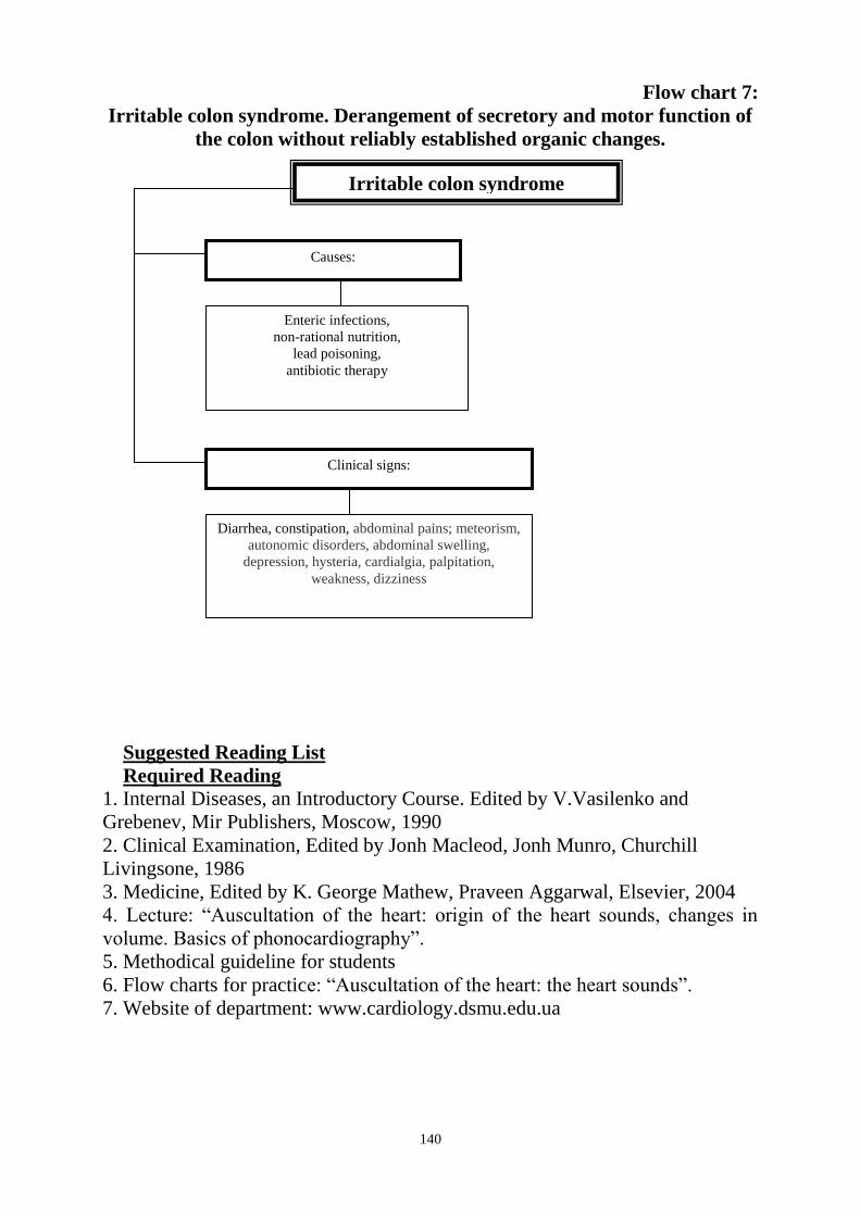
140
Flow chart 7:
Irritable colon syndrome. Derangement of secretory and motor function of
the colon without reliably established organic changes.
Suggested Reading List
Required Reading
1. Internal Diseases, an Introductory Course. Edited by V.Vasilenko and
Grebenev, Mir Publishers, Moscow, 1990
2. Clinical Examination, Edited by Jonh Macleod, Jonh Munro, Churchill
Livingsone, 1986
3. Medicine, Edited by K. George Mathew, Praveen Aggarwal, Elsevier, 2004
4. Lecture: ―Auscultation of the heart: origin of the heart sounds, changes in
volume. Basics of phonocardiography‖.
5. Methodical guideline for students
6. Flow charts for practice: ―Auscultation of the heart: the heart sounds‖.
7. Website of department: www.cardiology.dsmu.edu.ua
Irritable colon syndrome
Causes:
Enteric infections,
non-rational nutrition,
lead poisoning,
antibiotic therapy
Clinical signs:
Diarrhea, constipation, abdominal pains; meteorism,
autonomic disorders, abdominal swelling,
depression, hysteria, cardialgia, palpitation,
weakness, dizziness
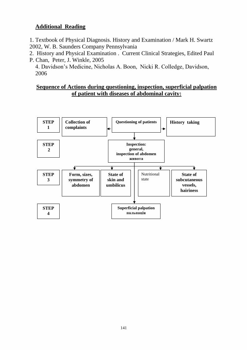
141
Additional Reading
1. Textbook of Physical Diagnosis. History and Examination / Mark H. Swartz
2002, W. B. Saunders Company Pennsylvania
2. History and Physical Examination . Current Clinical Strategies, Edited Paul
P. Chan, Peter, J. Winkle, 2005
4. Davidson‘s Medicine, Nicholas A. Boon, Nicki R. Colledge, Davidson,
2006
Sequence of Actions during questioning, inspection, superficial palpation
of patient with diseases of abdominal cavity:
Questioning of patients
Inspection:
general,
inspection of abdomen
живота
STEP
1
STEP
2
STEP
3
Collection of
complaints
History taking
Form, sizes,
symmetry of
abdomen
State of
subcutaneous
vessels,
hairiness
Nutritional
state State of
skin and
umbilicus
STEP
4
Superficial palpation
пальпація
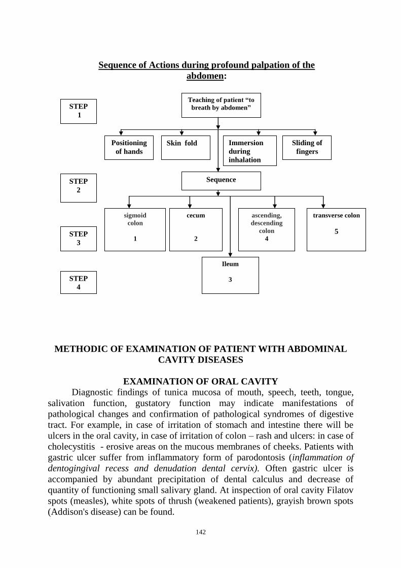
142
Sequence of Actions during profound palpation of the
abdomen:
METHODIC OF EXAMINATION OF PATIENT WITH ABDOMINAL
CAVITY DISEASES
EXAMINATION OF ORAL CAVITY Diagnostic findings of tunica mucosa of mouth, speech, teeth, tongue,
salivation function, gustatory function may indicate manifestations of
pathological changes and confirmation of pathological syndromes of digestive
tract. For example, in case of irritation of stomach and intestine there will be
ulcers in the oral cavity, in case of irritation of colon – rash and ulcers: in case of
cholecystitis - erosive areas on the mucous membranes of cheeks. Patients with
gastric ulcer suffer from inflammatory form of parodontosis (inflammation of
dentogingival recess and denudation dental cervix). Often gastric ulcer is
accompanied by abundant precipitation of dental calculus and decrease of
quantity of functioning small salivary gland. At inspection of oral cavity Filatov
spots (measles), white spots of thrush (weakened patients), grayish brown spots
(Addison's disease) can be found.
Teaching of patient “to
breath by abdomen”
ascending,
descending
colon
4
Sequence
cecum
2
STEP
1
STEP
2
STEP
3
STEP
4
sigmoid
colon
1
transverse colon
5
Positioning
of hands
Sliding of
fingers
Immersion
during
inhalation
Skin fold
Ileum
3

143
Change of taste.
Changes of taste has significant diagnostic value. They can be found at
infectious and gastrointestinal disturbance, pathology of oral and nasal cavity,
organic diseases of brain, long intake of some drugs.
In the basis of taste changes there is insufficient content of copper and
zinc in the organism. Deficiency of zinc in the saliva plays especially great role
in changes of taste.
Salivation
Analysis of salivation function plays significant role in diagnostic of some
diseases. Increased salivation or hypersalivation depends for a variety of
reasons and can be observed at the following physiological and pathological
states: irritation by food, influence of parasympathomimetic substances –
pilocarpine, muscarine etc; increased secretion of big digestive glands in
patients with gastric ulcer and pathology of pancreas, mercury or iodine
poisoning, reflex irritation of salivary glands in patients with helminthic
invasion, Parkinson's disease, rabies, pregnancy and vomiting and also at strong
irritation of rectal ampulla, urinary bladder, genitals. Decreased salivation or
hyposalivation appears in different cases: reflex impairment of salivary glands
and influence of anticholinergic drugs, nervousness and increased sweating,
water imbalance, after massive bleeding and long diarrhea, botulism, kidneys‘
diseases, malignant anemia, bilateral paralysis of facial nerve, progressive
paralysis.
Tongue inspection.
Great value in diagnostic of organism state according to state of tongue
has its color, character of incrustation, state of papilla, density and mobility of
corpus linguae. Angulation of tongue may be caused by somatic disorders.
Affection of the left part of the body (spleen, left lung) or right (liver, right lung)
causes changes of tongue volume and deviation of apex of the tongue to the
corresponding side. In norm tongue is pink colored with slight yellow
incrustation, apex of tongue does not deviate and does not quiver.
Lacquered tongue is characterized by bright-red color, it becomes bright
and smooth as a result of papilla atrophy. It can be found at oncological
diseases of gastrointestinal tract, severe chronic colitis, full-blown
hypovitaminosis.
Folded tongue is met at central nervous system and endocrine organs
affection, including affection of hypophysis. In case of very thick tongue with
rough transverse sulcus it is called ―scrotal tongue‖. This symptom is
characteristic for severe mental deficiency and psychic diseases especially if
patient doesn‘t maintainhygiene of oral cavity. Folded tongue can also be an
evidence of Melkersson-Rosenthal syndrome – diseases of unknown etiology
characterized by recurrent swelling of cheeks, lips and tongue accompanied by
paresis of facial nerve. Stomatologists differentiate folded tongue of adults with
sclerosing glossitis which develops at tertiary syphilis.
144
Typhoid tongue – is one of the first signs of typhoid fever. It is
characterized by thickening, presence of grayish-white incrustation with bright
hyperemia of the free of incrustation edges and apex.
Raspberry tongue is typical for scarlet fever.
Dark-red color of tongue “ham” is typical for obesity and chronic
alcoholism.
Cyanotic - cardiovascular collapse.
White tongue is characteristic for inflammatory diseases of digestive
organs especially intestine.
Appearance of white areas on the tongue or mucous membranes of cheeks
– leukoplakia demands necessary stomatological supervision and thorough
examination. Such white spots that are not covered with incrustation and
connected with epidermidalization are considered to be precancer. Often
leukoplakia develops as a result of vitamin A deficiency.
Twitching of tongue muscles or fasciculation develops at serious cerebral
affections, thyrotoxicosis, emotional excitement, fear.
EXAMINATION OF ABDOMINAL CAVITY
1. To conduct inspection of the abdomen in vertical position of patient:
assess form (usual, protruding, pendulous, hollow), symmetry, umbilicus
(smoothed, drawn-in, protrunding), state of skin (color, elasticity, presence of
rash), determine presence of venous collaterals and direction of blood flow in
them, dehiscence of rectus muscle of abdomen, hernia.
2. To conduct percussion of abdomen in vertical position of patient (for
detection of ascites): for that finger- plessimeter is positioned under umbilicus
on the midline (perpendicularly to it) and quiet downward percussion is
conducted with analysis of the sound that is created (transfer of tympanic sound
into the dull one that indicates presence of fluid).
3. To conduct inspection of the abdomen in horizontal position of the
patient: to assess form, symmetry, participation in breathing (active, weak,
doesn‘t participate (locally, diffusely)), visible peristalsis.
4. To conduct percussion of abdomen in horizontal position of patient (for
detection of ascites): for that finger- plessimeter is positioned under umbilicus
on the midline (parallel to it) and quiet percussion toward flanks is conducted
with analysis of the sound that is created (transfer of tympanic sound into the
dull one that indicates presence of fluid).
5. To conduct auscultation of abdomen: to sound systematically (in
vertical and horizontal positions of the patient) all abdominal regions (beginning
with left inguinal and then clockwise) analyzing intensity of intestinal murmurs
(normal – weak, increased, absent), presence or absence peritoneal murmur.
6. To conduct superficial palpation of abdomen: patient has to be laid on
the back with low head of the bed, hands are laid along the body (the same
position is for profound palpation), right hand of the doctor is placed flatwise
(first in the left inguinal region) and slightly presses by finger bones moving

145
them on 3-4sm to both sides analyzing painfulness, consistency, compliance of
abdominal wall and then all characteristics are compared with symmetric region
on the right side. The same way mesogastric and epigastric regions are
compared.
7. To conduct profound palpation of the abdomen according to Obraztsov-
Strazhesko method.
INVESTIGATION OF DUODENAL CONTENT AND BILE
I phase- of the common bile duct Is characterized by the‖A‖ bile portion
time of discharge 10-20min,
quantity 20ml
color is golden-yellow
II phase – of the closed Oddi's sphincter duration 2-6 min,
bile is absent
III phase- bile of the portion ―A‖ of the
distal part of common duct
time of discharge 3-5 min,
quantity 3-5 ml
IV phase – of the portion "В" time of discharge 20-30 min,
quantity 30-50 ml
dark olive green or brown color
V phase – of the portion "С" time of discharge 20-30 min,
quantity exceeds portion "В"
golden-yellow color
Assessment of results of fractional duodenal intubation permits to determine
different impairments of motor function of gallbladder and bile ducts.
1. hypotension of Oddi's sphincter – shortening of the phase of closed
Oddi's sphincter less than 3 min and discharge of bile with speed more than
2ml/min.
2. hypertension of Oddi's sphincter – increase of closed Oddi's sphincter
duration more than 6 min., discharge of B and C bile is discontinuous, delayed,
pain is possible, introduction of novocaine increases volume velocity of bile
flow.
3. hypotension of gallbladder - prolongation of discharge time of portion B –
more than 60 min, decrease of volume velocity of biliary excretion and increase
of portion B volume – more than 100 ml.
4. hypertension of gallbladder – decrease of discharge time of portion B –
less than 20 min, increase of volume velocity of biliary excretion – more than
5ml/min; volume of portion B is not changed significantly.
5. hypertension of bile duct – difficulty of bile discharge.
Microscopic study.
Leukocytes. Large quantity of leukocytes in portions B and C may
indicate presence of inflammatory process in bile ducts (cholecystitis,
146
cholecystocholangitis) and its non-bile origin (admixture of gastric and
pancreatic juice, migration from the mucous membrane of duodenum).
Epithelial cells – great amount of round epithelial cells in portions B and
C indicates on pathological changes in duodenum; column epithelial cells –
inflammation of bile ducts.
Bilirubin calcium and crystals of cholesterol. Detection of them is
typical for cholestasis, that is often met at cholelithiasis
Protozoa and helminthes. It is recommended to conduct investigation
of duodenal content if there is suspicion of liver and gallbladder helminthiasis
(opisthorchiasis, fascioliasis, clonorchiasis, dicroceliasis) and duodenum
(strongyloidiasis, trichostrongyloidiasis)
Bacteriological study of bile is conducted for determination of
microflora content and its antibiotic susceptibility.
Biochemical study of bile provides insight into concentration function of
gallbladder and colloidal stability of bile, presence of inflammatory process.
But these methods demand expensive equipment and because of their
complexity are mostly used in scientific researches.
Situational tasks
Task 1
28 years old patient P. complains of weakness, abdominal swelling, diarrhea
(stool is 4-5 times during the day, profuse). Coprological study: consistency is
porridge-like, color is grey-yellow with greasy luster, positive reaction on
bilirubin. Microscopy: increased quantity of fatty acids and soap, moderate
amount of neutral fat, petty amount of muscular fibers, digested cellulose,
significant amount of starch. In which part of gastrointestinal tract insufficiency
of digestion may give such picture of stool:
A. Insufficiency of gastric digestion
B. Insufficiency of pancreas
C. Insufficiency of digestion in intestine
D. Insufficiency of gastric digestion and pancreas
E. Insufficiency of digestion in intestine and insufficiency of pancreas
Task 2
27-years old patient S. complains of general weakness, skin is pale with
cteritous color, liver is not increased, spleen is on 2 sm lower than ribs‘
edge. Bilirubin in blood: total - 52,64 mcmole/l, conjugated – 0,
unconjugated – 52,64 mcmole/l, reaction of Van der Berg – indirect .
Bilirubin of urine – reaction is negative, urobilin – reaction is sharply
positive. Stool is of saturated yellow color, reaction on the stercobilin is
sharp positive. Increase of unconjugated bilirubin in the blood can be found
at:
A. pernicious anemia
B. obstructive jaundice

147
C. asiderotic anemia
D. cythemolytic icterus
E. posthemorrhagic anemia
Task 3 48 years old patient complains of periodical edema of legs during the last
year. During the last 3 months abdomen began to increase in volume. In the
past patient abused alcohol. Investigation: skin and mucous membranes are
subicteric. On the skin of trunk there are single vascular spiders. Tongue is
lacquered. Abdomen is increased by the expense of presence of free liquid and
meteorism. In horizontal position it is spread. Liver is beyond the right costal
margin on 5sm, its edge is acute, surface is dense, uneven and painless. Spleen is
on 3 sm lower of left costal margin. There are oedema of shins and feet.
Laboratory investigation: common blood analysis: Er – 3,3Т/l, Нb-126g/l, L-
5,6 M/l; ESR -18mm/hour. Total bilirubin - 27,6 mcmole/l; conjugated-14,6
mcmole/l, unconjugated -13,0 mcmole/l. АST-0,7mmole/ h l; АLT-1,2mmole/
h l. Alkaline phosphatase – 2,5mmole/ h l, whole protein – 50g/l, albumin –
49%, urea – 4,5 mmole/l. Fiber-optic gastroduodenoscopy: varix dilatation of
esophagus‘s veins. Scintigraphy of the liver: liver is diffusely increased.
Accumulation of radiopharmaceutical is uniformly decreased. Spleen is
increased and actively accumulates radiopharmaceutical. What causes such
changes?
А. Liver cirrhosis
B. Cholelithiasis
C. Heart failure.
D. Chronic hepatitis.
E. Fatty hepatosis.
Task 4
During patient‘s examination it was found: abdomen is swollen , asymmetric,
there is visible peristalsis, on the percussion – tympanic sound, splashing sound.
At superficial palpation: moderate painfulness in the left iliac area, abdominal
wall in that place is dense, after long slight stroking it becomes softer. Indicate
level of impediment that causes painfulness in the left iliac area.
A. Caecum.
B. Ascending part of colon.
C. Descending part of colon.
D. Transverse colon.
E. Sigmoid colon
Task 5
38 years old patient K. complains of very intensive pain in epigastrium, feeling
of abdominal swelling, general weakness. Complaints appeared suddenly. From
the case history it is known that patient has been suffering from duodenal ulcer
for 10 years. On percussion of the abdomen sound is laud, long, low, ringing and
148
is determined throughout the abdomen and under liver. Doctor suspected free
perforation and it was approved during the further examination of patient. What
kind of percussion sound is throughout the abdomen?
A. Dull.
B. Impaired .
C. Tympanic.
D. Impaired - tympanic.
E. Clear.
Task 6
67 years old patient D. was admitted to the hospital with complaints on swelling
pains in the left iliac area, 2-3 days of constipation, loss of appetite, loss of
weight on 5-6 kg during the last year. Examination: nutritional state is
decreased, abdomen is of usual form, painless at superficial palpation. Profound
palpation: sigmoid colon is located in the typical place, increased in diameter,
dense, painfulness. Other parts of colon are in the typical places, of the normal
sizes, painless, rumble. Which parts of the colon are projected in the left iliac
area?
A. Descending part of colon.
B. Ascending part of colon.
C. Distal part of intestine.
D. Sigmoid colon
E. Caecum.
Task 7
43 years old patient B. complains of intensive pain in the right iliac area, nausea,
single vomiting. At superficial palpation of the right iliac area there is acute
painfulness and expressed tension of abdominal wall muscles. Which parts of
the colon are projected in the left iliac area?
A. Caecum and appendage.
B. Ascending part of colon.
C. Descending part of colon.
D. Sigmoid colon.
E. Duodenum.
Task 8
36 years old patient D. complains of pains in the lower part of the abdomen in
the area of flanks. Pains appear after intake of fruits, milk, fatty food and are
dull, aching. Pains decrease after passage of gases and defecation. Examination:
patient is of normosthenic type, little bit decreased nutrition, tongue is with
grayish incrustation. Abdomen is swollen, there is moderate diffuse painfulness
during palpation. Profound palpation: sigmoid colon is painfulness, 4sm in
diameter, dense, moves on 6-7sm; caecum, ascending, descending and
transverse colon are painfulness, up to 4-5sm in diameter, rumble. Doctor
suspected colitis. Assess characteristics of sigmoid colon according to palpation
149
findings.
A. Norm.
B. Only diameter is changed.
C. Only consistency is changed.
D. Only mobility is changed.
E. All of the characteristics are changed.
Task 9
58 years old patient was admitted to the hospital with complaints on swelling
pains in the right iliac area, 2 days constipation, loss of appetite, loss of weight
on 6 kg during the last year. Profound palpation of the abdomen revealed:
sigmoid colon is painless, smooth, its diameter is1,5-2sm, elastic consistency,
doesn‘t rumble; ascending, descending and transverse colon can not be palpated
because of the marked subcutaneous fat.
Assess findings:
A. Norm .
B. Diameter of caecum is changed.
C. Diameter of sigmoid colon is changed.
D. Diameter of both colons is changed.
E. Consistency of both colons is changed.
Task 10 52 years old patient D. complains of weakness, absence of appetite, heaviness in
epigastric area after food intake. Examination of gastric secretion: total acidity –
0 t.u., free hydrochloric acid - 0 t.u.; first phase of secretion; basal secretion –
34ml, total acidity – 20 t.u., free hydrochloric acid – 0 t.u.; after test-breakfast
there were 190ml of residue; second phase of secretion: stimulated – 48ml, total
acidity – 20 t.u., free hydrochloric acid – 0 t.u. What kind of gastric juice does
the patient has?
A. Normal
B. Hypoacid
C. Anoacid
D. Hyperacid
E. Increased
Key answers:
Task 1: С. Task 2: D. Task 3: А. Task 4:В. Task 5: С.
Task 6: D. Task 7: А. Task 8: Е. Task 9:В. Task 10: В.
Revision tasks.
Task 1
During superficial palpation of patient‘s abdomen there is acute pain in the right
iliac area, abdominal wall is dense, after long slight stroking and changing of
attention hardening doesn‘t disappear. Assess findings of iliac area superficial
palpation:
150
A. Variant of norm.
B. There is autodefense of the abdominal wall.
C. There is muscular defense».
D. There is hypertrophy of abdominal wall muscles.
E. There is pathological mass of abdominal wall.
Task 2
52 years old patient Z. was admitted to the hospital with complaints on the
feeling of heaviness in the epigastrium, loss of appetite, fast satiation and feeling
of repletion even at small amount of food, foul-smelling eructation, frequent
nausea and vomiting with unpleasant smell and admixtures of food eaten 1-2
days ago, constipation, considerable weight loss. During 20 years patient has
been suffering from gastric ulcer. Examination: patient is of decreased nutrition,
scaphoid abdomen, painfulness at palpation especially in epigastrium and
paraumbilical area. Profound palpation: greater curvature of stomach is on 2sm
lower than umbilicus, painless, in a way of stairs, soft-elastic consistency,
movable in borders of 3-4sm.
Assess palpatory characteristics of greater curvature of stomach:
A. Only mobility is changed.
B. Only consistency is changed.
C. Only topography is changed.
D. All of the characteristics are changed.
E. Norm
Task 3
70 years old patient S. complains of weakness, subfebrile temperature, almost
constant pains in the right side of the abdomen, weight loss, constipation. Patient
is ill for 6 months. Profound palpation of ascending colon gave the following
results: moderate painfulness, diameter is about 6sm, gibbous, absolutely
immovable. Assess palpatory characteristics of the colon:
A. Only diameter is changed.
B. Only consistency is changed.
C. All of the characteristics are changed.
D. Only state of surface is changed.
E. Only mobility is changed.
Task 4
What for is barium suspensium used in contrast radiography?
A. Increase of peristalsis
B. Cleansing of stomach
C. Spasmolytic effect
D. Contrast study of the examined organ, its relief and evacuator function
E. Increase of gas-formation
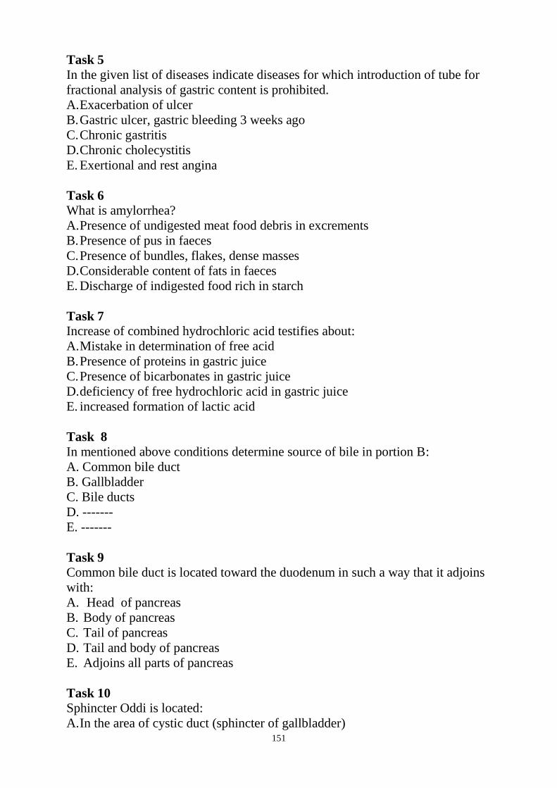
151
Task 5
In the given list of diseases indicate diseases for which introduction of tube for
fractional analysis of gastric content is prohibited.
A. Exacerbation of ulcer
B. Gastric ulcer, gastric bleeding 3 weeks ago
C. Chronic gastritis
D. Chronic cholecystitis
E. Exertional and rest angina
Task 6
What is amylorrhea?
A. Presence of undigested meat food debris in excrements
B. Presence of pus in faeces
C. Presence of bundles, flakes, dense masses
D. Considerable content of fats in faeces
E. Discharge of indigested food rich in starch
Task 7
Increase of combined hydrochloric acid testifies about:
A. Mistake in determination of free acid
B. Presence of proteins in gastric juice
C. Presence of bicarbonates in gastric juice
D. deficiency of free hydrochloric acid in gastric juice
E. increased formation of lactic acid
Task 8
In mentioned above conditions determine source of bile in portion B:
A. Common bile duct
B. Gallbladder
C. Bile ducts
D. -------
E. -------
Task 9
Common bile duct is located toward the duodenum in such a way that it adjoins
with:
A. Head of pancreas
B. Body of pancreas
C. Tail of pancreas
D. Tail and body of pancreas
E. Adjoins all parts of pancreas
Task 10
Sphincter Oddi is located:
A. In the area of cystic duct (sphincter of gallbladder)
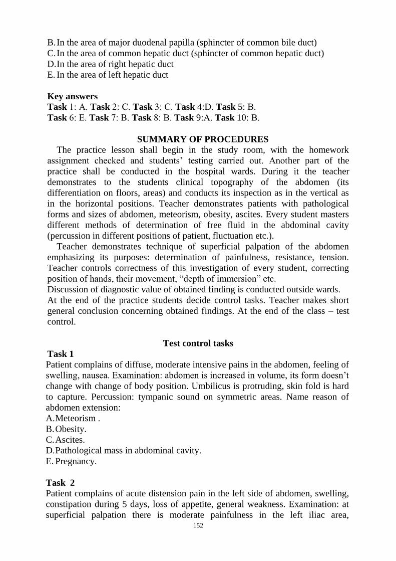
152
B. In the area of major duodenal papilla (sphincter of common bile duct)
C. In the area of common hepatic duct (sphincter of common hepatic duct)
D. In the area of right hepatic duct
E. In the area of left hepatic duct
Key answers
Task 1: А. Task 2: С. Task 3: С. Task 4:D. Task 5: В.
Task 6: Е. Task 7: В. Task 8: В. Task 9:А. Task 10: В.
SUMMARY OF PROCEDURES
The practice lesson shall begin in the study room, with the homework
assignment checked and students‘ testing carried out. Another part of the
practice shall be conducted in the hospital wards. During it the teacher
demonstrates to the students clinical topography of the abdomen (its
differentiation on floors, areas) and conducts its inspection as in the vertical as
in the horizontal positions. Teacher demonstrates patients with pathological
forms and sizes of abdomen, meteorism, obesity, ascites. Every student masters
different methods of determination of free fluid in the abdominal cavity
(percussion in different positions of patient, fluctuation etc.).
Teacher demonstrates technique of superficial palpation of the abdomen
emphasizing its purposes: determination of painfulness, resistance, tension.
Teacher controls correctness of this investigation of every student, correcting
position of hands, their movement, ―depth of immersion‖ etc.
Discussion of diagnostic value of obtained finding is conducted outside wards.
At the end of the practice students decide control tasks. Teacher makes short
general conclusion concerning obtained findings. At the end of the class – test
control.
Test control tasks
Task 1
Patient complains of diffuse, moderate intensive pains in the abdomen, feeling of
swelling, nausea. Examination: abdomen is increased in volume, its form doesn‘t
change with change of body position. Umbilicus is protruding, skin fold is hard
to capture. Percussion: tympanic sound on symmetric areas. Name reason of
abdomen extension:
A. Meteorism .
B. Obesity.
C. Ascites.
D. Pathological mass in abdominal cavity.
E. Pregnancy.
Task 2 Patient complains of acute distension pain in the left side of abdomen, swelling,
constipation during 5 days, loss of appetite, general weakness. Examination: at
superficial palpation there is moderate painfulness in the left iliac area,
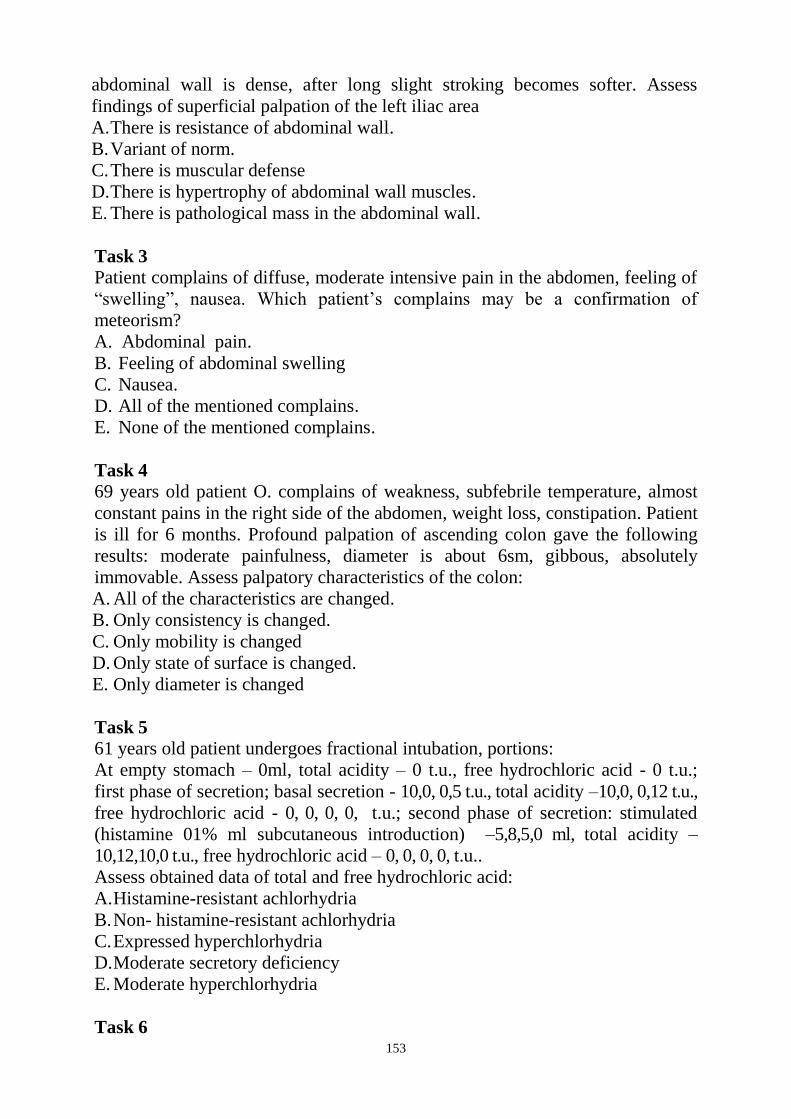
153
abdominal wall is dense, after long slight stroking becomes softer. Assess
findings of superficial palpation of the left iliac area
A. There is resistance of abdominal wall.
B. Variant of norm.
C. There is muscular defense
D. There is hypertrophy of abdominal wall muscles.
E. There is pathological mass in the abdominal wall.
Task 3 Patient complains of diffuse, moderate intensive pain in the abdomen, feeling of
―swelling‖, nausea. Which patient‘s complains may be a confirmation of
meteorism?
A. Abdominal pain.
B. Feeling of abdominal swelling
C. Nausea.
D. All of the mentioned complains.
E. None of the mentioned complains.
Task 4
69 years old patient O. complains of weakness, subfebrile temperature, almost
constant pains in the right side of the abdomen, weight loss, constipation. Patient
is ill for 6 months. Profound palpation of ascending colon gave the following
results: moderate painfulness, diameter is about 6sm, gibbous, absolutely
immovable. Assess palpatory characteristics of the colon:
A. All of the characteristics are changed.
B. Only consistency is changed.
C. Only mobility is changed
D. Only state of surface is changed.
E. Only diameter is changed
Task 5 61 years old patient undergoes fractional intubation, portions:
At empty stomach – 0ml, total acidity – 0 t.u., free hydrochloric acid - 0 t.u.;
first phase of secretion; basal secretion - 10,0, 0,5 t.u., total acidity –10,0, 0,12 t.u.,
free hydrochloric acid - 0, 0, 0, 0, t.u.; second phase of secretion: stimulated
(histamine 01% ml subcutaneous introduction) –5,8,5,0 ml, total acidity –
10,12,10,0 t.u., free hydrochloric acid – 0, 0, 0, 0, t.u..
Assess obtained data of total and free hydrochloric acid:
A. Histamine-resistant achlorhydria
B. Non- histamine-resistant achlorhydria
C. Expressed hyperchlorhydria
D. Moderate secretory deficiency
E. Moderate hyperchlorhydria
Task 6
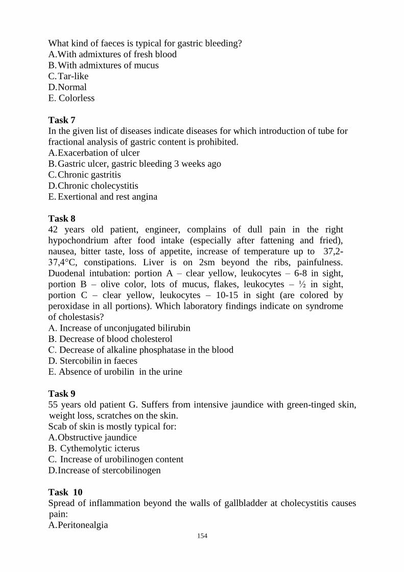
154
What kind of faeces is typical for gastric bleeding?
A.With admixtures of fresh blood
B. With admixtures of mucus
C. Tar-like
D. Normal
Е. Colorless
Task 7
In the given list of diseases indicate diseases for which introduction of tube for
fractional analysis of gastric content is prohibited.
A. Exacerbation of ulcer
B. Gastric ulcer, gastric bleeding 3 weeks ago
C. Chronic gastritis
D. Chronic cholecystitis
E. Exertional and rest angina
Task 8 42 years old patient, engineer, complains of dull pain in the right
hypochondrium after food intake (especially after fattening and fried),
nausea, bitter taste, loss of appetite, increase of temperature up to 37,2-
37,4°C, constipations. Liver is on 2sm beyond the ribs, painfulness.
Duodenal intubation: portion A – clear yellow, leukocytes – 6-8 in sight,
portion B – olive color, lots of mucus, flakes, leukocytes – ½ in sight,
portion C – clear yellow, leukocytes – 10-15 in sight (are colored by
peroxidase in all portions). Which laboratory findings indicate on syndrome
of cholestasis?
А. Increase of unconjugated bilirubin
В. Decrease of blood cholesterol
С. Decrease of alkaline phosphatase in the blood
D. Stercobilin in faeces
Е. Absence of urobilin in the urine
Task 9
55 years old patient G. Suffers from intensive jaundice with green-tinged skin,
weight loss, scratches on the skin.
Scab of skin is mostly typical for:
A. Obstructive jaundice
B. Cythemolytic icterus
C. Increase of urobilinogen content
D. Increase of stercobilinogen
Task 10
Spread of inflammation beyond the walls of gallbladder at cholecystitis causes
pain:
A. Peritonealgia

155
B. Distension
C. Vascular
D. Spastic
E. Any of the listed above

156
The Health Ministry of Ukraine
Donetsk National Medical University
Approved
at the meeting of
Propedeutic and Internal Medicine
Department
Head of department
Associate Member of NAMSc of
Ukraine,
Professor G. A. Ignatenko
« _______ »_____________ 2011 р.
STUDENT’S SELF-STUDY GUIDELINES FOR
PRACTICE ACTIVITIES
Subject Propedeutics of the Internal Medicine
Module 1 Inspection methods and the main
symptoms and syndromes in internal
diseases in the clinic of Internal
Medicine
Subject Module 2 Research Methods and the main
symptoms and syndromes in diseases of
the digestive and urinary
Practice A technique for investigation of urinary
tract. Collection of complaints, medical
history and physical examination of
patients with pathology of the urinary
system. Care and the providing of first
aid. Supporting research methods in
nephrology. The main syndromes in
nephrology. Changes in the oral cavity
in diseases of the genitourinary system.
Course 2
Faculty Dentistry
Donetsk 2011

157
Importance of the Subject : correct according to the method and technique
required anamnesis, inspection, percussion, auscultation, abdominal palpation of
the surface can often quickly identify pathological changes in abdominal organs,
including the state of emergency, to use additional methods of investigation. In
the study of abdominal palpation is given paramount importance. Methodically
carried out correctly palpation can detect not only the localization of the
pathological process, get an idea of his character, but also to determine the
subsequent tactics surveys using complementary techniques (X-rays,
sigmoidoscopy, etc.). Ability to palpate the kidneys, to assess their physical
properties is of great importance in the diagnosis of internal diseases, it often
allows the diagnosis. Early diagnosis of clinical signs of kidney damage, the use
of instrumental and laboratory studies allow them to optimize the subsequent
search for diagnostic and early treatment of patients, which improves the
prognosis. Proper care of patients with pathology of urinary system and timely
first aid to help in the treatment and future prognosis for patients.
Key Objective: to be able to carrying out an interview anamnesis of urinary
system, abdomen studies, to give the clinical evaluation of the data; palpate the
kidneys, to determine their physical properties and to evaluate the diagnostic
significance of the changes that are being detected; identify the leading
symptoms and signs in the clinic of internal diseases, in particular, in the
pathology of urinary , analyze the results of basic laboratory and instrumental
methods; to care for patients with kidney disease and urinary tract and first aid.
Specific Goals:
1. Provide inquiries and general inspection of the patient with the pathology of
urinary system and evaluate the data obtained.
2. Palpation of the kidneys to hold on a method Obraztsova - Strazhesko, learn
research methodology palpation of kidneys in a standing position and lying and
symptom a beating in the lumbar region, to assess the findings.
3. Learn methods of clinical and bacteriological examination of urine, urine
analysis for Nechiporenko, Zimnitsky and interpret the results obtained in renal
pathology.
4. Learn techniques of biochemical blood tests for renal disease (urea,
creatinine, total protein, cholesterol) and interpret their results.
5. Characterized by instrumental methods of investigation of the urinary tract -
ultrasound and X-ray, biopsy. Evaluate and interpret the results of these studies.
6. Identify the main syndromes in diseases of the urinary flow according to the
physical and complementary research methods.
7. Identify and evaluate the typical changes in the mucosa and the teeth-jaw
system in renal disease.
8. Own method of catheterization of the bladder catheter is soft.
9. Spend caring for patients with kidney disease and urinary tract infections, and
provide first aide.

158
Level of knowledge and skills before the practice
1 Know the structure of the kidneys and urinary tract. "Anatomy".
2. Know the function of kidneys and urinary tract. "Physiology."
3 Know the basic biochemical processes that occur in the kidneys and urinary
tract. Biochemistry.
4 Ethical relationships in society. "Philosophy".
Questions for self-assessment of the pre-practice knowledge
Q1.
Doctor needs to know how many nephrons in each kidney contains a healthy
person?
A. 1-1,3 million nephron
B. 20 million nephrons
C. 30 million nephrons
D. 0,2 million nephrons
E. 18-20 thousand nephrons
Q2. To assess kidney function, nephrologists need to know what function to perform
kidney nephron?
A. Glomerular filtration, tubular reabsorption and secretion.
B. Glomerular filtration rate, osmotic dilution and concentration of urine.
C. Glomerular filtration, secretion, osmotic dilution and concentration of urine.
D. Glomerular filtration, tubular reabsorption, osmotic dilution and
concentration of urine.
E. Glomerular filtration rate, glomerular reabsorption and secretion.
Q3.
To estimate the glomerular filtration rate in patient S., aged 30, a nephrologist
should use:
A. Uric acid
B. Glucose
C. Creatinine
D. Indikan
E. Albumin
Q4. Which material to choose a doctor to assess the glomerular filtration rate:
A. Which only filters
B. What is the only secreted
C. Which only reabsorption
D. Which is filtered and secreted
E. Which is filtered and reabsorbed
Q5.
On a section of the drug kidney shows that the medulla has a striped
appearance. What anatomical structures provide this picture?
A. renal corpuscles and vessels bringing
B. convoluted part and the efferent vessels

159
C. Henle Loop collected in tubes
D. Renal columns and arched artery
E. Slice the kidneys and mutual artery
Q6.
Normally, when a mixed meal urine is acidic or weakly acidic (pH = 5,3-6,8). In
this case, the acid reaction of urine is predetermined, mainly due to:
A. NaH 2PO4 and KH2PO4
B. NaH 2PO4 and K2HPO4
C. H3PO4 and Na2HPO4
D. NaH2PO4 and K2HPO4
Ye Na 2HPO4 and KH2PO4
Key answers: Q 1- A. Q 2- A. Q 3- C. Q 4- A. Q 5- S. Q 6- A.
Suggested Reading List for the initial level of knowledge
1.Gain MG, Lysenkov NK, Bushkovich VI Human Anatomy: A Textbook -
Saint-Petersburg.: Izdatel'stvo "Hippocrates", 1998.
2. Human Physiology. Ed. R. Schmidt and G. Thews .- Moscow: Izdatel'stvo
"Mir". 1986.
3. Normal physiology. Ed. VI Filimonov. - K.: Health, 1994. - 608s.
4. Nikolaev AY Biological Chemistry: A Textbook for medical schools. - M.:
Vyssh.shk., 2000. - 495s.
Contents of Practice
Theoretical questions to practice.
1. Questions and a general inspection of the patient with the pathology of
urinary flow.
2. Methods palpation of kidneys by the method of Strazhesko, palpation study of
kidneys in a standing position and lying down, check symptoms a beating in the
lumbar region.
3. Methods of clinical and bacteriological examination of urine, urine analysis
for Nechiporenko, Zimnitsky.
4. Biochemical blood analysis in the pathology of kidney: urea, creatinine, total
protein, cholesterol.
5. Ultrasound and X-ray examination in diseases of the kidneys and urinary
tract, kidney biopsy.
6. The main syndromes in diseases of the urinary system and symptoms on
physical data and additional research methods.
7. Typical changes of the mucous membrane of the mouth and teeth-jaw system
in renal disease.
8. The technique of catheterization of the bladder catheter is soft.
9. Features of care for patients with kidney disease and urinary tract infections
and the provision of first aid.
Practical skills that are performed on lesson. 1. Conduct inquiries and general inspection of the patient with kidney disease
and urinary tract infections and to evaluate the data obtained.
2. Hold palpation of kidneys by the Strazhevsko method, palpation study of

160
kidneys in a standing position and lying down, check symptoms a beating in the
lumbar region and assess the data obtained.
3. Interpretation of the results of clinical and bacteriological examination of
urine, urine analysis for Nechiporenko, Zimnitsky.
4. Interpretation of the results of biochemical studies of the blood in renal
disease (urea, creatinine, total protein, cholesterol).
5. Interpretation of the results of instrumental studies of kidney and urinary tract
(ultrasound, X-ray, biopsy).
6. Identify the main syndromes in pathology of urinary system according to the
physical and complementary research methods.
7. Determine the typical changes in oral mucosa and teeth-jaw system in renal
disease.
8. Spend caring for patients with nephrological and first aid.
9. Catheterization of the bladder to hold a soft catheter.
Required Glossary to Practice
Term Definition
Hematuria The presence of red blood cells in
urine
Leucocyturia The presence of an increased
number of leukocytes in urine
Proteinuria The presence of protein in the urine
Polyuria A large amount of urine, which
identifies a person within a
day. More than 2 liters
Oliguria Decrease in urine output of less than
500 ml per day
Anuria The absence or reduction of urine
output less than 50-100 ml per day
Nocturia The predominance of nocturnal
diuresis on day
Gipostenuriya The presence of urine with a low
density
Nephrotic syndrome clinical and laboratory syndrome
associated with long-term and
significant
increased permeability of the
nephron structures for a protein that
shows more than 3,5 g/l proteinuria,

161
hypoproteinemia, hypoalbuminemia,
hyperlipidemia and edema
Nephritic syndrome Pathological condition manifested
mandatory laboratory changes in the
urine, hypertension and edema
Tubulo-interstitial syndrome Presented by a group of
inflammatory, noninflammatory,
toxic and metabolic diseases that
occur with the overwhelming defeat
of the interstitial tissue of the kidney
and tubular apparatus
Glomerulonephritis Morphologically heterogeneous
group of immune inflammatory
diseases of the kidneys, mainly
affecting the nephron
Pyelonephritis Group of infectious and
inflammatory diseases of the
kidneys with an overwhelming
defeat pyelocaliceal apparatus
Chronic renal failure The final stage for any chronic
kidney disease, which is based on
the processes of irreversible decline
in kidney function and development
of uremic poisoning
Acute renal failure
Acutely emerged, but potentially
reversible renal dysfunction with
oligo-anuria and signs of uremic
poisoning
Coma Complete lack of consciousness
with preservation of corneal,
vomiting, and tendon reflexes

162
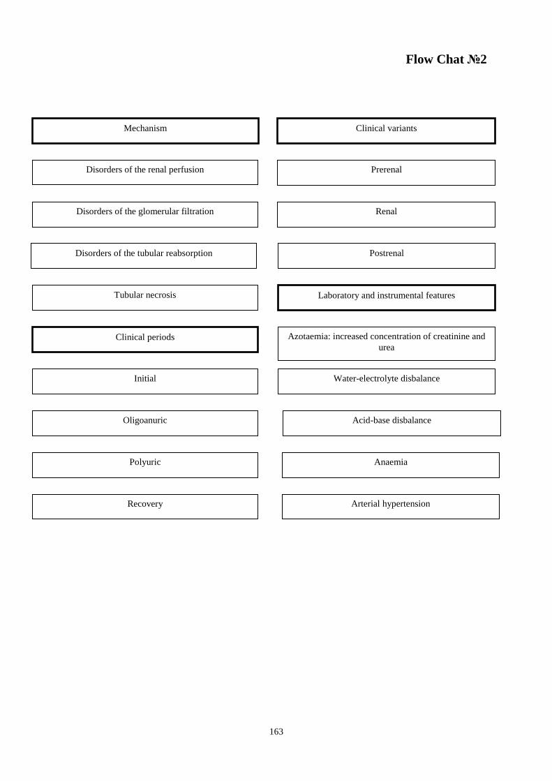
163
Flow Chat №2
Mechanism Clinical variants
Disorders of the renal perfusion Prerenal
Disorders of the glomerular filtration Renal
Disorders of the tubular reabsorption Postrenal
Tubular necrosis Laboratory and instrumental features
Clinical periods Azotaemia: increased concentration of creatinine and
urea
Initial Water-electrolyte disbalance
Oligoanuric
Polyuric
Acid-base disbalance
Recovery
Anaemia
Arterial hypertension
164
Suggested Reading List
Required Reading
1. Internal Diseases, an Introductory Course. Edited by V.Vasilenko and
Grebenev, Mir Publishers, Moscow, 1990
2. Clinical Examination, Edited by Jonh Macleod, Jonh Munro, Churchill
Livingsone, 1986
3. Medicine, Edited by K. George Mathew, Praveen Aggarwal, Elsevier, 2004
4. Lecture: Examination of the cardiovascular system: semiotics, inspection of
the heart, percussion: methodic and technique of determining normal borders
of the relative and absolute cardiac dullness, in the norm and pathology
5. Methodical guideline for students
6. Flow charts for Practice
7. Website of department: www.cardiology.dsmu.edu.ua
Additional Reading 1. Textbook of Physical Diagnosis. History and Examination / Mark H. Swartz
2002, W. B. Saunders Company Pennsylvania
2. History and Physical Examination . Current Clinical Strategies, Edited Paul
P. Chan, Peter, J. Winkle, 2005
3. Davidson‘s Medicine, Nicholas A. Boon, Nicki R. Colledge, Davidson, 2006

165
Estimated base operations (EBO).
Sequence of Actions in the Precordial Area Palpation and Examination
1. Algorithm of actions during the questioning, inspection, palpation of the
surface of patients with diseases of the abdominal cavity:
Anamnesis
general, inspection of the abdomen
STEP1
STEP 2
STEP 3
Collection complaints
History taking
Shape, size,
symmetry, stomach
status
subcutaneous
blood vessels,
body hair
fatness Skin &
navel
STEP 4 Surface palpation
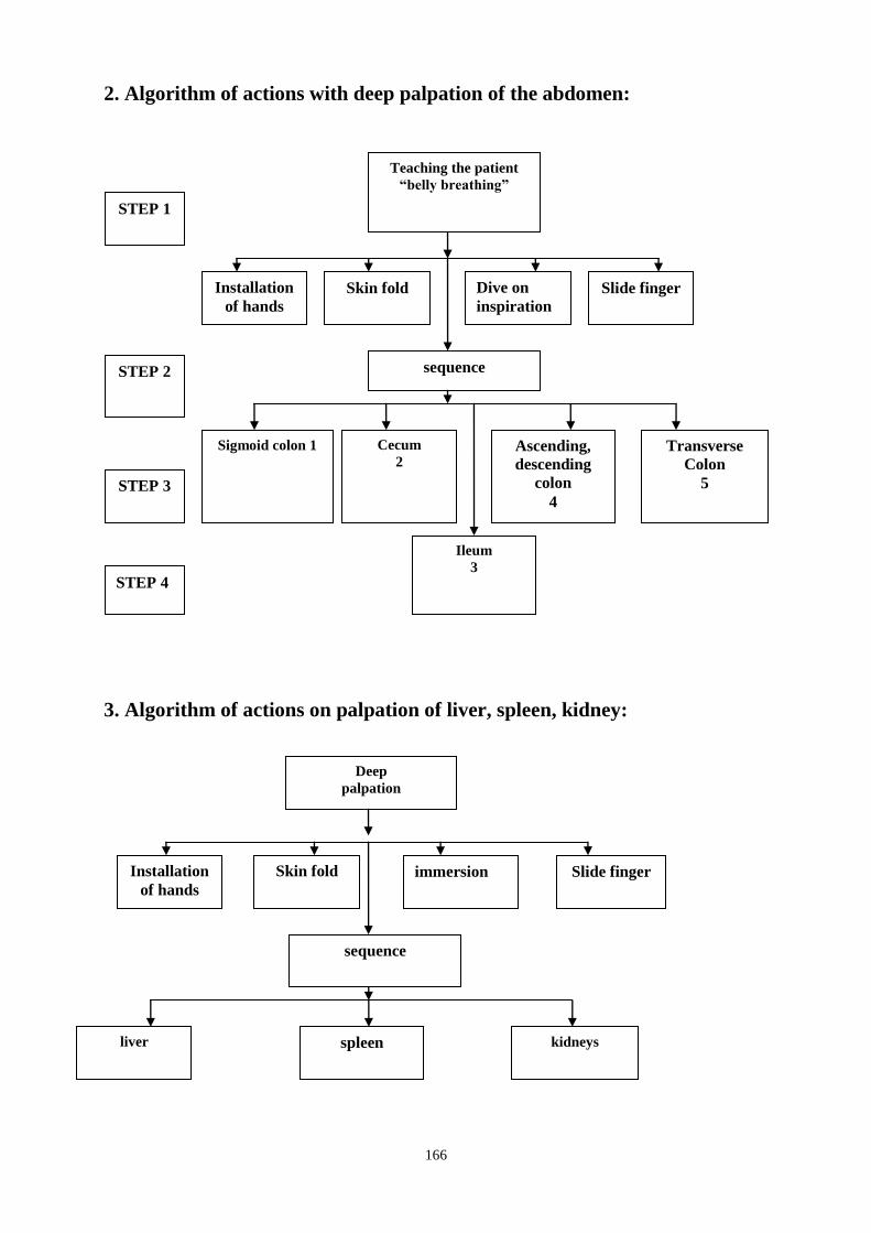
166
2. Algorithm of actions with deep palpation of the abdomen:
3. Algorithm of actions on palpation of liver, spleen, kidney:
Teaching the patient
“belly breathing”
Ascending,
descending
colon
4
sequence
Cecum
2
STEP 1
STEP 2
STEP 3
STEP 4
Sigmoid colon 1 Transverse
Colon
5
Installation
of hands
Slide finger Dive on
inspiration
Skin fold
Ileum
3
Deep
palpation
spleen
sequence
liver
kidneys
Installation
of hands
Slide finger immersion Skin fold
167
Method of palpation of the kidneys
Method of palpation of the right and left kidneys in the first horizontal and then
in the vertical position of the patient for this:
- Fingers of his left hand set in the most compliant portion of the corresponding
lumbar region, between the XII rib and the iliac wing.
- The right hand with a slightly bent middle (III) to put your finger flat on the
corresponding flank abdomen, slightly outward from the edge of the rectus
abdominis muscle, 2-3 cm above the navel, that is perpendicular to the direction
of his left hand.
- Using the relaxation of abdominal muscles during expiration (the patient
breathes "abdomen"), fingers of his right hand immersed in the abdomen, while
pushing with your left hand on the lumbar area to the fingers close together for
2-3 respiratory cycles.
- And then invite the patient to breathe deeply "belly" (the movable kidney is
displaced downward, but the anterior abdominal wall slightly raises his right
hand).
- During the next exhalation the patient to make a rapid convergence of hands,
mostly to the right (the kidney does not have time to shift up and you can assess
its consistency, tenderness, the state of edges and surfaces). If at this level to
probe the kidney fails, repeat the technique, slipping his right hand for 2-3 cm
above the lower is.
Situational Tasks
Q1.
Patient D., aged 65, from childhood diabetes. Past 6 years, notes the increase in
BP up to 160/110 mmHg. Art., swelling of fingers, legs mainly in the morning.
Admitted to the clinic, undergoing tests. Prescribed tests: a general clinical,
analysis Zimnitsky, daily urine for protein.
What portion of urine is used for the overall clinical study of urine? What
boundaries can vary the pH of urine? What pathological states lead to a shift in
urine pH in the acid side?
Q2.
17-year-old boy turned to the doctor with complaints about the appearance of
the urine-colored "slops meat, reduction of daily urine output, a little swelling
around the eyes.
What is the value of daily diuresis in healthy people? What does it depend
on? What syndrome is seen in young men?
Q3.
The patient under examination in a urine analysis: relative density - 1.010,
protein 0.13 g / l, erythrocyte unmodified 0-1 in the field of view, the modified
168
2-4 in field
view, 60-80 leukocytes in the field of view, the cylinders are hyaline, granular,
leukocyte, - 2-4 in sight, the renal epithelium. Evaluate the data analysis. What
tests (survey) it is expedient to appoint the person and for what purpose?
Q4.
The patient L., 48 pulmonary tuberculosis 7 years. Edema of the lower limbs last
2 weeks. In urine analysis: specific gravity 1.030, protein 8.5 g / day, 0-2
leukocytes, red blood cells 0-2 in a field of view. Enter the most accurate cause
of nephritic syndrome.
A. Amyloidosis of the kidneys.
B. Chronic pyelonephritis.
S. Kidney injury.
D. Acute pyelonephritis.
E. A acute cystitis.
Q5.
B. The patient, 39 years developed edematous syndrome. Suspected renal
amyloidosis. What research should be carried out to exclude renal disease as a
cause nephrotic syndrome.
A. Urine.
B. Sigmoidoscopy.
S. Scintigraphy kidneys.
D. Computed tomography of the kidneys.
E. Isotope radiorenografiya.
Q6.
The patient, D., 67 years against a background of chronic obstructive bronchitis
appeared massive edema of the lower extremities. In urine analysis: specific
gravity 1019, protein 6.5 g / liter. Leucocytes 1-2, erythrocytes 3-4, hyaline
cylinders 2-3 in the field of view. What is the likely cause of edema.
A. Heart failure.
B. Thrombosis of the portal vein.
S. Amyloidosis of the kidneys.
D. Acute thrombosis of lower limb veins.
E. Coronary heart disease.
Revision Questions
Q1.
The patient asked the doctor complaining of dull pain in the lumbar region,
headache, dizziness, which are associated with increased blood pressure (up to
180/100 mm Hg.), Frequent urination at night - fever up to 37,2-37 , 5C. With
deep palpation on both sides feel your kidneys, dense, hilly, of up to 23-25 cm
Estimate palpation properties of kidney:
A. Norm.
B. Changed only consistency.

169
C. Changed only the surface condition.
D. Changed only by size.
E. Changed all performance.
Q2.
Which of these laboratory studies can differentiate leukocyturia as a result of
prostatitis or urethritis leukocyturia of renal origin, or of the bladder:
A. Analysis of the urine Nechiporenko
B. Analysis of the urine Amburzhe
C. Analysis of the urine Zimnitsky
D. Bacterial urine culture
E. 3-cup test
Q3.
When examining a patient with nephrotic syndrome detected changes in the
clinical analysis of urine. Which of the following changes in the clinical analysis
of urine is most characteristic of this syndrome?
A. Proteinuria above 3.5 g / d
B. Eritrotsituriya
C. The shift urine pH to the alkaline side
D. Leukocyturia
E. The emergence of active leukocytes "
Q4.
Patients with chronic glomerulonephritis in the clinical analysis of urine
revealed the changes: the relative density - 1.005, protein 0.99 g / L, 15-20 red
blood cells to change in view. Appointed a urine sample for Zimnitsky.
What indicators of the relative density of urine sample Zimnitsky typical
hypoizostenuria?
A. 1020 - 1022
B. 1012-1014
C. 1015-1018
D. 1018-1020
E. 1020-1025
Q5.
The patient L., 48 pulmonary tuberculosis 7 years. Edema of the lower limbs last
2 weeks. In urine analysis: specific gravity 1030, protein 8.5 g / day, leukocytes
0-2, 0-2 red blood cells in the field of view. Enter the most accurate cause of the
nephrotic syndrome.
A. Amyloidosis of the kidneys.
B. Chronic pyelonephritis.
S. kidney injury.
D. Acute pyelonephritis.
E. Acute cystitis.

170
Q6.
The patient had chronic glomerulonephritis 50 years. Select a symptom that
often occurs with nephritic syndrome
A. Pyuria
B. Nocturia
C. Anuria
D. Hypoproteinemia
E. Oliguria
Q7.
The patient had end-stage chronic renal failure. The defeat of any systems of the
body is characteristic of this stage chronic renal failure:
A. Cardiovascular system;
B. Nervous system;
C. Hematopoietic system;
D. Gastrointestinal tract;
E. All of these systems.
Q8.
The patient, aged 59, was admitted to the emergency room unconscious. Mouth
smell of ammonia. Seen from the oral mucosa found whitish coating. Pathology
of any organ system can be suspected?
A. Respiratory
B. Cardiovascular
C. Urinary
D. Digestive
E. Endocrine
Q9.
What is the frequency of urine is collected for her research on Zimnitsky?
A. 12 hours
B. 6 hours
C. After 4 hours
D. 3 hours
E. After 1 hour
Q10.
Patients with acute urinary retention. What activities should be carried out for
emptying the bladder?
A. Catheterization
B. Adoption of 0,3 l mineral water
C. Receiving diuretic drugs
D. Ice pack on the lower abdomen
E. Half-sitting position

171
Standards of answers to problems.
Q 1: E. Q 2: E Q 3: A. Q 4: B. Q 5: A.
Q 6: E. Q 7: E. Q 8: C. Q 9: D. Q 10: A.
SUMMARY OF PROCEDURES
Classes begin in the classroom by checking the source of knowledge by means of
tests. In the future teacher in the classroom demonstrates questioning technique, the
general inspection and palpation of the kidneys in nephrological patient, interprets
the data of laboratory and instrumental examination of the patient, determines the
particular examination in the pathology of urinary flow, reveals the main syndromes
of renal disease and urinary tract in this case. Then the students themselves in the
House in the presence of a teacher conducting a study of patients with renal and then
evaluate and interpret results of physical and additional research patients, especially
care for it in the classroom.
At the end of the session summarizes their results and conducted a test control
(supervisory job type).
Final test questions
Q1.
Patients with acute urinary retention. What activities should be carried out for
emptying the bladder?
A. Catheterization
B. Adoption of 0,3 l mineral water
C. Receiving diuretic drugs
A. Blister with ice on the lower abdomen
B. Half-sitting position
Q2.
What is the frequency of urine is collected for her research in the propane
Zimnitsky?
A. 12 hours
B. 6 hours
C. After 4 hours
D. 3 hours
E. After 1 hour
Q3.
The patient, aged 59, was admitted to the emergency room unconscious. Mouth
smell of ammonia. Seen from the oral mucosa found whitish coating. Pathology
of any organ system can be suspected?
A. Respiratory
B. Cardiovascular
S. Urinary
D. Digestive
E. Endocrine
Q.4
The patient had end-stage HNN. The defeat of any systems of the body is
characteristic of this stage HNN:

172
A. Cardiovascular system;
B. Nervous system;
C. Hematopoietic system;
D. Gastrointestinal tract;
E. All of these systems.
Q5.
The patient had chronic glomerulonephritis 50 years. Select a symptom that
often occurs with nephritic syndrome
A. Pyuria
B. Nocturia
C. Anuria
D. Hypoproteinemia
E. Oliguria
Q6.
The patient L., 48 pulmonary tuberculosis 7 years. Edema of the lower limbs last
2 weeks. In urine analysis: specific gravity 1030, protein 8.5 g / day, 0-2
leukocytes, red blood cells 0-2 in a field of view. Enter the most accurate cause
of the nephrotic syndrome.
A. Amyloidosis of the kidneys.
B. Chronic pyelonephritis.
S. kidney injury.
D. Acute pyelonephritis.
EA acute cystitis.
Q7.
Patients with chronic glomerulonephritis in the clinical analysis of urine
revealed the changes: the relative density - 1.005, protein 0.99 g / L, 15-20 red
blood cells to change in view. Appointed a urine sample for Zimnitsky. What
indicators of the relative density of urine sample Zimnitsky characteristic
gipoizostenurii?
A. 1020 - 1022
B. 1012-1014
C. 1015-1018
D. 1018-1020
E. 1020-1025
Q8.
When examining a patient with nephrotic syndrome detected changes in the
clinical analysis of urine. Which of the following changes in the clinical analysis
of urine is most characteristic of this syndrome?
A. Proteinuria above 3.5 g / d
B. Eritrotsituriya
C. The shift urine pH to the alkaline side
D. Leukocyturia
E. The presents of active leukocytes "
Q.9
173
Which of the following laboratory studies can differentiate leukocyturia as a
result of prostatitis or urethritis leukocyturia of renal origin, or of the bladder:
A. Analysis of the urine Nechiporenko
B. Analysis of the urine Amburzhe
C. Analysis of the urine Zimnitsky
D. Bacterial urine culture
E. 3-cup test
Q.10
The patient asked the doctor complaining of dull pain in the lumbar region,
headache, dizziness, which are associated with increased blood pressure (up to
180/100 mm Hg.), Frequent urination at night - fever up to 37,2-37 , 5C. With
deep palpation on both sides feel your kidneys, dense, hilly, of up to 23-25 cm
Estimate palpation properties of kidney:
A. Norm.
B. Changed only consistency.
C. Changed only the surface condition.
D. Changed only by size.
E. Changed all performance.

174
The Health Ministry of Ukraine
Donetsk National Medical University
Approved
at the meeting of
Propedeutic and Internal Medicine
Department
Head of department
Associate Member of NAMSc of
Ukraine,
Professor G. A. Ignatenko
« _______ »_____________ 2011 р.
STUDENT’S SELF-STUDY GUIDELINES FOR
PRACTICE ACTIVITIES
Donetsk 2011
Subject Propedeutics of the Internal Medicine
Module 1
The main methods of examination of patient
and the main symptoms and syndromes on
internal diseases course
Topic 3 Module Methods of examination, the main symptoms
and syndromes in cardiovascular pathology
Practice
ECG: Laboratory and instrumental
investigation in the cardiology. The main
syndromes. Typical changes of mucous
membrane of the mouth in cardiovascular
pathology.
Course 2
Faculty Stomatological
175
Importance of the Subject:
ECG is unique and reliable method of early diagnostics of arrhythmias,
myocardial infarction in cardiology so it is important for doctors all
specialities determining the management and prognosis for patient
Key Objective: to develop skills in estimating technical characteristics of an
ECG in 12 standard leads, in carrying out the analysis of normal and pathologic
elements in the main cardiac disorders.
Specific Goals: 1. To determine the main attributes during interpretation of ECG: heart rate,
regularity, position of electrical axis QRS, configuration of waves and intervals
of ECG in norm and pathology.
2. To reveal ECG signs in the arrhythmia, heart block, ischemia, myocardial
infarction
3. To estimate laboratory sings in myocardial infarction (clinical blood analysis,
cardiac enzymes)
4. To know the main changes of mucous membrane in cardiovascular
pathology.
Level of Knowledge and Skills before the Practice:
1. To have knowledge of the structure and features of different parts of
cardiac system (Described in the Human Anatomy course.)
2. To have knowledge of the basic cardiac electrophysiology (Described in
the Physiology course.)
3. To have knowledge of the lead system for recording ECG (Described in the
Physiology course.)
4. To identify the main parameters of the cardiac cycle (Described in the
Physiology course)
5. To identify the main components of the normal ECG (Described in the
Physiology course)
Questions for Self-Assessment of the Pre-Practice Knowledge
Q1. Processes of repolarisation are disturbed in ventricular myocardium in
examined person. It will cause amplitude abnormalities of configuration and
duration of the wave:
A. R
B. P
C. Q
D. S
E. Т
Q2. Person has stable HR, not more than 40 bpm. What is the pacemaker of the
heart rhythm in this person?
A. Sinoatrial node
B. Purkinye' fibers

176
C. Branches of His' bundle
D. Atrioventricular node
E. His' bundle
Q3. What tissue shows the best electrical conducting?
A. Muscles
B. Liver
C. Kidneys
D. Blood
E. Lungs
Q4. Due to activation of ion channels of external membrane of excitable cell it's
rest potential has significantly increased. What channels were activated?
A. Natrium channels
B. Slow calcium channels
C. Natrium and calcium channels
D. Potassium channels
E. Fast calcium channels
Q5. While emotional excitement the heart rate in a 30-year-old person run up to
112 Bpm. What part of the conducting system of the heart caused it?
A. Intraventricular node
B. Synoatrial node
C. His bundle branches
D. His bundle
E. Purkinje's fibers
Answer Keys: E, D, A, D, B
Contents of Practice
Topics of Theory: 1. Characteristics of the main attributes of ECG, to determine heart rate,
regularity, position of electrical axis QRS, configuration of waves and
intervals of ECG in norm and pathology.
2. ECG signs of sinus tachycardia, sinus bradycardia and estimate their
diagnostic value
3. ECG signs of sinus arrhythmia and ECG signs of the premature contractions,
estimate their diagnostic value
4. ECG signs of the paroxysmal tachycardia, flutter and fibrillation and
diagnostic estimation of the analysis data obtained
5. ECG signs of atrioventricular block of different degrees
6. ECG signs of ischemia, myocardial infarction
7. Laboratory sings in myocardial infarction (clinical blood analysis, cardiac
enzymes)
8. The main changes of mucous membrane of the mouth in cardiovascular
pathology
177
Practical skills:
Students should be able to demonstrate mastery of the following practical
skills
1. Set electrodes, to record and evaluate the technical quality of ECG.
2. To determine the source of the driver's heart rate, regularity and rate,
electrical axis of heart, intrinsic interval.
3. Analyzing the duration, amplitude, the configuration of wave and spacing
ECG is normal and pathology.
4. To identify the sinus tachycardia, sinus bradycardia, premature contractions,
paroxysmal tachycardia, flutter, fibrillation, ischemia and myocardial infarction
on ECG and know their diagnostic value
5. Estimate laboratory sings of necrosis in myocardial infarction
6. To determine main changes of mucous membrane of the mouth in
cardiovascular pathology
Required Glossary to Practice
Term Definition
Automatism Ability of organs to work rhythmically
irrespective of connection with external
stimuli
Heart arrhythmia Disturbance of an organ rhythmical
activity(frequency, succession of cardiac
contractions and cardiac stimulation)
Asysote Absence of cardiac contractions at the
absence of bioelectric activity
Interval of intrinsic deflection
Distance from the beginning of
ventricular complex to the apex of wave
R
Interval of adhesion
Distance from the previous extrasysole
regular cycle PQRST of the main rhythm
to extrasysole
Compensatory pause Distance from extrasysole to the next
cycle PQRST of the main rhythm
R- on T phenomenon (early
premature contraction)
VPCs occurs on or near the peak of
previous T wave.
Interpolated premature contraction ventricular contraction occurring between
two normal beats without compensatory
pause.
Allorhythmia
extrasystoles have certain order for their
occurence (bigeminy, trigeminy,
guadrigemeny)
Ventricular bigeminy
occurs when ventricular premature
contractions alternate with normal
contractions
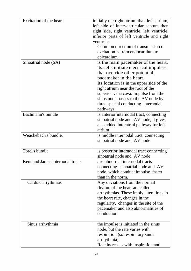
178
Excitation of the heart
initially the right atrium than left atrium,
left side of interventricular septum then
right side, right ventricle, left ventricle,
inferior parts of left ventricle and right
ventricle
Common direction of transmission of
excitation is from endocardium to
epicardium.
Sinoatrial node (SA)
is the main pacemaker of the heart,
its cells initiate electrical impulses
that override other potential
pacemaker in the heart.
Its location is in the upper side of the
right atrium near the root of the
superior vena cava. Impulse from the
sinus node passes to the AV node by
three special conducting internodal
pathways.
Bachmann's bundle
is anterior internodal tract, connecting
sinoatrial node and AV node, it gives
also added interatrial pathway for left
atrium
Weackebach's bundle.
is middle internodal tract connecting
sinoatrial node and AV node
Torel's bundle
is posterior internodal tract connecting
sinoatrial node and AV node
Kent and James internodal tracts
are abnormal internodal tracts
connecting sinoatrial node and AV
node, which conduct impulse faster
than in the norm.
Cardiac arrythmias
Any deviations from the normal
rhythm of the heart are called
arrhythmias. These imply alterations in
the heart rate, changes in the
regularity, changes in the site of the
pacemaker and also abnormalities of
conduction
Sinus arrhythmia
the impulse is initiated in the sinus
node, but the rate varies with
respiration (so respiratory sinus
arrhythmia).
Rate increases with inspiration and

179
decreases with expiration
Non- sinus rhythm genertation of impulses does not happen
in sunus node, changes in the site of the
pacemaker (atrioventricular escape
rhythm, ventricular escape rhythm)
Atrioventricular escape rhythm is non-sinus rhythm if SA node fails then
a lower escape pacemaker site in the
atrioventriular junction ( with rate 40-60
beats per min) take over to drive the
heart
Ventricular escape rhythm
is non-sinus rhythm if SA node fails
lower escape pacemaker site in the
ventricles (with rate 25-45 beats per
min) may take over to drive the heart

180
To master the material on the subject study the following flow charts:
Analysis of ECG
I.Analysis of heart rhythm
Rhythmicity analysis of
P wave and QRS
appearance
Analysis of P waves Ananlysis of QRS
complexes
P waves and QRS
complexes appear
simultaneously with rate
60- 80 per minute
P waves: 1) positive in
I,II and negative in
aVR; 2)have
permanent form; 3) P
waves appear before
each QRS
QRS complexes
appear
rhythmically. Have
normal duration
(0,06- 0,1sec)
Sinus rhythm of heart
II. Determination of heart electrical axis
visual method on tables
Analysis of amplitude of waves of QRS
complexes in I, II, III, aVR, aVL, aVF.
Analysis of amplitude of waves of QRS
complexes in I and III.
Variants of heart electrical axis: 1) normal position of heart electrical axis; 2) horizontal
position of heart electrical axis; 3) vertical position of heart electrical axis; 4) heart electrical
axis deviation to the left; 5) heart electrical axis deviation to the right; 6) indefinite position of
heart electrical axis (type SI-SII-SIII)
Analysis of separate fragments of ECG
P wave PQ interval QRS
complex ST segment T wave QT
interval Ampli-
tude,durati
on,pola-
rity and
form
duration Amplitude,
duration of
all complex
and sepa-
rate waves
difference
relative to
isoelectric
line
Ampli-
tude,duratio
n,pola-rity
and form
Duration
0,12-0,2sec
duration no
more than
0,1sec,ampli-
tude substan-
tially differs in
different leads
on isoline positive in
most leads,
obligatory
negative in
aVR,can be
negative in
V1
0,35-
0,40sec
NORM

181
Flow chart 1
Altered automaticity of the sino-atrial node
Sinus
bradycardia
Sinus
(respiratory)
arrhythmia
Increased
heart rate to
90-180 beats
per minute
Slow heart
rate 59-40
beats per
minute
Alterations
of cardiac
rhythm
associated
with
respiration
Constant
bradycardia
Decreased
duration of
the R-R
intervals
Regular
sinus rhythm
Increased
duration of
the R-R
intervals
Alterations
of duration
of the R-R
intervals
Bradycardia-
tachycardia
syndrome
Regular
sinus rhythm
Sinus rhythm
Periodic
ectopic (non-
sinus)
rhythms and
SA block
Sick-sinus
syndrome
Sinus
tachycardia
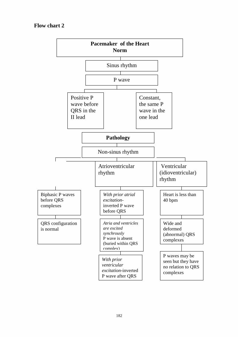
182
Flow chart 2
Pacemaker of the Heart
Norm
Sinus rhythm
P wave
Positive P
wave before
QRS in the
II lead
Constant,
the same P
wave in the
one lead
Pathology
Non-sinus rhythm
Atrioventricular
rhythm
Ventricular
(idioventricular)
rhythm
With prior atrial
excitation-
inverted P wave
before QRS
Atria and ventricles
are excited
synchrously
P wave is absent
(buried within QRS
complex)
With prior
ventricular
excitation-inverted
P wave after QRS
Biphasic P waves
before QRS
complexes
Wide and
deformed
(abnormal) QRS
complexes
QRS configuration
is normal
Heart is less than
40 bpm
P waves may be
seen but they have
no relation to QRS
complexes

183
Flow chart 3
Premature contractions
Atrioventricular
premature
contraction
Ventricular
premature
contraction
P wave is absent
or negative P wave
before of after
QRS complex
QRS configuration
is normal
Presence of not
fully
compensatory
pause
Changes of P wave
polarity, its
deformation
Wide and
deformed ORS, ST
segment and T
wave opposite in
direction to the
main deflection of
QRS complex
QRS configuration
is normal
P wave is absent
Presence of fully
compensatory
pause
Atrial
premature
contraction
Presence of not
fully
compensatory
pause

184
Flow chart 4

185
Flow chart 5
Hypertrophy of atriums
Analysis of P waves
P waves are dilate (it
duration more than
0,1sec) and often two-
humped
P waves are high( it
amplitude 3mm and
more) and often
sharp
Hypertrophy of left atrium Hypertrophy of right atrium
Hypertrophy of ventricles
I. analysis of QRS complexes
Horizontal position of heart electrical
axis or it declination to the left, R wave
is high in aVL
(it amplitude more than 11mm), V5-6 (
bigger than V4), deep S in V1-2, R
amplitude in V5 + S amplitude in V1
or in V5 > 35mm
Vertical position of heart electrical
axis, declination to the right or it
indefinite position. R is high in VF
(amplitude more than 11mm, S wave
in V5-6 > R or S=R, S amplitude V5-
6 >7mm, R wave in V1-2 = S or R>S
Hypertrophy of left ventricle Hypertrophy of right ventricle
II. Analysis of ST segments and T waves
Depression of ST segment and
negative T wave in I, aVL, V5-6
Depression of ST segment and
negative T wave in III, aVF, V1-2
Combination of changes of QRS complexes and ST
segments and T waves
Hypertrophy of right ventricle
with it overload
Hypertrophy of left ventricle
with it overload
Hypertrophy of heart chambers

186
Flow chart 6

187
Flow chart 7
Flow chart 8

188
Suggested Reading List
Required Reading
1. Internal Diseases, an Introductory Course. Edited by V.Vasilenko and
Grebenev, Mir Publishers, Moscow, 1990
2. Clinical Examination, Edited by Jonh Macleod, Jonh Munro, Churchill
Livingsone, 2000
3. Medicine, Edited by K. George Mathew, Praveen Aggarwal, Elsevier, 2004
4. Lecture: ECG in hypertrophy of the atriums and ventricles, myocardial
infarction, cardiac rhythm disorders (arrhythmias).
5. Methodical guideline for students
6. Flow charts for Practice
7. Website of department: www.cardiology.dsmu.edu.ua
Additional Reading
1. Textbook of Physical Diagnosis. History and Examination / Mark H. Swartz
2002, W. B. Saunders Company Pennsylvania
2. History and Physical Examination . Current Clinical Strategies, Edited Paul
P. Chan, Peter, J. Winkle, 2005
3. Davidson‘s Medicine, Nicholas A. Boon, Nicki R. Colledge, Davidson,
2006
Guide Sequence of Actions.
Algorithm of actions at analysis of ECG
STEP 1. Estimating possible technical defects of the curve (―induction‖) which
can arise at overlapping of alternating current or as a result of inappropriate
grounding of the equipment, as well as at an insufficient contact of electrodes
with the body. An obligatory check of the accuracy of control millivolt adjusted
before ECG.
STEP 2. Determining rhythm characteristics (sinus or nonsinus), regular or
irregular, measuring interval R-R and determining cardiac contraction rate.
STEP 3. Determining the position of electrical axis QRS in the frontal area
(angle alpha) by amplitude (algebraic sum) of waves QRS in standard leads I
and Ш and giving their assessment (R.Y. Pismenny‘s tables).
STEP 4. Determining time of local electronegativeness (time of pulse
propagation from endocardium to epicardium – in V1 for the right ventricle, іn
the position V6 for the left ventricle) and estimating it.
STEP 5. Determining systolic index – the ratio of electric systole to cardiac
cycle R-R, in percent and comparing it with the corresponding quantities (R.Y.
Pismenny‘s tables).
STEP 6. Analyzing amplitude, duration, configuration, position of the waves
and intervals of ECG and estimating them.
Methods and techniques of ECG registration Necessary equipment.
1. Electrocardiograph (components):
189
• receiving facility – electrodes applied to a patient‘s body should be well wetted
in advance with isotonic solution or resistant gel for a better contact.
• amplifiers that increase low voltage of the heart electromotive force so that it
can be registered.
• galvanometer,which measures voltage.
• recording unit (tape transport mechanism and time register)
• power unit
2. Order of electrode application.
(-) – negative electrode;
(+) – positive electrode;
red electrode – to the right arm;
yellow electrode – to the left arm;
green electrode – to the left leg;
black electrode – to the right leg (grounding wires)
Standard leads:
І- right arm (-) + left arm(+)
ІІ-right arm (-) + left leg(+)
ІІІ-left leg(+)+ left arm(-)
Amplified unipolar leads:
aVR – from the right arm;
aVL – from the left arm;
aVF – from the left leg.
Chest leads:
V1 – IV intercostal space at the right sternal border
V2– IV intercostal space at the left sternal border
V3– IV rib parasternal boundary on the left
V4– V intercostal space midclavicular boundary on the left
V5– V intercostal space anterior inguinal boundary on the left
V6– V intercostal space midinguinal boundary on the left
3. Writing down a patient‘s full name, age (date of birth); date and time of the
investigation, registering control millivolt followed by ECG registration.
4. After the procedure is accomplished, ECG device must be switched off,
electrodes should be removed.
5. Carrying out ECG analysis.
6. Making a conclusion.
Arrhythmias associated with altered automaticity of the sinoatrial node
Sinus tachycardia- increased automaticity of the sinus node
Impulses are initiated in the sinus node at a rate from 90 to 180 beats per minute.
It develops on intensified effect of the sympathetic nervous system.
Physiological conditions: Physical and emotional exertion.
Pathological conditions: myocarditis, hemorrhage, shock, heart failure, fever,
hyperthyroidism
1. increased heart rate from 90 to 180 beats per minute
2. decreased durations of the R-R interval

190
3. regular sinus rhythm
Sinus Bradycardia- decreased automaticity of sinus node
Increased influence of parasympathetic nervous system of the heart.
Physiological- in sportsmen
Pathological-hypothyroidism, increased intracranial pressure, typhoid fever,
jaundice, some drugs.
1. slow heart rate (less than 60 bpm)
2. increased durations of the R-R interval
3. regular sinus rhythm
Sinus Arrhythmias
The impulse is initiated in the sinus node, but the rate varies with respiration
(respiratory sinus arrhythmia)
Rate increases with inspiration and decreases with expiration
May be in the norm - in children, in young adults, in patients convalescing from
infection diseases
1. R-R intervals length may vary ( more than 0,16 seconds), it is associated with
respiration
2. Sinus rhythm
Sick Sinus Syndrome
1. Constant bradycardia
2. Periodic ectopic rhythm(escape rhythms)
3. SA block
4. Bradycardia-Tachycardia syndrome
Escape Passive Rhythms (Non-Sinus Rhythms)
They appear secondary to depression of the higher sinus pacemaker.
Atrial Rhythm - biphasic P wave before QRS complex.
Junctional (AV) rhythm- heart rate is 40-60 beats per minute, QRS
configuration is normal.
1. with prior atrial excitation- inverted P wave before QRS (retrograde
impulse conduction back into atria)
2. atria and ventricles are excited synchronously
without P wave, it is buried within QRS complex
3. with prior ventricular excitation - inverted P wave after QRS
(because of retrograde atrial activation)
Ventricular Rhythm- incomplete AV block
1. Heart rate is less than 40 beats per minute
2. wide and deformed (abnormal) QRS complex
3. P wave may be seen but it has no relation to QRS complex
191
Premature Complexes (Extrasystolic Arrhythmia)
Classification
Rate of occurrence
1. rare (<6 per minute)
2. frequent (>6 per minute)
3. single
4. couplet - couplet of two ventricular premature contractions (VPCs)
(uniform or multiform)
5. triplet - three VPCs in row (three and more ventricle tachycardia)
6. group VPCs- group of three and more VPCs
If extrasystoles have certain order for their occurrence
Allorhythmias
1. bigeminy (1N+1E)
2. trigeminy (2N+1E)
3. quadrigeminy (3N+1E)
Configuration
1. uniform VPCs (unifocal VPCs) - they have the same site of their occurrence
so in the same lead they have equal configuration.
2. multiform VPCs (multifocal VPCs) from different sites so they have different
shapes in the same lead
R- on T phenomenon – VPC occurs on or near the peak of previous T wave
Interpolated premature ventricular contraction occurring between two normal
beats without compensatory pause.
Lawn’s classification
1Grade - 1-30 VPC per hour
2 Grade >30 VPC per hour
3 Grade - multifocal VPCs
4 A Grade - couplet of VPCs
4B Grade - group VPCs
5 Grade - R on T phenomenon
Paroxysmal Tachycardia
This is sudden acceleration of the cardiac rhythm (from 180 to 240 beats per
minute). This impulses arise from ectopic focus because its high activity
inhibits the activity of sinoatrial node.
Paroxysmal atrial tachycardia
1. Sudden acceleration of the heart rate 140 to 220 beats per minute
2. Change P wave-negative or biphasic P wave
3. QRS complex configuration is normal
4. Regular rhythm.
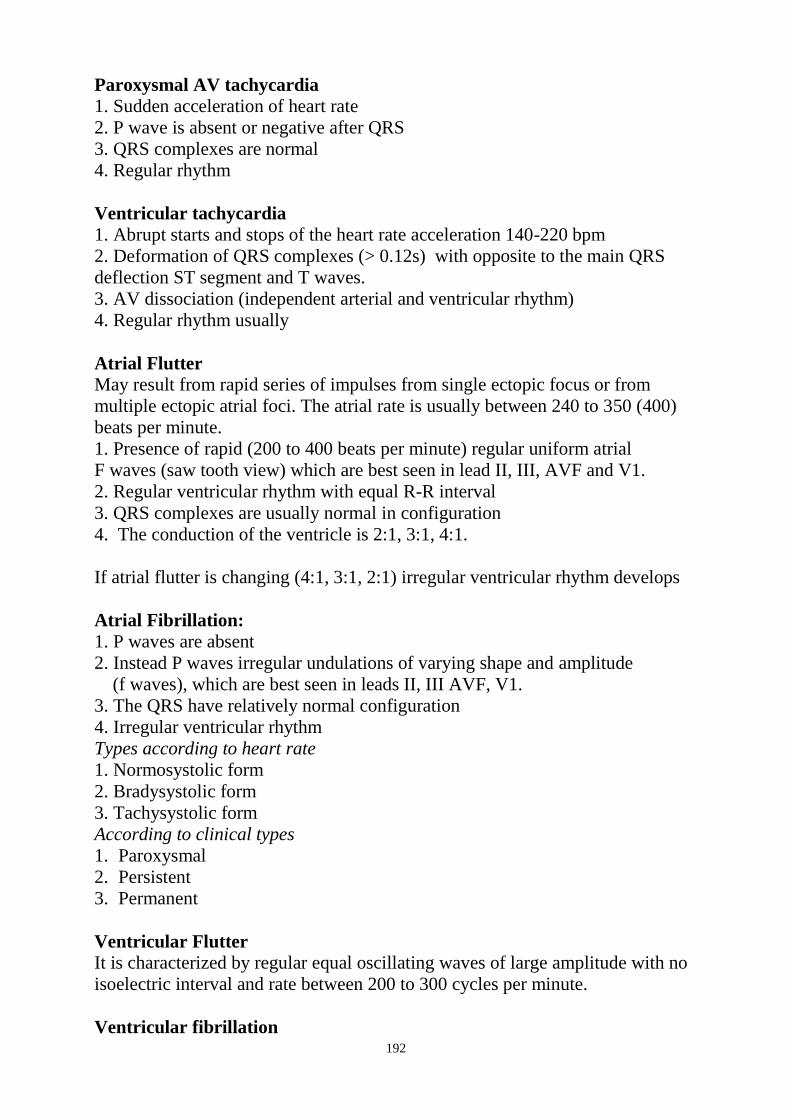
192
Paroxysmal AV tachycardia
1. Sudden acceleration of heart rate
2. P wave is absent or negative after QRS
3. QRS complexes are normal
4. Regular rhythm
Ventricular tachycardia
1. Abrupt starts and stops of the heart rate acceleration 140-220 bpm
2. Deformation of QRS complexes (> 0.12s) with opposite to the main QRS
deflection ST segment and T waves.
3. AV dissociation (independent arterial and ventricular rhythm)
4. Regular rhythm usually
Atrial Flutter
May result from rapid series of impulses from single ectopic focus or from
multiple ectopic atrial foci. The atrial rate is usually between 240 to 350 (400)
beats per minute.
1. Presence of rapid (200 to 400 beats per minute) regular uniform atrial
F waves (saw tooth view) which are best seen in lead II, III, AVF and V1.
2. Regular ventricular rhythm with equal R-R interval
3. QRS complexes are usually normal in configuration
4. The conduction of the ventricle is 2:1, 3:1, 4:1.
If atrial flutter is changing (4:1, 3:1, 2:1) irregular ventricular rhythm develops
Atrial Fibrillation:
1. P waves are absent
2. Instead P waves irregular undulations of varying shape and amplitude
(f waves), which are best seen in leads II, III AVF, V1.
3. The QRS have relatively normal configuration
4. Irregular ventricular rhythm
Types according to heart rate
1. Normosystolic form
2. Bradysystolic form
3. Tachysystolic form
According to clinical types
1. Paroxysmal
2. Persistent
3. Permanent
Ventricular Flutter
It is characterized by regular equal oscillating waves of large amplitude with no
isoelectric interval and rate between 200 to 300 cycles per minute.
Ventricular fibrillation

193
It is the irregular waves of varying amplitude and shape occurring 250 to 500
times per minute. Clinical cardiac arrest is present, there is no cardiac output
Revision Questions
Q1. Choose correct item:
A. Sinus rhythm with normal heart rate
B. Sinus rhythm with high heart rate (sinus tachycardia)
C. Sinus rhythm with low heart rate (sinus bradycardia)
D. Sinus rhythm with arrhythmia (sinus arrhythmia)
E. It is sinus rhythm
Q2. Choose correct item:
A. Sinus rhythm with normal heart rate
B. Sinus rhythm with high heart rate (sinus tachycardia)
C. Sinus rhythm with low heart rate (sinus bradycardia)
D. Sinus rhythm with arrhythmia (sinus arrhythmia)
E. Non sinus rhythm
Q3. Choose correct item:
A. Sinus rhythm with normal heart rate
B. Sinus rhythm with high heart rate (sinus tachycardia)
C. Sinus rhythm with low heart rate (sinus bradycardia)
D. Sinus rhythm with arrhythmia (sinus arrhythmia)
E. It is sinus rhythm
Q4. The following changes of the ECG can be fined:
A. Posterior Q-wave-MI, acute stage
B. Posterior Q-wave-MI, subacute stage
C. Posterior Q-wave-MI, stage of the cicatrisation
D. Ischemia of the anterior wall
E. Ischemic lesion of the anterior wall

194
Q5. The following changes of the ECG can be fined:
A. Ischemia of the anterior wall
B. Ischemia of the lateral wall
C. Anterior non-Q-wave-MI
D. Lateral wall Q-wave-MI
E. Lateral wall non-Q-wave-MI
Q6. The following changes of the ECG can be fined:
A. Anterior Q-wave-MI, acute stage
B. Lateral wall non-Q-wave-MI, acute stage
C. Anterior non-Q-wave-MI, subacute stage
D. Lateral wall Q-wave-MI, subacute stage
E. Anterior Q-wave-MI, stage of the cicatrisation
Q7. Characterize the following ECG:
A. Atrial premature beat (extrasystole)
B. Ventricular premature beat (extrasystole)
C. AV- nodal premature beat (extrasystole) with preceding depolarization of the
atria
D. AV-nodal beat (extrasystole) with preceding depolarization of the ventricles
E. Atrial flutter
Q8. Characterize the following ECG :
A. Atrial premature beats (extrasystoles)
III
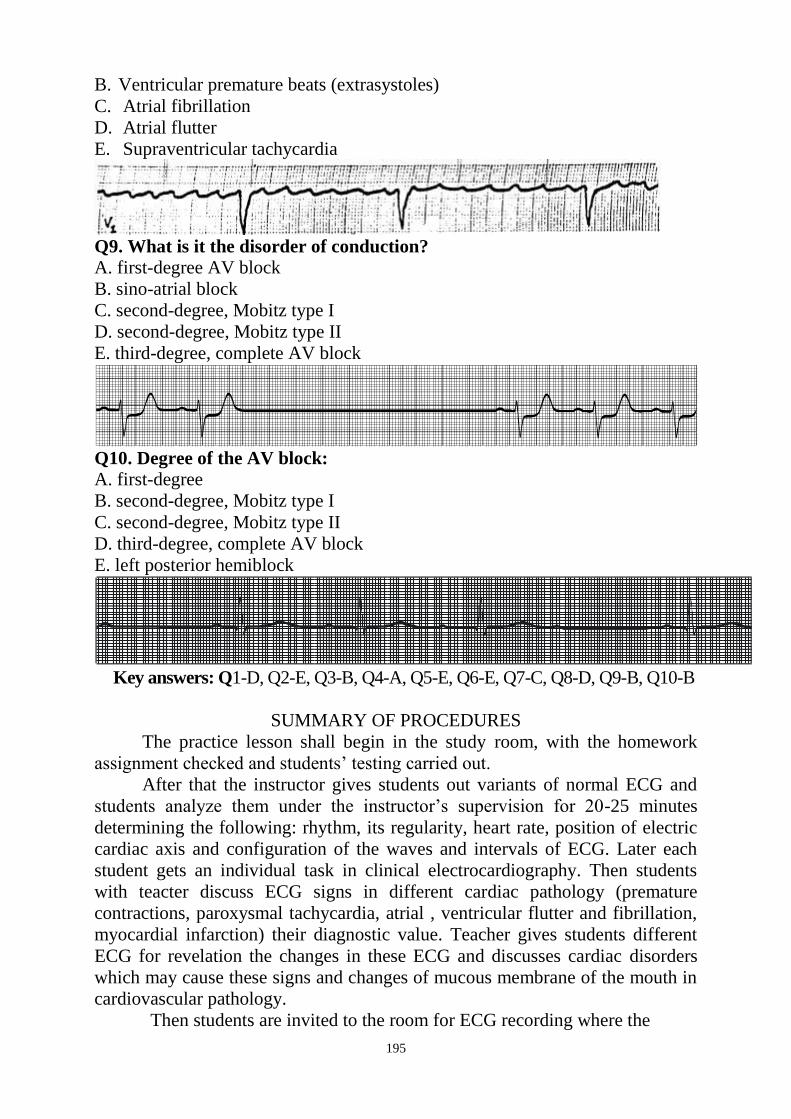
195
B. Ventricular premature beats (extrasystoles)
C. Atrial fibrillation
D. Atrial flutter
E. Supraventricular tachycardia
Q9. What is it the disorder of conduction?
A. first-degree AV block
B. sino-atrial block
C. second-degree, Mobitz type I
D. second-degree, Mobitz type II
E. third-degree, complete AV block
Q10. Degree of the AV block: A. first-degree
B. second-degree, Mobitz type I
C. second-degree, Mobitz type II
D. third-degree, complete AV block
E. left posterior hemiblock
Key answers: Q1-D, Q2-E, Q3-B, Q4-A, Q5-E, Q6-E, Q7-C, Q8-D, Q9-B, Q10-B
SUMMARY OF PROCEDURES
The practice lesson shall begin in the study room, with the homework
assignment checked and students‘ testing carried out.
After that the instructor gives students out variants of normal ECG and
students analyze them under the instructor‘s supervision for 20-25 minutes
determining the following: rhythm, its regularity, heart rate, position of electric
cardiac axis and configuration of the waves and intervals of ECG. Later each
student gets an individual task in clinical electrocardiography. Then students
with teacter discuss ECG signs in different cardiac pathology (premature
contractions, paroxysmal tachycardia, atrial , ventricular flutter and fibrillation,
myocardial infarction) their diagnostic value. Teacher gives students different
ECG for revelation the changes in these ECG and discusses cardiac disorders
which may cause these signs and changes of mucous membrane of the mouth in
cardiovascular pathology.
Then students are invited to the room for ECG recording where the
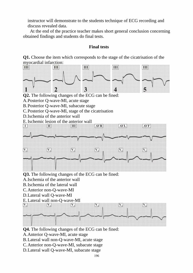
196
instructor will demonstrate to the students technique of ECG recording and
discuss revealed data.
At the end of the practice teacher makes short general conclusion concerning
obtained findings and students do final tests.
Final tests
Q1. Choose the item which corresponds to the stage of the cicatrisation of the
myocardial infarction:
Q2. The following changes of the ECG can be fined:
A. Posterior Q-wave-MI, acute stage
B. Posterior Q-wave-MI, subacute stage
C. Posterior Q-wave-MI, stage of the cicatrisation
D. Ischemia of the anterior wall
E. Ischemic lesion of the anterior wall
Q3. The following changes of the ECG can be fined:
A. Ischemia of the anterior wall
B. Ischemia of the lateral wall
C. Anterior non-Q-wave-MI
D. Lateral wall Q-wave-MI
E. Lateral wall non-Q-wave-MI
Q4. The following changes of the ECG can be fined:
A. Anterior Q-wave-MI, acute stage
B. Lateral wall non-Q-wave-MI, acute stage
C. Anterior non-Q-wave-MI, subacute stage
D. Lateral wall Q-wave-MI, subacute stage
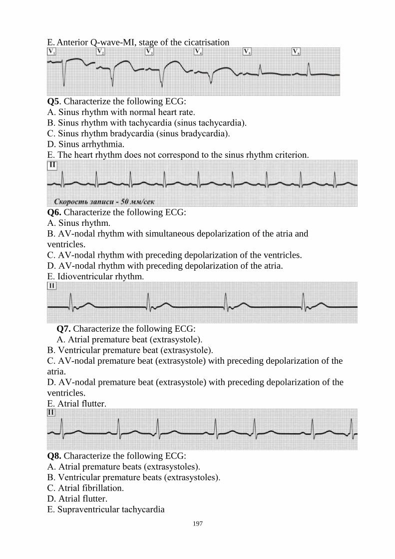
197
E. Anterior Q-wave-MI, stage of the cicatrisation
Q5. Characterize the following ECG:
А. Sinus rhythm with normal heart rate.
В. Sinus rhythm with tachycardia (sinus tachycardia).
С. Sinus rhythm bradycardia (sinus bradycardia).
D. Sinus arrhythmia.
Е. The heart rhythm does not correspond to the sinus rhythm criterion.
Q6. Characterize the following ECG:
А. Sinus rhythm.
В. АV-nodal rhythm with simultaneous depolarization of the atria and
ventricles.
С. АV-nodal rhythm with preceding depolarization of the ventricles.
D. АV-nodal rhythm with preceding depolarization of the atria.
Е. Idioventricular rhythm.
Q7. Characterize the following ECG:
А. Atrial premature beat (extrasystole).
В. Ventricular premature beat (extrasystole).
С. АV-nodal premature beat (extrasystole) with preceding depolarization of the
atria.
D. АV-nodal premature beat (extrasystole) with preceding depolarization of the
ventricles.
Е. Atrial flutter.
Q8. Characterize the following ECG:
А. Atrial premature beats (extrasystoles).
В. Ventricular premature beats (extrasystoles).
С. Atrial fibrillation.
D. Atrial flutter.
Е. Supraventricular tachycardia
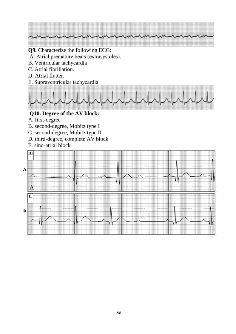
198
Q9. Characterize the following ECG:
А. Atrial premature beats (extrasystoles).
В. Ventricular tachycardia
С. Atrial fibrillation.
D. Atrial flutter.
Е. Supraventricular tachycardia
Q10. Degree of the AV block:
A. first-degree
B. second-degree, Mobitz type I
C. second-degree, Mobitz type II
D. third-degree, complete AV block
E. sino-atrial block
III
II
А
А
Б

199
Health Ministry of Ukraine
Donetsk National Medical University
Approved
at the meeting of
Propedeutic and Internal Medicine
Department
Head of department
Associate Member of NAMSc of
Ukraine,
Professor G. A. Ignatenko
« _______ »_____________ 2011 р.
STUDENT’S SELF-STUDY GUIDELINES FOR
PRACTICE ACTIVITIES
Donetsk 2011
Subject Propedeutics of the Internal Medicine
Module 1 The main methods of examination of
patient and the main symptoms and
syndromes on internal diseases course
Topic 5 Module Methods of examination and
interpretation data of laboratory
investigation in endocrine pathology,
blood disorders, musculoskeletal
disordes.
Practice Examination of the blood system.
Enquiry, objective examination of
patients with pathology of blood system.
Added methods of investigation in
hematology. Clinical estimation of blood
investigation. Changes of tunica mucosa
of mouth in blood pathology. Caring for
those patients and first medical aid.
Course 2
Faculty Dentistry

200
Importance of the Subject: Ability to interpret a clinical blood analysis and
reveal clinical signs and syndromes of blood system disorders is necessary for
the doctor of any speciality because changes in peripheral blood are both
specific for haematological pathology and for disorders of other organs and
systems.
Key Objective: To be able to estimate the blood analysis, make inquiries about
haematological pathology, carry out phisical examination of patients with blood
disorders and reveal the main syndromes, symptomes and laboratory criterions
of blood pathology.
Specific Goals:
1. To learn how to enquire patients with haematological pathologies about their
most disturbing complaints
2. To interpretate clinical blood analysis in the norm and pathology
3. To develop skills in carrying out the inspection and palpation of lymph nodes,
inspection, palpation and percussion of the bones, percussion and palpation of
the spleen
4. To be able to reveal changes of tunica mucosa of mouth in blood pathology
5. To develop skills in revealing the main symptoms and syndromes of blood
pathology (anaemia, haemorrhagic syndrome, disseminated intravascular
clotting syndrome, syndrome of enlarged lymph nodes, syndrome of
leukemia)
Level of Knowledge and Skills before the Practice:
1. To have knowledge of anatomical details and projection of the lymph nodes
and spleen (Described in the Human Anatomy course)
2. To distinguish the cellular elements of blood on their morphological
properties (Described in the Histology course)
3. To carry out clinical analysis of the blood – blood taking, preparation of
smears (for microscopic examination), cell count, ESR determination (Described
in the Physiology course)
4. To have knowledge of the links of the haemopoiesis, coagulation cascade
(Described in the Histology course)
Questions for Self-Assessment of the Pre-Practice Knowledge
Q1. A patient with tissue trauma was taken a blood sample for the
determination of blood clotting parameters. Specify the right sequence of
extrinsic pathway activation.
A. III – VIII: TF – Xa
B. III – VIIa – Xa
C. III – IV – Xa
D. IV – VIII: TF – Xa
E. IV – VIIa – Xa
201
Q2. A couple came for medical genetic counseling. The man has hemophilia,
the woman is healthy and there were no cases of hemophilia in her family. What
is the risk of having a sick child in this family?
A. 25%
B. 0
C. 100%
D. 75%
E. 50%
Q3. In the blood smear, stained according to Romanovsky-Giemsa method,
there are 20% big (20 mcm in diametre), rounded cells with pale-basophilic
cytoplasm and bean-shaped nucleus. How is this condition characterised
clinically?
A. Neutrophilosis
B. Leukopenia
C. Monocytosis
D. Lymphocytosis
E. Reticulocytosis
Q4. Blood sampling for bulk analysis is recommended to be performed on an
empty stomack and in the morning. What changes in blood composition can
occur if to perform blood sampling after food intake?
A. Reduced contents of erythrocytes
B. Increased contents of erythrocytes
C. Increased contents of leukocytes
D. Increased plasma proteins
E. Reduced contents of thrombocytes
Q5. Which of the following are the last precursors of red blood cells?
A. Platelets
B. Normocytes
C. Monocytes
D. Reticulocytes
E. Erythroblasts
Answer Keys: B, B, C, C, D
The following printed materials can be of help to improve your pre-practice
knowledge and skills: 1. Test book of Medical Physiology / Arthur C. Guyton, John I. Hall 2001, W.
B. Saunders company Pennsylvania 2001
2. Gray's Anatomy. Edited by T. Pickering Pick, F.R.C.S., 1995
3. M. Prives, N. Lysenkov, V. Bushovich; Human Anatomy
Contents of Practice
Topics of Theory:
1. The main complaints of patients with different blood pathology.
2. Clinical study of the blood in the norm and pathology. Clinical estimation of
blood investigation.

202
3. Changes of tunica mucosa of mouth in blood pathology.
4. The main clinical and laboratory features of anaemia (iron deficiency
anaemia, vitamin B12 and folate deficiency anaemia, congenital and acquired
haemolyic anaemia)
5. Haemorrhagic syndrome in thrombocytopenia, coagulopathy, hemorrhagic
vasculatis.
6. Laboratory investigations of haemorrhagic syndrome, coagulation tests
7. Clinical and laboratory signs and of disseminated intravascular clotting
8. Main clinical symptomes and laboratory signs in leukemia
Practical skills:
Students should be able to demonstrate mastery of the following pratical skills
1. To estimate clinical blood analysis
2. To enquire patients with haematological pathologies about their most
disturbing complaints
3. To be able to reveal and interpretate changes of tunica mucosa of mouth in
blood pathology
4. To carry out the inspection and palpation of lymph nodes, inspection,
palpation and percussion of the bones, spleen
5. To reveal the main laboratory criterions, clinical symptoms and syndromes of
blood pathology
Required Glossary to Practice
Term Definition
Colour index Haemoglobin content (haemoglobin saturation)
of each erythrocyte, it compare with normal
saturation= 1
Colour index = 3 * Hb
RBC(first three figures).
Reticulocyt Immature red cells; last precursor of red cells.
They have reticular formation.
Normoblasts
Immature nuclear (still containing nuclei) red
cells. They delivered into the blood from bone
marrow in its erythropoietic hypofunction.
Relative polycythemia
The concentration of the RBC becomes greater
than normal (but total red cell mass is normal)in
the circulating blood. This occurs as a result of
loss blood plasma. (dehadration -low fluid
intake,diarrhoea,vomiting, sweating)
Absolute polycythemia
(erythrocytosis)
There is an increase in the total red cell mass.
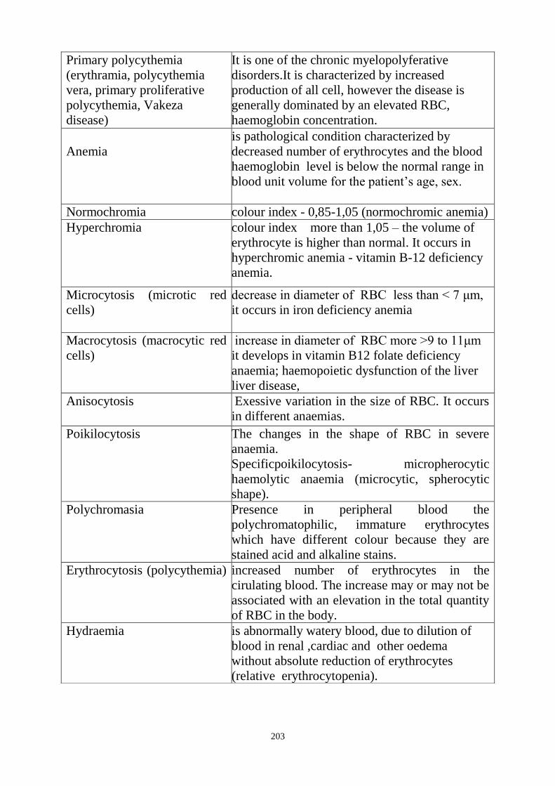
203
Primary polycythemia
(erythramia, polycythemia
vera, primary proliferative
polycythemia, Vakeza
disease)
It is one of the chronic myelopolyferative
disorders.It is characterized by increased
production of all cell, however the disease is
generally dominated by an elevated RBC,
haemoglobin concentration.
Anemia
is pathological condition characterized by
decreased number of erythrocytes and the blood
haemoglobin level is below the normal range in
blood unit volume for the patient‘s age, sex.
Normochromia colour index - 0,85-1,05 (normochromic anemia)
Hyperchromia
colour index more than 1,05 – the volume of
erythrocyte is higher than normal. It occurs in
hyperchromic anemia - vitamin B-12 deficiency
anemia.
Microcytosis (microtic red
cells)
decrease in diameter of RBC less than < 7 μm,
it occurs in iron deficiency anemia
Macrocytosis (macrocytic red
cells)
increase in diameter of RBC more >9 to 11μm
it develops in vitamin B12 folate deficiency
anaemia; haemopoietic dysfunction of the liver
liver disease,
Anisocytosis Exessive variation in the size of RBC. It occurs
in different anaemias.
Poikilocytosis
The changes in the shape of RBC in severe
anaemia.
Specificpoikilocytosis- micropherocytic
haemolytic anaemia (microcytic, spherocytic
shape).
Polychromasia
Presence in peripheral blood the
polychromatophilic, immature erythrocytes
which have different colour because they are
stained acid and alkaline stains.
Erythrocytosis (polycythemia)
increased number of erythrocytes in the
cirulating blood. The increase may or may not be
associated with an elevation in the total quantity
of RBC in the body.
Hydraemia
is abnormally watery blood, due to dilution of
blood in renal ,cardiac and other oedema
without absolute reduction of erythrocytes
(relative erythrocytopenia).

204
Required Glossary to Practice
Term Definition
Myeloplastic syndrome
(panmyelophtisis)
Is a large group of conditions of various aetiology and
pathogenesis whose main clinical syndroms are
determined by the inhibition of blood formation in the
bone marrow
Hypoplastic and
aplastic condition
Incomplete or complete inhibition of bone marrow
Panmyelophthisis In which the regeneration function of bone marrow is
inhibited in all directions
Haemorrhagic
syndrome
There are hemorrhagic diseases in which bleeding
disorders are main leading symptomes (bleeding into
skin, mucous membranes, muscles, joints, bleeding
from wounds, easy bruising) in haemophilia,
thrombocytopenic purpura
Immune idiopathic
thrombocytopenic
purpura (Werlhof‘s
disease)
Increased destruction of platelets due to production
anti-thrombocytic antibodies which fix on surface of
platelets and damage them
Haemophilia Is X – linked recessive genetic disorders of
coagulation. It results from reduction of coagulation
factors (haemophilia A - VIII factor, haemophilia B -
IX factor, haemophilia C - X factor)
Leukaemia Is characterized by a failure of cell maturation
(differentiation), proliferation of immature cells which
fill up the bone marrow so replacement (metaplasia) of
these pathological cells for normal cells of the
haemopoietic organs develops
Hiatus leucaemicus
Is typical for acute leukaemia. Presence blasts, mature
forms in leucocyte formule in peripheral blood and
absence intermediate forms

205
Flow chart 1
Syndrome of Anaemia
Anaemia
Clinical Signs
Complaints: weakness, dizziness,
fainting, fast fatigue, dyspnoea, ear
noise, palpitation, decreased appetite.
Inspection: pallor of skin
and mucous membrane,
trophic changes of skin and mucous membrane
Physical examination: dyspnoea, tachicardia,
functional systolic murmurs over heart and
vessels, cervical venous
hum.
Wax-like pailness
Pailness with lemon like tinge, smooth and glossy
tongue.
Pailness with golden yellow
tinge
Pica, dyspeptic complaints, dysphagia.
Burning sensation of the tongue, skin paraesthesia, changed gait, deranged
sleep, irritability.
Increased body temperature Increase in spleen and
liveer
Laboratory investigation
Blood analysis
Haemoglobin and RBC are reduced,
anisocytosis, poikilocytosis, increased ESR.
Other investigations
Hypochromia, microcytosis, frequently
thrombocytoleucopenias, eosinopenia
Hyperchromia, macro- and megalocytosis, Jolly boddies, Cabot rings, neutropenia,
eosinopenia, relative lymphocytosis, thrombocytopenia.
Normochromia, microspherocytosis, sicle
cells.
Decrease in plasma iron
Insignificant increase in unconjugated bilirubin
Increase in unconjugated bilirubin
in the blood, increase in stercobilin ,
decrease in osmotic resistance of
RBC .
Absence of reticulotes, thrombocytopenia.,
relative monocytosis and lymphocytosis .
Bone marrow study:
Hypocellular and acellular marrow.
Based on the cause of aneamia Posthaemorrhagic
Acute and chronic
Due to inadequate
production of red cells
Haemolytic
congen
Iron deficiency В12 and folate deficiency Hypoplastic and aplastic
metaplastic
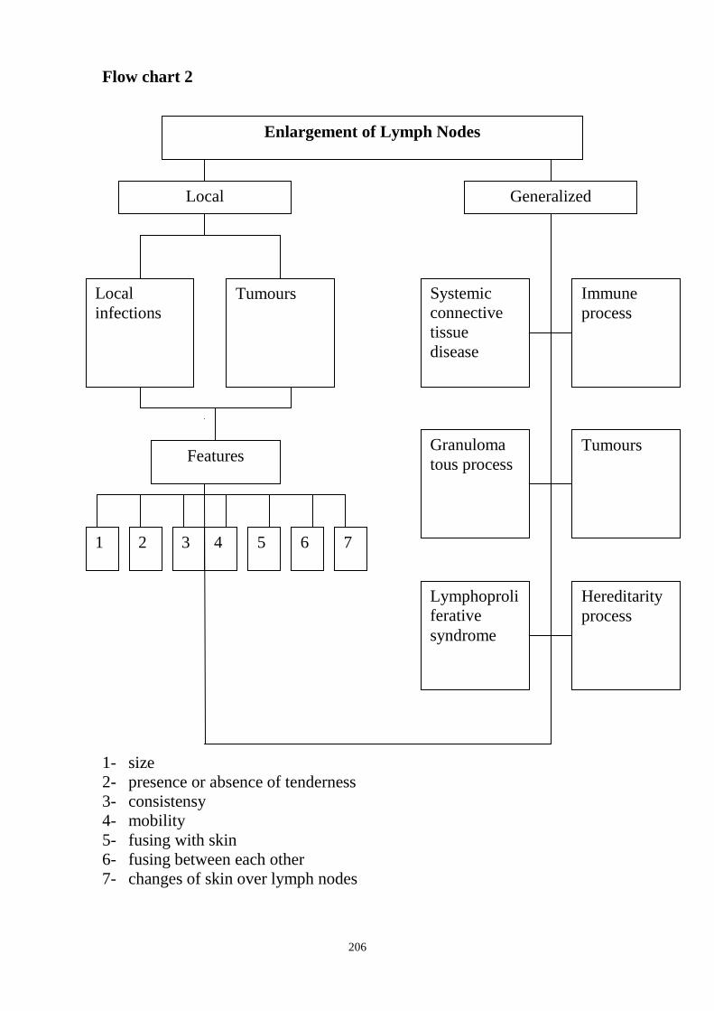
206
Flow chart 2
1- size
2- presence or absence of tenderness
3- consistensy
4- mobility
5- fusing with skin
6- fusing between each other
7- changes of skin over lymph nodes
Local Generalized
Tumours Local
infections
Systemic
connective
tissue
disease
Immune
process
Features
1 2 3 4 5 6
Tumours Granuloma
tous process
Hereditarity
process
Lymphoproli
ferative
syndrome
Enlargement of Lymph Nodes
7

207
Flow chart 3
Reasons Of Enlargement Of The Lymph Nodes
Enlarged lymph nodes
Common reasons of enlargement of lymph nodes
Cervical lymph nodes
Tonsillitis
Pharyngitis
Scarlatina (scarlet fever)
Diptheria
Lymphangioma
Tumour of thyroid gland
Submandibular lymph
nodes
Caries
Gingivitis
Tumour of larynx
Carcinoma of lips
Supraclavicular lymph
nodes
Tumour of mammary gland
Tumour of stomach (Virchow's metastasis)
Subclavicular lymph
nodes
Tumour of thyroid gland
Tumour of the lung
Axillary lymph nodes
Furunculosis
Paronychia
Pararitium (felon,whitlow)
Tumour of mammary gland
Tumour of lung
Inguinal lymph nodes
Paronychia, paraitium
Blister foot
Syphilis
Gonorrhea
Bartholinitis
General enlargement of
lymph nodes
Tbs
Sarcoidosis
Syphilis
Mononucleosis
Lymphoid leukaemia
Lymphogranulomatosis
Lymphosarcoma
Systemic connective tissue diseases
HIV
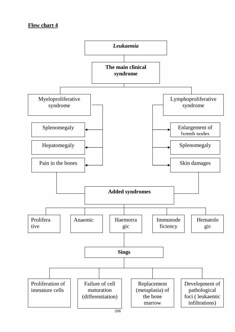
208
Flow chart 4
Leukaemia
The main clinical
syndrome
Lymphoproliferative
syndrome
Splenomegaly
Pain in the bones
Hepatomegaly
Enlargement of
lymph nodes
Splenomegaly
Skin damages
Added syndromes
Prolifera
tive
Anaemic Haemorra
gic
Immunode
ficiency
Hematolo
gic
Sings
Proliferation of
immature cells
Failure of cell
maturation
(differentiation)
Replacement
(metaplasia) of
the bone
marrow
Development of
pathological
foci ( leukaemic
infiltrations)
Myeloproliferative
syndrome

209
Qualitative Determination:
↓
Leucocyte formula: cellular
changes of the size, shape
(morphology of the cells)
Suggested Reading List
1. Internal Diseases, an Introductory Course. Edited by V.Vasilenko and
Grebenev, Mir Publishers, Moscow, 1990
2. Clinical Examination, Edited by Jonh Macleod, Jonh Munro, Churchill
Livingsone, 1986
3. Medicine, Edited by K. George Mathew, Praveen Aggarwal, Elsevier, 2004
4. Lecture: Functional investigation of the blood
5. Methodical guideline for students
6. Flow charts for Practice
7. Website of department: www.cardiology.dsmu.edu.ua
Additional Reading
1. Textbook of Physical Diagnosis. History and Examination / Mark H. Swartz
2002, W. B. Saunders Company Pennsylvania
2. History and Physical Examination . Current Clinical Strategies, Edited Paul
P. Chan, Peter, J. Winkle, 2005
3. Davidson‘s Medicine, Nicholas A. Boon, Nicki R. Colledge, Davidson, 2006
Clinical Study of the Blood
Includes:
Quantitative and
↓
Cell count
- erythrocyte count
- level of haemoglobin
- reticulocyte count
- platelet count
- leucocyte count
Red Blood study
- Red blood cells (erythrocytes)
- Haemoglobin
- Colour index
- Hemotocrit
- Reticulocyte count
The main function of ―Red‖ blood is taking part in gas exchange.
210
Haemoglobin
There are three main methods for determining Haemoglobin.
1)colorimetric (widely used in practical medficine-Sali‘s method)
2)Gasometric
3)Determination by the iron contained in the haemoglobin molecule.
The most accurate technique –cyanmethaemoglobin method.
*concentration of haemoglobin in healthy people varies from :
120-260 g/l in women
130-170 g/l in men
Neutrophils are classified into:
1) juvenile (metamylocytes) neutrophils (0%)- in peripheral blood
2) stab (band) neutrophils 2-7%
3) segmented neutrophils (50-70%) - mature neutrophils
Granules of neutrophilic leucocytes are small, stained brown-voilet colour.
Segmented neutrophils have nuclei which consist of 2 to 5 segments of various
size and shape connected by thready bridges
The main function of the neutrophils is phagocytining.
PLATELETS (Thrombocytes) are derived from marrow megakaryocytes; normal
life span is about 7-10days; blue or puple non-nucleated discs, with granular centre.
Their normal number is 180,0 – 320* 109/l per microlitre.per 1L (180,000-
320,000 per 1 ML) of blood . The main function of platelets is formation of
mechanical plug during the haemostatic response to vascular injury.
- adhesion
- aggregation
- platelet release reaction (vasoconstriction –
tromboxane A2, heparin neutralizing factor,
fibrinogen, serotonin).
IF the number of thrombocytes decreases significantly – thrombocytopenia a
tendency to haemorrhages develops. Critical figure at which haemorrhage
occurs is believed to 30-50*10 9 /L.
THROMBOCYTOPENIA
- immune (idiopathic) thombocytopenic purpura - Vergolf‘s disease (antiplatelet
antibodies) due to virus and bacterial infections (including HIV infections),
drugs (aspirin, indomethacin, penicillins, heparin)
-marrow aplasia (idiopathic,drugs)
(apalastic condition)
-marrow infilterations (leucaemia, myeloma).
-SLE,Vasculatis, DIC (dissminated intravascular coagulation)
-uraemia
-sepsis
-hypersplenism (portal hpertension, lymphomas, myeloproliferative disorders).
211
-B12, folate deficiency anaemia
THROBOCYTOSIS
Increase in risk of clots.
Occurs after haemorrhages, operations.
- polycythemia
- splenectomy
- malignant tumours
NEUTROPENIA
It is a kind of leucopenia (decrease in neutrophils). Due to disturbed or inhibted
leukopoiesis. It is observed in action of various toxic substances
1)The poisoning with benzelen, arsenic, sulphonamides.
2) ionising radiation
3) some drugs (sulpha drugs,aspirinamidobutrin, lutostic.
Actions some infections
4) typhoid fever, brucellosis, virus infections- influenza, measles,German
measles, chicken pox, infectious hepatitis.
Sometimes leukopenia is manifested in the form of agranulocytes which is
characterized by strong decrease or even absence of neutrophils and other
granulocytes in blood,when the number of granulocytes is below 0,75*10 9 /L or
total number of leukocytes is below 1*10 9 /L.
NEUTROPHILIA:
1) bacterial infections, pyogenic processes
2) different in toxications (uraemia, diabetes, acidosis, lead poisoning, digitalis,
mercury)
3) necrosis, damage of the tissues - myocardial infaction, burn, operations.
4) malignancies (carcionomas, sarcomas).
5) systemic diseases of the connective tissue
ACCELERATION OF ESR
Develops due to changes ratio between proteins in blood (globulins, albumin,
fibrinogen) and increased content of high dispersed protiens (globulins,
fibrinogen) these protiens absorbed on negative changed RBC and thus promote
their agglutination and sedimentation- infections and
inflammatory processes
1) bacterial infections (non - viral infections)
2) tbs
3) immune connective tissue diseases.
4) miocardial infarction
5) malignant tumours
6) liver diseases
7) multiple myeloma
8) amilodosis

212
9) nephrotic syndrome
10) anaemia – decreased number RBC accelarates ERS due to agglutination
arises more easier
11) pregnancy, vaccination
DECREASE IN ESR
1) polycythemia vera- Vakeza‘s disease.
2) typhoid fever.
3) viral infections
4) obstructive jaundice with significant cholemia.
ANAEMIA - is pathological condition characterized by decreased number of
red blood cells and blood haemoglobin level is below the normal range in blood
unit volume for the patient‘s age, sex.
CLASSIFICATION: Based on the cause of anaemia.
There are three main groups.
1) DUE TO BLOOD LOSS-Postthaemorrhagic anaemia.
a) Acute posthaemorrhagic anaemia- acute blood loss
(trauma, postpartum bleeding)-large volume over short period.
b) Chronic posthaemorrhagic anaemia- chronic blood loss small volume over
long period.it may considered as iron deficiency anaemia.(bleeding peptic ulcer,
haemorrhoids, excessive mentraual loss, look worms).
2) DUE TO INADEQUATE PRODUCTION OF RED CELLS:(disordered
haemopoiesis).
a) Iron deficiency anaemia due to iron deficiency-
-increased demand - physiological – pregnancy, lactation, adolescent growth,
mesuration.
-iron loss due to bleeding - hook worm infestation, menorrhagic postpartum
haemorrhage, peptic ulcer,piles, neoplastic diseases,erosion from in flammatory
drugs.
-malabsorption - inflammatory bowel disease a tropic gastritis.
-in adequate diet - postgastectomy.
b) Vitamin B12, folate deficiency anaemia.due to vitamin B12 and folate
deficiency
- malabsorption syndrome, postgastrectomy, atropic gastritis (lack of intrinsic
factor), gastric cancer, entritis, intestineotomy, helminthic infestation,
inadequate nutrition (alcoholics, purevegetarians), hepatitis, liver cancer,
cirrhosis.
c) Influence of toxic factors - chronic inflammatory and infective disease, renal
failure, hepatic failure
d) Marrow failure - hypoplastic, aplastic anaemia (condition)
- endogenous or exogenous toxicosis (inhibition of the bone marrow), radiation;
drugs (cytotoxic drugs, antibacterial drugs, tranquillizers, antirheumatic drugs),
chemicals (benzene,arsenic, mecury), viral and bacterial infections (viral
hepatitis, mononucleosis, HIV, parvovirus; tbs).

213
e) Metaplastic anaemia due to marrow invasion
- red bone marrow is replaced by other tissue (leukaemias, multiple mieloma,
fibrosis, metastasis).
3) DUE TO EXESSIVE DESTRUCTION OF RED CELLS
(HAEMOLYTIC ANAEMIAS)
1) Congenital genetic defects
a) Red cell membrane defects
- spherocytosis- Microspherocytosis anaemia Minkovskiy – Shoffar’s anaemia
(hereditary spherocytosis)
- Elliptocytosis
b) Haemogloblin defects: sickle cell anaemia- results from an abnormal
haemoglobin known as Hb –S or sickle cell Hb. The molecular lesion in Hbs is
the subsituation of valine for glutamic acid at the beta chain.
Thalasimia- reduction or absence synthesis of one of globulin chains – alpha or
beta thalassaemia it depends on which chains are affected.
c) Enzyme defects – G6PD (glulose 6- phosphate dehydrgenase) deficiency,
pyruvate kinase deficiency.
2) Acquired - influence of extra-erythrocytic factors:
- Infections (malaria, sepsis, influenza)
- Haemolytic disease of new borns.
- Bruns
- Mushroom poisoning
- Incompatible blood transfusion
- Drugs (asprin, sulphamides, nitrofrurtion, L-Dopa)
- Autoimmune process- antibodies against RBC.
- Proxysmal cold haemoglobinuria - attack of acute haemolysis on exposure to
cold.
Normal blood values
Haemoglobin (males) – 130 -170g ∕ l
Haemoglobin (females)- 120-160g ∕ l
Red blood cells (m)- 4·1012
/l -5,5·1012
/l
Red blood cells ( f )- 3,5· 1012
/l -5,0·1012
/l
Colour index - 0,85- 1,05
Diameter of RBC -7-9µm
Haematocrit (m) – 40-55%
Haematocrit (f) – 35-42%
Reticulocytes – 0, 2 -1,2% (2-12‰)
White blood cells (m)- 4,5 ·109/l -8,0 ·10
9/l
White blood cells ( f )- 3,5 ·109/l -7,0 ·10
9/l
Average white blood cell count - 4 ·109/l - 9 ·10
9/l
Eosinophils -0,5 – 3%
Basophiles -0-1%
Stab neutrophils -2-7%

214
Segmented neutrophils- 50-70%
Lymphocytes – 20- 35%
Monocytes – 5-10%
Myelocytes - 0%
Metamyelocytes - 0%
Platelets – 190 ·109/l - 320·10
9/l
ESR(m) – 3-12mm/l
ESR(f) – 5- 20mm/l
Osmotic resistance of red blood cells (osmotic fragitity test )
Minimal – 0,42 -0,46%NaCL
Maximum – 0,30 -0,36%NaCl
Haemopoiesis
Pluripotent stem cell
Erythropoesis
↓
Erythroblast
↓
Pronormocyte
↓
Basofile normocyte
↓
Polychromatofile normocyte with nucleus
↓
Oxyfile normocyte
↓
Reticulocyte
↓
RBC (life span 120days)
Myelopoiesis
↓
Myeloblast
↓
Promyelocyte
↓
Myelocyte
↓
Metamyelocyte (juvenile)
↓
Stab neutrophils ______ appear in peripheral blood
↓
Segmented neutrophils (basophils, eosinophils, neutrophils)
Thrombopoiesis
↓
Megacaryoblast
↓
Promegacaryoblast
↓
Megacaryocyte
↓
Platelet (tb) - life span 7-10days
Lymphopoiesis
↓
Lymphoblast
↓
Prolymphocyte T-cell
↓
Lymphocyte
B-cell
↓
Plasma cells
215
Monopoiesis
↓
Monoblast
↓
Promonocyte
↓
Monocyte
↓
Macrophage (in tissue)
HAEMORRHAGIC SYNDROME
There haemorrhagic diseases in which bleeding disorders are the main leading
symptoms. In some diseases the haemorragic syndrome is secondary one (liver
diseases; leukemia, aplastic, hypoplastic anaemias).
Clinic:
1) Bleeding into skin - purpura, petechia, echymoses
2) Bleeding into mucous membranes-epistaxis, haemorrhagic bullae in oral
mucosa; genitourinary, gastrointestinal bleeding
3) Bleeding into viscera, muscles, joints
4) Easy brusing
5) Prolonged bleeding from wounds
6) Intracranial haemorrhage
Coagulation Tests
1. Whole blood clotting time (blood coagulation time time - Lee and White
method).
Venous blood specimen is placed in a test tube and kept at 37deg.Co. In the
norm blood coagulation is 5-10 minutes.
*Less than 5min. – Hypercoagulation.
*More than 10min. – Hypocoagulation (deficiency of factors- haemophilia A, B;
administration of heparin).
*Vasopathy, Thrombocytopenia- N.
2. Bleeding time (By Duke‘s method) - finger tip in punctured by blood lancet to
depth 3mm then touch to skin with special paper every 15sec, until all bleeding
ceases. Normal time is 2-4min. It estimates adherence and fusing (aggregation)
features of Tb.
*Increase in time- in thrombocytopenia, thrombopathy, N- in haemophilia.
3. Retraction of blood clot (clot retraction): Also depends on number and
activity of Tb, retrozyne which realizes by Tbs and causes retraction.
Norm- 0,3 – 0,5 :
*decrease in the thrombocytopenia,thrombocytopathy.
4. Platelet count (decrease in thrombocytopenia, leukaemia, hypoplastic
conditions).
5. Activated partial thromboplastin time (APTT).
216
6. Thrombin time.
7. Prothrombin time.
8. Prothrombin index:- prothrombin time of donor‘s plasma / prothrombin time
of the patient‘s plasma= N- 80-10%
9. Fibrinogen level
10. Fibrin degradation products.(FDPs).
11. INR ( International Normalized Ratio)
12. Capillary permeability tests (capillary resistance tests):
a) Konchalovosky-Rumpel-Leede sign (tourniquet test –Hess test):- A
tourniguet is applied to the forearm and changes occuring in the skin are
assessed. If petechiae appear on the skin below the tourniquet, the test is
positive.
b) Cupping glass test.
c) Pinch test.
d) Mallet symptom- ecckymosis develops on the skin after tapping percussion
mallet.
* These tests estimate vascular component and they are positive in vessel wall
abnormilities – vasopathy sometimes in thrombocytopenia, thrombocytopathy.
Immune (Idiopathic) Thrombocytopenic Purpura (ITP) (Werlhof’s disease)
Anti-thrombocytic antibodies of IgG type are produced and fixed on surface of
TB to damage them.
Reasons: Viral and bacterial infections, drugs, vaccination.
Clinic: Multiple haemorrhages into skin- petechiae, echymoses (large spots),
purpura and mucous membrane.
- Prolonged bleeding from superficial cuts.
- Bleeding immediately after sugery, trauma.
Local pressure effective.
History of drug intake especially non-steroidal anti –inflammatory drugs (like
Aspirin), viral infections
Investigation:
- Decrease in Tbs.
- Prolonged bleeding time.
-Clot retraction is decreased.
-Blood coagulation time- N
- May be positive capillary resistance tests.
Haemophilia
Haemophilia is X-linked recessive genetic disorder of coagulation. It results
from reduction of (VIII th
coagulation factor – Haemophilia A, IX th
- B, X th
-C).
Clinic:
- Bleeding into viscera, muscles
- Into joints (haemarthroses); bleeding wounds
- Bleeding starts several hours after surgery or trauma (delayed bleeding)
- Local pressure ineffective
217
- East bruising
- Life- long history, family history
Investigation:
- Bleeding time, prothrombin time and platelet count are normal
- Clot retraction is normal
- Blood coagulation time is proglonged (more than 10 min)
- Negative capillary resistance tests.
- Prolonged time of plasma recalciffication factor.(norm is 60-70sec)
- Activated partial thromboplastin time (APTT) is typically prolonged.
- Deficiency of specific coagulation factor (VIII ,IX ,X).
Leukaemia
Leukaemia is characteized by a faliure of the maturation (diffrentiation),
proliferation of immature cells which fill up the bone marrow; replacement
(metaplasia) of these pathological cells for normal cells of the haemopoietic
organs and development of pathological foci (leukaemic infilterations) in other
organs.
1) Acute - lymphoid (lymphoblastic)
- myeloid (myelogenous)
2) Chronic – lymphoid (lymphocytic)
- myeloid (myelocytic)
*It should be remembered that differentiation between chronic and acute
leucosis first of all depends on the cytomorphological sign (the degree of cell
mature) not on the clinical course of the disease.( its morphological diagnosis)
- Myeloproliferative syndrome
- Lymphoproliferative syndrome
- Added symptoms:
Anaemia- anaemic syndrome.
Granulocytopenia- infections at various sites (oral, pharyngeal ulcers,
pneumonias, fever, septicaemia)
Thrombocytopenia – Haemorrhagic syndrome
Expanding cell mass in bone marrow (bone pains, sternal tendeness)
Leukaemic infilteration of tissues.
Leukaemias can have the following three variants
- Leukaemic form- with considerable increase in the quantity of pathological
cells in the periphiral blood
- Subleukaemic form – with moderate increase in their number.
- Aleukaemic form – Normal or decreased number of leucocytes.
Acute Leukaemia
Investigations
The most specific haemotological sign is the presence of blast cells in the
peripheral blood. Only the youngest and the most mature cells can be
218
revealed in the blood of most patients with acute leukaemia.While
intermediate forms are absent (hiatus leucaeemicus). Prevelence of
certain blast forms is determined by the haematological variant (acute
lymphoblastic leukaemia, acute myeloblastic leukaemia (myelogenous))
Total leucocyte count depends on the form (leukaemic, subleukaemic,
aleukaemic) usually marked raised.
Normochromia, normocytic anaemia
Decrease in platelet count.
Bone marrow study : hypercellular with replacement of normal elements
by leukaemic blast cells.
Chronic leukaemia
1)Myeloid (myelocytic)
-total leucocyte count is markedly raised
-full range of granulocyte precursors
ranging from myeloblasts, myelocytes,
promyelocytes, metamyelocytes,
to mature neutrophils are seen,
mature forms predominate.
myeloblast are less than 5-10%
(during exacerbation increses blast cells
-blast crisis phase)
-increase in basophils and eosinophils
- ↓ RBC, ↓ Pl
* Bone marrow study :
- hypercellular bone marrow with marked
proliferation of all granulocytic elements
- Philadelphia chromosome (Ph) –
is positive more than 95%
Revision Questions Q1. Macrocytosis is characteristic for:
A. iron deficiency anaemia
B. aplastic anaemia
C. folate deficiency anaemia
D. severe anaemia
E. relative erythrocytosis
Q2. Define the color index, if level of hemoglobin is 138 g/l and red blood cell
count is 4,4·1012
/l :
A. 0,74
B. 0,87
C. 0,94
D. 1,0
E. 1,2
2) Lymphoid (lymphocytic)
-lymphocytes make 80-95% are
mostly mature
-small amounts of prolymphocytes,
lymphoblasts
-specific Botkin – Gumprecht
shadows
(lymphocytes are very soft and
easily destroyed when preparing
smear)
-↓RBC, ↓ Pl
* Bone marrow study:
Marrow is hypercellular with
infilteration of small and
medium sized lymphocytes.
219
Q3. Choose the parameter which is not included into the clinical study of the
blood:
A. platelet count
B. reticulocyte count
C. level of haemoglobin
D. haematocrit
E. leucocyte formula
Q4. Choose the normal level of hemoglobin for men in the clinical investigation
of the blood :
A. 100 g/l
B. 110 g/l
C. 150 g/l
D. 180 g/l
E. 200 g/l
Q5. High ESR is typical for:
A. multiple myeloma
B. influenza
C. cardiac failure
D. polycythaemia vera
E. all listed variants
Q6. Patient S. 57 years with malignant tumour of lung and metastasises of malignant
tumour has enlargement of lymph nodes. Estimate features of lymph nodes after
metastasis in them:
A. Movable
B. Formation of fistulae
C. Do not adhere to the skin
D. Fuse with skin
E. Smooth surface
Q7. Choose normal quantity of the myelocytes in the clinical investigation of
the blood for women:
A. 0,5-1 %
B. 1-3%
C. 3-5 %
D. 5-7%
E. they must be absent
Q8. Patient A. 60 years has enlargement of cervical lymph node. Estimate
features of lymph nodes which are characteristic for lymphadenitis:
A. Usual colour above damaged lymph node
B. Firm consistency
C. Fuse with each other
D. Painless
E. Elastic consistency
Q9. What lymph nodes may be palpated in the norm:
A. submandibular lymph nodes
B. axillary lymph nodes
220
C. inguinal lymph nodes
D. all listed groups of lymph nodes
E. without correct previous variants of answers
Q10. Patient D. 56 years with lymphogranulomatosis has enlargement of lymph
nodes. Estimate features of lymph nodes which are characteristic for
lymphogranulomatosis:
A. Painful
B. Do not fuse with each other
C. Fuse with skin
D. Elastic consistency
E. Firm consistency
Answer Keys: Q1C, Q2C, Q3D, Q4C, Q5A, Q6D, Q7E, Q8E, Q9D, Q10E.
SUMMARY OF PROCEDURES
The practice lesson shall begin in the study room, with the homework
assignment checked and students‘ testing carried out.
Teacher gives to students blood analyses for revelation the changes in these
analyses and discuss disorders which may cause these changes, features of
tunica mucosa of mouth in blood pathology.
Students with teacher discuss signs of blood investigations for diagnostics
of different pathology and their diagnostic value. Then the sings characteristic of
syndromes of blood pathology, the methods of their determination (clinical,
laboratory) are discussed. After that students examine patients with blood
pathology in the ward and interpretate blood analysis independently.
At the end of the practice teacher makes short general conclusion
concerning obtained findings and students do final tests.
Final Tests
Q1. Choose changes of tunica mucosa of mouth in thrombocytopenia:
A. pigmentation of mucous membrane
B. haemorrhages of mucous mebrane
C. aphthae
D. enanthema
E. they must be absent
Q2. Decreased level of the haematocrit is occurred in:
A. increased quantity of red blood cells in the peripheral blood
B. increased quantity of white blood cells in the peripheral blood
C. increased quantity of platelets in the peripheral blood
D. hypovolemia (decreased plasma volume)
E. massive hemorrhage
Q3. Define character of the anemia if in the peripheral blood: red blood cell
count is 2,8· 1012
/l, hemoglobin is 64 g/l, reticulocyte count is 4 %:
A. hypochomic аregenerative
221
B. hypochomic regenerative
C. normochomic hyporegenerative
D. normochomic regenerative
E. hyperchomic аregenerative
Q4. High ESR is typical for:
A. multiple myeloma
B. myocardium infarction
C. nephrotic syndrome
D. tuberculosis
E. all listed variants
Q5. Poikilocytosis is characteristic for:
A. iron deficiency anaemia
B. vitamin B12 anaemia
C. aplastic anaemia
D. severe anaemia
E. relative erythrocytosis
Q6. Expressed hepatosplenomegaly is characteristic for:
A. anemic syndrome
B. hemorrhagic syndrome
C. myeloproliferative syndrome
D. lymphoproliferative syndrome
E. all listed syndromes
Q7. Immunodeficiency is characteristic for:
A. anemic syndrome
B. hemorrhagic syndrome
C. total myeloaplastic syndrome
D. all listed variants
E. without correct previous variants of answers
Q8. Patient M. 55 years has enlargement of axillary lymph node. Estimate
features which are characteristic for lymphadenitis of axillary node:
A. Usual colour above damaged lymph node
B. Fuse with each other
C. Firm consistency
D. Painless
E. Hyperemic colour above damaged lymph node
Q9. The thrombocytopenia is characteristic for:
A. malignant tumours
B. polycythaemia vera
C. radioactive irradiation
D. splenectomy
E. all listed
Q10. Patient V. 56 years with malignant tumour of intestines and metastasises of
malignant tumour has enlargement of lymph nodes. Estimate features of lymph
nodes after metastasis in them:
A. Smooth surface

222
B. Rough surface
C. Formation of fistulae
D. Elastic consistency
E. Do not adhere to the skin

223
Health Ministry Of Ukraine
Donetsk National Medical University
Approved
at the meeting of
Propedeutic and Internal Medicine
Department
Head of department
Associate Member of NAMSc of
Ukraine,
Professor G. A. Ignatenko
« _______ »_____________ 2011 р.
STUDENT’S SELF-STUDY GUIDELINES FOR
PRACTICE ACTIVITIES
Donetsk 2011
Subject Propedeutics of the Internal Medicine
Module 1 The main methods of examination of
patient and the main symptoms and
syndromes on internal diseases course
Topic 5 Module Methods of examination and
interpretation data of laboratory
investigation in endocrine pathology,
blood disorders, musculoskeletal
disordes.
Practice Examination of the endocrine system.
Enquiry, objective examination of
patients with pathology of endocrine
system. Added methods of investigation
in the endocrinology. The main
syndromes in the endocrinology.
Changes of tunica mucosa of mouth in
endocrine pathology.
Course 2
Faculty Dentistry
224
Importance of the Subject : Early diagnostics of endocrine pathology is
important for doctors of any specialities, it allows to render the first aid in
emergency situations, provide the patient with necessary treatment and prevent
development of complications increasing life-span of patients.
Key Objective: To be able to make inquiries about endocrine pathology,
determine the main syndromes and symptoms in endocrine pathology, estimate
results of laboratory and instrumental investigations.
Specific Goals:
6. To learn how to enquire patients with endocrine pathologies about their most
disturbing complaints
7. To develop skills in carrying out physical examination of patient with
endocrine pathology
8. To estimate results of added methods in patients with endocrine pathology
9. To determine the main syndromes and symptoms of endocrine pathology
10. To reveal changes of mucous membrane of the mouth in respiratory
pathology
Level of Knowledge and Skills before the Practice:
1. To have knowledge of anatomical details and projection of the organs of
endocrine system (Described in the Human Anatomy course)
2. To know the morphological properties of endocrine system (Described in the
Histology course)
3. To know physiology of endocrine system (Described in the Physiology
course)
Questions for Self-Assessment of the Pre-Practice Knowledge
Q1. A 70-year old patient L. has level glucose of blood - 5,4 mmol/l. Which of
the following refer to normal glucose level of the blood?
A. 2,2 -2,3 mmol/l
B. 3,3-4,4 mmol/l
C. 4,4-6,6 mmol/l
D. 6,6-8,8 mmol/l
E. 8,8-9,9 mmol/l
Q2. How does insulin influence on a carbohydrate metabolism?
A. Increases in glucose level
B. Decreases in glucose level
C. Increases in catalase level
D. Stimulates thyroid gland.
Е. No any influence on a carbohydrate metabolism.

225
Q3. How changes level of blood cholesterol in hypothyrodism?
A. Decrease
B. Increase
C. Without any influence
D. Increase only during physical activity
E. Decrease only at rest
Q4. Endocrine function of the pancreas?
А. Calcium metabolism.
В. Output of thyroid hormones.
C. Output of growth hormon
D. Output of cortisol
Е. Influence on carbohydrate metabolism.
Q5. Diabetes mellitus happens in affection of
A. Liver
B. Heart
C. Pancreas
D. Thyroid gland
E. Stomach
Answer Keys: C, B, B, E, C
The following printed materials can be of help to improve your pre-practice
knowledge and skills:
1. Test book of Medical Physiology / Arthur C. Guyton, John I. Hall 2001, W.
B. Saunders company Pennsylvania 2001
2. Gray's Anatomy. Edited by T. Pickering Pick, F.R.C.S., 1995
3. M. Prives, N. Lysenkov, V. Bushovich; Human Anatomy
Contents of Practice
Topics of Theory 1. The main complaints of patients with endocrine pathologies
2. Method and technique of inspection of the skin, mucous membranes,
inspection and palpation of thyroid gland, palpation and percussion liver and
pancreas
3. Diagnostic value of data of physical examination for endocrine system
4. Laboratory and instrumental investigations in patients with endocrine
pathology
5. The main syndromes and symptoms of endocrine pathology
6. Diagnostics of emergency situations in endocrine pathology
7. Caring for the patients with endocrine pathology
8. Changes of mucous membrane of the mouth in respiratory pathology
Practical skills:
Students should be able to demonstrate mastery of the following pratical skills
6. To enquire patients with endocrine pathologies about their most disturbing
complaints
7. To be able to reveal and interpretate changes of tunica mucosa of mouth in
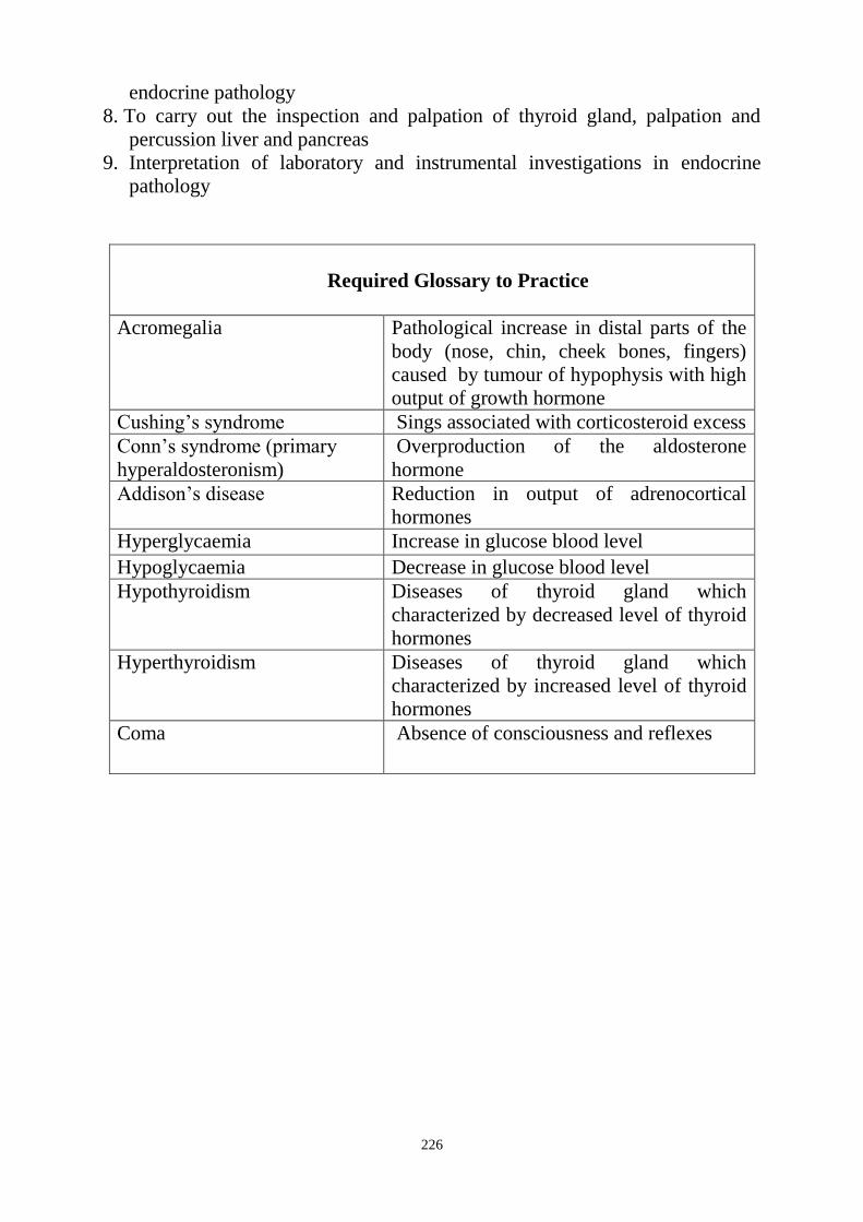
226
endocrine pathology
8. To carry out the inspection and palpation of thyroid gland, palpation and
percussion liver and pancreas
9. Interpretation of laboratory and instrumental investigations in endocrine
pathology
Required Glossary to Practice
Acromegalia Pathological increase in distal parts of the
body (nose, chin, cheek bones, fingers)
caused by tumour of hypophysis with high
output of growth hormone
Cushing‘s syndrome Sings associated with corticosteroid excess
Conn‘s syndrome (primary
hyperaldosteronism)
Overproduction of the aldosterone
hormone
Addison‘s disease Reduction in output of adrenocortical
hormones
Hyperglycaemia Increase in glucose blood level
Hypoglycaemia Decrease in glucose blood level
Hypothyroidism Diseases of thyroid gland which
characterized by decreased level of thyroid
hormones
Hyperthyroidism Diseases of thyroid gland which
characterized by increased level of thyroid
hormones
Coma
Absence of consciousness and reflexes
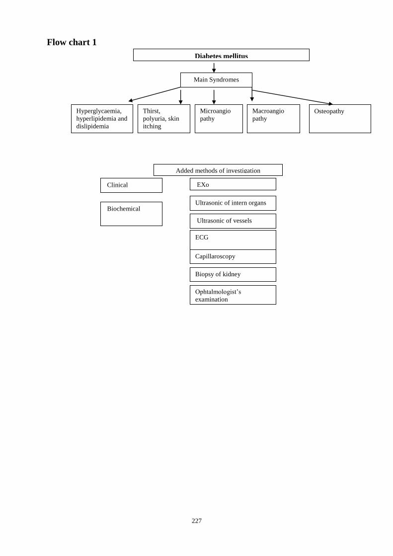
227
Flow chart 1
Diabetes mellitus
Main Syndromes
Added methods of investigation
Clinical
Biochemical
EXo
Hyperglycaemia,
hyperlipidemia and
dislipidemia
Thirst,
polyuria, skin
itching
Microangio
pathy
Macroangio
pathy
Osteopathy
Ultrasonic of vessels
ЕCG
Ultrasonic of intern organs
Capillaroscopy
Ophtalmologist‘s
examination
Biopsy of kidney
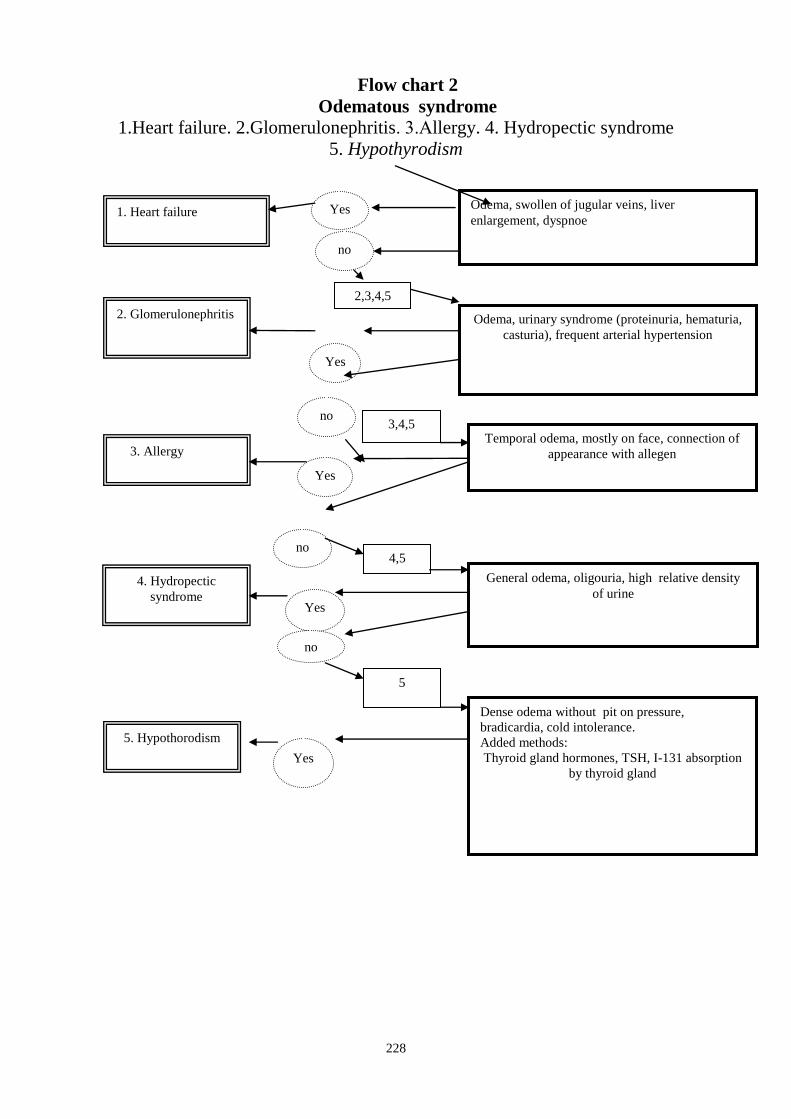
228
Flow chart 2
Odematous syndrome 1.Heart failure. 2.Glomerulonephritis. 3.Аllergy. 4. Hydropectic syndrome
5. Hypothyrodism
Odema, swollen of jugular veins, liver
enlargement, dyspnoe
Dense odema without pit on pressure,
bradicardia, cold intolerance.
Added methods:
Thyroid gland hormones, TSH, I-131 absorption
by thyroid gland
5
General odema, oligouria, high relative density
of urine
4,5
Temporal odema, mostly on face, connection of
appearance with allegen
3,4,5
Odema, urinary syndrome (proteinuria, hematuria,
casturia), frequent arterial hypertension
2,3,4,5
1. Heart failure
2. Glomerulonephritis
3. Allergy
4. Hydropectic
syndrome
5. Hypothorodism
Yes
Yes
no
Yes
no
Yes
no
Yes
no
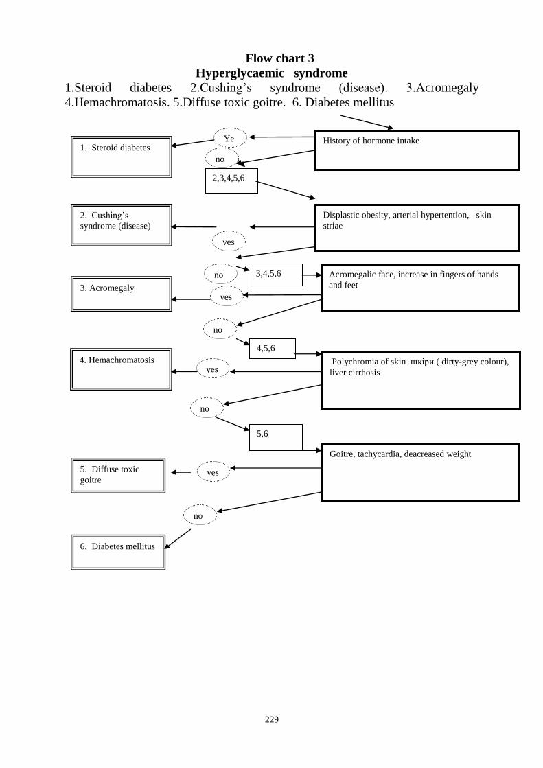
229
Flow chart 3
Hyperglycaemic syndrome 1.Steroid diabetes 2.Cushing‘s syndrome (disease). 3.Аcromegaly
4.Hemachromatosis. 5.Diffuse toxic goitre. 6. Diabetes mellitus
History of hormone intake
Goitre, tachycardia, deacreased weight
5,6
Polychromia of skin шкіри ( dirty-grey colour),
liver cirrhosis
4,5,6
Acromegalic face, increase in fingers of hands
and feet
3,4,5,6
Displastic obesity, arterial hypertention, skin
striae
2,3,4,5,6
1. Steroid diabetes
2. Cushing‘s
syndrome (disease)
3. Acromegaly
4. Hemachromatosis
5. Diffuse toxic
goitre
Ye
ss
yes
no
yes
no
yes
no
no
yes
no
6. Diabetes mellitus

230
Suggested Reading List
Required Reading
1. Internal Diseases, an Introductory Course. Edited by V.Vasilenko and
Grebenev, Mir Publishers, Moscow, 1990
2. Clinical Examination, Edited by Jonh Macleod, Jonh Munro, Churchill
Livingsone, 2000
3. Medicine, Edited by K. George Mathew, Praveen Aggarwal, Elsevier, 2004.
4. Caring for the sick by British Red Cross Society
5. First aid manual by British Red Cross Society
6. Davidson‘s Medicine, Edited by Nicholas A. Boon, Nicki R.
Colledge,Davidson, 2008
7. Manual for the Case Record, Edited by Ignatenko G.A., Department of
Propaedeutics of Internal Medicine , Donetsk, 2009
8. Lecture: Examination of the endocrine system
9. Methodical guideline for students
10. Flow charts for Practice
11. Website of department: www.cardiology.dsmu.edu.ua
Additional Reading
1. Textbook of Physical Diagnosis. History and Examination / Mark H. Swartz
2002, W. B. Saunders Company Pennsylvania
2. History and Physical Examination . Current Clinical Strategies, Edited Paul
P. Chan, Peter, J. Winkle, 2005
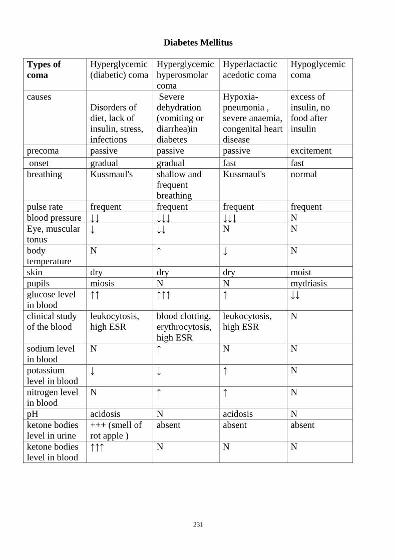
231
Diabetes Mellitus
Types of
coma
Hyperglycemic
(diabetic) coma
Hyperglycemic
hyperosmolar
coma
Hyperlactactic
acedotic coma
Hypoglycemic
coma
causes
Disorders of
diet, lack of
insulin, stress,
infections
Severe
dehydration
(vomiting or
diarrhea)in
diabetes
Hypoxia-
pneumonia ,
severe anaemia,
congenital heart
disease
excess of
insulin, no
food after
insulin
precoma passive passive passive excitement
onset gradual gradual fast fast
breathing Kussmaul's shallow and
frequent
breathing
Kussmaul's normal
pulse rate frequent frequent frequent frequent
blood pressure ↓↓ ↓↓↓ ↓↓↓ N
Eye, muscular
tonus
↓ ↓↓ N N
body
temperature
N ↑ ↓ N
skin dry dry dry moist
pupils miosis N N mydriasis
glucose level
in blood
↑↑ ↑↑↑ ↑ ↓↓
clinical study
of the blood
leukocytosis,
high ESR
blood clotting,
erythrocytosis,
high ESR
leukocytosis,
high ESR
N
sodium level
in blood
N ↑ N N
potassium
level in blood
↓ ↓ ↑ N
nitrogen level
in blood
N ↑ ↑ N
pH acidosis N acidosis N
ketone bodies
level in urine
+++ (smell of
rot apple )
absent absent absent
ketone bodies
level in blood
↑↑↑ N N N
232
Hyperthyroid coma(crisis)
Causes: after thyroidectomy, inadequately treated hyperthyroidism, severe
infections, rapid withdrawal of antithyroid therapy, use of iodine or contrast
agents, trauma, stress, physical exertion.
Pathogenesis: Proteinlysis, lipolysis, impaired glucose tolerance, water-
electrolytic disbalance, acid-base disbalance, dysfunction of central nervous
system,
hypothalamo-pituitary-adrenal gland dysfunction, sympathoadrenal system
dysfunction
Clinical features: hyperemia, hyperhidrosis (excessive profuse sweating), pulse
pressure increasing, cramps, hyperthermia, tachycardia, jaundice, muscular
weakness, diarrhea, anuria delirium, apathy.
Hypothroid (myxoedema) coma
Causes: severe infections, supercooling, trauma, operations, discontinue of
antithyroid therapy, use of iodine or contrast agents, trauma, stress, physical
exertion.
Clinical features: low blood pressure, bradycardia, bradypnoe, hypothermia,
anuria
Investigation: decreased level of glucose, sodium, chlorine, increased level of
potassium, increased level of urea, cholesterol, anemia, T3, T4-↓
Adrenal coma( acute adrenal insufficiency)
Low level of glucocorticoid and mineralocorticoid
Affection of adrenal glands – infections (tbc), adrenal infarction,
hemorrhage(meningococcaemia-Waterhouse-Friderichen syndrome), operation
on adrenal gland
Mechanisms – decrease in gluconeogenesis, excessive excretion of sodium and
chlorine with urine, lack excretion of potassium with urine, dehydration,
decreased volume of circulating blood, decrease in renal circulation.
Clinical features: hyperpigmentated and pale skin, acrocyanosis, thready, weak
pulse, low blood pressure, cold extremities
Laboratory signs – decrease in cortisol, sodium, glucose, increase in potassium,
urea, creatinine, eosinophils, leucocytosis
233
Diabetes Mellitus
Type I
Type II
Causes: pancreatitis, receptor disorders, Itsenko-Cushing syndrome,
thyrotoxicosis, remedies
Clinical features: thirst, polydipsia, polyuria, polyphagia, weight loss, muscular
weakness, skin itch, pyogenic skin diseases,non-healing of wounds,
paradontosis, microangiopathy.
Ketoacidosis: lose of appetite, vomiting, nausea.
Hypoglycemia – clinical features: headache, drowsiness, excitation,
hyperhidrosis (excessive profuse sweating), starvation, palpitation, hand tremor,
parestesia, salivation.
Causes: excess of insulin, no food after insulin insuloma, liver diseases,
gastrectomia, alimentary, alcohol intoxication, hard physical activity.
Hyperthyroidism Causes: duffuse throid enlargement, multinodular goiter, solitary throid goiter,
tbc, scarlet fever, encephalitis, somatotropin (acromegalia or giantism), typhus,
pertussis, rheumatism, viral meningitis, Itsenko-Cushing syndrome.
Clinical features: tachycardia, exophthalmos, enlargement of thyroid gland
(goiter), excitation, irritability, emotional lability, fine tremor, deranged sleep,
hyperhidrosis (excessive profuse sweating), muscular weakness, palpitation
(sinus tachycardia, atrial fibrillation), weight loss despite increased appetite,
increased stool frequency, increasing blood pressure, eye signs
Investigation– ↑ T3,T4, TSH is low, thyroid stimulating immunoglobulins,
hyperglycemia, hypocholesterolemia, increased basal metabolism, changes of
ECG (sinus tachycardia, atrial fibrillation )
Hypothyroidism Primary – affection of thyroid gland – resection of thyroid gland, Hashimoto's
thyroiditis, lack of iodine.
Secondary - affection of adenohypophysis – tumor of brain, encephalitis,
arachnoiditis, stroke, trauma
Tertiary - affection of hypothalamus – tumor of brain, trauma, encephalitis,
arachnoiditis, stroke
Clinical features: oedema of face and extremities(myxoedema), pain in
muscles, fast fatigue, drowsiness, cold intolerance, memory disorders,
depression, hoarseness, constipation,
dry, flaky, skin, alopecia, dyspnoea.
Mechanisms - decreasing of basal metabolism, decreasing of oxidative reaction,
disorder of protein synthesis.
234
Investigation– ↓T3,T4, TSH is low or high, anemia, high ESR,
hypercholesterolemia, hypoglycemia, reduction of basal metabolism, changes of
ECG(sinus bradycardia, low voltage QRS), accumulation of fluid in pericardial
cavity.
Revision tests
Q1. Choose the most typical stomatological problems in patients with diabetes
mellitus
A. Caries
B. Paradontosis
C. Hypersalivation
D. Gingivitis
E. All listed variants
Q2. Early parameter which shows diabetic nephropathy
A. Casturia
B. Leucocyturia
C. Hematuria
D. Microalbuminuria
E. Bacteriuria
Q3.The main symptoms in clinic of diabetes mellitus
A. Dryness of mouth mucosa and tongue, polydipsia, furuncles,
polyuria
B. Thirst, oliguria, muscular weakness, increased appetite
C. Fast fatigue, nausea, palpitation
D. Odema, depression, constipation, sore throat
E. Muscular weakness, tremor, thirst
Q4. Early signs of diabetes mellitus except for
A. thirst
B. polyuria
C. general weakness
D. skin itching
E. proteinuria
Q5.Which sign better characterizes compensation of carbohydrate metabolism?
A. Glucose level on empty stomach
B. Glucose level of urine
C. Glycosylated haemoglobin
D. Haemoglobin level
E. All listed variants
Q6.Patient 60 years old has sudden appearance of tremor, hunger, aggression,
sweating. He doesn‘t suffer from diabetes mellitus. Which diseases and
conditions can cause hypolycaemia except for diabetes mellitus
A. Insulinoma.
B. Tumour of thyroid gland
C. Alcoholic intoxication
D. Gastrectomy

235
E. Tumour of testicle
Q7.Which rhythm disorder happen more often in hyperthyrodism
А. Sinus tachycardia
В. AV block 1 degree
С. AV block 3 degree
D. Myocardium infarction
Е. Sinus bradycardia
Q8. Which ECG changes happen in hypothyrodism
A. High voltage of waves
B. Shortening of all intervals
C. Low voltage of waves
D. Atrial flutter
E. Sinus tachycardia
Q9. Patient 22 years old is admitted to the hospital without consciousness.
Breathing is noisy, deep, acetone smell from mouth. Skin is dry, decreased tone
of skin, muscles and eye balls. According to relatives he had thirst, dryness of
mouth, furuncles, increased appetite, decreased weight for last 4 months. What
happens with patient?
A. Hypoglycaemic coma
B. Diabetic coma
C. Hyperlactic coma
D. Hyperthyroid crisis
E. Toxic coma
Q10.The main symptoms in clinic of ketoacidosis
A. Odema, depression, constipation, sore throat
B. Thirst, oliguria, muscular weakness, increased appetite
C. Fast fatigue, nausea, palpitation, increased weight
D. Nausea, vomiting, absence of appetite, thirst
E. Muscular weakness, tremor, thirst
Answer Keys : Q1: B; Q2: D; Q3: A; Q4: E, Q5: C; Q6: A; Q7: A; Q8: C,
Q9: B; Q10: D.
SUMMARY OF PROCEDURES
The practice lesson shall begin in the study room, with the homework
assignment checked and students‘ testing carried out.
Another part of the practice shall be conducted in the hospital wards. Instructor
will demonstrate to the students how to make enquiring, physical examination of
patients with endocrine pathology.
Diagnostic value of the revealed in examined patients shall be discussed
outside of the ward. Instructor teaches to perceive the organism of man as
single unit, underline connection oral cavity pathology and internal organs, to
provide first aid to the patients before hospitalization and in urgent situations.
At the end of the practice teacher makes short general conclusion concerning
obtained findings and students do final tests.
236
Final Tests
Q1. Ketoacidotic coma characterized by:
A. Sudden occurrence
B. Acetone smell from mouth
C. Cold sweating
D. Sudden hunger
E. Hypoglycaemia
Q2.Hyperfunction of thyroid gland has such signs as:
A. Serum T3 and T4 level are high
B. Only serum level T3 is high
C. Only serum level T4 is high
D. Serum T3 level and T4 are high
E. Serum T3 is high but T4 level is high
Q3. Hypothyrodism is characterized by:
А. Tachycardia
В. Increased stool frequency
С. Fine tremor
D. Vomitting
E. Bradycardia
Q4.Hyperthyrodism characterized by these sigs except for:
A. High body temperature
B. Sensation of heating
C. Wet skin
D. Increased appetite
E. Increased weight
Q5.Absolute deficiency of insulin causes:
А.Azotemia
В.Ctalepsy
С.Hypolipidemia
D. Fat infiltration of liver
E. hyperglycaemia
Q6.Enlargement of liver in diabetes mellitus happens due to:
А.fat hepatosis
В.hepatitis
С.cirrhosis
D. diabetic retinopathy
E. heart failure
Q7. During ketoacidotic coma the breathing is:
A. Rare
B. Frequent
C. Noisy, deep
D. With temporary apnoe
E. Superficial
Q8. What is rubeosis?
237
A. Increase in thyroid hormones in the blood
B. Increase in blood glucose level
C. Decrease in blood protein
D. Accumulation of acetone and ketone bodies
Е. Decrease in thyroid hormones in the blood
Q9. What is action of insulin?
A. Increase in glycaemia
B. Decrease in glycaemia
C. Increase in catalase
D. Increase in lipase.
E. Without influence
Q10.Hyperthyrodism characterized by these sigs except for:
A. Tachycardia
B. Atrial fibrilation
C. Bradycardia
D. Increased loudness of heart sounds
E. Increase in cetone body
238
The Health Ministry of Ukraine
Donetsk National Medical University
Approved
at the meeting of
Propedeutic and Internal Medicine
Department
Head of department
Associate Member of NAMSc of
Ukraine,
Professor G. A. Ignatenko
« _______ »_____________ 2011 р.
STUDENT’S SELF-STUDY GUIDELINES FOR
PRACTICE ACTIVITIES
Subject Propedeutics of the Internal Medicine
Module 1 Methods of examination and basic
symptoms and syndromes at the
diseases of internals organs in the
clinic of internal medicines.
Topic 1 Module Introduction to the clinic of internal
illnesses.
Basic rules of inquiring and
inspection of sick.
Practice Examination methods and semiotics
of allergy manifestations. Collection
of complaints, anamnesis, objective
examination of patients with allergy.
Care for patients. Additional methods
of examination in allergology. Basic
syndromes in allergology. Rendering
first aid at anaphylactic shock,
asphyxias at the oedema of larynx.
Diagnostics of inflammatory and
degenerative diseases of locomotor
system. Joint syndromes (arthritic,
osteoarthritic). Features of care for
patients
Course 2
Faculty Dentistry
Donetsk 2011

239
Importance of the Subject: Allergic diseases are one of the basic in the
structure of morbidity of children, teenagers and young people. A lot of allergy
signs appear in oral cavity or around it. That is why have this knowledge is
necessary for dental specialists.
Diseases of joints, especially rheumatoid arthritis and osteoarthritis are essential
problem for the people of middle and old ages. Some time dentist first pay
attention on changes on movement of his patient and may correctly and timely
forward the patient to proper specialist.
That is why abilities to inquire patients about all this pathology and correctly
examine patient objectively are help for proper diagnose and early start
treatment.
Key Objective: able to determine the syndromes of allergy and joint syndrome,
using skills of gathering of complaints and anamnesis, allergist anamnesis,
objective examination (inspection, percussion, palpation and auscultation) and
additional methods of examination (scratch test, skin test, provocative allergist
tests, laboratory methods of diagnostics of allergy, immunological tests, X-Ray
research of organs of thorax and joints, sonography of joints).
Specific Goals:
1. To know a concept about an allergy.
2. To know types of allergic reactions.
3. To master inquiring of patients with allergy.
4. To know basic complaints: itch of skin, fever, edema, rashes, blisters,
difficulty in breathing, hacking cough, attacks of dispnoea, weakness, filling
of fear, head pain, nausea, fever.
5. To be able to collect allergy anamnesis.
6. Determination of presence of asthma.
7. To can conduct inspection of patients with allergy.
8. To know changes of skin as erythema, urticaria (nettle rush), local edema,
husky voice, difficulty and hoarse breathing, tachypnea.
9. To know places of rash, feature, types of blisters, their color, limits,
consistency.
10. To know other sings of allergy as fever, oedema of lips, eyelids, cheeks,
genitals, tongue, pharynx, larynx, violation of breathing and swallowing,
increase of lymphatic nods.
11. To know changes of mucus membranes of nose, eyes - rhinitis,
conjunctivitis. Epidermolysis.
12. To know variants of clinic in the case of development of anaphylactic shock.
13. To be able to help patients with allergy with violation of consciousness,
collapse, skin rash, cramps, pain in heart, stomach, syndrome (pseudo)
―acute abdomen‖.
14. To know an allergy on stomatological materials and medicines.

240
15. To know methods of laboratory diagnostics. Clinical, allergist,
immunological tests. Global analysis of blood. Eosinophilia. Determination
of T- and B- lymphocytes. Allergic diagnostic tests. Skin test. Endemic (with
an allergen). Elimination tests - stopping of contact with allergen.
16. To know signs of allergy and immune violations from the side of mucus
membranes of oral cavity and teeth and jaw system. Features of care for
patients.
17. Rendering first aid at anaphylactic shock, asphyxias at the edema of larynx.
18. Diagnose of inflammatory and degenerative diseases of locomotorium
system.
19. To master inquiring of the system of musculoskeletal apparatus.
20. To be able to conduct objective examination (inspection, percussion,
auscultation, palpation) and additional (instrumental, X-Ray, laboratory,
morphological) methods of diagnostics of illnesses of peripheral joints,
spine, mussels and periarticular tissues.
21. To know about examination of synovial liquid.
22. To know about joint syndromes (arthritic, osteoarthritic).
23. To know about mussels syndrome.
24. To know about features of care for patients.
Level of Knowledge and Skills before the Practice:
1. To know the structure of the immune system and joints. «Faculty of
Anatomy».
2. To estimate the features of function of the immune system and joints.
«Normal physiology».
3. To determine the basic mechanisms of development of anaphylactic reactions,
to the attack bronchial asthma and asthmatic status, osteoarthritis and
rheumatoid arthritis. «Pathophysiology».
Questions for Self-Assessment of the Pre-Practice Knowledge (correct
answers gone after last task).
Q1. Activated mast cells and basophils undergo a process called degranulation,
during which they release inflammatory chemical mediators from their granules
into the surrounding tissue causing several systemic effects. Which mediators do
you know?
A. histamine
B. prostaglandins
C. leukotrienes
D. interleukins and cytokines
E. all listed above
Q2. What antibodies overproduce at bronchial asthma?
A. IgA
B. IgM
241
C. IgG
D. IgE
E. IgM і A
Q3. Determine disease with anaphylactic allergy:
A. COPD
B. bronchial asthma
C. rheumatic fever
D. erysipelas
E. essential hypertension
Q4. Find immune organ:
A. liver
B. kidney
C. thymus
D. heart
E. thyroid gland
Q5. What kind of allergens do you know?
A. animal products
B. food
C. drugs
D. insect sings
E. all listed above
Q6. What can induce seasonal allergies?
A. food
B. insect sings
C. drugs
D. pollens
E. all listed above
Q7. What is atopy?
A. bronchial asthma exacerbation
B. seasonal allergy
C. polyvalent allergy
D. hereditary predisposition for allergy
E. all listed above
Key answers: 1-E, 2-D, 3-B, 4-C, 5-E, 6-D, 7-D.
The following printed materials can be of help to improve your pre-practice
knowledge and skills:
1. Test book of Medical Physiology / Arthur C. Guyton, John I. Hall 2001, W.
B. Saunders company Pennsylvania 2001
2. Gray's Anatomy. Edited by T. Pickering Pick, F.R.C.S., 1995
242
3. M. Prives, N. Lysenkov, V. Bushovich; Human Anatomy
Contents of Practice
Topics of Theory:
1. Concept about an allergy.
2. Types of allergic reactions.
3. Inquiring of patients with allergy.
4. Basic complaints: itch of skin, fever, oedema, rashes, blisters, difficulty in
breathing, hacking cough, attacks of dyspnoea, weakness, filling of fear,
head pain, nausea, fever.
5. Allergy anamnesis.
6. Bronchial asthma.
7. Inspection of patients with allergy.
8. Changes of skin as erythema, urticaria (nettle rush), local oedema, husky
voice, difficulty and hoarse breathing, tachypnea.
9. Places of rash, feature, types of blisters, their colour, limits, consistency.
10. Fever, oedema of lips, eyelids, cheeks, genitals, tongue, pharynx, larynx,
violation of breathing and swallowing, increase of lymphatic nods.
11. Changes of mucus membranes of nose, eyes - rhinitis, conjunctivitis.
Epidermolysis.
12. Variants of clinic in the case of development of anaphylactic shock.
13. Help patients with allergy with violation of consciousness, collapse, skin
rash, cramps, pain in heart, stomach, syndrome (pseudo) ―acute abdomen‖.
14. Allergy on stomatological materials and medicines.
15. Methods of laboratory diagnostics. Clinical, allergist, immunological tests.
Global analysis of blood. Eosinophilia. Determination of T- and B-
lymphocytes. Allergic diagnostic tests. Skin test. Endemic (with an allergen).
Elimination tests - stopping of contact with allergen.
16. Signs of allergy and immune violations from the side of mucus membranes
of oral cavity and teeth and jaw system. Features of care for patients.
17. Rendering first aid at anaphylactic shock, asphyxias at the oedema of larynx.
18. Diagnose of inflammatory and degenerative diseases of locomotorium
system.
19. Inquiring of the system of musculoskeletal apparatus.
20. Objective examination (inspection, percussion, auscultation, palpation) and
additional (instrumental, X-Ray, laboratory, morphological) methods of
diagnostics of illnesses of peripheral joints, spine, mussels and periarticular
tissues.
21. Examination of synovial liquid.
22. Joint syndromes (arthritic, osteoarthritis).
23. Mussels syndrome.
24. Features of care for patients.
243
Practical skills:
Students should be able to demonstrate mastery of the following practical skills
able to determine the syndromes of allergy and joint syndrome, using skills of
gathering of complaints and anamnesis, allergist anamnesis, objective
examination (inspection, percussion, palpation and auscultation) and additional
methods of examination (scratch test, skin test, provocative allergist tests,
laboratory methods of diagnostics of allergy, immunological tests, X-Ray
research of organs of thorax and joints, sonography of joints).
20. To gathering of complaints and anamnesis at patients with allergy
reaction.
21. To gathering of complaints and anamnesis at patients with joint
syndrome.
22. To perform inspection of the patient with allergy reaction.
23. To perform inspection of the patient with joint syndrome
24. To determine of the general state of patient with allergy reaction.
25. To determine posture of the patient in a bed with joint syndrome.
26. To determine presence of oedema.
27. To examine mucous membranes and skin with allergy reaction and joint
syndrome.
28. To examine muscles and joints at patients with rheumatoid arthritis and
osteoarthritis.
29. To find proper clinical syndromes.

244
Required Glossary to Practice
Term Definition
allergy is a disorder of the immune system
which is a form of hypersensitivity
allergen is a nonparasitic antigen capable of
stimulating a type-I hypersensitivity
reaction in atopic individuals
urticaria a skin condition characterized by the
formation of itchy red or whitish raised
patches, usually caused by an allergy
eosinophilia the presence of abnormally large
numbers of eosinophils in the blood,
occurring in various diseases and in
response to certain drugs
stridor is a high pitched sound resulting from
turbulent air flow in the upper airway
rheumatoid arthritis is a chronic, systemic inflammatory
disorder that may affect many tissues
and organs, but principally attacks
synovial joints
osteoarthritis also known as degenerative arthritis or
degenerative joint disease, is a group
of mechanical abnormalities involving
degradation of joints, including
articular cartilage and subchondral
bone
contracture constant limitation of mobility of the
damaged joint
ankylosis abnormal adhesion or immobility of
the bones in a joint, as by a direct
joining of the bones, a fibrous growth
of tissues within the joint, or surgery
myalgia pain in a muscle or a group of muscles
joint deformation is a major difference in the shape of
body part or joint compared to the
average shape of that part
joint defiguration change of form of joint because of
oedema of periarticular tissue, increase
of amount of synovia
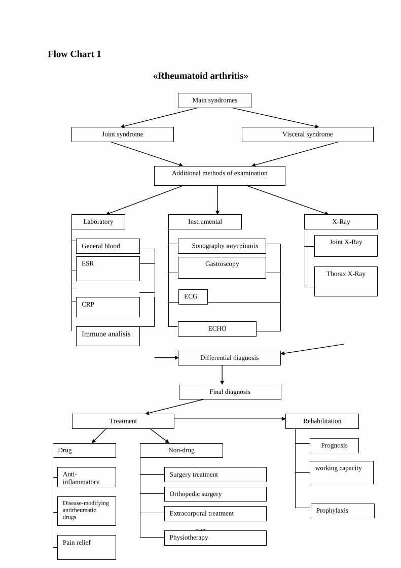
245
Flow Chart 1
«Rheumatoid arthritis»
Main syndromes
Joint syndrome Visceral syndrome
Additional methods of examination
Laboratory Instrumental X-Ray
General blood
ESR
CRP
Sonography внутрішніх
органів
Gastroscopy
ECHO
Joint X-Ray
Differential diagnosis
Final diagnosis
Treatment Rehabilitation
Drug Non-drug
Anti-
inflammatory
Disease-modifying
antirheumatic
drugs
Pain relief
Surgery treatment
Orthopedic surgery
Extracorporal treatment
Prognosis
working capacity
Prophylaxis
Immune analisis
Thorax X-Ray
Physiotherapy
ECG

246
Flow Chart 2
«Osteoarthritis»
Osteoarthritis
Main syndromes
Small joint affection Vertebra column
affection
Additional methods of examination
Laboratory
Instrumental
General blood
analysis
Sonography
Arthroscopy
Final diagnosis
Big joint affection
Joint X-Ray
Synovial fluid
analysis
Initial diagnosis

247
Differential diagnosis of rheumatoid arthritis and osteoarthritis
Rheumatoid arthritis Osteoarthritis
Age of origin More young old
Sex More women Equal
Origin antigen-induced
inflammation
dystrophy
Joints started Small joints of feet and
hands
Big joints
Number of affected
joints
Polyarthritis,
symmetrical
Initially oligoarthritis
Number of affected join
groups
3 and more 1 or oligoarthritis
Severe pain in morning During first hour at
morning
During first 10 min at
morning
All day pain Continuously pain all
day with some relief
Started pain (first several
seconds at movement or
all movement) but it go
out at rest
Limitation in the range
of motions, active and
passive
Presents and progressive
at all affected joints
May be presents
Anchylosis As rule develop Never develop
Deformation Present Present and pronounced
Articular crepitus Absent Present
Hyperemia of skin Present Absent
Changes in blood
analysis
High ESR, leucositosis Absent
Common symptoms of allergy
Affected organ Symptom
Nose swelling of the nasal mucosa (allergic rhinitis)
Sinuses allergic sinusitis
Eyes redness and itching of the conjunctiva (allergic
conjunctivitis)
Airways Sneezing, coughing, bronchoconstriction, wheezing and
dyspnea, sometimes outright attacks of asthma, in severe
cases the airway constricts due to swelling known as
laryngeal oedema
Ears feeling of fullness, possibly pain, and impaired hearing due
to the lack of eustachian tube drainage.
Skin rashes, such as eczema and hives (urticaria)
Gastrointestinal
tract
abdominal pain, bloating, vomiting, diarrhea

248
Suggested Reading List
Required Reading
1. Internal Diseases, an Introductory Course. Edited by V.Vasilenko and
Grebenev, Mir Publishers, Moscow, 1990
2. Clinical Examination. Edited by Jonh Macleod, Jonh Munro, Churchill
Livingsone, 1986
3. A system of case recording and clinical examination of patients on
propaedeutic of internal diseases.
4. Medicine, Edited by K. George Mathew, Praveen Aggarwal, Elsevier, 2004
5. Methodical guideline for students
6. Lecture: Inspection of the patient and its diagnostic value
7. Website of department: www.cardiology.dsmu.edu.ua
8. Flow charts for Practice
Additional Reading
1. Textbook of Physical Diagnosis. History and Examination / Mark H. Swartz
2002, W. B. Saunders Company Pennsylvania
2. History and Physical Examination. Current Clinical Strategies, Edited Paul P.
Chan, Peter, J. Winkle, 2005
3. Davidson‘s Medicine, Nicholas A. Boon, Nicki R. Colledge, Davidson, 2006
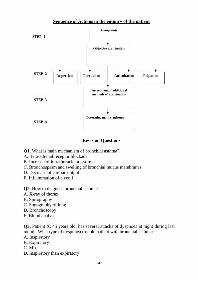
249
Sequence of Actions in the enquiry of the patient
Revision Questions
Q1. What is main mechanism of bronchial asthma?
А. Beta-adrenal receptor blockade
В. Increase of intrathoracic pressure
С. Bronchospasm and swelling of bronchial mucus membranes
D. Decrease of cardiac output
E. Inflammation of alveoli
Q2. How to diagnose bronchial asthma?
А. X-ray of thorax
В. Spirography
С. Sonography of lung
D. Bronchoscopy
E. Blood analysis
Q3. Patient X, 45 years old, has several attacks of dyspnoea at night during last
month. What type of dyspnoea trouble patient with bronchial asthma?
А. Inspiratory
В. Expiratory
С. Mix
D. Inspiratory than expiratory
Complaints
Objective examination
Assessment of additional
methods of examination
Determine main syndrome
STEP 1
STEP 2
STEP 3
STEP 4
Inspection Palpation Auscultation Percussion

250
E. Anyone
Q4. What kind of forced position at patient with bronchial asthma?
А. Lying
В. Sitting with legs down
С. Standing with bending forward
D. Sitting or standing with the fixing of the shoulder gird
E. Any one
Q5. Which joint usually more often affected with osteoarthritis?
А. Knee
В. Metacarpophalangeal
С. Proximal interphalangeal
D. Acromeoclavicular
E. Anyone
Q6. Which joint usually more often affected at rheumatoid arthritis?
А. Knee
В. Metacarpophalangeal
С. Hip
D. Acromeoclavicular
E. Anyone
Q7. Patient has angioneurotic (Quincke's) edema. What sings of this?
А. Tissues around the mouth, and the mucosa of the mouth and/or throat
swelling up over the period of minutes to several hours
В. Swelling can also occur elsewhere, typically in the hands
С. In severe cases, stridor of the airway occurs
D. May be slightly decreased sensation in the affected areas due to compression
of the nerves
E. All listed above
Q8. What is irreversible?
А. Dupuytren's contracture
В. Ankylosis
С. Kyphoscoliosis
D. Deformation
E. Any of them
Q9. Which parameter is main for assessing of bronchial conduction at bronchial
asthma?
А. Vital capacity
В. Residual volume
С. FEV1
D. Tidal volume
E. PEF

251
Q10. Patient 65 years old has severe pain, especially in the morning, in both
knee and hip joints, deformation and defiguration of both knee joint, restriction
in movement of them because of pain, crepitus in right knee. Disease started one
year ago. Analysis of blood reveal no pathology. What diagnosis may be first of
all?
А. Rheumatism
В. Gout
С. Rheumatoid arthritis
D. Osteoarthritis
E. It is variant of norm
Key answers: 1-C, 2-B, 3-B, 4-D, 5-A, 6-B, 7-E, 8-E, 9-C, 10-D.
SUMMARY OF PROCEDURES
The practice lesson shall be begun in the study room. Checking of the
homework, the test control is carried out. Then at the study room patient is
invited. Demonstration of methodic of general assessment and examination of
the patients with allergy reaction and affection of the joints. Then lesson is
transferred to ward: the teacher of students group perform examination of
patients, showing a different signs of pathology for this topics, inviting in turn of
each students on a fragment of research. After students formulate syndromes and
diagnosis if possible.
At the end of lesson in an educational room diagnostic value of available
symptoms is discussed.
Final tests
Q1. Patient at time of penicillin injection blackout. No dyspnoea revealed.
Vesicular breathing above lung. Blood pressure impossible to detect. Pulse is
filiformis with rate about 120 bpm. What happened with patient?
А. Anaphylactic shock
В. Stroke
C. Myocardial infarction
D. Hypertonic crisis
E. Cardiac arrest
Q2. Patient 60 years old has severe pain, especially in the morning, in both knee
joints, deformation and defiguration their, restriction in movement of them
because of pain, crepitus. Disease started five years ago. Analysis of blood
reveal no pathology. What first of all point on osteoarthritis rather than on
rheumatoid arthritis?
А. Crepitus
В. Defiguration

252
С. Deformation
D. severe pain
E. restriction in movement of them because of pain
Q3. Patient 39 years old has severe cough at night and after going out of home
and inspiring of cold air. Now he has attack of cough. His eyes are red because
of distraction of vessels in sclera because of cough. Severe expiratory dyspnoea
is observed. Which syndrome main at him?
А. Intoxication
В. Heart failure
C. Respiratory failure
D. Anaemic
E. Hemorrhagic
Q4. What should be done for patient with bronchial asthma as a first aid?
А. Unbutton the collar
В. Add fresh air
C. To help patient set in forced position
D. Give inhaler
E. All listed above
Q5. 15 min after washing with new soap the oedema of face is developed. What
is it?
А. Angioneurotic Quincke's oedema
В. Nephrotic syndrome
C. Chronic heart failure
D. Acute heart failure
E. Acute chemical burn of skin
Q6. What sign of home-dust allergy reaction?
А. Allergy reaction arise at spring and fall
В. Allergy reaction arise after clearing at home
C. Allergy reaction arise after work in garden
D. Allergy reaction not developed on dust
E. Allergy reaction developed after eating
Q7. Which parameter is main for assessing of bronchial conduction at bronchial
asthma?
А. Total lung capacity
В. Residual volume
С. Tidal volume
D. FEV1
E. any of them
Q8. What is sign of aspirin asthma?

253
А. Attack of asthma and aspirin intolerance
В. Attack of asthma at physical exertion
C. Attack of asthma at emotional exertion
D. Attack of asthma and ulcer disease
E. Attack of asthma at going out of home and inspiring of cold air
Q9. Which parameter allows to determine activity of rheumatoid arthritis?
А. ESR
В. ASLO level
C. Monocytes level
D. Eosinophil level
E. RBC
Q10. What sign of food allergy reaction?
А. Allergy reaction arise at spring and fall
В. Allergy reaction arise after clearing at home
C. Allergy reaction arise after work in garden
D. Allergy reaction not developed on dust
E. Allergy reaction developed after eating

254
Checklist of Practical Skills
1. Scheme of case recording. Main parts of anamnesis
2. General inspection
3. Palpation of lymph nodes and characteristic of received data
4. Inspection of head and neck
5. Inspection of trunk and limbs
6. Static inspection of the chest, diagnostic value of main symptoms
7. Dynamic inspection of the chest, diagnostic value of main symptoms
8. Inspection of heart region, diagnostic value of main symptoms
9. Inspection of abdomen, determination of main symptoms
10. Palpation of the vessels
11. Measurement of systolic and diastolic pressure and rules of its taking, pulse
pressure
12. Palpation of the chest, sequence, clinical value of main symptoms
13. Palpation of the heart region, sequence, clinical value of main symptoms
14. Palpation of the mammary gland
15. Superficial palpation of abdomen, interpretation the received data
16. Rules and principles of deep sliding palpation according to Obraztsov‗s
method
17. Palpation of sigmoid, caecum and terminal part of ileum, their normal
features
18. Methods of determination of greater curvature of stomach
19. Palpation of transverse colon, main features
20. Palpation of liver, diagnostic value of main symptoms
21. Palpation of spleen
22. Comparative percussion of the lungs. Main percussion notes, their origin
23. Technique of topographic percussion. Topographic data of the lungs in the
norm and pathology
24. Percussion of the heart-relative cardiac dullness, normal borders and their
changes in the pathology
25. Percussion of the heart-absolute cardiac dullness, normal borders and their
changes due to cardiac and extracardiac reasons
26. Percussion of the vascular bundle, diagnostic value
27. Percussion of liver, sequence, data in the norm and pathology
28. Percussion of spleen. Rules of determination, reasons of spleen
enlargement
29. Auscultation of the lungs. Main breath sounds, their quantitative and
qualitative changes
30. Auscultation of the lungs. Added breath sounds, classification, their origin
31. Determination of vocal resonance and its diagnostic value
32. Auscultation of the heart and vessels
33. Palpation of joints
34. Palpation of thyroid gland
35. Gastric lavage
36. Intestine irrigation: siphon enemas, cleansing enemas

255
LITERATURE
1. Test book of Medical Physiology, Arthur C. Guyton, John I. Hall 2001,
W. B. Saunders company Pennsylvania 2001
2. Internal Diseases an Introductory Course. Edited by V.Vasilenko and
Grebenev, Mir Publishers, Moscow, 1990
3. Clinical Examination, Edited by Jonh Macleod, Churchill Livingsone, 1988
4. Student‘s self-study guidelines for Practice Activities on propedeutics of
internal medicine for Secord Year Students of International Medical Faculty
(Dentistry) Module 1. Main methods of examination of patients with diseases of
internal organs. Symptomes and Syndromes of Internal Diseases, Edited by
Ignatenko G.A., Department of Propaedeutic and internal medecine, Donetsk,
2011
5. Introduction of the course of internal disease. Z.H. Semidotska, O.S.
Biltchenko, Kharkiv, 2006
6. Davidso′s Principles and Practice of Medicine.
7. Hutchisonsۥ Clinical Methods. Michael Swash, Michael Glynn. 2007 Elsevier.
8. Kumar and Clark Clinical Medicine. 2007 Elsevier.
9. CURRENT Medical Diagnosis & Treatment 2009 Stephen J. McPhee,
Maxine A. Papadakis, Eds. Ralph Gonzales, Roni Zeiger, Online Eds.

256
Здано до набору 01.09.2011 р. Підписано до друку 08.09.2011 р.
Формат 60х90/16. Папір друкарський. Друк офсетний.
Ум.друк.арк. 14,7. Тираж 100 примірників. Замовлення 19211.
__________________________________________
Видавництво ДонНМУ
м. Донецьк, пр. Ілліча, 16