Student Manual 20-21.pdf - Michigan Medicine
121
University of Michigan Medical School Family Medicine M2 Clerkship Student Profile Full Name: ______________________________________________ Nickname or name you prefer to be called: __________________________ Street Address: ______________________________________________ City, Zip: ______________________________________________ Telephone: ______________________________________________ Pager Number: ______________________________________________ Email address: ______________________________________________ Personal/Social: Name Spouse/Partner: ______________________________________________ (if applicable) Home Town: ________________________________________________ (if applicable) Names and Ages of Children (if applicable): ______________________________________________ ______________________________________________ Hobbies/Non-Professional Interests: ___________________________________________ ____________________________________________________________________________ In Case of Emergency, please contact: ____________________________________________ Do you have any physical limitations or medical problems about which your preceptor should be aware? YES NO If yes, please specify: ____________________________________________________
-
Upload
khangminh22 -
Category
Documents
-
view
1 -
download
0
Transcript of Student Manual 20-21.pdf - Michigan Medicine

University of Michigan Medical School Family Medicine M2 Clerkship
Student Profile Full Name: ______________________________________________ Nickname or name you prefer to be called: __________________________ Street Address: ______________________________________________ City, Zip: ______________________________________________ Telephone: ______________________________________________ Pager Number: ______________________________________________ Email address: ______________________________________________ Personal/Social: Name Spouse/Partner: ______________________________________________ (if applicable) Home Town: ________________________________________________ (if applicable) Names and Ages of Children (if applicable): ______________________________________________ ______________________________________________ Hobbies/Non-Professional Interests: ___________________________________________ ____________________________________________________________________________ In Case of Emergency, please contact: ____________________________________________ Do you have any physical limitations or medical problems about which your preceptor should be aware? YES NO If yes, please specify: ____________________________________________________

Educational Background: Institution, graduation year, degree, major: Undergraduate: _________________________________________________________ Graduate:________________________________________________________________ Clinical Background: 1. List the clerkships you have completed: _________________________________________________________________________ _________________________________________________________________________ _________________________________________________________________________ 2. What other types of clinical/ambulatory care experiences have you had? _________________________________________________________________________ _________________________________________________________________________ _________________________________________________________________________ Medical Interests: 1. What aspects of medicine do you find most interesting or appealing? Why? 2. What aspects of medicine do you find least interesting or appealing? Why? 3. What are your major career interests? (Primary care? Subspecialty? Undecided?) Other Information: (Information about you and your previous experience that might be of interest to your preceptor) PLEASE GIVE THIS TO YOUR PRECEPTOR FOR THE TWO OF YOU TO REVIEW AS YOU BEGIN YOUR CLERKSHIP.
September 2020

University of Michigan Medical School Department of Family Medicine
MEMORANDUM TO: All Faculty, Preceptors, and Residents FROM: Joel Heidelbaugh, M.D. RE: Mid-Clerkship Feedback Form and Direct Observation Checklist The Family Medicine Clerkship provides an ideal opportunity for the development of the problem-oriented skills required for caring for patients in the ambulatory setting. To facilitate this, we are asking preceptors to directly observe students in the performance of history-taking and physical examination skills, and to provide feedback with suggestions for improvement. This exercise is designed to be an educational rather than an evaluative exercise. Instructions are listed below and a checklist for documentation and feedback is attached. Instructions for Mid-Clerkship Feedback A faculty member who has worked with the student at least twice must meet with the student by the second Wednesday of the clerkship period to conduct a mid- clerkship feedback session. Please review each component of the form and give it to the student. It is the responsibility of the student to return the signed form to the clerkship staff by the end of the second week of the clerkship. Written comments should include a notation of deficiencies (if any) with a summary of strengths in performance. These should be reviewed with the student while giving timely verbal feedback to be most useful in facilitating their learning process. Instructions for Direct Observation Checklist Using the form on the back of the Mid-Clerkship Feedback Form, please observe the student taking a complete or partial history, conducting a complete or partial exam, providing prevention counseling to a patient or parent/family member, and making an oral case presentation to you. You may observe this with one patient or across several different patients. It is highly appropriate to observe only brief components of an interaction and we do not expect you to observe a complete history and physical being conducted by the student - it is not necessary to complete the entire form. Please mark off the appropriate items on the Direct Observation Checklist for each component of the interaction(s) and provide comments whenever possible indicating strengths and areas for improvement. These should be reviewed with the student while providing timely verbal feedback to be most useful in facilitating their learning process. This activity is intended primarily for providing formative feedback, not for inclusion within the final grade.

University of Michigan Clinical Trunk Family Medicine Rotation
Mid-Clerkship Feedback Form Student Name: _____________________________________ Period/Date: _____________________________________ Site: _____________________________________ Preceptor: _____________________________________ FACULTY Please comment on student Strengths and Needs Improvement for the items below. Discuss your comments with the student and return the form to the student. STUDENT It is your responsibility to schedule a time to discuss this feedback with your preceptor. Then you need to return it to the clerkship coordinator in Ann Arbor by the end of the second week of the clerkship. Skill/Area Strengths Needs Improvement Gather a coherent history. Conduct focused physical exam Write notes accurately reflecting visits Fund of knowledge Use of medical evidence/resources Work habits/professionalism Oral presentation skills Other Comments: Signature of Student: _____________________________________ Signature of Faculty: _____________________________________ Please return to Andrea Murawa, Clerkship Coordinator, by the end of week 2 of the Clerkship.
Email baxtera @umich.edu

University of Michigan Clinical Trunk - Family Medicine Rotation
Direct Observation Checklist
Scale for History, Exam, Student-Patient Interaction: Done NI (Needs Improvement) Not Done
Scale for Oral Case Presentation: S (Satisfactory) U (Unsatisfactory)
*At a level appropriate for patient understanding
Additional Comments:
Signature of Student: ___________________________________________________________
Signature(s) of Faculty: ___________________________________________________________
Oral Case Presentation S U Comments Overall presentation
History (all or part) Done NI Not Done Comments Chief Complaint HPI (focused vs. expanded) Began with open-ended questions Asked appropriate directed questions Obtained pertinent PMHx/PSHx Obtained pertinent Fam Hx Obtained pertinent Soc Hx Performed medication reconciliation
Exam (all or part) Done NI Not Done Comments Vital signs Skin HEENT Neck Lungs Heart/Pulses Breasts/Pelvic/Genital Abdomen/Rectal Extremities Neurologic Musculoskeletal
Communication Skills Done NI Not Done Comments Introduction/Establish rapport Professional approach Establishes clear goals with patient Convey plan (as appropriate)* Answer questions (as appropriate)* Conduct prevention counseling

Required Clinical Experiences (RCE) and Aquifer Family Medicine (AFM) Cases Required Clinical Experiences (RCE) and Patient Encounter Documentation Each student needs to use the RCE system to document completion of six required clinical experiences and to log at least 60 patient encounters during family medicine. You will need to provide patient name, age, and gender for each patient encounter that you document in RCE. You will need to type in the diagnosis or procedure that you experience into the free text box, labeled as “Other (if not listed above)” in the documentation system (diabetes, hypertension, pelvic exam, joint injection, etc.). You need to document at least 60 patient encounters.
• What Counts as an “Encounter?” o Performing a History o Performing a Physical Exam o Performing a Procedure o Observing an Attending or Resident performing a History o Observing an Attending or Resident performing a Physical Exam o Observing an Attending or Resident performing a Procedure o Assisting an Attending or Resident performing a Procedure o You may log a patient more than once if you see the patient more than once during the
clerkship. Required Clinical Experiences (RCE) During your family medicine clerkship, you are required to see and document at least one patient encounter in each of the following six RCE categories:
1. Musculoskeletal or Back Pain 2. Hypertension 3. Health Maintenance Examination (of any age or gender, which could
include a well-child/adolescent exam) 4. Diabetes mellitus 5. Dermatologic Symptom 6. Gastrointestinal Symptom
If you have not completed this requirement by the end of the second week of the clerkship, you must contact your preceptor, who will work with you to ensure that you see a patient in each of the RCE categories. If your preceptor is not able to do so by the end of the third week of the clerkship, you must complete one or more of the relevant Aquifer Family Medicine (AFM) Cases in that category before you take your shelf exam at the end of the clerkship.

Required Clinical Experiences (RCE) and Aquifer Family Medicine (AFM) Cases
The Family Medicine Aquifer Cases are part of a comprehensive Internet-based learning program designed for use by second-year medical students during their family medicine clerkship. 40 cases are available through the Aquifer system. Specific cases are available for use to remedy gaps in the RCE as outlined above. You may also choose to work through other cases as a way to learn more about the common conditions encountered in a family medicine setting. You should register TODAY! The website is https://umich-md.meduapp.com A link to Aquifer is also provided on Canvas.
• Select “Support” along the top. Then click the Logging In/Register tab on the left margin. • YOU MUST USE YOUR INSTITUTIONAL EMAIL ADDRESS. • You will receive an email w/a link to confirm your registration. • If you have already registered in the system to use the cases on your surgery,
pediatrics, or internal medicine clerkship, there is no need to register again. Use your same password to access Aquifer Family Medicine.
Required Clinical Experience Alternate Experience
1 Musculoskeletal or Back Pain AFM Case 4, 10, 11, or 25 or Clerkship PowerPoint and handout
2 Hypertension AFM Case 8
3 Health Maintenance Examination (of any age or gender, which could include a well-child/adolescent exam)
AFM Case 1, 2, 8 or Pediatrics Case 2, 3, 5, or 6
4 Diabetes mellitus
AFM Case 7
5 Dermatologic Symptom AFM Case 16 or Clerkship PowerPoint
6 Gastrointestinal Symptom
AFM Case, 20, or 24

University of Michigan Clinical Trunk – Family Medicine Rotation
Community Agency Report Instructors: Matthew Zimmer, LMSW Amy Hansen, LMSW Katherine Lehmann, LMSW Goals and Objectives
1. Develop knowledge of available community resources and agencies including: a. Purpose b. population served c. services provided d. effectiveness e. limitations
2. Discuss how you observed effective utilization of community resources/agencies during your family medicine clerkship
3. Reflect upon your observations of how community resources/agencies could better be utilized at the site where you spent your clinical time or within the community in general
At the end of the clerkship, there will be a debriefing session during which each of you will make a brief presentation regarding what you have learned about community resources/agencies during your family medicine clerkship. You will also be asked to turn in the brief report you wrote up to summarize what you learned.
Some of you will be at sites with social workers working as part of the team. Others will be at sites where staff will work with patients to identify community resources. Physicians or other clinicians will make referrals to agencies. Many of you will be at sites where patients will live in more than one county so that the services available to them will differ according to county of residence as well as income level or other demographics.
Activities Complete at least two of the following activities in completing your report
1. If there is a social worker (or another staff who functions as a resource to connect
patients to community resources) at your site, you can choose to interview that person to find out what his/her role is in helping provide care to the patients at that site. Identify one or more patients with specific issues that would benefit from being linked/connected to community resources. Find out what you can about the agency providing the services that would be relevant to those patients. There might be pamphlets or handouts at the site or there might be a link on the Internet. Learn as much as you can about that resource/agency.

University of Michigan Clinical Trunk – Family Medicine Rotation
Community Agency Report
2. Ideal examples could include identifying a patient with a newly diagnosed disease like Type 2 diabetes and go to a health education class, or seeing a pregnant patient and attending a prenatal class. If examples like these are not feasible, you could see a patient with a similar problem and then seek out what options would be available to that patient and summarize how the patient might go about accessing those resources.
3. Chelsea, Dexter, and Manchester are communities which are part of the 5 Healthy Towns Foundation (5HF). Read background information on the 5HF and describe how some patients you saw could have benefitted from these services. Or perhaps, you find out during an interaction with the patient or a family physician at your site that a patient is already involved in activities associated with 5HF. Summarize how this worked out (or could work out in the future). You do not have to be assigned to Chelsea or Dexter or Manchester to select this option as you could read about resources available via the 5HF which would be relevant to patients living and working in many other communities.
4. As you interact with patients, be intentional in learning what county they live in as at
most of the sites, patients you encounter will reside in more than one county and community resources will often vary from county to county. Pick a specific area like prenatal care or food banks or substance abuse or other thematic areas of interest to you and explore how access to these resources do or do not vary from one county to another.
How do you do what you need to do to fulfill the overall goals of this assignment?
During your first couple of days at your site, get a sense of these general characteristics. a) Is there a social worker there on a regular basis? Or someone else serving in the role of
helping connect patients to community resources?
b) Are there pamphlets or brochures or other written materials that are available in the waiting room or exam rooms or in other locations that direct patients where to go for additional assistance in addressing their health care needs.
Notes
• As you see patients; if the family physician or other health care providers at your site give materials to the patients or direct them to someone else at the site to do so. And as you see patients, note if there are recurring themes or common diagnoses of interest to you that you might want to learn more about in terms of what community resources/agencies might exist to help patients with those concerns.
• The goal is for this assignment to be as closely linked to actual patients and/or problems you encounter at your sites.

University of Michigan Clinical Trunk – Family Medicine Rotation
Community Agency Report Below is a list of agencies and resources with links to their websites or a PDF summarizing their services. These are all located in southeastern Michigan, primarily in the greater Washtenaw County region. For those of you assigned to sites outside the immediate Washtenaw County region, you will be able to use these as examples of what to look for in the community to which you are assigned for your clinical site.
Agency Links Ann Arbor Center for Independent Living https://www.annarborcil.org/ Arbor Hospice https://www.arborhospice.org/ Dawn Farm https://www.dawnfarm.org/ Faith in Action https://faithinaction.org/ First Steps of Washtenaw County https://www.washtenawsuccessby6.org/index.php/first-steps-ann-arbor FirstStep – Domestic Violence https://www.firststep-mi.org/ Five Healthy Towns (5H) https://www.5healthytowns.org/ Home of New Vision https://homeofnewvision.org/ Maternal Infant Health Program – Ann Arbor https://homecare.med.umich.edu/Maternal-Infant-Health-Program Ozone House https://ozonehouse.org/

National Kidney Foundation https://www.kidney.org/ Planned Parenthood Michigan https://www.plannedparenthood.org/planned-parenthood-michigan Safe House Center https://www.safehousecenter.org/ St. Louis Center https://stlouiscenter.org/ Turner Resource Center https://medicine.umich.edu/dept/geriatrics-center/community-programs/turner-senior-resource-center University of Michigan Visiting Care https://homecare.med.umich.edu/
Resource Links Barrier Busters https://www.washtenaw.org/588/Barrier-Busters Community Agencies and Transition Planning Resources https://washtenawisd.org/sites/default/files/Community%20Agencies%20Information.pdf Friends in Deed https://www.friendsindeedmi.org/ Senior Resource Directory for Washtenaw County https://csswashtenaw.org/wp-content/uploads/2020/03/2020-Senior-Resource-Directory-REV2-021720.pdf Washtenaw County Assistance Programs https://www.needhelppayingbills.com/html/washtenaw_county_assistance_pr.html Women’s Health Resource Center: Community
• http://careguides.med.umich.edu/womens-health-resource-center-community-resource-guide

Department of Family Medicine Clinical Trunk Rotation
Community Agency Report Form Name: Date: Please complete and turn in at the end of the clerkship. You will also use this as you present what you learned about community resources/agencies to the instructor(s) at your debriefing session.
1. What specific community resource or agency or type of patient need did you choose to explore in depth?
a. What led you to choose to explore this resource/agency or area of patient need?
2. Discuss how you observed effective utilization of community resources/agencies during your family medicine clerkship. Provide a couple of specific examples below.
3. Reflect upon your observations of how community resources/agencies could better be utilized at the site where you spent your clinical time or within the community in general. Provide a couple of specific examples below.
You may also choose to bring pamphlets or handouts or other materials related to the focus of your presentation.

UNIVERSITY OF MICHIGAN MEDICAL SCHOOL DEPARTMENT OF FAMILY MEDICINE
FAMILY MEDICINE CLERKSHIP
STUDENT MANUAL 2020 - 2021
University of Michigan Department of Family Medicine
300 North Ingalls Building, NI4C06 Ann Arbor, MI 48109-5435
(734) 998-7138
http://medicine.umich.edu/dept/family-medicine/education/predoctoral-education
September 2020

Clerkship Description The Family Medicine Clerkship is a required four-week clinical rotation for second-year medical students at the University of Michigan. The purpose of this clerkship is to expose each student to ambulatory family medicine in a community-based clinical setting. In addition, students will attend didactic teaching sessions which will present core concepts of family medicine and allow them to develop a knowledge base which will be reinforced through their clinical experiences in family medicine offices.
Family medicine encompasses the spectrum of medical care during a patient's life cycle. The student will be exposed to a wide range of clinical experiences, including but not limited to routine health maintenance exams for children and adults, evidence-based preventive medicine, acute care visits, prenatal care, office-based procedures, and chronic medical conditions within the paradigm of population management. Most importantly, the student will be exposed to the concept of primary care and the unique relationship that exists between the patients and their family physician in the patient-centered medical home model.
Clerkship Faculty and Staff
Clerkship Directors
Joel Heidelbaugh, M.D. Elizabeth Jones, MD Clerkship Director Clerkship Co-Director [email protected] [email protected]
Clerkship Coordinator
Andrea Murawa [email protected]
Clerkship Staff
Sandra Genske [email protected]
Clerkship Education
Department of Family Medicine 300 North Ingalls Building, NI4C06 Ann Arbor, MI 48109-5435 Phone: (734) 998-7138
http://medicine.umich.edu/dept/family-medicine/education/predoctoral-education

General Information The family medicine clerkship provides an opportunity for students to learn about the comprehensive diagnosis and management of patients with common undifferentiated problems, as well as provisions of chronic disease management. In addition, students will experience the key features of family medicine such as diagnosis and management in the ambulatory setting, continuity of care, caring for the whole patient, appreciation of the impact of social determinants of health on healthcare outcomes across populations, preventive medicine, and the team-based approach including involvement with community agencies. The clerkship experience will also provide opportunities for the students to improve their basic skills in doctor-patient communication, history-taking and physical examination, differential diagnosis formation, stepwise decision-making to yield a cogent therapeutic plan, and office-based procedures.
Family Medicine Clerkship Goals At the end of the family medicine clerkship, each student should be able to:
▪ Discuss the principles of family medicine. ▪ Gather information, formulate differential diagnoses, and propose therapeutic plans
for the initial evaluation and management of patients with common presentations. ▪ Manage follow-up visits with patients having one or more common chronic disease. ▪ Develop evidence-based health promotion/disease prevention plans for patients of any
age or gender. ▪ Demonstrate competency in advanced elicitation of history, communication, physical
examination, and critical thinking skills. ▪ Discuss the critical role of family physicians within any health care system.
Site Assignment All students will be assigned to a family medicine site for their patient care activities. Students will be notified of their site assignment prior to starting the clerkship and will be provided with the following information: primary preceptor’s name, phone number, address, and map. Some students will be assigned to sites outside the Ann Arbor/Washtenaw County area. Students may request community preceptor sites such as Holland, Kalamazoo, Marshall, and Petoskey but will not be placed at these sites unless specifically requested. Living accommodations for “outstate” areas are the responsibility of the student.
Orientation Session Orientation and all clerkship didactic sessions will be done via Zoom. A Zoom link for each session will be sent to your Outlook calendar. During these sessions it is required that all videos are turned on for attendance purposes.

Textbook 1. Our required textbook used during the clerkship is Case Files Family Medicine. 5th
Edition. Toy, Briscoe, Britton, Heidelbaugh, 2020. You can access the book at the following link. You may also purchase the book at Amazon.
https://casefiles-mhmedical-com.proxy.lib.umich.edu/CaseTOC.aspx?gbosContainerID=250&categoryID=40973
2. Core clerkship topics and teaching sessions are addressed by chapters in this text and by
chapters from a web-based resource described later (please refer to the Handouts and Readings section.)
Aquifer Family Medicine Cases The Aquifer Family Medicine Cases are part of a comprehensive Internet-based learning program designed for use by second-year medical students during their family medicine clerkship. 40 cases are available through the Aquifer system. Handouts and Readings In addition to the textbook, each student receives a set of handouts and readings. These include chapters from other textbooks, articles, and faculty developed materials. The U.S. Preventive Services Task Force Guide to Preventive Services is available as a web- based resource. The URL is provided in the Handouts and Readings Section. Clerkship Schedule/Didactic Sessions Students will learn the fundamentals of family medicine through a combination of clinical and classroom experiences. In general, students will spend 60% of the clerkship in patient care, 20% in clerkship sessions, 10% on clerkship assignments, and 10% in department conferences and the Friday seminars. Clerkship sessions include a series of presentations on core topics in family medicine as well as case discussions based on patients seen by the students during the clerkship. Clinical Experience The majority of the clerkship will be spent in patient care at the family medicine office to which you have been assigned. Students will see patients who have appointments at the site. The number of patients that the student will see is determined by his/her level of experience as well as by the office schedule and other constraints of the individual preceptors. By the end of the clerkship, the average student will see 4 to 6 patients per half-day.
Students are expected to see patients autonomously and obtain an initial history and physical as appropriate prior to discussion with the preceptor. Students will review and discuss each patient with the supervising attending physician or

resident. You are required to document one visit per half day with a progress note in the medical chart and/or electronic medical record. Chart documentation will vary between the different sites, however, there is one basic rule to follow: Each office has a set of documentation standards which should be followed. Sometimes special forms are used, such as for health maintenance exams, well child visits and prenatal visits. If this does not apply to your patient encounter, you should use the standard S.O.A.P. format to document your visit (see medical records documentation).
***Other activities and opportunities are available to students on an elective basis, known as the “excursion day". A list of options will be presented to students at the beginning of the clerkship, and although not required, each student is encouraged to choose an opportunity to see the breadth of family medicine outside of the outpatient clinic. These opportunities include hospital rounds, nursing home rounds, home visits, deliveries, and other "after hours" activities. There may also be opportunities for students to observe and assist during minor surgeries and procedures within the clinic setting. Documentation of Patient Encounters/Required Clinical Experiences Each student will be required to document a minimum of 60 patient encounters during the clerkship. Students should document 15-20 encounters per week using the documentation system provided by the medical school. Students must also document six required clinical experiences as described during the orientation session. Attendance Participation in all clerkship activities is essential to meeting the requirements for this clerkship. Unlike other clerkships, you cannot simply make up lost time by taking extra call or working over the weekend. You may have to work evening or Saturday clinics to make up missed patient care time. The schedule is arranged to maximize your clinical and educational experience. If you do have to miss any time, for whatever reason, you should contact the Education Office in Ann Arbor (734-998-7138) and your primary preceptor's office to let them know where you are. If you miss required activities, you will have to make these up. Due to our short clerkship there is a little flexibility in the attendance policy. Core Topics Listed below are the core topics which will be covered during the clerkship. Some of these will be addressed during the didactic teaching sessions but others will be sufficiently common as to be inevitably encountered by the students during your clinical activities. There handouts related to some of these topics. There are suggested readings for many of these topics.
• Abdominal and pelvic pain • Approach to children • Approach to the elderly • Asthma

• Chest pain • Common skin problems • Communication skills • Community agencies and resources • Contraception • COPD (chronic obstructive pulmonary disease) • Depression and anxiety • Diabetes mellitus • Doctor-patient relationship • Family life cycle and genogram • Headache • Health maintenance exam • Hypertension • Immunizations • Information Mastery • Lifestyle issues/modification • Low back pain • Men’s Health • Musculoskeletal problems • Prenatal care • Prevention and screening • Substance abuse • Upper respiratory infections • Vertigo/dizziness • Women’s Health
We suggest that you regularly review the patients that you document in the school’s documentation system with the preceptor to try to ensure that you are exposed to a broad range of conditions.
Grading Policy and Evaluation Clerkship grading is based on an assessment of the student's performance during the rotation. The following are used to determine the final grade:
1. Clinical performance 70% Expressed as Z score to incorporate "hawk/dove" phenomenon
2. Online NBME examination 30% 3. Miscellaneous requirements
a. Participation in educational sessions b. Mid-Clerkship Feedback Form/Direct Observation Checklist c. Documentation of patient encounters/required clinical experiences d. Case discussions e. Community agency report

Clinical Performance This component of the grade will be determined by the student's preceptor(s) at the clinical site to which the student has been assigned. When the student has worked with more than one preceptor, this portion of the grade will be based on the evaluations of all of his/her preceptors who had significant contact with the student. Online Written Examination All students will take the National Board of Medical Examiners (NBME) Family Medicine Subject Examination. This is a standardized, nationally-administered examination. It covers topics relevant to family medicine, including knowledge, principles and concepts that are learned in other clerkships. It will be administered on the final morning of the clerkship. Exam scores are generally available within three to five days. Final Grade Results of student performance on all of these evaluations are combined to determine a final grade. A Z score is used to determine final clinical grade to allow for "hawk/dove" phenomenon. A final grade is sent to the medical school’s Registrar’s Office electronically. Other grading and feedback materials are kept on file electronically for students to review if they have questions about their grades. Failure on the online examination will result in a grade of “I/A” and remediation of the “I/A” by re-taking the examination regardless of the summary clinical grade. Failure in the clinical component will result in a final grade of “Fail” regardless of exam performance. Approximately 25-30% of students will receive Honors, 30-40% receive High Pass, and 30-35% receive a grade of Pass, consistent across clerkships. Upward grade adjustments may occur at the end of the year when reviewing the grades for the entire class year. Concerns regarding documented unprofessional behavior will result in the lowering of your final grade. Grievance Policy If you have concerns about your final grade, please contact Dr. Heidelbaugh. If you have a grievance you will be directed to follow the grievance procedures outlined in the current version of the medical school’s “Policies and Procedures for Medical Student Evaluation, Advancement, and Promotion.”

Miscellaneous Requirements: Students are also required to complete other clerkship assignments.
a. Student attendance and participation in educational sessions is required. b. Review the mid-clerkship feedback form with the preceptor and return the
form to the clerkship coordinator, Andrea Murawa, by the end of the second week of the clerkship. Return the Direct Observation Checklist by the end of the clerkship.
c. Documentation of at least 60 patient encounters in the school’s documentation system is required, as is documentation of the six required clinical experiences.
d. Present at least one patient during case discussion sessions. e. Present and submit a community agency report. f. Documentation of at least 60 patient encounters in the school’s
documentation system is required, as is documentation of the six required clinical experiences.
g. Present at least one patient during case discussion sessions. h. Present and submit a community agency report.
Optional Each student will receive a blank Student Profile during clerkship orientation. Complete this profile and give it to your preceptor when you meet him/her. This will help provide background information the preceptor can use to get to know you better and to coordinate your clinical experience. Summary The family medicine clerkship is designed to give you an introduction to the types of patients and problems commonly encountered in family medicine and other primary care settings. The clerkship activities include clinical, classroom, and self-directed learning activities to help you accomplish the goals of the clerkship and acquire knowledge and skills which you will be able to apply as part of your general education as a medical student. Please do not hesitate to ask questions. You will be working in busy office practices with highly experienced physicians, nurses, other health care providers, and staff who are used to working with learners. If you have questions about the schedule, assignments, or other educational aspects of the clerkships contact Dr. Heidelbaugh or Andrea Murawa for clarification.
Family Medicine Clerkship Session Objectives, 2020-2021
This presentation is given at the very beginning of our 4-week clerkship rotation. It outlines the goals and objectives of the clerkship for the students with respect to their role in the outpatient setting. A brief history of the specialty is given, dating from the original paradigm of the “general practitioner” to the current day family physician and its many diverse roles. Specific attention is given to the role of the family physician in the healthcare system, ranging from predominantly outpatient care to inpatient and obstetrical/surgical/ER/ICU care, and focus on opportunities for developing a specialized niche through fellowship training (e.g. sports medicine, women’s health, academics, palliative and hospice care, etc.). Current data is highlighted that outlines the most common diagnoses encountered in family medicine, foreshadowing what students are expected to see during this rotation. Trends in supply and demand of primary care versus specialty care physicians are also discussed. Session Learning Objectives By the end of this session, students are expected to:
1. Define the evolution of the specialty of family medicine up to present day status, with acknowledgement that family medicine and primary care are in great demand but continue to be in decline with respect to student interest and recruitment
2. Understand the role of the family physician in the healthcare system at large, with specific focus on acute and chronic care relative to population management, cost-effective and evidence-based provisions of care, and the diverse roles and specialized niches that family physicians possess
3. Conceptualize what a common day in the practice of a family physician is comprised of, understanding the wide variety and depth of our specialty
4. Recognize the importance of the value of preventive medicine in achieving and maintaining good health and well-being, not only to prevent chronic diseases, but also to minimize preventable healthcare expenditures
Session Title Dermatology – Common Skin Problems Course title: M2 Family Medicine Clerkship Responsible Faculty Member (s): Joel J. Heidelbaugh, MD Contact Hours 1.5 Instructional Method Type: Lecture
This lecture is comprised of a vast compendium of slides highlighting many common skin disorders encountered in family medicine. Epidemiology of dermatologic conditions, both in general and with relation to specific disorders, is discussed. Students are taught how to identify and characterize various skin conditions using descriptive terms (e.g. macular, papular, vesicular, etc.) and how to formulate a differential diagnosis based upon such characteristics. Common skin conditions discussed are divided into categories of common rashes, infections and infestations, subcutaneous/cutaneous lesions, and atypical versus neoplastic lesions. Treatment options for each condition are discussed, with specific
Session Title Clerkship Orientation/Introduction to Family Medicine Course title: M2 Family Medicine Clerkship Responsible Faculty Member (s): Joel J. Heidelbaugh, MD Contact Hours 1 Instructional Method Type: Lecture

Family Medicine Clerkship Session Objectives, 2020-2021 focus on when a rash or lesion should be biopsied, and when a patient should be referred to dermatology for further evaluation. Epidemiology, identification, and preliminary triage of potentially cancerous skin lesions are highlighted. Session Learning Objectives By the end of this session, students are expected to:
1. Utilize dermatologic terms in developing a differential diagnosis for commonly encountered skin conditions in family medicine
2. Understand common treatment options of various dermatologic conditions 3. Determine indications for lesion biopsy and/or referral to dermatology for further evaluation
Session Title Musculoskeletal Overview – Approach to the Musculoskeletal Exam Course title: M2 Family Medicine Clerkship Responsible Faculty Member (s): Robert Kiningham, MD Contact Hours 1.5 Instructional Method Type: Lecture, Demonstration
Session Learning Objectives By the end of the session, students are expected to:
1. Identify the important information to obtain when interviewing a patient with a musculoskeletal problem.
2. Realize the importance of determining the patient’s baseline functioning and goals in formulating a treatment plan for musculoskeletal problems.
3. Appreciate the factors that perpetuate chronic musculoskeletal problems, and the importance of addressing these factors in the treatment plan.
4. Understand the basic principles and techniques of the musculoskeletal exam. 5. Apply a structured approach to the examination of joint complexes.
Session Title Musculoskeletal - Examination of the knee Course title: M2 Family Medicine Clerkship Responsible Faculty Member (s): Amy Miller, MD Contact Hours 1.5 Instructional Method Type: Lecture, Demonstration
Session Learning Objectives By the end of the session students are expected to:
1. Identify relevant knee anatomy and typical presentations or mechanisms of common knee injuries/conditions.
2. Take a complete focused history in a patient with a knee complaint. 3. Conduct a thorough exam on a patient with a knee complaint. 4. Determine appropriate use of radiological studies to assist in the evaluation of a patient with
knee complaints.

Family Medicine Clerkship Session Objectives, 2020-2021
Health promotion is an essential component of every person’s health care. Family physicians provide health promotion to all patients regardless of life stage or gender. Family physicians provide health promotion in at least three ways – during office visits for health promotion, during office visits for another purpose, and outside of office visits in other health care settings such as extended care facilities and hospitals and partnerships with community agencies or public health officials. Important characteristics of preventive care provided by family physicians include being evidence-based, individualized, opportunistic and prioritized (1). Session Learning Objectives At the end of this session, students will be able to
1. define primary, secondary, and tertiary prevention, USPSTF levels of evidence (MK-sm, PBLI-ca) 2. analyze one preventive health measure recommendation (MK-dm, PBLI-ca, CTD-ps) 3. apply this knowledge in order to present analysis of findings to peers (C-ch)
Each student is randomly assigned to one of three groups (acute, chronic or general cases) and expected to present the details of a case that they encountered during the clerkship to the small group and facilitate a discussion. Acute case discussions are presented initially as a chief complaint (e.g. a 58-year-old male presents with chest pain), then the group asks the student presenter details about the patient’s history. The primary focus in this exercise is to develop skills in forming a differential diagnosis that allows students to direct their line of questioning. Pertinent physical examination information is given, then students are asked to finalize their differential diagnosis. Based upon a preliminary differential diagnosis list, students are then asked to plan a reasonable therapeutic workup. The student who is presenting the case offers direction to the other students with regard to what is reasonable versus potentially unnecessary, then offers a resolution to the case on the workup that was actually performed, as well as any known test results and follow-up. Chronic case discussions are presented initially also with a chief complaint (e.g. 64-year-old male with long-standing Type 2 diabetes mellitus and hypertension), yet the focus is predominantly aimed at a group discussion on how to manage chronic diseases. Evidence-based guideline review (institutional, specialty organization, etc.) and primary literature review is incorporated. Specific detail toward guideline goals (e.g. HBA1C < 7.0%) for each chronic disease presented is discussed, as well as discussion of pay-for-performance initiatives and motivational interviewing relative to each case.
Session Title Preventive Health Lecture Course title: M2 Family Medicine Clerkship Responsible Faculty Member (s): Elizabeth Jones, MD Contact Hours 1.5 Instructional Method Type: Lecture
Session Title Acute, Chronic, and General Case Discussions Course title: M2 Family Medicine Clerkship Responsible Faculty Member (s): Joel J. Heidelbaugh, MD, Eric P. Skye, MD, Jean Wong, MD, Contact Hours 2.0 Instructional Method Type: Case-based Instruction/Learning

Family Medicine Clerkship Session Objectives, 2020-2021 General case discussions allow students to choose either an acute case or chronic case, and presentations follow the guidelines above. Session Learning Objectives By the end of these three sessions, students are expected to:
1. Develop skills to present a detailed acute or chronic medical case in a logical and organized fashion
2. Develop skills in formulating a differential diagnosis and cogent therapeutic care plan 3. Improve understanding of how to implement evidence-based guidelines in acute and chronic
disease management 4. Discuss challenging barriers encountered during patient encounters that impact health
outcomes (e.g. psychosocial issues, insurance issues, compliance issues, etc.) 5. Improve ability to lead a small group discussion
Session Title Acute Low Back Pain Course title: M2 Family Medicine Clerkship Responsible Faculty Member (s): Pamela Rockwell, D.O. Contact Hours .75 Instructional Method Type: Lecture, Demonstration
Session Learning Objectives By the end of this session, students are expected to:
1. Define common etiologies of acute low back pain. 2. Perform a complete history in working up a patient with low back pain. 3. Recognize the “red flags” which may present in the evaluation of a patient with low back pain
and what their importance is. 4. Perform a thorough physical exam on the patient with acute low back pain. 5. Recite the evidence-based recommendations regarding how to treat acute low back pain.
Session Title Vaccines Course title: M2 Family Medicine Clerkship Responsible Faculty Member (s): Pamela Rockwell, D.O. Contact Hours .1.25 Instructional Method Type: Lecture, Demonstration

Session Learning Objectives: By the end of this session, students are expected to:
Describe all the common forms of contraception available to women, including seeing and handling these contraceptives. 1. Explain the use of hormonal contraceptives and how they work 2. Describe how to better select various forms of contraception in accordance with what may work
best with individual patients 3. Understand the common side effects seen in patients on contraceptives and understand some
of the common clinical scenarios encountered in the family medicine office when problems occur in women using contraceptives.
4. Identify the medical conditions which may warrant the use of hormonal contraceptives 5. Understand HPV (human papilloma virus) and how it affects women and men
Session Learning Objectives: By the end of this session, students are expected to:
1. Have an understanding why community based programs are so important in the total health care of their patients
2. Have information on a variety of programs that are available in the community, throughout the state and throughout the country
3. Understand when it would be helpful to refer a patient to a community program and why 4. Understand how to make a referral to a community program 5. Develop an appreciation for difficulties many people face that interfere with getting health care
or being able to follow through on a treatment program 6. Recognize the burden that substance abuse, extremely dysfunctional families, mental health
issues, the working poor and those in extreme poverty put on our health system and why it is so costly to everyone
Session Title Contraception Course title: M2 Family Medicine Clerkship Responsible Faculty Member (s): Pamela Rockwell, DO Contact Hours .75 Instructional Method Type: Lecture
Session Title Community Agency Visit Debriefing Course title: M2 Family Medicine Clerkship Responsible Faculty Member (s): Amy Hansen, LMSW, Katherine Lehmann, MSW,
Matthew Zimmer, MSW Contact Hours 1.5 Instructional Method Type: Case-based Instruction/Learning

Family Medicine Clerkship Goals At the end of the family medicine clerkship, each student should be able to: Discuss the principles of family medicine. Gather information, formulate differential diagnoses, and propose plans for the initial
evaluation and management of patients with common presentations. Manage follow-up visits with patients having one or more common chronic disease. Develop evidence-based health promotion/disease prevention plans for patients of any
age or gender. Demonstrate competency in advanced elicitation of history, communication, physical
examination, and critical thinking skills. Demonstrate competency in advanced elicitation of history, communication, physical
examination, and critical thinking skills. Discuss the critical role of family physicians within any health care system.
Family Medicine Clerkship Learning Objectives
1. Biopsychosocial Model Patient-centered communication skills
Demonstrate an empathic response to patients using active listening skills.
Demonstrate the ability to set a collaborative agenda with the patient during any patient encounter.
Demonstrate the ability to elicit, prioritize, and attend to the patient’s specific concerns.
Review patient’s history, physical examination, and test results using terminology that the patient can understand.
Clarify information obtained by a patient from popular media, friends and family, or the Internet.
Validate a patient’s feelings by naming emotions and expressing empathy Effectively incorporate psychological issues into patient discussions and
care planning. Use empathy and active listening skills to improve patient adherence to
medications and lifestyle changes. Explain treatment plans for prevention and management of acute and
chronic conditions to the patients. Reflect on personal frustrations and the patient’s situation to better
understand why patients do not adhere to offered recommendations or plans.

Psychosocial awareness: Explain why physicians have difficulty in situations such as patients’
requests for disability documentation, non-adherence, and chronic narcotic use.
Describe the influence of psychosocial factors on a patient’s ability to provide a history and carry out a treatment plan.
Patient education:
Describe mechanisms to improve adherence to and understanding of screening recommendations.
Provide patient education tools that account for literacy and cultural factors (e.g., a handout on how to read nutrition labels.)
Describe the patient education protocols for core chronic illnesses at their assigned clerkship sites.
Identify resources in a local practice community that support positive health outcomes for diverse patients and families.
Promote the use of support groups and other community resources to assist patients with mental health needs.
Identify and distribute current resources for patients with substance abuse problems at their clinic sites (e.g., lists of treatment referral centers, self-help groups, substance abuse counselors, etc.)
2. Comprehensive Care
Information gathering and assessment: Apply critical appraisal skills to assess the validity of resources. Formulate clinical questions important to patient management. Conduct an appropriate and comprehensive literature search to
effectively answer clinical questions. Apply evidence-based medicine (EBM) to determine a cost-effective use
of diagnostic imaging in the evaluation of core, acute presentations. Demonstrate ability to discriminate between high and low-quality
evidence when searching the medical literature. Utilize high-quality Internet sites as resources for use in caring for
patients with core conditions. Curate a set of high quality mobile apps for quick reference when
delivering patient care.

Lifelong learning: Demonstrate an appropriate level of meta-cognitive skills to assess and
remediate one’s own learning needs. Describe an individualized, evidence-based process on how to keep
current with preventive services recommendations. Create an evolving set of learning goals and measures of success for
those goals that address areas for improvement.
3. Contextual Care Person in context of family:
Conduct an encounter that includes patient and families in the development of screening and treatment plans.
Demonstrate caring and respect when interacting with patients and their families even when confronted with atypical or emotionally charged behaviors.
Demonstrate interpersonal and communication skills that result in effective information exchange between patients of all ages and their families.
Person in context of community:
Incorporate knowledge of local community factors that affect the health of patients into daily patient care.
Demonstrate awareness of local, regional, and national health disparities and their impact on patient care.
Practice interpersonal and communication skills that result in effective information exchange between patients of all ages and professionals from the other disciplines and other specialties.
Person in context of their culture:
Communicate effectively with patients and families from diverse cultural backgrounds.
Identify areas where a patient’s cultural context can impact health through comprehension, cultural perspective, access and utilization of health care.
Describe one’s own cultural influences and biases as they impact one’s ability to effectively deliver patient care.
4. Continuity of Care Barriers to access:
Define social determinants of health and their role in continuity of care.

Describe the social determinants that can affect a patient’s ability to access and utilize the health care system at multiple levels:
o Individual patient barriers o Community barriers o Health care system barriers
5. Coordination/Complexity of Care
Team Approach: Describe the benefits of interdisciplinary health care teams in patient care
(e.g., pharmacy, nursing, social work, and allied health). Demonstrate skills in effective teamwork (e.g., sharing information, solving
clinical problems as a team, etc.).
Quality and Safety: Define clinical processes established to improve performance of a clinical
site.
Complexity of Care: Identify diagnostic uncertainty and the role of multi-systemic influence on a
patient’s condition. Adapt to changing patient presentation and needs Utilize effective patient care management strategies in patient’s presenting
with complex conditions. Describe the use of health information technology to enhance care
coordination. Summarize the importance of linking resources with patient and population
needs. Source: Society of Teachers of Family Medicine: National Clerkship Curriculum. Second Edition, 2018.

University of Michigan Clinical Trunk - Family Medicine Rotation
Summary of Student Responsibilities and Tasks
1. Review student manual and materials.
2. Attend and participate in all clerkship educational sessions.
3. Identify patients to present during the case discussions.
4. Attend at least 24 scheduled patient care sessions.
5. Write progress notes on patients you see in the office.
6. Accompany your preceptor to an “after hours” activity. a. (i.e. home visit, delivery, hospital rounds).
7. Document a minimum of 60 patient encounters using the school’s
documentation system.
8. Document six required clinical experiences in the school’s documentation system.
9. Return your mid-clerkship feedback form and direct observation
checklist after they are completed by your preceptor.
10. Present and submit your Community Agency Report/Form.
11. Return your textbook at the end of the clerkship.
12. Take the NBME examination.
13. Complete clerkship classroom teaching evaluation forms and evaluate the clerkship and your preceptor using the online system.
Contact the Education Office, 734-998-7138, if you have questions or concerns.

University of Michigan Clinical Trunk - Family Medicine Rotation
Summary of Preceptor Responsibilities and Tasks
1. Review preceptor manual materials and student information packet.
2. Discuss expectations with student at beginning of clerkship and frequently throughout the clerkship - students will often report that they are not given specific expectations. This includes a goal of 60 patient encounters of which 5 should be virtual care (if applicable).
3. Orient students to your site and support staff, offering opportunities for them to work
with team members to learn the patient centered medical home model of care.
4. Observe students taking histories, performing physicals, and giving assessments and plans and complete the direct observation form - completion of the entire form is NOT required as it is often not possible or feasible to observe a student in all areas.
5. Give students meaningful feedback daily and ensure that they are receptive of the
feedback - students will often report that they do not receive feedback.
6. Review and critique student progress notes - the goal is 1 progress note per half day session, the expectation should not be for more than 1 but students may do more than 1 if they desire.
7. Conduct a mid-clerkship debriefing to discuss progress, concerns, and other issues and
complete and return the mid-clerkship feedback form.
8. Encourage students to accompany you or your colleagues in an “excursion” visit outside of the clinic (see list of options).
9. Help students identify patients for their assigned case discussions (see preceptor
manual).
10. Conduct final debriefing to discuss progress, concerns, and summative evaluation.
11. Evaluate students using the on-line grading system or complete. Contact the Education Office, 734-998-7138, if you have questions or concerns, especially early on in the clerkship if appropriate.

REQUIREMENTS
CASE DISCUSSIONS REQUIRED READINGS September 2020

CASE DISCUSSIONS
Each student will present a brief patient case (~15 minutes) within one of the following categories. The purpose of the case presentations is for the group to discuss the relevant points of each case in detail, facilitated by a faculty preceptor. These sessions are designed to be interactive, as the majority of the information regarding each case should be obtained by the case presenter’s peers. While handouts are not needed for these discussions (and patient confidentiality should be protected), case presenters should know all pertinent details of each case to be presented. Presenters should be well acquainted with the case history, physical findings, genogram, lab and radiographic results, and treatments and outcomes (if known). If you have access to copies of EKG tracings, radiographs, photos, or other relevant materials you could bring one copy to pass around the table. Note: Special attention will be given to the role of the family physician within the healthcare system at large, the appropriateness of specialty referral, evidence-based guidelines, and cost-effective provisions of care. The concepts of the patient-centered medical home, population management, role of accountable care organizations, and quality metrics should be discussed in each case, where applicable. Be prepared to teach your classmates. Review of pertinent diagnostic and treatment guidelines by each presenter is strongly encouraged.
ACUTE CASES Group A Presenters will identify one case of a patient who presents with an acute problem or complaint (not for a routine follow-up visit pertaining to a chronic problem – see below) that requires a detailed evaluation and a diagnostic workup. Prior to the presentation, you will need to make some short notes on the case. Your presentation should include the presenting history, pertinent examination findings, any laboratory or radiographic investigations, and the initial management plan for the patient. The focus of the discussion will be to solicit group questioning on the differential diagnosis and therapeutic planning strategies for each case and on how these may differ in patients seen in primary care compared with other settings.
Do not share your case in advance with other students
CHRONIC CASES Group B Presenters will identify one case of a patient who is being followed for a chronic disease (e.g. hypertension, diabetes, asthma, chronic pain, coronary artery disease, chronic obstructive pulmonary disease, GERD, etc. – there are many possibilities here…). Prior to the presentation, you will need to make some short notes on the case. Your presentation should include a summary of the history and focus predominantly on the management of the case, including note of any agencies involved in the care or support of the patient as well as arrangements for follow-up of the patient. The focus of the discussion will be on the management of the complexities of the chronic disease and its impact on the life of the patient and his/her family, within the construct of the principles of population management – the goal should NOT be differential diagnosis.
Discuss your topic(s) with the other students within this group to prevent overlap (READ: not everyone should present a case on diabetes management)
GENERAL CASES Group C Presenters will identify one case that can fit into either the acute or chronic case discussion category as above. Any patient is appropriate to present during this session.
9/2020

Required Clerkship Readings 9/2020
REQUIRED CLERKSHIP READINGS
Students are required to read the following six chapters from the Guide to Clinical Preventive Services before the session on prevention and screening. Students are encouraged to read other chapters in this web-based resource that are relevant to patients that you see during the clerkship. Current Methods of the U.S. Preventive Services Task Force: A Review of the Process
1. Aspirin/NSAIDS for prevention of Colorectal Cancer 2. Screening for Lead Levels in Childhood and Pregnancy 3. Dementia (Alzheimer’s Disease) Screening 4. Screening for Breast Cancer 5. Screening for Prostate Cancer 6. Family Violence: Screening

Additional Readings
Clerkship URL List How to Decide Whether a Clinical Practice Guideline is Trustworthy The Patient-Centered Medical Home: A Brief Educational Agenda for Teachers of Family Medicine American Academy of Family Physicians Medscape September 2020

University of Michigan Clinic Trunk Policies and Procedures https://medstudents.medicine.umich.edu/umms-bulletin AAFP Health Maintenance and Counseling Topic list, PDF and Clinical Preventive Services Recommendations https://www.aafp.org/afp/topicModules/viewTopicModule.htm?topicModuleId=64 https://www.aafp.org/dam/AAFP/documents/journals/afp/USPSTFHealthCareSchedule2019.pdf https://www.aafp.org/family-physician/patient-care/clinical-recommendations/aafp-cps.html Prevention General/home page: http://www.ahrq.gov/professionals/clinicians-providers/ Clinical Task Force book: https://www.ahrq.gov/professionals/clinicians-providers/guidelines-recommendations/guide/index.html Sections for students to read: Background: http://www.ahrq.gov/professionals/clinicians-providers/index.html Screening for Lead Levels in Childhood & Pregnancy http://healthit.ahrq.gov/health-it-tools-and-resources/pediatric-rules-and-reminders/lead-age-2-years-due-screening Dementia (Alzheimer’s Disease): Screening http://archive.ahrq.gov/news/press/pr2003/dementpr.htm Bladder Cancer https://www.uspreventiveservicestaskforce.org/Page/Document/UpdateSummaryFinal/bladder-cancer-in-adults-screening Breast Cancer: https://www.uspreventiveservicestaskforce.org/Page/Document/RecommendationStatementFinal/breast-cancer-screening1 Lung Cancer https://www.uspreventiveservicestaskforce.org/uspstf/recommendation/lung-cancer-screening https://shouldiscreen.com/English/home Prostate Cancer: http://www.ahrq.gov/clinic/uspstf/uspsprca.htm

Healthy People 2010 http://www.healthypeople.gov/ Dermatology http://www.visualdxhealth.com/diseaseList.htm Osteoporosis https://www.uspreventiveservicestaskforce.org/uspstf/recommendation/osteoporosis-screening UMHS Guidelines for Clinical Care Acute low back pain: http://www.med.umich.edu/1info/FHP/practiceguides/back.html Pediatric Growth Charts: http://www.cdc.gov/growthcharts

VIEWPOINT
How to Decide Whether a Clinical PracticeGuideline Is TrustworthyDavid F. Ransohoff, MDMichael Pignone, MD, MPHHarold C. Sox, MD
THE PROLIFERATION OF PRACTICE GUIDELINES AND RE-cent controversies about cancer screening guide-lines highlight the need to decide which guidelinesare trustworthy. Cancer screening guidelines exem-
plify the challenge of public trust in guidelines. A firestormof controversy (created in part by news media, professionalorganizations, disease advocacy groups, and politicians) sur-rounds discussions of screening for prostate cancer (shouldscreening be routinely recommended, discussed, or discour-aged?), breast cancer (should screening start at age 40 or 50years?), and colon cancer (is colonoscopy preferred or are anyof several test strategies acceptable?). Trust is important be-cause guidelines set the de facto standard for medical prac-tice and therefore influence clinical decisions about indi-vidual patients, practice measures, insurance coverage, andreimbursement.
The question of trust is important enough that Congressin 2008 charged the Institute of Medicine (IOM) of the Na-tional Academies with developing standards for objective, sci-entifically valid, and consistent approaches to developing prac-tice guidelines.1 Measurement of adherence to such standardscould provide an indication of trustworthiness, which couldhelp users decide which of several conflicting guidelines toadopt. In a recent evaluation of 114 randomly chosen guide-lines, researchers found poor adherence to the IOM stan-dards, raising questions about the best approach to use guide-lines as a benchmark of excellent care.2
Concerns About the Processof Developing Practice GuidelinesThe public should trust practice guidelines only if the rec-ommendations accurately reflect the underlying evidenceabout benefits and harms to individual patients. Therefore,the first requirement for earning trust is a rigorous processfor assembling, evaluating, and summarizing the evidence.This requirement is satisfied by performing a systematic re-view and assessing the quality and strength of the body ofevidence. This process requires clinical epidemiological skillsand a substantial investment of resources.
The second requirement is a process for deciding, based onevidence,whichofthepossibleclinicalstrategiesoffersthemost
favorable balance of harms and benefits and should thereforebecome recommended practice.3 Because benefits and harmsare often measured in different units, quantitative estimationofnetbenefit isnecessarily subjectiveandthereforepotentiallyinfluenced by financial or intellectual conflicts of interest. Be-cause objective methods to estimate net effect at a populationor individual patient level are still in an early stage of devel-opment,3 a guideline development panel that is free from con-flicts of interest provides the best safeguard against bias.
Guideline panels have taken different approaches to ensuremethodological rigor and manage conflict of interest, therebyplacing thepublic andpracticingclinicians inavulnerablepo-sition;by2008,morethan350groupshadcreatedseveral thou-sandpracticeguidelines.4 Thischaoticpracticeguidelinessceneled to the Congressionally mandated IOM study.1
The IOM Committee’s WorkThe IOM committee built on the work of others. Since 2003,theAppraisalofGuidelines,ResearchandEvaluation(AGREE)system had been the most widely accepted set of standards forrating the quality of the process of guideline development.1,5
TheIOMcommitteetookamorecomprehensiveviewofguide-linedevelopment,summarizingitsrecommendationsas8stan-dards (BOX). The IOM included items that AGREE omit-ted: updating guidelines, external review and publiccomment, the funding of guideline development, and theinterplay of the guideline group with the team developingthe systematic review of pertinent evidence. Most impor-tantly, the IOM recommendations on managing conflict ofinterest were more extensive and much stronger than theAGREE standards. Of equal importance, while AGREE speci-fied a systematic search of the literature, the IOM commit-tee set a higher standard by specifying a systematic review,which is a more rigorous and better standardized approachto characterizing a body of evidence.6 Taken as a whole, theIOM recommendations are an important step forward. TheAmerican Cancer Society has adopted similar standards todirect its guidelines process,7 a hopeful sign that guide-lines developers may move toward a set of common prac-tices that strengthen trustworthiness.
Author Affiliations: Department of Medicine, University of North Carolina Schoolof Medicine, Chapel Hill (Drs Ransohoff and Pignone); and Dartmouth Institutefor Health Policy and Clinical Practice, Geisel School of Medicine, Dartmouth Col-lege, West Lebanon, New Hampshire (Dr Sox).Corresponding Author: Harold C. Sox, MD, Dartmouth Institute for Health Policyand Clinical Practice, Dartmouth College, 31 Faraway Ln, West Lebanon, NH 03784([email protected]).
©2013 American Medical Association. All rights reserved. JAMA, January 9, 2013—Vol 309, No. 2 139
Downloaded From: http://jama.jamanetwork.com/ by a University of Michigan User on 04/09/2013

While the IOM committee provided a comprehensive set ofstandards, it imposedan impracticaldefinitionof trustworthi-ness.Accordingto its report, “tobetrustworthy,aclinicalprac-ticeguidelineshouldcomplywithproposedstandards1-8.”Thisdefinition of trustworthiness sets a very high, inflexible stan-dard: a guideline is trustworthy only if it meets all 8 standards.Acloseanalysisof thisdefinitionreveals importantdifficulties.First, as summarized in the Box, most of the standards haveseveral elements; must each element of a standard be presentto declare that a guideline adheres to that standard, or wouldonly 1 element be sufficient? Second, the decision to de-fine trustworthiness as meeting all 8 standards—with any-thing less presumably constituting “untrustworthy”—does not meet the need for nuanced guidance in interpretingdifferent degrees of adherence to a standard. Suppose thatnone of the guidelines scored an 8 of 8. How should a guide-line user decide which guideline is the most trustworthy?
As noted earlier, a recent study showed poor adherenceto the IOM’s trustworthiness standards.2 By the IOM com-mittee’s strict definition, none of the 114 guidelines weretrustworthy. On this evidence, guideline users must choosebetween several imperfect guidelines. They will need amethod to measure partial adherence to the IOM standardsand their related subelements.
Future WorkThe task of developing measures of trustworthiness is for-midable but doable. First, identify a transparent, trustwor-thy process for developing a set of measures. Second, findreliable measures to express the degree of adherence to eachstandard, determine how to combine each individual ele-ment into a composite measure of adherence to an indi-vidual standard, and derive a total trustworthiness score, re-flecting adherence to all standards and subelements. Third,recognize that the process of developing measures of trust-worthiness is likely to be a work in progress, with stake-holders’ comments and revision playing an important role.Fourth, identify an institutional home that can sustain theprocess of developing measures of trustworthiness. Fifth,develop a marketplace for trustworthy guidelines, one inwhich trustworthiness ratings of guidelines for a problem(eg, screening for cancer) are displayed alongside the rec-ommendations.4 As with readers of publications that evalu-ate consumer goods, many guidelines users will gravitatetoward the most highly rated guidelines.
Guidelines, especially those that try to set limits,will alwaysraisecontroversy.Clinicians,patients,andpolicymakersshouldinsistuponaconstructivedialogabouttheevidenceanditstrans-lation into recommendations.Anexplicit, transparentprocessfor evaluating adherence to the IOM committee’s standardsshould elevate this conversation to a higher plane.
Conflict of Interest Disclosures: The authors have completed and submitted the ICMJEForm for Disclosure of Potential Conflicts of Interest. Dr Pignone reported receivinga grant from the National Cancer Institute and having a grant pending with the Na-tional Cancer Institute. Drs Ransohoff and Sox did not report any disclosures.Additional Contributions: The authors are members of the Institute of Medicinecommittee.
REFERENCES
1. Graham R, Mancher M, Wolman DM, Greenfield S, Steinberg E, eds. ClinicalPractice Guidelines We Can Trust. Washington, DC: National Academies Press;2011.2. Kung J, Miller RR, Mackowiak PA. Failure of clinical practice guidelines to meetInstitute of Medicine standards: two more decades of little, if any, progress. ArchIntern Med. 2012;172(21):1628-1633.3. Sox HC. Quality of life and guidelines for PSA screening. N Engl J Med. 2012;367(7):669-671.4. Eden J, Wheatley B, McNeil B, Sox H, eds. Knowing What Works in HealthCare: A Roadmap for the Nation. Washington, DC: National Academies Press; 2008.5. AGREE Collaboration. Development and validation of an international ap-praisal instrument for assessing the quality of clinical practice guidelines: the AGREEproject. Qual Saf Health Care. 2003;12(1):18-23.6. Eden J, Levit L, Berg A, Morton S, eds. Finding What Works in Health Care:Standards for Systematic Reviews. Washington, DC: National Academies Press;2011.7. Brawley O, Byers T, Chen A, et al. New American Cancer Society process forcreating trustworthy cancer screening guidelines. JAMA. 2011;306(22):2495-2499.
Box. A Summary of the Institute of Medicine(IOM) Standards for Trustworthiness
1. Transparent process: The processes by which a clini-cal practice guideline is developed and funded shouldbe described transparently.
2. Conflicts of interest: Potential guideline developmentgroup members should declare conflicts. None, or at mosta small minority, should have conflicts, including ser-vices from which a clinician derives a substantial propor-tion of income. The chair and co-chair should not haveconflicts. Eliminate financial ties that create conflicts.
3. Guideline development group composition: The groupshould be composed of methods experts, clinicians, rep-resentatives of stakeholders, and affected populations.
4. Systematic reviews: Essential to the process, systematicreviews must meet the IOM’s methodological standards.
5. Evidence quality and recommendation strength: Ex-plain the reasoning behind each recommendation, sum-marize evidence for benefits and harms, characterize thequality and quantity of relevant evidence and the roleof subjective judgments. Rate the level of evidence andthe strength of the recommendation. Describe differ-ences of opinion about recommendations.
6. Articulating recommendations: Describe the action rec-ommended by the guideline and when it should be used;wording should facilitate measurement of adherence.
7. External review: Essential to the process, external re-view should include a full spectrum of stakeholders, re-viewers not identified by name, explain all changes donein response to reviewers, and post for public comment.
8. Updating: Document the dates of the guideline, system-atic review, and planned update; monitor the literatureand update the guideline when new evidence suggeststhe need for change.
Source: Abstracted by the authors from the IOM committee report.1
VIEWPOINT
140 JAMA, January 9, 2013—Vol 309, No. 2 ©2013 American Medical Association. All rights reserved.
Downloaded From: http://jama.jamanetwork.com/ by a University of Michigan User on 04/09/2013

132 FEBRUARY 2013 • VOL. 45, NO. 2 FAMILY MEDICINE
PRESIDENT’SCOLUMN
The Patient-Centered Medical Home: A Brief Educational Agenda for Teachers of Family MedicineJerry Kruse, MD, MSPH
(Fam Med 2013;45(2):132-6.)
In 2007 and 2008, STFM President John Rogers, MD, MPH, MEd, wrote a series of articles that outlined an education-
al agenda for the patient-centered medical home (PCMH).1-6 Over the past 5 years, data compiled by the Patient Centered Primary Care Collaborative (PCPCC) have corrobo-rated the historical evidence and added new evidence that the PCMH is a clinical model that improves health care outcomes and low-ers health care costs.7,8 All family medicine ed-ucators should have a clear understanding of the effective elements of the PCMH, and this knowledge should be transmitted to all of our learners, to our colleagues in other disciplines, to the leaders of our local institutions, and to legislators, regulators, members of the media, insurers, and business leaders. The education-al agenda for the PCMH is broad, and I will address the current status of the agenda in this article.
A Brief History of the Effectiveness of the PCMHThe term “medical home” was coined by pedia-tricians in the late 1960s. The model used by this discipline coordinated health care servic-es for children with chronic illnesses. In 2002, consultants for the Future of Family Medicine project encouraged family physicians to use clear language to identify their profession and practice. The consultants recommended that we call ourselves family physicians (not fam-ily practitioners), that we call our discipline family medicine (not family practice), that we call our practice the Personal Medical Home, and that we call our facility for ambulatory
care the Family Medicine Center. In 2004 and 2005, a series of articles by Barbara Starfield, MD, MPH, articulated the elements of prima-ry care practice that improve outcomes and lowers costs.9,10 These essential elements are listed on the top portion of Figure 1 and sub-sequently became the foundational elements of the PCMH.
On September 28, 2006, Thomas Weida, MD, representing the American Academy of Family Physicians (AAFP), testified about the effec-tiveness of the Medical Home to the House En-ergy and Commerce Subcommittee on Health. This committee embraced the medical home concept as the basis for ongoing health care re-form legislation. In March 2007, the AAFP, the American Academy of Pediatrics, the American College of Physicians, and the American Os-teopathic Association produced The Joint Prin-ciples of the Patient-Centered Medical Home, a summary of which is found on the bottom portion of Figure 1. It was then that the term “patient-centered medical home” became the legislative definition for the type of primary care practice that improved outcomes and low-ered system-wide costs. The Joint Principles incorporated the seven effective elements ar-ticulated by Starfield and visionary elements that emphasize the effective use of health in-formation technology, evidence-based medicine, and care coordination. A critical appraisal of each of the bulleted points of the Joint Prin-ciples was published by Rosenthal.11 The two documents produced by the Patient Centered Primary Care Collaborative have corroborated the effectiveness of the visionary elements ad-vanced by the authors of the Joint Principles.7,8

FAMILY MEDICINE VOL.45,NO.2•FEBRUARY2013 133
PRESIDENT’S COLUMN
Definition and Implementation of the PCMHThe top portion of Figure 1 shows the sim-plicity of the seven foundational elements of the PCMH. Implementation of these relative-ly simple concepts has proven to be a complex process. Family medicine educators must treat foundational principles of the PCMH much like the major elements of a strategic plan. The foundational principles of the PCMH should be known cold by all family medicine educators and should be used to guide us as we develop the educational environment and learning objectives for the PCMH in our local environments. When developing educational programs, we must not become enveloped by details that do not directly relate to founda-tional principles of the PCMH. Though certi-fication agencies often include details outside the realm of the foundational principles, we must remain true to the foundational princi-ples when assembling the PCMH and devel-oping educational programs therein.
Educational Agenda for the Patient Centered Medical Home 1. Medical Student EducationFamily medicine educators must assure that the principles of the PCMH are taught to medical students at all level of training, Year 1 through Year 4. We must develop new ed-ucational models to deliver the PCMH cur-riculum. In 2007, in response to the evidence from Starfield and the Joint Principles, the De-partment of Family and Community Medicine (FCM) at Southern Illinois University (SIU) School of Medicine changed the focus of the 6-week, Year 3 clerkship. The major education-al emphasis of the clerkship changed from one that emphasized presenting complaints and care of acute and chronic illnesses to one that emphasized systems of care delivery and the PCMH model. Figure 1 is used as the major reference guide for students on this clerkship. Since the institution of this new model of clerk-ship training, the FCM clerkship has become the highest rated clerkship in the SIU system. Student scores on the Family Medicine Na-tional Board of Medical Examiners shelf test have not declined over this period of time.12 For such a model to be successful, family medicine clerkship directors must identify clinical prac-tices and preceptors who successfully model the foundational elements of the PCMH.
2. Residency EducationFamily medicine resident physicians should practice in environments that use the founda-tional elements of the PCMH. Most of these experiences should be in practices that are dedicated to residency training, and others should be undertaken in an apprenticeship model in PCMH practices with physicians that provide one-on-one training for the fam-ily medicine resident physician. Needless to say, all family medicine resident physicians should master the material cited in this article.
3. Longitudinal PCMH Training Experiences An excellent way to introduce the principles of effective primary care practice to medical students is through longitudinal experiences in exemplary PCMH practices as early as pos-sible in medical school training. The develop-ment of a hierarchal curriculum in effective practice by usual sources of comprehensive lon-gitudinal care will provide the student an ap-preciation for the effectiveness of such model.
4. Block Training in the PCMHLongitudinal experiences in the PCMH are not enough for medical students. Block PCMH experiences provide educational experience considerably different than that provided in longitudinal training. It is my opinion that ev-ery medical student should have one or more block experiences in an exemplary PCMH for a sustained period of time, at least 4 weeks. The student must live and breathe the practice day to day to truly understand the meaning of first contact access, comprehensive care, patient-focused care over time, and coordinated care. Family medicine resident physicians should also have block experiences in which they live and breathe such exemplary practices.
5. Inter-Professional EducationIf family physicians are to become adept at team-based and coordinated care, early in-ter-professional educational experiences are a necessity. The earlier such educational ex-periences occur in the student’s training, the more likely the student is to incorporate co-ordinated team-based care in later practice. Students from a variety of disciplines – medi-cal assistants, nursing students, nurse practi-tioner students, physician assistant students, pharmacy students, counselors, social work-ers, physicians—should have early training

134 FEBRUARY 2013 • VOL. 45, NO. 2 FAMILY MEDICINE
PRESIDENT’S COLUMN
Figure 1: The Patient-Centered Medical Home: Outline for the Southern Illinois University Family Medicine Clerkship
experiences together in a PCMH setting. A pro-gressive, inter-professional curriculum through training will give our medical students and resident physicians the best opportunity to “practice at the top of their training” and to allow those in other disciplines to do likewise.
6. Simulated PracticeSimulation in health care education is gaining in popularity and efficacy. Too often, simulation of team-based care in office and community settings is neglected. Family medicine edu-cators should develop curricula that provide

FAMILY MEDICINE VOL.45,NO.2•FEBRUARY2013 135
PRESIDENT’S COLUMN
simulated practice experience in community and primary care office-based settings.
7. Care CoordinationThe PCMH must model appropriate care co-ordination. At the least, family medicine resi-dent physicians should understand and should be able to supervise the following categories of care coordination: (1) Care coordination/case management oversight (project manager). This coordinator will have mastered the skills of the other three types of care coordinators be-low and will have skills in administration and team building, (2) Case management for the vulnerable, high-risk, high-cost patient (case manager). Patients in need of this level of care will be identified on a population basis and will require the services of an RN or social worker, (3) Care coordination for transitions of care (transition coordinator). Most transi-tions of care will occur between the hospital, the home, and the primary care office but also will include transitions to nursing homes and other living facilities. It is best that this care coordinator’s office be located physically in the PCMH, (4) Longitudinal care coordinator for registry function (registry coordinator). This position will manage data, registries, visit sum-maries, pre-visit preparation, referral tracking, and meaningful use.
8. Mental Health ServicesCo-location of mental health services under the roof of the PCMH has become increasing-ly popular in the United States and Canada. Comprehensive mental health services, which include counseling by social workers and li-censed clinical professional counselors and pe-riodic on-site consultations by psychiatrists, has proven beneficial as a component of the comprehensive care needed in the PCMH.
9. Information Technology and Health Information ExchangeAll of our learners must be adept at using elec-tronic medical records (EMRs) and interpret-ing data provided by EMRs. EMRs must give an advantage in the utilization of evidence-based medicine by providing point-of-service clinical decision support. The EMR must also provide rapid feedback of packaged informa-tion to the providers in a continuous quality improvement process. Family medicine educa-tors should insist that utilization of this type of information technology leads to the granting of continuing medical education credit, enabling
CME credit for utilization of point of service decision support and reinforcing CME credit for CQI processes.
10. AdvocacyFamily medicine educators should advocate for activities that will promote the development of a pervasive network of PCMHs. We must col-lectively develop advocacy skills for payment reform and blended systems of payment, for new legislation related to the PCMH, and for systems of health information exchange.
11. Leadership TrainingTo date, required curricular elements for lead-ership training for medical students and fam-ily medicine resident physicians have been meager. Required leadership training experi-ences are needed for family physicians to di-rect inter-professional care and to lead health system reform, both vital to the development of effective and pervasive PCMHs.
12. Faculty DevelopmentWe must provide resources to train faculty members in leading change and in PCMH edu-cational initiative. We await implementation of the Primary Care Faculty Development Initia-tive (PCFDI), an initiative developed jointly by HRSA, the American Board of Internal Medi-cine, the American Board of Pediatrics, and the American Board of Family Medicine as a pilot program designed to provide four inter-disciplinary faculty teams in internal medicine, pediatrics, and family medicine an opportu-nity to engage in a collaborative learning ex-perience that focuses on new models of health care delivery.
CORRESPONDENCE: Address correspondence to Dr Kruse, Southern Illinois University, Department of Family and Community Medicine, PO Box 19671, Springfield, IL 62794-9671. 217-545-0200. [email protected].
References1. Rogers J. Strengthen the core and stimulate progress:
assembling patient-centered medical homes. Fam Med 2007;39(7):465-8.
2. Rogers J. Assembling patient-centered medical homes—is this focus on patient care a distraction from STFM’s primary mission. Fam Med 2007;39(8):542-3.
3. Rogers J. Assembling patient-centered medical homes—the care principles. Fam Med 2007;39(10):697-9.
4. Rogers J. Assembling patient-centered medical homes—the promise and price of the infrastructure principles. Fam Med 2008;40(1):11-2.
5. Rogers J. Assembling patient-centered medical homes—teaching resources. Fam Med 2008;40(2):85-6.

136 FEBRUARY 2013 • VOL. 45, NO. 2 FAMILY MEDICINE
PRESIDENT’S COLUMN
6. Rogers J. Assembling patient-centered medical homes in teaching practices—one strategy. Fam Med 2008;40(4):237-40.
7. Grumbach K, Grundy P. Outcomes of implementing pa-tient centered medical home interventions: a review of the evidence from prospective evaluation studies in the United States. Center for Excellence in Primary Care at the Uni-versity of California-San Francisco and the Patient-Centered Primary Care Collaborative, November 16, 2010. http://www.pcpcc.net/files/evidence_outcomes_in_pcmh.pdf.
8. Nielsen M, Langner B, Zema C, et al. Benefits of imple-menting the primary care patient-centered medical home: a review of cost and quality results. Patient-Centered Primary Care Collaborative, September 2012. http://www.pcpcc.net/files/benefits_of_implementing_the_primary_care_pcmh.pdf
9. Starfield B, Shi L. The medical home, access to care, and insurance: a review of evidence. Pediatrics 2004;113:1493-9.
10. Starfield B, Shi L, Macinko D. Contributions of primary care to health systems and health. Milbank Q 2005;83(3):457-502.
11. Rosenthal T. The medical home: growing evidence to sup-port a new approach to primary care. J Am Board Fam Med 2008;21:427-40.
12. Lausen H, Kruse J, Barnhart A, Smith T. The patient-centered medical home: a new perspective for the family medicine clerkship. Fam Med 2011;43(10):718-20.

Please sign-up to receive daily updates and read articles relevant to family medicine.
American Academy of Family Physicians Articles relevant to family medicine. You many need to register for a FREE account.
https://www.aafp.org/home.html
Medscape Daily updates on the latest news in family medicine.
http://www.medscape.com/familymedicine

ORIENTATION 1. Orientation to Family Medicine 2. What is Family Medicine 3. Virtual Visits FAQ
September 2020

Orientation to FM 4/13
ORIENTATION TO FAMILY MEDICINE Practical Pointers to Help Make Outpatient Visit Run Smoothly
1. Review Chart/Electronic Medical Record Patient Name - Reason for this visit - Problem list - Medication list – Joint commission standard to update this at every site a) New Problem: think about what other information you might need (ie: cough? smoker?) b) Follow-up: Read previous notes/lab results/consultant letters etc. c) Health Maintenance: Take into account patient problems, age appropriate screening/advice re:
behavioral change 2. Knock and open door slowly in case pt. getting undressed or child behind the door 3. Introduce yourself: - Name - Student doctor working with (provider’s name) who will join you later HISTORY - Open ended: “Tell me about your concerns today” or Closed: “I see you are here for can you tell me about that: - “Is there something else you would like to discuss?” - Keep problem focused but be alert to related or other concerns. Only ROS that are related to problem are necessary. - Also keep chronic problems/med etc. in mind which might have an impact. PHYSICAL EXAM - If necessary for patient to undress, step out of room. - Keep focused on acute/chronic problem - HME”s – even this is focused on problems and prevention and screening - Explain what you’re doing and keep movements smooth but firm - Always have preceptor present for breast/genitourinary/rectal exams - Ask preceptor to recheck/refine your exam whenever you want feedback. ORAL PRESENTATION: - How formal/broad will depend on preceptor, but follow usual order unless asked to do otherwise. - If waiting for preceptor READ, go back and ask more questions, formulate ASSESSMENT AND PLAN - Mostly formulated by preceptor early on but late students do more of this. Big differences in
style - What is the working diagnosis, how certain? - Further testing - Treatment – recommendations and option - Discussion of what to expect/side effects, etc. - Follow-up – interval, warning signs - Instructions – Written if more complex

WHAT IS FAMILY MEDICINE?
4/14
DEFINITIONS General Practitioner A physician trained before 1969 to give primary care to individuals regardless of age, gender or type of health problem. Training included four years of medical school and a one-year rotating hospital based internship. Family Physician A physician trained (after 1969) in the discipline of family medicine to provide care to patients and their families with a focus on their community. The care provided is continuing, comprehensive, coordinative, preventive, and delivered in a personalized manner to patients regardless of age, gender, presence of disease or organ system affected. Family Medicine An academic discipline which includes a body of knowledge, skills and attitudes that constitute the medical discipline and are necessary for a family physician to provide clinical care and conduct the research and educational activities which affect the delivery of primary care. Primary Care Emphasizes first-contact and assumes…[continuing] responsibility for the patient in both health maintenance and therapy of illness. It is personal care involving a unique interaction and communication between the patient and the physician. It is comprehensive in scope and includes the overall coordination of the care of the patient’s health problems, be they biological, behavioral or social...[including] appropriate use of consultants and community resources (AAFP Reporter) Level of medical services which is community based as opposed to that of a consultant or specialist (secondary care) or use of hospital services (tertiary care). It includes the tasks of: 1) medical diagnosis and treatment 2) psychological assessment and management 3) personal support 4) communication of information about illness, prevention and health maintenance 5) maintenance of patients with chronic illness 6) prevention of disability and disease through detection, education, persuasion and preventive treatment. Ambulatory Care Personal health services rendered to individuals in an outpatient setting, at any time when they are not currently admitted to a hospital or health care institution.

WHAT IS FAMILY MEDICINE?
4/14
Medical Record Documentation After a patient encounter in the clinic, you will be expected to document the visit in the patient's medical record. The organization of the medical record will vary depending on your site. It is a good idea to familiarize yourself with the layout of the medical record during your first visit to your preceptor's clinic. Follow that site's guidelines for documentation. In general, most visits will be documented with a "S.O.A.P." note (see below). However, many clinics use special forms/on-line templates for certain types of visits, including but not limited to the following: health maintenance exams, prenatal visits, well child visits. Be aware of your clinic's guidelines and you will save yourself extra work. This information should be reviewed with you at the beginning of your first week - by all means ASK if you have any questions. S.O.A.P. notes If you have completed other clinical rotations, you are probably already familiar with this form of documentation. In general, visits should be documented in the progress notes using this format, unless you are instructed otherwise. If you are hand writing notes, always use a black pen. S: (Subjective)
This section describes the patient's complaint, or the reason for the visit. Use the patient's own words when appropriate. Use dates or “3 day history” rather than “last Friday.” Any pertinent past medical history, medication use, etc. should be included here. (Social History may be documented here)
O: (Objective) This section describes physical examination findings. Document only relevant positive or negative findings. You should always include the patient's vital signs (temperature, blood pressure, etc.) at the beginning of this section, if relevant, then list examination findings in the standard format you have been taught. A: (Assessment)
You should include the patient's diagnosis (obtained from the visit) in this section. If a patient has multiple diagnoses that were addressed at the visit, they should all be included in the assessment. This section should be written after you have discussed the case with your preceptor.
P: (Plan)
Outline the treatment plan for the patient. Be sure to include medications prescribed, tests ordered, and plans for future visits. This section should be written after you have discussed the case with your preceptor. Note: It is often beneficial to combine the assessment and plan when multiple
diagnoses/systems are addressed.

WHAT IS FAMILY MEDICINE?
4/14
Choose an interesting diagnosis, symptom, or treatment that you see in patient care and read more about it that night—this is one of the most effective methods to absorb and retain new information. Prescription Writing At your patient care site, you may be expected to write out prescriptions which will then be reviewed and co-signed by your preceptor. If you are not familiar with how to write a prescription, you may find it helpful to review this section prior to starting your clinical experience. Prescription Requirements
1. Patient Information - usually just the patient's full name.
2. Medication name, dosage and type a. Medication name - may use the generic or trade name b. Dosage - most medications come in difference strengths, so you need to include
the strength prescribed. c. Type - which form of the medication are you prescribing (capsule, suspension,
rectal suppository, etc.)
3. Directions for taking medication a. Amount (number of pills, quantity of suspension, etc.) b. Route of administration (orally, rectally, intramuscular, etc.) c. Frequency (how many times per day the medication should be taken)
4. Quantity Dispensed - this should be written in numerical form and then the quantity
should also be written out.
5. Number of Refills - this should never be left blank.
6. Date that the prescription was issued will automatically print.
7. Prescriber Information a. Physician signature b. Physician number (will automatically print on script)

WHAT IS FAMILY MEDICINE?
4/14
Sample Prescription Name John Doe Hydrochlorothiazide 25 mg Sig: i tab po Q Day Disp: 90 (thirty) Refill: 3 times Commonly used abbreviations a.c. before food or meals p.o. by mouth bid twice daily p.r. per rectum c with prn as needed cap capsule qh every hour gm gram qhs every bedtime gtt. drop qid four times/day h hour q 4h every 4 hours hs at bedtime s without p after stat now p.c. after food or meals tab tablet tid three times/day

Virtual Care in the Family Medicine Clerkship FAQ
Given the importance virtual visits will continue to play in the delivery of primary care, it is essential that our students are taught how to use this modality of care delivery. This document should help with logistics of working with and teaching students through virtual care. It includes recommended as well as alternative options.
Q: Will the student be on site?
A: This will depend upon individual clinic schedules.
- Recommended: Ideally if the session is virtual care only, students should work remotely in a HIPAA compliant space.
- Alternative: You could also offer students to work in your office with you for virtual visits assuming there is room for 6 feet minimum social distancing.
Q: Does my student have to be in the state of Michigan to participate in virtual visits? A: No, but keep in mind that the patients do!
Q: How will we confirm with patients that they are okay with seeing a student?
A: Please have your medical assistants ask during the pre-visit phone calls and add it to the notes section of the visit on your schedule

Q: How do we conduct video visits with students?
A: We recommend utilizing the multiple providers function in MiChart video visit. Here, each party can go to the rooming tab and click Connect. If this causes the video visit to lag, we recommend having the student conduct the visit and then staff with the preceptor via another platform (e.g. Blue Jeans, phone call, etc.) and finish the video visit on your own.
Of note, there is no longer a limitation on how many people can participate in a video visit with the new Zoom application.
Helpful links included below:
Trainee Virtual Visit Workflow
Multiple Providers Video Visits Tip Sheet
Q: How do we conduct phone visits with students?
A: 2 options, neither recommended over other (depends on situation):.
- (1) Have the student call the patient and take the history. The student then precepts (via Bluejeans as below). You then 3-way call the patient back to finish the encounter. We recommend using Doximity for this as it will hide your cell phone number and replace with your clinic or UM hospital number. Students can also download the app and use your clinic number to mask their own numbers.*
- (2) Have a 3-way call with the student and patient. You then mute yourself and let the student obtain the history and then talk through the plan with both the student and patient.
*In order to use Doximity for the 3-way call; the student should call you (or vice versa) first and then one of you call the patient through the Doximity and merge the calls into a 3-way call. Included below is an example of the merge icon on an iPhone. There are different versions on Android phones, but some will look similar and it should say something like “merge call” underneath the icon.

Q: How should feedback be given for virtual visits?
A:
- Recommended: We recommend having a Blue Jeans meeting open during the duration of the clinic session to provide feedback and education between patients. We recommend NOT using Zoom as you cannot start a Zoom meeting and conduct patient video visits at the same time. For simplicity, Blue Jeans will be a better platform.
- We have created standing “meetings” with links provided along with the monthly medical student schedule. Each medical student at each site will have a designated link e.g. BW student 1, BW student 2, BW student 3. Whenever you are working with BW student 1, you simply click on the link for that student. This will eliminate the need to create meetings as opposed to Zoom. It will also limit technical challenges related to using Zoom for patient video visits and a personal Zoom meeting at the same time. The links are included at the end of this document for each clinic. There are enough links for each clinic for the maximum amount of students each site may have during any rotation.
- A good way to keep the links easily available is to add them as bookmarks to your internet browser
- *NOTE: if the meeting is inactive >30 minutes will automatically close on your device. You simply need to reconnect via same link.
- Alternative option: call each other via phone. - students can text, message on Skype for Business, or call preceptor..
Student and preceptor are not required to share personal phone numbers and can use Doximity Dialer to hide their number. Be aware that text messaging is not considered secure for communication of PHI.
- Alternative option: you may discuss patient and give feedback in the Zoom patient encounter.
- *If you choose to give feedback through the Zoom patient encounter, please make sure to place the patient in the waiting room prior to any feedback, otherwise the patient will hear your conversation.
Q: How many patient encounters should students have per session?
A: The goal is for students to see as many patients as would be expected during a normal clinical session. The goal over the course of the rotation is 60 patient encounters.
Q: How should we contact the students so they know who to video visit/call?

A: If it is going to be an entirely virtual session, we recommend touching base with the student the day before you are scheduled to work with them via e-mail or phone to review schedules. The goal would be to touch base ahead of time to ease the workflow during your actual clinic time.
Q: This isn’t working well, who should I contact to troubleshoot?
A: Start with Alex Belakovskiy ([email protected]) or Beth Jones ([email protected]) and we will figure out how to help. If needed, Joel Heidelbaugh is always available as well. Feel free to share best practices as we move forward to optimize this initiative.
Blue Jeans Links by Clinic Briarwood
BW Student 1 https://bluejeans.com/990287390 BW Student 2 https://bluejeans.com/634289022 BW Student 3 https://bluejeans.com/274340245 BW Student 4 https://bluejeans.com/773321380
Chelsea
Chelsea Student 1 https://bluejeans.com/739882232 Chelsea Student 2 https://bluejeans.com/159129251 Chelsea Student 3 https://bluejeans.com/288851582 Chelsea Student 4 https://bluejeans.com/435123507
Dexter
Dexter Student 1 https://bluejeans.com/147784519

Dexter Student 2 https://bluejeans.com/679155436 Dexter Student 3 https://bluejeans.com/264762593
Domino Farms
DF Student 1 https://bluejeans.com/264762593 DF Student 2 https://bluejeans.com/758499060 DF Student 3 https://bluejeans.com/149123378 Livonia Livonia Student 1 https://bluejeans.com/673511253 Livonia Student 2 https://bluejeans.com/270097607 Livonia Student 3 https://bluejeans.com/755332432 Ypsilanti
Ypsilanti Student 1 https://bluejeans.com/435123507 Ypsilanti Student 2 https://bluejeans.com/781276656 Ypsilanti Student 3 https://bluejeans.com/264566824

Lecture Handouts
and Readings
Musculoskeletal
1. General/Shoulder 2. Knee Evaluation
Acute Low Back Pain Contraception
Preventive Health Lecture September 2020

DEPARTMENT OF
FAMILY MEDICINE
MUSCULOSKELETAL HANDOUTS
Robert Kiningham, M.A., M.D. Department of Family Medicine University of Michigan

Principles of the Musculoskeletal Exam
Robert Kiningham, MD, FACSM Clinical Associate Professor
Department of Family Medicine University of Michigan Health System
I. History
A. Presentation or chief complaint 1. Pain 2. Instability 3. Stiffness 4. Weakness 5. Catching or locking 6. Swelling 7. Paresthesias
B. History of current illness 1. Onset 2. Clinical course and influence of treatment 3. Progression of the process
a. Progressive b. Improving c. Up and down
4. Current status of the problem a. Character and intensity b. How does it interfere with activities of daily living, work, and
recreation? 5. Problems or symptoms from other muscles or joints 6. Systemic signs or symptoms (e.g., fever, night sweats, fatigue, etc)
C. Past medical history 1. Previous injuries or problems involving the same muscle group or joint 2. Previous history of injury 3. Previous surgeries 4. Medical problems
a. Rheumatologic diseases b. Endocrine disorders (e.g., diabetes mellitus, thyroid disease,
osteoporosis) c. Medications d. Drug allergies
D. Review of systems 1. Problems or symptoms from other muscles or joints 2. Systemic signs or symptoms (e.g., fever, night sweats, fatigue) 3. Other signs or symptoms (e.g., swelling, shortness of breath, chest pain)
E. Family history F. Social history

1. Job activity 2. Recreational activity and/or sports 3. Drug, ergogenic aide, and supplement use
G. Summary 1. Get enough information about the injury so that you can confidently classify
it into one of four categories: a. Acute injury b. Chronic injury c. Acute on chronic injury d. Chronic on acute injury
2. Try to generate a hypothesis regarding “why now?” II. Physical exam
A. Overall format (Hawkins) 1. Initial impression 2. Inspection 3. Palpation 4. Range of motion 5. Neurological examination 6. Stability assessment 7. Special tests 8. Measurements 9. Vascularity 10. Gait analysis 11. General assessment
B. General approach (Cyriak) 1. Observation 2. Active range of motion 3. Passive range of motion 4. Resisted muscle testing 5. Special tests
C. Observation 1. General distress and disability 2. Functional assessment 3. Gross swelling and/or deformity 4. Attitude: Position in which the patient holds the involved part relative to the
body, and the posture of the segment in that position. 5. Alignment
a. Varus deformity: Distal segment is deviated toward the midline b. Valgus deformity: Distal segment is deviated away from the midline
6. Muscle bulk and contour a. Acute hypertrophy indicates muscle spasm b. Atrophy indicates neurologic deficiency or musculotendinous rupture

D. Active range of motion 1. Observation of movement in the basic planes of movement
a. Frontal plane: flexion and extension b. Coronal plane: abduction (away from the body) and adduction
(toward the body) c. Transverse plane: internal and external rotation
2. Note range of motion (compare sides) and amount of pain (and when the pain occurred)
E. Passive range of motion 1. Patient relaxes the muscles completely while examiner moves the affected
joint through the planes of motion 2. Note range of motion and pain
F. Resisted muscle testing 1. Resist patient’s movement in the planes of movement – do not allow
movement 2. Position yourself at a mechanical advantage over the limb you are testing. 3. Note pain and strength
0 = no contraction felt 1 = muscle can be felt to tighten but cannot produce movement 2 = produces movement with gravity eliminated but cannot function against gravity 3 = can raise against gravity 4 = can raise against outside moderate resistance as well as against gravity 5 = normal full strength
G. Palpation 1. Assess for swelling 2. Be systematic in palpating. Use bones and joint lines as frames of reference 3. Identifying joint line pain is important
H. Special tests 1. Includes tests for ligament integrity and joint stability 2. Used primarily to confirm a diagnosis

1
COMMON MUSCULOSKELETAL PROBLEMS Case Presentation A 19-year-old female freshman runner presents with a 5 week history of bilateral anterior knee pain. She does not recall an acute injury. The pain is exacerbated by running hills, and over the past few weeks her knees ache during long car trips and with prolonged sitting in class. There is a small amount of knee swelling after work-outs. She denies any knee locking, but has experienced a few episodes of a “giving way” sensation while running, although she has not fallen down. Knee Injuries I. Patello-femoral stress syndrome A. The most common source of knee pain. May affect up to 1 out of 4 individuals - female athletes particularly susceptible B. Proper biomechanics of patella during knee extension requires balance of lateral and medial forces 1. Lateral patellar movement controlled by: a. Anterior projection of lateral femoral condyle b. Static pull of medial retinaculum c. Dynamic medical pull of vastus medialis oblique (VMO) 2. Forces pulling patella laterally a. Lateral retinaculum b. Ilio-tibial band C. Anatomic predispositions 1. Increased “Q-angle” 2. High riding patella (“patella alta”) or low riding patella (“patella baja”) 3. Small lateral femoral condyle 4. Shallow patellofemoral groove D. Biomechanical predispositions 1. Weak medial quadriceps mechanism 2. Foot strike pronation - causes a compensatory internal rotation of the tibia and increases the rotatory stress absorbed by the peripatellar structures 3. Hamstring muscle tightness - causes increased flexion of the knee, thereby increasing patellofemoral compression during the stance phase of gait 4. Gastrocnemius muscle tightness - gastroc and/or hamstring tightness causes compensatory foot pronation 5. Ilio-tibial band tightness 6. Medial structure laxity 7. Hip adductor muscle weakness - VMO muscle fibers originate from the tendon of the adductor magnus. A strong adductor magnus gives the VMO an anchor from which to contract

2
E. Evaluation of patellar pain 1. History a. Anterior knee pain around the patella. b. Exacerbated by walking steps (especially going down), raising from squatting position, prolonged sitting with knee flexed. c. “Pseudolocking” and “giving way” sensation 2. Physical exam a. Peripatellar tenderness, especially medially. b. Patellar apprehension test: Push patella laterally. c. “Distal push” or “inhibition test” d. Palpation of patellar tendon 1) Tenderness at origin - c/w patellar tendinitis 2) Tenderness at insertion onto tibial tuberosity - c/w Osgood-Schlatter’s disease in
the patient who is still growing. e. Evaluation of ligaments and menisci 3. Radiographs a. Standing AP patellar position & height symmetry of femoral condyles b. Lateral (30o of flexion) patellar height: ratio of patella length to tendon should be 1:1. Variation > 20% is abnormal c. Axial view height of femoral condyles depth of sulcus between condyles “sulcus angle” orientation of patella between condyles arthritic changes F. Patellofemoral syndromes 1. Patellar dislocation 2. Patellar subluxation 3. Patellar stress syndromes 4. Symptomatic patella plica G. Treatment 1. Physical therapy a. Vastus medialis strengthening straight leg raises quad sets closed chain exercises b. Hip adductor strengthening straight leg raises with femur externally rotated “pillow squeezes” resistance exercises

3
c. Stretching exercises to increase flexibility of hamstrings, gastrocnemius, ilio-tibial band 2. Cryotherapy 3. Non-steroidal anti-inflammatory medications (NSAIDs) 4. Patella stabilizing braces and taping 5. Surgery consider if no improvement after 6 months of physical therapy II. Meniscal injuries A. Symptoms Swelling proportional to activities Pain on rotary or flexion motion, particularly near extremes of flexion Pain on joint line Feeling of weakness and insecurity Giving way Locking Generalized aching in knee joint itself Popping, catching, or grinding B. Signs Effusion Joint line tenderness (71%) Locking C. Provocative maneuvers 1. Full extension and flexion- jointline pain indicative of meriscal tear 2. McMurray test a. Forced internal and external rotary motion of tibia with full flexion and valgus- varus stress b. Pain with internal rotation = lateral tear Pain with external rotation = medial tear c. Report as: “negative” “positive for joint line pain” “positive for pain and a clunk” 3. Steinman’s test a. Knee at 90o of flexion, external and internal rotation -external rotation with medical joint line pain -interanl rotation with lateral joint line pain b. Joint line pain moves posteriorly with flexion and anteriorly with extension 4. Appley’s compression and distraction tests 5. Anderson test Knee at 45o of flexion, apply valgus stress as knee is flexed and varus stress as knee is extended

4
D. Diagnostic imaging 1. X-rays to rule out other disorders (especially osteochondral defects) 2. MRI a. A good exam is more sensitive for meniscal tears than an MRI exam b. Useful when there is an intermediate probability of meriscal tear E. Treatment is symptom driven 1. Physical therapy 2. Arthroscopy III. Patella tendon overuse injuries A. Patella tendinitis (“Jumper’s knee”) B. Osgood-Schlatter disease (tibial tuberosity) C. Sinding-Larsen-Johansson disease (distal pole of patella) IV. Lateral knee pain Iliotibial band syndrome Lateral meniscus tear Lateral meniscal cyst Lateral joint arthrosis Lateral patellar facet syndrome Lateral collateral ligament sprain Biceps femoris tendinitis Popliteal tendinitis V. Medial knee pain Medial meniscal tear Medial collateral ligament sprain Semimembranosus tendonitis Pes anserine bursitis (sartorius, gracilus, semitendinosus insertion) VI. Instability testing for ligamentous tears A. Anterior cruciate tears 1. One plane anterior instability a. Lachman’s test (20-30o knee flexion) b. Anterior drawer test (90o knee flexion) 2. Rotary instability tests a. Anteromedial rotary instability 1) Anterior drawer in 25-30o of external rotation 2) Positive test implies disruption of posteromedial joint structures (MCL and posterior oblique fibers) as well as MCL b. Anterolateral rotary instability 1) MacIntosh lateral pivot shift Lift internally rotated foot off table with knee extended. Flex the knee while applying valgus stress. Reduction in full extension with subluxation

5
at 5-10o and sudden relocation at 30-40o is positive test. 2) Losee’s anterolateral subluxation test Knee and hip flexed to 45o with tibia externally rotated. Slowly extend knee while applying valgus stress. Tibia goes from reduced position in flexion to sudden subluxation as it approaches extension to reduction in full extension. 3) Hughston’s jerk test Start with leg at 45o flexion of hip and 90o flexion of knee. Hold tibia in internal rotation. Slowly extend the knee while applying valgus stress. Subluxation of the lateral tibial condyle on the femur occurs at about 20o of flexion, with sudden relocation with further extension. B. Posterior cruciate insufficiency 1. One-plane instability a. Posterior sag during extension b. Posterior drawer test at 90o of flexion 2. Posterolateral instability a. External rotation recurvatum test b. Reverse pivot shift
Start at 90o of flexion, external rotation, aply valgus stress while extending knee
Reduction of posterolateral subluxation occurs at about 30o. c. Abduction (valgus) stress in full extension. C. Lateral collateral ligament 1. Adduction (varus) stress with knee in full extension tests the LCL as well as the PCL and posterolateral capsule. 2. Adduction (varus) stress with knee flexed 20-30o and tibia externally rotated relaxes the cruciate ligaments and isolates the LCL. The IT band lies over the center of the lateral joint line in this position. D. Medial collateral ligament 1. Abduction (valgus) stress with knee in full extension tests the MCL as well as the posterior oblique ligament and the cruciate ligaments. 3+ opening indicates a PCL and MCL tear. 2. Abduction (valgus) stress with knee flexed 20-30o and tibia externally rotated isolates the MCL.

6
Case Presentation JS is a 46-year-old male who swims one mile a day 4 to 5 times a week. He complains of pain in the anterior aspect of his right shoulder over the past 4 weeks. He does not recall an acute injury. He first noticed the pain after his swim work-outs, but over the past few weeks it has been limiting his ability to finish his swims. The pain has also become more frequent in his daily activities, occurring with overhead activity or lifting of objects with a straight arm. During the past week, achiness in his shoulder has made it difficult to sleep at night. Shoulder Rotator Cuff Injuries I. Functional anatomy of shoulder girdle A. 4 articulations 1. Sternoclavicular joint accommodates 30o of clavicular elevation 2. Acromioclavicular joint accommodates 30o of clavicular elevation 3. Glenohumeral joint a. Designed for mobility: radius of humeral head is 3 time the radius of glenoid fossa b. Motions Abduction/Adduction Flexion/Extension Internal/external rotation Translation c. Capsule extends from the glenoid fossa to anatomic neck of the humerus with an inferior redundancy d. Glenoid labrum 1) Fibrocartilaginous rim that deepens the glenoid fossa 2) Attachment for capsule and long head of biceps 4. Scapulothoracic articulation a. Scapular rotation about 60o b. Scapular translation about 15 cm B. Passive stabilizing mechanisms 1. Bony Glenoid is tilted posteriorly and humeral head is retroverted 2. Labrum Inferior glenohumeral ligament attaches to anteroinferior aspect of labrum 3. Ligaments a. Coracoacromial arch b. Glenohumeral ligaments Inferior glenohumeral ligament is primary restraint to anterior and inferior translation when arm abducted >45o c. “Dynamic ligament”: Passive elements of subscapularis muscle restrain anterior translation up to 90o abduction

7
C. Dynamic stabilizing mechanisms 1. Maintain glenohumeral instant center of rotation 2. Force couples with rotator cuff muscles and scapular stabilizers 3. Muscles a. Rotator cuff Supraspinatus Infraspinatus Teres minor Subscapularis b. Major movers of humerus Latissimus dorsi Pectoralis major Deltoid Biceps brachi and triceps (long heads) c. Scapular stabilizers Trapezius Levator scapulae Rhomboids Serratus anterior D. Nerves Suprascapular (C4,5,6): Supraspinatus, Infraspinatus Subscapular (C6,7): Teres major, Subscapularis Axillary (C4,5,6): Deltoid, Teres minor Musculocutaneous (C5,6,7): Coracobrachialis, Biceps brachii Long thoracic (C5,6,7): Serratus anterior Dorsal scapular (C4,5): Rhomboids, Levator scapulae II. Pathomechanics of “impingement” A. Scapulothoracic dysfunction B. Rotator cuff dysfunction C. Capsular dysfunction III. Evaluation A. History 1. Pain localized anteriorly and anterolaterally on the shoulder. 2. Exacerbated by overhead activities. 3. Often painful at night while in bed. B. Physical exam 1. Assess active range of motion a. Observe scapular movement during shoulder abduction b. Apply’s scratch tests 2. Assess passive range of motion a. Impingement signs: 1) Pain at 90-110o of passive abduction 2) Anterior impingement with forward flexion

8
3) Pain with forward flexion and internal rotation while examiner applies downward pressure on the acromion. a) Cross flexion test for AC joint pain or posterior instability 3. Resisted muscle testing a. Rotator cuff strength testing 1) Resisted internal and external rotation 2) Supraspinatus testing (“Dump the cans” in 30o of forward flexion) b. Abduction and adduction c. Speed’s test for biceps tendinitis: Resisted shoulder flexion with the forearm supinated and the elbow extended. 4. Tests of glenohumeral stability and ligamentous laxity a. Apprehension test (Jobe’s relocation test) Firmly externally rotate the 90o abducted arm with the elbow flexed to 90o. Place your hand behind the shoulder and apply force anteriorly. Then place your hand anteriorly over the proximal humerus and apply force posteriorly. Apprehension and pain with external rotation that is relieved by anterior support is a “positive” relocation test b. Anterior and posterior glide test. c. Long arm glenoid traction to look for a visible sulcus sign (depression anteriorly and laterally in the subacromial region) C. Diagnostic imaging 1. Radiographs a. AP views in internal and external rotation True AP is a 40o posterior oblique view to visualize glenoid b. Axillary or scapular Y view (60o anterior oblique) Important to obtain a lateral view of some sort to assess glenohumeral alignment c. Special views 1) West Point axillary to demonstrate anteroinferior glenoid rim (“Bony Bankart lesion”) 2) Stryker notch view to better visualize the humeral head (Hill-Sachs lesion = compression fracture of posterior aspect of the humeral head) 2. Imaging the rotator cuff a. Ultrasound b. Shoulder arthrogram c. MRI High rate of “false positives”

9
IV. Differential diagnosis Instability vs. rotator cuff tendinitis Rotator cuff tear Glenoid labrum tear Adhesive capsulitis Distal clavicle osteolysis AC joint osteoarthritis Calcific tendinitis Biceps tendinitis Cervical spondylosis Suprascapular nerve compression Infection V. Treatment A. Acute inflammation and pain 1. Ice 2. Non-steroidal anti-inflammatory medications 3. Subacromial bursa injection of lidocaine (or bupivacaine) and corticosteroid B. Recovery phase 1. Range of motion exercises 2. Rotator cuff strengthening Avoid resisted external rotation in patients with glenohumeral instability 3. Strengthening of scapular stabilizers 4. Coordinated shoulder movements C. Surgery 1. Indicated for rotator cuff tears in patients who desire full use of shoulder and are committed to extensive rehabilitation after surgery 2. Stabilization procedures have best results in patients with history of acute, traumatic dislocations 3. Most cases of “impingement” best treated with activity modification and muscle strengthening. Resection of acromion does not treat underlying problem in most cases. 4. Glenoid labral tear should be suspected in throwing athletes with persistent “dead arm”. Diagnosis often only made during arthroscopy

10
Case Presentation CC is a 38-year-old female recreational tennis player with a 3 week history of pain on the lateral aspect of her right elbow. The pain occurs while playing tennis, particulary with backhand strokes. She had been playing 3 to 4 times a week in a tennis league, but has had to reduce her playing time because of the pain. She recently developed elbow pain while carrying her briefcase at work and with other lifting activities. Lateral epicondylits I. Functional anatomy A. Lateral epicondyle of the distal humerus is the origin of the extensor carpi radialis longus and brevis B. Extensor carpi radialis brevis is under maximum tension when contracting while the forearm is pronated with the wrist flexed and ulnar deviated C. Posterior interosseous nerve is the main motor branch of the radial nerve that passes between the 2 heads of the supinator under the “arcade of Frohse” D. Wrist extensor strength should be 45-50% of flexor strength - normally, wrist flexor strength > radial deviators > ulnar deviators > extensors. Supinator strength is normally > pronator strength. II. Pathology and etiology of lateral epicondylitis A. More recent studies have found that microtearing of the common extensor tendon with granulation and degenerative changes rather than a true tendinitis B. Etiolgic factors 1. Prolonged repetitive use of wrist extensors 2. Sustained contraction of wrist extensors 3. Grip, eccentric loading, and impact 4. Other areas that have been implicated include: periostitis radiohumeral bursitis lateral ligament sprain inflammation of annular ligament posterior interosseous nerve entrapment cervical radiculopathy III. Evaluation A. History 1. Area of pain, exacerbating activities, timing of pain in relation to activities 2. Previous history of elbow or wrist pain 3. Recent change in frequency or intensity of activity requiring wrist stabilization,
especially gripping and/or forceful pronation and supination 4. If involved in athletics, recent change in equipment (eg, tennis or racquetball racquet)

11
B. Physical exam 1. Local tenderness on lateral aspect of elbow at common extensor origin 2. Resisted wrist extension and radial deviation (with elbow extended) causes pain at extensor origin 3. Resisted middle finger extension causes pain 4. Passive wrist flexion and pronation causes pain at common extensor origin 5. Elbow should have full range of motion without pain C. Radiographs 1. Ordinarily not needed 2. Consider in the skeletally immature patient
3. Consider if decreased range of motion of elbow, or pain with elbow movement IV. Differential diagnosis A. Lateral epicondylitis B. C6 radiculopathy C. Posterior interosseous nerve entrapment D. In skeletally immature patient lateral epicondylar epiphyseal injure fragmentation of the capitellum (Panner’s disease) E. Elbow joint arthrosis V. Treatment A. Ice, nonsteroidal anti-inflammatory drugs, relative rest B. Local injection of lidocaine (or bupivocaine) and a corticosteroid C. Analysis of exacerbating activity 1. In tennis, common contributing factors are - poor stroke technique, using arm instead of body - raquet grip that is too small or large - too high string tension - racquet that is too heavy or stiff 2. In all cases, there is inadequate wrist extensor power, flexibility, and/or endurance for the demands of the activity 3. Orthoses a. Forearm band (epicondylar splint) b. Wrist resting splint (volar splint in 20o of extension) D. Physical therapy 1. Local modalities to common extensor origin 2. Stretching of forearm muscles 3. Isometric, then isotonic and eccentric, strengthening of wrist extensors 4. Gradual return to activity 5. Surgery a) Release of fascia and common extensor origin is most common b) A procedure of last resort

12
Case Presentation SR is a 26-year-old male who injured his left ankle while playing volleyball 3 days ago. He remembers jumping and “turning his ankle over” when he landed. He had difficulty bearing weight after the injury, but continued to play the remainder of the game. A few hours later he had a great deal of swelling over the lateral aspect of his ankle. He went to the local ER where he was told he did not have a fracture and was placed in a posterior splint with instructions to use crutches for walking. I. Ankle injuries A. Functional anatomy 1. “Ankle joint” is actually 2 joints a. Talocrural joint 1) Formed by the distal tibia, fibula, and the talus 2) Hinged joint that allows dorsiflexion and plantar flexion b. Subtalar (talocalcaneal) joint 1) Formed by talus, calcaneus, navicular, and cuboid 2) Gliding and rotation produces inversion and eversion 3) Posterior, anterior, and middle facets a) Sinus tarsi is an opening just anterolateral to the posterior facet. The insertion for the: extensor digitorum brevis lateral talocalcaneal ligamentextensor retinaculum b) Tarsal canal is a small area between the posterior and middle facets which houses the interosseus ligament and the artery of the tarsal canal 2. Ankle mortise a. Formed by the tibial and fibular components of the ankle which roof the talus b. Plafond = tibial portion of the mortise 3. Talus a. Head articulates with navicular bone b. Neck c. Body 1) Lateral process articulates with posterior calcaneal facet distal fibula 2) Posterior process: Formed by medial and lateral tubercles a) Flexor hallucis longus tendon runs in a groove between containing the tubercles b) Os trigonum 1) Located just posterior to lateral tubercle 2) Unfused accessory bone in 6.5% of adults 3) Elongated lateral tubercle (Steeda’s process) in 50% of adults 4) Posterior impingement symptoms can be caused by a symptomatic os trigonum or a fracture of the lateral tubercle (Shepherd’s fracture) 4. Supporting structures of ankle mortise a. Subtalar inversion limited by interosseous ligament peroneal tendons lateral ankle ligaments b. Subtalar eversion limited by deltoid ligament

13
posterior tibial tendon anterior tibial tendon 5. Lateral ligament complex a. Anterior talofibular ligament passes anteriorly from anterior aspect of the lateral malleolus to the lateral talar articular facet 1) Restrains anterior talar motion 2) Highest strain in plantar flexion b. Calcaneofibular ligament lies nearly vertical from the inferior tip of the lateral malleolus across the subtalar joint to the lateral calcaneus 1) Prevents excessive inversion 2) Highest strain in dorsiflexion c. Posterior talofibular ligament courses transversely from the posterior aspect of the lateral malleolus to the posterior process of talus (lateral tubercle) 1) Prevents posterior talar motion 2) Highest strain in full dorsiflexion, lax in normal standing position 6. Medial collateral ankle ligament = Deltoid ligament a. Superficial portion goes from the medial malleolus to the navicular tuberosity, sustentacular tali, and talus b. Deep portion attaches to medial talus c. Provides medial stability and prevents excessive abduction and eversion of the ankle 7. Distal tibiofibular articulation a. 4 syndesmosis ligaments Anterior inferior tibiofubular ligament Posterior inferior tibiofibular ligament Transverse tibiofibular ligament Interosseus ligament (distal portion of interosseus membrane b. Classic injury described as eversion and external rotation, but more commonly disrupted during ankle inversion injuries 8. Tendons: 13 tendons cross the ankle joint a. Laterally: Peroneal tendons 1) Function a) Major dynamic stabilizers of ankle b) Ankle eversion 2) Peroneus brevis a) Inserts on the base of 5th metatarsal b) Everts subtalar joint 3) Peroneus longus a) Runs across the foot to insert on the base of the 1st metatarsal b) Primary plantar flexor of first metatarsal-medial forefoot column b. Posteriorly Achilles tendon c. Medially: Tarsal tunnel 1) Contains (anterior to posterior from medial malleolus) Tibialis posterior

14
Flexor digitorium longus Posterior tibial artery, vein, and nerve Flexor hallucis longus 2) Flexor retinaculum: Roof of tarsal tunnel a) Distally encompasses the abductor hallucis b) Confluent with plantar fascia distally c) Anteriorly 1) Tibialis anterior *Major dorsiflexor of ankle and foot, and decelerator of foot during heel strike 2) Others Extensor hallucis longus Extensor digitorum longus Peroneus tertius B. Biomechanics of injury 1. Ankle joint is most stable in dorsiflexion - talus is locked between the tibia and fibula 2. Least stable in plantar flexion - the talus is more narrow posteriorly, and therefore there is less bony stability within the mortise when in plantar flexion 3. Inversion sprains constitute over 70% of all ankle injuries because of the relative weakness of the ligaments and the inherent instability when the ankle in inverted and plantar flexed 4. Accessory movements a. Plantarflexion 1) Fibula moves medially, posteriorly, and inferiorly 2) Anterior slide of the talus on the tibia 3) Results in approximation of the inferior tibiofibular joint and the malleoli move closer together b. Dorsiflexion 1) Fibula moves laterally, anteriorly, and superiorly 2) Posterior slide of the talus on the tibia 3) Results in spreading of the inferior tibiofibular joint and the malleoli separate C. Evaluation 1. History a. Mechanism of injury b. Level of activity after the injury - could the patient bear weight? c. Treatment after the injury d. Previous ankle injuries 2. Physical exam a. Inspection 1) Swelling, bruising, deformities 2) General assessment of foot biomechanicspes cavus vs. pes planus

15
b. Range of motion 1) Dorsiflexion 10-20o past right angle with leg 2) Plantar flexion 30-40o past neutral 3) Inversion and eversion normal is two thirds more inversion than eversion c. Palpation 1) Have the patient point to the area of most pain 2) Palpate other areas to assess for occult or coexisting injuries lateral ligament complex lateral and medial malleolus base of 5th metatarsal distal tibial-fibula articulation talus, cuboid, navicular bones deltoid ligament peroneal tendons Achilles tendon (Thompson test) proximal fibula d. Specific diagnostic tests 1) Anterior drawer test a) Assesses the integrity of the anterior talofibular ligament when done in slight plantar flexion b) Positive test Talus slides >3 mm more than uninjured ankle or absence of distinct endpoint c) Grading Grade I: stable Grade II: minimal anterior motion, firm endpoint Grade III: anterior motion with soft endpoint 2) Talar tilt test (inversion stress) a) Assesses anterior talofibular ligament when ankle is plantar flexed b) Assesses calcaneofibular ligament when ankle is in neutral or dorsiflexed c) Positive test Ankle opens > 25o or > 10o difference between ankles 3) Coronal drawer test (“Clunk” test) a) Grasp distal tibia and fibula just above the ankle with one hand while grasping the heel with the other hand. Move the heel medially and laterally with side-to-side repetitive motions to determine if there is any increased “play” in the coronal plane of the ankle (motion of talus and calcaneus). Compare to uninjured side. b) Assesses for syndesmosis injury 4) Peroneal tendon subluxation or dislocation a) Palpate the peroneal tendons while the patient everts the ankle from an inverted position in dorsiflexion b) Feel for subluxation or dislocation of the peroneal tendons as they pass around the lateral malleolus

16
c) Peroneus longus testing Patient everts the foot and resists dorsiflexion of 1st metatarsal 5) Posterior tibiales testing Resisted inversion of foot with foot starting in eversion a) Neurologic assessment Decreased sensation in the first web space indicates injury to the cutaneous branch of the deep peroneal nerve 3. Radiographs a. Standard views Anteroposterior Lateral Mortise (foot internally rotated 15-20o) b. Areas to assess on radiographs 1) Widening of the mortise (best seen on mortise view) a) The space between the talar margin and medial malleolus and between the plafond and lateral malleolus should be equal b) The distal tibiofibular joint space should not exceed 5 mm 2) Malleoli fractures 3) Calcaneal anterior process fractures 4) Lateral and posterior process fractures of the talus 5) Talar dome osteochondral fractures a. If syndesmosis disruption is suspected, or there is a deltoid ligament injury, always visualize the entire tibia and fibula to r/o b. Maisonneuve -type high fibular fracture c. Ottawa ankle rules In acute ankle injuries (presenting within 10 days of injury), an ankle radiographic series is only required if there is pain in the malleolar zone (distal 6 cm of tibia and fibula, and talus) and any of the following: ◊ Bone tenderness at posterior edge or tip of lateral malleolus ◊ Bone tenderness at posterior edge or tip of medial malleolus ◊ Inability to bear weight (4 steps) both immediately and in the office D. Differential diagnosis: Ankle inversion injury 1. Lateral ankle sprain (85%) 2. Fractures a) Avulsion of distal fibula most common b) Avulsion of base of 5th metatarsal at insertion of peroneus brevis c) Osteochondral fracture of talar dome d) Talar neck, anterior process of calcaneus, cuboid 3. Syndesmosis ruptures 4. Peroneal tendon subluxation or dislocation 5. Achilles tendon injury E. Syndesmosis ankle sprains 1. Mechanism of injury

17
a. External rotation with eversion (“pushed back on a planted foot”) is classic mechanism b. However, either internal or external rotation can cause the ankle mortise to widen 2. Physical exam a. Swelling can be minimal b. Difficulty bearing weight and rising up on toes c. Tenderness over anterior inferior tibiofibular ligament that may extend proximally up the interosseus membrane d. Dorsiflexion limited because of pain e. Squeeze test squeeze the fibula and tibia together at midshaft will cause pain in syndesmosis area f. External rotation stress test (Kleiger test) externally rotate ankle while stabilizing tibia and fibula with ankle in neutral position and kneee flexed 90o g. Always look for associated medial or lateral ligament injuries 3. Grading the sprain Grade 1: Interstitial tears to ligaments without elongationor loss of stability Grade 2: Partial tearing of ligaments with incomplete loss of stability Grade 3: Complete rupture of ligaments with resulting instability 4. Diagnostic imaging a. Most useful to distinguish grade 2 injuries, in which there is no widening of mortise or separation of tibia from fibula, from grade 3 b. Standard AP, lateral, and mortise views. Look for: 1) Avulsion fracture of tibial tubercle 2) Incongruence of tibial plafond and talar dome on lateral 3) Heterotropic ossification can be seen as early as 4 weeks post-injury 4) Synostosis c. External rotation stress radiographs 1) Widening of anterior tibial tubercle - fibula interval > 5 mm on mortise view 2) Widening of posterior tibial tubercle - fibula interval > 5 mm on AP view is most diagnostic d. Arthrogram will reveal leakage of contrast into distal tibiofibular syndesmosis in a grade 3 tear 5. Treatment a. Grade 1: Aggressive functional rehab b. Grade 2: Aggressive functional rehab May require crutches with partial weight bearing initially c. Grade 3: Stabilize syndesmosis by cast immobilization or syndesmosis screws 6. Prognosis and complications a. Recovery prolonged - at least twice that of uncomplicated lateral ligament sprains b. Heterotropic ossification in the interosseus membrane in the absence of frank synostosis does not effect long term outcome F. Treatment of lateral ankle inversion injuries 1. RICE (Rest, Ice, Compression, Elevation) 2. NSAIDs 3. Crutches

18
a. Use if patient cannot walk normally without pain b. Instruct on proper use - always walk with heel-to-toe motion and use crutches to reduce weight bearing c. Use of a functional brace will allow most patients to be off crutches within a few days 4. Functional bracing pneumatic compression braces lace-up braces semi-rigid support wraps 5. Early mobilization a. Non-weight bearing range of motion exercises (eg., “drawing the alphabet”) should start as soon as possible b. Functional bracing should allow early weight bearing 6. Physical therapy a. Range of motion exercises b. Muscle strengthening c. Proprioception exercises 7. Surgery a. Rarely indicated acutely in the stable ankle b. May be appropriate for chronic functional instability 8. Orthopedic referral a. Unstable ankles with significant widening and abnormal laxity of ankle mortise b. Significantly displaced fractures c. Talar dome fractures (including OCD) d. Non-healing stress fractures e. Achilles tendon rupture f. Chronic ankle instability unresponsive to physical therapy g. Any condition with which you are uncomfortable or unfamiliar Revised 4/08

DEPARTMENT OF
FAMILY MEDICINE
KNEE EVALUATION HANDOUTS
Amy Miller, M.D. Department of Family Medicine University of Michigan

Knee Evaluation
Amy Miller, M.D. The University of Michigan
Department of Family Medicine Knee Anatomy
I. Bony Anatomy A. Joints 1. Tibiofemoral Joint – modified hinge joint 2. Patellofemoral Joint – modified plane joint 3. Proximal Tibiofibular Joint – plane synovial joint B. Femoral Condylus 1. Primary motion is flexion and extension on its transverse axis 2. Secondary motion is axial rotation when the knee is flexed 3. Rolling and gliding form the basic motion

C. Tibial Plateau 1. Reciprocally curved surfaces separated by a blunt eminence with medial lateral spines extending anterior to posterior 2. The eminence is one of the factors preventing rotation in extension 3. The lateral tibial plateau is convex in the anteroposterior plane which allows the lateral femoral condyle to move further backwards and causes automatic internal tibial rotation with flexion D. Patella 1. Sesamoid bone with two concave surfaces separated by a vertical ridge containing the thickest layer of cartilage in the body 2. Embedded in the extensor mechanism/patellar tendon increasing efficiency of extension by 1 ½ times holding the extensor mechanism away from the tibiofemoral joint 3. Functions as a guide for the quadriceps or patellar tendon, decreases friction of the quadriceps mechanism, controls capsular tension I the knee, acts as a bony shield for the cartilage of the femoral condyles and improves the aesthetic appearance of the knee II. Ligaments A. Anterior Cruciate Ligaments (ACL) 1. Three bands – anteromedial, posterolateral, and intermediate – run inferiorly, anteriorly, and medially from high on the back of the lateral intercondylar notch to just lateral to the medical tibial eminence 2. Its main functions are to prevent anterior movement of the tibia on the femur, to check lateral rotation of the tibia in flexion, and to a lesser extent, to check extension and hyperextension at the knee. 3. Tighter in full extension and looser in midflexion 4. Mechanisms of injury a. Loads applied when the ligaments are under maximum tension result in greatest strain b. Most injuries occur during jumping and cutting of cleats or skis c. Noncontact, deceleration – valgus- external rotation is a common mechanism of injury d. Forced hyperextension and deceleration 0 internal rotation another possible mechanism e. Tibial spine avulsions are associated with ACL tears in children<13 years old B. Posterior Cruciate Ligament 1. A fan-shaped ligament that runs inferiorly, posteriorly, and laterally from
the front of the medical intercondylar notch to just lateral to the posterior tibial plateau

2. The primary restraint to posterior tibial subluxation and a secondary restraint to medial instability 3. Mechanisms of injury a. Direct blow to the front of the tibia with the knee flexed (i.e., car dashboard) is the most frequent cause of PCL injuries b. Hyperflexion is a common mechanism during athletics C. Medial Collateral Ligament 1. Attaches on the posterosuperior aspect of the medial femoral condyle
and runs anteroinferiorly to the upper end of the tibia 2. Maximal tension at full extension, minimal between 25 and 80 degrees 3. Abduction stress increases tension at increasing degrees of flexion 4. Mechanisms of injury a. Noncontact valgus stress with or without external rotation b. Contact valgus stress with or without external rotation D. Lateral Collateral Ligament 1. Attaches posterosuperiorly on the lateral femoral condyle and runs obliquely to attach anterior to the fibula styloid 2. Primary restraint to varus stress in extension 3. Adduction stress increases tension to peak at 70 degrees flexion 4. Structurally weakest of the four main ligaments but works in conjunction with the posterolateral corner to prevent varus and external rotation 5. Mechanisms of injury a. Contact varus stress b. Most LCL injuries are in combination with ACL or PCL injuries E. Posterior Oblique Ligament 1. Attaches the posterior medial meniscus to the tibia and femur 2. Resists external rotation and restrains posterior drawer in internal
rotation 3. Mechanism of injury – Contact to lateral lower thigh or upper tibia injures the posteromedial corner F. Arcuate Popliteal Ligament 1. Reinforcing ligament of the lateral side with injury resulting in posterolateral rotary instability 2. Attaches to the posterior horn of the lateral meniscus 3. Most important secondary restraint to posterior tibial subluxation 4. Mechanisms of injury a. Blow to the anteromedial tibia, which causes knee hyperextension is a mechanism for posterolateral corner tear b. Noncontact, hyperextension external rotation with varus force is a mechanism for posterolateral injury

III. Menisci A. Medial is “C” shaped and thicker posteriorly than anteriorly while Lateral is “O” shaped and equal thickness throughout B. Allows for load bearing joint stability and shock absorption C. Avasular in their cartilaginous inner two thirds and partly vascular and fibrous in their outer one third D. Held in place by the coronary ligaments attaching to the tibia E. Mechanism of injury 1. Combined valgus and external rotation, where the posterior horn of the medial meniscus is trapped by the posterior condyles 2. Forced extension of the knee where one of the menisci fails to move forward with the femur IV. Muscles

A. Quadriceps B. Hamstrings C. Iliotibial Tract (IT Band) D. Pes Anserine E. Mechanisms of injury – Usually from quick or explosive movement (strain/pull) V. Anterior Soft Tissues A. There are three synovial folds: suprapatellar, medial patellar, and lateral patellar B. The medial fold is most likely of all to become symptomatic (plica syndrome) C. The anterior fatpad is rich in small vessels and nerve endings D. There are several bursae of note including the prepatellar and pes anserinus bursae, which lubricate areas of repetitive friction Patient History I. Age A. Growth disorders such as Osgood Schlatter at younger age B. Older patients may have degenerative issues II. Occupation A. Key to injury may be in details of the patient’s occupation B. Recreational activities or hobbies may be related to onset of symptoms III. Inciting Trauma A. May be obvious macrotrauma or overuse/microtrauma B. Knee position and direction of forces contribute to the mechanism of injury which may give clues to type of injury sustained IV. Timing of onset A. Sudden vs. Gradual B. Temporally related to specific incident or new activity V. Nature of the symptoms A. Pain B. Swelling C. Numbness D. Weakness E. Loss of function or motion

VI. Location symptoms A. Location can help narrow the differential B. More generalized symptoms may be part of a larger process VII. Quality and quantity of symptoms A. Amount of pain can help determine the potential severity of problem B. Quality can sometimes help determine type of structure involved VIII. Duration and frequency of symptoms A. Constant vs. limited B. Random and often vs. occasional and specific timing IX. Exacerbating and relieving factors A. Motions or activities B. Therapeutic modalities C. Medications X. Prior history of similar presentation or injury in same location XI. Clicking or pop A. Clicking often described in patellofemoral issues B. Pop can be indicative of an ACL injury XII. Locking/catching or giving way A. Locking or catching can be related to meniscal pathology B. Giving way may suggest some sort of ligamentous instability XIII. Swelling A. Localized vs. more generalized (possible effusion) B. Timing of swelling important in cases such as ACL tear Observation I. Anterior View Standing A. Genu valgum – knock-knee B. Genu varum – bowleg C. Extension D. Swelling/Ecchymosis E. Patella position – “grasshopper eyes” or “Squinting patellae” II. Lateral View Standing A. Genu recurvatum – hyperextended B. Patella alta or baja

III. Posterior view Standing A. Similar positioning to anterior view B. Posterior swelling IV. Anterior and Lateral Views Sitting A. Patella position B. Obvious swellings C. Osgood-Schlatter’s changes D. Tibial torsion V. Gait A. General changes in gait B. Motion of patella Examination I. Palpation A. Anterior with knee extended 1. Swelling or Effusion 2. Patella, patellar tendon, plica 3. Quadriceps musculature 4. MCL/LCL 5. Pes Anserinus 5. IT Band B. Anterior with knee flexed 1. Medial and Lateral joint line 2. Tibial Plateau 3. Femoral Condyles C. Posterior with slight flexion 1. Hamstring musculature 2. Gastrocnemius 3. Arcuate-popliteus complex 4. Baker’s Cyst II. Range of Motion A. Active 1. Flexion – 135 degrees 2. Extension – 0 degrees 3. Medial rotation of tibia on femur – 20-30 degrees 4. Lateral rotation of the tibia on the femur – 30-40 degrees B. Passive 1. Flexion, extension, medial and lateral rotation 2. Medial and lateral motion of patella

III. Strength Testing (5 point scale) A. Flexion 1. Hamstring musculature L5 – S2 2. Grackles, Sartorial L2-3 3. Popliteus L4-S1 4. Gastrocnemius S1-2 5. Tensor Fascia Latae L4-5 6. Plantaris S1-2 B. Extension 1. Quadriceps L2-4 2. Tensor Fasciae Latae L4-5 IV. Special testing A. Collateral Ligaments 1. MCL – Valgus stress at 0 and 30 degrees (also see Anterior Drawer with ACL) 2. LCL – Varus stress at 0 and 30 degrees
B. Cruciate Ligaments 1. ACL a. Lachman – Lack of firm endpoint with anterior translation of tibia holding distal femur in one hand and proximal tibia in the other with knee flexed at 20 degrees

b. Anterior Drawer Test – Excessive translation of both tibial condyles when applying anterior pull to proximal tibia while patient supine and relaxed with hip at 45 degrees, knee at 90 degrees, and foot in neutral position (Can externally rotate foot looking for just anterior rotation of medial condyle to suggest MCL injury)
c. Pivot Shift – Patient supine and relaxed. Knee extended with foot and tibia internally rotated. Valgus applied at the knee and knee progressively flexed. Anterior subluxation of lateral tibial condyle seen and felt at 30 degrees which suddenly reduces with further flexion.
2. PCL a. Posterior Sag – Patient in anterior drawer position with tibia dropping or sagging back indicative of PCL tear b. Posterior Drawer – Same as anterior drawer with exception of looking for posterior translation with posterior force c. Godfrey (gravity) Test – Patient supine while examiner holds both legs while flexing the patient’s hips and knees to 90 degrees. Posterior sag/instability for PCL tear C. Meniscus 1. McMurray’s test – Patient supine and relaxed. Flex knee maximally with external tibial rotation (medial) or internal tibial rotation (lateral). While maintaining rotation, bring knee into full extension. Positive test is painful pop occurring over medial joint line or lateral joint line.
2. Apley’s compression test – Patient is in prone position. Knee flexed to 90 degrees with external tibial rotation (medial) or internal tibial rotation

(lateral). Apply axial compression to tibia while flexing and extending knee. Positive test is painful pop over medial joint line or lateral joint line 3. Bounce home test – Patient lies in supine position and the heel of the
patient’s foot is cupped in the examiner’s hand. The patient’s knee is completely flexed, and the knee is passively allowed to extend. If extension is not complete or if with sharp pain along joint line, may be meniscus tear.
D. Patellofemoral 1. Clarke’s Sign (Patellar Grind Test) – Pain with contraction of quadriceps as the examiner presses down slightly proximal to the upper pole of the patella with the patient supine, relaxed and with extended knee 2. Active patellar grind test – Crepitus felt by the examiner as the patient extends the knee while seated with the knee in 90 degrees flexion 3. Apprehension/Hypermobility test – Pain, apprehension, or excessive motion with lateral displacement of the patella by the examiner as the patient is supine and relaxed with the knee slightly flexed E. Others 1. Q angle - Angle defined as the angle between the quadriceps muscles and
the patellar tendon representing the angle of the quadriceps muscle force. Obtained by drawing a line from ASIS to midpoint of patella and intersecting with a line from tibial tubercle through midpoint of patella. Normal angle for males is 13 degrees and for females is 18 degrees.
2. Leg length – Measurement from ASIS to medial or lateral malleolus V. Diagnostic Imagine A. X-rays 1. Ottawa rules – X-rays only necessary in acute knee injuries if the patient
is great than or equal to 55 years of age or had isolated tenderness of the patella, tenderness at the head of the fibula, inability to flex the knee to 90 degrees, or an inability to walk four steps.
2. AP – weight bearing vs. non-weight bearing 3. Lateral 4. Skyline – used to evaluate the patellofemoral joint 5. Intercondylar Notch – Notch often smaller in women and may contribute to ACL injury. B. CT – Allows more detailed bony evaluation C. MRI – Can give better bony evaluation and very good evaluation of soft tissue structures. D. Ultrasound – Good, detailed evaluation of soft tissue structures

Principles of the Musculoskeletal Exam
Robert Kiningham, MD, FACSM Clinical Associate Professor
Department of Family Medicine University of Michigan Health System
I. History
A. Presentation or chief complaint 1. Pain 2. Instability 3. Stiffness 4. Weakness 5. Catching or locking 6. Swelling 7. Paresthesias
B. History of current illness 1. Onset 2. Clinical course and influence of treatment 3. Progression of the process
a. Progressive b. Improving c. Up and down
4. Current status of the problem a. Character and intensity b. How does it interfere with activities of daily living, work, and
recreation? 5. Problems or symptoms from other muscles or joints 6. Systemic signs or symptoms (e.g., fever, night sweats, fatigue, etc)
C. Past medical history 1. Previous injuries or problems involving the same muscle group or joint 2. Previous history of injury 3. Previous surgeries 4. Medical problems
a. Rheumatologic diseases b. Endocrine disorders (e.g., diabetes mellitus, thyroid disease,
osteoporosis) c. Medications d. Drug allergies
D. Review of systems 1. Problems or symptoms from other muscles or joints 2. Systemic signs or symptoms (e.g., fever, night sweats, fatigue) 3. Other signs or symptoms (e.g., swelling, shortness of breath, chest pain)
E. Family history F. Social history

1. Job activity 2. Recreational activity and/or sports 3. Drug, ergogenic aide, and supplement use
G. Summary 1. Get enough information about the injury so that you can confidently classify
it into one of four categories: a. Acute injury b. Chronic injury c. Acute on chronic injury d. Chronic on acute injury
2. Try to generate a hypothesis regarding “why now?” II. Physical exam
A. Overall format (Hawkins) 1. Initial impression 2. Inspection 3. Palpation 4. Range of motion 5. Neurological examination 6. Stability assessment 7. Special tests 8. Measurements 9. Vascularity 10. Gait analysis 11. General assessment
B. General approach (Cyriak) 1. Observation 2. Active range of motion 3. Passive range of motion 4. Resisted muscle testing 5. Special tests
C. Observation 1. General distress and disability 2. Functional assessment 3. Gross swelling and/or deformity 4. Attitude: Position in which the patient holds the involved part relative to the
body, and the posture of the segment in that position. 5. Alignment
a. Varus deformity: Distal segment is deviated toward the midline b. Valgus deformity: Distal segment is deviated away from the midline
6. Muscle bulk and contour a. Acute hypertrophy indicates muscle spasm b. Atrophy indicates neurologic deficiency or musculotendinous rupture

D. Active range of motion 1. Observation of movement in the basic planes of movement
a. Frontal plane: flexion and extension b. Coronal plane: abduction (away from the body) and adduction
(toward the body) c. Transverse plane: internal and external rotation
2. Note range of motion (compare sides) and amount of pain (and when the pain occurred)
E. Passive range of motion 1. Patient relaxes the muscles completely while examiner moves the affected
joint through the planes of motion 2. Note range of motion and pain
F. Resisted muscle testing 1. Resist patient’s movement in the planes of movement – do not allow
movement 2. Position yourself at a mechanical advantage over the limb you are testing. 3. Note pain and strength
0 = no contraction felt 1 = muscle can be felt to tighten but cannot produce movement 2 = produces movement with gravity eliminated but cannot function against gravity 3 = can raise against gravity 4 = can raise against outside moderate resistance as well as against gravity 5 = normal full strength
G. Palpation 1. Assess for swelling 2. Be systematic in palpating. Use bones and joint lines as frames of reference 3. Identifying joint line pain is important
H. Special tests 1. Includes tests for ligament integrity and joint stability 2. Used primarily to confirm a diagnosis

DEPARTMENT OF
FAMILY MEDICINE
LOW BACK PAIN EXERCISES
From: UMHS Guidelines for Clinical Care


DEPARTMENT
OF FAMILY MEDICINE
CONTRACEPTION
HANDOUTS

FDA-Approved Methods
Use Some Risks or Side Effects*
This chart does not list all of the risks and side effects for each product.
S terilizatio n Su rge ry
fo r W o m e n
Number of pregnancies expected
(per 100 Women)*
Less than 1 O n e tim e pro ce d u re .
P erm ane nt. Pain
B leed in g
In fectio n o r o th e r co m p lica tio n s a fte r su rge ry
S terilization
Im p lan t fo r W o m en
Less th a n 1 O n e tim e pro ce d u re .
P erm ane nt. Pain / cra m p in g
Pelvic o r b a ck d isco m fo rt
V aginal b le ed in g
Sterilizatio n S u rg e ry
fo r M en
Less th a n 1 O n e tim e pro ce d u re .
P erm ane nt.
IUD C o p p e r Less th a n 1 Inserted by a h e a lth care provid er.
Lasts up to 10 ye ars.
IUD w ith Pro gestin Less th a n 1
Im p lan tab le Rod Less th a n 1
Inserted by a h e a lth care provider.
Lasts up to 3-5 ye ars, d e p e n d in g on
th e type.
Inserted by a h e a lth care provider,
Lasts up to 3 years.
Sh o t / In jectio n 6 N eed a sh o t e ve ry 3 m onths.
Pain
B leed in g
Infection
C ra m p s
H eavier, lo n ge r p e rio ds
S p o ttin g be tw ee n perio ds
Irregu lar b le ed ing
No p e rio ds (a m en o rrh ea)
A b d o m in a l/p e lv ic pain
M e nstru al C h a n g e s M ood sw in gs o r d e p re sse d m ood
W e ig h t ga in H ead ach e
A cn e
Loss o f bo n e d e n sity
Irregu lar b le ed in g / B lee d in g be tw ee n p e rio ds
H e ad ach e s W e ig h t gain
N e rvo u sn e ss D izz in ess
O ral C o n tra ce p tive s
"The Pill"
(C o m b in e d Pill)
9 M u st sw allo w a pill e ve ry day.
O ral C o n tra ce p tive s
"The Pill" (Extended/
Continuous Use
9 M u st sw allo w a pill e ve ry day.
C o m b in e d Pill)
O ral C o n tra ce p tive s
"Th e M ini Pill"
(P ro ge stin O n ly)
9 M u st sw allo w a pill at th e sa m e tim e
e ve ry day.
Patch 9
A b d o m in a l d isco m fo rt
Sp o ttin g / b le e d in g be tw ee n p e rio ds
N ausea
B reast te n d e rn e ss
H ead ach e
Sp o ttin g / b le e d in g be tw ee n p e rio ds
N ausea
B reast te n d e rn e ss
H ead ach e
Sp o ttin g / b le e d in g be tw ee n p e rio ds
N ausea
B reast te n d e rn e ss
H ead ach e
S p o ttin g o r b le ed in g be tw ee n m e nstru al p e rio ds
N ausea S to m a ch pain
B reast te n d e rn e ss H ead ach e
V agina l C o n tra ce p tive
Ring
9
Skin irritation
V agina l d isch a rge , d isco m fo rt in th e vag in a , and m ild irritation.
H e ad ach e M o od ch an ges
N ausea B re ast te n d e rn e ss
D iap hragm w ith
Sp e rm ic id e
12
Put on a new pa tch each w e e k fo r 3
w e e k s (21 to ta l days).
D o n 't put on a patch d u rin g the
fo u rth w eek.
Put th e rin g into th e vagina yo u rse lf.
Keep th e rin g in y o u r vagin a fo r 3
w e e k s and th e n ta ke it o u t fo r one
w eek.
M u st use e ve ry tim e yo u have sex.
1 2 -2 4 M u st use e ve ry tim e yo u have sex.
Irritation
A lle rg ic re actio n s
U rin ary tra ct in fectio n
Irritation Sp o n g e w ith
Sp e rm ic id e
C e rv ica l C ap w ith
Sp e rm ic id e
17 -2 3 M u st use e ve ry tim e yo u have sex.
M ale Co n d o m 18
Irritation
A lle rg ic re actio n s
A b n o rm al Pap te st
Irritation
Allergic reactions
Fem ale Co n d o m 21 D isco m fo rt o r pain d u rin g in se rtion o r sex.
B u rn in g se n sa tio n , rash o r itch ing
Spermicide alone 28
M u st use e ve ry tim e yo u have sex.
P ro vide s pro te ctio n aga in st som e
STD s.
M u st use e ve ry tim e yo u have sex.
P ro vide s pro te ctio n aga in st som e
STD s.
M u st use e ve ry tim e yo u have sex. Irritation
A lle rg ic re actio n s
U rin ary tra ct in fectio n
OTHER CONTRACEPTION Emergency Contraceptives (EC): M ay be used if you did not use b irth co n tro l o r if y o u r re gu la r b irth co n tro l fa ils (such as a co n d o m b reaks). It sh o u ld n ot be used as a re gu la r
fo rm o f b irth co n tro l. E m e rg e n cy co n tra ce p tio n p re ve n ts a b o u t 55 - 8 5 % o f p re d icte d p re g n a n cie s.
Le vo n o rge stre l .75 m g (2 pills) w ou ld have go tte n p re gn a n t w ill
not b e co m e p re gn a n t a fte r ta k in g
this EC.
Levonorgestrel 1.5 mg (1 pill) 7 out of every 8 women who Menstrual changes
Headache N ausea
D izzin ess V o m itin g
B reast pain T ire d n e ss
Lo w er sto m a ch (ab d o m in a l) pain
U lipristal A cetate 6 o r 7 o u t o f e ve ry 10 w o m e n w ho
w ou ld have go tte n p re gn a n t w ill
not b e co m e p re gn a n t a fte r ta k in g
this EC.
Sw allo w th e pills w ith in 5
d ays a fte r h a vin g u n p ro te cted
sex.
H e ad ach e N ausea
M e nstru al pain A b d o m in a l pa in
T ire d n e ss D izz in ess
Swallow the pills as soon as
possible within 3 days after
having unprotected sex.
FDA
*
Use*
s.
(
( )
s
.
a
.
*For more information on the chance of getting pregnant while using a method or on the risks of a specific product, please check the product label or Trussell, J. (2011). "Contraceptive failure in theUnited States.“ Contraception 83(5):397-404.
BIRTH CONTROL GUIDEwww.fda.gov/birthcontrol
If you do not want to get pregnant, there are many birth control options to choose from. No one product is best for everyone. Some methods are more effective than others at preventing pregnancy. Check the pregnancy rates on this chart to get an idea of how effective the product is at preventing pregnancy. The pregnancy rates tell you the number of pregnancies expected per 100 women during the first year of typical use. Typical use shows how effective the different methods are during actual use (including sometimes using a method in a way that is not correct or not consistent). The only sure way to avoid pregnancy is not to have any sexual contact. Talk to your healthcare provider about the best method for you.
Mos
t Eff
ectiv
eLe
ast E
ffec
tive

Condition Sub-Condition Cu-IUD LNG-IUD Implant DMPA POP CHCI C I C I C I C I C I C
Diabetes a) History of gestational disease 1 1 1 1 1 1b) Nonvascular disease
i) Non-insulin dependent 1 2 2 2 2 2ii) Insulin dependent 1 2 2 2 2 2
c) Nephropathy/retinopathy/neuropathy‡ 1 2 2 3 2 3/4*d) Other vascular disease or diabetes
of >20 years’ duration‡ 1 2 2 3 2 3/4*
Dysmenorrhea Severe 2 1 1 1 1 1Endometrial cancer‡ 4 2 4 2 1 1 1 1Endometrial hyperplasia 1 1 1 1 1 1Endometriosis 2 1 1 1 1 1Epilepsy‡ (see also Drug Interactions) 1 1 1* 1* 1* 1*Gallbladder disease a) Symptomatic
i) Treated by cholecystectomy 1 2 2 2 2 2ii) Medically treated 1 2 2 2 2 3iii) Current 1 2 2 2 2 3
b) Asymptomatic 1 2 2 2 2 2Gestational trophoblastic disease‡
a) Suspected GTD (immediate postevacuation)i) Uterine size first trimester 1* 1* 1* 1* 1* 1*ii) Uterine size second trimester 2* 2* 1* 1* 1* 1*
b) Confirmed GTDi) Undetectable/non-pregnant ß-hCG levels 1* 1* 1* 1* 1* 1* 1* 1*
ii) Decreasing ß-hCG levels 2* 1* 2* 1* 1* 1* 1* 1*iii) Persistently elevated ß-hCG levels
or malignant disease, with no evidence or suspicion of intrauterine disease
2* 1* 2* 1* 1* 1* 1* 1*
iv) Persistently elevated ß-hCG levels or malignant disease, with evidence or suspicion of intrauterine disease
4* 2* 4* 2* 1* 1* 1* 1*
Headaches a) Nonmigraine (mild or severe) 1 1 1 1 1 1*b) Migraine
i) Without aura (includes menstrual migraine) 1 1 1 1 1 2*
ii) With aura 1 1 1 1 1 4*History of bariatric surgery‡
a) Restrictive procedures 1 1 1 1 1 1
b) Malabsorptive procedures 1 1 1 1 3COCs: 3P/R: 1
History of cholestasis a) Pregnancy related 1 1 1 1 1 2b) Past COC related 1 2 2 2 2 3
History of high blood pressure during pregnancy
1 1 1 1 1 2
History of Pelvic surgery 1 1 1 1 1 1HIV a) High risk for HIV 2 2 2 2 1 2* 1 1
b) HIV infection 1* 1* 1* 1* i) Clinically well receiving ARV therapy 1 1 1 1 If on treatment, see Drug Interactions ii) Not clinically well or not receiving ARV
therapy‡ 2 1 2 1 If on treatment, see Drug Interactions
Summary Chart of U.S. Medical Eligibility Criteria for Contraceptive UseCondition Sub-Condition Cu-IUD LNG-IUD Implant DMPA POP CHC
I C I C I C I C I C I CAge Menarche
to <20 yrs:2
Menarche to
<20 yrs:2
Menarche to
<18 yrs:1
Menarche to
<18 yrs:2
Menarche to
<18 yrs:1
Menarche to
<40 yrs:1
≥20 yrs:1 ≥20 yrs:1 18-45 yrs:1 18-45 yrs:1 18-45 yrs:1 ≥40 yrs:2>45 yrs:1 >45 yrs:2 >45 yrs:1
Anatomicalabnormalities
a) Distorted uterine cavity 4 4b) Other abnormalities 2 2
Anemias a) Thalassemia 2 1 1 1 1 1b) Sickle cell disease‡ 2 1 1 1 1 2c) Iron-deficiency anemia 2 1 1 1 1 1
Benign ovarian tumors (including cysts) 1 1 1 1 1 1Breast disease a) Undiagnosed mass 1 2 2* 2* 2* 2*
b) Benign breast disease 1 1 1 1 1 1c) Family history of cancer 1 1 1 1 1 1d) Breast cancer‡
i) Current 1 4 4 4 4 4ii) Past and no evidence of current
disease for 5 years 1 3 3 3 3 3
Breastfeeding a) <21 days postpartum 2* 2* 2* 4*b) 21 to <30 days postpartum i) With other risk factors for VTE 2* 2* 2* 3* ii) Without other risk factors for VTE 2* 2* 2* 3*c) 30-42 days postpartum i) With other risk factors for VTE 1* 1* 1* 3* ii) Without other risk factors for VTE 1* 1* 1* 2*d) >42 days postpartum 1* 1* 1* 2*
Cervical cancer Awaiting treatment 4 2 4 2 2 2 1 2Cervical ectropion 1 1 1 1 1 1Cervical intraepithelial neoplasia 1 2 2 2 1 2
Cirrhosis a) Mild (compensated) 1 1 1 1 1 1b) Severe‡ (decompensated) 1 3 3 3 3 4
Cystic fibrosis‡ 1* 1* 1* 2* 1* 1*Deep venous thrombosis (DVT)/Pulmonary embolism (PE)
a) History of DVT/PE, not receiving anticoagulant therapyi) Higher risk for recurrent DVT/PE 1 2 2 2 2 4ii) Lower risk for recurrent DVT/PE 1 2 2 2 2 3
b) Acute DVT/PE 2 2 2 2 2 4c) DVT/PE and established anticoagulant
therapy for at least 3 monthsi) Higher risk for recurrent DVT/PE 2 2 2 2 2 4*ii) Lower risk for recurrent DVT/PE 2 2 2 2 2 3*
d) Family history (first-degree relatives) 1 1 1 1 1 2e) Major surgery
i) With prolonged immobilization 1 2 2 2 2 4ii) Without prolonged immobilization 1 1 1 1 1 2
f ) Minor surgery without immobilization 1 1 1 1 1 1Depressive disorders 1* 1* 1* 1* 1* 1*
Abbreviations: C=continuation of contraceptive method; CHC=combined hormonal contraception (pill, patch, and, ring); COC=combined oral contraceptive; Cu-IUD=copper-containing intrauterine device; DMPA = depot medroxyprogesterone acetate; I=initiation of contraceptive method; LNG-IUD=levonorgestrel-releasing intrauterine device; NA=not applicable; POP=progestin-only pill; P/R=patch/ring ‡ Condition that exposes a woman to increased risk as a result of pregnancy. *Please see the complete guidance for a clarification to this classification: www.cdc.gov/reproductivehealth/unintendedpregnancy/USMEC.htm.
Key:1 No restriction (method can be used) 3 Theoretical or proven risks usually outweigh the advantages2 Advantages generally outweigh theoretical or proven risks 4 Unacceptable health risk (method not to be used)

Condition Sub-Condition Cu-IUD LNG-IUD Implant DMPA POP CHCI C I C I C I C I C I C
Hypertension a) Adequately controlled hypertension 1* 1* 1* 2* 1* 3*b) Elevated blood pressure levels
(properly taken measurements)i) Systolic 140-159 or diastolic 90-99 1* 1* 1* 2* 1* 3*ii) Systolic ≥160 or diastolic ≥100‡ 1* 2* 2* 3* 2* 4*
c) Vascular disease 1* 2* 2* 3* 2* 4*Inflammatory bowel disease (Ulcerative colitis, Crohn’s disease) 1 1 1 2 2 2/3*
Ischemic heart disease‡ Current and history of 1 2 3 2 3 3 2 3 4Known thrombogenic mutations‡ 1* 2* 2* 2* 2* 4*
Liver tumors a) Benigni) Focal nodular hyperplasia 1 2 2 2 2 2ii) Hepatocellular adenoma‡ 1 3 3 3 3 4
b) Malignant‡ (hepatoma) 1 3 3 3 3 4Malaria 1 1 1 1 1 1Multiple risk factors for atherosclerotic cardiovascular disease
( e.g., older age, smoking, diabetes, hypertension, low HDL, high LDL, or high triglyceride levels)
1 2 2* 3* 2* 3/4*
Multiple sclerosis a) With prolonged immobility 1 1 1 2 1 3b) Without prolonged immobility 1 1 1 2 1 1
Obesity a) Body mass index (BMI) ≥30 kg/m2 1 1 1 1 1 2b) Menarche to <18 years and BMI ≥ 30
kg/m2 1 1 1 2 1 2
Ovarian cancer‡ 1 1 1 1 1 1Parity a) Nulliparous 2 2 1 1 1 1
b) Parous 1 1 1 1 1 1Past ectopic pregnancy 1 1 1 1 2 1Pelvic inflammatory disease
a) Pasti) With subsequent pregnancy 1 1 1 1 1 1 1 1ii) Without subsequent pregnancy 2 2 2 2 1 1 1 1
b) Current 4 2* 4 2* 1 1 1 1Peripartum cardiomyopathy‡
a) Normal or mildly impaired cardiac function
i) <6 months 2 2 1 1 1 4ii) ≥6 months 2 2 1 1 1 3
b) Moderately or severely impaired cardiac function 2 2 2 2 2 4
Postabortion a) First trimester 1* 1* 1* 1* 1* 1*b) Second trimester 2* 2* 1* 1* 1* 1*c) Immediate postseptic abortion 4 4 1* 1* 1* 1*
Postpartum (nonbreastfeeding women)
a) <21 days 1 1 1 4b) 21 days to 42 days
i) With other risk factors for VTE 1 1 1 3*ii) Without other risk factors for VTE 1 1 1 2
c) >42 days 1 1 1 1Postpartum (in breastfeeding or non-breastfeeding women, including cesarean delivery)
a) <10 minutes after delivery of the placenta i) Breastfeeding 1* 2* ii) Nonbreastfeeding 1* 1*
b) 10 minutes after delivery of the placenta to <4 weeks 2* 2*
c) ≥4 weeks 1* 1*d) Postpartum sepsis 4 4 CS266008-A
Updated in 2017. This summary sheet only contains a subset of the recommendations from the U.S. MEC. For complete guidance, see: http://www.cdc.gov/reproductivehealth/unintendedpregnancy/USMEC.htm. Most contraceptive methods do not protect against sexually transmitted diseases (STDs). Consistent and correct use of the male latex condom reduces the risk of STDs and HIV.
Summary Chart of U.S. Medical Eligibility Criteria for Contraceptive UseCondition Sub-Condition Cu-IUD LNG-IUD Implant DMPA POP CHC
I C I C I C I C I C I CPregnancy 4* 4* NA* NA* NA* NA*Rheumatoid arthritis
a) On immunosuppressive therapy 2 1 2 1 1 2/3* 1 2b) Not on immunosuppressive therapy 1 1 1 2 1 2
Schistosomiasis a) Uncomplicated 1 1 1 1 1 1b) Fibrosis of the liver‡ 1 1 1 1 1 1
Sexually transmitted diseases (STDs)
a) Current purulent cervicitis or chlamydial infection or gonococcal infection 4 2* 4 2* 1 1 1 1
b) Vaginitis (including trichomonas vaginalis and bacterial vaginosis) 2 2 2 2 1 1 1 1
c) Other factors relating to STDs 2* 2 2* 2 1 1 1 1Smoking a) Age <35 1 1 1 1 1 2
b) Age ≥35, <15 cigarettes/day 1 1 1 1 1 3c) Age ≥35, ≥15 cigarettes/day 1 1 1 1 1 4
Solid organ transplantation‡
a) Complicated 3 2 3 2 2 2 2 4b) Uncomplicated 2 2 2 2 2 2*
Stroke‡ History of cerebrovascular accident 1 2 2 3 3 2 3 4Superficial venous disorders
a) Varicose veins 1 1 1 1 1 1b) Superficial venous thrombosis
(acute or history) 1 1 1 1 1 3*
Systemic lupus erythematosus‡
a) Positive (or unknown) antiphospholipid antibodies 1* 1* 3* 3* 3* 3* 3* 4*
b) Severe thrombocytopenia 3* 2* 2* 2* 3* 2* 2* 2*c) Immunosuppressive therapy 2* 1* 2* 2* 2* 2* 2* 2*d) None of the above 1* 1* 2* 2* 2* 2* 2* 2*
Thyroid disorders Simple goiter/ hyperthyroid/hypothyroid 1 1 1 1 1 1Tuberculosis‡
(see also Drug Interactions)a) Nonpelvic 1 1 1 1 1* 1* 1* 1*b) Pelvic 4 3 4 3 1* 1* 1* 1*
Unexplained vaginal bleeding
( suspicious for serious condition) before evaluation 4* 2* 4* 2* 3* 3* 2* 2*
Uterine fibroids 2 2 1 1 1 1Valvular heart disease
a) Uncomplicated 1 1 1 1 1 2b) Complicated‡ 1 1 1 1 1 4
Vaginal bleeding patterns a) Irregular pattern without heavy bleeding 1 1 1 2 2 2 1b) Heavy or prolonged bleeding 2* 1* 2* 2* 2* 2* 1*
Viral hepatitis a) Acute or flare 1 1 1 1 1 3/4* 2b) Carrier/Chronic 1 1 1 1 1 1 1
Drug InteractionsAntiretroviral therapyAll other ARV’s are 1 or 2 for all methods.
Fosamprenavir (FPV)1/2* 1* 1/2* 1* 2* 2* 2* 3*
Anticonvulsant therapy a) Certain anticonvulsants (phenytoin, carbamazepine, barbiturates, primidone, topiramate, oxcarbazepine)
1 1 2* 1* 3* 3*
b) Lamotrigine 1 1 1 1 1 3*Antimicrobial therapy
a) Broad spectrum antibiotics 1 1 1 1 1 1b) Antifungals 1 1 1 1 1 1c) Antiparasitics 1 1 1 1 1 1d) Rifampin or rifabutin therapy 1 1 2* 1* 3* 3*
SSRIs 1 1 1 1 1 1St. John’s wort 1 1 2 1 2 2


DEPARTMENT OF
FAMILY MEDICINE
Preventive Health Lecture
From: U.S. Preventive Services Task Force URL www.uspreventiveservicestaskforce.org

By Michael V. Maciosek, Ashley B. Coffield, Thomas J. Flottemesch, Nichol M. Edwards, and Leif I. Solberg
Greater Use Of PreventiveServices In U.S. Health Care CouldSave Lives At Little Or No Cost
ABSTRACT There is broad debate over whether preventive health servicessave money or represent a good investment. This paper analyzes theestimated cost of adopting a package of twenty proven preventiveservices—including tobacco cessation screening, alcohol abuse screening,and daily aspirin use—against the estimated savings that could begenerated. We find that greater use of proven clinical preventive servicesin the United States could avert the loss of more than two million life-years annually. What’s more, increasing the use of these services fromcurrent levels to 90 percent in 2006 would result in total savings of$3.7 billion, or 0.2 percent of U.S. personal health care spending. Thesefindings suggest that policy makers should pursue options that move thenation toward greater use of proven preventive services.
When is preventive medicine agood investment? Some ex-perts have suggested thatclinical preventive services—such as immunizations,
screenings, and counseling—are worthwhilewhen they save more money than they cost.Others have suggested that the appropriate stan-dard should instead be that prevention offergood “value” for the net dollars spent.1–3 Goodvalue can be defined as providing substantialhealthbenefit perdollar spentnet of any savings,without necessarily saving money.The long-standing focus on prevention and its
cost savings, rather than its value, has been chal-lenged by recent analyses that question the po-tential for preventive services to deliver broadsavings. For example, in a review of the cost-effectiveness literature on selected clinical pre-ventive services, Louise Russell found that theevidence does not support the idea that preven-tion typically reduces medical costs, although itsometimes does.4 Similarly, Joshua Cohen andcolleagues warned against sweeping statementsabout savings throughpreventionandpointed toevidence indicating that the vast majority of
clinical preventive services do not save money.5
David Brown reported in the Washington Postthat overall costs to the health care system typ-ically go up when disease-preventing strategiesare put into practice.6
Although these reviews reach the samegeneralconclusion that some clinical preventive servicessave money but overall they may not, none fo-cused on the specific services that evidence-based panels recommend. The pertinent ques-tion for policy makers who want to account forboth the disease and money savings is whetherthose evidence-based clinical preventive servicesoffer good value for the dollar.Others have assessed the cost-effectiveness of
various interventions. But to our knowledge, un-til now, no onehas estimated the impact on over-all populationhealth,medical costs, andmedicalsavings when a package of evidence-based pre-ventive services is delivered to a targeted popu-lation. This paper attempts to do that.Cost-effectiveness models were developed to
support the work of the National Commissionon Prevention Priorities.7 Using these models,we estimated the life-years saved, and totalmedi-cal costs and savings, that could have been
doi: 10.1377/hlthaff.2008.0701HEALTH AFFAIRS 29,NO. 9 (2010): 1656–1660©2010 Project HOPE—The People-to-People HealthFoundation, Inc.
Michael V. Maciosek([email protected]) is aresearch investigator at theHealthPartners ResearchFoundation, in Minneapolis,Minnesota.
Ashley B. Coffield isa principal fellow at thePartnership for Prevention, inMemphis, Tennessee.
Thomas J. Flottemesch isa research associate at theHealthPartners ResearchFoundation.
Nichol M. Edwards isa research project manager atthe HealthPartners ResearchFoundation.
Leif I. Solberg is associatemedical director for careimprovement research at theHealthPartners ResearchFoundation.
1656 Health Affairs September 2010 29:9
Costs & Payment
on Novem
ber 15, 2016 by HW
Team
Health A
ffairs by
http://content.healthaffairs.org/D
ownloaded from

achieved in 2006 from increased use of twentyclinical preventive serviceswithgoodevidenceofeffectiveness.We then compared the net costs toU.S. personal health care spending to provide acontext for the level of investment required toachieve those health benefits.
Study Data And MethodsData Sources The evidence-based preventiveservices we examinedwere limited to those clini-cal services recommended for the general pop-ulation by the U.S. Preventive Services TaskForce or the Advisory Committee on Immuniza-tion Practices. The services were previouslyevaluated for the National Commission on Pre-vention Priorities.7 They include the childhoodimmunization series, three adult immuniza-tions, three counseling services, and thirteenscreening services. The task force recommendsonly primary and secondary preventive servicesoffered by primary care clinicians to asympto-matic people in clinical settings. The includedservices are described in more detail in Table 1of the online Supplemental Appendix.8
The models developed for the National Com-mission on Prevention Priorities were carefullydesigned so that the results foreach service couldbe compared to those for all other services. Thedata to estimate the models were obtained fromstructured literature reviews.9
Analyses We first calculated the total life-years that could have been saved per 10,000 peo-ple in the U.S. population in 2006 if each servicehad been delivered to the recommended popu-lation at recommended intervals in prior years.Likewise, we calculated the medical costs andsavings per person-year of intervention.We then multiplied the costs per person-year
of intervention by the size of the target popula-tion in 2006 to compute the medical costs ofusing the services for the U.S. population. Wemultiplied the life-years and medical savingsby the same population size to approximatethe health benefits and medical savings thatcould have been realized in 2006, had the pop-ulation in 2006 used the services in prior years.Total medical costs, along with netmedical costs(costs minus savings), were then compared to2006 U.S. personal health care spending.10
In this analysis, medical costs include the ini-tial cost of the preventive service, such as screen-ing or counseling, plus follow-up. Follow-upcosts could include diagnostic testing; pharma-cotherapy; and intensive interventions, such asfor weight management. Savings include the ex-pense of all care prevented by avoiding injuryand disease or by treating at an earlier stage.We excluded the value of a patient’s time spent
to receive preventive services and any productiv-ity gains from reduced illness. We did not dis-count future costs and savings to their presentvalue. This budgetary approach permits directcomparisons of the results to U.S. personalhealth care spending, but it differs from cost-effectiveness analyses. The net costs of each ser-vice thus cannot be compared to net costs pro-duced by cost-effectiveness analysis models.When we used this budgetary approach, the
estimates of costs and savings reflect what thenet impact onU.S. personalhealth care spendingwould have been in 2006 if this package ofevidence-based clinical preventive services hadbeen used by 90 percent of the population forwhich each service was recommended.We calcu-lated both the total costs and savings of provid-ing the total package of services to 90 percent ofthe recommendedU.S. population, and the addi-tional—or marginal—costs and savings of in-creasing the use of the package from existingrates up to 90 percent.11 The estimate of addi-tional net costs shows the difference that couldhave been made in 2006 U.S. personal healthcare spending had these services been morewidelyused. Theestimate of total net costs showsthe impact of services that were delivered plusthe additional impact of undelivered services.Likewise, we computed the total and addi-
tional effects of achieving a 90 percent utiliza-tion rate on years of life saved for the U.S.population. We measured the additional gainsin life-years to approximate the number of peo-ple who could have been alive in 2006 if they hadreceived preventive care. We also measured thetotal gains in life-years to approximate the num-ber of people who were alive in 2006 becausethey had received preventive care plus those whocould have been alive if they had done so.We chose an upper bound of 90 percent uti-
lization to reflect the fact that for virtually allservices, there are contraindications for someportionof the target population. The risk-benefitratio for preventive services is an individual de-cision based on medical history, among otherfactors. Not everyone will obtain preventive ser-vices even if the services are promoted andwidely available. We assumed that the serviceswould be offered to 90 percent of the target pop-ulation with no refusals.Additional methods details, with illustrations
of how calculation issues were handled and asummary of limitations of the methods, are pro-vided in the online Supplemental Appendix.8
Study ResultsLife-years saved, medical costs, medical savings,and net costs for twenty clinical preventive ser-
September 2010 29:9 Health Affairs 1657
on Novem
ber 15, 2016 by HW
Team
Health A
ffairs by
http://content.healthaffairs.org/D
ownloaded from

vices are shown in Exhibit 1. Services that havethe potential to save the most life-years are thechildhood immunization series, smoking cessa-tion advice and assistance, discussion of dailyaspirin use to prevent cardiovascular disease,and breast and colorectal cancer screening.Clinical preventive services that produce net
medical savings from the budgetary perspectiveinclude the childhood immunization series,pneumococcal immunization for adults, discus-sion of daily aspirin use, smoking cessation ad-vice and assistance, vision screening in olderadults, alcohol screening and brief advice, andobesity screening.Increasing Use From Zero We estimated the
total cost of 90 percent utilization of the packageof services by the U.S. population in 2006 to be$72.1 billion, or 4.1 percent of U.S. personalhealth care spending in 2006 (Exhibit 2). Thetotal savings resulting from 90 percent utiliza-tion is $61.9 billion. The result then is a net costof $10.2 billion, or 0.6 percent of U.S. personalhealth care spending in 2006.Increasing Use From Current Rates In con-
trast, our calculated additional cost of increasinguseof theseservices fromcurrent levels to90per-
cent is less than the additional savings, resultingin a small negative net cost—or savings. The ad-ditional cost of increasing use to 90 percent is$18.3 billion, or 1.0 percent of U.S. personalhealth care spending in2006. The savings result-ing from increasinguse rates is $21.9 billion, andthe net cost is −$3.7 billion, or −0.2 percent ofU.S. personal health care spending in 2006.Influential Services These cost savings
from incremental improvements in use are theresult of gaps in the current use of services thathave the potential to save money. Three servicescontributed more than $1 billion each to the netadditional medical savings: tobacco cessationscreening and assistance; discussing daily as-pirin use; and alcohol screening with brief coun-seling. These three services plus colorectalcancer screening each would have contributedmore 100,000 years of life in 2006had screeningbeen increased to 90 percent.Large changes in any single service donot alter
the results. For example, doubling the cost of theservice that adds themost to the 2006 additionalcost ofpreventive care—colorectal cancer screen-ing—would increase our estimates of total andnet costs by only 0.25 percent of U.S. personal
EXHIBIT 1
Life-Years Saved, Costs, And Savings From Twenty Evidence-Based Clinical Preventive Services (2006 Dollars)
Clinical preventive service
Life-years saved per10,000 people per yearof intervention
Medical cost ofservice per personper year
Medical savings ofservice per personper year
Annual net medicalcosts per personper year
Childhood immunizations 1,233.1 $306 $573 −$267
Influenza immunization 23.8 28 20 8
Pneumococcal immunization 6.4 46 113 −67
Tetanus-diphtheria booster 0.1 4 0.2 4
Discuss daily aspirin use 63.0 21 87 −66
Discuss folic acid use 2.0 9 2 7
Smoking cessation adviceand assistance 97.5 10 50 −40
Alcohol screening andbrief counseling 7.0 9 20 −11
Breast cancer screening 45.0 64 3 61
Cervical cancer screening 2.1 49 8 41
Chlamydia screening 0.0 18 12 6
Cholesterol screening 27.8 128 24 104
Colorectal cancer screening 40.8 46 31 15
Depression screening 0.0 42 0 42
Hearing screening 0.0 23 0 23
Hypertension screening 10.7 79 50 29
Obesity screening 1.0 10 15 −5
Osteoporosis screening 1.5 90 19 71
Vision screening (adults) 2.1 5 22 −17
Vision screening (children) 0.0 14 0 14
Source Authors’ analyses; sources for data used in each model are available from the authors.
Costs & Payment
1658 Health Affairs September 2010 29:9
on Novem
ber 15, 2016 by HW
Team
Health A
ffairs by
http://content.healthaffairs.org/D
ownloaded from
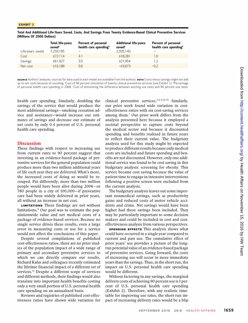
health care spending. Similarly, doubling thesavings of the service that would produce themost additional savings—smoking cessation ad-vice and assistance—would increase our esti-mates of savings and decrease our estimate ofnet costs by only 0.4 percent of U.S. personalhealth care spending.
DiscussionThese findings with respect to increasing usefrom current rates to 90 percent suggest thatinvesting in an evidence-based package of pre-ventive services for the general population couldproduce more than two million additional yearsof life each year they are delivered.What’s more,the increased costs of doing so would be re-couped. Put differently, more than two millionpeople would have been alive during 2006—or780 people in a city of 100,000—if preventivecare had been widely delivered in prior years,all without an increase in net cost.
Limitations These findings are not withoutlimitations.8 Our goal was to estimate the pop-ulationwide value and net medical costs of apackage of evidence-based services. Because nosingle service drives these results, even a largeerror in measuring costs or use for a servicewould not affect the conclusions of this paper.Despite several compilations of published
cost-effectiveness ratios, there are no prior stud-ies of the population impact of a wide range ofprimary and secondary preventive services towhich we can directly compare our results.Richard Kahn and colleagues recently estimatedthe lifetime financial impact of a different set ofservices.12 Despite a different scope of servicesand differentmethods, their findings would alsotranslate into important health benefits costingonly a very small portion of U.S. personal healthcare spending on an annualized basis.Reviews and registries of published cost-effec-
tiveness ratios have shown wide variation for
clinical preventive services.2,4–5,12–15 Similarly,our prior work found wide variation in cost-effectiveness ratios with six cost-saving servicesamong them.7 Our prior work differs from theanalysis presented here because it employed asocietal perspective to capture costs beyondthe medical sector and because it discountedspending and benefits realized in future yearsto reflect their current value. The budgetaryanalysis used for this study might be expectedtoproducedifferent results becauseonlymedicalcosts are included and future spending and ben-efits arenot discounted.However, only one addi-tional service was found to be cost saving in thisbudgetary analysis: screening for obesity. Thisservice became cost saving because the value ofpatient time to engage in intensive interventionsfollowing a positive screen were excluded fromthe current analysis.The budgetary analysis leaves out some impor-
tant nonmedical savings, such as productivitygains and reduced costs of motor vehicle acci-dents and crime. Net savings would have beenhigher had these savings been included. Theymay be particularly important to some decisionmakers and could be included in cost and cost-effectiveness analysis from various perspectives.Spending Effects This analysis shows what
could have occurred in a single year compared tocurrent and past use. The cumulative effect ofprior years’ use provides a picture of the long-runpotential value of an evidence-basedpackageof preventive services. Going forward, the costsof increasing use will occur in more immediateyears than the savings. Thus, in the short run, theimpact on U.S. personal health care spendingwould be different.Without factoring in any savings, themarginal
delivery costs of achieving90percentuse is 1 per-cent of U.S. personal health care spending(Exhibit 2). Therefore, with any realistic time-table for improving use rates, the short-run im-pact of increasing delivery rates would be a blip
EXHIBIT 2
Total And Additional Life-Years Saved, Costs, And Savings From Twenty Evidence-Based Clinical Preventive Services(Millions Of 2006 Dollars)
Total life-yearssaveda
Percent of personalhealth care spendingb
Additional life-yearssavedc
Percent of personalhealth care spendingb
Life-years saved 7,233,195 – 2,335,140 –
Cost $72,114 4.1 $18,281 1.0
Savings $61,927 3.5 $21,954 1.2
Net cost $10,188 0.6 −$3,673 −0.2
Source Authors’ analyses; sources for data used in each model are available from the authors. Note Costs minus savings might not addup to net costs because of rounding. aCost of 90 percent utilization of twenty clinical preventive services (see Exhibit 1). bPercentageof personal health care spending in 2006. cCost of eliminating the difference between existing use rates and 90 percent use rates.
September 2010 29:9 Health Affairs 1659
on Novem
ber 15, 2016 by HW
Team
Health A
ffairs by
http://content.healthaffairs.org/D
ownloaded from

in annual medical spending increases that haveaveraged 7 percent per year or more sincethe 1960s.Whether scaled to annual spending for the
nation, annual spending per person, or healthplan spendingpermemberpermonth, increaseduse would be a virtually undetectable portion ofannual health care spending increases and in thelong run would be cost-neutral, once savings arefactored in, while providing health benefits.Need To Specify Preventive Services As
pointed out by others, preventive services areoften lumped into one large, undifferentiatedgroup.3 There are certainly questionable preven-tive services for which there is not yet good evi-dence of effectiveness or cost-effectiveness.Payers and consumers should focus on reputableguidelines that are based on rigorous assess-ments of each service’s effectiveness, such asthose of the U.S. Preventive Services Task Force.Efforts to improve health could be further re-fined by first focusing on the most valuable evi-dence-based services.7 Some services with highcost savings are poorly used at present. Of those,two have very large health impact—smoking ces-sation advice and assistance, and discussion ofdaily aspirin use. Meanwhile, two services havelesser, but still important, health impacts: alco-hol screening with brief counseling, and
pneumococcal immunization.Increasing Use From Current Rates The
opportunities for cost savings were greater withincreasing use from current rates than they werewith getting from zero use to current rates. Thisis because current use is relatively low for ser-vices that can produce high cost savings. Thisdynamic explains our seemingly contradictoryestimates indicating that increasing all servicesfrom current use to 90 percent would result incost savings while increasing use from zero to90 percent would result in a small increase inU.S. personal health care spending.Conclusion These findings are good news for
purchasers and insurers. This evidence-basedpackage of preventive services is essentiallycost-neutral, while conferring large health ben-efits. That is also good news for patients. Payersand policy makers should support increased useof evidence-based preventive services for theright reasons and with reasonable expectationsof their impact on health spending. Preventiveservices, as well as diagnostic and treatmentservices, should be judged by their effectivenessin improving health and the resources they con-sume to do so. Effective clinical preventive ser-vices can achieve the dual goals of improving thehealth of all Americans and making prudent useof scarce resources. ▪
An early draft of this article waspresented and discussed at a closedmeeting of the National Commission onPrevention Priorities, April 24, 2008, inAustin, Texas. Funding for this research
was provided by the Centers for DiseaseControl and Prevention, Robert WoodJohnson Foundation, and WellPointFoundation. The authors thank membersof the National Commission on
Prevention Priorities for their guidance.Steven Woolf and Corinne Hustenprovided particularly helpful comments.
NOTES
1 Frieden TR, Mostashari F. Healthcare as if health mattered. JAMA.2008;299(8):950–2.
2 Russell LB. The role of prevention inhealth reform. N Engl J Med. 1993;329(5):352–4.
3 Woolf SH. The power of preventionand what it requires. JAMA. 2008;299(20):2437–9.
4 Russell LB. Prevention’s potentialfor slowing the growth of medicalspending [Internet]. Washington(DC): National Coalition on HealthCare; 2007 Oct [cited 2010 Aug 5].Available from: http://www.ihhcpar.rutgers.edu/downloads/nchc_report.pdf
5 Cohen JT, Neumann PJ, WeinsteinMC. Does preventive care savemoney? Health economics and thepresidential candidates. N EnglJ Med. 2008;358(7):661–3.
6 Brown D. In the balance: Somecandidates disagree, but studiesshow it’s often cheaper to let peopleget sick. Washington Post. 2008Apr 8.
7 Maciosek MV, Coffield AB, EdwardsNM, Flottemesch TJ, Goodman MJ,Solberg LI. Priorities among effectiveclinical preventive services: resultsof a systematic review and analysis.Am J Prev Med. 2006;31(1):52–61.
8 The online Supplemental Appendixis available by clicking on the Sup-plemental Appendix link in the boxto the right of the article online.
9 Maciosek MV, Edwards NM, CoffieldAB, Flottemesch TJ, Nelson WW,GoodmanMJ, et al. Priorities amongeffective clinical preventive services:methods. Am J Prev Med. 2006;31(1):90–6.
10 Catlin A, Cowan C, Hartman M,Heffler S. National health spendingin 2006: a year of change for pre-scription drugs. Health Aff (Mill-wood). 2008;27(1):14–29.
11 National utilization rates for theseclinical preventive services are pri-marily 2005 data obtained from theNational Health Interview Surveyand Behavioral Risk Factor Surveil-lance System. For services not ex-
amined in these surveys, weconducted literature searches forutilization rates.
12 Kahn R, Robertson RM, Smith R,Eddy D. The impact of prevention onreducing the burden of cardiovas-cular disease. Circulation. 2008;118(5):576–85.
13 Stone PW, Teutsch S, Chapman RH,Bell C, Goldie SJ, Neumann PJ. Cost-utility analyses of clinical preventiveservices: published ratios, 1976–1997. Am J Prev Med. 2000;19(1):15–23.
14 Tengs TO, Adams ME, Pliskin JS,Safran DG, Siegel JE,Weinstein MC,et al. Five-hundred life-saving inter-ventions and their cost-effectiveness.Risk Anal. 1995;15(3):369–90.
15 Center for the Evaluation of Valueand Risk in Health. CEA registry[home page on the Internet]. Boston(MA): Tufts Medical Center, Insti-tute for Clinical Research and HealthPolicy Studies; [cited 2009 Apr 23].Available from: https://research.tufts-nemc.org/cear/Default.aspx
Costs & Payment
1660 Health Affairs September 2010 29:9
on Novem
ber 15, 2016 by HW
Team
Health A
ffairs by
http://content.healthaffairs.org/D
ownloaded from