Secreted aspartic proteases of Candida albicans activate the NLRP3 inflammasome
Upload
independentCategory
view
3download
0
research papers
Acta Cryst. (2010). D66, 233–242 doi:10.1107/S0907444909054298 233
Acta Crystallographica Section D
BiologicalCrystallography
ISSN 0907-4449
Structure of the unbound form of HIV-1 subtype Aprotease: comparison with unbound forms ofproteases from other HIV subtypes
Arthur H. Robbins,a Roxana M.
Coman,a Edith Bracho-Sanchez,a
Marty A. Fernandez,a C. Taylor
Gilliland,a Mi Li,b,c Mavis
Agbandje-McKenna,a Alexander
Wlodawer,c Ben M. Dunna* and
Robert McKennaa*
aDepartment of Biochemistry and Molecular
Biology, University of Florida, Gainesville,
FL 32610, USA, bBasic Research Program,
SAIC-Frederick, Frederick, Maryland, USA, andcMacromolecular Crystallography Laboratory,
NCI-Frederick, Frederick, Maryland, USA
Correspondence e-mail: [email protected],
# 2010 International Union of Crystallography
Printed in Singapore – all rights reserved
The crystal structure of the unbound form of HIV-1 subtype A
protease (PR) has been determined to 1.7 A resolution and
refined as a homodimer in the hexagonal space group P61 to
an Rcryst of 20.5%. The structure is similar in overall shape and
fold to the previously determined subtype B, C and F PRs. The
major differences lie in the conformation of the flap region.
The flaps in the crystal structures of the unbound subtype B
and C PRs, which were crystallized in tetragonal space groups,
are either semi-open or wide open. In the present structure
of subtype A PR the flaps are found in the closed position, a
conformation that would be more anticipated in the structure
of HIV protease complexed with an inhibitor. The amino-acid
differences between the subtypes and their respective crystal
space groups are discussed in terms of the differences in the
flap conformations.
Received 17 November 2009
Accepted 16 December 2009
PDB Reference: HIV-1
subtype A protease, 3ixo.
1. Introduction
HIV protease (PR) is an aspartic PR that plays an essential
role in the viral life cycle and infectivity by converting a non-
infectious particle into a mature virion that is capable of
infecting new target cells. The PR functions as an obligatory
homodimer, with the active site located in the center and
covered by two flexible loops called flaps. This overall struc-
ture is well conserved and is very similar for all HIV subtypes
(Fig. 1).
HIV is genetically very diverse and as such is classified into
types, groups, subtypes, sub-subtypes and recombinant forms
(Brodine et al., 1995; Fleury et al., 2003; Kantor & Katzenstein,
Figure 1Ribbon diagram of the homodimer of the HIV-1 subtype A PR structure.Catalytic aspartic acids are shown as sticks. Blue spheres indicate aminoacids that vary between subtype A PR and subtype B PR (numbers referto amino-acid positions). Features of the PR structure are as labeled. Thisfigure was generated using PyMOL (DeLano, 2002).

2004). Although subtype B, which is common in the United
States, has been the subject of the majority of investigations,
90% of HIV-infected people worldwide harbor non-B-subtype
variants of HIV-1 (Hemelaar et al., 2006, 2008; Osmanov et al.,
2002; Taylor et al., 2008). In particular, subtypes A and C
account for more than 85% of global infections and more than
95% of infections in South and Central Africa (Foulkes et al.,
2006). Nevertheless, knowledge of resistance mutations in
non-B subtypes and their clinical relevance is limited or con-
troversial (Martinez-Cajas et al., 2009).
With the expanding access to antiretroviral therapy (ARV)
for patients living with HIV/AIDS in developing countries
(UNAIDS, 2008) and with the spread of non-B-subtype
variants of HIV-1 in developed countries (Baldanti et al., 2008;
Palma et al., 2007; Paraskevis et al., 2007), an important issue
of concern is the behavior of non-B subtypes and their
recombinant forms under the selective pressure of ARV
regimens. There is an ongoing debate in the HIV field about
how and to what extent naturally occurring polymorphisms
(NOPs) may affect the emergence of ARV drug resistance in
HIV-1 subtypes other than B. It has been argued that these
polymorphisms might play roles such as increasing the cata-
lytic activity of non-B-subtype PRs and enhancing virus
viability (Krauchenco et al., 2008; Velazquez-Campoy et al.,
2001), resulting in the development of diverse mutational
pathways during ARV treatment (Dumans et al., 2004;
Grossman et al., 2004), influencing the speed of acquisition of
PR inhibitor (PI) related resistance mutations (Hirsch et al.,
2000; Vergne et al., 2000) and contributing to resistance and/or
maintenance of viral fitness once primary resistance mutations
occur (Rose et al., 1996; Velazquez-Campoy et al., 2003).
Several recent studies have argued that there are no sub-
stantial differences regarding known resistance-associated
mutations and the newly emergent substitutions between non-
B-subtype and B-subtype strains (Monno et al., 2009; Pillay et
al., 2008). However, a recent study of the differences in the
impact of several PI-selected mutations on subtypes B and G
in 500 patients showed that the mutation L90M confers
different levels of resistance in subtypes B and G (Santos et al.,
2009). Another recent study indicated that even though in
general drug selective pressure and resistance pathways are
relatively similar in subtypes B and G some differences do
occur, leading to subtype-dependent substitutions (Palma et
al., 2009). With respect to the development of drug-resistance
mutations (DRMs), a recent study showed that non-B sub-
types tend to select for the same DRMs as described in sub-
type B in PR and reverse transcriptase (RT), although in
distinct proportions (Dumans et al., 2009; Kantor et al., 2005).
Also, an increasing body of evidence suggests that certain non-
B subtypes are often more susceptible to specific ARV drugs.
For example, the circulating recombinant form CRF02_AG
PR presents a higher susceptibility to nelfinavir and ritonavir
than subtypes C, F and G, whereas subtype G isolates are more
susceptible to tipranavir (Abecasis et al., 2006). Unexpectedly,
natural hypersusceptibility to lopinavir was also detected in
subtypes C and G (Gonzalez et al., 2003, 2008). In an extensive
review, Martinez-Cajas and coworkers searched 11 databases
and retrieved 3486 citations on all aspects of non-B-subtype-
related resistance research (Martinez-Cajas et al., 2008). They
concluded that the genetic diversity of HIV-1 can affect the
type of resistance mutations, the degree of resistance and the
timing of the emergence of antiretroviral resistance. These
observed differences in resistance pathways may impact on
cross-resistance and the selection of second-line regimens with
PIs.
These NOPs are found throughout the HIV genome,
including PR and RT, which are the main targets of currently
used anti-HIV therapeutic regimens. Thus, characterizing the
structural differences among various non-B-subtype PRs may
identify possible mechanisms that might cause the develop-
ment of divergent mutational pathways against currently used
PIs. Until recently, the only available crystal structures in the
Protein Data Bank (PDB; Berman et al., 2000) were those of
HIV subtype B PRs. More than 100 crystal structures of
subtype B PRs are currently available and include both wild
type and multidrug-resistant (MDR), both open and closed
and both unbound and bound to various inhibitors. The first
crystal structure of a non-B-subtype PR, subtype F, was solved
in 2004 (Sanches et al., 2004) and a detailed analysis and
comparison with subtype B followed (Sanches et al., 2007). A
thorough biochemical characterization of subtype C PR has
been published (Coman et al., 2007) and a detailed analysis of
the crystal structure of subtype C PR followed in 2008
(Coman, Robbins, Goodenow et al., 2008). Solving the crystal
structures of various non-B-subtype PRs has made structural
comparisons and analyses of the NOP effects on the overall
structure of the PR possible. It has previously been suggested
that these differences might affect the binding of substrates/
inhibitors through either long-range interactions or changes in
the flexibility of the enzyme (Coman, Robbins, Fernandez et
al., 2008; Gonzalez et al., 2008; Velazquez-Campoy et al., 2003).
These studies showed that there are differences in the position
of the outside loops, in which most NOPs are located. The
presence of these polymorphic substitutions has been reported
to cause a collapse of the elbow of the flap, resulting in the
displacement of the main chain of this loop towards the loop
76–83, stabilizing the catalytic S1/S10 pockets (Coman,
Robbins, Goodenow et al., 2008; Sanches et al., 2007).
Furthermore, it has also been observed that L89M can
displace the 60s loop laterally and downwards and could
participate in the formation of a more stable network of van
der Waals interactions (Coman, Robbins, Goodenow et al.,
2008). However, currently there is concern that the differ-
ences, which are observed especially in the outside loops of the
enzyme, might be secondary not only to NOPs but also to
different crystal contacts and different crystal packing.
Here, the first crystal structure of subtype A PR is described
at 1.7 A resolution and a detailed structural analysis and
comparison of the unbound subtype A, B and C PR structures
is presented. The results showed that although no inhibitor is
present in the active site, the subtype A PR has flaps in the
closed position and the crystal contacts are similar to those in
other unbound and bound forms of PR crystallized in the
hexagonal space group P61.
research papers
234 Robbins et al. � HIV-1 subtype A protease Acta Cryst. (2010). D66, 233–242

2. Materials and methods
2.1. Protein expression and purification
Protein expression and purification followed the protocol
used for subtype C PR (Coman, Robbins, Fernandez et al.,
2008). The HIV-1 PR DNA for the A subtype (obtained from
the NIH Research Reagents Program, accession code
p92RW009.6) was subcloned into pET23a expression vector
(Novagen) and transformed into Escherichia coli strain BL21
Star DE3 plysS (Invitrogen). Protease expression was per-
formed in M9 minimal media and was initiated when the
OD600 reached 0.8 by the addition of 1 mM IPTG. After 3 h,
the cells were harvested and lysed using an SLM Aminco
French pressure cell. Inclusion bodies containing the PR were
isolated by centrifugation through a 27% sucrose cushion. The
inclusion bodies were dissolved in 8 M urea and refolded by
dialysis against sodium phosphate buffer. The PR was purified
by ammonium sulfate precipitation and gel-filtration chro-
matography as described previously (Coman, Robbins,
Fernandez et al., 2008).
2.2. Crystallization
Crystals were grown using the hanging-drop vapor-diffusion
method (McPherson, 1982). The purified subtype A PR was
concentrated to 3.5 mg ml�1 with a buffer exchange to 50 mM
sodium acetate pH 4.7. 2 ml protein solution was mixed with
2.5 ml reservoir solution (30 mM citric acid pH 5.0, 1 M NaCl)
and 0.5 ml Anapoe-58 detergent (Hampton) on siliconized
glass cover slides. The droplets were suspended over 0.5 ml
reservoir solution and stored at room temperature. Hexagonal
rod-like crystals formed within 2 d.
2.3. Data collection and reduction
A crystal of subtype A PR was ‘quick-dipped’ in 35%
glycerol solution for 1 s and immediately flash-cooled to
100 K. X-ray diffraction data were measured using a MAR
CCD 300 detector on the SER-CAT beamline at the
Advanced Photon Source. The crystal-to-detector distance
was 200 mm and a total of 240 diffraction data frames were
measured using a 0.5� oscillation angle per frame. From these,
a subset of 140 data frames was integrated and scaled with the
HKL-2000 software (Otwinowski & Minor, 1997) to give a
complete data set to 1.7 A resolution.
Systematic absences in the diffraction data were consistent
with either a 61 or 65 screw axis in the c direction. The data
were merged and scaled separately in the hexagonal space
groups P61 and P6122, yielding 19 874 and 10 865 unique
reflections, respectively. Initial phasing was performed using
data in space group P6122, but the lower symmetry space
group was selected for the final refinement (see below). A
complete summary of data statistics for processing in both
space groups is included in Table 1.
From the unit-cell volume and the molecular weight of
subtype A PR, a reasonable VM value (Matthews, 1968) of
2.14 A3 Da�1 was calculated for one PR monomer per asym-
metric unit in space group P6122 (P6522) or for a complete
homodimer in P61 (P65). Additionally, the diffraction data
scaled in P61 were examined using the twin-detection software
in CNS (Brunger et al., 1998) and the Merohedral Twin
Detector server (Padilla & Yeates, 2003; Yeates, 1997).
2.4. Rotation and translation search
Cross-rotation function and translation-function searches
were performed in space group P6122, using X-ray diffraction
data between 15.0 and 4.0 A resolution, with the CNS package
(Brunger et al., 1998). The molecular template used was the
A chain of the HIV-1 subtype B homodimer in complex with
atazanavir (PDB code 2aqu; Clemente et al., 2006), with the
water molecules and the atazanavir inhibitor removed. A
translation-function search in space group P6522 failed to
produce a reasonable solution. Rigid-body refinement of the
molecular-replacement solution using data between 50 and
3.5 A resolution resulted in a model with Rcryst = 28.4% and
Rfree = 26.1%. Upon inspection of the resulting electron-
density maps the flaps were found to be in the closed position
and there was no density in the |Fo| � |Fc| difference map that
would indicate an alternative interpretation. An OMIT map,
research papers
Acta Cryst. (2010). D66, 233–242 Robbins et al. � HIV-1 subtype A protease 235
Table 1Crystal data and refinement.
Values in parentheses are for the highest resolution shell.
Crystal dataSpace group P61 P6122Unit-cell parameters (A) a = 62.2,
c = 82.5a = 62.2,
c = 82.5Molecules per ASU 2 1VM (A3 Da�1) 2.14 2.14Solvent content (%) 42.5 42.5
Data collectionResolution (A) 23.4–1.7
(1.76–1.7)23.4–1.7
(1.76–1.7)Unique reflections 19874 10865No. of frames 140 140Oscillation per frame (�) 0.5 0.5Rmerge† (%) 8.9 (50.9) 9.4 (54.8)I/�(I) 12.3 (2.2) 12.3 (2.2)Completeness (%) 99.6 (99.2) 99.5 (99.9)Data redundancy 4.4 (3.7) 8.0 (6.8)
Refinement statisticsNo. of reflections used 18760Rcryst‡ (%) 20.5Rfree (%) 24.4Reflections used for Rfree (|Fo| > 0) (%) 4.4R.m.s.d. bonds (A) 0.008R.m.s.d. angles (�) 1.49Ramachandran plot (%)
Most favored 90.0Allowed 9.4Generously allowed 0.6
ModelProtein atoms 1510Waters 111Average temperature factors (A2)
Main chain 26.8Side chain 29.0Waters 30.4
† Rmerge =P
hkl
Pi jIiðhklÞ � hIðhklÞij=
Phkl
Pi IiðhklÞ, where Ii(hkl) is the intensity of
an individual reflection and hI(hkl)i is the average intensity. ‡ Rcryst =Phkl
��jFobsj � jFcalcj
��=P
hkl jFobsj; Rfree is identical to Rcryst but was calculated for 5% ofdata omitted from refinement.

based upon the final refined coordinates and centered on the
active site, is shown in Fig. 2.
2.5. Refinement of the HIV-1 subtype A model
The problem of the proper choice of the space group for
refinement of the structure of HIV PR has been faced
repeatedly in the past (Wlodawer & Erickson, 1993; Sanches et
al., 2007) and is a consequence of the possible presence of
pseudo-symmetry, disorder, and/or twinning. An excellent
discussion of the effects of pseudo-symmetry on twin detec-
tion has been given in the analysis of a difficult refinement of
superoxide dismutase from Pyrobaculum aerophilum (Lee et
al., 2003). Since the scaling parameters for subtype A PR were
virtually identical in space groups P61 and P6122 (Table 1), the
high-symmetry space group appeared to be the correct choice.
However, the same result could be expected if the lower
symmetry space group were correct but the data were fully
twinned. Although twinning can normally be detected through
analysis of statistical distribution of the intensities, this does
not apply in this case since pseudo-symmetry restores the
normal distribution of intensities. Indeed, this was tested by
submitting the subtype A PR data scaled in space group P61 to
the merohedral twin-detection server (Padilla & Yeates, 2003;
http://nihserver.mbi.ucla.edu/pystats/). Not surprisingly, no
twinning was detected, whereas the twin-detection protocol in
CNS indicated a twin fraction of 0.48. Since the latter result
is based on a comparison of the intensities of potential twin
mates, it is not really applicable here. It may
be noted that since only a very small part of
the structure (a few residues) is responsible
for the effects of disorder/twinning, its
influence on the statistics of the intensities is
necessarily very small, making it impossible
to differentiate which of these two effects
occurs in the crystal.
Atomic positional and temperature-
factor refinements of the rigid-body model
were performed in the CNS suite (Brunger
et al., 1998), initially with data processed in
space group P6122. The target values used
for restraining the model geometry during
refinement were from Engh & Huber
(1991). A randomly selected subset of 5% of
the reflections was withheld from the
refinement cycles and was used to calculate
Rfree. Replacement of amino acids which
differed between subtype B PR and subtype
A PR was performed in the molecular-
graphics program Coot (Emsley & Cowtan,
2004). Interactive manual model refitting
using Coot and electron-density maps
calculated with 2|Fo| � |Fc| and |Fo| � |Fc|
coefficients were used to guide manual
refitting of the model. After several cycles,
the resulting Rcryst and Rfree values remained
higher than expected and positive peaks at
greater than 3.5� persisted in the |Fo| � |Fc|
difference electron-density map along the
main chain of the monomer. At this stage a
choice had to be made of either trying to
model the asymmetry of the dimers as
disorder or switching to the low-symmetry
space group P61 with assumed twinning.
Since the latter approach provides a simpler
description of the model, it was selected.
Since the free R list of reflections differed
between the data scaled in space groups P61
and P6122, the rigid-body model was used as
the starting model. In space group P61 the
asymmetric unit consists of the entire PR
research papers
236 Robbins et al. � HIV-1 subtype A protease Acta Cryst. (2010). D66, 233–242
Figure 3Stereo superposition of unbound PR homodimers. (a) Complete PR homodimers, (b) close-upof the flap regions shown in (a) viewed down the twofold axis. Color code: green, dimers intetragonal space groups with semi-open flaps; red, dimers in tetragonal space groups with wide-open flaps; blue, dimers in the hexagonal space group P61 with closed flaps. Note that the tipsof the flaps invert between the semi-open and closed conformations. PDB coordinates were asused in Fig. 5. This figure was generated using PyMOL (DeLano, 2002).
Figure 2Stereo 2|Fo| � |Fc| OMIT map centered on the active site of HIV-1 subtype A PR. The map iscontoured at 1.3� and includes a 4 A radius buffer around flap residues and water positions.All atoms of the flap (residues 46–55 and 460–550), the active-site aspartates Asp25 and Asp250
and two waters (depicted as red spheres) were omitted from the calculation of the map.

dimer, so the B monomer coordinates were calculated using
the twofold symmetry in the graphics program Coot (Emsley
& Cowtan, 2004). After appending to the A monomer, the
coordinates were given the chain identifier B for refinement
against the data that had been merged and scaled in P61.
Refinement and model building in the lower symmetry space
group improved the appearance of the electron-density maps
even before correction for twinning. Subsequent refinements
were performed with a twin law of k, h, �l and a twin fraction
of 0.5 and the free R list of reflections was reselected so that
pairs of twin-related reflections resided in either the working
list or the cross-validation list. To verify the effects of twinned
refinement, one round of positional refinement resulted in a
decrease of 3.7% in Rcryst and of 2.5% in Rfree when compared
with an identical refinement cycle starting with the same
molecular model but disregarding twinning. After several
rounds of refinement and model building, the final model,
consisting of two monomers and 111 water molecules, had an
Rcryst of 20.5% and an Rfree of 24.4%. A Ramachandran plot,
as calculated in PROCHECK (Laskowski et al., 1993), showed
that 90.0% of the amino acids were in the most favored region,
9.4% were in the additional allowed region and 0.6% were in
the generously allowed region. A more complete summary of
the final model of subtype A PR is included in Table 1.
Superimposition of the coordinates of the B monomer onto
the A monomer allowed the comparison of side-chain
conformations. Of a total of 99 amino acids, 20 have rotamer
differences between the two monomers, including three
valines, three isoleucines, Met46 and Phe53. The latter two
amino acids are involved in crystal packing, but the electron
density for the side chains of 53A and 53B are very weak in
OMIT maps (Fig. 2). Packing effects would be expected to
stabilize side chains, but in the case of Phe53 disorder between
the two conformations may contribute to the weak density. In
addition, although the flaps are in contact with flaps from
symmetry-related molecules, in the uncomplexed PR they are
separated by solvent cavities where inhibitors would normally
bind in the closed conformation.
2.6. Comparison of unbound HIV PR structures
In order to compare the structure presented here with other
crystal structures of unbound HIV PRs, 13 structures were
retrieved from the PDB and superimposed onto the refined
model of HIV subtype A PR (Fig. 3). The coordinates of the
dimers from the structures refined in space groups P41 or
P61 were used directly, whereas for those refined in P41212
the dimer coordinates were calculated using the twofold-
symmetry operation between the monomers and were saved
using Coot (Emsley & Cowtan, 2004). In two cases, the data-
base holds identical or nearly identical PR structures, namely
tethered subtype B PR [PDB codes 1lv1 (Kumar et al., 2002)
and 1g6l (Pillai et al., 2001)] and an MDR form of B PR [PDB
codes 1rpi (Logsdon et al., 2004) and 1tw7 (Martin et al.,
2005)]. Of these two similar pairs of coordinates, only the two
higher resolution structures, 1g6l and 1tw7, were included in
the comparison.
Nine of the remaining 11 unbound PR structures used for
comparison crystallized in either space group P41212 or P41.
They were the chemically synthesized HIV-1 subtype B PR
(PDB code 3hvp; Wlodawer et al.,
1989), wild-type (wt) subtype B
PR (3phv; Lapatto et al., 1989),
the F53L mutant subtype B PR
(2g69; Liu et al., 2006), subtype B
PR BRU isolate (1hhp; Spinelli et
al., 1991), 6� mutant subtype B
PR and two structures of the Q7K
mutant B PR (2hb2, 1tw7 and
2pc0; Heaslet et al., 2007), the
aforementioned MDR subtype B
PR (1tw7; Martin et al., 2005) and
subtype C PR (2r8n; Coman,
Robbins, Goodenow et al., 2008).
The structure of subtype F PR
(PDB code 2p3c; Sanches et al.,
2007) with the inhibitor coordi-
nates removed and the tethered
unbound B PR structures (PDB
code 1g6l; Pillai et al., 2001),
which also crystallize in space
group P61, were included in the
comparison with subtype A PR.
The C� coordinates of all 11
structures were superimposed
onto the subtype A coordinates
using the LSQ superposition
research papers
Acta Cryst. (2010). D66, 233–242 Robbins et al. � HIV-1 subtype A protease 237
Figure 4Sequences of HIV-1 PR structures.
option in Coot (Emsley & Cowtan,
2004). In order to superimpose pairs of
monomers, chain identifiers were
removed and each amino-acid residue
of the dimer was renumbered: 1001–
1099 (A chain) and 2001–2099 (B
chain). All 198 C� atoms were used in
the r.m.s.d. calculations, with the
exception of 1hb4, in which two amino-
acid residues of each flap were not
modeled owing to weak electron
density. The amino-acid sequences for
all 12 structures used for superposition
are listed in Fig. 4 and the pairwise root-
mean-square displacements (r.m.s.d.s)
are presented in Fig. 5. Figs. 4 and 5
introduce the color code also used in
Figs. 6 and 7: red, wide-open flaps;
green, semi-open flaps; blue, closed
flaps.
2.7. Analysis of packing of selectedunbound PR structures
The Chimera molecular-modeling
software (Pettersen et al., 2004) was
used to calculate the crystal-packing
contacts for the subset of unbound PR
dimers listed in Fig. 5 and shown in Fig. 6
with the refined subtype A PR. All
symmetry-related dimer coordinates in
a 3� 3� 3 grid of unit cells surrounding
the reference molecule were calculated
in order to ensure that all symmetry
contacts were included. The Chimera
default clash/contact cutoff distances
were used to define hydrogen-bond and
van der Waals interactions at the
packing interfaces. Fig. 5 indicates
whether or not each of the amino acids
of the PR chain is involved in these
contacts. No distinction was made
between hydrophobic or hydrophilic
contacts.
3. Results
3.1. Overall structure of subtype A PR
As expected, the HIV-1 subtype A
PR structure is similar in overall shape
and fold to subtype B, C and F PRs with
inhibitor bound (Fig. 1). The major
differences lie in the conformations of
the flap region. The flaps in the crystal
structures of the unbound subtype B PR
and the one subtype C PR crystallized
in tetragonal space groups are either
research papers
238 Robbins et al. � HIV-1 subtype A protease Acta Cryst. (2010). D66, 233–242
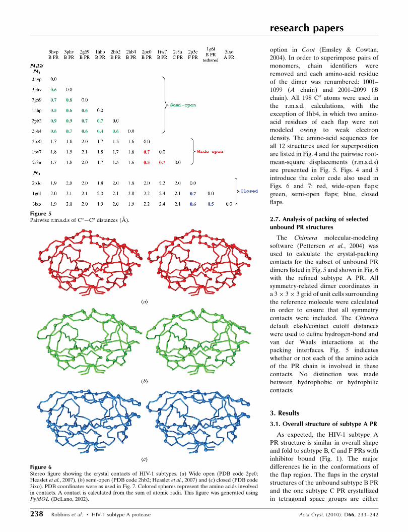
Figure 5Pairwise r.m.s.d.s of C�—C� distances (A).
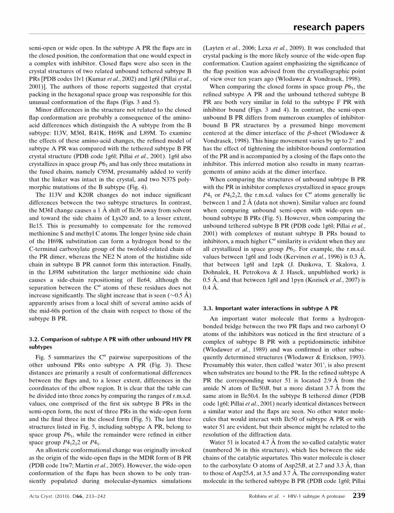
Figure 6Stereo figure showing the crystal contacts of HIV-1 subtypes. (a) Wide open (PDB code 2pc0;Heaslet et al., 2007), (b) semi-open (PDB code 2hb2; Heaslet et al., 2007) and (c) closed (PDB code3ixo). PDB coordinates were as used in Fig. 7. Colored spheres represent the amino acids involvedin contacts. A contact is calculated from the sum of atomic radii. This figure was generated usingPyMOL (DeLano, 2002).
semi-open or wide open. In the subtype A PR the flaps are in
the closed position, the conformation that one would expect in
a complex with inhibitor. Closed flaps were also seen in the
crystal structures of two related unbound tethered subtype B
PRs [PDB codes 1lv1 (Kumar et al., 2002) and 1g6l (Pillai et al.,
2001)]. The authors of those reports suggested that crystal
packing in the hexagonal space group was responsible for this
unusual conformation of the flaps (Figs. 3 and 5).
Minor differences in the structure not related to the closed
flap conformation are probably a consequence of the amino-
acid differences which distinguish the A subtype from the B
subtype: I13V, M36I, R41K, H69K and L89M. To examine
the effects of these amino-acid changes, the refined model of
subtype A PR was compared with the tethered subtype B PR
crystal structure (PDB code 1g6l; Pillai et al., 2001). 1g6l also
crystallizes in space group P61 and has only three mutations in
the fused chains, namely C95M, presumably added to verify
that the linker was intact in the crystal, and two N37S poly-
morphic mutations of the B subtype (Fig. 4).
The I13V and K20R changes do not induce significant
differences between the two subtype structures. In contrast,
the M36I change causes a 1 A shift of Ile36 away from solvent
and toward the side chains of Lys20 and, to a lesser extent,
Ile15. This is presumably to compensate for the removed
methionine S and methyl C atoms. The longer lysine side chain
of the H69K substitution can form a hydrogen bond to the
C-terminal carboxylate group of the twofold-related chain of
the PR dimer, whereas the NE2 N atom of the histidine side
chain in subtype B PR cannot form this interaction. Finally,
in the L89M substitution the larger methionine side chain
causes a side-chain repositioning of Ile64, although the
separation between the C� atoms of these residues does not
increase significantly. The slight increase that is seen (�0.5 A)
apparently arises from a local shift of several amino acids of
the mid-60s portion of the chain with respect to those of the
subtype B PR.
3.2. Comparison of subtype A PR with other unbound HIV PRsubtypes
Fig. 5 summarizes the C� pairwise superpositions of the
other unbound PRs onto subtype A PR (Fig. 3). These
distances are primarily a result of conformational differences
between the flaps and, to a lesser extent, differences in the
coordinates of the elbow region. It is clear that the table can
be divided into three zones by comparing the ranges of r.m.s.d.
values, one comprised of the first six subtype B PRs in the
semi-open form, the next of three PRs in the wide-open form
and the final three in the closed form (Fig. 5). The last three
structures listed in Fig. 5, including subtype A PR, belong to
space group P61, while the remainder were refined in either
space group P41212 or P41.
An allosteric conformational change was originally invoked
as the origin of the wide-open flaps in the MDR form of B PR
(PDB code 1tw7; Martin et al., 2005). However, the wide-open
conformation of the flaps has been shown to be only tran-
siently populated during molecular-dynamics simulations
(Layten et al., 2006; Lexa et al., 2009). It was concluded that
crystal packing is the more likely source of the wide-open flap
conformation. Caution against emphasizing the significance of
the flap position was advised from the crystallographic point
of view over ten years ago (Wlodawer & Vondrasek, 1998).
When comparing the closed forms in space group P61, the
refined subtype A PR and the unbound tethered subtype B
PR are both very similar in fold to the subtype F PR with
inhibitor bound (Figs. 3 and 4). In contrast, the semi-open
unbound B PR differs from numerous examples of inhibitor-
bound B PR structures by a presumed hinge movement
centered at the dimer interface of the �-sheet (Wlodawer &
Vondrasek, 1998). This hinge movement varies by up to 2� and
has the effect of tightening the inhibitor-bound conformation
of the PR and is accompanied by a closing of the flaps onto the
inhibitor. This inferred motion also results in many rearran-
gements of amino acids at the dimer interface.
When comparing the structures of unbound subtype B PR
with the PR in inhibitor complexes crystallized in space groups
P41 or P41212, the r.m.s.d. values for C� atoms generally lie
between 1 and 2 A (data not shown). Similar values are found
when comparing unbound semi-open with wide-open un-
bound subtype B PRs (Fig. 5). However, when comparing the
unbound tethered subtype B PR (PDB code 1g6l; Pillai et al.,
2001) with complexes of mutant subtype B PRs bound to
inhibitors, a much higher C� similarity is evident when they are
all crystallized in space group P61. For example, the r.m.s.d.
values between 1g6l and 1odx (Kervinen et al., 1996) is 0.3 A,
that between 1g6l and 1zpk (J. Duskova, T. Skalova, J.
Dohnalek, H. Petrokova & J. Hasek, unpublished work) is
0.5 A, and that between 1g6l and 1pyn (Kozisek et al., 2007) is
0.4 A.
3.3. Important water interactions in subtype A PR
An important water molecule that forms a hydrogen-
bonded bridge between the two PR flaps and two carbonyl O
atoms of the inhibitors was noticed in the first structure of a
complex of subtype B PR with a peptidomimetic inhibitor
(Wlodawer et al., 1989) and was confirmed in other subse-
quently determined structures (Wlodawer & Erickson, 1993).
Presumably this water, then called ‘water 301’, is also present
when substrates are bound to the PR. In the refined subtype A
PR the corresponding water 51 is located 2.9 A from the
amide N atom of Ile50B, but a more distant 3.7 A from the
same atom in Ile50A. In the subtype B tethered dimer (PDB
code 1g6l; Pillai et al., 2001) nearly identical distances between
a similar water and the flaps are seen. No other water mole-
cules that would interact with Ile50 of subtype A PR or with
water 51 are evident, but their absence might be related to the
resolution of the diffraction data.
Water 51 is located 4.7 A from the so-called catalytic water
(numbered 36 in this structure), which lies between the side
chains of the catalytic aspartates. This water molecule is closer
to the carboxylate O atoms of Asp25B, at 2.7 and 3.3 A, than
to those of Asp25A, at 3.5 and 3.7 A. The corresponding water
molecule in the tethered subtype B PR (PDB code 1g6l; Pillai
research papers
Acta Cryst. (2010). D66, 233–242 Robbins et al. � HIV-1 subtype A protease 239
et al., 2001) is in a more symmetrical location, with distances of
2.9, 3.1, 3.1 and 3.2 A from the four O atoms. Many other well
ordered water molecules are also present in the subtype A
active site, as well as diffuse chains of electron density that
may be disordered water molecules.
3.4. Comparison of crystal-packing contacts betweenunbound subtype A PR and other PRs
From Fig. 7 it is evident that the packing contacts are
grouped into the semi-open forms of unbound PRs, the wide-
open forms and the closed forms so far only seen in space
group P61. The lone outlier is the F53L mutant subtype B PR
(PDB code 2g69; Liu et al., 2006), which crystallizes in a form
with unique unit-cell parameters (a = b ’ 61, c ’ 56 A) when
compared with the range seen in the other tetragonal unbound
PR structures (a = b ’ 47, c ’ 108 A). Although its packing
differs from that of 1hhp (Spinelli et al., 1991) and 2hb2
(Heaslet et al., 2007), especially in the 50s area of the flaps, all
three contain semi-open flaps.
Also evident in Figs. 6 and 7 are the large number of resi-
dues that are involved in crystal contacts in the wide-open
forms of the unbound B and C PRs 2pc0 (Heaslet et al., 2007)
and 2r8n (Coman, Robbins, Goodenow et al., 2008). The flap
regions of these structures are extensively involved in crystal
contacts. Apparently, the wide-open crystal forms are more
tightly packed. This is borne out by the lower VM values for
these crystals [2.56 and 2.52 A3 Da�1, respectively, compared
with 3.12 and 3.15 A3 Da�1 for the semi-open forms 1hhp
(Spinelli et al., 1991) and 2hb2 (Heaslet et al., 2007), respec-
tively]. This close packing results in
interactions between the elbow residues
and the flap of a symmetry-related
molecule, as has been pointed out
previously (Layten et al., 2006).
4. Discussion
The crystal structure of HIV-1 subtype
A PR has now been solved and com-
pared with a set of 11 structures of other
variants of HIV PR. The rather unex-
pected finding of a closed conformation
of an unbound (apo) form of subtype A
HIV PR set the basis for comparing four
PR subtypes, A, B, C and F, crystallized
in two crystal systems: tetragonal (P41
and P4122) and hexagonal (P61). Three
conformations are compared in this
paper, defined by the position of the
flaps: semi-open, wide-open and closed.
The goal of this work is to bring further
understanding of the determinants for
the different positions of the flaps.
As shown in Fig. 5, the r.m.s.d. value
between the C� positions of refined
subtype A PR and subtype F PR in
complex with the inhibitor TL-3 (PDB
code 2p3c; Sanches et al., 2007) is only
0.6 A. Such a small difference in overall
fold is close to the experimental error of
the diffraction experiments, which is
typically 0.3–0.5 A. For comparison, the
r.m.s.d. value between the synthetic and
wt B PRs, PDB entries 3hvp (Wlodawer
et al., 1989) and 3phv (Lapatto et al.,
1989), is 0.57 A. One explanation might
be that crystal-packing energies in the
hexagonal space group induces flap
closure and the unbound PR assumes
the conformation of the inhibitor-bound
form. Each flap in subtype A PR is
research papers
240 Robbins et al. � HIV-1 subtype A protease Acta Cryst. (2010). D66, 233–242
Figure 7Packing contacts (colored cells represent crystal-packing contacts).
sandwiched between the flap of its twofold-related monomer
and the flap of a symmetry-related molecule. Another mole-
cule, related by a different symmetry operation, is in contact
with the opposite flap. The residues involved in the symmetry
contact are Phe53 and Met46 from each flap; this contact is
also present in those inhibitor complexes of B PR that crys-
tallize in space group P61. Indeed, the superpositions of
inhibitor-bound B-subtype PRs crystallized in space group P61
show the same crystal contacts as the two tethered B-subtype
structures (1lv1 and 1g6l) and similar r.m.s.d. values for the
superpositions that are evident in Fig. 5 (data not shown). If
the argument that crystal packing is key to flap conformation
has merit, perhaps it can be used to explain the difference
between the semi-open and wide-open flap conformations in
the tetragonal space groups P41 and P41212.
In conclusion, the conformation of the flaps in the different
subtypes, bound and unbound, may be a ‘cause-and-effect’
observation. The question that needs to be addressed is: do the
differences in flap/hinge amino-acid sequences and the asso-
ciated dynamics affect the crystallization process or do the
entropy-driven crystallization forces induce a conformation
onto the flaps?
We acknowledge the use of beamline 22-ID of the South-
east Regional Collaborative Access Team (SER-CAT) located
at the Advanced Photon Source, Argonne National Labora-
tory. Use of the APS was supported by the US Department of
Energy, Office of Science, Office of Basic Energy Sciences
under Contract No. W-31-109-Eng-38. This project was
supported in part by NIH grant AI-28571 to BMD, in part by
the Intramural Research Program of the NIH, National
Cancer Institute, Center for Cancer Research (AW) and in
part by Federal funds from the National Cancer Institute, NIH
under Contract No. HHSN2612008000001E (ML). The con-
tent of this publication does not necessarily reflect the views or
policies of the Department of Health and Human Services, nor
does the mention of trade names, commercial products or
organizations imply endorsement by the US Government.
References
Abecasis, A. B., Deforche, K., Bacheler, L. T., McKenna, P., Carvalho,A. P., Gomes, P., Vandamme, A. M. & Camacho, R. (2006). J.Antivir. Ther. 11, 581–589.
Baldanti, F., Paolucci, S., Ravasi, G., Maccabruni, A., Moriggia, A.,Barbarini, G. & Maserati, R. (2008). J. Med. Virol. 80, 947–952.
Berman, H. M., Westbrook, J., Feng, Z., Gilliland, G., Bhat, T. N.,Weissig, H., Shindyalov, I. N. & Bourne, P. E. (2000). Nucleic AcidsRes. 28, 235–242.
Brodine, S. K., Mascola, J. R., Weiss, P. J., Ito, S. I., Porter, K. R.,Artenstein, A. W., Garland, F. C., McCutchan, F. E. & Burke, D. S.(1995). Lancet, 346, 1198–1199.
Brunger, A. T., Adams, P. D., Clore, G. M., DeLano, W. L., Gros, P.,Grosse-Kunstleve, R. W., Jiang, J.-S., Kuszewski, J., Nilges, M.,Pannu, N. S., Read, R. J., Rice, L. M., Simonson, T. & Warren, G. L.(1998). Acta Cryst. D54, 905–921.
Clemente, J. C., Coman, R. M., Thiaville, M. M., Janka, L. K., Jeung,J. A., Nukoolkarn, S., Govindasamy, L., Agbandje-McKenna, M.,McKenna, R., Leelamanit, W., Goodenow, M. M. & Dunn, B. M.(2006). Biochemistry, 45, 5468–5477.
Coman, R. M., Robbins, A. H., Fernandez, M. A., Gilliland, C. T.,Sochet, A. A., Goodenow, M. M., McKenna, R. & Dunn, B. M.(2008). Biochemistry, 47, 731–743.
Coman, R. M., Robbins, A. H., Goodenow, M. M., Dunn, B. M. &McKenna, R. (2008). Acta Cryst. D64, 754–763.
Coman, R. M., Robbins, A., Goodenow, M. M., McKenna, R. &Dunn, B. M. (2007). Acta Cryst. F63, 320–323.
DeLano, W. L. (2002). The PyMOL Molecular Viewer. http://www.pymol.org.
Dumans, A. T., Barreto, C. C., Santos, A. F., Arruda, M., Sousa, T. M.,Machado, E. S., Sabino, E. C., Brindeiro, R. M., Tanuri, A., Duarte,A. J. & Soares, M. A. (2009). Infect. Genet. Evol. 9, 62–70.
Dumans, A. T., Soares, M. A., Machado, E. S., Hue, S., Brindeiro,R. M., Pillay, D. & Tanuri, A. (2004). J. Infect. Dis. 189, 1232–1238.
Emsley, P. & Cowtan, K. (2004). Acta Cryst. D60, 2126–2132.Engh, R. A. & Huber, R. (1991). Acta Cryst. A47, 392–400.Fleury, H., Recordon-Pinson, P., Caumont, A., Faure, M., Roques, P.,
Plantier, J. C., Couturier, E., Dormont, D., Masquelier, B. & Simon,F. (2003). AIDS Res. Hum. Retroviruses, 19, 41–47.
Foulkes, J. E., Prabu-Jeyabalan, M., Cooper, D., Henderson, G. J.,Harris, J., Swanstrom, R. & Schiffer, C. A. (2006). J. Virol. 80, 6906–6916.
Gonzalez, L. M., Brindeiro, R. M., Tarin, M., Calazans, A., Soares,M. A., Cassol, S. & Tanuri, A. (2003). Antimicrob. AgentsChemother. 47, 2817–2822.
Gonzalez, L. M., Santos, A. F., Abecasis, A. B., Van Laethem, K.,Soares, E. A., Deforche, K., Tanuri, A., Camacho, R., Vandamme,A. M. & Soares, M. A. (2008). J. Antimicrob. Chemother. 61, 1201–1204.
Grossman, Z., Paxinos, E. E., Averbuch, D., Maayan, S., Parkin, N. T.,Engelhard, D., Lorber, M., Istomin, V., Shaked, Y., Mendelson, E.,Ram, D., Petropoulos, C. J. & Schapiro, J. M. (2004). Antimicrob.Agents Chemother. 48, 2159–2165.
Heaslet, H., Rosenfeld, R., Giffin, M., Lin, Y.-C., Tam, K., Torbett,B. E., Elder, J. H., McRee, D. E. & Stout, C. D. (2007). Acta Cryst.D63, 866–875.
Hemelaar, J., Gouws, E., Ghys, P. D. & Osmanov, S. (2006). AIDS, 20,W13–W23.
Hemelaar, J., Gouws, E., Ghys, P. D. & Osmanov, S. (2008). AIDS, 22,322–323.
Hirsch, M. S., Brun-Vezinet, F., D’Aquila, R. T., Hammer, S. M.,Johnson, V. A., Kuritzkes, D. R., Loveday, C., Mellors, J. W., Clotet,B., Conway, B., Demeter, L. M., Vella, S., Jacobsen, D. M. &Richman, D. D. (2000). J. Am. Med. Assoc. 283, 2417–2426.
Kantor, R. et al. (2005). PLoS Med. 2, e112.Kantor, R. & Katzenstein, D. (2004). J. Clin. Virol. 29, 152–159.Kervinen, J., Thanki, N., Zdanov, A., Tino, J., Barrish, J., Lin, P. F.,
Colonno, R., Riccardi, K., Samanta, H. & Wlodawer, A. (1996).Protein Pept. Lett. 3, 399–406.
Kozisek, M., Bray, J., Rezacova, P., Saskova, K., Brynda, J., Pokorna,J., Mammano, F., Rulisek, L. & Konvalinka, J. (2007). J. Mol. Biol.374, 1005–1016.
Krauchenco, S., Martins, N. H., Sanches, M. & Polikarpov, I. (2009). J.Enzyme Inhib. Med. Chem. 24, 638–645.
Kumar, M., Kannan, K. K., Hosur, M. V., Bhavesh, N. S., Chatterjee,A., Mittal, R. & Hosur, R. V. (2002). Biochem. Biophys. Res.Commun. 294, 395–401.
Lapatto, R., Blundell, T., Hemmings, A., Overington, J., Wilderspin,A., Wood, S., Merson, J. R., Whittle, P. J., Danley, D. E., Geoghegan,K. F., Hawrylik, S. J., Lee, S. E., Scheld, K. G. & Hobart, P. M.(1989). Nature (London), 342, 299–302.
Laskowski, R. A., MacArthur, M. W., Moss, D. S. & Thornton, J. M.(1993). J. Appl. Cryst. 26, 283–291.
Layten, M., Hornak, V. & Simmerling, C. (2006). J. Am. Chem. Soc.128, 13360–13361.
Lee, S., Sawaya, M. R. & Eisenberg, D. (2003). Acta Cryst. D59, 2191–2199.
research papers
Acta Cryst. (2010). D66, 233–242 Robbins et al. � HIV-1 subtype A protease 241

Lexa, K. W., Damm, K. L., Quintero, J. J., Gestwicki, J. E. & Carlson,H. A. (2009). Proteins, 74, 872–880.
Liu, F., Kovalevsky, A. Y., Louis, J. M., Boross, P. I., Wang, Y.-F.,Harrison, R. W. & Weber, I. T. (2006). J. Mol. Biol. 358, 1191–1199.
Logsdon, B. C., Vickrey, J. F., Martin, P., Proteasa, G., Koepke, J. I.,Terlecky, S. R., Wawrzak, Z., Winters, M. A., Merigan, T. C. &Kovari, L. C. (2004). J. Virol. 78, 3123–3132.
Martin, P., Vickrey, J. F., Proteasa, G., Jimenez, Y. L., Wawrzak, Z.,Winters, M. A., Merigan, T. C. & Kovari, L. C. (2005). Structure, 13,1887–1895.
Martinez-Cajas, J. L., Pai, N. P., Klein, M. B. & Wainberg, M. A.(2009). J. Int. AIDS Soc. 12, 11.
Martinez-Cajas, J. L., Pant-Pai, N., Klein, M. B. & Wainberg, M. A.(2008). AIDS Rev. 10, 212–213.
Matthews, B. W. (1968). J. Mol. Biol. 33, 491–497.McPherson, A. (1982). Preparation and Analysis of Protein Crystals.
New York: Wiley.Monno, L., Scudeller, L., Brindicci, G., Saracino, A., Punzi, G.,
Chirianni, A., Lagioia, A., Ladisa, N., Lo Caputo, S. & Angarano,G. (2009). Antiviral Res. 83, 118–126.
Osmanov, S., Pattou, C., Walker, N., Schwardlander, B. & Esparza, J.(2002). J. Acquir. Immune Defic. Syndr. 29, 184–190.
Otwinowski, Z. & Minor, W. (1997). Methods Enzymol. 276, 307–326.
Padilla, J. E. & Yeates, T. O. (2003). Acta Cryst. D59, 1124–1130.Paraskevis, D. et al. (2007). J. Infect. Dis. 196, 1167–1176.Palma, A. C., Abecasis, A. B., Vercauteren, J., Carvalho, A. P.,
Cabanas, J., Vandamme, A. M. & Camacho, R. J. (2009). Infect.Genet. Evol., doi:10.1016/j.meegid.2009.06.019.
Palma, A. C., Araujo, F., Duque, V., Borges, F., Paixao, M. T. &Camacho, R. (2007). Infect. Genet. Evol. 7, 391–398.
Pettersen, E. F., Goddard, T. D., Huang, C. C., Couch, G. S.,Greenblatt, D. M., Meng, E. C. & Ferrin, T. E. (2004). J. Comput.Chem. 25, 1605–1612.
Pillai, B., Kannan, K. K. & Hosur, M. V. (2001). Proteins, 43, 57–64.
Pillay, V., Pillay, C., Kantor, R., Venter, F., Levin, L. & Morris, L.(2008). AIDS Res. Hum. Retroviruses, 24, 1449–1454.
Rose, R. E., Gong, Y. F., Greytok, J. A., Bechtold, C. M., Terry, B. J.,Robinson, B. S., Alam, M., Colonno, R. J. & Lin, P. F. (1996). Proc.Natl Acad. Sci. USA, 93, 1648–1653.
Sanches, M., Krauchenco, S., Martins, N. H., Gustchina, A.,Wlodawer, A. & Polikarpov, I. (2007). J. Mol. Biol. 369, 1029–1040.
Sanches, M., Martins, N. H., Calazans, A., de Moraes Brindeiro, R.,Tanuri, A., Augusto Ceva Antunes, O. & Polikarpov, I. (2004). ActaCryst. D60, 1625–1627.
Santos, A. F., Abecasis, A. B., Vandamme, A. M., Camacho, R. J. &Soares, M. A. (2009). J. Antimicrob. Chemother. 63, 593–599.
Spinelli, S., Liu, Q. Z., Alzari, P. M., Hirel, P. H. & Poljak, R. J. (1991).Biochimie, 73, 1391–1396.
Taylor, B. S., Sobieszczyk, M. E., McCutchan, F. E. & Hammer, S. M.(2008). N. Engl. J. Med. 358, 1590–1602.
UNAIDS (2008). Report on the Global HIV/AIDS Epidemic2008: Executive Summary. UNAIDS/08.27E/JC1511E. Geneva:UNAIDS.
Velazquez-Campoy, A., Todd, M. J., Vega, S. & Freire, E. (2001).Proc. Natl Acad. Sci. USA, 98, 6062–6067.
Velazquez-Campoy, A., Vega, S., Fleming, E., Bacha, U., Sayed, Y.,Dirr, H. W. & Freire, E. (2003). AIDS Rev. 5, 165–171.
Vergne, L., Peeters, M., Mpoudi-Ngole, E., Bourgeois, A., Liegeois, F.,Toure-Kane, C., Mboup, S., Mulanga-Kabeya, C., Saman, E.,Jourdan, J., Reynes, J. & Delaporte, E. (2000). J. Clin. Microbiol. 38,3919–3925.
Wlodawer, A. & Erickson, J. W. (1993). Annu. Rev. Biochem. 62,543–585.
Wlodawer, A., Miller, M., Jaskolski, M., Sathyanarayana, B. K.,Baldwin, E., Weber, I. T., Selk, L. M., Clawson, L., Schneider, J. &Kent, S. B. (1989). Science, 245, 616–621.
Wlodawer, A. & Vondrasek, J. (1998). Annu. Rev. Biophys. Biomol.Struct. 27, 249–284.
Yeates, T. O. (1997). Methods Enzymol. 276, 344–358.
research papers
242 Robbins et al. � HIV-1 subtype A protease Acta Cryst. (2010). D66, 233–242
Copyright © 2022 FDOKUMEN