Structure of the ovaries and follicular epithelium morphogenesis in Drosophila and its kin
13
REVIEW Structure of the ovaries and follicular epithelium morphogenesis in Drosophila and its kin Mariusz K. Jaglarz & Wieslaw Krzeminski & Szczepan M. Bilinski Received: 14 February 2008 / Accepted: 10 June 2008 / Published online: 22 July 2008 # Springer-Verlag 2008 Abstract In insects, the ovarian follicular epithelium morphogenesis has been intensively studied and best characterized in the fruit fly, Drosophila melanogaster . It is well established that initially identical somatic follicular cells (FCs) form a simple epithelium overlying the germline cells, but during oogenesis, they diversify into a number of morphologically distinct subpopulations each responsible for creating specific eggshell structures. In addition, some FC subpopulations (e.g. polar cells) are indispensable in establishing antero-posterior and dorso-ventral ovarian follicle axes and patterning of the developing embryo. The morphological and molecular changes that occur during follicular epithelium morphogenesis in Drosophila are frequently considered as a paradigm of the FC diversification in all flies. However, recent comparative studies indicate that, in dipterans, the functioning of the ovarian follicles is diverse, group-specific and may signif- icantly differ from the Drosophila model system. We discuss the similarities and differences of the ovary structure and follicular epithelium morphogenesis in differ- ent dipteran groups and put them into a phylognetic context. We suggest that the migratory activity of the FCs represents an evolutionary novelty that evolved in the ancestors of higher dipterans (Brachycera). Subsequently, during evolution of this subgroup, the number of migrating FC subpopulations has gradually increased from one (in Orthorrhapha) to four (in Cyclorrhapha). Keywords Polar cells . Insect ovary . Oogenesis . Diptera . Phylogeny Introduction Our present knowledge of molecular mechanisms underly- ing the morphogenesis and diversification of the follicular epithelium is based primarily on the studies of the model organism, the fruit fly Drosophila melanogaster . These studies have shown that in Drosophila, follicular cells (FCs) initially form a uniform epithelium that, during oogenesis, becomes patterned into several functionally different cell subpopulations (reviewed in Dobens and Raftery 2000; Horne-Badovinac and Bilder 2005; Wu et al. 2008). At least some of the FCs (e.g. border cells and centripetal cells) are able to rearrange or migrate over the germ cells. The specification of these subpopulations and their movements rely on inductive signaling mediated by various ligands, their corresponding receptors and extensive cytoskeletal rearrangements (reviewed in Dobens and Raftery 2000; Horne-Badovinac and Bilder 2005; Wu et al. 2008). During successive stages of oogenesis the FC subpopulations reach their final destination within the ovarian follicle and produce the regionally complex eggshell. The patterning of the follicular epithelium is additionally fundamental for the establishment of antero- sposterior and dorsoventral axes of the egg and, conse- quently, the embryo (Gonzalez-Reyes et al. 1995; Ray and Schupbach 1996; Moussian and Roth 2005). Dev Genes Evol (2008) 218:399–411 DOI 10.1007/s00427-008-0233-0 Communicated by P. Simpson M. K. Jaglarz : S. M. Bilinski (*) Department of Systematic Zoology, Institute of Zoology, Jagiellonian University, R. Ingardena 6, 30-060 Kraków, Poland e-mail: [email protected] W. Krzeminski Institute of Systematics and Evolution of Animals, Polish Academy of Sciences, Slawkowska 17, 31-016 Krakow, Poland
-
Upload
jagiellonian -
Category
Documents
-
view
0 -
download
0
Transcript of Structure of the ovaries and follicular epithelium morphogenesis in Drosophila and its kin

REVIEW
Structure of the ovaries and follicular epitheliummorphogenesis in Drosophila and its kin
Mariusz K. Jaglarz & Wieslaw Krzeminski &Szczepan M. Bilinski
Received: 14 February 2008 /Accepted: 10 June 2008 / Published online: 22 July 2008# Springer-Verlag 2008
Abstract In insects, the ovarian follicular epitheliummorphogenesis has been intensively studied and bestcharacterized in the fruit fly, Drosophila melanogaster. Itis well established that initially identical somatic follicularcells (FCs) form a simple epithelium overlying the germlinecells, but during oogenesis, they diversify into a number ofmorphologically distinct subpopulations each responsiblefor creating specific eggshell structures. In addition, someFC subpopulations (e.g. polar cells) are indispensable inestablishing antero-posterior and dorso-ventral ovarianfollicle axes and patterning of the developing embryo.The morphological and molecular changes that occurduring follicular epithelium morphogenesis in Drosophilaare frequently considered as a paradigm of the FCdiversification in all flies. However, recent comparativestudies indicate that, in dipterans, the functioning of theovarian follicles is diverse, group-specific and may signif-icantly differ from the Drosophila model system. Wediscuss the similarities and differences of the ovarystructure and follicular epithelium morphogenesis in differ-ent dipteran groups and put them into a phylogneticcontext. We suggest that the migratory activity of the FCsrepresents an evolutionary novelty that evolved in the
ancestors of higher dipterans (Brachycera). Subsequently,during evolution of this subgroup, the number of migratingFC subpopulations has gradually increased from one (inOrthorrhapha) to four (in Cyclorrhapha).
Keywords Polar cells . Insect ovary . Oogenesis . Diptera .
Phylogeny
Introduction
Our present knowledge of molecular mechanisms underly-ing the morphogenesis and diversification of the follicularepithelium is based primarily on the studies of the modelorganism, the fruit fly Drosophila melanogaster. Thesestudies have shown that in Drosophila, follicular cells(FCs) initially form a uniform epithelium that, duringoogenesis, becomes patterned into several functionallydifferent cell subpopulations (reviewed in Dobens andRaftery 2000; Horne-Badovinac and Bilder 2005; Wuet al. 2008). At least some of the FCs (e.g. border cellsand centripetal cells) are able to rearrange or migrate overthe germ cells. The specification of these subpopulationsand their movements rely on inductive signaling mediatedby various ligands, their corresponding receptors andextensive cytoskeletal rearrangements (reviewed in Dobensand Raftery 2000; Horne-Badovinac and Bilder 2005; Wuet al. 2008). During successive stages of oogenesis the FCsubpopulations reach their final destination within theovarian follicle and produce the regionally complexeggshell. The patterning of the follicular epithelium isadditionally fundamental for the establishment of antero-sposterior and dorsoventral axes of the egg and, conse-quently, the embryo (Gonzalez-Reyes et al. 1995; Ray andSchupbach 1996; Moussian and Roth 2005).
Dev Genes Evol (2008) 218:399–411DOI 10.1007/s00427-008-0233-0
Communicated by P. Simpson
M. K. Jaglarz : S. M. Bilinski (*)Department of Systematic Zoology, Institute of Zoology,Jagiellonian University,R. Ingardena 6,30-060 Kraków, Polande-mail: [email protected]
W. KrzeminskiInstitute of Systematics and Evolution of Animals,Polish Academy of Sciences,Slawkowska 17,31-016 Krakow, Poland

The Diptera (true flies) are a highly diverse group ofinsect comprising around 124,000 described speciesdivided into well over 100 families (for dipteran classifi-cation, see Oosterbroek and Courtney 1995; Yeates andWiegmann 1999; Yeates 2002; Wiegmann et al. 2003).Traditionally, two major Diptera suborders have beenrecognised: lower flies (midges) or Nematocera and higherflies or Brachycera (Yeates and Wiegmann 1999). It isworth noting, that only the latter group is consideredmonophyletic (Wiegmann et al. 2003). The paraphyleticNematocera comprise 26 families with contentious andunresolved phylogenetic relationships (Oosterbroek andCourtney 1995; Friedrich and Tautz 1997; Yeatesand Wiegmann 1999). Most recently, the group has beensplit into four distinct lineages: Tanyderomorpha+Psychodo-morpha, Neoneura, Polyneura and Anisoneura (Krzeminskiand Krzeminska 2003).
Extant Brachycera are divided into four monophyleticsubgroups: Stratiomyomorpha, Xylophagomorpha, Tabano-morpha and Muscomorpha (Yeates 2002; Wiegmann et al.2003). The last subgroup comprises Asiloidea, Empidoideaand the most-derived brachyceran lineage termed Cyclo-rrhapha or higher Brachycera. The familiar fruit and houseflies belong to Cyclorrhapha. Finally, Stratiomyomorpha,Xylophagomorpha, Tabanomorpha together with Asiloidea
and Empidoidea are combined into Orthorrhapha, alsoknown as lower Brachycera.
In this review, we focus primarily on the structure of theovaries and morphogenesis of the follicular epithelium inhigher dipterans (Brachycera), but relevant data on thelower dipterans (Nematocera) is also included. First, weprovide an overview of what is known on the ovarystructure and FC diversification in Drosophila and otherrepresentatives of Diptera; next, we present the state ofthese characters in the studied subgroups of Cyclorrhapha,Orthorrhapha and Nematocera, followed by more generalevolutionary considerations. Our comparative analysis ofthe FC differentiation in various dipteran groups gives anextra support to the currently accepted view that manyaspects of oogenesis and development in Drosophila (andother higher dipterans) are significantly different not onlyfrom other insects but even from more basal lineages offlies (e.g. Sander 1996; McGregor 2005; Gregor et al.2008).
Materials and methods
The ovarian follicles of the species investigated in thisstudy (see Table 1) were processed for the light, fluorescent
Table 1 List of the analyzed brachyceran taxa
Suborder Infraorder/family Species References
Brachycera: Orthorrhapha Stratiomyomorpha/Stratiomyidae Chloromyia formosa 6 (1) Kedra 2006Xylophagomorpha/Xylophagidae Xylophagus compeditus 6 (1) Kedra and Szklarzewicz 2004Tabanomorpha/Rhagionidae Ptiolina paradoxa 6 (1) Kubrakiewicz et al. 2003
Symphoromyia crassicornis 6 (1) Kubrakiewicz et al. 2003Rhagio tringarius 6 (1) Tworzydlo et al. 2005; this studyRhagio lineola 6 (1) Tworzydlo et al. 2005Rhagio notatuts 6 (1) Unpublished
Tabanidae Haematopota italica 6 (1) This studyTabanus sp. 6 (1) This study
Muscomorpha/ Asilidae Philonicus albiceps 6 (1) Kubrakiewicz et al. 2003Therevidae Thereva sp. 6 (1) Kubrakiewicz et al. 2003Dolichopodidae Sciapus sp. 6 (1) Tworzydlo et al. 2005Empididae Empis livida 5+1? (1) This study
Rhamphomyia sp. 5+1? (1) This studyHilara sp. 5+1? (1) Unpublished
Brachycera: Cyclorrhapha Sarcophagidae Sarcophaga bullata ? De Loof et al. 1990Phoridae Megaselia sp. 7 (3) Kubrakiewicz et al. 1998Conopidae Sicus ferrugineus 7 (3) This studyScatophagidae Scatophaga sp. 7 (3) This studyLonchopteridae Lonchoptera sp. 7 (3) This studyDrosophilidae Drosophila melanogaster 8 (4) Dobens and Raftery 2000; Horne-Badovinac
and Bilder 2005
The digits in the species column indicate the total number of FC subpopulations and the number of the migratory subpopulations (in parentheses)
400 Dev Genes Evol (2008) 218:399–411

and electron microscopy analysis as previously described(see Tworzydlo et al. 2005 for details). In each species, FCdiversification has been examined in two to four ovarianfollicles at the same stage of development.
Gross morphology of dipteran ovaries
All higher dipterans have paired meroistic–polytrophicovaries (for classification of insect ovaries, see Buning1994; Bilinski 1998) consisting of discrete elements termedovarioles. They start to differentiate in late larval/pupalstages, and distinct ovarioles can be distinguished as earlyas in the late pupa (King 1970; Godt and Laski 1995). Thenumber of the ovarioles constituting an individual ovaryranges from several (in Cyclorrhapha) to several dozen (inOrthorrhapha). Within each ovariole, four well-definedregions: the terminal filament, germarium, vitellarium andovariolar pedicel, can be distinguished (for further discus-sion see Buning 1994). The major part of the ovariole, i.e.the vitellarium, contains sequentially more mature ovarianfollicles (=egg chambers) in a linear arrangement. Thenumber of egg chambers per ovariole is lower (two to four)in orthorrhaphan flies and higher in Cyclorrhapha (seven onaverage). In all studied higher dipterans, the egg chambersare morphologically similar; they consist of 16 germ linecells: an oocyte, 15 polyploid nurse cells (generated by fourconsecutive and incomplete mitosis of the cystoblast, at leastinDrosophila) and are enveloped by a somatic (mesodermal)follicular epithelium (Figs. 1a,d,e). Both the germline andsomatic components develop simultaneously. The neighbour-ing egg chambers are connected by interfollicular stalks andordered in such a way that younger chambers lie closer to thegermarium, whereas those in advanced stages of oogenesisoccupy the posterior region of the ovariole.
It should be stressed that nematocerans are far lesscharacterised both in terms of the ovary structure and FCdiversification, and more comprehensive data exist only fora handful of species. Although nematoceran ovaries are ofthe meroistic–polytrophic type, the comparative analysisrevealed that they differ significantly from the ovaries ofthe higher flies. First, the differentiation of the ovary startsearlier: gonial cell divisions, cluster formation and the initialdevelopment of ovarioles take place exclusively duringlarval stages (reviewed in Mazurkiewicz and Kubrakiewicz1998, 2008). Second, the generation of germ cell clustersprecedes the rise of the somatic component of the ovarioles.Such a sequence of events was demonstrated in developingovaries of psychodids (Koch 1929; Mazurkiewicz andKubrakiewicz 1998), tipulids (Bauer 1933; Bayreuther1956; Buning 1994), chironomids (Wulker and Winter1970) and sciarids (Buning 1994). Third, the germ cell
clusters may contain a different number of cells (cysto-cytes), depending on the species/group. While in Tipulidae,Limonidae, Cylindrotomidae, Trichoceridae and Psychodidaethe clusters consist of 16 cystocytes (Koch 1929; Bauer 1933;Mazurkiewicz and Kubrakiewicz 1998, 2005, 2008), in thefungus gnat Bradysia tritici, there are only two cells percluster: one oocyte and one nurse cell (Wenzel et al. 1990).In Chironomus, the initial clones of eight cystocytes are splitinto four two-cell subgroups, three of which degenerate andonly one transforms into a fully functional egg chamber(Wulker and Winter 1970). Even more unusual is the clusterformation in the gall midges (Cecidomyiidae), where thetwo-cell cluster incorporates a variable number of ovariansomatic cells that fuse with one of the cystocytes. Theresulting ovarian follicle consists of an oocyte and a group ofnurse cell nuclei only one of which is of the germline origin(for further details, see Mahowald and Stoiber 1974;Matuszewski 1982; Schupbach and Camenzind 1983;Schupbach and Went 1983). Fourth, the fully developedovarioles consist only of the vitellarium (i.e. there is nogermarium). Fifth, the number of functional egg chambersper ovariole is reduced to one (Buning 1994; Mazurkiewiczand Kubrakiewicz 1998, 2005, 2008). Sixth, the smallnumber of egg chambers appears to be compensated by ahigh number of ovarioles (several dozen) per ovary. Seventh,the development of the ovarian follicles in all ovarioles isusually highly synchronised (reviewed in Kubrakiewicz et al.1998).
Diversification of FCs in Cyclorrhapha
The Drosophila case
In the best-studied cyclorrhaphan fly, D. melanogaster, thefollicular epithelium is formed in the posteriormost zone ofthe germarium where oocytes and accompanying nursecells become surrounded by somatic cells, the prefollicularcells. The resulting egg chambers bud off from thegermarium and migrate to the vitellarium. Soon after,cuboidal cells forming the uniform follicular epitheliumstart to diversify into several distinct subpopulations. At theonset of this process, two groups of up to five cells, referredto as the polar cells (PCs), are specified at the poles of theovarian follicle (Besse and Pret 2003). Soon however, PCscease to divide, round up and apoptosis reduces theirnumber to only two pairs at both the anterior and posteriorpoles (Fig. 2a; Besse and Pret 2003). The surviving PCs arecrucially important for subsequent patterning of the rest ofthe follicular epithelium (Gonzales-Reyes and St Johnston1998; Grammont and Irvine 2001, 2002; Denef andSchüpbach 2003).
Dev Genes Evol (2008) 218:399–411 401

Stages 1 to 5 of Drosophila oogenesis (for details seeKing 1970) are characterised by polyploidisation of thenurse cells and mitotic divisions of the FCs. By the end ofstage 6, FCs flanking the anterior and posterior PCs becomedistinct from the FCs covering lateral aspect of the follicle.These subpopulations have been termed the terminal cells(TCs) and the mainbody FCs, respectively (Fig. 2b). It hasbeen demonstrated that both terminal regions are originallysymmetrically prepatterned into distinct domains that adoptdifferent fates depending on the distance from the PCs(Gonzales-Reyes and St Johnston 1998; Grammont andIrvine 2001, 2002; Denef and Schupbach 2003). This initialsymmetry is broken when the signaling molecule, TGFαprotein encoded by gurken, secreted from the oocyteposterior pole induces terminal cells flanking the posteriorPCs to take up the posterior fate (Fig. 2c; Gonzales-Reyeset al. 1995; Ray and Schupbach 1996; Roth 2001; Denefand Schupbach 2003). In the absence of Gurken signalling,the anterior terminal cells remain subdivided into threedomains (Fig. 2c). According to a current model (Grammontand Irvine 2002; Xi et al. 2003), these domains are specifiedby the Unpaired ligand, a signaling molecule secreted by theanterior PCs. At stage 9, all mainbody FCs move posteriorlyand form a columnar epithelium over the oocyte. Concur-rently, the three anterior terminal domains differentiate intodistinct subpopulations: border, stretched and centripetalcells (Fig. 2c). The border cells (BCs) delaminate from theepithelium, surround the anterior PCs, invade the nurse cellcompartment and migrate towards the oocyte (Niewiadomskaet al. 1999; Montell 2003, 2006). After reaching the anterioroocyte pole, the BCs are responsible for the formation of themicropylar apparatus (micropyle) of the eggshell that allowsthe sperm cells to enter and fertilise the oocyte. At the timethe micropyle is secreted, the stretched cells (SCs) flatten,expand and form a squamous epithelium that covers thenurse cells (Fig. 2d). The centripetal cells (CCs) migrate inbetween the oocyte and the nurse cell compartment (Fig. 2d).Eventually, the leading edges of CCs reach the BCs and theentire anterior oocyte pole becomes covered with FCs andseparated from the nurse cells. During stage 10B, thecytoplasm of nurse cells is transferred into the oocyte. Thisrapid and massive process, termed nurse cell dumping orterminal injection, is associated with programmed cell death(apoptosis) of all nurse cells (McCall and Steller 1998;Matova et al. 1999). At the same stage, the oocyte signals tothe FCs once more (Fig. 2d, arrows). This time, Gurkenprotein concentrates in the anterior dorsal corner of theoocyte. In consequence, the mainbody FCs associated withthis region adopt a dorsal fate (Fig. 2d; for reviews, seeDobens and Raftery 2000; Dorman et al. 2004; Berg 2005).One of the consequences of the dorsal signalling is theappearance of two dorso-lateral groups of FCs that migrateanteriorly over the surface of the nurse cells, where they
form two elongated tubes (Tran and Berg 2003; Dormanet al. 2004; Berg 2005). These structures produce twoappendages of the eggshell that facilitate respiration inthe developing embryo. Taken together, eight FC sub-populations are created, and four FC migratory eventstake place during Drosophila ovarian follicle development(see Box 1).
During final stages of oogenesis, the FC subpopulationsare responsible for the formation of a regionally andradially complex eggshell (reviewed in Dobens and Raftery2000; Horne-Badovinac and Bilder 2005; Wu et al. 2008).
Fig. 1 The ovarian follicle structure and FC diversification inBrachycera: Cyclorrhapha (a-d) and Brachycera: Orthorrhapha (e–g).a Scatophaga sp. An early vitellogenic ovarian follicle. The nurse cellcompartment (nc) and the oocyte (o) are surrounded by cuboidalfollicular cells (cc). n Nurse cell nucleus, asterisk oocyte nucleus.Semithin longitudinal section, methylene blue. b, c Sicus ferrugineus.b The nurse cell compartment in the early vitellogenic ovarian follicleis surrounded by stretched follicular cells (sc) except the anterior pole,which is occupied by a cluster of anterior polar and border cells(arrow). n Nurse cell nucleus. Semithin longitudinal section, methy-lene blue. c A vitellogenic ovarian follicle. FCs are differentiated intoseveral subpopulations: stretched cells (sc) covering nurse cells (nc),centripetal cells (cc) penetrating between the nurse cell compartmentand the oocyte (o), mainbody cells (mc) covering the oocyte and acluster of polar and border cells (encircled) located at the anterior poleof the oocyte. The oocyte nucleus (asterisk) is excentrically located inthe antero-dorsal region of the oocyte (o). Semithin longitudinalsection, methylene blue. d Lonchoptera sp. A whole mount of theearly vitellogenic ovarian follicle stained with rhodamine-conjugatedphalloidin to show the distribution of actin filaments. Note a flatanterior oocyte (o) pole adjacent to the nurse cell (nc) compartment. eSciapus sp. A longitudinal semithin section of a previtellogenic (left)and vitellogenic (right) ovarian follicles. In the vitellogenic ovarianfollicle, the nurse cell compartment (nc) occupies a depression at theanterior pole of the oocyte (o) and is embraced by oocyte protrusions(arrowheads). The oocyte is surrounded by mainbody follicular cells(mc). The anterior aspect of the nurse cell compartment is coveredwith stretched cells (sc). Semithin longitudinal section, DAPI/propidium iodide staining. f, g Rhagio tringarius. A cross-section ofthe late vitellogenic ovarian follicle surrounded by mainbody follicularcells (mc). f The nurse cell compartment (nc) is partly embraced by theoocyte (o) protrusions. g Polar cells (arrow) reside in the center of theP/BC cluster (encircled). Asterisk Oocyte nucleus. Semithin sections,methylene blue. Scale bars, a–g=50 μm
b
Box 1Drosophila FC subpopulations and the eggshell regions they create(based on Margaritis 1985; Dobens and Raftery 2000; Horne-Badovinac and Bilder 2005; Wu et al. 2008). FCs engaged indirected migration are marked with an asterisk
•BCs + anterior PCs*—the micropyle•CCs* – the operculum•Mainbody FCs*—the mainbody chorion•Anterodorsal mainbody FCs*—the respiratory appendages•Posterior terminal FCs + posterior PCs—the hydropyle•SCs—degenerate
402 Dev Genes Evol (2008) 218:399–411

Dev Genes Evol (2008) 218:399–411 403

It has also been shown that some FC subpopulationsproduce localised developmental signals that affect thespecification of the oocyte axes and, consequently, deter-mine the polarity of the embryo (Gonzales-Reyes et al.1995; Ray and Schupbach 1996).
Other Cyclorrhapha
Comparative studies of ovaries in other cyclorrhaphans (i.e.Sarcophaga bullata, De Loof et al. 1990; Megaselia sp.,
Kubrakiewicz et al. 1998; Sicus ferrugineus, Scatophagasp. and Lonchoptera sp., this study) revealed that both theovariole/ovarian follicle organization and follicular epitheliummorphogenesis are virtually identical to those of Drosophila(Fig. 1a, d). Two similarities with the model system areparticularly worth emphasising. First, in the mature ovarianfollicles, at least in Megaselia, Scatophaga, Sicus andLonchoptera, there are two PCs at the anterior pole of thefollicles (Fig. 1b). Second, three out of four migratory FCsubpopulations (i.e. PC/BC clusters, SCs and CCs) typical
Fig. 2 A schematic representa-tion of the follicular epitheliumdiversification during oogenesisin Drosophila (a–d) andOrthorrhapha (e–h). See text fordetails
404 Dev Genes Evol (2008) 218:399–411

for Drosophila have also been distinguished, based onmorphological criteria, in these species (Fig. 1b, c; see alsoTable 1; Kubrakiewicz et al. 1998; this study). The fourthmigratory FC subpopulation described in D. melanogasterhas not been demonstrated in the studied species. However,the presence of diverse respiratory appendages in certainother cyclorrhaphans, e.g. Leptocera, Mydaea, Hebecnemaand other species of Drosophila (Hinton 1981), suggeststhat, at least in these taxa, the specific dorso-lateral groups ofFCs are also present. Taken together, these facts stronglyindicate that FC diversification in all Cyclorrhapha followssimilar pathways and involves directed and coordinatedmigrations of FC subpopulations. This mode of oogenesiswill be referred to as “oogenesis of the Drosophila type”.However, as Sander (1996) demonstrated in the case ofCalliphora, this does not necessarily mean that the similarityextends also to the molecular level. Obviously, the molecularaspect of FC diversification in various dipteran groupsrequires further study.
Diversification of FCs in Orthorrhapha (except Empididae)
Our previous analyses have shown that, in orthorrhaphanflies (see Table 1 for a list of the analysed taxa), neitherposterior nor centripetal migrations (of the mainbody andcentripetal FCs, respectively), typical for the fruit fly, haveever been observed (Kubrakiewicz et al. 2003; Tworzydloet al. 2005). This results in the absence of some FCsubpopulations and consequently certain eggshell special-isations. In orthorrhaphan flies, the only migrating FCs arethe border cells (Kubrakiewicz et al. 2003; Tworzydlo et al.2005). The mode of oogenesis that does not employextensive and directed migrations of the FCs will bereferred to as “oogenesis of the Rhagio type”.
Quite surprisingly, the process of FC diversification inorthorraphan flies and in Drosophila differs from the verybeginning. This is manifested primarily by a higher and notfixed number (from two to four) of PCs at both the anteriorand posterior poles of the ovarian follicles (Fig. 2e;Kubrakiewicz et al. 2003; Tworzydlo et al. 2005). In allinvestigated orthorrhaphans, the anterior PCs are encircledby a ring of six to eight FCs resembling Drosophila BCs(Fig. 1g; Kubrakiewicz et al. 2003; Tworzydlo et al. 2005).These cells, after reaching the anterior oocyte pole, form amicropylar plate, while the extensions of PCs mold themicropylar canal (Kubrakiewicz et al. 2003; Tworzydloet al. 2005). Furthermore, in some orthorrhaphous flies,the posterior PCs are surrounded by characteristic FCs,which, by their morphological similarity to the anteriorBCs, have been termed the border-like cells (BLCs)(Tworzydlo et al. 2005). Throughout previtellogenesis, theBCs and BLCs (at the anterior and posterior oocyte pole,respectively) are separated by uniform cuboidal FCs that do
not exhibit any migratory activity (Fig. 2f; Kubrakiewiczet al. 2003; Tworzydlo et al. 2005). Interestingly, thiscoincides with the appearance of long lamellipodia-likeoocyte protrusions penetrating anteriorwards between thenurse and FCs that leads to the formation of a characteristicdepression at the oocyte anterior pole and the eggcup shapeof the oocyte (Figs. 1e, f and 2f; Kubrakiewicz et al. 2003;Tworzydlo et al. 2005). Concomitantly, the nurse cellcompartment becomes partially embraced by a thin layerof the ooplasm, while the surface of the oocyte comes intocontact with a progressively higher number of cuboidal FCsthat adopt the mainbody fate (Figs. 1e–g and 2g; Tworzydloet al. 2005). The remaining cuboidal cells, i.e. thosemaintaining contact with the nurse cell compartment tothe final phase of the anteriorwards migration of the oocyteprotrusions, progressively flatten and transform into SCs(Figs. 1e and 2g). It has been suggested that the latter FCsubpopulation differentiates by a default mechanism as aconsequence of the lack of contact with the oocyte(Tworzydlo et al. 2005).
During mid-vitellogenesis, BCs surround anterior PCs,form broad projections, migrate towards the oocyte anteriorpole and locate at the bottom of the oocyte depression(Fig. 2g). After nurse cell dumping, the quickly growingoocyte pushes degenerating nurse cells (together withanterior PCs + BCs) towards the anterior pole of theovarian follicle (Fig. 2h). In consequence, the spaceembraced at the onset of yolk accumulation by the oocyteprotrusions (and occupied by posteriorly located nursecells) becomes gradually filled up with the yolky ooplasm(Fig. 2h). During the final stage of yolk accumulation, theanteriormost mainbody FCs form long and broad processesthat expand centripetally (Fig. 2h). In the absence ofmigratory CCs, characteristic for cyclorrhaphans, theseprocesses reach the BCs and micropylar plate, cover theanterior oocyte pole and secrete the “missing” eggshellregion (Kubrakiewicz et al. 2003; Tworzydlo et al. 2005).In this way, the oocyte becomes thoroughly covered withthe chorion, and the eggshell formation is completed. In thelight of these facts, we suggest that the centripetal processesof the anteriormost mainbody FCs of orthorrhaphan fliescan be regarded as a functional equivalent of the Drosophilacentripetal cells.
Diversification of FCs in Tabanidae (horse flies)
Although the ovarian follicles in Tabanidae follow oogenesisof the Rhagio type, they display some distinctive features.In Haematopota italica, Tabanus sp. (see Table 1) andpossibly other horse flies, two morphologically recognis-able clusters of FCs are present at the extremities of theearly previtellogenic ovarian follicle (Fig. 3a). The anteriorcluster, situated at the anterior pole of the nurse cell
Dev Genes Evol (2008) 218:399–411 405

Fig. 3 Follicular cell diversification in Tabanidae: Haematopotaitalica (a, c, d), Tabanus sp. (b). a A mid-previtellogenic ovarianfollicle. At the anterior (left) and posterior (right) poles of the ovarianfollicle morphologically distinct FCs are visible: the anterior P/BCcluster (encircled) and the posterior P/BLC cluster (encircled with adashed line). The lateral aspects of the nurse cell (nc) compartmentand the oocyte (o) are covered by cuboidal follicular cells (cc). Notecytoplasmic projections (arrowhead) at the apexes of the posteriorcells wedged into the oocyte cytoplasm. ifs Interfollicular stock, nnurse cell nucleus, asterisk oocyte nucleus. Semithin longitudinalsection, methylene blue. Scale bar, 30 μm. b An early vitellogenicovarian follicle. The nurse cell (nc) compartment is partly embraced bythe oocyte protrusion (arrowheads). The follicular epithelium is
diversified into mainbody cells covering the oocyte (o) and stretchedcells (sc) surrounding the nurse cells. Asterisk Oocyte nucleus.Semithin longitudinal section, methylene blue. Scale bar, 50 μm. cThe apex of the cytoplasmic projection of the anterior polar cell (aPC)penetrating between nurse cells (nc1, nc2). The projection containsdispersed microtubules (encircled) and small smooth-surfaced vesicles(arrowheads). m Mitochondria, mv microvilli, ncm nurse cellmembrane. TEM micrograph. Scale bar, 500 nm. d The arrangementof the follicular cells within the posterior polar/border-like cell clusterin the early previtellogenic follicle. The centrally located posteriorpolar cell (pPC1) is surrounded by border-like cells (BLC1, BLC2 andBLC3). bl Basal lamina, m mitochondria, n nucleus, o oocyte. TEMmicrograph. Scale bar, 2 μm
406 Dev Genes Evol (2008) 218:399–411
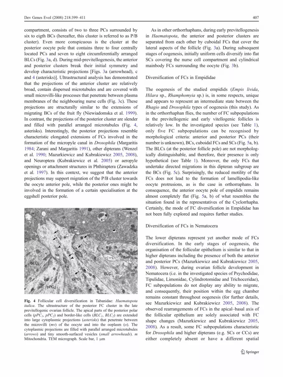
compartment, consists of two to three PCs surrounded bysix to eigth BCs (hereafter, this cluster is referred to as P/Bcluster). Even more conspicuous is the cluster at theposterior oocyte pole that contains three to four centrallylocated PCs and seven to eight circumferentially arrangedBLCs (Fig. 3a, d). During mid-previtellogenesis, the anteriorand posterior clusters break their initial symmetry anddevelop characteristic projections [Figs. 3a (arrowhead), cand 4 (asterisks)]. Ultrastructural analysis has demonstratedthat the projections of the anterior cluster are relativelybroad, contain dispersed microtubules and are covered withsmall microvilli-like processes that penetrate between plasmamembranes of the neighbouring nurse cells (Fig. 3c). Theseprojections are structurally similar to the extensions ofmigrating BCs of the fruit fly (Niewiadomska et al. 1999).In contrast, the projections of the posterior cluster are slenderand filled with parallel arranged microtubules (Fig. 4,asterisks). Interestingly, the posterior projections resemblecharacteristic elongated extensions of FCs involved in theformation of the micropyle canal in Drosophila (Margaritis1984; Zarani and Margaritis 1991), other dipterans (Wenzelet al. 1990; Mazurkiewicz and Kubrakiewicz 2005, 2008),and Neuroptera (Kubrakiewicz et al. 2005) or aeropyleopenings or attachment structures in Phthiraptera (Zawadzkaet al. 1997). In this context, we suggest that the anteriorprojections may support migration of the P/B cluster towardsthe oocyte anterior pole, while the posterior ones might beinvolved in the formation of a certain specialisation at theeggshell posterior pole.
As in other orthorrhaphans, during early previtellogenesisin Haematopota, the anterior and posterior clusters areseparated from each other by cuboidal FCs that cover thelateral aspects of the follicle (Fig. 3a). During subsequentstages of oogenesis, initially uniform cells diversify into flatSCs covering the nurse cell compartment and cylindricalmainbody FCs surrounding the oocyte (Fig. 3b).
Diversification of FCs in Empididae
The oogenesis of the studied empidids (Empis livida,Hilara sp., Rhamphomyia sp.) is, in some respects, uniqueand appears to represent an intermediate state between theRhagio and Drosophila types of oogenesis (this study). Asin the orthorrhaphan flies, the number of FC subpopulationsin the previtellogenic and early vitellogenic follicles isrelatively low. In the investigated species (see Table 1),only five FC subpopulations can be recognised bymorphological criteria: anterior and posterior PCs (theirnumber is unknown), BCs, cuboidal FCs and SCs (Fig. 5a, b).The BLCs (at the posterior follicle pole) are not morpholog-ically distinguishable, and therefore, their presence is onlyhypothetical (see Table 1). Moreover, the only FCs thatundertake directed migrations in this dipteran subgroup arethe BCs (Fig. 5c). Surprisingly, the reduced motility of theFCs does not lead to the formation of lamellipodia-likeoocyte protrusions, as is the case in orthorraphans. Inconsequence, the anterior oocyte pole of empidids remainsalmost completely flat (Fig. 5a, b) of what resembles thesituation found in the representatives of the Cyclorrhapha.Certainly, the mode of FC diversification in Empididae hasnot been fully explored and requires further studies.
Diversification of FCs in Nematocera
The lower dipterans represent yet another mode of FCsdiversification. In the early stages of oogenesis, theorganisation of the follicular epithelium is similar to that inhigher dipterans including the presence of both the anteriorand posterior PCs (Mazurkiewicz and Kubrakiewicz 2005,2008). However, during ovarian follicle development inNematocera (i.e. in the investigated species of Psychodidae,Tipulidae, Limonidae, Cylindrotomidae and Trichoceridae),FC subpopulations do not display any ability to migrate,and consequently, their position within the egg chamberremains constant throughout oogenesis (for further details,see Mazurkiewicz and Kubrakiewicz 2005, 2008). Theobserved rearrangements of FCs in the apical–basal axis ofthe follicular epithelium are solely associated with FCshape changes (Mazurkiewicz and Kubrakiewicz 2005,2008). As a result, some FC subpopulations characteristicfor Drosophila and higher dipterans (e.g. SCs or CCs) areeither completely absent or have a different spatial
Fig. 4 Follicular cell diversification in Tabanidae: Haematopotaitalica. The ultrastructure of the posterior FC cluster in the lateprevitellogenic ovarian follicle. The apical parts of the posterior polarcells (pPC1, pPC2) and border-like cells (BLC1, BLC2) are extendedinto large cytoplasmic projections (asterisks) that penetrate betweenthe microvilli (mv) of the oocyte and into the ooplasm (o). Thecytoplasmic projections are filled with parallel arranged microtubules(arrows) and tiny smooth-surfaced vesicles (small arrowheads). mMitochondria. TEM micrograph. Scale bar, 1 μm
Dev Genes Evol (2008) 218:399–411 407

distribution, and hence, it is difficult to draw homologywith FCs of the model system (Sander 1996; Kubrakiewiczet al. 1998; Mazurkiewicz and Kubrakiewicz 2005, 2008).
An interesting feature of FC diversification in certaingroups of nematocerans (e.g. Psychodidae and Sciaridae) isthe very early formation of the micropylar plate. Thisregion of the egg capsule is secreted by the anterior PCsand a few neighbouring FCs while they are still at theanterior pole of the ovarian follicle (i.e. over the nurse cells)and have no contact with the oocyte. The micropylar plateis positioned at the anterior oocyte pole only after the nursecell completely diminish (Sander 1996; Wenzel et al. 1990;Mazurkiewicz and Kubrakiewicz 2005, 2008).
Polar cells—a newly acquired character of dipterans
Several lines of evidence suggest that the crucial role inorganizing the anteroposterior polarity of the fruit flyovarian follicle (and consequently the embryo) is playedby the PCs that act as an organizer patterning surroundingFCs into distinct domains (Grammont and Irvine 2002;Besse and Pret 2003; Denef and Schupbach 2003; Xi et al.2003). Morphological analyses have indicated that the PCsare present also in the follicular epithelia of all studied sofar dipterans, including representatives of lower dipterans,the Nematocera (Mazurkiewicz and Kubrakiewicz 2005,2008) and both clades of higher dipterans (Brachycera):Orthorrhapha (Kubrakiewicz et al. 2003; this study) andCyclorrhapha (this study). Although the diversification ofFCs has been followed in several non-dipteran insects, e.g.Lepidoptera (Yamauchi and Yoshitake 1984), Phthiraptera(Zawadzka et al. 1997), Mecoptera (Bilinski et al. 1998),Neuroptera (Kubrakiewicz et al. 2005) and Heteroptera(Ogorzalek 2007), morphologically discernible PCs havenever been found.
Another interesting issue concerns the number of PCspresent at the oocyte poles. It has been shown, using a PC-specific enhancer-trap line, that in stages 2–4, in ovarianfollicles of Drosophila up to five PCs can be observed ateach pole (Besse and Pret 2003). Subsequently, this numberis restricted to two via an apoptosis-dependent mechanism.Since ovarian follicles with supernumerary PCs exhibitdefects in morphogenesis of both the BCs and SCs, thisrestriction seems to be required for correct anteriorpatterning of the FCs (Besse and Pret 2003). The numberof PCs at the poles of the ovarian follicles in orthorrha-phans is more variable and ranges from two to four(Tworzydlo et al. 2005; this study). In the light of theseresults, we propose that the precise restriction of the PCnumber to two (at both oocyte poles) is characteristic forthe cyclorrhaphan flies only. This is in line with thesuggestion that the process of PCs “production” isevolutionary conserved and that the removal of deleterious
Fig. 5 Follicular cell diversification in Empididae. a, b Empis livida.A previtellogenic ovarian follicle. Both the nurse cells (nc) and theoocyte are surrounded by cuboidal follicular cells (cc). Note that theoocyte anterior pole, adjacent to the nurse cell compartment, is flat;asterisk oocyte nucleus. a A whole mount of the ovarian folliclestained with rhodamine-conjugated phalloidin to show the distributionof actin filaments. b Semithin longitudinal section, DAPI/propidiumiodide staining. Scale bars, a=40 μm; b=30 μm. c Rhamphomyia sp.A border cell cluster (encircled) at the anterior pole of the oocyte (o).nc Nurse cell compartment, arrowhead accumulation of the micropy-lar plate material. Semithin longitudinal section, methylene blue(courtesy of J. Kubrakiewicz, University of Wroclaw, Poland). Scalebar, 20 μm
408 Dev Genes Evol (2008) 218:399–411

PCs represents evolutionary novelty that had been added tothe developmental scenario relatively late (Besse and Pret2003). Finally, it should be stressed that the “organizingpotentials” displayed by the anterior PCs in Drosophila andthose of orthorrhaphan flies are apparently different. In thefruit fly, the anterior PCs are able to specify up to threedistinct FC subpopulations that differentiate into the BCs,SCs and CCs (Grammont and Irvine 2002). In contrast, theanterior PCs of the orthorrhaphan flies are surrounded byonly one group (ring) of morphologically discernible FCs,i.e. the BCs, whereas the other “anterior” subpopulationsare either absent (i.e. CCs) or, as in the case of SCs,develop later directly from cuboidal FCs (Tworzydlo et al.2005; this study). Interestingly, the lower “organizingpotential” displayed by the PCs in orthorrhaphan fliescoincides with the involvement of these cells in theformation/molding of a micropyle (Tworzydlo et al. 2005;this study).
Conclusions
Comparative morphological studies have shown that the FCpatterning as well as extensive and coordinated migrations ofthe FCs described in detail in the fruit fly are characteristiconly for the closest relatives of this model species, which isrepresentatives of higher brachycerans or Cyclorrhapha (DeLoof et al. 1990; Kubrakiewicz et al. 1998; this study). Inother dipterans and in non-dipteran insects, this patterningtakes a different course. Most significantly, in more distantrelatives of Drosophila, the motility of FCs is markedlyreduced (lower brachycerans, Orthorrhapha) or virtuallyabsent (lower dipterans, the Nematocera and non-dipteraninsects) (Kubrakiewicz et al. 1998, 2003; Mazurkiewiczand Kubrakiewicz 2005, 2008; Tworzydlo et al. 2005).
Interpretation of the above-discussed data in a phylogeneticcontext leads to the following conclusions. The mostimportant events in a suggested scenario are superimposedon a simplified cladogram of dipterans.
– Distinct anterior and posterior PCs evolved in theancestors of dipterans (Fig. 6, event 1). In higherdipterans (Orthorrhapha+Cyclorrhapha), these cells areresponsible for specifying fates of the surrounding FCs(Fig. 6, event 2); this issue has not been analysed inNematocera.
– In more basal taxa (Nematocera, Orthorrhapha), theanterior PCs contribute (additionally?) to the forma-tion of the micropyle (Kubrakiewicz et al. 2003;Mazurkiewicz and Kubrakiewicz 2005; Tworzydloet al. 2005).
– Actively migrating FCs evolved in the ancestors ofhigher dipterans (Brachycera). In more basal clade, the
Orthorrhapha, the only FCs that undertake active anddirected migration are the BCs (Kubrakiewicz et al.2003; Tworzydlo et al. 2005; Fig. 6, event 3).
– Centripetally and posteriorly migrating FCs (CCs andmainbody FCs, respectively) evolved in the ancestorsof advanced higher dipterans, the Cyclorrhapha (Fig. 6,events 4 and 5).
– Reduction in the number of PCs to two (at both oocytepoles) emerged in the ancestors of the Cyclorrhapha(Fig. 6, event 6).
– In all investigated flies, the BCs participate in theformation of the micropyle (Margaritis et al. 1980;Margaritis 1985; Kubrakiewicz et al. 1998, 2003;Mazurkiewicz and Kubrakiewicz` 2005; Tworzydloet al. 2005).
– In certain brachyceran flies (e.g. Haematopota), border-like FCs might form additional eggshell specialisationsat the oocyte posterior pole (this study).
– In Drosophila, two additional antero-dorsal groups ofFCs are specified (Fig. 6, event 7). During final stagesof oogenesis, these subpopulations are responsible forthe formation of the respiratory appendages (Berg2005). Since the number of the respiratory appendagespresent at the anterior eggshell pole in certain cyclo-rrhaphan flies may range up to ten (for a review, seeHinton 1981), it may be anticipated that the number ofsuch anterior FC groups is, in these species, accord-ingly higher.
Fig. 6 Cladogram of major dipteran clades (simplified from Yeates2002). Important events (their origin) in a suggested scenario aresuperimposed: PCs (1), “organizing potential” of PCs (2), BCs (3),CCs (4), migrating mainbody FCs (5), reduction in the number of PCsto two (6), antero-dorsal FCs (7). The relative order of the events (2, 3and 4, 5, 6, 7) is unknown. Data: Nematocera (Kubrakiewicz et al.1998, 2003; Mazurkiewicz and Kubrakiewicz 2005, 2008); Strat.,Stratiomyomorpha (Chloromyia formosa, Kedra 2006.); Xylo., Xylo-phagomorpha (Xylophagus compeditus, Kedra and Szklarzewicz2004); Tab., Tabanomorpha (Ptiolina paradoxa, Symphoromyiacrassicornis, Kubrakiewicz et al. 2003; Rhagio tringarius, Rhagiolineola, Tworzydlo et al. 2005; Haematopota italica, Tabanus sp., thisstudy); Asiloidea (Philonicus albiceps, Thereva sp., Kubrakiewicz etal. 2003); Empidoidea (Empis livida, Rhamphomyia sp., this study;Sciapus sp., Tworzydlo et al. 2005); Cyclorrhapha (Sarcophagabullata, De Loof et al. 1990; Megaselia sp., Kubrakiewicz et al.1998; Sicus ferrugineus, Scatophaga sp., Lonchoptera sp., this study;Drosophila melanogaster, reviewed in Dobens and Raftery 2000;Horne-Badovinac and Bilder 2005; Wu et al. 2008)
Dev Genes Evol (2008) 218:399–411 409

Acknowledgements We would like to thank Prof. J. Kubrakiewicz(University of Wroclaw, Poland) for sharing with us unpublishedresults and for stimulating discussions. We are grateful to Ms. E.Kisiel for the drawings and help with the figures. This research wassupported by a grant from the Ministry of Science and HigherEducation: DS/BiNoZ/IZ/780.
References
Bauer H (1933) Die Histologie des Ovars von Tipula paludosa Meig.Z Wiss Zool 143:43–76
Bayreuther K (1956) Die Oogenese der Tipuliden. Chromosoma7:508–557
Berg CA (2005) The Drosophila shell game: patterning genes andmorphological change. Trends Genetics 21:346–354
Besse F, Pret A-M (2003) Apoptosis-mediated cell death within theovarian polar cell lineage of Drosophila melanogaster. Develop-ment 130:1017–1027
Bilinski SM (1998) Ovaries, oogenesis and insect phylogeny.Introductory remarks. Folia Histochem Cytobiol 36:43–145
Bilinski SM, Buning J, Simiczyjew B (1998) The ovaries ofMecoptera: basic similarities and one exception to the rule. FoliaHistochem Cytobiol 36:189–195
Buning J (1994) The insect ovary. Ultrastructure, previtellogenicgrowth and evolution. Chapman and Hall, London
De Loof A, Geysen J, Cardoen J, Verachter B (1990) Comparativedevelopmental physiology and molecular cytology of the poly-trophic ovarian follicles of the blowfly Sarcophaga bullata andfruitfly Drosophila melanogaster. Comp Biochem Physiol96:309–321
Denef N, Schupbach T (2003) Patterning: JAK-STAT signalling in theDrosophila follicular epithelium. Curr Biol 13:R388–R390
Dobens LL, Raftery LA (2000) Integration of epithelial patterning andmorphogenesis in Drosophila ovarian follicle cells. Dev Dynam218:80–93
Dorman JB, James KE, Fraser SE, Kiehart DP, Berg CA (2004)bullwinkle is required for epithelial morphogenesis duringDrosophila oogenesis. Dev Biol 267:320–341
Friedrich M, Tautz D (1997) Evolution and phylogeny of the Diptera:A molecular phylogenetic analysis using 28S rDNA. Mol BiolEvol 14:644–653
Godt D, Laski FA (1995) Mechanisms of cell rearrangement and cellrecruitment in Drosophila ovary morphogenesis and the require-ment of bric a brac. Development 125:173–187
Gonalez-Reyes A, St Johnston D (1998) Patterning of the follicle cellepithelium along the anterior-posterior axis during Drosophilaoogenesis. Development 125:2837–2846
Gonalez-Reyes A, Heather E, St Johnston D (1995) Polarization ofboth major body axes in Drosophila by gurken-torpedo signalling.Nature 375:654–658
Grammont M, Irvine KD (2001) fringe and Notch specify polar cellfate during Drosophila oogenesis. Development 128:2243–2253
Grammont M, Irvine KD (2002) Organizer activity of the polar cellsduring Drosophila oogenesis. Development 129:5131–5140
Gregor T, McGregor AP, Wieschaus EF (2008) Shape and function ofthe Bicoid morphogen gradient in dipteran species with differentsized embryos. Dev Biol 316:350–358
Hinton HE (1981) Biology of insect eggs. Pergamon Press, OxfordHorne-Badovinac S, Bilder D (2005) Mass transit: epithelial morpho-
genesis in the Drosophila egg chamber. Dev Dyn 232:559–574Kedra K (2006) Struktura jajnika oraz proces oogenezy muchowek
wyzszych z rodzin Xylophagidae oraz Stratiomyidae (Insecta,Diptera, Brachycera). Ph D Thesis, Jagiellonian University
Kedra K, Szklarzewicz T (2004) Oogenesis and organization of theovariole in Xylophagus compeditus (Insecta, Brachycera: Xylo-phagidae). Acta Biol Crac 46(suppl1):36
King RC (1970) Ovarian development in Drosophila melanogaster.Academic, New York
Koch M (1929) Die postembryonale Entwicklung der WeiblichenGenitaldrusen und ihrer Ausfuhrungsgang von Psychoda alter-nata Say. Z Morphol Okol Tiere 14:1–36
Krzeminski W, Krzeminska E (2003) Triassic Diptera: description,revision and phylogenetic relations. Acta Zool Cracov 46:153–184
Kubrakiewicz J, Bilinski SM, Mazurkiewicz M (1998) Diptera—ovary structure and oogenesis in midges and flies. FoliaHistochem Cytobiol 36:197–203
Kubrakiewicz J, Jablonska A, Mazurkiewicz M, Bilinski SM (2003)Differentiation and diversification of the follicular cells in flies:insight from the studies of the lower brachycerans’ ovaries.Genesis 36:214–224
Kubrakiewicz J, Jedrzejowska I, Szymanska B, Bilinski SM (2005)Micropyle in neuropterid insects. Structure and late stages ofmorphogenesis. Arthropod Struct Dev 34:179–188
Mahowald AP, Stoiber D (1974) The origin of the nurse chamber inovaries of Miastor (Diptera: Cecidomyiidae). Roux’s Arch DevBiol 1976:159–166
Margaritis LH (1984) Microtubules during formation of the micropylarcanal in Drosophila melanogaster. Cell Biol Int Rep 4:317–321
Margaritis LH (1985) Structure and physiology of the eggshell. In: KerkutGA, Gilbert LI (eds) Comprehensive insect physiology, biochemis-try and pharmacology, vol.1. Pergamon, Oxford, pp 153–226
Margaritis L, Kafatos F, Petri W (1980) The eggshell of Drosophilamelanogaster: I. Fine structure of the layers and regions of thewild-type eggshell. J Cell Sci 43:1–35
Matova N, Mahajan-Miklos S, Mooseker MS, Cooley L (1999)Drosophila Quail, a villin-related protein, bundles actin filamentsin apoptotic nurse cells. Development 126:5645–5657
Matuszewski B (1982) Diptera I: Cecidomyiidae. In: Bauer JH,Kayano H, Levan H, White A (eds) Animal cytogenetics, vol. 3.Insecta. Borntrarger, Berlin, pp 1–140
Mazurkiewicz M, Kubrakiewicz J (1998) Ontogenesis of the ovary ina moth midge, Tinearia alternata Say (Diptera: Psychodidae). JMorphol 236:167–177
Mazurkiewicz M, Kubrakiewicz J (2005) Differentiation and diversi-fication of follicular cells in polytrophic ovaries of crane flies(Diptera: Nematocera: Tipulomorpha and Trichoceridae). TissueCell 37:367–377
Mazurkiewicz M, Kubrakiewicz J (2008) Follicular cell differentiationin polytrophic ovaries of a moth midge, Tinearia alternata. Int JDev Biol 52:267–278
McCall K, Steller H (1998) Requirement for DCP-1 caspase duringDrosophila oogenesis. Science 279:230–234
McGregor AP (2005) How to get ahead: the origin, evolution andfunction of bicoid. BioEssays 27:904–913
Montell DJ (2003) Border-cell migration: the race is on. Nature RevMol Cell Biol 4:13–24
Montell DJ (2006) The social lives of migrating cells in Drosophila.Curr Opin Genet Dev 16:374–383
Moussian B, Roth S (2005) Dorsoventral axis formation in theDrosophila embryo—shaping and transducing a morphogengradient. Curr Biol 15:R887–R899
Niewiadomska P, Godt D, Tepass U (1999) DE-Cadherin is requiredfor intercellular motility during Drosophila oogenesis. J Cell Biol144:533–547
Ogorzalek A (2007) Structural and functional diversification offollicular epithelium in Coreus marginatus (Coreidae: Hetero-ptera). Arthopod Struc Dev 36:209–219
Oosterbroek P, Courtney G (1995) Phylogeny of the nematocerousfamilies of Diptera (Insecta). Zool J Linn Soc 115:267–311
410 Dev Genes Evol (2008) 218:399–411

Ray RP, Schupbach T (1996) Intercellular signaling and the polarization ofbody axes during Drosophila oogenesis. Genes Dev 10:1711–1723
Roth S (2001) Coordinating germ line and soma. Curr Biol 11:779–781Sander K (1996) Variants of embryonic patterning mechanisms in
insects: Hymenoptera and Diptera. Sem Cell Dev Biol 7:573–582Schupbach PM, Camenzind R (1983) Germ cell lineage and follicle
formation in paedogenetic development of Mycophila speyeriBarnes (Diptera, Cecidomyiidae). Int J Insect Morphol Embryol12:211–223
Schupbach PM, Went DF (1983) Cell fusions during formation of theoocyte-nurse chamber complex in the ovary of the dipteran insectMycophila speyeri. Roux’s Arch Dev Biol 192:228–233
Tran DH, Berg CA (2003) bullwinkle and shark regulate dorsal-appendage morphogenesis in Drosophila oogenesis. Develop-ment 130:6273–6282
Tworzydlo W, Jablonska A, Kisiel E, Bilinski SM (2005) Differingstrategies of patterning of follicular cells in higher and lowerbrachycerans (Diptera: Brachycera). Genesis 43:49–58
Wenzel F, Gutzeit HO, Zissler D (1990) Morphogenesis of themicropylar apparatus in ovarian follicles of the fungus gnatBradysia tritici (syn. Sciara ocellaris). Roux’s Arch Dev Biol199:146–155
Wiegmann BM, Yeates DK, Thorn JL, Kishino H (2003) Time flies, anew molecular time scale for brachyceran fly evolution without aclock. Syst Biol 52:745–756
Wu X, Tanwar PS, Raftery LA (2008) Drosophila follicle cells:morphogenesis in an eggshell. Semin Cell Dev Biol 19:271–282
Wulker W, Winter G (1970) Untersuchungen uber die Ultrastrukturder Gonaden von Chironomus (Diptera). I. Normale Entwicklungder Ovarien im 4 Larvenstadium. Z Zellforsch 106:348–370
Xi R, McGregor JR, Harrison DA (2003) A gradient of JAK pathwayactivity patterns the anterior-posterior axis of the follicularepithelium. Dev Cell 4:167–177
Yamauchi H, Yoshitake N (1984) Formation and ultrastructure ofmicropylar apparatus in Bombyx mori ovarian follicles. J Morph179:47–58
Yeates DK (2002) Relationships of extant lower Brachycera (Diptera):a quantitative synthesis of morphological characters. Zool Scripta31:105–121
Yeates DK, Wiegmann BM (1999) Congruence and controversy:toward a higher-level phylogeny of the Diptera. Ann RevEntomol 44:397–428
Zarani FE, Margaritis LH (1991) The eggshell of Drosophilamelanogaster, VII. Formation of the micropylar canal and therole of the paracrystalline structure. Roux’s Arch Dev Biol200:95–103
Zawadzka M, Jankowska W, Bilinski SM (1997) Egg shells ofmallophagans and anoplurans (Insecta: Phthiraptera): morpho-genesis of specialized regions and the relation to F-actincytoskeleton of follicular cells. Tissue Cell 29:665–673
Dev Genes Evol (2008) 218:399–411 411