Novel insights into DNA methylation features in spermatozoa: stability and peculiarities

J
S(
Ka
b
c
a
ARRA
KEGIMR
1
wekn(oCpdi1
pw(tetp
(
0d
ARTICLE IN PRESSG ModelMIC-1503; No. of Pages 8
Micron xxx (2010) xxx–xxx
Contents lists available at ScienceDirect
Micron
journa l homepage: www.e lsev ier .com/ locate /micron
tructure and ultrastructure of spermatozoa of Chrysomya megacephalaDiptera: Calliphoridae)
.P.O. Namea, J.R. Pujol-Luzc, S.N. Báob,∗
Programa de Pós-graduacão em Biologia Animal, Universidade de Brasília, DF, BrazilLaboratório de Microscopia Eletrônica, Departamento de Biologia Celular, Instituto de Ciências Biológicas, Universidade de Brasília, Distrito Federal, CEP: 70919-970, BrazilNúcleo de Entomologia Urbana e Forense, Departamento de Zoologia, Instituto de Ciências Biológicas, Universidade de Brasília, DF, Brazil
r t i c l e i n f o
rticle history:eceived 11 March 2010eceived in revised form 22 April 2010ccepted 24 April 2010
a b s t r a c t
The spermatozoa of Chrysomya megacephala are similar to those described for other Brachycera. In thisspecies, the spermatozoa are long and thin, measuring about 590 �m in length, of which the head regionmeasures approximately 60 �m. The head includes a monolayered acrosome with electron-lucid mate-
eywords:ntomologyerm cells
nsect sperm
rial, and the shape of the nucleus, in cross-sections, varies from circular to oval with completely condensedchromatin. The centriole was observed in the zone of flagellar implantation, below the “peg” region. Inthe region of overlap, the followings structures are observed: nucleus, centriolar adjunct, mitochondrialderivatives and axoneme. The two mitochondrial derivatives are of different lengths but similar diam-eter. The axoneme is of a conventional insectan type with a 9 + 9 + 2 microtubular arrangement, with
by ts defe
icroscopyeproductive system
accessory tubules flankedtract consists of testis, va
. Introduction
Approximately 150,000 species of flies are described in theorld and more than 24,000 in the Neotropical region (Amorim
t al., 2002). More than 1000 species of Calliphoridae are hithertonown, organized in 150 genera and 5 subfamilies: Calliphori-ae, Mesembrinellinae, Chrysomyinae, Toxotarsinae and RhiniinaeJames, 1970; Mello, 2003). In the neotropics about 130 speciesf blow flies are known (Carvalho and De Mello-Patiu, 2008).alliphoridae (blow flies) present a great ecological diversity, occu-ying several habitats–from organic matter to animal tissues inecomposition, and are distributed worldwide. They cause med-
cal problems and important losses to the animal industry (Zumpt,965; Ghandour, 1988; David et al., 2008).
Chrysomya megacephala, the Oriental latrine fly, has beenresent in the dipteran fauna of Brasil since the mid 1975s,hen it is believed to have been introduced, probably from Africa
Guimarães et al., 1978, 1979). Greenberg (1971, 1973) reportedhat this species is among the most dangerous dipteran vectors of
Please cite this article in press as: Name, K.P.O., et al., Structure and ulCalliphoridae). Micron (2010), doi:10.1016/j.micron.2010.04.015
nteric pathogens such as viruses, bacteria and helminthes. Never-heless, in many regions of the world this species is an importantollinator (Anderson et al., 1982; Castaneda-Vildózola et al., 1999).
∗ Corresponding author. Tel.: +55 61 3107 2905; fax: +55 61 3107 2904.E-mail addresses: [email protected] (K.P.O. Name), [email protected]
J.R. Pujol-Luz), [email protected] (S.N. Báo).
968-4328/$ – see front matter. Crown Copyright © 2010 Published by Elsevier Ltd. All rioi:10.1016/j.micron.2010.04.015
he electron-dense intertubular material. The male internal reproductiverens, seminal vesicle, accessory glands and ejaculatory duct.
Crown Copyright © 2010 Published by Elsevier Ltd. All rights reserved.
Recently, studies have demonstrated that the structure andultrastructure of internal reproductive organs and spermatozoain insects (Jamieson et al., 1999) provide additional charactersfor taxonomic analysis and so can contribute to our understand-ing of relationships (Dallai et al., 1993; Dallai and Afzelius, 1995;Carcupino et al., 1995).
The diversity in sperm structure in Diptera is greater than in allother insect groups taken together. Whereas the sperm structureof nematoceran Diptera is relatively well known, in the brachyc-eran group such studies are scarce. In the family Calliphoridae,only Calliphora vomitoria have been briefly described (Dallai andAfzelius, 1990). All the investigated species of brachyceran fliesshare a common spermatozoal model. There is an apical monolay-ered acrosome, a compact nucleus, fully crystallized mitochondrialderivatives and a 9 + 9 + 2 axoneme (Jamieson, 1987; Jamieson etal., 1999).
The aim of this study is, therefore, to describe the ultrastructureof spermatozoa of Chrysomya megacephala and, in futures studies,make the comparison with others species from the family Cal-liphoridae.
2. Materials and methods
trastructure of spermatozoa of Chrysomya megacephala (Diptera:
Chrysomya megacephala adult specimens were collected at thecampus of the University of Brasilia (UnB), Brazil, to start a colonyand begin laboratory analyses. The flies were attracted by fish car-casses and bovine minced meat, and collected with entomologicalnets.
ghts reserved.

ARTICLE IN PRESSG ModelJMIC-1503; No. of Pages 8
2 K.P.O. Name et al. / Micron xxx (2010) xxx–xxx
Fig. 1. (A and B) Light micrograph of male internal reproductive organs and structure of the testis in Chrysomya megacephala. (A) Male internal reproductive organs: testes(Te), vas deferens (Vd), seminal vesicle (Sv), accessory glands (Ag) and ejaculatory duct (Ed). (B) Cross-section shows the morphology of testis wall, the internal organizationo t in dm stis wg ithelia
taowod
2
tbdt(
twaM
mtpdpvc
p2oaa
2
ca
f the cyst and the spermatozoa. Peritoneal sheath (Ps), layer of muscles (Ml), cysicrograph (TEM) of a cross-section of the testis wall. In the strata, forming the te
rains; layer of muscle (Ml) delimited by basement membrane (Bm) and follicular epnd the DAPI-stained head region, respectively. Head (h) and tail (t).
In the laboratory, the flies were fed with a honey and water solu-ion (1:1) and water ad libitum for 1 week and then provided with100 g bovine minced meat as an oviposition medium. Numer-
us eggs were found in the underside of the meat after 1 day andere placed 20 to a cup in tapered plastic, each containing 20 g
f fresh bovine minced meat, until the pupariation and completeevelopment (adult) stages.
.1. Light microscopy
The flies were briefly submitted to cold-induced dormancy andhe reproductive system of males was removed in its entiretyy dissection of the animal in saline solution of NaCl 0.9%. Afterissection, these structures were placed on a slide for examina-ion. Observations and image acquisitions were made using a ZeissSPEMI 2000C) stereomicroscope.
Testes used for transmission electron microscopy were also sec-ioned for light microscopy. Semi-thin sections (4 �m) were stainedith 0.25% toluidine blue, pH 11, observed and photographed with
n Axiophot Zeiss® Microscope equipped with a Zeiss® AxiocamRc digital camera and Axiovision 4.5 software.Males and females were dissected and their testes and sper-
athecae were smeared on clean glass microscope slides to releasehe sperm within them, and fixed in a solution of 4% (wt/vol)araformaldehyde in 0.1 M sodium phosphate buffer, pH 7.2. Afterrying at room temperature, the preparations were observed in ahotomicroscope (Zeiss® Axiocam MRc digital camera and Axio-ision 4.5 software) equipped with differential interference andontrast phases.
For measurement of sperm nucleus length, some of thesereparations were stained for 15 min with 0.2 �g/ml 4,6-diamino--phenylindole (DAPI) in PBS, washed and mounted with a solutionf N-Propil-Galato. They were then examined by Axiophot Zeiss®
nd Confocal Leica SP5 microscopes equipped with epifluorescencend with 405 nm excitation filters, respectively.
Please cite this article in press as: Name, K.P.O., et al., Structure and ulCalliphoridae). Micron (2010), doi:10.1016/j.micron.2010.04.015
.2. Transmission electron microscopy
The testes and spermathecae were fixed for 4 h in a solutionontaining 2.5% glutaraldehyde, 4% paraformaldehyde, 5 mM CaCl2nd 3% sucrose, buffered in 0.1 M sodium cacodylate, at pH 7.2.
istinct stages of differentiation (star), spermatozoa (Z). (C) Transmission electronall, it is possible to see: external layer (El); peritoneal sheath (Ps) with pigmentedum (Fe). (D and E) Interferential differential contrast micrograph of a spermatozoon
After fixation, the specimens were rinsed in the same buffer, andpost-fixed in 1% osmium tetroxide, 0.8% potassium ferricyanide,and 5 mM CaCl2 in 0.1 M sodium cacodylate buffer. In some casesthe specimens were fixed in a mixture of 2.5% glutaraldehyde, 1%tannic acid in 0.1 M phosphate buffer, at pH 7.2, followed by block-staining in 1% uranyl acetate in distilled water (Afzelius, 1988). Thematerial was dehydrated in an ascending acetone series (30–100%)and embedded in Spurr’s resin. Ultrathin sections were stained withuranyl acetate and lead citrate and observed using a Jeol® 1011transmission electron microscope operating at 80 kV.
For detection of basic proteins, the ethanolic-phosphotungsticacid method (E-PTA), modified from Bloom and Aghajanian (1968),was applied. Testes were fixed with 2.5% glutaraldehyde in 0.1 Mphosphate buffer, pH 7.4, for 4 h at 4 ◦C. After washing in phosphatebuffer and dehydrating in alcohol, the material was treated “enbloc” with a solution of 2% de PTA in absolute ethanol for 24 h at 4 ◦C,washed in absolute ethanol and embedded in Spurr’s resin. Someultrathin sections were observed, unstained and partly stained withuranyl acetate.
3. Results
In sexually mature males of Chrysomya megacephala, the sper-matozoa are stored in the testes and seminal vesicle. In the sexuallymature females, these cells are stored in the spermathecae aftercopulation.
The male internal reproductive system in Chrysomya mega-cephala comprises the following organs: two testes, paired vasadeferentia and accessory glands, and one seminal vesicle and ejac-ulatory duct. Typically, each of the testes opens into the vasadeferentia, which then unites to connect with the seminal vesicle.The accessory glands are directly connected to the seminal vesicleand the ejaculatory duct is located at the end portion of this organ(Fig. 1A). The testes in this species have an intense and brilliantreddish-brown color. The color is light when the male fly emergesfrom the puparium and normal intensity is reached after sexual
trastructure of spermatozoa of Chrysomya megacephala (Diptera:
maturation. The arrangement of germ cells in the testes shows thatthis structure is formed by a unique and long follicle with severalcysts (Fig. 1B). The testicle is made up of an external wall, whichsurrounds the germinative cells. The testicular wall is formed bya peritoneal sheath, a muscular layer, a basement membrane, a
ARTICLE IN PRESSG ModelJMIC-1503; No. of Pages 8
K.P.O. Name et al. / Micron xxx (2010) xxx–xxx 3
F . Cystr of acrm s (n).
ftgim(
ofsa
li6
cmoe
ttt(
6muo(tidiott
ig. 2. (A–D) TEM of head region. (A) Cyst (Cy) showing spermatozoa head regionegions, distal (Rd) and proximal (Rp) and the nucleus (n). (C) In the proximal regionembrane (Mp). (D) The nuclear chromatin appears completely condensed. Nucleu
ollicular epithelium, tracheoles, and an epithelium at the base ofhe follicle. The cytoplasm of peritoneal sheath is rich in roundedrains containing reddish-brown pigments, which gives the organts characteristic color. When observed in transmission electron
icroscopy, these grains show different sizes and electron-densityFig. 1C).
The differentiation of the spermatids of Chrysomya megacephalaccurs within cysts. Inside each cyst, the spermatic cells are per-ectly aligned and in the same stage of maturation. The number ofpermatozoa per bundle is variable, but approximately 128 cellsre very commonly found in each cyst (Fig. 2A).
The spermatozoon of the examined species (Figs. 1D, 5) is veryong and filiform, measuring approximately 590 �m in total length,ncluding the head and tail regions. The region of the head is about0 �m in total length (Fig. 1E).
The head consists of an acrosome and nucleus. The acrosome isonical, monolayered and consists of a moderately electron-denseaterial. In longitudinal section, it fits closely into an indentation
n the surface of the nucleus, and in cross-section it displays anlliptic substructure (Figs. 2B and C, 5).
The structure of the acrosome is divided into two regions: dis-al and proximal. The distal region (rd), comprising about 45% ofhe total length of the acrosome, is anterior to the nucleus, andhe proximal region (pr) is closely associated with the nucleusFig. 2B, 5).
The nucleus of Chrysomya megacephala is long, measuring about0 �m when stained for DAPI and examined by epifluorescenceicroscopy (Fig. 1E). It is fusiform and filled with condensed and
niform chromatin (Fig. 2B). When submitted to E-PTA method-logy, the nucleus shows high concentrations of basic proteinsFig. 4E). In cross-sections, the nucleus shape varies from circularo oval (Figs. 2D, 3A, 5). During the last stages of spermiogenesisn Chrysomya megacephala, the nucleus shows a completely con-ensed chromatin, with many microtubules appearing around and
Please cite this article in press as: Name, K.P.O., et al., Structure and ulCalliphoridae). Micron (2010), doi:10.1016/j.micron.2010.04.015
t is possible to see the accessory membranes, in the cytoplasmf the cells (Fig. 3A). Since the initial stages of nuclear condensa-ion, until the complete maturity of the cell, cross-sections revealhe presence of an accessory membrane adjacent to the nuclear
ic cells (Cc); nucleus (n). (B) Longitudinal view of acrosome (Ac) showing its twoosome (Ac), the surface of this organelle is in contact with the nucleus (n). Plasmatic
envelope. There are two accessory membranes per nucleus, eachon opposite sides of this organelle.
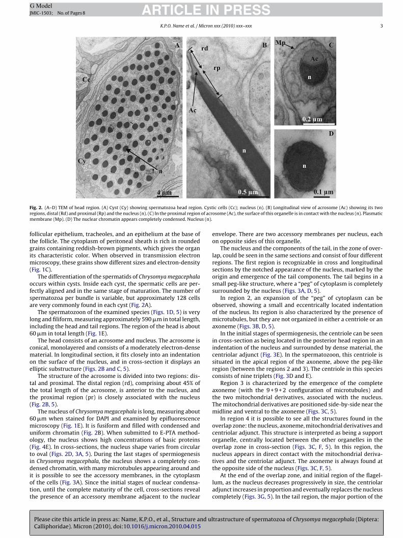
The nucleus and the components of the tail, in the zone of over-lap, could be seen in the same sections and consist of four differentregions. The first region is recognizable in cross and longitudinalsections by the notched appearance of the nucleus, marked by theorigin and emergence of the tail components. The tail begins in asmall peg-like structure, where a “peg” of cytoplasm is completelysurrounded by the nucleus (Figs. 3A, D, 5).
In region 2, an expansion of the “peg” of cytoplasm can beobserved, showing a small and eccentrically located indentationof the nucleus. Its region is also characterized by the presence ofmicrotubules, but they are not organized in either a centriole or anaxoneme (Figs. 3B, D, 5).
In the initial stages of spermiogenesis, the centriole can be seenin cross-section as being located in the posterior head region in anindentation of the nucleus and surrounded by dense material, thecentriolar adjunct (Fig. 3E). In the spermatozoon, this centriole issituated in the apical region of the axoneme, above the peg-likeregion (between the regions 2 and 3). The centriole in this speciesconsists of nine triplets (Fig. 3D and E).
Region 3 is characterized by the emergence of the completeaxoneme (with the 9 + 9 + 2 configuration of microtubules) andthe two mitochondrial derivatives, associated with the nucleus.The mitochondrial derivatives are positioned side-by-side near themidline and ventral to the axoneme (Figs. 3C, 5).
In region 4 it is possible to see all the structures found in theoverlap zone: the nucleus, axoneme, mitochondrial derivatives andcentriolar adjunct. This structure is interpreted as being a supportorganelle, centrally located between the other organelles in theoverlap zone in cross-section (Figs. 3C, F, 5). In this region, thenucleus appears in direct contact with the mitochondrial deriva-tives and the centriolar adjunct. The axoneme is always found atthe opposite side of the nucleus (Figs. 3C, F, 5).
trastructure of spermatozoa of Chrysomya megacephala (Diptera:
At the end of the overlap zone, and initial region of the flagel-lum, as the nucleus decreases progressively in size, the centriolaradjunct increases in proportion and eventually replaces the nucleuscompletely (Figs. 3G, 5). In the tail region, the major portion of the
ARTICLE IN PRESSG ModelJMIC-1503; No. of Pages 8
4 K.P.O. Name et al. / Micron xxx (2010) xxx–xxx
Fig. 3. (A–G) TEM of overlap region. (A) Region 1. In cross-sections, the cytoplasmic “Peg” the upper right portion of nucleus (n) can be seen. The accessory membranes (Ma)and microtubules (Mt) are visible in the cytoplasm. (B) Region 2. Showing the central pair of microtubules (Mt). Nucleus (n); axoneme (Ax). (C) Region 3. This region showst ) ands the no on 4.a ondria
ca
trroeob
mseprdfi
he complete axoneme (Ax), the two mitochondrial derivatives (Md), the nucleus (nhowing emergence of axoneme (Ax) from small indentation of nucleus “Peg”. Notef the nucleus. (E) The centriole (Ce) is seen posteriorly to the nucleus (n). (F) Regind axoneme (Ax). (G) The centriolar adjunct (Ca) can be seen between the mitoch
entriolar adjunct is seen under the nucleus (longitudinal section)nd appears compact and very electron-dense (Figs. 3D, 5).
The mitochondrial derivatives are positioned side-by-side nearhe midline and ventral to the axoneme. Paracrystalline mate-ial occupies the entire interior of the derivatives (Fig. 4A and B),esembling a honeycomb structure in cross-section (Fig. 4B). Theserganelles follow the axoneme along their length and one of themxtends for almost the entire length of the spermatozoon, while thether is smaller, finishing early (Figs. 4C, 5). They differ in length,ut not in diameter (Figs. 4D, 5).
The axoneme displays the typical insectan pattern of 9 + 9 + 2icrotubules, with electron-dense material between the acces-
ory tubules. The nine single accessory microtubules are the most
Please cite this article in press as: Name, K.P.O., et al., Structure and ulCalliphoridae). Micron (2010), doi:10.1016/j.micron.2010.04.015
xternal, followed internally by the nine doublets and a centralair (Figs. 4B, 5). The endpiece is characterized by a progressivelyeduced number of microtubular elements. In this species, the nineoublets finish first, followed by the two central microtubules and,nally by accessory ones (Fig. 4D).
the emergence of the centriolar adjunct (Ca). (D) Longitudinal view of overlap zone,otched appearance of nucleus (n). Centriolar adjunct (Ca) may be seen at the basis
Centriolar adjunct (Ca) surrounded by nucleus (n), mitochondrial derivatives (Md)l derivatives (Md), also in the tail region.
The centriolar adjunct and the axoneme with all their structuresare E-PTA positive. However, the mitochondrial derivatives onlysometimes appear as E-PTA positive (Fig. 4E).
4. Discussion
The male internal reproductive organs in Chrysomya mega-cephala (Calliphoridae) comprise: testes, vas deferens, accessoryglands, seminal vesicle, ejaculatory duct and resemble the patternstructure observed in other Diptera (Joly et al., 2003; Sinclair et al.,2007). Such reproductive organs in Diptera are poorly known, withfew detailed comparative studies (Sinclair et al., 2007).
In most species of Diptera studied, the testis is considered to be
trastructure of spermatozoa of Chrysomya megacephala (Diptera:
a subdivided sac-like organ equivalent to tubular follicles observedin the testes of other insect orders (Williamson, 1989). In Drosophila(Drosophilidae), the most widely studied group in Diptera (Kiefer,1966; Bairatti, 1967; Tokuyasu et al., 1972a,b; Hicks et al., 1999),as well as in Ceratitis capitata (Tephritidae) (Guillén, 1983; Báo

ARTICLE IN PRESSG ModelJMIC-1503; No. of Pages 8
K.P.O. Name et al. / Micron xxx (2010) xxx–xxx 5
Fig. 4. (A–E) TEM of the tail region. (A) Mitochondrial derivatives (Md), filled with paracrystalline material (star), were seen in longitudinal section. Axoneme (Ax). (B)Spermatozoon obtained from spermatheca and fixed with tannic acid. The axoneme (Ax) is made up of nine accessory microtubules, nine doublets and a central pair. Densefi rivati( izatiot ), cenm
aztoIiHob
CpiIncmbctat
bers (arrows head) are seen between the accessory microtubules. Mitochondrial deMd). Axoneme (Ax). (D) Final portion of flagellum showing the gradual disorganhe axoneme tip. (E) Cross-section of spermatozoa treated with E-PTA. Nucleus (n
itochondrial derivatives (Md) are sometimes positive.
nd Dolder, 1991) and in Chrysomya megacephala, the spermato-oa derive from primordial germ cells located at the apical end ofhe testes, and these stem cells go through a synchronous processesf mitosis without cellular division to produce the spermatocytes.n contrast, patterns of cellular development are quite differentn non-feeding oestrid flies. In the testis of the third instars ofypoderma (Oestridae), an apical zone with a mass of cells wasbserved and the spermatogonial cysts are arranged around itasally (Boullard, 1967).
In C. capitata and Anastrepha ludens (Tephritidae) as well as inhrysomya megacephala, the testes are made up of an external andigmented wall which surrounds the germ cells. The testicular wall
n both species consists of a peritoneal sheath and a muscular layer.n the peritoneal sheath it is possible to see the presence of a greatumber of vesicles containing pigment, which gives the organ itsharacteristic color. According to Báo and Dolder (1991), the pig-ented epithelium is responsible for forming an outer physical
arrier, protecting the testis and, for Valdez (2001), these vesicles
Please cite this article in press as: Name, K.P.O., et al., Structure and ulCalliphoridae). Micron (2010), doi:10.1016/j.micron.2010.04.015
ould store nutrients temporally, en route from the blood to theransforming germ cells. The muscular layer is presumably associ-ted with the peristaltic contractions, and possibly contributes tohe displacement of the spermatid bundle and frees spermatozoa
ves (Md). (C) In the posterior tip, it was possible to see one mitochondrial derivativen of the axoneme (Ax). Note the accessory microtubules as the last to be lost attriolar adjunct (Ca) and microtubules of axoneme (Ax) are E-PTA positive and the
into the testes and toward the posterior duct, leading out of thetestis. According to Chapman (1998), although the muscle layer isvery common in some Diptera, it is absent in most insects.
The spermatids found in the testes, at the distal and medialregions of this organ, are encapsulated by a somatic cell, formingthe cyst, where they completes the differentiation process. In theproximal region of the testes, near to the insertion of the seminalvesicle, these cells are free. These aspects of testicular organizationwere also described in some Tephritidae (Báo and Dolder, 1991;Valdez, 2001), in Drosophila melanogaster (Kiefer, 1966; Bairatti,1967; Tokuyasu et al., 1972a,b), in Sarcophaga bullata (Sarcophagi-dae) (Warner, 1971) and in a large number of other insect orders(Phillips, 1970).
In Chrysomya megacephala, as in most insects, the developmentof the germinative cells takes place within such cysts (Phillips,1970). According to Virkki (1969), archaic orders of insects havea greater number of sperm per bundle than recent orders, in otherwords, the most recent or specialized groups tend to have a smaller
trastructure of spermatozoa of Chrysomya megacephala (Diptera:
number of sperm per bundle.In most insects it was observed that the number of spermatozoa
per bundle is variable. Research shows that for Diptera, the numberof spermatids per bundle varies among different species depending

ARTICLE ING ModelJMIC-1503; No. of Pages 8
6 K.P.O. Name et al. / Micron
Far
oaFefftCaecfWtw
iBanDa
sbefa
ig. 5. Schematic representation of the Chrysomya megacephala spermatozoon: (i)crosome; (ii) nucleus; (iii) emergence of flagellum; (iv) overlap zone; (v) flagellaregion; (vi and vii) endpiece of flagellum.
n the number of spermatogonial premeiotic divisions (Oguma etl., 1987; Quagio-Grassiotto and Lello, 1996; Cruz-Landim, 2001).or Aedes aegypti (Culicidae), it was suggested by Owusu-Daakut al. (2007), that the maximum number of spermatozoa per cystor this species is probably 512, being the result of nine divisionsrom the original spermatocyst mother cell (stem cell), accordingo what has been proposed by Virkki (1969). In the RA mutant of. capitata the number of germ line cells observed was constant,pproximately 250 per cyst (Báo and Dolder, 1990), the result ofight divisions. In Chrysomya megacephala, the number of sperm peryst varies, but it is common to find 128 sperm per cyst. A similareature was observed in S. bullata (seven divisions) (Warner, 1971).
hen compared with other groups, this feature is consistent withhe current taxonomic and phylogenetic position of this speciesithin Diptera.
The structure of spermatozoa in Chrysomya megacephala is sim-lar to the general description for insect sperm (Phillips, 1970;accetti, 1972; see also reviews by Jamieson, 1987; Jamieson etl., 1999). They are filiform as in the majority of the Diptera and doot show great variations in length, as found in the spermatozoa of. melanogaster (Joly et al., 1991; Pitnick et al., 1995; Jamieson etl., 1999).
The monolayered condition of the acrosome observed in the
Please cite this article in press as: Name, K.P.O., et al., Structure and ulCalliphoridae). Micron (2010), doi:10.1016/j.micron.2010.04.015
permatozoa of Chrysomya megacephala is apparently shared by allrachyceran spermatozoa already examined. According to Dallait al. (1984), the acrosome in Diptera has different forms. In someamilies it appears small and devoid of perforatorium and extra-crosomal layers and in other families it is an elongated organelle,
PRESSxxx (2010) xxx–xxx
partially lateral to the nucleus and including an internal crystallinefiber. In the studied species, the acrosome possesses the latteraspect, but the crystalline fiber was not observed. In cross-sections,this structure is elliptical, becoming more circular as the sectionnears the tip. The striated filaments or crystalline fibers occurringin the acrosome were observed in C. capitata (Báo et al., 1989) andS. bullata (Warner, 1971).
The chromatin of the nucleus is highly condensed in Chrysomyamegacephala and this pattern is found in other Brachycera(Jamieson, 1987; Jamieson et al., 1999). When treated with E-PTA, this organelle is homogeneous and completely positive. Apeculiar characteristic, observed in the longitudinal and cross-sections of the nucleus, is the notched appearance of this structure,where the emergence of the tail components occurs. In the regionknow as overlap zone, basically all the organelles found in the tail(axoneme, mitochondrial derivatives, centriolar adjunct) are seenin the same section. This characteristic was described in Megaseliascalaris (Phoridae) (Curtis et al., 1989) and is not very common inother families of insects.
The two accessory membranes observed laterally in the nucleusin cross-sections in Chrysomya megacephala, also appear in sper-matids of Coelopa frigida (Coelopidae) (Schrankel and Schwalm,1974) and named ‘scroll-like structure’. These structures can beobserved, in cross-sections of spermatids, as two membranes par-allel two the nuclear envelope, extending along the nucleus. Withincreased condensation of the nucleus, the accessory membranestend to disappear. According to Schrankel and Schwalm (1974), thefunction of these structures is unknown. The accessory membraneswere described in C. capitata (Báo et al., 1989; Báo and Dolder, 1990)and in S. bulatta (Warner, 1971), where the term ‘whorl’ has beenused.
The centriole in the mature sperm was first proposed by Phillips(1970) as an organelle with nine microtubular triplets. The clas-sical structure proposed by Phillips was found in D. melanogaster(Dallai and Afzelius, 1991), in C. capitata (Báo and Dolder, 1991)and is described here in Chrysomya megacephala. However, in somespecies this structure shows great variations. In Dacus oleae (Tephri-tidae), as in other Diptera an unusual configuration was observedfor this structure: the presence of two central microtubules in theregion of the centriole (Dallai and Afzelius, 1991).
In most insects, the nucleus is attached to the flagellum by avery electron-dense structure identified as the centriolar adjunct(Jamieson, 1987; Jamieson et al., 1999). The centriolar adjunct inChrysomya megacephala is relatively long, and was observed inthe overlap region surrounded by the nucleus and flagellar struc-tures (mitochondrial derivatives and axoneme) and extends intothe region of the flagellum.
The mitochondrial derivatives in Chrysomya megacephala aresymmetric in diameter, but asymmetric in length, while in Muscadomestica (Muscidae) these structures are asymmetric in diameterand showing a considerable difference in length. In both speciesthe derivative is completely filled with paracrystalline material(Gassner, 1970; Gassner and Klemetson, 1981).
According to Dallai et al. (1993), the sperm structures ofaxoneme in Brachycera are relatively uniform, whereas in suborderNematocera there is a considerable diversity, which is greater thanthat in any other insect order.
As expected for most Brachycera (Dallai et al., 1993; Jamiesonet al., 1999), Chrysomya megacephala have an axoneme with the9 + 9 + 2 microtubules arranged parallel to each other, and in fact,the pattern is similar to that described for most other insects (Dallai
trastructure of spermatozoa of Chrysomya megacephala (Diptera:
and Afzelius, 1990).The end piece of flagellum in Chrysomya megacephala is char-
acterized by a progressively reduced number of microtubularelements with the nine doublets finishing first, followed by the twocentral microtubules and with the accessory being the last ones to

INJ
icron
tbste
ttccaaiap
tn
A
Jlddcd
R
A
A
A
BB
B
B
B
B
B
B
C
C
C
C
C
C
D
ARTICLEG ModelMIC-1503; No. of Pages 8
K.P.O. Name et al. / M
erminate. Unfortunately, the disorderly aspect of axoneme has noteen taken into consideration in studies of Diptera spermatozoa. Inome other groups, this parameter has been used as an importantool since it probably indicates a phylogenetic relationship (Zamat al., 2001, 2005).
In the flagellar region of Chrysomya megacephala spermatozoon,he ethanolic-phosphotungstic method permitted the identifica-ion of different microtubule types in the axoneme, and in theentriolar adjunct structure. Basic proteins are associated with theentral, peripheral and accessory microtubules and sometimes, tovery slight degree, with the mitochondrial derivatives. A similar
spect to the one described above for the tail region, was observedn Culex quinquefasciatus (Báo et al., 1992), where the centriolardjunct, the mitochondrial derivatives and the microtubules areositive for the technique.
The similarity of sperm structure in Chrysomya megacephala tohat of other brachycerans demonstrated here confirms the taxo-omic and phylogenetic value of sperm ultrastructure.
cknowledgements
K.P.O. Name would like to thank Professor J.A. Vexenat and.S. Souza (UnB), for teaching me to capture and identify the Cal-iphoridae species. This study was supported by Conselho Nacionale Desenvolvimento Científico e Tecnológico (CNPq), Fundacãoe Empreendimentos Científico e Tecnológico (FINATEC), Finan-iadora de Estudos e Projetos (FINEP) and Coordenacão de Pessoale Nível Superior (Capes).
eferences
fzelius, B.A., 1988. Microtubules in the spermatids of stick insects. J. Ultrastruct.Mol. Struct. Res. 98, 94–102.
nderson, D.L., Sedgley, M., Short, J.R.T., Allwood, A.J., 1982. Insect pollination ofmango in northern Australia Mangifera indica, includes Apis mellifera. Aust. J.Agric. Res. 33 (3), 541–548.
morim, D.S., Silva, V.C., Balbi, M.I.P.A., 2002. Estado do conhecimento dos DipteraNeotropicais. In: Costa, C., Vanin, S.A., Lobo, J.M., Melic, A. (Eds.), Proyecto de RedIberoamericana de Biogeografía y Entomología Sistemática, vol. 2. SEA, Zaragoza,pp. 29–36.
accetti, B., 1972. Insect sperm cell. Adv. Insect Physiol. 9, 315–397.áo, S.N., Dolder, H., 1990. Abnormalities observed during spermiogenesis of the
RA mutant of Ceratitis capitata (Diptera, Tephritidae): cystic cells. Acta Zool. 71,107–111.
áo, S.N., Dolder, H., 1991. Testicular organization in adult Ceratitis capitata (Diptera:Tephritidae): RA mutant and Wild-type lineages. Rev. Bras. Biol. 51 (2), 313–319.
áo, S.N., Lins, U., Farina, M., De Souza, W., 1992. Mitochondrial derivatives of Culexquinquefasciatus (Culicidae) spermatozoon. Some new aspects evidenced bycytochemistry and image processing. J. Struct. Biol. 109, 46–51.
áo, S.N., Quagio-Grassiotto, I., Dolder, H., 1989. Acrosome formation in Ceratitiscapitata (Diptera: Tephritidae). Cytobios 58, 93–100.
airatti, A., 1967. The structure and ultrastructure of the male genital apparatus ofthe Drosophila melanogaster Meig. I. The testis. Z. Zellforsch. Mikrosk. 76, 56–99.
loom, F.E., Aghajanian, G.K., 1968. Fine structure and cytochemical analysis of thestaining of synaptic junctions with phosphotungstic acid. J. Ultrastruct. Res. 22,361–375.
oullard, C., 1967. Etud du developpement post embryonnaire des gonads d’ Hypo-derme Diptere Oestride. PhD Tesis, Université de Paris, Paris.
arcupino, M., Profili, G., Kathirithamby, J., Mazzini, M., 1995. Sperm ultrastructureof Xenos vesparum (Rossi) and its significance in the taxonomy and phylogenyof Strepsiptera (Insecta). Mem. Mus. Natn. Hist. Nat. 166, 291–296.
arvalho, C.J.B., De Mello-Patiu, C.A., 2008. Key to the adults of the most commonforensic species of Diptera in South America. Rev. Bras. Entomol. 52 (3), 390–406.
astaneda-Vildózola, A., Equihua-Martínez, A., Valdés-Carrasco, A., Barrientos-Priego, A.F., Ish-Am, G., Grazit, S., 1999. Insectos polinizadores del aguacateroem los estados de México y Michoacán. Rev. Chap., Ser. Hort. 5, 129–136.
hapman, R.F., 1998. The Insects. Structure and Function. Cambridge UniversityPress, Cambridge, UK.
ruz-Landim, C., 2001. Organization of the cyst in bee (Hymenoptera: Apidae) testis:
Please cite this article in press as: Name, K.P.O., et al., Structure and ulCalliphoridae). Micron (2010), doi:10.1016/j.micron.2010.04.015
number of spermatozoa per cyst. Iheringia. Ser. Zool. 91, 183–189.urtis, S.K., Benner, D.B., Musil, G., 1989. Ultrastructure of the spermatozoon of
Megaselia scalaris Loew (Diptera: Brachycera: Cyclorrhapha: Phoridea: Phori-dae). J. Morphol. 200, 47–61.
allai, R., Afzelius, B.A., 1990. Microtubular diversity in insect spermatozoa: resultsobtained with a new fixative. J. Struct. Biol. 103, 164–179.
PRESSxxx (2010) xxx–xxx 7
Dallai, R., Afzelius, B.A., 1991. Sperm flagellum of Dacus oleae (Gmelin) (Tephriti-dae) and Drosophila melanogaster Meigen (Drosphilidae) (Diptera). Int. J. InsectMorphol. Embryol. 20 (4–5), 215–222.
Dallai, R., Afzelius, B.A., 1995. Phylogenetic significance of axonemal ultrastructure:examples from Diptera and Trichoptera. In: Jamieson, B.G.M., Ausio, J., Justine,J.-L. (Eds.), Advances in Spermatozoal Phylogeny and Taxonomy. Mem. Mus. Nat.Hist. Nat., p. 166.
Dallai, R., Baccetti, B., Mazzini, M., Sabatinelli, G., 1984. The spermatozoon of threespecies of Phlebotomus (Phlebotominae) and the acrosomal e evolution in nema-toceran dipterans. Int. J. Insect Morphol. Embryol. 13, 1–10.
Dallai, R., Bellon, P.L., Lanzavecchia, S., Afzelius, B.A., 1993. The dipteran sperm tail:ultrastructural characteristics and phylogenetics considerations. Zool. Scr. 22,193–202.
David, J.A.O., Rocha, T., Caetano, F.H., 2008. Ultramorphological characteristics ofChrysomya megacephala (Diptera, Calliphoridae) eggs and its eclosion. Micron39 (8), 1134–1137.
Gassner, G., 1970. Studies on the housefly centriole adjunct. J. Cell Biol. 47,69a.
Gassner, G., Klemetson, D.J., 1981. The centriole adjunct in house fly sperm,Abstracts: Twelfth Annual Meeting American Society for Cell Biology. J. Cell Biol.55, 81a.
Ghandour, A.M., 1988. Health hazards in humans and animals caused by importedlivestock diseases in Saudi Arabia. Pro Entomologia, Basle, Switzerland 9,468–477.
Greenberg, B., 1971. Flies and Disease: Ecology, Classification and Biotic Associations,vol. I. Princeton University Press, New Jersey.
Greenberg, B., 1973. Flies and Diseases: Biology and Disease Transmission, vol. II.Princeton University Press, Princeton, New Jersey.
Guillén, J.C., 1983. Manual for the Differentiation of Wild (Fertile) MediterraneanFruit Flies Ceratitis capitata (Wied.), From Irradiated (Sterile) Ones. SARH, Mex-ico.
Guimarães, J.H., Prado, A.P., Buralli, G.M., 1979. Dispersal and distribution of threenewly introduced species of Chrysomya Robineau-Desvoidy in Brazil (Diptera:Calliphoridae). Rev. Bras. Entomol. 23 (4), 245–255.
Guimarães, J.H., Prado, A.P., Linhares, X., 1978. Three newly introduced blowflyspecies in southern Brazil (Diptera: Calliphoridae). Rev. Bras. Entomol. 22 (1),53–60.
Hicks, J.L., Deng, W., Rogat, A.D., Miller, K.G., Bownes, M., 1999. Class VI unconven-tional myosin is required for spermatogenesis in Drosophila. Mol. Biol. Cell 10,4341–4353.
James, M.T., 1970. 102 – Family Calliphoridae. In: A Catalogue of the Diptera of theAmerican South of the United States. Mus. Zool. Univ. de São Paulo.
Jamieson, B.G.M., 1987. The Ultrastructure and Phylogeny of Insect Spermatozoa.Cambridge University Press, Cambridge, U.K.
Jamieson, B.G.M., Dallai, R., Afzelius, B.A., 1999. Insects: Their Spermatozoa andPhylogeny. Science Publishers, Inc., Enfield, New Hampshire (USA).
Joly, D., Bressac, C., Devaux, J., Lachaise, D., 1991. Sperm length diversity in Drosophil-idae. Drosophila Inf. Serv. 70, 104–108.
Joly, D., Bressac, C., Devaux, J., Lachaise, D., Lemullois, M., 2003. The sperm roller: amodified testicular duct linked to giant sperm transport within the male repro-ductive tract. J. Struct. Biol. 142, 348–355.
Kiefer, B.I., 1966. Ultrastructural abnormalities in developing sperm of X/0Drosophila melanogaster. Genetics 54, 144–152.
Mello, R.P., 2003. Chave para identificacão das formas adultas das espécies da famíliaCalliphoridae (Diptera: Brachycera: Cyclorrhapha) encontradas no Brasil. Ento-mol. Vect. 10 (2), 255–268.
Oguma, Y., Kurokawa, H., Kusama, T., 1987. Number of primary spermatocytes in theDrosophilla immigrans (Sturtevant) group (Diptera: Drosophilidae). Int. J. InsectMorphol. Embryol., Kidlingston 16, 85–89.
Owusu-Daaku, K.O., Butler, R.D., Wood, R.J., 2007. Meiotic drive by the Y-linked Dgene in Aedes aegypty (L.) (Diptera: Culicidae) is associated with disruption ofspermiogenesis, leading to premature senescence of spermatozoa. Arth. Struct.Dev. 36, 233–243.
Phillips, D.M., 1970. Insect sperm: their structure and morphogenesis. J. Cell Biol.44, 243–277.
Pitnick, S., Spicer, G.S., Markow, T.A., 1995. How long is a giant sperm? Nature 375(6527), p109.
Quagio-Grassiotto, I., Lello, E.DE., 1996. Cytoplasmic bridges, intercellular junctions,and individualization of germ cells during spermatogenesis in Dermatobia homi-nis (Diptera: Cuterebridae). J. Morphol. 227, 145–154.
Schrankel, K.R., Schwalm, F.E., 1974. Structures associated with the nucleus duringchromatin condensation in Coelopa frigida (Diptera) spermiogenesis. Cell Tiss.Res. 153, 44–53.
Sinclair, B.J., Borkent, A., Wood, D.M., 2007. The male genital tract and aedeagalcomponents of the Diptera with a discussion of their phylogenetic significance.Zool. J. Linn. Soc. 150, 711–742.
Tokuyasu, K.T., Peacock, W.J., Hardy, R.W., 1972a. Dynamics of spermiogenesisin Drosophila melanogaster. I. Individualization process. Z. Zellforsch. Mikrosk.Anat. (Viena, Austria) 124, 479–506.
Tokuyasu, K.T., Peacock, W.J., Hardy, R.W., 1972b. Dynamics of spermiogenesis in
trastructure of spermatozoa of Chrysomya megacephala (Diptera:
Drosophila melanogaster. II. Coiling process. Z. Zellforsch. Mikrosk. Anat. (Viena,Austria) 127, 492–525.
Valdez, J.M., 2001. Ultrastructure of the testis of the Mexican fruit fly (Diptera:Tephritidae). Morphol., Histol. Fine Struct. 94 (2), 251–256.
Virkki, N., 1969. Sperm bundles and phylogenesis. Z. Zellf. Mikrosk. Anat. (Viena,Austria) 101, 13–27.

INJ
8 icron
W
W
Z
ARTICLEG ModelMIC-1503; No. of Pages 8
K.P.O. Name et al. / M
Please cite this article in press as: Name, K.P.O., et al., Structure and ulCalliphoridae). Micron (2010), doi:10.1016/j.micron.2010.04.015
arner, F.D., 1971. Spermatid differentiation in the blowfly Sarcophaga bullata withparticular reference to flagellar morphogenesis. J. Ultrastruct. Res. 35, 210–232.
illiamson, D.L., 1989. Oogenesis and spermatogenesis. World Crop Pests, vol. 3A.In: Robinson, A.S., Hopper, G. (Eds.), Fruit Flies. Elsevier, Amsterdam.
ama, U., Lino-Neto, J., Dolder, H., 2001. Ultrastructure of spermatozoa in Plebeia(Plebeia) droyana (Hymenoptera, Apidae, Meliponina). J. Hym. Res. 10, 261–270.
PRESSxxx (2010) xxx–xxx
trastructure of spermatozoa of Chrysomya megacephala (Diptera:
Zama, U., Brito, P., Lino-Neto, J., Campos, L.A.O., Dolder, H., Báo, S.N., 2005. Spermmorphology of mud dauber Sceliphron fistularium Dahlbom (Hymenoptera:Apoidea: Sphecidae), as an indication of bees relation. J. Submicrosc. Cytol.Pathol. 37 (3–4), 313–321.
Zumpt, F., 1965. Myiasis in Man and in Animal in the Old World. Butterworths,London, p. 257.
Copyright © 2022 FDOKUMEN