Early Pacemaker Implantation after Transcatheter Aortic Valve ...
Upload
transumanistiCategory
view
1download
0
Nuclear Instruments and Methods in Physics Research B 216 (2004) 245–250
www.elsevier.com/locate/nimb
Structural and magnetic properties of Fe–Al silicacomposites prepared by sequential ion implantation
C. de Juli�an Fern�andez a,*, M.A. Tagliente b, G. Mattei a, C. Sada a,V. Bello a, C. Maurizio c, G. Battaglin d, C. Sangregorio e,
D. Gatteschi e, L. Tapfer b, P. Mazzoldi a
a Dip. Fisica, INFM Univ. Padova, via Marzolo 8, 54124 Padova, Italyb ENEA-CR Brindisi, SS.7 Appia km. 714, 72100 Brindisi, Italy
c INFM – GILDA, ESRF, Rue J. Horowitz, BP 200 Grenoble, Franced INFM Dip. Chimica-Fisica, Univ. Venezia, Dorsoduoro 2137, 30170 Mestre, Italy
e LAMM – Dip. Chimica, Univ. Firenze, via della Lastruccia 3, 50019 Sesto Fiorentino, Italy
Abstract
The nanostructural and magnetic properties of Fe–Al/SiO2 granular solids prepared by ion implantation have been
investigated. A strong effect of the implantation order of the Fe and Al ions has been evidenced. By implanting first the
Al ions and later Fe ions, 5–40 nm core–shell nanoparticles are formed with a magnetic behavior similar to that of Fe.
The lattice parameter of the nanoparticles is consistent with that of the a-Fe. By changing the implantation order, 10–15nm core–shell nanoparticles of a bcc Fe-based phase with a lattice 2.5% smaller than that of a-Fe are formed. Thetemperature dependence of the magnetization indicates a superparamagnetic behavior.
� 2003 Elsevier B.V. All rights reserved.
PACS: 36.40.C; 75.50; 61.46; 68.55.L
Keywords: Magnetic nanoparticles; Silica composites; Fe–Al alloys; Ion implantation; Superparamagnetism
1. Introduction
Composite materials made of metal particles
with nanometric dimensions dispersed in dielectric
or metallic matrices have received an increasing
interest due to their peculiar properties which can
* Corresponding author. Tel.: +39-049-827-7040; fax: +39-
049-827-7003.
E-mail address: [email protected] (C. de Juli�an
Fern�andez).
0168-583X/$ - see front matter � 2003 Elsevier B.V. All rights reser
doi:10.1016/j.nimb.2003.11.041
be exploited for magnetic, transport, optoelec-
tronic, photonic and catalytical applications. Inparticular, bimetallic nanoparticles are very
promising candidates for technological purposes
because the composition is a further freedom de-
gree to tailor their properties for specific purposes.
Ion-implantation is a suitable technique to obtain
bimetallic nanoparticles in dielectric matrix [1–3].
In this way, it has been obtained composite sys-
tems consisting of dielectric matrices with Au–Cu,Au–Ag, Au–Pd clusters with interesting optical
properties [1,3,4], with Co–Ni, Co–Cu, Fe–Pt,
ved.
246 C. de Juli�an Fern�andez et al. / Nucl. Instr. and Meth. in Phys. Res. B 216 (2004) 245–250
Co–Pt clusters with peculiar magnetic properties
[5–7] and with GaN, GaAs, ZnSn clusters for
luminescence applications [2,3,8].
In this work, we present a study of the nano-structural and magnetic properties of composites
prepared by sequentially implanting Fe and Al
ions in silica matrix in order to form Fe–Al
nanoparticles. In the bulk Fe–Al alloys it was
found that starting from pure Fe, the progressive
substitution of Fe sites by Al atoms leads to a
gradual decrease of the magnetization and for an
Al concentration of about 35% the alloy becomesnon-magnetic [9,10]. In this range of compositions
two ordered compounds are present: the ferro-
magnetic Fe3Al and the non-magnetic FeAl.
However, alloys prepared by non-equilibrium
techniques, like rapid quenching or mechanical
milling, are magnetic up to 50 at.% Al [11,12]. This
was explained considering that the composition
and the structural disorder produce the competi-tion of ferromagnetic and antiferromagnetic
interactions between the Fe atoms [12,13]. Ion
implantation technique is an out-of-equilibrium
technique, so it is expected that Fe–Al nanoparti-
cles show this effect even if it modified by the size
[13]. Moreover, in this paper the magnetic prop-
erties of these composites and the relationship with
the nanostructure and composition have beeninvestigated.
2. Experimental
The same doses of Fe and Al ions (15 · 1016/cm2) were sequentially implanted at room tem-
perature into fused silica (Heraeus Herasil), atenergies of 110 keV for Fe and 50 keV for Al, such
that the respective projected ranges were similar.
Also, single-implanted with Fe and Al samples
were prepared with the same implantation dose
and energies. Fe–Al implants were performed by
implanting first Fe ions and later Al ions whereas
in Al–Fe samples the ion implantation order was
reversed. Implants were performed at the ENEAIon Implantation laboratory in Brindisi (Italy).
Samples were not annealed after implantation.
GIXRD experiments were carried out employ-
ing a X-ray diffractometer Philips MPD PW1880
in Parallel Beam geometry, equipped with a X-ray
tube emitting CuKa radiation ðkCuKa ¼ 0:154186nm) and operated at 40 kV, 40 mA. The incident
X-ray beam was fixed at 0.5� (which correspondsto a penetration depth of 280 nm in silica) and the
detector was moved along the goniometer circle in
the 2h range between 10� and 70�. The samples fortransmission electron microscopy (TEM) were
prepared and examined at CNR-LAMEL Institute
in Bologna with a FEI TECNAI F20 Supertwin
field emission microscope, operating at 200 kV.
Rutherford backscattering spectrometry (RBS)measurements were performed using 4Heþ ions of
energy 2.2 MeV at the Van der Graaff Accelerator
of INFN Legnaro Laboratory. The magnetic
characterization was carried out using a Cryogenic
S600 SQUID magnetometer. Zero-field-cooled
(ZFC) and field-cooled (FC) measurements were
performed applying a magnetic field of 5 mT in the
plane of the glass slide.
3. Results and discussion
From RBS measurements, we observed that the
implanted ion doses in the Fe–Al and Al–Fe
samples differed from the nominal ones. In par-
ticular, in the Fe–Al sample the measured doseswere 15 · 1016Feþ/cm2 and 10 · 1016 Alþ/cm2 sothat the average composition was Fe60Al40. In the
Al–Fe sample, the respective doses were 17 · 1016Feþ/cm2 and 8 · 1016Alþ/cm2 and the average
composition was about Fe70Al30.
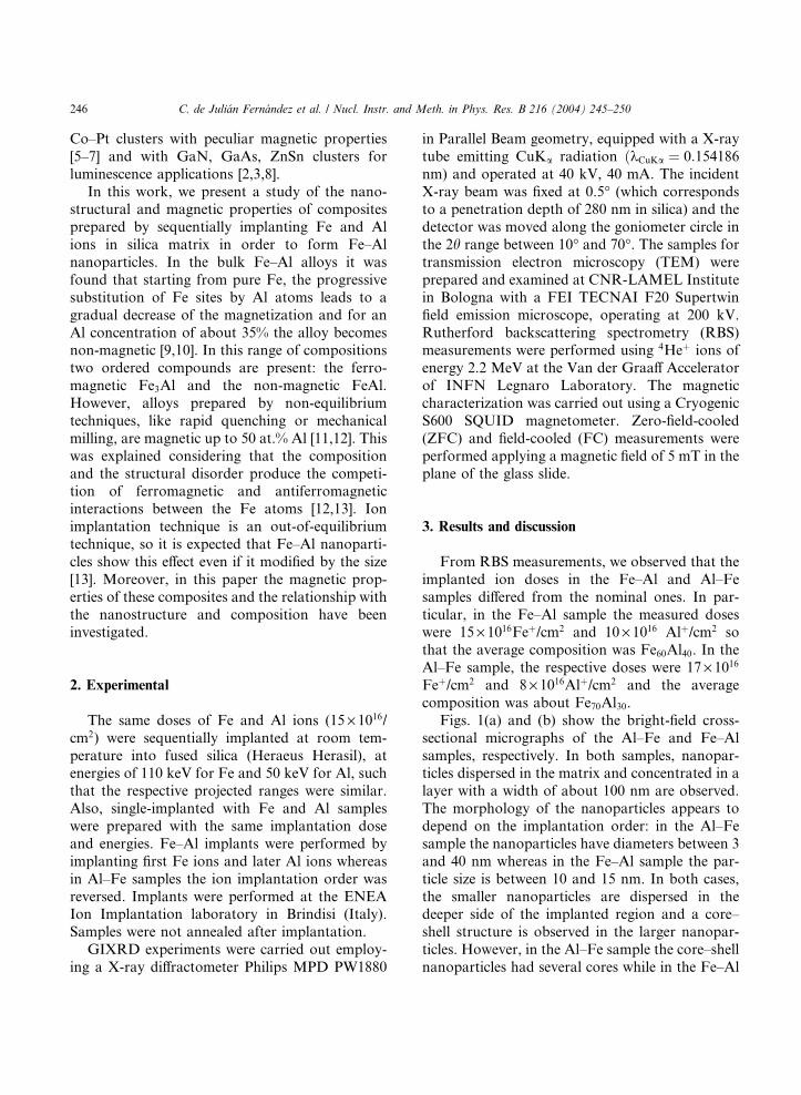
Figs. 1(a) and (b) show the bright-field cross-
sectional micrographs of the Al–Fe and Fe–Al
samples, respectively. In both samples, nanopar-ticles dispersed in the matrix and concentrated in a
layer with a width of about 100 nm are observed.
The morphology of the nanoparticles appears to
depend on the implantation order: in the Al–Fe
sample the nanoparticles have diameters between 3
and 40 nm whereas in the Fe–Al sample the par-
ticle size is between 10 and 15 nm. In both cases,
the smaller nanoparticles are dispersed in thedeeper side of the implanted region and a core–
shell structure is observed in the larger nanopar-
ticles. However, in the Al–Fe sample the core–shell
nanoparticles had several cores while in the Fe–Al
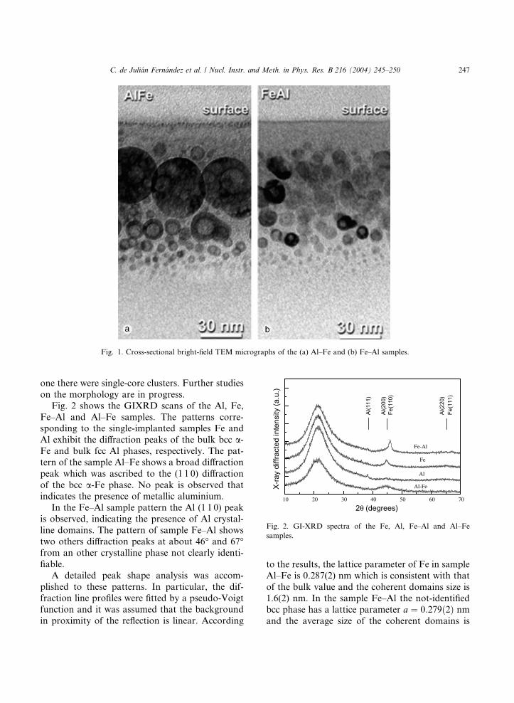
Fig. 1. Cross-sectional bright-field TEM micrographs of the (a) Al–Fe and (b) Fe–Al samples.
10 20 30 40 50 60 70
2θ (degrees)
Al(2
20)
Al(2
00)
Fe(1
10)
Al(1
11)
Fe(1
11)
Al-Fe
Fe-Al
Fe
Al
X-ra
y di
ffrac
ted
inte
nsity
(a.u
.)
Fig. 2. GI-XRD spectra of the Fe, Al, Fe–Al and Al–Fe
samples.
C. de Juli�an Fern�andez et al. / Nucl. Instr. and Meth. in Phys. Res. B 216 (2004) 245–250 247
one there were single-core clusters. Further studies
on the morphology are in progress.
Fig. 2 shows the GIXRD scans of the Al, Fe,
Fe–Al and Al–Fe samples. The patterns corre-sponding to the single-implanted samples Fe and
Al exhibit the diffraction peaks of the bulk bcc a-Fe and bulk fcc Al phases, respectively. The pat-
tern of the sample Al–Fe shows a broad diffraction
peak which was ascribed to the (1 1 0) diffraction
of the bcc a-Fe phase. No peak is observed thatindicates the presence of metallic aluminium.
In the Fe–Al sample pattern the Al (1 1 0) peakis observed, indicating the presence of Al crystal-
line domains. The pattern of sample Fe–Al shows
two others diffraction peaks at about 46� and 67�from an other crystalline phase not clearly identi-
fiable.
A detailed peak shape analysis was accom-
plished to these patterns. In particular, the dif-
fraction line profiles were fitted by a pseudo-Voigtfunction and it was assumed that the background
in proximity of the reflection is linear. According
to the results, the lattice parameter of Fe in sample
Al–Fe is 0.287(2) nm which is consistent with that
of the bulk value and the coherent domains size is
1.6(2) nm. In the sample Fe–Al the not-identified
bcc phase has a lattice parameter a ¼ 0:279ð2Þ nmand the average size of the coherent domains is
248 C. de Juli�an Fern�andez et al. / Nucl. Instr. and Meth. in Phys. Res. B 216 (2004) 245–250
6.5(4) nm. Such a phase could be the Fe phase
hardly stressed considering that it is contracted of
2.5% with respect to the lattice parameter mea-
sured in the Fe case. The observed pattern doesnot correspond with any Fe, Al oxides or silicate.
We observe that in both Fe–Al and Al–Fe samples
the TEM particle size is 5–10 times larger than the
XRD coherent domains dimensions, which indi-
cates that the clusters are polycrystalline. Finally,
we point out that Fe–Al bulk bcc alloys have a
larger lattice parameter than that of Fe. Therefore
the formation of the Fe–Al alloy in the nanopar-ticles of the Al–Fe and Fe–Al samples was ruled
out.
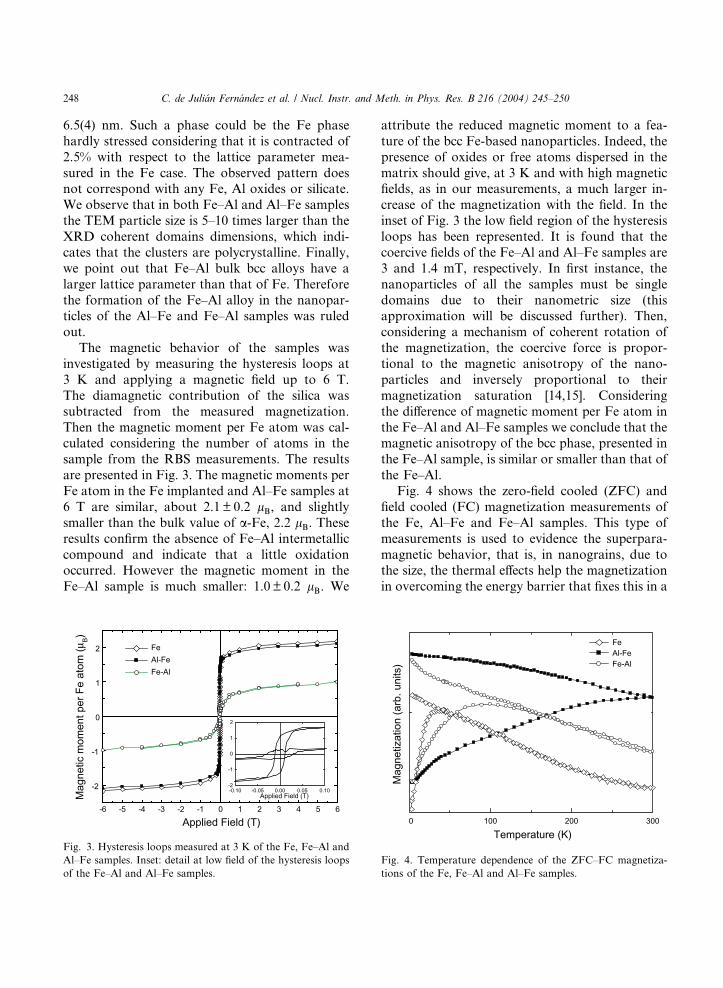
The magnetic behavior of the samples was
investigated by measuring the hysteresis loops at
3 K and applying a magnetic field up to 6 T.
The diamagnetic contribution of the silica was
subtracted from the measured magnetization.
Then the magnetic moment per Fe atom was cal-culated considering the number of atoms in the
sample from the RBS measurements. The results
are presented in Fig. 3. The magnetic moments per
Fe atom in the Fe implanted and Al–Fe samples at
6 T are similar, about 2.1 ± 0.2 lB, and slightlysmaller than the bulk value of a-Fe, 2.2 lB. Theseresults confirm the absence of Fe–Al intermetallic
compound and indicate that a little oxidationoccurred. However the magnetic moment in the
Fe–Al sample is much smaller: 1.0 ± 0.2 lB. We
-6 -5 -4 -3 -2 -1 0 1 2 3 4 5 6
-2
-1
0
1
2
-0.10 -0.05 0.00 0.05 0.10-2
-1
0
1
2
Mag
netic
mom
ent p
er F
e at
om (µ
B)
Applied Field (T)
Fe Al-Fe Fe-Al
Applied Field (T)
Fig. 3. Hysteresis loops measured at 3 K of the Fe, Fe–Al and
Al–Fe samples. Inset: detail at low field of the hysteresis loops
of the Fe–Al and Al–Fe samples.
attribute the reduced magnetic moment to a fea-
ture of the bcc Fe-based nanoparticles. Indeed, the
presence of oxides or free atoms dispersed in the
matrix should give, at 3 K and with high magneticfields, as in our measurements, a much larger in-
crease of the magnetization with the field. In the
inset of Fig. 3 the low field region of the hysteresis
loops has been represented. It is found that the
coercive fields of the Fe–Al and Al–Fe samples are
3 and 1.4 mT, respectively. In first instance, the
nanoparticles of all the samples must be single
domains due to their nanometric size (thisapproximation will be discussed further). Then,
considering a mechanism of coherent rotation of
the magnetization, the coercive force is propor-
tional to the magnetic anisotropy of the nano-
particles and inversely proportional to their
magnetization saturation [14,15]. Considering
the difference of magnetic moment per Fe atom in
the Fe–Al and Al–Fe samples we conclude that themagnetic anisotropy of the bcc phase, presented in
the Fe–Al sample, is similar or smaller than that of
the Fe–Al.
Fig. 4 shows the zero-field cooled (ZFC) and
field cooled (FC) magnetization measurements of
the Fe, Al–Fe and Fe–Al samples. This type of
measurements is used to evidence the superpara-
magnetic behavior, that is, in nanograins, due tothe size, the thermal effects help the magnetization
in overcoming the energy barrier that fixes this in a
0 100 200 300
Mag
netiz
atio
n (a
rb. u
nits
)
Temperature (K)
Fe Al-Fe Fe-Al
Fig. 4. Temperature dependence of the ZFC–FC magnetiza-
tions of the Fe, Fe–Al and Al–Fe samples.
C. de Juli�an Fern�andez et al. / Nucl. Instr. and Meth. in Phys. Res. B 216 (2004) 245–250 249
direction of the grain and then the magnetization
tilts freely. This energy barrier is responsible of the
hysteresis that the nanoparticles show and it is
proportional to the magnetic anisotropy and to theparticle volume. The thermal effects produce the
demagnetization of the particle: it seems as a
paramagnet with high moment. The temperature
threshold, called blocking temperature TB, abovewhich a grain of volume V and with a magneticanisotropy, Kef , shows the superparamagneticbehavior is [14,15]
TB ¼ KefVa kB
; ð1Þ
where kB is the Boltzman constant and a is aconstant equal to 25 in the case of the SQUID
measurements. The ZFC–FC curves presented in
Fig. 4 are representative of a distribution of TB,mainly associated to the broad size distribution,and being the temperature of the maximum of the
ZFC curve linked to the average TB. In additionthe magnetization does not follow a Curie law at
high temperature in these films. The temperature
at which ZFC–FC branches joints is the temper-
ature above which all the nanoparticles are su-
perparamagnetic. In the Fe and Fe–Al samples the
blocking temperature is below the room tempera-ture while for the Al–Fe sample TB is above theroom temperature. Considering the much larger
particle size of the Al–Fe than the Fe–Al sample,
the differences in the ZFC–FC curves can be dis-
cussed in terms of superparamagnetism consider-
ing the measured particle size. However we point
out that the nanoparticles of these samples have a
core–shell morphology. The magnetic anisotropywill include the contribution of the different mag-
netic surfaces (that of the core and those of the
shell) as occurs in many nanostructures [15]. The
core–shell structure promotes incoherent rotation
modes in which the magnetic anisotropy is not
related to the intrinsic properties of the material.
Moreover, as can be observed in Figs. 1(a) and (b)
nanoparticles are very densely packed so that in-terparticle interactions must be dominant giving
rise to different demagnetization processes.
Therefore, we think that the observed behavior
appearing similar to superparamagnetism is the
result of several magnetic effects and must be
investigated in order to analyse correctly the ZFC–
FC curves.
Finally we want to point out that, both fromthe point of view of the nanostructural and mag-
netic characterization, FeAl intermetallic alloy
nanoparticles were not formed in silica by using
sequential ion implantation. Several authors have
studied the mechanism of nucleation and alloying
of the nanoparticles concluding that it is driven by
competitive physical and chemical reactions be-
tween the implanted elements and those of thematrix [16,17]. For Fe and Al the respective Gibbs
energy of oxide formation are negative and very
high [18], so the formation of oxides is expected.
However, as it was shown in this work, metallic
nanoparticles were formed suggesting that com-
petitive chemical paths determine the nanostruc-
tural features as elsewhere suggested [16]. Several
studies are in progress to investigate the mecha-nism of formation of the different nanostructures
and their chemical state, the identification of the
bcc phase observed in the Fe–Al sample and the
characterization of the magnetic properties.
Acknowledgements
This work was financially supported by ENEA
in the framework of the PROTEMA project of
‘‘Intesa ENEA-MIUR’’ under project 4335/04 and
by the Italian MURST (National Projects).
References
[1] F. Gonella, P. Mazzoldi, in: H.S. Nalwa (Ed.), Handbook
of Nanostructured Materials and Nanotechnology, Vol. 4,
Academic Press, San Diego, 2000, p. 81.
[2] A. Meldrum, L.A. Boatner, C.W. White, Nucl. Instr. and
Meth. B 178 (2001) 7.
[3] G. Mattei, Nucl. Instr. and Meth. B 191 (2002) 323.
[4] G. Battaglin, E. Cattaruzza, G. De Marchi, F. Gonella, G.
Mattei, C. Maurizio, P. Mazzoldi, M. Parolin, C. Sada, I.
Calliari, Nucl. Instr. and Meth. B 191 (2002) 392.
[5] C. de Juli�an Fern�andez, C. Sangregorio, G. Mattei, C.
Maurizio, G. Battaglin, F. Gonella, S.A. Lascialfari, S. Lo
Russo, D. Gatteschi, P. Mazzoldi, J.M. Gonz�alez, F.
D�Acapito, Nucl. Instr. and Meth. B 175–177 (2001) 468.[6] E. Cattaruzza, F. d�Acapito, C. de Juli�an Fern�andez, A. deLorenzo, F. Gonella, G. Mattei, C. Maurizio, P. Mazzoldi,
250 C. de Juli�an Fern�andez et al. / Nucl. Instr. and Meth. in Phys. Res. B 216 (2004) 245–250
S. Padovani, B.F. Scremin, F. Zontone, Nucl. Instr. and
Meth. B 191 (2002) 40.
[7] C.W. White, S.P. Withrow, K.D. Sorge, A. Meldrum, J.D.
Budai, J.R. Thompson, L.A. Boatner, J. Appl. Phys. 93
(2003) 5656.
[8] E. Borsella, C. de Juli�an Fern�andez, M.A. Garc�ıa, G.Mattei, C. Maurizio, P. Mazzoldi, S. Padovani, C. Sada,
G. Battaglin, E. Cattaruzza, F. Gonella, A. Quaranta, F.
D�Acapito, M.A. Tagliente, L. Tapfer, Nucl. Instr. andMeth. B 191 (2002) 447.
[9] H.P.J. Wijn (Ed.), Physical Properties and Other Data,
Landolt-B€ornstein, Vol. III, Part 19b, Springer, Berlin,
1987, p. 295.
[10] J.S. Kouvel, J.H. Westbrook, R.L. Fleischer (Eds.), Inter-
metallic Compounds: Principles, Vol. 1, Wiley, New York,
1994, p. 935.
[11] G.R. Caskey, J.M. Franz, D.J. Sellmyer, J. Phys. Chem.
Solids 34 (1973) 1179.
[12] A. Hernando, X. Amils, J. Nogu�es, S. Suri~nach, M.D.
Bar�o, M.K. Ibarra, Phys. Rev. B 58 (1998) R11864.
[13] B.V. Reddy, S.C. Deevi, F.A. Reuse, S.N. Khanna, Phys.
Rev. B 64 (2001) 132408.
[14] J.L. Dormann, D. Fiorani, E. Tronc, Adv. Chem. Phys. 98
(1997) 283.
[15] X. Batlle, A. Labarta, J. Phys. D: Appl. Phys. 35 (2002)
R15.
[16] E. Cattaruzza, Nucl. Instr. and Meth. B 169 (2000) 141.
[17] T. Ito, O. Kitakami, Y. Shimada, Y. Kamo, S. Kikuchi,
J. Magn. Magn. Mater. 235 (2000) 165.
[18] O. Knacke, O. Kubaschewski, K. Hesselmann (Eds.),
Thermo-Chemical Properties of Inorganic Substances,
Springer, Berlin, 1991.
Copyright © 2022 FDOKUMEN