OTUB1 Co-opts Lys48-Linked Ubiquitin Recognition to Suppress E2 Enzyme Function
Structural Analysis of the UBA Domain of X-linkedInhibitor of Apoptosis Protein Reveals Different Surfacesfor Ubiquitin-Binding and Self-AssociationMan Kit Tse1,2., Sin Kam Hui2., Yinhua Yang3, Si-Tao Yin4, Hong-Yu Hu4, Bing Zou5, Benjamin Chun Yu
Wong5, Kong Hung Sze1,2*
1 Department of Microbiology and State Key Laboratory for Emerging Infectious Diseases, The University of Hong Kong, Hong Kong SAR, People’s Republic of China,
2 Department of Chemistry and Open Laboratory of Chemical Biology of the Institute of Molecular Technology for Drug Discovery and Synthesis, The University of Hong
Kong, Hong Kong SAR, People’s Republic of China, 3 Department of Biochemistry, Centre for Protein Science and Crystallography, The Chinese University of Hong Kong,
Shatin, Hong Kong SAR, People’s Republic of China, 4 State Key Laboratory of Molecular Biology, Institute of Biochemistry and Cell Biology, Shanghai Institutes for
Biological Sciences, Chinese Academy of Sciences, Shanghai, People’s Republic of China, 5 Department of Medicine, The University of Hong Kong, Pokfulam Road, Hong
Kong SAR, People’s Republic of China
Abstract
Background: Inhibitor of apoptosis proteins (IAPs) belong to a pivotal antiapoptotic protein family that plays a crucial rolein tumorigenesis, cancer progression, chemoresistance and poor patient-survival. X-linked inhibitor of apoptosis protein(XIAP) is a prominent member of IAPs attracting intense research because it has been demonstrated to be a physiologicalinhibitor of caspases and apoptosis. Recently, an evolutionarily conserved ubiquitin-associated (UBA) domain was identifiedin XIAP and a number of RING domain-bearing IAPs. This has placed the IAPs in the group of ubiquitin binding proteins.Here, we explore the three-dimensional structure of the XIAP UBA domain (XIAP-UBA) and how it interacts with mono-ubiquitin and diubiquitin conjugates.
Principal Findings: The solution structure of the XIAP-UBA domain was determined by NMR spectroscopy. XIAP-UBA adoptsa typical UBA domain fold of three tightly packed a-helices but with an additional N-terminal 310 helix. The XIAP-UBA bindsmono-ubiquitin as well as Lys48-linked and linear-linked diubiquitins at low-micromolar affinities. NMR analysis of the XIAP-UBA–ubiquitin interaction reveals that it involves the classical hydrophobic patches surrounding Ile44 of ubiquitin and theconserved MGF/LV motif surfaces on XIAP-UBA. Furthermore, dimerization of XIAP-UBA was observed. Mapping of the self-association surface of XIAP-UBA reveals that the dimerization interface is formed by residues in the N-terminal 310 helix,helix a1 and helix a2, separate from the ubiquitin-binding surface.
Conclusion: Our results provide the first structural information of XIAP-UBA and map its interaction with mono-ubiquitin,Lys48-linked and linear-linked diubiquitins. The notion that XIAP-UBA uses different surfaces for ubiquitin-binding and self-association provides a plausible model to explain the reported selectivity of XIAP in binding polyubiquitin chains withdifferent linkages.
Citation: Tse MK, Hui SK, Yang Y, Yin S-T, Hu H-Y, et al. (2011) Structural Analysis of the UBA Domain of X-linked Inhibitor of Apoptosis Protein Reveals DifferentSurfaces for Ubiquitin-Binding and Self-Association. PLoS ONE 6(12): e28511. doi:10.1371/journal.pone.0028511
Editor: Paul C. Driscoll, MRC National Institute for Medical Research, United Kingdom
Received May 27, 2011; Accepted November 9, 2011; Published December 15, 2011
Copyright: � 2011 Tse et al. This is an open-access article distributed under the terms of the Creative Commons Attribution License, which permits unrestricteduse, distribution, and reproduction in any medium, provided the original author and source are credited.
Funding: This work was supported by the Research Grants Council of Hong Kong (GRF 7755/08M and GRF 7765/09M) and a Special Equipment Grant (Projectno. SEG/CUHK09) from the University Grants Committee of Hong Kong SAR. The funders had no role in study design, data collection and analysis, decision topublish, or preparation of the manuscript.
Competing Interests: The authors have declared that no competing interests exist.
* E-mail: [email protected]
. These authors contributed equally to this work.
Introduction
The IAP (inhibitor of apoptosis) proteins are an evolutionarily
conserved family of cell-death regulators, which can block
apoptosis induced by diverse stimuli through direct interactions
with a variety of inducers and effectors of apoptosis [1–3]. This
places IAPs in a central position as inhibitors of death signals that
proceed through a number of different pathways. Defects in
apoptosis play an important role in the pathogenesis of a number
of diseases such as cancer, neurodegenerative and auto-immune
diseases. Indeed, IAPs are often overexpressed in tumors, and they
have been implicated in tumor development and progression,
chemoresistance and poor patient survival [4]. X-linked inhibitor
of apoptosis protein (XIAP) is the most versatile inhibitor of
caspases and apoptosis in vivo [5]. XIAP is also a cancer
biomarker, and it is regarded as a promising target for the
development of anticancer drugs.
XIAP contains three N-terminal zinc-binding baculovirus IAP
repeat (BIR) domains that have been shown to inhibit the activities
of caspase-3, caspase-7 and caspase-9 [6,7] as well as to mediate
the activation of NF-kB pathway via interaction with TAK1 [8].
XIAP also possesses a C-terminal really interesting new gene
PLoS ONE | www.plosone.org 1 December 2011 | Volume 6 | Issue 12 | e28511
(RING) domain that functions as an E3 ubiquitin ligase. The
RING domain of XIAP ubiquitinates a wide range of substrates,
thereby, affecting a broad range of cellular activities beyond
apoptotic suppression [9]. There is now compelling evidences that
XIAP also has significant roles in cell division, morphogenesis,
heavy metal homeostasis, NF-kB activation and MAP kinase
signaling [9,10]. The long stretch of sequence (,100 residues)
linking the BIR3 domain to the C-terminal RING domain was not
known to contain globular structural elements or functional
components until recently. Gyrd-Hansen et al. reported that they
had identified an evolutionarily conserved ubiquitin-associated
(UBA) domain within this region using sequence analysis and
structure prediction algorithm [11]. They also reported that
similar UBA domains also exist in other RING-bearing IAP
members, including XIAP, cellular IAP -1 and -2 (cIAP-1 and -2)
and IAP-like protein 2 (ILP-2) [11,12]. The conjugation of
ubiquitin to target proteins plays a crucial part in the formation of
signaling networks. The ubiquitination is mediated through low-
affinity, non-covalent interactions between ubiquitin and small
ubiquitin-binding domains present in specialized proteins that are
collectively referred to as ubiquitin-receptors. These receptors are
responsible for translating ubiquitin modifications into cellular
phenotypes. Ubiquitin can be attached to target proteins as a
single moiety (ubiquitin or mono-ubiquitin, monoUb), or as
polyubiquitin (polyUb) chains. For polyubiquitination, the ubiq-
uitin domains are most commonly linked through its Lys 48 (K48)
or Lys 63 (K63) residue. The K48-linked polyUb chains adopt a
kinked topology, whereas K63-linked polyUb chains are more
linear in conformation and resemble a ‘beads-on-a-string’
structure [13,14]. Ubiquitin-receptors that recognize the K48-
linked polyUb chains recruit the modified proteins to the
proteasome for degradation. In contrast, ubiquitin-receptors that
bind to monoUb or Lys 63 linkages enable non-degradative
signaling processes by recruiting mono-ubiquitinated or Lys 63-
polyubiquitylated proteins to downstream protein complexes.
K63-linked ubiquitination, for example, is used as a key signal
transducer for the activation of NF-kB and cell survival. Linear-
linked polyUb chain has a structure highly similar to that of K63-
linked polyUb chain [15]. The linear-linked polyUb chain, in
which the C-terminal Gly of ubiquitin is conjugated to the a-
amino group of the N-terminal Met of the successive ubiquitin,
can be generated by a unique ubiquitin ligase complex known as
the linear ubiquitin chain assembly complex (LUBAC) [16].
However, the physiological role of the linear-linked polyUb
remains largely unknown except that it can also act as a regulator
for the activation of NF-kB [17].
The UBA domain is a small (,40 residues) protein-protein
interaction module that mediates ubiquitin-binding and thus
enables host proteins to participate in ubiquitin-dependent
signaling processes [18]. Structural studies indicate that a
conserved hydrophobic patch on the UBA domain makes direct
contact with ubiquitin. The three-dimensional structures of a
number of UBA domains have been determined using NMR and
x-ray crystallography [19–28]. Despite remarkably low sequence
homology, their three-dimensional structures are highly similar,
consisting of a bundle of three a-helices. A majority of UBA
domains bind ubiquitin or ubiquitin-like domains using a
hydrophobic patch of residues in the a1–a2 loop (‘MGF’) and
two aliphatic residues at the end of the helix a3 (‘LL/V’) [29,30].
In spite of structural similarity, different types of UBA domain
show distinct monoUb and linkage selective polyUb-binding
ability [31]. Most studies reported so far describe UBA domain
binding to monoUb as a heterodimer with a stoichiometry of 1 to
1. Self-association has been reported previously for a few cases of
UBA and UBA-like domains [32–37]. However, only a few of
them, such as Cbl-b-UBA [35], c-Cbl-UBA [34] and p62-UBA
[36], have been structurally characterized.
Gyrd-Hansen et al. demonstrated with glutathione S-transferase
(GST) pull-down assays that the UBA domain-containing IAPs do
not associate with monoUb but they bind efficiently to K63-linked
polyUb and in some cases to K48-linked polyUb [11]. In
particular, XIAP binds exclusively to the structurally similar
K63-linked and linear-linked polyUbs [11]. Furthermore, it was
shown that the presence of a UBA domain and a dimerizable
RING domain are essential for XIAP’s ability to bind polyUb
[11]. However, in a later report, Blankenship et al. demonstrated
that the UBA domain of cIAP-1 binds monoUb and K48-linked
and K63-linked polyUb chains with low-micromolar affinities by
using multiple detection methods, including surface plasmon
resonance, isothermal titration calorimetry and NMR spectrosco-
py [12]. Nevertheless, these novel observations establish IAPs as
ubiquitin interacting proteins and highlight the need for a better
understanding of the interactions between the UBA domains of
IAPs with ubiquitin at molecular and structural levels.
In the present study, we have determined the solution structure of
the UBA domain in XIAP by NMR spectroscopy, which is the first
reported structure of a UBA domain in the IAP-family. In spite of a
marked overall conformational similarity with reported UBA
domain structures from other protein families, XIAP-UBA exhibits
notable structural differences in the helical length and inter-helical
packing as well as the presence of an N-terminal 310 helix. Using a
combination of chemical shift perturbation mapping and site-
directed mutagenesis, we have elucidated the binding interfaces for
ubiquitin as well as K48-linked and linear-linked diubiquitins (Ub2)
on the XIAP UBA domain. In addition to the characterization of
the hydrophobic ubiquitin-binding surfaces on XIAP-UBA, the
present study provides evidence for the dimerization of the XIAP
UBA domain. Mapping of the self-association surface of XIAP UBA
domain reveals that it is formed by residues in N-terminal 310 helix,
helix a1 and helix a2, and is separate from the ubiquitin-binding
surface. Our results provide a plausible model to explain how the
self-association of XIAP UBA domain could lead to the linkage-
specificity of polyubiquitin chain binding by XIAP.
Results
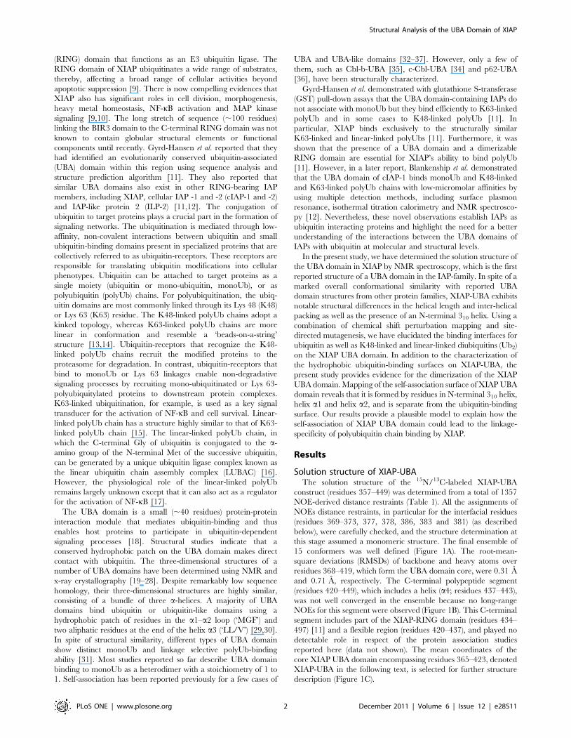
Solution structure of XIAP-UBAThe solution structure of the 15N/13C-labeled XIAP-UBA
construct (residues 357–449) was determined from a total of 1357
NOE-derived distance restraints (Table 1). All the assignments of
NOEs distance restraints, in particular for the interfacial residues
(residues 369–373, 377, 378, 386, 383 and 381) (as described
below), were carefully checked, and the structure determination at
this stage assumed a monomeric structure. The final ensemble of
15 conformers was well defined (Figure 1A). The root-mean-
square deviations (RMSDs) of backbone and heavy atoms over
residues 368–419, which form the UBA domain core, were 0.31 A
and 0.71 A, respectively. The C-terminal polypeptide segment
(residues 420–449), which includes a helix (a4; residues 437–443),
was not well converged in the ensemble because no long-range
NOEs for this segment were observed (Figure 1B). This C-terminal
segment includes part of the XIAP-RING domain (residues 434–
497) [11] and a flexible region (residues 420–437), and played no
detectable role in respect of the protein association studies
reported here (data not shown). The mean coordinates of the
core XIAP UBA domain encompassing residues 365–423, denoted
XIAP-UBA in the following text, is selected for further structure
description (Figure 1C).
Structural Analysis of the UBA Domain of XIAP
PLoS ONE | www.plosone.org 2 December 2011 | Volume 6 | Issue 12 | e28511
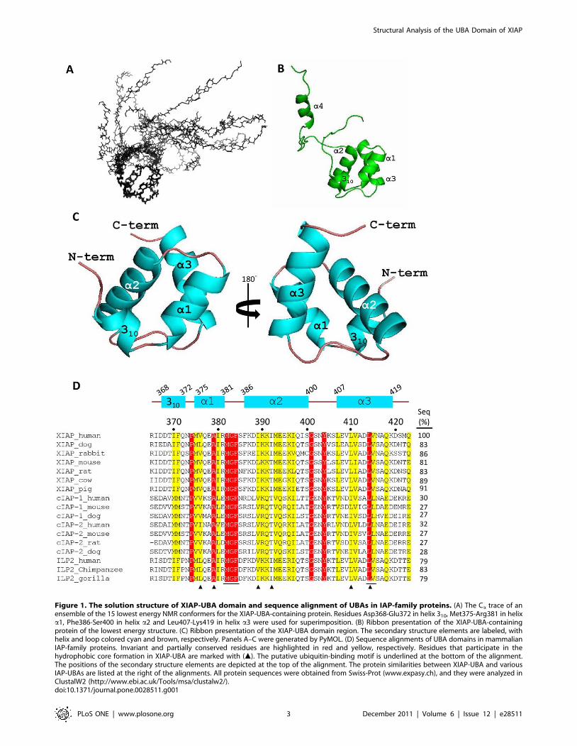
Figure 1. The solution structure of XIAP-UBA domain and sequence alignment of UBAs in IAP-family proteins. (A) The Ca trace of anensemble of the 15 lowest energy NMR conformers for the XIAP-UBA-containing protein. Residues Asp368-Glu372 in helix 310, Met375-Arg381 in helixa1, Phe386-Ser400 in helix a2 and Leu407-Lys419 in helix a3 were used for superimposition. (B) Ribbon presentation of the XIAP-UBA-containingprotein of the lowest energy structure. (C) Ribbon presentation of the XIAP-UBA domain region. The secondary structure elements are labeled, withhelix and loop colored cyan and brown, respectively. Panels A–C were generated by PyMOL. (D) Sequence alignments of UBA domains in mammalianIAP-family proteins. Invariant and partially conserved residues are highlighted in red and yellow, respectively. Residues that participate in thehydrophobic core formation in XIAP-UBA are marked with (m). The putative ubiquitin-binding motif is underlined at the bottom of the alignment.The positions of the secondary structure elements are depicted at the top of the alignment. The protein similarities between XIAP-UBA and variousIAP-UBAs are listed at the right of the alignments. All protein sequences were obtained from Swiss-Prot (www.expasy.ch), and they were analyzed inClustalW2 (http://www.ebi.ac.uk/Tools/msa/clustalw2/).doi:10.1371/journal.pone.0028511.g001
Structural Analysis of the UBA Domain of XIAP
PLoS ONE | www.plosone.org 3 December 2011 | Volume 6 | Issue 12 | e28511
The XIAP-UBA adopts a compact globular three-helix bundle
structure, which highly resembles the classical UBA domain topology.
The three helices (a1, a2 and a3) are packing against each other to
form a well-defined hydrophobic core, which is composed of residues
Val376, Ala379, Ile389, Ile392, Leu410 and Leu414. In addition,
XIAP-UBA possesses an N-terminal 310 helix (residues 368–372)
oriented nearly perpendicular to helix a1 (interhelical angle ,98u).This N-terminal short helix is so far reported only in Dsk2-UBA (helix
a0) where its function remains uncharacterized.
A DALI search for the structural homologues of XIAP-UBA
returned five hits with a Z-score .3.0, including Dsk2-UBA (Z-
score = 3.8, Ca RMSD = 2.2 A), hHR23A-UBA2 (Z-score = 3.6,
Ca RMSD = 2.9 A), Cbl-b-UBA (Z-score = 3.5, Ca RMSD =
2.4 A)[35], Ede1-UBA (Z-score = 3.3, Ca RMSD = 2.2 A) and
UQ1-UBA (Z-score = 3.1, Ca RMSD = 2.8 A) [38]. The DALI
results indicate that the overall structure of XIAP-UBA is well-
conserved even though the sequence similarities between XIAP-
UBA and the returned hits are very low (6–11%) (Figure S1).
Mapping of the mono-ubiquitin, K48-linked and linear-linked diubiquitin binding sites on XIAP-UBA
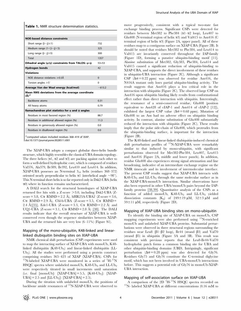
NMR chemical shift perturbation (CSP) experiments were used
to map the interacting surface of XIAP-UBA with monoUb, K48-
linked diubiquitin (K48-Ub2) and linear-linked diubiquitin (LL-
Ub2). All the studies were performed using a protein construct
comprising residues 365–423 of XIAP (XIAP-UBA). CSPs for15N-labeled XIAP-UBA were monitored in a series of 1H-15N
HSQC spectra where unlabeled monoUb, K48-Ub2 and LL-Ub2
were respectively titrated in small increments until saturation
(i.e. final [monoUb]: [XIAP-UBA] = 5.5, [K48-Ub2]: [XIAP-
UBA] = 2.5 and [LL-Ub2]: [XIAP-UBA] = 2.5).
During the titration with unlabeled monoUb, the positions of
backbone amide resonances of 15N-XIAP-UBA were observed to
move progressively, consistent with a typical two-state fast
exchange binding process. Significant CSPs were detected for
residues between Met382 to Phe384 (a1–a2 loop), Leu407 to
Glu408 (N-terminal region of helix a3) and Val415 to Asn416 (C
terminal region of helix a3) (Figure 2A, upper panel). All of these
residues map to a contiguous surface on XIAP-UBA (Figure 2B). It
should be noted that residues Met382 to Phe384, and Leu414 to
Val415 are invariantly conserved throughout the IAP-family
(Figure 1D), forming a putative ubiquitn-binding motif [11].
Alanine substitution of Met382, Gly383, Phe384, Leu414 and
Val415 caused a significant reduction of ubiquitin-binding to
XIAP-UBA, and supports the direct involvement of these residues
in ubiquitin-UBA interaction (Figure 2C). Although a significant
CSP (Ds= 0.22 ppm) was observed for residue Asn416, the
N416A mutant only loses partial ubiquitn-binding activity. The
result suggests that Asn416 plays a less critical role in the
interaction with ubiquitin (Figure 2C). The observed large CSP on
Asn416 upon ubiquitin binding likely results from conformational
effect rather than direct interaction with ubiquitin. Interestingly,
the resonance of a semi-conserved residue, Glu408 (position
equivalent to Asn428 of cIAP-1 and Asn414 of cIAP-2 [12]),
exhibited the largest CSP value (Ds= 0.68 ppm). Mutation of
Glu408 to an Asn had no adverse effect on ubiquitin binding
activity. In contrast, alanine substitution of Glu408 substantially
reduced the interaction with ubiquitin (Figure 2C). These results
imply that the polar side-chain of Glu408, which protrudes from
the ubiquitin-binding surface, is important for the interaction
(Figure 2B).
The K48-linked and linear-linked diubiquitin-induced chemical
shift perturbation profiles of 15N-XIAP-UBA were remarkably
similar to that induced by mono-ubiquitin, with significant
perturbations observed for Met382-Phe384, Leu407, Leu415
and Asn416 (Figure 2A, middle and lower panels). In addition,
residue Glu408 also experiences strong signal attenuation and line
broadening, indicative of an intermediate exchange regime on the
NMR timescale and its involvement of intermolecular interface.
The present CSP results suggest that XIAP-UBA interacts with
K48-Ub2 and LL-Ub2 through the same molecular surface as in
the XIAP-UBA-monoUb interaction. Similar observations have
also been reported in other UBA/monoUb pairs beyond the IAP-
family proteins [38,39]. Quantitative analysis of the CSPs as a
function of added monoUb, K48-Ub2 and LL-Ub2 yielded
dissociation constants (Kd) of 249619 mM, 5263 mM and
59611 mM, respectively (Figure 2D).
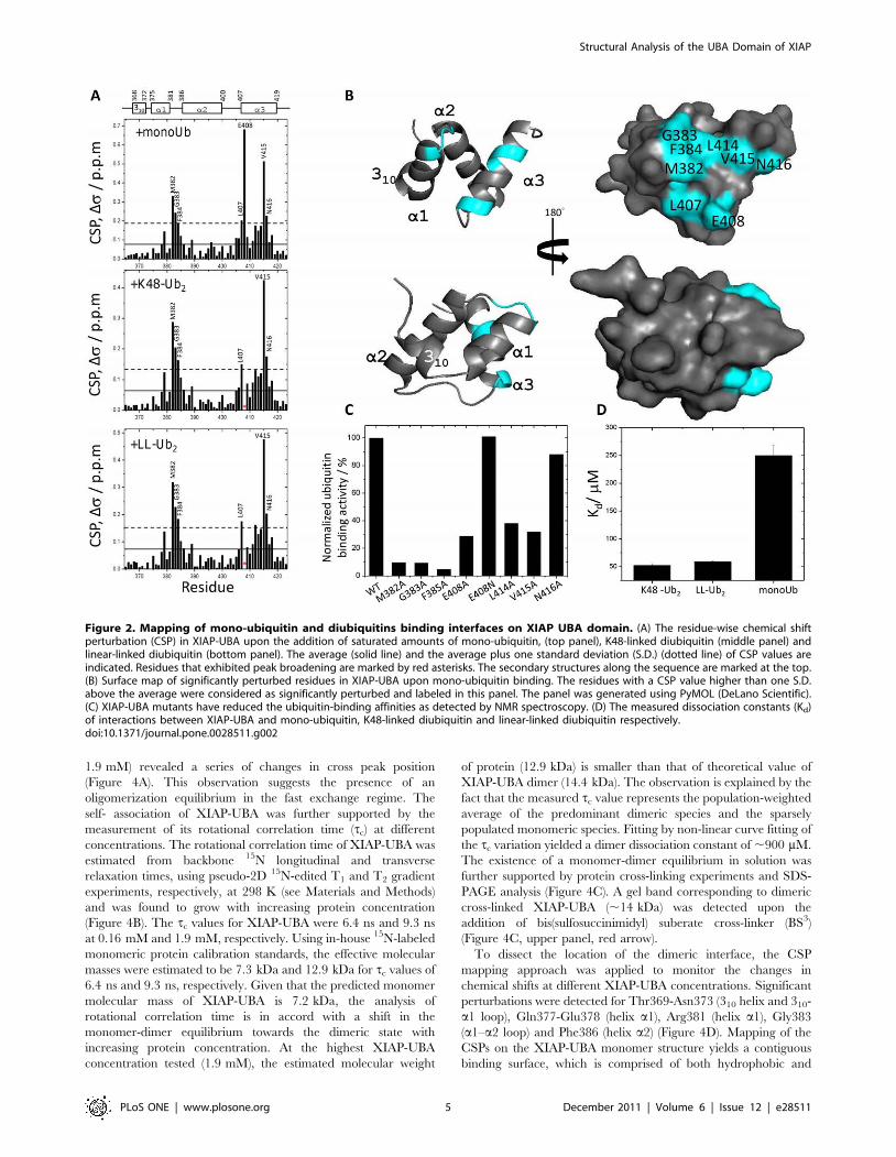
Mapping of XIAP-UBA binding sites on mono-ubiquitinTo identify the binding site of XIAP-UBA on monoUb, CSP
mapping experiments were also performed using 15N-enriched
monoUb and unlabeled XIAP-UBA proteins. Substantial pertur-
bations were observed in three structural regions surrounding the
residues near Leu8 (b1–b2 loop), Ile44 (strand b3) and Val70
(strand b5) in ubiquitin (Figure 3A and 3B). This result was
consistent with previous reports that the Leu8-Ile44-Val70
hydrophobic patch forms a common binding site for UBA and
other ubiquitin-binding domains (UBD). Intriguingly, significant
perturbation (Ds= 0.28 ppm) was also detected for Gly76.
Residues Gly75 and Gly76 constitute the C-terminal diglycine
motif, which has not been involved in UBA-monoUb interactions
before. This suggests a potential role of Gly76 in monoUb-XIAP-
UBA interaction.
Mapping of self-association surface on XIAP-UBAA comparison of the 2D 1H-15N HSQC spectra recorded on
15N- labeled XIAP-UBA at different concentrations (0.16 mM to
Table 1. NMR structure determination statistics.
NOE-based distance constraints
Short range (|i2j|#1) 732
Medium range (1,|i2j|,5) 325
Long range (|i2j|$5) 300
Total 1357
Dihedral angle (Q/y) constraints from TALOS: Q+y 53+53
Hydrogen bonds 42
Violations
NOE distance violations $0.5A 0
Torsion angles $5u 0
Average Van der Waal energy (kcal/mol) 2613.2
Mean RMS deviations from the average coordinate(A)#
Backbone atoms 0.31
All heavy atoms 0.79
Ramachandran plot statistics for Q and y angles
Residues in most favored region (%) 88.7
Residues in additional allowed region (%) 11.3
Residues in generously allowed region (%) 0.0
Residues in disallowed region (%) 0.0
#computed values included residues 368–419 of XIAP.doi:10.1371/journal.pone.0028511.t001
Structural Analysis of the UBA Domain of XIAP
PLoS ONE | www.plosone.org 4 December 2011 | Volume 6 | Issue 12 | e28511
1.9 mM) revealed a series of changes in cross peak position
(Figure 4A). This observation suggests the presence of an
oligomerization equilibrium in the fast exchange regime. The
self- association of XIAP-UBA was further supported by the
measurement of its rotational correlation time (tc) at different
concentrations. The rotational correlation time of XIAP-UBA was
estimated from backbone 15N longitudinal and transverse
relaxation times, using pseudo-2D 15N-edited T1 and T2 gradient
experiments, respectively, at 298 K (see Materials and Methods)
and was found to grow with increasing protein concentration
(Figure 4B). The tc values for XIAP-UBA were 6.4 ns and 9.3 ns
at 0.16 mM and 1.9 mM, respectively. Using in-house 15N-labeled
monomeric protein calibration standards, the effective molecular
masses were estimated to be 7.3 kDa and 12.9 kDa for tc values of
6.4 ns and 9.3 ns, respectively. Given that the predicted monomer
molecular mass of XIAP-UBA is 7.2 kDa, the analysis of
rotational correlation time is in accord with a shift in the
monomer-dimer equilibrium towards the dimeric state with
increasing protein concentration. At the highest XIAP-UBA
concentration tested (1.9 mM), the estimated molecular weight
of protein (12.9 kDa) is smaller than that of theoretical value of
XIAP-UBA dimer (14.4 kDa). The observation is explained by the
fact that the measured tc value represents the population-weighted
average of the predominant dimeric species and the sparsely
populated monomeric species. Fitting by non-linear curve fitting of
the tc variation yielded a dimer dissociation constant of ,900 mM.
The existence of a monomer-dimer equilibrium in solution was
further supported by protein cross-linking experiments and SDS-
PAGE analysis (Figure 4C). A gel band corresponding to dimeric
cross-linked XIAP-UBA (,14 kDa) was detected upon the
addition of bis(sulfosuccinimidyl) suberate cross-linker (BS3)
(Figure 4C, upper panel, red arrow).
To dissect the location of the dimeric interface, the CSP
mapping approach was applied to monitor the changes in
chemical shifts at different XIAP-UBA concentrations. Significant
perturbations were detected for Thr369-Asn373 (310 helix and 310-
a1 loop), Gln377-Glu378 (helix a1), Arg381 (helix a1), Gly383
(a1–a2 loop) and Phe386 (helix a2) (Figure 4D). Mapping of the
CSPs on the XIAP-UBA monomer structure yields a contiguous
binding surface, which is comprised of both hydrophobic and
Figure 2. Mapping of mono-ubiquitin and diubiquitins binding interfaces on XIAP UBA domain. (A) The residue-wise chemical shiftperturbation (CSP) in XIAP-UBA upon the addition of saturated amounts of mono-ubiquitin, (top panel), K48-linked diubiquitin (middle panel) andlinear-linked diubiquitin (bottom panel). The average (solid line) and the average plus one standard deviation (S.D.) (dotted line) of CSP values areindicated. Residues that exhibited peak broadening are marked by red asterisks. The secondary structures along the sequence are marked at the top.(B) Surface map of significantly perturbed residues in XIAP-UBA upon mono-ubiquitin binding. The residues with a CSP value higher than one S.D.above the average were considered as significantly perturbed and labeled in this panel. The panel was generated using PyMOL (DeLano Scientific).(C) XIAP-UBA mutants have reduced the ubiquitin-binding affinities as detected by NMR spectroscopy. (D) The measured dissociation constants (Kd)of interactions between XIAP-UBA and mono-ubiquitin, K48-linked diubiquitin and linear-linked diubiquitin respectively.doi:10.1371/journal.pone.0028511.g002
Structural Analysis of the UBA Domain of XIAP
PLoS ONE | www.plosone.org 5 December 2011 | Volume 6 | Issue 12 | e28511
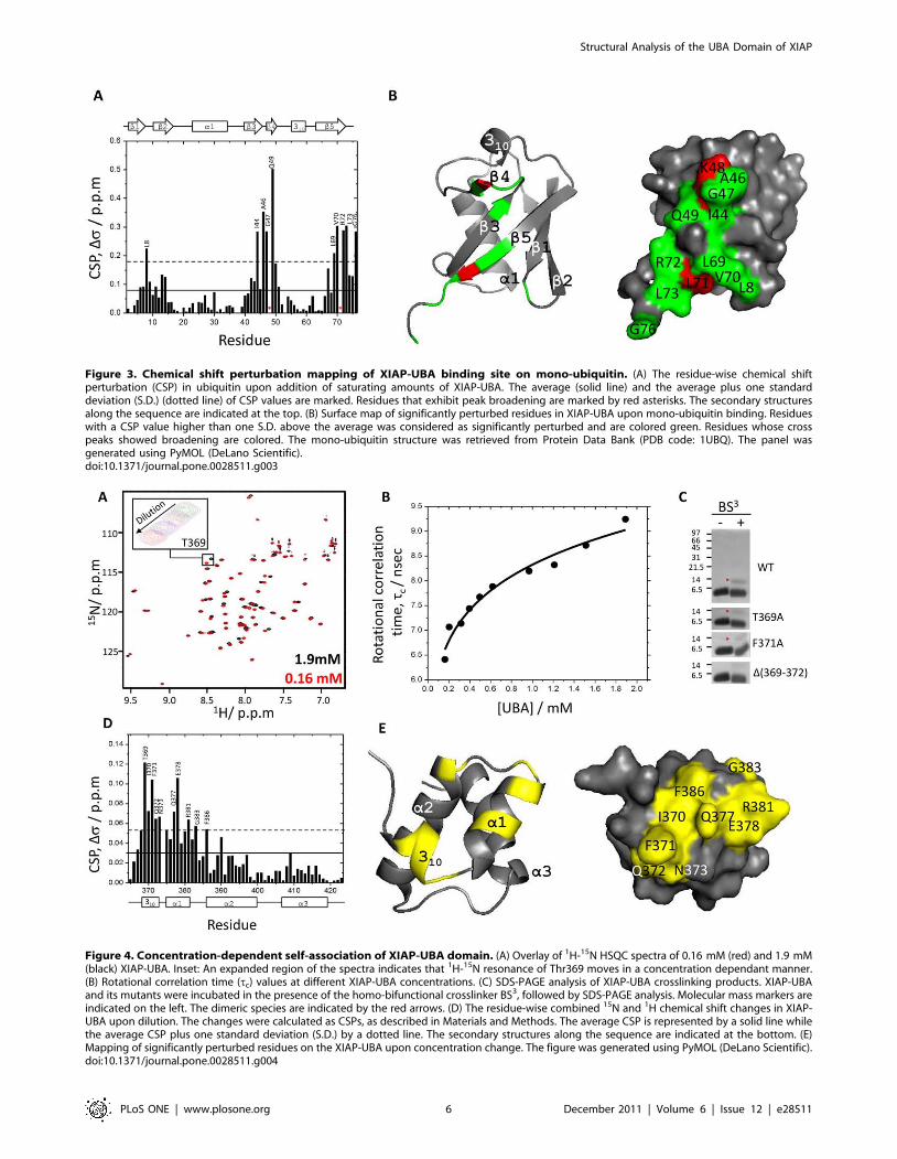
Figure 3. Chemical shift perturbation mapping of XIAP-UBA binding site on mono-ubiquitin. (A) The residue-wise chemical shiftperturbation (CSP) in ubiquitin upon addition of saturating amounts of XIAP-UBA. The average (solid line) and the average plus one standarddeviation (S.D.) (dotted line) of CSP values are marked. Residues that exhibit peak broadening are marked by red asterisks. The secondary structuresalong the sequence are indicated at the top. (B) Surface map of significantly perturbed residues in XIAP-UBA upon mono-ubiquitin binding. Residueswith a CSP value higher than one S.D. above the average was considered as significantly perturbed and are colored green. Residues whose crosspeaks showed broadening are colored. The mono-ubiquitin structure was retrieved from Protein Data Bank (PDB code: 1UBQ). The panel wasgenerated using PyMOL (DeLano Scientific).doi:10.1371/journal.pone.0028511.g003
Figure 4. Concentration-dependent self-association of XIAP-UBA domain. (A) Overlay of 1H-15N HSQC spectra of 0.16 mM (red) and 1.9 mM(black) XIAP-UBA. Inset: An expanded region of the spectra indicates that 1H-15N resonance of Thr369 moves in a concentration dependant manner.(B) Rotational correlation time (tc) values at different XIAP-UBA concentrations. (C) SDS-PAGE analysis of XIAP-UBA crosslinking products. XIAP-UBAand its mutants were incubated in the presence of the homo-bifunctional crosslinker BS3, followed by SDS-PAGE analysis. Molecular mass markers areindicated on the left. The dimeric species are indicated by the red arrows. (D) The residue-wise combined 15N and 1H chemical shift changes in XIAP-UBA upon dilution. The changes were calculated as CSPs, as described in Materials and Methods. The average CSP is represented by a solid line whilethe average CSP plus one standard deviation (S.D.) by a dotted line. The secondary structures along the sequence are indicated at the bottom. (E)Mapping of significantly perturbed residues on the XIAP-UBA upon concentration change. The figure was generated using PyMOL (DeLano Scientific).doi:10.1371/journal.pone.0028511.g004
Structural Analysis of the UBA Domain of XIAP
PLoS ONE | www.plosone.org 6 December 2011 | Volume 6 | Issue 12 | e28511
polar residues (Figure 4E). This oligomerization site is distinct from
the ubiquitin-binding site (Figure 2B). In order to test the
contribution of the 310 helix to the self-association, mutant
XIAP-UBA proteins, namely T369A and F371A, were respec-
tively subjected to BS3 cross-linking as described above. These two
XIAP-UBA mutants were still able to be cross-linked into
homodimers, although the cross-linking efficiency was apparently
weaker than for the wild type (WT) protein. Furthermore, deletion
of the 310 helix in the XIAP-UBA mutant, D (369–372), abolished
dimer formation (Figure 4C). These results indicate that the N-
terminal 310 helix is an important constituent of the surface
responsible for self-association.
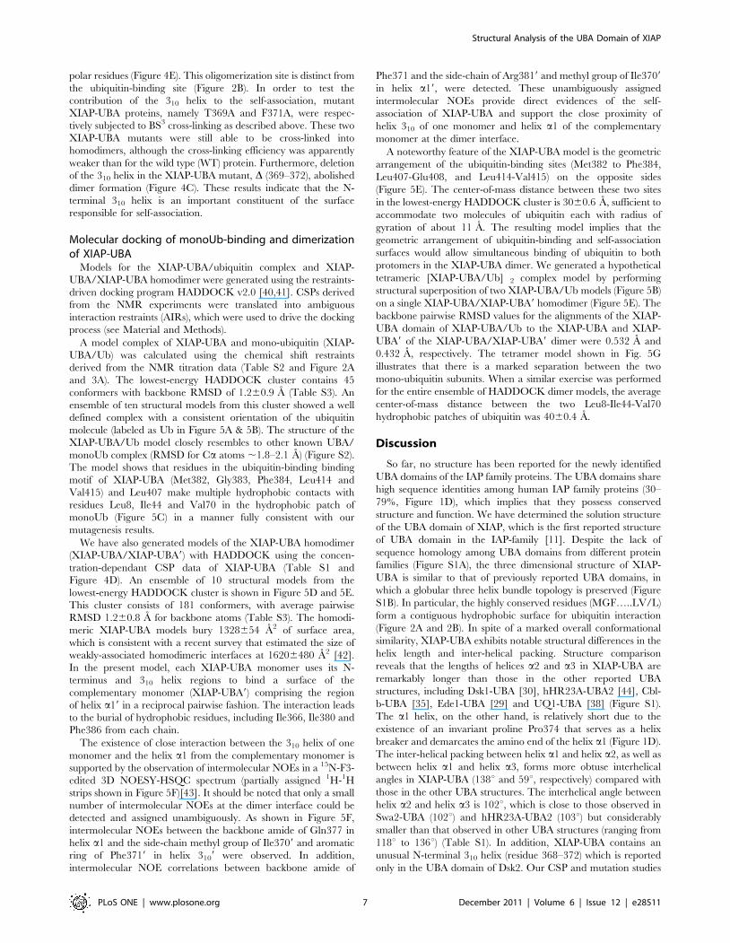
Molecular docking of monoUb-binding and dimerizationof XIAP-UBA
Models for the XIAP-UBA/ubiquitin complex and XIAP-
UBA/XIAP-UBA homodimer were generated using the restraints-
driven docking program HADDOCK v2.0 [40,41]. CSPs derived
from the NMR experiments were translated into ambiguous
interaction restraints (AIRs), which were used to drive the docking
process (see Material and Methods).
A model complex of XIAP-UBA and mono-ubiquitin (XIAP-
UBA/Ub) was calculated using the chemical shift restraints
derived from the NMR titration data (Table S2 and Figure 2A
and 3A). The lowest-energy HADDOCK cluster contains 45
conformers with backbone RMSD of 1.260.9 A (Table S3). An
ensemble of ten structural models from this cluster showed a well
defined complex with a consistent orientation of the ubiquitin
molecule (labeled as Ub in Figure 5A & 5B). The structure of the
XIAP-UBA/Ub model closely resembles to other known UBA/
monoUb complex (RMSD for Ca atoms ,1.8–2.1 A) (Figure S2).
The model shows that residues in the ubiquitin-binding binding
motif of XIAP-UBA (Met382, Gly383, Phe384, Leu414 and
Val415) and Leu407 make multiple hydrophobic contacts with
residues Leu8, Ile44 and Val70 in the hydrophobic patch of
monoUb (Figure 5C) in a manner fully consistent with our
mutagenesis results.
We have also generated models of the XIAP-UBA homodimer
(XIAP-UBA/XIAP-UBA9) with HADDOCK using the concen-
tration-dependant CSP data of XIAP-UBA (Table S1 and
Figure 4D). An ensemble of 10 structural models from the
lowest-energy HADDOCK cluster is shown in Figure 5D and 5E.
This cluster consists of 181 conformers, with average pairwise
RMSD 1.260.8 A for backbone atoms (Table S3). The homodi-
meric XIAP-UBA models bury 1328654 A2 of surface area,
which is consistent with a recent survey that estimated the size of
weakly-associated homodimeric interfaces at 16206480 A2 [42].
In the present model, each XIAP-UBA monomer uses its N-
terminus and 310 helix regions to bind a surface of the
complementary monomer (XIAP-UBA9) comprising the region
of helix a19 in a reciprocal pairwise fashion. The interaction leads
to the burial of hydrophobic residues, including Ile366, Ile380 and
Phe386 from each chain.
The existence of close interaction between the 310 helix of one
monomer and the helix a1 from the complementary monomer is
supported by the observation of intermolecular NOEs in a 15N-F3-
edited 3D NOESY-HSQC spectrum (partially assigned 1H-1H
strips shown in Figure 5F)[43]. It should be noted that only a small
number of intermolecular NOEs at the dimer interface could be
detected and assigned unambiguously. As shown in Figure 5F,
intermolecular NOEs between the backbone amide of Gln377 in
helix a1 and the side-chain methyl group of Ile3709 and aromatic
ring of Phe3719 in helix 3109 were observed. In addition,
intermolecular NOE correlations between backbone amide of
Phe371 and the side-chain of Arg3819 and methyl group of Ile3709
in helix a19, were detected. These unambiguously assigned
intermolecular NOEs provide direct evidences of the self-
association of XIAP-UBA and support the close proximity of
helix 310 of one monomer and helix a1 of the complementary
monomer at the dimer interface.
A noteworthy feature of the XIAP-UBA model is the geometric
arrangement of the ubiquitin-binding sites (Met382 to Phe384,
Leu407-Glu408, and Leu414-Val415) on the opposite sides
(Figure 5E). The center-of-mass distance between these two sites
in the lowest-energy HADDOCK cluster is 3060.6 A, sufficient to
accommodate two molecules of ubiquitin each with radius of
gyration of about 11 A. The resulting model implies that the
geometric arrangement of ubiquitin-binding and self-association
surfaces would allow simultaneous binding of ubiquitin to both
protomers in the XIAP-UBA dimer. We generated a hypothetical
tetrameric [XIAP-UBA/Ub] 2 complex model by performing
structural superposition of two XIAP-UBA/Ub models (Figure 5B)
on a single XIAP-UBA/XIAP-UBA9 homodimer (Figure 5E). The
backbone pairwise RMSD values for the alignments of the XIAP-
UBA domain of XIAP-UBA/Ub to the XIAP-UBA and XIAP-
UBA9 of the XIAP-UBA/XIAP-UBA9 dimer were 0.532 A and
0.432 A, respectively. The tetramer model shown in Fig. 5G
illustrates that there is a marked separation between the two
mono-ubiquitin subunits. When a similar exercise was performed
for the entire ensemble of HADDOCK dimer models, the average
center-of-mass distance between the two Leu8-Ile44-Val70
hydrophobic patches of ubiquitin was 4060.4 A.
Discussion
So far, no structure has been reported for the newly identified
UBA domains of the IAP family proteins. The UBA domains share
high sequence identities among human IAP family proteins (30–
79%, Figure 1D), which implies that they possess conserved
structure and function. We have determined the solution structure
of the UBA domain of XIAP, which is the first reported structure
of UBA domain in the IAP-family [11]. Despite the lack of
sequence homology among UBA domains from different protein
families (Figure S1A), the three dimensional structure of XIAP-
UBA is similar to that of previously reported UBA domains, in
which a globular three helix bundle topology is preserved (Figure
S1B). In particular, the highly conserved residues (MGF…..LV/L)
form a contiguous hydrophobic surface for ubiquitin interaction
(Figure 2A and 2B). In spite of a marked overall conformational
similarity, XIAP-UBA exhibits notable structural differences in the
helix length and inter-helical packing. Structure comparison
reveals that the lengths of helices a2 and a3 in XIAP-UBA are
remarkably longer than those in the other reported UBA
structures, including Dsk1-UBA [30], hHR23A-UBA2 [44], Cbl-
b-UBA [35], Ede1-UBA [29] and UQ1-UBA [38] (Figure S1).
The a1 helix, on the other hand, is relatively short due to the
existence of an invariant proline Pro374 that serves as a helix
breaker and demarcates the amino end of the helix a1 (Figure 1D).
The inter-helical packing between helix a1 and helix a2, as well as
between helix a1 and helix a3, forms more obtuse interhelical
angles in XIAP-UBA (138u and 59u, respectively) compared with
those in the other UBA structures. The interhelical angle between
helix a2 and helix a3 is 102u, which is close to those observed in
Swa2-UBA (102u) and hHR23A-UBA2 (103u) but considerably
smaller than that observed in other UBA structures (ranging from
118u to 136u) (Table S1). In addition, XIAP-UBA contains an
unusual N-terminal 310 helix (residue 368–372) which is reported
only in the UBA domain of Dsk2. Our CSP and mutation studies
Structural Analysis of the UBA Domain of XIAP
PLoS ONE | www.plosone.org 7 December 2011 | Volume 6 | Issue 12 | e28511
suggest that the 310 helix is a crucial constituent of specific self-
association surface of the XIAP-UBA domain.
Using a combination of chemical shift perturbation mapping
and site-directed mutagenesis, we found that XIAP-UBA has two
distinct surfaces available for ubiquitin-binding and self-association
(Figure 2B and 4E). The ubiquitin-binding surface on XIAP-UBA
is formed by the residues, (Met383-Gly383-Phe384 and Leu414-
Val415), which have been collectively termed the ubiquitin-
Figure 5. Models of the XIAP-UBA dimer, XIAP-UBA/Ub complex and [XIAP-UBA/Ub] 2 tetrameric complex. (A) and (D) Ca traces of theensemble of the 10 lowest energy HADDOCK structures of XIAP-UBA/Ub and the XIAP-UBA homodimer. (B) and (E) Ribbon representation of theensemble-averaged HADDOCK structures of XIAP-UBA/Ub and XIAP-UBA dimer. The center-of-mass distance between the ubiquitin-binding sites(cyan) were shown in (E). (C) Contacts at the XIAP-UBA/monoUb interface showing the burial of the key hydrophobic side chains from the ubiquitin-binding site (green) of XIAP-UBA and Leu8-Ile44-Val70-centred hydrophobic patch (blue with yellow dotted spheres) of ubiquitin. Residues andsecondary elements in one half of the XIAP-UBA homodimer are denoted with a prime to distinguish them from another half of the dimer. (F) Partiallyassigned 1H-1H strips for backbone amide of Gln377 and Phe371 of XIAP-UBA showing intermolecular NOEs from the 13C, 15N F1-filtered, 15N-F3-edited 3D NOESY-HSQC spectrum of the half-labeled XIAP-UBA dimer. The position of the diagonal peak is indicated by a red asterisk. (G) Ribbonrepresentation of the aligned models. The center-of-mass distance between the Leu8-Ile44-Val70-centred hydrophobic patches (yellow sphere) isshown. Ubiquitin domains on the XIAP-UBA and XIAP-UBA9 are labeled as Ub and Ub9 respectively. All figures (except (F)) were generated usingPyMOL (DeLano Scientific).doi:10.1371/journal.pone.0028511.g005
Structural Analysis of the UBA Domain of XIAP
PLoS ONE | www.plosone.org 8 December 2011 | Volume 6 | Issue 12 | e28511
binding motif [11]. Our findings are consistent with the typical
ubiquitin-binding patterns observed in the complex structures of
ubiquitin with Dsk1-UBA, UQ1-UBA and Ede1-UBA, in which
the ubiquitin-binding motif has extensive contacts with the Ile44-
center hydrophobic patch on ubiquitin [24,45]. Interestingly, our
CSP data indicated that the Gly76 cross peak of ubiquitin is
significantly perturbed upon XIAP-UBA binding. So far, it has
been reported that the ubiquitin diglycine motif (Gly75–Gly76) is
required for its interaction with zinc-finger ubiquitin binding
domain of USP5, but is not involved in any UBA-ubiquitin
recognition [24,46,47]. Self-association has been previously
reported for a number of UBA and UBA-like domains [32–37].
However, only a few of UBA dimers, such as Cbl-b-UBA [35], c-
Cbl-UBA [34] and p62-UBA [36], have been structurally
characterized. Here, we have delineated the dimerization interface
of XIAP-UBA which is formed by the residues mainly located in
helix 310 and helix a1. This interface is arranged differently from
that of Cbl-b-UBA or c-Cbl-UBA, in which helix a2 and helix a3
were reported to provide the main contribution to the dimeriza-
tion surface. In addition, the self-association and ubiquitin
interfaces of XIAP-UBA are located at two essentially non-
overlapping surfaces, which are also markedly different from that
observed in p62-UBA [36].
Contrary to the previous study which reported that either XIAP
or XIAP-UBA domain was unable to bind monoUb [11], we have
detected a low affinity (Kd = 249619 mM) for this interaction. The
Kd value obtained here is similar to the reported Kd values
for Mud1-UBA (Kd = 350650 mM) [26], hHR23A-UBA2
(Kd = 4006100 mM) [44] and Swa2-UBA (Kd = 535625 mM)
[48]. The ubiquitin-affinity of XIAP-UBA is, however, signifi-
cantly weaker than that of many reported UBAs, including Ede1-
UBA (Kd = 8369 mM)[29], Cbl-b-UBA (Kd = 53.163.1 mM)
[49], UQ1-UBA (Kd = 2466 mM)[38], Dsk2-UBA (Kd = 14.86
5.3 mM)[30] and cIAP-1 (Kd = 5664 mM) [12]. Our result could
explain the previous observation that substitution of XIAP-UBA
with Cbl-b-UBA in full length XIAP increased its potency in
NF-KB activation [11].
Previous studies indicated that UBA domains in the IAP family
exhibit binding selectivity towards polyubiquitin chain linkages
[11,15]. In particular, full length XIAP and cIAP-2 are highly
specific for K63-linked polyubiquitin, but not for K48-linked
polyubiquitin [11]. Linear-linked diubiquitin was shown to be
structurally similar to K63-linked diubiquitin, and XIAP was
reported to interact with both linear-linked and K63-linked
conjugates with a similar binding strength [11,15]. An interesting
aspect of our results is that the isolated XIAP-UBA domain alone
was able to bind K48-linked diubiquitin, and it did not
discriminate between K48-linked (Kd = 5263 mM) and linear-
linked (Kd = 59611 mM) diubiquitin chains. Therefore, our results
are consistent with Blankenship’s report that the isolated UBA
domain of cIAP-1 binds monoUb, K48-linked and K63-linked
polyUb chains with similar affinities in the low-micromolar range
[12]. It should be noted that our K48- and linear-linked
diubiqutin-induced CSP profiles of XIAP-UBA were remarkably
similar to that induced by monoUb. These observations suggested
that monoUb, K48-Ub2 and LL-Ub2 bind to XIAP-UBA in a
similar fashion. Our results imply that the structural features of a
single XIAP-UBA domain are not sufficient to rationalize the
selective recognition of the K63-linked (or linear-linked) poly-
ubiquitin over K48-linked polyubiquitin chain reported for full-
length XIAP.
The origin of polyubiquitin chain linkage selectivity by UBA-
containing proteins remains to be elucidated. It was recently
suggested that the orientations of the UBA domains, rather than
the intrinsic structural properties of a single UBA domain, give rise
to a diverse range of polyubiquitin linkage preference [39]. Here,
we reported that XIAP-UBA has two independent surfaces for
ubiquitin-binding and self-association. Our result suggests that
the self-association of a single XIAP-UBA domain is weak
(Kd,900 mM), but it is well known that XIAP exists as a dimer
in vivo under the strong homodimerization influence of its RING
and BIR1 domains [39,50]. Therefore, it is reasonable to expect
that the homodimerization of adjacent XIAP-RING domains at
the C-terminus of XIAP could bring the two XIAP-UBA domains
into close proximity and promote the formation of the UBA-UBA
homodimer (Figure 6D). This prediction is supported by our
preliminary evidence that the positions of 1H-15N HSQC cross-
peaks corresponding to the residues at the dimerization interface
(T369, F371, Q372, N373, Q377 and E378) were highly
comparable between predominantly dimeric 15N-XIAP-UBA
(1.9 mM) and dimeric 15N-XIAP-UBA-RING double-domain
construct (0.18 mM) but very different from those of a
predominately monomeric 15N-XIAP-UBA sample (0.16 mM)
(Figure S3B). Furthermore, the 15.7 kDa 15N-XIAP-UBA-RING
protein was detected as a dimer (apparent mass = 32.2 kDa) at a
concentration as low as 0.18 mM using size-exclusion chroma-
tography (Methods S1).
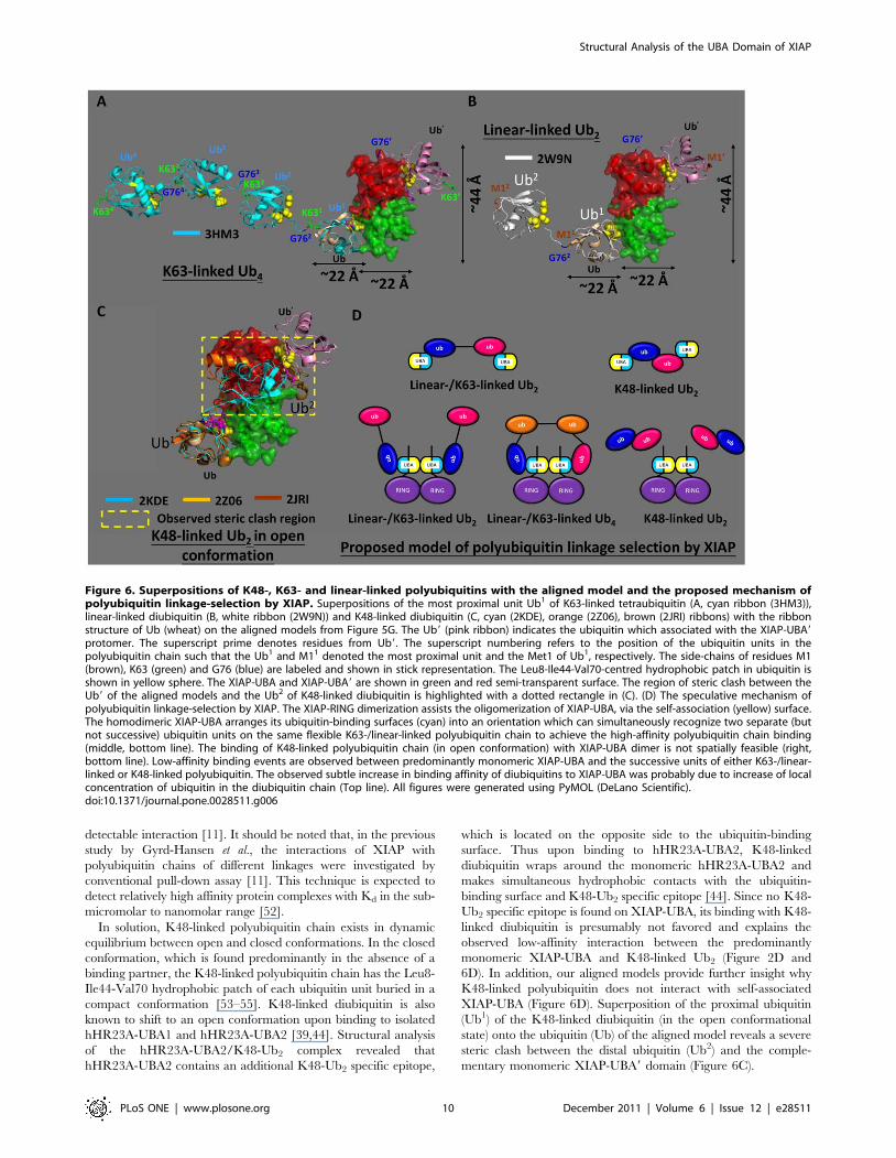
We speculate that the self-association of XIAP-UBA positions
the ubiquitin-binding surfaces in a suitable orientation to
simultaneously recognize two separate ubiquitin units on the same
flexible K63- or linear-linked polyubiquitin chain but to
discriminate against the binding of a K48-linked polyubiquitin
chain (Figure 6D) [36,51]. Our docking model of dimeric XIAP-
UBA provides support for this proposal. The ubiquitin-binding
sites are located on the opposite ends of the bowtie-shaped XIAP-
UBA dimer. As illustrated in the superposed models (Figure 5G),
the geometric arrangement of ubiquitin-binding sites allows
simultaneous binding of two molecules of ubiquitin with a well-
defined orientation to each other (labeled as Ub and Ub9 in
Figure 6A–C). In the absence of binding partners, the K63-/
linear-linked ubiquitin chain exhibits a flexible structure, in which
the individual ubiquitin moieties can be regarded as independent
rotationally unconstrained units. This structural plasticity allows a
high degree of freedom to support simultaneous binding of two
ubiquitin domains within the same polyubiquitin chain to a target
containing two separated ubiquitin binding sites.
A superimposition of the most proximal ubiquitin (Ub1) of the
K63-linked tetraubiquitin onto the ubiquitin (Ub in Figure 6A) of
the aligned models reveals a marked spatial separation (,22 A)
between the Gly76 of the succeeding Ub2 (Gly762) of K63-linked
tetraubiquitin and Gly76 of Ub9 (Gly769) of the aligned models
(Figure 6A). A similar separation has been observed in the
superimposition of linear-linked diubiquitin structure onto the align
models (Figure 6B). These observations imply that the simultaneous
binding between the two successive ubiquitin units of polyubiquitin
with the XIAP-UBA/XIAP-UBA9 appears to be spatially infeasible.
Thus, for the simultaneous interactions of a XIAP-UBA dimer with
two molecules of ubiquitin within the same chain, at least one and
probably two ubiquitins are required as spacers to allow the most
proximal and the distal ubiquitins to wrap around a XIAP-UBA
dimer (molecular dimensions 44622622 A) (Figure 6A and 6D).
This model provides an explanation why the observed binding
mode and affinity of linear-linked diubiquitin with dimeric XIAP-
UBA is comparable to the low-affinity interaction between linear-
linked diubiquitin and predominantly monomeric XIAP-UBA
(Kd,micromolar, Figure 2D and 6D). Our model is further
supported by the fact that dimeric full length XIAP requires a
minimum of four ubiquitin domains (i.e. tetraubiquitin) for a
Structural Analysis of the UBA Domain of XIAP
PLoS ONE | www.plosone.org 9 December 2011 | Volume 6 | Issue 12 | e28511
detectable interaction [11]. It should be noted that, in the previous
study by Gyrd-Hansen et al., the interactions of XIAP with
polyubiquitin chains of different linkages were investigated by
conventional pull-down assay [11]. This technique is expected to
detect relatively high affinity protein complexes with Kd in the sub-
micromolar to nanomolar range [52].
In solution, K48-linked polyubiquitin chain exists in dynamic
equilibrium between open and closed conformations. In the closed
conformation, which is found predominantly in the absence of a
binding partner, the K48-linked polyubiquitin chain has the Leu8-
Ile44-Val70 hydrophobic patch of each ubiquitin unit buried in a
compact conformation [53–55]. K48-linked diubiquitin is also
known to shift to an open conformation upon binding to isolated
hHR23A-UBA1 and hHR23A-UBA2 [39,44]. Structural analysis
of the hHR23A-UBA2/K48-Ub2 complex revealed that
hHR23A-UBA2 contains an additional K48-Ub2 specific epitope,
which is located on the opposite side to the ubiquitin-binding
surface. Thus upon binding to hHR23A-UBA2, K48-linked
diubiquitin wraps around the monomeric hHR23A-UBA2 and
makes simultaneous hydrophobic contacts with the ubiquitin-
binding surface and K48-Ub2 specific epitope [44]. Since no K48-
Ub2 specific epitope is found on XIAP-UBA, its binding with K48-
linked diubiquitin is presumably not favored and explains the
observed low-affinity interaction between the predominantly
monomeric XIAP-UBA and K48-linked Ub2 (Figure 2D and
6D). In addition, our aligned models provide further insight why
K48-linked polyubiquitin does not interact with self-associated
XIAP-UBA (Figure 6D). Superposition of the proximal ubiquitin
(Ub1) of the K48-linked diubiquitin (in the open conformational
state) onto the ubiquitin (Ub) of the aligned model reveals a severe
steric clash between the distal ubiquitin (Ub2) and the comple-
mentary monomeric XIAP-UBA9 domain (Figure 6C).
Figure 6. Superpositions of K48-, K63- and linear-linked polyubiquitins with the aligned model and the proposed mechanism ofpolyubiquitin linkage-selection by XIAP. Superpositions of the most proximal unit Ub1 of K63-linked tetraubiquitin (A, cyan ribbon (3HM3)),linear-linked diubiquitin (B, white ribbon (2W9N)) and K48-linked diubiquitin (C, cyan (2KDE), orange (2Z06), brown (2JRI) ribbons) with the ribbonstructure of Ub (wheat) on the aligned models from Figure 5G. The Ub9 (pink ribbon) indicates the ubiquitin which associated with the XIAP-UBA9protomer. The superscript prime denotes residues from Ub9. The superscript numbering refers to the position of the ubiquitin units in thepolyubiquitin chain such that the Ub1 and M11 denoted the most proximal unit and the Met1 of Ub1, respectively. The side-chains of residues M1(brown), K63 (green) and G76 (blue) are labeled and shown in stick representation. The Leu8-Ile44-Val70-centred hydrophobic patch in ubiquitin isshown in yellow sphere. The XIAP-UBA and XIAP-UBA9 are shown in green and red semi-transparent surface. The region of steric clash between theUb9 of the aligned models and the Ub2 of K48-linked diubiquitin is highlighted with a dotted rectangle in (C). (D) The speculative mechanism ofpolyubiquitin linkage-selection by XIAP. The XIAP-RING dimerization assists the oligomerization of XIAP-UBA, via the self-association (yellow) surface.The homodimeric XIAP-UBA arranges its ubiquitin-binding surfaces (cyan) into an orientation which can simultaneously recognize two separate (butnot successive) ubiquitin units on the same flexible K63-/linear-linked polyubiquitin chain to achieve the high-affinity polyubiquitin chain binding(middle, bottom line). The binding of K48-linked polyubiquitin chain (in open conformation) with XIAP-UBA dimer is not spatially feasible (right,bottom line). Low-affinity binding events are observed between predominantly monomeric XIAP-UBA and the successive units of either K63-/linear-linked or K48-linked polyubiquitin. The observed subtle increase in binding affinity of diubiquitins to XIAP-UBA was probably due to increase of localconcentration of ubiquitin in the diubiquitin chain (Top line). All figures were generated using PyMOL (DeLano Scientific).doi:10.1371/journal.pone.0028511.g006
Structural Analysis of the UBA Domain of XIAP
PLoS ONE | www.plosone.org 10 December 2011 | Volume 6 | Issue 12 | e28511
Materials and Methods
Cloning and sample preparationTwo XIAP UBA protein constructs have been used in this
study. The human XIAP-UBA -containing protein sequence,
corresponding to Glu357-Leu449, was amplified via PCR and
inserted into the pET-H expression vector as previously described
[56]. This His-tagged construct was used primarily for NMR
structure determination. For NMR titration, 15N-relaxation and
protein cross-linking studies, GB1-His-tagged protein construct
was prepared. The DNA sequence encoding the XIAP-UBA
(Arg365-Gln423) was subcloned into pGB1-HIS bacterial expres-
sion vector. This construct encodes N-terminal GB1 and polyHis
tags, which are separated from XIAP-UBA by a thrombin
cleavage sequence (LVPRG). All XIAP-UBA mutants were
prepared by site-directed mutagenesis of the wild-type GB1-His
tagged expression construct using a QuickChange Site-Directed
Mutagenesis Kit (Stratagene). All the DNA constructs were
transformed in E. coli host BL21 (DE3) (Novagen) for recombinant
protein expression. Unlabeled protein was prepared from cells
grown in Luria-Bertani (LB) media. Uniformly labeled 15N or15N/13C-labeled proteins were prepared from cells grown in M9
minimal media incorporating [U-13C]-glucose (Cambridge Iso-
topes Laboratories, CIL) and/or [U-15N]-ammonium chloride
(CIL) as the sole carbon or nitrogen sources. The expression and
purification of XIAP-UBA was essentially the same as described
for that of XIAP-UBA-containing protein [56]. Unlabeled and
uniformly 15N and 15N/13C-labeled samples of human ubiquitin
were produced as described previously [49]. Unlabeled K48-
linked and linear-linked diubiquitins were synthesized and
prepared as described by Reyes-Turcu et al [57]. All protein
samples were prepared in BisTris buffer (20 mM BisTris-HCl,
pH 6.7, 150 mM NaCl, 5 mM d10-DTT, 1 mM PMSF, 90%
H2O/10% D2O) for all NMR studies.
NMR spectroscopy and structure calculationNMR studies were performed at 298 K using a Bruker Advance
600 MHz or 700 MHz NMR spectrometer equipped with a TCI
cyroprobe. NMR data were processed with TOPSPIN software
(Bruker) and analyzed by SPARKY [58]. The backbone and side-
chain resonance assignments of a 0.5 mM XIAP-UBA-containing
protein (residues 357–449) were obtained using standard1H/13C/15N heteronuclear NMR experiments as previously
described [56]. The assignments were deposited in the Biological
Magnetic Resonance Data Bank under accession number 16478
[56].
The solution structure of XIAP-UBA-containing protein
(residues 357–449) was iteratively calculated using the CYANA
protocols based on the NOE-derived inter-proton distance,
backbone dihedral angle and hydrogen bonding constraints [59].
The inter-proton distance restraints were derived from 3D 15N-
edited NOESY (mixing time = 120 ms) and 3D 13C-edited
NOESY (mixing time = 120 ms) spectra, which were recorded
separately on 0.45 mM 15N-labeled and 0.5 mM 15N/13C-
enriched XIAP-UBA protein samples respectively. Cross-peaks
in NOESY-type spectra were interactively assigned and integrated
in XEASY. Backbone dihedral angle restraints were derived from
chemical shift using TALOS [60]. Hydrogen bonding restraints in
helical segment were determined on the basis of previously
reported chemical shift index (CSI) values, dihedral angle values
and characteristic NOE patterns. Out of 200 calculated conform-
ers, the final coordinates for an ensemble of 15 lowest energy
models were deposited into Protein Data Bank (PDB ID 2 kna),
and they were used for subsequent structural analysis. Structure
properties were analyzed using PROCHECK [61]. Interhelical
angles were calculated in MOLMOL [62]. Structural similarity
searches were performed with DALI using default settings [63]. All
molecular graphic images were generated by using PyMOL(De-
Lano Scientific) [64].
NMR signal assignments of ubiquitin were taken from literature
data [65].
The 15N relaxation experiments were performed using standard
Bruker pseudo-2D 15N-edited T1 and T2 gradient pulse programs,
hsqct1etf3gpsi3d and hsqct2etf3gpsi3d, respectively. Peaks in the
amide proton region between 7.9–9.5 ppm were chosen for
integration and analysis. Longitudinal and transverse relaxation
times, T1 and T2, were obtained using the relaxation module of
Topspin 2.1 (Bruker) as described in the user manual.
For the detection of intermolecular NOEs at the dimeric
interface of XIAP-UBA, a mixed sample containing 1 mM13C/15N-XIAP-UBA and 1 mM unlabeled XIAP-UBA was
prepared in BisTris buffer. Intermolecular NOEs were recorded
with a 13C, 15N F1-filtered, 15N-F3-edited 3D NOESY-HSQC
experiment (mixing time = 200 ms) and 13C, 15N F1-filtered, 13C-
F1-edited 3D NOESY-HSQC experiment (mixing time = 150 ms)
as described previously [43].
NMR titration studies of binding between UBA andmono-ubiquitin and diubiquitins
The mono-ubiquitin/diubiquitin binding surfaces on XIAP-
UBA were mapped using the CSP approach. A series of 2D1H-15N HSQC spectra of a 15N-labeled XIAP-UBA (0.5 mM)
were recorded as a function of the increasing amount of unlabeled
mono-ubiquitin, K48-linked diubiquitin and linear-linked diubi-
quitin, respectively. In general, 5 mM and 2.5 mM stock solutions
of unlabeled mono-ubiquitin and diubiquitin were prepared,
respectively. The NMR titration experiments were performed
until the molar ratios of [monoUb]/[XIAP-UBA], [K48-Ub2]/
[XIAP-UBA] and [LL-Ub2]/[XIAP-UBA] reached the values of
5.5, 2.5 and 2.5, respectively. In the final titration point, the final
concentration of 15N-labled XIAP-UBA was around 0.3 mM, in
which predominant monomeric XIAP-UBA was available for
ubiquitin interaction. Binding was monitored through the changes
in the cross-peaks positions of the 1H-15N HSQC spectra. These
changes of cross-peak chemical shifts were quantified using
combined amide CSP calculated as Ds= [(DsH)2+(DsN/5)2]1/2,
where DsH and DsN are the observed chemical shift changes for1H and 15N dimensions, respectively. The binding affinities
(dissociation constant, Kd) between XIAP-UBA and ubiquitin/
diubiquitin were quantified by fitting the CSPs as a function on the
protein and unlabeled ligand concentrations to the appropriate
stoichiometry/binding models as described [66] using ORIGIN
7.0 software (Origin Lab). To map the XIAP-UBA binding surface
on mono-ubiquitin, a similar titration experiment was performed
by adding unlabeled XIAP-UBA (2 mM stock solution) to 15N-
labled mono-ubiquitin until the molar ratio of [XIAP -UBA]/
[monoUb] was equal to 5.
The CSP approach was used to investigate ubiquitin-binding
activity in wild type (WT) XIAP-UBA and XIAP-UBA mutants.15N-labled monoUb was titrated with unlabeled XIAP-UBA,
including WT or mutants, up to a [XIAP-UBA]: [monoUb] molar
ratio of 5 as described above. At the final titration point, the
average CSP value (AvgDs) of 12 well-resolved resonances in the1H-15N HSQC spectrum of ubiquitin was calculated. The
normalized percentage ubiquitin activity of each XIAP-UBA
mutants was calculated as 1006[Avg(Dsmutant)/Avg(Dswild-type)],
where Avg(Dsmutant) and Avg(Dswild-type) are the average CSP
Structural Analysis of the UBA Domain of XIAP
PLoS ONE | www.plosone.org 11 December 2011 | Volume 6 | Issue 12 | e28511

values obtained using XIAP-UBA and the corresponding XIAP-
UBA mutant, respectively.
Rotational correlation time measurementThe rotational correlation time (tc) was determined to study the
concentration-dependent oligomerization of XIAP-UBA. Dilution
experiments were carried out over a range of XIAP-UBA
concentration (0.16–1.9 mM). The 2D 1H-15N HSQC spectrum
and the relaxation parameters (T1 and T2) were collected as
described above. The (tc) was calculated as tc = ((6T1/T2)-7)1/2/
4pvN, where vN is the 15N resonance frequency (in Hz) [67]. A
calibration plot (tc versus molecular weight) was generated using a
series of in-house 15N-labeled standard monomeric proteins of
known molecular weight (3–20 kDa).
The determination of dimer dissociation constant for XIAP-
UBA was the same as described previously [35], except that non-
linear regression analysis was performed for a plot of rotational
correlation time as a function of protein concentration.
Chemical cross-linkingProteins were extensively dialyzed into 10 mM HEPES, pH 7.5
prior to cross-linking experiments using bifunctional crosslinker Bis
(sulfosuccinimidyl) suberate (BS3) (Thermo scientific pierce). The
XIAP-UBA wild type and mutant proteins (1 mg/ml) were treated
with BS3 (500 mM) for 60 min at room temperature. The reactions
were terminated by the addition of Tris salt (100 mM). Cross-
linked products were separated by SDS-PAGE, followed by
Coomassie blue staining.
HADDOCK docking and analysisThe models of the XIAP-UBA/Ub complex and XIAP-UBA/
XIAP-UBA9 homodimeric complex were calculated by using the
HADDOCK web server (http://haddock.chem.uu.nl/services/
HADDOCK) [40,41]. The HADDOCK calculations of XIAP-
UBA/Ub complex were started with the coordinates of human
ubiquitin (PDB code: 1UBQ) and the averaged structure of the
XIAP UBA domain (residues 365–423, PDB code: 2KNA). The
starting structures for the docking calculation of XIAP-UBA/
XIAP-UBA9 complex were two identical coordinates of the XIAP
UBA domain (residues 365–423, PDB code: 2KNA).
The two docking procedures were driven by using ambiguous
interaction restraints (AIRs), which were defined according to the
CSP data obtained from NMR titration experiments of XIAP-
UBA in complex with monoUb and from the dilution experiment
of XIAP-UBA, respectively. Residues undergoing significant
chemical shift perturbations (Ds.1 S.D. above the mean value)
were defined as active residues. Residues having a CSP value
higher than the mean value but within 1 S.D. of the mean value
were selected as a passive residue. The active and passive residues
defined for the present calculations are summarized in Table S2.
All other parameters were kept at the default settings.
The HADDOCK docking protocols principally consist of three
stages, including a rigid-body energy minimization, a simulated
annealing in torsion angle space allowing semi-flexibility, and an
explicit water refinement. The web server returned 200 models,
which were clustered according to the pair-wise RMSD matrix
using a 5.0 A cut-off. Ten different clusters have been obtained in
the XIAP-UBA/Ub complex, while three different clusters have
been identified in the homodimeric XIAP-UBA/XIAP-UBA9
models. These clusters were ranked according to their HAD-
DOCK scores, which were defined as a weighted sum of van der
Waals, electrostatic, solvation and restraint violation energy terms.
The structural statistics of the top three clusters are shown in Table
S3. In both docking runs, cluster 1 has the best average
HADDOCK score. The 10 lowest energy structures from this
cluster were selected as representative models of each docking
complex. The aligned models were constructed as described in the
text by using the structural alignment function of graphic program
PyMOL (DeLano Scientific). The center-of-mass distance and
molecular dimension (in terms of radius of gyration) of the
representing model were calculated with PyMOL (DeLano
Scientific). All structure superimpositions were performed using
PyMOL (DeLano Scientific).
Supporting Information
Figure S1 Sequence alignment and superimposition ofvarious UBA domains from different proteins. (A)
Sequence alignment of various UBA fold domains. The relative
locations of secondary structure elements are boxed. The species
of the primary sequences were as followed: Sc, S. cerevisiae (budding
yeast); Sp, S. pombe (fission yeast); Hs, H. sapiens (human). The
multiple sequence alignments were generated using ClustalW2.
Summary of the output from the DALI server is shown on the
right of the alignments. RMSD: Root-Mean-Square-Deviation for
the structural alignment between the structures of corresponding
UBA-fold protein and XIAP-UBA. Seq: Sequence similarity. (B)
Superimposition Ca traces of XIAP-UBA with various UBA
domains. Only the structures with a DALI Z-score .3.0 are
selected for displayed. The color representation of each structure is
indicated at the bottom. The PDB codes for structural alignment
are as followed: Dsk2-UBA, 1WR1 (only structure available is in
complex with ubiquitin); hHR23A-UBA2, 1DV0; UQ1-UBA,
2YJ5; Ede1-UBA, 2G3Q; Cbl-b-UBA, 2JNH. Structural align-
ment was performed using DALI server, and the image was
created using PyMOL (DeLano Scientific).
(TIFF)
Figure S2 Structural comparison among various UBA/monoUb complexes. (A) The XIAP-UBA/Ub complex model
obtained in docking study is aligned with the published solution
structures of the ubiquitin complexes with the UBA domain of (B)
UQ1 (PDB code: 2JY6), (C) Dsk2 (PDB code: 1WR1), and (D)
Ede1 (PDB ID.: 2G3Q). The ubiquitin molecules are placed on
the right side, the a-helixes and b-strands were colored in red/
yellow and cyan, respectively; The UBA domains are placed on
the left side, the 310/a0, a1, a2 and a3 helixes are colored in red,
green, blue and magenta, respectively. The image was created by
MOLMOL (version 2K.1 by Reto Koradi).
(TIFF)
Figure S3 Evidence of XIAP-RING-assisted homodimer-ization of XIAP-UBA as revealed by 1H-15N HSQCspectrum. (A) Schematic representation of XIAP showing the
protein domains used. (B) Overlay plots of 1H-15N HSQC spectra
for the diluted sample of XIAP-UBA-RING (blue), the concen-
trated sample of XIAP-UBA (green) and the diluted sample of
XIAP-UBA (red). For clarity, only the regions showing the
resonances of dimerization interfacial residues are shown. Sample
preparation and analytical size exclusion chromatography of
XIAP-UBA-RING were described in Methods S1.
(TIFF)
Table S1 Interhelical angles of various UBA domains.#The abbreviations of protein followed the description in the
figure 2. The PDB code of each structure was followed: XIAP-
UBA, 2KNA; UQ1-UBA, 2YJ5; hHR23A-UBA1, 1IFY;
hHR23A-UBA2, 1DV0; Dsk2-UBA, 1WR1 (only structure
available is in complex with ubiquitin); Ede1-UBA, 2G3Q;
Mud1-UBA, 1Z96; Swa2-UBA, 1PGY; Cbl-b-UBA, 2JNH. $The
Structural Analysis of the UBA Domain of XIAP
PLoS ONE | www.plosone.org 12 December 2011 | Volume 6 | Issue 12 | e28511
inter-helical angles were measured by the software MOLMOL
(version 2K.1 by Reto Koradi) with the standard procedures in the
manual. Firstly, the backbone atoms of a helix were selected, and a
primitive of cylinder was added for the helix in spacing method by
macro ‘‘AddCylinder spacing’’. After cylinder was added for every
helix, the cylinders (instead of the helices) were selected and the
angles between the helix axes were calculated by the macro
‘‘CalcHelix’’.
(DOC)
Table S2 HADDOCK active and passive residue forconstruction of XIAP/Ub complex and XIAP-UBA/XIAP-UBA9complex.
(DOC)
Table S3 Summary of results from the top threeclusters of docking result for the model complexes ofXIAP-UBA/Ub and XIAP-UBA/XIAP-UBA9. # Root-Mean-
Square-Deviation from the overall lowest energy structure.
(DOC)
Methods S1 Sample preparation of XIAP-UBA-RINGand analytical size exclusion chromatography.(DOCX)
Author Contributions
Conceived and designed the experiments: MKT SKH KHS BCYW.
Performed the experiments: MKT SKH KHS. Analyzed the data: MKT
SKH YY. Contributed reagents/materials/analysis tools: S-TY BZ BCYW
H-YH. Wrote the paper: MKT SKH KHS.
References
1. Deveraux QL, Reed JC (1999) IAP family proteins–suppressors of apoptosis.Genes Dev 13: 239–252.
2. Salvesen GS, Abrams JM (2004) Caspase activation - stepping on the gas or
releasing the brakes? Lessons from humans and flies. Oncogene 23: 2774–2784.
3. Vucic D (2008) Targeting IAP (inhibitor of apoptosis) proteins for therapeuticintervention in tumors. Curr Cancer Drug Targets 8: 110–117.
4. Hunter AM, LaCasse EC, Korneluk RG (2007) The inhibitors of apoptosis
(IAPs) as cancer targets. Apoptosis 12: 1543–1568.
5. Schimmer AD, Dalili S, Batey RA, Riedl SJ (2006) Targeting XIAP for the
treatment of malignancy. Cell Death Differ 13: 179–188.
6. Takahashi R, Deveraux Q, Tamm I, Welsh K, Assa-Munt N, et al. (1998) Asingle BIR domain of XIAP sufficient for inhibiting caspases. J Biol Chem 273:
7787–7790.
7. Sun C, Cai M, Meadows RP, Xu N, Gunasekera AH, et al. (2000) NMRstructure and mutagenesis of the third Bir domain of the inhibitor of apoptosis
protein XIAP. J Biol Chem 275: 33777–33781.
8. Lin SC, Huang Y, Lo YC, Lu M, Wu H (2007) Crystal structure of the BIR1
domain of XIAP in two crystal forms. J Mol Biol 372: 847–854.
9. Galban S, Duckett CS (2010) XIAP as a ubiquitin ligase in cellular signaling.Cell Death Differ 17: 54–60.
10. Srinivasula SM, Ashwell JD (2008) IAPs: what’s in a name? Mol Cell 30:
123–135.
11. Gyrd-Hansen M, Darding M, Miasari M, Santoro MM, Zender L, et al. (2008)IAPs contain an evolutionarily conserved ubiquitin-binding domain that
regulates NF-kappaB as well as cell survival and oncogenesis. Nat Cell Biol
10: 1309–1317.
12. Blankenship JW, Varfolomeev E, Goncharov T, Fedorova AV, Kirkpatrick DS,et al. (2009) Ubiquitin binding modulates IAP antagonist-stimulated proteasomal
degradation of c-IAP1 and c-IAP2(1). Biochem J 417: 149–160.
13. Cook WJ, Jeffrey LC, Sullivan ML, Vierstra RD (1992) Three-dimensionalstructure of a ubiquitin-conjugating enzyme (E2). J Biol Chem 267:
15116–15121.
14. Varadan R, Assfalg M, Haririnia A, Raasi S, Pickart C, et al. (2004) Solution
conformation of Lys63-linked di-ubiquitin chain provides clues to functionaldiversity of polyubiquitin signaling. J Biol Chem 279: 7055–7063.
15. Komander D, Reyes-Turcu F, Licchesi JD, Odenwaelder P, Wilkinson KD,
et al. (2009) Molecular discrimination of structurally equivalent Lys 63-linkedand linear polyubiquitin chains. EMBO Rep 10: 466–473.
16. Kirisako T, Kamei K, Murata S, Kato M, Fukumoto H, et al. (2006) A ubiquitin
ligase complex assembles linear polyubiquitin chains. EMBO J 25: 4877–4887.
17. Iwai K, Tokunaga F (2009) Linear polyubiquitination: a new regulator of NF-
kappaB activation. EMBO Rep 10: 706–713.
18. Kirkin V, Dikic I (2007) Role of ubiquitin- and Ubl-binding proteins in cellsignaling. Curr Opin Cell Biol 19: 199–205.
19. Chang YG, Song AX, Gao YG, Shi YH, Lin XJ, et al. (2006) Solution structure
of the ubiquitin-associated domain of human BMSC-UbP and its complex withubiquitin. Protein Sci 15: 1248–1259.
20. Chim N, Gall WE, Xiao J, Harris MP, Graham TR, et al. (2004) Solution
structure of the ubiquitin-binding domain in Swa2p from Saccharomyces
cerevisiae. Proteins 54: 784–793.
21. Ciani B, Layfield R, Cavey JR, Sheppard PW, Searle MS (2003) Structure of theubiquitin-associated domain of p62 (SQSTM1) and implications for mutations
that cause Paget’s disease of bone. J Biol Chem 278: 37409–37412.
22. Dieckmann T, Withers-Ward ES, Jarosinski MA, Liu CF, Chen IS, et al. (1998)Structure of a human DNA repair protein UBA domain that interacts with HIV-
1 Vpr. Nat Struct Biol 5: 1042–1047.
23. Lowe ED, Hasan N, Trempe JF, Fonso L, Noble ME, et al. (2006) Structures of
the Dsk2 UBL and UBA domains and their complex. Acta Crystallogr D BiolCrystallogr 62: 177–188.
24. Mueller TD, Feigon J (2002) Solution structures of UBA domains reveal a
conserved hydrophobic surface for protein-protein interactions. J Mol Biol 319:1243–1255.
25. Panneerselvam S, Marx A, Mandelkow EM, Mandelkow E (2006) Structure of
the catalytic and ubiquitin-associated domains of the protein kinase MARK/
Par-1. Structure 14: 173–183.
26. Trempe JF, Brown NR, Lowe ED, Gordon C, Campbell ID, et al. (2005)
Mechanism of Lys48-linked polyubiquitin chain recognition by the Mud1 UBA
domain. EMBO J 24: 3178–3189.
27. Withers-Ward ES, Mueller TD, Chen IS, Feigon J (2000) Biochemical and
structural analysis of the interaction between the UBA(2) domain of the DNA
repair protein HHR23A and HIV-1 Vpr. Biochemistry 39: 14103–14112.
28. Yuan X, Simpson P, McKeown C, Kondo H, Uchiyama K, et al. (2004)
Structure, dynamics and interactions of p47, a major adaptor of the AAA
ATPase, p97. EMBO J 23: 1463–1473.
29. Swanson KA, Hicke L, Radhakrishnan I (2006) Structural basis for
monoubiquitin recognition by the Ede1 UBA domain. J Mol Biol 358: 713–724.
30. Ohno A, Jee J, Fujiwara K, Tenno T, Goda N, et al. (2005) Structure of the
UBA domain of Dsk2p in complex with ubiquitin molecular determinants for
ubiquitin recognition. Structure 13: 521–532.
31. Raasi S, Varadan R, Fushman D, Pickart CM (2005) Diverse polyubiquitin
interaction properties of ubiquitin-associated domains. Nat Struct Mol Biol 12:
708–714.
32. Bayrer JR, Zhang W, Weiss MA (2005) Dimerization of doublesex is mediated
by a cryptic ubiquitin-associated domain fold: implications for sex-specific gene
regulation. J Biol Chem 280: 32989–32996.
33. Bertolaet BL, Clarke DJ, Wolff M, Watson MH, Henze M, et al. (2001) UBA
domains mediate protein-protein interactions between two DNA damage-
inducible proteins. J Mol Biol 313: 955–963.
34. Kozlov G, Peschard P, Zimmerman B, Lin T, Moldoveanu T, et al. (2007)
Structural basis for UBA-mediated dimerization of c-Cbl ubiquitin ligase. J Biol
Chem 282: 27547–27555.
35. Peschard P, Kozlov G, Lin T, Mirza IA, Berghuis AM, et al. (2007) Structural
basis for ubiquitin-mediated dimerization and activation of the ubiquitin protein
ligase Cbl-b. Mol Cell 27: 474–485.
36. Long J, Garner TP, Pandya MJ, Craven CJ, Chen P, et al. (2010) Dimerisation
of the UBA domain of p62 inhibits ubiquitin binding and regulates NF-kappaB
signalling. J Mol Biol 396: 178–194.
37. Sasaki T, Funakoshi M, Endicott JA, Kobayashi H (2005) Budding yeast Dsk2
protein forms a homodimer via its C-terminal UBA domain. Biochem Biophys
Res Commun 336: 530–535.
38. Zhang D, Raasi S, Fushman D (2008) Affinity makes the difference: nonselective
interaction of the UBA domain of Ubiquilin-1 with monomeric ubiquitin and
polyubiquitin chains. J Mol Biol 377: 162–180.
39. Sims JJ, Haririnia A, Dickinson BC, Fushman D, Cohen RE (2009) Avid
interactions underlie the Lys63-linked polyubiquitin binding specificities
observed for UBA domains. Nat Struct Mol Biol 16: 883–889.
40. Dominguez C, Boelens R, Bonvin AM (2003) HADDOCK: a protein-protein
docking approach based on biochemical or biophysical information. J Am Chem
Soc 125: 1731–1737.
41. de Vries SJ, van Dijk AD, Krzeminski M, van Dijk M, Thureau A, et al. (2007)
HADDOCK versus HADDOCK: new features and performance of HAD-
DOCK2.0 on the CAPRI targets. Proteins 69: 726–733.
42. Dey S, Pal A, Chakrabarti P, Janin J (2010) The subunit interfaces of weakly
associated homodimeric proteins. J Mol Biol 398: 146–160.
43. Ogura K, Terasawa H, Inagaki F (1996) An improved double-tuned and
isotope-filtered pulse scheme based on a pulsed field gradient and a wide-band
inversion shaped pulse. J Biomol NMR 8: 492–498.
44. Varadan R, Assfalg M, Raasi S, Pickart C, Fushman D (2005) Structural
determinants for selective recognition of a Lys48-linked polyubiquitin chain by a
UBA domain. Mol Cell 18: 687–698.
45. Winget JM, Mayor T (2010) The diversity of ubiquitin recognition: hot spots and
varied specificity. Mol Cell 38: 627–635.
Structural Analysis of the UBA Domain of XIAP
PLoS ONE | www.plosone.org 13 December 2011 | Volume 6 | Issue 12 | e28511
46. Reyes-Turcu FE, Horton JR, Mullally JE, Heroux A, Cheng X, et al. (2006) The
ubiquitin binding domain ZnF UBP recognizes the C-terminal diglycine motif ofunanchored ubiquitin. Cell 124: 1197–1208.
47. Mueller TD, Kamionka M, Feigon J (2004) Specificity of the interaction between
ubiquitin-associated domains and ubiquitin. J Biol Chem 279: 11926–11936.48. Matta-Camacho E, Kozlov G, Trempe JF, Gehring K (2009) Atypical binding of
the Swa2p UBA domain to ubiquitin. J Mol Biol 386: 569–577.49. Zhou ZR, Gao HC, Zhou CJ, Chang YG, Hong J, et al. (2008) Differential
ubiquitin binding of the UBA domains from human c-Cbl and Cbl-b: NMR
structural and biochemical insights. Protein Sci 17: 1805–1814.50. Mace PD, Linke K, Feltham R, Schumacher FR, Smith CA, et al. (2008) Structures
of the cIAP2 RING domain reveal conformational changes associated withubiquitin-conjugating enzyme (E2) recruitment. J Biol Chem 283: 31633–31640.
51. Weeks SD, Grasty KC, Hernandez-Cuebas L, Loll PJ (2009) Crystal structuresof Lys-63-linked tri- and di-ubiquitin reveal a highly extended chain
architecture. Proteins 77: 753–759.
52. Charbonnier S, Gallego O, Gavin A-C (2008) The social network of a cell:Recent advances in interactome mapping. In: El-Gewely MR, ed. Biotechnology
Annual Review: Elsevier. pp 1–28.53. Varadan R, Walker O, Pickart C, Fushman D (2002) Structural properties of
polyubiquitin chains in solution. J Mol Biol 324: 637–647.
54. Ryabov Y, Fushman D (2006) Interdomain mobility in di-ubiquitin revealed byNMR. Proteins 63: 787–796.
55. Eddins MJ, Varadan R, Fushman D, Pickart CM, Wolberger C (2007) Crystalstructure and solution NMR studies of Lys48-linked tetraubiquitin at neutral
pH. J Mol Biol 367: 204–211.56. Hui SK, Tse MK, Yang Y, Wong BC, Sze KH (2010) Backbone and side-chain
1H, 13C and 15N assignments of the ubiquitin-associated domain of human X-
linked inhibitor of apoptosis protein. Biomol NMR Assign 4: 13–15.
57. Reyes-Turcu FE, Shanks JR, Komander D, Wilkinson KD (2008) Recognition
of polyubiquitin isoforms by the multiple ubiquitin binding modules of
isopeptidase T. J Biol Chem 283: 19581–19592.
58. Goddard TD, Kneller DG (1998) SPARKY 3.0. University of California, San
Francisco.
59. Guntert P (2004) Automated NMR structure calculation with CYANA. Methods
Mol Biol 278: 353–378.
60. Cornilescu G, Delaglio F, Bax A (1999) Protein backbone angle restraints from
searching a database for chemical shift and sequence homology. J Biomol NMR
13: 289–302.
61. Morris AL, MacArthur MW, Hutchinson EG, Thornton JM (1992) Stereo-
chemical quality of protein structure coordinates. Proteins 12: 345–364.
62. Koradi R, Billeter M, Wuthrich K (1996) MOLMOL: a program for display and
analysis of macromolecular structures. J Mol Graph 14: 51–55, 29–32.
63. Holm L, Kaariainen S, Rosenstrom P, Schenkel A (2008) Searching protein
structure databases with DaliLite v.3. Bioinformatics 24: 2780–2781.
64. DeLano WL (2002) Pymol. DeLano Scientific, South San Francisco, CA.
65. Jaravine VA, Zhuravleva AV, Permi P, Ibraghimov I, Orekhov VY (2008)
Hyperdimensional NMR spectroscopy with nonlinear sampling. J Am Chem
Soc 130: 3927–3936.
66. French M, Swanson K, Shih SC, Radhakrishnan I, Hicke L (2005) Identification
and Characterization of Modular Domains That Bind Ubiquitin. In:
Raymond JD, ed. Methods in Enzymology: Academic Press. pp 135–157.
67. Kay LE, Torchia DA, Bax A (1989) Backbone dynamics of proteins as studied by
15N inverse detected heteronuclear NMR spectroscopy: application to
staphylococcal nuclease. Biochemistry 28: 8972–8979.
Structural Analysis of the UBA Domain of XIAP
PLoS ONE | www.plosone.org 14 December 2011 | Volume 6 | Issue 12 | e28511
Copyright © 2022 FDOKUMEN