Numerical simulation of roof cavings in several ... - CDC stacks
Upload
hms-harvardCategory
view
1download
0
Stimulation through the T cell receptor leads to interactions between SHBand several signaling proteins
Michael Welsh1, Zhou Songyang5, J Daniel Frantz2,4, Thomas TruÈ b2,4, Kris A Reedquist3,4,TorbjoÈ rn Karlsson1, Masaya Miyazaki2,4, Lewis C Cantley5, Hamid Band3,4 andSteven E Shoelson2,4
1Department of Medical Cell Biology, Uppsala University, Uppsala, Sweden; 2Research Division, Joslin Diabetes Center, Boston,Massachusetts, USA; 3Lymphocyte Biology Section, Division of Rheumatology and Immunology, and 4Department of Medicine,Brigham and Women's Hospital and Harvard Medical School, Boston, Massachusetts, USA; 5Division of Signal Transduction,Beth Israel Hospital, Harvard Medical School, Boston, Massachusetts, USA
Shb is a recently described Src homology 2 (SH2)domain-containing adaptor protein. Here we show thatShb is expressed in lymphoid tissues, and is recruitedinto signaling complexes upon activation of Jurkat Tcells. Grb2 binds proline-rich motifs in Shb via its SH3domains. As a result, a number of proteins detected inanti-Shb and anti-Grb2 immunoprecipitates are shared,including phosphoproteins of 22, 36/38, 55/57 and70 kDa. Shb-association with p22, which represents theT cell receptor associated z chain, occurs through theShb SH2 domain. The central region of Shb binds p36/38. Since this interaction was inhibited by phosphotyr-osine, this region of Shb is likely to contain a non-SH2PTB (phosphotyrosine binding) domain. The Shb PTBdomain was found to preferentially bind the sequenceAsp-Asp-X-pTyr when incubated with a phosphopeptidelibrary. A peptide corresponding to a phosphorylationsite in 34 kDa Lnk inhibited association between Shband p36/38. Overexpression of Shb in Jurkat cells led toincreased basal phosphorylation of Shb-associated p36/38 and p70 proteins. Inactivation of the Shb SH2 domainby an R522K mutation resulted in a reduced stimulationof tyrosine phosphorylation of several proteins inresponse to CD3 crosslinking when expressed in Jurkatcells. Together, our results show three distinct domainsof Shb all participate in the formulation of multimericsignaling complexes in activated T cells. These resultsindicate that the Shb protein functions in T cell receptorsignaling.
Keywords: Shb; Jurkat T cells; T cell receptor; p36/38;SH2 domain; PTB domain
Introduction
T-cell activation is initiated by the engagement of theT-cell receptor (TCR) (Weiss and Littman, 1994;Perlmutter et al., 1993; Chan et al., 1994). This causesa rapid tyrosine phosphorylation of ITAM (immuno-receptor tyrosine-based activation motif) sequences on
the TCR associated CD3 and z chains (Weiss andLittman, 1994). The ITAMs are repeated sequences ofYXXL separated by 6 ± 8 amino acids and theirtyrosine phosphorylation generates binding sites forSrc homology 2 (SH2) domains of signaling molecules,such as those of ZAP 70 (Chan et al., 1992; Hutchcroftet al., 1992). This initiates a cascade of reactionsinvolving the adaptor protein p36/38 (Weber et al.,1992; Gilliland et al., 1992; Sieh et al., 1994), Lnk(Huang et al., 1995), Grb2 (Ravichandran et al., 1993)and the signaling pathways of phospholipase C g, PI-3kinase and MAP kinase (Ravichandran et al., 1993;Secrist et al., 1991; Thompson et al., 1992). Eventually,a signal is propagated which induces altered genetranscription, IL-2 production, di�erentiation and amodi®cation of the balance between cell proliferationand apoptosis (Weiss and Littman, 1994; Perlmutter etal., 1993).Shb is a recently characterized adaptor protein
(Welsh et al., 1994), with an SH2 domain in its C-terminus and proline-rich sequences in its N-terminus.The cDNA sequence contains two potential translationinitiation sites, the ®rst corresponding to a proteinproduct migrating as 66 kDa and containing ®veproline-rich motifs (Welsh et al., 1994; Karlsson andWelsh, 1996) and the second to a product migrating as55 kDa and containing two proline-rich motifs. Theproline-rich sequences of Shb have been shown tointeract with the Src homology 3 (SH3) domains of thep85 subunit of PI-3 kinase, Src tyrosine kinase and theepidermal growth factor-associated adaptor proteinEps8 (Karlsson et al., 1995). The SH2 domain of Shbbinds the autophosphorylated PDGF b-receptor andthe FGF receptor-1 (Karlsson et al., 1995). Theconsensus motif preferentially recognized by the SH2domain of Shb was pYTTL (Karlsson et al., 1995), witha strong selection for leucine in position 3 downstreamfrom the phosphotyrosine residue. With this knowledgein mind, it seemed appropriate to test for Shb-interactions in T cells, in which the ITAMs (withpaired YXXL phosphopeptide motifs) play an essentialrole in tyrosine kinase dependent signaling. The presentdata demonstrate the interactions of Shb with ITAMmotifs and several other signaling components. Wefurthermore characterize the central part of Shb ascorresponding to a phosphotyrosine binding (PTB)domain. All three regions of Shb appear signi®cant forShb's interactions in these cells. Together, these resultssuggest that Shb plays an important role for T-cellsignaling.
Correspondence: M Welsh, Department of Medical Cell Biology, Box571, Biomedicum, 75123 Uppsala, Sweden and SE Shoelson, JoslinDiabetes Center, Harvard Medical School, One Joslin Place, Boston,MA, 02215, USAReceived 3 July 1997; revised 29 September 1997; accepted 29September 1997
Oncogene (1998) 16, 891 ± 901 1998 Stockton Press All rights reserved 0950 ± 9232/98 $12.00

Results
Human Shb is expressed in multiple tissues and recruitedinto tyrosine kinase signaling in activated Jurkat cells
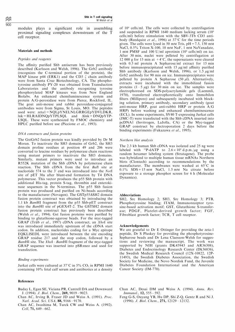
Northern blot analysis of multiple human tissues showsthat Shb mRNA is widely expressed as two transcriptsof 2.5 and 3.1 kb present in all 16 tissues analysed(Figure 1). Mouse tissues contain a predominant 3.1 kbmRNA (Welsh et al., 1994). Although the levels ofexpression were similar in most tissues, prostatecontains slightly more and peripheral blood leukocytessomewhat less Shb mRNA (Figure 1). Thus, it is likelythat lymphocytes express the Shb mRNA, since thesecells are present in spleen, thymus and bloodleukocytes.To assess a functional role of Shb in T cell
signaling, phosphotyrosine proteins present in anti-Shb immunoprecipitates of anti-CD3-stimulated Jur-kat T cells were determined (Figure 2a). Phosphotyr-osine proteins of molecular weights 22 kDa, 36/38 kDa, 55/57 kDa, 70 kDa, 100 kDa and 190 kDawere all found to co-precipitate with Shb after TCRstimulation. Although these six phosphotyrosineproteins were commonly seen in the anti-Shbimmunoprecipitates, their relative abundance variedbetween di�erent experiments (see Figure 3a). Theprecipitation of these products was speci®c for the Shbantibody compared with normal rabbit serum (NRS).When testing for Shb immunoreactivity, a broad bandof 55 ± 59 kDa was observed which was speci®callyimmunoprecipitated with the Shb antibody (Figure2a). In Jurkat cell extracts, the Shb antibodyrecognizes in addition to the 55 ± 59 kDa Shbproducts present in the immunoprecipitates bands of66, 77 and 80 kDa (see Figure 3d). Previously, Shbisoforms of 55, 66 and 77 kDa have been described,with the 66 kDa product representing Shb initiated atthe ®rst methionine codon, the 55 kDa protein Shbinitiated at the second methionine codon and the77 kDa component probably representing a post-translationally modi®ed Shb (Karlsson and Welsh,1996). Major phosphotyrosine products observed in
the Shb immunoprecipitates were the 55/57 kDaphosphoproteins, and next the time-course after CD3crosslinking for the tyrosine phosphorylation of thesewas studied (Figure 2b). The tyrosine phosphorylationof the 55/57 kDa products present in the Shbimmunoprecipitates rapidly increased (within 30 s)and remained elevated for up to 60 min of stimula-tion. To assess whether these products correspondedto Shb or were Shb associated, Jurkat cell extractsfrom unstimulated or CD3 crosslinked cells wereboiled in 1% SDS, diluted and subjected toimmunoprecipitation with the Shb antibody (Figure2c). The purpose of this procedure was to disrupt pre-existing complexes between Shb and other proteins.Western blot analysis of anti-Shb immunoprecipitatesunder these conditions revealed Shb immunoreactivitycorresponding to the molecular weights 55 ± 59 kDa(Figure 2c). Probing the same blot with thephosphotyrosine antibody PY-20, revealed the ab-sence of detectable amounts of phosphotyrosineproteins in these immunoprecipitates, suggesting thatthe phosphorylated 55/57 products indeed are Shb-associated. One of the supernatants after the anti-Shbimmunoprecipitation in Figure 2c was transferred to afresh tube and subjected to a second immunoprecipi-tation with the Shb antibody (Figure 2c). Shbimmunoreactivity was absent under these conditions,suggesting e�cient precipitation of Shb in the ®rstimmunoprecipitation. The results in Figure 2a and bsuggest that Shb is rapidly recruited into tyrosinekinase signaling upon T cell activation, implying afunctional role for Shb in this process.
Comparison of Shb and Grb2 co-immunoprecipitation ofphosphotyrosine proteins
To identify the Shb-associated phosphotyrosine pro-teins, anti-CD3 activated Jurkat cells were lysed andproteins co-immunoprecipitated with the Shb or Grbantibodies were compared. Co-precipitated proteinswere identi®ed by blotting for phosphotyrosine (Figure3a,b). Tyrosine phosphorylation levels increasedfollowing CD3 crosslinking including those of theanti-Shb precipitated proteins of 36/38 kDa, 55 ±57 kDa, 70 kDa and 190 kDa. The 36/38 kDa, 55/57and 70 kDa proteins were also immunoprecipitatedusing the Grb2 antibody. The immunoprecipitation ofthe 36/38 kDa product was more e�cient with theGrb2 antibody. Shorter exposure of the ECL reactionalso revealed an approximate doubling of thephosphotyrosine content of p36/38 associated withGrb2 in this experiment (results not shown). Whenassessing the co-immunoprecipitation of phosphotyr-osine proteins in the 20 ± 30 kDa molecular weightrange with the Shb and Grb2 antibodies, againsimilarities in the co-immunoprecipitation patternswere observed (Figure 3b). Both the Shb and theGrb2 antibodies co-immunoprecipitated two phospho-tyrosine proteins of 22 and 25 kDa solely in the CD3activated cells. The 22 kDa product is likely to be the zchain (Chan et al., 1991).Similarities in the co-immunoprecipitation patterns
between Shb and Grb2 suggested that the two mightassociate with one another. This was further investi-gated by testing Shb-immunoprecipitates for thepresence of Grb2 (Figure 3c). Grb2 immunoreactivity
he br pl lu li sm ki pa sp th pr te ov in co pbl
3.1 — 2.5 —
Figure 1 Northern blot of human tissues. (a) A multiple tissueNorthern blot was probed for the Shb cDNA. The positions ofthe 3.1 and 2.5 kb mRNAs have been indicated. The tissues areheart (he), brain (br), placenta (pl), lung (lu), liver (li), skeletalmuscle (sm), kidney (ki), pancreas (pa), spleen (sp), thymus (th),prostate (pr), testis (te), ovary (ov), intestine (in), colon (co) andperipheral blood leukocytes (pbl)
Shb in T cell signalingM Welsh et al
892

was demonstrated in Shb immunoprecipitates fromboth unstimulated and CD3 activated Jurkat cells(Figure 3c). The speci®city of the Shb immunoprecipita-tion of Grb2 was demonstrated in another experiment(Figure 3c). No Grb2 immunoreactivity was detectedafter precipitation with NRS whereas Grb2 immunor-eactivity was again observed after immunoprecipitationwith the anti-Shb antibody. The data suggest anassociation between Shb and Grb2 in Jurkat cells.The domain-interactions responsible for the associa-
tion between Shb and Grb2 were investigated byperforming GstGrb2 fusion protein binding experi-ments (Figure 3d). A large amount of the p66 Shbcomponent, and some of the p55 Shb isoform werefound in association with the GstGrb2 fusion protein.These interactions were independent of activation ofthe Jurkat cells. Since the Shb antibody used here wasgenerated by immunization with a Shb fusion protein,reactivity with the GstGrb fusion protein itself wastested (Figure 3d, bl=fusion protein not mixed withthe Jurkat cell extract). No detectable reactivity withthe 66 and 55 kDa components was seen. Theassociation between GstGrb2 and p66 and p55 Shbwas lost by inactivation of the Grb SH3 domains by
introduction of Pro to Leu point mutations atpositions 49 and 206 (Figure 3d).Finally, the association between Shb and the Grb2-
related protein GRAP (TruÈ b et al., 1997; Feng et al.,1996) was tested by transfecting a Myc-tagged GRAPcDNA into the Jurkat cells and immunoprecipitationwith the Myc antibody (Figure 3e). The results showno detectable Shb-like reactivity in the Myc antibodyimmunoprecipitates of mock transfected cells, whereasthe GRAP-tagged cDNA transfected cells showed Shb-reactivity corresponding to the p66 Shb isoform whenanalysed by Western blotting. The co-immunoprecipi-tation of this was independent of CD3 cross-linking.Thus the data suggest that both Grb2 and GRAP areassociated with Shb isoforms in the Jurkat cells, andthat this association shows little or no dependence onactivation of the T cell receptor.
Association of phosphotyrosine proteins with various Shbfusion proteins and identi®cation of p22 as the CD3associated z chain
To obtain an understanding for the mechanisms behindthe association of Jurkat cell phosphotyrosine proteins
Figure 2 Shb immunoprecipitation of phosphotyrosine proteins. (a) Jurkat cells (107) were unstimulated (7) or stimulated by CD3crosslinking for 2 min (+) before lysis and immunopreicipitation using normal rabbit serum (NRS) or the Shb antibody (Shb i.p.).The immunoprecipitates were electrophoresed on 9% acrylamide/0.24% Bis gels before Western transfer and incubation with thephosphotyrosine antibody PY-20. The blot has been divided into subsections to visualize the di�erent phosphotyrosine proteinssince these required di�erent ECL exposure times. Shb immunoreactivity in the NRS and Shb immunoprecipitates is also shown.The molecular weight standards and IgG heavy chain (IgGHC) are indicated to the left and the phosphotyrosine products to theright. (b) Jurkat cells (107) were stimulated for the time points indicated by CD3 cross-linking, lysed and immunoprecipitated usingthe Shb antibody. The immunoprecipitates were subjected to Western blotting using the phosphotyrosine antibody PY-20. Thereactivity with p55/57 kDa doublet is shown. (c) Jurkat cells (107) without treatment (7) or stimulated for 2 min with the CD3crosslinking antibody (+) were lysed, nuclei pelleted, and the supernatants boiled for 2 min in 1% SDS. The samples were thendiluted tenfold in lysis bu�er and Shb was then immunoprecipitated. The supernatant after immunoprecipitation from the untreatedlysate (7) was transferred to another tube and subjected to a second Shb immunoprecipitation. The samples were subjected toWestern blotting for Shb and phosphotyrosine. The positions of 55/59 Shb and IgG heavy chain (IgGHC) are indicated
– + – +
97 —
69 —
IgGHC —
46 —
21 —
— 190
— 100
— 70
— 55/57
— 36/38
— 22
NRS i.p. SHB i.p.
Blot: PY-20
IgGHC —
— 55-59 Shb
– + – +Blot: Shb
NRS i.p. SHB i.p.
a Blot: PY20
time: 0 0.5 1 2 5 15 60
b
55/59
IgGHC —
i.p.: shb shb shb+ shb shb shb+ shb shb
Blot: Shb PY20
– + – – + – c
Shb in T cell signalingM Welsh et al
893

to Shb, fusion proteins corresponding to the di�erentparts of Shb were synthesized and used for bindingexperiments (Figure 4a). GST itself did not associatewith any of the Jurkat cell proteins. The GSTSH2fusion protein corresponding to the SH2 domain ofShb bound two phosphotyrosine proteins of 22 and 36/38 kDa in cell extracts from CD3-crosslinked Jurkatcells. No detectable association of these was observedin the unstimulated cell extracts. Simultaneous additionof 20 mM phosphotyrosine (PY) to the extract fromCD3-stimulated cells greatly reduced the binding of the22 and 36/38 kDa proteins. Using a Shb fusion proteincorresponding to the proline-rich motifs of p55 Shband the central region of Shb but lacking the SH2domain (GSTp55DSH2), one major product fromstimulated cells was observed; the p36/38 doublet.The association of this phosphotyrosine product wascompletely blocked by 20 mM phosphotyrosine. The
p55 Shb product produced in the pET system boundthe 22, 36/38, and 190 kDa proteins in a CD3 receptorstimulation- and phosphorylation-dependent manner.In addition to these products, small amounts of a70 kDa phosphotyrosine protein was also found toassociate with the p55 Shb protein. Although weak inthis experiment, the association of the 70 kDacomponent to p55 Shb was strong in other experi-ments (see Figure 5b). The production of p66 Shb inthe pET system was ine�cient, and thus thecorresponding experiments using this Shb isoformcould not be carried out. The PY-20 phosphotyrosineantibody showed considerable background reactivitywith the GSTSH2 fusion protein (seen as a broad bandof 36 kDa), the GSTp55DSH2 fusion protein and itsproteolytic degradation products (72 kDa and smaller)and the p55 Shb (as 62 kDa protein due to thepresence of pET-derived sequences).
— p66 Shb
— p55 Shb
– + – +Blot: Shb
Transfection: mock Myc-GRAP
e
220 —
97 —
66 —
46 —
– + – – + +Blot: PY20
p36/38
shb grb2 shb grb2 cell extr. immunoprecipitation
a
IgGLC —
21 —
– + – – + +Blot: PY20
shb grb2 shb grb2 cell extr. immunoprecipitation
b
– + – + + +Blot: Grb2
cell extr. Shb i.p. NRS Shb i.p.
— Grb2
c
77 —
66 — 55 —
+ – + bl – + bl
extr. wt Grb2 mut-49/206 Grb2
Blot: shb
d
Figure 3 Association of Grb2, GRAP and other phosphotyrosine proteins with Shb. (a) Phosphotyrosine proteins associated withShb and Grb2. Jurkat cells (56107 cells) that were either unstimulated (7) or stimulated by CD3 crosslinking (+) were lysed andimmunoprecipitated using the Shb or Grb2 antibodies. The immunoprecipitates were electrophoresed on 9% polyacrylamide/0.24%Bis SDS-gels prior to transfer onto Immobilon ®lters. The ®lters were incubated with the PY-20 antibody and a secondary goat antimouse-HRP antibody before ECL. In parallel, cell extracts (cell extr, 106 cells) were analysed. Molecular weight markers areindicated to the left and p36/38 to the right. (b) Phosphotyrosine proteins associated with Shb and Grb2 in the 20 ± 30 kDamolecular weight range. Cells were treated and analysed as in a. The position for the 21 kDa molecular weight marker and IgG lightchain are indicated. (c) Shb co-immunoprecipitation of Grb2. Cell extracts (extr, 106 cells) or Shb immunoprecipitates (shb i.p.,26107 cells) from untreated (7) CD3 crosslinked (+) cells were subjected to SDS-gel electrophoresis on 12% polyacrylamide/0.32% Bis gels prior to Western transfer. The blot was incubated with the Grb2 antibody followed by an incubation with proteinA/G HRP prior to ECL. In another experiment (two right lanes), the anti-Shb immunoprecipitation was compared withprecipitation using normal rabbit serum (NRS). The position of Grb2 is indicated. (d) Binding of p66 and p66 Shb to wild-type orSH3 domain inactivated (mut 49/206) GstGrb2. Cell extracts (107 cells) from untreated (7) or CD3 crosslinked (+) cells wereincubated with the fusion proteins and subjected to Western blotting using the Shb-antibody preblocked with 1 mg/ml GstGrb2.Blotting of fusion protein not mixed with extract is shown (bl) as well as an aliquot (extr, 106 cells) of the initial cell extract. Thepositions of p55, 66 and 77 Shb are indicated. (e) Association between Shb and Myc-tagged GRAP. Cells were mock transfected ortransfected with the myc-tagged GRAP construct before lysis and immunoprecipitation using the Myc antibody. The samples wereanalysed by Western blotting using the Shb antibody. Untreated cells (7) or CD3 crosslinked cells (+) were immunoprecipitatedfrom 56107 cells each. The positions of p66 and p55 Shb are indicated
Shb in T cell signalingM Welsh et al
894

To identify the nature of p22, blots were probedwith a CD3 z antibody (Figure 4b). The cell extractscontained a strong band of 16 kDa present in bothunstimulated and CD3 crosslinked cells and a weaker22 kDa band only present in the cell extracts fromCD3 crosslinked cells. The 22 kDa product representsthe tyrosine phosphorylated z chain, whereas the16 kDa protein the unphosphorylated form of the zchain (Chan et al., 1991). Immunoprecipitation withthe Shb antibody revealed the coprecipitation of p22only in extracts from CD3 crosslinked cells (Figure 4b).When testing the association of the p22 z chain toimmobilized, bacterially produced p55 Shb, the binding
to the wild-type Shb protein was much more e�cientthan that to p55 Shb containing the R522K mutation(Figure 4b) which inactivates the Shb SH2 domain(Waksman et al., 1992). The binding of other
220 —
97 — 66 —
46 —
31 —
21 —
— 190
— 36/38
— 22
– + py – + py – + py – + py – +Blot: PY20
gst gstsh2 gst55shb 55shb cell ex.
a
∆sh2
22 —
16 —
— 190 — 70
— 36/38
– + – + + ++ +
Blot: CD3 ζ Blot: PY-20
R522KWT R522K W T
cell extr. Shb i.p. p55 Shb p55 Shb
b
Figure 4 Association of phosphotyrosine proteins and the CD3associated z chain with Shb fusion proteins. (a) Cells fromuntreated (7) or CD3 cross-linked Jurkat cells (+) were lysedand mixed with the immobilized fusion proteins (107 cells) for30 min on ice. In some cases, 20 mM phosphotyrosine (PY,pH=7.5) was added to the cell extract prior to mixing with thefusion proteins. The samples were electrophoresed before proteintransfer and incubation with the phosphotyrosine antibody PY-20, goat anti-mouse HRP as a secondary antibody and ECL.Aliquots of the cell extracts (106 cells) were also electrophoresed.The positions of molecular weight markers are shown to the left,and the positions of p22, p36/38 and p190 to the right. The fusionproteins used were GST (gst), the GST-Shb SH2 domain (gstsh2),the GST-p55Shb lacking the SH2 domain (gst55shbDsh2) and thep55 Shb in pET (55shb). The PY-20 antibody showed backgroundreactivity with the gstsh2 fusion protein (36 kDa), gst55shbDsh2(72 kDa and smaller degradation products) and 55shb (62 kDa).The background reactivity with the fusion proteins was equal inall three lanes for each fusion protein and dominates the reactivityin the unstimulated lanes. (b) Cells from untreated (7) or CD3crosslinked (+) Jurkat cells were lysed and immunoprecipitated(26107 cells) with the Shb antibody. Alternatively, aliquots of thecell extracts (107 cells) were mixed with immobilized, bacteriallyproduced p55 Shb or p55 Shb carrying the R522K mutation.After washing, the samples were electrophoresed on 12%acrylamide/0.32% Bis gels before electrical transfer and incuba-tion of the blot with the z chain and PY-20 antibodies. Aliquotsof cell extracts (106 cells) were also electrophoresed. The positionsof z chain isoforms 16 and 22 kDa are indicated, as well as 36/38,70 and 190 kDa phosphotyrosine proteins
— 22
concentration zeta-1 peptide: 0 10 100
Blot: PY20
a
70 —
36/38
peptide: 0 lnk 0 lnk lnk ∆lnk concentration: 0 10 0 10 100 100
gst55shb 55shb ∆sh2
Blot: PY20
b
extr p55Shb GstGrb2SH2
p36/38 —
peptide: 0 0 lnk 0 lnk
p36/38
GstGrb2SH2
Blot: PY-20
c
Peptide conc: 0 5 50 200
— gst55shb∆sh2
Blot: Shb
d
Figure 5 Peptide competition experiments. (a) E�ects of the zeta-1 peptide on the binding of p22 to Shb. Bacterially produced p55Shb was incubated with Jurkat cell extracts from CD3 crosslinkedcells in the presence of di�erent concentrations of phosphopeptide(in mM). Binding of tyrosine phosphorylated p22 was analysed byWestern blotting using the phosphotyrosine antibody PY-20. (b)Cell extracts from CD3 cross-linked cells were incubated with thep55 Shb fusion protein (55Shb) or the GST p55 Shb fusionprotein lacking the SH2 domain (gst55shbDsh2) in the presence ofdi�erent micromolar concentrations of lnk or Dlnk peptides. Thesamples were then subjected to Western blotting using thephosphotyrosine antibody PY-20. The positions of p36/38 andp70 are indicated. (c) Binding of p36/38 to p55 Shb or theGSTGrb2 SH2 domain fusion protein was determined with orwithout the addition of 100 mM lnk peptide. The bound proteinswere electrophoresed and subjected to Western blotting as in b.The positions of p36/38 and background reactivity of the PY-20antibody with the GSTGrb2 SH2 domain fusion protein areindicated. (d) Binding of the gst55shbDsh2 fusion protein tophosphotyrosine-Sepharose beads in the presence of di�erentmicromolar concentrations of the lnk peptide. Binding wasperformed for 30 min at 208C in 150 mM NaCl, 50 mM Tris,pH=7.5. The samples were washed three times in the bindingbu�er and subjected to Western blotting using the Shb antibody.The ®gure shows only binding of the full-sized (72 kDa) fusionprotein and a 68 kDa proteolytic fragment
Shb in T cell signalingM Welsh et al
895

phosphotyrosine proteins to wild-type or R522K p55Shb was similar (Figure 4b). The small amount of p22remaining bound to the R522K Shb protein could beindirect via other phosphotyrosine proteins. The resultsin Figure 4 suggest distinct domain interactionsbetween Shb and phosphotyrosine proteins p22, p36/38, p70 and p190. p22 is the CD3 associated z chainand binds primarily to the Shb SH2 domain.
Peptide inhibition experiments
A phosphotyrosine peptide corresponding to an ITAMmotif of the z chain inhibited almost completely theassociation of the putative z chain 22 kDa phosphotyr-osine protein to the p55 bacterially produced Shbprotein at 100 mM (Figure 5a). Assessment bydensitometric analysis of the association of p36/38 tothe p55 Shb protein after addition of this peptide at100 mM in the same experiment revealed a 71%inhibition of binding (results not shown). A phospho-tyrosine peptide corresponding to a proposed phos-phorylation site of the Lnk protein (Y297) andextending 8 amino acid residues towards the N-terminus (lnk peptide) caused a signi®cant, dose-dependent inhibition of the association of the p36/38phosphoproteins to bacterially expressed GSTp55DSH2and p55 Shb (Figure 5b). However, some bindingremained even at a peptide concentration of 100 mM,possibly representing indirect binding via otheradaptors, such as Grb2 and GRAP (TruÈ b et al.,1997; Fukazawa et al., 1995). A corresponding peptide(Dlnk) comprising the same six residues C-terminally,but only extending three amino acids towards the N-terminus relative to the phosphotyrosine, failed tosigni®cantly in¯uence the binding of p36/38 to p55 Shb(Figure 5b). This latter peptide is expected to onlyin¯uence SH2 domain recognition, contrary to the lnkpeptide which is predicted to inhibit both the SH2 andPTB domain interactions due to its N-terminalextension (Wolf et al., 1995; TruÈ b et al., 1995). Thebinding of the z chain to the p55 Shb protein was notin¯uenced by the lnk peptide (results not shown). InFigure 5c, the e�ects of the lnk peptide on theassociation of p36/38 with the Grb2 SH2 domain isshown. The lnk peptide inhibited e�ciently the bindingof p36/38 to p55 Shb, whereas that to the Grb2 SH2domain was not a�ected. Background reactivity of thephosphotyrosine antibody with the GSTGrb2 SH2domain fusion protein has been indicated. The lnkpeptide inhibited at 5 mM the binding of theGSTp55DSH2 fusion protein to phosphotyrosinebeads in in vitro experiments (Figure 5d). Thecombined data with the lnk peptide in Figure 6b and6d suggest that the GSTp55DSH2 fusion proteincontains a PTB domain which interacts with thispeptide.
PTB domain binding sequence
To assess the preferred binding sequence of the ShbPTB domain, immobilized GSTp55DSH2 fusionprotein was mixed with a phosphopeptide librarycontaining combinations of all amino acids exceptcysteine and tryptophane in positions 1-3 N-terminallyand C-terminally relative the phosphotyrosine residue.Amino acids binding to the Shb PTB domain with a
relative a�nity of 1.4 or higher at the di�erentpositions are listed in Table 1. There was a relativelystrong selection for aspartic acid in position 3 and aweaker selection for aspartic acid in position 2upstream of the phosphotyrosine. No preferentialbinding (41.5) of individual amino acids in the otherpositions could be observed. Thus the consensussequence for the Shb PTB domain recognition site isAsp-Asp-X-pTyr in these positions. High a�nitybinding between the Shb PTB domain and its targetsequence may require sequence speci®city in positions74 to 77 relative the phosphotyrosine residue as well.It should be noted that the lnk peptide which containsan aspartic acid in position 3 upstream of thephosphotyrosine inhibits the association between theShb PTB domain and p36/38.
Transfection experiments
To study the e�ects of Shb-overexpression on T cellreceptor signaling in Jurkat cells, JMC-T Jurkat cellswere transfected with the Shb cDNA by electropora-tion. The results demonstrated strong expression of the66 kDa Shb isoform (Figure 6a). This band migratedas a broad band, possibly as a doublet in theseexperiments. Elevated expression of the 55 kDa Shbisoform was also observed, although to a much lesserdegree. Besides these two Shb isoforms, severalprominent components of lower molecular weightswere seen (Figure 6a), probably representing proteoly-tic degradation products.The phosphotyrosine protein content of control or
Shb-transfected cells was also studied (Figure 6b).Comparison of the phosphotyrosine protein patternassessed by Western blotting using the phosphotyrosineantibody PY-20 revealed increased phosphotyrosinecontents of proteins of 36/38 and 70 kDa in theunstimulated Jurkat cells after Shb transfection. Thesephosphotyrosine proteins appear identical to thecorresponding components that interacted with thep55 Shb protein (Figure 4a). After stimulation by CD3crosslinking the content of phosphotyrosine proteinswas similar in the control and the Shb transfected cells(Figure 6b).To assess the e�ects of the Shb SH2 domain
inactivation mutation R522K on TCR dependentphosphotyrosine signaling, regular Jurkat cells weremock transfected or transfected with the mutatedShb cDNA by electroporation (Figure 6c). Amoderate increase in the contents of the p66 andthe p55 Shb proteins was observed. When the blotwas probed for phosphotyrosine content, the mocktransfected cells displayed increased phosphotyrosinecontents of several proteins after TCR stimulation
Table 1 Recognition speci®city of the Shb PTB domain
pY±3 pY±2 pY±1 pY+1 pY+2 pY+3
D (2.5) D (1.9) S (1.4)D (1.4)
D (1.4) D (1.5) D (1.5)
The GST55ShbDSH2 fusion protein was mixed with the phosphopep-tide library GAXXXpYXXXAKKK (with X any amino acid exceptC and W) and preferential binding at the di�erent positions wasdetermined as previously reported (Songyang et al., 1994). The valuesare relative enrichments for the amino acids indicated. Values below1.4 are not included. Values above 1.5 are indicated with bold letters
Shb in T cell signalingM Welsh et al
896

by CD3 crosslinking (Figure 6d). These were ofmolecular weights 36/38, 66, 70 and 100 kDa. In theJurkat cells transfected with the R522K mutated ShbcDNA the increase in tyrosine phosphorylation ofseveral of these in response to CD3 crosslinking wasdiminished, especially for those of molecular weightsof 36/38, 66 and 100 kDa (Figure 6d). To furtheraddress a role of Shb for TCR signaling, anotherShb construct which was entirely deleted ofsequences coding for the SH2 domain was trans-fected into Jurkat cells, and the tyrosine phosphor-ylation in response to CD3 crosslinking determined(Figure 6e). As in the case of the R522K mutated
Shb cDNA, Jurkat cells transfected with the ShbcDNA construct lacking the entire SH2 domain(ShbDSH2) displayed decreased tyrosine phosphor-ylation of the 36/38, 66 and 100 kDa phosphopro-teins in response to TCR stimulation. The combineddata in Figure 6 suggest that Shb plays a role forTCR dependent signaling and complex formation ofphosphotyrosine proteins.Transfection of Jurkat cells with the R522K mutated
Shb cDNA and subsequent clonal selection forneomycin resistance yielded one clone named Jur-katR522K-1 which over-expressed the 66 and 55 ±59 kDa Shb isoforms (Figure 7). No signs of elevated
Figure 6 Transfection of the Shb cDNA. (a) SV40T-expressing Jurkat cells (JMC-T, Fukazawa et al., 1995) were mock-transfectedor transfected with the Shb cDNA in pcDNA1 and were either untreated (7) or incubated with the CD3 antibody for 2 min (+).Cell extracts were prepared and analysed by Western blotting as in Figure 1. The blot was incubated with the Shb antibody andfollowed by ECL. The ®gure shows the Shb-reactivity of untreated extracts. The positions of p66 and p55 Shb are indicated. (b)Phosphotyrosine proteins of mock or Shb-transfected cells. Untreated (7) or CD3 cross-linked cell extracts taken 2 min afterantibody addition (+) were blotted as in a and probed for phosphotyrosine proteins using the PY-20 antibody. The positions of thep36/38 and 70 kDa proteins are indicated. (c) Regular Jurkat cells were electroporated with pcDNA1 (mock) or pcDNA1containing the R522K mutated Shb cDNA and maintained for 2 days before incubated in the absence (7) or presence (+) of theCD3 crosslinking antibody for 2 min. The cells were lysed (26106) and subjected to Western blotting for Shb after electrophoresison 6% acrylamide/0.16% Bis gels. The positions of p55 and p66 Shb are indicated. Equal amounts of protein were electrophoresedin the di�erent lanes. (d) The blot in c was probed for phosphotyrosine. The positions of molecular weight markers (left) and thep36/37, p66 and p100 proteins (right) are indicated. (e) A Shb cDNA lacking the SH2 domain (ShbDSh2) was constructed byinserting the 1.6 kb BamHI fragment of the Shb cDNA into the BamHI site of the expression vector pcDNA3.1 Myc-His. Jurkatcells were transfected by electroporation in the presence of empty vector (mock) or vector containing the partial Shb cDNA(ShbDSh2). The transfected cells were incubated in the absence (7) or presence (+) of the CD3 crosslinking antibody, and cellextracts were prepared for electrophoresis and Western blotting. The blot shows phosphotyrosine proteins of the molecular weightsindicated
— 66
— 55
Transfection: mock Shb
Blot: Shb
a
— 70
— 36/38
– – + +
Transfection: mock + + shb + +
Blot: PY-20
b
— 66
— 55
– + – +
Transfection: Mock R522K Shb
Blot: Shb
c
97 —
69 —
46 —
— 100
— 66
— 36/38
– + – +
Transfection: Mock R522K Shb
Blot: PY-20
d
— 100 kDa
— 66 kDa
— 36/38 kDa
– + – +
Transfection: mock Shb∆SH2
Blot: PY20
e
Shb in T cell signalingM Welsh et al
897

contents of the 77 and 80 kDa Shb immunoreactivitywas observed in these cells (Figure 7). The Jur-katR522K-1 cells displayed a pattern of tyrosinephosphorylation in response to CD3 activation whichwas very similar to that of the Jurkat cells transientlyexpressing the mutated Shb cDNA (Figure 7).Compared with a clone of neomycin resistant Jurkatcells (Jurkat-neo), the JurkatR522K-1 cells showed lesstyrosine phosphorylation of the 36/38, 66, 70, 100 and
190 kDa proteins after CD3 crosslinking (Figure 7),although the di�erence in tyrosine phosphorylation ofthe 66 kDa protein could be more easily visualized bya shorter ECL exposure (results not shown). Thedecreased tyrosine phosphorylation of 36/38, 66 and100 kDa proteins was also observed in the Jurkat cellstransiently expressing the R522K mutated Shb (seeFigure 6c). The JurkatR522K-1 cells also displayed adecreased MAP kinase activation after CD3 stimula-tion compared with the Jurkat-neo cells, as assessed bydecreased tyrosine phosphorylation of the MAP kinaseTEY motif (Figure 7). MAP kinase p42 was found tobe the main MAP kinase product tyrosine phosphory-lated in response to CD3 activation. Probing the sameblot for the presence of total p44 MAP kinase revealedequal amounts of this protein in the Jurkat-neo andJurkatR522K-1 cells (Figure 7). Thus, the alteredpattern of tyrosine phosphorylation seen in Jurkatcells expressing Shb with an inactivated SH2 domain isparalleled by decreased MAP kinase activation.
Discussion
In the present investigation we have attempted toassess the role of the adaptor protein Shb in signalingdownstreams of the T cell receptor. Based on bacterialexpression and transfection experiments, three Shb-isoforms of 55, 66 and 77 kDa have been describedpreviously, of which the ®rst two result fromtranslation initiation at the second and ®rst methio-nine codons, respectively, whereas the 77 kDa isoformis likely to result from some post-translationalmodi®cation (Karlsson and Welsh, 1996). Subsequentstudies of other cell types have revealed the existence ofadditional Shb isoforms. In PC12 cells, for instance,three major Shb isoforms of 55, 57 and 59 kDa can bedetected, all of which show elevated contents aftertransfection of these cells with the 2.3 kb human ShbcDNA (Karlsson and Welsh, unpublished data). Thusthe 2.3 kb Shb cDNA can generate many di�erent Shbisoforms di�erentially in various cells, presumably dueto several translational and post-translational pro-cesses. All the observed Jurkat Shb isoforms, with theexception of the 80 kDa product, thus seem to berelated to the published Shb coding sequence. Thepresent data show that transfection of Jurkat cells withthe Shb cDNA dramatically alters the relativeabundance of di�erent Shb isoforms compared withthe untransfected cells. A possible explanation for thisis Shb overexpression in itself, which could shift theprocessing of the Shb mRNA or proteins.Immunoprecipitation experiments reveal the rapid
phosphorylation of two bands of 55 and 57 kDa whichare likely to be Shb-associated proteins, since thesewere not detected as phosphotyrosine proteins in theShb-immunoprecipitates performed after SDS-dena-turation of the cell extracts, despite e�cient immuno-precipitation of the Shb isoforms under theseconditions. No evidence for tyrosine phosphorylationof p66 Shb was obtained, although this isoform wasnot e�ciently immunoprecipitated in the presentexperiments. In a previous study, p66 Shb (given theapparent molecular weight of 69 kDa, Karlsson et al.,1995) was immunoprecipitated, suggesting that theantibody has the ability to interact with this Shb
— 190
— 100
— 36/38
– + – +
70
66
Jurkat- JurkatR522K- neo 1
Blot: PY-20
— 80 — 66 — 55-59
– + – +Blot: Shb
Jurkat- JurkatR522K- neo 1
— p42 MAPK
– + – +Blot: Phosphorylated MAPK
Jurkat- JurkatR522K- neo 1
— p44 MAPK
– + – +
Jurkat- JurkatR522K- neo 1
Blot: MAPK
Figure 7 Phosphorylation of Jurkat cells stably expressingR522K Shb. Jurkat cells were transfected by electroporationwith pSV2-neo alone (Jurkat-neo) or the R522K mutated ShbcDNA in pcDNA1 together with pSV2-neo (JurkatR522K-1).Transfected cells were plated in multi-well dishes and selected forneomycin resistance in the presence of 1 mg/ml G418. Cells fromone clone of each were stimulated for 2 min by CD3 crosslinking(+) or left unstimulated (7), after which cell extracts wereelectrophoresed and subjected to Western blotting as in Figure 6c.The blot was tested for phosphotyrosine (PY-20), Shb (Shb),activated (tyrosine phosphorylated) MAP kinases and total p44MAP kinase. The positions of tyrosine phosphorylated proteins190, 100, 70, 66 and 36/38 kDa, as well as 80, 66 and 55 ± 59 kDaShb, p42 MAP kinase and p44 MAP kinase are indicated
Shb in T cell signalingM Welsh et al
898

isoform. However, the ine�cient immunoprecipitationwith the Shb-antiserum is likely to have additionalexplanations besides a relatively low a�nity for nativeShb isoforms. One possibility is proteolysis. Thisoption seems probable, both considering the patternof reactivity of the antibody with components of lowermolecular weight on blots after immunoprecipitation,but also the pattern of blotting reactivity aftertransfection of the Shb cDNA. Another explanationthat should be kept in mind is masking of antigenicepitopes on Shb by binding to other cellularcomponents. This is particularly relevant for theinteraction between Shb and p36/38, since the PTBdomain which is a major part of the associationbetween the two was also used for immunization togenerate the antibody.The combined data suggest that Shb is an important
adaptor linking the T cell receptor with other signalingcomponents in Jurkat cells. The SH2 domain seems tohave one major interactions; the z chain. Previousassessment of the preferred binding sequence of thisSH2 domain revealed strong selection for leucine inposition 3 downstream of the phosphotyrosine, makingbinding to ITAM motifs on the T cell receptorassociated CD3 and z chains a possibility. The ITAMsare regularly spaced motifs containing two phosphotyr-osine residues, each with a leucine in position three C-terminal to phosphotyrosine. The present data indeedsuggest that Shb is likely to bind via its SH2 domain tothe ITAM motif, although it is not clear whether onlyone or several of the ITAM phosphotyrosine residues isrecognized. Besides the z chain, we have obtained datasuggesting that p36/38 is also a binding substrate for theShb SH2 domain based on GST-fusion proteinexperiments. However, the interaction of p36/38 withp55 Shb is not likely to involve the SH2 domain to anylarge extent, since the Dlnk peptide which contains thepYTPL sequence failed to a�ect the interaction betweenp55 Shb and p36/38 and since the SH2 domaininactivation mutation (R522K Shb) bound p36/38almost as well as the wild-type p55 Shb.The data suggest that the central portion of Shb is a
PTB domain, and that the main phosphotyrosineprotein interacting with this PTB domain is p36/38.The fusion protein containing this part of Shb, butlacking the SH2 domain, associated in a phosphotyr-osine dependent manner with p36/38. The associationof p36/38 to the PTB-domain fusion protein or p55Shb was inhibited in a dose-dependent manner by a lnkpeptide extending 8 amino acid residues towards the N-terminus, but not by a similar peptide extending onlythree residues towards the N-terminus. A long N-terminal peptide extension relative the phosphotyrosineis a characteristic feature of PTB-domain recognition(Wolf et al., 1995; TruÈ b et al., 1995). Finally, thebinding of the PTB-domain fusion protein tophosphotyrosine beads was signi®cantly inhibited at5 mM of lnk peptide. Although the lnk peptideproduced a substantial inhibition of the associationof p36/38 to the Shb PTB domain, some remainingbinding was observed (Figure 5b). This could re¯ectindirect association through other adaptors which bindboth p36/38 and Shb. Two such candidates are Grb2and GRAP which interact with p36/38 via their SH2domains (TruÈ b et al., 1997; Fukazawa et al., 1995). Thebinding of these to p36/38 is not in¯uenced by the Dlnk
or lnk peptides (TruÈ b et al., 1997), and thus is retainedin the conditions of Figure 5b (see Figure 5c). Thecombined peptide inhibition data suggest the existenceof at least two tyrosine phosphorylation sites on p36/38, one interacting with the Shb PTB domain and theother with the Grb2 SH2 domain.The Grb2 and GRAP proteins associate mainly with
p66 Shb. The data suggest that this interaction occursbetween the SH3 domains and the proline-richsequences of Shb. In previous work we tested theinteraction between the N-terminal SH3 domain ofGrb2 and the 4th and 5th proline-rich motifs of Shb(Karlsson et al., 1995) and could only detect a weakassociation. The most e�cient association was pre-sently observed between p66 Shb (which contains all®ve proline-rich motifs) and full-sized Grb2 GSTfusion protein containing both SH3 domains, whereasinactivation of these by site-directed mutagenesisabolished the interaction.Transfection and expression of the Shb cDNA
enhanced the basal tyrosine phosphorylation state ofseveral proteins, mainly the 36/38 and 70 kDa proteins.The 70 kDa protein was also observed to complex withShb in the binding experiments. The results can beinterpreted to suggest that an increased Shb contentfacilitates complex formation in the absence of T cellreceptor stimulation, and that such complex formationenhances the tyrosine phosphorylation state of some ofits components. Based on the these observations, it canbe speculated that Shb-mediated complex formationmay play a role in the function of p36/38, previouslyimplicated in the possible regulation of the Raspathway (Sieh et al., 1994; Buday et al., 1994).Although we failed to detect an increased activationof MAP kinases in Jurkat cells over-expressing thewild-type Shb (results not shown), clonally selectedJurkat cells overexpressing R522K mutated Shbdisplayed decreased MAP kinase activation in re-sponse to CD3 stimulation, suggesting a role for Shbin this process. Furthermore, the data suggest that theShb-Grb2 complex may be targeted in parts to the Tcell receptor by binding of the Shb SH2 domain to thez chain. This notion is supported by the failure of theR522K mutated Shb, which has an inactivated SH2domain, to e�ciently bind the z chain. As aconsequence of this, the pattern of Jurkat cell tyrosinephosphorylation in response to TCR activation isdramatically altered. There has been a signi®cantrecent controversy about the potential role of Grb2versus Shc in linking the z chain to Sos and the Raspathway (Ravichandran et al., 1993; Osman et al.,1995). It is possible that potential di�erences in thesestudies partly derive from the participation of Shb.In summary, our data suggest that Shb forms
multimeric complexes in Jurkat cells, consisting of thez chain, Shb, p36/38 and Grb2 (or GRAP). Thiscomplex in the activated T cell has the potential todock other signaling components via SH2, SH3 andproline-rich domains and phosphotyrosine residuespresent on p36/38. Such other signaling componentsinclude the 55/57, 70, 100 and 190 kDa phosphotyr-osine proteins found to interact with Shb, as well asphospholipase C g, Sos and p85 PI-3 kinase, of whichthe three latter have been detected in association withGrb2 and p36/38. In conclusion, our results suggestthat Shb, which encompasses three distinct structural
Shb in T cell signalingM Welsh et al
899
modules plays a signi®cant role in assemblingproximal signaling complexes downstream of the Tcell receptor.
Materials and methods
Peptides and reagents
The a�nity puri®ed Shb antiserum has been previouslydescribed (Karlsson and Welsh, 1996). The Grb2 antibody(recognizes the C-terminal portion of the protein), theMAP kinase p44 (ERK1) and the CD3 z chain antibodywere from Santa Cruz Biotechnology, CA. The phospho-tyrosine antibody PY-20 was obtained from TransductionLaboratories and the antibody recognizing tyrosinephosphorylated MAP kinases was from New EnglandBiolabs. An enhanced chemiluminescence system andprotein A/G-peroxidase were from Pierce, Rockford, IL.The goat anti-mouse and rabbit peroxidase-conjugatedantibodies were from Sigma, St Louis, MO. The peptidesused were: zeta1=NQLpYNELNLGRRGEpYDVLDKR,lnk=HLRAIDNQpYTPLSQL and Dlnk=DNQpYTP-LSQL. These were synthesized by FMOC chemistry andHPLC puri®ed before use (Piccione et al., 1993).
DNA constructs and fusion proteins
The GstGrb2 fusion protein was kindly provided by Dr MMoran. To inactivate the SH3 domains of Grb2, the SH3domain proline residues at position 49 and 206 wereconverted to leucine residues by polymerase chain reactionsusing mutant primers to inactivate the SH3 domains.Similarly, mutant primers were used to introduce anR552K mutation of the Shb cDNA by polymerase chainreaction. The Shb cDNA from the ®rst BalI site atnucleotide 574 to the 3' end was introduced into the NotIsite of pET 30a after blunt-end formation by T4 DNApolymerase. This vector produces the p55 Shb protein withadditional His-tag, protein S-tag, thrombin and enteroki-nase sequences in the N-terminus. The p55 Shb fusionprotein was produced and puri®ed on Ni-beads accordingto the manufacturer (Novagen). The GSTp55DSH2 domainfusion protein construct was obtained by introducing the1.1 kb BamHI fragment from the p55 Shb-pET constructinto the BamHI site of pGEX4T-2. The GSTSH2 domainfusion protein construct has previously been described(Welsh et al., 1994). Gst fusion proteins were puri®ed bybinding to glutathione-agarose beads. For the myc-taggedGRAP (TruÈ b et al., 1997) cDNA construct, an XhoI sitewas introduced immediately upstream of the cDNA startcodon. In addition, nucleotides coding for a Myc epitopeEQKLISEDL were introduced between the site encodingGRAP residue 217 and the stop codon, followed by aBamHI site. The XhoI ±BamHI fragment of the myc-taggedGRAP sequence was inserted into pSRaneo and used fortransfection.
Binding experiments
Jurkat cells were cultured at 378C in 5% CO2 in RPMI 1640containing 10% fetal calf serum and antibiotics at a density
of 106 cells/ml. The cells were collected by centrifugationand suspended in RPMI 1640 medium lacking serum (108
cells/ml) before stimulation with the SBV-3Tb CD3 anti-body (Reedquist et al., 1996) at 378C for the time periodsgiven. The cells were lysed in 50 mM Tris, pH=7.5, 150 mM
NaCl, 0.5% Triton X-100, 10 mM NaF, 1 mM NaVanadate,1 mM PMSF and 100 U/ml aprotinin (108 cells/ml) on ice.After 30 min, nuclei were pelleted by centrifugation at12 000 g for 15 min at +48C, the supernatants were clearedwith 0.5 ml protein A Sepharose/ml extract for 15 minbefore immunoprecipitated with 15 mg/ml a�nity puri®edShb antibody (Karlsson and Welsh, 1996), or 1.5 mg/mlGrb2 antibody for 90 min on ice. Immunoprecipitates werepelleted by protein A Sepharose (50 ml). Alternatively,extracts were incubated with the immobilized fusionproteins (1 ± 5 mg) for 30 min on ice. The samples wereelectrophoresed on SDS-polyacrylamide gels (Laemmli,1970), transferred electrophoretically onto Immobilon®lters (Millipore) and subsequently incubated with block-ing solution, primary antibody, secondary antibody (goatanti-mouse HRP, goat anti-rabbit HRP or protein A/GHRP) before washing and enhanced chemiluminescence(ECL). In some experiments, SV40 T-expressing Jurkat cells(JMC-T) were transfected with the Shb cDNA inserted intopcDNA1 (Invitrogen, LaJolla, CA) or the Myc-taggedGRAP construct by electroporation 2 days before thebinding experiments (Fukazawa et al., 1995).
Northern blot analysis
The 2.3 kb human Shb cDNA was isolated and 25 ng werelabeled with 32P-dATP to 2.66109 d.p.m./mg using arandom hexamer labeling system (Stratagene). The probewas hybridized to multiple human tissue mRNAs Northernblots (Clontech) according to recommendations by themanufacturer. The membranes were washed at 658C in0.1% SDS+15 mM NaCl, 1.5 mM Na citrate beforeexposure to a storage phosphor screen for 6 h (MolecularDynamics).
AbbreviationsSH2, Src Homology 2; SH3, Src Homology 3; PTB,Phosphotyrosine binding; ITAM, Immunoreceptor tyro-sine-based activation motif; GST, Glutathione S-transfer-ase; PDGF, Platelet-derived growth factor; FGF,Fibroblast growth factor; TCR, T cell receptor.
AcknowledgementsWe are grateful to Dr E Ottinger for providing the zeta-1peptide, Dr S Pluskey for providing the phosphotyrosine-Sepharose beads and Dr Lena Claesson-Welsh for sugges-tions and reviewing the manuscript. The work wassupported by NIH (grants DK45943 and AR36308),Diabetes and Endocrinology Research Center (Dk36836),the Swedish Medical Research Council (12X-10822, 12P-11493), the Swedish Diabetes Association, the SwedishSociety for Medicine, the Novo Nordisk Fund, the JuvenileDiabetes Foundation International and the AmericanCancer Society (IM-770).
References
Buday L, Egan SE, Viciana PR, Cantrell DA and DownwardJ. (1994). J. Biol. Chem., 269, 9019 ± 9023.
Chan AC, Irving B, Fraser JD and Weiss A. (1991). Proc.Natl. Acad. Sci. USA, 88, 9166 ± 9170.
Chan AC, Iwashima M, Turck CW and Weiss A. (1992).Cell, 71, 649 ± 662.
Chan AC, Desai DM and Weiss A. (1994). Annu. Rev.Immunol., 12, 555 ± 592.
Feng G-S, Ouyang YB, Hu DP, Shi Z-Q, Gentz R and Ni J.(1996). J. Biol. Chem., 271, 12129 ± 12132.
Shb in T cell signalingM Welsh et al
900

Fukazawa T, Reedquist KA, Panchamoorthy G, Solto� S,TruÈ b T, Druker B, Cantley LC, Shoelson SE and Band H.(1995). J. Biol. Chem., 270, 20177 ± 20182.
Gilliland LK, Schieven GL, Norris NA, Kanner SB, Aru�oA and Ledbetter JA. (1992). J. Biol. Chem., 267, 13610 ±13616.
Huang X, Li Y, Tanaka K, Moore KG and Hayashi JI.(1995). Proc. Natl. Acad. Sci. USA, 92, 11618 ± 11622.
Hutchcroft JE, Harrison ML and Geahlen RL. (1992). J.Biol. Chem., 267, 8613 ± 8619.
Karlsson T and Welsh M. (1996). Oncogene, 13, 955 ± 961.Karlsson T, Songyang Z, Landgren E, Lavergne C, Di FiorePP, Ana® M, Pawson T, Cantley LC, Claesson-Welsh Land Welsh M. (1995). Oncogene, 10, 1475 ± 1483.
Laemmli UK. (1970). Nature, 227, 680 ± 685.Osman N, Lucas SC, Turner H and Cantrell D. (1995). J.
Biol. Chem., 270, 13981 ± 13986.Perlmutter RM, Levin SD, Appleby MW, Anderson SJ andAblerola-Ila J. (1993). Annu. Rev. Immunol., 11, 451 ± 499.
Piccione E, Case RD, Domchek SM, Hu P, Chadhuri M,Backer JM, Schlessinger J and Shoelson SE. (1993).Biochemistry, 32, 3197 ± 3202.
Ravichandran KS, Lee KK, Songyang Z, Cantley LC, BurnP and Burako� SJ. (1993). Science, 262, 902 ± 905.
Reedquist KA, Fukazawa T, Panchamoorthy G, LangdonWY, Shoelson SE, Druker BJ and Band H. (1996). J. Biol.Chem., 271, 8435 ± 8442.
Secrist JP, Karnitz L and Abraham RT. (1991). J. Biol.Chem., 266, 12135 ± 12139.
Sieh M, Batzer A, Schlessinger J and Weiss A. (1994). Mol.Cell. Biol., 14, 4435 ± 4442.
Thompson PA, Gutkind JS, Robbins KC, Ledbetter JA andBolen JB. (1992). Oncogene, 7, 719 ± 725.
Songyang Z et al. (1994). Mol. Cell. Biol., 14, 2777 ± 2785.TruÈ b T, Choi WE, Wolf G, Ottinger E, Chen Y-J, Weiss Mand Shoelson SE. (1995). J. Biol. Chem., 270, 18205 ±18208.
TruÈ b T, Frantz JD, Miyazaki M, Band H and Shoelson SE.(1997). J. Biol. Chem., 272, 894 ± 902.
Waksman G, Kominos D, Robertson SC, Pant N, BaltimoreD, Birge RB, Cowburn D, Hanafusa H, Mayer BJ,Overduin M, Resh MD, Rios CB, Silverman L andKuriyan J. (1992). Nature, 358, 646 ± 653.
Weber JR, Bell GM, Han MY, Pawson T and Imboden JB.(1992). J. Exp. Med., 176, 373 ± 379.
Weiss A and Littman DR. (1994). Cell, 76, 263 ± 274.Welsh M, Mares J, Karlsson T, Lavergne C, Bre ant B andClaesson-Welsh L. (1994). Oncogene, 9, 19 ± 27.
Wolf G, TruÈ b T, Ottinger E, Groninga L, Lynch A, WhiteMF, Miyazaki M, Lee J and Shoelson SE. (1995). J. Biol.Chem., 270, 27407 ± 27410.
Shb in T cell signalingM Welsh et al
901
Copyright © 2022 FDOKUMEN