Stem Wood and Bark Extracts of Delonix regia (Boj. Ex. Hook)
14
PEER-REVIEWED ARTICLE bioresources.com Salem et al. (2014). “Bioactivity of Delonix regia,” BioResources 9(2), 2382-2395. 2382 Stem Wood and Bark Extracts of Delonix regia (Boj. Ex. Hook): Chemical Analysis and Antibacterial, Antifungal, and Antioxidant Properties Mohamed Z. M. Salem, a, * Ahmed Abdel-Megeed, b,d and Hayssam M. Ali b,c In the present study, the fatty acid components of the wood, bark, and essential oil of wood from Delonix regia as well as its antibacterial, antifungal, and antioxidant properties were investigated for the potential ability to control plant and human pathogens. Myristic acid was found to be a major fatty acid in the wood and bark of Delonix regia, comprising 10.77% of wood and 9.63% of bark. According to the GC-MS results, naphthalene derivatives were detected in the essential oils from the wood samples. Heptadecane and acyclic hydrocarbons were found in a high percentage (14.05%). Methanol: chloroform (1:1 v/v) wood extract showed effective activity against Bacillus subtilis, Sarcina lutea, and Staphylococcus aureus, where the bark extract was most active against Escherichia coli. The essential oil showed good antibacterial activity against Pectobacterium carotovorum. The bark extract showed the maximum percentage inhibition of fungal mycelial growth against Penicillium selerotigenum (70.37%) and Paecilomyces variotii (77.78%), and the essential oil showed moderate inhibition against Aspergillus nigra (44.44%). The total antioxidant activity of essential oil, stem wood, and stem bark extract was 84.34%, 80.33%, and 70.21%, respectively. Keywords: Delonix regia; Wood; Bark; Fatty acids; Essential oil; Antibacterial; Antifungal agents; Antioxidant activity Contact information: a: Forestry and Wood Technology Department, Faculty of Agriculture (El-Shatby), Alexandria University, Alexandria, Egypt; b: Botany and Microbiology Department, College of Science, King Saud University, P.O. Box 2455, Riyadh 11451, Saudi Arabia; c: Timber Trees Research Department, Sabahia Horticulture Research Station, Horticulture Research Institute, Agriculture Research Center, Alexandria, Egypt; d: Plant Protection Department, Faculty of Agriculture (Saba Basha), Alexandria University, Egypt; *Corresponding author: [email protected]; [email protected] INTRODUCTION The monetary worth of annual global crop losses due to diseases has been estimated at 25 billion USD. A major part of this is due to fungal pathogens carried through seeds (Chandler 2005). Recently, the control of plant pathogens has required the use of alternative techniques because traditional handling with synthetic chemicals has been the cause of a variety of problems such as toxicity to users and impairment of beneficial organisms (Anderson et al. 2003; Whalen et al. 2003). Another important aspect is that pathogenic organisms have developed resistance to the active ingredient of some synthetic agrochemicals in response to selection pressure due to high dosage and continual application, causing great economic losses (Arteaga et al. 2005). An economical and efficient alternative for disease control is the use of natural products derived from plants (secondary metabolites), as it does not affect the environment and their residues are easily degraded (Wilson et al. 1999). There is significant potential to
-
Upload
khangminh22 -
Category
Documents
-
view
0 -
download
0
Transcript of Stem Wood and Bark Extracts of Delonix regia (Boj. Ex. Hook)

PEER-REVIEWED ARTICLE bioresources.com
Salem et al. (2014). “Bioactivity of Delonix regia,” BioResources 9(2), 2382-2395. 2382
Stem Wood and Bark Extracts of Delonix regia (Boj. Ex. Hook): Chemical Analysis and Antibacterial, Antifungal, and Antioxidant Properties
Mohamed Z. M. Salem,a,* Ahmed Abdel-Megeed,
b,d and Hayssam M. Ali
b,c
In the present study, the fatty acid components of the wood, bark, and essential oil of wood from Delonix regia as well as its antibacterial, antifungal, and antioxidant properties were investigated for the potential ability to control plant and human pathogens. Myristic acid was found to be a major fatty acid in the wood and bark of Delonix regia, comprising 10.77% of wood and 9.63% of bark. According to the GC-MS results, naphthalene derivatives were detected in the essential oils from the wood samples. Heptadecane and acyclic hydrocarbons were found in a high percentage (14.05%). Methanol: chloroform (1:1 v/v) wood extract showed effective activity against Bacillus subtilis, Sarcina lutea, and Staphylococcus aureus, where the bark extract was most active against Escherichia coli. The essential oil showed good antibacterial activity against Pectobacterium carotovorum. The bark extract showed the maximum percentage inhibition of fungal mycelial growth against Penicillium selerotigenum (70.37%) and Paecilomyces variotii (77.78%), and the essential oil showed moderate inhibition against Aspergillus nigra (44.44%). The total antioxidant activity of essential oil, stem wood, and stem bark extract was 84.34%, 80.33%, and 70.21%, respectively.
Keywords: Delonix regia; Wood; Bark; Fatty acids; Essential oil; Antibacterial; Antifungal agents;
Antioxidant activity
Contact information: a: Forestry and Wood Technology Department, Faculty of Agriculture (El-Shatby),
Alexandria University, Alexandria, Egypt; b: Botany and Microbiology Department, College of Science,
King Saud University, P.O. Box 2455, Riyadh 11451, Saudi Arabia; c: Timber Trees Research Department,
Sabahia Horticulture Research Station, Horticulture Research Institute, Agriculture Research Center,
Alexandria, Egypt; d: Plant Protection Department, Faculty of Agriculture (Saba Basha), Alexandria
University, Egypt; *Corresponding author: [email protected]; [email protected]
INTRODUCTION
The monetary worth of annual global crop losses due to diseases has been
estimated at 25 billion USD. A major part of this is due to fungal pathogens carried
through seeds (Chandler 2005). Recently, the control of plant pathogens has required the
use of alternative techniques because traditional handling with synthetic chemicals has
been the cause of a variety of problems such as toxicity to users and impairment of
beneficial organisms (Anderson et al. 2003; Whalen et al. 2003). Another important
aspect is that pathogenic organisms have developed resistance to the active ingredient of
some synthetic agrochemicals in response to selection pressure due to high dosage and
continual application, causing great economic losses (Arteaga et al. 2005). An
economical and efficient alternative for disease control is the use of natural products
derived from plants (secondary metabolites), as it does not affect the environment and
their residues are easily degraded (Wilson et al. 1999). There is significant potential to

PEER-REVIEWED ARTICLE bioresources.com
Salem et al. (2014). “Bioactivity of Delonix regia,” BioResources 9(2), 2382-2395. 2383
expand biomass found by the large volumes of unused residues such as wood and bark
from tree species. Currently, bark and wood found in large quantities as a result of
pruning trees are emerging as a feedstock for energy production and extraction of
valuable chemicals (Demirbas 2001).
Delonix regia or Poinaana regia (Boj. Ex. Hook) (Family: Fabaceae) is a
medium-sized tree found in tropical countries (Shewale et al. 2012), and the different
parts of the tree are used in traditional medicine (Ali et al. 1999). D. regia is an
ornamental tree and is grown in parks, gardens, and along roadsides in residential and
school compounds for shade and shelter (Webb et al. 1984). The yellow-brown wood is
weak, brittle, and soft, although it is durable and resistant to water, and has been used for
making fence posts and fuel (Sheikh 1993). The amount and chemical composition of the
extracts are dependent on tree species, tree part age, season, and location of the tree
(Bikovens et al. 2013). The extracts of D. regia consist of mixtures of various
components, such as anthocyanin, carotenoids, flavonol, and phenolic acid from its
flowers (Veigas et al. 2012; Adjé et al. 2010). Various types of non-polar compounds,
including free fatty acids such as myristic, palmitic, stearic, oleic, and linoleic, have been
found in seed oil of D. regia (Adewuyi et al. 2010; Hoasamani and Hosamani 1995).
The bark and flowers of D. regia have been reported to have broad spectrum
antibacterial, antifungal, and anti-inflammatory properties (Salem 2013; Ahmad and
Aquil 2003; Muruganandan et al. 2001). It has previously been reported that high
concentrations of polyphenol compounds including anthocyanins, flavonols, and phenolic
acids, found in the wood and bark of D. regia are the most bioactive natural compounds
given their antioxidant and antibacterial properties (Einbond et al. 2003; Fatmawaty and
Astuti 2013; Teow et al. 2007). Compounds such as lupeol, epilupeol, β-sitosterol,
stigmasterol, and p-methoxybenzaldehyde were found in petroleum ether and
dichloromethane fractions of a methanolic extract of D. regia stem bark (Jahan et al.
2010). Additionally, bark extracts showed a presence of gallic acid and other phenolic
acids such as sorbic, sinapic, p-coumaric, m-coumaric, ferulic, caffeic, 3-hydroxybenzoic,
4-hydroxycinnamic, and 4-hydroxybenzoic acids (Shabir et al. 2011). Other chemical
constituents such as kaempferol 3-rhamnoside (afzelin), quercetin 3-rhamnoside,
kaempferol 3-glucoide 3(astragalin), kaempferol 3-rutinoside, kaempferol 3-
neohesperidoside, quercetin 3-rutinoside, and quercetin 3-glucoside (isoquercetrin) were
isolated from leaf extracts (Azab et al. 2013).
The antibacterial properties of D. regia bark extract might be due to the presence
of flavonoids, alkaloids, and phenolic compounds (Salem 2013). The phytochemical
investigation of extracts revealed the presence of auroxanthin, mutatochrome, and
pyruvic acid (Jungalwala and Cama 1962) and bark extract contained ß-sitosterol,
saponins, alkaloids, carotene, hydrocarbons phytotoxins, and flavonoids (Fatmawaty and
Astuti 2013). The leaves are reported to have antibacterial and antimalarial properties
(Ankrah et al. 2003; Parekh et al. 2005). However, no data were found regarding the
composition of crude oil from wood, fatty acids from wood and bark as well as
pharmacological evaluation of the wood of the plant.
The aim of the present study was to investigate and evaluate unique antibacterial,
antifungal, and antioxidant activities of the extracts from the wood and bark of Delonix
regia grown in the city of Alexandria, Egypt.
PEER-REVIEWED ARTICLE bioresources.com
Salem et al. (2014). “Bioactivity of Delonix regia,” BioResources 9(2), 2382-2395. 2384
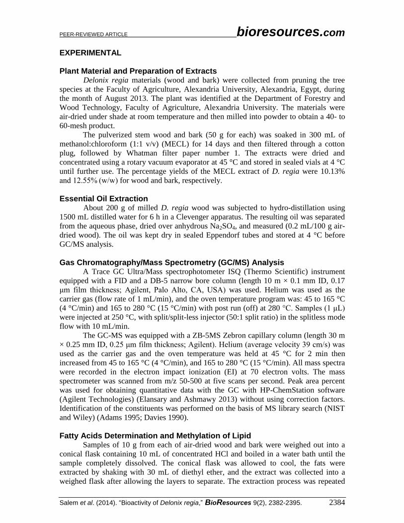
EXPERIMENTAL
Plant Material and Preparation of Extracts Delonix regia materials (wood and bark) were collected from pruning the tree
species at the Faculty of Agriculture, Alexandria University, Alexandria, Egypt, during
the month of August 2013. The plant was identified at the Department of Forestry and
Wood Technology, Faculty of Agriculture, Alexandria University. The materials were
air-dried under shade at room temperature and then milled into powder to obtain a 40- to
60-mesh product.
The pulverized stem wood and bark (50 g for each) was soaked in 300 mL of
methanol:chloroform (1:1 v/v) (MECL) for 14 days and then filtered through a cotton
plug, followed by Whatman filter paper number 1. The extracts were dried and
concentrated using a rotary vacuum evaporator at 45 °C and stored in sealed vials at 4 °C
until further use. The percentage yields of the MECL extract of D. regia were 10.13%
and 12.55% (w/w) for wood and bark, respectively.
Essential Oil Extraction About 200 g of milled D. regia wood was subjected to hydro-distillation using
1500 mL distilled water for 6 h in a Clevenger apparatus. The resulting oil was separated
from the aqueous phase, dried over anhydrous Na2SO4, and measured (0.2 mL/100 g air-
dried wood). The oil was kept dry in sealed Eppendorf tubes and stored at 4 °C before
GC/MS analysis.
Gas Chromatography/Mass Spectrometry (GC/MS) Analysis A Trace GC Ultra/Mass spectrophotometer ISQ (Thermo Scientific) instrument
equipped with a FID and a DB-5 narrow bore column (length 10 m × 0.1 mm ID, 0.17
μm film thickness; Agilent, Palo Alto, CA, USA) was used. Helium was used as the
carrier gas (flow rate of 1 mL/min), and the oven temperature program was: 45 to 165 °C
(4 °C/min) and 165 to 280 °C (15 °C/min) with post run (off) at 280 °C. Samples (1 μL)
were injected at 250 °C, with split/split-less injector (50:1 split ratio) in the splitless mode
flow with 10 mL/min.
The GC-MS was equipped with a ZB-5MS Zebron capillary column (length 30 m
× 0.25 mm ID, 0.25 μm film thickness; Agilent). Helium (average velocity 39 cm/s) was
used as the carrier gas and the oven temperature was held at 45 °C for 2 min then
increased from 45 to 165 °C (4 °C/min), and 165 to 280 °C (15 °C/min). All mass spectra
were recorded in the electron impact ionization (EI) at 70 electron volts. The mass
spectrometer was scanned from m/z 50-500 at five scans per second. Peak area percent
was used for obtaining quantitative data with the GC with HP-ChemStation software
(Agilent Technologies) (Elansary and Ashmawy 2013) without using correction factors.
Identification of the constituents was performed on the basis of MS library search (NIST
and Wiley) (Adams 1995; Davies 1990).
Fatty Acids Determination and Methylation of Lipid Samples of 10 g from each of air-dried wood and bark were weighed out into a
conical flask containing 10 mL of concentrated HCl and boiled in a water bath until the
sample completely dissolved. The conical flask was allowed to cool, the fats were
extracted by shaking with 30 mL of diethyl ether, and the extract was collected into a
weighed flask after allowing the layers to separate. The extraction process was repeated

PEER-REVIEWED ARTICLE bioresources.com
Salem et al. (2014). “Bioactivity of Delonix regia,” BioResources 9(2), 2382-2395. 2385
three times more and the solvent distilled off. The fat was then dried at 100 °C, cooled,
and weighed (Kirk and Sawyer 1991).
A lipid sample of 50 mg was weighed in a tube into which 50 mL of methanolic
sulfuric acid (1 mL of concentrated sulfuric acid and 100 mL of methanol) and 2 mL of
benzene were added. The tube was tightly closed and placed in a water bath at 90 °C for
an hour and half. The tube then was cooled and 8 mL water and 5 mL petroleum ether
were added. Subsequently, the tube was strongly shaken and the ethereal layer was
separated out and evaporated. Table 1 shows the conditions used for characterization of
fatty acids by GC. Standard fatty acids with C2-C25 were previously injected with the
same conditions used by GC (Radwan 1978).
Table 1. GC Condition for Analysis of Fatty Acids Device model HP (Hewlett Packard) 6890 GC Column HP-5 (5% diphenyl, 95% dimethyl polysiloxane), 30 m, 0.32
mm. ID, 0.25 µm film thickness Carrier gas/gas flow Nitrogen/1 mL/min Detector/temperature FID (flame ionization detector)/250 °C Injector temperature, Injection volume
220 °C, 2 µL in a splitless mode
Oven program Initial temp. 150 °C for 2 min
Ramps Rate °C/min Final Temp. °C Hold time 1 10 200 - 2 5 250 9 min
DPPH Radical-scavenging Assay The percent of the total antioxidant activity (TAA %) was evaluated by the 2.2-
diphenyl-1-picrylhydrazyl method (DPPH, Sigma-Aldrich) with some modifications
made (Salem et al. 2013). Two milliliters of a stock solution of 0.1 mM DPPH reagent
dissolved in pure methanol was added to a test tube containing 2 mL of the sample
solution in methanol (200 μg/L), and the mixture was well mixed for 10 s and left to
stand in the dark for 30 min at room temperature. Using a UV scanning
spectrophotometer (Unico® 1200, Alexandria, Egypt), the absorbance was measured at
517 nm and the TAA was determined by the formula TAA (%) = (A-control–A-
sample/A-control) × 100, where A-control is the absorbance of the control reaction
(containing all reagents except the test compound) and A-sample is the absorbance of the
test compound or the Tannic acid (positive control) solution. The control contained 2 mL
of DPPH solution and 2 mL of methanol. The measurements of DPPH radical scavenging
activity were carried out for three replicates.
Antibacterial Activity Assay Extracts, being wood and bark MECL and wood essential oil, with a
concentration of 2000 μg/mL were evaluated as antibacterial agents against the growth of
the Gram-positive bacteria Bacillus subtilis ATCC 6633, Sarcina lutea ATCC 9341, and
Staphylococcus aureus ATCC 6538 as well as the Gram-negative bacteria Escherichia
coli ATCC 8739 and Pectobacterium carotovorum subsp. carotovorum (strain No.
ippbc038) using the Kirby-Bauer disc diffusion susceptibility test (Bauer et al. 1966). A
sterile paper-filter disc, moistened with the extracts, was placed over the Petri dish
containing the culture medium that was seeded uniformly with bacterial or fungal strains.
The diameters of the inhibition zones (IZs) were measured in millimeters. Control discs

PEER-REVIEWED ARTICLE bioresources.com
Salem et al. (2014). “Bioactivity of Delonix regia,” BioResources 9(2), 2382-2395. 2386
were impregnated with 20 μL of dimethyl sulfoxide (DMSO, Sigma-Aldrich) solution.
Tetracycline (20 μg/disc) was used as a positive control with the tested bacteria. The
experiment was done in triplicate, and the means ± standard deviations are reported.
Minimum inhibitory concentrations (MICs) were determined by serial dilution (8, 16, 32,
64, 126, 250, 500, 1000, 2000, 4000, and 5000 μg/mL) of oil (wood) and MECL extract
(wood, bark) using 96-well micro-plates (Eloff 1998).
Antifungal Activity Assay The antifungal activity assay against the growth of Penicillium selerotigenum,
Paecilomyces variotii, and Aspergillus nigra was performed following Satish et al.
(2007), with some variation. About 15 mL of potato dextrose agar (PDA) medium
containing the concentrated extract (2000 μg/mL) was poured into each Petri dish and
allowed to solidify. A 1-cm disc of 7-day-old culture of the tested fungi was placed at the
center of the Petri dish and incubated at 25 ± 3 ºC for seven days. After incubation, the
colony diameter was measured in centimeters. A PDA medium with DMSO was used as
a control. The percentage inhibition of mycelial growth, in terms of fungitoxicity of the
extracts, was calculated using the following formula,
% inhibition = [(Mc–Mt)/Mc] ×100 (1)
where Mc is the average increase in mycelial growth in control and Mt is the average
increase in mycelial growth in treatment (Singh and Tripathi 1999). The experiment was
performed in triplicate.
RESULTS AND DISCUSSION
Figure 1 shows the GC/MS chromatogram analysis of the essential oil from D.
regia wood. The chemical characterization is presented in Table 2.
Fig. 1. GC/MS chromatogram of the essential oil from D. regia wood
PEER-REVIEWED ARTICLE bioresources.com
Salem et al. (2014). “Bioactivity of Delonix regia,” BioResources 9(2), 2382-2395. 2387
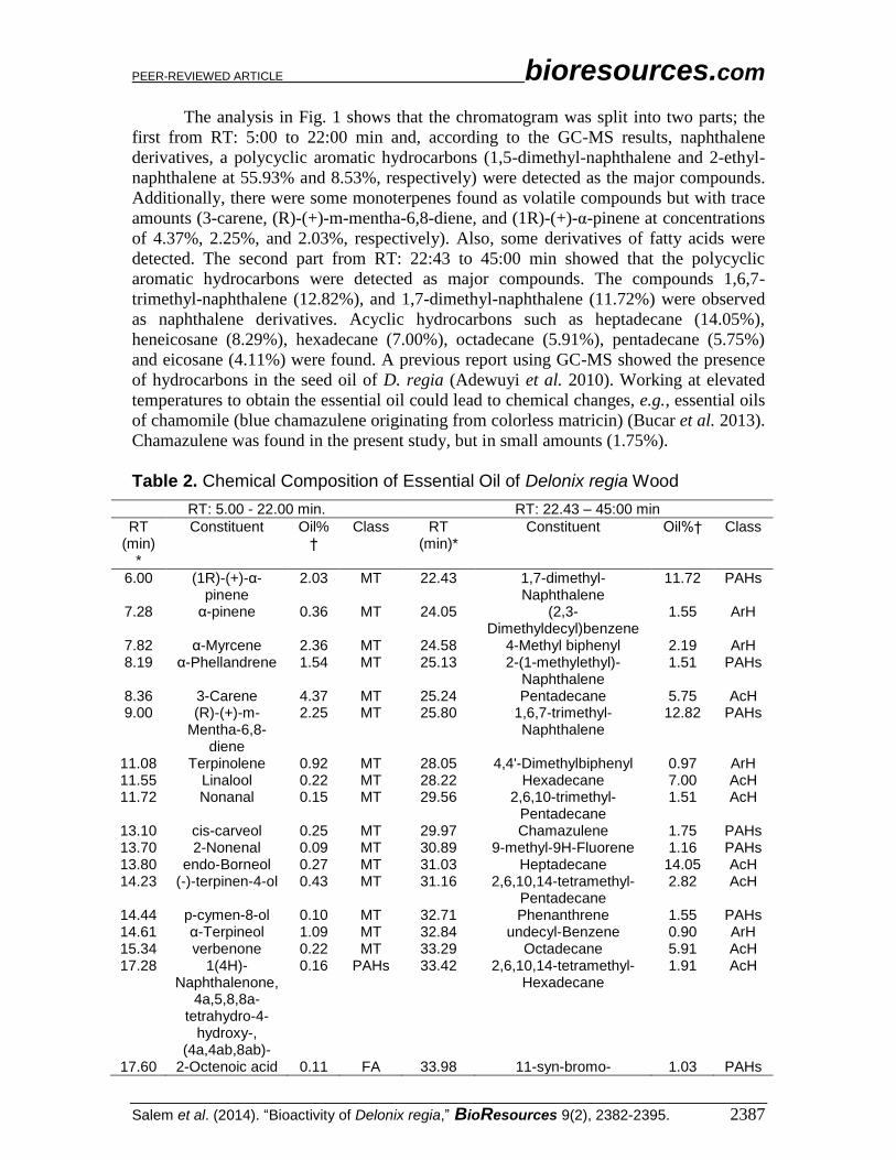
The analysis in Fig. 1 shows that the chromatogram was split into two parts; the
first from RT: 5:00 to 22:00 min and, according to the GC-MS results, naphthalene
derivatives, a polycyclic aromatic hydrocarbons (1,5-dimethyl-naphthalene and 2-ethyl-
naphthalene at 55.93% and 8.53%, respectively) were detected as the major compounds.
Additionally, there were some monoterpenes found as volatile compounds but with trace
amounts (3-carene, (R)-(+)-m-mentha-6,8-diene, and (1R)-(+)-α-pinene at concentrations
of 4.37%, 2.25%, and 2.03%, respectively). Also, some derivatives of fatty acids were
detected. The second part from RT: 22:43 to 45:00 min showed that the polycyclic
aromatic hydrocarbons were detected as major compounds. The compounds 1,6,7-
trimethyl-naphthalene (12.82%), and 1,7-dimethyl-naphthalene (11.72%) were observed
as naphthalene derivatives. Acyclic hydrocarbons such as heptadecane (14.05%),
heneicosane (8.29%), hexadecane (7.00%), octadecane (5.91%), pentadecane (5.75%)
and eicosane (4.11%) were found. A previous report using GC-MS showed the presence
of hydrocarbons in the seed oil of D. regia (Adewuyi et al. 2010). Working at elevated
temperatures to obtain the essential oil could lead to chemical changes, e.g., essential oils
of chamomile (blue chamazulene originating from colorless matricin) (Bucar et al. 2013).
Chamazulene was found in the present study, but in small amounts (1.75%).
Table 2. Chemical Composition of Essential Oil of Delonix regia Wood
RT: 5.00 - 22.00 min. RT: 22.43 – 45:00 min
RT (min)
*
Constituent Oil%†
Class RT (min)*
Constituent Oil%† Class
6.00 (1R)-(+)-α-pinene
2.03 MT 22.43 1,7-dimethyl-Naphthalene
11.72 PAHs
7.28 α-pinene 0.36 MT 24.05 (2,3-Dimethyldecyl)benzene
1.55 ArH
7.82 α-Myrcene 2.36 MT 24.58 4-Methyl biphenyl 2.19 ArH 8.19 α-Phellandrene 1.54 MT 25.13 2-(1-methylethyl)-
Naphthalene 1.51 PAHs
8.36 3-Carene 4.37 MT 25.24 Pentadecane 5.75 AcH 9.00 (R)-(+)-m-
Mentha-6,8-diene
2.25 MT 25.80 1,6,7-trimethyl-Naphthalene
12.82 PAHs
11.08 Terpinolene 0.92 MT 28.05 4,4'-Dimethylbiphenyl 0.97 ArH 11.55 Linalool 0.22 MT 28.22 Hexadecane 7.00 AcH 11.72 Nonanal 0.15 MT 29.56 2,6,10-trimethyl-
Pentadecane 1.51 AcH
13.10 cis-carveol 0.25 MT 29.97 Chamazulene 1.75 PAHs 13.70 2-Nonenal 0.09 MT 30.89 9-methyl-9H-Fluorene 1.16 PAHs 13.80 endo-Borneol 0.27 MT 31.03 Heptadecane 14.05 AcH 14.23 (-)-terpinen-4-ol 0.43 MT 31.16 2,6,10,14-tetramethyl-
Pentadecane 2.82 AcH
14.44 p-cymen-8-ol 0.10 MT 32.71 Phenanthrene 1.55 PAHs 14.61 α-Terpineol 1.09 MT 32.84 undecyl-Benzene 0.90 ArH 15.34 verbenone 0.22 MT 33.29 Octadecane 5.91 AcH 17.28 1(4H)-
Naphthalenone, 4a,5,8,8a-
tetrahydro-4-hydroxy-,
(4a,4ab,8ab)-
0.16 PAHs 33.42 2,6,10,14-tetramethyl-Hexadecane
1.91 AcH
17.60 2-Octenoic acid 0.11 FA 33.98 11-syn-bromo- 1.03 PAHs

PEER-REVIEWED ARTICLE bioresources.com
Salem et al. (2014). “Bioactivity of Delonix regia,” BioResources 9(2), 2382-2395. 2388
1,2,3,4,4a,9,10,10a-octahydro- 4a,10a-
Methanophenanthren-9á-ol
18.20 2-Methylnaphthal
ene
2.52 PAHs 34.20 2-methyl-Octadecane 1.99 AcH
18.38 (E,E)-2,4-decadien-1-al
5.29 FAld 34.55 2-methylanthracene 2.99 PAHs
18.62 Tridecane 0.09 AcH 35.41 n-Hexadecanoic acid 2.68 FA 18.76 1-
Methylnaphthalene
2.34 PAHs 35.73 Eicosane 4.11 AcH
19.88 Falcarinol 0.11 AcH 36.07 2,3-dimethyl-Phenanthrene
0.85 PAHs
19.97 2,5-Octadecadiynoic acid, methyl
ester
0.49 FA 36.59 Heneicosane 8.29 AcH
20.35 12,15-Octadecadiynoic acid, methyl
ester
0.24 FA 36.89 (Z)-7-Hexadecenal 0.89 FAld
20.47 9,10-dehydro-Isolongifolene
0.23 MT 38.58 Tetracosane 1.16 AcH
20.69 1-Phenylheptane
0.26 ArH Total 99.06%
21.12 Biphenyl 5.22 ArH 21.41 5,8,11-
Heptadecatriynoic acid, methyl
ester
0.15 FA
21.57 2-ethyl-Naphthalene
8.53 PAHs
21.94 1,5-dimethyl-Naphthalene
55.93
PAHs
Total 98.32%
* RT: Retention time (min). †Percentage of total FID area obtained on HP-5 capillary column. (MT): Monoterpene; (PAHs): Polycyclic aromatic hydrocarbons; (AcH); Acyclic Hydrocarbons; (ArH): Aromatic Hydrocarbon; (FA): Fatty acid; (FAld): Fatty aldehydes
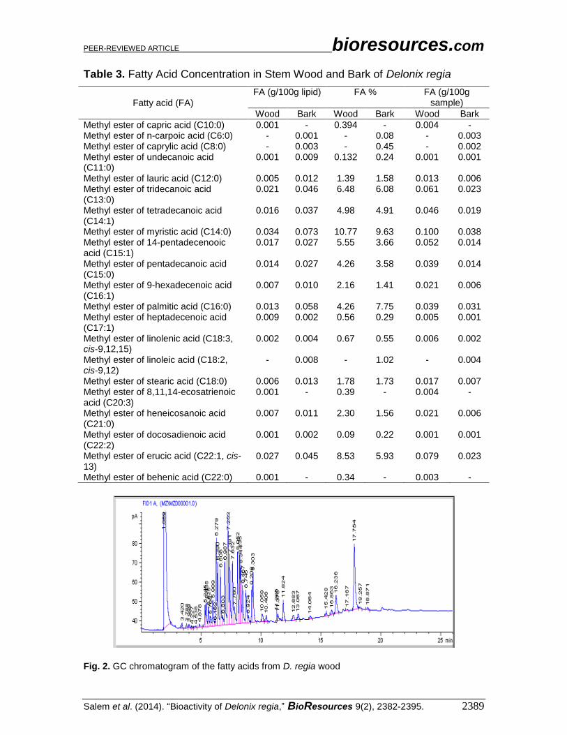
Fatty Acids Detected in Wood and Bark The total concentration of FA present in the wood and bark of D. regia were
0.177% and 0.386%, respectively. Additionally, the total amount found per sample was
0.514 and 0.200 g per 100 g, respectively. The nonpolar extractives (lipophilic
components) from the D. regia wood and bark were mainly composed of fatty acids and
fatty acid esters. GC analyses of fatty acids in the stem wood and bark of D. regia are
presented in Table 3. The major fatty acids detected in stem wood (Fig. 2) were myristic
acid (10.77%), erucic acid (8.532%), tridecanoic acid (6.48%), 14-pentadecenooic acid
(5.55%), tetradecanoic acid (4.98%), pentadecanoic acid (4.26%), and palmitic acid
(4.26%). The major fatty acid constituents found in bark (Figure 3) were myristic acid
(9.63%), palmitic acid (7.75%), tridecanoic acid (6.08%), erucic acid (5.93%),
tetradecanoic acid (4.91%), 14-pentadecenooic acid (3.66%), and pentadecanoic acid
(3.58%).
PEER-REVIEWED ARTICLE bioresources.com
Salem et al. (2014). “Bioactivity of Delonix regia,” BioResources 9(2), 2382-2395. 2389
Table 3. Fatty Acid Concentration in Stem Wood and Bark of Delonix regia
Fatty acid (FA)
FA (g/100g lipid) FA % FA (g/100g sample)
Wood Bark Wood Bark Wood Bark
Methyl ester of capric acid (C10:0) 0.001 - 0.394 - 0.004 - Methyl ester of n-carpoic acid (C6:0) - 0.001 - 0.08 - 0.003 Methyl ester of caprylic acid (C8:0) - 0.003 - 0.45 - 0.002 Methyl ester of undecanoic acid (C11:0)
0.001 0.009 0.132 0.24 0.001 0.001
Methyl ester of lauric acid (C12:0) 0.005 0.012 1.39 1.58 0.013 0.006 Methyl ester of tridecanoic acid (C13:0)
0.021 0.046 6.48 6.08 0.061 0.023
Methyl ester of tetradecanoic acid (C14:1)
0.016 0.037 4.98 4.91 0.046 0.019
Methyl ester of myristic acid (C14:0) 0.034 0.073 10.77 9.63 0.100 0.038 Methyl ester of 14-pentadecenooic acid (C15:1)
0.017 0.027 5.55 3.66 0.052 0.014
Methyl ester of pentadecanoic acid (C15:0)
0.014 0.027 4.26 3.58 0.039 0.014
Methyl ester of 9-hexadecenoic acid (C16:1)
0.007 0.010 2.16 1.41 0.021 0.006
Methyl ester of palmitic acid (C16:0) 0.013 0.058 4.26 7.75 0.039 0.031 Methyl ester of heptadecenoic acid (C17:1)
0.009 0.002 0.56 0.29 0.005 0.001
Methyl ester of linolenic acid (C18:3, cis-9,12,15)
0.002 0.004 0.67 0.55 0.006 0.002
Methyl ester of linoleic acid (C18:2, cis-9,12)
- 0.008 - 1.02 - 0.004
Methyl ester of stearic acid (C18:0) 0.006 0.013 1.78 1.73 0.017 0.007 Methyl ester of 8,11,14-ecosatrienoic acid (C20:3)
0.001 - 0.39 - 0.004 -
Methyl ester of heneicosanoic acid (C21:0)
0.007 0.011 2.30 1.56 0.021 0.006
Methyl ester of docosadienoic acid (C22:2)
0.001 0.002 0.09 0.22 0.001 0.001
Methyl ester of erucic acid (C22:1, cis-13)
0.027 0.045 8.53 5.93 0.079 0.023
Methyl ester of behenic acid (C22:0) 0.001 - 0.34 - 0.003 -
Fig. 2. GC chromatogram of the fatty acids from D. regia wood

PEER-REVIEWED ARTICLE bioresources.com
Salem et al. (2014). “Bioactivity of Delonix regia,” BioResources 9(2), 2382-2395. 2390
Linoleic acid was the major fatty acid distributed across the lipid fractions of seed
oil of D. regia, and the neutral lipids had the highest amount of stearic and linoleic acids
(Adewuyi et al. 2010). Normal fatty acids such as myristic (1.1 %), palmitic (14.0%),
stearic (11.0%), oleic (12.7%), and linoleic (50.7%) were found in D. regia seed oil
(Hoasamani and Hosamani 1995). Palmitic acid was found to be 13.1% in D. regia seed
oil (Adewuyi et al. 2010) while stearic acid was found at 12.1%.
Fig. 3. GC chromatogram of the fatty acids from D. regia bark
Antibacterial and Antifungal Activities of Extracts All tested essential oil and extracts exhibited antibacterial and antifungal activity
to differing degrees (Table 4). Based on the investigations, MECL wood extract of D.
regia was shown to be effective against B. subtilis, S. lutea, and S. aureus with respective
IZ values of 16.66 ± 0.57 mm, 14.33 ± 1.15 mm, and 20.66 ± 0.57 mm, where the MECL
extract of bark was most active against E. coli (21.33 ± 0.57 mm). The essential oil
showed good antibacterial activity against P. carotovorum (17.66 ± 0.57 mm), while the
MECL extract of bark showed no activity against P. carotovorum.
Table 4. Antibacterial and Antifungal Activities of Extract from Wood and Bark of Delonix regia
Extract IZ of bacterial strains (mm)a % Inhibition of fungal mycelial
growth
B. subtilis
S. lutea
S. aureus
E. coli
P. carotovorum
P. selerotigenum
P. variotii
A. nigra
Essential oil
13.66 ± 1.15 (250)
15.00 ± 0.00 (64)
14.33 ± 1.15 (500)
20.66 ± 0.57 (16)
17.66 ± 0.57 (250)
44.44 74.07
44.44
MECL wood
16.66 ± 0.57 (32)
14.33 ± 1.15 (1000)
20.66 ± 0.57 (32)
18.33 ± 0.57 (32)
14.33 ± 1.15 (64)
25.93
66.67
14.81
MECL bark
11.66 ± 1.52 (500)
12.66 ± 0.57 (1000)
13.66 ± 1.15 (250)
21.33 ± 0.57 (126)
na (>5000)
70.37
77.78
3.70
Negative control
na na na na na 0 0 0
Positive control
b
18 25 20 18 nt nt nt nt
aZone of inhibition in mm (include 5 mm disc). na - not active. nt – not tested
bTetracycline (20 μg/disc). Values in parentheses are MICs values (μg/mL).

PEER-REVIEWED ARTICLE bioresources.com
Salem et al. (2014). “Bioactivity of Delonix regia,” BioResources 9(2), 2382-2395. 2391
The MECL extract of bark showed the maximum percentage of inhibition of
fungal mycelial growth against P. selerotigenum (70.37%) and P. variotii (77.78%). On
the other hand, the essential oil showed moderate inhibition against A. nigra (44.44%).
The extracts from different parts of D. regia have been reported to possess anti-
bacterial, anti-malarial, and anti-fungal properties (Ankrah et al. 2003; Aqil and Ahmad
2007; Dutta et al. 1998; Parekh et al. 2005). Some carotenoids such as 2-carotene,
zeaxanthein, etc., present in the extract of D. regia (Jungalwala and Cama 1962) also
exhibited antimicrobial activities. Jahan et al. (2010) reported that the zones of inhibition
demonstrated by the petroleum ether, carbon tetrachloride, and dichloromethane fractions
of extracts of D. regia stem bark ranged from 9 to 14 mm, 11 to 13 mm, and 9 to 20 mm,
respectively, compared to kanamycin as a standard antibiotic with zones of inhibition
ranged between 20 mm and 25 mm. Other studies reported that constituents such as 2,4-
bis(1,1-dimethylethyl)-phenol, 12-methyl-tetradecanoic acid methyl ester, hexadecanoic
acid methyl ester, phytol, and octadecanoic acid methyl ester have proven antimicrobial
potential (Agoramoorthy et al. 2007; Bikovens et al. 2013; Namuli et al. 2011; Rani et al.
2011).
Total Antioxidant Activity The total antioxidant activity (TAA %) of essential oil, stem wood, and stem bark
MECL extracts were 84.34%, 80.33%, and 70.21%, respectively. The results in the
present study are close to or higher than the value of tannic acid (TAA 83%). From the
previous work, the TAA% from methanolic extract of stem bark was 78.35 ± 1.45 %
(Salem 2013). The detected phenolic compounds in D. regia extracts, i.e., caffeic acid,
salicylic acid, ferulic acid, gentisic acid, chlorogenic acid, 3-hydroxybenzoic acid, 4-
hydroxycinnamic acid, p-coumaric acid, gallic acid, and 4-hydroxybenzoic acid, are
known to have antioxidant properties (Merkl et al. 2010; Proestos et al. 2005). The
DPPH radical scavenging activity of D. regia extracts was found to be inconsistent with
previous studies (Shabir et al. 2011).
CONCLUSIONS
The use of natural extracts in controlling plant diseases has little to no
environmental impact, so they may become a viable option for the development of
organic and sustainable agriculture. However, it is necessary to study the molecular and
biochemical changes that these compounds may have on pathogens and plants. The
present study has shown the fatty acid profile of wood and bark from Delonix regia, as
well as the chemical characterization of the essential oil of its wood. The following points
can be drawn from the present study:
1. Naphthalene derivatives such as 1,5-dimethyl-naphthalene, 2-ethyl-naphthalene,
1,6,7-trimethyl-naphthalene (12.82%), and 1,7-dimethyl-naphthalene were detected
as polycyclic aromatic hydrocarbons in the essential oil of D. regia wood.
2. The major fatty acids detected in D. regia stem wood were myristic acid (10.77%),
erucic acid (8.532%), and tridecanoic acid (6.48%). The major fatty acid constituents
found in D. regia stem bark were myristic acid (9.63%), palmitic acid (7.75%), and
tridecanoic acid (6.08%).

PEER-REVIEWED ARTICLE bioresources.com
Salem et al. (2014). “Bioactivity of Delonix regia,” BioResources 9(2), 2382-2395. 2392
3. The methanol-chloroform wood extract of D. regia showed effective activity against
B. subtilis, S. lutea, and S. aureus, with IZ values of 16.66 ± 0.57 mm, 14.33 ± 1.15
mm, and 20.66 ± 0.57 mm, respectively, where the methanol-chloroform extract of D.
regia bark was the most active against E. coli (21.33 ± 0.57 mm). The essential oil
showed good antibacterial activity against P. carotovorum (17.66 ± 0.57 mm).
4. The methanol-chloroform extract of D. regia bark showed the maximum percentage
of inhibition of fungal mycelial growth against P. selerotigenum (70.37%) and P.
variotii (77.78%). On the other hand, the essential oil showed moderate inhibition
against A. nigra (44.44%).
5. The total antioxidant activity (TAA %) of essential oil, stem wood, and stem bark
MECL extract was 84.34%, 80.33%, and 70.21%, respectively, and the results in the
present study were close to or higher than that for tannic acid (TAA 83%).
ACKNOWLEDGMENTS
The authors extend their appreciation to the Deanship of Scientific Research at
King Saud University for the funding of their work through the research group project
No. RGPVPP-010.
REFERENCES CITED
Adewuyi, A., Oderinde, R. A., Rao, B. V. S. K., Prasad, R. B. N., and Anjaneyulu, B.
(2010). “Chemical component and fatty acid distribution of Delonix regia and
Peltophorum pterocarpum seed oils,” Food Sci. Technol. Res. 16(6), 565-570.
Adams, R. P. (1995). “Identification of essential oil components by gas chromatograph
/quadrupole mass spectroscopy,” Carol Stream, IL: Allured Publishing.
Adjé, F., Lozano, Y. F., Lozano, P., Adima, A., Chemat, F., and Gaydou, E. M. (2010).
“Optimization of anthocyanin, flavonol, and phenolic acid extractions from Delonix
regia tree flowers using ultrasound-assisted water extraction,” Ind. Crops Prod.
32(3), 439-444.
Agoramoorthy, G, Chandrasekaran, M., Venkatesalu, V., and Hsu, M. J. (2007).
“Antibacterial and antifungal activities of fatty acid methyl esters of the blind-your-
eye mangrove from India,” Braz. J. Microbiol. 38(4), 739-742.
Ahmad, I., and Aquil, F. (2003). “Broad spectrum antibacterial and antifungal activities
and potency of crude, alcoholic extract and fraction of Delonix regia flowers,” 2nd
World Congress on “Biotechnological Development of Herbal Medicine”, NBRI,
Lucknow, UP, India, page 74.
Ali, M. S., Azhar, I., Amtul, Z., Ahmad, V. U., and Usmanghani, K. (1999).
“Antimicrobial screening of some Caesalpiniaceae,” Fitoterapia 70(3), 299-304.
Anderson, B. S., Hunt, J. W., Phillips, B. M., Nicely, P. A., de Vlaming, V., Connor, V.,
Richard, N., and Tjeerdema, R. S. (2003). “Integrated assessment of the impacts of
agricultural drain water in the Salinas River,” Environ. Pollut. 124(3), 523-532.
Ankrah, N. A., Nyarko, A. K., and Addo, P. G. A. (2003). “Evaluation of efficacy and
safety of a herbal medicine used for the treatment of malaria,” Phytother. Res. 17(6),
697-701.

PEER-REVIEWED ARTICLE bioresources.com
Salem et al. (2014). “Bioactivity of Delonix regia,” BioResources 9(2), 2382-2395. 2393
Aqil, F. M., and Ahmad, I., (2007). “Antibacterial properties of traditionally used Indian
medicinal plants,” Meth. Findings Experim. Clini. Pharm. 29(2), 79-92.
Arteaga, S., Andrade, C. A. and Cárdenas, R. (2005). “Larrea tridentata (creosote bush),
an abundant plant of Mexican and US-American deserts and its metabolite
nordihydroguaiaretic acid,” J. Ethno pharmacol. 98(3), 231-239.
Azab, S. S., Abdel-Daim, M., and Eldahshan, O. A. (2013). “Phytochemical, cytotoxic,
hepatoprotective and antioxidant properties of Delonix regia leaves extract,” Med.
Chem. Res. 22(9), 4269-4277.
Bauer, A. W., Kirby, W. M., Sherris, J. C., and Turck, M. (1966). “Antibiotic
susceptibility testing by a standardized single disk method,” Am. J. Clin. Pathol.
45(4), 493-496.
Bikovens, O., Roze, L., Pranovich, A., Reunanen, M., and Telysheva, G. (2013).
“Chemical composition of lipophilic extractives from grey alder (Alnus incana),”
BioResources 8(1), 350-357.
Bucar, F., Wube, A., and Schmid, M. (2013). “Natural product isolation – How to get
from biological material to pure compounds,” Nat. Prod. Rep. 30(4), 525-545.
Chandler, J. (2005). “Cost reduction in SIT programmes using exosect auto-
dissemination as part of area wide integrated pest management,” Int. J. Pest Cont.
47(5), 257-260.
Davies, N. W. (1990). “Gas chromatographic retention indices of monoterpenes and
sesquiterpenes on methyl silicone and Carbowax 20M phases,” Journal of
Chromatography A 503, 1-24. British Pharmacopeia (1998). (Vol. II). HMSO:
London.
Demirbas, A. (2001). “Biomass resource facilities and biomass conversion processing for
fuel and chemicals,” Energ. Convers. Manage. 42(11), 1357-1378.
Dutta, B. K., Rahman, I., and Das, T. K. (1998). “Antifungal activity of Indian plant
extracts,” Mycoses. 41(11-12), 535-536.
Einbond, L. S., Reynertson, K. A., Luo, X. D., Basile, M. J., and Kennelly, E. J. (2003).
“Anthocyanin antioxidants from edible fruits,” Food Chem. 84(1), 23-28.
Elansary, H. O., and Ashmawy, N. A. (2013). “Essential oils of mint between benefits
and hazards,” Journal of Essential Oil Bearing Plants 16(4), 429-438.
Eloff, J. N. (1998). “A sensitive and quick microplate method to determine the minimal
inhibitory concentration of plant extracts for bacteria,” Planta Medica 64, 711-713.
Fatmawaty, F., and Astuti, H. (2013). “Antimalarial activity of Delonix regia on mice
with Plasmodium berghei,” J. Nat. Prod. 6, 61-66.
Hoasamani, K. M., and Hosamani, S. K., (1995). “Component fatty acids of Delonix
regis seed oil-A source of 7-(2-octacyclopropen-1-yI) heptanoic acid and 8-(2-
octacyclopropen-1-yl) octanoic acid,” Eur. J. Lipid. Sci. Tech. 97(11), 420-422.
Jahan, I., Rahman, M. S., Rahman, M. Z., Kaisar, M. A., Islam, M. S., Wahab, A., and
Rashid, M. A. (2010). “Chemical and biological investigations of Delonix regia
(Bojer ex Hook.) Raf,” Acta Pharmaceut. 60(2), 207-215.
Jungalwala, F. B., and Cama, H. R. (1962). “Carotenoids in Delonix regia (Gul Mohr)
flower,” Biochem. J. 85(1), 1-8.
Kirk, R. S., and Sawyer, R. (1991). Pearson’s Chemical Analysis of Foods, 9th
Ed.,
Longman Scientific and Technical, Harlow, Essex, UK.
Merkl, R., Hrádková, I., Filip, V., and Šmidrkal, J. (2010). “Antimicrobial and
antioxidant properties of phenolic acids alkyl esters,” Czech J. Food Sci. 28(4), 275-
279.

PEER-REVIEWED ARTICLE bioresources.com
Salem et al. (2014). “Bioactivity of Delonix regia,” BioResources 9(2), 2382-2395. 2394
Muruganandan, V., Srinivas, K., Tandan, S. K., Jawahar, L., Chandra, S., and
Raviprakash, V. (2001). “Anti-inflammatory and analgesic activity of some medicinal
plants,” J. Med. Arom. Plants Res. 22(4a), 56-85.
Namuli, A., Abdullah, N., Sieo, C. C., Zuhainis, S. W., and Oskoueian, E. (2011).
“Phytochemical compounds and antibacterial activity of Jatropha curcas Linn.
Extracts,” J. Med. Plants Res. 5(16), 3982-3990.
Parekh, J., Jadeja, D., and Chanda, S. (2005). “Efficacy of aqueous and methanol extracts
of some medicinal plants for potential antibacterial activity,” Turk. J. Biol. 29(4),
203-210.
Proestos, C., Chorianopoulos, N., Nychas, G. J. E., and Komaitis, M. (2005). “RP-HPLC
analysis of the phenolic compounds of plant extracts. Investigation of their
antioxidant capacity and antimicrobial activity,” J. Agric. Food Chem. 53(4), 1190-
1195.
Radwan, S. S. (1978). “Coupling of two-dimensional thin-layer chromatography with gas
chromatography for the quantitative analysis of lipid classes and their constituent
fatty acids,” J. Chrom. Sci. 16(11), 538-542.
Rani, M. J. P., Kannan, P. S. M., and Kumaravel, S. (2011). “Screening of antioxidant
activity, total phenolics and gas chromatograph and mass spectrometer (GC-MS)
study of Delonix regia,” Afr. J. Biochem. Res. 5(12), 341-347.
Salem, M. Z. M. (2013). “Evaluation of the antibacterial and antioxidant activities of
stem bark extracts of Delonix regia and Erythrina humeana grown in Egypt,” J.
Forest Prod. Ind. 2(2), 48-52.
Salem, M. Z. M., Ali, H. M., El-Shanhorey, N. A., and Abdel-Megeed, A. (2013).
“Evaluation of extracts and essential oil from Callistemon viminalis leaves:
Antibacterial and antioxidant activities, total phenolic and flavonoid contents,” Asian
Pac. J. Trop. Med. 6(10), 785-791.
Satish, S., Mohana, D. C., Ranhavendra, M. P., and Raveesha, K. A. (2007). “Antifungal
activity of some plant extracts against important seed borne pathogens of Aspergillus
sp.,” J. Agric. Tech. 3(1), 109-119.
Shabir, G., Anwar, F., Sultana, B., Khalid, Z. M., Afzal, M., Khan, Q. M., and
Ashrafuzzaman, M. (2011). “Antioxidant and antimicrobial attributes and phenolics
of different solvent extracts from leaves, flowers and bark of Gold Mohar [Delonix
regia (Bojer ex Hook.) Raf],” Molecules 16(9), 7302-7319.
Sheikh, M. I. (1993). Trees of Pakistan, GOP-USAID Forestry Planning and
Development Project, Islamabad, Pakistan.
Shewale, V. D., Tushar, A., Deshmukh, L., Patil, S., and Patil, V. R. (2012). “Anti-
inflammatory activity of Delonix regia (Boj. Ex. Hook),” Adv. Phar. Sci. 789713, 4.
Singh, J., and Tripathi, N. N. (1999). “Inhibition of storage fungi of blackgram (Vigna
mungo) by some essential oils,” Flav. Frag. J. 14(1), 1-4.
Teow, C. C., Truong, V. D., McFeeters, R. F., Thompson, R. L., Pecota, K. V., and
Yencho, G. C. (2007). “Antioxidant activities, phenolic and β-carotene contents of
sweet potato genotypes with varying flesh colours,” Food Chem. 103(3), 829-838.
Veigas, J. M., Divya, P., and Neelwarne, B. (2012). “Identification of previously
unreported pigments among carotenoids and anthocyanins in floral petals of Delonix
regia (Hook.) Raf,” Food Res. Int. 47(1), 116-123.
Webb, D. B., Wood, P. J., Smith, J. P., and Henman, G. S. (1984). “A guide to species
selection for tropical and sub-tropical plantations,” Tropical Forestry Papers, No. 15.
Oxford, UK: Commonwealth Forestry Institute, University of Oxford, p. 256.
PEER-REVIEWED ARTICLE bioresources.com
Salem et al. (2014). “Bioactivity of Delonix regia,” BioResources 9(2), 2382-2395. 2395
Whalen, M. M., Wilson, S., Gleghorn, C., and Loganathan, B. G. (2003). “Brief exposure
to triphenyltin produces irreversible inhibition of the cytotoxic function of human
natural killer cells,” Environ. Res. 92(3), 213-220.
Wilson, C. L., Ghaouth, A. E., and Wisniewski, M. E. (1999). “Prospecting in nature´s
storehouse for biopesticides,” Mex. J. Phytopath. 17(1), 49-53.
Article submitted: January 14, 2014; Peer review completed: March 3, 2014; Revised
version received and accepted: March 7, 2014; Published: March 13, 2014.