STATOCYST-INDUCED EYE MOVEMENTS IN ... - CiteSeerX
20
J. Exp. Biol. (1972). 57. 187-204 187 With 1 plate and 10 text-figures M'rinted in Great Britain STATOCYST-INDUCED EYE MOVEMENTS IN THE CRAB SCYLLA SERRATA I. THE SENSORY INPUT FROM THE STATOCYST* BY D. C. SANDEMAN AND A. OKAJIMAf Department of Neurobiology, Research School of Biological Sciences, Australian National University, Canberra, A.C.T., Australia (Received 4 January 1972) INTRODUCTION The slow and fast phases of the eye movements of crabs during optokinetic nystagmus have been well investigated in intact animals, and a model of the whole system exists (Horridge & Sandeman, 1964; Barnes & Horridge, 1969). It has not yet been possible to extend these studies to include an electrophysiological investigation of the central nervous mechanisms because the optic lobes in the eyestalk do not withstand isolation although the isolated brains of crabs respond to the stimulation of sensory nerves (Sandeman, 1971). It has proved impractical to use visual stimuli to drive the oculomotor system in isolated preparations. Preliminary experiments, however, have shown that the oculomotor system in semi-isolated crab-brain prepara- tions can be driven by irrigating the lumen of the statocyst with saline. The statocyst input alone is known to initiate nystagmus in crabs when they are rotated about the vertical axis (see review by Cohen & Dijkgraaf, 1961) and so our aim is to investigate fully the nervous control of crab eye movements that are generated by the directional input from the statocyst. Before analysis of the central nervous integration can be made it is essential to know the sensory input in detail and we therefore begin by characterizing the responses of the statocyst sensory cells and describe the direction of fluid flow produced naturally by rotation of the organ about its vertical axis. In this paper we describe the anatomy of the horizontal and vertical canal systems in the statocyst and the extracellular responses of units in the statocyst nerves following irrigation of these canals. Early comparisons of decapod statocysts with the vertebrate inner ear resulted in a belief that the statocyst was an organ of balance and hearing (Hensen, 1863). Subse- quently the supposed auditory function of the organ was disproved, but it was also claimed that the crab statocyst could not function in the same way as the semi- circular canals of vertebrates because the statocyst lumen is an open structure and not a system of canals (Prentiss, 1901), and this view persisted in the general literature. The similarity between the non-acoustic labyrinth system of vertebrates and the crab statocyst has nevertheless again been emphasized by Dijkgraaf (1955, 1956a, 1961). The most complete early description of the receptor hairs in crab statocysts is that • This paper is dedicated to Professor H. Kinosita on the occasion of his retirement from the University of Tokyo. T Present address: Biological Institute, Yokohama City University, Yokohama, Japan.
-
Upload
khangminh22 -
Category
Documents
-
view
2 -
download
0
Transcript of STATOCYST-INDUCED EYE MOVEMENTS IN ... - CiteSeerX

J. Exp. Biol. (1972). 57. 187-204 187With 1 plate and 10 text-figures
M'rinted in Great Britain
STATOCYST-INDUCED EYE MOVEMENTS IN THE CRABSCYLLA SERRATA
I. THE SENSORY INPUT FROM THE STATOCYST*
BY D. C. SANDEMAN AND A. OKAJIMAf
Department of Neurobiology, Research School of Biological Sciences, AustralianNational University, Canberra, A.C.T., Australia
(Received 4 January 1972)
INTRODUCTION
The slow and fast phases of the eye movements of crabs during optokineticnystagmus have been well investigated in intact animals, and a model of the wholesystem exists (Horridge & Sandeman, 1964; Barnes & Horridge, 1969). It has not yetbeen possible to extend these studies to include an electrophysiological investigationof the central nervous mechanisms because the optic lobes in the eyestalk do notwithstand isolation although the isolated brains of crabs respond to the stimulationof sensory nerves (Sandeman, 1971). It has proved impractical to use visual stimulito drive the oculomotor system in isolated preparations. Preliminary experiments,however, have shown that the oculomotor system in semi-isolated crab-brain prepara-tions can be driven by irrigating the lumen of the statocyst with saline. The statocystinput alone is known to initiate nystagmus in crabs when they are rotated about thevertical axis (see review by Cohen & Dijkgraaf, 1961) and so our aim is to investigatefully the nervous control of crab eye movements that are generated by the directionalinput from the statocyst.
Before analysis of the central nervous integration can be made it is essential to knowthe sensory input in detail and we therefore begin by characterizing the responses ofthe statocyst sensory cells and describe the direction of fluid flow produced naturallyby rotation of the organ about its vertical axis. In this paper we describe the anatomy ofthe horizontal and vertical canal systems in the statocyst and the extracellular responsesof units in the statocyst nerves following irrigation of these canals.
Early comparisons of decapod statocysts with the vertebrate inner ear resulted in abelief that the statocyst was an organ of balance and hearing (Hensen, 1863). Subse-quently the supposed auditory function of the organ was disproved, but it was alsoclaimed that the crab statocyst could not function in the same way as the semi-circular canals of vertebrates because the statocyst lumen is an open structure andnot a system of canals (Prentiss, 1901), and this view persisted in the general literature.The similarity between the non-acoustic labyrinth system of vertebrates and the crabstatocyst has nevertheless again been emphasized by Dijkgraaf (1955, 1956a, 1961).
The most complete early description of the receptor hairs in crab statocysts is that• This paper is dedicated to Professor H. Kinosita on the occasion of his retirement from the
University of Tokyo.T Present address: Biological Institute, Yokohama City University, Yokohama, Japan.

i88 D. C. SANDEMAN AND A. OKAJIMA
Text-fig, i. The system used to irrigate the statocyst canals. Two reservoirs (rt and r2) arelinked to vertical tubes (*i and £a) and through a three-way stopcock (sto) to a common outlettube. The stopcock position is monitored by a light source (not shown) and two photoresistorslinked to a differential amplifier. A shield on the stopcock tap is arranged so that it cuts off thelight from the photoresistors and causes a positive potential for positive pressures and a nega-tive potential for negative pressures. The stimulus can be changed directly from a positiveto a negative value and vice versa, or from zero to either a negative or positive pressure. Thecommon outlet tube is coupled to a pipette (p) which is inserted into the statocyst canals. Theintensity of the stimulus is controlled by the level of the reservoirs relative to that of thepreparation bath (6), which is monitored by a third vertical tube (t3) and kept constant by asiphon (sip). Cooled saline flows into the bath through a cannula (c). A further control of thestimulus intensity is achieved by shunting the flow from the reservoirs through a capillarytube before it reaches the statocyst. A syringe (syr) is incorporated into the system to aidfilling and to expel air bubbles in the system. Extracellular nerve recordings are made fromthe statocyst nerve with platinum-wire hook electrodes (e).
of Hensen (1863). He described three kinds of hair receptors within the statocyst-the hook hairs, group hairs and thread hairs. Dijkgraaf demonstrated, in Maja andCarcinus, the presence of a statolith supported on hook-shaped hairs. He called these'statolith' hairs and renamed Hensen's hook hairs as free hook hairs because they donot bear a statolith. Dijkgraaf showed that of the four types of hair, the slender threadhairs were the ones most likely to be associated with the detection of rotation of theanimal about the vertical axis (Dijkgraaf, 1956a). There is no electrophysiologicalinformation on the responses of the various statocyst receptors in crabs although theresponses of receptors in the lobster statocysts are known (Cohen, 1955, i960).
MATERIAL AND METHODS
The Australian mud crab, Scylla serrata, was used in all the experiments. Animalswere kept alive in the laboratory wrapped in wet hessian.
The anterior part of the crab was separated from the rest of the body and the brainwas removed after cutting the antennular nerves close to it. The proximal portion of

Statocyst-induced eye movements in crabs. I
Ventral
Text-fig. 2. A dissection of the basal joint of the right antennuleseen from behind. The proximalpart of the eyestalk has been cut away and also the posterior part of the basal joint of the anten-nule to show the horizontal and vertical canals of the statocyst and the nerves from them.The medial nerve bundle (I) does not contain sensory nerves from the statocyst but bothlateral bundles do (II and III). Also contained in the lateral bundles are motor fibres supplyingthe muscles grouped around the basal joint. Nerve II contains axons from the thread hairs,free hook hairs and some statolith hairs. Nerve III contains axons from the statolith hairs.
the eyestalk was cut away to reveal the basal joint of the antennule. The rear wall ofthe basal joint was carefully cut out to expose the canal system of the statocyst.
Controlled irrigation of the statocyst lumen was achieved as follows. Two holes weremade at each end of either the horizontal or vertical canal systems, depending onwhich group of receptors was under investigation. The tip of a glass pipette of internaldiameter +180 fim, was placed well into one of the holes and the intensity of positiveor negative flow of saline through the tip was controlled by raising or lowering tworeservoirs of saline attached to the pipette (Text-fig. 1). The level of saline coveringthe preparation, and the levels in the two reservoirs were compared in three verticaltubes. This allowed the establishment of a zero level and provided a means of calibrat-ing the rate of flow. In some preparations the lumen of the statocyst was opened andthe receptors were manipulated with needles. The onset and cessation of the flowthrough the pipette was monitored by a photocell attached to the stopcock whichcontrolled the flow.
Electrical recordings were made after de-sheathing and splitting the various nervebundles and lifting the strands on platinum-wire hook electrodes. A mineral-oil drop

190 D. C. SANDEMAN AND A. OKAJIMA
Text-fig. 3. Dissection of the basal joint of the antennule and statocyst. The posterior walls of thehorizontal and vertical canals have been removed and the positions of the group hairs, threadhairs, free hook hairs and statolith are shown. The thread hairs form a single line across thesensory cushion and project across the canals. The nerve supply to the free hook hairs (partof nerve II) has been removed to show the bundle of nerves running behind the statocyst tothe bases of the thread hairs.
around the electrodes prevented the strands from drying out. Responses were tape-recorded and the recordings were then analysed and plotted with a Hewlett Packard5480 B Signal Analyser to show the relative frequencies of the different receptors withrespect to stimulus intensity.
RESULTS
I. Anatomy
The statocyst lumen of Scylla is contained in the basal joint of the antennule and isclosed off from the outside by a suture along the dorsal surface of the basal joint. Thelumen of the statocyst is shaped like two hollow toroids joined almost at right angles toeach other (Text-fig. 2; PI. 1, figs 1, 2).
The dorsal toroid, referred to here as the horizontal canal, is formed by a pinching-in of the upper and lower walls of the statocyst. Its circumference lies tilted about 15 °from the horizontal, the medial edge being lower than the lateral edge.
The vertically oriented toroid, or vertical canal, is not completely joined at its centre

Statocyst-induced eye movements in crabs. I 191
Free-hook hairs
^ r *
Thread hairs
- - - . • -
/•ft; '^, Statolith
8
Text-fig. 4. Camera lucida drawings to show the disposition of the free hook hairs, statolithhairs and thread-hair bases within the statocyst. The free hook hairs are arranged along theposterior area of the vertical canal. Towards the bottom of the vertical canal the number of freehook hairs diminishes and there is a patch of statolith hairs surrounded by two circles of freehook hairs. The thread hairs are distributed over the sensory cushion in a single line, but thereare proportionately more hairs at the dorsal end of the cushion than at the ventral end.
but a central invagination of the medial wall of the statocyst ('sensory cushion' ofPrentiss, 1901) comes within 80 /on of the opposite side of the statocyst, leaving acanal around it of 500-750 /tm in diameter. The circumference of the vertical canal istilted about 20 ° from the vertical and is also rotated approximately 45 ° about thisaxis so that its circumferential plane does not lie along the horizontal longitudinalaxis of the animal. This is important with regard to the sensitivity of the system todisplacement about both horizontal axes (see Discussion).

192 D. C. SANDEMAN AND A. OKAJIMA
Within the canals there are four groups of hair-receptor organs which have essen-tially the same structure as the receptors described in Carcinus and Maja.
(1) Large 'group hairs' are situated along the most lateral part of the horizontalcanal (Text-fig. 3). They are 15 /im thick, 800 /im long, are not narrowed at their basesand are feathered along their length.
(2) 'Free hook hairs' which lie along the posterior arm of the vertical canal (Text-fig. 3). These are 40-60 /im long, feathered, about 5 /im wide at their widest pointand narrowed at their bases to 1-2 /im (Text-fig. 4). They are often bent into a hook-like shape as their name implies.
(3) 'Statolith hairs' are confined to a patch at the bottom of the vertical canal andsurrounded by two arrays of free hook hairs (Text-fig. 4). They are simple unfeatheredhairs 3-4 /an long and less than 1 /im in diameter.
(4) 'Thread hairs' are arranged in a single line over the surface of the sensorycushion, but more hairs are concentrated on the upper part of the cushion and oppositethe top of the vertical canal than at the bottom of it (Text-fig. 4). The hairs are about300/im long and about 2 /im wide where they project into the canals and about one-third this length where the summit of the cushion lies close to the apposed wall ofthe statocyst.
The antennular nerve enters the brain as three main bundles (Text-figs. 2, 3). Themost medial (I) extends from the distal joints of the antennule and does not receiveany input from the statocyst. The two lateral bundles, II and III, contain sensoryfibres from the statocyst and motor fibres which innervate the muscles of the basaljoint of the antennule. The lateral nerves can be easily split into smaller bundles whichcome from different parts of the statocyst. Nerve II consists of three smaller bundles;the two dorsal bundles contain fibres from the free hook hairs, and the ventral bundlecontains only fibres from the thread hairs. Part of III carries sensory axons from thestatolith hairs.
One bundle in the ventral part of II can be clearly traced into the sensory cushion,and electrical recordings from this nerve always give pure thread-hair-type responses(see below). Counts of thread hairs have shown between 80 and 96 hairs, but a countof fibres in the nerve bundle going into the sensory cushion reveals more than twicethis number. The extra axons may be accounted for by there being more than one nervecell for each receptor, as described for Astacus (Schone & Steinbrecht, 1968).
II. Physiology
Pure recordings from the thread hairs and statolith hairs were obtained relativelyeasily because most of the axons from these sense organs run in discrete bundles.Responses of free hook hairs alone were ensured by excising the statolith hairs and then,after recording, checking to see that all the statolith hairs had in fact been removed.
All the receptors can be made to respond, although with different stimulus intensi-ties, by directing the saline current into a hole made in the lateral part of the horizontalcanal and allowing the injected saline to escape through a hole at the medial end of thehorizontal canal. Free hook hairs can be stimulated more directly by opening the topand bottom of the vertical canal and placing the pipette tip into the upper hole.Similarly the statolith hairs can be stimulated directly by opening the statocyst abovethe statolith and placing the pipette tip close to it.

Statocyst-induced eye movements in crabs. I 193
50403020100
5040302010
. 0
g.e 50
403020100
50403020100
5 0 mm
10 15 20 25 30 35 40 45 50
7-5 mm
10 15 20 25 30 35 40 45 50
100 mm
10 15 20 25 30 35 40 45 50
D
. 12-5 mm
10 15 20 25Time (sec)
30 35 40 45 50
Text-fig. 5. Plots of impulse frequency against time show the response of a statolith hair toincreasing stimulus intensities, A-D. The duration of the stimulus is shown below each plotand the stimulus intensity is given in millimetres, which corresponds to the difference in levelbetween the preparation bath and the stimulating reservoir. The greater the difference betweenthese two the more intense the stimulus. A high-frequency initial response is followed by amaintained discharge, the frequency of which is dependent on the stimulus intensity. Thereis little or no adaptation and the discharge stops abruptly when the stimulus is removed.
Rotation of any circular canal containing a fluid from zero to a fixed velocity resultsin an initial movement of the fluid in a direction opposite to that of the imposedforce. This movement continues until the frictional forces acting in the fluid and wallsof the canal overcome the initial inertia of the fluid. The distance the fluid movesrelative to the canal walls depends upon the viscosity of the fluid, the diameter of thecanal, the acceleration of the canal and the terminal velocity of rotation. Relativelyshort pulses of saline applied to the canals therefore most closely resemble rotationof the statocysts, and all the receptors respond to this type of stimulus although withdifferent sensitivities.
In this study we have applied not only short pulses of saline to the statocyst buti maintained stimuli. A maintained stream of saline does not occur naturally in the
E X B 57

194 D. C. SANDEMAN AND A. OKAJIMA
808 70-» 608 50•i 40§• 30M 20
100
80706050403020100
80706050403020100
--------
1 mm
D80706050403020100
—-—-----
6 mm
10 15 20 25 10 15
80706050403020100
—-------
20 25
8 mm
10 15 20 25 10 15 20 25
4 mm
80706050403020100
12 mm
10 15 20 25 0 10 15 20 25
Time (sec)
Text-fig. 6. Impulse frequency plotted against time to show free-hook-hair responses toincreasing stimulus intensities A-F. Adaptation in these receptors is fairly rapid and so a rela-tively shorter stimulus was applied than for the statolith hairs. The receptor continues tofire for a few seconds after the stimulus has been removed. Frequency is again dependent onintensity.
statocyst but can be used to stimulate static changes caused by gravity. Also, differencesbetween the receptors can be more clearly brought out by choosing appropriatestimulus durations.
An indication of the relative intensities of stimuli applied while recording from thedifferent receptor groups is given in the figures in millimetres. This is the difference inlevel between the stimulating reservoir and the level of saline in the preparation bath.
All single units were unidirectionally sensitive although there were apparently anequal number of receptors sensitive to deflexion in one direction or the other so thatthe statocyst as a whole does not appear to have a unidirectionally sensitive bias. Theresponse characteristics of any particular group of hairs were the same, regardless ofthe direction of stimulation.

Statocyst-induced eye movements in crabs. I 195
50403020100
50403020100
§ 50•If 40
J 30g. 20
50403020100
50403020100
1 mm
1 j _
10 15 20 25 30 35 40 45
2 mm
10 15 20 25 30 35 40 45
3 mm
10 15 20 25 30 .35 40
4 mm
45
10 15 20 25 30 35 40
5 mm
45
10 15 20 25 30Time (sec)
35 40 45
50
I50
50
50
50
Text-fig. 7. Impulse frequency plotted against time to show thread-hair responses todifferent stimulus intensities increasing from A to E. Unlike the other receptors, the thread hairsdo not show quite such a large range in frequency, but the peak frequency of discharge isachieved at different intensities. The most intense stimulation produces an early cessation offiring.
Statolith hairs
Direct stimulation of the statolith produces a response which can be maintainedat a constant frequency for more than 60 sec. Characteristic of such a response is therelatively high-frequency initial phase which adapts rapidly (Text-fig. 5). Thefrequency of the following maintained discharge increases with an increase in theintensity of the flow of saline. Units always stopped firing abruptly when the stimulus
13-2

196 D. C. SANDEMAN AND A. OKAJIMA
403020100
2 mm 403020100
2 mm
3 0
40302010
-
--
0 L - ..
i
5
L
1
10B
i
15 20
2 mm
i i
40302010
10
D
15
2 mm
20
10 15 20 0
Time (sec)
10 15 20
Text-fig. 8. Plots of impulse frequency against time to show the responses of a thread-hairreceptor submitted to a prolonged stimulus. The unit stops firing 10 sec after the applicationof the stimulus in A. The preparation was not stimulated for 45 sec and then stimulatedagain with the same intensity. The response is much smaller although the resting dischargeis higher than before (B). C and D show the responses of the same preparation stimulated inthe same way except that a stimulus in the opposite direction is interposed between C and D.Also the actual delay between the stimuli applied to the thread hairs in C and D was severalseconds less than that between stimuli applied in A and B. In spite of this the response at Dis as large as that at C, showing that a short reversal of current direction has the effect ofrestoring the receptor to full sensitivity.
was removed. Gentle mechanical pressure against the statolith always producedthe same response as the water jet, provided the pressure was applied in the samedirection.
Free hook hairs
The receptor discharge at the onset of the stimulus, as in the statolith receptors, isrelatively higher in frequency than during the rest of the response, but the decline infrequency is less rapid. Units continue to fire for a few seconds after the stimulus isremoved. Free hook hairs stop firing in response to a maintained stimulus after about20 sec but unlike the thread hairs recover their excitability in a relatively short time(see below) (Text-fig. 6).
Thread hairs
Thread hairs respond to the onset of the stimulus with a discharge which graduallyincreases in frequency as the hair is displaced. The rate of change of the spike fre-quency increases with increasing intensities of stimulus. A short stimulus pulse ischaracteristically followed by a long discharge. Direct observation of the threadhairs shows that the actual movement of the hairs is very slight and that they continueto respond while gradually returning to their original upright position. Like the threadhairs in lobsters, they probably have a set of discharge rate for any particular angulardisplacement from their upright position (Text-fig. 7).

Statocyst-induced eye movements in crabs. I 197
Like the free hook hairs, the thread hairs also stop firing after 10 or 20 sec if they aresubjected to a maintained current of saline. To recover from this treatment they needto be left undisturbed for about 3 or 4 min, but can be restored to full excitabilitywithin a much shorter time (30 sec) if the direction of the current flow is reversed(Text-fig. 8). This behaviour can be explained if it is assumed that, as in thelobster statocyst, the receptors respond only over the first few degrees of their move-ment from the vertical position and that beyond this point they are blocked (Cohen,i960).
Relative sensitivities of receptors
Comparisons between the sensitivities of the three different receptors followingdirect stimulation with the saline jet show that the thread hairs are the most sensitive,the free hook hairs next and the statolith hairs least sensitive (Text-figs. 5-7).
There is also a difference in the range of the responses of the thread hairs and freehook hairs. Thread hairs reach their maximum response frequency at a stimuluspressure of 2 mm whereas the free hook hairs and statolith hairs do not reach theirmaxima until stimulus pressures of 12 mm are applied.
The velocity of flow through the pipette tip can be calculated knowing the rate offlow and diameter of the pipette tip. The velocity of fluid flow in the canals, revealedby observing the passage of carmine particles through them, was about a fifth to atenth the calculated velocity of the saline jet. Knowing the diameter of the horizontalcanal we estimate that the threshold for the detection of rotation by the thread hairsis about 6° per second or 1 rev/min. Free-hook-hair responses to direct stimulationare almost as sensitive as those of the thread hairs but are much less sensitive to fluidflow directed through the horizontal canal. The free hook hairs would almost certainlybe sensitive enough to detect rotation of fluid in the vertical canal and thus be capableof influencing eye movements following rotation about the horizontal axes, as hasbeen suggested by Dijkgraaf (1956a).
Statolith hairs need a relatively high intensity, direct stimulus to make them fire,and normal irrigation of the horizontal canal did not excite these hairs. It must bepointed out that the isolated preparation is in the head-down position. The statolithreceptors in this position may be less sensitive to fluid movement than when in theirnormal upright state. In general, irrigation of the canals with saline does not seem tobe as effective a stimulus for the statolith hairs as manipulation of the basal joint ofthe antennule.
III. Fluid movements in the statocyst
It is important for our investigation of the eye movements of crabs to know howclosely the fluid movements in the statocyst caused by irrigation resemble those causedby rotation of the closed intact system.
Initially we considered the statocyst to approximate to two circular canals joinedalong part of their circumferences (Text-fig. 9 A). In this model the vertically orientedcanal is inclined slightly out of the vertical, so that rotation about the vertical axisinduces fluid movements in the same direction in both canal systems, although thatin the horizontal canal will be more intense. The suggestion that this is not whatoccurs in the crab statocyst comes from observations of the movement of dye in a

198 D. C. SANDEMAN AND A. OKAJIMA
A B
Statolith
Text-fig. 9. A, The direction of fluid flow in a model consisting of two joined toroids; B, that ina model more closely resembling the statocyst. The models are rotated in an anticlockwisedirection about their vertical axes. The insets show the significance of the angles which thehorizontal and vertical canals make with each other at their junction, and how the fluid flowin the vertical canal in the statocyst is in the opposite direction to that predicted by thesimple model. C shows the direction of flow around (and across) the sensory cushion in thestatocyst when it is rotated in an anticlockwise direction about its vertical axis.

Statocyst-induced eye movements in crabs. I 199
glass model, made to resemble the shape of the canals as closely as possible (Text-fig.9B). Rotation of this model about its vertical axis showed clearly that the current inthe vertical canal was in the opposite direction to that expected from the first model.
To test the model, the lumen of the statocyst was removed from the basal joint,suspended in a glass chamber of saline from the spindle of a small electric motor andobserved from the side with a microscope. A small amount of dye was injected with amicrometer syringe and a glass micropipette through the translucent wall of the hori-zontal canal, near to its junction with the vertical canal. The micropipette was with-drawn from the canal and the statocyst was turned until the vertical canal was in viewand then stopped. The resulting dye movement is slight but easily observable withrotational velocities of about 36o°/sec. The movement of the dye confirms the predic-tion of the second glass model (Text-fig. o,B). Anticlockwise rotation of the right-sidestatocyst about its vertical axis causes a clockwise movement of the fluid in the hori-zontal canal including the part of the canal shared with the vertical canal. Fluid in thevertical canal moves down the posterior arm and up the anterior arm. The passage ofdye also showed that the fluid movement in the vertical canal is confined mainly to thecircumference and only very little flows over the centre surface of the sensory cushion(Text-fig. 9C).
Injection of dye into a statocyst with medial and lateral holes in the horizontalcanal shows that most dye flows around the back of the canal, across the top of thevertical canal and also down through the lower arm of the vertical canal. The flow istherefore identical to that induced by rotation of the statocyst about the vertical axis.
The direction of fluid flow in the vertical canal revealed by dye experiments wasconfirmed using the directional responses of the free hook hairs as follows. Thehorizontal canal was opened medially and laterally, fluid flow was induced in bothdirections from the lateral hole and the responses of several free hook hairs wererecorded. The pipette was then removed to the medial hole, the vertical canal wasopened ventrally and the horizontal canal was blocked by injecting petroleum jellyinto the lateral hole. Fluid movement was induced and the directional responses ofthe same free hook hairs (identified by amplitude) were recorded. The responsesconfirm that when positive pressure is applied to the lateral hole of the statocyst,fluid moves upwards in the posterior arm of the vertical canal (counter to arrows inText-fig. 9C) and in the opposite direction with negative pressure (as depicted inText-fig. 9C).
DISCUSSION
The movements of dye in the lumen of the statocyst indicate that in spite of therelatively open structure of the vertical canal fluid flow is confined mainly to itscircumference so that the whole statocyst system can be compared with the vertebratecircular canals.
The dominant factor determining the direction of fluid flow in each canal is itsangular relation to the axis of rotation. In the horizontal canals, for example, the mostpronounced fluid movements occur during rotation about the vertical axis. Fluid inthe vertical canal will be displaced by rotation about both the horizontal transverseand horizontal longitudinal axes because the plane of its circumference lies half waybetween these two axes (Text-fig. 10).

200 D. C. SANDEMAN AND A. OKAJIMA
Clockwise rotationHead downright-side down
Text-fig, io. Schematic representation of the right statocyst in situ to show its approximateorientation to the vertical and horizontal axes and the resultant fluid movement followingimposed rotation about these axes. Heavy arrows denote dominant direction of flow. Theupper and lower groups of long thread hairs (THa and THb) are shown projecting from thesensory cushion and the free hook hairs (FH) are represented as a series of lines along theposterior arm of the vertical canal.
The second factor determining the direction of fluid flow is the shape of the twocanals and the angles made at their junctions (Text-fig. 9 A, B). This is best demon-strated during rotation about the vertical axis when the greatest fluid flow will beinduced in the horizontal canal. In our first model (Text-fig. 9 A) the horizontal andvertical canals join each other at an acute angle so that fluid is pushed past this junctionto flow through the common arm and down the anterior arm of the vertical canal. Inthe statocyst, however, fluid at the junction of the horizontal canal with the medialcanal is presented with a Y junction and can flow as easily down into the lower armof the vertical canal as through the top of it, propelled in that direction by the rota-tional forces acting on it.
The third factor which may control the direction of fluid flow is the ratio of theresistances within the different canals. The effects of these differences are not easilymeasured on account of the complex shape of the statocyst. In cross-section all thecanals have approximately the same diameter, but there are convolutions and constric-tions in the canals which must modify fluid flow and which are not understood.
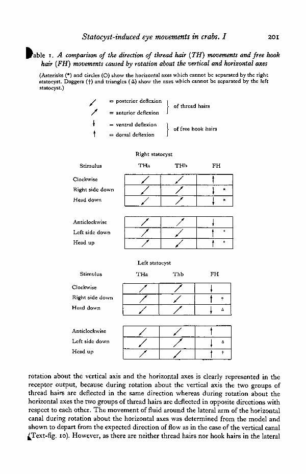
A table of the directions of fluid movements following displacement about the threemain axes has been deduced from the model and used to predict the direction of threadhair deflexions and free hook hair deflexions (Table 1). The thread hairs are consi-dered to form two main groups: the dorsal group in the common canal (THa) andthe ventral group situated in the lower part of the vertical canal (THb) (Text-fig. 10).
Assuming that the directionality of the receptor responses is important, the tableshows the information available to the central nervous system which is translatedinto an appropriate output to the eye muscles (Table 1). The difference between
Statocyst-induced eye movements in crabs. I 201
rable i. A comparison of the direction of thread hair (TH) movements and free hookhair (FH) movements caused by rotation about the vertical and horizontal axes
(Asterisks (•) and circles (O) show the horizontal axes which cannot be separated by the rightstatocyst. Daggers (+) and triangles (A) show the axes which cannot be separated by the leftstatocyst.)
/ = posterior deflexion
/ = anterior deflexion
f = ventral deflexion
I = dorsal deflexion
of thread hairs
of free hook hairs
Stimulus
Clockwise
Right side down
Head down
Anticlockwise
Left side down
Head up
Stimulus
Clockwise
Right side down
Head down
Anticlockwise
Left side down
Head up
Right statocyst
THa THb
Left statocyst
THa Thb
F H
///
///
1I *1 *
///
///
\\ 't •
FH
/
/
/
/
/
/
t
t +
rotation about the vertical axis and the horizontal axes is clearly represented in thereceptor output, because during rotation about the vertical axis the two groups ofthread hairs are deflected in the same direction whereas during rotation about thehorizontal axes the two groups of thread hairs are deflected in opposite directions withrespect to each other. The movement of fluid around the lateral arm of the horizontalcanal during rotation about the horizontal axes was determined from the model andshown to depart from the expected direction of flow as in the case of the vertical canal^Text-fig, io). However, as there are neither thread hairs nor hook hairs in the lateral

202 D. C. SANDEMAN AND A. OKAJIMA
Table 2. The predicted outputs from both statocysts during rotation about the horizontalaxes showing that where the right statocyst confuses two directions of rotation, the leftdiscriminates between them and vice versa
(The double line shows the discriminating statocyst. See Table 1 for key to symbols.)
Right statocyst Left statocyst
THa THb FH THa THb FH Stimulus
Right side down
Head down/
/
/
\
\
/
/
/
/
t1
/
/
/
/
\
\
/
/
/
/
\
\
/
/
/
/
tt
//
//
tt
//
//
\\
//
//
t
Right side down
Head up
Left side down
Head down
Head up
Left side down
part of the horizontal canal, flow direction in this part of the system during rotationabout the horizontal axes is unimportant.
One of the essential functions of the statocyst as a dynamic organ is to detect thedirection of rotation about any axis. It is therefore interesting to find that as far as thefree hook hairs and thread hairs are concerned, there are some horizontal axes whichcannot be distinguished by one statocyst alone, as shown in Table 1. The rightstatocyst, for example, produces the same output for right-side-down and for head-down; also for left-side-down and for head-up. The left statocyst confuses right-side-down with head-up and head-down with left-side-down. Table 2 compares the outputfrom both statocysts and shows that where one statocyst confuses two stimuli the otherdiscriminates between them. If the same conditions exist in the crab Maja, we canexplain its ability to distinguish correctly between rotations about all axes with onlythread hairs and free hook hairs intact (statolith input destroyed) as shown byDijkgraaf (1956a, 1962).
In our records unit responses from different groups of hairs are remarkably homo-geneous and, although within each group small differences of sensitivity occur, thethree classes of responses are always clearly defined. The relevance of the smalldifferences within each class is not known, but as single interneurones in the C.N.s.probably receive large numbers of sensory inputs, the small differences would immedi-ately be lost.
All the unit responses were unidirectional, a property of many crustacean mechano-receptors, and like other receptor systems (i.e. Pacinian corpuscles: Loewenstein 8f

Statocyst-induced eye movements in crabs. I 203
^lendelson, 1965; Ozeki & Sato, 1965; Hubbard, 1958; mammalian muscle spindles:Eippold, Nicholls and Redfearn, i960) differences in the receptor responses are quiteprobably a reflexion of the mechanical compliance of the transducing structure (inthis case the receptor hairs) and not any basic difference between the receptor cells.For example, one of the most striking differences in statocyst-receptor responses isrevealed at the termination of a stimulus. Thread hairs continue to fire for a longperiod, free hook hairs for a few seconds and statolith hairs stop firing immediately.The thread hairs are very long and return very slowly to their original position and theirresponses reflect this gradual change of position. The free hook hairs are shorter and soreturn to their initial position more rapidly. The statolith hairs are even shorter, andbecause they are clustered together like the bristles of a brush, spring back intoposition immediately after they are released from the stimulus.
Whatever the reason for their differences it is clear that the impulse patterns of thehairs agree with the functions ascribed to them by Dijkgraaf (1956a, b), and thethread hairs are the receptors most likely to be of interest for the production ofhorizontal eye movements.
The function of the group hairs is not so obvious. They have been said to controlthe movement of the fluid in the canals (Dijkgraaf, 1961) and certainly they aresupported on a very thin area of cuticle which if pressed in causes the hairs to swingout across the canal. How, or even if, this is achieved in the intact animal is not known.Stimulation of the motor nerves in the antennular nerve does not distort the canalsystem and produce any movement of the group hairs although contraction of all themuscles in the basal joint was observed; so an active control system seems unlikely.
SUMMARY
1. Irrigation of the statocysts of the crab Scylla serrata will activate the oculomotorneurones associated with eye movements.
2. An investigation of the central mechanism of statocyst-induced nystagmus hasbeen started with the description of the statocyst canals and a characterization of thesensory input from the hair receptors in the canals.
3. The canals are shaped like two toroids joined at approximately right angles toone another. Direct observation of isolated statoliths and glass models of them showsthat when they are rotated, fluid moves around the circumference of the statocystcanals and displaces the hair receptors protruding into them. The direction of dis-placement of the different groups of receptors in both statocysts is related to the axesof rotation and provides a unique output for rotation about each axis.
4. Electrical recordings from the three types of receptor hairs show that the threadhairs are most probably the receptors responsible for detection of rotation about thevertical axis. The free hook hairs are sensitive enough to detect rotation about thehorizontal axes. The statolith hairs are sensitive to maintained changes of position.

204 D. C. SANDEMAN AND A. OKAJIMA
REFERENCES
BARNES, W. J. P. & HORRIDGE, G. A. (1969). Interactions of the movements of the two eyecups in thecrab Carcinus. jf. exp. Biol. 50, 651-71.
COHEN, M. J. (1955). The function of receptors in the statocyst of the lobster Homarus americanus. J.Physiol., Lond. 130, 9-134.
COHEN, M. J. (i960). The response patterns of single receptors in the crustacean statocyst. Proc. Roy.Soc. SB 152, 30-49.
COHEN, M. J. & DIJKGRAAF, S. (1961). Mechanoreception. In The Physiology of Crustacea, vol. JI (ed.T. H. Waterman), pp. 65-108. New York: Academic Press.
DIJKGRAAF, S. (1955). Rotationssinn nach dem Bogengangsprinzip bei Crustaceen. Experientia zi, 407.DIKGRAAF, S. (1956a). Structure and functions of the statocyst in crabs. Experientia 12, 394.DIJKGRAAF, S. (19566). Uber die Kompensatorischen Augenstielbewegungen bei brachyuren. Publ.
Staz. Zool. Napoli 28, 341-58.DIJKGRAAF, S. (1961), The statocyst of Octopus vulgaris as a rotation receptor. Publ. Stas. Zool. Napoli
32, 64-87.DIJKGRAAF, S. (1962). Physiologie der Statocyste von Maja verrucosa (Brachyura). Wissenschaftlicher
Film D 843/1962. Gottingen.HENSEN, V. (1863). Studien iiber das Gehororgan der Decapoden. Z. teiss. Zool. 13, 319-412.HORRIDGE, G. A. & SANDEMAN, D. C. (1964). Nervous control of optokinetic responses in the crab.
Carcinus. Proc. Roy. Soc. B 161, 216-46.HUBBARD, S. J. (1958). A study of rapid mechanical events in a mechanoreceptor. J. Physiol., Lond. 141,
198-218.LIPPOLD, O. C. J., NICHOLLS, J. G. & REDFEARN, J. W. T. (i960). Electrical and mechanical factors in the
adaptation of a mammalian muscle spindle. J. Physiol., Lond. 153, 209-17.LOEWENSTEIN, W. R. & MENDELSON, M. (1965). Components of receptor adaptation in a Pacinian
corpuscle. J. Physiol., Lond. 177, 377-97.OZEKI, M. & SATO, M. (1965). Changes in the membrane potential and the membrane conductance
associated with a sustained compression of the non-myelinated nerve terminal in Pacinian corpuscles.J. Physiol., Lond. 180, 186-208.
PRENTISS, C. W. (1901). The otocyst of decapod Crustacea. Bull. Mus. Comp. Zool. Harvard 36, 167-254-
SANDEMAN, D. C. (1971). Excitation and electrical coupling of four identified motoneurons in the brainof the Australian mud crab, Scylla serrata. Z. vergl. Physiol. 72, 111-30.
SCHSNE, H. & STEINBRECHT, R. A. (1968). Fine structure of statocyst receptor of Astacus fluriatilis.Nature, Lond. 220, 184—6.
EXPLANATION OF PLATE
Stereopair photographs from the posterior (Fig. 1) and anterior (Fig. 2) of a statocyst dissected from thebasal joint of the antennule. The toroidal shape of the horizontal canal is clear in both. In Fig. 1 thefree hook hairs and statolith can be seen and in Fig. 2 the sensory cushion and group hairs are clearlyvisible.

Journal of Experimental Biology, Vol. 57, No. 1 Plate 1
D. C. SANDEMAN AND A. OKAJIMA {Facing p. 204)