Sustaining and Sharing Economic Growth in Tanzania - ISBN ...
Upload
independentCategory
view
3download
0
Human Cortical Areas Involved in SustainingPerceptual Stability During Smooth
Pursuit Eye Movements
Maja U. Trenner,1* Manfred Fahle,2 Oliver Fasold,1 Hauke R. Heekeren,3,1
Arno Villringer,1 and Rudiger Wenzel1
1Berlin NeuroImaging Center, Neurologische Klinik und Poliklinik, Charite Universitatsmedizin Berlin,Berlin, Germany
2Department of Human Neurobiology, University of Bremen, Bremen, Germany3Max Planck Institute for Human Development, Berlin, Germany
Abstract: Because both, eye movements and object movements induce an image motion on the retina, eyemovements must be compensated to allow a coherent and stable perception of our surroundings. The infer-ential theory of perception postulates that retinal image motion is compared with an internal referencesignal related to eye movements. This mechanism allows to distinguish between the potential sourcesproducing retinal image motion. Referring to this theory, we investigated referential calculation duringsmooth pursuit eye movements (SPEM) in humans using event-related functional magnetic resonanceimaging (fMRI). The blood oxygenation level dependent (BOLD) response related to SPEM in front of astable background was measured for different parametric steps of preceding motion stimuli and thereforeassumed for different states of the referential system. To achieve optimally accurate anatomy and moredetectable fMRI signal changes in group analysis, we applied cortex-based statistics both to all brainvolumes and to defined regions of interest. Our analysis revealed that the activity in a temporal region aswell as the posterior parietal cortex (PPC) depended on the velocity of the preceding stimuli. Previoussingle-cell recordings in monkeys demonstrated that the visual posterior sylvian area (VPS) is relevant forperceptual stability. The activation apparent in our study thus may represent a human analogue of thisarea. The PPC is known as being strongly related to goal-directed eye movements. In conclusion, temporaland parietal cortical areas may be involved in referential calculation and thereby in sustaining visualperceptual stability during eyemovements.HumBrainMapp 29:300–311, 2008. VVC 2007Wiley-Liss, Inc.
Key words: cortex-based; ‘Filehne’ illusion; functional magnetic resonance imaging; motion perception;PPC; reference signal; SPEM; VPS; VTS
INTRODUCTION
There are two potential sources of retinal image motion,namely self-motion versus object motion. Often, we moveour eyes and watch a moving object in our environment atthe same time; in this situation both of these potentialsources contribute to the retinal image motion. What is themechanism allowing the perception of stable surroundingsduring eye movements? On the basis of the early ideas ofvon Helmholtz [1910], the two types of retinal imagemotion might be dissociated by comparing the retinal
Contract grant sponsor: Berlin NeuroImaging Center; Contractgrant number: BMBF-DLR, FKZ 01 GO 020; Contract grant spon-sor: Kommission fur wissenschaftlichen Nachwuchs of ChariteUniversitatsmedizin Berlin
*Correspondence to: Maja Trenner, Department of Neurology,Charite Universitatsmedizin Berlin, 10098 Berlin, Germany.E-mail: [email protected]
Received for publication 10 November 2006; Revision 22 January2007; Accepted 5 February 2007
DOI: 10.1002/hbm.20387Published online 5 April 2007 in Wiley InterScience (www.interscience.wiley.com).
VVC 2007 Wiley-Liss, Inc.
r Human Brain Mapping 29:300–311 (2008) r

signal with an internal signal encoding eye movements[Sperry, 1950; von Holst and Mittelstaedt, 1950; Wertheim,1994]. The functional model of Wertheim [1994] goesbeyond the traditional inferential theory expanding theconcept of the internal reference signal. It incorporates thefact that not only eye movements but also head move-ments and vection resulting from optokinetic stimulationshould generate a reference signal. In summary, the inter-nal reference signal is described as a compound signalcomprising an eye movement component, a vestibularcomponent, and a visual component. Subtraction of theinternal reference signal from the retinal signal allows todiscriminate between self versus object movements. Anyretinal image motion remaining after subtraction of thisinternal signal is experienced as being due to movementsof the environment.When this referential mechanism is disturbed, deficits in
situations requiring the differentiation between self andobject motion can occur. For example, patient R.W., whohad bilateral extrastriate occipital and intraparietal corticallesions [Haarmeier et al., 1997], suffered from vertigo andnausea whenever he tracked a moving object with his eyesor fixated a stationary object during translational self-motion. In these situations R.W. not only experienced diffi-culties in every day life but also showed deficits in psycho-physical tests under laboratory conditions.Similarly, Wilhelm Filehne [1922] found that slight
imperfections in this referential mechanism result in anillusionary perception of object motion [Filehne, 1922; Free-man, 1999; Freeman and Banks, 1998; Mack and Herman,1973]. For example, when tracking a target moving in frontof a stationary structured background, an extraretinal sig-nal overestimating the eye movement velocity (too largereference signal) enhances the perception of backgroundmotion in the same direction as the pursuit movement.Under identical circumstances an extraretinal signal under-estimating the eye movement velocity (too small referencesignal) enhances the perception of background motion inthe opposite direction as the pursuit movement. This socalled ‘Filehne’ illusion can be modified by presenting acentral fixation point and a structured background patternmoving briefly in one direction at a defined velocity (con-ditioning stimulus) prior to the ‘Filehne’-type stimulus[Haarmeier et al., 2001; Haarmeier and Thier, 1996].Depending on the velocity and the direction of this back-ground motion the point at which the background is expe-rienced as stable is shifted, resulting in an illusionarybackground movement during the following smooth pur-suit eye movement in front of a stationary background.The influence of the first stimulus is not only transientsince the ‘Filehne’ illusion shows a tendency even toincrease in time when it is studied at various interstimulus-intervals (0.5, 3.0, 6.0 s) [Haarmeier and Thier, 1996]. Asexamined by adaptive two-forced-choice method condition-ing stimulus velocities of for example �88/s, �168/s, or�248/s led to the subjective perception of about �28/s,�48/s, or �68/s motion, respectively, of the non-moving
background during the subsequent ‘Filehne’-type stimulus.The amount of physical motion at the point of subjectivestability is a measure of the quantity of the ‘Filehne’illusion.To elucidate the neural mechanisms underlying the dis-
crimination of self-motion versus environmental motionduring smooth pursuit eye movements (SPEM), the role ofearly and late visual processing areas has been studied innonhuman primate single-cell recordings. Neurons in thevisual area V1 discharged invariably for self-induced ver-sus externally induced slow retinal image slip [Ilg andThier, 1996]. This finding stood in contrast to an earlywork of Galletti et al. [1984]. Whereas also neurons in thevisual area V4 and the middle temporal (MT) motion proc-essing area typically discharged invariably for both motionconditions a significant number of neurons in the medialsuperior temporal (MST) area, especially its dorsal region,responded preferentially to externally induced slow retinalimage slip [Erickson and Thier, 1991]. At a later date astudy of Thier et al. [2001] suggested that the visual poste-rior sylvian area (VPS), which is located near the dorsalend of the lateral sulcus might be relevant for perceptualstability during SPEM. How perceptual stability is inst-antiated in the human brain and which brain regions playa role in this process is still unclear. An electroencepha-lography (EEG) study [Haarmeier and Thier, 1998] andanother study employing magneto-encephalography (MEG)[Tikhonov et al., 2004] techniques found evidence for acrucial role of the parieto-occipital cortex in the perceptionof self-induced motion.On the basis of the theoretical framework of the inferen-
tial theory of perception the goal of the present study wasto investigate where in the human brain referential calcula-tion is accomplished and thereby perceptual stability dur-ing SPEM in humans is achieved. Therefore, we usedevent-related functional magnetic resonance imaging(fMRI) and a ‘Filehne’-type stimulus preceded by a stimu-lus, whose background moved at different parametricallystepped velocities. On the basis of the results of previousprimate and human studies, we expected temporal and pa-rietal cortical regions to respond differently to the identical‘Filehne’-type stimulus depending on the velocity of thepreceding stimulus, and hence, to underlie referentialcalculation during SPEM.
MATERIALS AND METHODS
Subjects
Six right-handed male students (mean age 25 years,range 20–30 years) were examined using functional mag-netic resonance imaging (fMRI). Subjects reported normalvisual acuity; none had a history of neurological or psychi-atric dysfunction. Subjects were naive as to the purpose ofthe study and were paid for their participation. Writtenand informed consent was obtained from each participantprior to investigation according to the declaration of
r Perceptual Stability During Eye Movements r
r 301 r

Helsinki. The study was approved by the local ethicscommittee.
Stimuli and Experimental Design
During the fMRI session each subject underwent twoevent-related fMRI experiments and completed sevenfMRI-runs: In experiment (I) each subject completed onefMRI-run consisting of 120 volume-scans, and in experi-ment (II) six fMRI-runs consisting of each 172 volume-scans. The first four scans of each run were discarded. Ineach condition of the two experiments 32 trials werepresented.Stimuli consisted of a red target dot (diameter 10 min
arc) and a background pattern, which had an extension of248 � 188 and consisted of 350 light gray dots (each ofdiameter 15 min arc) randomly spread over the dark graybackground area. Visual stimuli were projected onto a mir-ror mounted on the head-coil resulting in a viewing distanceof�22 cm. Average stimulus luminance was 22 cd/m2.In both experiments, (I) and (II), subjects were instructed
to track the red target dot as accurately as possible andwere trained outside the scanner at the beginning of eachsession.In experiment (I), subjects performed smooth pursuit
eye movements (SPEM). This experiment served to localizeregions involved in performing eye movements (SPEMand saccades) for subsequent regions of interest (ROI)analysis.SPEM stimuli: The target dot was presented for 0.5 s in
the center of the screen. Next it jumped 118 to the left andfrom there moved to the right at a constant velocity of118/s, resulting in a stimulus duration of 2.5 s. Finally, thetarget dot jumped back to the center of the screen.
In experiment (II), different degrees of change in thereference signal were induced in a parametric design bypreceding stimuli of three different velocities, which weredesigned according to Haarmeier et al. [1996]. An experi-mental trial consisted of one preceding stimulus (PS) andone critical stimulus (CS) presented at an interval of 6.6 s(cf. Fig. 1). The duration of one trial was 10.5 s. The inter-val between trials varied between 9.0–15.0 s (mean¼ 12.0 s).Preceding stimuli (PS): The target dot was presented for
1.4 s in the center of the screen, after 1.1 s followed by abackground pattern that was visible for 0.3 s. The patternmoved leftwards at one of three velocities (PS-8 ¼ �88/s,PS-16 ¼ �168/s, PS-24 ¼ �248/s), while subjects fixatedthe target in the center of the screen. Dots reaching theborder of the pattern area disappeared and were replacedby fresh dots appearing on the opposite side. According toour assumptions this stimulus served to modify parametri-cally the state of the referential system.Critical stimuli (CS): The target dot was presented for
0.5 s in the center of the screen. Next it jumped 118 to theleft and from there moved to the right at a constant veloc-ity of 118/s for 2.0 s. After 0.85 s of this SPEM a stationarybackground pattern appeared for 0.3 s while subjectscontinued to perform SPEM. Although CS was physicallyidentical they will be referred to as CS-8, CS-16, or CS-24depending on the velocity of the PS.Please note, that the background motion of the PS and
the SPEM during the CS were in opposite direction. Thisshould result in an enhanced relative opposite backgroundmotion effect of a stable background during the SPEM.PS velocity was constant during the course of each fMRI
run (either �88/s, or �168/s, or �248/s). There were twofMRI runs of either velocity; the sequence of runs was bal-anced across subjects. One of the six possible sequences of
Figure 1.
Temporal course of an experimental trial. Each trial consisted of
a preceding stimulus (PS) requiring fixation of a red dot in the
center of the screen while the background pattern moved to the
left at one of three velocities (�88/s, �168/s, or �248/s) for 0.3 s.
After an inter-stimulus-interval (ISI) of 6.6 s the critical stimulus
(CS) appeared requiring a smooth pursuit eye movement (SPEM)
tracking of a red dot that moved from left to right at 118/s for 2s with a stationary structured background pattern shown for 0.3
ms starting 0.85 s after begin of the SPEM.
r Trenner et al. r
r 302 r

conditions (1-2-3; 1-3-2; 2-1-3; 2-3-1; 3-1-2; 3-2-1) wasassigned to each subject and repeated for that subject. Inbetween stimulations a white fixation cross was shown inthe center of the dark gray screen.Eye movements were controlled by video-oculography
(MEyeTrackTM System, SensoMotoric Instruments GmbH,Teltow, Germany). The system uses infrared cameras tocapture the eye movements. Data were recorded at a sam-pling rate of 50 Hz. The system tracks corneal reflexes onthe iris together with the pupil compensating shifts of thecamera relative to the head. For calibration prior to experi-ment I subjects were asked to track nine predeterminedcalibration dots. Video-oculography data acquired in thescanner was used for qualitative analysis. Eye movementswere displayed on a computer monitor so that theperformance could be monitored continuously by theexperimenter.
Data Acquisition fMRI
Imaging was performed on a 1.5-T Siemens Vision echo-planar system with a standard head coil. The subject’shead was immobilized by a vacuum pad to minimizemovement artifacts. Echo-planar imaging (EPI) scans wereacquired in 16 slices covering the supratentorial parts ofthe brain in an oblique orientation (�258 to the anteriorcommissure-posterior commissure (ACPC) plane). Func-tional runs consisted of 120 (experiment (I)) or 172 (experi-ment (II)) whole brain volume scans using a T2*-weightedfree induction decay (FID) EPI sequence (repetition time(TR) ¼ 2 s, echo time (TE) ¼ 60 ms, flip angle ¼ 908). Slicethickness was 5 mm (skip 0.5) with a field of view of 256� 256 mm2 and an imaging matrix of 64 � 64 resulting ina plane resolution of 4 � 4 mm2.
Data Acquisition MRI
Following each functional session we collected a high-resolution T1-weighted 3D-MP RAGE dataset (TR ¼ 9.7 ms,TE ¼ 4 ms, flip angle ¼ 908, field of view ¼ 256 � 256 mm2,voxel size 1 � 1 � 1 mm3, 190 slices) to transform the EPIdata into three-dimensional space and to align functionaldata from different scanning sessions. For visual control ofthe spatial transformation process a 2D T1-weighted scan(TR ¼ 900 ms, TE ¼ 14 ms, voxel size 2 � 2 � 5 mm3,16 slices) in the same orientation as the EPI-slices comple-mented each experiment. For individual surface recon-struction and individual 3D intersession alignment a highresolution 3D-Flash dataset was collected for each subjectduring an additional session (TR ¼ 20 ms, TE ¼ 5 ms, flipangle ¼ 308, field of view ¼ 256 � 256 mm2, voxel size1 � 1 � 1 mm3, 190 slices, 2 acquisitions).
fMRI Data Analysis
Data were preprocessed and analyzed using BrainVoyager QX software (Brain Innovation B.V., Maastricht,
Netherlands). Preprocessing of the fMRI data involved a3D motion correction based on the estimates of three trans-lation and three rotation parameters of rigid body transfor-mation, a slice scan time correction and the removal ofslow linear and non linear trends. No further temporal orspatial filtering was applied. The latter because of spatialsmoothing may be critical when differences of corticalareas are examined that may be relatively small.
Cortex-based fMRI Data Analysis
Three-dimensional individual cortex reconstruction wasachieved by segmenting and tessellating the gray–whitematter boundary and by inflating the resulting surface meshon the basis of the 3D-Flash data [Carman et al., 1995; VanEssen et al., 1998]. The resulting individual reconstructionswere transferred into Talairach standard brain space andfurther morphed into spherical representations. The sub-jects’ individual spherical representations were aligned witheach other following a coarse-to-fine matching strategy ofthe cortical folding pattern. This is a moving-average,group-alignment version of the cortex-based alignment pro-cess that substantially improves spatial correspondencemapping between subjects’ brains beyond Talairach space[Fischl et al., 1999; Van Atteveldt et al., 2004].The fixed effects statistical group analysis was based on
a general linear model (GLM) involving a correction forlinear serial correlations. Predictor size was 2 s for the PSand 4 s for the CS. Individual time courses were con-volved with a hemodynamic response function [Boyntonet al., 1996]. Statistical maps were displayed on one indi-vidual reconstructed surface. Cortex-based statistical groupanalysis comprised three steps:
a. First, in order to identify cortical regions that are acti-vated during the condition that served to induce a‘Filehne’ illusion, a GLM was run on the BOLD(blood oxygenation level dependent) response to theCS stimuli (CS-8, CS-16, CS-24) from experiment (II)contrasted against fixation baseline; resulting activa-tions were displayed at a threshold criterion of P(Bonferroni corrected) < 0.0001 and a cluster sizecriterion of 50 mm3.
b. Second, to functionally localize cortical regions thatare activated during eye movements (SPEM and sac-cades) as ROIs, a GLM was run on the BOLD re-sponse to the SPEM stimuli from experiment (I) con-trasted against fixation baseline. Cortex-defined ROIswere localized by analysis of the activation duringthe SPEM task displayed at a threshold criterion of q(FDR) < 0.05 (false discovery rate, FDR) [Benjaminiand Hochberg, 1995; Genovese et al., 2002] and acluster size criterion of 25 mm3, and were defined onthe reconstructed surface. Bilateral early visual proc-essing areas (VIS), putative visual posterior sylvianarea (VPS), posterior parietal cortex (PPC), visual
r Perceptual Stability During Eye Movements r
r 303 r
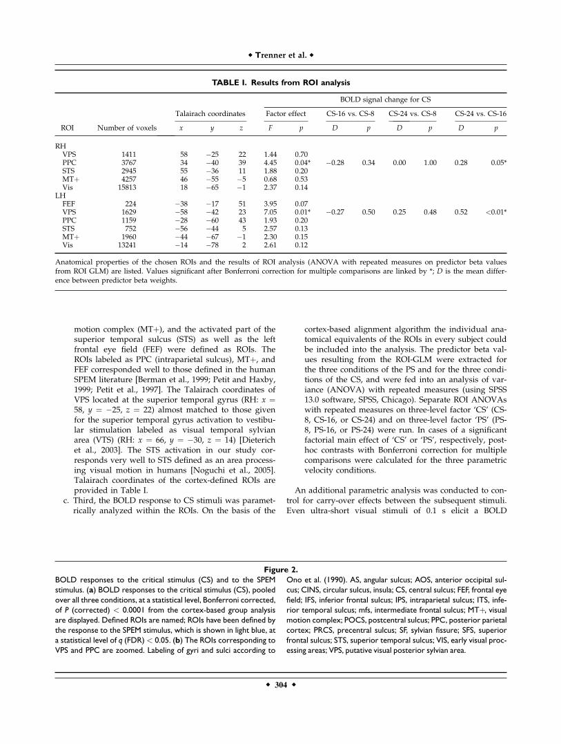
motion complex (MTþ), and the activated part of thesuperior temporal sulcus (STS) as well as the leftfrontal eye field (FEF) were defined as ROIs. TheROIs labeled as PPC (intraparietal sulcus), MTþ, andFEF corresponded well to those defined in the humanSPEM literature [Berman et al., 1999; Petit and Haxby,1999; Petit et al., 1997]. The Talairach coordinates ofVPS located at the superior temporal gyrus (RH: x ¼58, y ¼ �25, z ¼ 22) almost matched to those givenfor the superior temporal gyrus activation to vestibu-lar stimulation labeled as visual temporal sylvianarea (VTS) (RH: x ¼ 66, y ¼ �30, z ¼ 14) [Dieterichet al., 2003]. The STS activation in our study cor-responds very well to STS defined as an area process-ing visual motion in humans [Noguchi et al., 2005].Talairach coordinates of the cortex-defined ROIs areprovided in Table I.
c. Third, the BOLD response to CS stimuli was paramet-rically analyzed within the ROIs. On the basis of the
cortex-based alignment algorithm the individual ana-tomical equivalents of the ROIs in every subject couldbe included into the analysis. The predictor beta val-ues resulting from the ROI-GLM were extracted forthe three conditions of the PS and for the three condi-tions of the CS, and were fed into an analysis of var-iance (ANOVA) with repeated measures (using SPSS13.0 software, SPSS, Chicago). Separate ROI ANOVAswith repeated measures on three-level factor ‘CS’ (CS-8, CS-16, or CS-24) and on three-level factor ‘PS’ (PS-8, PS-16, or PS-24) were run. In cases of a significantfactorial main effect of ‘CS’ or ‘PS’, respectively, post-hoc contrasts with Bonferroni correction for multiplecomparisons were calculated for the three parametricvelocity conditions.
An additional parametric analysis was conducted to con-trol for carry-over effects between the subsequent stimuli.Even ultra-short visual stimuli of 0.1 s elicit a BOLD
TABLE I. Results from ROI analysis
ROI Number of voxels
Talairach coordinates
BOLD signal change for CS
Factor effect CS-16 vs. CS-8 CS-24 vs. CS-8 CS-24 vs. CS-16
x y z F p D p D p D p
RHVPS 1411 58 �25 22 1.44 0.70PPC 3767 34 �40 39 4.45 0.04* �0.28 0.34 0.00 1.00 0.28 0.05*STS 2945 55 �36 11 1.88 0.20MTþ 4257 46 �55 �5 0.68 0.53Vis 15813 18 �65 �1 2.37 0.14
LHFEF 224 �38 �17 51 3.95 0.07VPS 1629 �58 �42 23 7.05 0.01* �0.27 0.50 0.25 0.48 0.52 <0.01*PPC 1159 �28 �60 43 1.93 0.20STS 752 �56 �44 5 2.57 0.13MTþ 1960 �44 �67 �1 2.30 0.15Vis 13241 �14 �78 2 2.61 0.12
Anatomical properties of the chosen ROIs and the results of ROI analysis (ANOVA with repeated measures on predictor beta valuesfrom ROI GLM) are listed. Values significant after Bonferroni correction for multiple comparisons are linked by *; D is the mean differ-ence between predictor beta weights.
Figure 2.
BOLD responses to the critical stimulus (CS) and to the SPEM
stimulus. (a) BOLD responses to the critical stimulus (CS), pooled
over all three conditions, at a statistical level, Bonferroni corrected,
of P (corrected) < 0.0001 from the cortex-based group analysis
are displayed. Defined ROIs are named; ROIs have been defined by
the response to the SPEM stimulus, which is shown in light blue, at
a statistical level of q (FDR) < 0.05. (b) The ROIs corresponding to
VPS and PPC are zoomed. Labeling of gyri and sulci according to
Ono et al. (1990). AS, angular sulcus; AOS, anterior occipital sul-
cus; CINS, circular sulcus, insula; CS, central sulcus; FEF, frontal eye
field; IFS, inferior frontal sulcus; IPS, intraparietal sulcus; ITS, infe-
rior temporal sulcus; mfs, intermediate frontal sulcus; MTþ, visualmotion complex; POCS, postcentral sulcus; PPC, posterior parietal
cortex; PRCS, precentral sulcus; SF, sylvian fissure; SFS, superior
frontal sulcus; STS, superior temporal sulcus; VIS, early visual proc-
essing areas; VPS, putative visual posterior sylvian area.
r Trenner et al. r
r 304 r

Figure 2.
r Perceptual Stability During Eye Movements r
r 305 r

response lasting for about 6.0 s [Savoy et al., 1995]. Thus,the interstimulus-interval of 6.6 s in our study does notallow excluding an overlap between the BOLD-responses tothe 0.3 s PS and the subsequent CS. To control for carry-over effects, which would include the risk of attributing PSeffects to the CS, two different hemodynamic response func-tions (HRFs) (Boynton function [Boynton et al., 1996], 1.HRF: delta ¼ 2.5, tau ¼ 1.25, 2. HRF: delta ¼ 2.5, tau ¼1.75) were applied to the PS to modify the overlap betweenthe GLM predictors of the two stimuli. The idea of that isthat at a smaller overlap effects at the downward slope ofthe BOLD-response to the PS are less strongly modeled andwould have more influence on the CS.
RESULTS
Figure 2 illustrates the BOLD-responses to the CS, irre-spective of PS velocity, displayed on an inflated cortex(yellow patches). Group analysis revealed a significantincrease of the BOLD signal in a distributed pattern ofcortical areas involving bilateral occipital, superio-tempo-ral, intraparietal, central, and precentral regions. Activationspots found for the SPEM task (light blue patches on thecortex in Fig. 2a and in volume plots shown in Fig. 3)were located at similar positions; all ROIs defined bySPEM corresponded to areas also responding to CS.Grand-averaged event-related time courses from each
ROI for both, the PS and the CS, are shown in Figures 3and 4. Plots of the time courses differentiate between theactivations caused by the three different PS velocities. Itshould be noted that in both instances, PS and CS, stimula-tion is very similar on the level of the retina. Real motion(for PS) and perceived motion (for CS) yielded an activa-tion of up to 0.8% signal change found in the VIS for thehighest velocities. The time courses show that the BOLDresponses were stronger in left VPS and in right PPC forfaster (�248/s) than for slower (�168/s) velocities of thestimulus in each subject (see Fig. 3). This is true for both,the PS and the CS.ROI ANOVAs with repeated measures revealed a signif-
icant main effect of the three-level factor ‘CS’ on the BOLDresponse to CS in right-hemispheric PPC (F(2,5) ¼ 4.45,P ¼ 0.04) as well as in left-hemispheric VPS (F(2,5) ¼ 7.10,P ¼ 0.01). Post-hoc paired means comparison applyingBonferroni testing showed that this effect was due to astronger BOLD-signal for CS that had been preceded byfaster velocity PS (CS-24 vs. CS-16 comparison): in rightPPC (P ¼ 0.05) and in left VPS (P ¼ < 0.01). ROI ANOVAswith repeated measures did reveal an effect of the factor‘CS’ but not of the factor ‘PS’ in right PPC and left VPS(right PPC: F(2,5) ¼ 2.20, P ¼ 0.16 and left VPS F(2,5) ¼3.30, P ¼ 0.08). Table I summarizes the results from ROIANOVAs for the factor ‘CS’.To control for carry-over effects of the BOLD response
between the narrowly spaced PS and CS stimuli the para-metric analysis within ROIs was additionally modeledusing a stronger overlap. With less overlap of the BOLD-
responses to the PS and the CS using a narrow HRF theeffects in PPC and VPS were abolished or much reduced,respectively. Controlling PS effects stabilizes effects to theCS showing that these are not due to carry-over effectsfrom the PS.
DISCUSSION
In the present study, we investigated the cortical basisof perceptual stability during SPEM in humans based onthe theoretical framework of the inferential theory of per-ception. For that purpose, we used event-related fMRI anda ‘Filehne’-type stimulus preceded by a stimulus, whosebackground moved at different parametrically steppedvelocities. The background motion of the preceding stimu-lus and the subsequent SPEM during the ‘Filehne’-typestimulus were in opposite direction, which should resultin an enhanced relative opposite background motion effectof the stable background during the SPEM. We found thatthe cortical activity in two regions (left VPS and rightPPC) covaried with the velocity of the preceding stimulus.Hence, higher velocities of background motion of the PSelicited a stronger response in these areas, most probablydue to a larger shift of the ‘point of stationarity’ of thesystem that in turn induces a larger remaining motion sig-nal during the CS.The PPC activations to the SPEM stimulus were located
in the ventral intraparietal sulcus (IPS), which is consistentwith previous fMRI studies on SPEM [Berman et al., 1999;Petit and Haxby, 1999]. Also FEF and MTþ activationshave been found in previous SPEM studies [Berman et al.,1999; Petit and Haxby, 1999; Petit et al., 1997]. Referring tothe previous SPEM literature the activation spots in thesuperior temporal sulcus (labeled as STS) and in the supe-rior temporal gyrus (labeled as VPS) to the SPEM stimuluswere surprising to us. Human STS is known as an areaprocessing second-order visual motion and biologicalmotion [Noguchi et al., 2005; Servos et al., 2002]. VPS hasbeen described as a polymodal area in the inferior parietalcortex of the squirrel monkey [Guldin and Grusser, 1998]and corresponds to an area called the visual temporal syl-vian area (VTS) [Guldin and Gruesser, 1996]. Although itsmain input is optokinetic, arriving from motion processingarea MST, this area receives also vestibular afferences fromthe parieto-insular-vestibular cortex (PIVC) [Fasold et al.,2002; Gruesser et al., 1990a,b; Guldin and Grusser, 1998].In humans activation of the retroinsular cortex and theadjacent superior temporal gyrus or the transverse tempo-ral gyrus probably best fit with the nonhuman VPS or VTS[Dieterich et al., 2003]. Thus VPS is located posterior to theputative human correspondent of PIVC in the posteriorinsula [Dieterich et al., 2003; Petit and Beauchamp, 2003].The localization of the region of interest labeled VPS inour study according to its Talairach coordinates wouldallow an assignment to both, either to VPS or to PIVC.Although the immediate proximity of VPS and PIVC com-plicates their separation in human functional studies we
r 306 r
r Trenner et al. r

Figure 3.
BOLD responses in VPS and PPC covary with velocity of the
preceding stimulus. Top: The ROIs corresponding to left VPS
and right PPC are shown in a slice volume plot. Middle: Grand
averages of the event-related time courses depending on the
velocity of the PS are presented for two ROIs (left VPS and right
PPC). Zero on the x-axis corresponds to the time when the
critical stimulus (CS) is presented, �8 corresponds to the time
when the preceding stimulus is (PS) is presented. The baseline
for the time courses has been set to the time period from �8
to �7. The parametric variation is color coded (blue – PS/CS-8,
green – CS/PS-16, red – CS/PS-24). Bottom: The diagram gives
the differences between the beta values from the two paramet-
ric conditions that differ significantly (CS-24 and CS-16) in each
single subject. Additionally the mean effect with its standard
deviation is shown. PPC, posterior parietal cortex; PS, preceding
stimulus; VPS, putative visual posterior sylvian area.

decided to label the activation in the superior temporalgyrus in our study ‘‘VPS’’ because of its better fit with thelocalization of VPS (or VTS) described in the human andanimal literature [Dieterich et al., 2003; Guldin andGrusser, 1998; Karnath, 2001; Thier et al., 2001].The apparently higher activation associated with the
presentation of the ‘Filehne’-type stimulus as comparedwith the pure SPEM stimulus is due to the presence of thedot pattern background during the PS and CS stimuli butis not relevant to the aims of the present study.SPEM involves a network of cortical and subcortical areas
largely overlapping with those involved in planning and
executing saccades [Krauzlis, 2004; Petit and Haxby, 1999;Petit et al., 1997; Rosano et al., 2002]. The SPEM stimulus inexperiment I and the CS in experiment II involve both, SPEMand two saccades. Thus, resulting from the abovementionedoverlap between areas involved in SPEM and in saccadesactivations resulting from these two kinds of eye movementscannot be clearly distinguished in our experiments.The stimulus and experimental parameters in our study
have been chosen according to those used by Haarmeierand Thier who were the first to show a modification of the‘Filehne’ illusion by preceding stimuli (conditioning stim-uli) [Haarmeier and Thier, 1996]. In that study the ‘Filehne’
Figure 4.
Time courses from ROIs with no significant parametric effects.
Grand averages of the event-related time courses depending on
the velocity of the PS are presented for all ROIs. Zero on the x-
axis corresponds to the time when the critical stimulus (CS) is
presented, �8 corresponds to the time when the preceding
stimulus is (PS) is presented. The baseline for the time courses
has been set to the time period from �8 to �7. The parametric
variation is color coded (blue – PS/CS-8, green – CS/PS-16, red
– CS/PS-24). FEF, frontal eye field; MTþ, visual motion complex;
PPC, posterior parietal cortex; PS, preceding stimulus; STS,
superior temporal sulcus; VIS, early visual processing areas;
VPS, putative visual posterior sylvian area.
r Trenner et al. r
r 308 r

illusion was not significantly over-ridden by a motionaftereffect. Prolonged stimulation of the retina by a patternmoving continuously into one direction induces an illu-sionary perception of motion of a subsequently presentedstationary pattern into the opposite direction - a phenom-enon, which is known as the motion aftereffect (MAE)[Barlow and Hill, 1963; Huk et al., 2001; Wohlgemuth,1911]. In contrast to the temporal behavior of the MAE,which decays exponentially in time [Keck and Pentz,1977], the ‘Filehne’ illusion showed a tendency to increasein time when it was studied at various interstimulus-inter-vals (0.5, 3.0, 6.0 s) [Haarmeier and Thier, 1996]. Retinastimulation by a moving pattern in our study was verybrief (0.3 s). Taken together with these previous findingson the different temporal behaviors of the ‘Filehne’ illusionand the MAE it can thus be ruled out that the observedeffect is due to a MAE.Furthermore, the parametric effects on the BOLD re-
sponse to the CS in VPS and PPC are unlikely to be due todeviations from the required SPEM velocity of 118/s. Arecent study found that BOLD activity in MTþ correlatespositively with the velocity of SPEM [Nagel et al., 2006]. Inour data, we did not find parametric effects on the BOLDresponse to the CS in MTþ or in the FEF. Thus, sucheffects in VPS and PPC are unlikely to be induced by sys-tematic deviations of the SPEM velocity during the CS.Another potential concern could be an influence of the
BOLD response to the PS on that to the subsequent CS,because the two stimuli had been narrowly spaced (6.6 s).To address this issue, we modeled the responses to the PSand the CS using different overlaps of the hemodynamicresponse. At a smaller overlap effects at the downwardslope of the BOLD-response to the PS are less stronglymodeled and would have more influence on the CS. Ourresults show that the effects to the CS were abolished ordecreased at a less strongly modeled overlap, thus theeffects observed in the present study cannot be explainedby possible carry-over effects of the BOLD response to thePS. Giving further proof, an ANOVA with repeated meas-ures did reveal an effect of the factor ‘CS’ but not of thefactor ‘PS’. In other words, significant effects in right PPCand left VPS arose at the level of the CS but failed signifi-cance at the earlier level of the factor ‘PS’ itself.Thinking of areas potentially underlying the percept of a
stable world even during eye movements in humans onemight speculate that it is an achievement of the visualmotion complex MTþ. In humans, MTþ is located at thedorsal end of the inferior temporal sulcus [Dukelow et al.,2001; Watson et al., 1993]. It is also involved in perceivedmotion without retinal image motion caused by a motionafter effect [Goltz et al., 2003]. However, in accordancewith an event-related potential (ERP) study of Haarmeierand Thier [1998] our data do not support this hypothesis.The peak amplitudes of both the parieto-occipital N300and the frontal P300 but not of earlier components differedbetween two ‘Filehne’ conditions and hence correspondedto the subjective percept of visual motion. According to
the authors, the late components imply the dependency ofsubjective motion perception on a parieto-fronto-occipitalnetwork rather than on earlier stages in the stream of vis-ual motion processing up to area V5.Left VPS responded to the parametric variation in our
study. Previous single-cell recordings in rhesus monkeys[Thier et al., 2001] suggest that an area near the dorsal endof the lateral sulcus, labeled as VPS, might be relevant forperceptual stability. Therefore, we speculate that the tem-poro-parietal activation focus appearing in our data repre-sents a human analogue of VPS.Furthermore, the BOLD response in the right PPC
covaried with the parametric variation in our study. Visualneurons in the lateral intraparietal region (LIP) arestrongly connected to the frontal eye fields (FEF). Theseareas may construct an eye-centered representation of ocu-lomotor space in order to guide current or intended eyemovements based on a corollary discharge of the eyemovement command [Colby and Goldberg, 1999]. AreaLIP indeed exhibits a robust topographic organization in adelayed saccade task [Sereno et al., 2001]. In analogy to theremapping of visual space in the lateral intraparietal areaof monkeys [Duhamel et al., 1992], Medendorp et al.[2003] reported the evidence that an area at the same loca-tion updates its topographic representation for goaldirected movements in a gaze-centered reference frame inhumans.Because the sample size in our study is rather small
there may be more cortical areas in which a modulation ofthe BOLD-response by the background motion velocityof the preceding stimulus is present but in which the levelof significance was not reached due to a lack of power.This could be true for example for the visual motion com-plex MTþ and the frontal eye fields. Nonetheless the effectin VPS and PPC (see Fig. 3) is remarkably robust showingin all of the six subjects.According to the inferential theory of perception percep-
tual stability during eye movements is achieved throughthe comparison of the retinal signal with an internal refer-ence signal encoding for eye movements. Taken together,our data suggest that temporal and parietal brain regionssuch as VPS and PPC strongly contribute to referential cal-culation and thus, the achievement of perceptual stabilityduring eye movements.
ACKNOWLEDGMENT
We thank three anonymous reviewers for helpfull com-ments on an earlier version of the manuscript.
REFERENCES
Barlow HB, Hill RM (1963): Evidence for a physiological explana-tion of the waterfall phenomenon and figural after-effects.Nature 200:1345–1347.
Benjamini Y, Hochberg Y (1995): Controlling the false discoveryrate—A practical and powerful approach to multiple testing.J Royal Stat Soc B 57:289–300.
r Perceptual Stability During Eye Movements r
r 309 r

Berman RA, Colby CL, Genovese CR, Voyvodic JT, Luna B,Thulborn KR, Sweeney JA (1999): Cortical networks subservingpursuit and saccadic eye movements in humans: An FMRIstudy. Hum Brain Mapp 8:209–225.
Boynton GM, Engel SA, Glover GH, Heeger DJ (1996): Linearsystems analysis of functional magnetic resonance imaging inhuman V1. J Neurosci 16:4207–4221.
Carman GJ, Drury HA, Van Essen DC (1995): Computationalmethods for reconstructing and unfolding the cerebral cortex.Cereb Cortex 5:506–517.
Colby CL, Goldberg ME (1999): Space and attention in parietalcortex. Annu Rev Neurosci 22:319–349.
Dieterich M, Bense S, Lutz S, Drzezga A, Stephan T, Bartenstein P,Brandt T (2003): Dominance for vestibular cortical function inthe non-dominant hemisphere. Cereb Cortex 13:994–1007.
Duhamel JR, Colby CL, Goldberg ME (1992): The updating of therepresentation of visual space in parietal cortex by intendedeye movements. Science 255:90–92.
Dukelow SP, DeSouza JF, Culham JC, van den Berg AV, MenonRS, Vilis T (2001): Distinguishing subregions of the humanMTþ complex using visual fields and pursuit eye movements.J Neurophysiol 86:1991–2000.
Erickson RG, Thier P (1991): A neuronal correlate of spatial stabil-ity during periods of self-induced visual motion. Exp BrainRes 86:608–616.
Fasold O, von Brevern M, Kuhberg M, Ploner CJ, Villringer A,Lempert T, Wenzel R (2002): Human vestibular cortex as iden-tified with caloric stimulation in functional magnetic resonanceimaging. Neuroimage 17:1384–1393.
Filehne W (1922): Uber das optische Wahrnehmen von Bewegun-gen. Zeitschrift fur Sinnesphysiologie 53:34–145.
Fischl B, Sereno MI, Tootell RB, Dale AM (1999): High-resolutionintersubject averaging and a coordinate system for the corticalsurface. Hum Brain Mapp 8:72–284.
Freeman TC (1999): Path perception and Filehne illusion com-pared: Model and data. Vision Res 39:659–2667.
Freeman TC, Banks MS (1998): Perceived head-centric speed isaffected by both extra-retinal and retinal errors. Vision Res 38:41–945.
Galletti C, Squatrito S, Battaglini PP, Grazia MM (1984): ‘Real-motion’ cells in the primary visual cortex of macaque monkeys.Brain Res 301:5–110.
Genovese CR, Lazar NA, Nichols T (2002): Thresholding of statis-tical maps in functional neuroimaging using the false discoveryrate. Neuroimage 15:70–878.
Goltz HC, DeSouza JF, Menon RS, Tweed DB, Vilis T (2003): Inter-action of retinal image and eye velocity in motion perception.Neuron 39:69–576.
Gruesser OJ, Pause M, Schreiter U (1990a): Localization andresponses of neurones in the parieto-insular vestibular cortexof awake monkeys (Macaca fascicularis). J Physiol 430:37–557.
Gruesser OJ, Pause M, Schreiter U (1990b): Vestibular neurones inthe parieto-insular cortex of monkeys (Maca fascicularis): Visualand neck receptor responses. J Physiol 430:559–583.
Guldin WO, Gruesser OJ (1996):The anatomy of the vestibular cor-tices of primates. In: Collard M, Jeannerod M, Christen Y, edi-tors. Le cortex vestibulaire. Paris: Ipsen. pp 17–26.
Guldin WO, Grusser OJ (1998): Is there a vestibular cortex? TrendsNeurosci 21:54–259.
Haarmeier T, Thier P (1996): Modification of the Filehne illusionby conditioning visual stimuli. Vision Res 36:41–750.
Haarmeier T, Thier P (1998): An electrophysiological correlate ofvisual motion awareness in man. J Cogn Neurosci 10:64–471.
Haarmeier T, Thier P, Repnow M, Petersen D (1997): False percep-tion of motion in a patient who cannot compensate for eyemovements. Nature 389:49–852.
Haarmeier T, Bunjes F, Lindner A, Berret E, Thier P (2001): Opti-mizing visual motion perception during eye movements. Neu-ron 32:27–535.
Huk AC, Ress D, Heeger DJ (2001): Neuronal basis of the motionaftereffect reconsidered. Neuron 32:61–172.
Ilg UJ, Thier P (1996): Inability of rhesus monkey area V1 to dis-criminate between self-induced and externally induced retinalimage slip. Eur J Neurosci 8:156–1166.
Karnath HO (2001): New insights into the functions of the supe-rior temporal cortex. Nat Rev Neurosci 2:68–576.
Keck MJ, Pentz B (1977): Recovery from adaptation to moving gra-tings. Perception 6:19–725.
Krauzlis RJ (2004): Recasting the smooth pursuit eye movementsystem. J Neurophysiol 91:91–603.
Mack A, Herman E (1973): Position constancy during pursuit eye-movement—Investigation of filehne illusion. Q J Exp Psychol25:1–84.
Medendorp WP, Goltz HC, Vilis T, Crawford JD (2003): Gaze-cen-tered updating of visual space in human parietal cortex. J Neu-rosci 23:209–6214.
Nagel M, Sprenger A, Zapf S, Erdmann C, Kompf D, Heide W,Binkofski F, Lencer R (2006): Parametric modulation of corticalactivation during smooth pursuit with and without targetblanking. An fMRI study. Neuroimage 29:319–1325.
Noguchi Y, Kaneoke Y, Kakigi R, Tanabe HC, Sadato N (2005):Role of the superior temporal region in human visual motionperception. Cereb Cortex 15:592–1601.
Ono M, Kubik S, Abernathey CD (1990): Atlas of the CerebralSulci. Stuttgart: Georg Thieme Verlag.
Petit L, Beauchamp MS (2003): Neural basis of visually guidedhead movements studied with fMRI. J Neurophysiol 89:516–2527.
Petit L, Haxby JV (1999): Functional anatomy of pursuit eye move-ments in humans as revealed by fMRI. J Neurophysiol 82:63–471.
Petit L, Clark VP, Ingeholm J, Haxby JV (1997): Dissociation ofsaccade-related and pursuit-related activation in humanfrontal eye fields as revealed by fMRI. J Neurophysiol 77:386–3390.
Rosano C, Krisky CM, Welling JS, Eddy WF, Luna B, ThulbornKR, Sweeney JA (2002): Pursuit and saccadic eye movementsubregions in human frontal eye field: A high-resolution fMRIinvestigation. Cereb Cortex 12:07–115.
Savoy RL, Bandettini PA, O’Craven KM, Kwong KK, Davis TL,Baker JR, Weisskoff RM, Rosen BR (1995): Pushing the tempo-ral resolution of fMRI: Studies of very brief visual stimuli,onset variability and asynchrony, and stimulus correlatedchanges in noise. Proc Soc Magn Reson Med 1:450.
Sereno MI, Pitzalis S, Martinez A (2001): Mapping of contralateralspace in retinotopic coordinates by a parietal cortical area inhumans. Science 294:350–1354.
Servos P, Osu R, Santi A, Kawato M (2002): The neural substratesof biological motion perception: An fMRI study. Cereb Cortex12:72–782.
Sperry RW (1950): Neural basis of the spontaneous optokineticresponse produced by visual inversion. Comp Physiol Psychol43:82–489.
Thier P, Haarmeier T, Chakraborty S, Lindner A, Tikhonov A(2001): Cortical substrates of perceptual stability during eyemovements. Neuroimage 14:S33–S39.
r Trenner et al. r
r 310 r

Tikhonov A, Haarmeier T, Thier P, Braun C, Lutzenberger W(2004): Neuromagnetic activity in medial parietooccipital cortexreflects the perception of visual motion during eye movements.Neuroimage 21:93–600.
Van Atteveldt N, Formisano E, Goebel R, Blomert L (2004): Inte-gration of letters and speech sounds in the human brain. Neu-ron 43:71–282.
Van Essen DC, Drury HA, Joshi S, Miller MI (1998): Functionaland structural mapping of human cerebral cortex: Solutionsare in the surfaces. Proc Natl Acad Sci USA 95:88–795.
von Helmholtz H (1910): Handbuch der Physiologischen Optik,Vol. 3. Leipzig: Voss.
von Holst E, Mittelstaedt H (1950): Das reafferenzprinzip. Natur-wissenschaften 37:64–476.
Watson JD, Myers R, Frackowiak RS, Hajnal JV, Woods RP, Maz-ziotta JC, Shipp S, Zeki S (1993): Area V5 of the human brain:Evidence from a combined study using positron emission to-mography and magnetic resonance imaging. Cereb Cortex 3:9–94.
Wertheim AH (1994): Motion perception during self-motion: Thedirect versus inferential controversy revisited. Behav Brain Res17:93–355.
Wohlgemuth A (1911): On the aftereffect of seen movement. Br JPsychol Monogr Suppl 1:1–117.
r Perceptual Stability During Eye Movements r
r 311 r
Copyright © 2022 FDOKUMEN