
Squaring theory with practice in RNA design
10
Squaring theory with practice in RNA design JP Bida 1,2 and Rijhu Das 1,3 Ribonucleic acid (RNA) design offers unique opportunities for engineering genetic networks and nanostructures that self-assemble within living cells. Recent years have seen the creation of increasingly complex RNA devices, including proof- of-concept applications for in vivo three-dimensional scaffolding, imaging, computing, and control of biological behaviors. Expert intuition and simple design rules — the stability of double helices, the modularity of noncanonical RNA motifs, and geometric closure — have enabled these successful applications. Going beyond heuristics, emerging algorithms may enable automated design of RNAs with nucleotide-level accuracy but, as illustrated on a recent RNA square design, are not yet fully predictive. Looking ahead, technological advances in RNA synthesis and interrogation are poised to radically accelerate the discovery and stringent testing of design methods. Addresses 1 Department of Biochemistry, Stanford University, Stanford, CA 94305, USA 2 Department of Bioengineering, Stanford University, Stanford, CA 94305, USA 3 Department of Physics, Stanford University, Stanford, CA 94305, USA Corresponding author: Das, Rijhu ([email protected]) Current Opinion in Structural Biology 2012, 22:457–466 This review comes from a themed issue on Engineering and design Edited by Jane Clarke and William Schief For a complete overview see the Issue and the Editorial Available online 23rd July 2012 0959-440X/$ – see front matter, # 2012 Elsevier Ltd. All rights reserved. http://dx.doi.org/10.1016/j.sbi.2012.06.003 Ribonucleic acid (RNA) is, in many respects, an ideal polymer for biomolecular design. Natural and designed RNA chains can be exquisitely functional: they code for genetic information, assemble into intricate three-dimen- sional structures, catalyze chemical reactions, and engage in nearly every essential biological process in living cells (extensively reviewed in [1]). Most appealingly, these RNA behaviors can be explained and designed by rules that appear strikingly simple and, in favorable cases, are quantitatively predictive. Indeed, our motivation for study- ing RNA design puzzles is that they offer opportunities to discover and rigorously test such predictive rules, which might, in turn, enable the modeling of any RNA ‘from scratch’. This review focuses on current and missing rules for RNA design, starting with a description of three basic heuristics in universal use. The three subsequent sections review recent work to expand each rule into predictive theories and tools, highlighting current mismatches between theory and practice. We conclude with a perspective on trends that may radically accelerate the discovery of new RNA design theories and devices. Three heuristic design rules All successful RNA designs to date have leveraged three basic rules, many first explored in the interrogation of natural RNA systems [3] and the design of DNA devices [4]. We illustrate these ideas with an ‘RNA square’ (Figure 1a–g), recently assembled from eight strands as a potential scaffold for nanoscale chemical reactions (Figure 1a) and crystallized by the Hermann lab [2 ]. Rule 1. Watson–Crick base pairs generate stable double helices. Every RNA design made to date has taken advantage of helical stems formed as RNA strands double back on them- selves or associate with other strands to form Watson–Crick base pairs. For the RNA square, its edges are four helices, each involving association of four strands (Figure 1b). Rule 2. RNA motifs can preserve their behavior when copied and pasted into new contexts. Explorations of RNAs in living systems and elegant in vitro selections have revealed a plethora of natural RNA catalysts, sensors, and structures. Many of these molecules’ functions are due to small (4–15 nucleotide) RNA motifs with non-canonical structure. These motifs can be grafted into if care is taken to avoid mispairings with flanking sequences. For example, the four ‘nanocorners’ of the RNA square (Figure 1c) are copies of a five-nucleotide bulge motif, drawn from a right-angle- forming structure in the Hepatitis C virus genome [5]. Rule 3. Geometric closure ensures correct three-dimensional struc- ture. For 3D structures, the geometry of helical stems must successfully interconnect with noncanonical motifs — a stringent requirement for RNA designs that encompass closed ‘ring’ topologies (Figure 1d). For the RNA square, the choice of 10 base pairs for the edges leads to a well- ordered square assembly with no detectable alternative species. Extension to 11 base pairs precludes the closure of the square and gives higher-order assemblies [2 ]. Can these intuitive rules be explained and expanded into quantitatively predictive theories for RNA design? Expanding Rule 1: theories for RNA secondary structure design Physical theories underlying RNA secondary structure formation are the most developed models in RNA science and arguably amongst the most predictive theories avail- able in biophysics. Several decades of melt experiments on thousands of RNA sequences have been distilled into Available online at www.sciencedirect.com www.sciencedirect.com Current Opinion in Structural Biology 2012, 22:457–466
-
Upload
johnshopkins -
Category
Documents
-
view
2 -
download
0
Transcript of Squaring theory with practice in RNA design
Squaring theory with practice in RNA designJP Bida1,2 and Rijhu Das1,3
Available online at www.sciencedirect.com
Ribonucleic acid (RNA) design offers unique opportunities
for engineering genetic networks and nanostructures that
self-assemble within living cells. Recent years have seen the
creation of increasingly complex RNA devices, including proof-
of-concept applications for in vivo three-dimensional scaffolding,
imaging, computing, and control of biological behaviors. Expert
intuition and simple design rules — the stability of double helices,
the modularity of noncanonical RNA motifs, and geometric
closure — have enabled these successful applications. Going
beyond heuristics, emerging algorithms may enable automated
design of RNAs with nucleotide-level accuracy but, as illustrated
on a recent RNA square design, are not yet fully predictive.
Looking ahead, technological advances in RNA synthesis and
interrogation are poised to radically accelerate the discovery and
stringent testing of design methods.
Addresses1 Department of Biochemistry, Stanford University, Stanford, CA 94305,
USA2 Department of Bioengineering, Stanford University, Stanford, CA
94305, USA3 Department of Physics, Stanford University, Stanford, CA 94305, USA
Corresponding author: Das, Rijhu ([email protected])
Current Opinion in Structural Biology 2012, 22:457–466
This review comes from a themed issue on Engineering and design
Edited by Jane Clarke and William Schief
For a complete overview see the Issue and the Editorial
Available online 23rd July 2012
0959-440X/$ – see front matter, # 2012 Elsevier Ltd. All rights
reserved.
http://dx.doi.org/10.1016/j.sbi.2012.06.003
Ribonucleic acid (RNA) is, in many respects, an ideal
polymer for biomolecular design. Natural and designed
RNA chains can be exquisitely functional: they code for
genetic information, assemble into intricate three-dimen-
sional structures, catalyze chemical reactions, and engage in
nearly every essential biological process in living cells
(extensively reviewed in [1]). Most appealingly, these
RNA behaviors can be explained and designed by rules
that appear strikingly simple and, in favorable cases, are
quantitatively predictive. Indeed, our motivation for study-
ing RNA design puzzles is that they offer opportunities to
discover and rigorously test such predictive rules, which
might, in turn, enable the modeling of any RNA ‘from
scratch’.
This review focuses on current and missing rules for RNA
design, starting with a description of three basic heuristics
in universal use. The three subsequent sections review
www.sciencedirect.com
recent work to expand each rule into predictive theories
and tools, highlighting current mismatches between
theory and practice. We conclude with a perspective
on trends that may radically accelerate the discovery of
new RNA design theories and devices.
Three heuristic design rulesAll successful RNA designs to date have leveraged three
basic rules, many first explored in the interrogation of
natural RNA systems [3] and the design of DNA devices
[4]. We illustrate these ideas with an ‘RNA square’
(Figure 1a–g), recently assembled from eight strands as
a potential scaffold for nanoscale chemical reactions
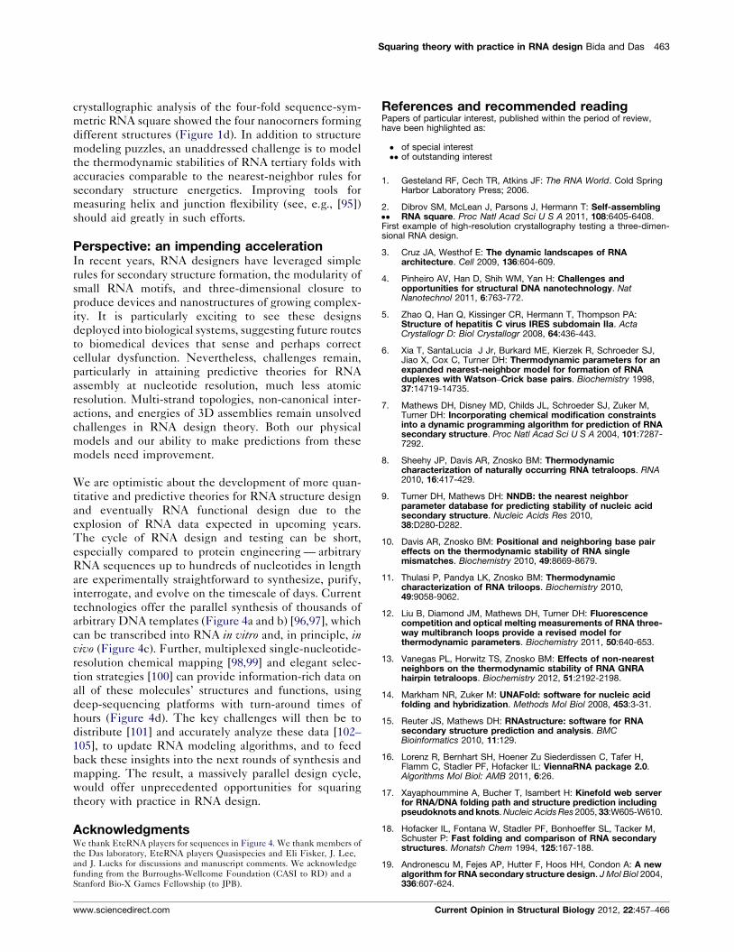
(Figure 1a) and crystallized by the Hermann lab [2��].
Rule 1. Watson–Crick base pairs generate stable double helices.Every RNA design made to date has taken advantage of
helical stems formed as RNA strands double back on them-
selves or associate with other strands to form Watson–Crick
base pairs. For the RNA square, its edges are four helices,
each involving association of four strands (Figure 1b).
Rule 2. RNA motifs can preserve their behavior when copied andpasted into new contexts. Explorations of RNAs in living
systems and elegant in vitro selections have revealed a
plethora of natural RNA catalysts, sensors, and structures.
Many of these molecules’ functions are due to small (4–15
nucleotide) RNA motifs with non-canonical structure.
These motifs can be grafted into if care is taken to avoid
mispairings with flanking sequences. For example, the four
‘nanocorners’ of the RNA square (Figure 1c) are copies of a
five-nucleotide bulge motif, drawn from a right-angle-
forming structure in the Hepatitis C virus genome [5].
Rule 3. Geometric closure ensures correct three-dimensional struc-ture. For 3D structures, the geometry of helical stems must
successfully interconnect with noncanonical motifs — a
stringent requirement for RNA designs that encompass
closed ‘ring’ topologies (Figure 1d). For the RNA square,
the choice of 10 base pairs for the edges leads to a well-
ordered square assembly with no detectable alternative
species. Extension to 11 base pairs precludes the closure
of the square and gives higher-order assemblies [2��].
Can these intuitive rules be explained and expanded into
quantitatively predictive theories for RNA design?
Expanding Rule 1: theories for RNA secondarystructure designPhysical theories underlying RNA secondary structure
formation are the most developed models in RNA science
and arguably amongst the most predictive theories avail-
able in biophysics. Several decades of melt experiments
on thousands of RNA sequences have been distilled into
Current Opinion in Structural Biology 2012, 22:457–466

458 Engineering and design
Figure 1
(e) 5′
G3′
C
U CA UCA
AGGAGGC
5′C3′U
CGG CA
GCC
5′
5′ C
3′
G A G GA
A CU
ACUG
GCAGCU
G
G
CC
C
3′
C
(a)
(b)
(f)
(g) (d)
(c)
C GGC
GG
CC
Pairings with neigh-boring nanocorners
Mg2+
Sr2+
Rosetta model (outer strand)
Cobalt hexammine3+
Crystallographic Modeled
Rosettaloop model(5 nucleotides)
Outer Strand: 5′-CCGGAGGAACUACUG-3 ′ [x4]
Inner Strand: 5′-CCGGCAGCCU-3 ′ [x4]
Current Opinion in Structural Biology
A square illustrates RNA design. (a) Sequences (inner and outer) determined ‘by hand’ by Hermann group [2��]; four copies of the two sequences form
the square. (b) Multi-strand secondary structure involves each inner and outer strand forming a nanocorner, and four nanocorners pairing through
three-nucleotide sticky ends. (c) Inspiration for the nanocorner, a 5-nt bulge that forms a 90 8C bend in the Hepatitis C virus internal ribosomal entry
site (crystallized in PDB ID 2PN4). (d) Crystallographic analysis for full RNA square (PDB ID 3P59). (e) Secondary structure predictions for square
design are inaccurate (NUPACK models shown [26]). (f) The ab initio modeling problem of rebuilding the nanocorner’s 5-nt bulge — even given the rest
of the coordinates — is not solvable at atomic resolution. (g) An RNA-Puzzle [70��]: blind prediction of the nanosquare conformation by Rosetta
methods is not atomically accurate, even when given the inner strand coordinates as a constraint.
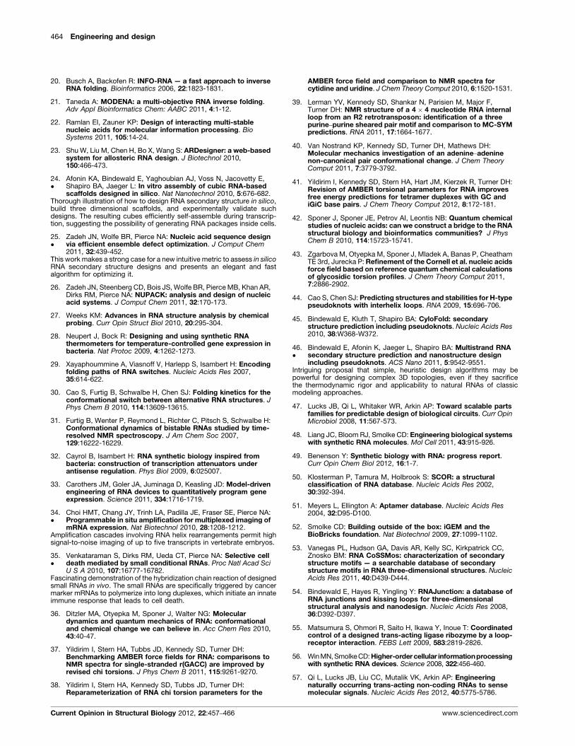
models that parameterize canonical base pair formation
into two dozen ‘nearest-neighbor’ parameters, where the
thermodynamic stability of a given base pair depends on
adjacent base pairs and temperature [6,7]. Further mod-
eling and measurements have provided approximate
energetic rules (Figure 2a) for many hairpin loops, base
pair mismatches, and more complex inter-helical motifs
such as three-way junctions [7–13]. Given such energetic
models, dynamic programming algorithms implemented
in packages such as mfold/UNAfold [14], RNAstructure
[15], and ViennaRNA [16] permit the comprehensive
Current Opinion in Structural Biology 2012, 22:457–466
statistical mechanical description of RNA secondary
structure ensembles for arbitrary sequences (Figure 2b)
with the ability to recover �70% of phylogenetically
determined base pairs [7]. Modeling methods for second-
ary structure formation kinetics are also under exploration
in packages such as Kinefold [17].
Most relevant for RNA design are recent approaches for
predicting sequences that fold into given target secondary
structures, the ‘‘inverse’’ folding problem. Methods range
from simple Monte Carlo sequence searches (Inver-
www.sciencedirect.com

Squaring theory with practice in RNA design Bida and Das 459
seRNA [18]) to faster hierarchical schemes (RNA-SSD
[19] and INFO-RNA [20]) that attempt to solve the
design problem for substructures or with simplifying
assumptions before merging or optimizing solutions
(Figure 2c). Most efforts have focused on finding
sequences whose minimum free energy conformations
recover the desired structure or, in some cases, multiple
structures [16,21–23,24�]. The NUPACK software has
presented optimization algorithms for a novel and intui-
tive target metric, the ‘ensemble defect’, which parame-
terizes the fluctuations of each nucleotide away from its
desired configuration [25�,26]. Systematic experimental
benchmarks of these design algorithms on novel second-
ary structure targets, perhaps using chemical accessibility
mapping [27], would be valuable but are not yet available.
RNA secondary structure prediction and design algorithms
are being widely used to help develop novel molecules.
Design of RNA thermometers for temperature-controlled
gene expression has made use of the nearest neighbor rules
[28] (Figure 2d). Isambert, Schwalbe, and other labs have
designed model systems with appealing simplicity to study
cotranscriptional folding and two-state switching [29–32].
Carothers and colleagues have calculated the properties of
RNA devices taking into account the folding rates of
ribozymes and aptamers calculated in Kinefold [33]. Pierce
and colleagues have imported the ‘hybridization chain
reaction’ first developed in DNA engineering to create
multiplexed amplifiers for in situ hybridization to mRNA
targets in zebrafish embryos [34�] (Figure 2e and f) and to
induce apoptosis of cultured human cells in response to
cancer marker RNAs [35�].
While making use of RNA secondary structure modeling
algorithms, these successful design efforts have still
required significant insight from experts and would be
challenging to generate automatically. For example, the
RNA square, which was designed ‘by hand’ [2��], is a
problem case for current secondary structure calculation
methods, which predict alternative structures (Figure 1e).
The squares’ nanocorners are especially stable motif
sequences that form noncanonical hydrogen bonds and
specifically coordinate multivalent ions — features that
are not treated in current RNA thermodynamic models.
In principle, quantum chemical and molecular dynamics
approaches should enable the ab initio calculation of these
‘missing’ parameters in arbitrary solution conditions but
are still under calibration [36–43]. Further, most second-
ary structure prediction packages do not yet model ring-
like or pseudoknot structures, although new extensions
are attempting to tackle this issue [44,45,46�].
Expanding Rule 2: new RNA motifs and newcombinationsA large arsenal of functional motifs is available for RNA
design (Figure 3a). Quite broadly, RNA sequences that
form specific three-dimensional structures, bind to small
www.sciencedirect.com
molecules or proteins, or mediate cellular localization,
degradation, transcription termination, splicing, editing,
or other cellular RNA processing events are widely used
in biological inquiry (reviewed recently in [47,48,49]).
Several groups are compiling databases with sequences
and structures of existing motifs derived from natural
functional RNAs or in vitro selections [50–54].
RNA designs have demonstrated the power of utilizing
multiple interacting motifs, often coupled allosterically via
secondary structure switches or kissing loops. Motifs
embedded in such devices include small-molecule-bind-
ing ‘aptamers’ such as the theophylline-binding motif,
catalytic modules such as the hammerhead ribozyme
three-way junction, and protein-binding motifs [55–57,58�,59]. The resulting devices have been expressed
in bacteria, yeast, and mammalian cells to create circuits
of growing complexity, including on/off switches [60,61]
(Figure 3a), RNA-based logic gates [56,62–64], cooperative
behaviors [56], and bacteria that swim toward and ingest
the herbicide atrazine [65]. Creative ways to discover and
select new functional motifs are also being pursued, for
example, through juxtaposition of randomized sequences
by tertiary scaffolds [66,67] (see also below). While all the
designs have leveraged the modularity of functional motifs,
many successes have required trial-and-error or selection invivo [57,65]. Presentation of failure cases and their in-depth
dissections, as are now carried out in the protein design
field [68,69], would be valuable.
Even with the growing database of known functional
motifs, there may always be components for RNA design
that are not naturally available, such as aptamers for
specific moieties in large macromolecules. A general
solution to such de novo design problems would be useful
but has not been demonstrated. Indeed, when the RNA
square crystallographic structure was offered as a blind
trial to RNA modelers, no algorithm reached atomic
accuracy predictions of the nanocorners even when given
the coordinates of four of the eight internal strands of the
motifs (Figure 1g) [70��]. Nevertheless, a recent
explosion of activity in RNA 3D classification [71–73]
and motif modeling [70��,74] (Figure 1f and g), with some
algorithms achieving atomic accuracy in favorable cases
[39,75�,76], suggests that de novo design may become a
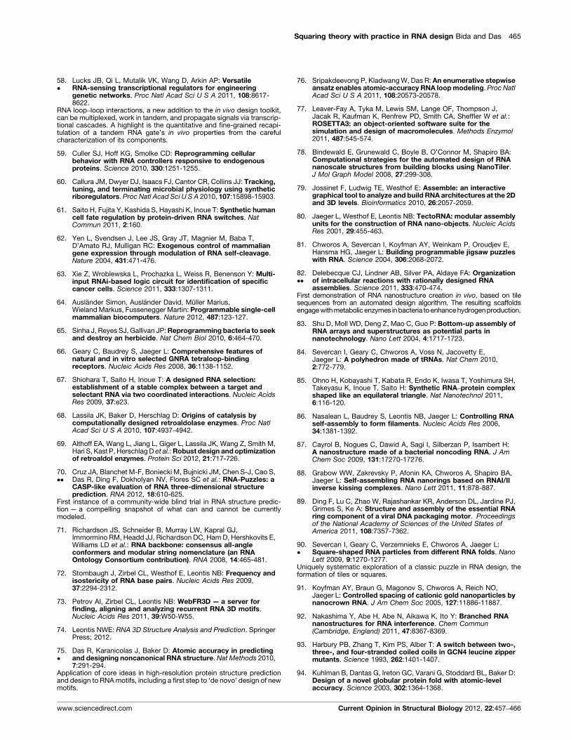
reality. As an early step, we adapted the Rosetta design
method for fixed-backbone biopolymer design [77] to
successfully thermostabilize a non-canonical motif in
the signal recognition particle RNA through mutations
previously unseen in Nature (Figure 3b) [75�].
Expanding Rule 3: theories for RNA tertiarystructure designThe successful design of highly complex 3D RNA struc-
tures continues to involve connecting RNA motifs and
helical stems, typically with additional tertiary motifs
and cross-strand interactions that generate ring-like
Current Opinion in Structural Biology 2012, 22:457–466

460 Engineering and design
Figure 2
G UCAAGAG CG
UC
AAGAG
C
Minimum freeenergy structure
orbase pair
probabilities
Target Structure HierarchicalDecomposition
Substructure SequenceOptimization
Global SequenceOptimization
(a)
GGAGA A
CUC
AA
A
+5.7 kcal/mol
–2.10 kcal/mol–2.40 kcal/mol
ΔG = +1.2 kcal/mol
(c)
A GACG
UCUG
AUC
AGACG
UCUG
AUC
A GACG
UCUG
AUC
GACG
UCUG
AUCA
AC
AGA G
ACG
UCUG
AUC
-3.0 -4.4 -4.6 -5.2 -5.8 -6.9 kcal/mol
17 °C 37 °C
(d)
(e)Metastable Hairpins
Initiators5′GACCCUAAGCAUACAUCGUCCUUC3′
Fluorescent in situhybridization chain
reaction
Tethered FluorescentAmplification Polymers
H2
H1H2
H1
H2
H2
H1
H1H2
H1
H2
H2
H1
H1H2
H1
U
U U
U
U
U
G
G
C
C
C
CCC
A
A
A
A
AA
A
5′
CA
U
UU
U
U
U
UU
U
U
U
U
C
CC
C
C
C
C
C
CC
AA
A
AA
A
A
A
A
A
A
A
AAG
G
GG
G
G
G
G
GGG
5′
3′
H2H1
TranslationNo translation
Ribosome binding Site Ribosome binding Site
H2
mRNA target
(f)
Traceback(b)
UG
C3′
U
U
U
U
U
U
UU
U
GG
G
G
G
GGG
CC
C
C
C
AA
A
A
A
A
G
AAGG
AG
UU
CA
C
C
5′ 3′
U
CU
CA
C
UU A
AGGAG
5′ 3′
AA
GGAG
CCUC
A
5′ 3′
AA
GGAGA
U
CC
U
5′ 3′
AA
GGAGA
U
CC
UU
5′ 3′
AA
GGAGA
U
CC
UU
5′ 3′
Current Opinion in Structural Biology
Theory and practice in RNA secondary structure design. (a) The free energy of a sequence in a given secondary structure conformation is calculated
by summing up the free energies of nearest neighbor terms derived from extensive empirical measurements (green) [6–13]. (b) Dynamic programming
algorithms can calculate the minimal free energy structure, base-pair probabilities, and partition function [14,15]. These methods recursively calculate
a substructure’s properties using a matrix to store the values for each smaller substructure. (c) Workflow adopted by many inverse RNA structure
Current Opinion in Structural Biology 2012, 22:457–466 www.sciencedirect.com

Squaring theory with practice in RNA design Bida and Das 461
Figure 3
Component-based model 3D Closure Search
mon
omer
o2F
Po3
FP
c3F
Po4
FP
c4F
Po5
FP
c5F
Po6
FP
c6F
P
Lower predicted Rosetta energy
KissingLoops
(a) (b)
(c)
(d)
No Trans -activation
Trans-activation
Ligand-freeaptamer
boundaptamer
TheophyllineTheophylline-
Designed sequences
Natural sequences
IS10noncoding
RNA
15 nm
5′
3′5′
5′ to 3′
Current Opinion in Structural Biology
Theory and practice in RNA tertiary structure design. (a) Cutting and pasting motifs: kissing loops, a theophylline aptamer, and the IS10 ncRNA were
rationally combined to engineer trans-acting ncRNAs that regulates gene translation that can be turned on or off with theophylline [57]. (b) New tertiary
motifs generated with Rosetta fixed backbone design for RNA, based on in silico energy minimization. The resulting designs for the most conserved
domain of the signal recognition particle RNA are distinct from natural RNAs and turn out to be more stable experimentally [75�]. (c) RNA tectonics:
combination of tertiary components and helices in three dimensions, optimized by hand or computationally [106]. (d) Nanoring assembled from RNAI/II
inverse kissing complexes, validated by atomic force microscopy (middle) and non-denaturing gel electrophoresis [88].
topologies (Figure 3d). This procedure, sometimes called
RNA ‘tectonics’, is accomplished with manually directed
programs such as NanoTiler [78] or Assemble [79] to put
together RNA components in silico.
prediction programs [19,20,25]. (d) Free energy calculations were used to d
temperature [28]. (e) Hybridization chain reaction: metastable hairpins (green
amplify the fluorescent signal during in situ hybridization experiments [34�].
elavl3 (green), ntla (yellow), and sox10 (purple) [34�].
www.sciencedirect.com
Visually compelling examples of designed 3D RNA
structures have proliferated since the first ‘TectoRNA’,
an RNA dimer stabilized by docking of tetraloops and
their cognate receptors [80]. Several groups have
esign RNA thermosensors that inhibit translation below a target
and orange) polymerize in the presence of an initiation sequence (red) to
(f) Zebrafish cross section with targeting TG(flk1:egfp) (red), tpm3 (blue),
Current Opinion in Structural Biology 2012, 22:457–466

462 Engineering and design
designed and validated RNA tiles [81,82��], multimers
[83], cubes [24�], prisms [84], triangles [85], fabrics
[81,82��], fibrils [86,87], and rings (Figure 3d) [88,89],
often using different components (tetraloop/receptors,
kissing loops, tRNA-based junctions) to make similar
objects [90�].
Many of these designs echo work in DNA nanotechnol-
ogy [4], but RNA design permits, in principle, the deploy-
ment of nanostructures in living cells. Notably,
Delebecque and colleagues [82��] have demonstrated
Figure 4
GGAAAGCACAGGAUAUGAUUAAGGGGAAAGACGAGGAUAUUACAAAAGGGAAAGACGAGGAUAUCAUAAAAGGGAAAGACGAGGAUAUCAUAAAGAGGAAAGAUCAGGAUAUUACAAAAGGGUAAGGCGAGGAUAUUGGGAAAUGGAAAUACAAGGAUAUUACAAAAGGGAAAGCAGAGGAUAUGAUGAAGAGGCAAGCACAGGAUAUUAAAAAAUGGACAGGCAAGGAUAUUAAACAAUGGCAGCGCAAGGAUAUUAUAACAAGGACAGGGCAGGAUAUUAAAACAU
(a) RNA sequence design
(d) Multiplexed chemical structure mapping
Massivparallel Rdesign c
Fiskers third simple switchMy first solution
Brourd’s Third simple switch
wisdave's 1st switch
S-RNA-S-1
LROppy's Simpleton Switch
Looking Glass
Halfway
NUPACK design 01
NUPACK design 02
NUPACK design 03
NUPACK design 04
CACAGGAUAUGAUUAAGGUCAGAAGGGUGUAAAAAAAAUU ... ...
ACGAGGAUAUUACAAAAGUAAGAAGGCGUC AAAAAAAA AG... ...
ACGAGGAUAUCAUAAAAGUGAGAAGGCGUC AAAAAAAA AG... ...
ACGAGGAUAUCAUAAAGAUGAGAAGGCGUC AAAAAAAA AG... ...
DMS
wisdave's 1st switch
Brourd's Thirdsimple switch
My firstsolution
Fiskers thirdsimple switch
SHAPE
A massively parallel design cycle should be possible for RNA. Thousands o
design tools are sent for high-throughput oligonucleotide template synthesis
and then falsified or validated at nucleotide resolution through multiplexed c
Current Opinion in Structural Biology 2012, 22:457–466
self-assembly of nanofabrics in vivo and used the resulting
structures to accelerate hydrogen production in bacteria
through the scaffolding of protein enzymes. Other appli-
cations include scaffolding gold particles for nanowires
[91], encaging other molecules [84], and protecting
duplexes intended for RNA silencing from degradation
[88,92].
Atomic-scale validation of 3D design — as was achieved
quite early for protein design [93,94] — has not yet been
demonstrated for RNA nanostructures. For example, the
(b) On-chip oligo synthesis
(c) Pooled RNA transcription
elyNA
ycle
Current Opinion in Structural Biology
f sequences (a) designed by experts, citizen scientists, and automated
(b) [96,97], (figure adopted from LC sciences) transcribed into RNA (c),
hemical mapping (d) utilizing-next generation-sequencing methods [99].
www.sciencedirect.com
Squaring theory with practice in RNA design Bida and Das 463
crystallographic analysis of the four-fold sequence-sym-
metric RNA square showed the four nanocorners forming
different structures (Figure 1d). In addition to structure
modeling puzzles, an unaddressed challenge is to model
the thermodynamic stabilities of RNA tertiary folds with
accuracies comparable to the nearest-neighbor rules for
secondary structure energetics. Improving tools for
measuring helix and junction flexibility (see, e.g., [95])
should aid greatly in such efforts.
Perspective: an impending accelerationIn recent years, RNA designers have leveraged simple
rules for secondary structure formation, the modularity of
small RNA motifs, and three-dimensional closure to
produce devices and nanostructures of growing complex-
ity. It is particularly exciting to see these designs
deployed into biological systems, suggesting future routes
to biomedical devices that sense and perhaps correct
cellular dysfunction. Nevertheless, challenges remain,
particularly in attaining predictive theories for RNA
assembly at nucleotide resolution, much less atomic
resolution. Multi-strand topologies, non-canonical inter-
actions, and energies of 3D assemblies remain unsolved
challenges in RNA design theory. Both our physical
models and our ability to make predictions from these
models need improvement.
We are optimistic about the development of more quan-
titative and predictive theories for RNA structure design
and eventually RNA functional design due to the
explosion of RNA data expected in upcoming years.
The cycle of RNA design and testing can be short,
especially compared to protein engineering — arbitrary
RNA sequences up to hundreds of nucleotides in length
are experimentally straightforward to synthesize, purify,
interrogate, and evolve on the timescale of days. Current
technologies offer the parallel synthesis of thousands of
arbitrary DNA templates (Figure 4a and b) [96,97], which
can be transcribed into RNA in vitro and, in principle, invivo (Figure 4c). Further, multiplexed single-nucleotide-
resolution chemical mapping [98,99] and elegant selec-
tion strategies [100] can provide information-rich data on
all of these molecules’ structures and functions, using
deep-sequencing platforms with turn-around times of
hours (Figure 4d). The key challenges will then be to
distribute [101] and accurately analyze these data [102–105], to update RNA modeling algorithms, and to feed
back these insights into the next rounds of synthesis and
mapping. The result, a massively parallel design cycle,
would offer unprecedented opportunities for squaring
theory with practice in RNA design.
AcknowledgmentsWe thank EteRNA players for sequences in Figure 4. We thank members ofthe Das laboratory, EteRNA players Quasispecies and Eli Fisker, J. Lee,and J. Lucks for discussions and manuscript comments. We acknowledgefunding from the Burroughs-Wellcome Foundation (CASI to RD) and aStanford Bio-X Games Fellowship (to JPB).
www.sciencedirect.com
References and recommended readingPapers of particular interest, published within the period of review,have been highlighted as:
� of special interest�� of outstanding interest
1. Gesteland RF, Cech TR, Atkins JF: The RNA World. Cold SpringHarbor Laboratory Press; 2006.
2.��
Dibrov SM, McLean J, Parsons J, Hermann T: Self-assemblingRNA square. Proc Natl Acad Sci U S A 2011, 108:6405-6408.
First example of high-resolution crystallography testing a three-dimen-sional RNA design.
3. Cruz JA, Westhof E: The dynamic landscapes of RNAarchitecture. Cell 2009, 136:604-609.
4. Pinheiro AV, Han D, Shih WM, Yan H: Challenges andopportunities for structural DNA nanotechnology. NatNanotechnol 2011, 6:763-772.
5. Zhao Q, Han Q, Kissinger CR, Hermann T, Thompson PA:Structure of hepatitis C virus IRES subdomain IIa. ActaCrystallogr D: Biol Crystallogr 2008, 64:436-443.
6. Xia T, SantaLucia J Jr, Burkard ME, Kierzek R, Schroeder SJ,Jiao X, Cox C, Turner DH: Thermodynamic parameters for anexpanded nearest-neighbor model for formation of RNAduplexes with Watson–Crick base pairs. Biochemistry 1998,37:14719-14735.
7. Mathews DH, Disney MD, Childs JL, Schroeder SJ, Zuker M,Turner DH: Incorporating chemical modification constraintsinto a dynamic programming algorithm for prediction of RNAsecondary structure. Proc Natl Acad Sci U S A 2004, 101:7287-7292.
8. Sheehy JP, Davis AR, Znosko BM: Thermodynamiccharacterization of naturally occurring RNA tetraloops. RNA2010, 16:417-429.
9. Turner DH, Mathews DH: NNDB: the nearest neighborparameter database for predicting stability of nucleic acidsecondary structure. Nucleic Acids Res 2010,38:D280-D282.
10. Davis AR, Znosko BM: Positional and neighboring base paireffects on the thermodynamic stability of RNA singlemismatches. Biochemistry 2010, 49:8669-8679.
11. Thulasi P, Pandya LK, Znosko BM: Thermodynamiccharacterization of RNA triloops. Biochemistry 2010,49:9058-9062.
12. Liu B, Diamond JM, Mathews DH, Turner DH: Fluorescencecompetition and optical melting measurements of RNA three-way multibranch loops provide a revised model forthermodynamic parameters. Biochemistry 2011, 50:640-653.
13. Vanegas PL, Horwitz TS, Znosko BM: Effects of non-nearestneighbors on the thermodynamic stability of RNA GNRAhairpin tetraloops. Biochemistry 2012, 51:2192-2198.
14. Markham NR, Zuker M: UNAFold: software for nucleic acidfolding and hybridization. Methods Mol Biol 2008, 453:3-31.
15. Reuter JS, Mathews DH: RNAstructure: software for RNAsecondary structure prediction and analysis. BMCBioinformatics 2010, 11:129.
16. Lorenz R, Bernhart SH, Hoener Zu Siederdissen C, Tafer H,Flamm C, Stadler PF, Hofacker IL: ViennaRNA package 2.0.Algorithms Mol Biol: AMB 2011, 6:26.
17. Xayaphoummine A, Bucher T, Isambert H: Kinefold web serverfor RNA/DNA folding path and structure prediction includingpseudoknots and knots. Nucleic Acids Res 2005, 33:W605-W610.
18. Hofacker IL, Fontana W, Stadler PF, Bonhoeffer SL, Tacker M,Schuster P: Fast folding and comparison of RNA secondarystructures. Monatsh Chem 1994, 125:167-188.
19. Andronescu M, Fejes AP, Hutter F, Hoos HH, Condon A: A newalgorithm for RNA secondary structure design. J Mol Biol 2004,336:607-624.
Current Opinion in Structural Biology 2012, 22:457–466
464 Engineering and design
20. Busch A, Backofen R: INFO-RNA — a fast approach to inverseRNA folding. Bioinformatics 2006, 22:1823-1831.
21. Taneda A: MODENA: a multi-objective RNA inverse folding.Adv Appl Bioinformatics Chem: AABC 2011, 4:1-12.
22. Ramlan EI, Zauner KP: Design of interacting multi-stablenucleic acids for molecular information processing. BioSystems 2011, 105:14-24.
23. Shu W, Liu M, Chen H, Bo X, Wang S: ARDesigner: a web-basedsystem for allosteric RNA design. J Biotechnol 2010,150:466-473.
24.�
Afonin KA, Bindewald E, Yaghoubian AJ, Voss N, Jacovetty E,Shapiro BA, Jaeger L: In vitro assembly of cubic RNA-basedscaffolds designed in silico. Nat Nanotechnol 2010, 5:676-682.
Thorough illustration of how to design RNA secondary structure in silico,build three dimensional scaffolds, and experimentally validate suchdesigns. The resulting cubes efficiently self-assemble during transcrip-tion, suggesting the possibility of generating RNA packages inside cells.
25.�
Zadeh JN, Wolfe BR, Pierce NA: Nucleic acid sequence designvia efficient ensemble defect optimization. J Comput Chem2011, 32:439-452.
This work makes a strong case for a new intuitive metric to assess in silicoRNA secondary structure designs and presents an elegant and fastalgorithm for optimizing it.
26. Zadeh JN, Steenberg CD, Bois JS, Wolfe BR, Pierce MB, Khan AR,Dirks RM, Pierce NA: NUPACK: analysis and design of nucleicacid systems. J Comput Chem 2011, 32:170-173.
27. Weeks KM: Advances in RNA structure analysis by chemicalprobing. Curr Opin Struct Biol 2010, 20:295-304.
28. Neupert J, Bock R: Designing and using synthetic RNAthermometers for temperature-controlled gene expression inbacteria. Nat Protoc 2009, 4:1262-1273.
29. Xayaphoummine A, Viasnoff V, Harlepp S, Isambert H: Encodingfolding paths of RNA switches. Nucleic Acids Res 2007,35:614-622.
30. Cao S, Furtig B, Schwalbe H, Chen SJ: Folding kinetics for theconformational switch between alternative RNA structures. JPhys Chem B 2010, 114:13609-13615.
31. Furtig B, Wenter P, Reymond L, Richter C, Pitsch S, Schwalbe H:Conformational dynamics of bistable RNAs studied by time-resolved NMR spectroscopy. J Am Chem Soc 2007,129:16222-16229.
32. Cayrol B, Isambert H: RNA synthetic biology inspired frombacteria: construction of transcription attenuators underantisense regulation. Phys Biol 2009, 6:025007.
33. Carothers JM, Goler JA, Juminaga D, Keasling JD: Model-drivenengineering of RNA devices to quantitatively program geneexpression. Science 2011, 334:1716-1719.
34.�
Choi HMT, Chang JY, Trinh LA, Padilla JE, Fraser SE, Pierce NA:Programmable in situ amplification for multiplexed imaging ofmRNA expression. Nat Biotechnol 2010, 28:1208-1212.
Amplification cascades involving RNA helix rearrangements permit highsignal-to-noise imaging of up to five transcripts in vertebrate embryos.
35.�
Venkataraman S, Dirks RM, Ueda CT, Pierce NA: Selective celldeath mediated by small conditional RNAs. Proc Natl Acad SciU S A 2010, 107:16777-16782.
Fascinating demonstration of the hybridization chain reaction of designedsmall RNAs in vivo. The small RNAs are specifically triggered by cancermarker mRNAs to polymerize into long duplexes, which initiate an innateimmune response that leads to cell death.
36. Ditzler MA, Otyepka M, Sponer J, Walter NG: Moleculardynamics and quantum mechanics of RNA: conformationaland chemical change we can believe in. Acc Chem Res 2010,43:40-47.
37. Yildirim I, Stern HA, Tubbs JD, Kennedy SD, Turner DH:Benchmarking AMBER force fields for RNA: comparisons toNMR spectra for single-stranded r(GACC) are improved byrevised chi torsions. J Phys Chem B 2011, 115:9261-9270.
38. Yildirim I, Stern HA, Kennedy SD, Tubbs JD, Turner DH:Reparameterization of RNA chi torsion parameters for the
Current Opinion in Structural Biology 2012, 22:457–466
AMBER force field and comparison to NMR spectra forcytidine and uridine. J Chem Theory Comput 2010, 6:1520-1531.
39. Lerman YV, Kennedy SD, Shankar N, Parisien M, Major F,Turner DH: NMR structure of a 4 � 4 nucleotide RNA internalloop from an R2 retrotransposon: identification of a threepurine–purine sheared pair motif and comparison to MC-SYMpredictions. RNA 2011, 17:1664-1677.
40. Van Nostrand KP, Kennedy SD, Turner DH, Mathews DH:Molecular mechanics investigation of an adenine–adeninenon-canonical pair conformational change. J Chem TheoryComput 2011, 7:3779-3792.
41. Yildirim I, Kennedy SD, Stern HA, Hart JM, Kierzek R, Turner DH:Revision of AMBER torsional parameters for RNA improvesfree energy predictions for tetramer duplexes with GC andiGiC base pairs. J Chem Theory Comput 2012, 8:172-181.
42. Sponer J, Sponer JE, Petrov AI, Leontis NB: Quantum chemicalstudies of nucleic acids: can we construct a bridge to the RNAstructural biology and bioinformatics communities? J PhysChem B 2010, 114:15723-15741.
43. Zgarbova M, Otyepka M, Sponer J, Mladek A, Banas P, CheathamTE 3rd, Jurecka P: Refinement of the Cornell et al. nucleic acidsforce field based on reference quantum chemical calculationsof glycosidic torsion profiles. J Chem Theory Comput 2011,7:2886-2902.
44. Cao S, Chen SJ: Predicting structures and stabilities for H-typepseudoknots with interhelix loops. RNA 2009, 15:696-706.
45. Bindewald E, Kluth T, Shapiro BA: CyloFold: secondarystructure prediction including pseudoknots. Nucleic Acids Res2010, 38:W368-W372.
46.�
Bindewald E, Afonin K, Jaeger L, Shapiro BA: Multistrand RNAsecondary structure prediction and nanostructure designincluding pseudoknots. ACS Nano 2011, 5:9542-9551.
Intriguing proposal that simple, heuristic design algorithms may bepowerful for designing complex 3D topologies, even if they sacrificethe thermodynamic rigor and applicability to natural RNAs of classicmodeling approaches.
47. Lucks JB, Qi L, Whitaker WR, Arkin AP: Toward scalable partsfamilies for predictable design of biological circuits. Curr OpinMicrobiol 2008, 11:567-573.
48. Liang JC, Bloom RJ, Smolke CD: Engineering biological systemswith synthetic RNA molecules. Mol Cell 2011, 43:915-926.
49. Benenson Y: Synthetic biology with RNA: progress report.Curr Opin Chem Biol 2012, 16:1-7.
50. Klosterman P, Tamura M, Holbrook S: SCOR: a structuralclassification of RNA database. Nucleic Acids Res 2002,30:392-394.
51. Meyers L, Ellington A: Aptamer database. Nucleic Acids Res2004, 32:D95-D100.
52. Smolke CD: Building outside of the box: iGEM and theBioBricks foundation. Nat Biotechnol 2009, 27:1099-1102.
53. Vanegas PL, Hudson GA, Davis AR, Kelly SC, Kirkpatrick CC,Znosko BM: RNA CoSSMos: characterization of secondarystructure motifs — a searchable database of secondarystructure motifs in RNA three-dimensional structures. NucleicAcids Res 2011, 40:D439-D444.
54. Bindewald E, Hayes R, Yingling Y: RNAJunction: a database ofRNA junctions and kissing loops for three-dimensionalstructural analysis and nanodesign. Nucleic Acids Res 2008,36:D392-D397.
55. Matsumura S, Ohmori R, Saito H, Ikawa Y, Inoue T: Coordinatedcontrol of a designed trans-acting ligase ribozyme by a loop-receptor interaction. FEBS Lett 2009, 583:2819-2826.
56. Win MN, Smolke CD: Higher-order cellular information processingwith synthetic RNA devices. Science 2008, 322:456-460.
57. Qi L, Lucks JB, Liu CC, Mutalik VK, Arkin AP: Engineeringnaturally occurring trans-acting non-coding RNAs to sensemolecular signals. Nucleic Acids Res 2012, 40:5775-5786.
www.sciencedirect.com
Squaring theory with practice in RNA design Bida and Das 465
58.�
Lucks JB, Qi L, Mutalik VK, Wang D, Arkin AP: VersatileRNA-sensing transcriptional regulators for engineeringgenetic networks. Proc Natl Acad Sci U S A 2011, 108:8617-8622.
RNA loop–loop interactions, a new addition to the in vivo design toolkit,can be multiplexed, work in tandem, and propagate signals via transcrip-tional cascades. A highlight is the quantitative and fine-grained recapi-tulation of a tandem RNA gate’s in vivo properties from the carefulcharacterization of its components.
59. Culler SJ, Hoff KG, Smolke CD: Reprogramming cellularbehavior with RNA controllers responsive to endogenousproteins. Science 2010, 330:1251-1255.
60. Callura JM, Dwyer DJ, Isaacs FJ, Cantor CR, Collins JJ: Tracking,tuning, and terminating microbial physiology using syntheticriboregulators. Proc Natl Acad Sci U S A 2010, 107:15898-15903.
61. Saito H, Fujita Y, Kashida S, Hayashi K, Inoue T: Synthetic humancell fate regulation by protein-driven RNA switches. NatCommun 2011, 2:160.
62. Yen L, Svendsen J, Lee JS, Gray JT, Magnier M, Baba T,D’Amato RJ, Mulligan RC: Exogenous control of mammaliangene expression through modulation of RNA self-cleavage.Nature 2004, 431:471-476.
63. Xie Z, Wroblewska L, Prochazka L, Weiss R, Benenson Y: Multi-input RNAi-based logic circuit for identification of specificcancer cells. Science 2011, 333:1307-1311.
64. Auslander Simon, Auslander David, Muller Marius,Wieland Markus, Fussenegger Martin: Programmable single-cellmammalian biocomputers. Nature 2012, 487:123-127.
65. Sinha J, Reyes SJ, Gallivan JP: Reprogramming bacteria to seekand destroy an herbicide. Nat Chem Biol 2010, 6:464-470.
66. Geary C, Baudrey S, Jaeger L: Comprehensive features ofnatural and in vitro selected GNRA tetraloop-bindingreceptors. Nucleic Acids Res 2008, 36:1138-1152.
67. Shiohara T, Saito H, Inoue T: A designed RNA selection:establishment of a stable complex between a target andselectant RNA via two coordinated interactions. Nucleic AcidsRes 2009, 37:e23.
68. Lassila JK, Baker D, Herschlag D: Origins of catalysis bycomputationally designed retroaldolase enzymes. Proc NatlAcad Sci U S A 2010, 107:4937-4942.
69. Althoff EA, Wang L, Jiang L, Giger L, Lassila JK, Wang Z, Smith M,Hari S, Kast P, Herschlag D et al.: Robust design and optimizationof retroaldol enzymes. Protein Sci 2012, 21:717-726.
70.��
Cruz JA, Blanchet M-F, Boniecki M, Bujnicki JM, Chen S-J, Cao S,Das R, Ding F, Dokholyan NV, Flores SC et al.: RNA-Puzzles: aCASP-like evaluation of RNA three-dimensional structureprediction. RNA 2012, 18:610-625.
First instance of a community-wide blind trial in RNA structure predic-tion — a compelling snapshot of what can and cannot be currentlymodeled.
71. Richardson JS, Schneider B, Murray LW, Kapral GJ,Immormino RM, Headd JJ, Richardson DC, Ham D, Hershkovits E,Williams LD et al.: RNA backbone: consensus all-angleconformers and modular string nomenclature (an RNAOntology Consortium contribution). RNA 2008, 14:465-481.
72. Stombaugh J, Zirbel CL, Westhof E, Leontis NB: Frequency andisostericity of RNA base pairs. Nucleic Acids Res 2009,37:2294-2312.
73. Petrov AI, Zirbel CL, Leontis NB: WebFR3D — a server forfinding, aligning and analyzing recurrent RNA 3D motifs.Nucleic Acids Res 2011, 39:W50-W55.
74. Leontis NWE: RNA 3D Structure Analysis and Prediction. SpringerPress; 2012.
75.�
Das R, Karanicolas J, Baker D: Atomic accuracy in predictingand designing noncanonical RNA structure. Nat Methods 2010,7:291-294.
Application of core ideas in high-resolution protein structure predictionand design to RNA motifs, including a first step to ‘de novo’ design of newmotifs.
www.sciencedirect.com
76. Sripakdeevong P, Kladwang W, Das R: An enumerative stepwiseansatz enables atomic-accuracy RNA loop modeling. Proc NatlAcad Sci U S A 2011, 108:20573-20578.
77. Leaver-Fay A, Tyka M, Lewis SM, Lange OF, Thompson J,Jacak R, Kaufman K, Renfrew PD, Smith CA, Sheffler W et al.:ROSETTA3: an object-oriented software suite for thesimulation and design of macromolecules. Methods Enzymol2011, 487:545-574.
78. Bindewald E, Grunewald C, Boyle B, O’Connor M, Shapiro BA:Computational strategies for the automated design of RNAnanoscale structures from building blocks using NanoTiler.J Mol Graph Model 2008, 27:299-308.
79. Jossinet F, Ludwig TE, Westhof E: Assemble: an interactivegraphical tool to analyze and build RNA architectures at the 2Dand 3D levels. Bioinformatics 2010, 26:2057-2059.
80. Jaeger L, Westhof E, Leontis NB: TectoRNA: modular assemblyunits for the construction of RNA nano-objects. Nucleic AcidsRes 2001, 29:455-463.
81. Chworos A, Severcan I, Koyfman AY, Weinkam P, Oroudjev E,Hansma HG, Jaeger L: Building programmable jigsaw puzzleswith RNA. Science 2004, 306:2068-2072.
82.��
Delebecque CJ, Lindner AB, Silver PA, Aldaye FA: Organizationof intracellular reactions with rationally designed RNAassemblies. Science 2011, 333:470-474.
First demonstration of RNA nanostructure creation in vivo, based on tilesequences from an automated design algorithm. The resulting scaffoldsengage with metabolic enzymes in bacteria to enhance hydrogen production.
83. Shu D, Moll WD, Deng Z, Mao C, Guo P: Bottom-up assembly ofRNA arrays and superstructures as potential parts innanotechnology. Nano Lett 2004, 4:1717-1723.
84. Severcan I, Geary C, Chworos A, Voss N, Jacovetty E,Jaeger L: A polyhedron made of tRNAs. Nat Chem 2010,2:772-779.
85. Ohno H, Kobayashi T, Kabata R, Endo K, Iwasa T, Yoshimura SH,Takeyasu K, Inoue T, Saito H: Synthetic RNA–protein complexshaped like an equilateral triangle. Nat Nanotechnol 2011,6:116-120.
86. Nasalean L, Baudrey S, Leontis NB, Jaeger L: Controlling RNAself-assembly to form filaments. Nucleic Acids Res 2006,34:1381-1392.
87. Cayrol B, Nogues C, Dawid A, Sagi I, Silberzan P, Isambert H:A nanostructure made of a bacterial noncoding RNA. J AmChem Soc 2009, 131:17270-17276.
88. Grabow WW, Zakrevsky P, Afonin KA, Chworos A, Shapiro BA,Jaeger L: Self-assembling RNA nanorings based on RNAI/IIinverse kissing complexes. Nano Lett 2011, 11:878-887.
89. Ding F, Lu C, Zhao W, Rajashankar KR, Anderson DL, Jardine PJ,Grimes S, Ke A: Structure and assembly of the essential RNAring component of a viral DNA packaging motor. Proceedingsof the National Academy of Sciences of the United States ofAmerica 2011, 108:7357-7362.
90.�
Severcan I, Geary C, Verzemnieks E, Chworos A, Jaeger L:Square-shaped RNA particles from different RNA folds. NanoLett 2009, 9:1270-1277.
Uniquely systematic exploration of a classic puzzle in RNA design, theformation of tiles or squares.
91. Koyfman AY, Braun G, Magonov S, Chworos A, Reich NO,Jaeger L: Controlled spacing of cationic gold nanoparticles bynanocrown RNA. J Am Chem Soc 2005, 127:11886-11887.
92. Nakashima Y, Abe H, Abe N, Aikawa K, Ito Y: Branched RNAnanostructures for RNA interference. Chem Commun(Cambridge, England) 2011, 47:8367-8369.
93. Harbury PB, Zhang T, Kim PS, Alber T: A switch between two-,three-, and four-stranded coiled coils in GCN4 leucine zippermutants. Science 1993, 262:1401-1407.
94. Kuhlman B, Dantas G, Ireton GC, Varani G, Stoddard BL, Baker D:Design of a novel globular protein fold with atomic-levelaccuracy. Science 2003, 302:1364-1368.
Current Opinion in Structural Biology 2012, 22:457–466
466 Engineering and design
95. Zhang Q, Stelzer AC, Fisher CK, Al-Hashimi HM: Visualizingspatially correlated dynamics that directs RNA conformationaltransitions. Nature 2007, 450:1263-1267.
96. Zhou X, Cai S, Hong A, You Q, Yu P, Sheng N, Srivannavit O,Muranjan S, Rouillard JM, Xia Y et al.: Microfluidic PicoArraysynthesis of oligodeoxynucleotides and simultaneousassembling of multiple DNA sequences. Nucleic Acids Res2004, 32:5409-5417.
97. LeProust EM, Peck BJ, Spirin K, McCuen HB, Moore B,Namsaraev E, Caruthers MH: Synthesis of high-quality librariesof long (150mer) oligonucleotides by a novel depurinationcontrolled process. Nucleic Acids Res 2010, 38:2522-2540.
98. Kladwang W, VanLang CC, Cordero P, Das R: A two-dimensionalmutate-and-map strategy for non-coding RNA structure. NatChem 2011, 3:954-962.
99. Lucks JB, Mortimer SA, Trapnell C, Luo S, Aviran S, Schroth GP,Pachter L, Doudna JA, Arkin AP: Multiplexed RNA structurecharacterization with selective 20-hydroxyl acylation analyzedby primer extension sequencing (SHAPE-Seq). Proc Natl AcadSci U S A 2011, 108:11063-11068.
100. Pitt JN, Ferre-D’Amare AR: Rapid construction of empirical RNAfitness landscapes. Science 2010, 330:376-379.
Current Opinion in Structural Biology 2012, 22:457–466
101. Rocca-Serra P, Bellaousov S, Birmingham A, Chen C, Cordero P,Das R, Davis-Neulander L, Duncan CD, Halvorsen M, Knight Ret al.: Sharing and archiving nucleic acid structure mappingdata. RNA 2011, 17:1204-1212.
102. Kladwang W, VanLang CC, Cordero P, Das R: Understanding theerrors of SHAPE-directed RNA structure modeling.Biochemistry 2011, 50:8049-8056.
103. Bindewald E, Wendeler M, Legiewicz M, Bona MK, Wang Y,Pritt MJ, Le Grice SF, Shapiro BA: Correlating SHAPE signatureswith three-dimensional RNA structures. RNA 2011,17:1688-1696.
104. Washietl S, Hofacker IL, Stadler PF, Kellis M: RNA folding withsoft constraints: reconciliation of probing data andthermodynamic secondary structure prediction. Nucleic AcidsRes 2012, 40:4261-4272.
105. Quarrier S, Martin JS, Davis-Neulander L, Beauregard A,Laederach A: Evaluation of the information content of RNAstructure mapping data for secondary structure prediction.RNA 2010, 16:1108-1117.
106. Kasprzak W, Bindewald E, Kim T-J, Jaeger L, Shapiro BA: Use ofRNA structure flexibility data in nanostructure modeling.Methods (San Diego, CA) 2011, 54:239-250.
www.sciencedirect.com