Spinal deformity in children - Acta Orthopaedica Et ...
8
57 acta OrthOpaedica et traumatOlOgica hellenica MINI revIew VOLUME 70 | ISSUE 2 | APRIL - JUNE 2019 ActA Spinal deformity in children Sekouris N 1 , Paspati I 2 , Fligger I 3 , Flouda L 4 1 Peadiatric Orthopaedic Department of “KAT” General Hospital, Kifisia, Greece 2 Peadiatric Orthopaedic Department of Penteli’ s Paediatric Hospital, Penteli, Greece 3 Peadiatric Orthopaedic Department of “AGIA SOFIA” Pediatric Hospital, Athens, Greece 4 Department of Anesthesiology of “AGIA SOFIA” Pediatric Hospital, Athens, Greece Nick Sekouris Consultant Paediatric Orthopaedic Surgeon Orthopedic Department, KAT General Hospital, Athens, Greece, Tel: 6985010730, Email: [email protected] The treatment of early onset scoliosis is a challenge because of the need to correct the deformity of the spine without using the traditional systems that require spinal fusion at this age. The conservative treatment of early onset scoliosis with brace is usually not effective but can help to postpone the surgical treatment at a time closer to skeletal maturation. The surgical procedure includes systems that do not fuse the spine but allow for further growth. These systems can be (a) the growing rods, (b) the guided rods and (c) compressive-based. These systems are a good option for the treatment of childhood scoliosis, but as they are not «protected» by a bone spine fusion they are subject to ongoing stress and forces. As a result, there is a higher failure rate of materials compared to the traditional fusion systems. KEY WORDS: early onset scoliosis; thoracic insufficiency syndrome; growing brace for scoliosis; growing rods; guided rods; compressive-based system AbstrAct correspoNdINg Author, guArANtor Introduction Scoliosis is a three-dimensional deformity of the spine in which there is a shift of the vertebrae, and abnormal curves that are mainly at the side of the spine. This deformity can be observed in children before puber- ty. In some cases, the scoliosis is progressive, and the deformation of the rib cage can affect the develop- ment of pulmonary alveoli. During this period there is a need to protect the rib cage from deformation and lessen the effects on the pulmonary parenchyma. On the other hand, we can’t correct the deformity with spinal fusion and suspend its development because that will limit the growth of the rib cage, causing ob- structive pulmonary disease. The inability to interfere in scoliosis developed in young children can lead to serious scoliosis with serious co-morbid obstructive pulmonary disease. For all the above reasons these deformities are a distinct clinical entity termed «early onset” scoliosis and appears before the age of 10 1 . The way the scoliosis is going to develop depends on the etiology (congenital, neuromuscular, syndromic, i.e. neurofibromatosis, and idiopathic). Some syndromic scoliosis with congenital anomalies such as asphyxi- ating thoracic dysplasia (Jeune’s Syndrome) and the vertebra-rib dysostosis (Jarcho-Levin-Syndrome) will take a very special and demanding treatment.
-
Upload
khangminh22 -
Category
Documents
-
view
0 -
download
0
Transcript of Spinal deformity in children - Acta Orthopaedica Et ...
57acta OrthOpaedica et traumatOlOgica hellenica
MINI revIewVOLUME 70 | ISSUE 2 | APRIL - JUNE 2019 ActASpinal deformity in children
Sekouris N1, Paspati I2, Fligger I3, Flouda L4
1Peadiatric Orthopaedic Department of “KAT” General Hospital, Kifisia, Greece2Peadiatric Orthopaedic Department of Penteli’ s Paediatric Hospital, Penteli, Greece
3Peadiatric Orthopaedic Department of “AGIA SOFIA” Pediatric Hospital, Athens, Greece4Department of Anesthesiology of “AGIA SOFIA” Pediatric Hospital, Athens, Greece
Nick SekourisConsultant Paediatric Orthopaedic SurgeonOrthopedic Department, KAT General Hospital, Athens, Greece, Tel: 6985010730, Email: [email protected]
The treatment of early onset scoliosis is a challenge because of the need to correct the deformity of the spine without using the traditional systems that require spinal fusion at this age. The conservative treatment of early onset scoliosis with brace is usually not effective but can help to postpone the surgical treatment at a time closer to skeletal maturation. The surgical procedure includes systems that do not fuse the spine but allow for further growth. These systems can be (a) the growing rods, (b) the guided rods and (c) compressive-based. These systems are a good option for the treatment of childhood scoliosis, but as they are not «protected» by a bone spine fusion they are subject to ongoing stress and forces. As a result, there is a higher failure rate of materials compared to the traditional fusion systems.
KEY WORDS: early onset scoliosis; thoracic insufficiency syndrome; growing brace for scoliosis; growing rods; guided rods; compressive-based system
AbstrAct
correspoNdINg Author, guArANtor
IntroductionScoliosis is a three-dimensional deformity of the spine in which there is a shift of the vertebrae, and abnormal curves that are mainly at the side of the spine. This deformity can be observed in children before puber-ty. In some cases, the scoliosis is progressive, and the deformation of the rib cage can affect the develop-ment of pulmonary alveoli. During this period there is a need to protect the rib cage from deformation and lessen the effects on the pulmonary parenchyma. On the other hand, we can’t correct the deformity with spinal fusion and suspend its development because that will limit the growth of the rib cage, causing ob-
structive pulmonary disease. The inability to interfere in scoliosis developed in young children can lead to serious scoliosis with serious co-morbid obstructive pulmonary disease. For all the above reasons these deformities are a distinct clinical entity termed «early onset” scoliosis and appears before the age of 101. The way the scoliosis is going to develop depends on the etiology (congenital, neuromuscular, syndromic, i.e. neurofibromatosis, and idiopathic). Some syndromic scoliosis with congenital anomalies such as asphyxi-ating thoracic dysplasia (Jeune’s Syndrome) and the vertebra-rib dysostosis (Jarcho-Levin-Syndrome) will take a very special and demanding treatment.
58 acta OrthOpaedica et traumatOlOgica hellenica
VOLUME 70 | ISSUE 2 | APRIL - JUNE 2019
Sekouris N, et al. Spinal deformity in children
Thoracic insufficiency syndromeThe goal of the treatment of early scoliosis is to stop the deterioration of scoliosis into severe deformation and to prevent the syndrome of thoracic insufficien-cy2. The number of pulmonary alveoli grows rapid-ly until the age of 2 years, and then continues to in-crease at a slower rate until the age of 8 years. After this age the cells increases in size but not in number3. Therefore, any deformation or reduction in the elas-ticity of the thoracic cage affects the development of the respiratory parenchyma and therefore the respira-tory function. The restriction of the lung parenchyma causes arterial pulmonary hypertension that leads to life-threatening diseases, i.e. pulmonary heart, heart failure4. This pathological condition is called syn-drome of thoracic insufficiency. It seems that there is a direct correlation of respiratory function with the level of the T1-T12 vertebrae, in children who were subject-ed to spinal fusion due to congenital scoliosis. In these children a decrease of the pulmonary vital capacity of 50% was observed after spinal fusion. In fact, in chil-dren younger than 8 years of age who were subjected to spinal fusion, the vital capacity decreased by 60%. These studies revealed that the height of T1-T12 must be greater than 20 cm in order to avoid the syndrome of thoracic insufficiency5. Therefore, in these ages spi-nal fusion is prohibitive because it reduces the vital ca-pacity of the lungs. The spinal fusion, also, decreases
the elasticity of the thoracic cage, which acts negative-ly on the respiratory function6,7. In addition, in chil-dren of this age, with a great expectancy of growth, treatment with posterior instrumented spinal fusion can cause the crankshaft phenomenon. This is a new rotation deformity that develops after spinal fusion, due to the growth of the body of the vertebra, which cannot be restrained by the posterior instrumented spinal fusion. Last contraindication for spinal fusion in early scoliosis is the asymmetry of the torso-limb that can be caused by the spinal fusion.
TreatmentConservative treatmentThe conservative treatment has poor results in terms of evolution of childhood scoliosis because it can’t stop the progression of scoliosis. However, it is a useful tool because a) it can help the patient with hypotonia stay in the sitting position, b) it can control the second-ary curves in patients with congenital scoliosis and c) it saves us some time before deciding upon a more active intervention1. For the conservative treatment brace of the spine, thermoplastic seat, back props, and casts of the spine can be used. In addition, spe-cial physical therapy programs such as SCHROTH, SEAS, and SCOLISMART can be helpful. The brace of the spine can be hard or soft. The hard brace is the most widely used and documented over time. The
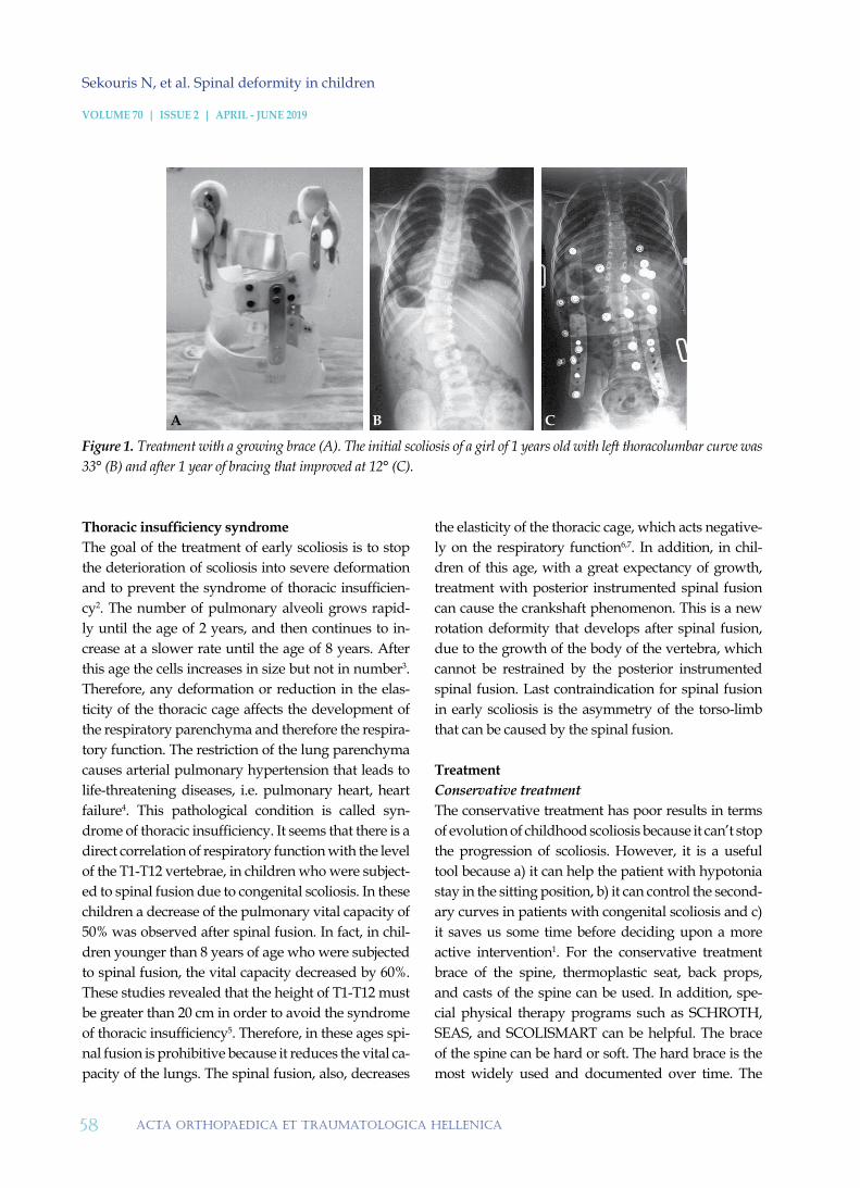
Figure 1. Treatment with a growing brace (A). The initial scoliosis of a girl of 1 years old with left thoracolumbar curve was 33° (B) and after 1 year of bracing that improved at 12° (C).
A B C

59acta OrthOpaedica et traumatOlOgica hellenica
VOLUME 70 | ISSUE 2 | APRIL - JUNE 2019
Sekouris N, et al. Spinal deformity in children
most common type is the Boston and the Cheneau brace. Attempts have been made to treat the scolio-sis with soft braces (i.e. Spinecor, Scolismart), but they did not prove their effectiveness. The traditional hard scoliosis braces can have some good results in idio-pathic scoliosis and in some syndromic scoliosis with characteristics of idiopathic scoliosis. On the contrary, the hard brace has poor results in neuromuscular and congenital scoliosis. A brace for scoliosis, applied to a child less than 3 years old will need to be changed every 4 months due to the development of the spine. In adolescence the brace needs to be changed every
1 ½ years. This increases the costs of bracing during childhood. For this reason, in our clinic we have de-signed and used a type Boston or Cheneau growing brace which consists of two parts. The first part is ap-plied to the pelvis, and the second part is applied to the torso and connects with the first part with three metal plates (Fig. 1). Using these plates, we can in-crease the brace’s height every 4 months. We can re-peat this increase in height 2-3 times, in order to keep the brace for 1 to 1 ½ years. These braces have been used since 2012 in 4 kids with success. In case that the scoliosis cannot be controlled by a brace, the next step is the use of a serial spinal cast. In this case, a plaster cast is placed to the child under general anesthesia. The child is placed at a type Cotrel-Dupusett table of traction and a plaster cast is applied. Special care is needed not to squeeze the viscera with the cast by cre-ating windows of relief (Fig. 2). This cast needs to be changed every 2 months. The complications that can be observed with conservative treatment are pressure of the abdominal viscera, reduction in the glomerular filtration rate, a slight decrease in the volume of the lungs, tubular thorax, flattening of the lumbar spine and psycho-social disorders.
Surgical treatmentWhen the conservative treatment is unsuccessful sur-gical treatment is needed. Surgical treatment at this age includes systems of correction of the spine with-
Figure 2. Procedure of serial casting for early onset scoliosis.
Figure 3. A young girl of 10 years with neurofibromatosis treated with growing rods.
60 acta OrthOpaedica et traumatOlOgica hellenica
VOLUME 70 | ISSUE 2 | APRIL - JUNE 2019
Sekouris N, et al. Spinal deformity in children
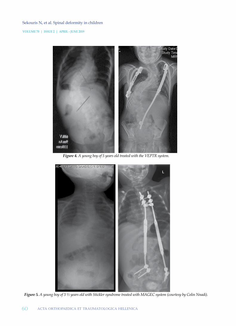
Figure 4. A young boy of 5 years old treated with the VEPTR system.
Figure 5. A young boy of 3 ½ years old with Stickler syndrome treated with MAGEC system (courtesy by Colin Nnadi).

61acta OrthOpaedica et traumatOlOgica hellenica
VOLUME 70 | ISSUE 2 | APRIL - JUNE 2019
Sekouris N, et al. Spinal deformity in children
out spinal fusion. These systems can be divided into three categories: systems with growing rods, systems with guided rods and compression-based systems1,9.
The traditional growing rods belong to the growing rods of the first category of systems, that need to be distracted, manually, every 6 months under general anaesthesia. This system is placed by posterior ap-proach by pedicle screws at two vertebrae above and two below the scoliosis. The rods have a mechanism that allows lengthening (Fig. 3). The fixation of the rod to the vertebrae, other than screws, can be done with hooks, sublaminar wires (or straps), or with rib rings. The most powerful support is achieved with the pedicle screws. Each support system has its bi-omechanical peculiarities and it is useful to have alternative options in case it is not possible to place pedicle screw or because we need to preserve the elasticity of the correction system that we use. These traditional growing rods are the «gold standard» of surgical treatment of childhood scoliosis. The system VEPTR (Vertical Expandable Prosthetic Rib) also belongs to the first category of systems with grow-ing mechanisms8. This system, basically, is a mech-
anism of stretching of the ribs. This system is fixed to the upper thoracic cage with rib rings and to the lower part of the scoliosis either with rib rings, pedi-cle screws or pelvic hooks (Fig. 4). The lengthening needs to be made manually every 4-6 months under general anesthesia. This system has limited effec-tiveness in the correction of scoliosis, but it is a good choice for patients with rib fusion and a restrictive chest cage (i.e. Jeune’s Syndrome). In order to avoid recurrent surgeries in children, a new system, the MAGEC (Magnetic Expansion Control), has been designed. This is placed with pedicle screws like the classic growing rods, but the MAGEC rods have a magnetic mechanism that allows lengthening with a remote control at the outpatient department10,11,12 (Fig. 5). Similarly, the APIFIX-EOS system was de-signed consisting of a self-growing rod that is placed in the concave side of the scoliosis by pedicle screws above and below the curve (Fig. 6)13,14. The lengthen-ing of this rod is made mechanically with the move-ment of the torso and with special physical therapy. Also, a similar system, EURO V2, designed by Lotfi Miladi, is placed with special sacroiliac screws and
Figure 6. A girl of 8 years old treated with Apifix EOS system.

62 acta OrthOpaedica et traumatOlOgica hellenica
VOLUME 70 | ISSUE 2 | APRIL - JUNE 2019
Sekouris N, et al. Spinal deformity in children
4 hooks at the upper part of scoliosis. The rods have a mechanism that allows the lengthening following the growing of the spine (Fig. 7).
In the second category includes the guided rods. The Luque-Trolley was the first system of this class and consists of two rods which are fixed in the spine with sublaminar wires. The wires can slip on the rod following the growing of the spine. This system is low cost and used since the 1970s with satisfactory results. However, the extensive subperiostal approach that needs to be done to place it can lead to early spontaneous spinal fu-sion. Later systems such as the Shilla15,19 and the Modern Trolley are placed with a minimal ap-proach. The system of Shilla has special pedicle screws that slide on the rod, like the sublaminar wires (Fig. 8), while the Modern Trolley is consists of pedicle screw with a head from polyester that are connected to and slip on the rods.
The third category includes the compression-based systems: Vertebral Body Stapling20 and Vertebra Body Tethering21. These systems affect the growth of the spine by hemiepiphysiodesis in a similar way as in the treatment of varus or valgus knee. The hemiepi-physiodesis includes the curved part of the scoliosis in order to stop the growing of the convex side of
the spine and to leaves the concave side free to grow correcting the scoliosis. The hemiepiphysiodesis of the vertebrae can be made with buckles (stapling) or dynamic fusion systems (tethering) which are placed by anterior approach (open or thoracoscopic). These systems modify the growing of the vertebra and use the viscoelastic properties of the intervertebral disc to correct the scoliosis. Therefore, these systems are used also for adults.
The materials of all these systems implemented at children without a spinal fusion undergo con-tinuous forces because they are not protected by a spinal fusion. For this reason, these systems are prone to complications of failure of materials such as fracture of the rod (15%) and failure of the upper support (up to 95%)16,17,18. Breakage of the pin of the magnetic mechanism has been observed with the use of the magnetic rod (MAGEC). With the com-pression systems subcorrection or overcorrection of scoliosis has been observed, because it is difficult to calculate the correct age to use these systems.
Figure 7. A girl of 10 years old treated with EURO V2 system.
Figure 8. A young boy with neurofibromatosis of 6 years old treated with Shilla system.

63acta OrthOpaedica et traumatOlOgica hellenica
VOLUME 70 | ISSUE 2 | APRIL - JUNE 2019
Sekouris N, et al. Spinal deformity in children
Also, breakage of the connective belt between the screws has been observed with these systems. The surgical revision of these systems is particularly difficult due to the anterior approach. Metallosis of growth-guided systems has been observed due to friction of materials. Another complication that may occur is wound infection (6.7%, of which 67% will need surgical debridement). Other complications include early spontaneous spinal fusion (that may occur due to the surgical approach), and junctional kyphosis (that may occur due to the sagittal imbal-ance and due to the increased «hardness» of the sta-bilized part of the instrumented spine). Although the new systems do not require regular revisions per semester, they may often need non-scheduled re-operations but, these are fewer compared to these needed with the classical system. We always
need to bear in mind is the psycho-social conse-quences of the repeated operations to the children.
ConclusionThe treatment of early onset scoliosis is a challenge because of the need to correct the deformity of the spine avoiding the spinal fusion with the traditional systems. The new systems offer new options to treat children with scoliosis and avoid the spinal fusion. Still the perfect system has not been invented. Every spine surgeon has to choose the system he is going to use to each individual child, and hence choose the complications he will encounter. A
Disclosure of interestThe author declares that he has no conflicts of interest concerning this article.
1. Early-onset scoliosis – Current treatment. V. Cunin. Orthopaedics & Traumatology: Surgery & Research, 2015
2. Vitale MG, Matsumoto H, Bye MR, et al. A retro-spective cohort study of pulmonary function, radi-ographic measures, and quality of life in children with congenital scoliosis: an evaluation of patient out comes after early spinal fusion. Spine 2008
3. Canavese F, Dimeglio. A normal and abnormal spine and thoracic cage development. World J Orthop 2013
4. Swank SM, Winter RB, Moe JH. Scoliosis and cor pul-monale. Spine 1982
5. Dimeglio A, Canavese F. The growing spine: how spinal deformities influence normal spine and tho-racic cage growth. Eur Spine J 2012
6. Emans JB, Ciarlo M, Callahan M, Zurakowski D. Prediction of thoracic dimensions and spine length based on individual pelvic dimensions in children and adolescents: an age-independent, individualized standard for evaluation of outcome in early onset spinal deformity. Spine 2005
7. Campbell Jr RM, Smith MD. Thoracic insufficiency syndrome and exotic scoliosis. J Bone Joint Surg Am 2007
8. Motoyama EK, Yang CI, Deeney VF. Thoracic mal-formation with early-onset scoliosis: effect of serial VEPTR expansion thoracoplasty on lung growth and function in children. Paediatr Resp Rev 2009
9. Skaggs DL, Akbarnia BA, Flynn JM, et al. A classi-fication of growth friendly spine implants. J Pediatr Orthop 2014
10. Miladi L, Dubousset J. Magnetic powered extensible rod for thorax or spine. In: Akbarnia BA, Yazici M, Thompson GH, editors. The growing spine: manage-ment of spinal disorders in young children. Berlin, Heidelberg: Springer-Verlag; 2010
11. Cheung K, Cheung JP, Samartzis D, et al. Magneti-cally controlled growing rods for severe spinal cur-vature in young children: a prospective case series. Lancet 2012
12. Akbarnia BA, Cheung K, Noordeen H, et al. Next generation of growth-sparing techniques. Prelim-inary clinical results of a magnetically controlled growing rod in 14 patients with early-onset scoliosis. Spine 2013
13. Sekouris N, Pilichou A, Sovatzoglou A, Soultanis K, Karavidas N, Flouda L, and Vlachos E. The Apifix Automatic Self-Correction Technique for Adoles-
refereNces

64 acta OrthOpaedica et traumatOlOgica hellenica
VOLUME 70 | ISSUE 2 | APRIL - JUNE 2019
Sekouris N, et al. Spinal deformity in children
cent. Idiopathic Scoliosis at 1 ½ Years Follow-Up: A Preliminary Report. Global Spine; Issue S 01, 2016
14. Armin U, El-Hawary R, Betz R, Lonner B, Floman Y. Preclinical Bench Testing on a Novel Posterior Dynamic Deformity Correction Device for Scoliosis. Spine Deformity 2019 (7) p 203-212
15. Sekouris N, Pilihou A, Vlahos E, Soultanis K, Sapkas G, Flouda L. The Shilla Growth Guidance Technique for Paediatric Scoliosis at 3-year Follow-up: a Pre-liminary Report. Global Spine Journal; Issue S 01, 2016
16. Skaggs KF, Brasher AE, Johnston CE, et al. Upper thoracic pedicle screw loss of fixation causing spi-nal cord injury: a review of the literature and multi-center case series. J Pediatr Orthop 2013
17. Sankar WN, Skaggs DL, Yazici M, et al. Lengthening
of dual growing rods and the law of diminishing re-turns. Spine 2011
18. Yazici M, Olgun ZD. Growing rod concepts: state of the art. Eur Spine J2013
19. McCarthy RE, Luhmann S, Lenke L, McCullough FL. The Shilla growth guidance technique for ear-ly-onset spinal deformities at 2-year follow-up: a preliminary report. J Pediatr Orthop 2014
20. Marks DS, Iqbal MJ, Thompson AG, Piggott H. Con-vex spinal epiphysiodesis in the management of progressive infantile idiopathic scoliosis. Spine 1996
21. Betz RR, Kim J, D’Andrea LP, Mulcahey MJ, Balsara RK, Clements DH. An innovative technique of verte-bral body stapling for the treatment of patients with adolescent idiopathic scoliosis: A feasibility, safety, and utility study. Spine 2003
Sekouris N, Paspati I, Fligger I, Flouda L. Spinal deformity in children. Acta Orthop Trauma Hell 2019; 70(2): 57-64.
reAdy - MAdecItAtIoN