ACTA Otorhinolaryngologica Italica
79
Official Journal of the Italian Society of Otorhinolaryngology Head and Neck Surgery Organo Ufficiale della Società Italiana di Otorinolaringologia e Chirurgia Cervico-Facciale 5 Otorhinolaryngologica Italica October 2014 Volume 34 ISSN 0392-100X Review HPV in oropharyngeal cancer: the basics to know in clinical practice Head and neck Parapharyngeal space tumours: the efficiency of a transcervical approach without mandibulotomy through review of 44 cases Supracricoid laryngectomies: oncological and functional results for 152 patients Contemporary role of pectoralis major regional flaps in head and neck surgery Technical refinements in mandibular reconstruction with free fibula flaps: outcome-oriented retrospective review of 99 cases Vestibology Association of cinnarizine and betahistine in prophylactic therapy for Ménière’s disease with and without migraine Pediatric otorhinolaryngology Long-term surgical and functional outcomes of the intact canal wall technique for middle ear cholesteatoma in the paediatric population Clinical techniques and technology Hyoid myotomy without suspension: a surgical approach to obstructive sleep apnoea syndrome Arterial microanastomoses on the reverse flow of the internal carotid artery reverse flow: an extreme solution in free-flap revascularisation. How we do it POSTE ITALIANE SPA - Spedizione in Abbonamento Postale - D.L. 353/2003 conv. in L. 27/02/2004 n° 46 art. 1, comma 1, DCB PISA - Iscrizione al tribunale di Pisa al n. 10 del 30-07-93 - Finito di stampare presso IGP, Pisa -luglio 2014 - Contiene I.E. Acta Otorhinolaryngologica Italica, XXXIV/5, 299-374, 2014
-
Upload
khangminh22 -
Category
Documents
-
view
0 -
download
0
Transcript of ACTA Otorhinolaryngologica Italica

w
Official Journal of the Italian Society of Otorhinolaryngology Head and Neck Surgery
Organo Ufficiale della Società Italiana di Otorinolaringologia e Chirurgia Cervico-Facciale 5
Otorhinolaryngologica Italica
October 2014
Volume 34
ISSN 0392-100X
ReviewHPV in oropharyngeal cancer: the basics to know in clinical practice
Head and neckParapharyngeal space tumours: the efficiency of a transcervical approach
without mandibulotomy through review of 44 cases
Supracricoid laryngectomies: oncological and functional results for 152 patients
Contemporary role of pectoralis major regional flaps in head and neck surgery
Technical refinements in mandibular reconstruction with free fibula flaps: outcome-oriented retrospective review of 99 cases
VestibologyAssociation of cinnarizine and betahistine in prophylactic therapy
for Ménière’s disease with and without migraine
Pediatric otorhinolaryngologyLong-term surgical and functional outcomes of the intact canal wall technique
for middle ear cholesteatoma in the paediatric population
Clinical techniques and technologyHyoid myotomy without suspension:
a surgical approach to obstructive sleep apnoea syndrome
Arterial microanastomoses on the reverse flow of the internal carotid artery reverse flow: an extreme solution in free-flap revascularisation. How we do it
POST
E ITA
LIAN
E SP
A - S
pedi
zione
in A
bbon
amen
to P
osta
le -
D.L.
353
/200
3 co
nv. i
n L.
27/
02/2
004
n° 4
6 ar
t. 1,
com
ma
1, D
CB P
ISA
- Isc
rizio
ne a
l trib
unal
e di
Pisa
al n
. 10
del 3
0-07
-93
- Fin
ito d
i sta
mpa
re p
ress
o IG
P, Pi
sa -l
uglio
201
4 - C
ontie
ne I.
E.
Acta O
torhinolaryngologica Italica, XX
XIV
/5, 299-374, 2014

Official Journal of the Italian Society of Otorhinolaryngology - Head and Neck SurgeryOrgano Ufficiale della Società Italiana di Otorinolaringologia e Chirurgia Cervico-Facciale
Otorhinolaryngologica Italica
Editorial BoardEditor-in-Chief: G. Paludetti President of S.I.O.: G. Spriano Former Presidents of S.I.O. and Editors-in-Chief: I. De Vincentiis, D. Felisati, L. Coppo, G. Zaoli, P. Miani, G. Motta, L. Marcucci, A. Ottaviani, P. Puxeddu, M. Maurizi, G. Sperati, D. Passali, E. de Campora, A. Sartoris, P. Laudadio, M. De Benedetto, S. Conticello, D. Casolino, A. Rinaldi Ceroni, M. Piemonte, A. Staffieri, F. Chiesa, R. Fiorella, A. Camaioni, A. Serra
Editorial StaffEditor-in-Chief: G. Paludetti Deputy Editor: J. Galli Associate Editors: G. Almadori, F. OttavianiEditorial Coordinator: E. De CorsoEditorial Assistant: P. Moore Treasurer:L. de Campora
Italian Scientific BoardL. Bellussi, G. Danesi, C. Grandi, A. Martini, L. Pignataro, F. Raso, R. Speciale, I. Tasca
International Scientific BoardJ. Betka, P. Clement, M. Halmagyi, L.P. Kowalski, M. Pais Clemente, J. Shah, H. Stammberger, R. Laszig, G. O’Donoghue, R.J. Salvi, R. Leemans, M. Remacle, F. Marshal, H.P. Zenner, B. Scola Yurrita, R.W. Gilbert
Editorial OfficeEditor-in-Chief: G. PaludettiDepartment of Head and Neck Surgery - OtorhinolaryngologyCatholic University of the Sacred Heart “A. Gemelli” Hospital L.go F. Vito, 1 - 00168 Rome, Italy Tel. +39 06 30154439 Fax + 39 06 3051194 [email protected]
Editorial Coordinator:E. De [email protected]
Editorial Secretary:R. [email protected]
Argomenti di Acta Otorhinolaryngologica ItalicaEditor-in-Chief: G. Paludetti Editorial Coordinator: M.R. [email protected]
© Copyright 2014 bySocietà Italiana di Otorinolaringologia e Chirurgia Cervico-FaccialeVia Luigi Pigorini, 6/300162 Rome, Italy
PublisherPacini Editore SpAVia Gherardesca, 156121 Pisa, ItalyTel. +39 050 313011Fax +39 050 [email protected]
Acta Otorhinolaryngologica Italica is cited in Index Medicus, MEDLINE, PubMed Central, Science Citation Index Expanded, Scopus, DOAJ, Open-J Gate, Free Medical Journals, Index Copernicus, Socolar
Journal Citation Reports: Impact Factor 1.439Acta Otorhinolaryngologica Italica is available on Google Scholar
Volume 34 – Number 5 – October 2014
Former Editors-in-Chief: C. Calearo†, E. de Campora, A. Staffieri, M. Piemonte, F. Chiesa

ReviewHPV in oropharyngeal cancer: the basics to know in clinical practiceHPV nel carcinoma dell’orofaringe: le nozioni base da conoscere nella pratica clinicaS. Elrefaey, M.A. Massaro, S. Chiocca, F. Chiesa, M. Ansarin ...................................................................................... pag. 299
Head and neckParapharyngeal space tumours: the efficiency of a transcervical approach without mandibulotomy through review of 44 casesTumori dello spazio parafaringeo: efficacia dell’approccio trans-cervicale senza mandibulotomia. Revisione di 44 casi.B. Basaran, B. Polat, S. Unsaler, M. Ulusan, I. Aslan, G. Hafiz ....................................................................................... » 310
Supracricoid laryngectomies: oncological and functional results for 152 patientsLaringectomie sopracricoidee: risultati oncologici e funzionali su 152 pazientiC.A. Leone, P. Capasso, G. Russo, P. D’Errico, P. Cutillo, P. Orabona ........................................................................ » 317
Contemporary role of pectoralis major regional flaps in head and neck surgeryAttuale ruolo dei lembi di grande pettorale nella chirurgia della testa e del colloF. Bussu, R. Gallus, V. Navach, R. Bruschini, M. Tagliabue, G. Almadori, G. Paludetti, L. Calabrese ......................... » 327
Technical refinements in mandibular reconstruction with free fibula flaps: outcome-oriented retrospective review of 99 casesAccorgimenti tecnici nelle ricostruzioni mandibolari con lembi liberi di fibula: analisi retrospettiva dei risultati su 99 casiG. Colletti, L. Autelitano, D. Rabbiosi, F. Biglioli, M. Chiapasco, M. Mandalà, F. Allevi ............................................ » 342
VestibologyAssociation of cinnarizine and betahistine in prophylactic therapy for Ménière’s disease with and without migraineL’associazione di betaistina e cinnarizina nella profilassi degli episodi vertiginosi nella Sindrome di Menière con e senza comorbidità emicranicaR. Teggi, O. Gatti, V. Sykopetrites, S. Quaglieri, M. Benazzo, M. Bussi ....................................................................... » 349
Pediatric otorhinolaryngologyLong-term surgical and functional outcomes of the intact canal wall technique for middle ear cholesteatoma in the paediatric populationRisultati funzionali e chirurgici nel lungo periodo nelle timpanoplastiche chiuse nella popolazione pediatrica affetta da colesteatoma dell’orecchio medio S.C. Prasad, C. La Melia, M. Medina, V. Vincenti, A. Bacciu, S. Bacciu, E. Pasanisi .................................................. » 354
Clinical techniques and technologyHyoid myotomy without suspension: a surgical approach to obstructive sleep apnoea syndromeMiotomia del muscolo ioideo senza sospensione: un approccio chirurgico per la sindrome delle apnee ostruttive nel sonnoE. Scarano, G. Della Marca, E. De Corso, S. Dittoni, W. Di Nardo, D. Meucci, G. Bastanza, R. Gallus, A. Losurdo, E. Testani, G. Paludetti .................................................................................................................................................... » 362
Arterial microanastomoses on the reverse flow of the internal carotid artery reverse flow: an extreme solution in free-flap revascularization. How we do itMicroanastomosi arteriosa su flusso retrogrado dell’arteria carotide interna: una soluzione estrema nella rivascolarizzazione dei lembi liberiA. Baj, A. Bolzoni, S. Torretta, L. Pignataro .................................................................................................................. » 368
Calendar of events - Italian and International Meetings and Courses .................................................................... » 372

299
ACTA oTorhinolAryngologiCA iTAliCA 2014;34:299-309
Review
HPV in oropharyngeal cancer: the basics to know in clinical practiceHPV nel carcinoma dell’orofaringe: le nozioni base da conoscere nella pratica clinica
S. ElrEfaEy1 2, M.a. MaSSaro1, S. ChioCCa3, f. ChiESa1, M. anSarin1
1 Division of otolaryngology head and neck Surgery, European institute of oncology, Milano, italy; 2 Department of otolaryngology, head and neck Surgery, alexandria University, alexandria, Egypt; 3 Department of Experimental oncology, ifoM-iEo Campus, European institute of oncology, Milano, italy
Summary
The incidence of oropharyngeal squamous cell carcinoma (OPSCC) is rising in contrast to the decreasing incidence of carcinomas in other subsites of the head and neck, in spite of the reduced prevalence of smoking. Human papilloma virus (HPV) infection, and in particular type 16 (HPV-16), is now recognized as a significant player in the onset of HPV positive OPSCC, with different epidemiological, clinical, anatomical, radiological, behavioural, biological and prognostic characteristics from HPV negative OPSCC. Indeed, the only subsite in the head and neck with a demonstrated aetiological viral link is, at present, the oropharynx. These observations lead to questions regarding management choices for patients based on tumour HPV status with important consequences on treatment, and on the role of vaccines and targeted therapy over the upcoming years.
Key wOrdS: Human Papillomavirus • Head and Neck cancer • Oropharyngeal cancer • Squamous Cell Carcinoma • Prognosis • Treatment • Prevention • Vaccination • Clinical Trial
RiaSSuNTO
L’incidenza del carcinoma spinocellulare dell’orofaringe (OPSCC) è in aumento in contrasto con la diminuzione dell’incidenza di carci-nomi in altre sedi del distretto cervico-facciale, nonostante la ridotta prevalenza del fumo. L’infezione da Papilloma Virus Umano (HPV), in particolare di tipo 16 (HPV 16), è ora riconosciuto come un importante fattore nell’insorgenza di HPV OPSCC positivo, con diverse caratteristiche radiologiche, epidemiologiche, cliniche, anatomiche, biologiche e prognostiche rispetto all’HPV OPSCC negativo. In effetti l’unica sede del distretto cervico-facciale con un collegamento virale eziologico dimostrato è, attualmente, l’orofaringe. Queste osserva-zioni portano a domande riguardanti le scelte di gestione per i pazienti in base allo stato del tumore HPV con importanti conseguenze sul trattamento e sul ruolo dei vaccini e terapia mirata per i prossimi anni.
ParOle CHIaVe: Papilloma Virus Umano • Tumori del distretto cervico-facciale • Tumori dell’orofaringe • Carcinoma spinocellulare • Prognosi • Trattamento • Prevenzione • Vaccinazione • Studi clinici
Acta Otorhinolaryngol Ital 2014;34:299-309
Introductionhead and neck cancer, which includes tumours that arise from the oral cavity, oropharynx, larynx, hypopharynx and sinonasal tract, represents a serious health care prob-lem in many parts of the world, and ranks as the sixth most common cancer worldwide 1. These tumours are linked by common characteristics including a male predominant ap-pearance in the 5-6th decade of life, a strong aetiological link with prior tobacco, alcohol use or betel nut chew-ing 2, and a histopathological resemblance 3. About 90% of head and neck cancers are squamous cell carcinomas (hnSCC).
The estimated annual burden of hnSCC is approximately 650,555 incident cases and approximately 300,000 result-ant deaths 4 5. it is considered the sixth leading cause of cancer mortality and oropharyngeal squamous cell carci-noma (oPSCC) accounts for approximately 50,000 inci-dent cases, which is low in comparison with other head and neck squamous cell carcinoma (hnSCC) 5 6.Multiple studies have demonstrated that the incidence of hnSCC has remained stable or even declined in the late 1980s, due to the gradual decrease in smoking and alco-hol which are the primary risk factors for these cancers 2. Despite this, the incidence of oropharyngeal squamous cell carcinoma with different characteristics, particularly

S. Elrefaey et al.
300
in the base of the tongue and tonsil subsites, has increased by 2-3% annually during 1973-2001, and then by 5.22% annually from 2000 to 2004 in the USA 7. Similar trends have been noted in other countries. in particular, one study suggests that the annual number of hPV-associated oro-pharyngeal cancers in the United States will overtake the incidence of invasive cervical cancer cases in the United States by 2020 8. There is also a discrepancy in incidence of oPSCC between developed and developing countries of oropharyngeal cancer 9.The developing world has a relatively low proportion of oPSCC (1-10% of hnSCCs), which appears to remain stable (or even to decrease) over time, while the incidence of hnSCC has steadily increased in most countries 4 10.The developed world features a relatively high and vari-able proportion of oPSCCs (15-30% of hnSCC). For ex-ample, a central belt of European countries has the highest oPSCC proportions in the developed world (up to 30% of hnSCCs) with the remainder of Europe being charac-terised by slightly lower oPSCC proportions, while the overall hnSCCs incidence has remained stable or has even shown a declining trend over the same period 4 11 12. These demographic data prompted researchers to search for further risk factors contributing to the incidence of oPSCC.
The impact of HPV as a risk factor for OPSCCMost studies have demonstrated that features tobacco and alcohol consumption are major, common risk factors for hnSCC, but over the last 10-15 years hPV infection has been increasingly recognised as a major aetiological fac-tor for a subset of hnSCCs 7-10, including mostly oPSCC. hPV infection in the aetiology of oPSCC was first shown by gillison et al. 13; numerous case series studies conduct-ed in the late 1990s and 2000s evaluated the prevalence hPV infection in oropharyngeal cancer using molecular techniques such as PCr and in situ hybridisation 8 14 15. in-deed, over the last five years it has become increasingly clear that hPV plays a pathogenic role in this subset of head and neck cancers, with distinct epidemiologic, clini-cal and molecular characteristics. These findings have created new opportunities for improved therapy and pri-mary prevention for these hnSCCs 16.At present, it should be clear that the only subsite in the head and neck with a demonstrated role for hPV infection in the aetiology of cancer is the oropharynx, as noted in the most important report by gillison et al. 13 and con-firmed by Stransky et al. in 2011 13 17.From a biological point of view, hPV is a DnA onco-virus and is epitheliotropic. There are over 120 differ-ent hPV subtypes, including the low-risk types such as hPV 6 and hPV 11, responsible for benign proliferation of epithelium, and the high-risk oncogenic types hPV 16
and hPV 18 which are both well-established initiators of over 90% of cervical cancers, 70% of anogenital cancers, 5% of non-oropharyngeal SCC 17 and 20-72% of oP-SCC 2 4 12 17. The oncogenic nature of high risk hPVs is due to the immortalising and transforming properties of hPV oncoproteins E6 and E7, which target the p53 and prB tumour suppressor pathways, respectively, rendering infected cells susceptible to mutations and cancer forma-tion 18 19. Since the majority of hPV-hnSCCs are oPSCC, we will mainly discuss oPSCC.
Classification of oropharyngeal cancer according to HPVAccording to the national Comprehensive Cancer net-work (nCCn) 20 guidelines, ‘hPV testing is recommend-ed for all oropharyngeal tumours’. in addition, according to the US national Cancer institute (nCi) 21 and Cancer Therapy Evaluation Programme (CTEP) 22, hPV status must be included as a stratification factor for trials includ-ing oropharynx cancer patients. Much evidence suggests that hPV-positive and hPV-negative oPSCCs represent distinct subgroups of oPSCC, each with unique epide-miological and biological profiles 4 5 17 21 23-27.
Differences between HPV positive and HPV negative OPSCCs
Epidemiological factorshPV-positive patients tend to be younger with a median age of diagnosis of 54 years, less exposure to tobacco and alcohol 28-30, and higher socioeconomic status and education 31. hPV positivity is less frequent in blacks than in Caucasians (4% of hnSCC in blacks vs. 34% in whites) 32, with a three fold higher incidence in males than females 28 33 34.As in cervical cancer, oral hPV infection appears to be a sexually-acquired disease. Although the natural history of oral hPV infection is not well defined, D’Souza and colleagues recently showed in a case-control study that a high (≥ 26) number of lifetime vaginal-sex partners and 6 or more lifetime oral-sex partners were associated with an increased risk of oPSCC [odd ratio (or) 3.1 and 3.4, respectively] 35. An increased risk of hPV-associated oP-SCC in female patients with a history of hPV-associated anogenital cancers and their male partners is also con-sistent with hPV transmission to the oropharyngeal cav-ity 36 37. The recent increased incidence of this disease may thus reflect societal changes in sexual behaviour that have occurred over time in the developed world 38 39.An important point to mention is that there is no clear case-control study addressing the evidence for hPV prior to development of oPSCC (i.e. temporal association), with the exception of a Scandinavian study by Mork et al.

HPV in oropharyngeal cancer
301
which showed that the presence of hPV 16 l1 antibod-ies in pre-diagnostic serum samples was associated with a 14.4-fold increased risk of oropharyngeal cancer. impor-tantly, the presence of hPV 16 antibodies preceded oro-pharyngeal cancers by more than 10 years, underscoring a temporal association. These data confirmed that oral hPV infection increases the risk of developing oPSSC 40.lastly, it is possible that in addition hPV infection, other risk factors or cofactors such as genetic susceptibility or nutritional factors or tobacco and alcohol interaction have an important role in cancer onset. There is an objective need for more analytic epidemiological studies in males and females diagnosed with hPV positive oropharyngeal cancer younger than 50 years of age 40.
Anatomical sitesSeveral studies have noted an increased incidence of hPV-associated oropharyngeal cancers, especially tonsil-lar and tongue cancer. For example, in the USA they have risen by 3.9% and 2.1% among men and women, respec-tively, in the age group from 20 to 44 years, between 1973 and 2004 2 41. Similar patterns have been noted in Sweden for tonsillar cancer which rose 2.9-fold between 1970 and 2001, increasing by 2.6% per year in men and 1.1% in women 11 42.The preference of hPV for the oropharynx is unexplained, but may be related to the unique presence of transitional mucosa in the oropharynx, predominantly found in the tonsillar tissue and which shows histological similarities to the cervical mucosa 2 11. Another possibility lies within the genetic features of hPV 16, which accounts for more than 90-95% of all hPV associated oropharyngeal can-cers, as it may facilitate survival in the tonsillar crypt epithelium 43 44. it is also possible that the invagination of the mucosal surface of the tonsil may favour virus capture and maintenance by promoting its access to basal cells (the only dividing cells in the epithelium) 45. if this is true, tonsillar tissue could be a reservoir for hPV in the upper aerodigestive tract. This view is partly supported by the fact that when oral samples are collected by oral rinse, the detection rate of hPV is much higher than with swabs. Finally, the persistence of hPV in tonsillar tissue might be of importance in the immune response to hPV 46.
Biological profilesrecent global genomic screening studies searching for a biological distinction among hPV-positive and negative oPSCC have shown that hPV-induced carcinogenesis has a clear impact on the acquisition and maintenance of spe-cific chromosomal gains and losses within tumour cells, in which oPSCCs with transcriptionally active hPV-DnA are characterised by occasional chromosomal loss/allelic imbalance 47. Conversely, those lacking hPV-DnA are characterised by gross deletions that involve entire or large parts of chromosomal arms 32 48.
Furthermore, ploidy studies have confirmed that hPV-positive tonsillar cancers feature a lower number of chro-mosomal alterations compared to their hPV-negative counterparts 49 50.The biology of hPV-positive oropharyngeal cancer is typified by p53 degradation, retinoblastoma protein (rB) down-regulation and p16 up-regulation. By contrast, to-bacco-related oropharyngeal cancer is characterised by p53 mutations, down-regulation of p16 and rB up-reg-ulation 45.interestingly, recent studies observed an inverse correla-tion between the presence of hPV and p53 mutations 17.
Clinical stage at presentationMultiple studies have shown that hPV-positive tumours are more likely to present with early T stage (T1-T2) 51 and higher n stage (usually cystic and multilevel) 52, and have distinct histological features, such as moderate/poor tumour differentiation and non-keratinising or basaloid pathology 14 19 51. The incidence of distant metastases was seen to be lower in patients with hPV positive tumours. Furthermore, metastases developed later and with a very different pattern from patients with hPV-negative tu-mours. hPV-positive oropharyngeal cancer had a 28% re-duction in the risk of death and a 49% reduction in the risk of disease recurrence 53. Secondary primary tumour (SPT) in patients with hPV-positive cancer is very rare, and has improved better survival rate compared to patients with hPV negative tumours 45.
Radiological imagingrecent studies have shown radiological difference be-tween hPV-positive and hPV-negative oropharyngeal cancer. Specifically, hPV-positive carcinomas often had small or even occult primary lesions with well-defined borders and cystic nodal metastases, whereas hPV-nega-tive primaries more often had poorly defined borders and invasion of adjacent muscle 52 54.
PrognosisSeveral studies have shown that patients with hPV-pos-itive oropharyngeal cancer, identified through PCr, in situ hybridisation or p16 immunohistochemistry on tu-mour tissues, have a significantly improved overall and disease-free survival compared to patients with hPV-negative oropharyngeal cancer patients 29 53 55-61 (Table i). This holds true even after adjustment for differences in favourable prognostic factors associated with hPV posi-tive patients (younger age, better performance status, fewer comorbidities, less smoking). Ang et al. reported that these prognostic factors explained only 10% of the observed survival differences between two subgroups 29. however, other studies reported that survival rates im-proved among non-smoker hPV positive patients com-

S. Elrefaey et al.
302
pared to smokers patients even in recurrent tumours, underscoring once again the benefits acquired from smoking cessation 62 63.Why does hPV positive orophrangeal cancer have a bet-ter prognosis?1. hPV-positive tumours may harbour fewer or different
genetic alterations, which can be associated with better response to therapy 17 64.
2. hPV-positive tumours have higher radiosensitivity, probably due to intact apoptotic response to radia-tion 58 65.
3. The absence of field cancerisation in hPV-positive tu-mours 53.
4. immunologic response may be play a role in the im-proved response to radio- and chemotherapy in hPV-positive tumours (due to the stimulation of immune response directed to viral specific tumour antigens 66.
5. younger age, good performance status, fewer comor-bidities of hPV-positive oropharyngeal cancer patients may also contribute to improved survival 67.
The impact of HPV on clinical managementThe standard treatment for oPSCCs at present is mainly dependent on the stage of the disease and patient and cli-nician preferences. Single-modality treatment, in the form of surgery or radiotherapy, is usually recommended for early (T1-T2, n0) disease. For advanced stage disease, standard treatments include chemoradiotherapy with or without neck dissection, or surgical resection with re-construction and postoperative chemoradiotherapy, as required. These current standard methods of treatment appear to apply to both hPV positive and negative sub-groups 68 69.
1) Non-surgical treatment options for OPSCCsThe emergence of hPV-oPSCCs in younger patients with better prognosis and survival rates in comparison to non-hPV oPSCCs have prompted clinicians to address changes in the non-surgical management according to hPV status 2.Multiple studies 29 58-61 68-70 tackling this issue have con-cluded that (Tables i, ii):1. overall survival rates increase with hPV positive sta-
tus, low EgFr and high p16 72.
2. Patients with hPV negative disease have a poorer prognosis, and therefore usually require more intensive treatment. Studies (TAX 324 61, Trog 02.02 59) have suggested that for patients with hPV DnA-negative tumours, treatment intensification improves outcomes compared to standard treatment, but overall outcome is still poor.
3. Smoking cessation and strategies to target EgFr and Bcl-xl 70 are important adjuncts in the treatment of oro-pharyngeal cancer.
4. Achievement of acceptable cure rates with minimal long-term morbidity with hPV positive oropharyngeal cancer is possible.
All these data suggest that hPV status can be used in the clinical decision-making processes to select patients for less aggressive non-surgical treatment. Thus, assessing hPV presence is of utmost importance. This is especially true considering long-term outcomes of hPV-positive younger patients, since they are at risk of a lifetime com-promised quality of life as a result of chronic toxicities due to chemoradiotherapy. p16 immunohistochemistry (ihC) is a current marker to detect hPV presence. however, it can be associated with a high rate of false positive/false negative responses, prompting the need for new surrogate markers for oral hPV infection. These concerns were also reported by rietbergen et al. 71 and Bussu et al. 72. Thus, in clinical practice it is not recommended to rely on p16 ihC alone to screen for hPV positivity.Currently, there are on-going oncological trials that at-tempt to answer some questions regarding deintensifica-tion of treatment (Table iii):1. Can we use neoadjuvant chemotherapy followed by re-
duced radiotherapy dose in hPV positive patients?2. What is the intensity of adjuvant therapy required in
p16-positive oropharynx cancer patients?3. Can cetuximab provide selective radiosensitisation
compared with cisplatin?4. Should the volume treated be reduced by not adminis-
tering prophylactic radiotherapy to areas at risk of mi-croscopic disease?
5. is it possible to reduce the dose of radiation therapy when given with standard doses of chemotherapy?
6. What is the exact role of immune activation in hPV positive patients?
Table I. Selected studies reporting the association of HPV infection with oropharyngeal cancer prognosis.
Study Author, year # of cases HPV detection Follow-up OS positive vs. negative tumours
ECOG 58 2399 Fakhry, 2008 96 HPV 16 DNA ISH 2 2-yr survival (95% vs. 62%)
RTOG 29 0129 Ang, 2010 323 HPV 16 DNA ISH 4.8 3-yr survival (82.4%, vs. 57.1%)
TROG 59 02.02 Rischin, 2010 185 p16 IHC 5 2-yr survival (91% vs. 74%)
DHANCA 60 6,7 Lassen, 2011 794 p16 IHC 5 (62% vs. 47%)c, (52% vs. 48%)*
TAX 61) 324 Posner, 2011 111 HPV 16 DNA PCR 5 5 yr survival (82%-35%)ISH: in situ hybridisation; IHC: immunohistochemistry; PCR: polymerase chain reaction; OS: overall survival; * accelerated radiotherapy; c conventional radiotherapy.
HPV in oropharyngeal cancer
303
2) Surgical treatment options for OPSCCsAll treatment modalities for oPSCC have similar on-cological outcomes 73, but functional outcomes have
significant and critical considerations when managing younger hPV positive patients with an longer expected lifetime. While nonsurgical deintensification trials are
Table II. Retrospective analyses of HPV status and/or p16 immunohistochemical staining status as a surrogate biomarker of HPV infection and survival outcome in Phase III outcome.
Study Treatment Regiment TotalN
(n included)
Progression-Free Survival
Overall Survival Conclusion
RTOG 0129Ang et al.2010 29
Standard-fractionated radiation + cisplatin vs. hyperfractionated radiation + cisplatin
743 (323) HPV+/p16 HPV-/p163-yr 73.7% 3 yr 43%3-yr 74.4% 3 yr 38%
HPV+/p16HPV-/p163-yr 82.4% 3 yr 57.1%3-yr 83.6% 3-yr 51.3%
No survival differences seen between the 2 treatment arms.
Secondary analysis confirmed significantly improved survival in patients with HPV-
positive tumours vs. HPV-negative disease.
DAHANCA 5Lassen et al.
2009 70 71
Radiation
Radiation+nimorazole
195 (156)
219 (175)
5-yr p16+ (70%)
5-yr p16- (40%)
5-yr p16+ (62%)
5-yr p16- (26%)
Improved loco-regional control when nimorazole was added to radiotherapy
was restricted to p16-negative patients. Improved survival in p16-positive patients
treated with radiotherapy alone.
DAHANCA 6&7Lassen et al.
2011 60
5 Fractions w/radiation
6 Fractions w/radiation
726 (385)
750 (409)
5-yr p16+ (78%)
5-yr p16- (64%)
5-yr p16+ (62%)
5-yr p16 -(47%)
Accelerated radiotherapy significantly improves outcome in HNSCC compared to conventional fractionation. The observed benefit was independent of tumour p16
status, and the use of a moderately accelerated radiotherapy regimen seemed
advantageous forHPV/p16 positive HNSCC.
TROG 02.02Rischin et al.2010 59
Radiation+cisplatin vs.radiation+cisplatin+tira
pazamine
861 (185) 2-yr p16+ (87%)
2-yr p16- (72%)
2-yr p16+ (91%)
2-yr p16- (74%)
While there was no difference in the p16-positive group, there was a trend for improved loco-regional control with tirapazamine in p16 negative patients.
The study clearly demonstrated that HPV associated oropharyngeal cancer treated with a standard regimen of concurrent
cisplatin and radiation has a better outcome compared with HPV-negative OPSCC.
Table III. On-going clinical trials (ClinicalTrials.gov).
Study ID NCI Trial ID
Trial Type
Total (N)
Treatment arm PrimaryEndpoint
E1308 NCT01084083 Phase II 160 Sequential therapy: cisplatin/paclitaxel/cetuximab
Complete response: IM RT (27 fractions)Non complete response: IM RT (33 fractions) 1cetuximab
2-yr PFS
J0988 NCT01088802 Phase I/II 60 IMRT (lower dose) + cisplatin Toxicity/LRC
National Cancer Institute (NCI)
NCT01585428 Phase II -
Fluderabine/cyclophosphamide/ Young TIL/Aldesleukin
Tumour response /duration
RTOG 1016 NCT01302834 Phase III 706 IMRT hyperfractionation+cisplatin vs.IMRT hyperfractionation +cetuximab
5-yr OS
University of Michigan Cancer Center
NCT01663259 - -Standard dose radiotherapy+cetuximab for stage III/IV
OPSCC
Rate of recurrence
ECOG1308 NCT01084083 Phase II 83 Induction chemotherapy followed by cetuximab Withlow dose vs. standard dose IMRT
2-yr PFS
Mount Sinai School of Medicine
NCT01358097 Observational - Biomarkers of immune function as predictors of HNSCC in response to therapy
-
OS: overall survival, LRC: loco-regional control; DFS: disease-free survival; IMRT: intensity modulated radiation therapy; PFS: progression-free survival; TIL: tumour infiltrating lymphocytes.

S. Elrefaey et al.
304
showing great promise 29 58-61 70-72, minimally-invasive ap-proaches, especially transoral robotic surgery (TorS), have gained more favour by achieving the satisfactory oncological outcomes without compromising functional outcome 73 74. indeed, three-dimensional visualisation al-lows the ability to manipulate 75 and perform reconstruc-tion of the oropharynx without the need for mandibulot-omy and/or pharyngotomy, thus reducing the morbidity of extensive surgery 76. it also facilitates safer exposure and resection of the primary tumour, thereby providing complete pathologic evaluation and impacting the use of clinically-established adjuvant therapies 77. These in-clude use of concurrent chemotherapy 78 and effective lower doses of radiotherapy, which contribute to a de-crease of swallowing dysfunction 79. The postoperative target volume for radiation is typically smaller, and with modern techniques such as intensity modulated radio-therapy (iMrT) this procedure can significantly reduce the dose delivered to uninvolved normal structures. in patients requiring postoperative concurrent chemoradia-tion, this offers the potential to reduce the risk of late complications 80.The incorporation of TorS, not only to improve oncolog-ic results but also to decrease the long-term toxicity risks caused by non-surgical strategies, is crucial for hPV posi-tive patients since they typically present at a younger age.To date, there are few surgical trials investigating the role of TorS in hPV positive patients. For instance, Cohen et al. 81 found no differences in oncological out-comes, overall survival or loco-regional control between hPV-positive and negative groups patients who under-went TorS surgery stratified by hPV status. nonethe-less, TorS surgery was suitable for both subgroups. The Mount Sinai group reported no differences in overall survival or loco-regional control in patients stratified by smoking status, with the assumption that patients without a smoking history are predominantly hPV positive 82.The failure to show statistically significant differences in hPV-positive and hPV-negative tumours in TorS sur-gical trials for early T stage differences is unclear. it is possible that these studies were small and thus lack the statistical power to show survival differences, or that the survival advantage in hPV-positive tumours does not ap-ply to early T-stage tumours that are surgically resected. lastly, one may argue that hPV-negative tumours are less radio-responsive, and surgical resection provides better prognosis in the cohort being studied 83.new multi-institutional studies are needed to confirm the exact impact of TorS on the quality of life and survival outcomes of hPV negative and positive oPSCC patients.
Future directions in HPV-positive OPSCCshPV-induced carcinogenesis has been extensively stud-ied in the most widely accepted hPV-related malignancy,
namely cervical cancer. hPV-associated cancers continu-ously express the hPV E6 and E7 viral oncogenes even during advanced stages, and repression of viral oncogene expression can prevent growth or survival of cervical can-cer cells 84. These findings raise the possibility that even late-stage hPV-associated cancers can be treated through hPV-targeted approaches with drugs that interfere with the expression or function of the viral oncoproteins or with therapeutic vaccines that elicit a cytolytic immune response in cells expressing these oncoproteins.
VaccinationThe world has greatly benefited from vaccine programmes in controlling the morbidity and mortality of infectious diseases. hepatitis B virus (hBV) vaccine, developed for the prevention of hepatitis B virus infection, is considered the first vaccine against a major human cancer, hepatocel-lular carcinoma 85. recently, a prophylactic hPV vaccine has been included in national immunisation programmes of most developed countries with the hope of also be-ing included in developing countries within the next few years, with the goal of preventing cervical and other non-cervical hPV related cancers 86.Two FDA-approved hPV prophylactic vaccines are currently available 87. The quadrivalent vaccine was ini-tially approved in the US in 2006, and is composed of four hPV type-specific virus-like particles (VlPs) from the major capsid protein l1 of hPV types 6, 11, 16 and 18, combined with aluminium phosphate adjuvant. These are the most common hPV types found in 70% of cer-vical cancers and 90% of non-cervical cancers 87 88. The bivalent hPV vaccine, approved in 2009, is composed of two hPV types, 16 and 18, which cause 70% of cervical cancers 86. The efficacy of the quadrivalent vaccine was 100% in preventing hPV 16 and 18 related cervical in-traepithelial neoplasia (Cin) grades 2/3 and vulvar and vaginal intraepithelial neoplasia (Vin) 2/3, and 98.9% in preventing hPV 6, 11, 16 and 18 related genital warts 89. The bivalent vaccine is 98.1% efficacious in hPV 16 and 18 related Cin 2/3 prevention 90.The Advisory Committee on immunization Practices (ACiP) and the Centers for Disease Control and Preven-tion (CDC) recommends 88 90 91:• routinevaccinationofgirlsaged11or12yearsthatcan
be started at 9 years of age;• catch-upvaccinationforfemalesaged13-26years;• routinevaccinationofboysaged11or12years;• routine vaccination recommended for bothmenwho
have sex with men (MSM) and immunocompromised individuals aged 22 through 26 years;
• menaged13to21yearswhowerenotpreviouslyvac-cinated;
• menaged22to26yearsmayalsoreceivethevaccine;• canbegiventolactatingwomen,patientswithminor

HPV in oropharyngeal cancer
305
acute illnesses and women with equivocal or abnormal Pap test.
hPV vaccine should be delivered through a series of 3 intramuscular injections over a 6-month period of time, at 0, 2 and 6 months for the quadrivalent vaccine and 0, 1 and 6 months for the bivalent one 89, inducing strong immune memory with persistent antibody up to 6.4 years (bivalent) and up to 9.5 years (hPV 16 VlP used in quad-rivalent) thus entailing long-term duration of protection against infections caused by pathogenic hPVs and their disease sequelae 92.The entrance of males into vaccination programmes is primarily due to the estimation of 7,500 cases of hPV-related cancer, primarily head and neck and anal can-cer, which occur in men each year in the United States alone 93. Furthermore, the rates of anal cancer in homosex-ual males are extremely high, and thus vaccination may contribute in immunisation with subsequent reduction of hPV sexual transmission.in the future, the currently available vaccines may also show promising results on preventing hPV-associated oPSCC caused by hPV 16, and longitudinal studies com-paring the incidence of disease before and after the intro-duction of the vaccine may clarify this issue.Unfortunately, the prophylactic vaccine is not effective on established infections and cancer lesions, so the study of a therapeutic hPV vaccine to treat hPV-associated cancer remains an area of crucial importance 94.Different immunotherapeutic vaccines targeting E7 and/or E6 have been developed over the last decade including peptide/protein, dendritic cell (DC), plasmid DnA and viral vector-based therapies, but with limited success in preclinical and clinical phase studies 95 96. A recent italian study developed a promising therapeutic vaccine based on an integrase defective lentiviral vector (iDlV) to de-liver a mutated non-oncogenic form of the hPV 16 E7 protein, considered as a tumour specific antigen for im-munotherapy of hPV-associated cervical cancer, fused to calreticulin (CrT), a protein that is able to activate natu-ral killer T cells (nKTs). A single intramuscular injection prevented tumour growth in 90% of early stage tumour-bearing mice, without adjuvants and/or drug treatments. These promising results may suggest that a safe antican-cer immunotherapeutic vaccine may be available in the future for human use 94.
Targeted therapiesEvaluation of epithelial growth factor receptor (EgFr)-targeted therapies in hnSCC patients have been based on the observation that EgFr is highly expressed in hn-SCC, and its over-expression has been associated with reduced survival in several studies 97. For clinical use, Eg-Fr can be targeted either by antibodies recognising the ligand-binding domain of EgFr or by EgrF tyrosine ki-
nase inhibitors (TKis). Cetuximab is a humanised mouse anti-EgFr igg1 monoclonal antibody, offering improved loco-regional control and overall survival in locally-ad-vanced hnSCC in combination with radiotherapy 98.other humanised anti-EgFr antibodies such a panitu-mumab or zalutumumab are currently being evaluated in phase ii/iii clinical trials and may evolve as alternatives to cetuximab 99. Additional prospective clinical trials are on-going to assess the value of cetuximab in management of hPV-positive oPSCCs.
ConclusionsTo date, the available data corroborate some well-estab-lished concepts: oropharynx tumours have been steadily increasing over the last 20 years compared to other can-cers of the head and neck worldwide, particularly in West-ern countries. SEEr data suggest that about 18% of all head and neck carcinomas in the USA were located in the oropharynx in 1973, compared to 31% of such squamous cell tumours in 2004. Similarly, in Sweden, the proportion of oropharyngeal cancers hPV positive has steadily in-creased, from 23% in the 1970s to 57% in the 1990s, and as high as 93% in 2007. These data indicate that hPV is now the primary cause of tonsillar cancer in north Amer-ica and Europe.The biology of hPV-positive oropharyngeal cancer is characterised by p53 degradation, retinoblastoma rB pathway inactivation and p16 up-regulation. in contrast, tobacco-related oropharyngeal cancer is characterised by p53 mutation and down-regulation of CDKn2A (en-coding p16ink4A). hPV-positive oropharyngeal cancer seems to be more responsive to chemotherapy and radia-tion than hPV-negative disease.The choice of the best viral detection method in tumours is a matter of controversy, and both in-situ hybridisation and PCr are commonly used; p16 ihC is also being used to detect hPV infection, but with unreliable results 71 72. Thus, there is clearly a need for new surrogate markers for hPV infection to give patients the best treatment strate-gies.The presence of hPV 16 can also be thought of as a prog-nostic marker for enhanced overall and disease-free sur-vival, but its use as a predictive marker has not yet been proven. Many questions about the natural history of oral hPV infection are still under investigation.regarding disease management, based on the present in-formation, we can consider hPV-positive oropharyngeal cancer as a distinct subset of hnSCC with a more favour-able outcome. Patients with hPV-positive oropharyngeal cancer are typically young and in good health. in future clinical trials, cancer centres should stratify head and neck patients by hPV status. regardless of treatment modality, an opportunity now exists to investigate less in-tense treatment strategies that do not compromise survival

S. Elrefaey et al.
306
outcomes, but lower the risk of fatal side effects. Thus, providing a high level quality of life with the fewest treat-ment complications are important considerations. Poten-tial long-term side effects of concurrent chemoradiation include dysphagia, xerostomia, feeding-tube dependency from fibrosis and scarring of the pharyngeal muscles, chronic aspiration and chronic fatigue.however, we must always emphasise that the best cure against cancer is prevention, especially in those malig-nancies in which the main pathogenic agent is known. Finally, the authors wish to suggest reader to consult two very recent and excellent reviews: “new insights into human papillomavirus-associated head and neck squamous cell carcinoma” 100 and “human papilloma virus (hPV) in head and neck region: review of litera-ture 101.
List of Abbreviations:CTEP: Cancer therapy evaluation programme.DhAnCA: Danish head And neck Cancer group.DnA: Deoxynucleic acidE6: Early oncoprotein6E7: Early oncoprotein7ECog: Eastern Cooperative oncology groupFDA: Food and Drug AdministrationEgFr: Epithelia growth Factor receptorhnSCC: head and neck Squamous Cell CarcinomahPV: human Papilloma VirusiSh: in situ hybridizationnCCn: national Comprehensive Cancer networkPCr: Polymerase Chain reactionprb: retinoblastoma tumour suppressoroPSCC: oroPharyngeal Squamous Cell CarcinomarTog: radiation Therapy oncology groupTKi: Tyrosine Kinase inhibitorsTlM: Transoral laser MicrosurgeryTrog: Trans-Tasman radiation oncology groupTorS: Trans oral robotic SurgeryUSA: United States of AmericaBCl-Xl: B-cell lymphoma-extra largeSEEr: Surveillance, Epidemiology and End results Program
References1 Ferlay J, Shin hr, Bray F, et al. Estimates of worldwide
burden of cancer in 2008: globocan 2008. int J Cancer 2010;127:2893-917.
2 Sturgis EM, AngKK. The epidemic of HPV-associated oro-pharyngeal cancer is here: is it time to change our treatment paradigms? J natl Compr Cancnetw 2011;9:665-73.
3 Johnson n, Franceschi S, Ferlay J, et al. Oral Cavity and Oropharynx. in: Barnes l, Eve J, reichart P, et al., editors. Int. Pathology and Genetics Head and Neck Tumors. lyon 2005. p. 163-208.
4 van Monsjou hS, Balm AJ, van den Brekel MM, et al. Oro-
pharyngeal squamous cell carcinoma: a unique disease on the rise? oral oncol 2010;46:780-5.
5 Mignogna MD, Fedele S, lo russo l. The world cancer report and the burden of oral cancer. Eur J Cancer Prev 2004,13:139-42.
6 horner MJ, ries lAg, Krapcho M, et al. SEER Cancer Sta-tistics Review, 1975-2006, National Cancer Institute. Bethes-da, MD, http://seer.cancer.gov/csr/1975_2006/, based on november 2008 SEEr data submission, posted to the SEEr web site, 2009.
7 Shiboski Ch, Schmidt Bl, Jordan rC. Tongue and tonsil carcinoma: increasing trends in the US population ages 20-44 years. Cancer 2005;103:1843-9.
8 Chaturvedi AK, Engels EA, Pfeiffer rM, et al. Human papil-loma virus and rising oropharyngeal cancer in the United States. J Clin oncol 2011;29:4294.
9 Sedrak M, rizzolo D. Understanding the link between hpv and oropharyngeal cancer. JAAPA 2009;22:42-6.
10 McKean-Cowdin r, Feigelson hS, ross rK, et al. Declining cancer rates in the 1990s. J Clin oncol 2000;18:2258-68.
11 hammarstedt l, Dahlstrand h, lindquist D, et al. The inci-dence of tonsillar cancer in Sweden is increasing. Acta oto-laryngol 2007;127:988-92.
12 robinson Kl, Macfarlane gJ. Oropharyngeal cancer inci-dence and mortality in Scotland: are rates still increasing? oral oncol 2003;39:31-6.
13 gillison Ml, Koch WM, Capone rB, et al. Evidence for a caus-al association between human papillomavirus and a subset of head and neck cancers. natl Cancer inst 2000;92:709-20.
14 gillison Ml. Human papillomavirus-associated head and neck cancer is a distinct epidemiologic, clinical, and molecu-lar entity. Semin oncol 2004;31:744-54.
15 Singhi AD, Westra Wh. Comparison of human papilloma-virus in situ hybridization and p16 immunohistochemistry in the detection of human papillomavirus-associated head and neck cancer based on a prospective clinical experience. Can-cer 2010;116:2166-73.
16 Adelstein DJ, rodriguez CP. Human papilloma virus: changing paradigm in oropharyngeal cancer. Curr oncol rep 2010;12:115-20.
17 Stransky n, Egloff AM, Tward AD, et al. The mutational landscape of head and neck squamous cell carcinoma. Sci-ence 2011;333(6046):1157-1160.
18 oh JE, Kim Jo, Shin Jy. Molecular genetic characteriza-tion of p53 mutated oropharyngeal squamous cell carcinoma cells transformed with human papillomavirus E6 and E7 on-cogenes. int J oncol 2013;43:383-93.
19 rampias T, Sasaki C, Weinberger P. E6 and E7 gene silenc-ing and transformed phenotype of human papillomavirus 16 - positive oropharyngeal cancer cells. J natl Cancer inst 2009;101:412-23.
20 national Comprehensive Cancer Center network. Practice guidelines in oncology-Head and Neck Cancers v. 2.2008.
21 national Cancer institute: PDQ® oropharyngeal Cancer Treatment. Bethesda, MD: national Cancer institute. Date last modified <02/15/2013>. Available at: http://cancer.gov/cancertopics/pdq/treatment/oropharyngeal/healthProfes-sional. Accessed <02/15/2013>

HPV in oropharyngeal cancer
307
22 Ansher SS, Scharf r. The Cancer Therapy Evaluation Pro-gram (CTEP) at the National Cancer Institute. Ann n y Acad Sci 2001;949:333-40.
23 lindel K, Beer KT, laissue J, et al. Human papillomavirus positive squamous cell carcinoma of the oropharynx: a ra-diosensitive subgroup of head and neck carcinoma. Cancer 2001;92:805-13.
24 Kreimer Ar, Cliffor gM, Boyle P, et al. Human papilloma virus types in head and neck squamous cell carcinomas worldwide: a systemic review. Cancer Epidemiol Biomark-ers Prev 2005;14:467-75.
25 D’Souza g, Dempsey A. The role of HPV in head and neck cancer and review of the HPV vaccine. Prev Med 2011;53(Suppl 1):S5-11.
26 llewellyn CD, linklater K, J Bell, et al. An analysis of risk factors for oral cancer in young people: a case-control study. oral oncol 2004;40:304-13.
27 gillison Ml, D’Sousa g, Westra W, et al. Distinct risk factor profiles for human papillomavirus type 16-positiveand hu-man papillomavirus type 16-negativehead and neck cancers. J natl Cancer inst 2008;100:407-20.
28 Chaturvedi AK, Engels EA, Anderson WF, et al. Incidence trends for human papillomavirus related and unrelated oral squamous cell carcinomas in the United States. J Clin oncol 2008;26:612-9.
29 Ang KK, harris J, Wheeler r, et al. Human papillomavirus and survival of patients with oropharyngeal cancer. n Engl J Med 2010;363:24-35.
30 D’Souza g, Kreimer Ar, Clifford gM, et al. Case control study of human papillomavirus and oropharyngeal cancer. n Engl J Med 2007;356:1944-56.
31 Benard VB, Johnson CJ, Thompson TD, et al. Examin-ing the association between socioeconomic status and po-tential human papillomavirus associated cancers. Cancer 2008;113:2910-8.
32 Settle K, Posner Mr, Schumaker lM, et al. Racial survival disparity in head and neck cancer results from low prevalence of human papillomavirus infection in black oropharyngeal cancer patients. Cancer Prev res (Phila) 2009;2:776-81.
33 ryerson AB, Peters ES, Coughlin SS, et al. Burden of potential-ly human papillomavirus-associated cancers of the oropharynx and oral cavity in the US, 1998-2003. Cancer 2008;113:2901-9.
34 Sturgis EM, Cinciripini PM. Trends in head and neck can-cer incidence in relation to smoking prevalence: an emerg-ing epidemic of human papilloma virus associated cancers? Cancer 2007;110:1429-35.
35 D’Souza g, Kreimer Ar, Viscidi r, et al. Case-control study of human papillomavirus and oropharyngeal cancer. n Engl J Med 2007;356:1944.
36 Frisch M, Biggar rJ. Aetiological parallel between ton-sillar and anogenital squamous-cell carcinomas. lancet 1999;354(9188):1442-3.
37 hemminki K, Dong C, Frisch M. Tonsillar and other upper aerodigestive tract cancers among cervical cancer patients and their husbands. Eur J Cancer Prev 2000;9:433-7.
38 heck JE, Berthiller J, Vaccarella S, et Sexual behaviours and the risk of head and neck cancers: a pooled analysis in the International Head and Neck Cancer Epidemiology (INHANCE) consortium. int J Epidemiol 2010;39:166-81.
39 Smith EM, ritchie JM, Summersgill KF, et al. Age, sexual behaviour, and human papillomavirus infection in oral cavity and oropharyngeal cancers. int J Cancer 2004;108:766-72.
40 Mork J, lie AK, glattre E, et al. Human papillomavirus in-fection as a risk factor for squamous- 0cell carcinoma of the head and neck. n Engl J Med 2001;344:1125-31.
41 hashibe M, Brennan P, Chuang SC, et al. Interaction be-tween tobacco and alcohol use and the risk of head and neck cancer: pooled analysis in the International Head and Neck Cancer Epidemiology Consortium. Cancer Epidemiol Bio-markers Prev 2009;18:541-50.
42 hammarstedt l, lindquist D, Dahlstrand h, et al. Human papillomavirus as a risk factor for the increase in incidence of tonsillar cancer. int J Cancer 2006;119:2620-3.
43 Klussman JP, Weissenborn SJ, Wieland U, et al. Prevalence, distribution, and viral load of human papillomavirus 16 DNA in tonsillar carcinomas. Cancer 2001;92:2875-84.
44 Joseph AW, D’Souza g. Epidemiology of human papilloma-virus-related head and neck cancer. otolaryngol Clin north Am 2012;45:739-64.
45 Chu A, genden E, Posner M, et al. A patient centered ap-proach to counseling patients with head and neck cancer un-dergoing human papillomavirus testing: a clinician’s guide. oncologist 2013;18:180-9.
46 Syrjanen S. HPV infections and tonsillar carcinoma. J Clin Pathol 2004;57:449-55.
47 Dahlstrand hM, Dalianis T. Presence and influence of hu-man papillomaviruses (HPV) in tonsillar cancer. Adv Can-cer res 2005;93:59-89.
48 Dahlgren l, Mellin h, Wangsa D, et al. Comparative genom-ic hybridization analysis of tonsillar cancer reveals a differ-ent pattern of genomic imbalances in human papillomavirus- positive and -negative tumors. int J Cancer 2003;107:244-9.
49 lohavanichbutr P, houck J, Fan W, et al. Genomewide gene expression profiles of HPV- positiveand HPV-negative oro-pharyngeal cancer: potential implications for treatment choices. Arch otolaryngol head neck Surg 2009;135:180-8.
50 Smeets SJ1, Braakhuis BJ, Abbas S, et al. Genome-wide DNA copy number alterations in head and neck squamous cell carcinomas with or without oncogene-expressing hu-manpapillomavirus. oncogene 2006;25:2558-64.
51 huang Sh, Perez-ordonez B, liu FF, et al. Atypical clini-cal behaviour of p16-confirmed HPV-related oropharyngeal squamous cell carcinoma treated with radical radiotherapy. int J radiat oncol Biol Phys 2012;82:276-83.
52 goldenberg D, Begum S, Westra Wh, et al. Cystic lymph node metastasis in patients with head and neck cancer: an HPV-associated phenomenon. head neck 2008;30:898-903.
53 Chaturvedi AK. Epidemiology and clinical aspects of HPV in head and neck cancers. head and neck Pathol 2012;6:S16-S24.
54 Cantrell SC, Peck BW, li g, et al. Differences in imaging characteristics of HPV-positive and HPV negative oro-pharyngeal cancers: A blinded matched-pair analysis. AJnr Am J neuroradiol 2013;34:2005-9.
55 olshan AF. Epidemiology, pathogenesis, and prevention of head and neck cancer. new york: Springer; 2010.

S. Elrefaey et al.
308
56 Marur S, D’Souza g, Westra Wh, et al. HPV-associated head and neck cancer: a virus-related cancer epidemic. lan-cet oncol 2010;11:781-9.
57 Chung Ch, gillison Ml. Human papillomavirus in head and neck cancer: its role in pathogenesis and clinical implica-tions. Clin Cancer res 2009;15:6758-62.
58 Fakhry C, Westra Wh, li S, et al. Improved survival of pa-tients with human papillomavirus-positive head and neck squamous cell carcinoma in a prospective clinical trial. J natl Cancer inst 2008;100:261-9.
59 rischin D, young rJ, Fisher r, et al. Prognostic significance of p16INK4A and human papillomavirus in patients with oropharyngeal cancer treated on TROG 02.02 phase III trial. J Clin oncol 2010;28:4142-8.
60 lassen P, Eriksen Jg, Krogdahl A, et al. The influence of HPV-associated p16 expression on accelerated fractionated radio-therapy in head and neck cancer: evaluation of the randomised DAHANCA 6&7 trial. radiother oncol 2011;100:49-55.
61 Posner M, lorche J, goloubeva o, et al. Survival and human papillomavirus in oropharynx cancer in TAX324: a subset analysis from an international phase III trial. Ann oncol 2011;22:1071-7.
62 Maxwell Jh, Kumar B, Feng Fy, et al. Tobacco use in hu-man papillomavirus-positive advanced oropharynx cancer-patients related to increased risk of distant metastases and tumor recurrence. Clin Cancer res 2010;16:1226-35.
63 hafkamp hC, Manni JJ, haesevoets A, et al. Marked differ-ences in survival rate between smokers and nonsmokers with HPV 16-associated tonsillar carcinomas. int J Cancer 2008 15;122:2656-64.
64 Klussmann JP, Mooren JJ, lehnen M, et al. Genetic signa-tures of HPV-related and unrelated oropharyngeal carci-noma and their prognostic implications. Clin Cancer res 2009;15:1779-86.
65 lindel K, Beer KT, laissue J, et al. Human papillomavirus positive squamous cell carcinoma of the oropharynx: a ra-diosensitive subgroup of head and neck carcinoma. Cancer 2001;92:805-13.
66 Vu hl, Sikora Ag, Fu S. HPV-induced oropharyngeal can-cer, immune response and response to therapy. Cancer lett 2010:28;288:149-55.
67 Ang KK, Sturgis EM. Human papillomavirus as a marker of the natural history and response to therapy of head and neck squa-mous cell carcinoma. Semin radiat oncol 2012;22:128-42.
68 lassen P, Eriksen Jg, hamilton-Dutoit S, et al. Effect of HPV-associated p16INK4A expression on response to radio-therapy and survival in squamous cell carcinoma of the head and neck. J Clin oncol 2009;27:1992-8.
69 lassen P, Eriksen Jg, hamilton-Dutoit S, et al. HPV-associ-ated p16- expression and response to hypoxic modification of radiotherapy in head and neck cancer. radiother oncol 2009;94:30-5.
70 Kumar B, Cordell Kg, lee JS, et al. EGFR, p16, HPV Titer, Bcl-xL and p53, sex, and smoking as indicators of response to therapy and survival in oropharyngeal cancer. J Clin on-col 2008 1;26:3128-37.
71 rietbergen MM, Snijders PJ, Beekzada D, et al. Molecular char-acterization of p16-immunopositive but HPV DNA-negative oro-pharyngeal carcinomas. int J Cancer 2014;134:2366-72.
72 Bussu F, Sali M, gallus r, et al. HPV infection in squamous cell carcinomas arising from different mucosal sites of the head and neck region. Is p16 immunohistochemistry a reli-able surrogate marker? Br J Cancer 2013;108:1157-62.
73 Dowthwaite SA, Franklin Jh, Palma DA, et al. The role of transoral robotic surgery in the management of oro-pharyngeal cancer: a review of the literature. iSrn oncol 2012;2012:945162.
74 Moore EJ, henstrom DK, olsen KD, et al. Transoral resec-tion of tonsillar squamous cell carcinoma. laryngoscope 2009;119:508-15.
75 Park yM, lee Jg, lee WS, et al. Feasibility of transoral lat-eral oropharyngectomy using a robotic surgical system for tonsillar cancer. oral oncol 2009;45:e62-6.
76 genden EM, Sambur iM, de Almeida Jr, et al. Human papil-loma virus and oropharyngeal cell squamous cell carcinoma: what the clinician should know. Eur Arch otorhinolaryngol 2013;270:405-16.
77 Quon h, richmon JD. Treatment deintensification strategies for HPV-associated head and neck carcinomas. otolaryngol Clin north Am 2012;45:845-61.
78 Cooper JS, Pajak TF, Forastiere AA, et al. Postoperative con-current radiotherapy and chemotherapy for high-risk squa-mous-cell carcinoma of the head and neck. n Engl J Med 2004;350:1937-44.
79 Ang KK, Trotti A, Brown BW, et al. Randomized trial ad-dressing risk features and time factors of surgery plus ra-diotherapy in advanced head-and-neck cancer. int J radiat oncol Biol Phys 2001;51:571-8.
80 Machtay M, Moughan J, Trotti A, et al. Factors associated with severe late toxicity after concurrent chemoradiation for locally advanced head and neck cancer: an RTOG analysis. J Clin oncol 2008;26:3582-3.
81 Cohen MA, Weinstein gS, o’Malley BW Jr, et al. Transoral robotic surgery and human papillomavirus status: Oncologic results. head neck 2011;33:573-80.
82 Stucken C, de Almeida Jr, Tong CCl, et al. Transoral ro-botic surgery for smokers with squamous cell carcinoma of the oropharynx. in: Multidisciplinary Head and Neck Cancer Symposium, Phoenix, AZ; 2012.
83 genden Er. The role of surgical management in HPV-Relat-ed oropharyngeal carcinoma. head neck Pathol 2012;6(Sup-pl 1):S98-103.
84 goodwin EC, yang E, lee CJ, et al. Rapid induction of se-nescence in human cervical carcinoma cells. Proc nat Acad Sci USA 2000;97:10978-83.
85 World health organization. Countries using Hepatitis B vac-cine. [cited: July 2012]. Available from: http://www.who.int/mediacentre/factsheets/fs204/en/.
86 Tota JE, Chevarie-Davis M, richardson lA, et al. Epi-demiology and burden of HPV infection and related dis-eases: implications for prevention strategies. Prev Med 2011;53(Suppl):S12-21.
87 gillison Ml, Chaturvedi AK, lowy Dr. HPV prophylactic vaccines and the potential prevention of noncervical cancers in both men and women. Cancer 2008;113:3036-46.
88 Markowitz lE, Dunne EF, Saraiya M, et al. Quadrivalent hu-man papillomavirus vaccine. recommendations of the Advi-sory Committee on immunization Practices (ACiP). Centers

HPV in oropharyngeal cancer
309
for Disease Control and Prevention 2007. Available at: http://www.cdc.gov/mmwr/preview/mmwrhtml/rr5602a1.htm?s_cid=rr5602a1_e (last accessed: nov 23, 2014).
89 Kjaer SK, Sigurdsson K, iversen oE, et al. A pooled analy-sis of continued prophylactic efficacy of quadrivalent human papillomavirus (Types 6/11/16/18) vaccine against high-grade cervical and external genital lesions. Cancer Prev res 2009;2:868-78.
90 Centers for Disease Control and Prevention. FDA licensure of bivalent human papillomavirus vaccine (HPV 2,Cervarix) for use in females and updated HPV vaccination recommenda-tions from the Advisory Committee on Immunization Practices (ACIP). MMWr Morb Mortal Wkly rep 2010;59:626-9.
91 Centers for Disease Control and Prevention. Recommen-dations on the use of quadrivalent human papillomavirus vaccine in males-Advisory Committee on Immunization Practices (ACIP), 2011. MMWr Morb Mortal Wkly rep 2011;60:1705-8.
92 Stanley M. Potential mechanisms for HPV vaccine-induced long-term protection. gynecol oncol 118(Suppl):S2-7.
93 Centers for Disease Control. Fact sheet on HPV and men. [Updated: February 23, 2012]. Available from: http://www.cdc.gov/std/hpv/stdfact-hpv-andmen.htm
94 grasso F, negri Dr, Mochi S, et al. Successful therapeutic
vaccination with integrase defective lentiviral vector ex-pressing nononcogenic human papillomavirus E7 protein. int J Cancer 2013;132:335-44.
95 Trimble Cl, Frazer ih. Development of therapeutic HPV vaccines. lancet oncol 2009;10:975-80.
96 Bellone S, Pecorelli S, Cannon MJ, et al. Advances in den-dritic-cell-based therapeutic vaccines for cervical cancer. Expert rev Anticancer Ther 2007;7:1473-86.
97 Psyrri A, Sasaki C, Vassilakopoulou M, et al. Future direc-tions in research, treatment and prevention of HPV-related squamouscell carcinoma of the head and neck. head neck Pathol 2012;6(Suppl 1):S121-8.
98 Bonner JA, harari PM, giralt J, et al. Radiotherapy plus ce-tuximab for squamous- cell carcinoma of the head and neck. n Engl J Med 2006;354:567-78.
99 Egloff AM, grandis Jr. Targeting epidermal growth factor receptor and SRC pathways in head and neck cancer. Semin oncol 2008;35:286-97.
100 Boscolo-rizzo P, Del Mistro A, Bussu F, et al. New insights in-to human papillomavirus-associated head and neck squamous cell carcinoma. Acta otorhinolaryngol ital 2013;33:77-87.
101 Mannarini l, Kratochvil V, Calabrese l, et al. Human Papil-loma Virus (HPV) in head and neck region: review of litera-ture. Acta otorhinolaryngol ital 2009;29:119-26.
Address for correspondence: Mohssen Ansarin, Division of head and neck Surgery, European institute of oncology, via ripamonti 435, 20077 Milano, italy. Tel. +39 02 57489490. Fax +39 02 55210169. E-mail: [email protected]
received: January 14, 2014 - Accepted: April 24, 2014

310
ACTA oTorhinolAryngologiCA iTAliCA 2014;34:310-316
Head and neck
Parapharyngeal space tumours: the efficiency of a transcervical approach without mandibulotomy through review of 44 casesTumori dello spazio parafaringeo: efficacia dell’approccio trans-cervicale senza mandibulotomia. Revisione di 44 casi
B. BASARAN, B. POLAT, S. UNSALER, M. ULUSAN, I. ASLAN, G. HAFIZ1 Department of Otolaryngology Head and Neck Surgery, Istanbul University Faculty of Medicine, Istanbul, Turkey
Summary
The aim of this study was to describe our experience with benign parapharyngeal space tumours resected via a transcervical route without mandibulotomy and to investigate associated postoperative sequelae and complications. The study investigated and analysed the retro-spective charts of 44 patients who underwent surgery for benign parapharyngeal space tumours over a 10-year period. The diagnosis was reached in all patients with clinical and radiologic findings; preoperative fine-needle aspiration biopsy was not performed in any case. The preferred means of accessing the parapharyngeal space in all patients was a transcervical route. In 5 of these patients, transparotid exten-sion was performed due to the position of the tumour. Tumours were classified radiologically as poststyloid in 27 cases and prestyloid in 17 cases. The final histopathologic diagnosis was vagal paraganglioma in 16 cases, pleomorphic adenoma in 13 cases, schwannoma in 10 cases and comparatively rarer tumours in the remaining 5 cases. In three patients, cranial nerve paralysis was observed during preoperative evaluation. Permanent cranial nerve paralysis occurred in 19 cases (43.2%) in the postoperative period, the majority of which were neuro-genic tumours such as vagal paraganglioma (n = 16) and schwannoma (n = 2), and one case of non-neurogenic parapharyngeal tumour. The median duration of follow-up was 61 ± 33 months. There was no local recurrence in any patient during the follow-up period. a transcervical approach should be the first choice for excision of parapharyngeal space tumours, except for recurrent or malignant tumours, considering its advantages of providing direct access to the neoplasm, adequate control of neurovascular structures from the neck and optimal aesthetic outcomes due to preservation of mandibular continuity with minimal morbidity and hospitalisation time.
Key wordS: Parapharyngeal • Benign • Tumour • Transcervical • Mandibulotomy
rIaSSunTo
Scopo del presente studio è di descrivere la nostra esperienza riguardo i tumori benigni della regione parafaringea sottoposti a resezione chirurgica per via transcervicale senza mandibulotomia e valutarne le complicanze post-operatorie. Questo studio analizza retrospettiva-mente una serie di 44 pazienti sottoposti ad intervento chirurgico per tumori benigni della regione parafaringea nell’arco temporale di 10 anni. La diagnosi è stata formulata in tutti i pazienti sulla base dei dati clinici e radiologici; in nessun caso è stato utilizzato lo studio citologico su agoaspirato (FNAB). In tutti i casi l’approccio di scelta alla neoformazione è stato quello transcervicale. In 5 pazienti è stato necessario un allargamento alla regione parotidea per via della localizzazione anatomica della lesione. I tumori sono stati classifi-cati radiologicamente in post-stiloidei in 27 casi e in prestiloidei in 17 casi. La diagnosi istopatologica definitiva è risultata in 16 casi di paraganglioma vagale, in 13 casi di adenoma pleomorfo, in 10 casi di schwannoma e di tumori relativamente rari nei rimanenti 5 casi. In tre pazienti è stata osservata paralisi di nervi cranici nel pre-operatorio. Paralisi permanente di nervi cranici è stata osservata in 19 casi (43.2%) nel post-operatorio, nella maggioranza di casi si trattava di tumori neurogenici quali paragangliomi del vago (n:16) e schwan-noma (n:2) e in un caso di tumore non-neurogenico della regione parafaringea. Il periodo medio di follow up è stato di 61 mesi (SD +/- 33.10) e in questo lasso di tempo non sono state osservate recidive locali di malattia in nessun paziente. L’approccio per via transcervicale dovrebbe costituire il trattamento chirurgico di prima scelta nei tumori della regione parafaringea eccezion fatta per le forme ricorrenti o maligne. I vantaggi sono legati all’accesso diretto alla regione parafaringea, adeguata esposizione chirurgica delle strutture neurovascola-ri del collo, miglior risultato estetico associato al mantenimento della continuità della mandibola e ridotte morbidità ed ospedalizzazione.
Parole chIave: Parafaringeo • Benigno • Tumore • Transcervicale • Mandibulotomia
Acta Otorhinolaryngol Ital 2014;34:310-316
IntroductionThe parapharyngeal space (PPS) is defined as the deep space that forms an inverted triangular pyramid in the neck
where the posterior belly of the digastric muscle and hy-oid bone forms the apex of the pyramid, and the temporal bone, its base. The fascia stretching from the styloid pro-

Parapharyngeal space tumours
311
cess to the tensor veli palatini muscle divides the PPS into prestyloid and poststyloid compartments. The prestyloid compartment contains the deep lobe of the parotid gland, fibroadipose tissues, medial and lateral pterygoid muscles and several lymph nodes. Additionally, the internal maxil-lary artery and vein, lingual, inferior alveolar and auricu-lotemporal nerves course through the prestyloid compart-ment. in contrast, the poststyloid compartment contains more vital structures such as the internal carotid artery, in-ternal jugular vein and cranial nerves (Cn) iX, X and Xi. The sympathetic nerve chain and numerous lymph nodes are also located in the poststyloid compartment. Primary tumours of the PPS are very rare, comprising ap-proximately 0.5% of all head and neck tumours 1.They of-ten present asymptomatic growth and can stay undetected for long periods of time or may be detected as an incidental mass during screening for another reason. These tumours frequently manifest via medial displacement of the lateral wall of the oropharynx or via a growth on the upper neck, and nearly 50% of patients present with a neck mass 2. Symptoms are generally related to the position of the tu-mour and may include foreign body sensation in the phar-ynx, difficulty in deglutition and hoarseness. Cranial nerve deficits and otologic manifestations such as hearing loss are rarely observed. A wide variety of primary tumours may be seen in this anatomical region; fortunately most are benign (70-80%) 3 4. The most frequent benign tumour is pleomor-phic adenoma followed by paraganglioma; the most com-mon malignancies are also of salivary gland origin 3. There are several approaches for surgery of PPS. The most preferred approaches involve a transcervical route for tumours in the prestyloid compartment and a com-bined transparotid-transcervical route for tumours in the poststyloid compartment or for those originating from the deep lobe of parotid gland. Transcervical approaches can also be combined with mandibulotomy for removal of malignant tumours, tumours with vascular origin and recurrent tumours 3 4. in surgical approaches combined with mandibulotomy, damage to the inferior alveolar nerve, malocclusion and non-union-malunion defects and loss of dentition may occur. Additionally, in some types of osteotomies, lip-splitting may be required. Due to damage to the floor of the mouth during the surgery, tracheostomy and nasogastric tube feeding may be re-quired. Fisch described an infratemporal fossa approach for extremely large PPS tumours invading the temporal bone and middle cranial fossa 5. An alternative to this ap-proach is the transcervical-transmastoid technique, which obtains proximal and distal control of the jugular bulb and the internal carotid artery by approaching the skull base from the neck and mastoid 4. in the classic transoral approach to PPS described by Ehrlich in 1950, a curved incision is made along the palatopharyngeal arch and the tumour is enucleated with blunt dissection 6. Due to its major drawbacks, Ducic et al. described a new superior
parapharyngeal space approach involving transsection of the soft palate 7. Transoral robotic surgical excision of PPS tumours is an evolving technique. Although robotic surgery is performed in the same way as the traditional transoral approach, there is less damage to the surround-ing major neurovascular structures than with the transoral approach; furthermore, in cases of pleomorphic adenoma, the likelihood of capsular violation is relatively high and there is insufficient long-term data on recurrence rates 8-10. other disadvantages of this technique are high cost and unavailability of the robotic device. As seen above, due to the complex anatomy of PPS, many surgical approach techniques have been utilised, and all are associated with adverse effects. herein, we discuss the efficacy, results and complications of transcervical ap-proaches for accessing the PPS in the presence of benign primary tumours.
Materials and methods in this study, the records of 67 patients who underwent surgery for PPS tumours between January 2001 and De-cember 2010 in a tertiary referral centre were retrospec-tively reviewed. All patients had the same surgical team. only tumours originating from the PPS were included and metastatic lesions or tumours extending to the PPS from other parts of the head and neck were excluded. The pre-operative clinical signs, symptoms, neurological evalu-ation of cranial nerves, operative technique, radiologic and histopathologic findings and operative complications were collected from clinical records. in 19 cases (28.4%), mandibulotomy was performed due to high suspicion of malignancy according to radiologi-cal findings or revision surgery; therefore, all of these pa-tients were excluded from the study. The remaining 48 patients were called for a follow-up examination to check for locoregional recurrence and cranial nerve deficits. Four patients could not be contacted and were excluded.Diagnosis was made with the help of clinical and radio-logical findings. Magnetic resonance imaging (Mri) was the preferred technique, except for patients who were unsuitable for Mri and were consequently examined by contrasted computerised tomography (CT). in cases with a high suspicion of a vascular tumour, Mri angiography was additionally performed. The proximity of tumours to major blood vessels and the parotid gland were deter-mined and their position was classified as prestyloid or poststyloid via imaging techniques. Preoperative evalua-tion did not involve FnAB or angiography and embolisa-tion in any case. During follow-up, at months 1, 2 and 6 after surgery, clinical examination was considered sufficient because all tumours had benign histopathologic diagnoses. At month 12 and yearly thereafter, a head and neck Mri was per-formed to detect possible recurrence of disease.

B. Basaran et al.
312
Resultsof the 44 cases, there were 15 males and 29 females with an age from 27 to 79 years (mean 44.6 years, SD ± 10.77). The most common clinical findings were neck mass (n = 24, 54.5%) and oropharyngeal mass pushing the phar-yngeal structures medially (n = 16, 36.4%) (Table i). other presenting symptoms were tinnitus, hoarseness, cough and dysphagia. in two patients (4.5%), the parapharyngeal mass was discovered incidentally during radiologic studies for other irrelevant pathologies of the head and neck. in 3 cases a contrast CT scan was preferred due to con-traindications for Mri. in the remaining 41 cases, a gado-linium contrasted Mri study was done. in the evaluation of tumours with Mri findings compatible with paragan-glioma, routine use of Mri angiography was considered unnecessary, but in 5 cases with a suspicion of vascular origin, an Mri angiography was also performed follow-ing the primary radiological study. radiologic findings compatible with benign tumour histology, which were used to assess the eligibility of the transcervical surgical approach, were defined as the following: well-circum-scribed, encapsulated tumour without invasion of sur-rounding tissues.The final histopathologic examination revealed vagal par-aganglioma in 16 cases (36.4%), pleomorphic adenoma in 13 cases (29.5%) and schwannoma in 10 cases (22.7%) (Table ii). only one schwannoma originated from a cra-nial nerve, which was expectedly identified as a hypoglos-sal schwannoma; the remaining schwannomas were from unidentified origins. The comparatively rare tumours ob-served were giant cell inflammatory granulation tissue (n = 2), neurofibroma (n = 1), lipoma (n = 1) and haeman-giopericytoma (n = 1). Mri findings were consistent with histopathologic findings in all cases, and tumours defined radiologically as benign were likewise histopathologi-cally benign. When tumours were classified radiologically according to location, 27 (61.4%) were discovered to originate from the poststyloid PPS and the remaining 17 (38.6%) originated from the prestyloid PPS. The mean tumour diameter was 5.51 cm (SD ± 1.13). The largest tumour was a pleomor-phic adenoma with the longest axis of 11 cm and the small-est was a vagal paraganglioma with a diameter of 3 cm.
in all 44 patients included in the study, transcervical ap-proaches were preferred. in 5 of these patients, a transpa-rotid extension was also done due to the location of the tumour. of these 5 cases, 4 were pleomorphic adenomas originating from the deep lobe of the parotid and the re-maining case was a haemangiopericytoma originating from the poststyloid PPS (Fig. 1).Cranial nerve paralysis was observed in three patients dur-ing preoperative evaluation (Table iii). The first patient underwent surgery for a PPS pleomorphic adenoma with the longest axis of 11 cm, where the preoperative cranial nerve (Cn) iX, X and Xii paralysis did not recover during post-operative follow-up of 18 months (Fig. 2). This was attributed to long-standing presence of the tumour in the PPS and consequent atrophy of the nerves under pressure. one patient underwent surgery for a vagal paraganglioma, with preoperative Cn X paralysis, and another had surgery for a schwannoma originating from the hypoglossal nerve (Fig. 3), with preoperative Cn Xii paralysis. Cn paralysis did not recover in any of these patients in the postopera-tive period because the cranial nerves had to be sacrificed for adequate tumour removal. in the remaining 41 patients (93.2%), no preoperative Cn deficit was detected. in the 41 patients without preoperative Cn paralysis, no Cn paralysis was observed postoperatively in 22 (50%). in addition to the three patients who showed no improvement in preoperative Cn paralysis, permanent Cn paralysis de-veloped in 19 cases, the details of which are listed in Table iii. no patient with vagal paralysis had obstructive res-piratory problems postoperatively owing to the one-sided sacrifice of the nerve. however, all patients had problems with feeding due to aspiration, which was resolved by an isshiki type i thyroplasty procedure performed under lo-cal anaesthesia within the first postoperative week. Patients with preoperative Cn X paralysis did not require any medi-alisation procedure because the long-lasting paralysis was compensated spontaneously in the preoperative period. The patient with Cn iX paralysis had a moderate velopharyn-geal insufficiency postoperatively, which was compensated within the second postoperative week.one patient who underwent surgery for a vagal paragan-glioma experienced a right-sided diffuse cerebral infarc-tion caused by an arterial embolism at the second post-
Table I. Clinical presentation of parapharyngeal space tumours.
Symptom Number of patients %
Neck mass 24 54.5
Oropharyngeal mass 16 36.4
Pulsatile tinnitus 3 6.8
Incidental 2 4.5
Hoarseness 2 4.5
Dysphagia 2 4.5
Cough 1 2.2
Table II. Final histopathologic diagnosis.
Histology Number of patients
%
Paraganglioma 16 36.4
Pleomorphic adenoma 13 29.5
Schwannoma 10 22.7
Giant cell inflammatory granulation tissue 2 4.5
Neurofibroma 1 2.3
Lipoma 1 2.3
Haemangiopericytoma 1 2.3

Parapharyngeal space tumours
313
operative day, and a left-sided hemiplegia developed. in another patient with a hypoglossal schwannoma, the in-ternal carotid artery was injured during surgery and was repaired primarily. however, during the postoperative 24 hours, an arterial embolism to the middle cerebral artery developed, which resolved in the second postoperative month without a permanent neurological deficit. Tracheotomy was performed in two patients (4.5%). The first patient was the one who had a 11 cm pleomorphic ad-enoma. Due to the risk of difficult intubation, preoperative elective tracheotomy was performed. The second patient was monitored in the intensive care unit for a long period after having diffuse cerebral infarct caused by an arterial embolism. The longest follow-up period was 150 months and the shortest was 30 months. The median follow-up duration was 61 months (SD ± 33.10), which was considered long
enough to evaluate local recurrence and possible late com-plications. There was no local recurrence in any patient during follow-up; however, 4 patients died due to reasons other than the primary disease (9.1%).
DiscussionThe transcervical route, first described in 1955 by Morfit 11, is most preferred surgical approach for resection of PPS tumours 2-4. A transcervical incision is performed at the level of the hyoid bone following orotracheal intu-bation. The incision can be extended to the submental ar-ea to perform lip splitting if a mandibulotomy is necessi-tated during the operation. For larger tumours originating from the deep lobe of the parotid and for tumours with a retrostyloid location, partial parotidectomy is required. in the parotidectomy extension, following the identification of the main trunk of the facial nerve, its marginal branch is isolated and the lower half of the superficial lobe of the parotid is removed. The stylohyoid muscle, the poste-rior belly of the digastric muscle and the stylomandibular ligament is transected and the mandible is dislocated an-teriorly. The internal and external carotid arteries, internal jugular vein, sympathetic chain and Cn iX, X and Xi are identified. The submandibular gland is pulled anteriorly or may be resected for exposure, and the mylohyoid mus-cle may be transected to reach the parapharyngeal space. Thereafter, the tumour is dissected bluntly from the sur-rounding tissues. The limited exposure of the parapharyn-geal space is accepted as the major disadvantage of the transcervical route by some authors.2 however, this issue remains controversial and conflicts with the results of our study. reviewing the literature on parapharyngeal tumours ap-proached transcervically, two case series were found. Chang et al. reported on 51 cases with the largest tumour size of 6.8 cm, while Presutti et al. described 18 cases with the largest tumour size of 8 cm 12 13. our case series in-
Table III. Complications.
Complications Tumour histology Details
CNParalysis
Patients with preoperative paralysis (n = 3, 6.8%)Pleomorphic adenoma (n = 1)Vagal paraganglioma (n = 1)Hypoglossal schwannoma (n = 1)
Patients with postoperatively developed paralysis (n = 19, 43.2%)Vagal paraganglioma (n = 15)Schwannoma (n = 2)Hemangiopericytoma (n = 1)Giant cell inflammatory granulation tissue (n = 1)
Affected CNIX, X, XII (permanent)X (permanent)XII (permanent)
XXIXX, XII
Vascular Injury (n = 2) Schwannoma
Vagal paraganglioma
Laceration of the internal carotid arteryRight-sided diffuse cerebral infarction caused by arterial embolism
Tracheotomy (n = 2) Pleomorphic adenoma
Vagal paraganglioma
Elective tracheotomy for difficult intubationRight-sided diffuse cerebral infarction, prolonged intubation
Fig. 1. Haemangiopericytoma of the parapharyngeal space. Note the submandibulary gland is excised, the digastric muscle is transected and its posterior belly is resected (white arrow). A parotidectomy extension is done (white star) and following identification of the facial nerve, its marginal branch is retracted superiorly (black arrow).

B. Basaran et al.
314
volved 44 cases and the largest tumour was a pleomorphic adenoma with a horizontal diameter of 11 cm. it should be emphasised that the vertical diameter should be evaluated rather than the horizontal diameter to determine whether the tumour is suitable for excision through a transcervical route. if the vertical extension of the tumour is suspicious for intracranial extention, a transcervical approach is dis-pensable. Furthermore, it should be taken into considera-tion, particularly in neurogenic tumours, that it might be very difficult to dissect the tumour from the surrounding tissues, especially in the vicinity of the cranial base. The
schwannoma that originated from the hypoglossal nerve is a good example for this situation among our patients. in this case, the tumour extended to the level of the hypo-glossal canal and the internal carotid artery was gradually thinned due to pressure applied by the mass. Consequent-ly, the internal carotid artery was lacerated during blunt dissection of the mass. For this reason, we do not recom-mend a transcervical approach for tumours with a long vertical dimension and radiologically suspected to invade the cranial foramens. The indications for mandibulotomy in PPS are malignant neoplasms, recurrent neoplasms, large benign neoplasms and highly vascular neoplasms with the need for im-proved vascular control 14. in our opinion, the indications for mandibulotomy should be limited only to malignant or recurrent tumours; however, size and hypervascularity of the tumour are not definite indications for mandibulotomy. As mentioned in our series, some hypervascular tumours such as haemangioperistoma and vagal paragangliomas can be safely excised via a transcervical approach. Addi-tionally, the giant pleomorphic adenoma with a diameter of 11 cm was a good example for large PPS tumours un-derlining that mandibulotomy is unnecessary just owing to the size of the tumour. in addition to transcervical and transmandibular techniques, transoral routes should be considered for well-selected cas-es. nevertheless, we do not employ this technique regard-less of tumour size in any parapharyngeal masses. Surgical exposure is extremely poor in this technique; the risk of tumour rupture is very high, and in the event of rupture, it is extremely difficult to clean the spilled tumour cells from the operative field successfully, so recurrence in such a vital body part is unavoidable. Furthermore, control of neurovascular structures is mostly inadequate, resulting in massive intraoperative blood loss and cranial nerve deficits. Mri seems to be superior to CT in diagnosis and assess-ment of PPS tumours because it demonstrates the size of the tumour and the neighbouring tissues more clearly. The presence of dystrophic calcifications seen on CT and the well-defined, smoothly lobulated tumour contour detected on Mri are the best predictors for pleomorphic adenoma, which is the most common PPS tumour in the literature 15. The second most common PPS tumour in the literature and the most common one in our series is vagal paragan-glioma, the diagnosis of which is usually made correctly depending on Mri characteristics 16. on Mri, the most characteristic finding of a paraganglioma is the presence of serpentine or punctate low–signal intensity regions, termed as the “salt and pepper” appearance, which results from its hypervascularity 17. other radiological findings of a PPS paraganglioma are the easily discernible deline-ation of tumour borders and the anterior displacement of the internal carotid artery. The second most common tu-mour type in our case series was the nerve sheet tumour, which also have characteristic radiological signs includ-
Fig. 2. Pleomorphic adenoma of the parapharyngeal space (black star). The submandibulary gland and anterior belly of the digastric muscle is pulled up anteriorly (black arrow). The posterior belly of the digastric muscle is re-tracted posteriorly (white arrow).
Fig. 3. A case of schwannoma originating from the hypoglossal nerve. The submandibulary gland is pulled anteriorly and the digastric muscle is retract-ed superiorly. The white arrow shows the hypoglossal nerve.

Parapharyngeal space tumours
315
ing well defined tumour margins and homogenous over-all appearance 18. These tumours should be considered in the differential diagnosis of PPS paragangliomas and are also characterised with Cn paralysis without the “salt and pepper” appearance on Mri 17. radiological studies can also distinguish infiltration in surrounding soft tissues and regional metastasis, which is considered as an important criterion of malignancy 19-21. it may be difficult to distin-guish malignant from benign tumours clinically, and thus the role of radiological assessment is very critical. Cn palsies and pain are significant symptoms that may be related to malignancy. nevertheless, they do not specifi-cally point to a malignant tumour. The radiological signs of a malignancy are finding of irregular tumour margins, spread into surrounding tissues and fat planes on CT or Mri and the evidence of enlarged necrotic lymph nodes in the retropharyngeal and cervical area. in our study, none of the patients radiologically diagnosed as having a benign tumour were reported to have a malignant tumour in postoperative pathological assessment. Therefore, we conclude that preoperative radiological evaluation is suf-ficient to distinguish malignant from benign tumours.PPS is an unusual target for FnAB and has some disad-vantages such as difficulty in access, tumour spillage and necessity for an experienced cytologist due to the rarity of these tumours. Transcervical FnAB with or without ultrasound guidance, which is used routinely in head and neck clinical applications, may not be adequate in PPS lesions. instead of the transcervical route, FnAB may be performed via a transoral approach in the outpatient suite or under CT guidance through a transfacial approach. however, its reliability is controversial in the literature because the diagnostic accuracy of FnAB in PPS tumours (other than pleomorphic adenomas) is dubious 22. A diag-nosis of the paraganglioma is made mostly with the help of radiology as mentioned above; moreover, FnAB of paragangliomas is not widely accepted due to its extreme vascularity and possible risks of haemorrhage inside the tumour 23. The third common tumour type in our series was schwannoma, in which the presence of hypocellular (Antoni B) areas and the particularly cystic degeneration in larger tumours also makes FnAB ineffective 24. Con-sequently, FnAB of PPS tumours is technically difficult and time consuming due to its need for a interdisciplinary team including head and neck surgeon, radiologist and cy-topathologist. The distinction between benign and malig-nant or hypo-hypervascular tumours can be made easily with the help of radiology, so we believe that preoperative FnAB will not change preoperative treatment planning. Moreover, we do not recommend incisional biopsy in or-der to prevent tumour spillage and recurrence.Due to its complex anatomic content, many types of tu-mours are expected to occur in the PPS. Shahab published the second largest series in the literature with 114 cases and among those, 96 patients (84%) had benign pathology, of
which 34 were pleomorphic adenoma, 33 were paragangli-oma (including carotid paragangliomas), 11 were schwan-noma and 3 were neurofibroma 3. Cohen published 166 cas-es, 145 of which were benign PPS tumours: 34 tumours of salivary gland origin, 65 paraganglioma, 16 schwannoma, and 7 neurofibroma 4. All of our 44 cases had benign his-topathologic diagnoses, which were vagal paraganglioma in 16 cases (36.4%), pleomorphic adenoma in 13 cases (29.5%) and schwannoma in 10 cases (22.7%). Although tumours with glandular origin are accepted to be the most common neoplasms in the PPS, neurogenic tumours had the greatest prevalence in our series, which is consistent with Cohen’s series 4. our clinic is a tertiary referral centre, which may account for the high proportion of neurogenic tumours due to the difficulty of their surgical excision and the higher rate of preoperative and postoperative complica-tions relative to glandular tumours. There is much controversy recently as to whether these neurogenic tumours should be resected or monitored in order to observe their biological behaviour. our principle in the treatment of such tumours is as follows: depend-ing on the patient’s preference after informed consent, we prefer surgical treatment for younger patients because of their long life expectancy. it is also clear that the sooner the tumour is excised, the fewer the resultant complica-tions. Moreover, in the event of a complication such as nerve deficit, their tolerance is greater and rehabilitation is easier owing to better physical capacity compared with older patients. in contrast, we prefer to monitor older pa-tients radiologically if tumours are likely to be benign neurogenic neoplasms and patients are asymptomatic. nonsurgical treatments such as radiotherapy and stereo-tactic radiosurgery can be used in symptomatic cases. The most serious complication of PPS surgery is Cn pa-ralysis involving Cn Vii, iX, X, Xi and Xii 13. Among our patients, excluding three cases that had preoperative paraly-sis, permanent Cn paralysis occurred in 19 cases (43.2%) in the postoperative period. however, regarding the fact that 15 of these cases were vagal paragangliomas and two were schwannomas, the high rate of paralysis is considered reasonable since Cn X has to be sacrificed in the surgery of vagal paraganglioma. neurogenic tumours like paragan-gliomas, particularly vagal paragangliomas are accepted to have the greatest risk of neurological sequels in comparison with other PPS tumours 4. informing the patient about the possible neurological complications prior to the operation will improve the patients’ compliance with the rehabilita-tion program because speech and swallowing therapy may be necessary during postoperative rehabilitation of patients with paralysis of Cn iX, X or Xi. Because most of these tumours are benign and grow slowly, the morbidity that would be caused by Cn sacrifice should be taken into con-sideration while making the decision of surgical treatment, especially in older patients. The principle of ‘primum non nocere’ should be kept in mind.

B. Basaran et al.
316
There were no recurrences during the follow-up period of 61 months. This result demonstrates the efficacy of the transcervical technique, but might also be related to the fact that the majority of cases were well-capsulated neu-rogenic tumours.
ConclusionsThe ideal surgical approach for the PPS should be one that does not damage important surrounding structures. To prevent possible perioperative vascular and postop-erative neurological morbidities, all of the lower cranial nerves, internal carotid artery and internal jugular vein must be identified. The transcervical approach should be the first choice for excision of PPS tumours owing to its advantages of providing direct access to the PPS and control of neurovascular structures from the neck. With improvements in combination with video-assisted and image-guided minimally-invasive surgical techniques, the transcervical approach will be much more useful in the future 25. Because the majority of these tumours are benign and their en-bloc excision with safe margins is sufficient for treatment, it is unnecessary to increase the postoperative morbidity by performing mandibulotomy or other highly invasive procedures.
References1 Stell PM, Mansfield Ao, Stoney PJ. Surgical approaches
to tumors of the parapharyngeal space. Am J otolaryngol 1985;6:92-7.
2 Carrau rl, Myers En, Johnson JT. Management of tu-mors arising in the parapharyngeal space. laryngoscope 1990;100:583-9.
3 Shahab r, heliwell T, Jones AS. How we do it: a series of 114 primary pharyngeal space neoplasms. Clin otolaryngol 2005;30:364-7.
4 Cohen SM, Burkey BB, netterville Jl. Surgical management of parapharyngeal space masses. head neck 2005;27:669-75.
5 Fisch U. Infratemporal fossa approach to tumours of the temporal bone and base of the skull. J laryngol otol 1978;92:949-67.
6 Ehrlich h. Mixed tumors of the pterygomaxillary space; op-erative removal; oral approach. oral Surg oral Med oral Pathol 1950;3:1366-71.
7 Ducic y, oxford l, Pontius AT. Transoral approach to the superomedial parapharyngeal space. otolaryngol head neck Surg 2006;134:466-70.
8 o’Malley BW Jr, Quon h, leonhardt FD, et al. Transoral
robotic surgery for parapharyngeal space tumors. orl J otorhinolaryngol relat Spec 2010;72:332-6.
9 lee hS, Kim J, lee hJ, et al. Transoral robotic surgery for neurogenic tumors of the prestyloid parapharyngeal space.Auris nasus larynx 2012;39:434-7.
10 Chan Jy, Tsang rK, Eisele DW, et al. Transoral robotic sur-gery of the parapharyngeal space: A case series and systemat-ic review. head neck. 2013 nov 29. doi: 10.1002/hed.23557.
11 Morfit hM. Retromandibular parotid tumors; their surgical treatment and mode of origin. AMA Arch Surg 1955;70:906-13.
12 Chang SS, goldenberg D, Koch WM. Transcervical ap-proach to benign parapharyngeal space tumors. Ann otol rhinol laryngol 2012;121:620-4.
13 Presutti l, Molteni g, Malvè l, et al. Parapharyngeal space tumors without mandibulotomy: our experience. Eur Arch otorhinolaryngol 2012;269:265-73.
14 Kolokythas A, Eisele DW, El-Sayed i, et al. Mandibular osteotomies for access to select parapharyngeal space neo-plasms. head neck 2009;31:102-10.
15 Som PM, Sacher M, Stollman Al, et al. Common tumors of the parapharyngeal space: refined imaging diagnosis. radi-ology 1988;169:81-5.
16 olsen Wl, Dillon WP, Kelly WM, et al. MR imaging of para-gangliomas. Am J roentgenol 1987;148:201-4.
17 Som PM, Braun iF, Shapiro MD, et al. Tumors of the para-pharyngeal space and upper neck: MR imaging characteris-tics. radiology 1987;164:823-9.
18 Miller Fr, Wanamaker Jr, lavertu P, et al. Magnetic res-onance imaging and the management of parapharyngeal space tumors. head neck 1996;18:67-77.
19 nour Sg, lewin JS. Parapharyngeal and Masticator spaces. in: Mafee MF, Valvassori gE, Becker M (editors). imaging of the Head and Neck, 2nd ed. new york: Thieme; 2005. p 580-624
20 Shirakura S, Tsunoda A, Akita K, et al. Parapharyngeal space tumors: anatomical and image analysis findings. Auris nasus larynx 2010;37:621-5.
21 lawson Vg, leliever WC, Makerewich lA, et al. Unusual parapharyngeal lesions. J otolaryngol 1979;8:241-9.
22 oliai Br, Sheth S, Burroughs Fh, et al. “Parapharyngeal space” tumors: a cytopathological study of 24 cases on fine-needle aspiration. Diagn Cytopathol 2005;32:11-5.
23 Fleming MV, oertel yC, rodríguez Er, et al. Fine-needle aspiration of six carotid body paragangliomas. Diagn Cyto-pathol 1993;9:510-5.
24 yu gh, Sack MJ, Baloch Z, et al. Difficulties in the fine nee-dle aspiration (FNA) diagnosis of schwannoma. Cytopathol-ogy 1999;10:186-94.
25 Beswick DM, Vaezi A, Caicedo-granados E, et al. Minimally invasive surgery for parapharyngeal space tumors. laryngo-scope 2012;122:1072-8.
Address for correspondence: Bora Basaran, istanbul Tıp Fakültesi hastanesi, KBB AD, 34093, Fatih/Capa, istanbul, Turkey. Tel. 90 532 4538608. Fax 90 212 4142359. E-mail: [email protected]
received: March 3, 2014 - Accepted: June 6, 2014

317
ACTA oTorhinolAryngologiCA iTAliCA 2014;34:317-326
Head and neck
Supracricoid laryngectomies: oncological and functional results for 152 patientsLaringectomie sopracricoidee: risultati oncologici e funzionali su 152 pazienti
C.A. Leone, P. CAPAsso, G. Russo, P. D’eRRiCo, P. CutiLLo, P. oRAbonA otolaryngology Head and neck surgery unit of “Azienda ospedaliera di Rilievo nazionale dei Colli - ospedale Monaldi”, naples, italy
Summary
The purpose of this study was to evaluate the oncological and functional outcomes in patients who underwent supracricoid laryngectomies with a crico-hyoidopexy (SCL-CHP) or a crico-hyoido-epiglottopexy (SCL-CHEP) for the treatment of primary and reccurent laryngeal cancer. a retrospective study was conducted on 152 consecutive patients seen from January 1996 to December 2006. Overall survival (OS) and disease-free survival (DFS) were analysed using the Kaplan-meier method, and were compared according to the type of surgery and clinical stage of the tumour. The mean period before decannulation, nasogastric tube (NGT) removal and recovery of a normal diet and speech were evaluated, and statistical analyses were performed regarding the association with the type of surgery and arytenoidectomy. The median follow-up period was 49.9 months (range: 10−110 months). The 3- and 5-year OS were 87.5 and 83.5%, respectively, and 3- and 5-year DFS were 78.3 and 73.7%, respectively. For patients with early stages tumours, the 5-year OS and DFS were 92.3 and 84.6% respectively, whereas for patients with locally advanced stage tumours, the OS and DFS were 74.3 and 62.2%, respectively. Significant dif-ferences in OS and DFS for patients who had early or locally advanced cancers were found (p = 0.0004 and p = 0.0032, respectively). The rate of overall local control was 92.1%, while the mean period until decannulation or NGT removal was 25.1 and 16.6 days, respectively. The mean period until NGT removal was significantly different according to the type of surgery (p = 0.0001) and whether arytenoidectomy was performed (p = 0.0001). The reliable oncological and functional results of SCL for early and locally advanced laryngeal cancers are confirmed by our series of patients.
KEy wOrDS: Supracricoid laryngectomies • Functional outcome • Laryngeal cancer • Organ-preserving surgery
riaSSuNTO
Lo scopo di questo lavoro è stato valutare i risultati oncologici e funzionali in pazienti sottoposti a laringectomia parziale sopracricoidea con crico-ioido-pessia (CIP) e crico-ioido-epiglotto-pessia (CIEP) per lesioni primitive e recidive di cancro laringeo. È stato condotto uno studio retrospettivo su 152 pazienti consecutivi dal mese di gennaio 1996 a dicembre 2006. La sopravvivenza globale e la sopravvivenza libera da malattia sono state analizzate con il metodo di Kaplan-Meier e sono state confrontate secondo il tipo di intervento chirurgico e lo stadio clinico. Abbiamo valutato, inoltre, la media del tempo di decannulazione e rimozione del sondino nasogastrico, così come la ripresa della fonazione, deglutizione e respirazione. L’analisi statistica è stata eseguita in base al tipo di intervento chirurgico e alla procedura di aritenoidectomia. La mediana del follow-up è stata di 49,9 mesi (intervallo 10-110 mesi). La sopravvivenza globale e libera da malattia a 3 e 5 anni è stata del 87,5%, 83,5% e del 78,3%, 73,7% rispettivamente. I pazienti con stadio precoce hanno riportato una sopravvivenza globale e libera da malattia a 5 anni di 92,3% e 84,6%, mentre per quelli con stadio avanzato i valori sono stati di 74,3% e 62,2%. Una differenza statisticamente significativa è stata riscontrata confrontando la sopravvivenza globale e libera da malattia a 5 anni per gli stadi precoci rispetto a quelli avanzati (p = 0,0004 and p = 0,0032 rispettivamente). Il controllo locale è stato del 92,1%. La media del tempo di decannulazione è stata di 25,1 giorni; quella di rimozione del sondino nasogastrico di 16,6 giorni. Quest’ultima è risultata statisticamente significativa nel confronto sia in base all’intervento chirurgico (p=0,0001) che alla eventuale aritenoidectomia (p = 0,0001). L’attendibi-lità dei risultati oncologici e funzionali è stata confermata dalla nostra casistica, sia per i pazienti con stadio precoce che avanzato.
ParOLE CHiavE: Laringectomie sopracricoidee • Outcome funzionale • Cancro della laringe • Protocollo chirurgico di conservazione d’organo
Acta Otorhinolaryngol Ital 2014;34:317-326
Introduction
Supracricoid laryngectomy (SCl) is an organ-preserving surgery that is indicated for selected patients with T1b to T4a laryngeal cancer; SlC was first described by Mayer and rieder in 1959 and was modified by Piquet et al. in
1974. SCls involve the removal of the thyroid cartilage, both true and false cords, ventricles, paraglottic and pre-epiglottic spaces and the epiglottis 1 2. The latter may or may not be resected depending on the extent of the tumour. According to the type of reconstruction, SCls are classi-fied as SCl-crico-hyoidopexy (SCl-ChP) or SCl-crico-

C.A. Leone et al.
318
hyoido-epiglottopexy (SCl-ChEP) 3 4. it is necessary to preserve the cricoid cartilage, hyoid bone and at least one functional cricoarytenoid unit to preserve the airways and functioning of the laryngeal sphincter to avoid permanent tracheostomy. SCls with ChP and ChEP provide reli-able oncological and functional results for selected pa-tients with glottic and supraglottic carcinomas, as well as for patients who need salvage surgery for recurrence 5-8. The purpose of this study was to evaluate the oncological and functional outcomes of a cohort of 152 patients at 3 and 5 years after treatment with SCl as organ-preserving surgery.
Materials and methods
A retrospective study was conducted on 152 consecu-tive patients who underwent SCl from January 1996 to December 2006 at the otolaryngology head and neck Surgery Unit of “Azienda ospedaliera di rilievo nazion-ale dei Colli – ospedale Monaldi”, naples (italy). The patients included 138 men (90.8%) and 14 (9.2%) wom-en with a median age of 45 years (range: 24−78 years) as shown in Table i. of the 152 patients, 29 had glottic cancer and 123 had supraglottic cancer; 16 patients were clinically categorised as n1. SCl was the primary treatment for 144 patients (94.8%), whereas 8 (5.2%) underwent surgery for local relapse af-ter a previous treatment: 4 (2.6 %) had been previously treated with a Co
2 transoral laser resection and 4 (2.6%)
had received previous radiotherapy.All patients had been diagnosed with squamous cell car-cinoma of the larynx that was histologically confirmed by biopsy. Clinical staging of the tumour according to 2010 UiCC classification system 9 was performed by instru-mental analysis and functional imaging, including opti-
cal fibre videolaryngoscopy, direct microlaryngoscopy under general anaesthetic with biopsy, bronchoscopy, oe-sophagoscopy, blood gas analysis, spirometry, neck ultra-sonography, contrast-enhanced CT and/or Mri scanning of the neck, high-resolution chest CT and, as of 2005, total body CT with positron-emission tomography (18F-FDg PET/CT) for patients with locally advanced disease 10.The type of SCl chosen was based on the localisation and extension of the tumour. The indications for ChEP were: (1) glottic tumours clas-sified as T1b as limited to the vocal cords, with both vocal cords having normal mobility; (2) glottic tumours classi-fied as T2, with tumour extension to the ventricle, false vocal cord, petiole of the epiglottis, and/or an impaired true vocal cord; (3) selected glottic tumours classified as T3 due to paraglottic spread without fixation of the ar-ytenoid cartilage; or (4) selected tumours classified as T4a due to limited anterior invasion through the thyroid cartilage. The indications for ChP were: (1) supraglottic tumours classified as T2 due to invasion of the vocal cords and anterior commissure, with or without impaired mobil-ity of the true vocal cords; (2) selected supraglottic-glottic tumours classified as T3 due to fixation of the true vo-cal cords and/or invasion of the pre-epiglottic space, but without fixation of arytenoid cartilage; and (3) selected tumours classified as T4a due to limited anterior invasion through the thyroid cartilage.The local contraindications for SCl were: (1) invasion of the posterior commissure; (2) massive invasion of the posterior paraglottic space, with potential diffusion to the submucosa of the pyriform sinus or involvement of the retrocricoid area or trachea; (3) cancer involving both arytenoids; (4) true arytenoid fixation; (5) invasion of the cricoid cartilage, front or posterolateral subglottic invasion of > 5 mm; (6) extralaryngeal spread of tumour; and (7) cn of ≥ 2. For our team, the general contraindica-tions for SCl were uncontrolled diabetes mellitus, severe chronic obstructive pulmonary disease (FEV1 of <30%), severe heart failure, age over 75 years (except in very spe-cific exceptions), psychiatric syndromes, personal moti-vations and a Karnofsky index less than 80% 11.The resection of one arytenoid was indicated in the surgi-cal register by the symbol “+A”, followed by the side from which the arytenoid was removed. in all cases, laryngeal reconstruction was performed if the intraoperative exami-nation of resection margins using frozen sections was re-ported as negative; later, the margins were checked again using definitive pathology.
Surgical treatment Thirty-one (20.4%) patients underwent SCl-ChEP +A, 27 (17.7%) underwent SCl-ChEP, 64 (42.1%) under-went SCl-ChP +A, and 30 (19.8 %) underwent SCl-ChP. neck dissection (nD) was performed in 138/152 (90.8%) of patients: nD was bilateral in 92 (60.5%) cases
Table I. Characteristics of patients who underwent SCL according to age, gender and Karnofsky performance status.
Characteristic N %
Patients 152
Age (years) -
<55 32 21%
55-72 104 68.4%
>72 16 10.6%
Median (years) 45 -
Range (years) 24–78 -
Gender
Male 138 90.8%
Female 14 9.2%
Karnofsky performance status
100 37 24.3%
90 69 45.3%
80 46 30.4%

Conservative surgical procedures to treat laryngeal cancer
319
and unilateral in 46 (30.3%) cases. Elective nD was per-formed in 122 cn0 patients (80.3%) and curative nD was performed in 16 cn+ (10.5%) patients. Fourteen patients affected by cT1bn0 glottic cancer (9.2%) did not undergo nD (10 were between 70−75 years old and 4 had previ-ously been treated by carotid endarterectomy).Unilateral selective nD (SnD ii-iV) was performed in 46 patients staged cT2n0; bilateral SnD was performed in 76 patients (50%), 14 of whom were staged cT1bn0 and 63 of whom were staged cT3n0. Modified radical nD (MrnD) Type iii ipsilateral to the lesion and contralateral SnD were performed in 14 patients (9.2%) with clinical evidence of neck metastasis. Finally, ipsilateral MrnD Type iii and contralateral SnD extended to level Vi and thyroidectomy was performed in 2 patients affected with cT4an1 cancer. The diseases were reclassified according to the 2010 UiCC classification system 9 as follows: pathological examina-tion of the specimen showed that 27 patients had glottic cancer: 23 pT1b and 4 pT4a. Among the 125 patients who were affected by supraglottic cancer, 55 were pT2 and 70 pT3. A total of 106 patients (76.8%) were pn0, whereas 32 patients (23.2%) had at least one positive lymph node (reported as pn+): 6 of these patients were pn1, 10 pa-tients were n2a, 13 patients were n2b and 3 patients were n2c (Table ii). Early (local or systemic) and later compli-cations were assessed in all patients.
Postoperative treatment Adjuvant radiotherapy was recommended for patients with histopathologically positive n stage tumours, posi-tive surgical margins, or extracapsular tumour spread. in our series, 32 (21%) pn+ patients were treated with adjuvant radiotherapy according to the guidelines of the national Comprehensive Cancer network (nCCn) 12. The dose range was 44 to 60 gy (2.0−1.6 gy per fraction) since pathological examination did not reveal extracapsu-lar spread or tumour-positive margins.
The same rehabilitation protocol was applied to all pa-tients, except for those who developed severe early com-plications. our protocol entailed the following: A) from the second postoperative day, placement of a non-cuffed fenestrated tracheostomy tube for phonation exercises; B) from the sixth postoperative day onward, intermit-tent occlusion of the tracheostomy tube during the day and breathing exercises; and C) starting from the eighth postoperative day, rehabilitative exercises for swallowing.
Oncological outcomesThe primary oncological endpoints included overall survival (oS) and disease-free survival (DFS) calculated from the date of surgery. The endpoint for oS was the date of death, regardless of cause, whereas the endpoint for DFS was date of recurrence. in both cases, censored data were calculated based on the date of last follow-up. Three- and five-year data were reported. We calculated the above-cited endpoints ac-cording to T stage (early T1b-T2 vs. locally advanced T3-T4a) for both surgical procedures (ChP vs. ChEP). Survival curves were calculated using the Kaplan-Meier method.
Functional outcomesFor evaluation of the short-term postoperative recovery, the primary endpoints included the mean time until de-cannulation and nasogastric tube (ngT) removal, calcu-lated from the date of surgery. The ngT was removed under the supervision of a speech therapist after the pa-tient was able to swallow liquids without aspiration; the tracheostomy tube was removed if the patient tolerated the procedure well without dyspnoea. We evaluated the above-cited endpoints according to the surgical procedure (ChP vs. ChEP) and removal of one cricoarytenoid unit (SCl vs. SCl +A). For evaluation of long-term postoperative recovery, we utilized the Performance Status Scale for head and neck Cancer Patients edited by list 13 one year after the surgi-cal procedure. This scale includes three major outcomes, each one corresponding to a subscale: normalcy of diet, intelligibility of speech and eating in public.The normalcy of diet subscale assessed the degree to which a patient is able to eat a normal diet; it involves a ranking of 10 food categories that are arranged from easy-to-eat at the low end to hard-to-eat at the high end. The rating is based on the highest-ranking food that the patient is able to eat. The intelligibility of speech subscale is a 5-item scale with descriptors ranging from “never understandable” to “always understandable.” The rating is based on the de-gree to which the interviewer is able to understand the patient’s speech. The latter subscale assesses the degree to which the pa-tient eats in the presence of others. it consists of five levels ranging from “always eats alone” at the low end to “no restriction of place, food, or companion” at the high end.
Table II. Pathological staging in the cohort (UICC 2010).
pTNM GlotticN %
SupraglotticN %
TotalN
pT1b 23 15.1 0 0 23
pT2 0 0 55 36.2 55
pT3 0 0 70 46.1 70
pT4a 4 2.6 0 0 4
152
pN0 9 6.5 97 70.3 106
pN1 2 1.5 4 3 6
pN2 0 0 26 18.7 26
pN2a 0 0 10 7.2 10
pN2b 0 0 13 9.4 13
pN2c 0 0 3 2.2 3
138

C.A. Leone et al.
320
Statistical analysisFor comparison of survival curves, we used the log-rank and Wilcoxon tests. For comparison of mean values, we used a univariate AnoVA. For all tests, the α level was fixed at 0.05. Statistical analyses were performed using JMP®, Version 10.0.0. SAS institute inc., Cary, nC, 1989-2007.
Results
The median follow-up period was 49.9 months (range: 10−110 months); 17 patients were lost to follow-up.
Oncological resultsThirteen patients died of laryngeal carcinoma, 9 due to lo-coregional recurrence and 4 for distant metastasis, while 12 patients died from causes other than laryngeal carcinoma.For the entire cohort of patients, the oS was 87.5% after 3 years and 83.5% after 5 years, whereas the DFS after 3 and 5 years was 78.3% and 73.7%, respectively, as shown in Figure 1.The SCl-ChP subgroup had a 5-year oS of 76.6% and a 5-year DFS of 67.1%, whereas the SCl-ChEP subgroup had a 5-year oS of 83.7% and a 5-year DFS of 81% (Fig. 2-A and Fig. 3-A).For patients with early stage tumours, 5-year oS was 92.3% and 5-year DFS was 84.6%. For patients with lo-cally advanced tumours, 5-year oS was 74.3%, whereas the 5-year DFS was 62.2% (Fig. 2-B and Fig. 3-B).The difference between the oS for patients with early or locally advanced tumours was significant (log-rank (df = 1) = 10.323, p = 0.0013; Wilcoxon (df = 1) = 12.534, p = 0.0004), whereas the difference between oS for the SCl-ChP and SCl-ChEP subgroups was not signifi-cant (log-rank (df = 1) = 1.2003, p = 0.2733; Wilcoxon (df = 1) = 1.3436, p = 0.2464).
The difference between DFS of patients with early or locally advanced tumours was significant (log-rank (df = 1) = 9.425, p = 0.0021; Wilcoxon (df = 1) = 8.679, p = 0.0032), whereas the difference between DFS of the SCl-ChP and SCl-ChEP subgroups was not signifi-cant (log-rank (df = 1) = 2.919, p = 0.0875; Wilcoxon (df = 1) = 2.790, p = 0.0948). oncologic results are re-ported in Table iii.in the subgroup of patients who had been previously treated, 5-year oS and DFS was 100% (4/4) and 75% (3/4) respec-tively, for patients treated with Co2
transoral laser surgery, while 5-year oS and DFS was 50% (2/4) and 50% (2/4) respectively for patients treated with radiation therapy.
Local control and recurrenceThe overall recurrence rate was 26.3% (40/152), while the locoregional recurrence rate was 13.2% (20/152). There were 12 local recurrences, for which the following salvage treatments were given: total laryngectomy (Tl) alone in 3 cases, Tl and adjuvant radiotherapy in 6 cases, Tl and neck dissection limited to the sixth level in 2 cas-es and Tl and adjuvant chemoradiotherapy in one case. Three patients died after salvage surgery, whereas at the last follow-up, 9 patients were alive and disease-free. The overall local control rate was 92.1%.recurrence in the neck was observed in 8 patients, 3 of whom who were previously classified as cn0 and 5 as cn1. Five pa-tients were treated with MrnD and adjuvant chemotherapy, whereas 3 patients underwent chemotherapy alone. Two pa-tients died from regional recurrence and 6 patients were alive and disease free at the last follow-up; overall, after neck sal-vage therapy, the regional control rate was 94.7%.Sixteen patients developed distant metastases, including 13 cases of lung metastases, 2 cases of bone metasta-ses and one case of liver metastases. Four patients died, whereas 12 were still alive at the last follow-up; the rate
Fig. 1. Probability of overall survival (A) and disease-free survival (B) for the entire cohort.

Conservative surgical procedures to treat laryngeal cancer
321
of distant metastases was 10.5% (16/152). Finally, the frequency of second primary tumours was 2.6%, with 4 cases of second primary cancers of the colon.
Functional results1. Comparison of short-term postoperative recoveryrespiratory function of all patients was restored by natural means, with decannulation after an average of 25.1 days (median: 25, range: 22−30). one patient who had been treated with SCl-ChP +A was not able to swallow with-
out aspiration and it was necessary to use percutaneous gastrostomy until the subsequent functional recovery of swallowing occurred 3 months later. The mean time until ngT removal was 16.6 days (median: 16, range: 12−63).The mean time until ngT removal was 13.6 days after SCl-ChEP and 18.4 days after SCl-ChP: these values were significantly different (p = 0.0001). in contrast, the mean time until decannulation was 25.2 days after SCl-ChEP and 25 days after SCl-ChP, with no significant difference (p = 0.502).
Table III. Analysis of oncological results according to type of surgery and clinical stage.
Global Early Locally advanced p value CHP CHEP p value
5-year OS 83.5% 92.3% 74.3% 0.0004 76.6% 83.7% 0.2464
5-year DFS 73.7% 84.6% 62.2% 0.0032 67.1% 81% 0.0948OS, overall survival; DFS, disease-free survival; CHP, cricohyoidopexy; CHEP, cricohyoidoepiglottopexy
Fig. 2. Probability of overall survival according to the type of surgery (A) and pathological stage (B). CHEP: crico-hyoido-epiglottopexy; CHP: crico-hyoidopexy. Pathological staging was conducted according to the 2010 UICC classification system.
Fig. 3. Probability of disease-free survival according to the type of surgery (A) and pathological stage (B). CHEP: crico-hyoido-epiglottopexy; CHP: crico-hyoidopexy. Pathological staging was conducted according to the 2010 UICC classification system.

C.A. Leone et al.
322
The mean time until ngT removal was 12.9 days when both of the arytenoids were spared and 18.8 days when one cricoarytenoid unit (+A) was removed. The difference between these subgroups was significant (p = 0.0001). in contrast, the mean time until decannulation was 25.2 days when both of the arytenoids were spared and 25 days when one cricoarytenoid unit (+A) was removed; these values were not significantly different (p = 0.493).Considering only SCl-ChEP patients according to whether an arytenoidectomy performed, the difference be-tween the mean times until ngT removal was significant (p = 0.0001), whereas the difference between the mean times until decannulation was not significant (p = 0.443).Considering only SCl-ChP patients according to wheth-er an arytenoidectomy performed, the mean times until ngT removal were significantly different (p = 0.0001), whereas the difference between the mean times until de-cannulation was not significantly different (p = 0.703). The short-term functional results are reported in Table iV.2. Comparison of long-term postoperative recoveryThe mean score for intelligibility of speech was 94.6. The difference between the SCl-ChEP (mean 96.5) and SCl-ChP (93.3) groups was not significant (p = 0.126). like-wise, there was no significant difference (p = 0.143) be-tween the group that received one arytenoidectomy (+A) (mean 93.4) and the group that in which both arytenoids were spared (mean 96.5).The average score for eating in public was 92.7 for the entire cohort. The difference between the SCl-ChEP (mean 93.1) and SCl-ChP (92.5) groups was not signifi-cant (p = 0.818). likewise, there was no significant dif-ference (p = 0.884) between the group that received one arytenoidectomy (+A) (mean 92.9) and the group that in which both arytenoids were spared (mean 92.5).The mean score for the normalcy of the diet was 95.7. There was no significant difference (p = 0.717) between the SCl-ChEP (mean 96.0) and SCl-ChP (95.5) groups. likewise, there was no significant difference (p = 0.631) between the group that received one arytenoidectomy (+A) (mean 95.5) and the group in which both arytenoids were spared (mean 96.1).
ComplicationsEarly complications occurred in 7 (4.5%) of patients,
whereas late sequelae were observed in 6 (4%) cases (Ta-ble V). All complications were treated successfully with medical or surgical treatment. it was necessary to reduce the mucosal flap using Co
2 laser surgery in 3 patients (2
ChP and 1 ChEP) because of narrowing of the airway space at the level of the neoglottis.in terms of the type of surgery provided, we observed 4 complications in the ChEP patients (2 early and 2 late) and 9 in ChP patients (5 early and 4 late). no patient died of complications.
DiscussionThe management of laryngeal cancer is focused on im-proving survival while preserving function; total laryn-gectomy is undoubtedly an effective oncological surgery, but substantially impairs the quality of life, mainly due to the permanent tracheostoma and loss of voice 14.Various types of surgical and non-surgical approaches to avoid total laryngectomy have been used. Currently, radiation therapy alone, concurrent chemoradiotherapy, transoral laser surgery and supracricoid laryngectomy are generally utilised of treatment for early and selected lo-cally-advanced laryngeal cancers that have the advantage of preserving laryngeal function, although the optimal primary treatment is still debated 15-20.nevertheless, attention to functional preservation and conservative approaches that are gradually replacing to-tal laryngectomy as primary treatment are most likely some of the reasons for the failure in improving sur-vival rates of patients with laryngeal cancer in the last 30 years 21-23.Since the reports by Mayer and rieder and Piquet et al. describing SCl with ChEP or ChP for the treatment of selected glottic and supraglottic carcinomas, numerous reports have demonstrated the reliable oncological and acceptable functional results of this procedure 12.When the indications and contraindications are carefully assessed and surgery is performed with precise attention to the technical details of the procedure, goods result can be achieved using SCl 24.in the case of any of the oncological contraindications for SCl that were described in the Materials and Meth-ods section, organ preservation can be achieved only by
Table IV. Analysis of the functional results according to the type of surgery and arytenoidectomy.
Global CHP CHEP p value SCL SCL + A p value
Decannulation, days 25.1 25 25.2 0.502 25.2 25 0.493
NGT removal, days 16.6 18.4 13.6 0.0001 12.9 18.8 0.0001
Global CHEP CHEP + A p value CHP CHP + A p value
Decannulation, days 25.1 25.4 25 0.443 25 25.1 0.703
NGT removal, days 16.6 11.5 15.4 0.0001 14.1 20.4 0.0001CHP, cricohyoidopexy; CHEP, cricohyoidoepiglottopexy; SCL, supracricoid laryngectomy; A, arytenoidectomy.

Conservative surgical procedures to treat laryngeal cancer
323
chemoradiotherapy, even if total laryngectomy is often the most appropriate option 25-31.The literature includes reports of 3-year survival rates of 71.4−95.7% and 5-year survival rates of 79−88% for pa-tient with laryngeal cancer 26-29. in our series, the 3- and 5-year oS rates were 87.5% and 83.5%, respectively, with 3- and 5-year DFS rates of 78.3% and 73.7%, respectively.our series of laryngeal cancer patients is one of largest described to date, and the results are in agreement with those of previous papers.our data demonstrated a significant correlation (p = 0.0013 and p = 0.0004 for oS; p = 0.0021 and p = 0.0032 for DFS) between pathological stage (pT) and overall/disease-free survival rates; as expected, survival was inversely correlated with pT score.in contrast, there was no correlation (p = 0.2733 and p = 0.2464 for oS; p = 0.0875 and p = 0.0948 for DFS) between the type of surgery performed and survival rates, indicating that both ChEP and ChP are effective tech-niques for management of laryngeal cancers.recurrence and metastasis of laryngeal cancer have also been discussed in the literature. Pinar et al. 8 reported a 92.5% local control rate (lC) in SCl patients; in the se-ries reported by Topaloglu 24, the local control rate was 97.8 % following ChP. Dufour et al. 25 reported a 5-year local control rate of 91.4%; similarly, Chevalier 31 re-ported a rate of 92% in patients treated with ChEP. of our 152 patients, 7.9% developed local recurrence, 5.3% developed regional metastasis, 10.5% developed distant metastasis and 2.6% developed a second primary tumour, for a local control rate of 92.1%.in the subgroup of patients for whom previous treat-ment had failed to control the same type of laryngeal cancer, SCl performed as a salvage procedure provided a good oncological outcome (patients previously treated with Co2 trans-oral laser had oS and DFS of 100% and 75% respectively, whereas patients treated with radiation therapy had oS and DFS of 50% and 50% respectively). These results are consistent with those of recent reports and demonstrate that SCl is a reliable salvage procedure; in addition, the outcome of SlCs compare favourably with the mainstay of salvage and oncological laryngeal
surgery in general, namely total laryngectomy 7 20 22.Although the neoglottic mechanism for swallowing and generating voice after SCl resembles that of the nor-mal larynx, laryngeal function is not normal and there is evidence that these patients have chronic problems with swallowing and aspiration and have a perceptibly abnor-mal voice with a breathy, strained and rough quality 33-36.The mean time until decannulation and ngT removal, which indicate proper functioning of the neoglottis, are are commonly used to evaluate short-term functional out-come of SCls.The time of decannulation is the most important event for functional success in terms of respiration, and its delay may be associated with laryngeal stenosis, granulations and impaired laryngeal elevation, resulting in dyspha-gia. The times reported in the literature vary from 7 to 38 days 37-39. our rate of decannulation was 100% with a mean time until decannulation of 25.1 days.The time of ngT removal is used to evaluate functional success in terms of swallowing. The rate of ngT removal reported in the literature varies from 92% to 100% and the mean time until ngT removal after SCls that has been reported is between 15 and 70 days 37-39. in our study, the rate of ngT removal was 99.3% and the mean time until ngT removal was 16.6 days. Therefore, the results ob-tained in the current series are in agreement with previous reports.The SCl techniques for the removal (ChP) or preserva-tion (ChEP) of the suprahyoid epiglottis differ; therefore, the consequences of these surgeries on the dynamics of swallowing and aspiration are different, which was also the case for patients with locally advanced tumours who received unilateral arytenoid resection. our series is one of the largest in which functional outcome was related to the type of surgery and arytenoidectomy.Pinar et al. 8 recently reported that the mean time until de-cannulation and ngT removal was significantly longer in ChP patients than in ChEP patients; yuce et al. 40 report-ed a significant difference for both functional endpoints between patients treated with ChP or with ChP +A. in the paper by Park et al. 41, there was significant difference between both mean values for patients who were treated with ChEP or ChEP +A, but not for the subgroups treat-ed with ChP or ChP +A, most likely owing to the small number of such patients. According to Bron et al. 5, the type of SCl and arytenoidectomy did not have a signifi-cant effect on swallowing. our results showed a signifi-cant difference in the time until ngT removal according to the type of surgery (p = 0.0001) and whether unilateral arytenoidectomy was performed (p = 0.0001), whereas there was no significant difference in the time until decan-nulation of these groups (p = 0.502 and p = 0.493, respec-tively). in the present series, complications related to aspiration (5/152) occurred more frequently in ChP patients (4/5)
Table V. Early and late postoperative complications.
Complication N %
Early
Acute cervical bleeding 2 1.3
Aspiration pneumonia 3 2
Wound infection 2 1.3
Late
Frequent aspiration 2 1.3
Laryngeal soft tissue stenosis 4 2.6
Laryngeal fibrosis 0 0

C.A. Leone et al.
324
than in ChEP patients (1/5), whereas laryngeal steno-sis (4/152) occurred with similar rates in these two sub-groups, and was related to arytenoid oedema, posterior prolapse of the epiglottis or a mucosal flap of the neolar-ynx. none of the patients required a revision of the pexy or conversion to total laryngectomy.The present results confirm that the preserved suprahyoid portion of the epiglottis may help protect the airway dur-ing swallowing; the resection of one arytenoid in addi-tion to the SCl has a negative effect on this function. The compensatory overactivity of the remaining arytenoid and a mass effect due to hypertrophy of the mucosa cover-ing the arytenoidectomy site lead to complete glottic clo-sure 41. in any case, it is clear that SCl patients require extensive rehabilitation after surgery to achieve good functional outcomes 36.The long-term functional outcomes involve recovery of 3 main functions of the larynx: speech, breathing and swal-lowing. Some authors 42 have stressed that speech in SCl patients is obviously worse than that of normal subjects. nevertheless, most authors have reported acceptable functional results for SCl 8 33. luna-ortiz et al. 43 evaluated patients who underwent SCls, mostly with preservation of both arytenoids, using the Performance Status Scale for head and neck Cancer Patients, whereas Bron et al. 7 used the same scale for pa-tients treated with SCl, mostly associated with a unilat-eral arytenoidectomy. in spite of the differences in these series, the results obtained were similar. our results demonstrated no significant differences in the scores for the 3 sections of the Performance Status Scale for head and neck Cancer Patients according to the type of surgery and arytenoidectomy (intelligibility of speech: p = 0.126 and p = 0.143; eating in public: p = 0.818 and p = 0.884; normalcy of diet: p = 0.717 and p = 0.631, re-spectively) in agreement with the results of previous re-ports. Currently, the oncological results of treating early laryn-geal carcinomas with laryngeal transoral laser surgery (TlS) and radiotherapy are comparable to those of SCl, reaching 80% to 95% control of local disease 15 16; the former options are valid, less invasive and provide good functional results and fewer problems in functional recov-ery. Furthermore, TlS appears to be a cost-effective alter-native to surgical procedures and radiotherapy 44.however, local control by TlS may be inadequate when the cancer has invaded the anterior commissure 45 46; in this case and/or in the case of difficulties in exposition, SCl may be indicated.The impossibility of addressing laryngeal areas that are out of direct sight, hidden, or placed “around the corner” during TlS, along with the reduced local control for pa-tients affected by anterior commissural cancer who are undergoing TlS 45 46, may be remedied by using transoral robotic surgery (TorS) 47. given its intrinsic features,
such as angled 3D visualisation of the surgical field, in-strumentation with multiple degrees of rotation and in-creased surgical accuracy and precision, it should be con-sidered complementary and not as an alternative to TlS. Whenever the indications for TlS or TorS are not ful-filled, SCl is desirable.Currently, the non-surgical options for locally advanced cancer are radiotherapy alone or chemoradiotherapy, ac-cording to the protocols of Department of Veteran Affairs laryngeal Cancer Study group (VA) and the rTog91-11 trials 18 19; these organ-sparing treatments have a higher rate of acute and late toxicities 48 and appear to be associ-ated with lower rates of survival and organ-preservation than SCl 28.Therefore, in our opinion, when patients with contrain-dications are carefully excluded, SCl should be the pre-ferred option for treatment of locally advanced laryngeal squamous cell carcinomas, considering its results in terms of disease control and functional preservation 49.
Conclusions
reliable oncological results in terms of the overall and disease-free survival rates and local control for patients undergoing SCl have been reported in the literature and were confirmed in our analysis of patients with early and locally advanced laryngeal cancer. SCl-ChEP and SCl-ChP provided similar short-term functional results con-sidering the time to ngT removal and closure of the tra-cheostomy. The long-term functional results, as assessed by recovery of normal diet and speech were also compa-rable. We strongly believe that the fundamental require-ments to obtain good oncological and functional results are proper patient selection, adherence to surgical indica-tions and early, intensive rehabilitation after surgery.
References1 Majer Eh, rieder W. Technic of laryngectomy permitting the
conservation of respiratory permeability (cricohyoidopexy). Ann otolaryngol 1959;76:677-81.
2 Piquet JJ, Desaulty A, Decroix g. Crico-hyoido-epiglotto-pexy. Surgical technique and functional results. Ann otolar-yngol Chir Cervicofac 1974;91:681-6.
3 laccourreye h, laccourreye o, Weinstein g, et al. Supracri-coid laryngectomy with cricohyoidopexy: a partial laryngeal procedure for selected supraglottic and transglottic carcino-mas. laryngoscope 1990;100:735-41.
4 laccourreye h, laccourreye o, Weinstein g, et al. Supracri-coid laryngectomy with cricohyoidoepiglottopexy: a partial laryngeal procedure for glottic carcinoma. Ann otol rhinol laryngol 1990;99(6 Pt 1):421-6.
5 Bron l, Brossard E, Monnier P, et al. Supracricoid partial laryngectomy with cricohyoidoepiglottopexy and cricohy-oidopexy for glottic and supraglottic carcinomas. laryngo-scope 2000;110:627-34.
6 Cho KJ, Joo yh, Sun Di, et al. Supracricoid laryngectomy:

Conservative surgical procedures to treat laryngeal cancer
325
oncologic validity and functional safety. Eur Arch otorhi-nolaryngol 2010;267:1919-25.
7 Pellini r, Pichi B, ruscito P, et al. Supracricoid partial lar-yngectomies after radiation failure: a multi-institutional se-ries. head neck 2008;30:372-9.
8 Pinar E, imre A, Calli C, et al. Supracricoid partial laryn-gectomy: analyses of oncologic and functional outcomes. otolaryngol head neck Surg 2012;147:1093-8.
9 Union international Contre le Cancer. TNM classification of malignant tumors. 7th edn. new york: Wiley-liss. 2010.
10 AIOCC-AIRO-AIOM Guidelines 2012 on Head and Neck Cancer (www.aiocc.it).
11 Karnofsky DA, Burchenal Jh. The clinical evaluation of chemotherapeutic agents in cancer. in: Macleod CM (edi-tor). Evaluation of chemotherapeutic agents. new york: Co-lumbia University Press; 1949. p. 199-205.
12 Pfister Dg, Ang KK, Brizel DM, et al.National Compre-hensive Cancer Network. Head and neck cancers, version 2.2013. Featured updates to the NCCN guidelines. J natl Compr Canc netw 2013;11:917-23. Erratum in: J natl Com-pr Canc netw 2013;11:1458
13 list MA, ritter-Sterr C, lansky SB. A performance sta-tus scale for head and neck cancer patients. Cancer 1990;66:564-9.
14 nakayama M, laccourreye o, holsinger FC, et al. Function-al organ preservation for laryngeal cancer: past, present and future. Jpn J Clin oncol 2012;42:155-60.
15 Caicedo-granados E, Beswick DM, Christopoulos A, et al. Oncologic and functional outcomes of partial laryngeal sur-gery for intermediate-stage laryngeal cancer. otolaryngol head neck Surg 2013;148:235-42.
16 Mendenhall WM, Amdur rJ, Morris Cg, et al. T1-T2N0 squamous cell carcinoma of the glottic larynx treated with radiation therapy. J Clin oncol 2001;19:4029-36.
17 Ambrosch P. The role of laser microsurgery in the treatment of laryngeal cancer. Curr opin otolaryngol head neck Surg 2007;15:82-8.
18 The Department of Veterans Affairs laryngeal Cancer Study group. Induction chemotherapy plus radiation compared with surgery plus radiation in patients with advanced laryn-geal cancer. n Engl J Med 1991;324:1685-90.
19 Forastiere AA, goepfert h, Maor M, et al. Concurrent chemotherapy and radiotherapy for organ preservation in advanced laryngeal cancer. n Engl J Med 2003;349:2091-8.
20 Mercante g, grammatica A, Battaglia P, et al. Supracricoid partial laryngectomy in the management of t3 laryngeal can-cer. otolaryngol head neck Surg 2013;149:714-20.
21 Siegel r, Ward E, Brawley o, et al. Cancer statistics, 2011: the impact of eliminating socioeconomic and racial disparities on premature cancer deaths. CA Cancer J Clin 2011;61:212-36.
22 de Vincentiis M, De Virgilio A, Bussu F, et al. Oncologic re-sults of the surgical salvage of recurrent laryngeal squamous cell carcinoma in a multicentric retrospective series: Emerg-ing role of supracricoid partial laryngectomy. head neck 2013 Dec 10.
23 hoffman hT, Porter K, Karnell lh, et al. Laryngeal can-cer in the United States: changes in demographics, patterns
of care, and survival. laryngoscope 2006;116(9 Pt 2 Suppl 111):1-13.
24 Topaloğlu i, Bal M, Salturk Z. Supracricoid laryngectomy with cricohyoidopexy: oncological results. Eur Arch otorhi-nolaryngol 2012;269:1959-65.
25 Dufour X, hans S, De Mones E, et al. Local control after supracricoid partial laryngectomy for “advanced” endola-ryngeal squamous cell carcinoma classified as T3. Arch oto-laryngol head neck Surg 2004;130:1092-9.
26 lima rA, Freitas EQ, Dias Fl, et al. Supracricoid laryn-gectomy with cricohyoidoepiglottopexy for advanced glottic cancer. head neck 2006;28:481-6.
27 gallo A, Manciocco V, Simonelli M, et al. Supracricoid par-tial laryngectomy in the treatment of laryngeal cancer: uni-variate and multivariate analysis of prognostic factors. Arch otolaryngol head neck Surg 2005;131:620-5.
28 Bussu F, Paludetti g, Almadori g, et al. Comparison of total laryngectomy with surgical (cricohyoidopexy) and nonsur-gical organ-preservation modalities in advanced laryngeal squamous cell carcinomas: a multicenter retrospective anal-ysis. head neck 2013;35:554-61.
29 laccourreye o, Veivers FD, hans S, et al. Metachronous sec-ond primary cancers after successful partial laryngectomy for invasive squamous cell carcinoma of the true vocal cord. Ann otol rhinol laryngol 2002;111(3 Pt 1):204-9.
30 holsinger FC, Weinstein gS, laccourreye o. Supracricoid partial laryngectomy: an organ-preservation surgery for la-ryngeal malignancy. Curr Probl Cancer 2005;29:190-200.
31 Page C, Mortuaire g, Mouawad F, et al. Supracricoid laryn-gectomy with cricohyoidoepiglottopexy (CHEP) in the man-agement of laryngeal carcinoma: oncologic results. A 35-year experience. Eur Arch otorhinolaryngol 2013;270:1927-32.
32 león X, lópez M, garcía J, et al. Supracricoid laryngec-tomy as salvage surgery after failure of radiation therapy. Eur Arch otorhinolaryngol 2007;264:809-14.
33 Simonelli M, ruoppolo g, de Vincentiis M, et al. Swal-lowing ability and chronic aspiration after supracri-coid partial laryngectomy. otolaryngol head neck Surg 2010;142:873-8.
34 lewin JS, hutcheson KA, Barringer DA, et al. Functional analysis of swallowing outcomes after supracricoid partial laryngectomy. head neck 2008;30:559-66.
35 Webster KT, Samlan rA, Jones B, et al. Supracricoid partial laryngectomy: swallowing, voice, and speech outcomes. Ann otol rhinol laryngol 2010;119:10-6.
36 Clayburgh Dr, graville DJ, Palmer AD, et al. Factors asso-ciated with supracricoid laryngectomy functional outcomes. head neck 2013;35:1397-403.
37 Cho KJ, Joo yh, Sun Di, et al. Supracricoid laryngectomy: oncologic validity and functional safety. Eur Arch otorhi-nolaryngol 2010;267:1919-25.
38 Szyfter W, leszczyńska M, Wierzbicka M. Outcome after supracricoid laryngectomies in the material of ENT De-partment, Poznan University of Medical Sciences. Eur Arch otorhinolaryngol 2011;268:879-83.
39 Saito K, Araki K, ogawa K, et al. Laryngeal function after supracricoid laryngectomy. otolaryngol head neck Surg 2009;140:487-92.

C.A. Leone et al.
326
40 yüce i, Cağli S, Bayram A, et al. The effect of arytenoid re-section on functional results of cricohyoidopexy. otolaryngol head neck Surg 2009;141:272-5.
41 Park Jo, Joo yh, Cho KJ, at al. Functional and oncologic results of extended supracricoid partial laryngectomy. Arch otolaryngol head neck Surg 2011;137:1124-9.
42 Alicandri-Ciufelli M, Piccinini A, grammatica A, et al. Voice and swallowing after partial laryngectomy: factors influenc-ing outcome. head neck 2013;35:214-9.
43 luna-ortiz K, núñz-Valencia Er, Tamez-Velarde M, et al. Quality of life and functional evaluation after supracricoid partial laryngectomy with cricohyoidoepiglottopexy in Mexi-can patients. J laryngol otol 2004;118:284-8.
44 remacle M, hantzakos A, Eckel h, et al. Endoscopic supra-glottic laryngectomy: a proposal for a classification by the working committee on nomenclature, European Laryngolog-ical Society. Eur Arch otorhinolaryngol 2009;266:993-8.
45 Steiner W, Ambrosch P, rödel rM, et al. Impact of anterior
commissure involvement on local control of early glottic carcinoma treated by laser microresection. laryngoscope 2004;114:1485-91.
46 lefebvre Jl, Ang KK; larynx Preservation Consensus Pan-el. Larynx preservation clinical trial design: key issues and recommendations--a consensus panel summary. head neck 2009;31:429-41.
47 Vicini C, leone CA, Montevecchi F. Successful applica-tion of transoral robotic surgery in failures of traditional transoral laser microsurgery: critical consideration. orl 2014;76:98-104.
48 rizzotto g, Crosetti E, lucioni M, et al. Subtotal laryngec-tomy: outcomes of 469 patients and proposal of a compre-hensive and simplified classification of surgical procedures. Eur Arch otorhinolaryngol 2012;269:1635-46.
49 Succo g, Peretti g, Piazza C, et al. Open partial horizontal laryngectomies: a proposal for classification by the working committee on nomenclature of the European Laryngological Society. Eur Arch otorhinolaryngol 2014;271:2489-96.
Address for correspondence: Carlo Antonio leone, otolaryngology head and neck Surgery Unit of “Azienda ospedaliera di rilievo nazionale dei Colli – ospedale Monaldi”, via leonardo Bianchi, 80131 naples, italy. Tel. +39 081 7062431. Fax +39 081 7064270. E-mail: [email protected]
received: February 18, 2014 - Accepted: June 30, 2014

327
ACTA oTorhinolAryngologiCA iTAliCA 2014;34:327-341
Head and neck
Contemporary role of pectoralis major regional flaps in head and neck surgeryAttuale ruolo dei lembi di grande pettorale nella chirurgia della testa e del collo
F. Bussu1, R. Gallus1, V. NaVaCH2, R. BRusCHINI2, M. TaGlIaBuE2, G. alMadoRI1, G. PaludETTI1, l. CalaBREsE2
1 Istituto di Clinica otorinolaringoiatrica, università Cattolica del sacro Cuore-Policlinico Gemelli, Rome, Italy; 2 divisione di Chirurgia Cervico Facciale, Istituto Europeo di oncologia, Milano, Italy
Summary
Countless disadvantages of the “old” pectoralis major have been listed while the amazing versatility of the free flap armamentarium gives the opportunity to suit the defect deriving from virtually every ablative head and neck surgery with a tailored reconstruction. Nevertheless, pectoralis major is still the “workhorse” for head and neck reconstruction in developing countries thanks to its ease of harvest, and mini-mal requirements in term of instrumentation. Furthermore, even in facilities with a high volume of reconstructions by free flaps, a certain number of pectoralis major flaps is still raised every year. The history, present role and current indications of the most widely head and neck reconstructive procedure ever has been reviewed.
Key wordS: Pectoralis major • Head and neck reconstruction • Regional flaps • Myocutaneous flaps • Fasciocutaneous flaps
riaSSuNTo
Mediante i lembi liberi microlascolari è ad oggi possibile una ricostruzione “su misura” virtualmente di ogni difetto nel distretto testa e collo. I vantaggi delle innumerevoli possibilità ricostruttive offerte dai lembi liberi rispetto al ”vecchio” lembo peduncolato di grande pettorale sono ben noti. Tuttavia il lembo di grande pettorale è tuttoggi il workhorse per la ricostruzione della regione testa e collo nei paesi in via di sviluppo, grazie alla relativa semplicità dal punto di vista tecnico, ed al minimo strumentario chirurgico richiesto. Inoltre, anche nelle strutture con un alto volume di procedure ricostruttive mediante lembi liberi, viene sempre indicata in una quota di casi una ricostruzione con lembo di grande pettorale. Nel presente lavoro vengono prese in esame la storia, il ruolo e le attuali indicazioni della procedura ricostruttiva più utilizzata nel testa e collo.
PaRole cHiave: Grande pettorale • Ricostruzione testa e collo • Lembi regionali • Lembi mio cutanei • Lembi fasciocutanei
Acta Otorhinolaryngol Ital 2014;34:327-341
History of the pectoralis majorEvolution of the techniquePectoralis major myocutaneous (PMMC) flap, based on the thoracoacromial artery, whose first description is com-monly attributed to Ariyan in 1979 who exhaustively de-scribed the technique and its anatomical basis 1, was how-ever reported in 1977 by at least two authors 2 3. it has been considered for decades the workhorse for head and neck reconstruction, and is still often mentioned as a reliable and easy-to-raise flap 4. Since then, an impressive number of modifications to the original technique have been pro-posed over the years, with different goals:• to reconstruct different types of defects;• to increase mobility of the flap itself;• to reduce donor site morbidity.All these modifications and technical refinements have
been ultimately aimed at widening the indications for pec-toralis major reconstruction.Since 1984, a variant of the flap called pectoralis major myofascial (PMMF) flap, without the skin paddle over-lying the muscle, and intended to reduce the thickness and bulkiness of the original flap, has been described and increasingly used mainly for the reconstruction of hypo/oropharyngeal defects 5-9. obviously, while PMMC is more suitable for reconstruction involving skin defects, the PMMF variant better complies with mucosal defects as it is covered early by a substantially normal mucosal layer 10.Between the 1980s and the 1990s other major variants of the PM flap that incorporated a bony component, poten-tially very useful for combined mandibular reconstruc-tions, have been described, including a vascularised sec-tion of the fifth rib 11 12 or a non-vascularised section of

F. Bussu et al.
328
the iliac crest 13 14. A third variant of the osteomyocutane-ous flap that included a segment of the outer table of the sternum, sterno-costal joint and rib was reported by Jones and Sommerland who used it for the reconstruction of the zygoma, temporo-mandibular joint and hemi-mandible 15.Maruyama et al. described the transfer of the flap through a midline section of the clavicle 3, a technique that has since been more extensively studied and refined in ca-daver models by Freeman et al. who reported a length-ening of the pedicle within the range of 0.5 to 6.5 cm 16. The section of the clavicle is the only way to guarantee greater length and a wider range of movement to the flap: in 2001, Kerawala et al. showed that the subclavicular route was both feasible and safe 17, although later other authors exposed some reservations about the effective ad-vantages offered by this solution 18. Shortly after the report of their first experience with the PMMC, Maruyama et al. proposed a dynamic variant in which the transfer of the flap was combined with selective neuro-anastomoses to provide dynamic muscle action 19. Aesthetical concerns about PMMC in women led to describe “female-friendly” variants with modifications in the harvesting site (as the inframammary paddle described by Ariyan 20) or in the shape of the skin paddle (as the crescentic skin paddle raised medially to the nipple described by Crosher 21).Since 1980, some authors have reported double paddle variants of the flap for reconstruction of defects involving two separate areas 22-24, which required the preservation of both the thoracoacromial artery and the lateral thoracic arteries, and allowed preservation of the nipple area com-plex 25 26. With the same aim, many authors have described combinations of the pectoralis major myofascial flap with latissimus dorsi 27, deltopectoral flap both in combina-tion 28 or assembled in a bilobed flap version 29 30, and with free flaps such as radial forearm flap, fibula flap and an-terolateral thigh flap 31-33. in 1981, Sharzer et al. reported a parasternal paddle that crossed the midline reaching the contralateral internal mammary perforators 34, and during the same year Dennis and Kashima described a “Janus flap” variant used to repair defects that required two skin surfaces (i.e. cervical oesophagus and pharynx) 35. later, in the same year, Strawberry et al. described reconstruction with a bilateral PMMC 36. A tubed version of the flap was described by neifeld et al. in 1983, representing a great advance in oesophageal reconstruction 37. With an eye to-wards function sparing, in 1985 Morain et al. proposed a medially based segmental transfer of a single intercos-tal portion of the pectoralis major muscle supplied by a single perforating branch of the internal thoracic artery 38. The same year lawson described a PMMF flap covered with amnion to prevent partial necrosis in diabetic and el-derly patients 39. Katsantonis tried to solve the problem related to function in total glossectomy reconstruction by describing an animal model of neurotisation of PMMC by the hypoglossal nerve 40. in 1988, in an effort to find
new ways to reduce thickness, even liposuction of the flap was proposed 41, although this technique, like many oth-ers, was most likely never actually employed.Wei et al., in 1984, described a technique to harvest a muscle-free, longer, and much more movable pedicle by transecting the pectoralis major muscle along its hori-zontal fibres above the emergence of the thoracoacromial artery 42. Further modifications of this variant have been recently described 43 44, as it remains the most widely used trick, with less risk for the pedicle and patient, to increase the mobility of the flap and reduce undesired bulk in the neck.it was 1993 when a pectoralis major free flap made its first appearance in the literature, and described by gate-ley et al. in a case report that showed a composite vari-ant based on the thoracoacromial artery 45. Chaturvedi et al. have proposed both a sub-mammary approach for women and a flap harvested exclusively through the skin paddle incision 46 47. An endoscopic assisted technique to harvest the pedicle was proposed by Turkmen and Perks in 2005 48. other innovations involved vascularisation of the flap, which was originally based only on the thora-coacromial artery. in 2006, a technique that preserved the lateral thoracic artery was published 49 and three years lat-er rikimaru et al. described another way to improve vas-cularisation and avoid possible complications related to circulation by including the third intercostal perforating branch of the internal thoracic artery 50. The most widely used technique for closure of the donor site is undoubt-edly primary closure. nevertheless, it may be difficult or impossible to achieve when particularly wide skin pads are harvested to accomplish a proper reconstruction, and several authors have described alternative closure tech-niques for these cases 51-54.
Evolution of indicationsin its long history, the pectoralis major flap has seen alter-nating indications. Since the beginning of the 1980s, until the end of the 1990s, the indications for pectoralis major flaps have progressively broadened, made possible by the unprecedented reconstructive possibilities they offered, which were further increased by the above cited techni-cal refinements. Therefore, many head and neck surgeons worldwide have been trained in these techniques, which became the most widely used reconstructive procedure in the head and neck region to date, the so-called “work-horse of head and neck reconstruction”. This reconstruc-tive potential also contributed to enlarging the indications for ablative surgery itself, in cases in which the limita-tion was related to the possibility to close the defect (i.e. extensive pharyngeal or skin involvement), making many more head and neck malignancies operable.The preponderance of pectoralis major flaps as a recon-structive option in the head and neck was progressively called into question in the beginning of the XXi century,

Contemporary role of pectoralis major regional flaps in head and neck surgery
329
and pectoralis major is no longer the first option, at least in developed countries, and is often replaced by a wide choice of free flaps that can suit virtually every defect in the head and neck region. Accordingly, reconstruction is no longer an issue for ablative surgery in the head and neck. nevertheless, many pectoralis major flaps are still harvested, and while its indications have changed and are less extensive, it still remains a very useful tool in several clinical situations.in 1979, before the development of the techniques that would have pushed this flap to its theoretical limits, Ari-yan described his experience with the reconstruction after exenteratio orbitae 55 and temporal bone resection 56. Pri-mary reconstruction of oropharyngeal, hypopharyngeal (including the base of the tongue), pharyngolaryngeal and oral cavity defects (including full thickness cheek de-fects, hemi- and total glossectomy) after highly invasive oncological surgery 3 5-9 35 57-59; carotid artery protection af-ter radical neck dissection or repair of an orocutaneous, tracheoesophageal and pharyngocutaneous fistula 28 60-62; stomal recurrence after laryngectomy 63; management of osteoradionecrosis of the mandible 64; and salvage of ne-crotic wounds of the pharynx and neck 65 were among the first indications to be proposed.The original technique, compared with late variants, al-lowed only limited mobility of the skin paddle. With the introduction of variants with longer pedicles 16 44 66, tubed 37, split and combined with other flaps 22 23, greater mobility and the possibility to reconstruct larger defects have been achieved, allowing broadening of the indica-tions for reconstruction of nasopharyngeal defects, the anterior skull base 67 and the cervical oesophagus, as well as all skin defects located as far as the forehead 68 and ex-tremely wide as midfacial reconstructions 69. in the 1980s, variants of the flap were also proposed for treatment of facial palsy 70, septic and necrotic wounds 71 and recon-struction of mediastinal trachea 72.During 1990s, while free flaps were strongly gaining mo-mentum and direct comparisons between free and pedi-cled flaps were starting to appear in the literature 73-75, the main indications for pectoralis major flaps partially changed in favour of salvage operations. reconstruction after free flap failure is still one of the major indications for pectoralis major flaps due to the high failure rate of second free tissue transfers 76-79. During the last 10 years, many authors have reported on their experience with this flap, which underlined a shift in the type of subsite re-constructed, with less reconstruction of oral cavity defects over the years, and more hypopharyngeal and oropharyn-geal reconstructions 80-82. While the superiority of free flaps over pedicled flaps for tongue and mandibular re-constructions due to functional and esthetical advantages was clearly demonstrated 83-87, due to a higher total failure rate of free flaps other authors tended to still consider the pectoralis major an option when a bone reconstruction is
not needed 88, especially in patients with poor overall con-ditions or in case of unsuccessful microsurgical flaps 89. other applications that are still valid include management of carotid artery exposure 90, cervical skin defects, dead space filler, infections 78, vessel depleted necks 91, mandi-ble reconstruction when a free flap is not advisable, by rolling the PMMF on a metal plate 88 92 93, pharyngocutane-ous fistula 94, closure after total laryngectomies to prevent fistula 95 and reconstruction of full thickness defects of the cheek 96.The above described evolutions in the indications for pedicled pectoralis reconstruction refers mainly to devel-oped countries, and to settings where patients have access to free flap reconstruction, even though it is noted that the majority of the world population is not in the same con-dition. in developing densely populated countries, such as india, pectoralis major flaps remain the workhorse for head and neck reconstruction with all of their original in-dications 97.
Currently adopted techniquesAmong the main considerations for currently recommend-ing a pectoralis major reconstruction in a free flap facility in developed countries are shorter operating time, ease of harvest and favourable early and late morbidity profile. For these reasons, the above described complex variants, with multiple paddles, with bone, with a higher rate of failure and/or of donor site complications, or simply with longer operative time, are only seldom raised.in general, we believe that two main variants: PMMF and PMMC, are by far the most employed techniques. Even if every surgeon has his own way and little tricks for rais-ing a pectoralis major flap, at present most surgeons, to increase the flap mobility while reducing at the same time the bulk in the neck and over the clavicle, prefer the “is-land” technique described by Wei 42, with successive mod-ifications 43 44, rather than more complex and invasive steps such as cutting the clavicle or passing the flap below the clavicle itself 98 since this increases operative time, mor-bidity, complications and even causes vascular/pedicle is-sues for the flap itself. in the same way and for similar reasons, most surgeons in the current literature transfer the flap to the head and neck region through a supracla-vicular subplatysmal tunnel, without any incision of the skin overlying the clavicle (which would compromise the vascular supply of a deltopectoral flap (or of its propel-ler variant internal mammary perforator flap). The only technical differences between the two variants are that the PMMF is harvested without skin, with consequent chang-es in skin incision and aesthetic outcomes of the donor site, and in operative time (which is even shorter).
Relative advantages and indicationsThe two variants, even if very similar in terms of the har-

F. Bussu et al.
330
vesting technique, show many relevant differences for re-constructive potential, postoperative rearrangements and results with clear relative advantages and disadvantages that have an impact on their indications. The PMMC flap was described first and is probably still the most employed variant worldwide. its advantages lay mostly in the short operative time, ease of harvest, potentially large amount of well-vascularised tissue, relatively low morbidity on the donor site and, most of all, great versatility, as it can close virtually every defect in the head and neck region. The disadvantages and limits should be assessed in com-parison with the other reconstructive options available, mainly free flaps, but also the PMMF variant.Comparison with free flaps, which offer on the whole a huge spectrum of reconstructive options, and are in most situations the gold standard for long-term functional and aesthetic results, which is probably the key issue when evaluating the present role of pectoralis major flaps and will be discussed in detail below.The skin of the PMMC is not 100% reliable, especially if it is not over the muscle and/or if the skin island is too
small. in fact, the vascular supply to the skin comes from musculocutaneous perforators from the pectoralis branch-es of the thoracoacromial artery, and partial skin losses are relatively frequent (from 5 to 30%) in all series as the perforating vessels are usually not looked for. At any rate, in general, when a skin defect is created, the PMMC variant is always preferable (Figs. 1, 2). The PMMC can be used for mucosal resurfacing with good results as well (Fig. 3).PMMF, in comparison with PMMC, is characterised by an even faster harvest, a wider and more pliable myofas-cial surface (potentially the entire surface of the pec-toralis major muscle) for the closure of the defect. it is thinner, is not covered by hair and is usually resurfaced by the mucosa which normally covers the resected area, whose borders with native mucosa are not usually recog-nisable in few weeks after insetting (Fig. 4). The shape of the area closed by muscle fascia can be arbitrarily chosen in this case by the surgeon by applying a stitch along the desired suture line.The reduced thickness and the coverage with normal, not
Fig. 1. A second neck submandibular recurrence with skin invasion (A, B) of a cutaneous SCC in a 92-year-old patient, was treated with a revision of the neck dissection and a wide skin excision (C). The large defect (B, C) was closed with a PMMC flap with a large (15 cm major axis) skin paddle (C, E), partially lying beyond the inferior limit of the pectoralis major muscle (C). For this reason a small distal part of the skin paddle (C), not vascularised enough by muscolocutaneous perforators, has undergone partial necrosis (E). Despite the large dimensions of the skin paddle, a primary closure of the donor site was successfully performed without any complication (D).
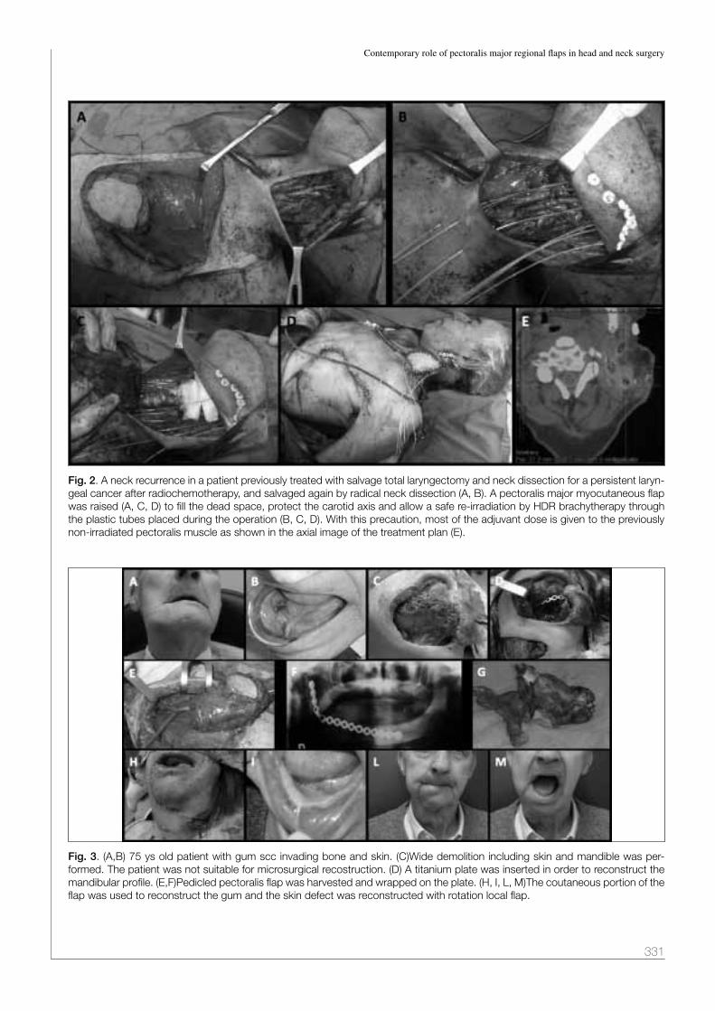
Contemporary role of pectoralis major regional flaps in head and neck surgery
331
Fig. 2. A neck recurrence in a patient previously treated with salvage total laryngectomy and neck dissection for a persistent laryn-geal cancer after radiochemotherapy, and salvaged again by radical neck dissection (A, B). A pectoralis major myocutaneous flap was raised (A, C, D) to fill the dead space, protect the carotid axis and allow a safe re-irradiation by HDR brachytherapy through the plastic tubes placed during the operation (B, C, D). With this precaution, most of the adjuvant dose is given to the previously non-irradiated pectoralis muscle as shown in the axial image of the treatment plan (E).
Fig. 3. (A,B) 75 ys old patient with gum scc invading bone and skin. (C)Wide demolition including skin and mandible was per-formed. The patient was not suitable for microsurgical recostruction. (D) A titanium plate was inserted in order to reconstruct the mandibular profile. (E,F)Pedicled pectoralis flap was harvested and wrapped on the plate. (H, I, L, M)The coutaneous portion of the flap was used to reconstruct the gum and the skin defect was reconstructed with rotation local flap.

F. Bussu et al.
332
hairy, mucosa are undoubtedly a relevant argument sup-porting the use of PMMF in reconstructions of the oro- and hypopharynx (Figs. 4, 5), as the bulk and the hair of the PMMC variant (and also most free flaps) can cause a functional impairment and complaints from patients. on the other hand, one of the main issues considering the functional results of pectoralis major reconstructions, also deriving in pedicled flaps from muscle denervation, is muscle fibrosis and consequent retraction. This is much more evident in the PMMF variant in which it involves the surface of the defect: in pharyngeal defects involv-ing a wide angle of the circumference in the axial plane, it is associated with a high rate of long-term dysphagia, stricture and dependence on PEg 99 100 even in our personal experience (Fig. 5).on the other hand, the wide pliable myofascial surface can be very useful to be rolled around a reconstruction plate in case of a segmental mandibular defect (Fig. 3). Even in these cases, if no skin area is resected, PMMF may be an alternative to PMMC 93 97 101 102. Considering that most free bony tissue transfers, characterised by rel-evant donor site morbidity, surgical complexity and dura-tion and complications, are not followed by restoration of dentition through osteointegrated implants, they remain a undeniably valuable option for reconstruction of mandib-ular defects, especially if lateral, with less aesthetic and functional issues 102.
Salvage total laryngectomies after irradiation are charac-terised by a high rate of fistulas 103; in these cases, pec-toralis major flaps may work as dead space fillers, bring well-vascularised tissue in the irradiated field, reinforce the pharyngeal suture and have been demonstrated to markedly reduce the risk of this complication. For this reason, in post- radiation laryngectomies several au-thors propose to electively reinforce the suture line and the pharyngeal wall with a pectoralis major 95 104 105. if a skin island is not needed for a skin defect, better results have been shown with a thin, well vascularised PMMF 95.For the same reason, many authors use the pectoralis ma-jor to protect the carotid axis after a salvage radical neck dissection following radiotherapy failure 8 78 106. At the same time, the well vascularised muscular tissue fills the dead space, and the vascular supply reduces post-irradia-tion neck fibrosis. if a PMMC is raised, its well vascular-ised skin reduces the tension and retraction of the suture line and later of the scar (Fig. 2). The inset of the PMMC flap in vessel depleted necks after salvage neck dissec-tion has allowed us in some cases to place plastic tubes to administer further adjuvant perioperative brachytherapy (Fig. 2).The protective and trophic effect of pectoralis muscle on neck vessels and tissues is an advantage in all cas-es after radiotherapy and neck dissection, even if the reconstructed defect is in the pharynx, oral cavity, or
Fig. 4. The local recurrence after radiochemotherapy of a SCC of the left posterior faucial pillar (A,C) was salvaged by a partial pharyngectomy through a transmandibular approach (C, D). The defect was restored through a PMMF flap (B, D), with complete re-epithelialisation and acceptable aesthetic results (E, F). The patient was able to feed with a normal diet at 3 weeks after the salvage surgery and did not report dysphagia.

Contemporary role of pectoralis major regional flaps in head and neck surgery
333
other head and neck sites: in fact, the humeral end of the raised muscle always tends to remain in the neck (Figs. 1-5). Many free flap surgeons use both the vari-ants of the pectoralis flap in combination with free tis-sue transfer, based upon these considerations and with similar indications, also for coverage and protection of microanastomosis 83 115.
Complications and morbidityPMMC and PMMF are both related to some complica-tions that have partially changed both in type and frequency, thanks to evolution of the technique. The very first report of complications in 42 cases of reconstruction with PMMC 57
did not show any major necrosis, while minor necrosis was present in three cases (7%). appeared later report document-ed a wider range of complications including those involving the donor site, such as chest wall haematoma and infections, the reconstructed areas, including fistula and neck abscess, or the flap itself with partial and total necrosis 108.later, in 1983, two interesting and larger reports pro-vided deeper insight into the actual complication rates. Mehrhof et al. 109, describing their experience on 67 patients treated with 73 PMMC, reported a complica-tion rate of 54%. Major complications were total flap necrosis (4%) and partial flap necrosis (12%). The vast
majority were minor complications that did not require second procedures for correction: orocutaneous fistu-las (16.5%), suture line dehiscence (12%), neck wound complications (11%), donor site complication (1.3%) and sequelae related to mandibular replacement (not a complication of the flap itself but related to a metal de-vice used by the authors to restore mandibular continuity in 10 patients included in this series) (11%). in the same year, ossoff et al. 110, while reporting an overall compli-cation rate similar to those previously reported, warned other authors about the insidious possibility of a delayed detection of recurrence hidden by the flap itself, which occurred three times in their experience (86 patients, 95 PMMC). in 1984, Donegan et al. reported another rare complication, namely osteomyelitis of the rib in the do-nor area of the PMMF 111. Even if unusual, this compli-cation gave rise to a number of publications discussing its treatment, with the use of a vertical rectus abdominis myocutaneous flap proposed by nagarjuna et al. being the latest 112. other reported complications include expo-sure of the plate covered by the flap in reconstructions of the mandible 91 and cervical contracture leading to se-vere functional impairment 113. Cervical contracture has been specifically addressed by some authors by second-ary myectomy and subclavicular route 17 114, but a certain amount of contracture is unavoidable, deriving from the
Fig. 5. Local recurrence after radiochemotherapy of a hypopharyngeal postcricoid SCC was salvaged by total laryngectomy and subtotal pharyngectomy (B). The pharyngeal tube was restored suturing the superficial fascia of a PMMF flap (A, C) to the residual posterior hypopharyngeal mucosa (D, E, F, G). The portion of the hypopharyngeal circumference restored with the procedure was more than 50% (D) and the patient, after the recovery of a normal swallowing without complications, showed progressive dyspha-gia after 4 months. A recurrent hypopharyngeal stricture was diagnosed, and after two endoscopic dilatations a gastrostomy tube was placed endoscopically (PEG).

F. Bussu et al.
334
fibrotic retraction of the denervated muscle, even in the “island” technique.in 1990, an interesting report on 168 cases of PMMC and PMMF performed at MD Anderson Cancer Center in Texas by Kroll et al. was published 115 in which the rate of overall complications was 63%, but with a flap loss rate of only 2.4%, with a relatively higher compli-cation rate in smokers and patients with larger tumours. later, in the same year, Shah reported his experience 116
which consisted of 211 patients treated with PMMC. Similar to that seen at MD Anderson, he reported an overall complication rate of 63%. on the other hand, despite the apparently high complication rate, he re-ported only 2 cases in which a second flap was required (1%), and 33 cases in which reoperation was required to resolve complications. Such a low rate of major com-plications was substantially confirmed in the 1990s by other authors 117. A correlation between complications and some variables was also shown, namely: age over 70 years, female sex, overweight, albumin less than 4 g/dl, oral cavity reconstruction after total glossec-tomy and systemic disease. Some of these correlations had been previously reported by Kroll et al. and later by other authors with the adjunct of prior chemothera-py and secondary reconstructions 117-119. The higher rate of flap necrosis seen in women by those authors was attributed to the interposition of breast tissue between muscle and the skin paddle. in fact, the authors noted that only PMMC were involved by this phenomena, while in their series PMMF had the same complication rates between men and women 115. Both Shah and later Keidan et al. outlined the absence of a significant cor-relation between prior radiation therapy and complica-tion rates 120.Starting from 2000, reports in literature from both wealthy and poor-developing countries, substantially confirm a slight drop in complications, with very few to no total flap losses recorded and an overall complication rate around 20–50% as seen in many papers published during this period 7 91 113 119 121 122. At present such complications rates are probably even lower, at least in developed countries. A rare complication of PMMC with very few reports in literature published in the last 15 years is metastasis at the flap donor site, for which some possible mechanisms have been proposed based on anatomical and physiologi-cal considerations 123.
Comparison with free flapsSince the development of reconstructive techniques in-volving free flaps 124, we participated in a lively debate on the advantages and disadvantages of both pedicled and free flap techniques applied to head and neck sites. in an attempt to summarise the data from the literature, it should be kept in mind that pectoralis major is often
confronted to different types of free flaps and in differ-ent types of reconstructions, thus making it impossible to make any general statements. Each type of reconstruction would ideally require a separate discussion to come to de-finitive conclusions. During the last 30 years, the three main topics of discussion have been functional results, morbidity and costs of the two techniques. in all these areas, when comparing free flap with pectoralis major pa-tients, several biases can be seen, the most important be-ing in our opinion that in the free flap era many patients were recommended pedicled flaps when they are in poor general conditions, with the preference for an option with short operative time, technical ease and low morbidity. As a result, radically different groups, at least in terms of comorbidities, are often compared. For this reasons, we believe that comorbidity, or at least age, should be consid-ered and matched in every attempt at comparison.
Functional resultsEach subsite would ideally deserve a separate discussion, even if not all the papers in the literature analytically eval-uate the functional differences according site and size of the defect. Paradoxically, despite the amount of publica-tions evaluating reconstructive techniques in the head and neck, there is still a lack of evidence-based data about the functional differences between free and pedicled flaps, and no definitive conclusions can be made at present. Ac-tually, the concept of function in head and neck cancer patients is controversial and consensus about the objec-tive evaluation of functional results is lacking. The most used tools are questionnaires, which are not specific and sensitive in evaluating the functional aspects addressed by the different reconstructive techniques. nonetheless, a few investigations give some clues with which to initiate definite functional comparison site by site.An impressive series was presented by hsing et al. in 2011 125. They reported their experience with 491 patients treated for oral cavity cancer, 100 of which completed a quality of life questionnaire. They found no signifi-cant difference in overall quality of life between patients treated with PMMC versus free flaps, nor did they find differences in the domains regarding pain, appearance, activity, recreation, swallowing, chewing, taste, saliva, or anxiety. nevertheless, they reported significant dif-ferences between the free flap and PMMC flap groups in speech, shoulder mobility and mood domains with better outcomes for the free flap group.Xiao et al., also focusing specifically on oral cavity can-cer and analysing two groups, one characterised by recon-struction performed with an anterolateral tight (AlT) flap and the other by PMMC, found no significant differences between groups for pain, activity, recreation, swallow-ing, chewing, speech, taste, saliva, or mood and anxiety domains. however, significant differences were seen be-tween the PMMC and AlT groups for the shoulder and

Contemporary role of pectoralis major regional flaps in head and neck surgery
335
appearance domains, and a higher emotional impact left by the scar and the aesthetic appearance left by PMMC 126. A similar study with similar results was presented in 2014 by Zhang et al., with the only difference being slightly worse speech outcome for patients treated by AlT 127.o’neill et al., in 2010, published an interesting compari-son between radial forearm free flaps and PMMC in re-construction of oral and oropharyngeal defects 128. in their report, they outlined that while no differences in deform-ity, diet, or socialisation were found, only 22.2% of pa-tients reconstructed with PMMC were satisfied with their speech compared with 53.1% of patients reconstructed with rFFF. Deganello et al. noted a difference in diet with all PMMC in their series able to feed only by soft or liquid diet postoperatively 99 129.As free flaps show a predisposition to better performance in functional outcomes 86, many authors have tried to close the gap by developing variants of the PMMC/PMMF meant to improve functional results, such as the dynamic variant 19 and attempts of neurotisation of the flap with the hypoglossal nerve 40. These complex techniques, however, are associated with loss of the renowned advantages of pectoralis flaps in terms of ease and speed.
MorbidityPectoralis major has a rate of complications that is not to be underestimated, and the rate of overall complications raised some concerns in the past. An interesting compari-son of complications rate between the free tissue trans-fer option and the PMMC were made by Mallet et al. in 2009 130 who analysed the results of 70 consecutive pa-tients treated alternatively with both techniques (25 FTT and 45 PMMC). They outlined a similar rate of postopera-tive complications and the same duration of use of trache-ostomy tube and feeding tube. Similar results, prone to a substantially similar morbidity of free flaps and PMMF/PMMC, have been reported by other groups 125 131, some of which also highlighted a similar failure rate 78. o’neill, reporting on a series of 114 patients treated alternatively with PMMC or radial forearm free flap (rFFF) reported that the only significant differences in complications were suture dehiscence, which were more frequent in PMMC, and atelectasis, which was more frequent in rFFF; major flap complications, leading to another surgery, were more frequent in the rFFF group 128.in a similar study, de Bree et al. reported a higher rate of medical complications, flap failures and admissions to intensive care units in the radial forearm group, but more overall surgical complications (partial flap necrosis, de-hiscence, wound abscesses and fistula) with PMMC 132, as seen in other studies 73 133, thus leading to a longer post-operative hospitalisation for treatment of complications.
Costsin the last years, in light of spending reviews and ration-
alisations of resources, especially in italy, the costs of re-constructive procedures must be taken into account. The main factors contributing to the increase in costs, in our experience, are the time spent in the hospital postopera-tively (heavily influenced by the complication rate), time spent in the operating room and number of surgeons re-quired. PMMC is often recognised as a faster technique considering operating time 125 126 129-132 134-136, and most re-ports outline that free flap surgery requires more surgeons at the same time 129 130 137. in many departments, FFT in the head and neck are performed by two teams at the same time, thus reducing the operative time, but not the overall costs. Data regarding discharge time after surgery seems to be uniform in the literature with most authors outlin-ing a similar if not shorter time needed after a procedure involving free flaps 128-132. nevertheless, when it comes to costs estimated in single experiences, data are rather conflicting. Some authors reported higher costs with free flaps 91 129 134 136 138, while others have documented that overall costs for PMMC are either higher 115 132 or not sig-nificantly different 131 139. in particular, Funk et al. hypoth-esised that although surgical costs were higher for free flaps, postoperative hospitalisation due to treatment of complications, which is higher in PMMC, balanced the costs and abolished any economic advantages 139.While a generally acceptable, definitive statement about re-spective costs is probably not possible at present, according to the literature we can at least definitely accept that, even with highly significant differences between various reports, the time spent in the operating room for reconstruction with a pedicled flap is undeniably shorter. importantly, as outlined by de Bree et al. 132, most studies on costs have selection biases, and do not consider differences in age and comorbidity, thus probably complicating any possible eval-uation on discharge time and complication rates.notably, in settings where resource constraints are a mat-ter of fact, along with the lack of available technologies and expertise, such as in developing countries, extensive use of pectoralis major at the expense of free flaps re-mains a reality 91 97 138.
Our experienceour 20-year clinical experience in pectoralis major recon-structions at the main big facilities in italy performing a high volume of free flap procedures is shown in Table i. Virtually every defect in the head and neck region has been addressed, with a very low rate of complications (less than 5% of patients experienced any kind of pro-cedure-related sequelae). Such complication rate is lower than what reported in most paper in the literature, nev-ertheless in our opinion it reflects the state of the art as for pectoralis major reconstruction and appears the main general argument supporting the use of pectoralis major regional flaps in most cases. The 274 cases (Table i) have

F. Bussu et al.
336
been operated along 20 years, in this long period several changes in the use and indications for pectoralis major reconstructions have been occurred, mostly reflecting the above cited literature evidences. in fact the number of pectoralis major flaps raised per year has on the whole decreased, and in particular it is now only seldom used for tongue and skin defect. The median age of the patient has increased together with their comorbidities. interestingly, in recent years pectoralis major inset has been also useful for allow safe further irradiation by brachytherapy in a few cases, by protecting neck vessels and bringing non-previously irradiated tissue (Fig. 2).
ConclusionsWe are definitely in the era of free flap surgery. in de-veloped countries, in excellence centres, and in patients with good general conditions, the amazing versatility of the free flap armamentarium gives the opportunity to suit the defect deriving from virtually every ablative head and neck surgery with a tailored reconstruction. Such possi-bilities, even in the absence of definitive evidence-based data on better functional outcomes compared with pecto-ralis major flaps, make free tissue transfer the standard for head and neck reconstruction. Countless disadvantages of the “old” pectoralis major have been listed, such as flap thickness, functional and cosmetic defects, excessive bulk in obese or muscular patients, poor reliability of the skin in PMMC, difficulty in obtaining reliable bone tissue and long-term fibrotic muscle retraction.nevertheless, even in facilities with a high volume of re-constructions by free flaps, a certain number of pectora-lis major flaps is still raised every year in the head and neck. The easier indication is reconstruction after a free flap failure due to the high failure rate of second free tis-sue transfers 76 77. other indications, which are much more common, are probably controversial, and therefore more interesting to discuss. They can be driven by the setting in which patients are cured, by clinical status and comor-bidities of patients and perhaps, in very selected cases, by the type of the defect.The healthcare system and the facilities in which patients are cured are also key considerations in determining epi-demiology and death rates for cancer worldwide. rais-ing a pectoralis major just requires a knife, forceps and retractors as surgical instrumentation, few hours of op-erative time, shorter training time and less surgical skills. Mainly for these reasons in developing countries such as india the pectoralis major remains the workhorse for head and neck reconstruction 97. nevertheless, in forthcoming years, the public discussion about health system resources and spending reviews may bring these arguments to many Western countries in recession. As previously outlined, another reason why pectoralis major can still be indicated is related to patient conditions, which may require ease of
Table I. Descriptive statistics of the main variables concerning patients, primary sites and defects. Characteristic N = 274
Age at diagnosis
Median 70
Range 29-92
Sex [N (%)]
Male 199 (72.6%)
Female 75 (27.4%)
Variant (PMMC vs. PMMF) [N (%)]
PMMC 237 (86.5%)
PMMF 37 (13.5%)
Primary site [N (%)]
Larynx and Hypopharynx 21 (7.7%)
Mandible 19 (7%)
Oropharynx 85 (31%)
Skin 5 (1.8%)
Occult primary 1 (0.3%)
Oral cavity Retromolar Tongue
143 (52.2%)56 (20.4%)87 (31.8%)
Hystotype N (%)
SCC 255 (93%)
Salivary histotypes 17 (6.2%)
Malignant ameloblastoma 1 (0.4%)
Osteosarcoma 1 (0.4%)
Type of defect
Mucosa 141 (51.5%)
Mucosa+bone 112 (40.9%)
Mucosa+skin 11 (4%)
Skin 10 (3.6%)
Previous treatments for HNSCC
None 209 (76.3%)
Radio+chemotherapy 48 (17.5%)
Surgery 3 (1.1%)
Surgery+ radio+chemotherapy 14 (5.1%)
Adjuvant /perioperative treatments
None 154 (56.2%)
Radiotherapy 102 (37.2%)
Brachytherapy 4 (1.5%)
Radio+chemotherapy 14 (5.1%)
Timing
Immediate 265 (96.7%)
Delayed (management of radiotherapy sequelae)
9 (3.3%)
Early and late complications of the procedure
Partial skin necrosis 8 (2.9%)
Total necrosis 2 (0.7%)
Donor site dehiscence 2 (0.7%)
Late hypopharyngeal stricture/ gastrostomy 1 (0.4%)
Death 1 (0.4%)
None 261 (95.3%)

Contemporary role of pectoralis major regional flaps in head and neck surgery
337
harvest and shorter operative times (and therefore, espe-cially in the elderly, a lower perioperative complication rate).Finally, there are some situations in which pectoralis major still appears as a valuable option even in facilities performing free flaps and for patients in good general conditions 78. one such situation is prevention of phar-yngocutaneous fistula in case of salvage total laryngec-tomies after irradiation. in these cases, several authors have proposed to electively reinforce the suture line and pharyngeal wall with a PMMF 95 104 105. in the same way, PMMF/PMMC can be used to protect the carotid axis af-ter salvage radical neck dissection following radiotherapy failure 8 78 106, and in these cases a further adjuvant periop-erative brachytherapy can be safely administered (Fig. 2).lastly, recent reports stress 7 80 99 100 140 the utility of pecto-ralis major flaps for reconstruction of pharyngeal defects. in case of circumferential defects, long-term stricture due to muscle fibrotic retraction is probably frequent (Fig. 5), but in lateral defects (less than 50% of pharyngeal cir-cumference) the reduced thickness and coverage with normal, not hairy, mucosa are relevant advantages sup-porting the use of PMMF (Fig. 4). in case of a segmental lateral mandibular defect, if restoration of dentition is not planned, a PMMC or PMMF rolled around a reconstruc-tion plate may be a valid option characterised by low mor-bidity 93 97 101 102.
References1 Ariyan S. The pectoralis major myocutaneous flap. A versa-
tile flap for reconstruction in the head and neck. Plast recon-str Surg 1979;63:73-81.
2 Brown rg, Fleming Wh, Jurkiewicz MJ. An island flap of the pectoralis major muscle. Br J Plast Surg 1977;30:161-5.
3 Maruyama y, Fujino T, Aoyagi F, et al. One stage reconstruc-tion of oral cavity by use of pectoralis major myocutaneous island flap. Keio J Med 1978;27:47-52.
4 El-Marakby hh. The reliability of pectoralis major myocu-taneous flap in head and neck reconstruction. J Egypt natl Canc inst 2006;18:41-50.
5 robertson MS, robinson JM. Pharyngoesophageal recon-struction. Is a skin-lined pharynx necessary? Arch otolaryn-gol 1985;111:375-376.
6 Phillips Jg, Postlethwaite K, Peckitt n. The pectoralis major muscle flap without skin in intra-oral reconstruction. Br J oral Maxillofac Surg 1988;26:479-85.
7 Montemari g, rocco A, galla S, et al. Hypopharynx recon-struction with pectoralis major myofascial flap: our experi-ence in 45 cases. Acta otorhinolaryngol ital 2012;32:93-7.
8 Shindo Ml, Costantino PD, Friedman CD, et al. The pec-toralis major myofascial flap for intraoral and pharyn-geal reconstruction. Arch otolaryngol head neck Surg 1992;118:707-11.
9 righi PD, Weisberger EC, Slakes Sr, et al. The pectoralis major myofascial flap: clinical applications in head and neck reconstruction. Am J otolaryngol 1998;19:96-101.
10 Johnson MA, langdon JD. Is skin necessary for intraoral re-construction with myocutaneous flaps? Br J oral Maxillofac Surg 1990;28:299-301.
11 Cuono CB, Ariyan S. Immediate reconstruction of a compos-ite mandibular defect with a regional osteomusculocutane-ous flap. Plast reconstr Surg 1980;65:477-84.
12 green MF, gibson Jr, Bryson Jr, et al. A one-stage correction of mandibular defects using a split sternum pectoralis major osteo-musculocutaneous transfer. Br J Plast Surg 1981;34:11-6.
13 Bell MS, Barron PT. The rib-pectoralis major osteomyocuta-neous flap. Ann Plast Surg 1981;6:347-353.
14 Phillips Jg, Falconer DT, Postlethwaite K, et al. Pectoralis major muscle flap with bone graft in intra-oral reconstruc-tion. Br J oral Maxillofac Surg 1990;28:160-3.
15 Jones nF, Sommerlad BC. Reconstruction of the zygoma, temporo-mandibular joint and mandible using a compound pectoralis major osteo-muscular flap. Br J Plast Surg 1983;36:491-7.
16 Freeman MS, Thomas Jr, Zipper JA. Clavicular division technique. A new approach for lengthening the pectoralis flap. Arch otolaryngol head neck Surg 1989;115:224-7.
17 Kerawala CJ, Sun J, Zhang Zy, et al. The pectoralis major myocutaneous flap: Is the subclavicular route safe? head neck 2001;23:879-84.
18 Vanni CM, Pinto Fr, de Matos ll, et al. The subclavicular versus the supraclavicular route for pectoralis major myocu-taneous flap: a cadaveric anatomic study. Eur Arch otorhi-nolaryngol 2010;267:1141-6.
19 Maruyama y, nakajima h, Fujino T. A dynamic reconstruc-tion of a facial defect with a pectoralis major myocutaneous flap. Br J Plast Surg 1980;33:145-9.
20 Ariyan S. The donor site of the pectoralis major myocutane-ous flap. Plast reconstr Surg 1980;66:165-6.
21 Crosher r, llewelyn J, Mitchell r. A modification of the pec-toralis major myocutaneous flap that reduces the defect at the donor site. Ann r Coll Surg Engl 1995;77:389-91.
22 Morain WD, geurkink nA. Split pectoralis major myocuta-neous flap. Ann Plast Surg 1980;5:358-61.
23 Freeman Jl, gullane PJ, rotstein lM. The double pad-dle pectoralis major myocutaneous flap. J otolaryngol 1985;14:237-40.
24 ord rA, Avery BS. Side-by-side double paddle pectora-lis major flap for cheek defects. Br J oral Maxillofac Surg 1989;27:177-85.
25 Freeman Jl, Walker EP, Wilson JS, et al. The vascular anat-omy of the pectoralis major myocutaneous flap. Br J Plast Surg 1981;34:3-10.
26 Espinosa Mh, Phillip JA, Khatri VP, et al. Double skin island pectoralis major myocutaneous flap with nipple-areola com-plex preservation: a case report. head neck 1992;14:488-91.
27 Maruyama y, nakajima h, Fujino T. One-stage reconstruc-tion of a esophagostome with a latissimus dorsi myocuta-neous flap and a pectoralis major myocutaneous flap (case report). Acta Chir Plast 1980;22:80-5.
28 Maisel rh, liston Sl. Combined pectoralis major myocuta-neous flap with medially based deltopectoral flap for closure of large pharyngocutaneous fistulas. Ann otol rhinol laryn-gol 1982;91:98-100.

F. Bussu et al.
338
29 Meyer r, Kelly TP, Abul Failat AS. Single bilobed flap for use in head and neck reconstruction. Ann Plast Surg 1981;6:203-6.
30 Weaver AW, Vandenberg hJ, Atkinson DP, et al. Modified bilobular (“gemini”) pectoralis major myocutaneous flap. Am J Surg 1982;144:482-8.
31 harii K, iwaya T, Kawaguchi n. Combination myocutane-ous flap and microvascular free flap. Plast reconstr Surg 1981;68:700-11.
32 Chen hC, Demirkan F, Wei FC, et al. Free fibula osteosep-tocutaneous-pedicled pectoralis major myocutaneous flap combination in reconstruction of extensive composite man-dibular defects. Plast reconstr Surg 1999;103:839-45.
33 Choi Jo, Choi g, Chae SW, et al. Combined use of pec-toralis major myocutaneous and free radial forearm flaps for reconstruction of through-and-through defects from excision of head and neck cancers. J otolaryngol 1999;28:332-6.
34 Sharzer lA, Kalisman M, Silver CE, et al. The parasternal paddle: a modification of the pectoralis major myocutaneous flap. Plast reconstr Surg 1981;67:753-62
35 Dennis D, Kashima h. Introduction of the Janus flap. A modified pectoralis major myocutaneous flap for cervical es-ophageal and pharyngeal reconstruction. Arch otolaryngol 1981;107:431-5.
36 Strawberry CW, de Fries ho, Deeb ZE. Reconstruction of the hypopharynx and cervical esophagus with bilateral pectoralis major myocutaneous flaps. head neck Surg 1981;4:161-4.
37 neifeld JP, Merritt WA, Theogaraj SD, et al. Tubed pecto-ralis major musculocutaneous flaps for cervical esophageal replacement. Ann Plast Surg 1983;11:24-30.
38 Morain WD, Colen lB, hutchings JC. The segmental pec-toralis major muscle flap: a function-preserving procedure. Plast reconstr Surg 1985;75:825-30.
39 lawson Vg. Pectoralis major muscle flap with amnion in oral cavity reconstruction. Aust n Z J Surg 1986;56:163-6.
40 Katsantonis gP. neurotization of pectoralis major myocuta-neous flap by the hypoglossal nerve in tongue reconstruc-tion: clinical and experimental observations. laryngoscope 1988;98:1313-23.
41 Cueva r, Thomas Jr, Davidson TM. Liposuction to debulk the pectoralis major myocutaneous flap. Am J otolaryngol 1988;9:106-10.
42 Wei Wi, lam Kh, Wong J. The true pectoralis major my-ocutaneous island flap: an anatomical study. Br J Plast Surg. 1984;37:568-73.
43 Kasler M, Banhidy Fg, Trizna Z. Experience with the modi-fied pectoralis major myocutaneous flap. Arch otolaryngol head neck Surg 1992;118:931-932.
44 Palmer Jh, Batchelor Ag. The functional pectoralis major musculocutaneous island flap in head and neck reconstruc-tion. Plast reconstr Surg 1990;85:363-7.
45 gateley Dr, Moss Al, Blenkinsopp P. Reconstruction of the anterior lower third of the face using a composite pectoralis major free flap. J r Soc Med 1993;86:731-2.
46 Chaturvedi P, Prabhudesai Sg, Kanitkar g, et al. Sub-mam-mary approach for pectoralis major myocutaneous flap in females. J Surg oncol 2003;84:255-6.
47 Chaturvedi P, Pathak KA, Pai PS, et al. A novel technique of raising a pectoralis major myocutaneous flap through the skin paddle incision alone. J Surg oncol 2004;86:105-6.
48 Turkmen A, Perks Ag. Endoscopic assisted harvest of the pedicled pectoralis major muscle flap. Br J Plast Surg 2005;58:170-4.
49 Po-Wing yA. Preservation of lateral thoracic artery to improve vascular supply of distal skin without com-promising pedicle length in harvesting pectoralis ma-jor myocutaneous flap. J Plast reconstr Aesthet Surg 2006;59:1433-5.
50 rikimaru h, Kiyokawa K, Watanabe K, et al. New method of preparing a pectoralis major myocutaneous flap with a skin paddle that includes the third intercostal perforating branch of the internal thoracic artery. Plast reconstr Surg 2009;123:1220-8.
51 Belt PJ, Emmett J. Local transposition flap repair of the pec-toralis major myocutaneous flap donor site. Plast reconstr Surg 2004;114:732-7.
52 Chan yW, ng rW, yuen AP. Lateral thoracic flap for donor site repair of pectoralis major myocutaneous flap. J Plast re-constr Aesthet Surg 2009;62:1004-7.
53 Kruse Al, luebbers hT, gratz KW, et al. A new method for closure of large donor side defects after raising the pectora-lis major flap. oral Maxillofac Surg 2011;15:251-2.
54 Benech A, Arcuri F, Poglio g, et al. Vacuum-assisted closure therapy in reconstructive surgery. Acta otorhinolaryngol ital 2012;32:192-7.
55 Ariyan S. Further experiences with the pectoralis major myocutaneous flap for the immediate repair of defects from excisions of head and neck cancers. Plast reconstr Surg 1979;64:605-12.
56 Arlyan S, Cuono CB. Use of the pectoralis major myocu-taneous flap for reconstruction of large cervical, facial or cranial defects. Am J Surg 1980;140:503-6.
57 Biller hF, Krespi yP, lawson W, et al. A one-stage flap re-construction following resection for stomal recurrence. oto-laryngol head neck Surg 1980;88:357-60.
58 Theogaraj SD, Merritt Wh, Acharya g, et al. The pectoralis major musculocutaneous island flap in single-stage recon-struction of the pharyngoesophageal region. Plast reconstr Surg 1980;65:267-6.
59 Evans Ph, Das gupta Ar. The use of the pectoralis major myocutaneous flap for one-stage reconstruction of the base of the tongue. J laryngol otol 1981;95:809-16.
60 Withers Eh, Franklin JD, Madden JJ Jr, et al. Pectoralis ma-jor musculocutaneous flap: a new flap in head and neck re-construction. Am J Surg 1979;138:537-43.
61 lee yT. Carotid artery protection by pectoralis major mus-cle flap. Am J Surg 1980;140:464.
62 Siu KF, Wei Wi, lam Kh, et al. Use of the pectoralis major muscle flap for repair of a tracheoesophageal fistula. Am J Surg 1985;150:617-9.
63 Sisson gA, goldman ME. Pectoral myocutaneous island flap for reconstruction of stomal recurrence. Arch otolaryn-gol 1981;107:446-9.
64 Baker Sr. Management of osteoradionecrosis of the mandi-ble with myocutaneous flaps. J Surg oncol 1983;24:282-9.

Contemporary role of pectoralis major regional flaps in head and neck surgery
339
65 Price JC, Davis rK, Koltai PJ. The pectoralis myocutane-ous flap for salvage of necrotic wounds. laryngoscope 1985;95:146-50.
66 Krespi yP, Sisson gA. Reconstruction after total or subtotal glossectomy. Am J Surg 1983;146:488-92.
67 Sasaki CT, Ariyan S, Spencer D, et al. Pectoralis major my-ocutaneous reconstruction of the anterior skull base. laryn-goscope 1985;95:162-6.
68 Deganello A, gitti g, Struijs B, et al Palliative combined treatment for unresectable cutaneous basosquamous cell carcinoma of the head and neck. Acta otorhinolaryngol ital 2013;33:353-6.
69 Morgan rF, Sargent lA, hoopes JE. Midfacial and total na-sal reconstruction with bilateral pectoralis major myocuta-neous flaps. Plast reconstr Surg 1984;73:824-6.
70 Milroy BC, Korula P. Vascularized innervated transfer of the clavicular head of the pectoralis major muscle in established facial paralysis. Ann Plast Surg 1988;20:75-81.
71 Joseph CA, Fayman MS, Kadwa MA, gregor rT. The pecto-ralis major flap for reconstruction of septic wounds. S Afr J Surg 1988;26:107-9.
72 Fleischer A, Khafif r. Reconstruction of the mediastinal tra-chea with a tubed pectoralis major myocutaneous flap. Plast reconstr Surg 1989;84:342-6.
73 Schusterman MA, Kroll SS, Weber rS, et al. Intraoral soft tissue reconstruction after cancer ablation: a comparison of the pectoralis major flap and the free radial forearm flap. Am J Surg 1991;162:397-9.
74 Kroll SS, reece gP, Miller MJ, et al. Comparison of the rectus ab-dominis free flap with the pectoralis major myocutaneous flap for reconstructions in the head and neck. Am J Surg 1992;164:615-8.
75 Cordeiro Pg, hidalgo DA. Soft tissue coverage of mandibu-lar reconstruction plates. head neck 1994;16:112-115.
76 Wei FC, Demirkan F, Chen hC, et al. The outcome of failed free flaps in head and neck and extremity reconstruction: what is next in the reconstructive ladder? Plast reconstr Surg 2001;108:1154-60.
77 Bozikov K, Arnez ZM. Factors predicting free flap compli-cations in head and neck reconstruction. J Plast reconstr Aesthet Surg 2006;59:737-42.
78 Schneider DS, Wu V, Wax MK. Indications for pedicled pectoralis major flap in a free tissue transfer practice. head neck 2012;34:1106-10.
79 Colletti g, Autelitano l, Tewfik K, et al. Autonomized flaps in secondary head and neck reconstructions. Acta otorhi-nolaryngol ital 2012;32:329-35.
80 Spriano g, Piantanida r, Pellini r. Hypopharyngeal recon-struction using pectoralis major myocutaneous flap and pre-vertebral fascia. laryngoscope 2001;111:544-7.
81 Saussez S, Cuno A, Urbain F, et al. Reconstruction of circum-ferential oro- and hypopharyngeal defects with U-shaped pectoralis major myocutaneous flap. otolaryngol head neck Surg 2006;134:823-9.
82 liu hl, Chan Jy, Wei Wi. The changing role of pectoralis major flap in head and neck reconstruction. Eur Arch otorhi-nolaryngol 2010;267:1759-63.
83 Talesnik A, Markowitz B, Calcaterra T, et al. Cost and out-come of osteocutaneous free-tissue transfer versus pedicled
soft-tissue reconstruction for composite mandibular defects. Plast reconstr Surg 1996;97:1167-78.
84 giordano l, Bondi S, Ferrario F, et al. Radial forearm free flap surgery: a modified skin- closure technique improving donor-site aesthetic appearance. Acta otorhinolaryngol ital 2012;32:158-63.
85 Pellini r, Mercante g, Spriano g. Step-by-step mandibular reconstruction with free fibula flap modelling. Acta otorhi-nolaryngol ital 2012;32:405-9.
86 Tarsitano A, Vietti MV, Cipriani r, et al. Functional results of microvascular reconstruction after hemiglossectomy: free anterolateral thigh flap versus free forearm flap. Acta otorhinolaryngol ital 2013;33:374-9.
87 Baj A, Beltramini gA, Demarchi M, et al. Extended-pedicle peroneal artery perforator flap in intraoral reconstruction. Acta otorhinolaryngol ital 2013;33:282-5.
88 iJsselstein CB, hovius SE, ten have Bl, et al. Is the pec-toralis myocutaneous flap in intraoral and oropharyngeal reconstruction outdated? Am J Surg 1996;172:259-62.
89 Croce A, Moretti A, D’Agostino l, et al. Continuing validity of pectoralis major muscle flap 25 years after its first appli-cation. Acta otorhinolaryngol ital 2003;23:297-304.
90 leemans Cr, Balm AJ, gregor rT, et al. Management of ca-rotid artery exposure with pectoralis major myofascial flap transfer and split-thickness skin coverage. J laryngol otol 1995;109:1176-80.
91 Kekatpure VD, Trivedi nP, Manjula BV, et al. Pectoralis ma-jor flap for head and neck reconstruction in era of free flaps. int J oral Maxillofac Surg 2012;41:453-7.
92 Kiyokawa K, Tai y, inoue y, et al. Reliable, minimally inva-sive oromandibular reconstruction using metal plate rolled with pectoralis major myocutaneous flap. J Craniofac Surg 2001;12:326-36.
93 Salvatori P, Motto E, Paradisi S, et al. Oromandibular recon-struction using titanium plate and pectoralis major myocuta-neous flap. Acta otorhinolaryngol ital 2007;27:227-32.
94 Cordova A, Corradino B, Pirrello r, et al. Surgical treatment of pharyngostomes in irradiated patients. Our experience with musculocutaneous pectoralis major flap and hyperbaric oxygen therapy. Acta otolaryngol 2005;125:759-64.
95 gilbert Mr, Sturm JJ, gooding WE, et al. Pectoralis ma-jor myofascial onlay and myocutaneous flaps and pharyn-gocutaneous fistula in salvage laryngectomy. laryngoscope 2014;124:2680-6..
96 Bhola n, Jadhav A, Borle r, et al. Is there still a role for bilobed/bipaddled pectoralis major myocutaneous flap for single-stage immediate reconstruction of post ablative on-cologic full-thickness defects of the cheek? oral Maxillofac Surg 2014 Aug 17 [Epub ahead of print].
97 gadre KS, gadre P, Sane VD, et al. Pectoralis major my-ocutaneous flap--still a workhorse for maxillofacial recon-struction in developing countries. J oral Maxillofac Surg 2013;71:2005.
98 Vartanian Jg, Carvalho Al, Carvalho SM, et al. Pectoralis major and other myofascial/myocutaneous flaps in head and neck cancer reconstruction: experience with 437 cases at a single institution. head neck 2004;26:1018-23.
99 Mura F, Bertino g, occhini A, et al. Advanced carcinoma of the hypopharynx: functional results after circumferen-

F. Bussu et al.
340
tial pharyngolaryngectomy with flap reconstruction. Acta otorhinolaryngol ital 2012;32:154-7.
100 Mura F, Bertino g, occhini A, et al. Surgical treatment of hypopharyngeal cancer: a review of the literature and pro-posal for a decisional flow-chart. Acta otorhinolaryngol ital 2013;33:299-306.
101 Wada T, nakatani K, hiraishi y, et al. Usefulness of myofas-cial flap without skin in contemporary oral and maxillofacial reconstruction. J oral Maxillofac Surg 2011;69:1815-25.
102 Deleyiannis FW, rogers C, lee E, et al. Reconstruction of the lateral mandibulectomy defect: management based on prognosis and location and volume of soft tissue resection. laryngoscope 2006;116:2071-80.
103 galli J, Valenza V, Parrilla C, et al. Pharyngocutaneous fis-tula onset after total laryngectomy: scintigraphic analysis. Acta otorhinolaryngol ital 2009;29:242-4.
104 oosthuizen JC, leonard DS, Kinsella JB. The role of pecto-ralis major myofascial flap in salvage laryngectomy: a single surgeon experience. Acta otolaryngol 2012;132:1002-5.
105 righini C, lequeux T, Cuisnier o, et al. The pectoralis myo-fascial flap in pharyngolaryngeal surgery after radiothera-py. Eur Arch otorhinolaryngol 2005;262:357-61.
106 Mclean Jn, Carlson gW, losken A. The pectoralis major myocutaneous flap revisited: a reliable technique for head and neck reconstruction. Ann Plast Surg 2010;64:570-3.
107 Blackwell KE, Buchbinder D, Biller hF, et al. Reconstruction of massive defects in the head and neck: the role of simulta-neous distant and regional flaps. head neck 1997;19:620-8.
108 Maisel rh, liston Sl, Adams gl. Complications of pecto-ralis myocutaneous flaps. laryngoscope 1983;93:928-30.
109 Mehrhof Ai, Jr., rosenstock A, neifeld JP, et al. The pecto-ralis major myocutaneous flap in head and neck reconstruc-tion. Analysis of complications. Am J Surg 1983;146:478-82.
110 ossoff rh, Wurster CF, Berktold rE, et al. Complications after pectoralis major myocutaneous flap reconstruction of head and neck defects. Arch otolaryngol 1983;109:812-4.
111 Donegan Jo, gluckman Jl. An unusual complication of the pec-toralis major myocutaneous flap. head neck Surg 1984;6:982-3.
112 nagarjuna M, Patil Br, nagraj n, et al. Use of superiorly based vertical rectus abdominis myocutaneous flap for the correc-tion of costal osteomyelitis at the pectoralis major myocutane-ous flap donor site. J oral Maxillofac Surg 2013;71:e132-6.
113 liu r, gullane P, Brown D, et al. Pectoralis major myocu-taneous pedicled flap in head and neck reconstruction: ret-rospective review of indications and results in 244 consecu-tive cases at the Toronto General Hospital. J otolaryngol 2001;30:34-40.
114 van rossen ME, Verduijn PV, Mureau MA. Survival of pedi-cled pectoralis major flap after secondary myectomy of mus-cle pedicle including transection of thoracoacromial vessels: does the flap remain dependent on its dominant pedicle? J Plast reconstr Aesthet Surg 2011;64:323-8.
115 Kroll SS, goepfert h, Jones M, et al. Analysis of complica-tions in 168 pectoralis major myocutaneous flaps used for head and neck reconstruction. Ann Plast Surg 1990;25:93-7.
116 Shah JP, haribhakti V, loree Tr, et al. Complications of the pectoralis major myocutaneous flap in head and neck recon-struction. Am J Surg 1990;160:352-5.
117 Mehta S, Sarkar S, Kavarana n, et al. Complications of the pectoralis major myocutaneous flap in the oral cavity: a prospective evaluation of 220 cases. Plast reconstr Surg 1996;98:31-7.
118 Castelli Ml, Pecorari g, Succo g, et al. Pectoralis major myocutaneous flap: analysis of complications in difficult pa-tients. Eur Arch otorhinolaryngol 2001;258:542-5.
119 you yS, Chung Ch, Chang yJ, et al. Analysis of 120 pec-toralis major flaps for head and neck reconstruction. Arch Plast Surg 2012;39:522-7.
120 Keidan rD, Kusiak JF. Complications following recon-struction with the pectoralis major myocutaneous flap: the effect of prior radiation therapy. laryngoscope 1992;102:521-4.
121 Avery CM, gandhi n, Peel D, et al. Indications and out-comes for 100 patients managed with a pectoralis major flap within a UK maxillofacial unit. int J oral Maxillofac Surg 2014;43:546-54.
122 Jena A, Patnayak r, Sharan r, et al. Outcomes of pectoralis major myocutaneous flap in female patients for oral cavity de-fect reconstruction. J oral Maxillofac Surg 2014;72:222-31.
123 Bansal r, Patel TS, Bhullar C, et al. Metastases to the do-nor site of the pectoralis major myocutaneous flap following reconstructive surgery: a rare complication. Plast reconstr Surg 2004;114:1965-6.
124 Seidenberg B, rosenak SS, hurwitt ES, et al. Immediate re-construction of the cervical esophagus by a revascularized isolated jejunal segment. Ann Surg 1959;149:162-71.
125 hsing Cy, Wong yK, Wang CP, et al. Comparison between free flap and pectoralis major pedicled flap for reconstruc-tion in oral cavity cancer patients - a quality of life analysis. oral oncol 2011;47:522-7.
126 Xiao y, Zhu J, Cai X, et al. Comparison between ante-rolateral thigh perforator free flaps and pectoralis major pedicled flap for reconstruction in oral cancer patients--a quality of life analysis. Med oral Patol oral Cir Bucal 2013;18:e856-61.
127 Zhang X, li MJ, Fang Qg, et al. A comparison between the pectoralis major myocutaneous flap and the free anterolat-eral thigh perforator flap for reconstruction in head and neck cancer patients: assessment of the quality of life. J Craniofac Surg 2014;25:868-71.
128 o’neill JP, Shine n, Eadie PA, et al. Free tissue transfer versus pedicled flap reconstruction of head and neck malig-nancy defects. ir J Med Sci 2010;179:337-43.
129 Deganello A, gitti g, Parrinello g, et al. Cost analysis in oral cavity and oropharyngeal reconstructions with mi-crovascular and pedicled flaps. Acta otorhinolaryngol ital 2013;33:380-7.
130 Mallet y, El BS, Penel n, et al. The free vascularized flap and the pectoralis major pedicled flap options: compara-tive results of reconstruction of the tongue. oral oncol 2009;45:1028-31.
131 Smeele lE, goldstein D, Tsai V, et al. Morbidity and cost dif-ferences between free flap reconstruction and pedicled flap reconstruction in oral and oropharyngeal cancer: Matched control study. J otolaryngol 2006;35:102-7.
132 de Bree r, reith r, Quak JJ, et al. Free radial forearm flap versus pectoralis major myocutaneous flap reconstruction of

Contemporary role of pectoralis major regional flaps in head and neck surgery
341
oral and oropharyngeal defects: a cost analysis. Clin otolar-yngol 2007;32:275-2.
133 Chepeha DB, Annich g, Pynnonen MA, et al. Pectoralis ma-jor myocutaneous flap vs revascularized free tissue transfer: complications, gastrostomy tube dependence, and hospitali-zation. Arch otolaryngol head neck Surg 2004;130:181-6.
134 Tsue TT, Desyatnikova SS, Deleyiannis FW, et al. Com-parison of cost and function in reconstruction of the poste-rior oral cavity and oropharynx. Free vs pedicled soft tissue transfer. Arch otolaryngol head neck Surg 1997;123:731-7.
135 Petruzzelli gJ, Brockenbrough JM, Vandevender D, et al. The influence of reconstructive modality on cost of care in head and neck oncologic surgery. Arch otolaryngol head neck Surg 2002;128:1377-80.
136 McCrory Al, Magnuson JS. Free tissue transfer versus pedi-
cled flap in head and neck reconstruction. laryngoscope 2002;112:2161-5.
137 rosenthal E, Carroll W, Dobbs M, et al. Simplifying head and neck microvascular reconstruction. Head Neck. 2004;26:930-6.
138 Metgudmath rB, Metgudmath Ar, Metgudmath VV, et al. Versatility of pectoralis major myocutaneous flap in onco-surgery and its role in developing countries. indian J otolar-yngol head neck Surg 2013;65:80-4.
139 Funk gF, Karnell lh, Whitehead S, et al. Free tissue transfer versus pedicled flap cost in head and neck cancer. otolaryn-gol head neck Surg 2002;127:205-12.
140 van der Putten l, Spasiano r, de Br, et al. Flap reconstruc-tion of the hypopharynx: a defect orientated approach. Acta otorhinolaryngol ital 2012;32:288-96.
Address for correspondence: roberto gallus, institute of otorhi-nolaryngology, Università Cattolica-Policlinico Agostino gemelli00168 rome, italy. E-mail: [email protected]
received: February 3, 2014 - Accepted: August 5, 2014

342
ACTA oTorhinolAryngologiCA iTAliCA 2014;34:342-348
Head and neck
Technical refinements in mandibular reconstruction with free fibula flaps: outcome-oriented retrospective review of 99 casesAccorgimenti tecnici nelle ricostruzioni mandibolari con lembi liberi di fibula: analisi retrospettiva dei risultati su 99 casi
G. Colletti1, l. AutelitAno1, D. RAbbiosi1, F. biGlioli1, M. ChiApAsCo2, M. MAnDAlà3, F. Allevi11 Department of Maxillo Facial surgery, “san paolo” hospital, university of Milano, italy; 2 Department of oral surgery, “san paolo” hospital, university of Milano, italy; 3 Department of otolaryngology, Azienda ospedaliera universitaria integrata, university of verona, italy
Summary
Congenital disease, major trauma, tumour resection and biphosphonate-related osteonecrosis can lead to partial, subtotal, or total loss of the mandibular bone. minor defects can be easily reconstructed using bone grafts, whereas microvascular free tissue transfer may be unavoid-able in the case of major bone loss or poor quality of soft tissue. Simple bone or composite osteocutaneous fibula free flaps have proven invaluable and remain the workhorse for microvascular mandibular reconstruction in daily practice. Our experience with 99 consecutive fibular free flaps confirms the available data in terms of high success rate. In these cases, 90% had total success, while 7 had complete flap failures. Three of our patients showed skin paddle necrosis with bony conservation. This report focuses on the technical refinements used by the authors that can prove valuable in obtaining predictable and precise results: in particular, we discuss surgical techniques that avoid vascular pedicle ossification by removing the fibular periosteum from the vascular pedicle itself and reduce donor site morbidity and aid in management of the position in the new condylar fossa. Finally, new technologies such as intraoperative CT and custom premodelled fixa-tion plates may also increase the predictability of morpho-functional results.
Key wOrdS: Mandibular reconstructions • Fibula free flap
rIaSSunTO
Patologie congenite, traumi, osteonecrosi da bifosfonati, osteoradionecrosi o resezioni mandibolari conseguenti a patologie neoplastiche spesso comportano deficit ossei mandibolari parziali, subtotali o totali. In presenza di piccoli difetti, si ricorre generalmente all’impiego di innesti ossei; diverso il caso di grandi deficit che richiedono ricostruzioni complesse che si servono di lembi liberi microvascolari. In que-sto articolo gli autori presentano la loro esperienza con 99 lembi liberi microvascolari di fibula impiegati nelle ricostruzioni mandibolari, confermando l’elevato tasso di successo già riportato in letteratura: questo lavoro infatti riporta un successo totale nel 90% dei casi, con 7 perdite complete del lembo e 3 perdite parziali, vale a dire limitate alla padella cutanea del lembo. Gli autori hanno posto l’attenzione su accorgimenti tecnici che contribuiscono a garantire eccellenti risultati, in particolar modo per quel che concerne l’ossificazione del peduncolo vascolare del lembo ricostruttivo, la morbidità a carico del sito di prelievo e il posizionamento della nuova testa condilare nella fossa articolare. Infine vengono prese in considerazione le nuove tecnologie proposte recentemente in letteratura, quali l’impiego della TC intraoperatoria e di mezzi di sintesi customizzati premodellati, che garantiscono una maggiore predicibilità dei risultati.
ParOle ChIave: Ricostruzioni mandibolari • Lembo libero microvascolare di fibula
Acta Otorhinolaryngol Ital 2014;34:342-348
Introduction
Mandibular integrity can be affected by a variety of caus-es, including infection, benign and malignant neoplasms, osteoradionecrosis and trauma. restoration of mandibu-lar integrity after such insults is pivotal in restoring form and function. Furthermore, adequate bone height must be provided if dental fixture positioning is requested by the patient 1.
in case of minor resections with total or nearly-total con-servation of the mandibular margin, a bone graft usually provides more than adequate bone height for reconstruc-tion and subsequent implant positioning 2.larger defects and/or defective recipient soft tissues, as in the case of osteonecrosis or radiation therapy, pose a completely different surgical challenge. Such defects may only be addressed through free tissue transfer of osseous flaps with or without skin paddles 1 3 4.

Mandibular reconstruction with fibula free flap
343
Different flaps have already been proposed including the iliac crest microvascular free flap 5, radial forearm flap with partial radius inclusion 6 and scapula osteocutaneous flap 7. nevertheless, the fibula flap, introduced by Taylor and colleagues 8 and rendered popular in oral reconstruc-tions by hidalgo 9, has become the gold standard in man-dibular reconstruction due to its favourable characteristics (co-harvesting with multiple skin paddles 10, harvesting as a neurosensory flap 11, optimal form restoration and ac-ceptable functional results 1), high rate of success and low rate of complications in both recipient and donor sites.While ancillary techniques such as virtual surgical plan-ning using computer-assisted modelling 12 13 and distrac-tion osteogenesis 14 are undoubtedly changing the rules of fibula flap mandibular reconstructions, there are neverthe-less some basic, somewhat minor, aspects of fibula free flap harvesting and defect reconstructions that must not be forgotten in order to ensure the highest rates of success.The authors retrospectively review all their fibula flap mandibular reconstructions performed between 2002 and 2010, providing information on patients, procedures and results, with the aim of describing the technical refine-ments used during planning and execution which have proven valuable in obtaining precise and predictable re-sults.
Materials and methodsWe performed a retrospective review to identify all pa-tients who underwent free flap mandibular reconstruc-tions between 2002 and 2010. A total of 109 free flap mandibular reconstructions were performed. Ten patients who underwent mandibular reconstruction using free
flaps other than fibula were excluded from this study. The remaining 99 patients underwent free fibula flap recon-struction and were included in the study. Mean age was 56.98 ± 14.78 years (minimum age 14 years, maximum age 76 years). Patient demographics, specific information on presentation at diagnosis, type of resection and type of reconstruction are detailed in Table i. Mean follow-up time was 8.62 ± 2.42 years (minimum 4.48 years, maxi-mum 12.76 years).All procedures were carried out under general anaesthesia following the widely used technique originally proposed by hidalgo 9. Antithrombotic agents were routinely used during the postoperative period. All patients underwent preoperative CT angiography to investigate the presence of peripheral vascular disease and tibio-peroneal arterial anatomic variants, such as peroneal arteria magna 15 and peroneal artery absence 16.
ResultsThe incidence of complete flap loss was 7% (7 flaps); par-tial flap losses (fibular skin paddle necrosis) were encoun-tered in 3 cases (3%). overall, the total success rate was 90% with fibular free flaps for mandibular reconstruction; the partial success rate was 93%.other common complications included the ossification of the vascular pedicle of the fibula around the periostium (4%) and partial or total necrosis affecting the skin graft used to reconstruct the donor site (18%). one patient pre-sented extensive muscle and skin necrosis of the leg sur-rounding the site of harvest and required major microvas-cular reconstruction of these soft tissues; this procedure was performed elsewhere. Two of our most representative
Table I. Demographic and clinical data on sex, reason for mandibular resection, extent of mandibular resection and type of free fibula flap employed in re-construction.
Number of patients % of patients
Sex
male 71 72%
female 28 28%
Reason for mandibular resection
resection of malignant tumour 85 86%
resection of benign tumour 9 9%
trauma 3 3%
osteonecrosis 2 2%
Extent of mandibular resection
partial mandibulectomy 13 13%
hemimandibulectomy 34 34%
total mandibulectomy 52 53%
Type of flap used
osteofascial flap 81 82%
osteofasciocutaneous flap 18 18%
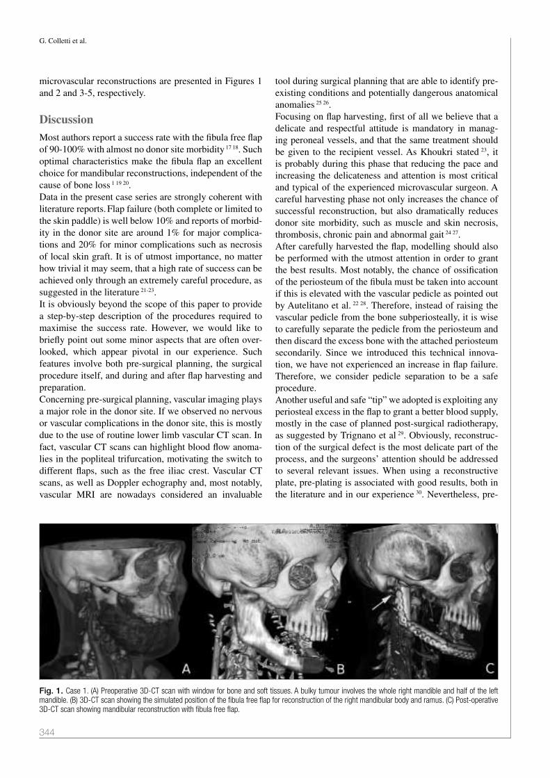
G. Colletti et al.
344
microvascular reconstructions are presented in Figures 1 and 2 and 3-5, respectively.
DiscussionMost authors report a success rate with the fibula free flap of 90-100% with almost no donor site morbidity 17 18. Such optimal characteristics make the fibula flap an excellent choice for mandibular reconstructions, independent of the cause of bone loss 1 19 20.Data in the present case series are strongly coherent with literature reports. Flap failure (both complete or limited to the skin paddle) is well below 10% and reports of morbid-ity in the donor site are around 1% for major complica-tions and 20% for minor complications such as necrosis of local skin graft. it is of utmost importance, no matter how trivial it may seem, that a high rate of success can be achieved only through an extremely careful procedure, as suggested in the literature 21-23.it is obviously beyond the scope of this paper to provide a step-by-step description of the procedures required to maximise the success rate. however, we would like to briefly point out some minor aspects that are often over-looked, which appear pivotal in our experience. Such features involve both pre-surgical planning, the surgical procedure itself, and during and after flap harvesting and preparation.Concerning pre-surgical planning, vascular imaging plays a major role in the donor site. if we observed no nervous or vascular complications in the donor site, this is mostly due to the use of routine lower limb vascular CT scan. in fact, vascular CT scans can highlight blood flow anoma-lies in the popliteal trifurcation, motivating the switch to different flaps, such as the free iliac crest. Vascular CT scans, as well as Doppler echography and, most notably, vascular Mri are nowadays considered an invaluable
tool during surgical planning that are able to identify pre-existing conditions and potentially dangerous anatomical anomalies 25 26.Focusing on flap harvesting, first of all we believe that a delicate and respectful attitude is mandatory in manag-ing peroneal vessels, and that the same treatment should be given to the recipient vessel. As Khoukri stated 23, it is probably during this phase that reducing the pace and increasing the delicateness and attention is most critical and typical of the experienced microvascular surgeon. A careful harvesting phase not only increases the chance of successful reconstruction, but also dramatically reduces donor site morbidity, such as muscle and skin necrosis, thrombosis, chronic pain and abnormal gait 24 27.After carefully harvested the flap, modelling should also be performed with the utmost attention in order to grant the best results. Most notably, the chance of ossification of the periosteum of the fibula must be taken into account if this is elevated with the vascular pedicle as pointed out by Autelitano et al. 22 28. Therefore, instead of raising the vascular pedicle from the bone subperiosteally, it is wise to carefully separate the pedicle from the periosteum and then discard the excess bone with the attached periosteum secondarily. Since we introduced this technical innova-tion, we have not experienced an increase in flap failure. Therefore, we consider pedicle separation to be a safe procedure.Another useful and safe “tip” we adopted is exploiting any periosteal excess in the flap to grant a better blood supply, mostly in the case of planned post-surgical radiotherapy, as suggested by Trignano et al 29. obviously, reconstruc-tion of the surgical defect is the most delicate part of the process, and the surgeons’ attention should be addressed to several relevant issues. When using a reconstructive plate, pre-plating is associated with good results, both in the literature and in our experience 30. nevertheless, pre-
Fig. 1. Case 1. (A) Preoperative 3D-CT scan with window for bone and soft tissues. A bulky tumour involves the whole right mandible and half of the left mandible. (B) 3D-CT scan showing the simulated position of the fibula free flap for reconstruction of the right mandibular body and ramus. (C) Post-operative 3D-CT scan showing mandibular reconstruction with fibula free flap.

Mandibular reconstruction with fibula free flap
345
plating is not feasible whenever the tumour has involved the surrounding soft tissues and or has deformed the origi-nal shape of the mandible. in these cases, a custom pre-modelled plate, CT custom osteotomy guides, or surgery simulators can offer the best reconstructive chances 31 32. however, the high cost of these instruments is an issue that still must be addressed, and we have been unable to implement them at our institution.if the resection involves the condylar process, it is impera-tive to correctly place the newly created condyle in the articular fossa. if this is not achieved, then a postoperative loss of occlusion is very likely to occur. We usually ad-here to the following “condyle position check” protocol: after resection, if the glenoid fossa is near and visible, a blind tentative positioning can be performed. having po-sitioned the neocondyle, the plates used for rigid fixation are secured and mandibular movements are checked. This procedure, however, can be tricky and even in the most experienced hands it is prone to failure. intraoperative
CT can be useful to verify the condyle position in dif-ficult cases. if intraoperative CT scan is not available, the glenoid fossa must be accessed surgically to validate the position of the condyle. Even if more sparing accesses to the condylar fossa are available 34 35, we believe that the most appropriate access to the glenoid fossa in major mandibular reconstructions is the preauricular access with or without endaural modification. once the glenoid fossa is reached a resorbable suture can be passed through the newly created condyle and the bony glenoid fossa by in-serting a hole in both bony stumps. The suture can finally be tightened to hold the new condyle in place.one last important consideration should be given to inter-maxillary fixation. We routinely use intermaxillary fixa-tion in order to maximise the precision of the reconstruc-tion. There are, however, many new techniques that may be employed to achieve intermaxillary fixation. While intermaxillary fixation screws are one of the most popular techniques, despite their routine use in mandibular trau-ma, we do not rely on them in major mandibular recon-structions since conventional arch bars are more precise where teeth contacts are less immediate and where more stability is needed.After having harvested and modelled the flap, most of the surgeons’ attention is devoted to reconstructing the surgi-cal defect, and donor site health if often jeopardised. For example, one patient in our series suffered from extensive necrosis of the skin and muscles around the donor site. This complication can be related to two major causes: pri-mary closure of the donor site with excess tension and local administration of anaesthetics at the donor site that may have masked the signs of compartmental syndrome. Aggressive debridement followed by free flap reconstruc-tion of the donor site skin defect can circumvent this the complication. We therefore strongly advise against ad-ministration of long-acting anaesthetics for control of post-operative pain.lastly, another interesting method that may be performed at the end of the procedure should be mentioned, namely bone banking. if a major part of the fibula is to be dis-carded, it is possible to bank the remaining bone at the harvesting site. in one of our patients, who required a second surgical procedure for pre-prosthetic purposes, we harvested the banked bone which had survived with mini-mal resorption. This issue needs further investigation, but it might prove useful, or prudent at least, to perform in any patient where a major bone discard is required. it is important to not mistake bone banking with flap prefab-rication prosed by nazerani et al. 33, the use of which we strongly discourage.in our experience, if all of the above steps are followed, a more predictable and precise outcome can be expected in mandibular reconstructions with free fibular flaps.
Fig. 2. Case 1. (A) Intra-operative view of the mandibulectomy specimen including the right condylar process (arrow). Intra-operative image showing the fibula free flap placed to reconstruct the mandible (small blue arrows) and stabilised with a reconstructive plate and screws (yellow arrow). Note the microvascular anastomosis (blue and red arrows).

G. Colletti et al.
346
Conclusion
While many reconstructive surgeons consider the fibula as the workhorse for mandibular reconstructions, we believe that many small details are often underlooked. however,
greater attention to a few crucial aspects can help the sur-geon to obtain a predictable result 36. Many new techniques, such as intraoperative CT scan and custom premodelled reconstructive plates, are powerful tools that can be of con-siderable value during the reconstructive phase.
Fig. 3. Case 2. (A) Frontal pre-operative picture showing severe facial asymmetry characterised by bulging in the right ramus and condylar region. (B) Frontal view of the patient 2 years after surgery showing the excellent symmetry of the face.
Fig. 4. Case 2. (A) Pre-operative CT scan showing the neoplasm, which extends through the whole right mandibular ramus and the posterior part of the body. (B) Post-operative 3D-CT scan showing microvascular mandibular reconstruction using a fibula free flap.

Mandibular reconstruction with fibula free flap
347
References1 hayden rE, Mullin DP, Patel AK. Reconstruction of the seg-
mental mandibular defect: current state of the art. Curr opin otolaryngol head neck Surg 2012;20:231-6.
2 Chiapasco M, Colletti g, romeo E, et al. Long term results of mandibular reconstruction with autogenous bone grafts and oral implants after tumor resection. Clin oral implants res 2008;19:1074-80.
3 Deganello A, gitti g, Parrinello g, et al. Cost analysis in oral cavity and oropharyngeal reconstructions with mi-crovascular and pedicled flap. Acta otorhinolaryngol ital 2013;33:380-7.
4 Tarsitano A, Vietti MV, Cipriani r, et al. Functional results of microvascular reconstruction after hemiglossectomy: free anterolateral thigh flap versus free forearm flap. Acta otorhinolaryngol ital 2013;33:274-9.
5 Taylor gi, Townsend P, Corlett r. Superiority of the deep cir-cumflex iliac vessels as the supply for free groin flaps. Plast reconstr Surg 1979;64:595-604.
6 Soutar DS, Scheker lr, Tanner nS, et al. The radial fore-arm flap: a versatile method for intraoral reconstruction. Br J Plast Surg 1983;36:1-8.
7 Swartz WM, Banis JC, newton ED, et al. The osteocutane-ous scapular flap for mandibular and maxillary reconstruc-tion. Plast reconstr Surg 1986;77:530-45.
8 Taylor gi, Miller gD, ham FJ. The free vascularized bone graft. A clinical extension of microvascular techniques. Plast reconstr Surg 1975;55:533-44.
9 hidalgo DA. Fibula free flap: a new method of mandible re-construction. Plast reconstr Surg 1989;84:71-9.
10 Daya M. Peroneal artery perforator chimeric flap: changing the perspective in free fibula flap use in complex oromandib-ular reconstruction. J reconstr Microsurg 2008;24:413-18.
11 o’leary MJ, Martin PJ, hayden rE. The neurocutaneous free fibula flap in mandibular reconstruction. otolaryngol Clin north Am 1994;27:1081-96.
12 Cutting C, Bookstein Fl, grayson B, et al. Three-dimension-al computer-assisted design of craniofacial surgical proce-dures: optimization and interaction with cephalometric and CT-based models. Plast reconstr Surg 1986;77:877-87.
13 hirsch Dl, garfein ES, Christensen AM, et al. Use of com-puter-aided design and computer-aided manufacturing to produce orthognathically ideal surgical outcomes: a para-digm shift in head and neck reconstruction. J oral Maxillofac Surg 2009;67:2115-22.
14 Sacco Ag, Chepeha DB. Current status of transport-disc-distraction ostegenesis for mandibular reconstruction. lan-cet oncol 2007;8:323-30.
15 rosson gD, Singh nK. Devascularizing complications of free fibula harvest: peronea arteria magna. J reconstr Mi-crosurg 2005;21:533-8.
16 young DM, Trabulsy PP, Anthony JP. The need for preopera-tive leg angiography in fibula free flap. J reconstr Microsurg 1994;10:283-7.
17 ho MW, Brown JS, Magennis P, et al. Salvage outcomes of free tissue transfer in Liverpool: trends over 18 years (1992-2009). Br J oral Maxillofac Surg 2012;50:13-8.
18 Salvatori P, Paradisi S, Calabrese l, et al. Patients’ survival after free flap reconstructive surgery of head and neck squa-mous cell carcinoma: a retrospective multicentre study. Acta otorhinolaryngol ital 2014;34:99-104.
Fig. 5. Case 2. (A) Intra-operative view of the right hemimandibulectomy specimen and (B) fibula flap before its position in the surgical site.

G. Colletti et al.
348
19 hanasono MM, Militsakh on, richmon JD, et al. Man-dibulectomy and free flap reconstruction for biphosphonate related osteonecrosis of the jaws. JAMA otolaryngol head neck Surg 2013;139:1135-42.
20 Deganello A, gitti g, Strijs B, et al. Palliative combined treatment for unresectable cutaneous basosquamous cell carcinoma of the head and neck. Acta otorhinolaryngol ital 2013;33:353-6.
21 Khouri rK. Avoiding free flap failure. Clin Plast Surg 1992;19:773-81.
22 Tarsitano A, Sgarzani r, Betti, et al. Vascular pedicle os-sification of free fibular flap: is it a rare phenomenon? Is it possible to avoid this risk? Acta otorhinolaryngol ital 2013;33:307-10.
23 Baj A, Beltramini gA, Demarchi M, et al. Extended-pedicle peroneal artery perforator flap in intraoral reconstruction. Acta otorhinolaryngol ital 2013;33:282-5.
24 ling XF, Peng X. What is the price to pay for a free fibula flap? A systematic review of donor-site morbidity following free fibula flap surgery. Plast reconstr Surg 2012;129:657-74.
25 Akashi M, nomura T, Sakakibara S, et al. Preoperative MR angiography for free fibula osteocutaneous flap transfer. Mi-crosurgery 2013;33:454-9.
26 Tarsitano A, Pizzigallo A, Sgarzani r, et al. Head and neck cancer in elderly patients: is microsurgical free-tis-sue transfer a safe procedure? Acta otorhinolaryngol ital 2012;32:371-5.
27 Colletti g, Autelitano l, Tewfik K, et al. Autonomized flaps in secondary head and neck reconstructions. Acta otorhi-nolaryngol ital 2012;32:329-35.
28 Autelitano l, Colletti g, Bazzacchi r, et al. Ossification of vascular pedicle in fibular free flaps: a report of four cases. int J oral Maxillofac Surg 2008;37:669-71.
29 Trignano E, Fallico n, Faenza M, et al. Free fibular flap with periosteal excess for mandibular reconstruction. Microsur-gery 2013, Aug 14 [Epub ahead of print].
30 Marchetti C, Bianchi A, Mazzoni S, et al. Oromandibu-lar reconstruction using a fibula osteocutaneous free flap: four different “preplating” techniques. Plast reconstr Surg 2006;118:643-51.
31 Stirling Craig E, yuhasz M, Shah A, et al. Simulated surgery and cutting guides enhance spatial positioning in free fibu-lar mandibular reconstruction. Microsurgery 2014, Jan 28 [Epub ahead of print].
32 Coppen C, Weijs W, Bergé SJ, et al. Oromandibular recon-struction using 3D planned triple template method. J oral Maxillofac Surg 2013;71:e243-7.
33 nazerani S, Behnia h, Motamedi Mh. Experience with the prefabricated free fibula flap for reconstruction of maxillary and mandibular defects. J oral Maxillofac Surg 2008;66:260-4.
34 Biglioli F, Colletti g. Mini-retromandibular approach to con-dylar fractures. J Craniomaxillofac Surg 2008;36:378-83.
35 Biglioli F, Colletti g. Transmasseter approach to condylar fractures by mini-retromandibular access. J oral Maxillofac Surg 2009;67:2418-24.
36 Pellini r, Mercante g, Spriano g. Step-by-step mandibular reconstruction with free fibula flap modeling. Acta otorhi-nolaryngol ital 2012;32:405-9.
Address for correspondence: giacomo Colletti, University of Mi-lano, Department of Maxillo-Facial Surgery, “San Paolo” hospital, via di rudinì 8, 20140 Milano, italy. Tel. +39 02 81844594. Fax +39 02 81844704. E-mail: [email protected]
received: February 4, 2014 - Accepted: october 6, 2014

349
ACTA oTorhinolAryngologiCA iTAliCA 2014;34:349-353
Vestibology
Association of cinnarizine and betahistine in prophylactic therapy for Ménière’s disease with and without migraineL’associazione di betaistina e cinnarizina nella profilassi degli episodi vertiginosi nella Sindrome di Ménière con e senza comorbidità emicranica
R. Teggi1, O. gaTTi1, V. SykOpeTRiTeS1, S. QuaglieRi2, M. BenazzO2, M. BuSSi11 Division of enT, San Raffaele Scientific institute, Milan, italy; 2 Division of enT, iRCCS policlinico San Matteo Foundation, university of pavia, italy
SummAry
Prophylactic therapy of ménière’s disease (mD) includes betahistine and calcium-blockers (the latter also useful for migraine prevention). The aim of our work was to assess the efficacy of combined therapy with cinnarizine and betahistine in mD subjects both with and without migraine and poorly responsive to betahistine alone. Fifty-two mD subjects were included who were poorly responsive to betahistine dur-ing 6 months of follow-up; 29 were migraineurs. Combined therapy was administered with betahistine 48 mg/day and cinnarizine 20 mg BiD for 1 month, 20 mg/day for 2 weeks and 20 mg every 2 days for 2 more weeks, and then repeated. results were collected over 6 months of follow-up. mD subjects with and without migraine demonstrated a decrease in both vertigo spells and migrainous attacks during combined therapy (from 9.4 to 3.8 and from 6.8 to 5.9 in 6 months, respectively, for vertigo spells, while migraine decreased from 3.8 to 1 in 6 months, respectively). A correlation was seen between decrease of vertigo spells and headaches in the sample of mD subjects with migraine. our data support a proactive role for cinnarizine in preventing vertigo spells, especially in mD patients with migraine.
Key worDS: Ménière’s Disease • Migraine • Therapy • Betahistine • Calcium-Blockers • Cinnarizine
riASSunTo
La betaistina e i calcio-antagonisti si sono dimostrati efficaci nella profilassi della Sindrome di Ménière; i calcio-antagonisti sono utilizzati anche nella prevenzione degli episodi di cefalea emicranica. Scopo del nostro lavoro è stato quello di stabilire l’efficacia della terapia com-binata con cinnarizina e betaistina nella prevenzione delle crisi vertiginose in un gruppo di pazienti affetti da Sindrome di Ménière senza e con comorbidità per emicrania. Cinquantadue pazienti affetti da Sindrome di Ménière, poco responsivi alla sola terapia con betaistina in un periodo di 6 mesi, sono stati inclusi nello studio, 29 dei quali emicranici. Nei 6 mesi successivi è stata effettuata terapia combinata con betaistina (48 mg al giorno) e cinnarizina 20 mg due volte al giorno per 1 mese, 20 mg al giorno per 2 settimane e 20 mg a giorni alterni per 2 ulteriori settimane; lo schema terapeutico è stato indi ripetuto. I dati relativi alla frequenza delle crisi vertiginose sono stati collezionati nei 6 mesi successivi. In entrambi i gruppi è stato dimostrato un decremento delle crisi vertiginose (da 9.4 a 3.8 in 6 mesi e da 6.8 a 5.9 in 6 mesi rispettivamente nel gruppo con e senza comorbidità per emicrania; le crisi di cefalea si sono inoltre ridotte da 3.8 a 1 in 6 mesi). È stata evidenziata una correlazione tra la diminuzione degli attacchi di vertigine ed emicrania. I nostri dati sottolineano un ruolo terapeutico della cinnarizina nella prevenzione degli attacchi di vertigine soprattutto nei soggetti con comorbidità emicranica.
PArole ChiAve: Sindrome di Ménière • Emicrania • Terapia • Calcio-antagonisti • Cinnarizina • Betaistina
Acta Otorhinolaryngol Ital 2014;34:349-353
Introductionménière’s disease is an inner ear disorder characterised by recurrent episodes of vertigo, hearing loss, fullness and tinnitus. increased endolymphatic pressure is commonly accepted as the pathogenetic mechanism 1, although ac-cording to some authors hydrops may be the consequence of a primitive damage of the inner ear 2.
Criteria for the diagnosis of definite mD, established in 1995 by the AAo-hnS, are mainly based on phenotype of episodic vertigo, consisting in the presence of at least two episodes of vertigo of at least 20 min, audiometrically confirmed sensorineural hearing loss on at least one occa-sion, tinnitus or aural fullness during episodes and exclu-sion of other possible causes of vertigo 3. Demonstrated

R. Teggi et al.
350
hearing loss in the vertigo-free period is not required for the diagnosis of definite mD.migraine is a neurological disorder characterised by epi-sodic headaches of pulsatile quality, often associated with phono- and photophobia, with a prevalence of 15-17% in women and 5-8% in men 4.epidemiological studies have shown an association be-tween mD and migraine, variously reported between 43% and 56% 5 6 and described by Prosper ménière himself 7. The high frequency of migraine in mD population may underline a pathophysiological link between the two disor-ders 8. recent papers have focused on increasing evidence that migraine per se may provoke episodic vertigo, and clinical entity is defined as vestibular migraine (vm) 9.Among other symptoms, diagnosis of mD relies more on audiometric findings, even though fluctuation of hear-ing level has also been reported in patients with vm 10. in some cases, at the initial stages of episodic vertigo, differ-ential diagnosis between mD and vm may be a puzzling dilemma 11.Betahistine has been demonstrated to be useful in preven-tion of episodic vertigo in mD 12 13, while calcium-block-ers are among the most widely used drugs in prophylactic therapy of migraine 14. Cinnarizine has also been studied as monotherapy for mD, but demonstrated a lower effi-cacy compared to betahistine in preventing vertigo spells in a sample of 36 mD subjects 15. A recent report focused on the possibility that nimodipine may increase efficacy of betahistine in prevention of vertigo spells in mD pa-tients 16.The aim of our investigation was to confirm the efficacy of combined prophylactic therapy with calcium-blocker and betahistine in mD subjects with and without comor-bidity for migraine, and to establish a possible correlation between reduction of both headache and vertigo spells in mD subjects with migraine.
Materials and methodsin our study we included 29 patients affected by definite ménière’s Disease according to AAo-nhS criteria also presenting a diagnosis of migraine according to ihS cri-teria and 23 patients with mD but without migraine. They were recruited among outpatients who attended the Cen-tre for vestibular disorders at San raffaele hospital in mi-lan and Policlinico San matteo in Pavia between January 2010 and July 2011.They were consecutively recruited if they completed 6 months’ follow-up among patients poorly responsive to monotherapy with betahistine 48 mg/day during the first 6 months. During the next 6 months, they underwent com-bined therapy with betahistine 24 mg BiD and cinnarizine 20 mg BiD for 1 month, then 20 mg/day for 2 weeks and 20 mg every 2 days for 2 further weeks; successively, they restarted cinnarizine 40 mg/day.
exclusion criteria were current exposition to noise, a middle ear disorder or previous intratympanic therapies with gen-tamycin or steroids. During the 12 months of the study, no further therapies for mD or migraine (e.g. diuretics, steroids, antiepileptics) or drugs active on the central nervous system (benzodiazepines and selective serotonin reuptake inhibitors) were given. however, dietary measures were suggested in all patients, especially increased fluids and reduced salt intake.Clinical history included the presence of a familial history of vertigo and an autoantibody screening (anti-nucleum, smooth muscle, thyroids, cardiolipin, lupus-like antico-agulant and b2-glycoprotein). in our sample, 4 patients referred a familial history of vertigo. in the group of pa-tients with migraine, 11 of 29 (38%) presented positivity for at least one of the autoantibodies (in 7 anti-thyroids), including 5 of 23 among those without migraine (22.7%).mean age at inclusion of the sample of migraineurs was 48.8 ± 9.6 years and 22 were females, while non-mi-graineurs presented with a mean age of 49.2 ± 7.6. no sta-tistical difference was detected between the two groups.The first attack of vertigo was noted at 36.9 ± 7.8 years of age in the sample of migraineurs and 40.9 ± 10.4 among non-migraineurs, with the first headache at the age of 28.9 ± 5.5 years. headache preceded the occurrence of vertigo in all subjects except one, in which they occurred in the same year. Ten patients referred that a vertiginous attack was followed by headache in at least two cases.Audiometric values were saved at the beginning of com-bined therapy and the 12 month control, and the mean value of pure tone audiometry (PTA) at 500, 1000, 2000 and 3000 hz at the beginning and at the end of combined therapy was calculated.main outcome was considered the number of spells dur-ing the 6 months before combined therapy and the fol-lowing 6 months. According to AAo-hnS guidelines, ef-ficacy was evaluated with the formula (x/y) x 100, where x is the average number of vertigo spells per month in the 6 months of combined therapy, and y the average number of spells per month in the 6 months of previous mono-therapy. The same formula was used to assess efficacy of therapy in controlling headaches. The lower the number, the more beneficial the therapy.
Statistical analysesThe significance of any difference in continuously distrib-uted variables between the two groups was examined by t-test for independent samples. Significance of non-nor-mal distributed values was assessed with a mann-whitney test. The chi-square test was used to assess differences for nominal values. Correlations were assessed with a Spear-man test.
Resultsresults and statistics of mD patients with migraine are

Prophylaxis of Ménière’s disease with betahistine and cinnarizine
351
summarised in Table i, while Table ii shows the results in the sample of subjects with mD without migraine.Among migraineurs, the value of the formula (x/y) x 100 for vertigo spells was lower than 50% in 13 cases, in the range between 50-75% in 3 more cases, while in 13 cases therapy demonstrated no efficacy. in this sample, seven of 11 subjects with positivity for autoantibodies, and 6 of 18 without positivity, presented a value lower than 50 (p = 0.1).The value of the formula (x/y) x 100 for headache attacks was below 50% in 17 cases, in the range between 50-75% in 2 cases and over 75% in 10 cases. nine of eleven sub-jects with positive autoantibodies and 8 of 18 remaining subjects presented a value lower than 50% (p = 0.05).Among non-migraineurs, the value of the formula (x/y) x 100 for vertigo spells was lower than 50% in only 1 subject, in the range between 50-75% in 3 cases, while non-responders was 19. A chi square test on the number of responders to the therapy demonstrated significant differ-ence between the 2 groups (p = 0.001). A mann-whitney test demonstrated a significant decrease of vertigo spells in migraineurs than in non-migraineurs after combined therapy (p = 0.003). Finally, in the sample of mD subjects with migraine, a correlation was seen between decrease of vertigo spells and headaches (p = 0.03).
DiscussionAs previously reported, migraine and mD often present with a comorbidity 17. moreover, around 51% of mi-graineurs suffer from vertigo or dizziness 18 19. in some cases, differential diagnosis between mD and vm is complicated 11, and relies mostly on audiometric exam 20. however, a fluctuating low-frequency hearing loss has also been described in vestibular migraine 10.Calcium-channel blockers have been demonstrated to be useful in migraine prophylaxis 21, and there is evidence of efficacy of antimigrainous drugs (including Ca2+-blockers) in preventing vertigo spells 22. Above all, cinnarizine has
been reported to ameliorate vestibular vertigo 23, and both cochlear and vestibular symptoms have been reported to improve in mD sufferers 23 24. Cinnarizine has long been believed to act through a direct block of voltage-gated Ca2+ currents 25. A recent work, however, suggests that its main action is an inhibition of K+ currents, which may be activated in case of endolymphatic hydrops 26. Flunarizine has also been shown to be effective in peripheral vertigo due to its calcium entry blocking properties provoking an increase in inner ear perfusion 27. A recent retrospective study reported that mD patients undergoing prophylactic therapy with an association of nimodipine and betahistine presented a decreased number of vertigo spells compared to patients receving betahistine alone 16.our study confirms the efficacy of the association of be-tahistine and a Ca2+blocker in both mD patients with and without migraine, although significantly better results were obtained in migraineurs. nonetheless, hearing loss progressed without beneficial effects in either group.Two possibilities should be considered to explain our findings. it has been suggested that the association of the two diseases could depend on a vascular alteration. vasos-pasm has long been considered a characteristic of some migraine-associated features (such as visual auras) 28. Some authors propose that migraine vasospasm causes ischaemic damage of small arteries of the inner ear, and endolymphatic hydrops develops on the previously dam-aged ear 29. mD, therefore, would complicate migraine 30.on the other hand, migraine may have an impact on fre-quency of mD attacks. mD patients refer increased spells in the catamenial period, similarly to when migraineurs experience headache more frequently 31. The occasional low-frequency hearing loss in young women suffering from migraine has also been described mainly during the menstrual period 31. in both pathogenic theories, Ca2+-blockers should be useful in preventing mD spells.A common underlying susceptibility to the two diseases may be explained by the importance of calcium and other ions in both disorders. ion channels in the inner ear are
Table I. Number of vertigo spells, headaches and PTA average (db HL) in 6 months of monotherapy and 6 months of combined therapy in the sample of MD patients with migraine (n = 29).
Betahistine Betahistine +cinnarizine
P value
Vertigo spells 9.4 ± 4.3 3.8 ± 3.4 p = 0.0001
Headaches 3 ± 1.9 1 ± 0.6 p = 0.0001
PTA mean value 45.7 ± 8 53.4 ± 7.8 p = 0.001
Table II. Number of vertigo spells, headaches and PTA average (db HL) in 6 months of monotherapy and 6 months of combined therapy in the sample of MD patients without migraine (n = 23).
Betahistine Betahistine + cinnarizine P value
Vertigo spells 6.8 ± 3.5 5.9 ± 3.9 p = 0.01
PTA mean value 50.5 ± 7.5 55.8 ± 9.2 p = 0.002

R. Teggi et al.
352
essential for the high potassium-concentration needed for endolymph maintenance. Both elevated and reduced con-centrations of Ca2+ have been shown to suppress transduc-tion currents 32 33. it has been reported that mice lacking Ca2+ channels suffer from delayed-onset hearing loss 34. moreover, experimentally induced endolymphatic hy-drops in guinea pigs was accompanied by increased Ca2+ level in the vestibular end-organ 35.This study, although performed on a small sample, under-lines the different effects of cinnarizine in mD patients with and without migraine; it also infers a different ac-tion of cinnarizine on vestibular and cochlear symptoms in mD. Further studies are needed to define the origin of this disparity.
Conclusionsour data confirm the efficacy of association of betahistine and cinnarizine in prophylaxis of mD, especially with co-morbidity with migraine; further studies should confirm if prevention of migraine in these subjects may play a role in reduction of vertigo spells.
Acknowledgementswe thank professor eugenio mira, Department of otolar-yngology, university of Pavia, italy, for his encourage-ment and suggestions.
References1 Sajjadi h, Paparella mm. Ménière’s disease. lancet
2008:372:406-14.2 merchant S, Adams J, nadol J. Pathophysiology of Ménière’s
syndrome: are symptoms caused by endolymphatic hydrops? otol neurotol 2005;26:74-81.
3 Committee on hearing and equilibrium. Guidelines for the diagnosis and evaluation of therapy in Ménière’s disease. otolaryngol head neck Surg 1995;113:181-5.
4 headache Classification Committee of the internation-al headache Society. The international classification of headache disorders, 3rd edition (beta version). Cephalalgia 2013;33:629-808.
5 rassekh Ch, harker lA. The prevalence of migraine in Mé-nière’s disease. laryngoscope 1992;102:135-8.
6 radtke A, lempert T, gresty mA, et al. Migraine and Mé-nière’s disease. neurology 2002;59:1700-4.
7 ménière P. Pathologie auriculaire: memoires sur une lesion de l’oreille interne donnant lieu a des symptoms de congestion cerebrale apoplectiforme. gaz med Paris 1861;16:597-601.
8 neff BA, Staab JP, eggers SD, et al. Auditory and vestibular symptoms and chronic subjective dizziness in patients with Ménière’s disease, vestibular migraine, and Ménière’s dis-ease with concomitant vestibular migraine. otol neurotol 2012;33:1235-44.
9 lempert T, olesen J, Furman J, et al. Vestibular migraine: diagnostic criteria. J vestib res 2012;22:167-72.
10 Battista rA. Audiometric findings of patients with migraine-associated dizziness. otol neurotol 2004;25:987-92.
11 Teggi r, Fabiano B, recanati P, et al. Case report on two patients with episodic vertigo, fluctuating hearing loss and migraine responding to prophylactic drugs for migraine. Mé-nière’s disease or migraine associated vertigo? Acta otorhi-nolaryngol ital 2010;30:217.
12 lezius F, Adrion C, mansmann u, et al. High-dosage beta-histine dihydrochloride between 288 and 480 mg/day in pa-tients with severe Ménière’s disease: a case series. eur Arch otolaryngol 2011;268:1237-40.
13 Della Pepa C, guidetti g, eandi m. Betahistine in the treatment of vertiginous syndromes: a meta-analysis. Acta otorhinolaryngol ital. 2006;26:208-15.
14 Silberstein SD, holland S, Freitag F, et al. Quality Stand-ards Subcommittee of the American Academy of Neurology and the American Headache Society. Evidence-based guide-line update: pharmacologic treatment for episodic migraine prevention in adults: report of the Quality Standards Sub-committee of the American Academy of Neurology and the American Headache Society. neurology 2012;78:1337-45.
15 Djelilovic-vranic J, Alajbegovic A, Tiric-Campara m, et al. Betahistine or Cinnarizine for treatment of Ménière’s dis-ease. med Arch 2012;66:396-8.
16 monzani D, Barillari mr, Alicandri Ciufelli m, et al. Effect of a fixed combination of nimodipine and betahistine versus betahistine as monotherapy in the long-term treatment of Ménière’s disease: a 10-year experience. Acta otolaryngol ital 2012;32:393-43.
17 ibekwe, TS., Fasunla JA, ibekwe Pu, et al. Migraine and Ménière’s disease: two different phenomena with frequent-ly observed concomitant occurrences. J natl med Assoc 2008;100:334-8.
18 vuković v, Plavec D, galinović i, et al. Prevalence of vertigo, dizziness, and migrainous vertigo in patients with migraine. headache 2007;47:1427-35.
19 Parker w. Ménière’s disease. Etiologic considerations. Arch otolaryngol head neck Surg 1995;121:377-82.
20 Boyev KP. Ménière’s disease or migraine? The clinical sig-nificance of fluctuating hearing loss with vertigo. Arch oto-laryngol head neck Surg 2005;131:457-9.
21 Snow v. weiss K, wall em, et al. Pharmacologic manage-ment of acute attacks of migraine and prevention of migraine headache. Ann intern med 2002;137:840-9.
22 Bikhazi P, Jackson C, ruckenstein mJ. Efficacy of antimi-grainous therapy in the treatment of migraine-associated dizziness. Am J otol 1997;18:350-4.
23 ganança mm, Caovilla hh, munhoz mS, et al. Optimizing the pharmacological component of integrated balance thera-py. Braz J otorhinolaryngol 2007;73:12-8.
24 novotný m, Kostrica r. Fixed combination of cinnar-izine and dimenhydrinate versus betahistine dimesylate in the treatment of Ménière’s disease: a randomized, double-blind, parallel group clinical study. int Tinnitus J 2002;8:115-23.
25 Arab SF, Düwel P, Jüngling e, et al. Inhibition of volt-age-gated calcium currents in type II vestibular hair cells by cinnarizine. naunyn Schmiedebergs Arch Pharmacol 2004;369:570-5.
Prophylaxis of Ménière’s disease with betahistine and cinnarizine
353
26 haasler T, homann g, Duong Dinh TA, et al. Pharmacologi-cal modulation of transmitter release by inhibition of pres-sure-dependent potassium currents in vestibular hair cells. naunyn Schmiedebergs Arch Pharmacol 2009;380:531-8.
27 olesen J Calcium antagonists in migraine and vertigo. Pos-sible mechanisms of action and review of clinical trials. eur neurol 1990;30(Suppl 2):31-4.
28 viirre e, Baloh rw. Migraine as a cause of sudden hearing loss. headache 1996;36:24-8.
29 Baloh rw. Neurotology of migraine. headache 1997;37:615-21.30 Cha yh, Kane mJ, Baloh rw. Familial clustering of mi-
graine, episodic vertigo, and Ménière’s disease. otol neuro-tol 2008;29:93-6.
31 Andrews JC, honrubia v. Premenstrual exacerbation of
Ménière’s disease revisited. otolaryngol Clin north Am 2010;43:1029-40.
32 ohmori h. Mechano-electrical transduction currents in isolated vestibular hair cells of the chick. J Physiol 1985;359:189-217.
33 Tanaka y, Asanuma A, yanagisawa K. Potentials of outer hair cells and their membrane properties in cationic environ-ments. hear res 1980;2:431-8.
34 Tabuchi K, Suzuki m, mizuno A, et al. Hearing impairment in TRPV4 knockout mice. neurosci lett 2005;382:304-8.
35 meyer zum gottesberge-orsulakova Am, Kaufmann r. Is an imbalanced calcium-homeostasis responsible for the experi-mentally induced endolymphatic hydrops? Acta otolaryngol 1986;102:93-8.
Address for correspondence: roberto Teggi, Division of enT, San raffaele Scientific institute, via olgettina 60, 20132 milan, italy. Tel. +39 02 26433522. Fax +39 02 26433508. e-mail: [email protected]
received: march 30, 2014 - Accepted: may 3, 2014

354
ACTA oTorhinolAryngologiCA iTAliCA 2014;34:354-361
Pediatric Otorhinolaryngology
Long-term surgical and functional outcomes of the intact canal wall technique for middle ear cholesteatoma in the paediatric populationRisultati funzionali e chirurgici nel lungo periodo nelle timpanoplastiche chiuse nella popolazione pediatrica affetta da colesteatoma dell’orecchio medio
S.C. PRASAD1, C. LA MELIA2, M. MEDINA1, V. VINCENTI2, A. BACCIU2, S. BACCIU2, E. PASANISI2 1 Department of Otology & Skull Base Surgery, Gruppo Otologico, Piacenza-Rome, Italy; 2 Department of Clinical and Experimental Medicine, Unit of Otolaryngology and Otologic Microsurgery University of Parma, Italy
SUMMAry
in this paper, we report the postoperative outcomes in canal wall up procedures with second stage surgery in 40 children undergoing interven-tion for cholesteatoma of the middle ear. The residuals, recurrences and the hearing results were analysed. All 40 patients had a follow-up of at least five years. of the 39 patients who underwent two staged surgery, 18 (46.1%) had a residual lesion that was identified and excised during the second surgery. over a five year follow-up period, there were five (12.5%) patients with recurrences, all belonging to the group in whom a residual cholesteatoma was identified during the second staged surgery. The rate of residual cholesteatoma tends to decrease as age increases. The type of cholesteatoma, acquired or congenital middle ear, were not statistically related to the incidence of residual chole-steatoma. hearing analysis showed that hearing recovery was excellent with canal wall up procedures and remained stable over five years.
KEy WorDS: Cholesteatoma • Paediatric • Canal Wall Up (CWU) • Second stage surgery • Ossiculoplasty • Recurrence • Residual • Hearing results • Follow up
riASSUnTo
In questo lavoro, abbiamo riportato i risultati nel post-operatorio di 40 bambini operati per colesteatoma dell’orecchio medio e sottoposti a timpanoplastica chiusa in 2 tempi. Colesteatoma residuo, ricorrente e risultati audiologici sono stati analizzati. Tutti e 40 i pazienti han-no avuto un follow-up di almeno 5 anni. In 39 pazienti sottoposti ad un secondo tempo, 18 (46,1%) hanno avuto un colesteatoma residuo che è stato identificato e rimosso durante il secondo tempo chirurgico. Nei 5 anni di follow-up, ci sono stati 5 (12,5%) pazienti con cole-steatoma ricorrente, tutti e cinque rientranti nel gruppo nel quale un colesteatoma residuo era stato individuato durante il secondo tempo chirurgico. Il tasso di colesteatoma residuo tende a ridursi con il progredire dell’età. Il tipo di colesteatoma, acquisito o congenito, non è statisticamente correlato all’incidenza di colesteatoma ricorrente. I risultati audiologici mostrano che il recupero uditivo è stato ottimo nelle timpanoplastiche chiuse e si è mantenuto stabile nei cinque anni.
PAROLE CHIAVE: Colesteatoma • Pediatrico • Timpanoplastica chiusa • Secondo tempo chirurgico • Ossiculoplastica • Ricorrente • Residuo • Risultati audiologici • Follow-up
Acta Otorhinolaryngol Ital 2014;34:354-361
IntroductionThe debate between intact canal wall or canal wall up (CWU) and canal wall down (CWD) mastoidectomy both in children and adults is on-going since the 1970s. This is-sue is pertinent because, in children, the effects of deafness can have social, educational and behavioural consequences, thereby necessitating the need for hearing preservation. Though CWU mastoidectomy achieves the important objec-tive of hearing preservation, two of its main drawbacks are recurrence of cholesteatoma due to limited intraoperative exposure and occurrence of postoperative retraction pockets due to a dysfunctional Eustachian tube (ET). This led some
surgeons to focus on disease elimination by CWD mastoid-ectomy at the cost of hearing. however, the drawbacks of CWU, namely recurrence and retraction, were addressed re-spectively by the addition of a second look surgery and po-sitioning of cartilage in the attic region to prevent retraction. This technique has gained favour among most surgeons for cholesteatoma in the paediatric population. Despite this, the CWU technique is not always a suitable option. it is not ad-visable to perform CWU in certain situations like extensive disease, disease in a well pneumatised mastoid, low lying dura, anterior placed sigmoid or a cleft palate, where CWD surgery is the surgery of choice 1.

Long-term surgical and functional outcomes of the intact canal wall technique for middle ear cholesteatoma in the paediatric population
355
it has been shown in various studies 1-4 that recurrences of cholesteatoma in CWU surgery normally take several years to manifest. Silvola et al. 2, reported that 92% of all their recurrences occurred within 5 years. Similar obser-vations have been made by other authors who advocate a minimum of a 5-year follow up 1-4. however, there have been very few studies in the past with a long-term follow- up. it is in this context that our series assumes importance as we present the results of 5 year follow-up in homog-enous study conditions.
Materials and methodsA retrospective chart review of patients operated for cho-lesteatoma at the EnT Department of the University of Parma, Italy from 2001 to 2005 was carried out. Of a total of 273 patients treated for CoM with cholesteatomas, 54 patients in the age group of two years to 16 years who underwent surgery for acquired or congenital middle ear cholesteatomas were identified. 14 patients with simple retraction pockets and patients with revision surgeries resulting from operations performed elsewhere were ex-cluded from the study. Two additional patients who un-derwent CWD procedures were excluded, resulting in a total of 40 patients operated by the CWU technique in the study population. otomicroscopy findings, preoperative pure tone air conduction (AC), bone conduction (BC), speech reception audiometry and tympanometry were noted. hrCT was done in all cases. otomicroscopy and pure tone audiometry findings in the 1st, 3rd, 6th 12th, 24th
months and at the 5th year were recorded. Pre- and post-operative pure tone averages (PTA) were calculated using thresholds at 500, 1000, 2000 and 3000 hz. residual cho-lesteatoma at second stage surgery and recurrences dur-ing the follow-up period were recorded. residual disease was defined as persistence of disease due to incomplete removal revealed during second stage surgery. recur-rent cholesteatoma was defined as a newly formed dis-ease process secondary to a retraction pocket after second stage surgery.
Surgical technique A postauricular approach was used in all patients. Mastoidectomy was performed preserving the poste-rior canal wall. A posterior tympanotomy was done and the facial recess was opened. The cholesteatoma sac was identified and excised using a combined ap-proach. A silastic sheet was placed in the middle ear and antrum followed by reconstruction of the tympan-ic membrane by temporalis fascia. Bone patè in case of small defects or homologous rib cartilage5 for large defects was used to reconstruct the scutum. A second surgery was planned after 6-12 months to discover any residual or recurrent cholesteatoma. The silastic sheet was removed and ossicular reconstruction was done
using autologous incus. When the incus was absent, a homologous rib cartilage was used.
Reconstruction When the stapes was present and mobile, a partial ossicu-lar cartilage prosthesis (PORP) was sculpted. At the end of the shaft, an indentation (1 mm wide and 0.5-1 mm in depth) was created with a diamond burr in order to ac-commodate the capitulum of the stapes. The head of the prosthesis was placed in contact with the tympanic mem-brane or the graft used for its repair. When the malleus was present, the prosthesis was placed parallel to it and a groove was created to accommodate the handle. When the stapes superstructure was absent, a total ossicular car-tilage prosthesis (TORP) was used. In this case, the shaft was longer than in a PORP but the head of the T shaped prosthesis was identical. The end of the shaft was placed in contact with the footplate and the head was placed in contact with the tympanic membrane or the graft used for its repair.5
Statistical analysisData was analysed with a statistical software programme (SPSS Statistics for Windows version 20, Chicago, IL). Continuous data was summarised as mean ± interval of confidence at 95% (Ci). Categorical data was presented as frequencies and percentages. Factors related to the rate of recidivism were estimated with univariate analysis. Pre-operative and postoperative hearing results were evalu-ated for the entire sample and the acquired and congenital middle ear cholesteatoma groups. P values below 0.05 were considered statistically significant.
Results 40 patients fitted into this study criteria of CWU proce-dures for paediatric cholesteatomas. 31 (77.5%) patients had acquired cholesteatoma and 9 (22.5%) had congenital middle ear cholesteatoma. 24 (60%) were males and 16 (40%) were females, with an average age of 10.10 years (95% Ci 8.83-11.37; range 2-16 years) at the time of sur-gery. The age distribution is shown in Figure 1.
Clinical findings The most common symptoms were otorrhoea seen in 34 (85%) of patients, followed by hearing loss in 19 (47.5%) patients. otorrhoea and hearing loss coexisted in 15 (37.5%) patients. According to Zini & Sanna’s classifica-tion, the cholesteatoma was localised in the mesotympa-num (type A) in 20 (50%) patients, in the epitympanum (type B) in 10 (25%) patients, in the mesoepitympanum (type AB) in one (2.5%) patient and retrotympanum (con-genital middle ear) in nine (22.5%) patients (Fig. 2). In two (5%) patients a polyp was found in the external audi-tory canal. otomicroscopy in the contralateral ear showed

S.C. Prasad et al.
356
a retraction pocket in the tympanic membrane in 13 (32.5%) patients, middle ear effusion in 2 (5%), atelecta-sis of the middle ear in 3 (7.5%), a large tympanosclerosis in one (2.5%) and a tympanic membrane perforation in one (2.5%) patient.
Surgical procedure All 40 patients underwent CWU surgery. no labyrinthine fistulae were detected intraoperatively in any patient. The cholesteatoma was found to be diffuse in 30 (75%) and encapsulated in 10 (25%) cases. in all but one patient, CWU mastoidectomies were performed in two stages. in the remaining case, CWU was performed in a single stage owing to the small size of the cholesteatoma and preservation of the ossicular chain. in 22 (55%) patients, the scutum was found to be eroded and was reconstructed
with bone patè or homologous rib cartilage. ossicles were found to be eroded in 39 (97.5%) patients and were re-constructed in all. homologous rib cartilage was used in 21 (53.8%) patients, autologous malleus in 4 (10.3%) and incus in 14 (35.9%).
Recidivism in the second stage surgery of the 39 patients who underwent a second stage surgery, 18 (46.1%) had a residual lesion. in eight (44.4%) patients the cholesteatoma appeared as a single pear shaped le-sion, in seven (38.9%) as multiple pear shaped lesions and in three (16.7%) as a diffuse lesion. in three of the 18 (16.7%) patients, ossicular reconstruction was postponed to a third stage after another 6-12 months due to infiltra-tive disease. of nine patients with congenital middle ear cholesteatomas, four (44.4%) had a residual cholesteato-ma found during second staged surgery. The clinical type of cholesteatoma (acquired vs. congenital middle ear) and histological type (encapsulated vs. diffuse) were not statistically related to the incidence of residual cholestea-toma (p = 0.85 and 0.24 respectively, chi-square test). The average age in the group of patients that had a residual lesion was 8.72 years (95% Ci 6.80-10.70), and 11 years (95% Ci 9.30-12.79) (p = 0.06, U Mann-Whitney test) in the group that did not have a residual lesion. This dif-ference, although not statistically significant, is worthy of mention as this was the only factor closest to achiev-ing significance among all factors analysed in relation to the incidence of residual lesion. Moreover, when patients were categorized by age into three groups (≤5 years old, 6 and 10 years and ≥ 11 years), the rate of residual cho-lesteatoma was 66.7%, 46.7% and 38.9%, respectively (p>0.05 Fisher’s exact test).
Follow-up All patients were followed-up for at least five years. The mean follow-up was 84 months (range 60-120 months). Eight (20%) patients had a 10 year follow-up, 24 (60%) between six and nine years and eight (20%) patients for five years. in the follow-up period, there were five (12.5%) patients with recurrences, all belonging to the group in whom a residual cholesteatoma was identified and removed during the second staged surgery. of these, one was converted into a CWD mastoidectomy, while in another case, due to extensive pathology and erratic fol-low up, a radical mastoidectomy was done. The cavity was obliterated with fat and a cul-de-sac closure of EAC was performed. in the only patient who underwent a single stage CWU mastoidectomy, a suspicious residual cholesteatoma was detected after 2 years and the patient underwent a second stage surgery. The cholesteatoma was confirmed to be near the stapes, which was removed.The average duration of onset of recurrence was 2.5 years after surgery (range 1-4 years). There were no recurrences in the group of patients who were disease-free during the
Fig. 2. Localisation of cholesteatomas according to the Zini-Sanna clas-sification.
Fig. 1. Age distribution of paediatric patients with cholesteatomas.

Long-term surgical and functional outcomes of the intact canal wall technique for middle ear cholesteatoma in the paediatric population
357
second staged surgery. Three patients developed a retrac-tion pocket, one developed an atelectasis and three pa-tients developed a glue-ear. There were no cases of perfo-rations of the tympanic membrane.
Hearing outcomes Preoperatively, pure tone audiometry could not be con-ducted in two patients because one suffered from Down’s syndrome and the other was too young (two years) at the time of surgery. Pre- and postoperatively pure tone audio-grams of 38 patients with CWU mastoidectomies with a follow-up of five years were available and analysed. of the 38 patients, six were excluded from further study be-cause one suffered from Down’s syndrome and five had recurrent cholesteatoma after the second stage surgery. in addition, the audiogram of one patient was incomplete and therefore excluded. Consequently, pre- and postop-erative audiograms of 31 patients were compared. no pa-tients developed a dead ear postoperatively.
Hearing results in the whole sampleThe mean air conduction (AC), bone conduction (BC) and air bone gap (ABg) noted preoperatively, six months and five years postoperatively are shown in Table i and Figure 3. Postoperatively, AC improved significantly from 36.21 dB to 28.06 dB (p = 0.02, Wilcoxon test). This result remained significant at five years of follow-up (p=0.006). There was also a significant improvement between preoperative ABg (23.53 dB) and postoperative ABg at 6 months (17.24 dB) (p = 0.01). This improve-ment consolidated at five years of follow-up and remained significant (p = 0.001). Mean preoperative BC was 12.47 dB and remained unchanged postoperatively at six months (p = 0.75) and five years (p = 0.43) after surgery. Preoperatively, the average ABG was < 10 dB in four (10.5%) patients, between 11 and 20 dB in nine (26.3%) patients, between 21 and 30 dB in 10 (28.9%) patients and > 30 dB in 11 (34.2%) patients. Postoperatively at six months follow-up, ABG was < 10 dB in 12 (34.3%) patients, between 11 and 20 dB in 10 (28.6%) patients, between 21 and 30 dB in seven (20%) patients and > 30 dB in six (17.1%) patients. At five years of follow-up, the ABG was < 10 dB in 10 (32.3%) patients, between 11 and 20 dB in nine (29%) patients, between 21 and 30 dB in 8 (25.8%) patients and > 30 dB in four (12.9%) patients (Fig. 4).
Hearing results in acquired cholesteatomaThe mean AC, BC and ABg preoperatively, and postop-eratively at six months and five years in patients with ac-quired cholesteatoma are shown in Table ii. The mean AC improved from 34 dB before surgery to 28 dB at 6 months and at 5 years (p = 0.07, U Mann-Whitney test). The re-duction in ABg was statistically significant from a mean ABg of 22 dB before surgery to 17 dB at 6 months and 5 years (p = 0.007). Mean preoperative BC, which was 12 dB, remained unchanged postoperatively at six months (p = 0.13) and five years (p = 0.22).
Hearing results in congenital middle ear cholesteatomaThe mean AC, BC and ABg preoperatively and postoper-atively at six months and five years for patients with con-genital middle ear cholesteatoma are shown in Table iii. Among the nine patients with congenital middle ear cho-lesteatomas, one patient suffering from Down’s syndrome was non-compliant. in the remaining patients, there was significant improvement between preoperative AC (mean PTA of 42.50) and postoperative AC (mean PTA of 25.63 dB) at six months (p = 0.042, Wilcoxon test). The re-duction in ABg from 27.96 dB preoperatively to 17 dB postoperatively at six months was statistically significant (p = 0.006). Both results were statistically significant at
Table I. Hearing outcomes in middle ear cholesteatomas after CWU surgery preoperatively, 6 months and 5 years of follow-up.
Mean PTA (95% CI) PreoperativePostoperative
Follow-up at 6 months Follow-up at 5 years
Air conduction 36.21 (26.96-42.45) 28.06 (23.23-32.89) 26.46 (21.66-31.26)Bone conduction 12.47 (9.41-15.54) 10.80 (9.06-12.54) 10.43 (8.21-12.64)Air bone gap 23.53 (18.99-28.08) 17.24 (12.69-21.80) 16.12 (11.91-20.32)
Fig. 3. Box-plot showing median AC, BC and ABG according to follow-up. Preoperatively (PRE), 6 months after surgery (6M) and at 5 years follow-up (5Y).

S.C. Prasad et al.
358
five years follow up (p = 0.028 and p = 0.028 respective-ly). Mean preoperative BC, which was 13 dB, remained unchanged postoperatively at six months (p = 0.86) and five years (p = 0.22). There were no statistically significant differences in the preoperative AC, BC and ABg between acquired and congenital middle ear cholesteatoma groups (p > 0.05, U Mann-Whitney test). however, in the congenital group, the improvement in AC (20.20 dB, 95% Ci 28-12) and ABg (-14.20 dB 95% Ci, -21 -[-7.5]) at five years was better than the AC (7 dB, 95% Ci 15-[-1]) and ABg (-5.6 dB 95% Ci, -11 -[-0.33]), in the acquired group p=0.017 and p=0.029, respectively (U Mann-Whitney test).
Discussion The incidence of cholesteatoma in childhood is estimat-ed to be 3-6 per 100,000 individuals 6. Various arguments dictate the surgical choice in paediatric cholesteatoma. While some authors 7-10 consider that cholesteatoma is a more aggressive in children with a higher percentage of recidivism than in adults and hence warrants more radi-cal surgery, others 11 12 are of the opinion that the behav-iour of the cholesteatoma is the same in children as in
adults. The second disagreement is that of CWU versus CWD surgery in dealing with paediatric cholesteatomas. While it is important to preserve hearing in the child, which plays a major role in social and educational inte-gration after surgery, it is even more important to eradi-cate the disease without complications. The introduc-tion of the staged procedure in CWU, use of cartilage for reconstructing the attic to prevent postoperative re-tractions and advances in ossicular reconstruction have helped surgeons to tackle cholesteatomas in the paediat-ric population with a CWU technique, thereby preserv-ing hearing and eliminating the risk of recurrence. Fur-thermore, studies using DW-non-EPI and DWI propeller sequences show promising results in improved diagnos-tic sensitivity and specificity for even small (< 5 mm) cholesteatomas, thus allowing avoidance of second-look surgery in the future 13-16.
Principles behind surgery We prefer a CWU mastoidectomy with a planned second stage surgery as the primary option. A CWD is used in patients with extensive cholesteatoma, difficult anatomy, cleft palate and unsure follow up. The second stage sur-
Table II. Hearing outcomes in acquired cholesteatoma.
PTA results Preoperative PostoperativeFollow-up 6 months Follow-up 5 years
PTA AC 34 (27-41) 29 (23-34) 28 (22-33)PTA BC 12 (9-16) 12 (10-13) 11 (8- 13)ABG 22 (17-28) 17 (12-22) 17 (12-22)
Table III. Hearing outcomes in congenital middle ear cholesteatoma.
PTA results Preoperative PostoperativeFollow-up 6 months Follow-up 5 years
PTA AC 42.50 (31.97-53.03) 25.63 (11.02-40.23) 22.29 (12.70-31.87)PTA BC 13.00 (7.65-18.35) 7.42 (1.29-13.54) 8.33 (2.91-13.75)ABG 27.96 (20.10-35.82) 17.01 (11.80-22.22) 13.75 (4.32-23.17)
Fig. 4. Bar diagrams showing the number of patients in each ABG group before surgery, at 6 months follow-up, and at 5 years follow-up.

Long-term surgical and functional outcomes of the intact canal wall technique for middle ear cholesteatoma in the paediatric population
359
gery is planned 6-12 months after the 1st surgery. This is because the healing process of a middle ear with active infection, granulation tissue, and/or areas of absent mu-cosa will be variable. Formation of adhesions between the medial wall of the mesotympanum and the tympanic membrane or the ossicular implant can promote tympanic membrane retraction and/or implant displacement 17. To avoid this, we place a silastic sheet in the middle ear which remains until the time of the second stage surgery, when it is removed and ossicular reconstruction is performed. if residual cholesteatoma is present and is a small pearl, we remove it and continue with ossicular reconstruction in the same session. if it is found to be extensive, or in the presence of infection, the cholesteatoma is removed and the ossiculoplasty is postponed to another session. We do not hesitate to convert the CWU into a CWD as and when necessary.in assessing outcomes in our series, we agree with previ-ous authors that the total percentage of cholesteatoma re-cidivism should be the basis for evaluation of the outcome of surgery 2. We defined residual disease as persistence of disease after incomplete removal as revealed during second stage surgery. recurrent cholesteatoma was de-fined as a newly formed disease process secondary to a retraction pocket after second stage surgery. it is unlikely that a good surgeon will leave behind (residual) cholestea-toma after two surgeries. This differentiation is crucial in reporting data. We compared our study to those of other authors, and the results are summarised in Table IV. In our series, as in most others, the residual cholesteatoma was identified during the second stage procedure mostly in the form of a white pearl that could be removed easily. The recurrent cholesteatoma presented to us between a minimum of one year and a maximum of five years, possi-bly due to poor Eustachian tube function. The age differ-ence between patients who had a residual lesion (average 8.72 years), and those who did not (average 11 years) was
close to significance (p = 0.06). it is also important to note that the rate of residual cholesteatomas decreases as the age progresses [(66.7% (≤ 5 years), 46.7% (6-10 years) and 38.9% (≥ 11)], respectively (p > 0.05 Fisher’s exact test). This may point to the fact that younger patients may indeed have a more aggressive disease. The reported rate of residual cholesteatoma in closed procedures is between 19-63.5% and that of recurrent cholesteatoma is between 7.5-22% 17-26. While none of our patients who did not have a residual cholesteatoma identified at the second stage went on to develop a recurrence, Schraff et al. 27 reported a 1% recurrence rate in such a category. A period of five years of follow-up is essential for all children undergoing CWU surgery.
Hearing outcomes our analysis of the hearing outcomes validates the fact that CWU procedures bring about a significant improve-ment in AC that is statistically significant. Moreover, this improvement is consolidated at five years and remains sta-tistically significant. likewise, the improvement in ABg is also maintained at five years. Patients with congenital middle ear cholesteatoma tend to have similar preopera-tive AC, BC and ABg than with acquired cholesteatoma. however, at 5 years, the postoperative improvement in hearing was superior in congenital middle ear cholestea-toma patients compared to those with acquired cholestea-toma. Studies indicate that the status of the ossicles, their recon-struction and the type of surgical technique are important factors for postoperative hearing restoration in cholestea-toma surgery. incus transposition is most frequently used for reconstructing an ossicular discontinuity. however, cholesteatoma usually causes significant destruction of ossicles, especially the incus, so that other materials must be considered 28. in the absence of the incus, we routinely use rib cartilage for the reconstruction of the attic and the
Table IV. Comparison of residual and recurrence cholesteatoma in our series with other authors.
Authors # patients Residual % Recurrent % Mean follow-up (years)closed open closed open
Dodson et al. 17 66 19 12 22 0 9.2Mutlu et al. 18 83 38 11 11 0 4Sanna et al. 19 151 40 50 11 25 1.5 Schimd et al. 20 57 24 7.5 12 7.5 7 Magnan et al. 21 210 26 0 19.5 0 NATriglia et al. 22 80 41 35 16 8 4Desaulty et al. 23 80 63.5 0 7.5 0 2.5Roger et al. 24 199 54 72 19 0 2.5Lerosey et al. 25 57 26 28 12 12.5 7Charachon et al. 26 160 31 38 20 0 NAOur series 43 45 0 12 0 8NA=Not available

S.C. Prasad et al.
360
ossicles. Because of their flexibility and elasticity, carti-lage prostheses rarely damage the stapes or the footplate during the surgical maneuver 5.As stated by Dodson et al. 26, serviceable hearing depends primarily on middle ear parameters such as mucosal sta-tus (stenosis and granulation), condition of the TM (thick-ness, contour), depth of the middle ear cleft and presence or absence of stapes suprastructure erosion. Mutlu et al. 18 demonstrated a ≤ 25 dB hearing loss in 85% of those with an intact stapes supra structure. historically, worse hear-ing results have often been cited as a criticism of CWD but several studies dispute this notion 29-31. Schraff et al. 27 showed that those undergoing CWD have worse hearing pre-operatively and less improvement post-operatively compared with children using the iCW approach. This may be a result of more aggressive disease rather than failure in technique. nevertheless, CWU surgeries pro-vide a distinct advantage due to the fact that children do not need to worry about mastoid cavities, can swim and may benefit from a good fitting of a hearing aid when nec-essary.
ConclusionsA two-staged CWU mastoidectomy is the surgery of choice for paediatric cholesteatomas. Placing a silastic sheet to promote regeneration of mucosa with ossiculoplasty in the second stage and reconstruction of the scutum to avoid re-traction have made CWU procedures an effective option in dealing with paediatric cholesteatomas. hearing analysis showed that hearing recovery was excellent with canal wall up procedures and remained stable over five years. There is a higher incidence of recurrence during long-term follow-up in patients in whom residual cholesteatoma was detect-ed during second stage surgery. The rate of residual chole-steatoma tends to decrease as the age increases. Follow-up for a minimum of five years is recommended in all patients with paediatric cholesteatoma.
References1 Abramson M. Open or closed tympanomastoidectomy for
cholesteatoma in children. Am J otol 1985;6:167-9. 2 Silvola J, Palva T. Long-term results of pediatric primary
one-stage cholesteatoma surgery. Int J Pediatr Otorhi-nolaryngol 1999;48:101-7.
3 Kuo Cl, Shiao AS, liao Wh, et al. How long is long enough to follow up children after cholesteatoma surgery? A 29-year study. laryngoscope 2012;122:2568-73.
4 Yung M, Jacobsen NL, Vowler SL. A 5-year observational study of the outcome in pediatric cholesteatoma surgery. otol neurotol 2007;28:1038-40.
5 Quaranta N, Fernandez-Vega Feijoo S, Piazza F, et al. Closed tympanoplasty in cholesteatoma surgery: long-term (10 years) hearing results using cartilage ossiculoplasty. Eur Arch otorhinolaryngol 2001;258:20-4.
6 harker lA. Cholesteatoma: an incidence study. in: Sohet JA, De Jong Al, eds. The management of pediatric cholestea-toma. otolaryngol Clin north Am 2002;35:841-51.
7 Glasscock ME, Dickins JFE, Wiet R. Cholesteatoma in chil-dren. laryngoscope 1981;91:1743-53.
8 Tos M. Treatment of cholesteatoma in children. A long-term study of results. Am J otol 1983;4:189-97.
9 Palva A, Karma P, Karja J. Cholesteatoma in children. Arch otolaryngol 1997;103:74-7.
10 Bujia J, Holly A, Antoli-Candela F, et al. Immunobiologi-cal peculiarities of cholesteatoma in children: quantifica-tion of epithelial proliferation by MIB1. laryngoscope 1996;106:865-8.
11 Sheehy Jl. Recurrent and residual disease in cholesteatoma surgery. Clin otolaryngol Allied Sci 1978;3:393-403.
12 Sanna M, Zini C, gamoletti r, et al. The surgical man-agement of childhood cholesteatoma. J laryngol otol 1987;101:1221-6.
13 Baráth K, Huber AM, Stämpfli P, et al. Neuroradi-ology of cholesteatomas. AJnr Am J neuroradiol 2011;32:221-9.
14 Schwartz KM, lane Ji, Bolster BD Jr, et al. The utility of diffusion weighted imaging for cholesteatoma evaluation. AJnr Am J neuroradiol 2011;32:430-6.
15 Dremmen MH, Hofman PA, Hof JR, et al. The diagnostic accuracy of non-echo-planar diffusion-weighted imag-ing in the detection of residual and/or recurrent chole-steatoma of the temporal bone. AJnr Am J neuroradiol 2012;33:439-44.
16 De Foer B, Vercruysse JP, Bernaerts A, et al. The value of single-shot turbo spin-echo diffusion-weighted MR imaging in the detection of middle ear cholesteatoma. neuroradiol-ogy 2007;49:841-8.
17 Dodson EE, hashisaki gT, hogbood TC. Intact canal wall down mastoidectomy with tympanoplasty for cholesteatoma in children. laryngoscope 1998;108:977-83.
18 Mutlu C, Khashaba A, Saleh E, et al. Surgical treatment of cholesteatoma in children. otolaryngol head and neck Surg 1993;113:56-60.
19 Sanna M, Zini C, Bacciu S, et al. Surgery for cholesteato-ma in children. In: Tos M. ed. Cholesteatoma and mastoid surgery. Amsterdam, Berkeley, Milano: Kugler & ghedini; 1989. pp. 685-8.
20 Schimd H, Dort JC, Fisch U. Long-term results of treatment of children’s cholesteatoma. Am J otol 1991;12:83-7.
21 Magnan J, Chays A, Florence A, et al. L’eradication du cho-lesteatome chez l’enfant. J Fr ORL 1992;41:418-26.
22 Triglia JM, gillot JC, giovanni A, et al. Les cholesteatome de l’oreille moyenne chez l’enfant: a propos de 80 obser-avtion et revue de la letterature. Ann Otolaryngol (Paris) 1993;110:437-43.
23 Desaulty A, Masteau l, nguyen KT, et al. Le cholesteatome de l’oreille moyenne chez l’enfant: a propos de 127 cas. Ann otolaryngol (Paris) 1994;111:371-6.
24 Roger G, Tashjian G, Roelly P, et al. Poches de retraction fix-ees et cholesteatomes de l’enfant: notre experience a propos du 199 cas. Ann Otolaryngol (Paris) 1994;111:103-9.
25 Lerosey Y, Andrieu-Guitrancourt J, Marie JP, et al.
Long-term surgical and functional outcomes of the intact canal wall technique for middle ear cholesteatoma in the paediatric population
361
Le cholesteatome de l’oreille moyenne chez l’enfant: criteres du choix chirurgical. Ann Otolaryngol (Paris) 1998;115:215-21.
26 Charachon r. Surgery of cholesteatoma in children. J laryn-gol otol 1988;102:680-4.
27 Schraff SA, Strasnick B. Pediatric cholesteatoma: a retrospective review. Int J Pediatr Otorhinolaryngol 2006;70:385-93.
28 Stankovic M. Follow-up of cholesteatoma surgery: open
versus closed tympanoplasty. orl J otorhinolaryngol relat Spec 2007;69:299-305.
29 Roden D, Honrubia VF, Weit R. Outcome of residual cho-lesteatoma and hearing in mastoid surgery. J otolaryngol 1996;25:178-81.
30 hirsch BE, Kamerer DB, Doshi S. Single-stage management of cholesteatoma. otolaryngol head neck Surg 1992;106:351-4.
31 Toner Jg, Smyth gD. Surgical treatment of cholesteatoma:a comparison of three techniques. Am J otol 1990;11:247-9.
Address for correspondence: Claudio la Melia, Department of Clinical and Experimental Medicine, Unit of otolaryngology and Otologic Microsurgery University of Parma, via Volturno, 43125 Parma, Italy. Tel. +39 0521 033608. Fax +39 0521 290447. E-mail: [email protected]
received: June 6, 2013 - Accepted: February 13, 2014

362
ACTA oTorhinolAryngologiCA iTAliCA 2014;34:362-367
Clinical techniques and technology
Hyoid myotomy without suspension: a surgical approach to obstructive sleep apnoea syndromeMiotomia del muscolo ioideo senza sospensione: un approccio chirurgico per la sindrome delle apnee ostruttive nel sonno
E. Scarano1, G. DElla Marca2, E. DE corSo1, S. Dittoni1, W. Di narDo1, D. MEucci1, G. BaStanza1, r. GalluS1, a. loSurDo2, E. tEStani2, G. PaluDEtti11 institute of otorhinolaryngology, catholic university rome, italy; 2 Department of neurosciences, catholic university rome, italy
SummAry
The aim of this study was to verify if hyoid myotomy without hyoid suspension is effective in surgical treatment of obstructive sleep apnoea syndrome (oSAS). We recruited six patients with oSAS, aged between 34 to 60 years, with retropalatal and retrolingual upper airway ob-struction, non-obese (Bmi < 27) and non-compliant to continuous positive airway pressure therapy. Pre-surgical clinical and instrumental evaluations included clinical examination, cephalometry, polysomnography (PSg) and sleep endoscopy. Surgical treatment included nasal surgery, uvulopalatopharyngoplasty, tonsillectomy and hyoid myotomy without hyoid suspension. Follow-up evaluations were performed with serial PSgs, performed early (one week after surgery), and at 1, 6 and 18 months after surgery. We observed that surgery was fol-lowed by immediate normalisation of breathing parameters evaluated by PSg that persisted after 18 months. Thus, hyoid myotomy without suspension combined with nasal and palatal surgery may be considered a valid treatment of non-obese oSAS patients with retrolingual and retropalatal collapse. Furthermore, we suggest that hyoid bone suspension, binding it to mandibular or to thyroid cartilage, might be unnecessary in selected cases.
Key WordS: Obstructive sleep apnoea syndrome • Hyoid myotomy • Uvulopalatopharyngoplasty • Sub-hyoid muscles
riASSunTo
Obiettivo di questo studio è stato verificare se la miotomia ioidea senza sospensione è efficace nel trattamento chirurgico della sindrome delle apnee ostruttive del sonno (OSAS) nell’ambito del primo step della chirurgia multilivello. Abbiamo reclutato sei pazienti affetti da OSAS, di età compresa tra i 34 e i 60 anni, con un’ostruzione delle alte vie aeree a livello retropalatale e retrolinguale, non obesi (BMI <27) e che mostravano scarsa tolleranza nei confronti della terapia con ventilazione meccanica a pressione positiva continua delle alte vie aeree. Durante la valutazione clinica pre-intervento i pazienti sono stati sottoposti a esame clinico otorinolaringoiatrico, cefalometria, polisonnografia (PSG) e sleep-endoscopy. Tutti i pazienti sono stati sottoposti nella stessa seduta a chirurgia nasale, uvulopalatofaringo-plastica, tonsillectomia e miotomia ioidea senza sospensione. Nel corso del follow-up i pazienti sono stati sottoposti a PSG seriali, all’i-nizio (una settimana dopo l’intervento chirurgico), ed a 1, 6 e 18 mesi dopo l’intervento chirurgico. I dati polisonnografici hanno messo in evidenza un significativo miglioramento dei parametri respiratori nell’immediato post operatorio che si è mantenuto stabile per almeno 18 mesi. Alla luce della nostra analisi preliminare l’intervento di miotomia ioidea senza sospensione in combinazione con la chirurgia nasale e palatale può essere considerato un valido trattamento per i pazienti affetti da OSAS, non obesi, con collasso delle alte vie aeree a livello retrolinguale e retropalatale. Per tale motivo la sospensione ioidea, con ancoraggio alla mandibola o alla cartilagine tiroidea, in alcuni casi selezionati, potrebbe non essere necessaria.
PArole ChiAve: Sindrome delle apnee ostruttive del sonno • Miotomia ioidea • Uvulopalatofaringoplastica • Muscoli sottoioidei
Acta Otorhinolaryngol Ital 2014;34:362-367
Introductionobstructive sleep apnoea syndrome (oSAS) is a multifac-torial pathology consequent to upper airway obstruction and often associated with cardiovascular and metabolic comorbidities 1 2. Treatment of oSAS must be based on the exact determination of the site of upper airway col-lapse obtained through careful clinical evaluation includ-ing endoscopy of the upper airway during awake status and during drug-induced sleep endoscopy 3-6.
Several surgical and non-surgical procedures have been proposed as an alternative to continuous positive airway pressure (CPAP) for treatment of patients with oSAS. Among the non-surgical techniques, good results have been obtained with the use of oral devices, especially when there is evidence of improvement of the breath-ing space after mandibular advancement manoeuvres during sleep endoscopy 7. Surgical approaches have been described to treat oSAS patients and include na-

Hyoid myotomy without suspension
363
sal surgery, pharyngeal surgery, tongue surgery, hyoid bone surgery 8-11 and maxillary surgery with several vari-ations and personalisations 12-15 with the aim of achiev-ing good and safe results 16. riley et al. 8 9 proposed a multilevel surgical approach consisting of two phases. The first includes nasal surgery, uvulopalatopharyngo-plasty (uPPP), genioglossus advancement and hyoid bone suspension; the second phase (applied to patients not responding to the first phase of treatment) includes bi-maxillary advancement and tongue base surgery.in the present report, we describe a multilevel surgical approach to the treatment of adult, non-obese patients with severe oSAS and both retropalatal and retrolingual collapse in agreement with the protocol of riley et al. 8 9 combining nasal surgery, uPPP and hyoid myotomy. in contrast to other surgical procedures, we do not perform hyoid suspension. The hyoid bone is freed from its lower connections (by cutting the sub-hyoid muscles), but it is not suspended to nearby anatomical structures. herein, we present our surgical technique and the results achieved.
Clinical techniques and technologyWe enrolled 6 patients (5 males/1 female; mean age 44.3 ± 8.8) with a body mass index < 27 kg/m2, cephalo-metric parameters consistent with retropalatal and retrolin-gual obstruction confirmed by sleep endoscopy, absence of major cranio-facial abnormalities, apnoea-hypopnoea index (Ahi) > 20 events per hour of sleep documented by full-night laboratory-based polysomnography. The study conformed with the declaration of helsinki and was ap-proved by the local ethics Committee. All patients gave written informed consent for surgical treatment.According to standard protocols 17, pre-surgery evalua-tion included clinical otorhinolaryngoiatric examination, cephalometric evaluation [in particular the posterior air-way space (PAS) measured as the minimal distance be-tween the base of the tongue and the posterior pharyn-geal wall, and the mandibular planus–hyoid bone distance (mP-h)], nasal and oro-pharyngeal endoscopy in awake status and during drug-induced sleep.Four full-night laboratory nocturnal polysomnographies (PSgs) were performed: the first before surgical treatment (PSg#1), the second at one week after surgery (PSg#2), the third after 6 months follow-up (PSg#3) and the fourth at 18 months after treatment (PSg#4). PSgs were record-ed with a micromed System ’98 digital polygraph with electroencephalographic electrodes, electro-oculographic electrodes, electromyography of submental and intercos-tal muscles, airflow measured by oronasal thermocou-ple, thoracic and abdominal effort, eKg (v2 modified derivation) and peripheral haemoglobin saturation. Sleep registration lasted from 11:00 pm to 7:00 am. A trained technician was present during data acquisition. Sleep stages were visually classified according to the criteria
of rechtschaffen and Kales 18. The scoring and classifica-tion of sleep-related respiratory events was made visually. Apnoeas were defined as a decrease of the airflow signal up to >= 90% from the baseline for more than 10 sec. hy-popnoea events were defined by a decrease of amplitude of the airflow signal by 30% or more and the event lasts at least 10 sec and the blood oxygenation drops by 4% or more or the event is associated with an eeg arousal (3 sec or more of fast, desynchronised activity). Ahi was the ratio of apnoeas plus hypopnoea per hour of sleep (ex-cluding central events). PSg features of patients are sum-marised in Table i.All patients, after diagnostic assessment, were prescribed a nocturnal ventilator treatment (nasal CPAP or Bilevel) and underwent a titration night. none of the 6 patients were compliant to ventilator therapy and voluntarily chose to undergo upper airway surgery.Turbinate ablation with radiofrequency and septoplasty was performed in all patients. Palatoplasty and tonsillec-tomy were also performed in all patients according to the technique described by riley et al. 8 9 Successively, sub-hyoid myotomy was performed via a 3 cm transversal cut, including total sectioning of the sub-hyoid muscles. in contrast to the other techniques proposed, the hyoid bone was not anchored to either thyroid cartilage or the man-dibular bone. Surgery was well tolerated by all patients, and all patients were able to swallow normally at 7 to 10 days after surgery.The results of PSg studies are detailed in Table i (for sleep-related breathing abnormalities). Before surgery, baseline PSg results were consistent with severe oSAS. The Ahi ranged from 27.0 to 54.6 with a mean of 35.4 ± 13.7. im-mediately after surgery, all PSg parameters showed im-provement relative to sleep breathing abnormalities: in particular, the Ahi decreased from 35.4 ± 13.7 events/hour before surgery to 7.9 ± 4.1 events/hour after surgery (Student’s t-test p = 0.0054). After 18 months of follow-up, the Ahi was 7.4 ± 3.2.At cephalometry before surgery, the posterior air-ways space (PAS) ranged from 3 mm to 9 mm (mean 6.0 ± 2.4 mm). After surgery, a dramatic increase in PAS was seen, ranging from 12 mm to 16 mm (mean 14.8 ± 1.5 mm). This increase was statistically significant (Student’s t-test p = 0.00079). The mandibular Planus – hyoid bone distance (mP-h) before surgery ranged from 10 mm to 16 mm (mean 12.8 ± 1.9 mm). After surgery the mP-h ranged from 6.8 mm to 13 mm (mean 9.0 ± 2.1 mm) thus show-ing a significant reduction (Student’s t-test p = 0.00026). The values were unchanged after 18 months of follow-up.
Discussionhyoid bone suspension can be achieved by several sur-gical procedures 4-10. All of these interventions aim to displace the hyoid complex forward and upward, and to

E. Scarano et al.
364
fix it to a nearby anatomical structure, either the man-dible or thyroid cartilage. This movement increases the PAS, both in its posterior and, mainly, in its lateral por-tions 4.The choice to leave the hyoid bone free was suggested by the hypothesis that, by fixing the hyoid to another anatom-ical structure as in classical surgery, this could induce pas-sive lengthening of the supra-hyoid muscles (upper airway dilators). Therefore, this might impair the physiological action of these muscles in keeping the upper airways open during sleep. in this view, hyoid suspension might abolish one of the physiological mechanisms aimed at preventing upper airways collapse during sleep. moreover, when the hyoid bone is fixed to thyroid cartilage, supra-hyoid mus-
cle is subject to chronic stretching induced by inspiration; such stretching could possibly worsen oSAS 19. hyoid myotomy without suspension could theoretically prevent such mechanical stress.The most relevant finding in our study was that the em-ployed surgical approach was effective in non-obese patients with severe oSAS. Furthermore, these positive results persisted after 18 months. The improvement was not associated with significant variation in the Bmi. The omohyoid, the sternohyoid and thyrohyoid muscles were sectioned. All these muscles act by lowering the hyoid bone and opposing the upward movement of the larynx. hyoid myotomy induces a spontaneous movement up-ward and forward of the hyoid bone. We did not perform
Table I. PSG features of patients before and after surgery.
Before surgery 1 week after surgery
Breathing abnormalities 1 2 3 4 5 6 Mean SD 1 2 3 4 5 6 Mean SD
All Apnoeas 82 69 23 29 44 117 60.67 35.77 25 70 24 19 56 77 45.17 25.64 n°
All Hypopnoeas 62 319 178 89 55 420 187.17 151.29 17 29 8 20 26 32 22.00 8.83 n°
Central Apnoeas 6 1 3 0 0 0 1.67 2.42 0 3 3 0 4 4 2.33 1.86 n°
Central Hypopnoeas 3 6 2 9 6 54 13.33 20.08 2 1 2 0 1 5 1.83 1.72
Mixed Apnoeas 2 3 1 0 0 0 1.00 1.26 1 4 1 5 2 8 3.50 2.74 n°
Mixed Hypopnoeas 7 70 36 23 29 29 32.33 20.90 0 10 1 5 2 4 3.68 3.61 n°
Obstructive Apnoeas 16 65 19 29 44 117 48.33 38.19 24 63 20 14 50 65 39.33 22.73 n°
Obstructive Hypopnoeas 52 243 140 57 20 337 141.50 125.25 15 18 5 15 23 23 16.50 6.69 n°
Apnea + Hypopnea Index 24.0 54.6 28.5 24.0 25.1 66.3 37.09 18.56 5.1 12.3 3.2 4.8 9.3 12.8 7.92 4.12 Per hour
Desaturations
Total number 63 296 139 103 130 425 192.67 214.28 33 74 17 28 71 79 50.33 27.27
Desaturation index 26.3 46.3 17.8 25.1 19.2 50.1 30.80 13.92 4.1 9.6 2 3.4 8.5 10.1 6.28 3.52 Per hour
Baseline SaO2
98 97 98 98 98 97 97.33 0.82 97 98 98 98 98 97 97.67 0.52
Minimum SaO2
80 71 82 76 82 64 75.83 7.17 85 76 90 86 91 83 85.17 5.42
Time with SaO2 < 90% 14 38 32 18 29 47 29.67 12.31 3 4 1 2 4 8 3.67 2.42 % of SPT
6 months after surgery 12 months after surgery
Breathing abnormalities 1 2 3 4 5 6 Mean SD 1 2 3 4 5 6 Mean SD
All Apnoeas 33 53 24 28 43 61 40.33 14.61 40 51 27 27 35 54 39.00 11.61 n°
All Hypopnoeas 22 19 8 22 30 30 21.83 8.16 29 27 18 17 14 36 23.50 8.50 n°
Central Apnoeas 2 5 0 0 5 3 2.50 2.26 4 4 6 8 2 4 4.67 2.07 n°
Central Hypopnoeas 3 0 0 0 5 6 2.33 2.73 6 5 2 1 3 4 3.50 1.87 n°
Mixed Apnoeas 0 1 0 9 0 0 1.67 3.61 2 6 2 3 3 4 3.33 1.51 n°
Mixed Hypopnoeas 0 6 0 3 5 0 2.33 2.73 2 5 4 1 0 4 2.67 1.97 n°
Obstructive Apnoeas 31 47 24 19 38 58 36.17 14.61 34 41 19 16 30 46 31.00 11.87 n°
Obstructive Hypopnoeas 19 13 8 19 20 24 17.17 5.71 21 17 12 15 11 28 17.33 6.35 n°
Apnea + Hypopnea Index 5.9 9.9 3.5 3.8 8.6 11.9 7.27 3.41 4.6 12.1 5.2 4.3 7.8 10.4 7.40 3.27 Per hour
Desaturations
Total number 47 59 21 39 65 78 51.50 20.24 40 65 32 35 48 77 49.50 17.92
Desaturation index 3.6 10.5 4.6 3.9 9.6 9.9 7.02 3.30 4.1 9.6 5.0 3.8 7.0 9.3 6.52 2.64 Per hour
Baseline SaO2
98 99 98 98 98 98 98.17 0.41 98 98 98 97 98 98 97.83 0.41
Minimum SaO2
83 74 91 83 92 85 84.67 6.53 81 80 89 82 91 95 84.67 4.50
Time with SaO2 < 90% 3 3 1 1 5 7 3.33 2.34 2 2 1 1 4 2 2.00 1.10 % of SPT

Hyoid myotomy without suspension
365
Table I. PSG features of patients before and after surgery.
Before surgery 1 week after surgery
Breathing abnormalities 1 2 3 4 5 6 Mean SD 1 2 3 4 5 6 Mean SD
All Apnoeas 82 69 23 29 44 117 60.67 35.77 25 70 24 19 56 77 45.17 25.64 n°
All Hypopnoeas 62 319 178 89 55 420 187.17 151.29 17 29 8 20 26 32 22.00 8.83 n°
Central Apnoeas 6 1 3 0 0 0 1.67 2.42 0 3 3 0 4 4 2.33 1.86 n°
Central Hypopnoeas 3 6 2 9 6 54 13.33 20.08 2 1 2 0 1 5 1.83 1.72
Mixed Apnoeas 2 3 1 0 0 0 1.00 1.26 1 4 1 5 2 8 3.50 2.74 n°
Mixed Hypopnoeas 7 70 36 23 29 29 32.33 20.90 0 10 1 5 2 4 3.68 3.61 n°
Obstructive Apnoeas 16 65 19 29 44 117 48.33 38.19 24 63 20 14 50 65 39.33 22.73 n°
Obstructive Hypopnoeas 52 243 140 57 20 337 141.50 125.25 15 18 5 15 23 23 16.50 6.69 n°
Apnea + Hypopnea Index 24.0 54.6 28.5 24.0 25.1 66.3 37.09 18.56 5.1 12.3 3.2 4.8 9.3 12.8 7.92 4.12 Per hour
Desaturations
Total number 63 296 139 103 130 425 192.67 214.28 33 74 17 28 71 79 50.33 27.27
Desaturation index 26.3 46.3 17.8 25.1 19.2 50.1 30.80 13.92 4.1 9.6 2 3.4 8.5 10.1 6.28 3.52 Per hour
Baseline SaO2
98 97 98 98 98 97 97.33 0.82 97 98 98 98 98 97 97.67 0.52
Minimum SaO2
80 71 82 76 82 64 75.83 7.17 85 76 90 86 91 83 85.17 5.42
Time with SaO2 < 90% 14 38 32 18 29 47 29.67 12.31 3 4 1 2 4 8 3.67 2.42 % of SPT
6 months after surgery 12 months after surgery
Breathing abnormalities 1 2 3 4 5 6 Mean SD 1 2 3 4 5 6 Mean SD
All Apnoeas 33 53 24 28 43 61 40.33 14.61 40 51 27 27 35 54 39.00 11.61 n°
All Hypopnoeas 22 19 8 22 30 30 21.83 8.16 29 27 18 17 14 36 23.50 8.50 n°
Central Apnoeas 2 5 0 0 5 3 2.50 2.26 4 4 6 8 2 4 4.67 2.07 n°
Central Hypopnoeas 3 0 0 0 5 6 2.33 2.73 6 5 2 1 3 4 3.50 1.87 n°
Mixed Apnoeas 0 1 0 9 0 0 1.67 3.61 2 6 2 3 3 4 3.33 1.51 n°
Mixed Hypopnoeas 0 6 0 3 5 0 2.33 2.73 2 5 4 1 0 4 2.67 1.97 n°
Obstructive Apnoeas 31 47 24 19 38 58 36.17 14.61 34 41 19 16 30 46 31.00 11.87 n°
Obstructive Hypopnoeas 19 13 8 19 20 24 17.17 5.71 21 17 12 15 11 28 17.33 6.35 n°
Apnea + Hypopnea Index 5.9 9.9 3.5 3.8 8.6 11.9 7.27 3.41 4.6 12.1 5.2 4.3 7.8 10.4 7.40 3.27 Per hour
Desaturations
Total number 47 59 21 39 65 78 51.50 20.24 40 65 32 35 48 77 49.50 17.92
Desaturation index 3.6 10.5 4.6 3.9 9.6 9.9 7.02 3.30 4.1 9.6 5.0 3.8 7.0 9.3 6.52 2.64 Per hour
Baseline SaO2
98 99 98 98 98 98 98.17 0.41 98 98 98 97 98 98 97.83 0.41
Minimum SaO2
83 74 91 83 92 85 84.67 6.53 81 80 89 82 91 95 84.67 4.50
Time with SaO2 < 90% 3 3 1 1 5 7 3.33 2.34 2 2 1 1 4 2 2.00 1.10 % of SPT
hyoid suspension following hyoid myotomy: in this view, the proposed technique differs from the most widely ap-plied techniques 4 8-11. Although commonly employed in literature, hyoid bone suspension and fixation was not necessary for clinical improvement in our sample of oS-AS patients. in our opinion, several factors can explain this result:a) After the described intervention, the respiratory me-
chanics of upper airway muscle appear to be modified: during inspiration, the decrease of thoracic pressure cannot be transmitted to the hyoid bone and tongue base, thus preventing the downward stretching of hy-popharyngeal structures. in this way, the supra-hyoid (involved in digestive functions) and sub-hyoid mus-
cular systems (involved in respiration) act “in parallel” rather than “serially”.
b) Following sectioning of sub-hyoid muscles, the hy-oid is pulled forward and upward (Fig. 1). Accord-ingly, the supra-hyoid muscles (pharyngeal dilators) are consequently stretched upwards 4 10 19. it is well known that the force generated by muscle contraction largely depends on muscle length at rest 20. Thus, it can be hypothesised that, by fixing the hyoid to an-other anatomical structure, classical surgery induces passive stretching of the supra-hyoid muscles (upper airway dilators). Such stretching modifies the operat-ing length of supra-hyoid muscles, and it might there-fore interfere with the ability of these muscles to keep

E. Scarano et al.
366
the upper airways open during sleep 20, and by abol-ishing one of the physiological mechanisms aimed at preventing upper airway collapse in sleep.
nonetheless, it must be stressed that all our patients were affected by not extremely severe oSAS. moreover, all were young, and with relatively short clinical history of snoring and sleep apnoea. in fact, it has been reported that the natural history of non-treated oSAS induces morphological, physiological, biochemical and histo-chemical modifications in the upper airway muscles. it is conceivable that, in our group of patients, early diag-nosis allowed surgical treatment before the appearance of such modifications. Therefore, the integrity of the up-per airway dilator (supra-hyoid) muscles did not require fixation to the hyoid bone.in conclusion, the present data, although collected from a small sample, suggests that hyoid myotomy is an effec-tive, minimally-invasive and well-tolerated surgical tech-nique that may be associated with nasal and palatal sur-gery in a multilevel approach for patients with oSAS and retrolingual and retropalatal collapse. For these reasons, we suggest that hyoid bone suspension to mandibular or thyroid cartilage might be unnecessary in selected cases. Future studies with a larger number of patients are neces-sary to confirm our preliminary observations.
References1 Fusetti m, Fioretti AB, valenti m, et al. Cardiovascular and
metabolic comorbidities in patients with obstructive sleep ap-noea syndrome. Acta otorhinolaryngol ital 2012;32:320-5.
2 Passali d, Caruso g, Arigliano lC, et al. Database appli-cation for patients with obstructive sleep apnoea syndrome. Acta otorhinolaryngol ital 2012;32:252-5.
3 Fujita S. Midline laser glossectomy with linguoplasty: a treatment of sleep apnea syndrome. op Tech otolaryngol hnS 1991;2:127-31.
4 Sher Ae, Schechtman KB, Piccirillo F. The efficacy of surgi-cal modifications of the upper airway in adults with obstruc-tive sleep apnea syndrome. An American sleep disorders as-sociation review. Sleep 1996;19:156-77.
5 de Corso e, Fiorita A, rizzotto g, et al. The role of drug-induced sleep endoscopy in the diagnosis and management of obstructive sleep apnoea syndrome: our personal experi-ence. Acta otorhinolaryngol ital 2013;33:405-13.
6 Salamanca F, Costantini F, Bianchi A, et al. Identification of obstructive sites and patterns in obstructive sleep apnoea syndrome by sleep endoscopy in 614 patients. Acta otorhi-nolaryngol ital 2013;33:261-6.
7 milano F, mondini S, Billi mC, et al. The impact of a mul-tidisciplinary approach on response rate of mandibular ad-vancing device therapy in patients with obstructive sleep ap-noea syndrome. Acta otorhinolaryngol ital 2013;33:337-42.
8 riley rW, Powell nB, guilleminault C. Obstructive sleep apnea syndrome: a surgical protocol for dynamic upper air-way reconstruction. J oral maxillofac Surg. 1993;51:742-7; discussion 748-9.
9 riley rW, Powell nB, guilleminault C. Obstructive sleep apnea and the hyoid: a revised surgical procedure. otolaryn-gol head neck Surg 1994;111:717-21.
Fig. 1. (A) Hyoid muscles before surgery. (B) Hyoid bone and tongue displacement after surgery. Arrows indicate strength vectors. Following the sectioning of the sub-hyoid muscles, the hyoid is pulled forward and upward, and the supra-hyoid muscles (pharyngeal dilators) are consequently stretched upwards. The tongue base moves forward, inducing an increase in the posterior airway space.

Hyoid myotomy without suspension
367
10 Sher Ae. Upper airway surgery for obstructive sleep apnea. Sleep med rev 2002;6:195-212.
11 Passali d, Tatti P, Toraldo m, et al. OSAS and metabolic diseases: Round Table, 99(th) SIO National Congress, Bari 2012. Acta otorhinolaryngol ital 2014;34:158-66.
12 mantovani m, minetti A, Torretta S, et al. The “Barbed Ro-man Blinds” technique: a step forward. Acta otorhinolaryn-gol ital 2013;33:128.
13 mantovani m, minetti A, Torretta S, et al. The velo-uvulo-pharyngeal lift or “roman blinds” technique for treatment of snoring: a preliminary report. Acta otorhinolaryngol ital 2012;32:48-53.
14 Tarsitano A, marchetti C. Unusual presentation of obstruc-tive sleep apnoea syndrome due to a giant mandible osteo-ma: case report and literature review. Acta otorhinolaryngol ital 2013;33:63-6. review.
15 mesolella m, Cimmino m, Cantone e, et al. Management of otolaryngological manifestations in mucopolysaccharidoses: our experience. Acta otorhinolaryngol ital 2013;33:267-72.
16 giarda m, Brucoli m, Arcuri F, et al. Efficacy and safety of maxillomandibular advancement in treatment of obstruc-tive sleep apnoea syndrome. Acta otorhinolaryngol ital 2013;33:43-6.
17 Shepard JW Jr, gefter WB, guilleminault C, et al. Evalua-tion of the upper airway in patients with obstructive sleep apnea. Sleep. 1991;14:361-371.
18 rechtschaffen A, Kales A. eds. A manual of standardized terminology, technique and scoring system for sleep stages of human subjects. Brain information Service / Brain research institute, university of California, los Angeles (uClA), 1968.
19 Partinen m, guilleminault C, Quera-Salva mA, et al. Ob-structive sleep apnea and cephalometrci roentgenograms. The role of anatomic upper airway abnormalities in the definition of abnormal breathing during sleep. Chest 1998;93:1199-205.
20 Series F. Upper airway muscles awake and asleep. Sleep med rev 2002;6:229-42.
received: February 5, 2014 - Accepted: october 6, 2014
Address for correspondence: giacomo della marca, institute of neurology, department of neurosciences, Catholic university, l.go gemelli 8, 00168 roma, italy. Tel. +39 06 30154276. Fax +39 06 35501909. e-mail: [email protected]

368
ACTA oTorhinolAryngologiCA iTAliCA 2014;34:368-371
Clinical Techniques and technology
Arterial microanastomoses on the reverse flow of the internal carotid artery reverse flow: an extreme solution in free-flap revascularisation. How we do itMicroanastomosi arteriosa su flusso retrogrado dell’arteria carotide interna: una soluzione estrema nella rivascolarizzazione dei lembi liberi
A. BAj1, A. Bolzoni1, S. TorreTTA2, l. PignATAro2
1 Department of Maxillofacial Surgery, Department of Specialistic Surgical Sciences, “Fondazione irCCS Ca’ granda ospedale Maggiore Policlinico”, Università di Milano; 2 Department of enT, Department of Specialistic Surgical Sciences “Fondazione irCCS Ca’ granda ospedale Maggiore Policlinico”, Università di Milano
SummAry
microvascular free tissue transfer in head and neck reconstruction requires suitable recipient vessels, which are frequently compromised by prior surgery, radiotherapy, or size of the tumour. A surgical description of an arterial free flap pedicle anastomosis on the reverse internal carotid arterial flow in a vessel-depleted neck is presented. A 66-year-old male with a relapse of hypopharyngeal squamous cell carcinoma previously treated with both surgical and radiation therapy for carcinoma of the tongue and the larynx was successfully reconstructed using a free forearm flap with reverse internal carotid arterial flow. The involvement of the carotid glomus and prior surgery excluded the other vessels as recipients. The forearm free flap survived without any complications. This procedure can be considered an alternative rescue technique for salvage reconstruction in a vessel-depleted neck.
Key wordS: Head and neck cancer • Free-flap • Microanastomoses • Carotid artery • Reverse flow
riASSunTo
La ricostruzione con lembi liberi richiede la presenza di vasi riceventi che spesso possono essere compromessi da precedenti interventi chirurgici, radioterapia o dalle dimensioni del tumore. In questo articolo abbiamo riportato la tecnica chirurgica da noi utilizzata per la realizzazione di una microanastomosi effettuata sul segmento distale dell’arteria carotide interna sfruttando il suo flusso retrogrado effettuata in un collo privo di altri vasi utilizzabili. Un paziente di 66 anni con recidiva di carcinoma squamocellulare dell’ipofaringe, precedentemente trattato con chirurgia e radioterapia per un carcinoma squamocellulare della lingua e della laringe, è stato ricostruito con successo con un lembo libero di avambraccio rivascolarizzato con il flusso retrogrado dell’a. carotide interna. Il coinvolgimento del glomo carotideo ed il precedente trattamento chirurgico avevano impedito l’utilizzo di altri vasi del collo. Il lembo di avambraccio non ha riportato complicanze nel post-operatorio. Questa procedura può essere considerata un’alternativa estrema per consentire la ricostruzio-ne nei casi in cui i vasi del collo risultino inadeguati e/o assenti.
PArole ChiAve: Tumori testa e collo • Lembo libero • Microanastomosi • Arteria carotide • Flusso retrogrado
Acta Otorhinolaryngol Ital 2014;34:368-371
IntroductionTo date, microvascular free-tissue transfer represents a reliable technique for reconstruction of defects following surgical treatment of head and neck cancer. Free tissue transfers have a high overall success rate, ranging between 91% and 99% of cases 1, and are generally performed with good functional and aesthetic outcomes, even considering donor-site morbidity 2 3. however, the procedure requires specific surgical skills, especially for management of the vascular pedicle. in particular, identification and prepara-tion of recipient vessels and microsurgical vascular anas-
tomoses are crucial steps in the field of reconstructive surgery 4 5. meticulous attention to these points should be paid to avoid dangerous and life-threatening complica-tions, and improve the overall success rate.in fact, the management of vascular pedicles can be a trou-blesome aspect, especially in pre-irradiated graft beds, on the basis of vascular fibrosis and endothelial thickening. in addi-tion to this, surgeons faced with free-flap reconstruction have to be trained, to modify in itiner during a radial forearm flap set up for reconstruction following resection of a relapsing hypopharyngeal tumor in an irradiated patient.

Microanastomoses on internal carotid artery reverse flow
369
Clinical techniques and technologyA microvascular transfer of the radial forearm was planned to reconstruct the digestive tract in a 66-year-old man with a relapsing hypopharyngeal squamous cell carcinoma (SCC). The patient’s relevant history began 14 years before when he underwent a partial glossectomy with bilateral functional neck dissection for an undiffer-entiated SCC, followed by post-operative radiotherapy. in July 2010, a total laryngectomy was performed for the de-velopment of an undifferentiated SCC in the left pyriform sinus, extending to the homolateral larynx.The patient was also affected by arterial hypertension and chronic obstructive pulmonary disease.For the tumour recurrence in the residual hypopharyngeal lateral left wall (Fig. 1), also considering his previous irra-diation history, surgery was therefore planned. An en bloc tumour resection englobing the proximal portion of the involved common carotid artery at the level of the carotid artery bifurcation (previously undetected to the pre-oper-atory radiologic assessment) was performed, and a right fascio-cutaneous radial forearm flap was set up and tubu-lised for reconstructive purposes (Figs. 1, 2).due to the lack of appropriate arterial flow on the arteries tributary to both the ipsilateral and contralateral external carotid artery, the reversed flow in the distal portion of the left internal carotid artery was used for arterial microa-nastomoses. venous microanastomosis was performed
between the right tyreo-linguo-facial venous trunk and the cephalic donor vein.A left pectoral muscle flap was then transposed to protect the residual carotid artery, and a cutaneous Tiersch graft taken from the anterolateral left thigh was used to cover the residual donor site defect.
DiscussionTo date, microsurgical reconstruction of the head and neck after oncological surgery based on free flaps is, in experi-enced hands, an effective and successful technique 6. The most common used recipient vessels for arterial microa-nastomoses are the branches of the external carotid artery, such as the facial, superior thyroid, and lingual arteries. however, it is generally accepted that each artery having, once resected, any pulsatile and adequate flow at its distal end can be used as a recipient vessel 7.however, in managing head and neck reconstruction, surgeons must be ready to modify in itinere their recon-structive strategy on the basis of unexpected anatomic extension of the disease (e.g. vascular involvement) or impairment of vascular structures suitable for microanas-tomoses due to unsuspected flow obstruction or reduced vascular flow (e.g. related to atheromatous disease, post-attinic atherosclerosis, or, less frequently, to intimal injury after the positioning of intra-arterial infusion catheter). in addition to these, some technical difficulties may arise, in-
Fig. 1. Ablation.

A. Baj et al.
370
cluding troublesome pedicle management due to limited pedicle length, reduced vessel caliber or kinking of the pedicle 8 9. in order to overcome these pitfalls, some au-thors 10 have suggested that interposition of venous grafts can be used to reach an adequate pedicle length. however, it must be pointed out that surgical strategies using fragile and thin-walled vessels such as graft veins may predis-pose to intravascular thrombosis.Therefore, under unexpected and unfavourable condi-tions, aside from other less effective reconstructive tech-niques such as delayed locoregional flaps 11, the use of reverse arterial flow can be considered as an extreme sur-gical choice. in fact, neligan and co-workers described 28 cases of superior thyroid and facial artery reverse flow used for free flap revascularisation with good clinical out-comes 8. These positive results may be related to adequate
arterial flow, corresponding to 57-76% of systemic arterial pressure as documented by clinical and physiopathologic studies 9. in addition to this, Batchelor 7 stated that the dis-tal flow of an extracranial reverse flow vessel would be en-hanced by recruitment of its peripheral capillary branches resulting in overflow leading to an adequate flap blood supply. At any rate, it may be speculated that the above mentioned conditions related to extracranial reverse flow recipients would be valid in case of intracranial vessels, thus leading to a successful outcome such in this case.however, to our knowledge, no previous descriptions of arterial microanastomoses performed on the reverse flow of the internal carotid artery have been reported.with regards to the donor site, forearm free flaps should be considered as the preferred choice compared to per-forator flaps in such difficult situations, on the basis of
Fig. 2. Reconstruction.T: tongue; IJV: internal jugular vein, C: carotid artery.

Microanastomoses on internal carotid artery reverse flow
371
their long and well-caliber pedicles 12 13. indeed, in our pa-tient, the forearm flap allowed us to reach the distal por-tion of the inner carotid artery and use its reverse flow for revascularisation. moreover, the large size of the radial artery used achieved a good matching with the most of the secondary arterial neck branches and even with the internal carotid artery. in addition to this, a long pedicle is useful when contralateral arterial supply management is required, as in our report, due to impaired patency of the ipsilateral arterial.in conclusion, microvascular surgery in vessel-depleted necks is a challenge and somewhat troublesome aspect. The case presented herein suggests the feasibility of in-ternal carotid artery reflow for free flap revascularisation when no other safer or easier technical options are possi-ble.
References1 Tarsitano A, Pizzigallo A, Sgarzani r, et al. Head and neck
cancer in elderly patients: is microsurgical free-tissuetransfer a safe procedure? Acta otorhinolaryngol ital 2012;32:371-5.
2 deganello A, gitti g, Parrinello g, et al. Cost analysis in oral cavity and oropharyngeal reconstructions with mi-crovascular and pedicled flaps. Acta otorhinolaryngol ital 2013;33:380-7.
3 Giordano L, Bondi S, Ferrario F, et al. Radial forearm free flap surgery: a modified skin-closure technique improving donor-site aesthetic appearance. Acta otorhinolaryngol ital 2012;32:158-63.
4 Baj A, Beltramini gA, demarchi m, et al. Extended-pedicle peroneal artery perforator flap in intraoral reconstruction. Acta otorhinolaryngol ital 2013;33:282-5.
5 Tarsitano A, Sgarzani r, Betti e, et al. Vascular pedicle os-sification of free fibular flap: is it a rare phenomenon? Is it possible to avoid this risk? Acta otorhinolaryngol ital 2013;33:307-10.
6 Pellini r, mercante g, Spriano g. Step-by-step mandibular reconstruction with free fibula flap modelling. Acta otorhi-nolaryngol ital 2012;32:405-9.
7 Batchelor A. Discussion: Reverse flow as an option in mi-crovascular recipient anastomoses. Plast reconstr Surg 1997;100:1786-7.
8 neligan PC, She-yue h, gullane PJ. Reverse flow as an op-tion in microvascular recipient anastomoses. Plast reconstr Surg 1997;100:1780-5.
9 Hölzle F, Hohlweg-Majert B, Kesting MR, et al. Reverse flow facial artery as recipient vessel for perforator flaps. mi-crosurgery 2009;29:437-42.
10 Biemer d. Vein grafts in microvascular surgery. Br J Plast Surg 1977;30:197–9.
11 Colletti g, Autelitano l, Tewfik K, et al. Autonomized flaps in secondary head and neck reconstructions. Acta otorhi-nolaryngol ital 2012; 32:329-35.
12 Tarsitano A, vietti mv, Cipriani r, et. al. Functional results of microvascular reconstruction after hemiglossectomy: free anterolateral thigh flap versus free forearm flap. Acta otorhinolaryngol ital 2013;33:374-9.
13 van der Putten l, Spasiano r, de Bree r, et al. Flap recon-struction of the hypopharynx: a defect orientated approach. Acta otorhinolaryngol ital 2012;32:288-96.
Address for correspondence: Alessandro remigio Bolzoni, de-partment of Clinical Sciences and Community health, università di Milano, Fondazione IRCCS Ca’ Granda Ospedale Maggiore Poli-clinico, via F. Sforza 35, 20122 Milano, Italy. Tel. +39 02 50320245. Fax +39 02 50320248. E-mail: [email protected]
received: may 27, 2014 - Accepted: August 31, 2014

372
ACTA oTorhinolAryngologiCA iTAliCA 2014;34:372-374
Calendar of events – Italian and International Meetings and Courses
Acta Otorhinolaryngol Ital 2014;34:372-374
Information, following the style of the present list, should be submitted to the Editorial Secretariat of Acta Otorhinolaryngologica Italica ([email protected]). In accordance with the Regulations of S.I.O. and Ch.C.-F. (Art. 8) Members of the Society organis-ing Courses, Congresses or other scientific events should inform the Secretary of the Association (A.U.O.R.L., A.O.O.I.) within the deadlines set down in the respective Statutes and Regulations.
november-december 2014
ORL ENDO 2014 • Modena – ItalyChirurgia endoscopica dei seni paranasali (Endoscopic sinus surgery) • November 11-12, 2014
Course Director: Livio Presutti. Scientific Secretariat: Angelo Ghidini, Daniele Marchioni. Tel. +39 059 4222402 - 4223022 – E-mail: [email protected], [email protected] - Website: www.meetand-work.it/orl-endo2014
The MOdeRn SInOnASAL SURgeRy • Varese – ItalyAdvanced Course: November 17-19, 2014
Director: Paolo Castelnuovo. Website: www.ospedalivarese.net/corsinformazione/con-iscrizione
InTeRdISCIPLInARy TeAChIng COURSe On heAd And neCK BRAChyTheRAPy November 20-28, 2014 • Rome – Italy
Course Directors: G. Kovacs, G. Paludetti, V. Valentini. Website: www.brachiterapiaitalia.it/entcourse Antonella Sales: Phone + 390630156054 – Email: [email protected]
ISIAN-IRS-PARS 2014. RHINOLOGY UNITES • November 21, 2014 • Dubai – UAE
President: Reda Kamel. Website: www.isian-irs-pars2014.org
IV CONGRESSO NAZIONALE AIOCC • November 21, 2014 • Genova – Italy
Info: [email protected]. Website: www.aiocc.it
60° RADUNO “DOPO LA TRACHEOTOMIA” • November 29, 2014 • Piacenza – Italy
Direttore: Domenico Cuda. Website: www.piacenzaorl.it
X edITIOn: hAndS-On MICROSURgeRy COURSe In FLAPS ReCOnSTRUCTIOn December 3-5, 2014 • Pavia – Italy
Course Director: Marco Benazzo. Email: [email protected] – [email protected]
WORKShOP 2014 - ChIRURgIA endOSCOPICA FUnZIOnALe nASO-SInUSALeDecember 4-5, 2014 • Budrio (BO) – Italy
Direttore del Corso: Ernesto Pasquini (Bologna). Informazioni: [email protected]
RhInOFORUM 2014 • December 5-6, 2014 • Warszawa – Poland
President: Antoni Krzeski. Website: www.rhinoforum.pl
1° CORSO dI RInORAdIOLOgIA e ChIRURgIA In dIReTTA December 10-11, 2014 • Rovereto (TN) – Italy
Website: www.entcourses.eu
Cal
end
ar o
f ev
ents
373
3° MeeTIng ITALO-BULgARO dI RInOLOgIA “IL RUOLO deLLA dIAgnOSTICA PeR IMMAgInI In RInOLOgIA” • December 11-13, 2014 • Rovereto (TN) – Italy
Website: www.entcourses.eu
january-december 2015
CORSO dI dISSeZIOne OTOLOgICA OTOneUROLOgICA e IMPLAnTOLOgIA UdITIVA January 6-8, 2015 • Paris – France
Direttori: Olivier Sterkers e Daniele Bernardeschi. Istituto di anatomia, Università Saint Pères, 45 rue de Saint Pères, Paris.E-mail: [email protected]
4° CORSO TEORICO PRATICO DI LARINGOLOGIA PEDIATRICA • February 2-3, 2015 • Rome – Italy
Ospedale Pediatrico Bambino Gesù – IRCCS. Palidoro, Rome (Italy).Direttori del Corso: Sergio Bottero, Angelo Ghidini. Email: [email protected]. Website: www.ospedalebam-binogesu.it
27th SVO InTeRnATIOnAL WInTeR COURSe • March 15-21, 2015 • Sesto - Val Pusteria – Italy
President: Gregorio Babighian. Website: www.otologytoday.it
8° CORSO InTeRnAZIOnALe “BIennALe MILAnO MASTeRCLASS”A. ChIRURgIA endOSCOPICA RInOSInUSALe, ORBITA e BASe CRAnICAB. RInOPLASTICA: dA MInIMAMenTe InVASIVA A STRUTTURALeMarch 20–24, 2015 • Milan – Italy
Direttori: Paolo Castelnuovo e Pietro Palma. Segreteria Organizzativa: MZ Congressi, via C. Farini 81, 20159 Milano.Tel. +39 02 66802323 – Fax +39 02 49542900 | –E-mail: [email protected] – Website: www.milanomas-terclass.it
5th InTeRnATIOnAL hAndS-On COURSe “TRAnSnASAL CORRIdORS TO SKULL BASe And ORBIT” April 28-30, 2015 • Wien – Austria
Course Directors: P. Castelnuovo, P. Nicolai, M. Tschabitscher. Organizing Secretariat: [email protected]. E-mail: www.attingo-edu.it
16th WORLd COngReSS OF RhInOLOgy • April 30-May 2, 2015 • São Paulo – Brazil
Website: http://www.rhinology2015.com/Scientific-program.htm
102° COngReSSO nAZIOnALe SIO (Società italiana e Otorinolaringologia e Chirurgia Cervico-Facciale) May 27-30, 2015 • Rome – Italy
Presidente: Giuseppe Spriano. Segreteria Organizzativa NordEst Congressi. Tel. +39 06 68807925 – Fax +39 06 68212211 – Website: www.sio2015.com
3rd CONGRESS OF CE ORL-HNS • June 7-11, 2015 • Prague – Czech Republic
Website: Congress secretariat: GUARANT International Na Pankraci 17, 14021 Prague4, Czech Republic. Website: www.CEorl-hnsprague2015.com
5th hAndS On dISSeCTIOn AdVAnCed COURSe: “FROM ReMOVAL TO ReCOnSTRUCTIOn In heAd & neCK CAnCeRS” • June 16-19, 2015 • Paris – France
Directors: Marco Benazzo, Department of Otolaryngology HN Surgery, University of Pavia; Fausto Giuseppe Chie-sa, Department of Otolaryngology HN Surgery, IEO Milan. Organizing Secretariat: Bquadro Congressi srl, via S. Gio-vanni in Borgo 4, 27100 Pavia. Tel. +39 0382 302859 – Fax +39 0382 27697 – E-mail: [email protected] – Werbsite: www.bquadro-congressi.it
22nd INTERNATIONAL CONGRESS ON THE EDUCATION OF THE DEAF • July 6-9, 2015 • Athens – Greece
Website: www.iced2015.com
WORLD CONGRESS ON LARYNX CANCER 2015 • July 26-30, 2015 • Queensland – Australia
Website: www.wclc2015.org

Cal
end
ar o
f ev
ents
374
7th InTeRnATIOnAL SyMPOSIUM On MenIeRe’S dISeASe And InneR eAR dISORdeRS October 17-20, 2015 • Rome – Italy
Website: meniere2015.eu
VII InTeRnATIOnAL SyMPOSIUM On ReCenT AdVAnCeS In RhInOSInUSITIS And nASAL POLyPOSIS October 22-25, 2015 • Panama
Information: [email protected]