Spherical resonators coated by glass and glass-ceramic films
13
Spherical resonators coated by glass and glass-ceramic films Davor Ristic 1 , Andrea Chiappini 1 , Alessandro Chiasera 1 , Cristina Armellini 1 , Alessandro Carpentiero 1 , Maurizio Mazzola 1 , Enrico Moser 2 , Stefano Varas 1 , Simone Berneschi 3,4 , Gualtiero Nunzi Conti 4 , Stefano Pelli 4 , Silvia Soria 4 , Giorgio Speranza 5 , Lorenzo Lunelli 5 , Cecilia Pederzolli 5 , Francesco Prudenzano 6 , Patrice Feron 7 , Mile Ivanda 8 , Gilles Cibiel 9 , Giancarlo C. Righini 4 , Maurizio Ferrari 1,* 1 IFN – CNR CSMFO Lab., via alla Cascata 56/C Povo, 38123 Trento, Italy. 2 Dip. di Fisica, Nanoscience Lab., Trento University, via Sommarive 14, Povo, 38123 Trento, Italy. 3 Museo Storico della Fisica e Centro di Studi e Ricerche “Enrico Fermi”, Piazza del Viminale 2, 00184 Roma, Italy. 4 IFAC - CNR, “Nello Carrara” Institute of Applied Physics, MDF Lab., Via Madonna del Piano 10, 50019 Sesto Fiorentino, Italy. 5 FBK, Via Sommarive 18, Povo 38123 Trento, Italy. 6 Dipartimento di Elettrotecnica ed Elettronica, Politecnico di Bari, Via Orabona, 4,70100 Bari. 7 ENSSAT-FOTON (CNRS-UMR 6082), ENSSAT-Université de Rennes 1, 6 rue de Kerampont, BP 80518, 22300 Lannion, France. 8 Ruđer Bošković Institute, P.O.Box 180, 10002 Zagreb, Croatia. 9 Centre National d’Etudes Spatiales (CNES), 31401 Toulouse Cedex 9, France. ABSTRACT Coating of spherical microresonators is a very promising technique for optimizing their optical properties. Optical coatings are constituted by glasses, polymer, and glass ceramics, passive or activated by luminescent species, Glass ceramic activated by rare earth ions are nanocomposite systems that exhibit specific morphologic, structural and spectroscopic properties allowing to develop interesting new physical concepts, for instance the mechanism related to the transparency, as well as novel photonic devices based on the enhancement of the luminescence. At the state of art the fabrication techniques based on bottom-up and top-down approaches appear to be viable although a specific effort is required to achieve the necessary reliability and reproducibility of the preparation protocols. In particular, the dependence of the final product on the specific parent glass and on the employed synthesis still remain an important task of the research in material science. Looking to application, the enhanced spectroscopic properties typical of glass ceramic in respect to those of the amorphous structures constitute an important point for the development of integrated optics devices, including coating of spherical microresonators. Here we present a review regarding spherical microresonators coated by glass and glass-ceramic film activated by Er 3+ ions. Er 3+ ions appear to be embedded in a crystalline or amorphous environment and the lifetime dynamic is influenced by the geometry and by the morphology of the system. Photoluminescence results and morphologic properties are discussed for both amorphous and glass ceramic films. Keywords: Sol-gel coating; Optical properties; Microresonators; Glasses; Glass ceramics; Erbium * Contact author: [email protected] Invited Paper Laser Resonators, Microresonators, and Beam Control XIV, edited by Alexis V. Kudryashov, Alan H. Paxton, Vladimir S. Ilchenko, Lutz Aschke, Kunihiko Washio, Proc. of SPIE Vol. 8236, 82361W · © 2012 SPIE · CCC code: 0277-786X/12/$18 · doi: 10.1117/12.910104 Proc. of SPIE Vol. 8236 82361W-1 Downloaded from SPIE Digital Library on 20 Feb 2012 to 193.205.206.25. Terms of Use: http://spiedl.org/terms
Transcript of Spherical resonators coated by glass and glass-ceramic films

Spherical resonators coated by glass and glass-ceramic films
Davor Ristic1, Andrea Chiappini1, Alessandro Chiasera1, Cristina Armellini1, Alessandro Carpentiero1, Maurizio Mazzola1, Enrico Moser2, Stefano Varas1, Simone Berneschi3,4, Gualtiero Nunzi Conti4, Stefano Pelli4, Silvia Soria4, Giorgio Speranza5, Lorenzo Lunelli5, Cecilia
Pederzolli5, Francesco Prudenzano6, Patrice Feron7, Mile Ivanda8, Gilles Cibiel9, Giancarlo C. Righini4, Maurizio Ferrari1,*
1IFN – CNR CSMFO Lab., via alla Cascata 56/C Povo, 38123 Trento, Italy. 2Dip. di Fisica, Nanoscience Lab., Trento University, via Sommarive 14, Povo, 38123 Trento, Italy.
3Museo Storico della Fisica e Centro di Studi e Ricerche “Enrico Fermi”, Piazza del Viminale 2, 00184 Roma, Italy.
4IFAC - CNR, “Nello Carrara” Institute of Applied Physics, MDF Lab., Via Madonna del Piano 10, 50019 Sesto Fiorentino, Italy.
5FBK, Via Sommarive 18, Povo 38123 Trento, Italy. 6Dipartimento di Elettrotecnica ed Elettronica, Politecnico di Bari, Via Orabona, 4,70100 Bari.
7ENSSAT-FOTON (CNRS-UMR 6082), ENSSAT-Université de Rennes 1, 6 rue de Kerampont, BP 80518, 22300 Lannion, France.
8Ruđer Bošković Institute, P.O.Box 180, 10002 Zagreb, Croatia. 9Centre National d’Etudes Spatiales (CNES), 31401 Toulouse Cedex 9, France.
ABSTRACT
Coating of spherical microresonators is a very promising technique for optimizing their optical properties. Optical coatings are constituted by glasses, polymer, and glass ceramics, passive or activated by luminescent species, Glass ceramic activated by rare earth ions are nanocomposite systems that exhibit specific morphologic, structural and spectroscopic properties allowing to develop interesting new physical concepts, for instance the mechanism related to the transparency, as well as novel photonic devices based on the enhancement of the luminescence. At the state of art the fabrication techniques based on bottom-up and top-down approaches appear to be viable although a specific effort is required to achieve the necessary reliability and reproducibility of the preparation protocols. In particular, the dependence of the final product on the specific parent glass and on the employed synthesis still remain an important task of the research in material science. Looking to application, the enhanced spectroscopic properties typical of glass ceramic in respect to those of the amorphous structures constitute an important point for the development of integrated optics devices, including coating of spherical microresonators. Here we present a review regarding spherical microresonators coated by glass and glass-ceramic film activated by Er3+ ions. Er3+ ions appear to be embedded in a crystalline or amorphous environment and the lifetime dynamic is influenced by the geometry and by the morphology of the system. Photoluminescence results and morphologic properties are discussed for both amorphous and glass ceramic films. Keywords: Sol-gel coating; Optical properties; Microresonators; Glasses; Glass ceramics; Erbium * Contact author: [email protected]
Invited Paper
Laser Resonators, Microresonators, and Beam Control XIV, edited by Alexis V. Kudryashov, Alan H. Paxton, Vladimir S. Ilchenko, Lutz Aschke, Kunihiko Washio,
Proc. of SPIE Vol. 8236, 82361W · © 2012 SPIE · CCC code: 0277-786X/12/$18 · doi: 10.1117/12.910104
Proc. of SPIE Vol. 8236 82361W-1
Downloaded from SPIE Digital Library on 20 Feb 2012 to 193.205.206.25. Terms of Use: http://spiedl.org/terms

1. INTRODUCTION
Coating of spherical microresonators is a very promising technique for optimizing their optical
properties [1,2]. Uniformity and homogeneity of the coating is a necessary requirement in order to preserve the high Q-factor of the microresonator, so most of the work done in this field has been concerning methods which lead to uniform coatings (Figure 1), although some inhomogeneous coating techniques have been employed too, to produce microresonators which have the coating only on one side of the sphere [3] for the so called terrace-shaped microresonators [4]. Sol-gel chemistry is attracting a growing interest as a very flexible and cheap method for producing optical materials. The possibility of starting from molecular precursors and elementary building blocks permits to tailor structures at the molecular level and to create new materials with enhanced performances [1,5]. Two major approaches can be followed when using sol-gel processes to produce spherical microresonators. In fact, besides synthesizing a "bulk" glass with a spherical shape as shown in Fig. 2 [6,7], one can deposit an uniform sol-gel onto a microsphere previously produced by melting or sol-gel [8] as shown in Fig. 3 [9]. Among the different kind of possible coatings here we present two examples i.e.: i) Sol-gel glass coatings, ii) Sol-gel glass-ceramic coatings. We will conclude the paper with some perspectives.
2. GLASS COATING
The spherical microresonators were obtained by melting a rod of silicate glass of the same composition employed for the cladding of the SMF-28 fibers. This glass presents a very low concentration of impurities (<0.3 ppm) and OH groups (<0.2 ppm), the attenuation coefficient is very low (≅ 0.2 dB/km at 1550 nm).
Proc. of SPIE Vol. 8236 82361W-2
Downloaded from SPIE Digital Library on 20 Feb 2012 to 193.205.206.25. Terms of Use: http://spiedl.org/terms

Figure 1: Illustration of a microsphere of diameter d and refractive index ns coated by a film of thickness t and refractive index nc.
d
t
ns
nc
Proc. of SPIE Vol. 8236 82361W-3
Downloaded from SPIE Digital Library on 20 Feb 2012 to 193.205.206.25. Terms of Use: http://spiedl.org/terms

Figure 2: Optical micrograph of a sol gel-derived single Er3+-activated silica sphere of about 100 μm stuck onto a tapered silica fiber by means a optically transparent adhesive component.
Figure 3: SEM images of 1% mol Er3+- activated 70SiO2 - 30HfO2 films deposited on silica microsphere (a) and on the fiber (b) allowing to estimate the thickness of the deposited film.
Proc. of SPIE Vol. 8236 82361W-4
Downloaded from SPIE Digital Library on 20 Feb 2012 to 193.205.206.25. Terms of Use: http://spiedl.org/terms

As a result a glass of very high purity with attenuation <0.3 dB / km were employed for microspheres fabrication. In order to obtain diameters between 80 and 130 µm the rod is stretched and thinned using flame. The protocol for cleaning the rod consist in a first step with ethanol and acetone followed by flame annealing-avoiding combustion residues. The diameters of the microspheres were determined by optical microscope and their ellipticities estimated from the microscope images are reported in Table 1. The nine samples, namely equivalent, are labeled with the letter F and a progressive number. The incertitude in the determination of the diameter is estimated to be ±5 μm. F1 F2 F3 F4 F5 F6 F7 F8 F9 d [±5 μm] 155 135 140 140 140 145 130 155 145 Ellipticity [±1%] 4% 7% 4% 5% 7% 5% 4% 3% 3%
Table 1: Silica microspheres fabricated by melting a SMF-28 rod. The diameters and ellipticity are also reported.
The microspheres F2,F3,F4, and F5 were coated with a film with molar composition 70SiO2-30HfO2 activated with 0.3 mo% Er3+. The films were coated using a dip coating technique fabrication protocol as reported in the flow chart of Fig. 4. The sol was prepared by mixing a solution of Si(OC2H5)4 (TEOS), HCl, H2O, CH3CH2OH (ethanol) and HfOCl2. The molar ratio between TEOS, water and the catalyst(HCl) was n(H2O):n(Si(OC2H5)4):n(HCl)=200:100:1. The molar ratio between the TEOS and HfOCl2 was chosen to be n(Si(OC2H5)4):n(HfOCl2) =70:30 on the basis of previous several experiments [1], [2,5]. The overall amount of Si and Hf in the sol was controlled so that c(Si(OC2H5)4)+c(HfOCl2)=4.48 10-4 mol/mL in order for the sol to have the viscosity which makes the coating speed to be about 40 nm/dip. Er(NO3)3 x 5H2O was added to activate the film so that the molar concentration of Er3+ in the sol is 0.3 %. The mixture of TEOS, HCl and H2O was prehydrolyzed at 65 oC for 1 hour before adding the HfOCl2 and the Er(NO3)3 x 5H2O. The entire mixture was then stirred for 16 h until the sol became transparent. Each microsphere was dipped 30 times into the sol. The dipping speed was 40 mm/min and the dipping time 20s. After each dip the microspheres were densified at 900 oC for 50 s and after every 10 dips an additional densification was made for 2 min. At the end of the deposition a final densification for 5 min at 900 oC was made. One waveguide sample was also made by coating a vitreous silica slide with the same sol used for the coating of the F5 microsphere. The thickness of the waveguide was determined by m-line spectroscopy at two different wavelengths and the results are presented in Table 2.
Wavelength [nm]
Mode polarization
number of modes refractive index thickness [µm]
632.8 TE 3 1.6165±0.0003 1.25±0.01 543.5 TE 3 1.6215±0.0003 1.25±0.01
Table 2: Thickness, refractive index, and modal parameters of the planar waveguide used as reference for the sol gel coating of the microsphere. Values are obtained by m-line measurement. The film composition was 70SiO2-30HfO2 activated with 0.3 mo% Er3+.
Proc. of SPIE Vol. 8236 82361W-5
Downloaded from SPIE Digital Library on 20 Feb 2012 to 193.205.206.25. Terms of Use: http://spiedl.org/terms

Since the thickness of the waveguide is 1.25 μm and since the microspheres were coated under
the same conditions as the waveguide we presume that the film thickness on the microspheres is around 1.25 μm. An optical microscope image of the microsphere before and after coating is presented in Figure 5. No significant change of the sphere shape is evident.
The microspheres were characterized by photoluminescence measurements in the visible and in the near infrared regions. The Er3+ 4I13/2 → 4I15/2 transition emission at around 1540 nm upon excitation at 488 nm is presented in Figure 6. The spectra were taken with a with a Hamamatsu H9170-45/-75 photomultiplier tube in photon-counting. Details about the experimental set up are reported in [10].
All the photoluminescnce spectra showed that the deposited films are amorphous, and that there is no difference between the shape of the spectra of different microspheres and the waveguide, indicating that the coating procedure is reproducible. This conclusion is confirmed by 4I13/2 lifetime measurements. The lifetime measurements of the 4I13/2 energy level of Er3+ were obtained exciting the sample with the 488 nm line of an Argon laser periodically chopped at the frequency of 10 Hz.
Figure 4: Flow-chart showing of the fabrication protocol for the sol-gel coating of the microspheres
Proc. of SPIE Vol. 8236 82361W-6
Downloaded from SPIE Digital Library on 20 Feb 2012 to 193.205.206.25. Terms of Use: http://spiedl.org/terms

Figure 5: Optical microscope image of the F5 microsphere before (left) and after (right) the coating process. The composition of the coated film was 70SiO2-30HfO2 activated with 0.3 mo% Er3+.
1400 1450 1500 1550 1600 1650 1700
0.0
0.2
0.4
0.6
0.8
1.0 F4 F5 Waveguide
Inte
nsity
[arb
. uni
ts]
Wavelength [nm] Figure 6: Room temperature photoluminescence spectra of the 4I13/2 → 4I15/2 transition of Er3+ ions for the F4 (triangles) and F5 (circles) sol gel coated spherical microresonators and the planar waveguide used as reference (dotted curve). The film composition was 70SiO2-30HfO2 activated with 0.3 mo% Er3+.
The lifetimes were measured using a photomultiplier detector and analyzed using a Tektronix TDS 350 2 channel oscilloscope. The lifetimes of the microspheres were between 5.4±0.1 and 5.5±0.1 ms and to be compared with that of 6.2±0.1 ms measured for the reference waveguide. All
Proc. of SPIE Vol. 8236 82361W-7
Downloaded from SPIE Digital Library on 20 Feb 2012 to 193.205.206.25. Terms of Use: http://spiedl.org/terms
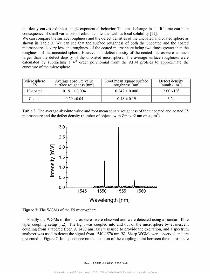
the decay curves exhibit a single exponential behavior The small change in the lifetime can be a consequence of small variations of erbium content as well as local solubility [11]. We can compare the surface roughness and the defect densities of the uncoated and coated sphere as shown in Table 3. We can see that the surface roughness of both the uncoated and the coated microspheres is very low, the roughness of the coated microsphere being two times greater than the roughness of the uncoated sphere. However the defect density of the coated microsphere is much larger than the defect density of the uncoated microsphere. The average surface roughness were calculated by subtracting a 4th order polynomial from the AFM profiles to approximate the curvature of the microsphere. Microsphere
F5 Average absolute value surface roughness [nm]
Root mean square surface roughness [nm]
Defect density [numb./µm2]
Uncoated 0.191 ± 0.004 0.242 ± 0.006 2.00 x10²
Coated 0.29 ±0.04 0.48 ± 0.19 6.24
Table 3: The average absolute value and root mean square roughness of the uncoated and coated F5 microsphere and the defect density (number of objects with Zmax>2 nm on a µm2).
1545 1550 1555 15600.0
0.5
1.0
1.5
2.0
2.5
3.0
Wavelength [nm]
Inte
nsity
[nW
]
Figure 7: The WGMs of the F5 microsphere
Finally the WGMs of the microspheres were observed and were detected using a standard fibre taper coupling setup [1,2]. The light was coupled into and out of the microsphere by evanescent coupling from a tapered fiber. A 1480 nm laser was used to provide the excitation, and a spectrum analyzer was used to detect the signal from 1540-1570 nm [8]. Sharp WGMs were observed and are presented in Figure 7. In dependence on the position of the coupling point between the microsphere
Proc. of SPIE Vol. 8236 82361W-8
Downloaded from SPIE Digital Library on 20 Feb 2012 to 193.205.206.25. Terms of Use: http://spiedl.org/terms
and the taper different modes were excited. The FSR range between two adjacent modes was measured to be 3.7±0.1 nm. Since the FSR between two adjacent modes is known to be:
, ² (1) where λ is the wavelength of the signal (in our case 1550 nm), N is the refractive index and d is the diameter of the sphere. Since the refractive index of the coating is 1.6 that means that the free spectral range of about 3.7 nm corresponds to a microsphere with a diameter of 130 μm which is very close to the value we measured with a optical microscope (140 μm). The full width at half maximum of the modes was measured as 0.05 nm which is just the resolution of the spectrum analyser. Therefore we were unable to measure the Q-factor, we only can tell that it is higher than 3x10⁴. All the measurements were made with the laser current at 300 mA, which corresponds to a laser power of about 120 mW. The peak power of the detected modes was always in the range of nanowatts, the highest power detected being 30 nW.
3. GLASS CERAMIC COATING
In this section we present an attempt to fabricate silica microspheres coated by silica-hafnia glass ceramic film doped with erbium ions [12]. Silica spherical micro resonators were fabricated by melting the end of a freshly stripped and cleaved standard single mode optical fiber using a commercial fusion splicer. After solidification, controlled by surface tension, a sphere with a radius in the order of one hundred of micrometers, was achieved. The coating of the microspheres was a 70SiO2-30HfO2 film doped with 0.3 mol% of Er3+ ions. The preparation protocol of the silica-hafnia sol is analogous to that reported above in the section dealing with rare-earth-activated glass coating. In the present case an additional heat treatment was performed in air at a temperature of 900 °C for 20 min in order to nucleate nanocrystals inside the film. Figure 8 compares the photoluminescence spectrum obtain for a silica-hafnia coated microsphere (Fig. 8(a)) with a glass-ceramic waveguide (Fig. 8(b)) fabricated by top-down technique from the parent amorphous waveguide (Fig. 8(c)). The film coating and the waveguides have the same composition 70SiO2-30HfO2 with 0.3 mol% of Er3+ ions.
Proc. of SPIE Vol. 8236 82361W-9
Downloaded from SPIE Digital Library on 20 Feb 2012 to 193.205.206.25. Terms of Use: http://spiedl.org/terms

1400 1450 1500 1550 1600 1650 1700
(c)
(b)
Inte
nsity
[arb
. uni
ts]
Wavelength [nm]
(a)
Figure 8: Room temperature photoluminescence spectra of the 4I13/2 → 4I15/2 transition of Er3+ ion for: (a) spherical microresonator coated by a film of 70SiO2-30HfO2 activated by 0.3 mol% Er3+, annealed for 20 min in air at 900°C; (b) 70SiO2-30HfO2 glass-ceramic planar waveguide activated by 0.3 mol% Er3+; (c) 70SiO2-30HfO2 amorphous planar waveguide activated by 0.3 mol% Er3+.
The photoluminescence spectrum of the coated microsphere (Fig. 8(a)) shows well-resolved Stark
components typical of Er3+ emission in a crystalline-like environment. Moreover a significant narrowing of the emission bandwidth is observed. These features are the fingerprint of a glass-ceramic system as confirmed by the photoluminescence spectrum of the glass-ceramic waveguide of Fig. 8(b). The bandwidth measured at 3 dB from the maximum of the intensity is 51, 17, and 12 ± 1 nm for the amorphous waveguide (Fig. 8(c)), the glass-ceramic waveguide (Fig. 8(b)), and the glass-ceramic microresonator (Fig. 8(a)), respectively. The photoluminescence spectrum of the glass-ceramic microresonator exhibits a more significant narrowing in respect to that of the glass-ceramic waveguide, indicating that in the case of microresonator there is height homogeneity of the local sites where the Er3+ ion is accommodated.
The decay curves of the metastable level 4I13/2 are shown in Fig. 9 for the glass-ceramic waveguide (Fig. 9(a)) and the glass-ceramic microsphere (Fig. 9(b)). Both the decays exhibit a single exponential profile with a lifetime of 7.4 and 9.7 ± 0.3 ms for the waveguide and the microsphere, respectively. The observation of a single decay is a further indication of the high homogeneity of the rare earth local environment and in particular the absence of a multisite structure. The important difference between the lifetimes of the two systems asks for accurate investigation of the local structure. A better embedding of the Er3+ ions in the HfO2 nanocrystals in respect to the waveguide can be excluded because the reproducibility of the waveguiding system and lifetime measurements. The only difference is due to the different geometry. When the microresonator is excited, although in the far field configuration, it is possible a resonant coupling with at least one of the whispering-gallery modes sustained by the microresonator and in particular in the coated region. Higher is the
Proc. of SPIE Vol. 8236 82361W-10
Downloaded from SPIE Digital Library on 20 Feb 2012 to 193.205.206.25. Terms of Use: http://spiedl.org/terms

quality factor longer is the time that the light remain entrapped in the microresonator [1,2]. For this reason we can assign the lengthening of the lifetime to the effect of light trapping in the microresonator.
0 5 10 15 200.05
0.1
0.3
1
(b) 9.7 ms
Inte
nsity
[arb
. uni
ts]
Time [ms]
(a) 7.4 ms
Figure 9: Decay curves of the luminescence from the 4I13/2 metastable state of Er3+ ions for (a) 70SiO2-30HfO2 glass-ceramic planar waveguide activated by 0.3 mol% Er3+; (b) glass-ceramic spherical microresonator coated by a film of 70SiO2-30HfO2 activated by 0.3 mol% Er3+.
Although the coated resonator exhibits the significant properties above discussed, losses still remain an important drawback of the coating, in particular in the case of glass-ceramic coating where the surface can present some important roughness. AFM measurements performed on the surface of the glass-ceramic microsphere show structures of 50-70 nm height and 100-200 nm width, assigned to HfO2 nanocrystals.
4. CONCLUSION
Glass and glass-ceramic activated by rare earth ions are systems that exhibit specific morphologic, structural, and spectroscopic properties allowing to develop interesting new physical concepts, as well as novel photonic devices based on the enhancement of the luminescence. Fabrication techniques based on bottom-up and top-down approaches appear to be viable although a specific effort is required to achieve the necessary reliability and reproducibility of the preparation protocols. In the present paper only two particular examples of glass and glass-ceramic coated spherical microresonators have been discussed. Another important application of transparent coatings onto spherical microresonators concerns the possibility of reducing the thermal shift of the WGM resonances due to microsphere heating by the dissipated pump energy. Actually, the shift is due both to the temperature-induced change in the refractive index (the thermo-optic coefficient) and to the temperature-induced increase of the diameter of the particle (coefficient of thermal expansion)
Proc. of SPIE Vol. 8236 82361W-11
Downloaded from SPIE Digital Library on 20 Feb 2012 to 193.205.206.25. Terms of Use: http://spiedl.org/terms

[2]. This argument is of great interest in the research regarding suitable materials for coating and deals with polymers and several kinds of nanocomposites.
As far as concern the examples reported in this work it appears that silica-hafnia is a suitable system for the development of glass and glass-ceramic functionalized coating, although further work is required for the preparation of hafnia nanocrystals activated by rare earth ions using the colloidal synthesis to achieve optimized nanoparticles to be imbedded in the amorphous host. Another important challenge, common to any fabrication techniques, is to obtain that the rare earth ions have to be present only in the nanocrystals.
ACKNOWLEDGMENTS This research was performed in the framework of the SHYRO (CNES 2011-2014) and NSMBO (PAT, 2010-2013) research projects.
REFERENCES [1] A. Chiasera, Y. Dumeige, P. Féron, M. Ferrari, Y. Jestin, G. Nunzi Conti, S. Pelli, S. Soria, and G. C. Righini, “Spherical whispering-gallery-mode microresonators”, Laser & Photonics Reviews 4, pp. 457-482 (2010). [2] G.C. Righini, Y. Dumeige, P. Féron, M. Ferrari, G. Nunzi Conti, D. Ristic, and S. Soria, “Whispering gallery mode microresonators: fundamentals and applications”, Rivista del Nuovo Cimento 34, pp. 435-488 (2011). [3] A. Beltaos and A. Meldrum, “Whispering gallery modes in silicon-nanocrystal-coated silica microspheres”, J. Lumin. 126, pp. 607-613 (2007). [4] S. Shibata, S. Ashida, H. Segawa, and T. Yano, “Coated microsphere as spherical cavity Raman laser”, J. Sol-Gel Sci. Technol. 40, pp. 379-384 (2006). [5] A. Chiappini, A. Chiasera, C. Armellini, S. Varas, A. Carpentiero, M. Mazzola, E. Moser, S. Berneschi, G. C. Righini, and M. Ferrari, “Sol-gel-derived photonic structures: fabrication, assessment, and application”, J. Sol-Gel Sci. Technol. 60, pp. 408-425 (2011). [6] G. C. Righini, F. Cosi, G. Nunzi Conti, S. Pelli, S. Soria, E. Moser, Y. Jestin, M. Ferrari, P. Féron, A. Chiasera, A. Chiappini, and C. Armellini, “Photonic properties and applications of glass micro- and nanospheres”, Physica Status Solidi A, 206, pp 898-903 (2009). [7] G. C. Righini, C. Armellini, A. Chiasera, Y. Jestin, M. Ferrari, A. Chiappini, Maurizio Montagna, Claire Arfuso Duverger, Patrice Féron, Simone Berneschi, Massimo Brenci, G. Nunzi Conti, S. Pelli, C. Gonçalves, and R. M. Almeida, “Er3+-activated sol-gel silica derived spherical microresonators”, Glass Technology - European Journal of Glass Science & Technology Part A 48, pp. 200-203 (2007). [8] Y. Jestin, C. Armellini, A. Chiappini, A. Chiasera, Y. Dumeige, M. Ferrari, P. Féron, L. Ghisa, G. Nunzi Conti, S. Trebaol, and G.C. Righini, “Photonic properties of erbium activated coated microspheres.”, Proceedings of SPIE 6890, pp. 689008-1/8 (2008). [9] K. Tran Ngoc, P.H. Huy, N. D. Chien, M. Ferrari, A. Chiasera, C. Armellini, E. Moser, M. Montagna, A. Chiappini, M. Brenci, G. Nunzi Conti, S. Pelli, G.C. Righini, “Silica microspheres coated by Er3+-activated silica-hafnia sol-gel layers”, Proceedings ICPE 2006 International Conference on Engineering Physics 2006 including the 9th Vietnamese-German Seminar on Physics and Engineering, the 4th Vietnam-Korea International Joint Symposium on Advanced Materials Science and Their Processing, and the Second Vietnamese-Italian International Joint Workshop on
Proc. of SPIE Vol. 8236 82361W-12
Downloaded from SPIE Digital Library on 20 Feb 2012 to 193.205.206.25. Terms of Use: http://spiedl.org/terms

Photonics and Nanotechnology. 9-13 October 2006, Hanoi. University of Technology Hanoi, Vietnam, pp. 30-31 (2006). [10] M. Ferrari, G.Alombert-Goget, C. Armellini, S. Berneschi, S.N.B. Bhaktha, B. Boulard, M. Brenci, A. Chiappini, A. Chiasera, C. Duverger-Arfuso, P. Féron, R.R. Gonçalves, Y. Jestin, L. Minati, E. Moser, G. Nunzi Conti, S. Pelli, D.N. Rao, R. Retoux, G.C. Righini, and G. Speranza, “Er3+ - activated photonic structures fabricated by sol-gel and rf-sputtering techniques”, Proceedings of SPIE 7366, pp. 73660E-1/15 (2009). [11] G.C. Righini and M. Ferrari, “Photoluminescence of rare-earth-doped glasses”, La Rivista del Nuovo Cimento 28, pp. 1-53 (2005). [12] G. Alombert-Goget, C. Armellini, S. Berneschi, A. Chiasera, F. Cosi, G. Nunzi Conti, P. Féron, M. Ferrari, L. Lunelli, E. Moser, C. Pederzolli, G.C. Righini, “Glass-ceramics coating of silica microspheres”, Proceedings 3rd ICTON – Paper Fr2B.2 (2009).
Proc. of SPIE Vol. 8236 82361W-13
Downloaded from SPIE Digital Library on 20 Feb 2012 to 193.205.206.25. Terms of Use: http://spiedl.org/terms