The changing pace of insular life: 5000 years of insular evolution in the Orkney vole
Upload
independentCategory
view
8download
0
(This is a sample cover image for this issue. The actual cover is not yet available at this time.)
This article appeared in a journal published by Elsevier. The attachedcopy is furnished to the author for internal non-commercial researchand education use, including for instruction at the authors institution
and sharing with colleagues.
Other uses, including reproduction and distribution, or selling orlicensing copies, or posting to personal, institutional or third party
websites are prohibited.
In most cases authors are permitted to post their version of thearticle (e.g. in Word or Tex form) to their personal website orinstitutional repository. Authors requiring further information
regarding Elsevier’s archiving and manuscript policies areencouraged to visit:
http://www.elsevier.com/copyright

Author's personal copy
Spatial segregation of somato-sensory and pain activations in the humanoperculo-insular cortex
Laure Mazzola a,d,e,f,g,⁎, Isabelle Faillenot a,d,f,g, Fabrice-Guy Barral b,f,g,François Mauguière c,d,e,g, Roland Peyron a,d,f,g
a Neurology Department, University Hospital, St-Etienne, Franceb Radiology Department, University Hospital, St-Etienne, Francec Functional Neurology and Epilepsy Department, Neurological Hospital, Hospices Civils de Lyon, Lyon, Franced Team ‘Central Integration of Pain’ Lyon Neuroscience Research Center, INSERM U 1028, CNRS UMR 5292 Lyon, Francee Claude Bernard University Lyon 1, Lyon, Francef Jean Monnet University St-Etienne, F-42023, Franceg Pole of Research & University Teaching (PRES) Lyon, France
a b s t r a c ta r t i c l e i n f o
Article history:Received 21 June 2011Revised 20 December 2011Accepted 26 December 2011Available online 5 January 2012
Keywords:PainFunctional MRIInsulaSecondary somatosensitive area (SII)Somato-sensory stimuli
The role of operculo-insular region in the processing of somato-sensory inputs, painful or not, is nowwell estab-lished. However, availablemaps fromprevious literature show a substantial overlap of cortical areas activated bythese stimuli, and the region referred to as the “secondary somatosensory area (SII)” is widely distributed in theparietal operculum. Differentiating SII from posterior insula cortex, which is anatomically contiguous, is noteasy, explaining why the “operculo-insular” label has been introduced to describe activations by somatosensorystimuli in this cortical region. Based on the recent cyto-architectural parcellation of the human insular/SII cortices(Eickhoff et al., 2006, Kurth et al., 2010), the present study investigates with functional MRI (fMRI), whetherthese structural subdivisions could subserve distinct aspects of discriminative somato-sensory functions, includ-ing pain. Responses to five types of stimuli applied on the left hand of 25 healthy volunteers were considered:i) tactile stimuli; ii) passive movements; iii) innocuous cold stimuli; iv) non-noxious warm and v) heat pain.Our results show different patterns of activation depending on the type of somato-sensory stimulation. Theposterior part of SII (OP1 area), contralateral to stimuli, was the only sub-region activated by all type of stimuliand might therefore be considered as a common cortical target for different types of somato-sensory inputs.Proprioceptive stimulation by passive finger movements activated the posterior part of SII (OP1 sub-region)bilaterally and the contralateral median part of insula (PreCG and MSG). Innocuous cooling activated the contra-lateral posterior part of SII (OP1) and the dorsal posterior and median part of insula (OP2, PostCG). Pain stimuliinduced the most widespread and intense activation that was bilateral in SII (OP1, OP4) and distributed to allsub-regions of contralateral insula (except OP2) and to the anterior part of the ipsilateral insula (PreCG, MSG,ASG). However, the posterior granular part of insula contralateral to stimulus (Ig area) and the anterior part ofSII bilaterally (OP4) were specifically activated during pain stimulation. This raises the question whether theselatter areas could be the anatomical substrate of the sensory-discriminative processing of thermal pain.
© 2012 Elsevier Inc. All rights reserved.
Introduction
The role of operculo-insular (OI) cortex in the processing ofpainful sensations is now well established on the basis of variousexperimental approaches, such as single unit recordings (Robinson
and Burton, 1980a, 1980b), anatomical studies in monkeys (Burtonand Jones, 1976; Craig, 1995; Mesulam and Mufson, 1982; Mufsonand Mesulam, 1984), laser evoked potentials in humans, as recordedon the scalp (Valeriani et al., 2000; Vogel et al., 2003) or with intra-cerebral electrodes (Frot and Mauguière, 2003), and direct electricalstimulations of the human cortex with depth-intracerebral electrodes(Afif et al., 2008; Mazzola et al., 2006; Nguyen et al., 2009; Ostrowskyet al., 2002; Stephani et al., 2011).
Functional imaging studies carried out in humans in the recentyears, consistently reported that the cortical regions located in theupper bank of the lateral sulcus, including the second somatosensoryarea (SII), and the insular cortex are activated by nociceptive stimuli(reviews in Apkarian et al., 2005; Garcia-Larrea et al., 2002; Peyron
NeuroImage 60 (2012) 409–418
Abreviations: OP1, OP2, OP3, OP4, the four cyto-architectonic sub-divisions ofparietal operculum; Ig, granular sub-divisions of the posterior insula; Id1, dysgranularsub-division of the posterior insula; PostCG, post-central insular gyrus; PreCG, pre-central insular gyrus; MSG, middle short insular gyrus; ASG, anterior short insular gyrus.⁎ Corresponding author at: INSERM U 879, Neurology Department, Hôpital Nord,
CHU de St Etienne, 42055 Cedex2, France. Fax: +33 4 77 12 05 43.E-mail address: [email protected] (L. Mazzola).
1053-8119/$ – see front matter © 2012 Elsevier Inc. All rights reserved.doi:10.1016/j.neuroimage.2011.12.072
Contents lists available at SciVerse ScienceDirect
NeuroImage
j ourna l homepage: www.e lsev ie r .com/ locate /yn img

Author's personal copy
et al., 2000; Treede et al., 2000; Vogel et al., 2003). These two supra-sylvian pain areas are also involved in the processing of innocuoussomato-sensory inputs such as cold (Craig et al., 2000; Hua et al.,2005), warm (Moulton et al., 2005; Tseng et al., 2010), tickling(Carlsson et al., 2000), non-painful cutaneous stimuli (Ferretti et al.,2004) or passive movements (Alary et al., 2002; Druschky et al.,2003; Mima et al., 1999).
However, functional imaging studies showed a substantial overlapin all these activations, noxious or not, within SII or insula (Coghillet al., 1994). In addition, differentiation between the functionalproperties of the contiguous SII and posterior insular cortices provedvery difficult in both functional imaging and electrophysiologicalstudies. Since the exact anatomical border between the granularcortex of the inner part of the operculum and the posterior granularinsular cortex cannot be delineated on individual MRI, the SII/insularregion (especially in its posterior extent), has been considered asa single functional entity in most pain imaging studies (for reviews,see Apkarian et al., 2005; Peyron et al., 2000).
Using an observer-independent approach, Eickhoff et al. (2006a,b)and Kurth et al. (2010) solved this anatomical limitation by identifyingdistinct cytoarchitectonic areas in the human parietal operculumand posterior insula. Although this new scheme does not contradictprevious classification of SII or insular cortex based on its granularity,it further develops these maps by demonstrating the existence ofseveral individual areas within these major zones. Given the varietyof functions ascribed to the SII–insula cortices based on functionalimaging studies and connectivity, it seems reasonable to hypothesizethat these subdivisions based on anatomical substrates might reflecta diversified mosaic of structurally and functionally distinct areasin the human operculo-insular cortex. Since the cyto-architectonicprobabilistic maps of the OI cortex have been computed by Eickhoffet al. (2006a, b) and Kurth et al. (2010) in the standard MNI space,the present study was carried out to investigate with functionalMRI (fMRI), whether these structural subdivisions could subservedistinct aspects of discriminative somato-sensory functions, includingpain. We therefore investigated responses to i/ heat pain in com-parison to ii/ non-noxious warm and iii/ innocuous cold stimuli, iv/tactile sensations and v/ passive movements, taking into account therecent advances in insular and SII structural anatomy. Since recentstudies showed some degree of somatotopic organization both inSII (Krubitzer et al., 1995) and in insula (Brooks et al., 2005; Mazzolaet al., 2009), all types of stimuli were strictly applied on the samebody area (the left hand) to avoid topographic changes of activationlocation due to this somatotopic organization.
Subjects and methods
Subjects
Twenty-five healthy right handed volunteers (12 male, 13 female,mean age 30.5 years, range 19–39 years) participated in this study,which was approved by the local ethics committee. Written informedconsent was obtained from each subject prior to investigations.
Stimulation procedures
Volunteers laid supine inside the MRI scanner. Five runs wereperformed, with a paradigm alternating rest and one of the fivesomato-sensory stimuli (tactile; passive finger movement; cold;warm; heat pain). Each session was made of 24 blocks of 7 s duringwhich one of the five somato-sensory stimulations was delivered. Restblocks had a randomized duration from 5 s to 25 s (mean=15 s;SD=6.7 s). The order of the sessions (i.e. of the stimulations) wasarranged in a randomized order. The experimenter wore a headphone.A high-pitched sound was delivered to announce that stimulation wasimminent. Then two low-pitched sounds were produced to indicate
respectively the beginning and the end of the stimulation time. Stimu-lation onsets were randomized.
All stimuli were applied to the left hand, on its dorsum for cutane-ous stimuli. Passive movements were applied to the 2nd finger, sothat they concerned the same somatotopic territory for all modalities.Tactile stimulation was obtained by rubbing a soft brush back andforth from proximal to distal (0.5 Hz). Cold stimulation consisted inthe application a plastic pack filled with a frozen fluid. The surfacetemperature checked at the beginning and the end of the sessions,showed a good stability (mean temperature±SD=1.8 °C±2.4). Forwarm and heat pain stimulations, we used hand heaters for outdooruse, consisting in a plastic pack filled with a fluid having good thermicinertia, that could be heated at 37 °C and 47 °C respectively (meantemperature±SD=36.8 °C±0.3 and 47.9°±0.2). Passive flexor andextensor movements of the metacarpo-phalangeal joint were appliedmechanically by the experimenter and were paced at a frequency of0.5 Hz. The intensity of pain in all conditionswas assessed individuallybefore and after the experiment by a visual analogic scale (VAS).
Even though anexperimental paradigmalternating thefive differentstimuli in a same run would have been ideal, we prefered to performfive sessions with one single stimulus alternating with rest. Reasonsfor this choice were that it was impossible, in a same run, to managefive different and intermingled stimuli with only 5–25 s intervals. Inaddition, such intermingled stimuli might have interfered together asthey were applied at only 5–25 s intervals whereas the aim of thisstudy required to avoid any interference between stimuli (for examplecold inhibiting warm and heat pain sensations Craig et al., 2000;Yarnitsky and Ochoa, 1990). For similar reasons, we deliberatelyselected an experimental paradigm without psychophysical assesse-ment to minimize cognitive activities that may be associated withsensory or pain intensity scoring (Kong et al., 2006; Lötsch et al., 2011).
Acquisitions
MRI images were acquired in a 1.5 T scanner (Symphony MaestroClass, Siemens, Erlangen, Germany). For functional images, an EPI(Echo Planar) sequence was used with following parameters: 28slices, 4 mm thick without gap, with a 64×64 matrix and a256×256 mm2
field of view (FOV); TR/TE=2700/55 ms. Each subjecthad 5 EPI runs of 200 images each (9 min 5 s) corresponding to thefive types of stimuli. Anatomic scans were acquired using a 3D T1sequence (MPRAGE) including 160 sagittal slices and the followingsequence parameters: TI/TR/TE; 920/1700/4.35 ms, matrix=256×256voxels, FOV=256×256 mm2.
Image analysis
fMRI data were analyzed using Statistical Parametric Mapping(SPM8 — Wellcome Department of Cognitive Neurology, UK; http://www.fil.ion.ucl.ac.uk/spm/) and some SPM compatible toolboxes:MarsBar (Brett et al., 2002), Anatomy (Eickhoff et al., 2005) andWFU_PickAtlas (Maldjian et al., 2003). Moreover, in order to buildanatomical regions of interest (ROI), MRIcroN (http://www.cabiatl.com/mricro/) and AAL regions (Automated Anatomical Labels; Tzourio-Mazoyer et al., 2002) were used.
PreprocessingThe two first volumes of each run were discarded. To correct for
subject's movements, all scans of each individual were realigned toeach other in a two pass procedure: they were first aligned to thefirst image and then, to the mean image. Data from all subjectsshowed motion corrections during each run less than 3 mm and 2°.A mean functional volume was constructed for each subject fromthe realigned images. This image was coregistred on the anatomicalimage and then transformation parameters applied to all EPI images.The anatomical image was normalized to the MNI template brain
410 L. Mazzola et al. / NeuroImage 60 (2012) 409–418

Author's personal copy
image using the segmentation step. The parameters estimated fromthis normalization process were then applied to all functional images.Since the interindividual variability was about 5 mm after normaliza-tion (Crinion et al., 2007), we considered that a smoothing of 6 mmFWHM was appropriate for the voxel wise analysis (SPM 8). For theROI study that focused on small ROIs, a 6 mm smoothing wouldhave blurred the resolution necessary for segregating distinct insularsub-regions, and therefore a 3 mm kernel was used for that analysis.The resulting voxel size in standard stereotaxic coordinates was2×2×2 mm3. For localization purpose, the anatomical images of allsubjects were averaged.
ROI definitionSince the aim of the present study was to investigate specifically
the operculo-insular responses to different noxious and non-noxious stimuli, we did not consider the results of SPM analysisoutside the a priori defined ROIs. SII and insula were investigatedwith a template of ROIs that were defined within the MNI space andtheir localization was checked on the normalized anatomical imageof each subject (Fig. 1).
Insula. The insula region was defined as the union of the AAL's(Automated Anatomical Labels; Tzourio-Mazoyer et al., 2002) insulaand the post-insula of the Anatomy toolbox (Id1, Ig1 and Ig2).When superimposed on the mean image of the group, the rightinsula provided in AAL needed to be 2 mm-shifted towards the leftand 2D-dilated by 2 mm in both directions. After unification of ROIs,7 sub-regions were defined inside each insular cortex. The insularcentral sulcus was drawn on the mean T1 image, separating theanterior from the posterior insula. Inside the posterior insula foursub-regions were delimited: Id1, Ig1, Ig2 (as defined by Eickhoffet al., 2005; Kurth et al., 2010) and the post-central gyrus (PostCG)caudal to the central sulcus. Within the anterior insula, the anteriorinsular sulcus and the precentral insular sulcus were drawn definingthree gyri: anterior short gyrus (ASG), middle short gyrus (MSG)and precentral insular gyrus (PreCG) (see Fig. 1). As the actual resolu-tion for our data was of approximately 7 mm (size of the resolutionelement estimated by SPM between 23 and 26 voxels), we measuredthe size of each ROI and concluded that three of the cytoarchitectonicsub-regions delineated by Kurth et al. (2010) and Eickhoff et al.(2006a,b) were definitely too small to be anatomically segregatedand studied. We therefore dropped Id1 from the list of the studiedROIs and pooled together Ig1 and Ig2 (becoming the ROI labeled Ig)(see Fig. 1C). Hence, whatever the considered plane (sagittal, coronalor axial), all ROIs had an over-all size ≥1 cm and could be separatedfrom each other with a spatial resolution of 7 mm.
Parietal opercular cortex. The opercular cortex was defined as thecombination of the four sub-regions described by Eickhoff et al.(2006a,b): OP1, OP2, OP3 and OP4. Actually, the OP2 and OP3 subdi-visions of the opercular cortex include part of the postero-superiorportion of the insular cortex, covering at maximum its upper third.Therefore we considered OP2 and OP3 as parts of posterior insulaand assimilated the OP1 andOP4 cytoarchitectonic regions as anatom-ical homologue of the functionally defined human SII area (Eickhoffet al., 2006b) (Fig. 1).
A global opercular-insular ROI was built including all of the pre-cited insular and opercular ROIs, i.e. 9 ROIs in the right hemisphereand 9 in the left (Fig. 1C(d)).
Statistics
First level analysis (individual level) used a general linear model(GLM, Friston et al., 1995). Then, three random-effects analyseswere performed at the group level. Studies were limited to SII andinsula areas on both sides, after having checked that the global
activation profile in response to heat pain stimuli met the standardof what has been described as the “pain matrix” in previous painfMRI studies, including anterior cingulate cortex and primarysomato-sensitive area (Peyron et al., 2000).
Individual analysisThe functional MRI signal in response to stimulation was modeled
using a general linear model approach. The regressors of interestwere constructed by convolving the stimulus input function with acanonical hemodynamic response function (HRF) and its time deriv-ative. The estimated time courses of the head motion parameterswere included as covariates of no interest to further control for theartefacts linked to subject movements. Data were high-pass filtered(cut-off period was set to 128 s). Serial correlations were correctedusing an autoregressive model. Contrast images were obtained foreach subject and each run for the canonical HRF regressor, reflectingthe contrasts of interest (beta-weight for stimulation vs. rest). Theseindividual contrast images were entered into second level (random-effects) analyses to study group activations.
Voxel wise group analysisSPM8 was used to test group activations inside the global
operculoinsular ROI. In order to describe activations by each stimula-tion compared to rest, five one-sample t-tests were performed on thecontrast images of the individual analysis. In order to describe com-mon activation for all conditions, a one-way ANOVA was designedwith 5 dependant groups with unequal variance, each group wasconsisted in individual contrast images of each stimulation.
Statistical results were reported with an uncorrected threshold(pb0.001) at the voxel level, and a FWE corrected threshold (pb0.05)at the cluster level.
ROI group analysisFor each condition, the value of the voxels (beta weight) of each
ROI was extracted on the contrast individual images and the medianwas calculated.
In order to describe the within group activation in each of the 18regions of interest, a one-sample t-test was performed using MarsBarfor the 5 conditions. ROIs were considered as significantly activatedif their associated p-value was b0.05 after Bonferroni correction forthe 18 comparisons.
In order to assess if some ROIs responded differently to the differ-ent types of stimuli, a two-way ANOVA was performed with stimulus(5 levels) and ROI (9 levels) as factors. The dependant variablewas the individual median value extracted for each subregion.Interactions between factors and post-hoc Scheffe tests were doneusing SPSS. Significant threshold was set to p=0.05.
In order to look for correlations between activations and painintensity, the mean VAS score of each volunteer was correlated tothe median value of each ROI after noxious stimulation. Significantcorrelations were reported if the Spearman (non-parametric) testp value was b0.05.
Results
Because of storage failure and corrupted data, data were missingfor passive movement, pain or warm in three, four and five subjectsrespectively, and for the two conditions brush and cold in two subjects.
Psychophysics
Not surprisingly, brush, warm, cold and passive movements stim-uli were scored nil on pain VAS scale (VAS=0), while noxious heatwas scored 6.7±1.1 and 6.9±1.3, respectively before and afterexperiment, showing both a rather intense pain sensation and a sta-bility of pain perception during the whole session.
411L. Mazzola et al. / NeuroImage 60 (2012) 409–418

Author's personal copy
Cortical activations
All types of stimuli applied on the left hand led to statisticallysignificant increases in fMRI signal intensity in several cortical areas.
Voxel wise group analysis (see Fig. 2; Table 1s)For non-painful stimulations, skin brushing was associated with
the largest activation in the contra- and ipsilateral SII and in theposterior part of the contralateral insula. Passive movement of fingers
Fig. 1. A: Representation of the operculo-insular ROIs available in the Anatomy toolbox (Eickhoff et al., 2005) superimposed onto the mean MRI of our subjects. Color indicates theprobability for each voxel to be included in the cytoarchitectonic area (Eickhoff et al., 2006a, 2006b). B: Construction of ROIs based on the macroscopic anatomy of insula. Central (I),precentral (II) and anterior (III) insular sulci were identified and drawn on the averaged MRI of the subjects, defining 3 gyri within the anterior insular cortex: anterior short gyrus(ASG), middle short gyrus (MSG) and precentral insular gyrus (PreCG). C: Axial (a) and sagittal (b) representation of the different ROIs individualized in the insula and SII andsuperimposed on the group averaged anatomical image. Posterior insula includes Ig1, Ig2, Id1 and the post-central gyrus (PostCG) according to the probabilistic maps based oncytoarchitectonic data and defined within the MNI space (Kurth et al., 2010). OP2 and OP3 are located at the boundary between SII and insula, they were considered here asparts of the insular cortex. OP1 and OP4 were considered as corresponding to SII sub-divisions. A schematic mask of these ROIs was drawn (c). Because of spatial resolution, Ig1and Ig2 have been pooled together (entitled Ig) and Id1 has been removed. The new mask (d), representing the ROI used, was superimposed to our results to make the visual anal-ysis of Fig. 4 easier.
412 L. Mazzola et al. / NeuroImage 60 (2012) 409–418

Author's personal copy
activated the contra- and ipsilateral SII and the middle-anterior partof contralateral insula. After cold stimulations, only the contralateralSII and the extreme posterior part of contralateral insula (at theboundary with SII cortex) were activated. Warm activated the contra-lateral SII and bilateral insula predominantly in the anterior part. Heatpain resulted in a broad activation of bilateral SII and insular cortex.
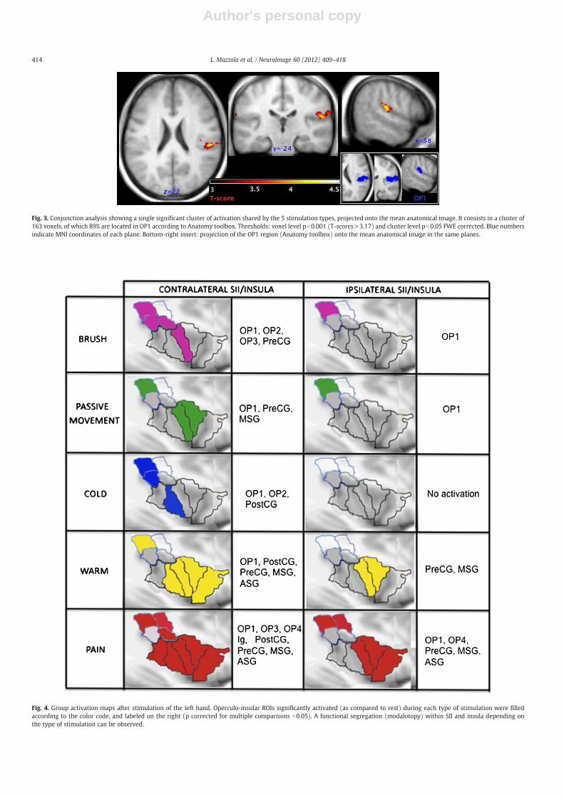
A conjunction analysis was performed, as an indicator of whichareas were activated in all stimulation modalities. Shared activationconsisted in a single significant cluster of 163 voxels (pb0.001 FWEcorrected, maximum peak coordinates: x=54, y=−26, y=22), ofwhich 89% were located in OP1 according to Anatomy toolbox (seeFig. 3).
ROI analyses (see Fig. 4)Statistics for significantly activated ROI are given in Table 1.Brush stimulation activated predominantly SII (bilateral OP1),
contralateral OP2-OP3 and PreCG insular region. Passive movementsof fingers activated only the OP1 part of SII bilaterally, and thePreCG and MSG of contralateral insula. Cold stimulations activatedOP1–OP2 and PostCG contralateral sub-regions. Warm activated con-tralateral OP1 SII sub-region, the anterior half of the contralateralinsula (PostCG, PreCG, MSG, ASG) and the medium-anterior part of
the ipsilateral insula (PreCG, MSG). Heat pain induced an activationof bilateral OP1/OP4, all sub-regions of contralateral insula exceptOP2 (OP3, Ig, PostCG, PreCG, MSG, ASG) and the anterior part of theipsilateral insula (PreCG, MSG, ASG).
Comparisons between different stimulation types and correlations withpain scores for each ROI (see Fig. 5; Table 2s)
The two-way ANOVA indicated that there is a strong interactionbetween stimuli and ROIs (right: F(32)=4.2, pb10−3; left: F(32)=2.6, pb10−3). As illustrated in Fig. 5, activation of all SII and insularregions except OP2 and OP3 contralateral to stimulus, was moreintense after pain stimulation than after any other stimulation types,but these differences did not reach significance levels for all stimulustypes in all regions (see Table 2s for details). Only contralateral OP4in SII and PreCG, MSG, ASG in the anterior part of insula bilaterallywere significantly more activated by pain stimulation than by anyother modalities (pb0.05). Activation in OP1 was more intense afterpassive movement stimulation than after warm and cold stimulationbilaterally, and than after brush stimulation in the right OP1 (pb0.05).
Themean pain VAS score of each volunteer correlated to the inten-sity of activation of the contralateral OP4 only (p=0.035; r=0.45).
Fig. 2. Projection of significant activations for each stimulation type, as compared to rest, onto the mean MRI (voxel wise group analysis). A: In the axial plane, for each type ofstimulation applied on the left hand, in the global operculo-insular ROI, where the right hemisphere is represented on the right side of each slice. B: Sagittal slices of the right insula.(Voxel threshold pb0.001; corrected cluster threshold pb0.05).
413L. Mazzola et al. / NeuroImage 60 (2012) 409–418
Author's personal copy
Fig. 4. Group activation maps after stimulation of the left hand. Operculo-insular ROIs significantly activated (as compared to rest) during each type of stimulation were filledaccording to the color code, and labeled on the right (p corrected for multiple comparisons b0.05). A functional segregation (modalotopy) within SII and insula depending onthe type of stimulation can be observed.
Fig. 3. Conjunction analysis showing a single significant cluster of activation shared by the 5 stimulation types, projected onto the mean anatomical image. It consists in a cluster of163 voxels, of which 89% are located in OP1 according to Anatomy toolbox. Thresholds: voxel level pb0.001 (T-scores>3.17) and cluster level pb0.05 FWE corrected. Blue numbersindicate MNI coordinates of each plane. Bottom-right insert: projection of the OP1 region (Anatomy toolbox) onto the mean anatomical image in the same planes.
414 L. Mazzola et al. / NeuroImage 60 (2012) 409–418

Author's personal copy
Discussion
All of the five types of somato-sensory stimulations that we testedactivated at least one region in SII and insular cortex. Using anapproach based on cytoarchitectonic subdivisions of these areas, ourresults show that it does exist different patterns of activation. These
findings suggest that several different networks of sub-regions inthe SII–insula cortex are activated depending upon the nature ofsomato-sensory stimulation. Contralateral OP1, corresponding to theposterior part of SII, was the only sub-region to be activated by alltype of stimuli and might therefore be a common unspecific corticaltarget for different types of somato-sensory inputs.
Proprioceptive stimulation by passive finger movements alsoactivated the contralateral OP1 sub-region in addition to ipsilateralOP1 and the contralateral median part of insula (PreCG and MSG).Previous functional imaging studies have shown that passivemovement activates an extensive cortical somatosensory network,including SI and SII regions (Alary et al., 2002; Druschky et al.,2003; Mima et al., 1999). The involvement of insula is less describedalthough some studies mentioned the role of perisylvian regions(Druschky et al., 2003; Kavounoudias et al., 2008). This implicationof the median part of insula is consistent with anatomical connectionsof this region with both sensory (especially SII) and motor (BA6,SMA) areas (Mesulam and Mufson, 1982). Interestingly, both brushand passive movements activated OP1 bilateraly, and PreCG contra-lateral to stimulus. This reminds of similarities between the twostimuli in terms of stimulated peripheral fibers (large myelinatedfibers), but also suggests that discrimination between the two stimulimay be integrated in contralateral MSG and in the medial subdivisionof SII (i.e. OP2 and OP3) that are activated by only one of these twotypes of stimuli.
The activation induced by innocuous cooling was restricted to thecontralateral posterior part of SII (OP1), the dorsal posterior part
Fig. 5. Comparison between the magnitude of activation in each ROI depending on the type of stimulation. Mean contrast estimates (beta weights) for each type of stimulation vs.rest are reported for the right ROIs, in SII (A), posterior (C) and anterior (D) insula. Vertical bars represent the standard deviation. B is a schematic representation showing signif-icant differences between stimulations: the red (or green) ovoids indicate that pain (or passive movement) induced a significantly stronger increase in BOLD signal compared to thestimulations represented by symbols inside the oval (pb0.05). Pain; Warm; Cold; Brush; Passive Movement.
Table 1Significantly activated operculo-insular ROIs as compared to rest, for each type ofstimulation. T and P values for activated ROI showing a p valueb0.05 after Bonferronicorrection for multiple comparisons. NS = non significant.
ROI Brush Passive movement Cold Warm Pain
Right hemisphereOP1 7.9/b0.001 8.3/b0.001 4.4/0.002 4.7/0.001 6.9/b0.00010P2 3.4/0.025 NS 3.7/0.013 NS NSOP3 3.9/0.006 NS NS NS 4.4/0.0030P4 NS NS NS NS 4.2/0.005Ig NS NS NS NS 3.8/0.011postCP NS NS 3.1/0.046 4.2/0.005 4.6/0.002preCG 3.3/0.029 4.7/0.001 NS 4.0/0.007 6.3/b0.001MSG NS 3.4/0.025 NS 4.1/0.006 8.2/b0.001ASG NS NS NS 5.61b0.001 7.01b0.001
Left hemisphereOP1 5/0,001 5.1/0.001 NS NS 4.5/0.0020P4 NS NS NS NS 3.5/0.022preCG NS NS NS 3.7/0.015 4.4/0.003MSG NS NS NS 3.9/0.010 6.0/b0.001ASG NS NS NS NS 5.71b0.001
415L. Mazzola et al. / NeuroImage 60 (2012) 409–418

Author's personal copy
of insula (OP2) and the median posterior part of insula (PostCG). Thisfinding fits well functional anatomy in monkeys indicating thatdedicated thalamic nucleus (VMpo) relays topographic, discrimina-tive thermoreceptive-specific spino-thalamic projections to thedorsal margin of middle/posterior insular cortex (Craig, 1995). Like-wise, cold allodynia can occur in subjects with isolated ischemic le-sions of the posterior insular/retroinsular cortex (Veldhuijzen et al.,2010). This region has already been reported to be activated usingPositron Emission Tomography (PET) after graded innocuous coolingstimuli in humans (Craig et al., 2000) and a rostro-caudallly orga-nized somatotopic map of innocuous cooling activation was also de-scribed using fMRI in the dorsal posterior insular cortex (Hua et al.,2005). So, this region corresponding to the cytoarchitectonic OP2operculo-insular sub-region seems to be reliably and consistently ac-tivated by cold stimuli suggesting a possible primary cortex for coldsensation.
Consistent with our results, non-noxious thermal and noxiousheat stimuli have been shown to activate SII and insula cortices(Brooks et al., 2002; Casey et al., 1996; Craig et al., 2000; Huaet al., 2005; Peyron et al., 2000). Warm and heat pain conditionshad bilateral representations, in the contralateral but also theipsilateral insula. Pain representation largely goes beyond that ofwarm both in the posterior contralateral insula (OP3, Ig) and inthe ipsilateral anterior insula (ASG). These findings are in agree-ment with several functional imaging studies comparing activa-tions in processing innocuous vs. noxious contact heat showingresponses to noxious but not to innocuous stimuli in the contralateralposterior insula (Moulton et al., 2005; Tseng et al., 2010) or increasedactivation during noxious compared to innocuous thermal stimuliin bilateral anterior insula and contralateral posterior insula (Peltzet al., 2011).
Pain stimuli induced the most widespread and intense activationbilaterally in SII (OP1, OP4), contralaterally to stimulus in all sub-regions of insula (except OP2) and ipsilateral to stimulus in the ante-rior part of insula (PreCG, MSG, ASG). To avoid the risk of interactionsbetween stimuli processing at peripheral or central levels, for exam-ple cold inhibiting warm and heat pain sensations (Craig et al.,2000; Yarnitsky and Ochoa, 1990), we did not intermingle stimuliof different types in the same run of acquisition. As a consequencewe cannot not rule out the possibility that a thresholding effectcould participate to the different spatial activation patterns that weobserved. However, to strengthen the statistical power of the studywe increased both the number of subjects and of repetitions foreach stimulus to a level compatible with description of differencesbetween statistical maps of activation.
Our paradigm did not include on-line scoring of intensity, salienceor emotional valence of somatosensory stimuli. This choice was madeto avoid the risk of collecting cognitive activations (of no-interest inour study) that are known to occur for instance during somatosenso-ry or pain scoring (Kong et al., 2006; Lötsch et al., 2011). Therefore wecannot exclude that part of differences in activation maps betweenthe different stimulation conditions could reflect differences inadditional factors differentiating the painful vs. non-painful qualityof the stimuli, such as differences in the intensity of the elicitingsomatosensory input, as well as in their salience.
Contralateral posterior insula was activated only after pain stimu-lation and especially Ig sub-region that seems to be specific of thepain condition as it was not activated by any of the other stimuli.Interestingly, this finding fits well with the functional anatomy inmonkeys indicating that dedicated thalamic nucleus (posterior partof the ventral medial nucleus VMpo) relays topographic, discrim-inative nociceptive-specific lamina I spino and trigemino-thalamicprojections to the dorsal margin of middle/posterior insular cortex(Craig, 1995). Likewise, direct electrical stimulation of posteriorinsula can elicit pain in restricted parts of the body (Isnard et al.,2004; Mazzola et al., 2006; Ostrowsky et al., 2002) with a certain
degree of somatotopic organization (Brooks et al., 2005; Mazzola etal., 2009). A recent study found that cortical stimulation sites whereelectrical stimulation elicited pain sensation were located in Ig1–Ig2insula sub-regions (corresponding to our Ig sub-region), fitting withour results (Stephani et al., 2011). Considering human models of le-sions, it has been recently shown that a selective lesion in the poste-rior insula and the inner parietal operculum could be associated withan increase of cold and warm detection thresholds but also withspontaneous pain and allodynia (Garcia-Larrea et al., 2010).
In agreement with intracerebral LEPs recordings (Frot et al.,2007), recent functional imaging studies suggested that posteriorinsula is involved in the encoding of pain intensity (Bornhövd et al.,2002), and/or stimulus intensity (Craig et al., 2000; Moayedi andWeissman-Fogel, 2009; Owen et al., 2010). Even though the experi-mental paradigm was not designed to address this question (onlyone intensity of pain stimulus was delivered for each patient), thefinding that inter-individual VAS scores correlated with activity inOP4 sub-region (ie the anterior part of SII) suggests that this subdivi-sion plays an important role in pain intensity processing. Very recent-ly, functional connectivity studies have shown that during noxiousthermal stimulation, posterior insula is strongly connected to areasknown as being involved in sensory-discriminative processingsuch as S1 (Peltz et al., 2011). All these data converge on a sensorydiscriminative role of the posterior insular cortex in pain processing,mainly in Ig cytoarchitectonic sub-region, and of the anterior part ofSII (OP4 sub-region).
Cutaneous warmth and heat pain were the only stimuli associatedwith the activation of the very anterior part of insula (ASG). Inter-estingly, warm and pain are known to be associated with emotionaland affective components often related to the anterior insulawhereas activations of posterior insula were shown to correlatewith the intensity, but not with the pleasantness/unpleasantness,of the thermal stimuli (Carlsson et al., 2006; Owen et al., 2010;Rolls et al., 2008; Schreckenberger et al., 2005). Brooks et al.(2002), reported a clear lateralized response after pain stimulationin a small region of the posterior insula, which was contralateral tothe hand stimulation and did not depend on the attentional contextof the experiment, contrary to more anterior insular activations.Most of the recent imaging studies report a simultaneous co-activation of anterior insula and anterior cingulate cortex or/andprefrontal cortex, postulating complementary limbic sensory andmotor processing functions (Craig, 2009; Craig et al., 2000; Oshiroet al., 2009; Peltz et al., 2011). This functional segregation betweena posterior insula supporting sensory-discriminative functions anda rather emotional and cognitive anterior insula is also supportedby studies assessing the empathy for pain. Using fMRI, Singer et al.(2004) investigated pain without self-stimulation (ie: pain inflictedto a beloved person) compared to self-pain. They found that bilateralanterior insula was activated both when subjects received pain andwhen viewing a loved one experiencing pain. In contrast, activity inthe posterior insula (and SII) was specific to painful self-sensation.Thus, activation of the anterior insula occurs in a pain context butdoes not need a self-experience of pain while the posterior insulaand SII are involved in self-pain intensity coding (Apkarian et al.,2005; Botvinick et al., 2005; Jackson et al., 2006; Kong et al., 2006;Singer et al., 2004).
In a fMRI study Ferretti et al. (2004) described a functional topog-raphy of SII cortex for non-painful and painful stimulation of medianand tibial nerves. Contrary to our results, they suggested that theposterior region within SII could be more specifically involved inthe processing of noxious stimuli. In their recent meta-analysis,Eickhoff et al. (2006b) compared the functionally defined SII region(in fMRI and PET studies) to their cytoarchitectonic map of the parie-tal operculum, showing a good fit with OP1 and OP4 sub-regions.They suggested that pain-related activations might be more caudalthan non-pain related activations. However, they also emphasized
416 L. Mazzola et al. / NeuroImage 60 (2012) 409–418

Author's personal copy
the wide distribution the region referred as the “SII region” in theparietal operculum, and the difficulty in differentiating contiguousSII and posterior insula cortices in functional imaging studies.Spatial discrepancies between the different studies raise doubtabout whether they indeed reflect activations of the same area, sothat no definite conclusion can be drawn regarding functionalsegregation (Eickhoff et al., 2006b). The present study, designedto answer specifically this question argues against such a posteriorlocalization of nociceptive relative to other activities in SII. Whereasthe posterior part of SII (OP1) is activated by all type of somato-sensory inputs, the activation of its anterior part (OP4 sub-region)seems to be specific to pain stimuli.
Therefore, our results argue in favor of a modality dependantactivation by somatosensory and pain stimuli within the SII–insulacortices. By studying for the first time five distinct modalities of so-matosensory stimulations in the same group of subjects we identifieddifferent patterns of activation, as the co-activation of several sub-regions in the SII–insula cortex, depending on the type of somato-sensory stimulation. Pain-related activations consisted of a wideSII and insula activation but OP4 and Ig areas seem to be specificallyactivated during pain stimulation. This may raise the questionwhether these areas could be the anatomical substrate of thermalpain sensory-discrimination.
Supplementarymaterials related to this article can be found onlineat doi:10.1016/j.neuroimage.2011.12.072.
References
Afif, A., Hoffmann, D., Minotti, L., Benabid, A.L., Kahane, P., 2008. Middle short gyrusof the insula implicated in pain processing. Pain 138, 546–555.
Alary, F., Simões, C., Jousmäki, V., Forss, N., Hari, R., 2002. Cortical activation associatedwith passive movements of the human index finger: an MEG study. NeuroImage15 (3), 691–696.
Apkarian, A.V., Bushnell, M.C., Treede, R.-D., Zubieta, J.-K., 2005. Human brain mecha-nisms of pain perception and regulation in health and disease. Eur. J. Pain 9,463–484.
Bornhövd, K., Quante, M., Glauche, V., Bromm, B., Weiller, C., Büchel, C., 2002. Painfulstimuli evoke different stimulus–response functions in the amygdala, prefrontal,insula and somatosensory cortex: a single-trial fMRI study. Brain 125 (Pt 6),1326–1336.
Botvinick, M., Jha, A.P., Bylsma, L.M., Fabian, S.A., Solomon, P.E., Prkachin, K.M., 2005.Viewing facial expressions of pain engages cortical areas involved in the directexperience of pain. NeuroImage 25 (1), 312–319.
Brett, M., Anton, J.L., Valabregue, R., Poline, J.B., 2002. Region of interest analysis usingan SPM toolbox [abstract] 8th International Conference on Functional Mapping ofthe Human Brain, June 2–6, Sendai, Japan: Available on CD-ROM in NeuroImage,vol. 16 (No 2).
Brooks, J.C., Nurmikko, T.J., Bimson, W.E., Singh, K.D., Roberts, N., 2002. fMRI of thermalpain: effects of stimulus laterality and attention. NeuroImage 15 (2), 293–301.
Brooks, J.C.W., Zambreanu, L., Godinez, A., Craig, A.D., Tracey, I., 2005. Somatotopicorganisation of the human insula to painful heat studied with high resolution func-tional imaging. NeuroImage 27, 201–209.
Burton, H., Jones, E.G., 1976. The posterior thalamic region and its cortical projection inthe New World and Old Word monkeys. J. Comp. Neurol. 168, 249–301.
Carlsson, K., Petrovic, P., Skare, S., Petersson, K.M., Ingvar, M., 2000. Tickling expecta-tions: neural processing in anticipation of a sensory stimulus. J. Cogn. Neurosci.12 (4), 691–703.
Carlsson, K., Andersson, J., Petrovic, P., Petersson, K.M., Ohman, A., Ingvar, M., 2006.Predictability modulates the affective and sensory-discriminative neural processingof pain. NeuroImage 32 (4), 1804–1814 (1).
Casey, K.L., Minoshima, S., Morrow, T.J., Koeppe, R.A., 1996. Comparison of humancerebral activation pattern during cutaneous warmth, heat pain, and deep coldpain. J. Neurophysiol. 76 (1), 571–581.
Coghill, R.C., Talbot, J.D., Evans, A.C., Meyer, E., Gjedde, A., Bushnell, M.C., Duncan, G.H.,1994. Distributed processing of pain and vibration by the human brain. J. Neurosci.14 (7), 4095–4108.
Craig, A.D., 1995. Supraspinal projections of lamina I neurons. In: Guilbaud, J.M., Ollat,G. (Eds.), Forebrain Areas Involved in Pain Processing. John Libbey Eurotext,Montrouge, pp. 13–26.
Craig, A.D., Chen, K., Bandy, D., Reiman, E.M., 2000. Thermosensory activation of insularcortex. Nat. Neurosci. 3 (2), 184–190.
Craig, A.D., 2009. How do you feel–now? The anterior insula and human awareness.Nat. Rev. Neurosci. 10 (1), 59–70.
Crinion, J., Ashburner, J., Leff, A., Brett, M., Price, C., Friston, K., 2007. Spatial normaliza-tion of lesioned brains: performance evaluation and impact on fMRI analyses.NeuroImage 37 (3), 866–875 (Sep 1).
Druschky, K., Kaltenhäuser, M., Hummel, C., Druschky, A., Huk, W.J., Neundörfer, B.,Stefan, H., 2003. Somatosensory evoked magnetic fields following passive move-ment compared with tactile stimulation of the index finger. Exp. Brain. Res. 148(2), 186–195.
Eickhoff, S.B., Stephan, K.E., Mohlberg, H., Grefkes, C., Fink, G.R., Amunts, K., Zilles, K.,2005. A new SPM toolbox for combining probabilistic cytoarchitectonic maps andfunctional imaging data. Neurimage 25 (4), 1325–1335 (1).
Eickhoff, S.B., Schleicher, A., Zilles, K., Amunts, K., 2006a. The human parietaloperculum. I. Cytoarchitectonic mapping of subdivisions. Cereb. Cortex 16 (2),254–267.
Eickhoff, S.B., Amunts, K., Mohlberg, H., Zilles, K., 2006b. The human parietal operculum.II. Stereotaxic maps and correlation with functional imaging results. Cereb. Cortex16 (2), 268–279.
Eickhoff, S.B., Heim, S., Zilles, K., Amunts, K., 2006c. Testing anatomically specifiedhypotheses in functional imaging using cytoarchitectonic maps. NeuroImage 32,570–582.
Ferretti, A., Del Gratta, C., Babiloni, C., Caulo, M., Arienzo, D., Tartaro, A., Rossini, P.M.,Romani, G.L., 2004. Functional topography of the secondary somatosensory cortexfor nonpainful and painful stimulation of median and tibial nerve: an fMRI study.NeuroImage 23, 1217–1225.
Friston, K.J., Holmes, A.P., Worsley, K.J., Poline, J.P., Frackowiak, R.S.J., 1995. Statisticalparametric maps in functional imaging: a general linear approach. Hum. Brain.Mapp. 2, 189–210.
Frot, M., Mauguière, F., 2003. Dual representation of pain in the operculo-insular cortexin humans. Brain 126, 438–450.
Frot, M., Magnin, M., Mauguière, F., Garcia-Larrea, L., 2007. Human SII and posteriorinsula differently encode thermal laser stimuli. Cereb. Cortex 17 (3), 610–620.
Garcia-Larrea, L., Convers, P., Magnin, M., André-Obadia, N., Peyron, R., Laurent, B.,Mauguière, F., 2002. Laser-evoked potential abnormalities in central pain patients:the influence of spontaneous and provoked pain. Brain 125 (Pt 12), 2766–2781.
Garcia-Larrea, L., Perchet, C., Creac'h, C., Convers, P., Peyron, R., Laurent, B., Mauguière,F., Magnin, M., 2010. Operculo-insular pain (parasylvian pain): a distinct centralpain syndrome. Brain 133 (9), 2528–2539.
Hua, L.H., Strigo, I.A., Baxter, L., Johnson, S.C., Craig, A.D., 2005. Anteroposterior somato-topy of innocuous cooling activation focus in human dorsal posterior insularcortex. Am. J. Physiol. Regul. Integr. Comp. Physiol. 289, R319–R325.
Isnard, J., Guénot, M., Sindou, M., Mauguière, F., 2004. Clinical manifestations of insular lobeseizures: a stereo-electroencephalographic study. Epilepsia 45 (9), 1079–1090.
Jackson, P.L., Rainville, P., Decety, J., 2006. To what extent do we share the pain ofothers? Insight from the neural bases of pain empathy. Pain 125 (1–2), 5–9.
Kavounoudias, A., Roll, J.P., Anton, J.L., Nazarian, B., Roth, M., Roll, R., 2008. Proprio-tactile integration for kinesthetic perception: an fMRI study. Neuropsychologia46 (2), 567–575 (31).
Kong, J., White, N., Kwong, K., Vangel, M., Rosman, I.S., Gracely, R.H., Gollub, R.L., 2006.Using fMRI to dissociate sensory encoding from cognitive evaluation of heat painintensity. Hum. Brain Mapp. 27, 715–721.
Krubitzer, L., Clarey, J., Tweedale, R., Elston, G., Calford, M., 1995. A redefinition ofsomatosensory areas in the lateral sulcus of macaque monkeys. J. Neurosci. 15(5 Pt 2), 3821–3839.
Kurth, F., Eickhoff, S.B., Schleicher, A., Hoemke, L., Zilles, K., Amunts, K., 2010.Cytoarchitecture and probabilistic maps of the human posterior insular cortex.Cereb. Cortex 20 (6), 1448–1461.
Lötsch, J., Walter, C., Felden, L., Preibisch, C., Nöth, U., Martin, T., Anti, S., Deichmann, R.,Oertel, B.G., 2011. Extended cortical activations during evaluating successive painstimuli. Soc. Cogn. Affect. Neurosci. (Jul 18. [Epub ahead of print]).
Maldjian, J.A., Laurienti, P.J., Burdette, J.B., Kraft, R.A., 2003. An automated method forneuroanatomic and cytoarchitectonic atlas-based interrogation of fMRI data sets.NeuroImage 19, 1233–1239.
Mazzola, L., Isnard, J., Mauguière, F., 2006. Somatosensory and pain responses tostimulation of the second somatosensory area (SII) in humans. A comparisonwith SI and insular responses. Cereb. Cortex 16, 960–968.
Mazzola, L., Isnard, J., Peyron, R., Guénot, M., Mauguière, F., 2009. Somatotopic organi-zation of pain responses to direct electrical stimulation of the human insularcortex. Pain 146 (1–2), 99–104.
Mesulam, M.M., Mufson, E.J., 1982. Insula of the old world monkey. III: Efferent corticaloutput and comments on function. J. Comp. Neurol. 212 (1), 38–52 (20).
Mima, T., Sadato, N., Yazawa, S., Hanakawa, T., Fukuyama, H., Yonekura, Y., Shibasaki,H., 1999. Brain structures related to active and passive finger movements in man.Brain 122, 1989–1997.
Moayedi, M., Weissman-Fogel, I., 2009. Is the insula the “how much” intensity coder?J. Neurophysiol. 102 (3), 1345–1347.
Moulton, E.A., Keaser, M.L., Gullapalli, R.P., Greenspan, J.D., 2005. Regional intensiveand temporal patterns of functional MRI activation distinguishing noxious andinnocuous contact heat. J. Neurophysiol. 93 (4), 2183–2193.
Mufson, E.J., Mesulam, M.M., 1984. Thalamic connections of the insula in the rhesusmonkeys and comments on the paralimbic connectivity of the medial pulvinarnucleus. J. Comp. Neurol. 227, 109–120.
Nguyen, D.K., Nguyen, D.B., Malak, R., Leroux, J.M., Carmant, L., Saint-Hilaire, J.M., Giard,N., Cossette, P., Bouthillier, A., 2009. Revisiting the role of the insula in refractorypartial epilepsy. Epilepsia 50 (3), 510–520.
Oshiro, Y., Quevedo, A.S., McHaffie, J.G., Kraft, R.A., Coghill, R.C., 2009. Brain mechanismssupporting discrimination of sensory features of pain: a new model. J. Neurosci. 29(47), 14924–14931 25.
Ostrowsky, K., Magnin, M., Ryvlin, P., Isnard, J., Guenot, M., Mauguière, F., 2002.Representation of pain and somatic sensation in the human insula: a study ofresponses to direct electrical cortical stimulation. Cereb. Cortex 12, 376–385.
417L. Mazzola et al. / NeuroImage 60 (2012) 409–418

Author's personal copy
Owen, D.G., Clarke, C.F., Ganapathy, S., Prato, F.S., St Lawrence, K.S., 2010. Using perfusionMRI to measure the dynamic changes in neural activation associated with tonicmuscular pain. Pain 148 (3), 375–386.
Peltz, E., Seifert, F., DeCol, R., Dörfler, A., Schwab, S., Maihöfner, C., 2011. Functionalconnectivity of the human insular cortex during noxious and innocuous thermalstimulation. NeuroImage 54 (2), 1324–1335 15.
Peyron, R., Laurent, B., Garcia-Larrea, L., 2000. Functional imaging of brain responses topain. A review and meta-analysis. Neurophysiol. Clin. 30, 263–288.
Robinson, C.J., Burton, H., 1980a. Somatic submodality distribution within the secondsomatosensory SII, 7b, retroinsular, postauditory, and granular insular corticalareas of M. fascicularis. J. Comp. Neurol. 192, 93–108.
Robinson, C.J., Burton, H., 1980b. Organization of somatosensory receptive fields incortical areas 7b, retroInsula, postauditory and granular Insula of M. fascicularis.J. Comp. Neurol. 192, 69–92.
Rolls, E.T., Grabenhorst, F., Parris, B.A., 2008. Warm pleasant feelings in the brain.NeuroImage 41 (4), 1504–1513 (15).
Schreckenberger, M., Siessmeier, T., Viertmann, A., Landvogt, C., Buchholz, H.G.,Rolke, R., Treede, R.D., Bartenstein, P., Birklein, F., 2005. The unpleasantnessof tonic pain is encoded by the insular cortex. Neurology 64 (7), 1175–118312.
Stephani, C., Fernandez-Baca Vaca, G., Maciunas, R., Koubeissi, M., Lüders, H.O.,2011. Functional neuroanatomy of insular lobe. Brain Struct. Funct. 216 (2),137–149.
Singer, T., Seymour, B., O'Doherty, J., Kaube, H., Dolan, R.J., Frith, C.D., 2004. Empathyfor pain involves the affective but not sensory components of pain. Science 303(5661), 1157–1162 20.
Treede, R.D., Apkarian, A.V., Bromm, B., Greenspan, J.D., Lenz, F.A., 2000. Cortical repre-sentation of pain: functional characterization of nociceptive areas near the lateralsulcus. Pain 87 (2), 113–119.
Tseng, M.T., Tseng, W.Y., Chao, C.C., Lin, H.E., Hsieh, S.T., 2010. Distinct and shared ce-rebral activations in processing innocuous versus noxious contact heat revealedby functional magnetic resonance imaging. Hum. Brain. Mapp. 31 (5), 743–757.
Tzourio-Mazoyer, N., Landeau, B., Papathanassiou, D., Crivello, F., Etard, O., Delcroix, N.,Mazoyer, B., Joliot, M., 2002. Automated anatomical labelling of activations in spmusing a macroscopic anatomical parcellation of the MNI MRI single subject brain.NeuroImage 15, 273–289.
Valeriani, M., Restuccia, D., Barba, C., Le Pera, D., Tonali, P., Mauguiere, F., 2000. Sourcesof cortical responses to painful CO2 laser skin stimulation of the hand and foot inthe human brain. Clin. Neurophysiol. 111, 1103–1112.
Veldhuijzen, D.S., Greenspan, J.D., Kim, J.H., Lenz, F.A., 2010. Altered pain and thermal sensa-tion in subjectswith isolated parietal and insular cortical lesions. Eur. J. Pain 14 (5), 535.
Vogel, H., Port, J.D., Lenz, F.A., Solaiyappan, M., Krauss, G., Treede, R.D., 2003. Dipolesource analysis of laser-evoked subdural potentials recorded from parasylviancortex in humans. J. Neurophysiol. 89, 3051–3060.
Yarnitsky, D., Ochoa, J.L., 1990. Release of cold-induced burning pain by block of cold-specific afferent input. Brain 113, 893–902.
418 L. Mazzola et al. / NeuroImage 60 (2012) 409–418
Copyright © 2022 FDOKUMEN