SOCS1 Is a Critical Inhibitor of Interferon γ Signaling and Prevents the Potentially Fatal Neonatal...
12
Cell, Vol. 98, 597–608, September 3, 1999, Copyright 1999 by Cell Press SOCS1 Is a Critical Inhibitor of Interferon g Signaling and Prevents the Potentially Fatal Neonatal Actions of this Cytokine presentation, activation of macrophages, antiviral, and antiproliferative responses (reviewed by Tatake and Zeff, 1993; Boehm et al., 1997). The major signal transduction pathway initiated by interferons has been elucidated by a series of elegant Warren S. Alexander,* ²k Robyn Starr,* ²k Jennifer E. Fenner, ‡ Clare L. Scott,* ² Emanuela Handman,* Naomi S. Sprigg,* ² Jason E. Corbin,* ² Ann L. Cornish,* ² Rima Darwiche,* Catherine M. Owczarek, ‡ Thomas W. H. Kay,* Nicos A. Nicola,* ² Paul J. Hertzog, ‡ Donald Metcalf,* ² biochemical and genetic studies (Darnell et al., 1994; Ihle et al., 1994; Schindler and Darnell, 1995; Bach et and Douglas J. Hilton* ²§ * The Walter and Eliza Hall Institute of Medical Research al., 1997; Darnell, 1997; Leonard and O’Shea, 1998). IFNg acts by binding to and inducing the multimerization of ² The Cooperative Research Centre for Cellular Growth Factors a cell surface receptor composed of the IFNGR1 and IFNGR2 chains (Hemmi et al., 1994; Novick et al., 1994). PO Royal Melbourne Hospital Victoria 3050 This results in juxtaposition of janus kinase 1 (JAK1) bound to IFNGR1 and JAK2 bound to IFNGR2, which Australia ‡ Center For Functional Genomics And Human Disease cross-phosphorylate and activate each other (Muller et al., 1993a; Watling et al., 1993; Igarashi et al., 1994). Institute of Reproduction and Development Monash Medical Centre Activated JAKs phosphorylate tyrosine residues within the cytoplasmic domains of the receptor subunits, which Victoria 3165 Australia act as docking sites for signal transducer and activator of transcription 1 (STAT1; Greenlund et al., 1994; Heim et al., 1995). Phosphorylation of a C-terminal tyrosine (Y701) in STAT1 facilitates interaction with the SH2 do- Summary main of a second STAT1 molecule, mediating dimeriza- tion (Shuai et al., 1994). STAT1 dimers subsequently Mice lacking suppressor of cytokine signaling-1 (SOCS1) migrate to the nucleus, where they bind to gamma-acti- develop a complex fatal neonatal disease. In this study, vated sequence (GAS) elements contained within the SOCS1 2/2 mice were shown to exhibit excessive re- promoters of IFNg-inducible genes (Shuai et al., 1992; sponses typical of those induced by interferon g (IFNg), Muller et al., 1993b), including the genes for inducible were hyperresponsive to viral infection, and yielded nitric oxide synthase (iNOS) and the transcription factor macrophages with an enhanced IFNg-dependent ca- interferon regulatory factor 1 (IRF1). The increased sus- pacity to kill L. major parasites. The complex disease ceptibility to Mycobacteria, Leishmania major, and in SOCS1 2/2 mice was prevented by administration of some viruses in mice or humans harboring mutations in anti-IFNg antibodies and did not occur in SOCS1 2/2 the genes for IFNg, its receptor, IRF1, or iNOS highlights mice also lacking the IFNg gene. Although IFNg is the importance of this pathway in resistance to infection essential for resistance to a variety of infections, the (Huang et al., 1993; Matsuyama et al., 1993; Kamijo et potential toxic action of IFNg, particularly in neonatal al., 1994; MacMicking et al., 1995; Durbin et al., 1996; mice, appears to require regulation. Our data indicate Meraz et al., 1996; Newport et al., 1996; Lu et al., 1998). that SOCS1 is a key modulator of IFNg action, allowing The actions of IFNg are not always beneficial, since the protective effects of this cytokine to occur without infections may elicit a host response of sufficient magni- the risk of associated pathological responses. tude to become life threatening, for example, morbidity associated with Staphylococcus aureus infection and Introduction the hepatotoxicity associated with hepatitis B infection (Billiau and Vandekerckhove, 1991; Ando et al., 1993; Resistance to infections is dependent on the coordi- Matthys et al., 1995). The potentially toxic effects of nated action of the cytokine network. Key contributions IFNg, including fatty degeneration of the liver, have also are made by interferons (IFN; Billiau, 1996; Boehm et been demonstrated in experiments in which circulating al., 1997; De Maeyer and De Maeyer-Guignard, 1998), IFNg levels are experimentally elevated in neonatal mice which comprise two broad groups. Type I interferons (Gresser, 1982; Toyonaga et al., 1994). Regulatory mech- include the closely related forms of IFNa and a single anisms must therefore exist to maintain the fine balance form of IFNb, whereas IFNg is the sole type II interferon between beneficial and detrimental responses to IFNg. (De Maeyer and De Maeyer-Guignard, 1998). The IFNa This is achieved in part through the production of in- proteins and IFNb are produced by many cell types in terleukin-4 (IL-4), IL-10, and IL-13, which counteract the response to viral infection (De Maeyer and De Maeyer- effects of IFNg (Paul, 1991; Moore et al., 1993; Zurawski Guignard, 1998). In contrast, IFNg is produced exclu- and de Vries, 1994), and by negative regulation of IFNg sively by activated T cells and natural killer (NK) cells signal transduction, for example, by the SH2 domain– (Billiau, 1996; Boehm et al., 1997; De Maeyer and De containing phosphatase SHP1 (Massa and Wu, 1996). Maeyer-Guignard, 1998). IFNg serves to upregulate ex- In vitro studies have also implicated a family of SH2- pression of a wide variety of genes involved in antigen containing proteins, the suppressors of cytokine signal- ing (SOCS) proteins, in the negative regulation of cyto- § To whom correspondence should be addressed (e-mail: hilton@ kine signal transduction. Of the eight SOCS proteins wehi.edu.au). k These authors contributed equally to this work. (SOCS1 to 7 and CIS), SOCS1 and SOCS3 appear to be
Transcript of SOCS1 Is a Critical Inhibitor of Interferon γ Signaling and Prevents the Potentially Fatal Neonatal...

Cell, Vol. 98, 597–608, September 3, 1999, Copyright 1999 by Cell Press
SOCS1 Is a Critical Inhibitor of Interferon gSignaling and Prevents the Potentially FatalNeonatal Actions of this Cytokine
presentation, activation of macrophages, antiviral, andantiproliferative responses (reviewed by Tatake andZeff, 1993; Boehm et al., 1997).
The major signal transduction pathway initiated byinterferons has been elucidated by a series of elegant
Warren S. Alexander,*†‖ Robyn Starr,*†‖Jennifer E. Fenner,‡ Clare L. Scott,*†
Emanuela Handman,* Naomi S. Sprigg,*†
Jason E. Corbin,*† Ann L. Cornish,*† Rima Darwiche,*Catherine M. Owczarek,‡ Thomas W. H. Kay,*Nicos A. Nicola,*† Paul J. Hertzog,‡ Donald Metcalf,*† biochemical and genetic studies (Darnell et al., 1994;
Ihle et al., 1994; Schindler and Darnell, 1995; Bach etand Douglas J. Hilton*†§
*The Walter and Eliza Hall Institute of Medical Research al., 1997; Darnell, 1997; Leonard and O’Shea, 1998). IFNgacts by binding to and inducing the multimerization of†The Cooperative Research Centre for Cellular Growth
Factors a cell surface receptor composed of the IFNGR1 andIFNGR2 chains (Hemmi et al., 1994; Novick et al., 1994).PO Royal Melbourne Hospital
Victoria 3050 This results in juxtaposition of janus kinase 1 (JAK1)bound to IFNGR1 and JAK2 bound to IFNGR2, whichAustralia
‡Center For Functional Genomics And Human Disease cross-phosphorylate and activate each other (Muller etal., 1993a; Watling et al., 1993; Igarashi et al., 1994).Institute of Reproduction and Development
Monash Medical Centre Activated JAKs phosphorylate tyrosine residues withinthe cytoplasmic domains of the receptor subunits, whichVictoria 3165
Australia act as docking sites for signal transducer and activatorof transcription 1 (STAT1; Greenlund et al., 1994; Heimet al., 1995). Phosphorylation of a C-terminal tyrosine(Y701) in STAT1 facilitates interaction with the SH2 do-Summarymain of a second STAT1 molecule, mediating dimeriza-tion (Shuai et al., 1994). STAT1 dimers subsequentlyMice lacking suppressor of cytokine signaling-1 (SOCS1)migrate to the nucleus, where they bind to gamma-acti-develop a complex fatal neonatal disease. In this study,vated sequence (GAS) elements contained within theSOCS12/2 mice were shown to exhibit excessive re-promoters of IFNg-inducible genes (Shuai et al., 1992;sponses typical of those induced by interferon g (IFNg),Muller et al., 1993b), including the genes for induciblewere hyperresponsive to viral infection, and yieldednitric oxide synthase (iNOS) and the transcription factormacrophages with an enhanced IFNg-dependent ca-interferon regulatory factor 1 (IRF1). The increased sus-pacity to kill L. major parasites. The complex diseaseceptibility to Mycobacteria, Leishmania major, andin SOCS12/2 mice was prevented by administration ofsome viruses in mice or humans harboring mutations inanti-IFNg antibodies and did not occur in SOCS12/2
the genes for IFNg, its receptor, IRF1, or iNOS highlightsmice also lacking the IFNg gene. Although IFNg isthe importance of this pathway in resistance to infectionessential for resistance to a variety of infections, the(Huang et al., 1993; Matsuyama et al., 1993; Kamijo etpotential toxic action of IFNg, particularly in neonatalal., 1994; MacMicking et al., 1995; Durbin et al., 1996;mice, appears to require regulation. Our data indicateMeraz et al., 1996; Newport et al., 1996; Lu et al., 1998).that SOCS1 is a key modulator of IFNg action, allowing
The actions of IFNg are not always beneficial, sincethe protective effects of this cytokine to occur withoutinfections may elicit a host response of sufficient magni-the risk of associated pathological responses.tude to become life threatening, for example, morbidityassociated with Staphylococcus aureus infection andIntroductionthe hepatotoxicity associated with hepatitis B infection(Billiau and Vandekerckhove, 1991; Ando et al., 1993;Resistance to infections is dependent on the coordi-Matthys et al., 1995). The potentially toxic effects ofnated action of the cytokine network. Key contributionsIFNg, including fatty degeneration of the liver, have alsoare made by interferons (IFN; Billiau, 1996; Boehm etbeen demonstrated in experiments in which circulatingal., 1997; De Maeyer and De Maeyer-Guignard, 1998),IFNg levels are experimentally elevated in neonatal micewhich comprise two broad groups. Type I interferons(Gresser, 1982; Toyonaga et al., 1994). Regulatory mech-include the closely related forms of IFNa and a singleanisms must therefore exist to maintain the fine balanceform of IFNb, whereas IFNg is the sole type II interferonbetween beneficial and detrimental responses to IFNg.(De Maeyer and De Maeyer-Guignard, 1998). The IFNaThis is achieved in part through the production of in-proteins and IFNb are produced by many cell types interleukin-4 (IL-4), IL-10, and IL-13, which counteract theresponse to viral infection (De Maeyer and De Maeyer-effects of IFNg (Paul, 1991; Moore et al., 1993; ZurawskiGuignard, 1998). In contrast, IFNg is produced exclu-and de Vries, 1994), and by negative regulation of IFNgsively by activated T cells and natural killer (NK) cellssignal transduction, for example, by the SH2 domain–(Billiau, 1996; Boehm et al., 1997; De Maeyer and Decontaining phosphatase SHP1 (Massa and Wu, 1996).Maeyer-Guignard, 1998). IFNg serves to upregulate ex-
In vitro studies have also implicated a family of SH2-pression of a wide variety of genes involved in antigencontaining proteins, the suppressors of cytokine signal-ing (SOCS) proteins, in the negative regulation of cyto-§ To whom correspondence should be addressed (e-mail: hilton@kine signal transduction. Of the eight SOCS proteinswehi.edu.au).
‖ These authors contributed equally to this work. (SOCS1 to 7 and CIS), SOCS1 and SOCS3 appear to be

Cell598
the most potent inhibitors of cytokine signaling (Nichol- IFNg signal transduction, was readily detected in theson et al., 1999). SOCS1 was initially identified in a func- livers of 14-day-old SOCS12/2 mice but not SOCS11/2
tional screen for proteins capable of inhibiting IL-6 sig- or wild-type mice (Figure 1A). We found, however, nonaling (Starr et al., 1997). Subsequent studies have evidence of STAT3 activation (Figure 1A; data notshown that STAT activation in response to many cyto- shown), suggesting that dysregulated signaling medi-kines results in increased transcription of the SOCS1 ated by other inflammatory cytokines such as leukemiaand SOCS3 genes and that, when overexpressed, inhibitory factor (LIF), IL-6, and oncostatin M (OSM) thatSOCS1 and SOCS3 inhibit the biological effect of cyto- utilize STAT3 was not occurring in SOCS12/2 animals.kines, including IFNg, that act through the JAK/STAT In addition, expression of mRNA for the IFNg-induc-pathway (Wang et al., 1996; Endo et al., 1997; Matsu- ible genes IRF1 and iNOS was elevated in tissues ofmoto et al., 1997; Naka et al., 1997; Starr et al., 1997; SOCS12/2 mice (Figure 1B). Likewise, class I MHC ex-Adams et al., 1998; Auernhammer et al., 1998; Bjorbaek pression was markedly elevated in the liver (Figure 1C)et al., 1998; Hilton et al., 1998; Sakamoto et al., 1998; and in hematopoietic cells, including thymic and splenicSong and Shuai, 1998; Ito et al., 1999). Consistent with T cells, bone marrow and splenic B cells, and monocytesthe observation that SOCS1 can inhibit the action of a of SOCS12/2 mice (Figure 1D and data not shown). Ele-broad range of cytokines, SOCS1 appears to bind to all vation of class I MHC expression in the thymus, bonefour members of the JAK family and inhibit their catalytic marrow, and spleen cells was observed at birth, prioractivity (Endo et al., 1997; Naka et al., 1997; Nicholson to the development of overt disease.et al., 1999). These studies have led to the view thatSOCS1 may be part of a general negative feedback loop IFNg Is Essential for Disease Developmentregulating cytokine action. and Premature Death in Neonatal
The importance of this regulatory pathway was revealed SOCS12/2 Micein mice lacking a functional SOCS1 gene (SOCS12/2
To directly assess the role of IFNg in the onset andmice). These mice die between 2 and 3 weeks of age development of disease in SOCS12/2 mice, three littersof a disease that involves fatty degeneration and necro- of mice (29 in total) from SOCS11/2 parents were injectedsis of the liver, macrophage infiltration of several organs, twice weekly from birth with a neutralizing anti-IFNgand multiple hematopoietic abnormalities, including se- antibody. Only one death occurred (at day 1, of avere lymphopenia (Starr et al., 1998; Metcalf et al., 1999). SOCS11/2 mouse), while the remaining 28 mice re-In this report, we establish that SOCS12/2 mice are hy- mained in good health and were of normal body weightpersensitive to IFNg and that the complex multiorgan when analyzed at 3 weeks of age (Table 1). Genotypingdisease and premature death that develops in these subsequently revealed that six of these mice were ho-mice can be prevented by administration of neutralizing mozygous SOCS1 mutants, indicating that IFNg wasanti-IFNg antibodies and is absent in mice lacking both absolutely required for the premature death of SOCS12/2
functional SOCS1 and IFNg genes. We conclude that mice (Figures 2A and 2B).SOCS1 is a critical regulator of cellular sensitivity to
At 21 days, anti-IFNg-treated SOCS12/2 mice wereinterferon-g, balancing the beneficial immunological ac-
compared with moribund 12- to 21-day-old SOCS12/2
tivities with the fatal neonatal effects of this cytokine.mice and 12- to 21-day-old SOCS11/1 and SOCS11/2
mice. Untreated SOCS12/2 mice showed a uniform se-Resultsries of pathological changes (Figure 3; Tables 1 and 2),including fatty degeneration and necrosis of liver cells,Aberrant IFNg Signaling in SOCS12/2 Miceinfiltration of the liver, lungs, pancreas, heart, and skinThe capacity of SOCS1 to inhibit IFNg signaling in vitrowith macrophages, and thickening of the skin epithelium(Starr et al., 1997; Sakamoto et al., 1998; Song and Shuai,with keratinization. Untreated SOCS12/2 mice also ex-1998) and the observation that administration of thishibited profound alterations in hematopoiesis and lym-cytokine to neonatal mice induces pathology similar tophopoiesis with atrophy of the thymic cortex, failure ofthat observed in SOCS12/2 mice (Gresser et al., 1981)lymphoid follicle development in the spleen, and bonesuggested that the SOCS1-deficient disease may bemarrow lymphopenia (Figures 4A and 4B; Tables 1 andmediated by IFNg. To directly compare the effects of2). Remarkably, most organs from anti-IFNg antibody–IFNg administration with SOCS12/2 pathology, neonataltreated SOCS12/2 mice were normal, the exceptionsC57BL/6 mice were injected daily for 14 days with 3 mgbeing minor cuffing of lung vessels and persistence ofof IFNg. Like SOCS12/2 mice, the injected mice diederythropoiesis in the spleen (Figure 3; Tables 1 and 2).in the second and third weeks of life, and histologicalSimilarly, hematopoietic abnormalities, including lym-examination showed that in addition to hepatic changes,phopenia, were also reduced, although not entirely elimi-their lungs, heart, and pancreas were infiltrated withnated (Figure 4; Tables 1 and 2). Protection from diseasemacrophages. Moreover, the hematopoietic abnormali-was specifically associated with inhibition of IFNg be-ties observed, including severe lymphopenia and mod-cause injection of SOCS12/2 mice with either anti-IL-6erate granulocytosis, were also similar to those seen inor control rat immunoglobulin did not alter the kineticsSOCS12/2 mice (data not shown).of onset or typical multiorgan nature of the disease (Fig-To determine more directly whether IFNg plays a roleure 2B).in the development of pathology in SOCS12/2 mice, we
Because treatment of SOCS12/2 mice with anti-IFNgexamined them for the presence of a dysregulated signalantibody did not entirely eliminate disease, we wishedtransduction response to IFNg. Using an oligonucleotideto determine whether the residual pathology was dueprobe capable of binding to STAT1 and STAT3 in electro-
mobility shift assays, activation of STAT1, a key step in to the involvement of other cytokines or to the inefficient

SOCS1 Is a Key Regulator of IFNg Signaling599
Figure 1. Experimentally Unmanipulated SOCS12/2 Mice Show Evidence of an Ongoing Response to IFNg
(A) EMSA of extracts of livers from SOCS11/2 mice injected with 2 mg of IFNg or untreated SOCS11/1 and SOCS12/2 mice. Prior to DNAbinding, samples were treated, as indicated, with antibodies to either STAT1 or STAT3.(B) Northern blot analyses showing mRNA from brain (b), kidney (k), liver (li), lung (lu), spleen (s), and thymus (t) from SOCS11/1 and SOCS12/2
mice hybridized with an iNOS probe (upper panel), IRF1 probe (central panel), or GAPDH probe (lower panel).(C) Immunohistochemistry showing class I MHC expression in the liver. Liver sections were stained with anti-class I MHC antibody or isotypecontrol antibody and a peroxidase-labeled secondary antibody. Sections were counterstained with Giemsa.(D) FACS analysis of class I MHC and Mac1 expression on bone marrow cells from SOCS11/1 and SOCS12/2 mice. Cells were stained witha biotinylated anti-class I MHC antibody and streptavidin-phycoerythrin followed by an FITC-conjugated anti-Mac1 antibody and were analyzedby flow cytometry. Data shown are representative of at least three independent experiments.
neutralization of IFNg. To distinguish between these males and females have proven fertile. Interestingly, 8out of 12 SOCS12/2 IFNg1/2 mice became moribundpossibilities, we interbred SOCS11/2 and IFNg2/2 mice
to yield animals lacking both SOCS1 and IFNg. As previ- between 2 and 12 weeks of age (Figure 2D), while theremaining 4 mice were alive after 15 weeks and wereous studies of IFNg2/2 mice have reported no postnatal
death (Dalton et al., 1993), it was not surprising to ob- fertile and healthy.Together, these data from IFNg antibody treatmentserve that all SOCS11/1 and SOCS11/2 mice remained
healthy irrespective of their IFNg genotype (data not and intercrosses with IFNg2/2 mice indicate that the fullspectrum of disease that results in the postnatal deathshown). All SOCS12/2 IFNg1/1 mice died or became mor-
ibund in the second and third weeks of life (Figure 2D), of SOCS12/2 mice is dependent on the action of IFNg.and their disease was identical to that of unmanipulatedSOCS12/2 mice (Figures 3 and 4; Tables 1 and 2). Con- SOCS1 Deficiency Results in
IFNg Hyperresponsivenesssistent with the antibody administration studies, allSOCS12/2 IFNg2/2 mice survived normally to weaning The disease in SOCS12/2 mice could reflect either an
intrinsic cellular hypersensitivity to IFNg and/or the pro-age and appeared overtly healthy (Figure 2D). A detailedanalysis of 12 SOCS12/2 IFNg2/2 mice at 3 weeks of duction of elevated amounts of the cytokine. In this
regard, SOCS12/2 granulocyte-macrophage progenitorage revealed normal body and organ weight and nohematological abnormalities (Figure 4D and Table 1). cells have been shown to be hypersusceptible to inhibi-
tion by IFNg (Metcalf et al., 1999). To further investigateOnly 2 of 12 mice showed lymphoid cuffing of lung ves-sels, and 8 of 12 mice had thymi with an enlarged me- these possibilities, the capacity of macrophages grown
from bone marrow of wild-type or SOCS12/2 mice todulla but normal cortex (Table 2), neither of which werehistological features of the typical SOCS12/2 disease. kill the intracellular parasite Leishmania major following
IFNg stimulation was determined. Macrophages wereSOCS12/2 IFNg2/2 mice, some of which are now over 6months old, have remained uniformly healthy, and both infected 6 hr after stimulation with various doses of IFNg
Cell600
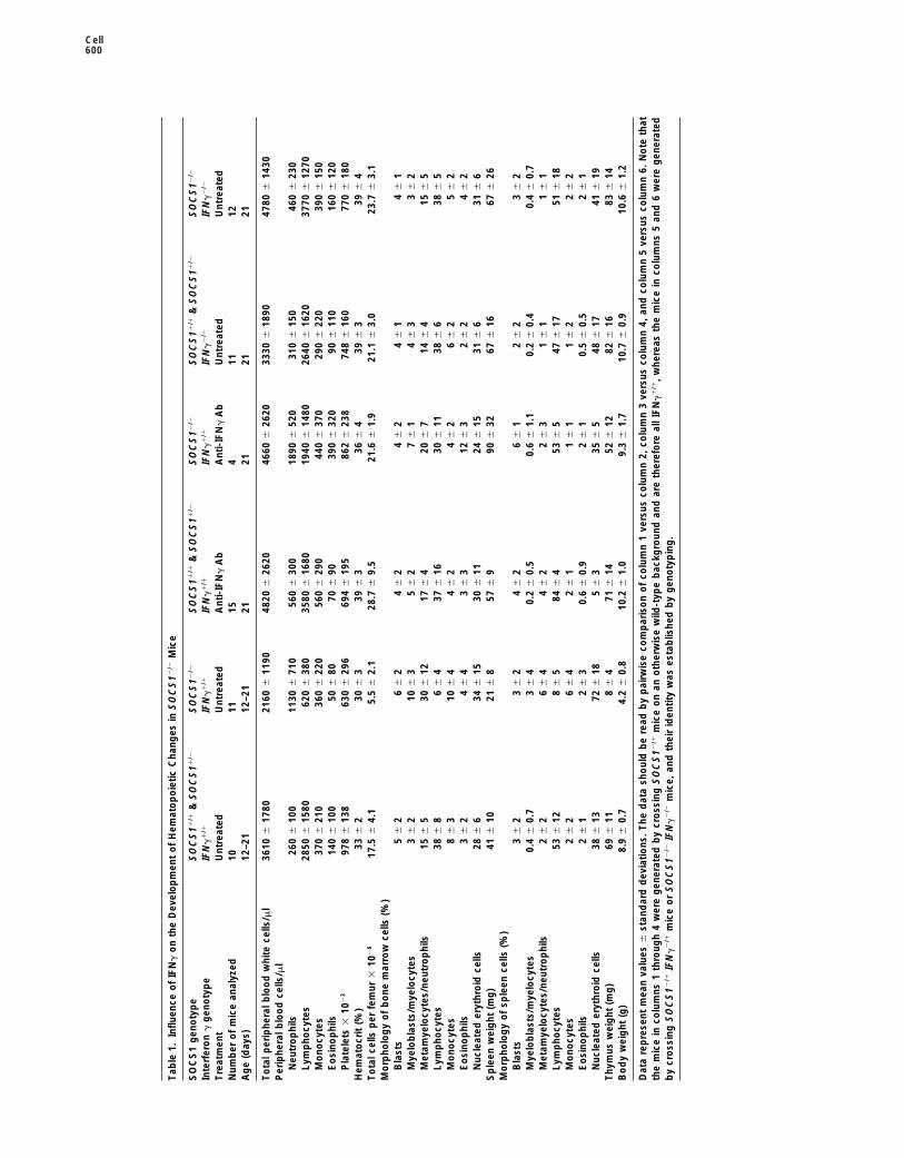
Tab
le1.
Influ
ence
of
IFN
go
nth
eD
evel
op
men
to
fH
emat
op
oie
ticC
hang
esin
SO
CS
12/ 2
Mic
e
SO
CS
1g
eno
typ
eS
OC
S11
/ 1&
SO
CS
11/ 2
SO
CS
12/ 2
SO
CS
11/ 1
&S
OC
S11
/ 2S
OC
S12
/ 2S
OC
S11
/ 1&
SO
CS
11/ 2
SO
CS
12/ 2
Inte
rfer
on
gg
eno
typ
eIF
Ng
1/ 1
IFN
g1
/ 1IF
Ng
1/ 1
IFN
g1
/ 1IF
Ng
2/ 2
IFN
g2
/ 2
Tre
atm
ent
Unt
reat
edU
ntre
ated
Ant
i-IF
Ng
Ab
Ant
i-IF
Ng
Ab
Unt
reat
edU
ntre
ated
Num
ber
of
mic
ean
alyz
ed10
1115
411
12A
ge
(day
s)12
–21
12–2
121
2121
21
To
talp
erip
hera
lblo
od
whi
tece
lls/m
l36
106
1780
2160
611
9048
206
2620
4660
626
2033
306
1890
4780
614
30P
erip
hera
lblo
od
cells
/ml
Neu
tro
phi
ls26
06
100
1130
671
056
06
300
1890
652
031
06
150
460
623
0Ly
mp
hocy
tes
2850
615
8062
06
380
3580
616
8019
406
1480
2640
616
2037
706
1270
Mo
nocy
tes
370
621
036
06
220
560
629
044
06
370
290
622
039
06
150
Eo
sino
phi
ls14
06
100
506
8070
690
390
632
090
611
016
06
120
Pla
tele
ts3
102
397
86
138
630
629
669
46
195
862
623
874
86
160
770
618
0H
emat
ocr
it(%
)33
62
306
339
63
366
439
63
396
4T
ota
lcel
lsp
erfe
mur
310
26
17.5
64.
15.
56
2.1
28.7
69.
521
.66
1.9
21.1
63.
023
.76
3.1
Mo
rpho
log
yo
fb
one
mar
row
cells
(%)
Bla
sts
56
26
62
46
24
62
46
14
61
Mye
lob
last
s/m
yelo
cyte
s3
62
106
35
62
76
14
63
36
2M
etam
yelo
cyte
s/ne
utro
phi
ls15
65
306
1217
64
206
714
64
156
5Ly
mp
hocy
tes
386
86
64
376
1630
611
386
638
65
Mo
nocy
tes
86
310
64
46
24
62
66
25
62
Eo
sino
phi
ls3
62
46
43
63
126
32
62
46
2N
ucle
ated
eryt
hro
idce
lls28
66
346
1530
611
246
1531
66
316
6S
ple
enw
eig
ht(m
g)
416
1021
68
576
990
632
676
1667
626
Mo
rpho
log
yo
fsp
leen
cells
(%)
Bla
sts
36
23
62
46
26
61
26
23
62
Mye
lob
last
s/m
yelo
cyte
s0.
46
0.7
36
40.
26
0.5
0.6
61.
10.
26
0.4
0.4
60.
7M
etam
yelo
cyte
s/ne
utro
phi
ls2
62
66
44
62
26
31
61
16
1Ly
mp
hocy
tes
536
128
65
846
453
65
476
1751
618
Mo
nocy
tes
26
26
64
26
11
61
16
22
62
Eo
sino
phi
ls2
61
26
30.
66
0.9
26
10.
56
0.5
26
1N
ucle
ated
eryt
hro
idce
lls38
613
726
185
63
356
548
617
416
19T
hym
usw
eig
ht(m
g)
696
118
64
716
1452
612
826
1683
614
Bo
dy
wei
ght
(g)
8.9
60.
74.
26
0.8
10.2
61.
09.
36
1.7
10.7
60.
910
.66
1.2
Dat
are
pre
sent
mea
nva
lues
6st
and
ard
dev
iatio
ns.
The
dat
ash
oul
db
ere
adb
yp
airw
ise
com
par
iso
no
fco
lum
n1
vers
usco
lum
n2,
colu
mn
3ve
rsus
colu
mn
4,an
dco
lum
n5
vers
usco
lum
n6.
No
teth
atth
em
ice
inco
lum
ns1
thro
ugh
4w
ere
gen
erat
edb
ycr
oss
ing
SO
CS
12/ 1
mic
eo
nan
oth
erw
ise
wild
-typ
eb
ackg
roun
dan
dar
eth
eref
ore
allI
FN
g1
/ 1,
whe
reas
the
mic
ein
colu
mns
5an
d6
wer
eg
ener
ated
by
cro
ssin
gS
OC
S12
/ 1IF
Ng
2/ 1
mic
eo
rS
OC
S12
/ 2IF
Ng
2/ 2
mic
e,an
dth
eir
iden
tity
was
esta
blis
hed
by
gen
oty
pin
g.

SOCS1 Is a Key Regulator of IFNg Signaling601
effector involved in the intracellular killing of L. major(data not shown).
The hyperresponsiveness of cells from SOCS12/2
mice to IFNg in vitro was paralleled by an increasedresistance of SOCS12/2 mice to viral infection in vivo.When 5-day-old mice were infected with Semliki forestvirus, all SOCS11/1 and SOCS11/2 mice died within 2 to3 days. However, SOCS12/2 mice largely survived thisearly postinfection period and only became ill 3 to 21days after viral challenge (Figure 5B). This suggests analmost complete resistance to infection, because thesurvival pattern of infected and uninfected SOCS12/2
mice was similar.IFNg levels in the serum and in media conditioned by
organs taken from SOCS12/2 or control littermate micewere investigated using ELISA and/or antiviral assays.At a limit of detection of 3 U/ml, we were unable todetect IFNg in serum of mice of either genotype, andmedia conditioned by organs from SOCS12/2 mice didnot reproducibly contain more IFNg than those fromtheir wild-type littermates.
Neonatal Death of SOCS12/2 Mice Occursunder Germ-Free ConditionsBecause IFNg was clearly central to the developmentof disease in SOCS12/2 mice, we were interested todetermine whether subclinical infection in conventionalmice might induce increased production of IFNg andprecipitate the early neonatal death of SOCS12/2 mice.Three litters of pups produced from the mating ofSOCS11/2 mice were delivered by cesarean section into
Figure 2. Development of Disease in SOCS12/2 Mice Is Dependent a strictly germ-free environment. As expected, SOCS11/1
on IFNg and Occurs in a Germ-Free Environment and SOCS11/2 pups were among the progeny, and these(A) Rapid onset of morbidity of untreated SOCS12/2 but not were weaned normally and as adults exhibited enlargedSOCS11/1 or SOCS11/2 mice.
ceca characteristic of mice in a germ-free environment.(B) Twice weekly injection of neonatal SOCS12/2 mice with anti-In contrast, germ-free SOCS12/2 pups died during theIFNg but not anti-IL-6 or control antibody prevents morbidity.second and third weeks of life, the same age at which(C) Rapid onset of morbidity in SOCS12/2 but not SOCS11/1 or
SOCS11/2 mice born and reared in a germ-free environment. conventional SOCS12/2 mice die (Figure 2C). Analyses(D) Prevention of morbidity in SOCS12/2 IFNg2/2 mice and ameliora- of seven of these moribund germ-free SOCS12/2 micetion of morbidity in SOCS12/2 IFNg1/2 mice in comparison with revealed that their organ pathology and peripheral bloodSOCS12/2 IFNg1/1 mice. Seventeen SOCS12/2 IFNg2/2 mice have
abnormalities were identical to SOCS12/2 mice raisedbeen examined to date, with twelve animals being sacrificed forunder conventional conditions.detailed histological and hematological analysis at 3 weeks of age
and the remaining five being left for long-term observation.
Discussion
SOCS1 Is a Critical Regulator of IFNg-Mediatedin the presence of 100 ng/ml lipopolysaccharide. After2 hr at all IFNg concentrations, 30% to 50% of macro- Signal Transduction
SOCS1 is indispensable for survival beyond the postna-phages from both SOCS11/1 and SOCS12/2 mice wereinfected, and this percentage persisted for up to 48 hr tal period. Mice lacking this regulator die before weaning
from a complex disease characterized by fatty degener-in macrophages of both genotypes in the absence ofcytokine (Figure 5A). The ability of IFNg activation to ation of the liver, macrophage infiltration of several or-
gans, and severe lymphopenia (Starr et al., 1998). Weenhance macrophage killing of L. major was demonstra-ble, because only 1% to 3% of cells from both SOCS11/1 demonstrate here that this fatal syndrome is associated
with the specific activation of STAT1 and elevated ex-and SOCS12/2 mice were still infected with live para-sites 48 hr after treatment with 10 U/ml of IFNg (Figure pression of IRF1, iNOS, and class I MHC, strongly im-
plying that IFNg signaling pathways are aberrantly active5A). Strikingly, although #0.1 U/ml IFNg had little or noeffect on killing by SOCS11/1 macrophages, cells from in vivo in the absence of SOCS1. Remarkably, all organ
pathology was eliminated or markedly reduced, andSOCS12/2 mice maintained efficient killing when stimu-lated with as little as 0.01 U/ml IFNg (Figure 5A), thus death was prevented by treatment of neonatal SOCS12/2
mice with a neutralizing anti-IFNg antibody. The amelio-exhibiting at least 100-fold increased sensitivity to IFNg.The hypersensitivity of macrophages from SOCS12/2 ration of disease was specific to the inhibition of IFNg
because treatment of SOCS12/2 mice with neutralizingmice to IFNg was confirmed by the demonstration of anincreased capacity to produce nitric oxide, the major anti-IL-6 antibodies or control immunoglobulin had no

Cell602
Figure 3. Prevention of Tissue Pathology inSOCS12/2 Mice by Treatment with Anti-IFNg
Antibody
Neonatal SOCS12/2 mice were treated twiceweekly with either anti-IFNg antibody or con-trol antibody. Anti-IFNg antibody–treatedmice were healthy at 21 days of age and weresacrificed. Control antibody–treated mice de-veloped disease between 9 and 16 days ofage and were sacrificed when moribund. Thenormal histological appearance of organsfrom anti-IFNg antibody-treated SOCS12/2
mice (right-hand panels) is contrasted withthe atrophy of the cortex of the thymus, thefatty degeneration, hematopoietic infiltrationand necrosis of the liver, macrophage accu-mulation around the bronchi and in the alveo-lar walls in the lung, and macrophage infiltra-tion and acinar tissue destruction of thepancreas observed in SOCS12/2 mice treatedwith control antibody (left-hand panels).
protective effect. We were also unable to find any evi- also displayed no consistent differences in IFNg concen-trations. Finally, while SOCS12/2 IFNg2/2 mice have re-dence of dysregulated activation of STAT3 in SOCS12/2
mice, the STAT protein used preferentially by inflamma- mained disease free, some SOCS12/2 IFNg1/2 mice dodie with a more slowly developing disease. The delayedtory cytokines such as IL-6 and LIF (Ihle et al., 1994). In
addition, the full spectrum of disease development was disease in mice containing only a single functional IFNgallele suggests that IFNg concentrations may be limitingcompletely prevented in mice genetically deficient in
IFNg as well as SOCS1, and a delayed disease devel- in these mice and therefore unlikely to be grossly ele-vated in SOCS12/2 mice. IFNg is thought to be usuallyoped in SOCS12/2 mice that had only a single functional
copy of the IFNg gene. The absence of SOCS1 corre- produced by T cells and NK cells in response to infec-tion. Since SOCS12/2 mice succumb to disease inlated with marked in vitro hypersensitivity to IFNg in
macrophages infected with the intracellular parasite L. strictly germ-free conditions, the IFNg required for dis-ease development in SOCS12/2 mice might either repre-major. Hypersensitivity to IFNg was also observed when
nitric oxide (NO) production was measured following sent basal production or be produced aberrantly. Whileour studies have clearly demonstrated an indispensablestimulation of macrophages with LPS and IFNg. Like-
wise, hematopoietic cells from SOCS12/2 mice have also role for IFNg in the multiorgan pathology of SOCS12/2
mice, the source of this cytokine and the identity ofbeen found to be more susceptible to inhibition of prolif-eration by IFNg but normally responsive to most hemato- potential downstream effectors of IFNg action remain
to be examined.poietic cytokines that also act through the JAK/STATpathway (Metcalf et al., 1999). Together these dataclearly establish that SOCS1 is an essential physiologi-cal regulator of cellular sensitivity to IFNg signaling. Specificity of SOCS1 Action In Vivo
Previous in vitro data suggested that SOCS1 can directlyOur results do not exclude the possibility that an ele-vated production of IFNg in SOCS12/2 mice might con- interact with all four members of the JAK family, resulting
in the inhibition of their catalytic activity (Endo et al.,tribute to disease development. However, within the sensi-tivity of the assays used, as in normal animals, we were 1997; Naka et al., 1997). Consistent with this range of
action, overexpression of SOCS1 in cell lines inhibitedunable to detect IFNg in the circulation of SOCS12/2 mice.Media conditioned by organs of SOCS12/2 or normal mice signal transduction from a large number of cytokines,

SOCS1 Is a Key Regulator of IFNg Signaling603
a lethal hyperresponsiveness to IFNg, abnormal re-sponses to other cytokines may not be manifested sodramatically. Our studies to date suggest that responsesto most hematopoietic cytokines are not altered inSOCS12/2 mice (Metcalf et al., 1999). However, the post-natal lethality in mice lacking this protein has preventedanalysis of signaling by these or other cytokines in theadult mouse. Clearly, the availability of healthy SOCS12/2
IFNg2/2 mice will now allow the full spectrum of in vivoSOCS1 activity to be definitively resolved.
Binding of IFNg to its cell surface receptor triggersthe activation of JAK1 and JAK2. While in vitro studieshave shown SOCS1 is able to inhibit the function of boththese kinases, the biochemical specificity of SOCS1 ac-tion in vivo remains to be determined. The inhibition ofJAK activity by SOCS1 may regulate IFNg signaling inseveral ways, for example, by determining the thresholdconcentration of IFNg capable of triggering a biologicalresponse or by regulating the magnitude or length of aresponse to a particular concentration of IFNg. Distin-guishing between these possibilities will require a care-ful comparison of the capacity of different doses of IFNg
Figure 4. Prevention of B Lymphopenia in SOCS12/2 Mice by Treat- to induce JAK1 and JAK2 activation, IFNg receptorment with Anti-IFNg Antibody or Generation of SOCS12/2 IFNg2/2
phosphorylation, STAT1 activation, and expression ofMice STAT1-regulated genes in wild-type and SOCS1-defi-Spleen cells were collected from (A) wild-type mice, (B) SOCS12/2
cient cells.mice treated with a control antibody, (C) SOCS12/2 mice treated
As SOCS1 is a member of a larger family of relatedwith anti-IFNg antibody, and (D) SOCS12/2 IFNg2/2 mice. Cells wereproteins, it is also feasible that a regulatory role forstained with anti-B220 antibody and a mixture of anti-IgM and anti-this protein in other cytokine signaling pathways is notIgD antibodies and analyzed by FACS.evident in SOCS12/2 mice due to the compensatory ac-tions of other SOCS proteins. In vitro studies have clearlyshown that SOCS1 and SOCS3 have overlapping activi-including IFNa, IFNb, IFNg, IL-6, LIF, OSM, thrombopoie-ties, and both molecules are induced by and inhibit thetin, growth hormone, and stem cell factor (SCF). More-actions of a similar spectrum of cytokines when overex-over, each of these cytokines is able to rapidly inducepressed (Adams et al., 1998; Sakamoto et al., 1998; Songthe synthesis of SOCS1 mRNA (Endo et al., 1997; Starrand Shuai, 1998; Ito et al., 1999). However, for regulationet al., 1997; Adams et al., 1998; Auernhammer et al.,of IFNg signaling, SOCS1 appears to be considerably1998; Bjorbaek et al., 1998; Sakamoto et al., 1998; Songmore active than SOCS3 (Sakamoto et al., 1998; Songand Shuai, 1998; Ito et al., 1999; De Sepulveda et al.,and Shuai, 1998). Consistent with these in vitro studies,1999).the phenotype of SOCS12/2 mice reveals key roles forGiven the promiscuity of SOCS1 action in vitro, dis-SOCS1 in regulating IFNg responses that cannot beease in mice lacking this protein might potentially havecompensated by SOCS3 in vivo. Whether SOCS3 alsobeen due to deregulation of signals from a multitude ofacts to regulate a unique set of cytokine signal transduc-cytokines. Remarkably, however, the full spectrum oftion pathways and whether there are other pathwaysthe complex disease in SOCS12/2 mice, including liverregulated by both SOCS1 and SOCS3 under physiologi-degeneration, hematopoietic infiltration of multiple tis-cal conditions remains to be clarified.sues, and profound loss of T and B lymphocytes, can
be attributed to a failure to regulate signal transductionby a single cytokine, IFNg. This implies that in contrast SOCS1 Balances the Beneficial and Potentially
Deleterious Actions of IFNgto its actions in vitro, under physiological conditionsSOCS1 has no indispensable role in the regulation of The potentially devastating actions of IFNg in vivo have
been well established in studies in which IFNg levelssignal transduction from the majority of cytokines thatutilize the JAK-STAT pathway. have been experimentally elevated (Gresser et al., 1981,
1987; Gresser, 1982), and these pathologies were faith-As the in vitro studies all involved overexpression ofSOCS1, it is possible that these approaches overesti- fully reproduced in mice lacking SOCS1. In addition,
inappropriate IFNg production and action is implicatedmated the breadth of SOCS action, simply because thelevel of SOCS gene expression achieved experimentally in the pathogenesis of disease following certain infec-
tions. In a mouse transgenic model of hepatitis B, theis likely to be far higher than expression controlled by anendogenous promoter. Moreover, ectopic expression destruction of liver cells and resultant morbidity appears
to be caused by excess endogenous IFNg (Ando et al.,may also be temporally inappropriate because physio-logical transcription of SOCS genes appears to be de- 1993). In other models of infection, however, the actions
of IFNg are beneficial (Huang et al., 1993; Newport etpendent on exposure to cytokine, allowing SOCS pro-teins to act in vivo only after signaling has commenced. al., 1996; Lu et al., 1998), and this action is also amplified
in mice lacking SOCS1. SOCS1-deficient macrophagesHowever, although it is clear that a lack of SOCS1 causes

Cell604
Tab
le2.
Influ
ence
of
IFN
go
nth
eD
evel
op
men
to
fH
isto
log
ical
Cha
nges
inO
rgan
so
fS
OC
S12
/ 2M
ice
SO
CS
1g
eno
typ
eS
OC
S11
/ 1&
SO
CS
11/ 2
SO
CS
12/ 2
SO
CS
11/ 1
&S
OC
S11
/ 2S
OC
S12
/ 2S
OC
S11
/ 1&
SO
CS
11/ 2
SO
CS
12/ 2
Inte
rfer
on
gg
eno
typ
eIF
Ng
1/ 1
IFN
g1
/ 1IF
Ng
1/ 1
IFN
g1
/ 1IF
Ng
2/ 2
IFN
g2
/ 2
Tre
atm
ent
Unt
reat
edU
ntre
ated
Ant
i-IF
Ng
Ab
Ant
i-IF
Ng
Ab
Unt
reat
edU
ntre
ated
Ag
e(d
ays)
12–2
112
–21
2121
2121
Live
rF
atty
deg
ener
atio
n0/
2112
/18
0/22
0/6
0/8
0/12
Nec
rosi
s0/
216/
180/
220/
60/
80/
12E
xces
she
mat
op
oie
sis
0/21
18/1
80/
220/
60/
80/
12Lu
ng Cuf
fing
by
mφ
and
neut
1/21
17/1
80/
222/
60/
82/
12H
eart
Infil
trat
ion
by
mφ
and
neut
0/21
15/1
70/
191/
60/
80/
12K
idne
yG
lom
erul
arim
mat
urity
2/21
16/1
81/
180/
61/
81/
12T
ubul
arim
mat
urity
6/21
11/1
80/
180/
60/
80/
12S
ple
enLy
mp
hoid
folli
cles
20/2
11/
1921
/21
5/5
8/8
12/1
2E
xces
ser
ythr
op
oie
sis
1/21
15/1
70/
214/
53/
89/
12T
hym
usC
ort
ical
atro
phy
0/21
15/1
60/
221/
50/
80/
12M
edul
lary
enla
rgem
ent
1/21
1/16
0/22
1/5
1/8
8/12
Pan
crea
sA
cina
rat
rop
hy2/
1916
/18
0/22
0/5
0/8
0/12
Infil
trat
ion
by
mφ
0/19
13/1
80/
220/
50/
80/
12S
kin Mφ
infil
trat
ion
0/20
14/1
70/
101/
40/
80/
12E
pid
erm
alth
icke
ning
0/20
15/1
70/
100/
40/
80/
12K
erat
iniz
atio
n0/
2014
/17
0/10
0/4
0/8
0/12
Eo
sino
phi
linf
iltra
tion
0/20
6/17
0/10
0/4
0/8
0/12
Mar
row
Mo
nos
.g
ranu
locy
tes
16/2
02/
1816
/19
3/5
8/8
12/1
2G
ranu
locy
tes
.m
ono
s4/
2016
/18
3/19
2/5
0/8
0/12
The
ratio
sin
dic
ate
the
num
ber
of
org
ans
sho
win
ga
par
ticul
arle
sio
nve
rsus
the
tota
lnum
ber
of
org
ans
exam
ined
.T
hed
ata
sho
uld
be
read
by
pai
rwis
eco
mp
aris
on
of
colu
mn
1ve
rsus
colu
mn
2,co
lum
n3
vers
usco
lum
n4,
and
colu
mn
5ve
rsus
colu
mn
6.N
ote
that
the
mic
ein
colu
mns
1th
roug
h4
wer
eg
ener
ated
by
cro
ssin
gS
OC
S12
/ 1m
ice
on
ano
ther
wis
ew
ild-t
ype
bac
kgro
und
and
are
ther
efo
real
lIF
Ng
1/ 1
,whe
reas
the
mic
ein
colu
mns
5an
d6
wer
eg
ener
ated
by
cro
ssin
gS
OC
S12
/ 1IF
Ng
2/ 1
mic
eo
rS
OC
S12
/ 2IF
Ng
2/ 2
mic
e,an
dth
eir
iden
tity
was
conf
irm
edb
yg
eno
typ
ing
.No
tal
lorg
ans
wer
ese
ctio
ned
fro
mal
lani
mal
s.N
eut,
neut
rop
hil;
mo
nos,
mo
nonu
clea
rce
lls.

SOCS1 Is a Key Regulator of IFNg Signaling605
environment was monitored closely and tested continually for theabsence of bacterial organisms and a variety of viral pathogens.
Mice were genotyped by Southern blot analysis of genomic DNAobtained from tail tips, as described (Starr et al., 1998). For analysisof the SOCS1 gene, genomic DNA was digested with EcoRI andfilters were probed with a 1.5 kilobase pairs (kbp) EcoRI/HindIIIfragment of the mouse SOCS1 gene, which hybridizes to a 5.3 kbpband for the wild-type allele and an 8.0 kbp fragment for the nullallele. For analysis of the IFNg gene, DNA was cut with BamHI andfilters were probed with a 450 bp PstI fragment of the mouse IFNg
cDNA that detects a fragment of 11.0 kbp for the wild-type alleleand 13.0 kbp for the null allele.
Neonatal mice were injected intraperitoneally twice weekly for upto 3 weeks with 16 mg/kg of neutralizing rat anti-IFNg antibody (R4-6A2; American Type Culture Collection, Manassas, VA), 200 mg/kgof an ammonium sulfate–precipitated neutralizing polyclonal rabbitanti-IL-6 antisera, as described (Liu et al., 1995), 16 mg/kg controlpurified rat IgG antibody (Sigma Chemical Co., St. Louis, MO), or200 mg/kg ammonium sulfate–precipitated preimmune rabbit seraand were sacrificed when moribund or at the end of the experiment.The efficacy of anti-IL-6 treatment was verified at the end of theexperiment by testing serum from injected mice for its capacity toinhibit IL-6-induced differentiation of M1 cells. In a titration experi-ment, a 1 in 100 dilution of serum from mice injected with the anti-IL-6 but not control antibody was found to inhibit 60 ng/ml of IL-6.
In some experiments, 5-day-old mice were injected intraperitone-ally with 50 ml of Semliki forest virus containing 100 times the virusdose that kills 50% of fibroblasts in vitro. The mice were monitoreddaily and sacrificed when moribund.
Figure 5. Absence of SOCS1 Renders Cells Hyperresponsive toIFNg AssaysIFNg and Mice Hyperresistant to Semliki Forest Virus InfectionIFNg in serum was assayed using cytopathic effect reduction with
(A) Macrophages from the bone marrow of SOCS11/1 (filled sym-mouse L929 cells as targets for Semliki forest virus challenge as
bols) and SOCS12/2 mice (open symbols) were stimulated with 100described (Hertzog et al., 1991). A standard cytokine sandwich
ng/ml LPS and the indicated concentration of IFNg and infectedELISA was also used to quantitate IFNg in mouse serum and organ-
with L. major. After 2 hr (circles) and 48 hr (squares), the percentageconditioned media. IFNg-specific antibodies (R4-6A2 as the capture
of macrophages containing parasites was determined.antibody, XMG1.2 for detection) were obtained from Pharmingen
(B) Mice generated by crossing SOCS11/2 mice were infected with(San Diego, CA), and the assay was performed essentially as de-
Semliki forest virus at 5 days of age and monitored.scribed by the manufacturer.
Hematological Analysis, Histology, and Immunohistochemistrystimulated with IFNg killed intracellular parasites consid- Hematological analyses were performed as described (Starr et al.,erably more efficiently than their wild-type counterparts, 1998; Metcalf et al., 1999). For histological examination, tissues were
fixed in 10% (v/v) formalin in phosphate-buffered saline (PBS) andand SOCS12/2 mice were more resistant than normalsections were prepared and stained by standard techniques (Starrmice to Semliki forest virus infection. Clearly, the actionset al., 1998). Tissue preparation for immunohistochemistry was car-of IFNg in vivo require exquisite control. The data pre-ried out as described (Thomas et al., 1998), and sections were
sented here establish that SOCS1 is central to the bal- stained with a rat anti-mouse class I MHC antibody, 34-12S, and aance between the beneficial and potentially harmful ef- peroxidase-labeled secondary antibody. Sections were counter-fects of IFNg. While the behavior of neonatal mice may stained with Giemsa (Ozato et al., 1982).not necessarily predict the occurrence of similar re-sponses to overstimulation by IFNg in humans, these Culture of Macrophages and Leishmanicidal Assays
To obtain bone marrow–derived macrophages, femoral bone mar-observations nevertheless raise the prospect that smallrow cells collected in PBS were dispersed, centrifuged, and resus-molecule mimetics or antagonists of SOCS1 might provepended in 10 ml Dulbecco’s modified Eagle’s medium (DMEM) sup-clinically valuable in enhancing the actions of IFNg inplemented with 100 ng/ml murine macrophage colony-stimulating
combating viral and parasitic infection or in dampening factor (M-CSF; Cetus Corporation, Emeryville, CA) and 10% (v/v)its pathological side effects in other disease states. fetal bovine serum (FBS). After 3 days of culture in a humidified
incubator at 378C in 5% (v/v) CO2 in air, medium and nonadherentcells containing an expanded precursor population were resus-Experimental Procedurespended at 104 cells/ml and cultured for an additional 5 days in thepresence of M-CSF, after which a relatively pure and homogeneousGeneration and Maintenance of Mice and Injection
of Antibodies, Cytokine, and Virus population of adherent mature bone marrow macrophages werepresent.SOCS12/2 mice were generated as described previously (Starr et
al., 1998) and maintained on a mixed 129/Sv and C57BL/6 genetic The cloned line of L. major LRC-L137 V 121 has been described indetail (Handman, 1983). Promastigotes were grown in M199 mediumbackground. IFNg2/2 mice on an inbred C57BL/6 background
(C57BL/6-Ifngtm1Ts) were obtained from the Jackson Laboratories via with 10% FBS and were in the stationary phase of growth. Infectionof macrophages was carried out using a modification of a methodMonash University (Dalton et al., 1993). Mice were routinely housed
in clean but not specific pathogen-free conditions (SPF) at the Walter described previously (Proudfoot et al., 1995). Briefly, 5 3 104 bonemarrow–derived macrophages were transferred onto glass cov-and Eliza Hall Institute of Medical Research. To raise mice under
germ-free conditions, pups were delivered by cesarean section and erslips in 24-well trays, allowed to adhere for 24 hr, and stimulatedwith 100 ng/ml LPS and various concentrations of IFNg. After 6 hrplaced with BALB/c foster mothers that had been maintained in
germ-free microisolators for several generations. The sterility of this of cytokine stimulation, cells were infected at a ratio of five parasites

Cell606
per cell for 2 hr at 378C. The free parasites were removed by vigorous expression and adrenocorticotropin secretion. Mol. Endocrinol. 12,954–961.washing before incubation for a further 24 or 48 hr, after which cells
were stained with Giemsa. The percentage of macrophages infected Bach, E.A., Aguet, M., and Schreiber, R.D. (1997). The IFN gammawith parasites was determined at each time point, with at least 400 receptor: a paradigm for cytokine receptor signaling. Annu. Rev.cells per sample counted. Immunol. 15, 563–591.
Billiau, A. (1996). Interferon-gamma: biology and role in pathogene-Northern Blots sis. Adv. Immunol. 62, 61–130.Following sacrifice, organs were removed from neonatal wild-type Billiau, A., and Vandekerckhove, F. (1991). Cytokines and their inter-and SOCS12/2 mice. PolyA1 mRNA was purified, and Northern blot actions with other inflammatory mediators in the pathogenesis ofhybridization was performed essentially as described (Alexander et sepsis and septic shock. Eur. J. Clin. Inv. 21, 559–573.al., 1995). Probes used were as follows: a 1.1 kbp PstI fragment of
Bjorbaek, C., Elmquist, J.K., Frantz, J.D., Shoelson, S.E., and Flier,the chicken glyceraldehyde 6-phosphate dehydrogenase (GAPDH)
J.S. (1998). Identification of SOCS-3 as a potential mediator of cen-cDNA, a 2 kbp EcoRI fragment of the mouse IRF1 gene, and a 1.8 tral leptin resistance. Mol. Cell 1, 619–625.kbp NcoI fragment of the mouse iNOS cDNA.
Boehm, U., Klamp, T., Groot, M., and Howard, J.C. (1997). Cellularresponses to interferon-gamma. Annu. Rev. Immunol. 15, 749–795.
Flow CytometryDalton, D.K., Pitts-Meek, S., Keshav, S., Figari, I.S., Bradley, A., andSingle-cell suspensions of femoral bone marrow, thymocytes, andStewart, T.A. (1993). Multiple defects of immune cell function in micesplenocytes were prepared and erythrocytes were lysed by incuba-with disrupted interferon-gamma genes. Science 259, 1739–1742.tion in 156 mM ammonium chloride (pH 7.3) at 378C for 3 min.Darnell, J.E., Jr. (1997). STATs and gene regulation. Science 277,The cells were stained with a biotinylated or FITC-conjugated rat1630–1635.monoclonal antibody specific for the cell surface markers of interestDarnell, J.E., Jr., Kerr, I.M., and Stark, G.R. (1994). Jak-STAT path-(Class I MHC, CD4, CD8, surface IgM, surface IgD, B220, or Mac1),ways and transcriptional activation in response to IFNs and otherfollowed where necessary by streptavidin-phycoerythrin and ana-extracellular signaling proteins. Science 264, 1415–1421.lyzed by flow cytometry as described (Strasser et al., 1991).De Maeyer, E., and De Maeyer-Guignard, S. (1998). Interferons. InThe Cytokine Handbook, A. Thomson, ed. (San Diego, CA: AcademicElectrophoretic Mobility Shift AssaysPress), pp. 491–516.Nuclei were extracted from the livers of unmanipulated SOCS12/2
De Sepulveda, P., Okkenhaug, K., La Rose, J., Hawley, R.G., Du-mice and control littermates or from mice 15 min after intraperitonealbreuil, P., and Rottapel, R. (1999). Socs1 binds to multiple signalingadministration of 2 mg IFNg (Ruff-Jamison et al., 1993). Electropho-proteins and suppresses Steel factor dependent proliferation.retic mobility shift assays (EMSA) were performed on 1–2 mg ofEMBO J. 18, 904–915.nuclear protein using the m67 oligonucleotide probe as described
(Novak et al., 1995). Durbin, J.E., Hackenmiller, R., Simon, M.C., and Levy, D.E. (1996).Targeted disruption of the mouse Stat1 gene results in compromisedinnate immunity to viral disease. Cell 84, 443–450.AcknowledgmentsEndo, T.A., Masuhara, M., Yokouchi, M., Suzuki, R., Sakamoto, H.,Mitsui, K., Matsumoto, A., Tanimura, S., Ohtsubo, M., Misawa, H.,We are grateful to Drs. David Hume, David Levy, and Peter Murrayet al. (1997). A new protein containing an SH2 domain that inhibitsfor their advice and input. We thank Dr. Andreas Strasser for sugges-JAK kinases. Nature 387, 921–924.tions of experiments and provision of antibodies; Dr. Richard Simp-
son for the anti-IL-6 antibodies that were kindly purified by Dr. Jian- Greenlund, A.C., Farrar, M.A., Viviano, B.L., and Schreiber, R.D.Guo Zhang; Gillian Carter and Kathy Hanzinikolas for expert animal (1994). Ligand-induced IFN gamma receptor tyrosine phosphoryla-husbandry; Ladina Di Rago, Sandra Mifsud, Joan Curtis, Anabel tion couples the receptor to its signal transduction system (p91).
EMBO J. 13, 1591–1600.Silva, Dale Cary, Bronwyn Roberts, Elizabeth Viney, Alison Farley,and Steven Mihajlovic for first-class technical assistance; Dr. Sandra Gresser, I. (1982). Can interferon induce disease? Interferon 4,Nicholson for help in generating the 2fTGH cell lines; Dr. Tracy 95–127.Willson for preparing the IFNg cDNA probe; and Dr. Ian Kerr for Gresser, I., Aguet, M., Morel-Maroger, L., Woodrow, D., Puvion-generously providing 2fTGH cells. R. S. was supported by a Post- Dutilleul, F., Guillon, J.C., and Maury, C. (1981). Electrophoreticallydoctoral Fellowship from the Australian Research Council. This work pure mouse interferon inhibits growth, induces liver and kidney le-was supported by the Anti-Cancer Council of Victoria, Melbourne, sions, and kills suckling mice. Am. J. Pathol. 102, 396–402.Australia; AMRAD Operations Pty. Ltd., Melbourne, Australia; The Gresser, I., Woodrow, D., Moss, J., Maury, C., Tavernier, J., andNational Health and Medical Research Council, Canberra, Australia; Fiers, W. (1987). Toxic effects of recombinant tumor necrosis factorThe J. D. and L. Harris Trust; The National Institutes of Health, in suckling mice. Comparisons with interferon alpha/beta. Am. J.Bethesda, Maryland (Grant CA-22556), and the Australian Federal Pathol. 128, 13–18.Government Cooperative Research Centres Program.
Handman, E. (1983). Association of serum proteins with culturedLeishmania: a warning note. Parasite Immunol. 5, 109–112.
Received April 1, 1999; revised July 20, 1999.Heim, M.H., Kerr, I.M., Stark, G.R., and Darnell, J.E., Jr. (1995). Con-tribution of STAT SH2 groups to specific interferon signaling by the
References Jak-STAT pathway. Science 267, 1347–1349.
Hemmi, S., Bohni, R., Stark, G., Di Marco, F., and Aguet, M. (1994).Adams, T.E., Hansen, J.A., Starr, R., Nicola, N.A., Hilton, D.J., and A novel member of the interferon receptor family complements func-Billestrup, N. (1998). Growth hormone preferentially induces the tionality of the murine interferon gamma receptor in human cells.rapid, transient expression of SOCS-3, a novel inhibitor of cytokine Cell 76, 803–810.receptor signaling. J. Biol. Chem. 273, 1285–1287.
Hertzog, P.J., Wright, A., Harris, G., Linnane A.W., and Mackay,Alexander, W.S., Metcalf, D., and Dunn, A.R. (1995). Point mutations I.R. (1991). Intermittent interferonemia and interferon responses inwithin a dimer interface homology domain of c-mpl induce constitu- multiple sclerosis. Clin. Immunol. Immunopathol. 58, 18–32.tive receptor activity and tumorogenesis. EMBO J. 14, 5569–5578. Hilton, D.J., Richardson, R.T., Alexander, W.S., Viney, E.M., Willson,Ando, K., Moriyama, T., Guidotti, L.G., Wirth, S., Schreiber, R.D., T.A., Sprigg, N.S., Starr, R., Nicholson, S.E., Metcalf, D., and Nicola,Schlicht, H.J., Huang, S.N., and Chisari, F.V. (1993). Mechanisms of N.A. (1998). Twenty proteins containing a C-terminal SOCS box formclass I restricted immunopathology. A transgenic mouse model of five structural classes. Proc. Natl. Acad. Sci. USA 95, 114–119.fulminant hepatitis. J. Exp. Med. 178, 1541–1554. Huang, S., Hendriks, W., Althage, A., Hemmi, S., Bluethmann, H.,Auernhammer, C.J., Chesnokova, V., Bousquet, C., and Melmed, S. Kamijo, R., Vilcek, J., Zinkernagel, R.M., and Aguet, M. (1993). Im-(1998). Pituitary corticotroph SOCS-3: novel intracellular regulation mune response in mice that lack the interferon-gamma receptor.
Science 259, 1742–1745.of leukemia-inhibitory factor-mediated proopiomelanocortin gene
SOCS1 Is a Key Regulator of IFNg Signaling607
Igarashi, K., Garotta, G., Ozmen, L., Ziemiecki, A., Wilks, A.F., Aono, A., Nishimoto, N., Kajita, T., Taga, T., Yoshizaki, K., et al.Harpur, A.G., Larner, A.C., and Finbloom, D.S. (1994). Interferon- (1997). Structure and function of a new STAT-induced STAT inhibi-gamma induces tyrosine phosphorylation of interferon-gamma re- tor. Nature 387, 924–929.ceptor and regulated association of protein tyrosine kinases, Jak1 Newport, M.J., Huxley, C.M., Huston, S., Hawrylowicz, C.M., Oostra,and Jak2, with its receptor. J. Biol. Chem. 269, 14333–14336. B.A., Williamson, R., and Levin, M. (1996). A mutation in the inter-Ihle, J.N., Witthuhn, B.A., Quelle, F.W., Yamamoto, K., Thierfelder, feron-gamma-receptor gene and susceptibility to mycobacterial in-W.E., Kreider, B., and Silvennoinen, O. (1994). Signaling by the cyto- fection. N. Eng. J. Med. 335, 1941–1949.kine receptor superfamily: JAKs and STATs. Trends Biochem. Sci. Nicholson, S.E., Willson, T.A., Starr, R., Zhang, J.-G., Baca, M., Alex-19, 222–227. ander, W.S., Metcalf, D., Nicola, N.A. and Hilton, D.J. (1999). Muta-Ito, S., Ansari, P., Sakatsume, M., Dickensheets, H., Vasquez, N., tional analyses of the SOCS proteins suggest a dual domain require-Donnelly, R.P., Larner, A.C., and Finbloom, D.S. (1999). Interleukin- ment but distinct mechanisms for inhibiton of LIF and IL-6 signal10 inhibits expression of both interferon a- and interferon g-induced transduction. EMBO J. 18, 375–385.genes by suppressing tyrosine phosphorylation of STAT1. Blood Novak, U., Harpur, A.G., Paradiso, L., Kanagasundaram, V., Jawor-93, 1–9. owski, A., Wilks, A.F., and Hamilton, J.A. (1995). Colony-stimulatingKamijo, R., Harada, H., Matsuyama, T., Bosland, M., Gerecitano, J., factor 1-induced STAT1 and STAT3 activation is accompanied byShapiro, D., Le, J., Koh, S.I., Kimura, T., Green, S.J., et al. (1994). phosphorylation of Tyk2 in macrophages and Tyk2 and JAK1 inRequirement for transcription factor IRF-1 in NO synthase induction fibroblasts. Blood 86, 2948–2956.in macrophages. Science 263, 1612–1615. Novick, D., Cohen, B., and Rubinstein, M. (1994). The human inter-Leonard, W.J., and O’Shea, J.J. (1998). Jaks and STATs: biological feron alpha/beta receptor: characterization and molecular cloning.implications. Annu. Rev. Immunol. 16, 293–322. Cell 77, 391–400.Liu, Z., Simpson, R.J., and Cheers, C. (1995). Role of interleukin-6 in Ozato, K., Mayer, N.M., and Sachs, D.H. (1982). Monoclonal antibod-T-cell activation during primary and secondary infection with Listeria ies to mouse major histocompatability complex antigens. Trans-monocytogenes. Infect. Immun. 63, 2790–2792. plantation 34, 113–120.Lu, B., Ebensperger, C., Dembic, Z., Wang, Y., Kvatyuk, M., Lu, Paul, W.E. (1991). Interleukin-4: a prototypic immunoregulatory lym-T., Coffman, R.L., Pestka, S., and Rothman, P.B. (1998). Targeted phokine. Blood 77, 1859–1870.disruption of the interferon-gamma receptor 2 gene results in severe Proudfoot, L., Schneider, P., Ferguson, M.A., and McConville, M.J.immune defects in mice. Proc. Natl. Acad. Sci. USA 95, 8233–8238. (1995). Biosynthesis of the glycolipid anchor of lipophosphoglycanMacMicking, J.D., Nathan, C., Hom, G., Chartrain, N., Fletcher, D.S., and the structurally related glycoinositolphospholipids from Leish-Trumbauer, M., Stevens, K., Xie, Q.W., Sokol, K., Hutchinson, N., et mania major. Biochem J. 15, 45–55.al. (1995). Altered responses to bacterial infection and endotoxic Ruff-Jamison, S., Chen, K., and Cohen, S. (1993). Induction by EGFshock in mice lacking inducible nitric oxide synthase. Cell 81, and interferon-gamma of tyrosine phosphorylated DNA binding pro-641–650. teins in mouse liver nuclei. Science 261, 1733–1736.Massa, P.T., and Wu, C. (1996). The role of protein tyrosine phospha- Sakamoto, H., Yasukawa, H., Masuhara, M., Tanimura, S., Sasaki,tase SHP-1 in the regulation of IFN-gamma signaling in neural cells. A., Yuge, K., Ohtsubo, M., Ohtsuka, A., Fujita, T., Ohta, T., et al.J. Immunol. 157, 5139–5144. (1998). A Janus kinase inhibitor, JAB, is an interferon-gamma-induc-Matsumoto, A., Masuhara, M., Mitsui, K., Yokouchi, M., Ohtsubo, M., ible gene and confers resistance to interferons. Blood 92, 1668–Misawa, H., Miyajima, A., and Yoshimura, A. (1997). CIS, a cytokine 1676.inducible SH2 protein, is a target of the JAK-STAT5 pathway and Schindler, C., and Darnell, J.E., Jr. (1995). Transcriptional responsesmodulates STAT5 activation. Blood 89, 3148–3154. to polypeptide ligands: the JAK-STAT pathway. Annu. Rev. Bio-Matsuyama, T., Kimura, T., Kitagawa, M., Pfeffer, K., Kawakami, T., chem. 64, 621–651.Watanabe, N., Kundig, T.M., Amakawa, R., Kishihara, K., Wakeham, Shuai, K., Schindler, C., Prezioso, V.R., and Darnell, J.E., Jr. (1992).A., et al. (1993). Targeted disruption of IRF-1 or IRF-2 results in Activation of transcription by IFN-gamma: tyrosine phosphorylationabnormal type I IFN gene induction and aberrant lymphocyte devel- of a 91-kD DNA binding protein. Science 258, 1808–1812.opment. Cell 75, 83–97.
Shuai, K., Horvath, C.M., Huang, L.H., Qureshi, S.A., Cowburn, D.,Matthys, P., Mitera, T., Heremans, H., Van Damme, J., and Billau,
and Darnell, J.E., Jr. (1994). Interferon activation of the transcriptionA. (1995). Anti-IFN-g and anti-IL-6 antibodies affect staphylococcal
factor Stat91 involves dimerization through SH2-phosphotyrosylenterotoxin B-induced weight loss, hypoglycemia and cytokine re-
peptide interactions. Cell 76, 821–828.lease in D-galactosamine sensitized and unsensititized mice. Infect.
Song, M.M., and Shuai, K. (1998). The suppressor of cytokine signal-Immun. 63, 1158–1164.ing (SOCS) 1 and SOCS3 but not SOCS2 or CIS proteins inhbitMeraz, M.A., White, J.M., Sheehan, K.C., Bach, E.A., Rodig, S.J.,interferon-mediated antiviral and antiproliferative activities. J. Biol.Dighe, A.S., Kaplan, D.H., Riley, J.K., Greenlund, A.C., Campbell,Chem. 273, 35056–35062.D., et al. (1996). Targeted disruption of the Stat1 gene in mice revealsStarr, R., Willson, T.A., Viney, E.M., Murray, L.J., Rayner, J.R., Jen-unexpected physiologic specificity in the JAK-STAT signaling path-kins, B.J., Gonda, T.J., Alexander, W.S., Metcalf, D., Nicola, N.A.,way. Cell 84, 431–442.and Hilton, D.J. (1997). A family of cytokine-inducible inhibitors ofMetcalf, D., Alexander, W.S., Elefanty, A.G., Nicola, N.A., Hilton, D.J.,signaling. Nature 387, 917–921.Starr, R., Mifsud, S., and Di Rago, L. (1999). Aberrant hematopoiesisStarr, R., Metcalf, D., Elefanty, A.G., Brysha, M., Willson, T.A., Nicola,in mice with inactivation of the gene encoding SOCS-1. LeukemiaN.A., Hilton, D.J., and Alexander, W.A. (1998). Liver degeneration13, 926–934.and lymphoid deficiencies in mice lacking suppressor of cytokineMoore, K.W., O’Garra, A., de Waal Malefyt, R., Vieira, P., and Mos-signaling-1. Proc. Natl. Acad. Sci. USA 95, 14395–14399.mann, T.R. (1993). Interleukin-10. Annu. Rev. Immunol. 11, 165–190.Strasser, A., Harris, A.W., and Cory, S. (1991). Bcl-2 transgene inhib-Muller, M., Briscoe, J., Laxton, C., Guschin, D., Ziemiecki, A., Silven-its T cell death and perturbs thymic self-censorship. Cell 67,noinen, O., Harpur, A.G., Barbieri, G., Witthuhn, B.A., Schindler, C.,889–899.et al. (1993a). The protein tyrosine kinase JAK1 complements de-Tatake, R.J., and Zeff, R.A. (1993). Regulated expression of thefects in interferon-alpha/beta and -gamma signal transduction. Na-major histocompatibility complex class I genes. Proc. Soc. Exp.ture 366, 129–135.Biol. Med. 203, 405–417.Muller, M., Laxton, C., Briscoe, J., Schindler, C., Improta, T., Darnell,Thomas, H.E., Parker, J.L., Schreiber, R.D., and Kay, T.W.H. (1998).J.E., Jr., Stark, G.R., and Kerr, I.M. (1993b). Complementation of aIFN-g action on pancreatic beta cells causes class I MHC upregula-mutant cell line: central role of the 91 kDa polypeptide of ISGF3tion but not diabetes. J. Clin. Invest. 102, 1249–1257.in the interferon-alpha and -gamma signal transduction pathways.
EMBO J. 12, 4221–4228. Toyonaga, T., Hino, O., Sugai, S., Wakasugi, S., Abe, K., Shichiri,M., and Yamamura, K. (1994). Chronic active hepatitis in transgenicNaka, T., Narazaki, M., Hirata, M., Matsumoto, T., Minamoto, S.,

Cell608
mice expressing interferon-gamma in the liver. Proc. Natl. Acad.Sci. USA 91, 614–618.
Wang, D., Stravopodis, D., Teglund, S., Kitazawa, J., and Ihle, J.N.(1996). Naturally occurring dominant negative variants of Stat5. Mol.Cell. Biol. 16, 6141–6148.
Watling, D., Guschin, D., Muller, M., Silvennoinen, O., Witthuhn, B.A.,Quelle, F.W., Rogers, N.C., Schindler, C., Stark, G.R., Ihle, J.N., andKerr, I.M. (1993). Complementation by the protein tyrosine kinaseJAK2 of a mutant cell line defective in the interferon-gamma signaltransduction pathway. Nature 366, 166–170.
Zurawski, G., and de Vries, J.E. (1994). Interleukin 13, an interleukin4-like cytokine that acts on monocytes and B cells, but not on Tcells. Immunol. Today 15, 19–26.