
Smelling odors, understanding actions
18
PLEASE SCROLL DOWN FOR ARTICLE This article was downloaded by: [Castiello, Umberto] On: 8 April 2010 Access details: Access Details: [subscription number 920981922] Publisher Psychology Press Informa Ltd Registered in England and Wales Registered Number: 1072954 Registered office: Mortimer House, 37- 41 Mortimer Street, London W1T 3JH, UK Social Neuroscience Publication details, including instructions for authors and subscription information: http://www.informaworld.com/smpp/title~content=t741771143 Smelling odors, understanding actions Federico Tubaldi a ; Luca Turella a ; Andrea C. Pierno a ; Wolfgang Grodd b ; Roberto Tirindelli c ;Umberto Castiello a a University of Padua, Padua, Italy b University Hospital Tuebingen, Tuebingen, Germany c University of Parma, Parma, Italy First published on: 07 April 2010 To cite this Article Tubaldi, Federico , Turella, Luca , Pierno, Andrea C. , Grodd, Wolfgang , Tirindelli, Roberto andCastiello, Umberto(2010) 'Smelling odors, understanding actions', Social Neuroscience,, First published on: 07 April 2010 (iFirst) To link to this Article: DOI: 10.1080/17470911003691089 URL: http://dx.doi.org/10.1080/17470911003691089 Full terms and conditions of use: http://www.informaworld.com/terms-and-conditions-of-access.pdf This article may be used for research, teaching and private study purposes. Any substantial or systematic reproduction, re-distribution, re-selling, loan or sub-licensing, systematic supply or distribution in any form to anyone is expressly forbidden. The publisher does not give any warranty express or implied or make any representation that the contents will be complete or accurate or up to date. The accuracy of any instructions, formulae and drug doses should be independently verified with primary sources. The publisher shall not be liable for any loss, actions, claims, proceedings, demand or costs or damages whatsoever or howsoever caused arising directly or indirectly in connection with or arising out of the use of this material.
-
Upload
independent -
Category
Documents
-
view
0 -
download
0
Transcript of Smelling odors, understanding actions
PLEASE SCROLL DOWN FOR ARTICLE
This article was downloaded by: [Castiello, Umberto]On: 8 April 2010Access details: Access Details: [subscription number 920981922]Publisher Psychology PressInforma Ltd Registered in England and Wales Registered Number: 1072954 Registered office: Mortimer House, 37-41 Mortimer Street, London W1T 3JH, UK
Social NeurosciencePublication details, including instructions for authors and subscription information:http://www.informaworld.com/smpp/title~content=t741771143
Smelling odors, understanding actionsFederico Tubaldi a; Luca Turella a; Andrea C. Pierno a; Wolfgang Grodd b; Roberto Tirindelli c;UmbertoCastiello a
a University of Padua, Padua, Italy b University Hospital Tuebingen, Tuebingen, Germany c Universityof Parma, Parma, Italy
First published on: 07 April 2010
To cite this Article Tubaldi, Federico , Turella, Luca , Pierno, Andrea C. , Grodd, Wolfgang , Tirindelli, RobertoandCastiello, Umberto(2010) 'Smelling odors, understanding actions', Social Neuroscience,, First published on: 07 April2010 (iFirst)To link to this Article: DOI: 10.1080/17470911003691089URL: http://dx.doi.org/10.1080/17470911003691089
Full terms and conditions of use: http://www.informaworld.com/terms-and-conditions-of-access.pdf
This article may be used for research, teaching and private study purposes. Any substantial orsystematic reproduction, re-distribution, re-selling, loan or sub-licensing, systematic supply ordistribution in any form to anyone is expressly forbidden.
The publisher does not give any warranty express or implied or make any representation that the contentswill be complete or accurate or up to date. The accuracy of any instructions, formulae and drug dosesshould be independently verified with primary sources. The publisher shall not be liable for any loss,actions, claims, proceedings, demand or costs or damages whatsoever or howsoever caused arising directlyor indirectly in connection with or arising out of the use of this material.
SOCIAL NEUROSCIENCE, 2010, iFirst, 1–17
© 2010 Psychology Press, an imprint of the Taylor & Francis Group, an Informa businesswww.psypress.com/socialneuroscience DOI: 10.1080/17470911003691089
PSNS Smelling odors, understanding actions
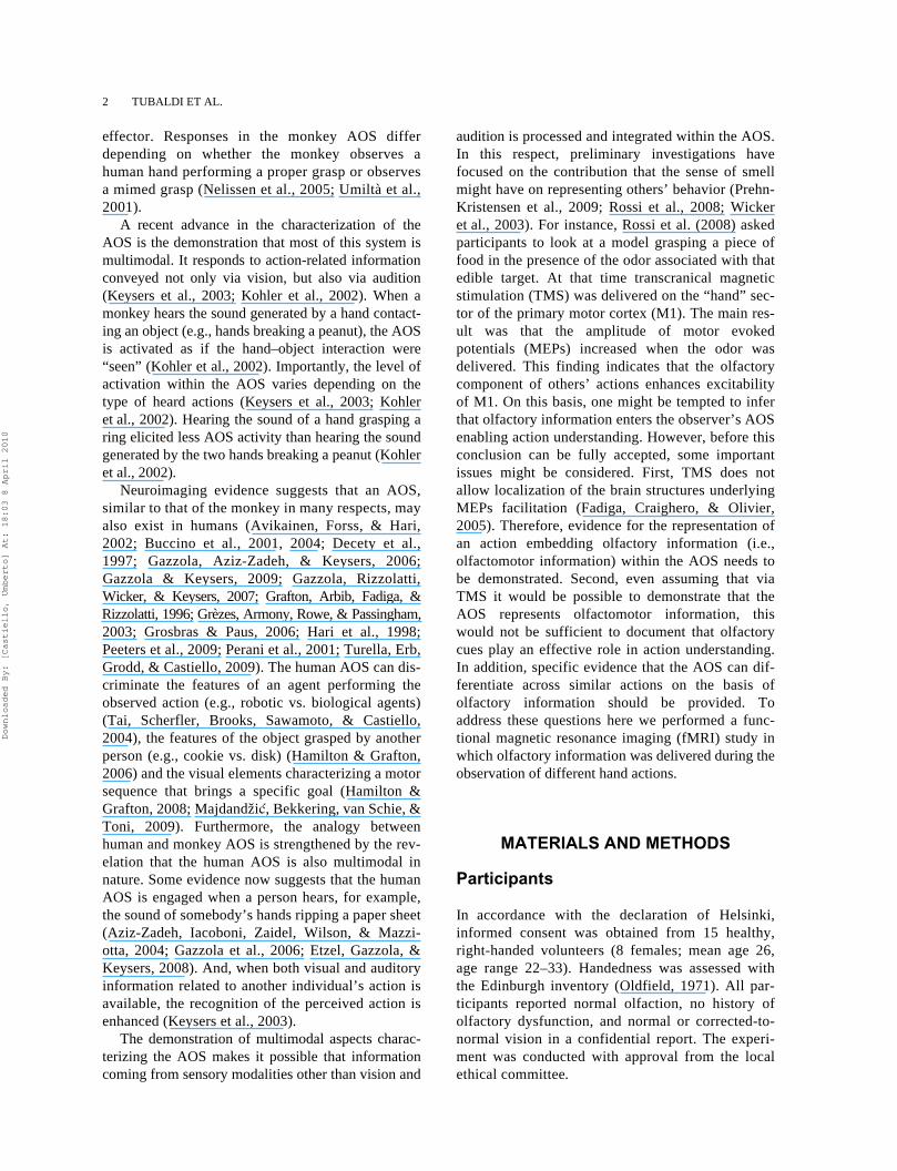
Olfaction in Action Federico Tubaldi, Luca Turella, and Andrea C. PiernoUniversity of Padua, Padua, Italy
Wolfgang GroddUniversity Hospital Tuebingen, Tuebingen, Germany
Roberto TirindelliUniversity of Parma, Parma, Italy
Umberto CastielloUniversity of Padua, Padua, Italy
Previous evidence indicates that we understand others’ actions not only by perceiving their visual features butalso by their sound. This raises the possibility that brain regions responsible for action understanding respond tocues coming from different sensory modalities. Yet no studies, to date, have examined if this extends to olfaction.Here we addressed this issue by using functional magnetic resonance imaging. We searched for brain activityrelated to the observation of an action executed towards an object that was smelled rather than seen. The resultsshow that temporal, parietal, and frontal areas were activated when individuals observed a hand grasping asmelled object. This activity differed from that evoked during the observation of a mimed grasp. Furthermore,superadditive activity was revealed when the action target-object was both seen and smelled. Together thesefindings indicate the influence of olfaction on action understanding and its contribution to multimodal actionrepresentations.
Keywords: Action; Olfaction; Odor; Multisensory integration; Functional MRI.
INTRODUCTION
Neurophysiological research on neural processingunderlying the understanding of others’ actions hasrevealed activity within a network of brain regionsincluding the premotor cortex, the primary motor andsomatosensory cortices, several parietal areas, and theposterior temporal-occipital cortex (Evangeliou, Raos,Galletti, & Savaki, 2009; Gallese, Fadiga, Fogassi, &Rizzolatti, 1996; Peeters et al., 2009; Puce & Perret,2003; Raos, Evangeliou, & Savaki, 2004, 2007). Thismotor circuitry, termed the action observation system
(AOS), enables the representation of the visual fea-tures characterizing the observed action (Keysers &Perret, 2004; Rizzolatti, Fogassi, & Gallese, 2001).For instance, when a monkey observes a humanmodel grasping an object, the AOS exhibits a differ-ential level of activity depending on the nature of boththe visual object (e.g., edible vs. non edible; Fogassiet al., 2005) and the acting model (e.g., an entiremodel vs. an arm/hand ensemble; Nelissen, Luppino,Vanduffel, Rizzolatti, & Orban, 2005). Furthermore,activity within this system appears to be modulated bythe interaction between the target object and the moving
Correspondence should be addressed to: Umberto Castiello, Department of General Psychology, University of Padua, Via Venezia 8,35131 Padova, Italy. E-mail: [email protected]
We thank Heidi Chapman for comments on a previous version of this manuscript. This work was funded by a project grant from theUniversity of Padua to UC. FT was supported by the European Chemoreception Research Organization.
Downloaded By: [Castiello, Umberto] At: 18:03 8 April 2010
2 TUBALDI ET AL.
effector. Responses in the monkey AOS differdepending on whether the monkey observes ahuman hand performing a proper grasp or observesa mimed grasp (Nelissen et al., 2005; Umiltà et al.,2001).
A recent advance in the characterization of theAOS is the demonstration that most of this system ismultimodal. It responds to action-related informationconveyed not only via vision, but also via audition(Keysers et al., 2003; Kohler et al., 2002). When amonkey hears the sound generated by a hand contact-ing an object (e.g., hands breaking a peanut), the AOSis activated as if the hand–object interaction were“seen” (Kohler et al., 2002). Importantly, the level ofactivation within the AOS varies depending on thetype of heard actions (Keysers et al., 2003; Kohleret al., 2002). Hearing the sound of a hand grasping aring elicited less AOS activity than hearing the soundgenerated by the two hands breaking a peanut (Kohleret al., 2002).
Neuroimaging evidence suggests that an AOS,similar to that of the monkey in many respects, mayalso exist in humans (Avikainen, Forss, & Hari,2002; Buccino et al., 2001, 2004; Decety et al.,1997; Gazzola, Aziz-Zadeh, & Keysers, 2006;Gazzola & Keysers, 2009; Gazzola, Rizzolatti,Wicker, & Keysers, 2007; Grafton, Arbib, Fadiga, &Rizzolatti, 1996; Grèzes, Armony, Rowe, & Passingham,2003; Grosbras & Paus, 2006; Hari et al., 1998;Peeters et al., 2009; Perani et al., 2001; Turella, Erb,Grodd, & Castiello, 2009). The human AOS can dis-criminate the features of an agent performing theobserved action (e.g., robotic vs. biological agents)(Tai, Scherfler, Brooks, Sawamoto, & Castiello,2004), the features of the object grasped by anotherperson (e.g., cookie vs. disk) (Hamilton & Grafton,2006) and the visual elements characterizing a motorsequence that brings a specific goal (Hamilton &Grafton, 2008; Majdandžic, Bekkering, van Schie, &Toni, 2009). Furthermore, the analogy betweenhuman and monkey AOS is strengthened by the rev-elation that the human AOS is also multimodal innature. Some evidence now suggests that the humanAOS is engaged when a person hears, for example,the sound of somebody’s hands ripping a paper sheet(Aziz-Zadeh, Iacoboni, Zaidel, Wilson, & Mazzi-otta, 2004; Gazzola et al., 2006; Etzel, Gazzola, &Keysers, 2008). And, when both visual and auditoryinformation related to another individual’s action isavailable, the recognition of the perceived action isenhanced (Keysers et al., 2003).
The demonstration of multimodal aspects charac-terizing the AOS makes it possible that informationcoming from sensory modalities other than vision and
audition is processed and integrated within the AOS.In this respect, preliminary investigations havefocused on the contribution that the sense of smellmight have on representing others’ behavior (Prehn-Kristensen et al., 2009; Rossi et al., 2008; Wickeret al., 2003). For instance, Rossi et al. (2008) askedparticipants to look at a model grasping a piece offood in the presence of the odor associated with thatedible target. At that time transcranical magneticstimulation (TMS) was delivered on the “hand” sec-tor of the primary motor cortex (M1). The main res-ult was that the amplitude of motor evokedpotentials (MEPs) increased when the odor wasdelivered. This finding indicates that the olfactorycomponent of others’ actions enhances excitabilityof M1. On this basis, one might be tempted to inferthat olfactory information enters the observer’s AOSenabling action understanding. However, before thisconclusion can be fully accepted, some importantissues might be considered. First, TMS does notallow localization of the brain structures underlyingMEPs facilitation (Fadiga, Craighero, & Olivier,2005). Therefore, evidence for the representation ofan action embedding olfactory information (i.e.,olfactomotor information) within the AOS needs tobe demonstrated. Second, even assuming that viaTMS it would be possible to demonstrate that theAOS represents olfactomotor information, thiswould not be sufficient to document that olfactorycues play an effective role in action understanding.In addition, specific evidence that the AOS can dif-ferentiate across similar actions on the basis ofolfactory information should be provided. Toaddress these questions here we performed a func-tional magnetic resonance imaging (fMRI) study inwhich olfactory information was delivered during theobservation of different hand actions.
MATERIALS AND METHODS
Participants
In accordance with the declaration of Helsinki,informed consent was obtained from 15 healthy,right-handed volunteers (8 females; mean age 26,age range 22–33). Handedness was assessed withthe Edinburgh inventory (Oldfield, 1971). All par-ticipants reported normal olfaction, no history ofolfactory dysfunction, and normal or corrected-to-normal vision in a confidential report. The experi-ment was conducted with approval from the localethical committee.
Downloaded By: [Castiello, Umberto] At: 18:03 8 April 2010
OLFACTION IN ACTION 3
Stimuli and experimental conditions
The experimental stimuli consisted of video-clips(Audio Video Interleave format, 25 frames/s, resolu-tion 400 × 300 pixels; duration 3 s) representing ahuman right hand together with an object. For theentire duration of the movie either odorized or odorlessair was delivered. The task for participants was toobserve the presented video-clips.
There were eight experimental conditions, as follows.
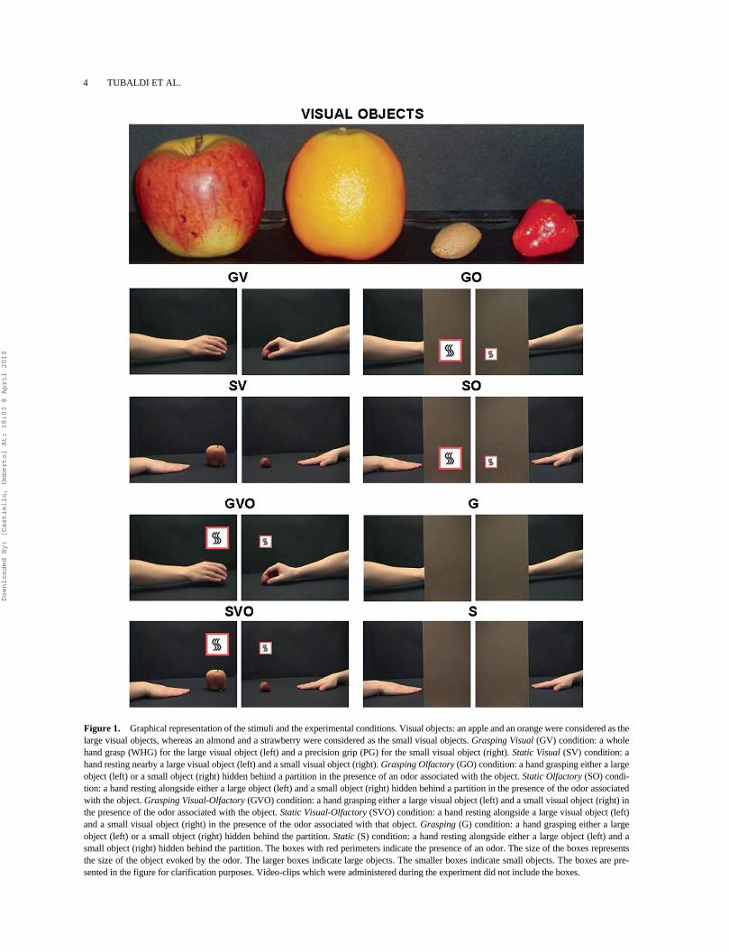
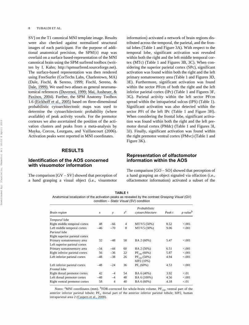
1. A Grasping Visual (GV) condition, in whichparticipants observed the hand grasping either alarge or a small object in the absence of odor(refer to “Visual objects” and “GV” in Figure 1).
2. A Static Visual (SV) condition, in which partici-pants observed the hand resting alongside oneof the four visual objects presented in Figure 1(“Visual objects”), in a prone position, with thepalm towards the working surface in the absenceof odor (“SV”).
3. A Grasping Olfactory (GO) condition in whichparticipants observed a hand grasping an objectas for the GV condition, but the object was hid-den behind a brown-colored partition. Duringthe observation of the video-clip an odor associ-ated with the hidden object was delivered (“GO”in Figure 1; the boxes within the panel indicatethe presence of an odor).
4. A Static Olfactory (SO) condition, in which par-ticipants observed a stationary hand as for theSV condition, but the object was hidden behinda brown-colored partition. During the observa-tion of the video-clip an odor associated with thehidden object was delivered (refer to “SO” inFigure 1; the boxes within the panel indicate thepresence of an odor).
5. A Grasping Visual-Olfactory (GVO) condition,in which participants observed video-clips iden-tical to those utilized for the GV conditionexcept that during the observation of the video-clip an odor associated with the visual objectwas also delivered (“GVO” in Figure 1; theboxes within the panel indicate the presence ofan odor).
6. A Static Visual-Olfactory (SVO) condition, inwhich participants observed video-clips identi-cal to those utilized for the SV condition exceptthat during the observation of the video-clip anodor associated with the visual object was alsodelivered (“SVO” in Figure 1; the boxes withinthe panel indicate the presence of an odor).
7. A Grasping (G) condition, in which participantsobserved video-clips identical to those utilized
for the GO condition except that during theobservation of the video-clip no odor was deliv-ered ( “G” in Figure 1). Therefore, participantswere presented with mimed hand grasp move-ments, i.e., hand grasping movements without areal end-goal.
8. A Static (S) condition, in which participantsobserved video-clips identical to those utilizedfor the SO condition except that during theobservation of the video-clip no odor was deliv-ered (“S” in Figure 1).
Participants’ point of view within the scanner wasalso considered. Therefore, in half of the video-clipsthe hand entered the scene from the left, whereas inthe other half the hand entered the scene from theright side (panels from “GV” to “S” in Figure 1). Thisresulted in a total of eight different experimental stim-uli (i.e., four different objects by two different handpositions) per condition. This set of experimentalstimuli was repeated four times within each condition(i.e., 32 stimuli corresponding to 32 experimental tri-als were administered).
Apparatus
All the experimental stimuli were presented by usingthe software Presentation (Neurobehavioral Systems,Albany, CA, www.neuro-bs.com) which ensured syn-chronization with the MR scanner. An LCD com-puter-controlled projector (NEC, resolution 1024 ×768, refresh rate 60 Hz) was employed to present themovies in color at the centre of a screen positionedoutside the bore of the magnet. The stimulus wasviewed by the participants through a mirror mountedon the head coil. When projected onto the mirror, themovies were 26.8 cm wide × 20.1 cm high and sub-tended visual angles of 20° × 15°. An MRI-compati-ble, custom-built computer-controlled olfactometerwith eight channels (Department of ExperimentalPsychology, University of Oxford, UK) was used toadminister olfactory stimuli. The olfactometer wascapable of rapid delivery of discrete odor pulses in theabsence of tactile, thermal, or auditory variation. Eachodor generator consisted of a glass boat containingone of four odor solutions. The odor solutions ofstrawberry, almond, orange, and apple were obtainedmixing 6000 ml of propylene glycol and 180 ml (3%),60 ml (1%), 420 ml (7%), and 45 ml (0.75%) of thespecific odorant compound, respectively (Cerizza,Milan, Italy). These odor solutions were adoptedbecause they generated odors that were judged tohave equal intensity, hedonic tone, and familiarity in
Downloaded By: [Castiello, Umberto] At: 18:03 8 April 2010
4 TUBALDI ET AL.
Figure 1. Graphical representation of the stimuli and the experimental conditions. Visual objects: an apple and an orange were considered as thelarge visual objects, whereas an almond and a strawberry were considered as the small visual objects. Grasping Visual (GV) condition: a wholehand grasp (WHG) for the large visual object (left) and a precision grip (PG) for the small visual object (right). Static Visual (SV) condition: ahand resting nearby a large visual object (left) and a small visual object (right). Grasping Olfactory (GO) condition: a hand grasping either a largeobject (left) or a small object (right) hidden behind a partition in the presence of an odor associated with the object. Static Olfactory (SO) condi-tion: a hand resting alongside either a large object (left) and a small object (right) hidden behind a partition in the presence of the odor associatedwith the object. Grasping Visual-Olfactory (GVO) condition: a hand grasping either a large visual object (left) and a small visual object (right) inthe presence of the odor associated with the object. Static Visual-Olfactory (SVO) condition: a hand resting alongside a large visual object (left)and a small visual object (right) in the presence of the odor associated with that object. Grasping (G) condition: a hand grasping either a largeobject (left) or a small object (right) hidden behind the partition. Static (S) condition: a hand resting alongside either a large object (left) and asmall object (right) hidden behind the partition. The boxes with red perimeters indicate the presence of an odor. The size of the boxes representsthe size of the object evoked by the odor. The larger boxes indicate large objects. The smaller boxes indicate small objects. The boxes are pre-sented in the figure for clarification purposes. Video-clips which were administered during the experiment did not include the boxes.
Downloaded By: [Castiello, Umberto] At: 18:03 8 April 2010
OLFACTION IN ACTION 5
previous investigations (Tubaldi, Ansuini, Demattè,Tirindelli, & Castiello, 2008a; Tubaldi, Ansuini,Tirindelli, & Castiello, 2008b). A fifth glass boat con-taining propylene glycol was used for the delivery ofodorless air. The air passed over the odor solutionsand the propylene glycol at a flow rate of 8 l/min andwas delivered on both nostrils to subjects via Teflontubing to a facial mask (Tubaldi et al., 2008a, 2008b).
Activation paradigm
During the experiment participants lay supine in thescanner and observed all the displayed movies eitherin the absence or in the presence of an odor. An exper-imental trial consisted of a single event (i.e., a moviewhile odorized or odorless air was delivered) thatlasted 3 s. The time between the trial offset and theonset of the next trial (interstimulus interval, ISI) was10,500 ms. If an odor was delivered, an ISI of 10,500ms allowed recovery from any odor adaptation (Hum-mel, Knecht, & Kobal, 1996). During ISI, a black fix-ation cross on a blank screen was presented andodorless air was delivered. For each experimentalcondition, 32 trials were administered. Trial order wasfully randomized, except that in order to minimize theeffects of stimulus repetition and odor habituation theobject (or the odor associated with the hidden object)differed in every trial with respect to that adminis-tered in the previous trial (Gottfried & Dolan, 2003).The experiment consisted of four functional runs.Within each of these functional runs, two of the eightexperimental conditions were presented. Specifically,trials for the GV condition and trials for the SV condi-tion were randomly presented within a first functionalrun (i.e., visual run). A second functional run con-sisted of trials representing the GO condition and theSO condition (i.e., olfactory run). A third functionalrun included trials related to the GVO and the SVOconditions (i.e., visual-olfactory run). Finally, trialsfor the G and the S conditions were randomly pre-sented within a fourth functional run (i.e., mimedrun). Each functional run lasted 882 s, and started andended with 8-s and 10.5-s rest periods respectively,each consisting of a black fixation cross on a blankscreen. Consecutive functional runs were intermin-gled with a 5-min break during which no kind of stim-ulation was delivered. By administering twoconditions per run, we ensured that signal related tothe contrasts GV – SV, GO – SO, GVO – SVO, and G –S spanned frequency bands above the cut-off selectedfor the high-pass filter (see “Data analyses” section).For the visual run, the olfactory run, and the visual-olfactory run, six different presentation orders were
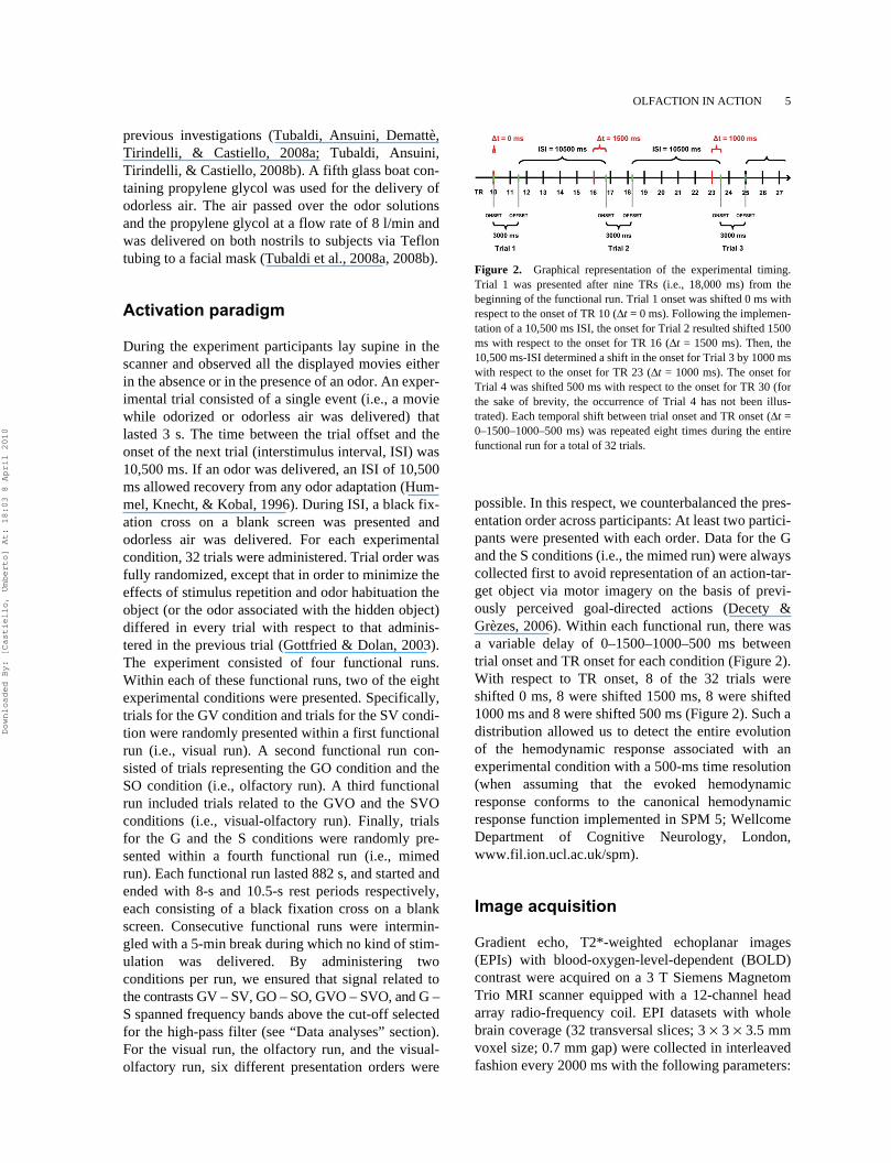
possible. In this respect, we counterbalanced the pres-entation order across participants: At least two partici-pants were presented with each order. Data for the Gand the S conditions (i.e., the mimed run) were alwayscollected first to avoid representation of an action-tar-get object via motor imagery on the basis of previ-ously perceived goal-directed actions (Decety &Grèzes, 2006). Within each functional run, there wasa variable delay of 0–1500–1000–500 ms betweentrial onset and TR onset for each condition (Figure 2).With respect to TR onset, 8 of the 32 trials wereshifted 0 ms, 8 were shifted 1500 ms, 8 were shifted1000 ms and 8 were shifted 500 ms (Figure 2). Such adistribution allowed us to detect the entire evolutionof the hemodynamic response associated with anexperimental condition with a 500-ms time resolution(when assuming that the evoked hemodynamicresponse conforms to the canonical hemodynamicresponse function implemented in SPM 5; WellcomeDepartment of Cognitive Neurology, London,www.fil.ion.ucl.ac.uk/spm).
Image acquisition
Gradient echo, T2*-weighted echoplanar images(EPIs) with blood-oxygen-level-dependent (BOLD)contrast were acquired on a 3 T Siemens MagnetomTrio MRI scanner equipped with a 12-channel headarray radio-frequency coil. EPI datasets with wholebrain coverage (32 transversal slices; 3 × 3 × 3.5 mmvoxel size; 0.7 mm gap) were collected in interleavedfashion every 2000 ms with the following parameters:
Figure 2. Graphical representation of the experimental timing.Trial 1 was presented after nine TRs (i.e., 18,000 ms) from thebeginning of the functional run. Trial 1 onset was shifted 0 ms withrespect to the onset of TR 10 (Δt = 0 ms). Following the implemen-tation of a 10,500 ms ISI, the onset for Trial 2 resulted shifted 1500ms with respect to the onset for TR 16 (Δt = 1500 ms). Then, the10,500 ms-ISI determined a shift in the onset for Trial 3 by 1000 mswith respect to the onset for TR 23 (Δt = 1000 ms). The onset forTrial 4 was shifted 500 ms with respect to the onset for TR 30 (forthe sake of brevity, the occurrence of Trial 4 has not been illus-trated). Each temporal shift between trial onset and TR onset (Δt =0–1500–1000–500 ms) was repeated eight times during the entirefunctional run for a total of 32 trials.
Downloaded By: [Castiello, Umberto] At: 18:03 8 April 2010
6 TUBALDI ET AL.
field-of-view, 192 × 192 mm; in-plan resolution 64 ×64 voxels; echo time, 33 ms; bandwidth, 2442 Hz/Px.For each functional run, a total of 441 volumes werecollected, minus 5 “dummy” volumes to permit T1equilibration. In addition, high-resolution T1-weightedimages (anatomical scans) were acquired for each par-ticipant (MP-RAGE, 160 sagittal slices, in-plane res-olution 224 × 256 voxels, 1 mm isotropic voxels, TR =2300 ms, TE = 3 ms).
Odor recognition task and paced-breathing session
In order to ensure that participants were able to identifyeach of the four delivered odors (i.e., orange, apple,strawberry, and almond), we asked to participants toperform an odor recognition task. Before enteringwithin the scanner, volunteers were presented with thefour visual objects (see “Visual objects” in Figure 1).Then, an odor was presented for 2 s and participantswere instructed to indicate the object associated withthat odor. The odors were delivered by using the olfac-tometer employed as to administer olfactory stimula-tion within the MR scanner. A total of eight trials (twofor each type of odor) were presented in randomizedorder. When performing the task, participants showedno errors.
Before the initiation of each functional run, partici-pants took part in a paced-breathing session. Duringthis session, participants were trained to synchronizetheir breathing cycle according to the rhythm withwhich odor would have been delivered during thefMRI experiment. They performed 15 paced air inha-lations within one training block lasting 210 s (fortechnical details see Tabert et al., 2007). This ensuredthat odor administration during the fMRI experimentwas always synchronized with the participants’ inha-lation phase and that the sampling of the deliveredodor was uniform across scans and participants. Fur-thermore, visual inspection of the participants’ breath-ing patterns (i.e., respiration rate) during the fMRIexperiment revealed no differences across the experi-mental runs in which an odor was delivered (i.e.,olfactory run and visual-olfactory run) and those inwhich no odor was administered (i.e., visual run andmimed run).
Data analyses
MRI data were analyzed using SPM 5 software, imple-mented in Matlab 7.0 (Mathworks, Natick, MA). First,EPI images were realigned to the first functional volume
of each run in order to correct for any head movementoccurring within the run. Second, high-quality T1images were co-registered to the mean EPI image andsegmented. The coregistered gray matter segment wasnormalized onto the grey matter template (available inthe SPM 5 “apriori” directory), and the resultingnormalization parameters applied to all EPI images(re-sampled voxels at 2 × 2 × 2 mm). The T1 imagewas also normalized to the MNI space using the sameparameters, keeping the original resolution of 1 × 1 ×1 mm. Finally, EPI data were spatially smoothedadopting an 8-mm full-width-at-half-maximum Gaussiankernel. The event-related functional data were ana-lyzed using the general linear model (Friston et al.,1995). Eight regressors of interest were defined basedon the timing of presentation for each experimentalcondition (duration = 3 s). These functions were con-volved with a canonical hemodynamic response func-tion. Subject-specific movement parameters wereincluded to account for translation and rotation alongthe three possible dimensions as measured during therealignment stage. A high-pass filter (cut-off, 128 s)was also applied to remove low-frequency drifts insignal. The parameter estimates for each regressorwere calculated for all brain voxels (i.e., beta imageswere computed). Then beta images referring to theGV, the GO, the GVO, and the G conditions and tothe four corresponding control conditions (i.e., theSV, the SO, the SVO, and the S conditions) wereextracted for each subject and then entered into a 2 × 4flexible factorial design. The two within-subjects fac-tors were “Hand” (Grasping/Static) and “Object”(Visual/Olfactory/Visual-Olfactory/Absent). A thirdfactor of no interest was also modeled, i.e., the effectof subjects.
The hypotheses underlying the present study wereconcerned with the possibility that the AOS repre-sented olfactomotor information (either in isolation orin combination with visuomotor information) andused the olfactory component of this information foraction discrimination. Therefore, testing for thesehypotheses was confined to the relevant neuralsystem, i.e., the AOS. We localized the AOS con-cerned with visuomotor information by performingthe contrast [GV – SV] at whole brain level [intensitythreshold, p = .015 FDR corrected; cluster-extentthreshold = 15 voxels] (Friston, Rotshein, Geng,Sterzer, & Henson, 2006; Friston & Henson, 2006;Kriegeskorte, Simmons, Bellgowan, & Baker, 2009).Then, by using Marsbar SPM Toolbox (Brett, Anton,Valabregue, & Poline, 2002), the beta value corre-sponding to each control condition (i.e., SV, SO, SVO,and S) and each experimental condition (i.e., GV, GO,GVO, and G) was extracted for each participant from
Downloaded By: [Castiello, Umberto] At: 18:03 8 April 2010
OLFACTION IN ACTION 7
the peak-voxel of each identified AOS area. Next, bysubtracting the averaged beta values across partici-pants for each control condition from the averaged betavalues across participants for the corresponding experi-mental condition, activation values GV – SV, GO –SO, GVO – SO, and G – S were computed. Experimen-tal hypotheses were verified by performing statisticalcomparisons on these activation values (Friston et al.,2006; Kilner, Neal, Weiskopf, Friston, & Frith, 2009;Saxe, Brett, & Kanwisher, 2006).
First, to evaluate whether the AOS representedolfactomotor information, we tested for a greater acti-vation for the GO condition compared to the SO con-dition [GO – SO, intensity threshold, p = .05].
Second, to ascertain whether or not visuomotorand olfactomotor information integrated within theAOS, we first tested whether there was a greater acti-vation for the GVO condition compared to the SVOcondition [GVO – SVO, intensity threshold, p = .05].Next, we evaluated whether such activation wasgreater than the sum of the activations for the GV andthe GO conditions (Beauchamp, 2005a; Laurienti,Perrault, Stanford, Wallace, & Stein, 2005). In esti-mating this superadditive model, the activation [G – S]was added to the activation [GVO – SVO]. Thisresulted in the interaction contrast: [(GVO – SVO) +(G – S)] – [(GV – SV) + (GO – SO)], intensity thresh-old, p = .05.
Third, we assessed whether two similar actionscould be differentiated within the AOS on the basis ofolfactory information. To this end, we first testedwhether the activation for the G condition was greaterthan activation for the S condition [G – S, intensitythreshold, p = .05] within the AOS areas already exhib-iting greater activation for the GO than for the SO con-dition. Following this, we tested whether the extent ofactivation when comparing the G with the S conditionwas different to the extent of activation observed whencomparing the GO with the SO condition. This resultedin the interaction contrast: [G – S] – [GO – SO], intensitythreshold, p = .05. If the interaction contrast is signific-ant, then differential activation can solely be ascribed tothe olfactory information signaling the target-object.This is because both the amount and the type ofperceived movement (e.g., hand shaping and trajectory,digits’ opening and closing) is identical for the comparedactions. A t-test was performed for each contrast.
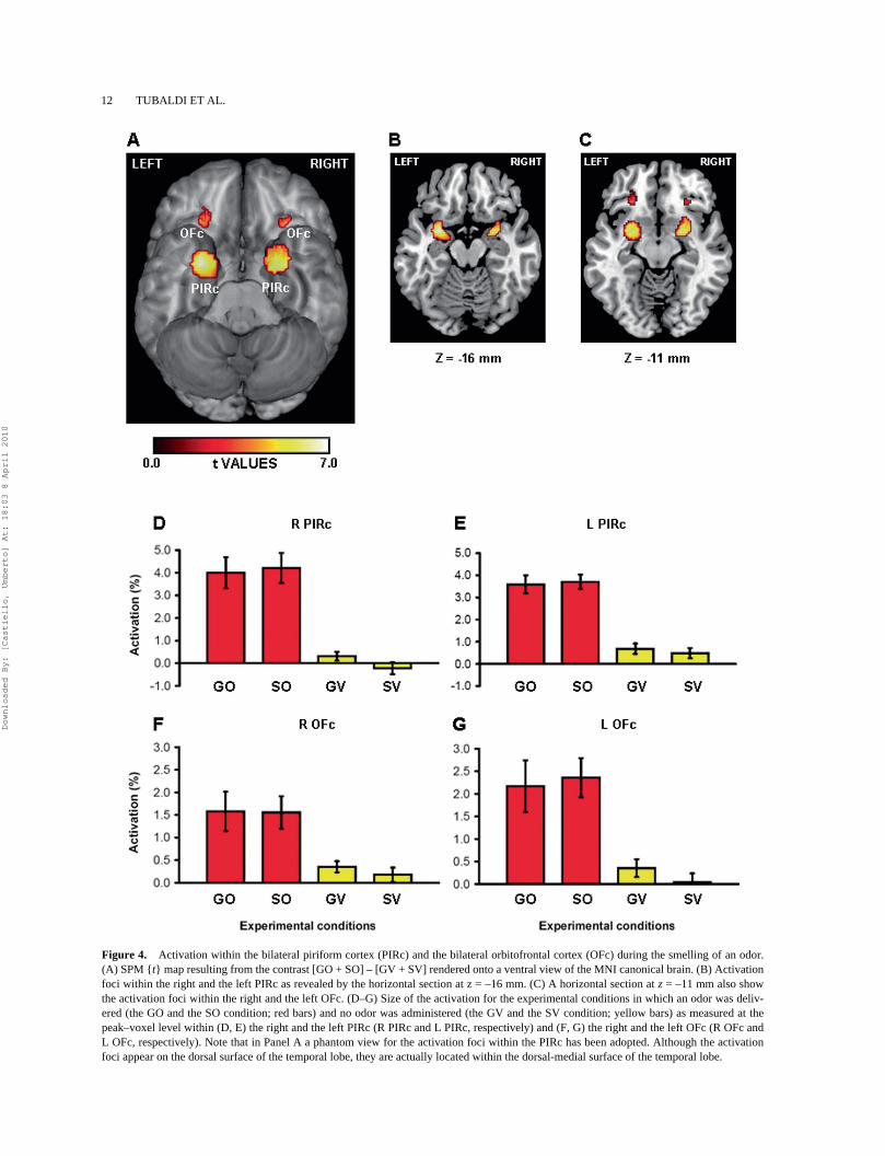
Finally, we assessed whether both the primary andthe secondary olfactory cortices were recruited during thesmelling of an odor. To this end, we performed the con-trast [GO + SO] – [GV + SV] within such regions [inten-sity threshold, p = .015 FDR corrected; cluster-extentthreshold = 15 voxels]. To establish the localization ofthe primary olfactory cortex, we considered the MNI
coordinates of peak-voxel reported in previous imagingstudies in which: (a) the neural characterization ofbasic olfactory processing was the central aim; (b)odor-evoked activity was not complicated by the useof aversive odorants; and (c) subjects were not askedto make any cognitive olfactory judgments (other thanodor detection) during scanning. On the basis ofsuch criteria data from four brain imaging studies(Gottfried, Deichmann, Winston, & Dolan, 2002;Gottfried, Winston, & Dolan, 2006; Poellinger et al.,2001; Zatorre, Jones-Gotman, Evans, & Meyer, 1992)were considered. A total of 12 coordinates were iden-tified, including 9 for the left hemisphere and 8 for theright hemisphere. All of these studies reported signi-ficant bilateral activation. Voxel coordinate meantogether with the standard error was computed sepa-rately for the right and the left hemisphere. The resultsindicated that the left and the right primary olfactorycortices were localized in the MNI space at (x = –24 ±2 mm, y = 1 ± 1 mm, z = –17 ± 4 mm) and at (x = 21 ±1 mm, y = 5 ± 2 mm, z = –15 ± 4 mm), respectively.From an anatomical perspective, neural loci associ-ated with these coordinates were located within thepiriform cortex, alongside the dorsal-medial surfaceof the temporal lobe at the level of the fronto-temporaljunction. To establish the localization of the secondaryolfactory cortex we used data from a meta-analysisperformed by Gottfried & Zald (2005). According tothese authors, the left and the right secondary olfac-tory cortex are located within the orbital surface of thefrontal lobe at (x = –21 mm, y = 31 mm, z = –16 mm)and at (x = 24 mm, y = 34 mm, z = –12 mm), respec-tively (Talairach coordinates). By applying the Mat-thew Brett’s “tal2mni” function (http://imaging.mrc-cbu.cam.ac.uk/downloads/MNI2tal/tal2mni.m) to theTalairach coordinates we obtained the correspondingMNI coordinates for the left and the right secondaryolfactory cortex: (x = –21 mm, y = 33 mm, z = –17mm), and (x = 24 mm, y = 35 mm, z = –12 mm),respectively. By using WFU_PickAtlas (an SPM 5extension available at www.fmri.wfubmc.edu/cms/software) we built four spheres (radius = 10 mm) andcentered each sphere on each set of MNI coordinates.The four spheres were joined to compose an individ-ual brain mask. The brain volume which was includedwithin the mask represented the search volume for thecontrast [GO + SO] – [GV + SV].
Localization
Anatomical details of significant signal changes wereobtained by superimposing the SPM{t} maps resultingfrom the contrasts [GV – SV] and [GO + SO] – [GV +
Downloaded By: [Castiello, Umberto] At: 18:03 8 April 2010
8 TUBALDI ET AL.
SV] on the T1 canonical MNI template image. Resultswere also checked against normalized structuralimages of each participant. For the purpose of addi-tional anatomical precision, the SPM{t} map wasoverlaid on a surface based-representation of the MNIcanonical brain using the SPM surfrend toolbox (writ-ten by I. Kahn; http://spmsurfrend.sourceforge.net).The surface-based representation was then renderedusing FreeSurfer (CorTechs Labs, Charlestown, MA)(Dale, Fischl, & Sereno, 1999; Fischl, Sereno, &Dale, 1999). We used two atlases as general neuroana-tomical references (Duvernoi, 1999; Mai, Assheuer, &Paxinos, 2004). Further, the SPM Anatomy Toolbox1.6 (Eickhoff et al., 2005) based on three-dimensionalprobabilistic cytoarchitectonic maps was used todetermine the cytoarchitectonic probability (whereavailable) of peak activity voxels. For the premotorcortexes we also ascertained the position of the acti-vation clusters and peaks from a meta-analysis byMayka, Corcos, Leurgans, and Vaillancourt (2006).Activation peaks were reported in MNI coordinates.
RESULTS
Identification of the AOS concerned with visuomotor information
The comparison [GV – SV] showed that perception ofa hand grasping a visual object (i.e., visuomotor
information) activated a network of brain regions dis-tributed across the temporal, the parietal, and the fron-tal lobes (Table 1 and Figure 3A). With respect to thetemporal lobe, significant activation was revealedwithin both the right and the left middle temporal cor-tex (MTc) (Table 1 and Figures 3B, 3C). When con-sidering the superior parietal cortex (SPc), significantactivation was found within both the right and the leftprimary somatosensory area (Table 1 and Figures 3D,3E). Furthermore, significant activation was foundwithin the sector PFcm of both the right and the leftinferior parietal cortex (IPc) (Table 1 and Figures 3F,3G). Parietal activity within the left sector PFcmspread within the intraparietal sulcus (IPS) (Table 1).Significant activation was also detected within thesector PFt of the left IPc (Table 1 and Figure 3H).When considering the frontal lobe, significant activa-tion was found within both the right and the left pre-motor dorsal cortex (PMdc) (Table 1 and Figures 3I,3J). Finally, significant activation was found withinthe right premotor ventral cortex (PMvc) (Table 1 andFigure 3K).
Representation of olfactomotor information within the AOS
The comparison [GO – SO] showed that perception ofa hand grasping an object signaled via olfaction (i.e.,olfactomotor information) activated a subset of the
TABLE 1 Anatomical localization of the activation peaks as revealed by the contrast Grasping Visual (GV)
condition – Static Visual (SV) condition
Brain region x y zaProbabilistic
cytoarchitecture Peak t p valueb
Temporal lobeRight middle temporal cortex 48 –66 4 MT/V5 (50%) 8.52 <.001Left middle temporal cortex –46 –70 8 MT/V5 (30%) 9.06 <.001Parietal lobeRight superior parietal cortexPrimary somatosensory area 32 –48 58 BA 2 (60%) 5.47 <.001Left superior parietal cortexPrimary somatosensory area –34 –44 60 BA 2 (50%) 6.51 <.001Right inferior parietal cortex 56 –36 22 PFcm (60%) 5.87 <.001Left inferior parietal cortex –48 –38 26 PFcm (50%) 4.94 <.001
hIP2 (10%)Left inferior parietal cortex –48 –24 36 PFt (60%) 4.53 <.001Frontal lobeRight dorsal premotor cortex 42 –4 54 BA 6 (40%) 3.92 <.01Left dorsal premotor cortex –48 –4 48 BA 6 (100%) 4.56 <.001Right ventral premotor cortex 58 4 40 BA 6 (60%) 4.18 <.01
Notes: aMNI coordinates (mm). bFDR-corrected for whole-brain volume. PFcm, ventral part of theanterior inferior parietal lobule; PFt, dorsal part of the anterior inferior parietal lobule; hIP2, humanintraparietal area 2 (Caspers et al., 2008).
Downloaded By: [Castiello, Umberto] At: 18:03 8 April 2010
OLFACTION IN ACTION 9
Figure 3. Functional modulation of the AOS activity depending on the nature of sensorimotor information. (A) SPM {t} map resulting fromthe contrast GV – SV rendered onto the MNI canonical brain. (B–K) Size of the activation for the GV (yellow bars), the GO (red bars), theGVO (blue bars), and the G (gray bars) conditions with respect to the SV, the SO, the SVO, and the S conditions, respectively, as measured atthe peak-voxel level within (B, C) the right and the left middle temporal cortex (R MTc and L MTc, respectively), (D, E) the right and the leftsuperior parietal cortex (R SPc and L SPc, respectively), (F, G) the sector PFcm of the right and the left inferior parietal cortex (R IPc – PFcmand L IPc – PFcm, respectively), (H) the sector PFt of the left inferior parietal cortex (L IPc – PFt), (I, J) the right and the left premotor dorsalcortex (R PMdc and L PMdc, respectively), and (K) the right premotor ventral cortex (R PMvc). Error bars represent standard errors of theactivations. Asterisks indicate the statistically significant results obtained from the contrast [GV – SV] performed at whole brain level.
Downloaded By: [Castiello, Umberto] At: 18:03 8 April 2010
10 TUBALDI ET AL.
cerebral areas identified for visuomotor information(Figures 3B–3K). Specifically, perceiving olfactomo-tor information brought a significant activation withinthe bilateral MTc (right side: t = 9.32, p < .001; leftside: t = 8.36, p < .001) (Figures 3B, 3C). When con-sidering the parietal lobe, no significant activationwas found within the bilateral SPc (right side: t =1.67, p > .05; left side: t = 1.39, p > .05) (Figures 3D,3E). Significant activation was found within the sec-tor PFcm of the bilateral IPc (right side: t = 3.47, p <.01; left side: t = 2.73, p < .01) (Figures 3F, 3G).Finally, no significant activation was found within thesector PFt of the left IPc (t = –0.84, p > .05) (Figure3H). With respect to the frontal lobe, significant acti-vation was found within the bilateral PMdc (rightside: t = 2.25, p < .05; left side: t = 2.31, p < .05,respectively) (Figures 3I, 3J). Significant activationwas also found within the right PMvc (t = 2.69, p <.01) (Figure 3K).
Integration of visuomotor and olfactomotor information within the AOS
The comparison [GVO – SVO] showed that perceiv-ing a hand grasping an object signaled via both visionand olfaction, i.e., visuo-olfactomotor information,determined significant activation within the bilateralMTc (right side: t = 7.44, p < .001; left side: t = 7.75,p < .001) (Figures 3B, 3C), the bilateral SPc (rightside: t = 4.39, p < .001; left side: t = 4.77, p < .001)(Figures 3D, 3E), and the sector PFcm of the bilateralIPc (right side: t = 5.47, p < .001; left side: t = 3.69,p < .001) (Figures 3F, 3G). With respect to the sectorPFt of the left IPc, no significant activation was found(t = 1.69, p > .05) (Figure 3H). Significant activationwas also found for the bilateral PMdc (right side: t
= 3.07, p < .01; left side: t = 1.73, p < .05) (Figures 3I,3J). Activation was not significant within the rightPMvc (t = 1.52, p > .05) (Figure 3K). Furthermore,the superadditive model [(GVO – SVO) + (G – S)] –[(GV – SV) + (GO – SO)] accounted for the activa-tion elicited by perception of visuo-olfactomotorinformation within both the right MTc and the leftSPc (Table 2).
Representation of olfactory information within the AOS
The comparison [G – S] showed that the perception ofa mimed grasp determined significant activationwithin the areas of the AOS that were also activatedfor olfactomotor information (Figures 3B–3K). Specif-ically, significant activation was found within thebilateral MTc (right side: t = 10.08, p < .001; left side:t = 9.32, p < .001) (Figures 3B, 3C), the sector PFcmof the bilateral IPc (right side: t = 5.84, p < .001; leftside: t = 3.82, p < .001) (Figures 3F, 3G), and the bilat-eral PMdc (right side: t = 4.59, p < .001; left side: t =3.03, p < .01) (Figures 3I, 3J). Significant activationwas also found within the right PMvc (t = 2.64, p <.01) (Figure 3K). Crucially, the interaction contrast [G– S] – [GO – SO] indicated that activation for the per-ception of olfactomotor information differed withrespect to that obtained while perceiving a mimedgrasp. Specifically, significant differential activationwas found within the left MTc (t = 1.96, p < .05)(Figure 3C), the sector PFcm of the bilateral IPc (rightside: t = 3.09, p < .05; left side: t = 2.35, p < .05)(Figures 3F, 3G), and the bilateral PMdc (right side:t = 3.40, p < .01; left side: t = 1.83, p < .05) (Figures3I, 3J). No significant differential activation was foundfor the right MTc (t = 1.65, p > .05) (Figure 3B) andthe right PMvc (t = 1.12, p > .05) (Figure 3K).
TABLE 2 Neural sites of integration between visuomtor and olfactomotor information
Brain regionsProbabilistic
cytoarchitecture Averaged activation (%)a t value p value
Temporal lobeRight middle temporal cortex MT/V5 (50%) 0.93 (0.50) 1.85 <.05Parietal lobeLeft superior parietal cortex BA 2 (50%) 0.90 (0.44) 2.01 <.05
Notes: Averaged activation across participants for the superadditive model is reported. Standard errors are shown inparentheses. Statistical assessment for the superadditive model [(GVO – SVO) + (G – S)] – [(GV – SV) + (GO – SO)] isalso presented. aSuperadditive combination of betas as measured at the peak voxel revealed by the contrast [GV – SV].GVO, Grasping Visual-Olfactory condition; SVO, Static Visual-Olfactory condition; G, Grasping condition; S, Static con-dition; GV, Grasping Visual condition; SV, Static Visual condition; GO, Grasping Olfactory condition; SO, Static Olfactorycondition.
Downloaded By: [Castiello, Umberto] At: 18:03 8 April 2010
OLFACTION IN ACTION 11
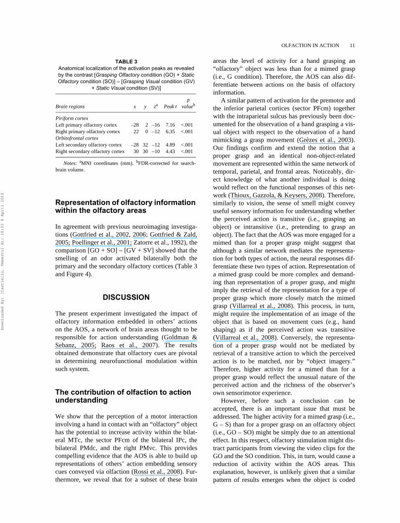
Representation of olfactory information within the olfactory areas
In agreement with previous neuroimaging investiga-tions (Gottfried et al., 2002, 2006; Gottfried & Zald,2005; Poellinger et al., 2001; Zatorre et al., 1992), thecomparison [GO + SO] – [GV + SV] showed that thesmelling of an odor activated bilaterally both theprimary and the secondary olfactory cortices (Table 3and Figure 4).
DISCUSSION
The present experiment investigated the impact ofolfactory information embedded in others’ actionson the AOS, a network of brain areas thought to beresponsible for action understanding (Goldman &Sebanz, 2005; Raos et al., 2007). The resultsobtained demonstrate that olfactory cues are pivotalin determining neurofunctional modulation withinsuch system.
The contribution of olfaction to action understanding
We show that the perception of a motor interactioninvolving a hand in contact with an “olfactory” objecthas the potential to increase activity within the bilat-eral MTc, the sector PFcm of the bilateral IPc, thebilateral PMdc, and the right PMvc. This providescompelling evidence that the AOS is able to build uprepresentations of others’ action embedding sensorycues conveyed via olfaction (Rossi et al., 2008). Fur-thermore, we reveal that for a subset of these brain
areas the level of activity for a hand grasping an“olfactory” object was less than for a mimed grasp(i.e., G condition). Therefore, the AOS can also dif-ferentiate between actions on the basis of olfactoryinformation.
A similar pattern of activation for the premotor andthe inferior parietal cortices (sector PFcm) togetherwith the intraparietal sulcus has previously been doc-umented for the observation of a hand grasping a vis-ual object with respect to the observation of a handmimicking a grasp movement (Grèzes et al., 2003).Our findings confirm and extend the notion that aproper grasp and an identical non-object-relatedmovement are represented within the same network oftemporal, parietal, and frontal areas. Noticeably, dir-ect knowledge of what another individual is doingwould reflect on the functional responses of this net-work (Thioux, Gazzola, & Keysers, 2008). Therefore,similarly to vision, the sense of smell might conveyuseful sensory information for understanding whetherthe perceived action is transitive (i.e., grasping anobject) or intransitive (i.e., pretending to grasp anobject). The fact that the AOS was more engaged for amimed than for a proper grasp might suggest thatalthough a similar network mediates the representa-tion for both types of action, the neural responses dif-ferentiate these two types of action. Representation ofa mimed grasp could be more complex and demand-ing than representation of a proper grasp, and mightimply the retrieval of the representation for a type ofproper grasp which more closely match the mimedgrasp (Villarreal et al., 2008). This process, in turn,might require the implementation of an image of theobject that is based on movement cues (e.g., handshaping) as if the perceived action was transitive(Villarreal et al., 2008). Conversely, the representa-tion of a proper grasp would not be mediated byretrieval of a transitive action to which the perceivedaction is to be matched, nor by “object imagery.”Therefore, higher activity for a mimed than for aproper grasp would reflect the unusual nature of theperceived action and the richness of the observer’sown sensorimotor experience.
However, before such a conclusion can beaccepted, there is an important issue that must beaddressed. The higher activity for a mimed grasp (i.e.,G – S) than for a proper grasp on an olfactory object(i.e., GO – SO) might be simply due to an attentionaleffect. In this respect, olfactory stimulation might dis-tract participants from viewing the video clips for theGO and the SO condition. This, in turn, would cause areduction of activity within the AOS areas. Thisexplanation, however, is unlikely given that a similarpattern of results emerges when the object is coded
TABLE 3 Anatomical localization of the activation peaks as revealed by the contrast [Grasping Olfactory condition (GO) + Static Olfactory condition (SO)] – [Grasping Visual condition (GV)
+ Static Visual condition (SV)]
Brain regions x y za Peak tp
valueb
Piriform cortexLeft primary olfactory cortex –28 2 –16 7.16 <.001Right primary olfactory cortex 22 0 –12 6.35 <.001Orbitofrontal cortexLeft secondary olfactory cortex –28 32 –12 4.89 <.001Right secondary olfactory cortex 30 30 –10 4.43 <.001
Notes: aMNI coordinates (mm). bFDR-corrected for search-brain volume.
Downloaded By: [Castiello, Umberto] At: 18:03 8 April 2010
12 TUBALDI ET AL.
Figure 4. Activation within the bilateral piriform cortex (PIRc) and the bilateral orbitofrontal cortex (OFc) during the smelling of an odor.(A) SPM {t} map resulting from the contrast [GO + SO] – [GV + SV] rendered onto a ventral view of the MNI canonical brain. (B) Activationfoci within the right and the left PIRc as revealed by the horizontal section at z = –16 mm. (C) A horizontal section at z = –11 mm also showthe activation foci within the right and the left OFc. (D–G) Size of the activation for the experimental conditions in which an odor was deliv-ered (the GO and the SO condition; red bars) and no odor was administered (the GV and the SV condition; yellow bars) as measured at thepeak–voxel level within (D, E) the right and the left PIRc (R PIRc and L PIRc, respectively) and (F, G) the right and the left OFc (R OFc andL OFc, respectively). Note that in Panel A a phantom view for the activation foci within the PIRc has been adopted. Although the activationfoci appear on the dorsal surface of the temporal lobe, they are actually located within the dorsal-medial surface of the temporal lobe.
Downloaded By: [Castiello, Umberto] At: 18:03 8 April 2010
OLFACTION IN ACTION 13
via vision. For instance, we found higher activity for amimed grasp (i.e., G – S) than for a proper grasp on avisual object (i.e., GV – SV) within the right PMdc(t = 2.80, p < .05) (Figure 3I). In such circumstances,no odour was present, therefore no distraction mighthave occurred. This suggests that the lower activ-ity elicited by the viewing of a proper grasp, whenthe object is signaled via olfaction, might beattributed to the olfactory information cueing theaction target-object.
A point worth mentioning is that action discrimina-tion (i.e., GO condition vs. G condition) did not occurwithin the right PMvc. This area responded similarlyto olfactomotor information and a mimed grasp. It ispossible that at this level there is a distinct type ofanalysis of others’ action. That is, the PMvc might besensitive to how an individual performs an action, dis-regarding the goal and the intended effects of theaction (Thioux et al., 2008). At this level, olfactoryinformation, which signals the presence/absence ofthe target-object, would not be coded. Continuing onthis analysis, because actions to grasp an “olfactory”object and to mime a grasp movement were per-formed in a similar fashion, the representation of aproper grasp and a mimed grasp would not elicit dis-cernible activity. A number of fMRI studies supportthis proposal (Buccino et al., 2001; Lui et al., 2008;Sakreida, Schubotz, Wolfensteller, & von Cramon,2005; Wheaton, Thompson, Syngeniotis, Abbott, &Puce, 2004). Specifically, perception of both goal-directed action and mimes of the same action withouta visual object increased activity within the bilateralpremotor cortex, with activity occurring within differ-ent sectors of the premotor cortex depending on themoving effector (hand, mouth, and foot; Buccinoet al., 2001).
A further issue that needs to be discussed concernshow olfaction compares in terms of functions withvision in humans. It is well known that olfaction pro-vides us with detailed information about the worldbeyond our body surface (Stockhorst & Pietrowsky,2004) and to recognize individuals, objects, andevents within the environment (Stevenson & Wilson,2007) as well as vision does (Ullman, 1996). Further-more, recent evidence indicates that a similar parallel-ism between vision and olfaction might apply foraction guidance (Castiello, Zucco, Parma, Ansuini, &Tirindelli, 2006; Tubaldi et al., 2008a). For instance,as viewing a fruit elicits action planning, smelling theodor of a fruit triggers the planning of a grasp suitedfor interacting with that fruit (Castiello et al., 2006;Tubaldi et al., 2008a). And, when the type of gripevoked by the odor does not coincide with that for theto-be-grasped fruit, interference effects are evident on
hand kinematics (Tubaldi et al., 2008b). Here we go astep further by revealing that these parallel qualitiesacross modalities are also evident at the level ofaction understanding. Olfaction, like vision, has theability to code information regarding others’ actionsin a format that is fully manageable by the AOS.
Integration of visuomotor information with olfactomotor information
Some studies in monkeys uncovered that the premotorcortex discharged when the animal either saw a handacting on a visual target-object (i.e., visuomotorinformation) or heard the sound related to such inter-action (i.e., audiomotor information) (Kohler et al.,2002; Keysers et al., 2003). Importantly, the contribu-tions of visuomotor and audiomotor information tothe neuronal population activity were not independ-ent, and when both pieces of sensorimotor informa-tion were available, the representation of anotherindividual’s action was optimal (Keysers et al., 2003).Subsequent investigations seemed to suggest that asimilar integration mechanism might operate inhumans (Etzel et al., 2008; Gazzola et al., 2006). Herewe crucially extend the boundaries of the (multi)sen-sory motor territory within which this process occursby demonstrating the contribution of the olfactorycues embedded into others’ actions. The present find-ings show that the AOS responded when visuomotorand olfactomotor information co-occurred (i.e., theGVO condition). This indicates that the two kinds ofsensorimotor information are processed in concert,raising the possibility that an integration mechanismoperates within the AOS. Evidence supporting thiscomes from our finding that both the right MTc andthe left SPc exhibited a superadditive activation pat-tern when visuomotor information and olfactomotorinformation were presented simultaneously (i.e., theGVO condition). The sum of the functional responsesfor each type of sensorimotor input (i.e., the GV andthe GO conditions) did not predict the level of activityevoked by the condition in which both visuomotorinformation and olfactomotor information werepresent. Therefore, when visuomotor information andolfactomotor information were both available someform of interaction occurred, and the level of activityreflected a new visuo-olfactomotor product, synthe-sized from visuomotor and olfactomotor information.Along these lines, our results suggest that the fusionof visuomotor and olfactomotor information mayincrease the likelihood of identifying others’ actionsor speed up action recognition (Bonaiuto, Rosta, &Arbib, 2007; Oztop & Arbib, 2002).
Downloaded By: [Castiello, Umberto] At: 18:03 8 April 2010
14 TUBALDI ET AL.
When considering AOS activity in greater detail, itemerges that the two integrative areas (the right MTcand the left SPc) fall within two distinct functionalcategories. The right MTc not only exhibited a super-additive pattern for visuo-olfactomotor information,but also responded to both visuomotor and olfacto-motor information when presented in isolation. There-fore, any evidence of action, be it visuomotor orolfactomotor, was sufficient per se to elicit a full-blownrepresentation of the action. In this view, superadditiveactivity for visuo-olfactomotor information wouldindicate the combination of two originally distinct rep-resentations, each based on specific types of sensorim-otor information. The left SPc was sensitive tovisuomotor information, but it was not responsive toolfactomotor information. Therefore it is unlikely thatsuperadditive activity indicates the combination of vis-uomotor and olfactomotor action representation. In thisrespect, we suggest that the left SPc represents otherpeople’s action based on visuomotor information, andinformation conveyed via olfaction enriches such rep-resentation. Superadditive activity would reflect thisprocess of sensorimotor enrichment.
Two further points regarding the present findingsshould be noted. First, they make a novel contributionto the mapping of convergence zones within the pri-mate brain, which might allow for the integration ofsignals from different senses. Previous cell record-ings, tracing work, and neuroimaging studies (Stein &Stanford, 2008; Driver & Noesselt, 2008) stronglyindicate that the parietal cortex receives convergingfeedforward projections from visual, auditory, andsomatosensory areas to merge incoming informationfor object recognition and attentional orienting. In thisconnection, we show that the synergy among perceptsalso involves olfactomotor and visuomotor informa-tion and, most importantly, such synergy contributesto a meaningful representation of ecologically relev-ant actions. Second, superadditive activity was foundwithin the MTc. Recent neuroimaging investigationshave shown that motion cues conveyed via differentsensory modalities are represented within the MTc(Bartels, Logothetis, & Moutoussis, 2008; Born &Bradley, 2005; Hagen et al., 2002; Alink, Singer, &Muckli, 2008; Scheef et al., 2009). Furthermore, theMTc is able to integrate visual, auditory, and tactilemotion cues in order to stabilize and enhance motionperception (Beauchamp, 2005b). Visuomotor andolfactomotor information can be regarded as complexpatterns that specify the motion of the reach-to-grasptoward the object. Therefore, the finding that thesepatterns do integrate confirms the role of the middletemporal cortex in multimodal motion integration,and extends it to the olfactory domain.
A final point is concerned with the locus withinwhich visuomotor information and olfactomotorinformation integrate. Previous evidence indicates thatwhen the task is to perceive the odor of a visible object,then olfactory and visual information are integratedwithin multisensory centers subserving object recogni-tion, namely the orbitofrontal cortex (Gottfried &Dolan, 2003; Österbauer et al., 2005). However, whenthe task is to localize the source of the odor, olfactoryinformation is encoded within the superior temporalgyrus, an area within which multimodal spatial mapsare represented (Porter, Anand, Johnson, Khan, &Sobel, 2005). Here we add to this literature by demon-strating that when the task requires observation of agoal-directed action, visual-olfactory binding occurswithin the various components of the AOS. Hence weprovide further evidence for the notion that the maindeterminant for assigning the locus of sensorimotorintegration is the nature of the task.
Manuscript received 13 October 2009Manuscript accepted 9 February 2010
First published online
REFERENCES
Alink, A., Singer, W., & Muckli, L. J. (2008). Capture ofauditory motion by vision is represented by an activationshift from auditory to visual motion cortex. Journal ofNeuroscience, 28, 2690–2697.
Avikainen, S., Forss, N., & Hari, R. (2002). Modulatedactivation of the human SI and SII cortices during obser-vation of hand actions. NeuroImage, 15, 640–646.
Aziz-Zadeh, L., Iacoboni, M., Zaidel, E., Wilson, S., &Mazziotta, J. (2004). Left hemisphere motor facilitationin response to manual action sounds. European Journalof Neuroscience, 19, 2609–2612.
Bartels, A., Logothetis, N. K., & Moutoussis, K. (2008).fMRI and its interpretations: An illustration on direc-tional selectivity in area V5/MT. Trends in Neuro-science, 31, 444–453.
Beauchamp, M. S. (2005a). Statistical criteria in fMRIstudies of multisensory integration. Neuroinformatics,3, 93–113.
Beauchamp, M. S. (2005b). See me, hear me, touch me:Multisensory integration in lateral occipital-temporalcortex. Current Opinion in Neurobiology, 15, 145–153.
Bonaiuto, J., Rosta, E., & Arbib, M. (2007). Extending themirror neuron system model, I: Audible actions andinvisible grasps. Biological Cybernetics, 96, 9–38.
Born, R. T., & Bradley, D. C. (2005). Structure and functionof visual area MT. Annual Review of Neuroscience, 28,157–189.
Brett, M., Anton, J. K., Valabregue, R., & Poline, J. B.(2002). Region of interest analysis using an SPM tool-box. NeuroImage, 16, abstract 497.
Buccino, G., Binkofski, F., Fink, G. R., Fadiga, L., Fogassi,L., Gallese, V., et al. (2001). Action observation activatespremotor and parietal areas in a somatotopic manner: An
Downloaded By: [Castiello, Umberto] At: 18:03 8 April 2010
OLFACTION IN ACTION 15
fMRI study. European Journal of Neuroscience, 13,400–404.
Buccino, G., Lui, F., Canessa, N., Patteri, I., Lagravinese,G., Benuzzi, F., et al. (2004). Neural circuits involved inthe recognition of actions performed by nonconspecifics:An fMRI study. Journal of Cognitive Neuroscience, 16,114–126.
Caspers, S., Eickhoff, S. B., Geyer, S., Scheperjans, F.,Mohlberg, H., Zilles, K., et al. (2008). The human infe-rior parietal lobule in stereotaxic space. Brain Structure& Function, 212, 481–495.
Castiello, U., Zucco, G. M., Parma, V., Ansuini, C., & Tir-indelli, R. (2006). Cross-modal interactions betweenolfaction and vision when grasping. Chemical Senses,31, 665–671.
Dale, A. M., Fischl, B., & Sereno, M. I. (1999). Corticalsurface-based analysis. I: Segmentation and surfacereconstruction. NeuroImage, 9, 179–194.
Decety, J., & Grèzes, J. (2006). The power of simulation:Imagining one’s own and other’s behavior. BrainResearch, 1079, 4–14.
Decety, J., Grezes, J., Costes, N., Perani, D., Jeannerod, M.,Procyk, E., et al. (1997). Brain activity during observa-tion of actions: Influence of action content and subject’sstrategy. Brain, 120, 1763–1777.
Driver, J., & Noesselt, T. (2008). Multisensory interplayreveals crossmodal influences on ‘sensory-specific’brain regions, neural responses, and judgments. Neuron,57, 11–23.
Duvernoi, H. M. (1999). The human brain: Surface, three-dimensional sectional anatomy with MRI, and bloodsupply (2nd ed.). New York: Springer.
Eickhoff, S. B., Stephan, K. E., Mohlberg, H., Grefkes,C., Fink G. R., Amunts, K., et al. (2005). A new SPMtoolbox for combining probabilistic cytoarchitectonicmaps and functional imaging data. NeuroImage, 25,1325–1335.
Etzel, J. A., Gazzola, V., & Keysers, C. (2008). Testing sim-ulation theory with cross-modal multivariate classifica-tion of fMRI data. PLoS ONE, 3, e3690.
Evangeliou, M. N., Raos, V., Galletti, C., & Savaki, H. E.(2009). Functional imaging of the parietal cortex duringaction execution and observation. Cerebral Cortex, 19,624–639.
Fadiga, L., Craighero, L., & Olivier, E. (2005). Humanmotor cortex excitability during the perception of others’action. Current Opinion in Neurobiology, 15, 213–218.
Fischl, B., Sereno, M. I., & Dale, A. M. (1999). Cortical sur-face-based analysis. II: Inflation, flattening, and a surface-based coordinate system. NeuroImage, 9, 195–207.
Fogassi, L., Ferrari, P. F., Gesierich, B., Rozzi, S., Chersi,F., & Rizzolatti, G. (2005). Parietal lobe: From actionorganization to intention understanding. Science, 29,662–667.
Friston, K. J., Holmes, A. P., Worsley, K. J., Poline, J. B.,Frith, C. D., & Frackowiak, R. S. J. (1995). Statisticalparametric maps in functional imaging: A general linearapproach. Human Brain Mapping, 2, 189–210.
Friston, K. J., & Henson, R. N. (2006). Commentary on:Divide and conquer; a defense of functional localisers.NeuroImage, 30, 1097–1110.
Friston, K. J., Rotshtein, P. Geng, J. J., Sterzer, P., & Hen-son, R. N. (2006). A critique of functional localisers.NeuroImage, 30, 1077–1087.
Gallese, V., Fadiga, L., Fogassi, L., & Rizzolatti, G. (1996).Action recognition in the premotor cortex. Brain, 119,593–609.
Gazzola, V., Aziz-Zadeh, L., & Keysers, C. (2006). Empa-thy and the somatotopic auditory mirror system inhumans. Current Biology, 16, 1824–1829.
Gazzola, V., & Keysers, C. (2009). The observation and exe-cution of actions share motor and somatosensory voxels inall tested subjects: Single-subject analyses of unsmoothedMRI data. Cerebral Cortex, 19, 1239–12355.
Gazzola, V., Rizzolatti, G., Wicker, B., & Keysers, C.(2007). The anthropomorphic brain: The mirror neuronsystem responds to human and robotic actions. NeuroIm-age, 35, 1674–1684.
Goldman, A. I., & Sebanz, N. (2005). Simulation, mirror-ing, and a different argument from error. Trends in Cog-nitive Sciences, 9, 320.
Gottfried, J. A., Deichmann, R., Winston, J. S., & Dolan,R. J. (2002). Functional heterogeneity in human olfac-tory cortex: An event-related functional magnetic reso-nance imaging study. Journal of Neuroscience, 22,10819–10828.
Gottfried, J. A., & Dolan, R. J. (2003). The nose smellswhat the eye sees: Crossmodal visual facilitation ofhuman olfactory perception. Neuron, 39, 375–386.
Gottfried, J. A., Winston, J. S., & Dolan, R. J. (2006). Dis-sociable codes of odor quality and odorant structure inhuman piriform cortex. Neuron, 49, 467–479.
Gottfried, J. A., & Zald, D. H. (2005). On the scent ofhuman olfactory orbitofrontal cortex: Meta-analysis andcomparison to non-human primates. Brain ResearchReviews, 50, 287–304.
Grafton, S. T., Arbib, M. A., Fadiga, L., & Rizzolatti, G.(1996). Localization of grasp representations in humansby positron emission tomography. 2: Observation com-pared with imagination. Experimental Brain Research,112, 103–111.
Grèzes, J., Armony, J. L., Rowe, J., & Passingham, R. E.(2003). Activations related to “mirror” and “canonical”neurones in the human brain: An fMRI study. NeuroIm-age, 18, 928–933.
Grosbras, M. H., & Paus, T. (2006). Brain networksinvolved in viewing angry hands or faces. Cerebral Cor-tex, 16, 1087–1096.
Hagen, M. C., Franzén, O., McGlone, F., Essick, G.,Dancer, C., & Pardo, J. V. (2002). Tactile motion acti-vates the human middle temporal/V5 (MT/V5) complex.European Journal of Neuroscience, 16, 957–964.
Hamilton, A. F., & Grafton, S. T. (2006). Goal representa-tion in human anterior intraparietal sulcus. Journal ofNeuroscience, 26, 1133–1137.
Hamilton, A. F., & Grafton, S. T. (2008). Action outcomesare represented in human inferior frontoparietal cortex.Cerebral Cortex, 18, 1160–1168.
Hari, R., Forss, N., Avikainen, S., Kirveskari, E., Salenius,S., & Rizzolatti, G. (1998). Activation of human primarymotor cortex during action observation: A neuromagneticstudy. Proceedings of the National Academy of Sciencesof the United States of America, 95, 15061–15065.
Hummel, T., Knecht, M., & Kobal, G. (1996). Peripherallyobtained electrophysiological responses to olfactorystimulation in man: Electro-olfactograms exhibit asmaller degree of desensitization compared with subjec-tive intensity estimates. Brain Research, 717, 160–164.
Downloaded By: [Castiello, Umberto] At: 18:03 8 April 2010
16 TUBALDI ET AL.
Keysers, C., Kohler, E., Umiltà, M. A., Nanetti, L., Fogassi,L., & Gallese, V. (2003). Audiovisual mirror neuronsand action recognition. Experimental Brain Research,153, 628–636.
Keysers, C., & Perrett, D. I. (2004). Demystifying socialcognition: A Hebbian perspective. Trends in CognitiveSciences, 8, 501–507.
Kohler, E., Keysers, C., Umiltà, M. A., Fogassi, L., Gallese,V., & Rizzolatti, G. (2002). Hearing sounds, understand-ing actions: Action representation in mirror neurons. Sci-ence, 297, 846–848.
Kilner, J. M., Neal, A., Weiskopf, N., Friston, K. J., &Frith, C. D. (2009). Evidence of mirror neurons inhuman inferior frontal gyrus. Journal of Neuroscience,29, 10153–10159.
Kriegeskorte, N., Simmons, W. K., Bellgowan, P. S., &Baker, C. I. (2009). Circular analysis in systems neuro-science: The dangers of double dipping. Nature Neuro-science, 12, 535–540.
Laurienti, P., Perrault, T. J., Stanford, T. R., Wallace, M. T.,& Stein, B. E. (2005). On the use of superadditivity as ametric for characterizing multisensory integration infunctional neuroimaging studies. Experimental BrainResearch, 166, 289–297.
Lui, F., Buccino, G., Duzzi, D., Benuzzi, F., Crisi, G.,Baraldi, P., et al. (2008). Neural substrates for observingand imagining non-object-directed actions. Social Neu-roscience, 3, 261–275.
Mai, J. K., Assheuer, J., & Paxinos, G. (2004). Atlas of thehuman brain (2nd ed.). San Diego, CA: Elsevier Aca-demic Press.
Majdandžic, J., Bekkering, H., van Schie, H. T., & Toni, I.(2009). Movement-specific repetition suppression inventral and dorsal premotor cortex during action obser-vation. Cerebral Cortex, 19, 2736–2745.
Mayka, M. A., Corcos, D. M., Leurgans, S. E., & Vaillan-court, D. E. (2006). Three-dimensional locations andboundaries of motor and premotor cortices as defined byfunctional brain imaging: A meta-analysis. NeuroImage,31, 1453–1474.
Nelissen, K., Luppino, G., Vanduffel, W., Rizzolatti, G.,& Orban, G. A. (2005). Observing others: Multipleaction representation in the frontal lobe. Science, 310,332–336.
Oldfield, R. C. (1971). The assessment and analysis ofhandedness: The Edinburgh inventory. Neuropsycholo-gia, 9, 97–113.
Österbauer, R. A., Matthews, P. M., Jenkinson, M., Beck-mann, C. F., Hansen, P. C., & Calvert, G. A. (2005).Color of scents: Chromatic stimuli modulate odorresponses in the human brain. Journal of Neurophysiol-ogy, 93, 3434–3441.
Oztop, E., & Arbib, M. A. (2002). Schema design andimplementation of the grasp-related mirror neuron sys-tem. Biological Cybernetics, 87, 116–140.
Peeters, R., Simone, L., Nelissen, K., Fabbri-Destro, M.,Vanduffel, W., Rizzolatti G., et al. (2009). The represen-tation of tool use in humans and monkeys: Common anduniquely human features. Journal of Neuroscience, 29,11523–11539.
Perani, D., Fazio, F., Borghese, N. A., Tettamanti, M., Fer-rari, S., Decety, J., et al. (2001). Different brain corre-lates for watching real and virtual hand actions.NeuroImage, 14, 749–758.
Poellinger, A., Thomas, R., Lio, P., Lee, A., Makris, N.,Rosen, B. R., et al. (2001). Activation and habituation inolfaction: An fMRI study. NeuroImage, 13, 547–560.
Porter, J., Anand, T., Johnson, B., Khan, R. M., & Sobel, N.(2005). Brain mechanisms for extracting spatialinformation from smell. Neuron, 47, 581–592.
Prehn-Kristensen, A., Wiesner, C., Bergmann, T. O., Wolff,S., Jansen, O., Mehdorn, H. M., et al. (2009). Inductionof empathy by the smell of anxiety. PLoS ONE, 4,e5987.
Puce, A., & Perret, D. (2003). Electrophysiology and brainimaging of biological motion. Philosophical Transac-tions of the Royal Society of London: Series B, Biologi-cal Sciences, 358, 435–445.
Raos, V., Evangeliou, M. N., & Savaki, H. E. (2004).Observation of action: Grasping with the mind’s hand.NeuroImage, 23, 193–201.
Raos, V., Evangeliou, M. N., & Savaki, H. E. (2007). Men-tal simulation of action in the service of action percep-tion. Journal of Neuroscience, 27, 12675–12683.
Rizzolatti, G., Fogassi, L., & Gallese, V. (2001). Neuro-physiological mechanisms underlying the understandingand imitation of action. Nature Reviews Neuroscience, 2,661–670.
Rossi, S., De Capua, A., Pasqualetti, P., Ulivelli, M.,Fadiga, L., Falzarano, V., et al. (2008). Distinct olfactorycross-modal effects on the human motor system. PLoSONE, 3, e1702.
Sakreida, K., Schubotz, R. I., Wolfensteller, U., & von Cra-mon, D. Y. (2005). Motion class dependency in observers’motor areas revealed by functional magnetic resonanceimaging. Journal of Neuroscience, 25, 1335–1342.
Saxe, R., Brett, M., & Kanwisher, N. (2006). Divide andconquer: A defense of functional localizers. NeuroIm-age, 30, 1088–1096.
Scheef, L., Boecker, H., Daamen, M., Fehse, U., Lands-berg, M. W., Granath, D. O., et al. (2009). Multimodalmotion processing in area V5/MT: Evidence from anartificial class of audio-visual events. Brain Research,1252, 94–104.
Stein, B. E., & Stanford, T. R. (2008). Multisensory integra-tion: Current issues from the perspective of the singleneuron. Nature Reviews Neuroscience, 9, 255–266.
Stevenson, R. J., & Wilson, D. A. (2007). Odour percep-tion: An object-recognition approach. Perception, 36,1821–1833.
Stockhorst, U., & Pietrowsky, R. (2004). Olfactory percep-tion, communication, and the nose-to-brain pathway.Physiology & Behavior, 83, 3–11.
Tabert, M. H., Steffener, J., Albers, M. W., Kern, D. W.,Michael, M., Tang, H., et al. (2007). Validation and opti-mization of statistical approaches for modelling odorant-induced fMRI signal changes in olfactory-related brainareas. NeuroImage, 34, 1375–1390.
Tai, Y. F., Scherfler, C., Brooks, D. J., Sawamoto, N., &Castiello, U. (2004). The human premotor cortex is ‘mir-ror’ only for biological actions. Current Biology, 14,117–120.
Thioux, M., Gazzola, V., & Keysers, C. (2008). Actionunderstanding: How, what and why. Current Biology,18, 431–434.
Tubaldi, F., Ansuini, C., Demattè, M. L., Tirindelli, R., &Castiello, U. (2008a). Effects of olfactory stimuli on armreaching duration. Chemical Senses, 33, 433–440.
Downloaded By: [Castiello, Umberto] At: 18:03 8 April 2010
OLFACTION IN ACTION 17
Tubaldi, F., Ansuini, C., Tirindelli, R., & Castiello, U. (2008b).The grasping side of odours. PLoS ONE, 3, e1795.
Turella, L., Erb, M., Grodd, W., & Castiello, U. (2009). Vis-ual features of an observed agent do not modulate humanbrain activity during action observation. NeuroImage,46, 844–853.
Ullman, S. (1996). High-level vision: Object recognitionand visual cognition. Cambridge, MA: MIT Press.
Umiltà, M. A., Kohler, E., Gallese, V., Fogassi, L., Fadiga,L., Keysers, C., et al. (2001). I know what you are doing:A neurophysiological study. Neuron, 31, 155–165.
Villarreal, M., Fridman, E. A., Amengual, A., Falasco, G.,Gerscovich, E. R., Ulloa, E. R., et al. (2008). The neural
substrate of gesture recognition. Neuropsychologia, 46,2371–2382.
Wheaton, K. J., Thompson, J. C., Syngeniotis, A., Abbott, D.F., & Puce, A. (2004). Viewing the motion of human bodyparts activates different regions of premotor, temporal, andparietal cortex. NeuroImage, 22, 277–288.
Wicker, B., Keysers, C., Plailly, J., Royet, J. P., Gallese, V.,& Rizzolatti, G. (2003). Both of us disgusted in myinsula: The common neural basis of seeing and feelingdisgust. Neuron, 40, 655–664.
Zatorre, R.J., Jones-Gotman, M., Evans, A. C., & Meyer, E.(1992). Functional localization and lateralization ofhuman olfactory cortex. Nature, 360, 339–340.
Downloaded By: [Castiello, Umberto] At: 18:03 8 April 2010