Smac Mimetic Increases Chemotherapy Response and Improves Survival in Mice with Pancreatic Cancer
18
Smac mimetic increases chemotherapy response and improves survival in mice with pancreatic cancer Sean P. Dineen 1,2 , Christina L. Roland 1,2 , Rachel Greer 2 , Juliet G. Carbon 1,2 , Jason E. Toombs 1,2 , Puja Gupta 2,3 , Nabeel Bardeesy 4 , Haizhou Sun 5 , Noelle Williams 6 , John D. Minna 2,7,8 , and Rolf A. Brekken 1,2,8 1 Division of Surgical Oncology, Department of Surgery, University of Texas Southwestern Medical School, Dallas, TX 75390-8593 2 Hamon Center for Therapeutic Oncology Research, University of Texas Southwestern Medical School, Dallas, TX 75390-8593 3 Department of Pediatrics, University of Texas Southwestern Medical School, Dallas, TX 75390-8593 4 Department of Medicine, Massachusetts General Hospital, Harvard Medical School, Boston MA, 02166 5 Joyant Pharmaceuticals, Dallas, TX, 75207 6 Department of Biochemistry, University of Texas Southwestern Medical School, Dallas, TX 75390-8593 7 Department of Internal Medicine, University of Texas Southwestern Medical School, Dallas, TX 75390-8593 8 Department of Pharmacology, University of Texas Southwestern Medical School, Dallas, TX 75390-8593 Abstract Failure of chemotherapy in the treatment of pancreatic cancer is often due to resistance to therapy- induced apoptosis. A major mechanism for such resistance is the expression and activity of inhibitors of apoptosis proteins (IAPs). Smac is a mitochondrial protein that inhibits IAPs. We show that JP1201, a Smac mimetic, is a potent enhancer of chemotherapy in robust mouse models of pancreatic cancer. Combination of JP1201 with gemcitabine reduced primary and metastatic tumor burden in orthotopic xenograft and syngenic tumor models, induced regression of established tumors, and prolonged survival in xenograft and transgenic models of pancreatic cancer. The effect of JP1201 was phenocopied by XIAP siRNA in vitro and correlated with elevated levels of TNFα protein in vivo. The continued development of JP1201 and other strategies designed to enhance therapy- induced apoptosis in pancreatic cancer is warranted. Keywords pancreatic cancer; smac; apoptosis; gemcitabine; IAPs Address for correspondence: Rolf A. Brekken, PhD, 6000 Harry Hines Blvd, Hamon Center for Therapeutic Oncology Research, UT- Southwestern Medical Center, Dallas, TX 75390-8593, Ph: 214-648-5151, Fax: 214-648-4940, [email protected]. Disclosure of Potential Conflicts of Interest R.A. Brekken: commercial research grant from Joyant Pharmaceuticals NIH Public Access Author Manuscript Cancer Res. Author manuscript; available in PMC 2011 April 1. Published in final edited form as: Cancer Res. 2010 April 1; 70(7): 2852–2861. doi:10.1158/0008-5472.CAN-09-3892. NIH-PA Author Manuscript NIH-PA Author Manuscript NIH-PA Author Manuscript
Transcript of Smac Mimetic Increases Chemotherapy Response and Improves Survival in Mice with Pancreatic Cancer

Smac mimetic increases chemotherapy response and improvessurvival in mice with pancreatic cancer
Sean P. Dineen1,2, Christina L. Roland1,2, Rachel Greer2, Juliet G. Carbon1,2, Jason E.Toombs1,2, Puja Gupta2,3, Nabeel Bardeesy4, Haizhou Sun5, Noelle Williams6, John D.Minna2,7,8, and Rolf A. Brekken1,2,81Division of Surgical Oncology, Department of Surgery, University of Texas Southwestern MedicalSchool, Dallas, TX 75390-85932Hamon Center for Therapeutic Oncology Research, University of Texas Southwestern MedicalSchool, Dallas, TX 75390-85933Department of Pediatrics, University of Texas Southwestern Medical School, Dallas, TX75390-85934Department of Medicine, Massachusetts General Hospital, Harvard Medical School, Boston MA,021665Joyant Pharmaceuticals, Dallas, TX, 752076Department of Biochemistry, University of Texas Southwestern Medical School, Dallas, TX75390-85937Department of Internal Medicine, University of Texas Southwestern Medical School, Dallas, TX75390-85938Department of Pharmacology, University of Texas Southwestern Medical School, Dallas, TX75390-8593
AbstractFailure of chemotherapy in the treatment of pancreatic cancer is often due to resistance to therapy-induced apoptosis. A major mechanism for such resistance is the expression and activity of inhibitorsof apoptosis proteins (IAPs). Smac is a mitochondrial protein that inhibits IAPs. We show thatJP1201, a Smac mimetic, is a potent enhancer of chemotherapy in robust mouse models of pancreaticcancer. Combination of JP1201 with gemcitabine reduced primary and metastatic tumor burden inorthotopic xenograft and syngenic tumor models, induced regression of established tumors, andprolonged survival in xenograft and transgenic models of pancreatic cancer. The effect of JP1201was phenocopied by XIAP siRNA in vitro and correlated with elevated levels of TNFα protein invivo. The continued development of JP1201 and other strategies designed to enhance therapy-induced apoptosis in pancreatic cancer is warranted.
Keywordspancreatic cancer; smac; apoptosis; gemcitabine; IAPs
Address for correspondence: Rolf A. Brekken, PhD, 6000 Harry Hines Blvd, Hamon Center for Therapeutic Oncology Research, UT-Southwestern Medical Center, Dallas, TX 75390-8593, Ph: 214-648-5151, Fax: 214-648-4940, [email protected] of Potential Conflicts of InterestR.A. Brekken: commercial research grant from Joyant Pharmaceuticals
NIH Public AccessAuthor ManuscriptCancer Res. Author manuscript; available in PMC 2011 April 1.
Published in final edited form as:Cancer Res. 2010 April 1; 70(7): 2852–2861. doi:10.1158/0008-5472.CAN-09-3892.
NIH
-PA Author Manuscript
NIH
-PA Author Manuscript
NIH
-PA Author Manuscript
IntroductionPancreatic cancer is a deadly disease with a dismal 5-year survival (1). Surgery offers a chancefor a cure, but most patients have advanced disease and are not candidates for resection andmust rely on chemotherapy. Resistance to chemotherapy-induced apoptosis is a significantfactor contributing to poor prognosis of these patients (2). Tumor cells often circumvent theapoptotic cascade and proliferate in the face of apoptotic stimuli, which facilitates tumorprogression and metastasis (3). Thus, restoring apoptotic response in tumor cells is an attractivestrategy to improve the prognosis of pancreatic cancer patients (2).
Two major pathways induce apoptosis: the extrinsic or death receptor mediated pathway andthe intrinsic or mitochondrial pathway (4). The extrinsic pathway is activated by the bindingof molecules such as TRAIL or TNFα to their cognate receptors (3). Members of the TNFreceptor superfamily induce apoptosis by recruitment of FADD and formation of the deathinducing signaling complex (5). The intrinsic pathway is initiated when cellular stress causesa change in mitochondrial membrane permeability, which releases proteins such as cytochromec and second mitochondria-derived activator of caspase (Smac) resulting in the recruitment ofApaf-1 and formation of the apoptosome (4). Executioner caspases are activated by bothpathways resulting in subsequent cell death (5).
Chemotherapy and radiation ultimately cause tumor cell death by inducing apoptosis (5), whichis compromised by tumor cell resistance to apoptosis (2). Many cancer cells express elevatedlevels of inhibitor of apoptosis proteins (IAPs) and through the activity of IAPs escapeapoptosis (4). IAPs prevent the activation of caspases and, as such, block the extrinsic andintrinsic apoptotic cascades (5). X-linked IAP (XIAP) is one of the best characterized IAPsand has been shown to be expressed at a higher level in pancreatic cancer cell lines (n=19)(6) and pancreatic tumors (14/18) compared to normal pancreas (7,8). XIAP is an attractivetarget for anti-cancer therapy as it functions as a “gatekeeper” of caspase activation (4). Themitochondrial protein Smac inhibits IAPs, including XIAP, thus promoting caspase activationand subsequent cell death. Smac has been shown to bind to XIAP, cIAP-1 and cIAP-2 andSmac mimetics sensitize tumors to programmed cell death (7,9–11).
In this series of experiments, we explore the effect of a novel Smac mimetic, JP1201, incombination with chemotherapy. We show that JP1201 enhances the efficacy of chemotherapyand improves survival in multiple animal models of pancreatic cancer. These effects aremediated in part by inhibition of XIAP and induction of TNFα.
Materials and MethodsCell Lines
Human pancreatic cancer cell lines (MIA PaCa-2, PANC-1, BxPC-3, AsPC-1, Capan-1,Capan-2, Hs 766T, and Hs 700T) were obtained from ATCC (Manassas, VA). The murinepancreatic cancer cell line Pan02 (also known as Panc02) was obtained from NCI (Frederick,MD). Cell lines were confirmed to be pathogen free and human cell lines were authenticatedto confirm origin prior to use. Cell lines were grown in DMEM (Invitrogen, Carlsbad CA)containing 10% FBS and maintained at 37°C in a humidified incubator with 5% CO2 and 95%air.
In vitro cytotoxicity and Drug Response assayAssays were performed in 96-well format as described (12). Briefly, cells were plated on day0 and drug was added on day 1 in four fold dilutions. For gemcitabine (GEM, Eli Lilly andCompany, Indianapolis, IN) alone and the GEM-JP1201 (100 nM) combination the highestdose of GEM given was 2000 nM. For JP1201 alone the highest concentration given was 100
Dineen et al. Page 2
Cancer Res. Author manuscript; available in PMC 2011 April 1.
NIH
-PA Author Manuscript
NIH
-PA Author Manuscript
NIH
-PA Author Manuscript

µM. Relative cell number was determined by adding MTS (Promega, Madison, WI, finalconcentration 333 µg/ml), incubating for 1 to 3 hours at 37°C, and reading absorbance at 490nm plate reader (Spectra Max 190, Molecular Devices, Downington, PA). Drug sensitivitycurves and IC50s were calculated using in-house software.
siRNA and Gemcitabine combination therapyFor reverse transfection, 0.25 µl of 20 µM stock of each siRNA in a volume of 19.75 µl ofserum free DMEM was delivered to each well of a 96 well plate. 0.125 µl of Dharmafect 1(Dharmacon, Lafayette, CO) in 9.875 µl of serum free DMEM was then delivered into eachwell. RNA-lipid complexes were allowed to form (20–30 min). Following the incubation 8,000cells were added to each well in DMEM with 5% FBS, total volume per well 100 µl. On day1, GEM was added to each plate in DMEM with 5% FBS in four fold serial dilutions asdescribed above. Plates were read on day five using an MTS assay as described.
Animal StudiesAll animals were housed in a pathogen-free facility with 24-hour access to food and water.Experiments were approved by, and performed in accordance with, the IACUC at UTSouthwestern (Dallas, TX). Athymic nu/nu mice were purchased from NCI (Frederick, MD);C57Bl/6 mice were purchased from Jackson Laboratories (Bar Harbor, MD); and SCID micewere obtained from an on campus supplier. At sacrifice, the pancreas and tumor were excisedand weighed en bloc to determine primary tumor burden. Metastases were identified throughvisual inspection of the surface of the liver, diaphragm, peritoneal surfaces and lymph nodes.Samples were fixed in 10% formalin (Sigma, St. Louis, MO) or snap-frozen in liquid nitrogenfor further studies.
Early intervention model—6–8 week old athymic nu/nu female mice were injected withMIA PaCa-2 cells (1 × 10⁶) as described (13). Animals were randomized following tumor cellinjection (TCI) into treatment groups and therapy was initiated one week following TCI.Therapy with TRAIL (20 mg/kg) alone or in combination with JP1201 (0.2, 0.6, 2.0, 6.0 mg/kg) was given three times weekly (MWF) via lateral tail vein for a total of six injections. Alldrugs were diluted in saline. Animals were sacrificed 2.5 weeks after the cessation of therapy.
Late Intervention model—On day 28 post TCI therapy was initiated and continued for sixdoses. The dosing was similar except JP1201 was only given at 6.0 mg/kg and GEM was dosedat 175 mg/kg on day 28, and 100 mg/kg on days 30, 37, 40. A replicate experiment with GEMat 25 mg/kg given 6 times over 2 weeks was also performed. The animals were sacrificed onday 42, two days following the last treatment.
Syngenic model—Pan02 cells were injected orthotopically into C57Bl/6 mice and therapywas initiated on day 21 post TCI. Therapy consisted of saline, GEM alone (25 mg/kg), JP1201(6.0 mg/kg), and the combination of GEM and JP1201. The schedule of therapy was the sameas above.
Survival Study—MIA PaCa-2 tumors were established in nude mice in an identical mannerto the late intervention study. Therapy was given on the same schedule with GEM at 25 mg/kg. Following therapy, animals were weighed three times weekly and assessed for weight lossor ascites. Assessment of animals was by an experienced observer blinded to treatment group.Animals were sacrificed for humane purposes if weight loss was more than 15% of body weightor if they had ascites and weight gain of greater than 10%. These animals were counted as adeath on the day of sacrifice.
Dineen et al. Page 3
Cancer Res. Author manuscript; available in PMC 2011 April 1.
NIH
-PA Author Manuscript
NIH
-PA Author Manuscript
NIH
-PA Author Manuscript

Transgenic model of pancreatic ductal adenocarcinoma—p48-Cre:KrasG12D:Ink4a/Arflox/lox mice (14,15) were genotyped shortly after birth and therapy withsaline, GEM (25 mg/kg) or JP1201 (6 mg/kg) combined with GEM was delivered by ipinjection 3x/week. Endpoint and survival studies were performed as described above.
Pharmacokinetics—MIA PaCa-2 tumor cells were implanted into the pancreas of athymicnu/nu mice. Forty one days later animals were placed in groups of three and given either GEMalone (100 mg/kg – 2 mg total, IP), JP1201 alone (6 mg/kg – 0.12 mg total, IV), or a combinationof GEM and JP1201. Animals were sacrificed at varying times after dosing (5 min, 30 min, 2,8, 12, and 24 hr) and plasma and tumor sampled. The processing and analytical methods usedfor detection of GEM (dFdC), JP1201, and the GEM inactive metabolite, dFdU are describedin detail in the online supplemental material.
HistologyFormalin-fixed tissues were embedded in paraffin and sectioned (10 µm) by the MolecularPathology core at UTSW where routine hematoxylin & eosin staining was also performed.Antibodies were used at 5 µg/ml and included goat anti-PCNA (SC-9857, Santa CruzBiotechnology, Santa Cruz, CA), rabbit anti- human TNFα (ab6671, Abcam, Cambridge, MA)rat anti-mouse TNFα (506302, Biolegend, San Diego, CA). TUNEL staining was performedper manufacturer instructions (Promega, Madison, WI).
StatisticsData were analyzed using GraphPad software (GraphPad Software, San Diego, CA). Resultsare expressed as mean ± SEM. Data was analyzed by t-test or ANOVA and results areconsidered significant at p < 0.05.
RESULTSJP1201 + TRAIL slows the growth of orthotopic pancreatic tumors
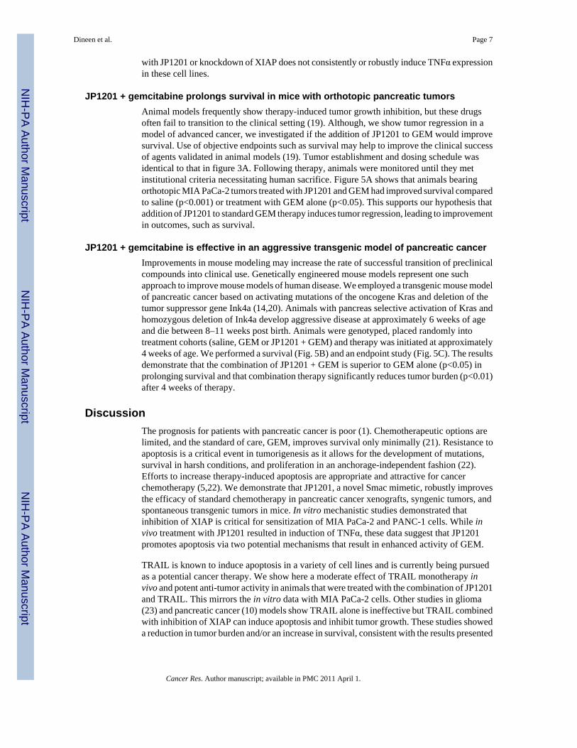
Smac mimetics induce apoptosis in vitro when combined with TRAIL or TNFα (9). JP1201did not induce apoptosis in MIA PaCa-2 as a single agent in vitro; however, addition of JP1201decreased the IC50 of TRAIL from 4.5 to 1.2 nM. We assessed how JP1201 in combinationwith TRAIL affected pancreatic cancer growth in vivo. In this early intervention model, micebearing MIA PaCa-2 tumors were treated with saline, TRAIL or TRAIL + JP- 1201 at 0.2, 0.6,2 or 6 mg/kg starting one week following tumor cell injection and sacrificed 2.5 weeks afterthe last dose of therapy (Fig. 1A). Therapy with TRAIL + JP1201 at doses of 2 mg/kg or 6 mg/kg reduced the final size of pancreatic tumors by approximately 50 percent (control, 0.46 g vs.0.23 g and 0.24 g, respectively; Fig. 1B). TRAIL alone had a modest effect on the size ofxenografts compared to saline. Tumors were removed en bloc. At the time of sacrifice, weobserved significantly less tumor burden and more residual normal pancreas in mice treatedwith TRAIL + JP1201. Histologic analysis showed extensive tumor burden in saline treatedanimals. In contrast, mice treated with combination therapy using the highest doses of JP1201showed scattered foci of tumor growth with a large amount of residual normal pancreas (Fig.1C).
In addition to controlling primary tumor burden, metastases were reduced by JP1201 + TRAIL.There were no metastatic events in the TRAIL + JP1201 (6 mg/kg) group, compared to 15 totalmetastatic events in control animals (Fig. 1D). These results show that JP1201 sensitizespancreatic tumors to therapy with the death receptor ligand TRAIL. Animals treated withcombination therapy did not display any weight loss or other signs of systemic toxicity.
Dineen et al. Page 4
Cancer Res. Author manuscript; available in PMC 2011 April 1.
NIH
-PA Author Manuscript
NIH
-PA Author Manuscript
NIH
-PA Author Manuscript

Inhibition of XIAP improves sensitivity to gemcitabineDespite being the standard of care for pancreatic cancer, gemcitabine (GEM) is only minimallyeffective. Blocks in apoptosis, including overexpression of IAPs might mediatechemoresistance in pancreatic cancer (16). To determine if JP1201 affected the sensitivity invitro to GEM, we screened a panel of cell lines against GEM alone, JP1201 alone or thecombination (Table 1). We found that in general a low dose (100 nM) of JP1201 enhanced thesensitivity of pancreatic cancer cells to GEM by approximately 20-fold. However the range offold-reduction was wide with two cell lines showing no change in the IC50 of GEM (Capan-2and Hs 766T) while PANC-1 cells demonstrated a 170-fold reduction.
Because JP1201 binds to and inhibits IAPs, we hypothesized that siRNA-mediated knockdownof IAPs would increase sensitivity of pancreatic cancer cells to GEM similar to treatment withJP1201. We found that siRNA-mediated knockdown of XIAP but not cIAP1 or cIAP2sensitized MIA PaCa-2 and PANC-1 (Fig. 2) cells to treatment with GEM. The increase insensitivity was similar to treatment with JP1201 (Fig. 2). This data expands work in otherpancreas cancer cell lines (8,17) that show resistance to chemotherapy is mediated in part byXIAP.
JP1201 + gemcitabine is efficacious for established pancreatic tumorsA challenge in treating pancreatic cancer is the fact that patients typically present at an advancedstage. To more closely parallel human disease presentation, we performed a late interventionstudy. MIA PaCa-2 tumors were grown for 28 days (tumor burden approximately 500 mg)prior to the initiation of therapy. Therapy was given as six doses over 2 weeks. A cohort ofanimals from each group was sacrificed 24 hours after the first dose of therapy (day 29) toacquire tissue for histological analysis (see below). The remaining animals were sacrificed onday 42, two days following the end of treatment. As demonstrated in figure 3A, treatment withJP1201 + TRAIL or GEM reduced tumor weight significantly (***, p<0.001 vs saline). JPalone also had a significant effect (*, p<0.05, vs. saline) on pancreatic tumor weight, despitehaving little effect as a single agent in vitro.
Comparison of mean tumor weights on day 42 to the mean weight of tumors harvested on atday 29 (mean of 15 tumors harvested) shows that animals treated with JP1201 in combinationwith GEM or TRAIL had reductions in mean tumor size of ~50%, while mice treated withsaline or GEM had increased mean tumor weight by 150 and 50%, respectively (Fig. 3B).Treatment with JP1201 alone caused a cessation of tumor growth. These data strongly suggestthat mice treated with combination therapy underwent tumor regression rather than growthinhibition. To confirm this robust effect we performed histological analysis, which revealedlarge poorly differentiated tumor tissue in the saline and GEM treated animals with littleevidence of remaining normal pancreas tissue. However, treatment with JP1201 alone or incombination with GEM or TRAIL revealed reduced primary tumor burden and an increase theamount of residual normal pancreas (dark purple) as seen by H&E analysis (Fig. S1). Areplicate experiment revealed similar results (Fig. 3C). In the replicate experiment the dose ofGEM was dropped to 25 mg/kg. We also evaluated the response of orthotopic BxPC-3 tumorsusing a similar dosing strategy and found that treatment with JP1201 in combination with GEMwas effective at controlling tumor size (saline, 0.94 g; combination therapy, 0.35 g).
Impaired drug delivery is proposed as a complicating factor in the poor response of pancreatictumors to standard therapy. With this in mind, we evaluated the general pharmacokineticparameters of GEM, its inactive metabolite dFdU, and JP1201 in mice bearing large orthotopicMIA PaCa-2 tumors (Table S1). 54 mice with a mean (+/− SD) pancreas/tumor of 0.47 +/−0.2 g were used for the study. We found that the plasma half-life and AUC of GEM wereelevated by combination with JP1201; however, the contribution of this to the dramatic increase
Dineen et al. Page 5
Cancer Res. Author manuscript; available in PMC 2011 April 1.
NIH
-PA Author Manuscript
NIH
-PA Author Manuscript
NIH
-PA Author Manuscript

in response in mice treated with combination therapy is unclear. We also identified asurprisingly long plasma and tumor half-life of JP1201, 300–400 min and 900 –1000 min,respectively.
JP1201 maintains effectiveness as a chemosensitizer in a syngenic modelTo determine if JP1201 + GEM remained effective in a syngenic model of pancreatic cancerwe performed a therapy experiment in C57Bl/6 mice using the murine pancreatic cancer cellline Pan02. Therapy was started three weeks after tumor cell injection and animals weresacrificed on day 35. The combination of JP1201 + GEM was effective at reducing tumorburden in this fully immunocompetent model (Fig. 3D). Of note 5 out of 10 mice in thecombination therapy group had no identifiable tumor at the time of necropsy where as 100%of the saline treated mice and mice treated with single agent therapy had significant pancreatictumor masses. We did find in this model that GEM (25 mg/kg) induced splenomegaly and thiswas exacerbated by combination with JP1201 (spleen weight: saline, 0.11 g; GEM, 0.19 g; JP0.11 g; JP/GEM, 0.25 g).
JP1201 reduces metastatic diseaseWe assessed metastatic burden in the xenograft late interventions studies and found that overallmetastatic incidence was 55% in the saline-treated animals and this was reduced to 35, 43, 31,and 14% by treatment with GEM, JP1201, JP1201 + GEM, and JP1201 + TRAIL, respectively.
JP1201 induces apoptosis and TNFα expression in orthotopic tumorsTo assess induction of apoptosis after therapy, MIA PaCa-2 tumors from animals sacrificedon day 29 and day 42 (Fig. 3A) were analyzed. TUNEL immunofluorescence revealed thatthere was an acute induction of apoptosis in the tumor following administration of JP1201combined with GEM. However after the full course of therapy there was no difference in theTUNEL signal between any of the treatment groups (Fig. 4A). We also evaluated cellproliferation by immunohistochemistry for PCNA. PCNA reactivity was elevated in thecombination (JP/GEM) at the early time point (d29); however single agent and combinationtherapy significantly reduced PCNA reactivity at the late time point (Fig. 4B). Furthermore,cell proliferation in tumors treated with JP1201 and the combination of (JP/GEM) were reducedsignificantly compared to treatment with GEM alone (Fig. 4B).
TNFα has been implicated in the response to Smac mimetics (18). Therefore we evaluated thelevel of TNFα protein in tumors from each group at early (d29) and late (d42) time points.TNFα levels were increased modestly by combination therapy at the early time point but wereelevated dramatically by JP1201 alone or in combination with GEM at the late time point (Fig.4C,D). We believe that the TNFα detected is of human tumor cell origin. Pan02 tumor tissuefrom controls and animals treated with JP1201 alone or in combination with GEM revealedsparse reactivity with the rabbit anti- TNFα antibody used in figure 4 (Fig. S2) and a rat anti-mouse TNFα antibody (data not shown). These results are consistent with PCR-based analysisof Pan02 tumor tissue, which did not show induction of TNFα (data not shown). Taken togetherour observations suggest that Pan02 cells do not produce TNFα upon treatment with JP1201and that the majority of TNFα staining observed in MIA PaCa-2 xenografts is of tumor cellorigin.
Additionally, we evaluated if TNFα induction was detectable in vitro after treatment withJP1201 or siRNA-mediated XIAP knockdown (Table S2). Cells (MIA PaCa-2, PANC-1,AsPC-1, and BxPC3) were treated with JP1201 (100 nM) or subjected to XIAP knockdown.Conditioned media was harvested 12, 48, and 96 hours post treatment and analyzed forTNFα levels by ELISA. These results demonstrate that during this time frame a single treatment
Dineen et al. Page 6
Cancer Res. Author manuscript; available in PMC 2011 April 1.
NIH
-PA Author Manuscript
NIH
-PA Author Manuscript
NIH
-PA Author Manuscript
with JP1201 or knockdown of XIAP does not consistently or robustly induce TNFα expressionin these cell lines.
JP1201 + gemcitabine prolongs survival in mice with orthotopic pancreatic tumorsAnimal models frequently show therapy-induced tumor growth inhibition, but these drugsoften fail to transition to the clinical setting (19). Although, we show tumor regression in amodel of advanced cancer, we investigated if the addition of JP1201 to GEM would improvesurvival. Use of objective endpoints such as survival may help to improve the clinical successof agents validated in animal models (19). Tumor establishment and dosing schedule wasidentical to that in figure 3A. Following therapy, animals were monitored until they metinstitutional criteria necessitating human sacrifice. Figure 5A shows that animals bearingorthotopic MIA PaCa-2 tumors treated with JP1201 and GEM had improved survival comparedto saline (p<0.001) or treatment with GEM alone (p<0.05). This supports our hypothesis thataddition of JP1201 to standard GEM therapy induces tumor regression, leading to improvementin outcomes, such as survival.
JP1201 + gemcitabine is effective in an aggressive transgenic model of pancreatic cancerImprovements in mouse modeling may increase the rate of successful transition of preclinicalcompounds into clinical use. Genetically engineered mouse models represent one suchapproach to improve mouse models of human disease. We employed a transgenic mouse modelof pancreatic cancer based on activating mutations of the oncogene Kras and deletion of thetumor suppressor gene Ink4a (14,20). Animals with pancreas selective activation of Kras andhomozygous deletion of Ink4a develop aggressive disease at approximately 6 weeks of ageand die between 8–11 weeks post birth. Animals were genotyped, placed randomly intotreatment cohorts (saline, GEM or JP1201 + GEM) and therapy was initiated at approximately4 weeks of age. We performed a survival (Fig. 5B) and an endpoint study (Fig. 5C). The resultsdemonstrate that the combination of JP1201 + GEM is superior to GEM alone (p<0.05) inprolonging survival and that combination therapy significantly reduces tumor burden (p<0.01)after 4 weeks of therapy.
DiscussionThe prognosis for patients with pancreatic cancer is poor (1). Chemotherapeutic options arelimited, and the standard of care, GEM, improves survival only minimally (21). Resistance toapoptosis is a critical event in tumorigenesis as it allows for the development of mutations,survival in harsh conditions, and proliferation in an anchorage-independent fashion (22).Efforts to increase therapy-induced apoptosis are appropriate and attractive for cancerchemotherapy (5,22). We demonstrate that JP1201, a novel Smac mimetic, robustly improvesthe efficacy of standard chemotherapy in pancreatic cancer xenografts, syngenic tumors, andspontaneous transgenic tumors in mice. In vitro mechanistic studies demonstrated thatinhibition of XIAP is critical for sensitization of MIA PaCa-2 and PANC-1 cells. While invivo treatment with JP1201 resulted in induction of TNFα, these data suggest that JP1201promotes apoptosis via two potential mechanisms that result in enhanced activity of GEM.
TRAIL is known to induce apoptosis in a variety of cell lines and is currently being pursuedas a potential cancer therapy. We show here a moderate effect of TRAIL monotherapy invivo and potent anti-tumor activity in animals that were treated with the combination of JP1201and TRAIL. This mirrors the in vitro data with MIA PaCa-2 cells. Other studies in glioma(23) and pancreatic cancer (10) models show TRAIL alone is ineffective but TRAIL combinedwith inhibition of XIAP can induce apoptosis and inhibit tumor growth. These studies showeda reduction in tumor burden and/or an increase in survival, consistent with the results presented
Dineen et al. Page 7
Cancer Res. Author manuscript; available in PMC 2011 April 1.
NIH
-PA Author Manuscript
NIH
-PA Author Manuscript
NIH
-PA Author Manuscript

here. Thus, our data support that Smac mimetics in combination with TRAIL is a promisinganticancer strategy.
Since GEM is the standard of care for pancreatic cancer, we determined if inhibition of XIAPwould sensitize tumors to GEM therapy. We found that knockdown of XIAP but not relatedIAPs using siRNA increases sensitivity to GEM in pancreatic cancer cell lines. This dataexpands on work in other pancreatic cancer cell lines that shows resistance to chemotherapyis mediated in part by XIAP (8,17). We also show that the sensitization to GEM with XIAPknockdown is strikingly similar to the sensitization seen with JP1201 in vitro; supporting theconcept that JP1201 sensitizes pancreatic cancer to GEM through inhibition of XIAP.
We also explored whether JP1201 would sensitize tumors to GEM treatment. We demonstrateda significant decrease in tumor weights in animals treated with the combination of JP1201 andGEM compared to saline in a late intervention model in both immune-compromised andimmune-competent mice. The JP1201 sensitization was also evident at lower doses of GEM.In a separate experiment, this effect translated into increased survival in tumor-bearing animals.These data alone are very suggestive that Smac mimetics combined with standard therapy couldgreatly improve pancreatic cancer patient response to therapy. This study adds to the growingbody of literature that demonstrate the potency of Smac mimetics in combination with variousstandards of care, such as a study done in a glioblastoma model where Smac mimetics wereshown to enhance tumor cell death after irradiation treatment (24). We further explored theeffect of combination therapy in an aggressive transgenic model of pancreatic cancer and againsaw increased survival.
We also found that treatment with JP1201 induced TNFα in vivo. Previous reports (12,25,26)have shown that Smac mimetic sensitive cell lines produce TNFα, can be induced to producemore TNFα after Smac mimetic treatment, and are dependent on TNFα for Smac mimetic-induced cell death. However, there is currently no published data indicating Smac mimeticsinduce TNFα in resistant cell lines, such as MIA PaCa-2. Possible explanations includeprolonged treatment with JP1201 may induce TNFα production. Alternatively, the in vivotumor microenvironment may be required to facilitate TNFα production after treatment withJP1201 or other Smac mimetics. In that regard, it is worth noting that while the pancreatic celllines showed in vitro resistance to JP1201, xenografts did show reduction of tumor burden withJP1201 as a monotherapy, which might be linked to the production of TNFα. Theseobservations suggest that alternative pathways may be engaged that promote cell death.TNFα expression has been linked to non-canonical activation of NFκB as a result of cIAP1/2degradation (25,27). NFκB can in turn stimulate autocrine expression of TNFα (25,26,28,29),which activates receptor interacting protein kinase 1 (RIPK1) via the TNF receptor andsubsequently induces caspase activation. In support of this, preliminary studies suggest thatNFκB actively particpates in JP1201-mediated sensitization of pancreatic tumor cells, astreatment of cells with GEM, JP1201 and an IKK inhibitor (SC-514) increased the IC50 forGEM 3–13 fold compared to JP1201 + GEM (data not shown). Thus JP1201 could promoteapoptosis by inducing the degradation of cIAP1/2, resulting in NFκB activation, and inhibitingthe activity of XIAP both of which could potentiate the efficacy of GEM (30).
In summary resistance to therapy-induced apoptosis is a major road block in the treatment ofpatients with pancreatic cancer. Another significant challenge is the lack of predictive powerof pre-clinical models of pancreatic cancer. In an effort to address both of these challenges, wehave demonstrated potent anti-tumor activity of a unique Smac mimetic in animal models ofpancreatic cancer that are robust and clinically relevant. We believe our data support thedevelopment of agents designed to sensitize pancreatic tumors to apoptosis and suggest JP1201deserves serious consideration for clinical investigation.
Dineen et al. Page 8
Cancer Res. Author manuscript; available in PMC 2011 April 1.
NIH
-PA Author Manuscript
NIH
-PA Author Manuscript
NIH
-PA Author Manuscript

Supplementary MaterialRefer to Web version on PubMed Central for supplementary material.
AcknowledgmentsGrant Support: This work was supported in part by a sponsored research agreement from Joyant Pharmaceuticals(to. R. Brekken), the Effie Marie Cain Scholarship in Angiogenesis Research (to R. Brekken), and postdoctoralfellowship from Susan G. Komen for the Cure (to S. Dineen).
We thank Drs. Xiaodong Wang, Lai Wang, and Lin Li for advice and technical support and members of the Brekkenlab and Drs. Niranjan Awasthi and Roderich Schwarz for support and thoughtful discussion.
REFERENCES1. Jemal A, Siegel R, Ward E, Murray T, Xu J, Thun MJ. Cancer statistics, 2007. CA: a cancer journal
for clinicians 2007;57:43–66. [PubMed: 17237035]2. Westphal S, Kalthoff H. Apoptosis: targets in pancreatic cancer. Molecular cancer 2003;2:6. [PubMed:
12605713]3. Fesik SW. Promoting apoptosis as a strategy for cancer drug discovery. Nature reviews 2005;5:876–
885.4. Salvesen GS, Duckett CS. IAP proteins: blocking the road to death's door. Nat Rev Mol Cell Biol
2002;3:401–410. [PubMed: 12042762]5. Reed JC. Apoptosis-targeted therapies for cancer. Cancer cell 2003;3:17–22. [PubMed: 12559172]6. Lopes RB, Gangeswaran R, McNeish IA, Wang Y, Lemoine NR. Expression of the IAP protein family
is dysregulated in pancreatic cancer cells and is important for resistance to chemotherapy. Internationaljournal of cancer 2007;120:2344–2352.
7. Karikari CA, Roy I, Tryggestad E, et al. Targeting the apoptotic machinery in pancreatic cancers usingsmall-molecule antagonists of the X-linked inhibitor of apoptosis protein. Molecular cancertherapeutics 2007;6:957–966. [PubMed: 17339366]
8. Shrikhande SV, Kleeff J, Kayed H, et al. Silencing of X-linked inhibitor of apoptosis (XIAP) decreasesgemcitabine resistance of pancreatic cancer cells. Anticancer research 2006;26:3265–3273. [PubMed:17094439]
9. Li L, Thomas RM, Suzuki H, De Brabander JK, Wang X, Harran PG. A small molecule Smac mimicpotentiates TRAIL- and TNFalpha-mediated cell death. Science (New York, NY 2004;305:1471–1474.
10. Vogler M, Walczak H, Stadel D, et al. Small molecule XIAP inhibitors enhance TRAIL-inducedapoptosis and antitumor activity in preclinical models of pancreatic carcinoma. Cancer Res2009;69:2425–2434. [PubMed: 19258513]
11. Vogler M, Walczak H, Stadel D, et al. Targeting XIAP bypasses Bcl-2-mediated resistance to TRAILand cooperates with TRAIL to suppress pancreatic cancer growth in vitro and in vivo. Cancer Res2008;68:7956–7965. [PubMed: 18829553]
12. Petersen SL, Wang L, Yalcin-Chin A, et al. Autocrine TNFalpha signaling renders human cancercells susceptible to Smac-mimetic-induced apoptosis. Cancer cell 2007;12:445–456. [PubMed:17996648]
13. Dineen SP, Lynn KD, Holloway SE, et al. Vascular endothelial growth factor receptor 2 mediatesmacrophage infiltration into orthotopic pancreatic tumors in mice. Cancer research 2008;68:4340–4346. [PubMed: 18519694]
14. Aguirre AJ, Bardeesy N, Sinha M, et al. Activated Kras and Ink4a/Arf deficiency cooperate to producemetastatic pancreatic ductal adenocarcinoma. Genes Dev 2003;17:3112–3126. [PubMed: 14681207]
15. Bardeesy N, DePinho RA. Pancreatic cancer biology and genetics. Nature reviews 2002;2:897–909.16. Hamacher R, Schmid RM, Saur D, Schneider G. Apoptotic pathways in pancreatic ductal
adenocarcinoma. Molecular cancer 2008;7:64. [PubMed: 18652674]
Dineen et al. Page 9
Cancer Res. Author manuscript; available in PMC 2011 April 1.
NIH
-PA Author Manuscript
NIH
-PA Author Manuscript
NIH
-PA Author Manuscript

17. Li Y, Jian Z, Xia K, et al. XIAP is related to the chemoresistance and inhibited its expression by RNAinterference sensitize pancreatic carcinoma cells to chemotherapeutics. Pancreas 2006;32:288–296.[PubMed: 16628085]
18. He S, Wang L, Miao L, et al. Receptor interacting protein kinase-3 determines cellular necroticresponse to TNF-alpha. Cell 2009;137:1100–1111. [PubMed: 19524512]
19. Man S, Munoz R, Kerbel RS. On the development of models in mice of advanced visceral metastaticdisease for anti-cancer drug testing. Cancer metastasis reviews 2007;26:737–747. [PubMed:17846863]
20. Olive KP, Jacobetz MA, Davidson CJ, et al. Inhibition of Hedgehog signaling enhances delivery ofchemotherapy in a mouse model of pancreatic cancer. Science (New York, NY 2009;324:1457–1461.
21. Fujioka S, Niu J, Schmidt C, et al. NF-kappaB and AP-1 connection: mechanism of NF-kappaB-dependent regulation of AP-1 activity. Molecular and cellular biology 2004;24:7806–7819.[PubMed: 15314185]
22. Hanahan D, Weinberg RA. The hallmarks of cancer. Cell 2000;100:57–70. [PubMed: 10647931]23. Fulda S, Wick W, Weller M, Debatin KM. Smac agonists sensitize for Apo2L/TRAIL- or anticancer
drug-induced apoptosis and induce regression of malignant glioma in vivo. Nature medicine2002;8:808–815.
24. Vellanki SH, Grabrucker A, Liebau S, et al. Small-molecule XIAP inhibitors enhance gamma-irradiation-induced apoptosis in glioblastoma. Neoplasia (New York, NY 2009;11:743–752.
25. Vince JE, Wong WW, Khan N, et al. IAP antagonists target cIAP1 to induce TNFalpha-dependentapoptosis. Cell 2007;131:682–693. [PubMed: 18022363]
26. Varfolomeev E, Blankenship JW, Wayson SM, et al. IAP antagonists induce autoubiquitination ofc-IAPs, NF-kappaB activation, and TNFalpha-dependent apoptosis. Cell 2007;131:669–681.[PubMed: 18022362]
27. Bai L, Chen W, Wang X, Tang H, Lin Y. IKKbeta-mediated nuclear factor-kappaB activationattenuates smac mimetic-induced apoptosis in cancer cells. Molecular cancer therapeutics2009;8:1636–1645. [PubMed: 19509265]
28. Gaither A, Porter D, Yao Y, et al. A Smac mimetic rescue screen reveals roles for inhibitor of apoptosisproteins in tumor necrosis factor-alpha signaling. Cancer Res 2007;67:11493–11498. [PubMed:18089776]
29. Wang L, Du F, Wang X. TNF-alpha induces two distinct caspase-8 activation pathways. Cell2008;133:693–703. [PubMed: 18485876]
30. Lu J, Bai L, Sun H, et al. SM-164: a novel, bivalent Smac mimetic that induces apoptosis and tumorregression by concurrent removal of the blockade of cIAP-1/2 and XIAP. Cancer Res 2008;68:9384–9393. [PubMed: 19010913]
Dineen et al. Page 10
Cancer Res. Author manuscript; available in PMC 2011 April 1.
NIH
-PA Author Manuscript
NIH
-PA Author Manuscript
NIH
-PA Author Manuscript

Figure 1. JP1201 in combination with TRAIL is effective in controlling pancreatic tumor xenograftsA, Early intervention treatment algorithm. 1×10⁶ MIA PaCa-2 cells were injected into miceon Day 0. Therapy was initiated on Day 7 post tumor cell injection (TCI). Therapy was givenby iv or ip injection every other day (MWF) for two weeks on the days indicated. Mice receivedsaline, TRAIL (20 mg/kg), or TRAIL + JP1201 (JP, at 0.2, 0.6, 2.0, or 6.0 mg/kg). Animalswere sacrificed 2.5 weeks after the last dose of therapy (D36). B, A scatter plot of tumor weightsat the time of sacrifice showing the mean +/− SEM as well as the weight of each tumor fromindividual mice. *, p < 0.05 by one-way ANOVA with Dunn’s posttest. C, Representative H&Estaining of formalin-fixed, paraffin embedded sections of tumor tissue from mice treated withSaline or JP/TRAIL. The upper row are images at 100× total magnification (scale bar, 100 µm)while the lower row is at 400× total magnification (scale bar, 50 µm). The white box in theupper row is the area magnified. D, The mean +/− SEM number of metastases in each treatmentgroup as well as the number of metastatic events in each animal is shown in the scatter plot.Metastases were evaluated by careful visual inspection at the time of necropsy. Note theanimals treated with JP/TRAIL at the highest dose did not have any visible metastases. Thedata represent a single animal experiment with 7–9 animals in each treatment group.
Dineen et al. Page 11
Cancer Res. Author manuscript; available in PMC 2011 April 1.
NIH
-PA Author Manuscript
NIH
-PA Author Manuscript
NIH
-PA Author Manuscript

Figure 2. Inhibition of XIAP improves sensitivity to gemcitabine in pancreatic cancer cell linesA, Pancreatic cancer cells, MIA PaCa-2 and PANC-1, were treated with increasingconcentrations of gemcitabine alone, or in combination with siRNA (100 nM) specific for theindicated target or JP1201 (100 nM). Cell viability was estimated 4 days after initiation oftreatment by a MTS assay in which each condition was replicated 8 times. The data shown isrepresentative of three independent experiments. Transfection with siRNA specific forluciferase (si-Luciferase) was used as a control for cell toxicity mediated by transfection alone.B, Western blot analysis of XIAP, cIAP1, cIAP2 demonstrating specificity of siRNA-mediatedknockdown. Controls included no transfection (Cntl 1), transfection reagent alone (Cntl 2),and transfection with a siRNA specific for luciferase (Luc).
Dineen et al. Page 12
Cancer Res. Author manuscript; available in PMC 2011 April 1.
NIH
-PA Author Manuscript
NIH
-PA Author Manuscript
NIH
-PA Author Manuscript

Figure 3. JP1201 combined with gemcitabine has potent anti-tumor activity against establishedpancreatic tumorsA–C, MIA PaCa-2 (1×10⁶) were injected into the pancreas of nude mice on day 0. On day 28post tumor cell injection (TCI) therapy was initiated and consisted of saline, JP1201 (JP, 6.0mg/kg), JP + TRAIL (TR, 20 mg/kg) 3x/week via iv injection on the days indicated.Gemcitabine (GEM) alone (n=5) or in combination with JP (A & B, n=4) was given ip at adose of 175 mg/kg on day 28, and at 100 mg/kg on days 30, 37, and 40 (A, B) and 25 mg/kg3x/week for two weeks (C, n=12/group). Scatter plots showing mean +/− SEM tumor weightas well as the weights individual tumors for each group are shown (A, C). *, p<0.05; **, p<0.01;***, p<0.005 by one-way ANOVA. B, Three tumors from each treatment group shown in panelB (n=15 total) were harvested on day 29 post TCI. Final tumor size of each group (day 42) wascompared to the mean tumor size on day 29. The percent change in tumor size is shown. *,p<0.05; ***, p<0.0001 vs. saline by one-way ANOVA. D, Pan02 murine pancreatic cancercells (1×10⁶) were injected orthotopically into C57Bl/6J mice on day 0. Therapy with saline,GEM (25 mg/kg), JP (6.0 mg/kg) or the combination was initiated on day 21 post TCI andgiven 3x/week for two weeks. The mice were sacrificed on day 35 post TCI and resulting tumor
Dineen et al. Page 13
Cancer Res. Author manuscript; available in PMC 2011 April 1.
NIH
-PA Author Manuscript
NIH
-PA Author Manuscript
NIH
-PA Author Manuscript

weights are displayed as % of body weight in a scatter plot showing mean +/− SEM (n= 9–10/group). *, p<0.05; **, p<0.01 vs. saline by one-way ANOVA.
Dineen et al. Page 14
Cancer Res. Author manuscript; available in PMC 2011 April 1.
NIH
-PA Author Manuscript
NIH
-PA Author Manuscript
NIH
-PA Author Manuscript

Figure 4. Induction of apoptosis using combination therapyA–D, Tumors from animals sacrificed one day after initiation of therapy (EARLY (D29)) orafter the cessation of therapy (LATE (D42)) were formalin fixed and paraffin embedded. A–C, Data from a minimum of 4 tumors from each group are normalized to the saline group andare representative of at least two independent assays on the same tumor tissue. A, TUNELanalysis was performed and analyzed using immunofluorescence. *, p<0.05; **, p<0.01; ***,p<0.001 vs. saline by one-way ANOVA. B, Cell proliferation was measured byimmunofluorescence for PCNA. **, p<0.01; ***, p<0.001 by one way ANOVA. C & D, Theexpression level of TNFα was determined by immunofluorescence. *p<0.05; ***, p<0.001 by
Dineen et al. Page 15
Cancer Res. Author manuscript; available in PMC 2011 April 1.
NIH
-PA Author Manuscript
NIH
-PA Author Manuscript
NIH
-PA Author Manuscript
one way ANOVA. D, Representative images of TNFα (green) immunofluorescence, scale bar,50 µm.
Dineen et al. Page 16
Cancer Res. Author manuscript; available in PMC 2011 April 1.
NIH
-PA Author Manuscript
NIH
-PA Author Manuscript
NIH
-PA Author Manuscript

Figure 5. Combination of JP1201 with gemcitabine enhances survival of mice with pancreaticcancerA, Mice bearing established MIA PaCa-2 tumors were treated using the late interventionprotocol described in Fig. 3A. Mice were treated with saline (black line), gemctiabine (GEM,25 mg/kg, green line), JP1201 (6.0 mg/kg, red line), or the combination of JP/GEM (blue line)3x/week for two weeks. Animals were monitored and sacrificed when they demonstratedobjective signs of tumor burden or when they appeared moribund (as determined by a blindedobserver). B, p48-Cre:KRasG12D:Ink4a/Arflox/lox PDAC animals were treated with saline,GEM or combination of JP/GEM starting at 4 weeks of age. Therapy was delivered ipcontinuously 3x/week until sacrifice. C, A second cohort of PDAC animals underwent 4 weeksof therapy (Saline, n=5; GEM, n=4; JP/GEM, n=6) and were sacrificed at 8 weeks of age.Tumors were excised and weighed and a scatter plot showing mean +/− SEM tumor weight isdisplayed. Compared to control (1.124 ± 0.1135 g), JP/GEM significantly reduced tumorgrowth (0.3833 ± 0.1492 g) **, p<0.01. Each panel represents a single animal experiment.
Dineen et al. Page 17
Cancer Res. Author manuscript; available in PMC 2011 April 1.
NIH
-PA Author Manuscript
NIH
-PA Author Manuscript
NIH
-PA Author Manuscript

NIH
-PA Author Manuscript
NIH
-PA Author Manuscript
NIH
-PA Author Manuscript
Dineen et al. Page 18
Tabl
e 1
JP12
01 se
nsiti
zes p
ancr
eatic
tum
or c
ells
to g
emci
tabi
ne
GE
M (n
M)
GE
M (n
M) +
JP1
201
(100
nM
)Fo
ldre
duct
ion
JP12
01 (µ
M)
Cel
l Lin
eA
ssay
sM
edia
nSD
Ass
ays
Med
ian
SDA
ssay
sM
edia
nSD
AsP
C-1
417
73
414
92.
51.
195
611.
3
BxP
C-3
413
.41.
26
7.55
3.7
1.77
44.
981.
2
Cap
an-1
25.
051
20.
529.
712
12.1
Cap
an-2
420
001
320
001.
11.
004
100
1
Hs 7
66T
420
001
420
001
1.00
410
01
Hs 7
00T
427
.13.
24
11.5
2.2
2.36
410
01
MIA
PaC
a-2
819
.12
61.
22.
915
.92
660
.42.
5
PAN
C-1
620
001.
96
11.5
3.7
173.
914
8.85
1.2
Pan0
26
4314
621
.23.
82.
03
Cel
l gro
wth
ass
ays w
ere
perf
orm
ed in
96-
wel
l for
mat
for f
ive
days
(n=8
/con
ditio
n/as
say)
. On
day
0, c
ells
wer
e pl
ated
and
on
day
1 dr
ugs w
ere
adde
d in
four
fold
dilu
tions
. The
hig
hest
dos
e of
gem
cita
bine
(GEM
) was
200
0 nM
. The
hig
hest
con
cent
ratio
n of
JP12
01 a
lone
was
100
µM
. On
day
5, re
lativ
e ce
ll nu
mbe
r was
est
imat
ed u
sing
MTS
(Pro
meg
a, fi
nal c
once
ntra
tion
333
µg/m
l), p
late
s wer
e th
en in
cuba
ted
for 1
to 3
hou
rs a
t 37°
C, a
nd re
ad a
t 490
nm
. Dru
g se
nsiti
vity
cur
ves a
nd IC
50s w
ere
calc
ulat
ed u
sing
in-h
ouse
softw
are.
The
num
ber i
ndep
ende
nt a
ssay
per
form
ed a
nd th
e m
edia
n G
EM IC
50 +
/− S
D (n
M) f
or G
EM a
lone
and
GEM
+ JP
1201
, the
fold
redu
ctio
n in
GEM
IC50
in th
e pr
esen
ce o
f JP1
201
(100
nM) a
nd se
nsiti
vity
toJP
1201
alo
ne is
dis
play
ed.
Shad
ed ro
ws i
ndic
ate
cell
lines
that
wer
e te
sted
in v
ivo.
Cancer Res. Author manuscript; available in PMC 2011 April 1.