Sleep after spatial learning promotes covert reorganization of brain activity
6
Sleep after spatial learning promotes covert reorganization of brain activity Pierre Orban* † , Ge ´ raldine Rauchs*, Evelyne Balteau*, Christian Degueldre*, Andre ´ Luxen*, Pierre Maquet* ‡ , and Philippe Peigneux* § *Cyclotron Research Center, University of Lie ` ge, Ba ˆ timent B30, 4000 Lie ` ge, Belgium; and ‡ Department of Neurology, Centre Hospitalier Universitaire, University of Lie ` ge, 4000 Lie ` ge, Belgium Edited by Marcus E. Raichle, Washington University School of Medicine, St. Louis, MO, and approved March 17, 2006 (received for review November 24, 2005) Sleep promotes the integration of recently acquired spatial mem- ories into cerebral networks for the long term. In this study, we examined how sleep deprivation hinders this consolidation pro- cess. Using functional MRI, we mapped regional cerebral activity during place-finding navigation in a virtual town, immediately after learning and 3 days later, in subjects either allowed regular sleep (RS) or totally sleep-deprived (TSD) on the first posttraining night. At immediate and delayed retrieval, place-finding naviga- tion elicited increased brain activity in an extended hippocampo- neocortical network in both RS and TSD subjects. Behavioral performance was equivalent between groups. However, striatal navigation-related activity increased more at delayed retrieval in RS than in TSD subjects. Furthermore, correlations between striatal response and behavioral performance, as well as functional con- nectivity between the striatum and the hippocampus, were mod- ulated by posttraining sleep. These data suggest that brain activity is restructured during sleep in such a way that navigation in the virtual environment, initially related to a hippocampus-dependent spatial strategy, becomes progressively contingent in part on a response-based strategy mediated by the striatum. Both neural strategies eventually relate to equivalent performance levels, in- dicating that covert reorganization of brain patterns underlying navigation after sleep is not necessarily accompanied by overt changes in behavior. functional MRI hippocampus sleep deprivation memory consolidation striatum T he hypothesis that sleep represents a crucial, albeit not always indispensable, neurophysiological state that actively promotes learning-dependent brain plasticity (1–5) has become a main topic of interest in neuroscience. In this view, an active processing of freshly acquired memories may occur in the sleeping brain, supporting the gradual consolidation process by which labile recent memories are restructured and incorporated into stable memories for the long term (6, 7). Memories that can be consciously and symbolically expressed belong to the declarative memory system. At the neuroanatomical level, their encoding and initial maintenance rely upon the integrity of the medial temporal lobe, with the hippocampus at its core (8, 9). Episodic memory for personally experienced events set in a spatiotemporal context is defined as a subclass of declarative memory (10). Animal (11) and human (12) studies suggest that the hippocampus is involved in spatial learning, because encoding is based on a flexible knowledge of relationships between environ- mental cues. Hippocampus-dependent spatial memory in animals is thus phylogenetically viewed as a homologue of human episodic declarative memory (13, 14). However, spatial navigation in a well learned environment may also be mediated by the striatum through the expression of stimulus–response associations (15, 16). Current research further suggests that rodents (17, 18) and humans (19) tend to initially use a hippocampus-dependent scheme early in training and then a striatum-dependent response strategy after repeated practice. Several studies showed a favorable effect of sleep on the consolidation of hippocampus-dependent memories in human, especially non-rapid-eye-movement sleep that includes slow- wave sleep (SWS) and stage II sleep. SWS deprivation (20, 21) and administration of the cholinergic agonist physostigmine during SWS (21) impair the sleep-dependent retention of lists of word pairs, whereas transcranial direct current stimulation dur- ing SWS, but not wakefulness, positively affects their retention (22). In addition, performance at morning or evening recall of word pairs (23) and overnight retention of face–name associa- tions (24) correlate with spindle density during intervening stage II sleep. Likewise, recognition memory performance for land- scapes correlates with amounts of SWS gained during a post- learning nap (25). SWS deprivation impairs the retention of spatial rotation abilities (26), and memory for spatial locations is impaired in proportion to lower amounts of SWS in schizo- phrenic patients (27). Besides, extensive navigation in a virtual maze increases stage II sleep duration on the first posttraining night (28). Finally, our group reported a positron emission tomography study showing that hippocampal activity associated with place learning in a large-scale virtual town during wake- fulness increases during SWS (and stage II sleep) on the first posttraining night and more so than after procedural learning (29). Importantly, overnight improvement in navigation accu- racy correlated with hippocampal activity during posttraining SWS. These functional brain imaging data contributed to com- plement the results of electrophysiological studies in rodents, which demonstrated at the cellular level the reexpression of hippocampal and neocortical patterns of spatial-related activity mainly during non-rapid-eye-movement sleep (30, 31). In the present study, we tested the hypothesis that sleep deprivation after spatial learning should hinder the plastic changes in cerebral activity that underpin the long-term consol- idation of recent memory traces. To do so, we used functional MRI (fMRI) to map the brain activity of subjects engaged in place-oriented navigation within a complex virtual town, both immediately and 3 days after learning (Fig. 1 a and b). On the first posttraining night, half of the subjects were totally sleep- deprived (TSD), whereas the others benefited from regular sleep (RS). All were then allowed two nights of RS before the delayed retrieval session, to preclude in TSD subjects the confounding aftereffect of extended wakefulness on regional cerebral activity, alertness, and behavioral performance on the next day (32, 33). Conflict of interest statement: No conflicts declared. This paper was submitted directly (Track II) to the PNAS office. Abbreviations: fMRI, functional MRI; RS, regular sleep; SWS, slow-wave sleep; TSD, totally sleep-deprived. Data deposition: The neuroimaging data reported in this paper have been deposited with the fMRI Data Center, www.fmridc.org (accession no. 2-2006-121CE). † Present address: Functional Neuroimaging Unit, University of Montreal, 4565, Queen Mary, Montreal, QC, Canada H3W 1W5. § To whom correspondence should be addressed. E-mail: [email protected]. © 2006 by The National Academy of Sciences of the USA 7124 –7129 PNAS May 2, 2006 vol. 103 no. 18 www.pnas.orgcgidoi10.1073pnas.0510198103
Transcript of Sleep after spatial learning promotes covert reorganization of brain activity

Sleep after spatial learning promotes covertreorganization of brain activityPierre Orban*†, Geraldine Rauchs*, Evelyne Balteau*, Christian Degueldre*, Andre Luxen*, Pierre Maquet*‡,and Philippe Peigneux*§
*Cyclotron Research Center, University of Liege, Batiment B30, 4000 Liege, Belgium; and ‡Department of Neurology, Centre Hospitalier Universitaire,University of Liege, 4000 Liege, Belgium
Edited by Marcus E. Raichle, Washington University School of Medicine, St. Louis, MO, and approved March 17, 2006 (received for reviewNovember 24, 2005)
Sleep promotes the integration of recently acquired spatial mem-ories into cerebral networks for the long term. In this study, weexamined how sleep deprivation hinders this consolidation pro-cess. Using functional MRI, we mapped regional cerebral activityduring place-finding navigation in a virtual town, immediatelyafter learning and 3 days later, in subjects either allowed regularsleep (RS) or totally sleep-deprived (TSD) on the first posttrainingnight. At immediate and delayed retrieval, place-finding naviga-tion elicited increased brain activity in an extended hippocampo-neocortical network in both RS and TSD subjects. Behavioralperformance was equivalent between groups. However, striatalnavigation-related activity increased more at delayed retrieval inRS than in TSD subjects. Furthermore, correlations between striatalresponse and behavioral performance, as well as functional con-nectivity between the striatum and the hippocampus, were mod-ulated by posttraining sleep. These data suggest that brain activityis restructured during sleep in such a way that navigation in thevirtual environment, initially related to a hippocampus-dependentspatial strategy, becomes progressively contingent in part on aresponse-based strategy mediated by the striatum. Both neuralstrategies eventually relate to equivalent performance levels, in-dicating that covert reorganization of brain patterns underlyingnavigation after sleep is not necessarily accompanied by overtchanges in behavior.
functional MRI � hippocampus � sleep deprivation � memoryconsolidation � striatum
The hypothesis that sleep represents a crucial, albeit notalways indispensable, neurophysiological state that actively
promotes learning-dependent brain plasticity (1–5) has becomea main topic of interest in neuroscience. In this view, an activeprocessing of freshly acquired memories may occur in thesleeping brain, supporting the gradual consolidation process bywhich labile recent memories are restructured and incorporatedinto stable memories for the long term (6, 7).
Memories that can be consciously and symbolically expressedbelong to the declarative memory system. At the neuroanatomicallevel, their encoding and initial maintenance rely upon the integrityof the medial temporal lobe, with the hippocampus at its core (8,9). Episodic memory for personally experienced events set in aspatiotemporal context is defined as a subclass of declarativememory (10). Animal (11) and human (12) studies suggest that thehippocampus is involved in spatial learning, because encoding isbased on a flexible knowledge of relationships between environ-mental cues. Hippocampus-dependent spatial memory in animalsis thus phylogenetically viewed as a homologue of human episodic�declarative memory (13, 14). However, spatial navigation in a welllearned environment may also be mediated by the striatum throughthe expression of stimulus–response associations (15, 16). Currentresearch further suggests that rodents (17, 18) and humans (19) tendto initially use a hippocampus-dependent scheme early in trainingand then a striatum-dependent response strategy after repeatedpractice.
Several studies showed a favorable effect of sleep on theconsolidation of hippocampus-dependent memories in human,especially non-rapid-eye-movement sleep that includes slow-wave sleep (SWS) and stage II sleep. SWS deprivation (20, 21)and administration of the cholinergic agonist physostigmineduring SWS (21) impair the sleep-dependent retention of lists ofword pairs, whereas transcranial direct current stimulation dur-ing SWS, but not wakefulness, positively affects their retention(22). In addition, performance at morning or evening recall ofword pairs (23) and overnight retention of face–name associa-tions (24) correlate with spindle density during intervening stageII sleep. Likewise, recognition memory performance for land-scapes correlates with amounts of SWS gained during a post-learning nap (25). SWS deprivation impairs the retention ofspatial rotation abilities (26), and memory for spatial locationsis impaired in proportion to lower amounts of SWS in schizo-phrenic patients (27). Besides, extensive navigation in a virtualmaze increases stage II sleep duration on the first posttrainingnight (28). Finally, our group reported a positron emissiontomography study showing that hippocampal activity associatedwith place learning in a large-scale virtual town during wake-fulness increases during SWS (and stage II sleep) on the firstposttraining night and more so than after procedural learning(29). Importantly, overnight improvement in navigation accu-racy correlated with hippocampal activity during posttrainingSWS. These functional brain imaging data contributed to com-plement the results of electrophysiological studies in rodents,which demonstrated at the cellular level the reexpression ofhippocampal and neocortical patterns of spatial-related activitymainly during non-rapid-eye-movement sleep (30, 31).
In the present study, we tested the hypothesis that sleepdeprivation after spatial learning should hinder the plasticchanges in cerebral activity that underpin the long-term consol-idation of recent memory traces. To do so, we used functionalMRI (fMRI) to map the brain activity of subjects engaged inplace-oriented navigation within a complex virtual town, bothimmediately and 3 days after learning (Fig. 1 a and b). On thefirst posttraining night, half of the subjects were totally sleep-deprived (TSD), whereas the others benefited from regular sleep(RS). All were then allowed two nights of RS before the delayedretrieval session, to preclude in TSD subjects the confoundingaftereffect of extended wakefulness on regional cerebral activity,alertness, and behavioral performance on the next day (32, 33).
Conflict of interest statement: No conflicts declared.
This paper was submitted directly (Track II) to the PNAS office.
Abbreviations: fMRI, functional MRI; RS, regular sleep; SWS, slow-wave sleep; TSD, totallysleep-deprived.
Data deposition: The neuroimaging data reported in this paper have been deposited withthe fMRI Data Center, www.fmridc.org (accession no. 2-2006-121CE).
†Present address: Functional Neuroimaging Unit, University of Montreal, 4565, QueenMary, Montreal, QC, Canada H3W 1W5.
§To whom correspondence should be addressed. E-mail: [email protected].
© 2006 by The National Academy of Sciences of the USA
7124–7129 � PNAS � May 2, 2006 � vol. 103 � no. 18 www.pnas.org�cgi�doi�10.1073�pnas.0510198103

ResultsNavigational Performance. For each of the 10 tests of place findingadministered within each fMRI session, subjects were required
to reach, as fast as possible, a given target from one designatedstarting point. Distance traveled toward destination was the mainmeasure of navigation performance (see Methods). Mean dis-tance scores per session for the TSD group were 6.8 (arbitraryunits, SD � 7.2) and 6.2 (SD � 4.2) at immediate (day 1) anddelayed retrieval (day 4), respectively. Distances scores were 8.1(SD � 7.8) at immediate and 8.2 (SD � 5.7) at delayed retrievalfor the RS group (Fig. 2a). A two-way ANOVA computed onaverage within-session individual scores with group (TSD vs.RS) and retrieval session (immediate vs. delayed) factors did notshow any significant main effects of group or session or agroup-by-session interaction effect [all F(1,20) � 0.45, P � 0.5].These results suggest that both groups gained a moderateknowledge of the virtual town that persisted 3 days afterlearning. Most importantly, they revealed that sleep deprivationon the first posttraining night did not alter subjects’ ability to findtheir way in the virtual town at delayed retrieval. Other behav-ioral measures did not differ between groups at delayed retrieval:number of stops [TSD vs. RS; 1.3 � 0.6 vs. 1.6 � 0.7; t(20) ��0.8, P � 0.4] and effective navigation speed [TSD vs. RS;26.3 � 3.8 vs. 27.1 � 3; t(20) � �0.52, P � 0.6].
Navigation-Related Brain Activity. Brain imaging results are re-ported descriptively at P � 0.001 (uncorrected) in Tables 1–3,which are published as supporting information on the PNAS website. Only activations found significant after correction formultiple comparisons in a small spherical volume (radius 10 mm;psvc(10 mm) � 0.05; ref. 34) based on a priori coordinates (Table4, which is published as supporting information on the PNASweb site) will be discussed hereafter.
A random effects analysis (35) revealed increased blood–oxygen level-dependent (BOLD) response in an extended hip-pocampo-neocortical network during place finding in both theTSD and RS groups (Fig. 2b). At immediate retrieval, activationswere observed bilaterally in the hippocampus [30, �30, 0 mm instandard stereotactic space; Z � 4.33; �26, �30, �4; Z � 3.22;psvc(10 mm) �0.05] and surrounding cortex as well as in occipital,parietal, and localized frontal and cerebellar areas (Table 1).
Fig. 1. Experimental protocol and memory task. (a) Experimental design.After training on a desktop computer and subsequent testing in the scanneron day 1 (immediate retrieval), subjects were either TSD or allowed RS on thefirst posttraining night. They were all retested under identical conditionsduring a second fMRI session on day 4 (delayed retrieval). (b) The map depictsan aerial view of the color 3D virtual town in which subjects navigated at theground level. Snapshots show the three locations used as targets for testingduring the fMRI sessions. The 10 starting points are represented by numbers,with associated symbols indicating the target location to reach.
Fig. 2. Navigation accuracy and place-finding-related brain network. Contrasts are displayed at P � 0.001 (uncorrected) superimposed on the average T1-weightedMRI scan.Colorbarscodethevalueof the t statisticassociatedwitheachvoxel. (a)Meandistancescoresat the immediate (onthe left)anddelayed(ontheright) retrievalsessions for theRS (blue)andTSD(red)groups.Errorbarsarestandarddeviations. (b)Hippocampo-neocorticalnetworkactivated inbothpopulationsduringnavigationin the virtual town at immediate retrieval (sagittal and coronal sections). Blue crosshair, right hippocampus (30, �30, 0 mm; Z � 4.33). (c) Brain areas whose activitypositively correlated, at the between-subjects level, with accuracy of place finding at immediate retrieval (sagittal and coronal sections). Blue crosshair, righthippocampus (30, �32, 2 mm; Z � 3.65). The scatter plot shows the positive correlation (r � 0.32) between each subject’s overall performance and level of activity intherighthippocampus,at thesamecoordinates. (d)Changes inplace-finding-relatedbrainactivitybetweenimmediateanddelayedretrieval sessions (coronal sections).(Left) Brain areas more activated at delayed than immediate retrieval. Blue crosshair, left caudate nucleus (�14, 20, 18 mm; Z � 3.57), right middle cingulate cortex (4,�34, 50 mm; Z � 3.81), right precuneus (2, �62, 50 mm; Z � 3.34), and right dorsolateral prefrontal cortex (18, 58, 18 mm; Z � 4.31). (Right) Decreased activity in thehippocampal complex at delayed as compared with immediate retrieval. Blue crosshair, left hippocampal area (�20, �28, �18 mm; Z � 4.40).
Orban et al. PNAS � May 2, 2006 � vol. 103 � no. 18 � 7125
NEU
ROSC
IEN
CE
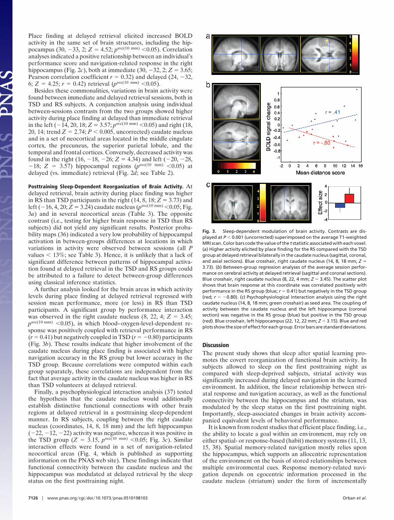
Place finding at delayed retrieval elicited increased BOLDactivity in the same set of brain structures, including the hip-pocampus (30, �33, 2; Z � 4.52; psvc(10 mm) �0.05). Correlationanalyses indicated a positive relationship between an individual’sperformance score and navigation-related response in the righthippocampus (Fig. 2c), both at immediate (30, �32, 2; Z � 3.65;Pearson correlation coefficient r � 0.32) and delayed (24, �32,6; Z � 4.25; r � 0.42) retrieval (psvc(10 mm) �0.05).
Besides these commonalities, variations in brain activity werefound between immediate and delayed retrieval sessions, both inTSD and RS subjects. A conjunction analysis using individualbetween-sessions contrasts from the two groups showed higheractivity during place finding at delayed than immediate retrievalin the left (�14, 20, 18; Z � 3.57; psvc(10 mm) �0.05) and right (18,20, 14; trend Z � 2.74; P � 0.005, uncorrected) caudate nucleusand in a set of neocortical areas located in the middle cingulatecortex, the precuneus, the superior parietal lobule, and thetemporal and frontal cortices. Conversely, decreased activity wasfound in the right (16, �18, �26; Z � 4.34) and left (�20, �28,�18; Z � 3.57) hippocampal regions (psvc(10 mm) �0.05) atdelayed (vs. immediate) retrieval (Fig. 2d; see Table 2).
Posttraining Sleep-Dependent Reorganization of Brain Activity. Atdelayed retrieval, brain activity during place finding was higherin RS than TSD participants in the right (14, 8, 18; Z � 3.73) andleft (�16, 4, 20; Z � 3.24) caudate nucleus (psvc(10 mm) �0.05; Fig.3a) and in several neocortical areas (Table 3). The oppositecontrast (i.e., testing for higher brain response in TSD than RSsubjects) did not yield any significant results. Posterior proba-bility maps (36) indicated a very low probability of hippocampalactivation in between-groups differences at locations in whichvariations in activity were observed between sessions (all Pvalues � 13%; see Table 3). Hence, it is unlikely that a lack ofsignificant difference between patterns of hippocampal activa-tion found at delayed retrieval in the TSD and RS groups couldbe attributed to a failure to detect between-group differencesusing classical inference statistics.
A further analysis looked for the brain areas in which activitylevels during place finding at delayed retrieval regressed withsession mean performance, more (or less) in RS than TSDparticipants. A significant group by performance interactionwas observed in the right caudate nucleus (8, 22, 4; Z � 3.45;psvc(10 mm) �0.05), in which blood–oxygen-level-dependent re-sponse was positively coupled with retrieval performance in RS(r � 0.41) but negatively coupled in TSD (r � �0.80) participants(Fig. 3b). These results indicate that higher involvement of thecaudate nucleus during place finding is associated with highernavigation accuracy in the RS group but lower accuracy in theTSD group. Because correlations were computed within eachgroup separately, these correlations are independent from thefact that average activity in the caudate nucleus was higher in RSthan TSD volunteers at delayed retrieval.
Finally, a psychophysiological interaction analysis (37) testedthe hypothesis that the caudate nucleus would additionallyestablish distinctive functional connections with other brainregions at delayed retrieval in a posttraining sleep-dependentmanner. In RS subjects, coupling between the right caudatenucleus (coordinates, 14, 8, 18 mm) and the left hippocampus(�22, �12, �22) activity was negative, whereas it was positive inthe TSD group (Z � 3.15, psvc(10 mm) �0.05; Fig. 3c). Similarinteraction effects were found in a set of navigation-relatedneocortical areas (Fig. 4, which is published as supportinginformation on the PNAS web site). These findings indicate thatfunctional connectivity between the caudate nucleus and thehippocampus was modulated at delayed retrieval by the sleepstatus on the first posttraining night.
DiscussionThe present study shows that sleep after spatial learning pro-motes the covert reorganization of functional brain activity. Insubjects allowed to sleep on the first posttraining night ascompared with sleep-deprived subjects, striatal activity wassignificantly increased during delayed navigation in the learnedenvironment. In addition, the linear relationship between stri-atal response and navigation accuracy, as well as the functionalconnectivity between the hippocampus and the striatum, wasmodulated by the sleep status on the first posttraining night.Importantly, sleep-associated changes in brain activity accom-panied equivalent levels of behavioral performance.
It is known from rodent studies that efficient place finding, i.e.,the ability to locate a goal within an environment, may rely oneither spatial- or response-based (habit) memory systems (11, 13,15, 38). Spatial memory-related navigation mostly relies uponthe hippocampus, which supports an allocentric representationof the environment on the basis of stored relationships betweenmultiple environmental cues. Response memory-related navi-gation depends on egocentric information processed in thecaudate nucleus (striatum) under the form of incrementally
Fig. 3. Sleep-dependent modulation of brain activity. Contrasts are dis-played at P � 0.001 (uncorrected) superimposed on the average T1-weightedMRI scan. Color bars code the value of the t statistic associated with each voxel.(a) Higher activity elicited by place finding for the RS compared with the TSDgroup at delayed retrieval bilaterally in the caudate nucleus (sagittal, coronal,and axial sections). Blue crosshair, right caudate nucleus (14, 8, 18 mm; Z �3.73). (b) Between-group regression analyses of the average session perfor-mance on cerebral activity at delayed retrieval (sagittal and coronal sections).Blue crosshair, right caudate nucleus (8, 22, 4 mm; Z � 3.45). The scatter plotshows that brain response at this coordinate was correlated positively withperformance in the RS group (blue; r � 0.41) but negatively in the TSD group(red; r � �0.80). (c) Psychophysiological interaction analysis using the rightcaudate nucleus (14, 8, 18 mm; green crosshair) as seed area. The coupling ofactivity between the caudate nucleus and the left hippocampus (coronalsection) was negative in the RS group (blue) but positive in the TSD group(red). Blue crosshair, left hippocampus (22, 12, 22 mm; Z � 3.15). Blue and redplots show the size of effect for each group. Error bars are standard deviations.
7126 � www.pnas.org�cgi�doi�10.1073�pnas.0510198103 Orban et al.

acquired stimulus–response associations. Neuropharmacologi-cal studies also suggest that rats preferentially perform naviga-tion tasks based on a spatial strategy early in training and on aresponse scheme later on, after a sufficient amount of practice(17, 18). Likewise, in humans, neuroimaging experiments dem-onstrated that efficient navigation in virtual environments couldbe mediated by dissimilar spatial and response cognitive strat-egies respectively relying on hippocampal and caudate nucleusmemory systems (16). fMRI studies in healthy subjects haveshown that, as learning of a virtual environment progresses, ashift from a spatial to a response cognitive strategy of navigationmay occur, accompanied by increased brain response in thecaudate nucleus (19, 39). Moreover, studies conducted in pa-tients suffering from either unilateral hippocampal lesion (39) orcaudate nucleus damage arising from Huntington’s disease (40)have demonstrated that each system may compensate to someextent for the other one, eventually giving rise to similar levelsof behavioral performance.
Changes in memory-related brain activity were observedbetween retrieval sessions, irrespective of sleep status on the firstposttraining night. Whereas hippocampal activity decreasedfrom immediate to delayed retrieval, brain response increased inthe caudate nucleus and specific neocortical areas distributedwithin the temporal, parietal, cingulate, frontal, and prefrontalcortices. These effects cannot be characterized as purely time-dependent, because they might also reflect continued sleep-dependent influences taking place during the two recoverynights. Also, it is unclear whether these activation patterns mayreflect the hippocampo-neocortical transfer of information sug-gested by standard memory consolidation models, which positthat the hippocampus has a time-limited role in the storage ofdeclarative memories, so that their final repository lies in theneocortex (refs. 41 and 42, but see ref. 43). A 3-day intervalbetween sessions might be too short to demonstrate such atransfer effect. Indeed, several previous studies failed to show adiminution of hippocampal involvement in a recognition mem-ory task performed immediately, 1 day or 1 week after learning(44), or even found enhanced retrieval-related hippocampalactivity after a consolidation period ranging from 10 min to 1month (45, 46). However, a recent study revealed a progressivedisengagement of the hippocampus during declarative memoryretrieval over a time course ranging from a few hours to 3 months(25). Besides, it is unlikely that decreased hippocampal activityin our study resulted from a repetition-suppression effect (47).Stimuli encountered during navigation were already highly fa-miliar at the start of the first fMRI session, and the delaybetween sessions was probably too long (48). Hence, irrespectiveof sleep deprivation on the first posttraining night, a relativehippocampal disengagement at delayed retrieval might be ac-counted for by two forms of systems consolidation processessupporting a gradual transfer of mnemonic representationstoward either the neocortex or the striatum.
In the framework of our task, results indicate that sleep on thefirst posttraining night promotes hippocampo-striatal memoryreorganization. The observed patterns of correlation betweenstriatal activity and behavioral performance crucially supportthe view that the effect of sleep is related to navigation and notto other confounding factors. In addition, the functional con-nectivity between the hippocampus and the caudate nucleus wasmodified in a posttraining sleep-dependent manner at delayedretrieval, although the overall level of hippocampal activity wasnot. As stated above, hippocampus- and caudate nucleus-centered memory systems subtend different cognitive strategiesat different time steps of practice, allowing equivalent perfor-mance (19, 39). Differential involvement of the hippocampusand striatum at different steps of practice was similarly evi-denced by using a classification learning task (49, 50). Addition-ally, the development of a negative coupling between hippocam-
pal and caudate nucleus responses during learning was reportedby using the same task (49, 50). We similarly observed a negativerelationship between striatal and hippocampal navigation-related regions at delayed retrieval in the RS group, whereasactivity in these regions was positively coupled in TSD subjects.A sensible explanation could be that posttraining sleep-dependent promotion of a response strategy of navigationmediated by the caudate nucleus has competitively prevented theexpression of possibly reinforced spatial memories in RS sub-jects. In the rat hippocampus and striatum, however, acetylcho-line release was found to occur in parallel after extended practicewithin a single session, without intervening sleep (18). Thesignificance of this dissociation in coupling patterns remains tobe specifically investigated. In the rat hippocampus, posttrainingpharmacological inactivation actually strengthens later caudatenucleus-dependent place finding (51), further showing thatinteractions between striatal and hippocampal memory systemsdevelop during offline consolidation periods. Our results extendthese animal findings by demonstrating that memory systemsmay interact well beyond the learning episode in humans, aprocess in which sleep is assigned an important role.
Finally, our behavioral data speak against the confoundingpossibility that different levels of performance could haveexplained between-group differences in brain activity. In anunchanged environment, efficiency in place finding could havebeen equally supported by either a spatial- or response-basedstrategy, or a combination of both, eventually yielding similarlevels of performance. The hippocampo-neocortical networkalone is believed to subtend the processing of genuine spatial�declarative memories, thus leading to a map-like cognitiverepresentation of the environment (14, 52), whereas the striatumis thought to support a more automated, less f lexible form ofnavigation (16). The use of navigational tests in which familiarpathways would be blocked, thus forcing subjects to find novelpathways using exclusively a spatial strategy, could possibly leadto the detection of sleep-dependent effects and between-sessionsdifferences in behavioral performance occurring in parallel withchanges in brain activity. Note that efficient navigation based ona spatial strategy in TSD subjects may well be explained ifhippocampus-dependent memories have been actively main-tained during the extended awake period on the first posttrainingnight in these subjects, and then sleep-dependent consolidationoccurred on the following nights. Accordingly, a detrimentalinfluence of sleep deprivation on declarative memory retentionwas found in studies in which retrieval was assessed immediatelyafter the sleep deprivation episode (3, 20, 26) but not after 1week (53, 54). Conversely, it could be tested whether navigationis less impaired in the framework of a dual-task paradigm in RSthan TSD subjects, under the assumption that the former wouldbe more able to rely on a routine strategy to perform the task ina divided attention condition. Also, a caudate-dependent routinenavigation could be characterized by fewer interruptions, that is,stops that would allow explicit retrieval of map-like spatialinformation needed to plan further moves, provided that theexperimental design does not specifically require subjects to beas fast as possible, as in the present study, in which case thisbehavioral measure could be irrelevant. Future work is neededto assess these hypotheses in the framework of brain imagingstudies.
ConclusionEvidence for a role of sleep in declarative memory processes ismostly based on sleep-dependent changes in behavioral perfor-mance. Here, we show that posttraining sleep promotes thelong-term reorganization of memory-related brain networkseven when overt behavioral performance is unaffected by sleepdeprivation. Functional neuroimaging data suggest that sleepfavors a shift of brain activity on the long term, so that
Orban et al. PNAS � May 2, 2006 � vol. 103 � no. 18 � 7127
NEU
ROSC
IEN
CE

navigation, which is initially mostly based on a hippocampus-dependent spatial strategy, progressively relies in part on aresponse-based strategy mediated by the striatum. These find-ings help to characterize the sleep-for-memory hypothesis byshowing that a lack of effect of sleep on overt behavioralperformance does not automatically imply that sleep has nocovert effect on the brain networks that subtend memoryconsolidation.
MethodsSubjects. Twenty-four healthy right-handed volunteers (12 males,12 females; mean age, 23.5 years; range, 19–29 years) gave theirinformed written consent to participate in this experimentapproved by the Ethics Committee of the University of Liege.They were paid for their participation, were free of neurologicalor psychiatric disease, and had a normal structural MRI brainscan. In the sleep group (RS; six males, six females), subjectswere allowed to sleep as usual at home for the three posttrainingnights. In the TSD group (six males, six females), subjects stayedawake in the laboratory the first posttraining night until 7:00 a.m.(Fig. 1a). During this night, participants’ physical activity wasmaintained as low as possible under constant supervision by theexperimenters. They had to pursue their usual diurnal activitiesand slept normally at home on the two following posttrainingnights. Data from two TSD subjects were excluded from theanalyses because of technical problems during fMRI acquisition.Circadian types were similarly sampled across the two groups. Asindicated by self reports, sleep duration and quality were similarbetween groups during the experimental period except on thefirst recovery night, when sleep duration was longer for the TSDgroup than for the RS group, as expected by a stronger drive forsleep after sleep deprivation (55). Further details on sleepparameters are reported in Supporting Text, which is publishedas supporting information on the PNAS web site.
Navigation Task. Subjects were trained in a virtual environmentadapted from a commercially available computer game (DukeNukem 3D; 3D Realms Entertainment, Apogee Software, Gar-land, TX) using the editor provided (Build, Ken Silverman,Realms Entertainment) (29). The environment was a complextown composed of three districts that were made distinct fromeach other by different visual backgrounds and objects along thestreets. Each of these districts contained one target locationidentified by a rotating medallion. Unknown to the participantsduring training, the virtual town contained 10 starting points thatwere each 35 virtual units apart (optimal path) from theirassociated target location (Fig. 1b). Subjects navigated within theenvironment at the ground level using a four-direction keypadwith their right hand at a constant speed of 1.25 virtual unit persecond. During training, the virtual environment was presentedon a desktop 800-MHz Pentium III (Siemens, Munich) personalcomputer (screen size, 17 inches). For testing in the scanner, amirror allowed the participants to see the display of the virtualtown projected on a screen.
On day 1, participants were trained outside of the scannerduring a 35-min exploration period. They were explicitly in-structed to learn the spatial layout of streets, districts, and targetlocations by moving freely within the environment. During theentire training session, pictures of the three target locations andtheir associated names were continuously available to the sub-ject. Immediately after the end of learning on day 1 (immediateretrieval) and on day 4 (delayed retrieval), volunteers werescanned by using fMRI while performing a series of navigationtests in the virtual town. To control for potential circadianinterferences, sessions took place at the same time of day(between 10:00 a.m. and 5:00 p.m.) at immediate and delayedretrieval. Each session consisted of 10 blocks of tests, each lastingfor 28 s, that alternated with 10 blocks of rest, during which a
black screen was displayed for 10–17 s. For each test, participantswere assigned a starting point and instructed to reach, as fast aspossible, a given target location. Because the target was sepa-rated from the starting point by 28 s at constant speed, reachingthe destination could be achieved only if they selected the idealpath and, in the meantime, did not stop at all during navigation.After time elapsed, a quantitative measure of behavioral per-formance was computed by subtracting the shortest distanceremaining between the subject’s actual location and the finaldestination from the length of the optimal path. Additionalmeasures of behavior consisted of the number of stops (definedas stops at landmarks or crossroads and U turns) and navigationspeed (expressed as the distance effectively traveled during testduration). Within the last 2 s of the rest period preceding eachtest, the target location for the test was indicated orally throughmagnetic resonance-compatible headphones. The same 10 testswere administered in a counterbalanced order, both at thebetween- and within-subjects levels.
Imaging Parameters. Brain imaging data were obtained by usinga 3-T head-only MRI system (Magnetom VISION; SiemensMedical Systems, Erlangen, Germany) equipped with an activeshielded gradient coil (30 mT�m). A structural MRI scan usinga standard 3D (0.9 � 0.9 � 0.9 mm) T1-weighted sequence wasacquired for each subject [repetition time (TR) � 1,960 ms; echotime (TE) � 4.43 ms; f lip angle (FA) � 8°; 176 slices; field ofview (FoV) � 230 � 173 mm2; matrix size � 256 � 192 � 176;voxel size � 0.9 � 0.9 � 0.9 mm]. For each testing session, 206functional multislice T2-weighted images were obtained by usinga blood–oxygen level-dependent-sensitive single-shot echo pla-nar imaging (EPI) sequence (TR � 2130 ms; TE � 40 ms; FA �90°; FoV � 220 � 220 mm2; matrix size � 64 � 64 � 32) coveringthe whole brain (126 mm high). Each functional volume con-sisted of 32 slices, with a thickness of 3 mm (interslice gap � 1mm) and a pixel size of 3.4 � 3.4 � 3 mm. The four initial scansof each session were discarded to control for magnetic saturationeffects.
fMRI Data Analysis. A detailed description of fMRI data analysismethods is provided as Supporting Text. Only essential informa-tion is reported here. Preprocessing and analysis of functionalvolumes were performed by using the Statistical ParametricMapping software SPM2 (Wellcome Department of CognitiveNeurology, London) implemented in MATLAB 6.1 (Mathworks,Sherbom, MA). Preprocessing included realignment and adjust-ment for movement-related effects, coregistration of functionaland anatomical data, spatial normalization into standard ste-reotactic Montreal Neurological Institute (MNI) space, andspatial smoothing using a Gaussian kernel of 6 mm full width athalf maximum (FWHM). Functional data were analyzed by usinga mixed-effects model that aimed at showing stereotypical effectin the population from which the subjects are drawn (35). Foreach subject, a first-level intraindividual analysis tested effects ofinterest by linear contrasts convolved with a canonical hemody-namic response function, generating statistical parametric maps[SPM(T)]. Movement parameters were included as confoundingfactors. Cutoff period for high-pass filtering was 128 s. Linearcontrasts estimated the main effect of navigation at immediateand delayed retrieval separately, and the difference of naviga-tion-related activity from immediate to delayed retrieval (andconversely). Summary statistic images were thresholded at P �0.95 (uncorrected) and spatially smoothed (6-mm FWHMGaussian kernel), then entered into a second-level (random-effects) analysis to evaluate differences and commonalitiesbetween groups. To test whether modifications of neuronalactivity in navigation-related areas were related to performancein place finding, correlation analyses estimated at the random-effects level the coupling between the mean behavioral perfor-
7128 � www.pnas.org�cgi�doi�10.1073�pnas.0510198103 Orban et al.

mance per session and the effect size at every voxel in individualstatistical maps derived from relevant navigation-related con-trasts. Additionally, psychophysiological interaction analyses(37) were computed to test the hypothesis that the availability ofposttraining sleep would modulate functional connectivity be-tween navigation-related areas. Coordinates of voxels of interestwere determined based on results from the random-effectsanalysis described above. In all analyses, the resulting set of voxelvalues for each contrast constituted a map of the t statistic[SPM(T)], thresholded at P � 0.001 (uncorrected for multiplecomparisons). Statistical inferences were then obtained aftercorrections at the voxel level by using Gaussian random fieldtheory at psvc(10 mm) � 0.05 corrected in a small spherical volume
(34) around a priori coordinates previously reported in neuro-imaging experiments that investigated human navigation usingvirtual reality devices (Table 4). Finally, posterior probabilitymaps enabled conditional or Bayesian inferences about region-ally specific effects, allowing us to ensure that a lack of significantstatistical effect in a given contrast was not merely due to afailure to detect this effect using classical inferences (36).
We thank Dr. V. Bohbot for discussion on this research and E. Van derLoo for help with drawing Fig. 1. This study was funded by the BelgianFonds National de la Recherche Scientifique (FNRS), FondationMedicale Reine Elisabeth, Research Fund of the University of Liege,Pole d’ Attraction Interuniversitaire P5�04, and Fondation Fyssen. E.B.,P.M., and P.O. are supported by FNRS.
1. Maquet, P., Smith, C. & Stickgold, R. (2003) Sleep and Brain Plasticity (OxfordUniv. Press, Oxford).
2. Peigneux, P., Laureys, S., Delbeuck, X. & Maquet, P. (2001) NeuroReport 12,A111–A124.
3. Gais, S. & Born, J. (2004) Learn. Mem. 11, 679–685.4. Rauchs, G., Desgranges, B., Foret, J. & Eustache, F. (2005) J. Sleep Res. 14,
123–140.5. Walker, M. P. & Stickgold, R. (2006) Annu. Rev. Psychol. 57, 1–28.6. McGaugh, J. L. (2000) Science 287, 248–251.7. Dudai, Y. (2004) Annu. Rev. Psychol. 55, 51–86.8. Scoville, W. B. & Milner, B. (1957) J. Neurol. Neurosurg. Psychiatry 20, 11–21.9. Squire, L. R., Stark, C. E. & Clark, R. E. (2004) Annu. Rev. Neurosci. 27,
279–306.10. Tulving, E. (1983) Elements of Episodic Memory (Oxford Univ. Press, Cam-
bridge, U.K.).11. Morris, R. G., Garrud, P., Rawlins, J. N. & O’Keefe, J. (1982) Nature 297,
681–683.12. Ekstrom, A. D., Kahana, M. J., Caplan, J. B., Fields, T. A., Isham, E. A.,
Newman, E. L. & Fried, I. (2003) Nature 425, 184–188.13. O’Keefe, J. & Nadel, L. (1978) The Hippocampus as a Cognitive Map (Oxford
Univ. Press, Cambridge, U.K.).14. Burgess, N., Maguire, E. A. & O’Keefe, J. (2002) Neuron 35, 625–641.15. Packard, M. G. & Knowlton, B. J. (2002) Annu. Rev. Neurosci. 25, 563–593.16. Hartley, T., Maguire, E. A., Spiers, H. J. & Burgess, N. (2003) Neuron 37,
877–888.17. Packard, M. G. (1999) Proc. Natl. Acad. Sci. USA 96, 12881–12886.18. Chang, Q. & Gold, P. E. (2003) J. Neurosci. 23, 3001–3005.19. Iaria, G., Petrides, M., Dagher, A., Pike, B. & Bohbot, V. D. (2003) J. Neurosci.
23, 5945–5952.20. Plihal, W. & Born, J. (1997) J. Cognit. Neurosci. 9, 534–547.21. Gais, S. & Born, J. (2004) Proc. Natl. Acad. Sci. USA 101, 2140–2144.22. Marshall, L., Molle, M., Hallschmid, M. & Born, J. (2004) J. Neurosci. 24,
9985–9992.23. Gais, S., Molle, M., Helms, K. & Born, J. (2002) J. Neurosci. 22, 6830–6834.24. Clemens, Z., Fabo, D. & Halasz, P. (2005) Neuroscience 132, 529–535.25. Takashima, A., Petersson, K. M., Rutters, F., Tendolkar, I., Jensen, O., Zwarts,
M. J., McNaughton, B. L. & Fernandez, G. (2006) Proc. Natl. Acad. Sci. USA103, 756–761.
26. Plihal, W. & Born, J. (1999) Psychophysiology 36, 571–582.27. Goder, R., Boigs, M., Braun, S., Friege, L., Fritzer, G., Aldenhoff, J. B. &
Hinze-Selch, D. (2004) J. Psychiatr. Res. 38, 591–599.28. Meier-Koll, A., Bussmann, B., Schmidt, C. & Neuschwander, D. (1999) Percept.
Mot. Skills 88, 1141–1159.
29. Peigneux, P., Laureys, S., Fuchs, S., Collette, F., Perrin, F., Reggers, J., Phillips,C., Degueldre, C., Del Fiore, G., Aerts, J., et al. (2004) Neuron 44, 535–545.
30. Wilson, M. A. & McNaughton, B. L. (1994) Science 265, 676–679.31. Skaggs, W. E. & McNaughton, B. L. (1996) Science 271, 1870–1873.32. Thomas, M., Sing, H., Belenky, G., Holcomb, H., Mayberg, H., Dannals, R.,
Wagner, H., Thorne, D., Popp, K., Rowland, L., et al. (2000) J. Sleep Res. 9,335–352.
33. Rogers, N. L., Dorrian, J. & Dinges, D. F. (2003) Front. Biosci. 8, s1056–s1067.34. Worsley, K. J. (1996) Hum. Brain Mapp. 4, 58–73.35. Penny, W. & Holmes, A. (2003) in Human Brain Function, eds. Frackowiak, R.,
Friston, K., Frith, C., Dolan, R., Price, C., Zeki, S. J. A. & Penny, W.(Academic, London), pp. 843–850.
36. Friston, K. J. & Penny, W. (2003) NeuroImage 19, 1240–1249.37. Friston, K. J., Buechel, C., Fink, G. R., Morris, J., Rolls, E. & Dolan, R. J.
(1997) NeuroImage 6, 218–229.38. Packard, M. G., Hirsh, R. & White, N. M. (1989) J. Neurosci. 9, 1465–1472.39. Bohbot, V. D., Iaria, G. & Petrides, M. (2004) Neuropsychology 18, 418–425.40. Voermans, N. C., Petersson, K. M., Daudey, L., Weber, B., Van Spaendonck,
K. P., Kremer, H. P. & Fernandez, G. (2004) Neuron 43, 427–435.41. Alvarez, P. & Squire, L. R. (1994) Proc. Natl. Acad. Sci. USA 91, 7041–7045.42. Frankland, P. W. & Bontempi, B. (2005) Nat. Rev. Neurosci. 6, 119–130.43. Nadel, L. & Moscovitch, M. (1997) Curr. Opin. Neurobiol. 7, 217–227.44. Stark, C. E. & Squire, L. R. (2000) Hippocampus 10, 329–337.45. Bosshardt, S., Degonda, N., Schmidt, C. F., Boesiger, P., Nitsch, R. M., Hock,
C. & Henke, K. (2005) Hippocampus 15, 1026–1040.46. Bosshardt, S., Schmidt, C. F., Jaermann, T., Degonda, N., Boesiger, P., Nitsch,
R. M., Hock, C. & Henke, K. (2005) Cortex 41, 486–498.47. Henson, R. N., Cansino, S., Herron, J. E., Robb, W. G. & Rugg, M. D. (2003)
Hippocampus 13, 301–304.48. Brozinsky, C. J., Yonelinas, A. P., Kroll, N. E. & Ranganath, C. (2005)
Hippocampus 15, 557–561.49. Poldrack, R. A., Clark, J., Pare-Blagoev, E. J., Shohamy, D., Creso Moyano, J.,
Myers, C. & Gluck, M. A. (2001) Nature 414, 546–550.50. Poldrack, R. A. & Rodriguez, P. (2004) Neurobiol. Learn. Mem. 82, 324–332.51. Schroeder, J. P., Wingard, J. C. & Packard, M. G. (2002) Hippocampus 12,
280–284.52. Maguire, E. A., Burgess, N., Donnett, J. G., Frackowiak, R. S., Frith, C. D. &
O’Keefe, J. (1998) Science 280, 921–924.53. Smith, C. (1995) Behav. Brain Res. 69, 137–145.54. Smith, C. (2001) Sleep Med. Rev. 5, 491–506.55. Borbely, A. A. & Achermann, P. (1999) J. Biol. Rhythms 14, 557–568.
Orban et al. PNAS � May 2, 2006 � vol. 103 � no. 18 � 7129
NEU
ROSC
IEN
CE