Site-specific integration in CHO cells mediated by ... - CORE
12
General rights Copyright and moral rights for the publications made accessible in the public portal are retained by the authors and/or other copyright owners and it is a condition of accessing publications that users recognise and abide by the legal requirements associated with these rights. • Users may download and print one copy of any publication from the public portal for the purpose of private study or research. • You may not further distribute the material or use it for any profit-making activity or commercial gain • You may freely distribute the URL identifying the publication in the public portal If you believe that this document breaches copyright please contact us providing details, and we will remove access to the work immediately and investigate your claim. Downloaded from orbit.dtu.dk on: Dec 20, 2017 Site-specific integration in CHO cells mediated by CRISPR/Cas9 and homology- directed DNA repair pathway Lee, Jae Seong; Beuchert Kallehauge, Thomas; Pedersen, Lasse Ebdrup; Kildegaard, Helene Faustrup Published in: Scientific Reports Link to article, DOI: 10.1038/srep08572 Publication date: 2015 Document Version Publisher's PDF, also known as Version of record Link back to DTU Orbit Citation (APA): Lee, J. S., Beuchert Kallehauge, T., Pedersen, L. E., & Kildegaard, H. F. (2015). Site-specific integration in CHO cells mediated by CRISPR/Cas9 and homology-directed DNA repair pathway. Scientific Reports, 5, [8572]. DOI: 10.1038/srep08572
-
Upload
khangminh22 -
Category
Documents
-
view
4 -
download
0
Transcript of Site-specific integration in CHO cells mediated by ... - CORE

General rights Copyright and moral rights for the publications made accessible in the public portal are retained by the authors and/or other copyright owners and it is a condition of accessing publications that users recognise and abide by the legal requirements associated with these rights.
• Users may download and print one copy of any publication from the public portal for the purpose of private study or research. • You may not further distribute the material or use it for any profit-making activity or commercial gain • You may freely distribute the URL identifying the publication in the public portal
If you believe that this document breaches copyright please contact us providing details, and we will remove access to the work immediately and investigate your claim.
Downloaded from orbit.dtu.dk on: Dec 20, 2017
Site-specific integration in CHO cells mediated by CRISPR/Cas9 and homology-directed DNA repair pathway
Lee, Jae Seong; Beuchert Kallehauge, Thomas; Pedersen, Lasse Ebdrup; Kildegaard, Helene Faustrup
Published in:Scientific Reports
Link to article, DOI:10.1038/srep08572
Publication date:2015
Document VersionPublisher's PDF, also known as Version of record
Link back to DTU Orbit
Citation (APA):Lee, J. S., Beuchert Kallehauge, T., Pedersen, L. E., & Kildegaard, H. F. (2015). Site-specific integration in CHOcells mediated by CRISPR/Cas9 and homology-directed DNA repair pathway. Scientific Reports, 5, [8572]. DOI:10.1038/srep08572

Site-specific integration in CHO cellsmediated by CRISPR/Cas9 andhomology-directed DNA repair pathwayJae Seong Lee, Thomas Beuchert Kallehauge, Lasse Ebdrup Pedersen & Helene Faustrup Kildegaard
The Novo Nordisk Foundation Center for Biosustainability, Technical University of Denmark, 2970 Hørsholm, Denmark.
Chinese hamster ovary (CHO) cells are the most widely used mammalian hosts for production oftherapeutic proteins. However, development of recombinant CHO cell lines has been hampered by unstableand variable transgene expression caused by random integration. Here we demonstrate efficient targetedgene integration into site-specific loci in CHO cells using CRISPR/Cas9 genome editing system andcompatible donor plasmid harboring a gene of interest (GOI) and short homology arms. This strategy hasenabled precise insertion of a 3.7-kb gene expression cassette at defined loci in CHO cells following a simpledrug-selection, resulting in homogeneous transgene expression. Taken together, the results displayed herecan help pave the way for the targeting of GOI to specific loci in CHO cells, increasing the likelihood ofgenerating isogenic cell lines with consistent protein production.
Chinese hamster ovary (CHO) cells have been used as the predominant workhorses for production ofrecombinant therapeutic proteins with complex glycoforms1,2. Traditionally, development of recombin-ant CHO (rCHO) cell lines relies on random integration of a gene of interest (GOI) into the genome,
followed by selection of cells carrying the transgene1,2. However, lack of control of gene insertion can give rise tounwanted phenotypic heterogeneity due to the varying accessibility of integration sites for gene expression –termed the position effect variation2,3. In addition, gene amplification methods are traditionally used to increaseexpression. As a result, these cell lines are often unstable and show reduced production over time4. Due to thisvariation in expression and genomic composition, subsequent screening of multiple clones is necessary to selectproper clones suitable for high and stable expression of recombinant proteins1,2. In this article, we show thattargeting of transgenes into specific desirable sites in the CHO genome in a controlled manner reduces thevariation in expression, generating a uniform population with stable transgene expression.
In order to circumvent uncontrollable gene insertion during rCHO cell line construction, different approacheshave been applied for site-directed integration of transgenes using site-specific recombinases, which includes Cre/loxP system, Flp/FRT system, and phiC31/R4 integrases5–7. However, these systems are limited by the need of aprior establishment of platform cell lines with the insertion of recombination site into random or a limitednumber of specific genomic regions, which is a prerequisite for retargeting to generate cell lines expressing theGOI.
With draft genomes of several CHO cell lines recently being made available8,9, it is now possible to efficientlyengineer the genomic sequence of CHO cells with engineered nucleases. Customized nucleases such as zinc-fingernucleases (ZFNs), transcription activator-like effector nucleases (TALENs), and clustered regularly interspacedshort palindromic repeats (CRISPR)-associated (Cas) RNA guided nucleases are technologies readily available toinduce targeted insertion/deletion (indel) mutations or precise sequence changes in a broad range of organismsand cell types (reviewed in Ref. 10). Upon site-specific DNA double-strand breaks (DSBs) induced by engineerednucleases, the target locus will typically be repaired by one of two major DNA damage repair pathway: non-homologous end-joining (NHEJ) or homology-directed repair (HDR). Compared with the error-prone NHEJ,which can result in efficient indel mutation generation subsequently leading to knockout of target locus, HDR canbe used to modify endogenous loci precisely in the presence of homologous pieces of endogenous/exogenousDNA. Although HDR is typically observed at a lower or more variable frequency than NHEJ, HDR can beleveraged to generate targeted integrations with engineered nucleases in mammalian cells11–15. Notably, CHO cellshave previously been described as a cell type recalcitrant to homology-based integration of large DNA constructs,thus targeted integration in CHO cells has been confined to NHEJ based approaches16,17.
OPEN
SUBJECT AREAS:
MOLECULAR BIOLOGY
BIOTECHNOLOGY
GENETIC ENGINEERING
Received20 October 2014
Accepted26 January 2015
Published25 February 2015
Correspondence andrequests for materials
should be addressed toH.F.K. (hef@
biosustain.dtu.dk)
SCIENTIFIC REPORTS | 5 : 8572 | DOI: 10.1038/srep08572 1

Compared with protein-based genome editing tools with custo-mizable DNA binding specificities, such as ZFNs and TALENS, thenewer CRISPR/Cas9 platform is based on simple base-pairingbetween an engineered RNA and the targeted genomic site, whichenables rapid design, ease of use, and low costs18. The CRISPR/Cas9system, which was initially identified in the bacterial immune system,is composed of two RNA elements, CRISPR-RNA (crRNA) and trans-activating crRNA (tracrRNA), together with the Cas9 nuclease14,19.The active cleavage-complex is generated by base-pairing betweenthe genomic sequence and crRNA, followed by hybridization of thetracrRNA to the crRNA. Subsequently, the Cas9 nuclease associateswith the DNA:RNA complex to form the active cleavage complex,which in turn will generate site specific DSBs at the genomic locus19.In recent studies, the original type II CRISPR system from S. pyogeneswas modified to induce targeted genome editing in eukaryotic cells byfusing the two RNA complexes termed single guide RNA (sgRNA)11,14,19.We previously developed a CRISPR/Cas9 system optimized for CHOcells, and demonstrated high efficiency of indels generated in CHO cellsup to 47.3%20. Assuming that the high rate of DSB introduction willinduce DNA damage repair pathways, and the concurrent introductionof a donor plasmid will be used as the repair template, the targetedintegration at specific genomic sites can be feasible with high fidelity.
Here we demonstrate efficient targeted gene integration into site-specific loci in CHO cells using CRISPR/Cas9 genome editing systemand compatible donor plasmid harboring short-homology arms,GOI, and a fluorescent marker gene as an indicator of random integ-ration. Simultaneous introduction of active sgRNAs, Cas9 nucleases,and donor plasmid enabled insertion of a 3.7-kb gene expressioncassette at three different loci in CHO cells, following a simpledrug-selection. A low level of off-target mutation in most potentialoff-target sites and homogeneous expression level of GOI wasobserved in targeted integrants, which supports the robustness andefficiency of the current strategy.
ResultsDevelopment of a HDR mediated targeted integration platformfor CHO. To facilitate improved construction of rCHO cell lines, weaimed at developing a CRISPR and HDR mediated targeted integra-tion system to obtain controlled and precise integration of transgenes.The system is based on donor plasmids harboring short homologyarms, which flank the Cas9 cleavable sgRNA target site, used as theintegration sites (Fig. 1a). The donor plasmids were constructed asfollows: Regardless of loci and sgRNAs, the homology arms weredesigned to be exactly next to the whole 23 bp of sgRNA genomictarget sequences (Supplementary Table 1). The length of each 59 and39 homology arm was set at 750 bp unless otherwise mentioned. AnmCherry expression cassette and a neomycin resistance gene expres-sion cassette were placed within the homology arms. A ZsGreen1-DRexpression cassette was placed outside the homology arms to detectrandom integration events. Upon correct targeting of the donorplasmids to the desired locus via HDR, cells will lose ZsGreen1-DRexpression and only express mCherry together with neomycin resis-tance. If cells express both mCherry and ZsGreen1-DR after neomycinselection, random integration of the donor plasmid has occurred. Thisassumption was applied in order to enrich for targeted integrants byexcluding clones expressing ZsGreen1-DR. To evaluate correct inte-gration by HDR, 59/39 junction PCR amplicons were designed for eachtarget site (Fig. 1a). We targeted a total of four sites in three differentgenomic loci of CHO cells; one site in C1GALT1C1 (COSMC) encod-ing the C1GALT1-specific chaperone 1, two sites in Mgat1 encodingthe mannosyl (alpha-1,3-)-glycoprotein beta-1,2-N-acetylglucosaminyl-transferase, and one site in LdhA encoding the lactate dehydrogenase A.
Precise targeted integration of transgene at the COSMC locusin CHO cells. The COSMC locus has previously been effectivelymodified by CRISPR/Cas9 system in CHO cells20. Thus, this locus
was chosen for initial validation of the targeted integration strategy.The region targeted by sgRNA2, which gave rise to high indelefficiency in CHO-K1 cells in our previous study20, was selected asthe integration site. Based on this site, 59 and 39 homology arms weredesigned to generate the targeting donor plasmid (Fig. 1a). CHO-Scells were transfected with the vectors encoding CHO codon opti-mized Cas9, COSMC sgRNA2, and donor plasmid. Genomic DNAwas extracted either three days after transfection (transient expression)or two weeks after G418 selection (stable expression). No integrationevents were detected in the transiently transfected pools by the 59/39
junction PCR (Fig. 1b, Lane 1–5). In the stable selected pools, both 59
and 39 junction PCR verified targeted integration of donor DNA intothe COSMC locus (Fig. 1b, Lane 7 and 10). In agreement with aprevious observation16, linearization of the donor DNA outside ofthe homology arms resulted in fainter PCR positive bands from 59/39 junction PCR compared with those from circular donor DNA(Fig. 1b, Lane 8 and 11). Stepwise decreasing the length of 59 and 39
homology arms flanking the same COSMC integration site from750 bp to 100 bp indicated that homology arms with a length downto 250 bp were capable of mediating homology-based targetedintegration, although the PCR band intensity was graduallydecreased (Supplementary Fig. 1). In accordance with theassumption that cells expressing both ZsGreen1-DR and mCherrywere the result of random integration, stable cells expressing bothmCherry and ZsGreen1-DR, which were isolated by FACS, showedincomplete and faint junction PCR positive bands indicating amixture of integration events. (Fig. 1b, Lane 13 and 15). To verifyprecise integration into the genome, the 59/39 genome-donorboundaries were sequenced (Fig. 1c). Indeed, the sequences of the59/39 junction PCR amplicons confirmed precise insertion of thetarget expression cassette into the COSMC locus. We then screened588 G418-resistant clones and isolated 138 clones, which displayedhomogeneous expression of mCherry. 23 clones were excluded due tovery slow growth resulting in 115 clones, which were furthercharacterized. 83 out of 115 (72.2%) were 59/39 junction PCRpositive (Table 1). Out-out PCR with primers specific for genomicsites revealed that 70 out of 83 junction PCR positive clones (84.3%,i.e. 60.9% in total) were correctly targeted with the intact targetintegration unit, generating expected size of amplicon (wild typeamplicon: 1.6 kb 1 target integration unit: 3.7 kb < 5.3 kb; Fig. 1d,Lane 4, Table 1). As a result, a targeting efficiency of 27.8% wasobtained based on the percentage of out-out PCR positive clones inmCherry only expressing clones and a proportion of the mCherry onlyexpressing cells in the stable cell pool (Table 1). These 70 out-out PCRpositive clones were expanded and adapted to grow in suspension. Atotal of 45 clones were recovered, followed by further characterization.42 out of 45 clones had complete disruptions of the COSMC locus, asindicated by qPCR-based relative copy number analysis compared withgenomic DNA from CHO-S wild type cells (Fig. 1e, left panel).Assuming that gene copies must be integer values, the value ofrelative copy number is expected to be 0, 0.5, or 1 for targeted clonalcells. Three clones without these values were therefore not consideredas clonal cells. Relative mCherry copy number result amongst selectedclones revealed that most clones harbored the same number ofmCherry transgene, except two clones with 2- or 3-fold highernumber (Fig. 1e, right panel). Successful targeting of transgenes atthe COSMC locus validated our integration method, and promptedus to apply to different genomic loci showing distinct properties.
Application of targeted integration platform to the functional hemi-zygous locus, Mgat1. The Mgat1 locus was selected as a second inte-gration site to test the applicability of the targeted integration systemat a different genomic locus. Some CHO cell lines contain one func-tional Mgat1 allele that renders this locus more prone to be disruptedby chemical mutagenesis or genome editing tools, leading to func-tional knockout of Mgat121,22. Since Mgat1 adds N-acetylglucosamine
www.nature.com/scientificreports
SCIENTIFIC REPORTS | 5 : 8572 | DOI: 10.1038/srep08572 2

a
b
c
Reference
Conserva�on
5’ junc�on 3’ junc�on
Target locus:sgRNATarget(23 bp)
////
// 5’ arm SV40 Neo(R) SV40 pABGH pAEF1α mCherry 3’ arm CMV BGH pAZsGreen1 //Donor:
3.7 kb
HDR
5’ arm SV40 Neo(R) SV40 pABGH pAEF1α mCherry 3’ arm// //
Out
In
In
Out
5’ junc�on 3’ junc�on
Transient Stable
- + - + +
- - + + +
+ +
- +
- - + + +M M - + M
+
+
+
+ +
- +
- + M
+
+
+
5’ junc�on 3’ junc�on
*Cas9
Donor
COSMC sgRNA2
+ +
+ -
+ - M
+
+
+
3’ junc�on
+
-
- M
5’ junc�on
Stable [mCherry+ / ZG1+]
1000bp
1500bp
*
1 2 3 4 5 6 7 8 9 10 11
1000bp
1500bp
12 13 14 15
*
d+ + + +
- + + +
- + + +
M
Cas9
Donor
COSMC sgRNA2
1500bp
6000bp5000bp
Pool Clone
- + - +Junc�on PCR
1 2 3 4
e
Rela
�ve
COSM
C co
py n
umbe
r
0.0
0.2
0.4
0.6
0.8
1.0
1 2 3 4 5 6 8 11 12 13 14 16 18 19 20 23 26 28 33 37 38 39 42 44 45 47 50 51 52 53 56 57 58 61 62 63 64 65 69 77 81 84 86 106
108
CHO
-S
Rela
�ve
mCh
erry
cop
y nu
mbe
r
0
1
2
3
4
5
1 2 3 4 5 6 8 11 12 13 14 16 18 19 20 23 26 28 33 37 38 39 42 44 45 47 50 51 52 53 56 57 58 61 62 63 64 65 69 77 81 84 86 106
108
5’ arm 3’ arm
Figure 1 | Targeted integration into COSMC locus using CRISPR/Cas9. (a) Schematic illustration of the targeting strategy for the specific locus of
interest. Donor plasmid consists of three parts: short homology arms flanking sgRNA target site cleaved by Cas9 (red triangle), mCherry and neomycin
resistance gene expression cassettes inside homology arms, and ZsGreen1-DR expression cassette outside homology arms. Upon DSBs induced by
CRISPR/Cas9, HDR-mediated repair can be used to insert a total size of 3.7 kb of expression cassettes through recombination of the target locus with
donor plasmids. Primer position for 59/39 junction PCR is indicated. (b) Agarose gel of 59/39 junction PCR on transiently transfected cells and stable cell
pools. An asterisk indicates the use of linearized donor plasmid. M, 1 kb DNA ladder (c) Sanger sequencing of the 59/39 junction PCR amplicons.
Amplicons from the stable cell pool were purified and directly sequenced after PCR amplification. The chromatogram sequence of junction PCR
amplicon was compared with the reference sequence at the genome-donor boundaries. (d) Agarose gel of out-out PCR results of stable cell pools or clonal
cells. Primer pairs annealing to genomic DNA region were used resulting in PCR products of either wild type (1.6 kb) or targeted integration (5.3 kb). (e)
Relative copy number of COSMC and mCherry regions in clonal cells. Each plot shows the relative copy number of each region in comparison to the
reference sample. Genomic DNA of wild type CHO-S and Clone #1 and was used as the reference for COSMC and mCherry region, respectively (shown in
red). The error bars represent the standard deviations (n $ 3).
www.nature.com/scientificreports
SCIENTIFIC REPORTS | 5 : 8572 | DOI: 10.1038/srep08572 3
to the Man5GlcNAc2 (Man5) N-glycan structure, Mgat1-disruptedCHO cell lines are capable of producing recombinant proteins withterminal mannose residues and show resistance to Ricinus communisagglutinin-I (RCA-I), a cytotoxic lectin that binds Man5GlcNAc222.In order to select efficient sgRNA target sites for the Mgat1 locus,the indel efficiency of five sgRNAs was analyzed by deep sequenc-ing (Fig. 2a). At post-transfection with Cas9 and sgRNA expressionvectors, the five tested sgRNAs generated detectable range of indelsfrom 10.0% to 20.7% in the Mgat1 locus (Fig. 2b). Two sgRNAs,sgRNA1 and sgRNA5, were selected for targeted integration into theMgat1 locus. CHO-S cells were transfected with Cas9, sgRNA, anddonor plasmid to generate two cell pools, one for each sgRNA.Junction PCR analysis of the stable selected pools were performed(Fig. 2c, Lane 9–16) and compared with junction PCR analysis ontransiently transfected cells (Fig. 2c, Lane 1–8). Similar to the COSMClocus, 59/39 junction PCR was only positive for the stable pools (Fig. 2c,Lane 10 and 12 for sgRNA1 site; Lane 14 and 16 for sgRNA5 site).Furthermore, stable cells expressing both mCherry and Zsgreen1-DR,denoted by 11, showed less clean and intense bands than cellsexpressing only mCherry, denoted by 1-, which indicated incompleteintegration (Fig. 2c, Lane 17–24). The sequencing results of 59/39
junction PCR amplicons confirmed precise gene insertion occurringat the Mgat1 locus (Fig. 2d).
The amount of Mgat1-disrupted cells in two stable pools targeted foreither sgRNA1 or sgRNA5 site was estimated by analyzing fluoresceinlabeled RCA-I (F-RCA-I) stained cells (Fig. 2e). In wild type CHO-Scells, the majority of cells (. 94%) were stained with green fluorescentF-RCA-I (Supplementary Fig. 2a). Likewise, the stable pools, onlytransfected with donor plasmid, showed a complete shift from greenfluorescence negative (Q3 1 Q4) to green fluorescence positive popu-lation (Q1 1 Q2) upon F-RCA-I staining, indicating that the majorityof cells have functional MGAT1 as expected. On the other hand, thestable pools transfected with Cas9, sgRNA, and donor plasmid revealeda fraction of 4.6–12.5% of green fluorescence negative cells regardless ofmCherry expression from donor plasmid, generating red fluorescence.It demonstrated the presence of cells with knockout of the Mgat1 gene(Fig. 2e). Next, 59/39 junction PCR analysis was applied to evaluate thepercentage of target specific knock-ins following isolation of G418-resistant and mCherry positive/ZsGreen1-DR negative clones. In con-trast to the high efficiency observed in COSMC locus, we did notobtain junction PCR positive clones, indicating very low efficiencyof targeted integration in the Mgat1 locus (Table 1). It is likely thatmost cells belonging to red fluorescence positive/green fluorescencenegative (Q4) population upon F-RCA-I staining correspond tomCherry expressors with NHEJ mutation in Mgat1 locus, and notthe targeted integrants. Hence, we used RCA-I lectin selection in orderto enrich for Mgat1-disrupted clones occurred by targeted integration.RCA-I treatment effectively induced cell growth arrest and subsequentcell death in wild type CHO-S cells, implying RCA-I induced cyto-toxicity (Supplementary Fig. 2b). 7 days post-RCA-I treatment, RCA-Iresistant cells grew to confluency only in the stable pools transfected
with Cas9, sgRNA, and donor plasmid (Supplementary Fig. 2c). F-RCA-I staining of RCA-I enriched cell pools resulted in the majorfraction of green fluorescence negative cells preserved in the popu-lation of Q3 1 Q4, indicating efficient enrichment for the Mgat1-disrupted population (Fig. 2e). Deep sequencing of RCA-I enrichedpools confirmed this observation with indel frequencies of 83.5% and99.0% for the sgRNA1 and sgRNA5 target sites, respectively(Supplementary Fig. 2d). Thus, lectin selection can be applied toincrease the frequency of Mgat1-disrupted clones. RCA-I resistantclones were then isolated from two RCA-I resistant stable cell poolsin order to analyze Mgat1 targeted integrants. 59/39 junction PCRanalysis of clonal cells revealed the presence of 32.3% and 27.1%PCR positive clones for sgRNA1 and sgRNA5 target sites, respectively(Table 1). Out-out PCR specific for two Mgat1 integration sites veri-fied the correct targeting of target integration unit accompanying theexpected size of amplicons (wild type amplicon: 1.9 kb and 2.0 kb 1
target integration unit: 3.7 kb < 5.6 kb and 5.7 kb; Percentage of out-out PCR positive clones from 59/39 junction PCR positive clones 5
90% and 100%, i.e. 29.0% and 27.1% in total; Fig. 2f, Lane 4 and 8,Table 1). As a result, targeting efficiencies of 10.2% and 16.4% for thesgRNA1 and sgRNA5 target sites respectively, were obtained(Table 1). As observed in COSMC locus, Mgat1 locus was also com-pletely disrupted in most targeted clonal cells (20 out of 25 clones),which showed a relative Mgat1 copy number of close to 0 comparedwith the Mgat1 in CHO-S wild type cells (Fig. 2g, left panel). ThemCherry gene dosage was found to be consistent amongst clonesexcept three clones, as shown by relative quantification of mCherrygene (Fig. 2g, right panel). Precise targeted integration at the Mgat1locus determined that this platform was not restricted to the COSMClocus and/or COSMC-targeting sgRNA.
Application of targeted integration platform to the LdhA locus.We also tested the current integration strategy on LdhA locus. LdhAencodes the A subunit of lactate dehydrogenase that is responsible forinterconversion of pyruvate and lactate, one of the main accumulatedwaste products during CHO cell culture. LdhA is therefore a pro-mising metabolic target gene for knockout in order to enhanceculture performance. Five sgRNAs were designed for targeting exon1, exon 3, exon 4, and exon 5 of the LdhA locus (Fig. 3a), followed bydeep sequencing analysis to compare the indel efficiency (Fig. 3b).Indel frequencies were reported from 7.6% to 15.9%. Based on this,sgRNA2 target site was selected for homology arm design andintegration site. Comparable to the previous loci tested, co-transfection of the donor plasmid with Cas9 and sgRNA plasmidresulted in precise targeted integration of donor DNA into theLdhA locus. It was only observed in the stable selected pool asdetermined by junction PCR analysis with expected DNA bases atthe genome-donor boundaries (Fig. 3c, Lane 6 and 8, Fig. 3d).Amongst 61 isolated clonal cells, 10 clones were identified as bothjunction PCR positive (16.4%, resulting in targeting efficiency of 7.4%,Table 1). In contrast to relative copy number of COSMC and Mgat1
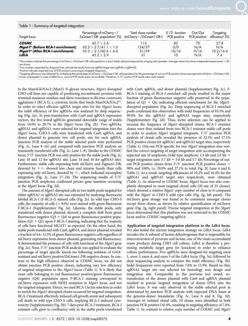
Table 1 | Summary of targeted integration
Target locusPercentage of mCherry1/
ZsGreen1-DR- populationa (%)Total clone number
(mCherry1/ZsGreen1-DR-)59/39 JunctionPCR positive
Out/OutPCR positivec
Targetingefficiencyd (%)
COSMC 45.7 6 2.6 115 83 70 27.8Mgat1b (Before RCA-I enrichment) 62.2 6 2.7/41.1 6 1.0 124/37 0/0 N/A N/AMgat1b (After RCA-I enrichment) 35.3 6 2.1/60.4 6 6.6 31/59 10/16 9/16 10.2/16.4LdhA 45.2 6 2.9 61 10 N/A 7.4aThe numbers indicate the percentage of mCherry1/ZsGreen1-DR- cell population in each stable cell pool measured by imaging cell cytometer. Average values from three independent experiments 6 SDare shown.bTwo numbers separated by diagonal lines indicate the results from two sgRNA target sites (sgRNA1/sgRNA5).cPositive indicates expected size of PCR products occurring by targeted integration.dTargeting efficiency was obtained by multiplying the percentage of mCherry1/ZsGreen1-DR- cell population by the percentage of out/out PCR positive clones amongst total mCherry1/ZsGreen1-DR-clones investigated. In case of LdhA locus, out/out PCR results were not available. Therefore, 59/39 junction PCR results were used instead.
www.nature.com/scientificreports
SCIENTIFIC REPORTS | 5 : 8572 | DOI: 10.1038/srep08572 4

Rela
�ve
Mga
t1 c
opy
num
ber
(sgR
NA5
targ
et si
te)
0.0
0.2
0.4
0.6
0.8
1.0
2 3 4 5 6 7 8 9 10 11 12 13 14 15 16 17
CHO
-S
Rela
�ve
mCh
erry
cop
y nu
mbe
r
0
1
2
3
2 3 4 5 6 7 8 9 10 11 12 13 14 15 16 17
Mgat1 locus:
sgRNA5
////
sgRNA1 sgRNA2sgRNA3 sgRNA4
a b
c
d
- - - -
- - + +
- - + +
5’ junc�on
Cas9
Donor 5
Mgat1 sgRNA1
Transient
- - - -M MMgat1 sgRNA5 - - + + M
- + - +
- - + +
- - - -
Stable
- -
+ -
+ -
-
+
+
3’ junc�on
-
-
-
M
5’ junc�on
- - --
+ +
+ -
- -
+
+
-
3’ junc�on
+
-
-
M
5’ junc�on
+ - +-
- + - +Donor 1 - - - - + + ++ - - --
Stable [Sorted] ++ ++ +- +- ++ ++ +- +-
-
+
+
-
+
-
+
+
-
+
-
+
+
-
+
-
+
+
-
+
+
+
-
+
+
-
M + +
+
+
-
+
+
-
++
- - --
5’ 3’ 5’ 3’ 5’ 3’ 5’ 3’
e
MiSeq Mgat1
sgRN
A3
sgRN
A2
sgRN
A1
sgRN
A4
0%
20%
40%
60%
80%
100%
wt sequence
Indel
Replicate #1 Replicate #2
sgRN
A3
sgRN
A2
sgRN
A5
sgRN
A4
sgRN
A5
sgRN
A1
- F-R
CA+
F-RC
A
Red fluorescence intensity
Gre
en fl
uore
scen
ce in
tens
ity
Cas9
Donor1
Mgat1 sgRNA1Mgat1 sgRNA5
---
-++
- -
++-
+--
+-
Donor5+ + --
RCA selec�on - - - -
-++-
+
+
++-+
-
+
5’ Ju
nc�o
n3’
Junc
�on
Donor1 + Cas9 + sgRNA1
Donor5 + Cas9 + sgRNA5
Reference
Conserva�on
Reference
Conserva�on
5’ Ju
nc�o
n3’
Junc
�on
Reference
Conserva�on
Reference
Conserva�on
1 2 3 4 5 6 7 8 9 10 11 12 13 14 15 16 17 18 19 20 21 22 23 241000bp
1500bp
- - - -- + + +- + + +
M- - - -
2000bp
6000bp5000bp
Cas9
Donor1
Mgat1 sgRNA1Mgat1 sgRNA5
Donor5+ + + +
Pool Clone Pool Clone
+ + + +- + + +- - - -- + + +
- - - -
MJunc�on PCR - + - + - + - +
1 2 3 4 5 6 7 8
Rela
�ve
Mga
t1 c
opy
num
ber
(sgR
NA1
targ
et si
te)
0.0
0.2
0.4
0.6
0.8
1.0
2 3 4 5 6 7 8 9 10
CHO
-S
Rela
�ve
mCh
erry
cop
y nu
mbe
r
0
1
2
3
4
2 3 4 5 6 7 8 9 10
89.5 % 91.0 %0.4 % 0.6 %12.5 % 4.6 %
Q1 Q2
Q3 Q4
f g
Figure 2 | Targeted integration into Mgat1 locus using CRISPR/Cas9. (a) Illustration of the five sgRNA target genomic sites in Mgat1 locus. (b) Indel
frequency in Mgat1 locus analyzed by deep sequencing. Genomic DNA was extracted 3 days after transfection with plasmids expressing Cas9 gene and
sgRNAs. The genomic regions covering sgRNA target sites were amplified, then subjected to Miseq analysis. The percentage of wt and indel sequences are
described in the bar plot. The values from control samples transfected only with plasmid expressing Cas9 were subtracted from test samples. (c) Agarose
gel of 59/39 junction PCR on transiently transfected cells and stable cell pools. (11) Stable cells expressing both mCherry and Zsgreen1-DR; (1-) stable
cells expressing only mCherry. (d) Sanger sequencing of the 59/39 junction PCR amplicons. Amplicons from the stable cell pools were purified and directly
sequenced after PCR amplification. M, 1 kb DNA ladder. (e) Population analysis of Mgat1 disrupted cells by F-RCA-I staining. Based on red/green
fluorescence intensity of stable cell pools, which was further selected with RCA-I or not, alteration of fluorescence intensity was analyzed upon F-RCA-I
staining. Each scatter plot was divided by four quadrants, denoted by Q1 to Q4. Q3 and Q4 populations, marked by red squares, represent negative stained
cells with F-RCA-I, indicating functional knockout of Mgat1 locus. Numerals below the red squares show the percentage of Q3 and Q4. (f) Agarose gel of
out-out PCR results of stable pools or clonal cells. Primer pairs annealing to genomic DNA region were used resulting in PCR products of either wild type
(2.0 kb for sgRNA1 target site; 1.9 kb for sgRNA5 target site) or targeted integration (5.6 kb for sgRNA1 target site; 5.5 kb for sgRNA5 target site). (g)
Relative copy number of Mgat1 and mCherry regions in clonal cells, as described in Fig. 1(e). (Top) sgRNA1 target site (Bottom) sgRNA5 target site.
www.nature.com/scientificreports
SCIENTIFIC REPORTS | 5 : 8572 | DOI: 10.1038/srep08572 5

locus, most LdhA targeted clonal cells (eight out of 10 clones) yielded apartial number of relative LdhA copy number, indicating heterozygousdisruptions at the LdhA locus caused by monoallelic knock-ins
(Fig. 3e, left panel). Unexpectedly, two clones (clone #4 and #5) hadunmodified copy numbers of LdhA relative to CHO-S wild type cells,probably due to non-clonal cell population or chromosomal
LdhA locus:
a
b
c d
1 2 3 4 5 6 7// //
sgRNA2
Transient Stable
- + - +
- - + +
- - + +M M
5’ junc�on
Cas9
Donor
LdhA sgRNA
1500bp
2000bp
+ -
+ -
+
+
3’ junc�on
-
- M
5’ junc�on
+ + ++
sgRNA1
sgRNA3
sgRNA4sgRNA5
Reference
Conserva�on
3’ junc�on5’ junc�on
MiSeq LdhA
sgRN
A3
sgRN
A2
sgRN
A1
sgRN
A4
0%
20%
40%
60%
80%
100%
wt sequence
Indel
Replicate #1 Replicate #2
sgRN
A3
sgRN
A2
sgRN
A5
sgRN
A4
sgRN
A5
sgRN
A1
1 2 3 4 5 6 7 8
e
Rela
�ve
LdhA
cop
y nu
mbe
r
0.0
0.5
1.0
2 3 4 5 6 7 8 9 10
CHO
-S 1
Rela
�ve
mCh
erry
cop
y nu
mbe
r
0
1
2
3
2 3 4 5 6 7 8 9 101
Figure 3 | Targeted integration into LdhA locus using CRISPR/Cas9. (a) Illustration of the five sgRNA target genomic sites in LdhA locus. (b) Indel
frequency in LdhA locus analyzed by deep sequencing. Genomic DNA was extracted 3 days after transfection with plasmids expressing Cas9 gene and
sgRNA. The genomic regions covering sgRNA target sites were amplified, then subjected to deep sequencing analysis using Miseq. The percentage of
wt and indel sequences are described in the bar plot. The values from control samples transfected only with plasmid expressing Cas9 were subtracted from
test samples. Investigation of target specific knock-in in transiently transfected and stable cell pools analyzed by (c) Agarose gel of 59/39 junction PCR (d)
Sanger sequencing of the 59/39 junction PCR amplicons. Amplicons from the stable cell pool were purified and directly sequenced after PCR
amplification. M, 1 kb DNA ladder. (e) Relative copy number of LdhA and mCherry regions in clonal cells, as described in Fig. 1(e).
www.nature.com/scientificreports
SCIENTIFIC REPORTS | 5 : 8572 | DOI: 10.1038/srep08572 6

instability. Heterozygous modification at LdhA locus led to decrease inmRNA expression levels of LdhA by approximately 50%, resulting inreduction of protein expression levels as well (Supplementary Fig. 3).Notably, deep sequencing analysis of nonintegrated allele revealedevidence of in-frame deletions created in LdhA targeted clonal cells,which is unlikely to lead to a loss-of-function (Supplementary Fig. 4).As a result of heterozygous modification in LdhA locus, we could notobtain expected size of PCR products from out-out PCR due to existenceof a residual allele. From analysis of the relative mCherry copy number,similar results of consistent copy number across LdhA target clonal cellswere obtained (Fig. 3e, right panel). Targeted integration at the LdhAlocus was achieved using the current platform, which confirmed that thismethod was also applicable at a diploid locus.
Off-target effects associated with CRISPR/Cas9 mediated targetedintegration. To assess potential off-target mutations introduced bythe CRISPR/Cas9 activity, we first identified the most probablepotential off-target sites of each sgRNA target sequence within theCHO genome. Prediction of off-target sites was based on threecriteria (see Methods section), followed by analysis of the NHEJ-mediated indel frequencies at the on-target and off-target sites ingenomic DNA isolated from stable cell pools transfected withCas9, sgRNA, and donor plasmid by deep sequencing (Fig. 4). Alow level (, 0.5%) of off-target mutations were detected at all fiveputative off-target sites for the COSMC sgRNA, despite high levels ofCRISPR/Cas9 activity at the corresponding on-target site, which
generated indels at frequencies of 56.8% (Fig. 4a). A similar off-target tendency was observed for the Mgat1 sgRNA1, but unlikesgRNA1, Mgat1 sgRNA5 displayed off-target indel generation at afrequency of 10.7% at off-target 1 (Fig. 4b,c). RCA-I enrichment forMgat1-disrupted cells drastically increased an indel frequency from16.4% and 37.3% to 83.4% and 98.9% at sgRNA1 and sgRNA5 on-target sites (Fig. 4b,c), but did not induce additional off-target indelmutations (Supplementary Fig. 5). Three out of four off-target sitesfor the LdhA sgRNA displayed off-target activity (Fig. 4d). Sitesharboring one- or two-base mismatches contained indel mutationsat a frequency between 0.9% and 5.1%. Comparing the indelfrequencies observed at the off-target sites with those at the on-target site, all on-target frequencies were 8.4-fold higher as aminimum. Upon enrichment for positive integrant pools by RCA-Iselection, on-target to off-target frequency difference was greatlyincreased from 9.3-fold to .10,000-fold (Fig. 4b,c). Due to theseon-target to off-target frequency differences, integration events atthe potential off-target sites will be less likely to occur. PCR amp-lification of regions, covering off-target sites in mCherry positive/ZsGreen1-DR negative targeted clones, showed no off-target integra-tions, which supports this hypothesis (Supplementary Fig. 6). The lowmutation level of overall off-target sites supported the reliability of theuse of CRISPR/Cas9 system for targeted integration.
Correlation between targeted integration and homogeneous expres-sion of GOI. To assess the effect of targeted integration on cell culture
On-target
Off-target1
Target sequenceGAATATGTGAGTGTGGATGGAGG
GAAGCTCTGAGTGTGGATGGAGG
GGAGGTATGAGTGTGGATGGAGG
ACTAATGTGAGTGTGGATGGTGG
ACGTAATTGAGTGTGGATGGCGG
AATCACTTGAGTGTGGATGGGGG
Off-target2
Off-target3
Off-target4
Off-target5
COSMC
Mismatches (nt)
3
4
4
5
5
-
Posi�on
NW_003614471 (-) 528266
NW_003613631 (-) 2008240
NW_003616800 (+) 22442
NW_003613801 (-) 473486
NW_003613869 (+) 1436175
NW_003628455 (-) 847
0%
20%
40%
60%
80%
100%
wt sequenceIndel
On-target
Off-target1
Target sequence
GCTGGGCACTGATACCGACAAGG
GCTGGGCACTGATGCCGACAGGG
GCTGGACACTGATGCCGACAAGG
GTTGGGCACTGATGCCGACAAGG
TCCTGGCACTGATATCGACACGG
Off-target2
Off-target3
Off-target4
LdhA
Mismatches (nt)
1
2
2
4
-
Posi�on
NW_003613933 (-) 768769
NW_003614158 (+) 1034506
NW_003616055 (+) 76807
NW_003613598 (-) 1611480
NW_003614690 (+) 181885
0%
20%
40%
60%
80%
100%
On-target
Off-target1
Target sequence
GTGGAGTTGGAGCGGCAGCGGGG
ACGGAGATGGAGAGGCAGCGGGG
GAGGAAATGGAGTGGCAGCGGGG
GCAGACTTGGAGCTGCAGCGTGG
CAGAAGCTGGAGCAGCAGCGAGG
Off-target2
Off-target3
Off-target4
Mgat1 sgRNA5 site
Mismatches (nt)
4
4
4
5
-
Posi�on
NW_003613681 (-) 1678866
NW_003613686 (+) 2563608
NW_003614274 (+) 602970
NW_003614984 (+) 61961
NW_003614027 (+) 1191945
On-target [RCA+]
0%
20%
40%
60%
80%
100%
On-target
Off-target1
Target sequence
GCTCACACCCTTACGGCCAAAGG
GCAAGCTCCCTTACCGCCAAGGG
GCACCAGCCCTTACTGCCAAGGGOff-target2
Mgat1 sgRNA1 site
Mismatches (nt)
5
5
-
Posi�on
NW_003613771 (-) 915191
NW_003613798 (-) 376677
NW_003614027(-) 1192681
0%
20%
40%
60%
80%
100%
On-target [RCA+]
a
b
c
d
> 2800 x
> 5600 x 132 x
> 2800 x
> 1400 x
> 8000 x
> 8000 x 206 x
9.3 x > 10000 x
> 10000 x
49 x
8.4 x
32 x 1060 x
Figure 4 | Comparison of mutation rates at on-target and potential off-target sites for (a) COSMC sgRNA2, (b) Mgat1 sgRNA1, (c) Mgat1 sgRNA5,and (d) LdhA sgRNA2. Information on position of sequences consists of contig, strand, and position within the corresponding contig. Mismatched bases
and PAM sequences are shown in red and blue, respectively. Genomic DNA extracted from stable cell pools transfected with Cas9, sgRNA, and donor
plasmid was used as template for PCR. PCR amplicons spanning corresponding on-target sites and potential off-target sites were subjected to deep
sequencing analysis using Miseq. Indel frequencies at off-target sites of Mgat1 sgRNA1/sgRNA5 site were based on genomic DNA extracted from RCA-I
resistant cell pools. Fold differences in indel frequencies between on-target and off-target sites were presented next to the bar plot.
www.nature.com/scientificreports
SCIENTIFIC REPORTS | 5 : 8572 | DOI: 10.1038/srep08572 7

performance, differences in expression of mCherry and growth ratesbetween targeted integrants and random integrants were analyzed.mCherry positive/ZsGreen1-DR negative cell population was isolatedfrom stable cell pool transfected with Cas9, sgRNA2, and donorplasmid targeting COSMC locus by limiting dilution, then classifiedas either targeted integrants or random integrants according to resultsfrom 59/39 junction PCR and out-out PCR (Supplementary Fig. 7).Both 59/39 junction PCR and out-out PCR positive clones weredefined as targeted integrants, and out-out PCR negative clones were
regarded as random integrants regardless of 59/39 junction PCR results.Amongst positive targeted integrants, clones with complete disruptionat the COSMC locus and similar levels of relative mCherry copynumber were selected (Fig. 1e, n541). Cell growth and expressionlevel of GOI of the two populations was then compared. Bothpopulations displayed similar variations in the relative specificgrowth rate (CV: 13.8% versus 12.4%; Fig. 5a). An averageexpression level of targeted integrants was slightly lower than thatof random integrants (RFU average: 103 versus 140, p , 0.05;
Figure 5 | Effect of targeted integration on homogeneous expression of GOI. Targeted and random integrants isolated from the stable cell pool
transfected with Cas9, sgRNA, and donor plasmid targeting COSMC locus were cultivated to assess (a) RFU and (b) relative specific growth rate (m).
Average mean intensity of mCherry fluorescence measured by imaging cell cytometer was used for RFU. The specific growth rate was calculated from a
plot of viable cell concentration against culture time from day 0 to day 3. Each measured value was normalized by that of the clone number 1. Center lines
show the medians; box limits indicate the 25th and 75th percentiles as determined by R software; whiskers extend 1.5 times the interquartile range from
the 25th and 75th percentiles, outliers are represented by dots; data points are plotted as open circles. n 5 41, 40 sample points. Each sample point
represents average value of measurements at 3–5 different passages during cultivation for a month. (CV) coefficient of variation.
www.nature.com/scientificreports
SCIENTIFIC REPORTS | 5 : 8572 | DOI: 10.1038/srep08572 8

Fig. 5b). Interestingly, targeted integrants displayed more stablemCherry expression with 4.4-fold less variation than that of therandom integrants (CV: 14.3% versus 63.3%; Fig. 5b), indicatingmore homogeneous and predictive expressions from site-specificintegration clones. The variability in expression levels of randomintegrants was caused by the influence of both copy number andintegration site (Supplementary Fig. 8). Some of random integrantswith 2- or 3-fold higher copy number exhibited 2- or 3-fold higherexpression levels relative to the representative targeted integrationclone. On the other hand, various expression levels were observedin a few random integrants despite similar levels of copy number.Moreover, although the percentage of mCherry positive/ZsGreen1-DR negative cells was initially close to 100% in all clones investigated,it was notably decreased in some clones (five out of 40 clones, i.e. 12.5%in total) only from the population of random integrants while cells wereexpanding, underscoring unstable expression from random integration(Supplementary Fig. 9). These data show that CRISPR and HDRmediated targeted integration allowed stable and reproducibletransgene expression in rCHO cell lines without affecting cell growth.
DiscussionSite-specific integration of large transgenes during rCHO cell lineconstruction has been impeded by extremely low integration effi-ciencies and specific requirements, including construction of plat-form cell lines, which limit flexible targeting to any desired sites. Herewe developed a method for targeted integration by combining theCRISPR/Cas9 system with a donor plasmid in CHO cells. The pro-cedure allowed precise HDR-mediated integration of large geneexpression cassettes into desired sites with a targeting efficiencybetween 7.4–27.8% depending on target locus and CRISPR/Cas9activity. With the previous targeted integration studies in CHO cellsrelying on the NHEJ pathway for integration of short (, 100 bp)oligonucleotides16 or linear transgenes17, our approach is liberatedfrom the issues concerning orientation of transgene integration andimperfect ligation caused by degradation of donor ends by exonu-clease activity or end resection of the chromosome. Furthermore, wereport additional benefits common to the CRISPR/Cas9 system suchas high efficiency, robustness, simplicity, ease of design, and low cost.
Compared with the successful HDR-mediated integration of trans-genes in human cells using the CRISPR/Cas9 system, which wasobserved 72 hours after transfection14, we did not detect integrationevents in transiently expressed cells, but only in stable selected poolsalthough transgene size and experimental conditions were different(Fig. 1b, 2c, and 3c). This result supports intrinsically lower levels ofHDR-based transgenesis in CHO cells, which agrees with previousstudies16,17. In particular, the Mgat1 locus had non-appreciable fre-quencies of integration before RCA-I enrichment, which showed thelocus-to-locus variation in terms of integration frequency (Table 1).Although the CRISPR/Cas9 system generated indel mutations viaNHEJ with high efficiency between 16.4–56.8%, the rate of HDR-mediated integration remained relative low (Fig. 4, Table 1). Thepresent targeting efficiency is sufficient for the generation of rCHOcells when paired with drug or phenotypic selection, but requirementsfor selection process can limit the practical application of targetedintegration at certain loci in an actual industrial production process.However, the current method has sufficient room for furtherimprovement in targeting efficiency by implementing recentadvancements, such as rational design of highly active sgRNAs23,enrichment of cells with genome editing events24, or constructionof expression vectors bearing both Cas9 and the sgRNA11 since theCRISPR/Cas9 system is continuously undergoing optimizations toimprove activity. Transfection efficiency, which was measured basedon the fraction of mCherry positive/ZsGreen1-DR positive cells72 hours post transfection, was in the range of 24.4–34.0% whenthree plasmids, including donor plasmid, Cas9, and sgRNA plasmid,were co-transfected. Compared with transfection efficiency of 33.0–
54.6% for control samples transfected with the donor plasmid only,transfection of multiple plasmids lowered the transfection efficiencyconsiderably. Additionally, expression levels from separate constructsmay not be tightly coupled. This potential limitation can be overcomeby using the aforementioned single expression vector11 to drive bothCas9 and sgRNA or FACS enrichment to aid selection of transfectedcells. As observed in the on-target indel efficiency in stable cells andthe targeting efficiency, the higher on-target indel efficiency is likelyto lead to higher HDR-mediated integration (Fig. 4, Table 1).Therefore, the aforementioned approaches will aid in the increasein current targeting efficiencies. Moreover, single-strand breaks(SSBs) created by single sgRNA-Cas9 nickases have reported to becapable of inducing homologous recombination in mammalian cells,albeit at reduced frequency compared with wild-type Cas925,26. Inconjunction with improvements in targeting efficiency, targetedintegration via SSBs-induced homologous recombination will alsobe an attractive strategy with regard to higher specificity and fidelity.
Previous studies have reported that the PAM-proximal 8–13 bp ofthe target region is crucial for target recognition and cleavage byCas911,19, and within PAM-proximal region, a single-base mismatchcan be acceptable to conserve activity. Furthermore, up to three to fivemismatches in the PAM-distal region can be tolerated withoutimpairing the ability to cleave DNA27-29. More recently, genome-widemapping of Cas9 binding sites using catalytically inactive Cas9 andChIP-seq analysis has revealed that a 5-nucleotide seed region next toPAM is required for Cas9 binding in vivo and in vitro30. Based onthis, we selected potential off-target sites for each target site to meetthese criteria, and evaluated off-target mutation rates (Fig. 4). Nineout of 15 potential off-target sites were not affected. In four off-targetsites, we detected indels with a frequency of 0.5–1.3%, which was rareenough to allow high fidelity of sgRNAs targeting on-target sites. Thelast two off-target sites, however, contained detectable off-target cut-tings at a frequency of 5.1% and 10.7%. A recent paper has suggestedthat in addition to finding off-targets at sites with few mismatchescompared with the target sequence, off-targets may also be found atsites with small inserts/deletions compared with the target sequenceunder certain circumstances31. This type of off-targets have not beenconsidered in the current work, but guide RNA design in the futureshould aim to identify such additional potential off-target sites so thatappropriate care may be taken. Off-target mutations may not becritical to rCHO cell line construction compared with human genetherapy. However, potential problems in connection with off-targetmutations could cause unpredictable toxic effects. This latent obstaclecould also be mitigated by using approaches to improve on-targetspecificity of Cas9, such as the double-nicking approach26,29, truncatedguide RNAs32, or dimeric CRISPR RNA-guided FokI nucleases33.
Cultivation of targeted integrants displayed more stable and highlyreproducible expression levels when compared with random inte-grants. Although this result demonstrates the minimal variability inexpression level, it is limited to the specific locus and expressioncassette. The expression level can depend on the target locus andthe cassette design, particularly relating to the promoter used andits susceptibility to silencing. Thus, additional studies in various lociwith different transgene expression cassettes are warranted to con-firm the generality of this finding. Despite homogeneous transgeneexpression levels in targeted integrants, some of the random integ-ration clones exhibited 2- or 3-fold higher expression levels relativeto representative targeted integration clone (Fig. 5b, SupplementaryFig. 8). These observations were due to random integration intohighly active chromosomal regions with the CHO genome or integ-ration of several copies of the donor plasmid. Given that the ultimategoal for construction of production cell lines is rapid generation ofhigh and stable producers, ensuring comparable quality for regula-tory approval, the selection of ideal transgene insertion sites is highlydemanded. Ideal insertion sites, also called ‘Safe harbors’ or ‘Hotspots’, should allow high and stable transcription/expression levels
www.nature.com/scientificreports
SCIENTIFIC REPORTS | 5 : 8572 | DOI: 10.1038/srep08572 9

of the GOI without perturbing essential regulatory elements or dis-rupting genes which are responsible for the quality of the products.Previous extensive studies on the selection and identification ofdesirable target sites in human cells provide reliable guidelines tofacilitate rapid identification of such ‘‘hot spots’’ in CHO cells13,34.A further refinement of existing Chinese hamster reference genomeand all of the relevant sequence information of CHO host cell lines,together with new data on genome stability at the chromosomal levelwill facilitate identification of potential target sites as well as a bettercharacterization of the current targeted integration strategy. Moreover,a recent study demonstrated that actively transcribed chromatin struc-ture was preferentially repaired by homologous recombination overNHEJ upon DSB35. This trait fits perfectly into the presented platformand will reinforce the targeting efficiency. Thus, this approach shouldbe able to provide a valuable genetic tool to characterize differentpotential good integration sites in CHO cells, ultimately leading to arational construction of production cell lines assuring sustainable andhigh expression levels.
MethodsPlasmid design and construction. The CHO codon optimized Cas9 expressionvector and sgRNA expression vector applied in this study were constructed asdescribed previously20. The CRISPy bioinformatics tool20 was applied for generatingsgRNA target sequences (Supplementary Table 1). Subsequently, single strandedoligos comprising the variable region of the sgRNA (Supplementary Table 2) weresynthesized, annealed, and mixed with the expression vector backbone harboring U6promoter, scaffold RNA sequence, and termination sequence to generate sgRNAexpression vectors.
Donor plasmids were constructed with uracil-specific excision reagent (USER)cloning method according to Ref. 36. The different parts of the plasmid were amp-lified with uracil-containing primers and proofreading polymerase PfuX7. 59 and 39
homology arms flanking each sgRNA target sequence were amplified from genomicDNA of CHO-S cells, which were extracted by GeneJET Genomic DNA PurificationKit (Thermo Fisher Scientific, Waltham, MA). EF-1a promoter, NeoR expressioncassette (pSV40-NeoR–SV40 pA), and expression vector backbone parts weredirectly amplified from commercial expression vectors, but ZsGreen1-DR expressioncassette (pCMV-ZsGreen1-DR-BGH pA) and mCherry-BGH pA parts were amp-lified from pre-assembled expression vectors that were constructed with USERcloning method using commercial expression vectors. Primer sequences and allplasmids used as PCR templates are listed in Supplementary Table 2 and 3, respect-ively. Following purification of PCR products by agarose gel separation andNucleoSpinH Gel and PCR Clean-up kit (Macherey-Nagel, Duren, Germany), thepurified 7 PCR fragments including both homology arms, EF-1a promoter, mCherry-BGH pA, NeoR expression cassette, ZsGreen1-DR expression cassette, and backbonewere assembled with USER enzyme (New England Biolabs, Ipswich, MA) andtransformed into E. coli Mach1 competent cells (Life Technologies, Leuven, Belgium).Plasmids were harvested from transformants selected on LB agar plates with ampi-cillin. All constructs were verified with sequencing and purified using NucleoBondXtra Midi EF (Macherey-Nagel) according to manufacturer’s instructions.
Cell culture, transfection, and stable cell line construction. CHO-S cells from LifeTechnologies were grown in CD CHO medium supplemented with 8 mM L-Glutamine (Life Technologies) and cultivated in a 125-mL Erlenmeyer flask (Sigma-Aldrich, St. Louis, MO) with a working volume of 30 mL. The cells were incubated at37uC, 5% CO2 with 120 rpm shaking and passaged every 3 days. Cells weretransfected with expression vectors encoding CHO codon optimized Cas9, sgRNAtargeting the integration site and corresponding donor plasmid. For each sample, 3 3
106 cells were transfected with a total of 3.75 mg of DNA using FreeStyleTM MAXreagent together with OptiPRO SFM medium (Life Technologies) according tomanufacturer’s recommendations. 16 hours post transfection, the samples wereincubated at 30uC for 32 hours before transferred back to 37uC.
Stable cell pools were generated by seeding cells in CELLSTARH 6 well AdvancedTC plates (Greiner Bio-one, Frickenhausen, Germany) on day 3 followed by selectionprocess under G418 (500 mg/mL; Sigma-Aldrich). During selection, medium waschanged every 3–4 days. After 2 weeks of selection, cells were detached with TrypLE(Life Technologies) and adapted to grow in suspension in non-tissue treated plates orErlenmeyer flask depending on cell concentrations. For clonal selection, limitingdilution step was followed using the stable cell pools, which were either bulk sorted formCherry positive/ZsGreen1-DR negative cell population using a BD FACSJazz cellsorter (BD Biosciences, San Jose, CA) or not. 1 cell was seeded per well in 200 ulmedium in 96 well plates. The generated colonies were analyzed by a Celigo ImagingCell Cytometer (Nexcelom Bioscience, Lawrence, MA) applying the Direct cellcounting application to check for single colonies. Only the wells with round shapedcolonies expressing mCherry were selected for further analysis.
Genomic DNA extraction and PCR amplification. Genomic DNA was extractedfrom the cell pellets using QuickExtract DNA extraction solution (Epicentre,
Illumina, Madison, WI) or GeneJET Genomic DNA Purification Kit depending onthe purpose of experiment. For single cell clones, DNA was isolated by adding 20 mLof QuickExtract DNA extraction solution when cells were confluent in 96 well plates.The mixture was then incubated at 65uC for 15 min, followed by 5 min incubation at98uC. Of this mixture, 1–2 mL were used as template for junction PCR and out-outPCR, as described below. For the other experiments, GeneJET Genomic DNAPurification Kit was used to extract DNA from cell pellets up to 5 3 106 cellsaccording to manufacturer’s instructions. 59/39 junction PCR was carried out usingDreamTaq DNA polymerase (Thermo Fisher Scientific) by touchdown PCR (95uCfor 2 min; 103: 95uC for 30 s, 68uC–58uC (21uC/cycle) for 30 s, 72uC for 2 min;203: 95uC for 30 s, 58uC for 30 s, 72uC for 2 min; 72uC for 5 min). Out-Out PCR wasperformed using Phusion Hot Start II High-Fidelity DNA Polymerase (ThermoFisher Scientific) by touchdown PCR (98uC for 30 s; 203: 98uC for 10 s, 64uC–54uC(20.5uC/cycle) for 30 s, 72uC for 3 min; 303: 98uC for 10 s, 54uC for 30 s, 72uC for3 min; 72uC for 10 min). PCR primers are listed in Supplementary Table 2.
Deep sequencing analysis. Deep sequencing was performed on a MiSeq BenchtopSequencer (Illumina, San Diego, CA). Libraries were prepared based on Illumina ‘16SMetagenomic Sequencing Library Preparation’ with some modifications. Genomicregions flanking target genomic sites were amplified using Phusion Hot Start II HFPfu polymerase (Thermo Fisher Scientific) by standard PCR (98uC for 30 s; 253:98uC for 10 s, 59uC for 30 s, 72uC for 30 s; 72uC for 10 min), with primers containingoverhang sequences compatible with Illumina Nextera XT indexing (forward primeroverhang: TCGTCGGCAGCGTCAGATGTGTATAAGAGACAG, reverse primeroverhang: GTCTCGTGGGCTCGGAGATGTGTATAAGAGACAG). PCR primersare listed in Supplementary Table 2. After PCR amplification, amplicons werepurified using AMPure XP beads (Beckman Coulter, Brea, CA). Illumina Nextera XTIndex (Illumina # FC-131-1001) sequencing adapters were integrated into theamplicons by PCR (98uC for 3 min; 83: 95uC for 30 s, 55uC for 30 s, 72uC for 30 s;72uC for 5 min). The final libraries were purified with AMPure XP beads beforequantification. The purified libraries were quantified using the QubitH dsDNA BRAssay Kit (Life Technologies), and the product size within the library was verified by2% agarose gel. Sequencing was performed as a 151 paired-end run. Sequence datawas analyzed using custom python code that compared the sequence data with theexpected wild-type sequence from the CHO genome20. In short, it first paired thepaired end fastq sequence files. Then, it de-multiplexed the sequence reads based on59 or 39 similarity to the expected wild-type sequence followed by detection ofpotential indels to categorize sequences as either wild type or indel-containing. Thefinal product of these steps is a list for each target or off-target with numbers for howoften we see the wild type and how often we see indels and the size of these indels.
Batch culture. Exponentially growing cells were inoculated at a concentration of 3.0–4.0 3 105 cells/mL into Polystyrene transparent square 96-half-deepwell microplateswith Sandwich cover (Enzyscreen, Leiden, Netherlands) containing 250–300 mL ofculture medium, and the culture plates were then incubated on the Multitron cell(INFORS HT, Bottmingen, Switzerland) at 350 rpm in humidified 5% CO2 at 37uC.Viable cell concentration was estimated using PrestoBlueH Cell Viability Reagent(Life Technologies) according to manufacturer’s instructions. Briefly, 10 mL ofPrestoBlue reagent was directly added to 90 mL of the mixture of cells and media in 96well plates. The plates were incubated for . 30 minutes at 37uC, and thenfluorescence was measured by microplate reader (Synergy Mx, BioTek Instruments,Inc., Winooski, VT). The resulting fluorescence intensity was used to determineviable cell concentration based on the standard curve created with CHO-S cells. Onday 3 after a new passage, cells were incubated with NucBlueH Live ReadyProbesHReagent (Life Technologies) for 20 minutes, and red and green fluorescence wasmeasured by a Celigo Imaging Cell Cytometer using the mask 1 target 1 1 target 2application. Cells were identified using the blue fluorescence channel as mask, and thered and green fluorescent channels were used as target 1 and target 2, respectively. Forcytometer analysis, about 10,000–20,000 cells were used per sample.
Ricinus Communis Agglutinin-I (RCA-I) selection and phenotypic analysis ofMgat1 knockout cells. Mgat1 knockout cells were enriched in culture medium with5 mg/mL of RCA-I (Vector Laboratories, Peterborough, UK) using the stable cellpools. After 7 days of selection, cells were adapted to grow in suspension, followed byphenotypic analysis. Cells were stained with NucBlueH Live ReadyProbesH Reagentwith and without fluorescein labeled RCA-I (F-RCA-I, Vector Laboratories) at finalconcentration of 20 mg/ml for 45 min at RT. Cells were then washed three times withfresh culture medium and medium was completely removed. Celigo Imaging CellCytometer was used to measure fluorescence using the mask 1 target 1 1 target 2application as described earlier.
Off-target prediction. Off-targets were predicted using custom python code thatcompared the targets with all other potential targets in the CHO genome. Off-targetswere then scored based on the following criteria: i) The first 5 bp immediatelyadjacent to the PAM sequence: cutoff value of 0; ii) The number of mismatches in thefirst 13 bp: cutoff value of up to 1; iii) The total number of mismatches includingnumber of mismatches in first 13 bp: cutoff value up to 5.
Quantitative Real Time PCR (qRT-PCR) for copy number analysis. For relativedetermination of copy number of transgene, qRT-PCR was performed on genomicDNA samples using Brilliant III Ultra-Fast SYBRH Green QPCR Master Mix (AgilentTechnologies, Santa Clara, CA) on Mx3005P qPCR System (Agilent Technologies)
www.nature.com/scientificreports
SCIENTIFIC REPORTS | 5 : 8572 | DOI: 10.1038/srep08572 10

according to manufacturer’s instructions. Reaction mixtures contained SYBR GreenQPCR master mix, 250 nM of forward and reverse primers, reference dye, and 20 ngof genomic DNA. Amplification was executed with the following conditions: 95uC for10 min; 403: 95uC for 20 s, 60uC for 30 s. The primers were designed usingPrimerQuest (Integrated DNA Technologies, Coralville, IA) against transgeneencompassing EF-1a – mCherry, target genes including COSMC, Mgat1 (sgRNA1 andsgRNA5 target site), and LdhA, and reference gene, Vcl (Supplementary Table 2). Allprimers were validated empirically by melting curve analysis and agarose gelelectrophoresis. Standard curves generated with 4-fold serial dilutions of genomicDNA samples over 5 grades showed a good linearity (r2 . 0.98) and acceptableamplification efficiencies between 90% and 110% for all primer pairs. Each PCRreaction included no template controls in every PCR running, and had 3 replicateswith 2 times repetition. Using a delta-delta threshold cycle (DDCT) method37, relativecopy number was estimated with respect to wild type CHO-S cells for target genes andrepresentative clones for mCherry transgene.
1. Noh, S. M., Sathyamurthy, M. & Lee, G. M. Development of recombinant Chinesehamster ovary cell lines for therapeutic protein production. Curr. Opin. Chem.Eng. 2, 391–397 (2013).
2. Wurm, F. M. Production of recombinant protein therapeutics in cultivatedmammalian cells. Nat. Biotechnol. 22, 1393–1398 (2004).
3. Wilson, C., Bellen, H. J. & Gehring, W. J. Position effects on eukaryotic geneexpression. Annu. Rev. Cell Biol. 6, 679–714 (1990).
4. Kim, S. J., Kim, N. S., Ryu, C. J., Hong, H. J. & Lee, G. M. Characterization ofchimeric antibody producing CHO cells in the course of dihydrofolate reductase-mediated gene amplification and their stability in the absence of selectivepressure. Biotechnol. Bioeng. 58, 73–84 (1998).
5. Huang, Y. et al. An efficient and targeted gene integration system for high-levelantibody expression. J. Immunol. Methods 322, 28–39 (2007).
6. Kito, M., Itami, S., Fukano, Y., Yamana, K. & Shibui, T. Construction ofengineered CHO strains for high-level production of recombinant proteins. Appl.Microbiol. Biotechnol. 60, 442–448 (2002).
7. Lieu, P. T. et al. Generation of site-specific retargeting platform cell lines for drugdiscovery using phiC31 and R4 integrases. J. Biomol. Screen. 14, 1207–1215(2009).
8. Lewis, N. E. et al. Genomic landscapes of Chinese hamster ovary cell lines asrevealed by the Cricetulus griseus draft genome. Nat. Biotechnol. 31, 759–765(2013).
9. Xu, X. et al. The genomic sequence of the Chinese hamster ovary (CHO)-K1 cellline. Nat. Biotechnol. 29, 735–741 (2011).
10. Carroll, D. Genome engineering with targetable nucleases. Annu. Rev. Biochem.83, 409–439 (2014).
11. Cong, L. et al. Multiplex Genome Engineering Using CRISPR/Cas Systems.Science 339, 819–823 (2013).
12. Hockemeyer, D. et al. Efficient targeting of expressed and silent genes in humanESCs and iPSCs using zinc-finger nucleases. Nat. Biotechnol. 27, 851–857 (2009).
13. Lombardo, A. et al. Site-specific integration and tailoring of cassette design forsustainable gene transfer. Nat. Methods 8, 861–869 (2011).
14. Mali, P. et al. RNA-guided human genome engineering via Cas9. Science 339,823–826 (2013).
15. Miller, J. C. et al. A TALE nuclease architecture for efficient genome editing. Nat.Biotechnol. 29, 143–148 (2011).
16. Orlando, S. J. et al. Zinc-finger nuclease-driven targeted integration intomammalian genomes using donors with limited chromosomal homology. NucleicAcids Res. 38, e152 (2010).
17. Cristea, S. et al. In vivo cleavage of transgene donors promotes nuclease-mediatedtargeted integration. Biotechnol. Bioeng. 110, 871–880 (2013).
18. Pennisi, E. The CRISPR craze. Science 341, 833–836 (2013).19. Jinek, M. et al. A programmable dual-RNA-guided DNA endonuclease in
adaptive bacterial immunity. Science 337, 816–821 (2012).20. Ronda, C. et al. Accelerating genome editing in CHO cells using CRISPR Cas9 and
CRISPy, a web-based target finding tool. Biotechnol. Bioeng. 111, 1604–1616(2014).
21. Chen, W. & Stanley, P. Five Lec1 CHO cell mutants have distinct Mgat1 genemutations that encode truncated N-acetylglucosaminyltransferase I. Glycobiology13, 43–50 (2003).
22. Sealover, N. R. et al. Engineering Chinese hamster ovary (CHO) cells forproducing recombinant proteins with simple glycoforms by zinc-finger nuclease
(ZFN)-mediated gene knockout of mannosyl (alpha-1,3-)-glycoprotein beta-1,2-N-acetylglucosaminyltransferase (Mgat1). J. Biotechnol. 167, 24–32 (2013).
23. Doench, J. G. et al. Rational design of highly active sgRNAs for CRISPR-Cas9-mediated gene inactivation. Nat. Biotechnol. doi: 10.1038/nbt.3026. [Epub aheadof print] (2014).
24. Ramakrishna, S. et al. Surrogate reporter-based enrichment of cells containingRNA-guided Cas9 nuclease-induced mutations. Nat. Commun. 5, 3378 (2014).
25. Vriend, L. E., Jasin, M. & Krawczyk, P. M. Assaying Break and Nick-InducedHomologous Recombination in Mammalian Cells Using the DR-GFP Reporterand Cas9 Nucleases. Methods Enzymol. 546, 175–191 (2014).
26. Ran, F. A. et al. Double nicking by RNA-guided CRISPR Cas9 for enhancedgenome editing specificity. Cell 154, 1380–1389 (2013).
27. Fu, Y. et al. High-frequency off-target mutagenesis induced by CRISPR-Casnucleases in human cells. Nat. Biotechnol. 31, 822–826 (2013).
28. Hsu, P. D. et al. DNA targeting specificity of RNA-guided Cas9 nucleases. Nat.Biotechnol. 31, 827–832 (2013).
29. Mali, P. et al. CAS9 transcriptional activators for target specificity screening andpaired nickases for cooperative genome engineering. Nat. Biotechnol. 31, 833–838(2013).
30. Wu, X. et al. Genome-wide binding of the CRISPR endonuclease Cas9 inmammalian cells. Nat. Biotechnol. 32, 670–676 (2014).
31. Lin, Y. et al. CRISPR/Cas9 systems have off-target activity with insertions ordeletions between target DNA and guide RNA sequences. Nucleic Acids Res. 42,7473–7485 (2014).
32. Fu, Y., Sander, J. D., Reyon, D., Cascio, V. M. & Joung, J. K. Improving CRISPR-Cas nuclease specificity using truncated guide RNAs. Nat. Biotechnol. 32, 279–284(2014).
33. Tsai, S. Q. et al. Dimeric CRISPR RNA-guided FokI nucleases for highly specificgenome editing. Nat. Biotechnol. 32, 569–576 (2014).
34. Papapetrou, E. P. et al. Genomic safe harbors permit high b-globin transgeneexpression in thalassemia induced pluripotent stem cells. Nat. Biotechnol. 29,73–78 (2011).
35. Aymard, F. et al. Transcriptionally active chromatin recruits homologousrecombination at DNA double-strand breaks. Nat. Struct. Mol. Biol. 21, 366–374(2014).
36. Lund, A. M. et al. A Versatile System for USER Cloning-Based Assembly ofExpression Vectors for Mammalian Cell Engineering. PLoS One 9, e96693 (2014).
37. Livak, K. J. & Schmittgen, T. D. Analysis of relative gene expression data usingreal-time quantitative PCR and the 2(-Delta Delta C(T)) Method. Methods 25,402–408 (2001).
AcknowledgmentsThe authors thank Anna Koza for her assistance with the MiSeq experiments and KarenKathrine Brøndum for her assistance with the FACS. This work was supported by the NovoNordisk Foundation.
Author contributionsJ.S.L. and H.F.K. designed the experiments and wrote the manuscript. J.S.L., T.B.K., L.E.P.performed the experiments. J.S.L., L.E.P. and H.F.K. analyzed the data. T.B.K. and L.E.P.commented on the manuscript.
Additional informationSupplementary information accompanies this paper at http://www.nature.com/scientificreports
Competing financial interests: The authors declare no competing financial interests.
How to cite this article: Lee, J.S., Kallehauge, T.B., Pedersen, L.E. & Kildegaard, H.F.Site-specific integration in CHO cells mediated by CRISPR/Cas9 and homology-directedDNA repair pathway. Sci. Rep. 5, 8572; DOI:10.1038/srep08572 (2015).
This work is licensed under a Creative Commons Attribution 4.0 InternationalLicense. The images or other third party material in this article are included in thearticle’s Creative Commons license, unless indicated otherwise in the credit line; ifthe material is not included under the Creative Commons license, users will needto obtain permission from the license holder in order to reproduce the material. Toview a copy of this license, visit http://creativecommons.org/licenses/by/4.0/
www.nature.com/scientificreports
SCIENTIFIC REPORTS | 5 : 8572 | DOI: 10.1038/srep08572 11