SiO2-Ag Composite as a Highly Virucidal Material
19
nanomaterials Article SiO 2 -Ag Composite as a Highly Virucidal Material: A Roadmap that Rapidly Eliminates SARS-CoV-2 Marcelo Assis 1,2 , Luiz Gustavo P. Simoes 3 , Guilherme C. Tremiliosi 3 , Dyovani Coelho 1 , Daniel T. Minozzi 3 , Renato I. Santos 3 , Daiane C. B. Vilela 3 , Jeziel Rodrigues do Santos 1 , Lara Kelly Ribeiro 1 , Ieda Lucia Viana Rosa 1 , Lucia Helena Mascaro 1 , Juan Andrés 2, * and Elson Longo 1 Citation: Assis, M.; Simoes, L.G.P.; Tremiliosi, G.C.; Coelho, D.; Minozzi, D.T.; Santos, R.I.; Vilela, D.C.B.; Santos, J.R.d.; Ribeiro, L.K.; Rosa, I.L.V.; et al. SiO 2 -Ag Composite as a Highly Virucidal Material: A Roadmap that Rapidly Eliminates SARS-CoV-2. Nanomaterials 2021, 11, 638. https://doi.org/10.3390/ nano11030638 Academic Editors: Miguel Gama and Francesco Paolo La Mantia Received: 7 February 2021 Accepted: 26 February 2021 Published: 4 March 2021 Publisher’s Note: MDPI stays neutral with regard to jurisdictional claims in published maps and institutional affil- iations. Copyright: © 2021 by the authors. Licensee MDPI, Basel, Switzerland. This article is an open access article distributed under the terms and conditions of the Creative Commons Attribution (CC BY) license (https:// creativecommons.org/licenses/by/ 4.0/). 1 CDMF, LIEC, Federal University of São Carlos—(UFSCar), 13565-905 São Carlos, SP, Brazil; [email protected] (M.A.); [email protected] (D.C.); [email protected] (J.R.d.S.); [email protected] (L.K.R.); [email protected] (I.L.V.R.); [email protected] (L.H.M.); [email protected] (E.L.) 2 Department of Physical and Analytical Chemistry, University Jaume I (UJI), 12071 Castellon, Spain 3 Nanox Tecnologia S/A, 13562-400 São Carlos, SP, Brazil; [email protected] (L.G.P.S.); [email protected] (G.C.T.); [email protected] (D.T.M.); [email protected] (R.I.S.); [email protected] (D.C.B.V.) * Correspondence: [email protected] Abstract: COVID-19, as the cause of a global pandemic, has resulted in lockdowns all over the world since early 2020. Both theoretical and experimental efforts are being made to find an effective treatment to suppress the virus, constituting the forefront of current global safety concerns and a significant burden on global economies. The development of innovative materials able to prevent the transmission, spread, and entry of COVID-19 pathogens into the human body is currently in the spotlight. The synthesis of these materials is, therefore, gaining momentum, as methods providing nontoxic and environmentally friendly procedures are in high demand. Here, a highly virucidal material constructed from SiO 2 -Ag composite immobilized in a polymeric matrix (ethyl vinyl acetate) is presented. The experimental results indicated that the as-fabricated samples exhibited high antibacterial activity towards Escherichia coli (E. coli) and Staphylococcus aureus (S. aureus) as well as towards SARS-CoV-2. Based on the present results and radical scavenger experiments, we propose a possible mechanism to explain the enhancement of the biocidal activity. In the presence of O 2 and H 2 O, the plasmon-assisted surface mechanism is the major reaction channel generating reactive oxygen species (ROS). We believe that the present strategy based on the plasmonic effect would be a significant contribution to the design and preparation of efficient biocidal materials. This fundamental research is a precedent for the design and application of adequate technology to the next-generation of antiviral surfaces to combat SARS-CoV-2. Keywords: COVID-19; virus elimination; antiviral surfaces; SiO 2 -Ag composite; ethyl vinyl acetate; surface plasmon resonance effect 1. Introduction The current worldwide public health and economic crisis resulting from COVID-19 has become a critical problem [1]. At present, there are no vaccines or antiviral drugs available for the prevention or treatment of COVID-19 infections. Currently, many different antiviral agents, including repurposed drugs, are under testing in clinical trials to assess their efficacy, but the quest for an effective treatment against COVID-19 is still ongoing [2–5]; therefore, it is essential to explore any other effective intervention strategies that may reduce the mortality and morbidity rates of the disease. Some excellent reviews of therapeutics and tools that inactivate SARS-CoV-2 have been published [6–8]. SARS-CoV-2 spreads mainly via human fluids, and individuals may acquire the virus after touching different contaminated surfaces [9]. It is known that SARS-CoV-2 remains Nanomaterials 2021, 11, 638. https://doi.org/10.3390/nano11030638 https://www.mdpi.com/journal/nanomaterials
-
Upload
khangminh22 -
Category
Documents
-
view
4 -
download
0
Transcript of SiO2-Ag Composite as a Highly Virucidal Material

nanomaterials
Article
SiO2-Ag Composite as a Highly Virucidal Material: A Roadmapthat Rapidly Eliminates SARS-CoV-2
Marcelo Assis 1,2 , Luiz Gustavo P. Simoes 3, Guilherme C. Tremiliosi 3, Dyovani Coelho 1 ,Daniel T. Minozzi 3 , Renato I. Santos 3, Daiane C. B. Vilela 3, Jeziel Rodrigues do Santos 1 , Lara Kelly Ribeiro 1,Ieda Lucia Viana Rosa 1, Lucia Helena Mascaro 1, Juan Andrés 2,* and Elson Longo 1
�����������������
Citation: Assis, M.; Simoes, L.G.P.;
Tremiliosi, G.C.; Coelho, D.; Minozzi,
D.T.; Santos, R.I.; Vilela, D.C.B.;
Santos, J.R.d.; Ribeiro, L.K.; Rosa,
I.L.V.; et al. SiO2-Ag Composite as a
Highly Virucidal Material: A
Roadmap that Rapidly Eliminates
SARS-CoV-2. Nanomaterials 2021, 11,
638. https://doi.org/10.3390/
nano11030638
Academic Editors: Miguel Gama and
Francesco Paolo La Mantia
Received: 7 February 2021
Accepted: 26 February 2021
Published: 4 March 2021
Publisher’s Note: MDPI stays neutral
with regard to jurisdictional claims in
published maps and institutional affil-
iations.
Copyright: © 2021 by the authors.
Licensee MDPI, Basel, Switzerland.
This article is an open access article
distributed under the terms and
conditions of the Creative Commons
Attribution (CC BY) license (https://
creativecommons.org/licenses/by/
4.0/).
1 CDMF, LIEC, Federal University of São Carlos—(UFSCar), 13565-905 São Carlos, SP, Brazil;[email protected] (M.A.); [email protected] (D.C.); [email protected] (J.R.d.S.);[email protected] (L.K.R.); [email protected] (I.L.V.R.); [email protected] (L.H.M.);[email protected] (E.L.)
2 Department of Physical and Analytical Chemistry, University Jaume I (UJI), 12071 Castellon, Spain3 Nanox Tecnologia S/A, 13562-400 São Carlos, SP, Brazil; [email protected] (L.G.P.S.);
[email protected] (G.C.T.); [email protected] (D.T.M.); [email protected] (R.I.S.);[email protected] (D.C.B.V.)
* Correspondence: [email protected]
Abstract: COVID-19, as the cause of a global pandemic, has resulted in lockdowns all over theworld since early 2020. Both theoretical and experimental efforts are being made to find an effectivetreatment to suppress the virus, constituting the forefront of current global safety concerns and asignificant burden on global economies. The development of innovative materials able to preventthe transmission, spread, and entry of COVID-19 pathogens into the human body is currently in thespotlight. The synthesis of these materials is, therefore, gaining momentum, as methods providingnontoxic and environmentally friendly procedures are in high demand. Here, a highly virucidalmaterial constructed from SiO2-Ag composite immobilized in a polymeric matrix (ethyl vinyl acetate)is presented. The experimental results indicated that the as-fabricated samples exhibited highantibacterial activity towards Escherichia coli (E. coli) and Staphylococcus aureus (S. aureus) as well astowards SARS-CoV-2. Based on the present results and radical scavenger experiments, we proposea possible mechanism to explain the enhancement of the biocidal activity. In the presence of O2
and H2O, the plasmon-assisted surface mechanism is the major reaction channel generating reactiveoxygen species (ROS). We believe that the present strategy based on the plasmonic effect wouldbe a significant contribution to the design and preparation of efficient biocidal materials. Thisfundamental research is a precedent for the design and application of adequate technology to thenext-generation of antiviral surfaces to combat SARS-CoV-2.
Keywords: COVID-19; virus elimination; antiviral surfaces; SiO2-Ag composite; ethyl vinyl acetate;surface plasmon resonance effect
1. Introduction
The current worldwide public health and economic crisis resulting from COVID-19has become a critical problem [1]. At present, there are no vaccines or antiviral drugsavailable for the prevention or treatment of COVID-19 infections. Currently, many differentantiviral agents, including repurposed drugs, are under testing in clinical trials to assesstheir efficacy, but the quest for an effective treatment against COVID-19 is still ongoing [2–5];therefore, it is essential to explore any other effective intervention strategies that may reducethe mortality and morbidity rates of the disease. Some excellent reviews of therapeuticsand tools that inactivate SARS-CoV-2 have been published [6–8].
SARS-CoV-2 spreads mainly via human fluids, and individuals may acquire the virusafter touching different contaminated surfaces [9]. It is known that SARS-CoV-2 remains
Nanomaterials 2021, 11, 638. https://doi.org/10.3390/nano11030638 https://www.mdpi.com/journal/nanomaterials

Nanomaterials 2021, 11, 638 2 of 19
viable on solids for extended periods (for up to 1 week on hard surfaces such as glass andstainless steel) [10,11]. Consequently, not only is the identification of materials capableof killing viruses by contact and having low cytotoxicity clearly a high priority for allscientists around the world, but the detection of new and effective materials to decontam-inate surfaces is also of great concern [12–14]. Given the significance of surface and aircontamination in the spread of the virus, attention should also be paid to the developmentof biocidal (virus, bacteria, fungus) materials against the spread of contamination facilitatedby frequently touched surfaces, such as protecting hospital environments and the surfacesof biomedical devices, along with decontamination equipment and technologies [6,15–18].
In this scenario, metals, semiconductors, and inorganic materials are gaining increasedattention as broad-spectrum antiviral agents to protect surfaces and packaging, thus pre-venting new infections in humans [19]. Very recently, Ghaffari et al. [20] discussed effortsto deploy nanotechnology, biomaterials, and stem cells in each step of the fight againstSARS-CoV-2, while Basak and Packirisamy [21] have discussed several nanotechnologicalstrategies that can be used as antiviral coatings to inhibit viral transmission by preventingviral entry into host cells. In this context, metal oxide nanoparticles and their compositeswere established as potent antibacterial agents due to the induced generation of reac-tive oxygen species (ROS) and the subsequent oxidative stress [22,23]. They can stillenter the microorganism’s membranes, reacting with the existing phosphate and sulfategroups, impairing their functioning, and consequently leading to the microorganism’sdeath [24,25]. ROS can still inhibit the replication activities of DNA/plasmid and someprotein enzymes, due to their interaction with phosphate/sulfate groups or even due togenetic changes [25,26]. All of these results, in combination with the permeability of ROSunder the cell membrane, can affect the expression of proteins essential for the correctfunctioning of microorganisms, as well as their replication [24,27–31].
In particular, silver (Ag) is a widely known element for its antimicrobial properties andhas been used in colloidal silver compounds or as adsorbed particles in a colloidal carrier [32].In addition, Ag nanoparticles (Ag NPs) display the antimicrobial properties of bulk Ag, with asignificant reduction in the toxic effects observed with Ag cations [33–35]. The antimicrobialeffects of Ag NPs are accomplished by a unique physiochemical property to generate moreefficient contact with microorganisms and enhance interactions with microbial proteins [36].Ag NPs present excellent activity against many kinds of bacteria [37–42] and are capableof disrupting the mitochondrial respiratory chain, leading to the production of ROS [43],and have also demonstrated promising antifungal [44,45] and antiviral capabilities againstviruses such as HIV, Tacaribe virus, and several respiratory pathogens, including aden-ovirus, parainfluenza, and influenza (H3N2) [31,46–49]. Specifically with regard to antiviralactivities, AgNPs are thought to inhibit the entry of the virus into cells due to the bindingof envelope proteins, such as glycoprotein gp120, which prevents CD4-dependent virionbinding, fusion, and infectivity [31]. In most cases, Ag NPs present the disadvantageof their tendency to agglomerate, leading to a loss of effectiveness. In recent years, theconstruction of Ag metal/semiconductor composite materials has been identified as apromising strategy for responding to the above problems. Therefore, the strong surfaceplasmon resonance (SPR) adsorption and high electron trapping ability of Ag NPs arebeneficial for promoting the charge transmission bridge [29,50–55]. This modification ofAg NPs by light establishes a coulombic restoring force and prompts a charge density, andthey are frequently used in plasmonic composites.
Among the large number of metal/semiconductor composites, SiO2-Ag has attractedconsiderable attention due to its excellent properties, because SiO2 is thermally stable andhighly bioactive, and could not only prevent the agglomeration of particles and enhancethe surface hydrophilicity but also further improve their stability [56–65]. Recently, ithas been demonstrated that mesoporous silica nanoparticle/Ag composite presents greatpotential as a candidate for the development of products aiming to prevent the spread ofdrug-resistant pathogens [66,67].
Nanomaterials 2021, 11, 638 3 of 19
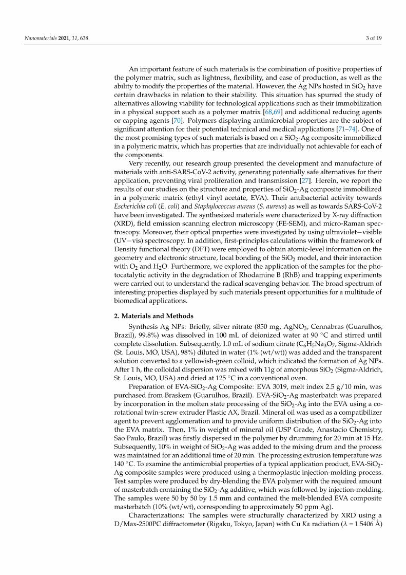
An important feature of such materials is the combination of positive properties ofthe polymer matrix, such as lightness, flexibility, and ease of production, as well as theability to modify the properties of the material. However, the Ag NPs hosted in SiO2 havecertain drawbacks in relation to their stability. This situation has spurred the study ofalternatives allowing viability for technological applications such as their immobilizationin a physical support such as a polymer matrix [68,69] and additional reducing agentsor capping agents [70]. Polymers displaying antimicrobial properties are the subject ofsignificant attention for their potential technical and medical applications [71–74]. One ofthe most promising types of such materials is based on a SiO2-Ag composite immobilizedin a polymeric matrix, which has properties that are individually not achievable for each ofthe components.
Very recently, our research group presented the development and manufacture ofmaterials with anti-SARS-CoV-2 activity, generating potentially safe alternatives for theirapplication, preventing viral proliferation and transmission [27]. Herein, we report theresults of our studies on the structure and properties of SiO2-Ag composite immobilizedin a polymeric matrix (ethyl vinyl acetate, EVA). Their antibacterial activity towardsEscherichia coli (E. coli) and Staphylococcus aureus (S. aureus) as well as towards SARS-CoV-2have been investigated. The synthesized materials were characterized by X-ray diffraction(XRD), field emission scanning electron microscopy (FE-SEM), and micro-Raman spec-troscopy. Moreover, their optical properties were investigated by using ultraviolet−visible(UV−vis) spectroscopy. In addition, first-principles calculations within the framework ofDensity functional theory (DFT) were employed to obtain atomic-level information on thegeometry and electronic structure, local bonding of the SiO2 model, and their interactionwith O2 and H2O. Furthermore, we explored the application of the samples for the pho-tocatalytic activity in the degradation of Rhodamine B (RhB) and trapping experimentswere carried out to understand the radical scavenging behavior. The broad spectrum ofinteresting properties displayed by such materials present opportunities for a multitude ofbiomedical applications.
2. Materials and Methods
Synthesis Ag NPs: Briefly, silver nitrate (850 mg, AgNO3, Cennabras (Guarulhos,Brazil), 99.8%) was dissolved in 100 mL of deionized water at 90 ◦C and stirred untilcomplete dissolution. Subsequently, 1.0 mL of sodium citrate (C6H5Na3O7, Sigma-Aldrich(St. Louis, MO, USA), 98%) diluted in water (1% (wt/wt)) was added and the transparentsolution converted to a yellowish-green colloid, which indicated the formation of Ag NPs.After 1 h, the colloidal dispersion was mixed with 11g of amorphous SiO2 (Sigma-Aldrich,St. Louis, MO, USA) and dried at 125 ◦C in a conventional oven.
Preparation of EVA-SiO2-Ag Composite: EVA 3019, melt index 2.5 g/10 min, waspurchased from Braskem (Guarulhos, Brazil). EVA-SiO2-Ag masterbatch was preparedby incorporation in the molten state processing of the SiO2-Ag into the EVA using a co-rotational twin-screw extruder Plastic AX, Brazil. Mineral oil was used as a compatibilizeragent to prevent agglomeration and to provide uniform distribution of the SiO2-Ag intothe EVA matrix. Then, 1% in weight of mineral oil (USP Grade, Anastacio Chemistry,São Paulo, Brazil) was firstly dispersed in the polymer by drumming for 20 min at 15 Hz.Subsequently, 10% in weight of SiO2-Ag was added to the mixing drum and the processwas maintained for an additional time of 20 min. The processing extrusion temperature was140 ◦C. To examine the antimicrobial properties of a typical application product, EVA-SiO2-Ag composite samples were produced using a thermoplastic injection-molding process.Test samples were produced by dry-blending the EVA polymer with the required amountof masterbatch containing the SiO2-Ag additive, which was followed by injection-molding.The samples were 50 by 50 by 1.5 mm and contained the melt-blended EVA compositemasterbatch (10% (wt/wt), corresponding to approximately 50 ppm Ag).
Characterizations: The samples were structurally characterized by XRD using aD/Max-2500PC diffractometer (Rigaku, Tokyo, Japan) with Cu Kα radiation (λ = 1.5406 Å)

Nanomaterials 2021, 11, 638 4 of 19
in the 2θ range of 10–50◦ and a scanning speed of 1◦ min-1. Furthermore, micro-Ramanspectra were recorded using the iHR550 spectrometer (Horiba Jobin-Yvon, Kyoto, Japan)coupled with a Silicon CCD detector and an argon-ion laser (Melles Griot, Rochester, NYUSA), which operated at 514.5 nm with a maximum power of 200 mW; moreover, a fiberoptic microscope was also employed. Fourier-transform infrared spectroscopy (FT-IR,Bruker Vector 22 FTIR, Billerica, MA, USA) of the samples was recorded at 400–4000 cm−1.UV–vis diffuse reflectance measurements were obtained using a Varian Cary spectrometermodel 5G in diffuse reflectance mode, with a wavelength range of 2000 to 250 nm and ascan speed of 300 nm min−1. An analysis of the thermal stability of samples was conductedon a thermogravimetric (TG/DTA) analyzer (NETZSCH—409 Cell) from 30 to 700 ◦C at aheating rate of 10 ◦C min−1 and in an oxidizing atmosphere (O2) with 50 mL min−1 flux.The morphologies, textures, and sizes of the samples were observed with a FE-SEM, whichoperated at 2 kV (Supra 35-VP, Carl Zeiss, Jena, Germany). A Jem-2100 LaB6 (Jeol, Tokyo,Japan) high-resolution transmission electron microscope (HR-TEM) with an acceleratingvoltage of 200 kV coupled with an INCA Energy TEM 200 (Oxford, Abingdon, UK) wasused to obtain larger magnifications and to clearly verify the samples. AFM imageswere obtained using a Flex-AFM controlled by Easyscan 2 software (Nanosurf, Liestal,Switzerland) in contrast phase mode on an active vibration isolation table (model TS-150,Table Stable LTD®). The cantilever used for image acquisition was the silicon Tap190G(resonant frequency 190 kHz, force constant 48 N/m, Budget Sensors) in setpoint of 50%.
Bactericidal Tests: The bactericidal activity towards E. coli and S. aureus of the purepolymer and the composite with SiO2-Ag was evaluated according to the standard testmethodology described in ISO 22196—Measurement of antibacterial activity on plasticsand other non-porous surfaces [75], carried out in Nanox’s microbiology laboratory. A100-µL volume of the bacterial solution (in a concentration of 105 CFU/mL) was inoculatedin triplicate over the surface of the samples. The inoculum was then covered with asterile plastic film which was gently pressed to be distributed throughout the samplearea. Samples were incubated in a bacteriological oven at 36 ◦C for 24 h at 90% humidity.After incubation, the inoculum was recovered with 10 mL of SCDLP broth followed byserial dilution to 10−4 in PBS buffer. One mL of each dilution was plated with StandardCount Agar by Pour Plate. After solidification of the culture medium, the Petri dishes wereincubated in the inverted position in a bacteriological oven at 36 ◦C for 24 h. The logarithmicreduction and percentage reduction by the CFU/mL count were then determined by thefollowing equation:
R = (Ut −U0)− (At −U0) = Ut −At (1)
where R is the antibacterial activity; U0 is the average of the common logarithm of thenumber of viable bacteria, in cells/cm2, recovered from the untreated test specimensimmediately after inoculation; Ut is the average of the common logarithm of the number ofviable bacteria, in cells/cm2, recovered from the untreated test specimens after 24 h, andAt is the average of the common logarithm of the number of viable bacteria, in cells/cm2,recovered from the treated test specimens after 24 h.
Antiviral Tests: The antiviral activity of the pure polymer and the composite withSiO2-Ag was evaluated by adapting the standard model ISO 21702—Measures of antiviralactivity on plastics and other non-porous surfaces [76] and the method used by Tremiliosiet al. [27]. The tests were carried out in a NB3 (biosafety level 3) laboratory at the Universityof São Paulo, following the recommendations of ANVISA. SARS-CoV-2 was inoculatedinto liquid media; EVA polymer and the EVA-SiO2-Ag composite samples were incubatedfor 2 different time intervals (2 and 10 min). Then, they were seeded in Vero CCL-81 cellcultures. After incubation, the viral genetic material was quantified by quantitative PCR inreal time and, based on the control, the ability of each sample to inactivate SARS-CoV-2was determined.
Reactive Oxygen Species (ROS) Identification: To investigate the active species gener-ated in the photocatalytic RhB (Aldrich, 95%) degradation process over SiO2-Ag composite,a trapping experiment was conducted with ascorbic acid (AA), ammonium oxalate (AO),

Nanomaterials 2021, 11, 638 5 of 19
and tert-butyl alcohol (TBA) as the capture agent of hydroxyl radical (OH∗), hole (h•),and hydroperoxyl radical (O2H∗), respectively. The trapping experimental procedure wasidentical to photocatalytic degradation except that an additional capture agent was addedeach time. In this way, 50 mg of the sample was dispersed in 50 mL of RhB solution(1 × 10⁻⁵ M), and it was kept in the dark for 30 min at 20 ◦C, and then 6 visible lamps(Philips TL-D, 15W) were switched on. After 60 min, an aliquot was removed and cen-trifuged to obtain only the liquid phase. The variations in the standard absorption of RhB(554 nm) were discerned through analysis of absorption spectroscopy in the UV–vis regionon a V-660 spectrophotometer (JASCO, Tokyo, Japan).
Computational Method: The calculations were performed with the Gaussian 09package [77] by using density functional theory (DFT), with the hybrid functional B3LYPand 6-31 ++ G ** basis set. In the Supplementary Materials, the model systems employed inthis study are presented. An analysis based on the results of the natural bond orbital (NBO)method (Reed et al.) and the map electrostatic potential (MEP) is employed to investigatethe charge transfer process between the SiO2 model and O2 and H2O.
3. Results and Discussion
The X-ray diffraction (XRD) measurements are presented in Figure 1. An analysis ofthe results shows that the sample SiO2-Ag has a characteristic peak of amorphous SiO2 ataround 2θ = 22.2◦ [78–81]. No additional peak is observed regarding possible Ag phases.For pure EVA, a high crystallinity of the polymer is observed, which is in line with whathas been observed in other studies in the literature [82,83]. The SiO2-Ag particles wereadded in a polymeric matrix, EVA, which has the role of carrier. For the EVA-SiO2-Agcomposite, there is an amorphization of the polymeric structure; that is, the symmetry andperiodicity break at long-range. This is due to the high degree of disorder of the distortedtetrahedral clusters of [SiO4] present in amorphous SiO2 [84], which cause an induction toamorphization of the polymeric EVA chains. As a result of this union, a broad band locatedat 2θ = 19.9◦ is observed.
Figure 1. X-ray diffractograms of SiO2-Ag, EVA-SiO2-Ag, and EVA samples.
In order to complement the results obtained by XRD, micro-Raman measurementswere performed, seeking to analyze the degree of order of the samples at short range(Figure 2). For the SiO2-Ag sample, a peak of approximately ~240 cm−1 is observed,referring to the scissoring of the distorted tetrahedral of the [SiO4] clusters [85]. For pureEVA, there are five distinct groups of vibrations in the micro-Raman spectrum [86]. Thevibrations in the range 500–700 cm−1 correspond to the deformation movements of theacetate groups of the EVA monomers [86–88]. A set of peaks related to the C-C stretches of

Nanomaterials 2021, 11, 638 6 of 19
the constituent monomers is observed in the range of 750 to 1250 cm−1 [86,89]. The peaksbetween 1300 and 1500 cm−1 were ascribed to the bending and twisting vibrations of theethylene groups in the monomers of EVA [86,87]. Between 1700 and 1900 cm−1, stretchesrelated to C=O bonds are observed [86,90]. At the highest wavelengths, located between2800 and 3050 cm−1, C–H aliphatic stretches of the EVA are observed [87,91]. In contrast tothe XRD observations, the composite does not lose its organization at short range; that is,its constituent monomers maintain their degree of structural order. The SiO2-Ag mode inthe composite can also be observed, indicating good incorporation in the EVA polymer.According to Shen et al., this mode at ~240 cm−1 may also refer to vibrations of the Ag-Obonds, which can be formed from the interaction of the O atoms of the carbonyl groups ofthe EVA with the Ag contained in SiO2-Ag [87].
Figure 2. Micro-Raman spectra of SiO2-Ag, EVA-SiO2-Ag, and EVA samples.
Fourier-transform infrared spectroscopy (FTIR) was performed to analyze changes inthe functional groups of the samples and to verify the formation of the composite EVA-SiO2-Ag (Figure 3). For SiO2-Ag, there is a broad band located near 3400 cm−1 and anotherlocated at 1627 cm−1, both corresponding to the O–H stretching of water and the formedsilanol groups (Si–OH), respectively [92,93]. The bands observed at 1100 and 475 cm−1,on the other hand, are attributed to symmetrical stretching and bending of Si–O–Si bonds,respectively [92,94,95]. The peaks located at 950 and 845 cm−1 indicate the bending of theO–Si–O moiety [80,95]. The low-intensity mode located at 552 cm−1 can be attributed toAg-O stretching, showing the presence of Ag in SiO2-Ag [96,97]. For EVA, bands referringto the fingerprint of the polymer are observed at 2954, 2850, 1467, 1243, 874, 707, and546 cm−1, related to the EVA aliphatic groups [98–102]. At 1020 cm−1, the bending ofthe C–O–C bonds is observed [102], and at 1801 and 1739 cm−1, the C=O bond stretchingrefers to two different types of carbonyl groups [101,102], as noted by Poljansek et al. [103].For the EVA-SiO2-Ag composite, changes are observed especially for the stretching of theC=O bonds and throughout the low-wavelength region, where the SiO2 vibrational modesappear. This is because EVA monomers interact through ionic and van der Waals forceswith SiO2 and Ag, shown by the overlap of some vibrational modes of the samples and theappearance of new ones. These findings indicate the interactions between the polymer, atshort and long range, with the particles of SiO2 and Ag.

Nanomaterials 2021, 11, 638 7 of 19
Nanomaterials 2021, 11, x FOR PEER REVIEW 7 of 19
appearance of new ones. These findings indicate the interactions between the polymer, at
short and long range, with the particles of SiO2 and Ag.
Figure 3. FTIR spectra of SiO2-Ag, EVA-SiO2-Ag, and EVA samples.
The thermogravimetric (TG) and differential thermal analysis (DTA) curves are
shown in Figure 4. In the SiO2-Ag sample, a small loss of mass (9.3%) is observed at 50°C,
due to the loss of water molecules adsorbed onto the material surface, demonstrating its
high thermal stability [104,105]. The degradation of the EVA polymer occurs in two main
stages: the first is due to the loss of acetate groups (between 300 and 400 °C) and the sec-
ond is due to the decomposition of the remaining ethylene groups (between 400 and 650
°C) [106,107]. For the composite, there are no significant differences in the TG/DTA pro-
files compared to the pure polymer, but a slightly smaller loss of mass occurs for this
compound (96.8%) than for the EVA (98.2%). This difference is due to the addition of SiO2-
Ag in the polymeric structure, which, due to its high thermal stability, does not decom-
pose at higher temperatures.
.
Figure 4. TG/DTA curves of SiO2-Ag, EVA-SiO2-Ag, and EVA samples.
Figure 5A shows the diffuse reflectance spectra (DRS) of pure EVA and EVA-SiO2-
Ag, in which light absorption is observed in the range of 685 to 480 nm, attributed to the
presence of composite SiO2-Ag in the polymer blend. The absorptions on near-infrared
wavelengths are ascribed to EVA, where the peaks at 1218, 1440, and 1750 nm are the
4000 3500 3000 2500 2000 1500 1000 500
Tra
nsm
ita
nce
(a
rb.u
nt.
)
Wavelength (cm-1)
O-H groups
CH
Asy
mm
etri
c
Str
etch
ing
CH
Sym
met
ric
Str
etch
ing
CO
2
C=
OS
tret
chin
g
-CH
2 -
Ben
din
g
-CH
2 -
Rock
ing
-CH
2 -
Def
orm
atio
n
C-O
-C B
endin
g
CH
Ben
din
g
O-H groups
Si-O-Si
StretchingSi-O
Bending
Ag-O
Stretching
Si-O-Si
Bending
10 20 30 40 50
Inte
nsi
ty (
arb
.un
t.)
2q (°)
EVA
EVA-SiO2-Ag
SiO2-Ag
0 100 200 300 400 500 600 700 800
0
20
40
60
80
100
Ma
ss (
%)
Temperature (°C)
EVA
EVA-SiO2-Ag
SiO2-Ag
-3
-2
-1
0
1
DT
A/(
uV
/mg
)
Figure 3. FTIR spectra of SiO2-Ag, EVA-SiO2-Ag, and EVA samples.
The thermogravimetric (TG) and differential thermal analysis (DTA) curves are shownin Figure 4. In the SiO2-Ag sample, a small loss of mass (9.3%) is observed at 50 ◦C,due to the loss of water molecules adsorbed onto the material surface, demonstrating itshigh thermal stability [104,105]. The degradation of the EVA polymer occurs in two mainstages: the first is due to the loss of acetate groups (between 300 and 400 ◦C) and thesecond is due to the decomposition of the remaining ethylene groups (between 400 and650 ◦C) [106,107]. For the composite, there are no significant differences in the TG/DTAprofiles compared to the pure polymer, but a slightly smaller loss of mass occurs for thiscompound (96.8%) than for the EVA (98.2%). This difference is due to the addition of SiO2-Ag in the polymeric structure, which, due to its high thermal stability, does not decomposeat higher temperatures.
Figure 4. TG/DTA curves of SiO2-Ag, EVA-SiO2-Ag, and EVA samples.
Figure 5A shows the diffuse reflectance spectra (DRS) of pure EVA and EVA-SiO2-Ag,in which light absorption is observed in the range of 685 to 480 nm, attributed to thepresence of composite SiO2-Ag in the polymer blend. The absorptions on near-infraredwavelengths are ascribed to EVA, where the peaks at 1218, 1440, and 1750 nm are thevibrational modes of the C−H groups in the polymer chain, while the absorption from 1780to 2000 nm is due to the vinyl acetate group [108–110]. The high absorption from 425 nm toultraviolet wavelengths is attributed to the UV absorber added to the EVA production. Thepeak at 680 nm is observed for both samples, EVA and EVA-SiO2-Ag. The broad absorptiondue to the presence of SiO2-Ag is ascribed to the Ag2O nanoparticles in the SiO2, as shown

Nanomaterials 2021, 11, 638 8 of 19
in Figure 5B. The same effect was observed by Paul et al. [111] for Ag2O nanoparticlesgrowth on TiO2 nanorods, in which the composite reduces the bandgap from 2.80 eV (pureTiO2) to 1.68 eV. In another report, Deng and Zhu [112] produced nanocomposite spheresof TiO2/SiO2/Ag/Ag2O with a bandgap in the range of 2.19–3.01 eV. Although the Ag2Obulk material showed a bandgap from 1.2 to 1.43 eV [113], these values depended onthe size of the particle, where the smaller the particle, the higher its bandgap. Here, thebandgap of the SiO2-Ag is shown in the inset of Figure 5B, calculated from an indirectinterband transition with a value of 1.81 eV. The bandgaps at around 3.03 eV are attributedto the absorption of the UV, which added to EVA production. If a direct electronic transitionwere considered, only the absorption of the UV absorber would be detected due to thedrastic decrease in the diffuse reflectance below 425 nm. The direct transition presents anaverage energy of approximately 3.26 eV (Figure 5C).
Nanomaterials 2021, 11, x FOR PEER REVIEW 8 of 19
vibrational modes of the CH groups in the polymer chain, while the absorption from
1780 to 2000 nm is due to the vinyl acetate group [108–110]. The high absorption from 425
nm to ultraviolet wavelengths is attributed to the UV absorber added to the EVA produc-
tion. The peak at 680 nm is observed for both samples, EVA and EVA-SiO2-Ag. The broad
absorption due to the presence of SiO2-Ag is ascribed to the Ag2O nanoparticles in the
SiO2, as shown in Figure 5B. The same effect was observed by Paul et al. [111] for Ag2O
nanoparticles growth on TiO2 nanorods, in which the composite reduces the bandgap
from 2.80 eV (pure TiO2) to 1.68 eV. In another report, Deng and Zhu [112] produced
nanocomposite spheres of TiO2/SiO2/Ag/Ag2O with a bandgap in the range of 2.19–3.01
eV. Although the Ag2O bulk material showed a bandgap from 1.2 to 1.43 eV [113], these
values depended on the size of the particle, where the smaller the particle, the higher its
bandgap. Here, the bandgap of the SiO2-Ag is shown in the inset of Figure 5B, calculated
from an indirect interband transition with a value of 1.81 eV. The bandgaps at around 3.03
eV are attributed to the absorption of the UV, which added to EVA production. If a direct
electronic transition were considered, only the absorption of the UV absorber would be
detected due to the drastic decrease in the diffuse reflectance below 425 nm. The direct
transition presents an average energy of approximately 3.26 eV (Figure 5C).
Figure 5. (A) Diffuse reflectance spectra, (B) indirect interband transition and (C) direct interband transition of pure EVA
and EVA-SiO2-Ag.
Figure 6 shows the FE-SEM and HR-TEM images for the SiO2-Ag sample. It is ob-
served that SiO2 microparticles have no defined morphology, due to their degree of amor-
phization. In addition, on the surface of the larger particles, the deposition of some NPs
with greater contrast is observed, indicating the deposition of Ag NPs on the surface of
SiO2 (Figures 6A,B). To confirm the nature of these deposited NPs, HR-TEM measure-
ments of this sample were performed (Figures 6C,D). As observed in XRD, in the SiO2
microparticles, crystalline planes are not observed, confirming that they are amorphous.
In addition, smaller crystalline particles associated with a high-contrast surface are ob-
served, as shown in Figure 6D. Fourier-transform (FT) analysis of the crystalline planes of
(C)
1.5 2.0 2.5 3.0 3.5 4.0 4.5
EVA (3.26 eV)
[F(R
d)hn] 2
/ a
.u.
hn / eV
EVA-SiO2-Ag
EVA
EVA-SiO2-Ag
(3.28 eV)
(A)
Ag-NP absorption
(B)
300 600 900 1200 1500 1800 2100
0
20
40
60
80
100
Rd / %
Wavelength / nm
EVA-SiO2-Ag
EVA
1.5 2.0 2.5 3.0 3.5 4.0 4.5
EVA (3.03 eV)
EVA-SiO2-Ag
EVA
[F(R
d)hn] 1
/2 /
a
.u.
hn / eV
EVA-SiO2-Ag
(3.03 eV)
Ag-NP absorption
1.5 1.8 2.1 2.4 2.7 3.0hn / eV
EVA-SiO2-Ag
(Eg 1.81 eV)
Figure 5. (A) Diffuse reflectance spectra, (B) indirect interband transition and (C) direct interbandtransition of pure EVA and EVA-SiO2-Ag.
Figure 6 shows the FE-SEM and HR-TEM images for the SiO2-Ag sample. It is observedthat SiO2 microparticles have no defined morphology, due to their degree of amorphization.In addition, on the surface of the larger particles, the deposition of some NPs with greatercontrast is observed, indicating the deposition of Ag NPs on the surface of SiO2 (Figure 6A,B).To confirm the nature of these deposited NPs, HR-TEM measurements of this sample wereperformed (Figure 6C,D). As observed in XRD, in the SiO2 microparticles, crystalline planesare not observed, confirming that they are amorphous. In addition, smaller crystallineparticles associated with a high-contrast surface are observed, as shown in Figure 6D.Fourier-transform (FT) analysis of the crystalline planes of these particles shows that aninterplanar distance of 2.35Å was obtained, which is associated with the metallic Ag plane(111) with a cubic structure, according to the card n◦44387 [114] in the Inorganic CrystalStructure Database (ICSD), confirming the formation of the SiO2-Ag interface. From theEDX analysis of the sample, a Si/Ag ratio (wt/wt) of 25.84 was obtained (Figure S1).

Nanomaterials 2021, 11, 638 9 of 19
Figure 6. (A,B) FE-SEM images of SiO2-Ag and (C,D) TEM and HR-TEM of SiO2-Ag sample.
The 2D AFM images shown in Figure 7 present different characteristics after mod-ification of the EVA with the formation of the SiO2-Ag composite. In Figure 7A,B, theheight and phase contrast profile for the sample of EVA without the silica-based compositeis presented, which provides a surface roughness of 65 nm (root mean square deviation)and a uniform phase contrast with few regions of well-defined contrast. However, thesample EVA-SiO2-Ag shows a small surface roughness, 32 nm, and well-defined regionsof contrast phase (Figure 7D,E). The 3D AFM images clearly display the roughness differ-ences between the EVA and EVA-SiO2-Ag samples, as shown in Figure 7C,F, respectively.Moreover, the dark domains in the contrast phase correspond to the SiO2-Ag composite forFigure 7E and present particles of several size scales distributed in the polymeric matrix.Using the image of EVA-SiO2-Ag in contrast phase and assuming that all dark domainsare SiO2-Ag composite, it is possible to verify the presence of 599 particles on the surfacein a size scale span from 30 to 385,000 nm2. The AFM results are in agreement with theFE-SEM images of the polymer samples. The EVA presents a granular morphology, whichis caused by the cure of the polymer blend in its extrusion. After the addition of SiO2-Ag, adistribution of particles is observed on the surface of the polymer composite in a broadsize scale span, which is in accordance with the AFM images. The broad size distributionof the particles was observed by Hui et al. [115] in the investigation of the low-densitypolyethylene/ethylene vinyl acetate modification with SiO2. Furthermore, the decrease inthe surface roughness with the addition of Ag in the polymer matrix was noticed by Filipet al. [116] in their study of polyurethane modified with Ag to produce bionanocomposites.

Nanomaterials 2021, 11, 638 10 of 19
Figure 7. AFM images of (A–C) EVA and (D–F) EVA-SiO2-Ag samples. SEM images of the (G) EVAand (H) EVA-SiO2-Ag samples.
Once the SiO2-Ag particles were successfully incorporated into the EVA, microbiologi-cal tests were carried out against E. coli, S. aureus, and the SARS-CoV-2 virus, due to the highoxidizing power of the Ag NPs combined with the SiO2 capacity to produce ROS, which cancause irreversible damage to these microorganisms. The elimination values against E. coliand S. aureus are shown in Table 1 and the inhibition values against SARS-CoV-2 in Table 2.
Table 1. Results of the efficacy evaluation of biocides incorporated into specimens against S. aureus(ATCC 6538) and E. coli (ATCC 8739).
EVA Eva-SiO2-Ag Reduction in Relationto Control
CFU*/testpiece
(recovery)
Log10 ofCFU*/test
piece(recovery)
CFU*/testpiece
(recovery)
Log10 ofCFU*/test
piece(recovery)
Reductionin Log10
Percentagereduction
S. aureus 5.53 × 10⁵ 5.74 <1.0 × 10⁻¹ <1.0 >4.74 >99.99%E. coli 6.40 × 10⁵ 5.80 <1.0 × 10⁻¹ <1.0 >4.80 >99.99%
* CFU–colony forming units.

Nanomaterials 2021, 11, 638 11 of 19
Table 2. Copies per mL of SARS-CoV-2 at different times of incubation.
Sample IncubationTime
Day 1 Day 2
Copies/mL(SARS-CoV-
2)
ViralInactivation
(%)
Copies/mL(SARS-CoV-
2)
ViralInactivation
(%)
EVA 2 min 7.68 × 10⁹ − 3.85 × 10⁸ −EVA-SiO2-Ag 2 min 7.27 × 10⁷ 99.05 2.87 × 10⁶ 99.26
EVA 10 min 2.21 × 10⁹ − 5.21 × 10⁸ −EVA-SiO2-Ag 10 min 3.28 × 10⁶ 99.85 1.98 × 10⁶ 99.62
For both bacteria, E. coli and S. aureus, a 99.99% reduction is observed when in contactwith the composite after 24 h of incubation. In contrast to the SARS-CoV-2 virus, 99.05%inactivation is observed in 2 min and 99.85% in 10 min for day 1, and 99.26% in 2 min and99.62% in 10 min for day 2. In both cases, there was no elimination of microorganisms forpure EVA—that is, without the addition of the SiO2-Ag composite. This behavior provesthe synergistic effect of SiO2 microparticles and Ag NPs with EVA.
The microbicidal tests were performed for the EVA-SiO2-Ag sample after forced agingby ultraviolet irradiation, following ISO 4892-2: 2013 [117], which aims to reproduce theeffects of weathering (temperature, humidity, and/or wetting) that occur when materialsare exposed in real-life environments to daylight or daylight filtered through window glass.It was observed that after simulating two years of aging (1200 h of exposure), there is still a99.950% reduction in the elimination of S. aureus and E. coli. Thus, the durability definedfor the EVA-SiO2-Ag was a minimum of two years.
Figure 8 shows the degradation behaviors of the SiO2-Ag composite. An analysisof the results shows that the SiO2-Ag sample has a photocatalytic efficiency of 23.7% in60 min (see the degradation kinetics in Figure S2), with a reduction of 0.0, 7.3, and 5.7% inthe presence of AA, AO, and TBA, respectively. These findings demonstrate that h•, OH∗,and O2H∗ are involved in the photodegradation mechanism. These reactive species appearthrough the formation of e′ − h• pairs generated in the valence band (VB) and conductionband (CB) [118,119] of the SiO2-Ag composite, with subsequent reaction with O2 and H2O.
Nanomaterials 2021, 11, x FOR PEER REVIEW 11 of 19
Table 2. Copies per mL of SARS-CoV-2 at different times of incubation.
Sample Incubation
Time
Day 1 Day 2
Copies/mL
(SARS-CoV-
2)
Viral
Inactivation
(%)
Copies/mL
(SARS-CoV-
2)
Viral
Inactivation
(%)
EVA 2 min 7.68 × 10⁹ - 3.85 × 10⁸ -
EVA-SiO2-Ag 2 min 7.27 × 10⁷ 99.05 2.87 × 10⁶ 99.26
EVA 10 min 2.21 × 10⁹ - 5.21 × 10⁸ -
EVA-SiO2-Ag 10 min 3.28 × 10⁶ 99.85 1.98 × 10⁶ 99.62
For both bacteria, E. coli and S. aureus, a 99.99% reduction is observed when in contact
with the composite after 24 h of incubation. In contrast to the SARS-CoV-2 virus, 99.05%
inactivation is observed in 2 min and 99.85% in 10 min for day 1, and 99.26% in 2 min and
99.62% in 10 min for day 2. In both cases, there was no elimination of microorganisms for
pure EVA—that is, without the addition of the SiO2-Ag composite. This behavior proves
the synergistic effect of SiO2 microparticles and Ag NPs with EVA.
The microbicidal tests were performed for the EVA-SiO2-Ag sample after forced ag-
ing by ultraviolet irradiation, following ISO 4892-2: 2013 [117], which aims to reproduce
the effects of weathering (temperature, humidity, and/or wetting) that occur when mate-
rials are exposed in real-life environments to daylight or daylight filtered through win-
dow glass. It was observed that after simulating two years of aging (1200 h of exposure),
there is still a 99.950% reduction in the elimination of S. aureus and E. coli. Thus, the dura-
bility defined for the EVA-SiO2-Ag was a minimum of two years.
Figure 8 shows the degradation behaviors of the SiO2-Ag composite. An analysis of
the results shows that the SiO2-Ag sample has a photocatalytic efficiency of 23.7% in 60
min (see the degradation kinetics in Figure S2), with a reduction of 0.0, 7.3, and 5.7% in
the presence of AA, AO, and TBA, respectively. These findings demonstrate that h•, OH∗,
and O2H∗ are involved in the photodegradation mechanism. These reactive species ap-
pear through the formation of 𝑒′ − ℎ• pairs generated in the valence band (VB) and con-
duction band (CB) [118,119] of the SiO2-Ag composite, with subsequent reaction with O2
and H2O.
Figure 8. Comparison of photocatalytic degradation of RhB in the presence of different scavengers
under visible light irradiation.
SiO2 is an n-type semiconductor with a defined electronic structure, bandgap, and
position of both CB and VB. Considering the close relation between the photocatalytic and
biocidal properties of semiconductors, their activity can be exerted though similar mech-
anisms. Activation of water (H2O) and molecular oxygen (O2) are the most important
SiO2 AA AO TBA
0
5
10
15
20
25
Eff
icie
ncy
(%
)
SiO2 AA AO TBA
• ∗
∗
23.7%
0.0%
7.3%5.7%
Figure 8. Comparison of photocatalytic degradation of RhB in the presence of different scavengersunder visible light irradiation.
SiO2 is an n-type semiconductor with a defined electronic structure, bandgap, andposition of both CB and VB. Considering the close relation between the photocatalyticand biocidal properties of semiconductors, their activity can be exerted though similarmechanisms. Activation of water (H2O) and molecular oxygen (O2) are the most importantchemical processes involved in both photocatalytic and biocide activities, and the ROS

Nanomaterials 2021, 11, 638 12 of 19
are the key signaling molecules in both processes. As demonstrated by the results of theradical scavenger experiments, SiO2 interacts with H2O and O2 to provoke the formationof ROS (OH∗ and O2H∗) [120–123] and is effective in inhibiting protein adhesion [124,125].
First-principles calculations were performed to analyze the interaction of H2O andO2 molecules with the SiO2 model. We optimize the SiO2 model and then the map ofthe molecular electrostatic potential (MEP) is calculated to investigate the charge transferprocess between SiO2 and H2O and O2 and these results are presented in the SupplementaryMaterials (Figures S3–S5 and Table S1). The MEP displays the nucleophilic and electrophilicregions where energetically favorable interactions with H2O and O2 take place, respectively.At the minima of both interactions, there is an electronic charge of 0.04 e− from H2O toSiO2 and 0.10 e− from SiO2 to O2. These events can be considered the early stages of theformation of OH∗ and O2H∗.
The recognized mechanism corresponding to SPR and associated with photoreactivityhas not yet been strictly established. In the present study, the proposed photocatalysisand biocidal mechanism of SiO2-Ag composites is summarized in Figure 9. The Ag NPsand SiO2 particles absorb the incident photons, and the e′ in the VB in SiO2 are excitedafterwards. The excited e′ move to the CB; at the same time, the same amount of h• isgenerated in the VB. Because of the higher work function of Ag compared with that of SiO2,partially excited e′ would transfer from SiO2 CB to the surface-loaded Ag NPs, since theFermi energy level of Ag metal is lower than that of SiO2. When the Ag NPs and the SiO2semiconductor come into contact, free electrons migrate from the Fermi level of metallic Agto the CB of SiO2 to reach an equilibrium Fermi state. As a consequence, the whole energyband of the SiO2 semiconductor is increased, while that of metallic Ag decreases; this leadsto the formation of a depletion layer and an internal electrical field at the interface. Themigration of e′ away from the depleted region causes the creation of excess positive andnegative charges on the Ag NPs’ surface and in the SiO2 semiconductor, respectively. Thus,the internal electrical field is directed from Ag NP toward the SiO2 semiconductor. Sincethe SiO2-Ag composite is able to absorb the near-ultraviolet to visible light, this helps toabsorb more photons and further excite more e′ within SiO2, resulting in the accumulationof more h•. The e′ come up against the O2 molecule; meanwhile, h• is quenched by H2O tocomplete the cycle. Therefore, the biocidal activity of SiO2 would be greatly improved if theAg NPs were anchored onto SiO2. To the best of our knowledge, there are still no reportson the utilization of SiO2-Ag composite to target SARS-CoV-2. The SiO2-Ag compositeencapsulated EVA with a narrow bandgap not only efficiently increases the e′ flow of theSiO2 but also largely facilitates the charge separation. The subsequent deposition of AgNPs promotes electron transfer ability, which leads to higher biocidal activity. Moreover,the contact of Ag NPs with the surface of the semiconductor SiO2 can result in an electron-enhanced area in their interface that could effectively facilitate the uptake of electrons andthen improve the reduction activity. These reactions can be increased due to the formationof an intense local electric field close to the surface of the Ag NPs (SPR effect) (Figure 9A).At the Ag–semiconductor interface, the number of charge carriers is greater due to thegenerated electric field, increasing the corresponding separation process (Figure 9B). Onthe other hand, the interaction with the e′ in a cluster is represented by the transition of e′
from occupied to unoccupied states in the band structure. The occupied states are belowthe Fermi level, and the unoccupied states are mostly above the Fermi level. In the specificcase of bacteria, fungi, and viruses, there is an interaction of the region of lower electronicdensity of the crystal surface with H2O. In this interaction, H2O loses an e′, forming ahydroxyl radical (OH∗) and a proton (H•). Simultaneously, an e′ is transferred to the O2molecule, forming the superoxide anion (O′2). This ion, in turn, to maintain the balanceof charge and mass, interacts with the H•, forming the hydrogen peroxide radical (O2H∗).The results summarized above are exemplified in Figure 9C.

Nanomaterials 2021, 11, 638 13 of 19Nanomaterials 2021, 11, x FOR PEER REVIEW 13 of 19
Figure 9. A schematic representation of plasmon-induced hot electrons over SiO2-Ag composite: (A) in Ag NP particles;
(B) in metal semiconductor; and (C) proposed mechanism for biocidal activity. (CB and VB represent the conduction band
and valence band, respectively.).
4. Conclusions
The development of new technologies for constructing highly efficient biocidal ma-
terials, particularly coating strategies to prevent SARS-CoV-2, is of great significance.
Here, a plasmonic SiO2-Ag composite immobilized in a polymeric matrix (ethyl vinyl ac-
etate) was successfully prepared and the as-fabricated samples exhibited high antibacte-
rial activity towards Escherichia coli (E. coli) and Staphylococcus aureus (S. aureus) as well as
towards SARS-CoV-2. The enhancement is mainly due to the SPR effect of the Ag NPs
anchored onto the SiO2. Considering the close relation between the photocatalytic and
biocidal properties of semiconductors, their activity can be exerted though similar mech-
anisms. The active species trapping experiments suggested that ℎ•, OH∗, and O2H∗ were
the main active species for the photocatalytic degradation of RhB and biocidal activity.
Given that EVA has high mechanical resistance and stability to water and heat and that
the procedure for obtaining the composites is simple and uses low-cost reagents, the SiO2-
Ag composite has potential advantages for application as a material biocide, and the elim-
ination of SARS-CoV-2. Finally, we propose emerging technologies that have not yet been
used for bactericide/virucide purposes but hold great promise and potential for the future
engineering of biocidal surfaces. This is the case for the reusable mask manufactured us-
ing the EVA-SiO2-Ag composite presented here, which has high durability, requiring only
Eletric Field
Eletric Cloud
+++
+++
- - -
- - -
VB
CB
Ef
ℎ• ℎ• ℎ•
𝑒′ 𝑒′ 𝑒′
Metal
Semiconductor
A)
Ag
Ag
Ag
𝑒′ 𝑒′ 𝑒′
𝑒′ 𝑒′
𝑒′
ℎ• ℎ• ℎ•
CB
VB
Defects
1.8
1 e
V
Plasmonic
Effect
′
• ∗
′ •
∗
E.
coli
S. aureus
SARS-CoV-
2
B)
C)
Figure 9. A schematic representation of plasmon-induced hot electrons over SiO2-Ag composite: (A) in Ag NP particles; (B)in metal semiconductor; and (C) proposed mechanism for biocidal activity. (CB and VB represent the conduction band andvalence band, respectively.).
4. Conclusions
The development of new technologies for constructing highly efficient biocidal mate-rials, particularly coating strategies to prevent SARS-CoV-2, is of great significance. Here, aplasmonic SiO2-Ag composite immobilized in a polymeric matrix (ethyl vinyl acetate) wassuccessfully prepared and the as-fabricated samples exhibited high antibacterial activitytowards Escherichia coli (E. coli) and Staphylococcus aureus (S. aureus) as well as towardsSARS-CoV-2. The enhancement is mainly due to the SPR effect of the Ag NPs anchoredonto the SiO2. Considering the close relation between the photocatalytic and biocidalproperties of semiconductors, their activity can be exerted though similar mechanisms.The active species trapping experiments suggested that h•, OH∗, and O2H∗ were the mainactive species for the photocatalytic degradation of RhB and biocidal activity. Given thatEVA has high mechanical resistance and stability to water and heat and that the procedurefor obtaining the composites is simple and uses low-cost reagents, the SiO2-Ag compos-ite has potential advantages for application as a material biocide, and the eliminationof SARS-CoV-2. Finally, we propose emerging technologies that have not yet been usedfor bactericide/virucide purposes but hold great promise and potential for the futureengineering of biocidal surfaces. This is the case for the reusable mask manufactured using

Nanomaterials 2021, 11, 638 14 of 19
the EVA-SiO2-Ag composite presented here, which has high durability, requiring only thereplacement of its filters to have a technology applicable to current demands (Figure 10).
Figure 10. Reusable mask manufactured using the EVA-SiO2-Ag composite.
Supplementary Materials: The following are available online at https://www.mdpi.com/2079-4991/11/3/638/s1, Figure S1 Chemical composition from EDX analysis of the SiO2-Ag; Figure S2 (A)Relative concentration of RhB dye (Cn/C0). (B) Reaction kinetics of the RhB degradation −ln(Cn/C0)versus time (min) for SiO2-Ag composite; Figure S3. Schematic representation of the different ringsused for modeling SiO2. Silicon (yellow) and Oxygen (red); Figure S4. The optimized SiO2 modelused in the calculations; Table S1 Bond angles and lengths of the structure used; Figure S5. MEP (ineV) of SiO2 model.
Author Contributions: M.A., L.G.P.S., G.C.T., D.C., D.T.M., R.I.S., D.C.B.V., J.R.d.S., L.K.R. and J.A.,conceptualization, methodology, validation, formal analysis, investigation, data curation, writing—original draft preparation, writing—review and editing; I.L.V.R., L.H.M., J.A. and E.L., conceptualiza-tion, writing—review and editing, supervision. All authors have read and agreed to the publishedversion of the manuscript.
Funding: This research was funded by Fundação de Amparo à Pesquisa do Estado de São Paulo—FAPESP (FAPESP CEPID—finance code 2013/07296-2, FAPESP/SHELL—finance code 2017/11986-5and PIPE—finance codes 15/50113-3 and 11/51084-4), FINEP (finance code 03/2013 Ref. 0555/13),Conselho Nacional de Desenvolvimento Cientifico e Tecnológico—CNPq (finance code 166281/2017-4), CAPES (finance code 001), Universitat Jaume I (project UJI-B2019-30), and the Ministerio deCiencia, Innovación y Universidades (Spain) (project PGC2018094417-B-I00).
Data Availability Statement: The data that support the findings of this study are available from thecorresponding author: J.A., upon reasonable request.
Conflicts of Interest: The authors declare no conflict of interest.
References1. World Health Organization. Infection Prevention and Control during Health Care when Novel Coronavirus (nCoV) Infection is Suspected—
Interim Guidance; WHO: Geneva, Switzerland, 2020.2. Li, G.; De Clercq, E. Therapeutic options for the 2019 novel coronavirus (2019-nCoV). Nat. Rev. Drug Discov. 2020, 19, 149–150.
[CrossRef]3. Wang, M.; Cao, R.; Zhang, L.; Yang, X.; Liu, J.; Xu, M.; Shi, Z.; Hu, Z.; Zhong, W.; Xiao, G. Remdesivir and chloroquine effectively
inhibit the recently emerged novel coronavirus (2019-nCoV) in vitro. Cell Res. 2020, 30, 269–271. [CrossRef] [PubMed]4. Colson, P.; Rolain, J.-M.; Lagier, J.-C.; Brouqui, P.; Raoult, D. Chloroquine and hydroxychloroquine as available weapons to fight
COVID-19. Int. J. Antimicrob. Agents 2020, 55, 105932. [CrossRef]5. Gao, J.; Tian, Z.; Yang, X. Breakthrough: Chloroquine phosphate has shown apparent efficacy in treatment of COVID-19 associated
pneumonia in clinical studies. Biosci. Trends 2020, 14, 72–73. [CrossRef]6. Weiss, C.; Carriere, M.; Fusco, L.; Capua, I.; Regla-Nava, J.A.; Pasquali, M.; Scott, J.A.; Vitale, F.; Unal, M.A.; Mattevi, C.; et al.
Toward Nanotechnology-Enabled Approaches against the COVID-19 Pandemic. ACS Nano 2020, 14, 6383–6406. [CrossRef]7. Sportelli, M.C.; Izzi, M.; Kukushkina, E.A.; Hossain, S.I.; Picca, R.A.; DiTaranto, N.; Cioffi, N. Can Nanotechnology and Materials
Science Help the Fight against SARS-CoV-2? Nanomaterials 2020, 10, 802. [CrossRef]8. Tharayil, A.; Rajakumari, R.; Chirayil, C.J.; Thomas, S.; Kalarikkal, N. A short review on nanotechnology interventions against
COVID-19. Emergent Mater. 2021, 1, 1–11. [CrossRef]

Nanomaterials 2021, 11, 638 15 of 19
9. Luo, W.; Majumder, M.S.; Liu, D.; Poirier, C.; Mandl, K.D.; Lipsitch, M.; Santillana, M. The role of absolute humidity ontransmission rates of the COVID-19 outbreak. medRxiv 2020. [CrossRef]
10. Chin, A.; Chu, J.; Perera, M.; Hui, K.; Yen, H.-L.; Chan, M.; Peiris, M.; Poon, L. Stability of SARS-CoV-2 in different environmentalconditions. medRxiv 2020. [CrossRef]
11. Van Doremalen, N.; Bushmaker, T.; Lloyd-Smith, J.O.; De Wit, E.; Munster, V.J.; Morris, D.H.; Holbrook, M.G.; Gamble, A.;Williamson, B.N.; Tamin, A.; et al. Aerosol and Surface Stability of SARS-CoV-2 as Compared with SARS-CoV-1. N. Engl. J. Med.2020, 382, 1564–1567. [CrossRef] [PubMed]
12. Fathizadeh, H.; Maroufi, P.; Momen-Heravi, M.; Dao, S.; Köse, S.; Ganbarov, K.; Pagliano, P.; Esposito, S.; Kafil, H.S. Protectionand disinfection policies against SARS-CoV-2 (COVID-19). Infez. Med. 2020, 28, 185–191. [PubMed]
13. Derraik, J.G.B.; Anderson, W.A.; Connelly, E.A.; Anderson, Y.C. Rapid Review of SARS-CoV-1 and SARS-CoV-2 Viability,Susceptibility to Treatment, and the Disinfection and Reuse of PPE, Particularly Filtering Facepiece Respirators. Int. J. Environ.Res. Public Health 2020, 17, 6117. [CrossRef] [PubMed]
14. Behzadinasab, S.; Chin, A.; Hosseini, M.; Poon, L.L.M.; Ducker, W.A. A Surface Coating that Rapidly Inactivates SARS-CoV-2.ACS Appl. Mater. Interfaces 2020, 12, 34723–34727. [CrossRef] [PubMed]
15. Kampf, G. Potential role of inanimate surfaces for the spread of coronaviruses and their inactivation with disinfectant agents.Infect. Prev. Pract. 2020, 2, 100044. [CrossRef]
16. Kampf, G.; Todt, D.; Pfaender, S.; Steinmann, E. Persistence of coronaviruses on inanimate surfaces and their inactivation withbiocidal agents. J. Hosp. Infect. 2020, 104, 246–251. [CrossRef] [PubMed]
17. Otter, J.A.; Donskey, C.; Yezli, S.; Douthwaite, S.; Goldenberg, S.; Weber, D.J. Transmission of SARS and MERS coronaviruses andinfluenza virus in healthcare settings: The possible role of dry surface contamination. J. Hosp. Infect. 2016, 92, 235–250. [CrossRef][PubMed]
18. The Lancet. CCOVID-19: Protecting health-care workers. Lancet 2020, 395, 922. [CrossRef]19. Imani, S.M.; Ladouceur, L.; Marshall, T.; MacLachlan, R.; Soleymani, L.; Didar, T.F. Antimicrobial Nanomaterials and Coatings:
Current Mechanisms and Future Perspectives to Control the Spread of Viruses Including SARS-CoV-2. ACS Nano 2020, 14,12341–12369. [CrossRef] [PubMed]
20. Ghaffari, M.; Mollazadeh-Bajestani, M.; Moztarzadeh, F.; Uludag, H.; Hardy, J.G.; Mozafari, M. An overview of the use ofbiomaterials, nanotechnology, and stem cells for detection and treatment of COVID-19: Towards a framework to address futureglobal pandemics. Emergent Mater. 2021, 1–16. [CrossRef]
21. Basak, S.; Packirisamy, G. Nano-based antiviral coatings to combat viral infections. Nano-Struct. Nano-Obj. 2020, 24, 100620.[CrossRef]
22. Podder, S.; Halder, S.; Roychowdhury, A.; Das, D.; Ghosh, C.K. Superb hydroxyl radical-mediated biocidal effect inducedantibacterial activity of tuned ZnO/chitosan type II heterostructure under dark. J. Nanoparticle Res. 2016, 18, 1–12. [CrossRef]
23. Prasanna, V.L.; Vijayaraghavan, R. Insight into the Mechanism of Antibacterial Activity of ZnO: Surface Defects MediatedReactive Oxygen Species Even in the Dark. Langmuir 2015, 31, 9155–9162. [CrossRef]
24. Alkhouri, N.; Zein, N.N. Protease inhibitors: Silver bullets for chronic hepatitis C infection? Clevel. Clin. J. Med. 2012, 79, 213–222.[CrossRef]
25. Kehrer, J.P. The Haber–Weiss reaction and mechanisms of toxicity. Toxicology 2000, 149, 43–50. [CrossRef]26. Durán, N.; Marcato, P.D.; De Conti, R.; Alves, O.L.; Costa, F.T.M.; Brocchi, M. Potential use of silver nanoparticles on pathogenic
bacteria, their toxicity and possible mechanisms of action. J. Braz. Chem. Soc. 2010, 21, 949–959. [CrossRef]27. Tremiliosi, G.C.; Simoes, L.G.P.; Minozzi, D.T.; Santos, R.I.; Vilela, D.C.B.; Durigon, E.L.; Machado, R.R.G.; Medina, D.S.;
Ribeiro, L.K.; Rosa, I.L.V.; et al. Engineering polycotton fiber surfaces, with antimicrobial activity against S. aureus, E. Coli, C.albicans and SARS-CoV-2. Jpn. J. Med. Sci. 2020, 1, 47–58.
28. Assis, M.; Cordoncillo, E.; Torres-Mendieta, R.; Beltrán-Mir, H.; Minguez-Vega, G.; Oliveira, R.; Leite, E.R.; Foggi, C.C.;Vergani, C.E.; Longo, E.; et al. Towards the scale-up of the formation of nanoparticles on α-Ag2WO4 with bactericidal propertiesby femtosecond laser irradiation. Sci. Rep. 2018, 8, 1–11. [CrossRef] [PubMed]
29. Assis, M.; Filho, F.C.G.; Pimentel, D.S.; Robeldo, T.; Gouveia, A.F.; Castro, T.F.D.; Fukushima, H.C.S.; De Foggi, C.C.;Da Costa, J.P.C.; Borra, R.C.; et al. Ag Nanoparticles/AgX (X=Cl, Br and I) Composites with Enhanced Photocatalytic Ac-tivity and Low Toxicological Effects. ChemistrySelect 2020, 5, 4655–4673. [CrossRef]
30. Akbarzadeh, A.; Kafshdooz, L.; Razban, Z.; Tbrizi, A.D.; Rasoulpour, S.; Khalilov, R.; Kavetskyy, T.; Saghfi, S.; Nasibova, A.N.;Kaamyabi, S.; et al. An overview application of silver nanoparticles in inhibition of herpes simplex virus. Artif. Cells Nanomed.Biotechnol. 2018, 46, 263–267. [CrossRef] [PubMed]
31. Lara, H.H.; Ayala-Nuñez, N.V.; Ixtepan-Turrent, L.; Rodriguez-Padilla, C. Mode of antiviral action of silver nanoparticles againstHIV-1. J. Nanobiotechnology 2010, 8, 1–10. [CrossRef]
32. Silvestry-Rodriguez, N.; Sicairos-Ruelas, E.E.; Gerba, C.P.; Bright, K.R. Silver as a Disinfectant. In Reviews of EnvironmentalContamination and Toxicology; Springer New York: New York, NY, USA, 2007; pp. 23–45. ISBN 978-0-387-69163-3.
33. Kedziora, A.; Speruda, M.; Krzyzewska, E.; Rybka, J.; Łukowiak, A.; Bugla-Płoskonska, G. Similarities and Differences betweenSilver Ions and Silver in Nanoforms as Antibacterial Agents. Int. J. Mol. Sci. 2018, 19, 444. [CrossRef] [PubMed]
34. Beer, C.; Foldbjerg, R.; Hayashi, Y.; Sutherland, D.S.; Autrup, H. Toxicity of silver nanoparticles—Nanoparticle or silver ion?Toxicol. Lett. 2012, 208, 286–292. [CrossRef]

Nanomaterials 2021, 11, 638 16 of 19
35. Pauksch, L.; Hartmann, S.; Rohnke, M.; Szalay, G.; Alt, V.; Schnettler, R.; Lips, K.S. Biocompatibility of silver nanoparticles andsilver ions in primary human mesenchymal stem cells and osteoblasts. Acta Biomater. 2014, 10, 439–449. [CrossRef]
36. Wei, L.; Lu, J.; Xu, H.; Patel, A.; Chen, Z.-S.; Chen, G. Silver nanoparticles: Synthesis, properties, and therapeutic applications.Drug Discov. Today 2015, 20, 595–601. [CrossRef]
37. Wang, Z.; Fang, Y.; Zhou, X.; Li, Z.; Zhu, H.; Du, F.; Yuan, X.; Yao, Q.; Xie, J. Embedding ultrasmall Ag nanoclusters in Luria-Bertaniextract via light irradiation for enhanced antibacterial activity. Nano Res. 2020, 13, 203–208. [CrossRef]
38. Monerris, M.; Broglia, M.F.; Yslas, E.I.; Barbero, C.A.; Rivarola, C.R. Highly effective antimicrobial nanocomposites based onhydrogel matrix and silver nanoparticles: Long-lasting bactericidal and bacteriostatic effects. Soft Matter 2019, 15, 8059–8066.[CrossRef]
39. Luo, S.; Fan, L.; Yang, K.; Zhong, Z.; Wu, X.; Ren, T. In situ and controllable synthesis of Ag NPs in tannic acid-basedhyperbranched waterborne polyurethanes to prepare antibacterial polyurethanes/Ag NPs composites. RSC Adv. 2018, 8,36571–36578. [CrossRef]
40. Jeremiah, S.S.; Miyakawa, K.; Morita, T.; Yamaoka, Y.; Ryo, A. Potent antiviral effect of silver nanoparticles on SARS-CoV-2.Biochem. Biophys. Res. Commun. 2020, 533, 195–200. [CrossRef]
41. Burdus, el, A.-C.; Gherasim, O.; Grumezescu, A.M.; Mogoantă, L.; Ficai, A.; Andronescu, E. Biomedical Applications of SilverNanoparticles: An Up-to-Date Overview. Nanomaterials 2018, 8, 681. [CrossRef] [PubMed]
42. Bhakya, S.; Muthukrishnan, S.; Sukumaran, M.; Muthukumar, M. Biogenic synthesis of silver nanoparticles and their antioxidantand antibacterial activity. Appl. Nanosci. 2016, 6, 755–766. [CrossRef]
43. Asharani, P.V.; Mun, G.L.K.; Hande, M.P.; Valiyaveettil, S. Cytotoxicity and Genotoxicity of Silver Nanoparticles in Human Cells.ACS Nano 2009, 3, 279–290. [CrossRef]
44. Lara, H.H.; Romero-Urbina, D.G.; Pierce, C.G.; Lopez-Ribot, J.L.; Arellano-Jiménez, M.J.; Jose-Yacaman, M. Effect of silvernanoparticles on Candida albicans biofilms: An ultrastructural study. J. Nanobiotechnol. 2015, 13, 1–12. [CrossRef] [PubMed]
45. Kim, K.-J.; Sung, W.S.; Suh, B.K.; Moon, S.-K.; Choi, J.-S.; Kim, J.G.; Lee, D.G. Antifungal activity and mode of action of silvernano-particles on Candida albicans. BioMetals 2008, 22, 235–242. [CrossRef]
46. Chen, N.; Zheng, Y.; Yin, J.; Li, X.; Zheng, C. Inhibitory effects of silver nanoparticles against adenovirus type 3 in vitro. J. Virol.Methods 2013, 193, 470–477. [CrossRef]
47. Galdiero, S.; Rai, M.; Gade, A.; Falanga, A.; Incoronato, N.; Russo, L.; Galdiero, M.; Gaikwad, S.; Ingle, A. Antiviral activity ofmycosynthesized silver nanoparticles against herpes simplex virus and human parainfluenza virus type 3. Int. J. Nanomed. 2013,8, 4303–4314. [CrossRef] [PubMed]
48. Speshock, J.L.; Murdock, R.C.; Braydich-Stolle, L.K.; Schrand, A.M.; Hussain, S.M. Interaction of silver nanoparticles withTacaribe virus. J. Nanobiotechnol. 2010, 8, 1–9. [CrossRef] [PubMed]
49. Xiang, D.; Zheng, Y.; Duan, W.; Li, X.; Yin, J.; Shigdar, S.; O’Connor, M.L.; Marappan, M.; Zhao, X.; Miao, Y.; et al. Inhibition ofA/Human/Hubei/3/2005 (H3N2) influenza virus infection by silver nanoparticles in vitro and in vivo. Int. J. Nanomed. 2013, 8,4103–4114. [CrossRef]
50. Huo, Y.; Wang, Z.; Zhang, J.; Liang, C.; Dai, K. Ag SPR-promoted 2D porous g-C3N4/Ag2MoO4 composites for enhancedphotocatalytic performance towards methylene blue degradation. Appl. Surf. Sci. 2018, 459, 271–280. [CrossRef]
51. Zhang, Z.; Wang, W.; Gao, E.; Sun, S.; Zhang, L. Photocatalysis Coupled with Thermal Effect Induced by SPR on Ag-LoadedBi2WO6 with Enhanced Photocatalytic Activity. J. Phys. Chem. C 2012, 116, 25898–25903. [CrossRef]
52. Mitsushio, M.; Miyashita, K.; Higo, M. Sensor properties and surface characterization of the metal-deposited SPR optical fibersensors with Au, Ag, Cu, and Al. Sensors Actuators A Phys. 2006, 125, 296–303. [CrossRef]
53. Vasa, P.; Lienau, C. Strong Light–Matter Interaction in Quantum Emitter/Metal Hybrid Nanostructures. ACS Photon. 2018, 5,2–23. [CrossRef]
54. Kim, S.M.; Lee, S.W.; Moon, S.Y.; Park, J.Y. The effect of hot electrons and surface plasmons on heterogeneous catalysis. J. Phys.Condens. Matter 2016, 28, 254002. [CrossRef]
55. Wang, Z.; Cao, D.; Wen, L.; Xu, R.; Obergfell, M.; Mi, Y.; Zhan, Z.; Nasori, N.; Demsar, J.; Lei, Y. Manipulation of charge transferand transport in plasmonic-ferroelectric hybrids for photoelectrochemical applications. Nat. Commun. 2016, 7, 1–8. [CrossRef]
56. Jeon, H.-J.; Yi, S.-C.; Oh, S.-G. Preparation and antibacterial effects of Ag–SiO2 thin films by sol–gel method. Biomaterials 2003, 24,4921–4928. [CrossRef]
57. Zhang, Y.; Chen, J.; Tang, H.; Xiao, Y.; Qiu, S.; Li, S.; Cao, S. Hierarchically-structured SiO2-Ag@TiO2 hollow spheres withexcellent photocatalytic activity and recyclability. J. Hazard. Mater. 2018, 354, 17–26. [CrossRef] [PubMed]
58. Chi, Y.; Yuan, Q.; Li, Y.; Tu, J.; Zhao, L.; Li, N.; Li, X. Synthesis of Fe3O4@SiO2–Ag magnetic nanocomposite based on small-sizedand highly dispersed silver nanoparticles for catalytic reduction of 4-nitrophenol. J. Colloid Interface Sci. 2012, 383, 96–102.[CrossRef] [PubMed]
59. He, Y.; Zhang, L.; Teng, B.; Fan, M. New Application of Z-Scheme Ag3PO4/g-C3N4 Composite in Converting CO2 to Fuel.Environ. Sci. Technol. 2015, 49, 649–656. [CrossRef] [PubMed]
60. Zeng, J.; Xuan, Y. Enhanced solar thermal conversion and thermal conduction of MWCNT-SiO2/Ag binary nanofluids. Appl.Energy 2018, 212, 809–819. [CrossRef]
61. Gu, G.; Xu, J.; Wu, Y.; Chen, M.; Wu, L. Synthesis and antibacterial property of hollow SiO2/Ag nanocomposite spheres. J. ColloidInterface Sci. 2011, 359, 327–333. [CrossRef]

Nanomaterials 2021, 11, 638 17 of 19
62. Flores, J.; Torres, V.; Popa, M.; Crespo, D.; Calderon-Moreno, J. Preparation of core–shell nanospheres of silica–silver: SiO2@Ag. J.Non-Crystalline Solids 2008, 354, 5435–5439. [CrossRef]
63. Lambert, S.; Cellier, C.; Grange, P.; Pirard, J.-P.; Heinrichs, B. Synthesis of Pd/SiO2, Ag/SiO2, and Cu/SiO2 cogelled xerogelcatalysts: Study of metal dispersion and catalytic activity. J. Catal. 2004, 221, 335–346. [CrossRef]
64. Liu, C.; Yang, D.; Jiao, Y.; Tian, Y.; Wang, Y.; Jiang, Z. Biomimetic Synthesis of TiO2–SiO2–Ag Nanocomposites with EnhancedVisible-Light Photocatalytic Activity. ACS Appl. Mater. Interfaces 2013, 5, 3824–3832. [CrossRef]
65. Deng, Z.; Chen, M.; Wu, L. Novel Method to Fabricate SiO2/Ag Composite Spheres and Their Catalytic, Surface-EnhancedRaman Scattering Properties. J. Phys. Chem. C 2007, 111, 11692–11698. [CrossRef]
66. Abduraimova, A.; Molkenova, A.; Duisembekova, A.; Mulikova, T.; Kanayeva, D.; Atabaev, T. Cetyltrimethylammonium Bromide(CTAB)-Loaded SiO2–Ag Mesoporous Nanocomposite as an Efficient Antibacterial Agent. Nanomaterials 2021, 11, 477. [CrossRef]
67. Liu, J.; Li, S.; Fang, Y.; Zhu, Z. Boosting antibacterial activity with mesoporous silica nanoparticles supported silver nanoclusters.J. Colloid Interface Sci. 2019, 555, 470–479. [CrossRef] [PubMed]
68. Fullenkamp, D.E.; Rivera, J.G.; Gong, Y.-K.; Lau, K.A.; He, L.; Varshney, R.; Messersmith, P.B. Mussel-inspired silver-releasingantibacterial hydrogels. Biomaterials 2012, 33, 3783–3791. [CrossRef]
69. Otari, S.V.; Patil, R.M.; Waghmare, S.R.; Ghosh, S.J.; Pawar, S.H. A novel microbial synthesis of catalytically active Ag–alginatebiohydrogel and its antimicrobial activity. Dalton Trans. 2013, 42, 9966–9975. [CrossRef]
70. Rawat, K.A.; Majithiya, R.P.; Rohit, J.V.; Basu, H.; Singhal, R.K.; Kailasa, S.K. Mg2+ ion as a tuner for colorimetric sensing ofglyphosate with improved sensitivity via the aggregation of 2-mercapto-5-nitrobenzimidazole capped silver nanoparticles. RSCAdv. 2016, 6, 47741–47752. [CrossRef]
71. Hassanien, R.; Al-Hinai, M.; Al-Said, S.A.F.; Little, R.; Šiller, L.; Wright, N.G.; Houlton, A.; Horrocks, B.R. Preparation andCharacterization of Conductive and Photoluminescent DNA-Templated Polyindole Nanowires. ACS Nano 2010, 4, 2149–2159.[CrossRef]
72. Shoeb, M.; Mobin, M.; Rauf, M.A.; Owais, M.; Naqvi, A.H. In Vitro and in Vivo Antimicrobial Evaluation of Graphene–Polyindole (Gr@PIn) Nanocomposite against Methicillin-Resistant Staphylococcus aureus Pathogen. ACS Omega 2018, 3, 9431–9440. [CrossRef] [PubMed]
73. Xiao, W.; Xu, J.; Liu, X.; Hu, Q.; Huang, J. Antibacterial hybrid materials fabricated by nanocoating of microfibril bundles ofcellulose substance with titania/chitosan/silver-nanoparticle composite films. J. Mater. Chem. B 2013, 1, 3477–3485. [CrossRef]
74. Liang, X.; Sun, M.; Li, L.; Qiao, R.; Chen, K.; Xiao, Q.; Xu, F. Preparation and antibacterial activities of polyaniline/Cu0.05Zn0.95Onanocomposites. Dalton Trans. 2012, 41, 2804–2811. [CrossRef] [PubMed]
75. ISO. ISO 22196—Measurement of Antibacterial Activity on Plastics and Other Non-Porous Surfaces; ISO: Geneva, Switzerland, 2019.76. ISO. ISO 21702—Measurement of Antiviral Activity on Plastics and Other Non-Porous Surfaces; ISO: Geneva, Switzerland, 2019.77. Frisch, M.J.; Trucks, G.W.; Schlegel, H.B.; Scuseria, G.E.; Robb, M.A.; Cheeseman, J.R.; Scalmani, G.; Barone, V.; Pe-tersson, G.A.;
Nakatsuji, H.; et al. Gaussian 09 2016; Gaussian: Wallingford, CT, USA, 2016.78. Tinio, J.V.G.; Simfroso, K.T.; Peguit, A.D.M.V.; Candidato, R.T. Influence of OH−Ion Concentration on the Surface Morphology of
ZnO-SiO2Nanostructure. J. Nanotechnol. 2015, 2015, 1–7. [CrossRef]79. Ferreira, C.S.; Santos, P.L.; Bonacin, J.A.; Passos, R.R.; Pocrifka, L.A. Rice Husk Reuse in the Preparation of SnO2/SiO2Nanocomposite.
Mater. Res. 2015, 18, 639–643. [CrossRef]80. Tran, T.N.; Pham, T.V.A.; Le, M.L.P.; Nguyen, T.P.T.; Tran, V.M. Synthesis of amorphous silica and sulfonic acid functionalized
silica used as reinforced phase for polymer electrolyte membrane. Adv. Nat. Sci. Nanosci. Nanotechnol. 2013, 4, 045007. [CrossRef]81. Music, S.; Filipovic-Vincekovic, N.; Sekovanic, L. Precipitation of amorphous SiO2 particles and their properties. Braz. J. Chem.
Eng. 2011, 28, 89–94. [CrossRef]82. He, X.; Zhang, R.; Yang, C.; Rong, Y.; Huang, G. Study on orientation in EVA/Fe3O4 composite hot-melt adhesives. Int. J. Adhes.
Adhes. 2013, 44, 9–14. [CrossRef]83. Bidsorkhi, H.C.; Soheilmoghaddam, M.; Pour, R.H.; Adelnia, H.; Mohamad, Z. Mechanical, thermal and flammability properties
of ethylene-vinyl acetate (EVA)/sepiolite nanocomposites. Polym. Test. 2014, 37, 117–122. [CrossRef]84. Van Hoang, V. Molecular Dynamics Simulation of Amorphous SiO2Nanoparticles. J. Phys. Chem. B 2007, 111, 12649–12656.
[CrossRef]85. Borowicz, P.; Taube, A.; Rzodkiewicz, W.; Latek, M.; Gierałtowska, S. Raman Spectra of High-κDielectric Layers Investigated with
Micro-Raman Spectroscopy Comparison with Silicon Dioxide. Sci. World J. 2013, 2013, 1–6. [CrossRef]86. Chernev, B.S.; Hirschl, C.; Eder, G.C. Non-Destructive Determination of Ethylene Vinyl Acetate Cross-Linking in Photovoltaic
(PV) Modules by Raman Spectroscopy. Appl. Spectrosc. 2013, 67, 1296–1301. [CrossRef] [PubMed]87. Shen, Y.; Chen, Z.; Zhou, Y.; Lei, Z.; Liu, Y.; Feng, W.; Zhang, Z.; Chen, H. Solvent-free electrically conductive Ag/ethylene vinyl
acetate (EVA) composites for paper-based printable electronics. RSC Adv. 2019, 9, 19501–19507. [CrossRef]88. Peike, C.; Kaltenbach, T.; Weiß, K.-A.; Koehl, M. Non-destructive degradation analysis of encapsulants in PV modules by Raman
Spectroscopy. Sol. Energy Mater. Sol. Cells 2011, 95, 1686–1693. [CrossRef]89. Shimoyama, M.; Maeda, H.; Matsukawa, K.; Inoue, H.; Ninomiya, T.; Ozaki, Y. Discrimination of ethylene/vinyl acetate
copolymers with different composition and prediction of the vinyl acetate content in the copolymers using Fourier-transformRaman spectroscopy and multivariate data analysis. Vib. Spectrosc. 1997, 14, 253–259. [CrossRef]
Nanomaterials 2021, 11, 638 18 of 19
90. Kuna, L.; Eder, G.C.; Leiner, C.; Peharz, G. Reducing shadowing losses with femtosecond-laser-written deflective optical elementsin the bulk of EVA encapsulation. Prog. Photovoltaics Res. Appl. 2014, 23, 1120–1130. [CrossRef]
91. Hirschl, C.; Biebl–Rydlo, M.; DeBiasio, M.; Mühleisen, W.; Neumaier, L.; Scherf, W.; Oreski, G.; Eder, G.; Chernev, B.; Schwab, W.;et al. Determining the degree of crosslinking of ethylene vinyl acetate photovoltaic module encapsulants—A comparative study.Sol. Energy Mater. Sol. Cells 2013, 116, 203–218. [CrossRef]
92. Sakthisabarimoorthi, A.; Dhas, S.M.B.; Jose, M. Nonlinear optical properties of Ag@SiO2 core-shell nanoparticles investigated bycontinuous wave He-Ne laser. Mater. Chem. Phys. 2018, 212, 224–229. [CrossRef]
93. Sun, D.-H.; Lu, P.; Zhang, J.-L.; Liu, Y.-L.; Ni, J.-Z. Synthesis of the Fe3O4@SiO2@SiO2-Tb(PABA)3 luminomagnetic microspheres.J. Nanosci. Nanotechnol. 2011, 11, 9774–9779. [CrossRef] [PubMed]
94. Ramalla, I.; Gupta, R.K.; Bansal, K. Effect on superhydrophobic surfaces on electrical porcelain insulator, improved technique atpolluted areas for longer life and reliability. Int. J. Eng. Technol. 2015, 4, 509–519. [CrossRef]
95. Liang, Y.; Ouyang, J.; Wang, H.; Wang, W.; Chui, P.; Sun, K. Synthesis and characterization of core–shell structuredSiO2@YVO4:Yb3+, Er3+ microspheres. Appl. Surf. Sci. 2012, 258, 3689–3694. [CrossRef]
96. Siddiqui, M.R.H.; Adil, S.; Assal, M.; Ali, R.; Al-Warthan, A.A. Synthesis and Characterization of Silver Oxide and Silver ChlorideNanoparticles with High Thermal Stability. Asian J. Chem. 2013, 25, 3405–3409. [CrossRef]
97. Oje, A.I.; Ogwu, A.; Mirzaeian, M.; Tsendzughul, N. Electrochemical energy storage of silver and silver oxide thin films in anaqueous NaCl electrolyte. J. Electroanal. Chem. 2018, 829, 59–68. [CrossRef]
98. Aguilar-Reynosa, A.; Romaní, A.; Rodríguez-Jasso, R.M.; Aguilar, C.N.; Garrote, G.; Ruiz, H.A. Microwave heating processing asalternative of pretreatment in second-generation biorefinery: An overview. Energy Convers. Manag. 2017, 136, 50–65. [CrossRef]
99. Ramírez-Hernández, A.; Aguilar-Flores, C.; Aparicio-Saguilán, A. Fingerprint analysis of FTIR spectra of polymers containingvinyl acetate. DYNA 2019, 86, 198–205. [CrossRef]
100. Marcilla, A.; Gómez, A.; Menargues, S. TGA/FTIR study of the catalytic pyrolysis of ethylene–vinyl acetate copolymers in thepresence of MCM-41. Polym. Degrad. Stab. 2005, 89, 145–152. [CrossRef]
101. Marcilla, A.; Gómez, A.; Menargues, S. TGA/FTIR study of the evolution of the gases evolved in the catalytic pyrolysis ofethylene-vinyl acetate copolymers. Comparison among different catalysts. Polym. Degrad. Stab. 2005, 89, 454–460. [CrossRef]
102. Hoang, T.; Chinh, N.T.; Trang, N.T.T.; Hang, T.T.X.; Thanh, D.T.M.; Hung, D.V.; Ha, C.-S.; Aufray, M. Effects of maleic anhydridegrafted ethylene/vinyl acetate copolymer (EVA) on the properties of EVA/silica nanocomposites. Macromol. Res. 2013, 21,1210–1217. [CrossRef]
103. Poljanšek, I.; Fabjan, E.; Burja, K.; Kukanja, D. Emulsion copolymerization of vinyl acetate-ethylene in high pressure reactor-characterization by inline FTIR spectroscopy. Prog. Org. Coat. 2013, 76, 1798–1804. [CrossRef]
104. Iijima, M.; Omori, S.; Hirano, K.; Kamiya, H. Free-standing, roll-able, and transparent silicone polymer film prepared by usingnanoparticles as cross-linking agents. Adv. Powder Technol. 2013, 24, 625–631. [CrossRef]
105. Jia, Y.; Wang, H.; Tian, K.; Li, R.; Xu, Z.; Jiao, J.; Cao, L.; Wu, Y. A combined interfacial and in-situ polymerization strategy toconstruct well-defined core-shell epoxy-containing SiO2-based microcapsules with high encapsulation loading, super thermalstability and nonpolar solvent tolerance. Int. J. Smart Nano Mater. 2016, 7, 221–235. [CrossRef]
106. Duquesne, S.; Jama, C.; Le Bras, M.; Delobel, R.; Recourt, P.; Gloaguen, J. Elaboration of EVA–nanoclay systems—characterization,thermal behaviour and fire performance. Compos. Sci. Technol. 2003, 63, 1141–1148. [CrossRef]
107. Zanetti, M.; Camino, G.; Thomann, R.; Mülhaupt, R. Synthesis and thermal behaviour of layered silicate–EVA nanocomposites.Polymer 2001, 42, 4501–4507. [CrossRef]
108. Shimoyama, M.; Hayano, S.; Matsukawa, K.; Inoue, H.; Ninomiya, T.; Ozaki, Y. Discrimination of ethylene/vinyl acetatecopolymers with different composition and prediction of the content of vinyl acetate in the copolymers and their melting pointsby near-infrared spectroscopy and chemometrics. J. Polym. Sci. Part B Polym. Phys. 1998, 36, 1529–1537. [CrossRef]
109. Watari, M.; Ozaki, Y. Calibration Models for the Vinyl Acetate Concentration in Ethylene-Vinyl Acetate Copolymers and itsOn-Line Monitoring by Near-Infrared Spectroscopy and Chemometrics: Use of Band Shifts Associated with Variations in theVinyl Acetate Concentration to Improve the Models. Appl. Spectrosc. 2005, 59, 912–919. [CrossRef]
110. Li, H.-Y.; Perret-Aebi, L.-E.; Théron, R.; Ballif, C.; Luo, Y.; Lange, R.F.M. Optical transmission as a fast and non-destructive tool fordetermination of ethylene-co-vinyl acetate curing state in photovoltaic modules. Prog. Photovoltaics Res. Appl. 2011, 21, 187–194.[CrossRef]
111. Paul, K.K.; Ghosh, R.; Giri, P.K. Mechanism of strong visible light photocatalysis by Ag2O-nanoparticle-decorated monoclinicTiO2(B) porous nanorods. Nanotechnology 2016, 27, 315703. [CrossRef]
112. Deng, A.; Zhu, Y. Synthesis of TiO2/SiO2/Ag/Ag2O and TiO2/Ag/Ag2O nanocomposite spheres with photocatalytic perfor-mance. Res. Chem. Intermed. 2018, 44, 4227–4243. [CrossRef]
113. Shume, W.M.; Murthy, H.C.A.; Zereffa, E.A. A Review on Synthesis and Characterization of Ag2O Nanoparticles for PhotocatalyticApplications. J. Chem. 2020, 2020, 1–15. [CrossRef]
114. Spreadborough, J.; Christian, J.W. High-temperature X-ray diffractometer. J. Sci. Instrum. 1959, 36, 116–118. [CrossRef]115. Hui, S.; Chaki, T.K.; Chattopadhyay, S. Effect of silica-based nanofillers on the properties of a low-density polyethylene/ethylene
vinyl acetate copolymer based thermoplastic elastomer. J. Appl. Polym. Sci. 2008, 110, 825–836. [CrossRef]116. Filip, D.; Macocinschi, D.; Paslaru, E.; Munteanu, B.S.; Dumitriu, R.P.; Lungu, M.; Vasile, C. Polyurethane biocompatible silver
bionanocomposites for biomedical applications. J. Nanoparticle Res. 2014, 16, 1–7. [CrossRef]

Nanomaterials 2021, 11, 638 19 of 19
117. ISO. ISO 4892-2:2013 Plastics—Methods of Exposure to Laboratory Light Sources—Part 2: Xenon-arc Lamps; ISO: Geneva, Switzerland,2013.
118. Da Silva, J.S.; Machado, T.R.; Martins, T.A.; Assis, M.; Foggi, C.C.; Macedo, N.G.; Beltrán-Mir, H.; Cordoncillo, E.; Andrés, J.;Longo, E. α-AgVO3 Decorated by Hydroxyapatite (Ca10(PO4)6(OH)2): Tuning Its Photoluminescence Emissions and BactericidalActivity. Inorg. Chem. 2019, 58, 5900–5913. [CrossRef] [PubMed]
119. Assis, M.; Robeldo, T.; Foggi, C.C.; Kubo, A.M.; Mínguez-Vega, G.; Condoncillo, E.; Beltran-Mir, H.; Torres-Mendieta, R.; Andrés,J.; Oliva, M.; et al. Ag Nanoparticles/α-Ag2WO4 Composite Formed by Electron Beam and Femtosecond Irradiation as PotentAntifungal and Antitumor Agents. Sci. Rep. 2019, 9, 1–15. [CrossRef]
120. Sun, R.; Chen, Z.; Yang, Y.; Peng, J.; Zheng, T. Effects and mechanism of SiO2 on photocatalysis and super hydrophilicity of TiO2films prepared by sol-gel method. Mater. Res. Express 2018, 6, 046409. [CrossRef]
121. Yao, C.; Zhu, J. Synthesis, Characterization and Photocatalytic Activity of Au/SiO2@TiO2 Core-Shell Microspheres. J. Braz. Chem.Soc. 2020, 31, 589–596. [CrossRef]
122. Salgado, B.C.B.; Valentini, A. Evaluation of the Photocatalytic Activity of SiO2@TiO2 Hybrid Spheres in the Degradation ofMethylene Blue and Hydroxylation of Benzene: Kinetic and Mechanistic STUDY. Braz. J. Chem. Eng. 2019, 36, 1501–1518.[CrossRef]
123. Hou, Y.-X.; Abdullah, H.; Kuo, D.-H.; Leu, S.-J.; Gultom, N.S.; Su, C.-H. A comparison study of SiO2 /nano metal oxide compositesphere for antibacterial application. Compos. Part B Eng. 2018, 133, 166–176. [CrossRef]
124. Shen, J.-N.; Ruan, H.-M.; Wu, L.-G.; Gao, C.-J. Preparation and characterization of PES–SiO2 organic–inorganic compositeultrafiltration membrane for raw water pretreatment. Chem. Eng. J. 2011, 168, 1272–1278. [CrossRef]
125. Yu, H.; Zhang, Y.; Zhang, J.; Zhang, H.; Liu, J. Preparation and antibacterial property of SiO2–Ag/PES hybrid ultrafiltrationmembranes. Desalin. Water Treat. 2013, 51, 3584–3590. [CrossRef]