Single-unit responses to 22 kHz ultrasonic vocalizations in rat perirhinal cortex
10
Behavioural Brain Research 182 (2007) 327–336 Research report Single-unit responses to 22 kHz ultrasonic vocalizations in rat perirhinal cortex Timothy Alexander Allen a,1 , Sharon Christine Furtak a,1 , Thomas Huntington Brown a,b,∗ a Department of Psychology, Yale University, 2 Hillhouse Ave., New Haven, CT 06520, United States b Department of Cellular and Molecular Physiology, Yale University, 2 Hillhouse Ave., New Haven, CT 06520, United States Received 3 September 2006; received in revised form 22 February 2007; accepted 11 March 2007 Available online 16 March 2007 Abstract Rats emit ultrasonic vocalizations (USVs) as social signals in several situations. Lesion studies have shown that rat perirhinal cortex (PR), a polymodal sensory region that is reciprocally connected with the amygdala, is critical for normal fear conditioning to so-called “22 kHz USVs”. Here we evaluated single-unit responses in rat PR to 22 kHz USVs and other acoustic stimuli. One question was whether PR circuits are specifically and preferentially tuned, prior to fear conditioning, to respond to USVs and USV-like stimuli. Two 22 kHz USVs were pre-recorded from different conspecifics. Each USV consisted of a “bout” of several discrete calls. Using experimentally na¨ ıve rats, single-unit responses to the USVs were compared with responses to continuous or discontinuous tones that had the same root frequency as the USVs (19 or 22 kHz). The on/off patterns of the discontinuous tones were temporally matched to the call structure in the corresponding USVs. Compared to continuous tones, the USVs were no more likely to elicit single-unit firing changes in PR. On the other hand, the continuous tones and USVs clearly did elicit different firing patterns in many units. More specifically, the USVs sometimes elicited a transient increase in discharge frequency to each call in a bout of calls. Interestingly, the USVs and the temporally matched tone segments usually elicited similar firing patterns. The USV-elicited firing pattern in PR thus appears to be controlled by the on/off temporal structure of the calls rather than by the frequency or amplitude modulations associated with each call in a bout of calls. © 2007 Elsevier B.V. All rights reserved. Keywords: Species-specific behavior; Biological preparedness; Auditory social signals; Alarm calls; Rhinal cortex 1. Introduction Rats emit 22 kHz ultrasonic vocalizations (USVs) under a variety of conditions [1,3–9,14,21,23,35,37,40]. These vocal- izations, which commonly serve as alarm signals to warn conspecifics of danger, consist of a series of discrete “calls” that are organized into “bouts” (groups) of calls [4,6]. Twenty- two kHz USVs are characterized by frequency and amplitude modulations, variable call and bout durations, and variable inter- call intervals [5–7]. Although referred to as “22 kHz USVs”, the principle frequency of these ethologically-important signals can actually range from 18 to 32 kHz [6]. Twenty-two kHz USVs are easily distinguishable from so-called “50 kHz USVs”, which are ∗ Corresponding author at: Department of Psychology, Yale University, 2 Hill- house Ave., New Haven, CT 06520, United States. Tel.: +1 203 432 7008; fax: +1 203 432 7009. E-mail address: [email protected] (T.H. Brown). 1 Tel.: +1 203 432 7008; fax: +1 203 432 7009. typically associated with what might be termed positive affect and have been referred to as “rat laughter” [6,20]. To gain some insight into the neurophysiological process- ing of 22 kHz USVs, we recorded single-unit responses in rat cortex to three types of stimuli: pre-recorded USVs; frequency- and temporally matched tone segments; and continuous tones with the same or lower principle frequencies. For this first neu- rophysiological study, single-unit responses were recorded in the perirhinal cortex (PR) of freely behaving rats. PR is a poly- modal association area that receives unimodal inputs from all sensory modalities as well as polymodal inputs from other asso- ciation cortices [11,12]. In the rat, auditory information furnishes a major portion of the cortical afferents to PR [11]. Rat PR has extensive reciprocal connections with the entorhi- nal cortex, the hippocampal formation, and the amygdala [11,31,41]. The anatomical connectivity of PR places this peri- allocortical region at a pivotal intersection between perceptual processing in neocortex and the declarative and emotional memory systems associated with various medial temporal lobe 0166-4328/$ – see front matter © 2007 Elsevier B.V. All rights reserved. doi:10.1016/j.bbr.2007.03.009
Transcript of Single-unit responses to 22 kHz ultrasonic vocalizations in rat perirhinal cortex
A
pHaccowpIte©
K
1
victtmcpae
h+
0d
Behavioural Brain Research 182 (2007) 327–336
Research report
Single-unit responses to 22 kHz ultrasonic vocalizationsin rat perirhinal cortex
Timothy Alexander Allen a,1, Sharon Christine Furtak a,1, Thomas Huntington Brown a,b,∗a Department of Psychology, Yale University, 2 Hillhouse Ave., New Haven, CT 06520, United States
b Department of Cellular and Molecular Physiology, Yale University, 2 Hillhouse Ave., New Haven, CT 06520, United States
Received 3 September 2006; received in revised form 22 February 2007; accepted 11 March 2007Available online 16 March 2007
bstract
Rats emit ultrasonic vocalizations (USVs) as social signals in several situations. Lesion studies have shown that rat perirhinal cortex (PR), aolymodal sensory region that is reciprocally connected with the amygdala, is critical for normal fear conditioning to so-called “22 kHz USVs”.ere we evaluated single-unit responses in rat PR to 22 kHz USVs and other acoustic stimuli. One question was whether PR circuits are specifically
nd preferentially tuned, prior to fear conditioning, to respond to USVs and USV-like stimuli. Two 22 kHz USVs were pre-recorded from differentonspecifics. Each USV consisted of a “bout” of several discrete calls. Using experimentally naıve rats, single-unit responses to the USVs wereompared with responses to continuous or discontinuous tones that had the same root frequency as the USVs (19 or 22 kHz). The on/off patternsf the discontinuous tones were temporally matched to the call structure in the corresponding USVs. Compared to continuous tones, the USVsere no more likely to elicit single-unit firing changes in PR. On the other hand, the continuous tones and USVs clearly did elicit different firing
atterns in many units. More specifically, the USVs sometimes elicited a transient increase in discharge frequency to each call in a bout of calls.nterestingly, the USVs and the temporally matched tone segments usually elicited similar firing patterns. The USV-elicited firing pattern in PRhus appears to be controlled by the on/off temporal structure of the calls rather than by the frequency or amplitude modulations associated withach call in a bout of calls.2007 Elsevier B.V. All rights reserved.
signa
ta
icawrtm
eywords: Species-specific behavior; Biological preparedness; Auditory social
. Introduction
Rats emit 22 kHz ultrasonic vocalizations (USVs) under aariety of conditions [1,3–9,14,21,23,35,37,40]. These vocal-zations, which commonly serve as alarm signals to warnonspecifics of danger, consist of a series of discrete “calls”hat are organized into “bouts” (groups) of calls [4,6]. Twenty-wo kHz USVs are characterized by frequency and amplitude
odulations, variable call and bout durations, and variable inter-all intervals [5–7]. Although referred to as “22 kHz USVs”, the
rinciple frequency of these ethologically-important signals canctually range from 18 to 32 kHz [6]. Twenty-two kHz USVs areasily distinguishable from so-called “50 kHz USVs”, which are∗ Corresponding author at: Department of Psychology, Yale University, 2 Hill-ouse Ave., New Haven, CT 06520, United States. Tel.: +1 203 432 7008; fax:1 203 432 7009.
E-mail address: [email protected] (T.H. Brown).1 Tel.: +1 203 432 7008; fax: +1 203 432 7009.
sca
n[apm
166-4328/$ – see front matter © 2007 Elsevier B.V. All rights reserved.oi:10.1016/j.bbr.2007.03.009
ls; Alarm calls; Rhinal cortex
ypically associated with what might be termed positive affectnd have been referred to as “rat laughter” [6,20].
To gain some insight into the neurophysiological process-ng of 22 kHz USVs, we recorded single-unit responses in ratortex to three types of stimuli: pre-recorded USVs; frequency-nd temporally matched tone segments; and continuous tonesith the same or lower principle frequencies. For this first neu-
ophysiological study, single-unit responses were recorded inhe perirhinal cortex (PR) of freely behaving rats. PR is a poly-
odal association area that receives unimodal inputs from allensory modalities as well as polymodal inputs from other asso-iation cortices [11,12]. In the rat, auditory information furnishesmajor portion of the cortical afferents to PR [11].
Rat PR has extensive reciprocal connections with the entorhi-al cortex, the hippocampal formation, and the amygdala
11,31,41]. The anatomical connectivity of PR places this peri-llocortical region at a pivotal intersection between perceptualrocessing in neocortex and the declarative and emotionalemory systems associated with various medial temporal lobe
3 rain
sfc[
2
SilacUawWtw
(DFsemij1Dbd
AbnaaRspvuTn
sp(tp2ost
RelbswtT
sS
asiwedtftwdti
bbeBpaft
(uSissfirsu
i(itfabsaloevts[
3
setpaoc
28 T.A. Allen et al. / Behavioural B
tructures [26,27,39]. Removal of rat PR impairs subsequentear conditioning to 22 kHz USVs [24,29] but has no signifi-ant effect on conditioning to continuous tones cues at the same24,29] or lower frequencies [10,24,34].
. Materials and methods
Single units were recorded in PR from 25 experimentally naıve maleprague–Dawley rats weighing 250–400 g (Charles-River Laboratories, Wilm-
ngton, MA). Each animal was singly housed on a 12 h light/dark cycle with adibitum access to food and water. All animals were handled 3 days prior to surgerynd 1 day prior to experiments. Animal care and experimental procedures wereonducted in accordance with Yale University’s Institutional Animal Care andse Committee guidelines. Subjects were surgically implanted unilaterally withcustom-made microwire bundle into the right PR. Electrode microwire bundlesere composed of eight tungsten wires, each 25 �m in diameter (California Fineire Co., Grover Beach, CA). The design of the electrode assembly allowed
he microwire bundle tip to be secured to the lateral surface; whereas the pinsetas attached to the dorsal surface of the skull.
Animals were anesthetized with ketamine (100 mg/kg, i.p.) and xylazine20 mg/kg, i.p.) and then secured in a stereotaxic instrument (Stoelting, Woodale, IL). The skin on the dorsal surface of the head was cut to expose the skull.our holes were drilled through the skull and threaded with stainless-steel anchorcrews. A ground wire was attached to one of these screws and used as a referencelectrode. The lateral surface of the skull was exposed by pulling the temporaluscle away from the skull with tissue spreaders. A 2-mm hole, drilled at the
ntersection of the zygomatic arch and the parietal bone, exposed temporal cortexust dorsal to PR. Using a micromanipulator, the electrode bundle tip was driven–2 mm into cortex toward PR at a 45◦ angle relative to the horizontal plane.ental cement (Stoelting, Wood Dale, IL) permanently secured the electrodeundle in place on the lateral surface of the skull and secured the pin set on theorsal surface of the skull.
PR single-unit activity was monitored in awake, freely behaving animals.unity-gain headstage passed the signal to a high-gain amplifier (15,000×;
and-pass filtered at 154 Hz and 13 kHz; Plexon Inc., Dallas, TX). The sig-al was processed by a Multichannel Acquisition Processor (Plexon), whichllowed real-time thresholding and waveform shape discrimination. Electricalctivity was also monitored with a digital oscilloscope (TDS 30334, Tektronix,ichardson, TX). Units that had a signal-to-noise ratio of at least 5:1 were
tored for further off-line analysis. Cluster cutting techniques were used withrinciple components, spike width, and spike amplitude to further isolate indi-idual waveforms (Offline Sorter, Plexon). Cross-correlations between singlenits recorded on different wires were analyzed to reduce possible redundancy.hese procedures were sufficient to isolate action potentials from individual PReurons.
At the end of the recording sessions, subjects were given an overdose ofodium pentobarbital (100 mg/kg, i.p.). A small marking lesion was made byassing current through the tips of two of the wires in the electrode bundle5 s, 50 �A dc; Lesion Maker, Grass Instruments, Quincy, MA). Subjects wereranscardially perfused with 0.1 M phosphate-buffered-saline, followed by 4%araformaldehyde. Brains were removed and placed in 4% paraformaldehyde for4 h and cryoprotected with 30% sucrose for 72 h. Horizontal sections (70 �m),btained with a freezing microtome, were Nissl-stained with cresyl violet. Brainections were examined under a light microscope to determine the location ofhe marking lesion site.
Auditory stimuli were played with an Enhanced Real-Time ProcessorP2.1 (Tucker-Davis Technologies, TDT, Alachua, FL) through high-frequencylectrostatic loudspeakers (ES1, TDT) that were powered by an electrostaticoudspeaker driver (ED1, TDT). Speakers were located at the top of an octagonal-ased Plexiglas® chamber (35 cm diameter × 50 cm height) enclosed in a largeround-attenuating Faraday chamber (custom built). Loudness measurementsere made at floor level using either an ultrasonic (Ultraprobe 9000, UE Sys-
ems Inc., Elmsford, NY) or audible decibel meter (RadioShack, Fort Worth,X).
Experiment 1 evaluated single-unit responses to three types of auditorytimuli: a 4 kHz continuous tone (10 ms rise/fall time, 10 s duration, 70 dBPL); a 22 kHz continuous tone (10 ms rise/fall time, 10 s duration, 70 dB SPL);
−lc[
Research 182 (2007) 327–336
nd a pre-recorded 22 kHz USV (7.8 s duration, 60 dB SPL). Similar auditorytimuli have previously been used as cues to support Pavlovian fear condition-ng [24,29]. Experiment 2 used six stimuli (including four new stimuli) thatere designed to isolate certain features of USVs that may be responsible for
liciting single-unit responses: two pre-recorded USVs (∼65 dB SPL), whichiffered markedly from each other in terms of call structure; two frequency- andemporally matched tone segments (10 ms rise/fall time, 65 dB SPL); and tworequency-matched continuous tones (10 ms rise/fall time, 65 dB SPL). Two ofhese stimuli (one of the two USVs plus its frequency-matched continuous tone)ere also used in Experiment 1. Fig. 1 shows the spectrograms of six of the sevenifferent stimuli that were examined (the spectrogram of the continuous 4 kHzone is not shown). Auditory stimuli were presented 20 times each, randomlynterleaved.
The two 22 kHz USVs were elicited from two naıve Sprague–Dawley ratsy foot shocks delivered through a grid floor of a chamber (1 s, 1 mA; Coul-ourn Instruments, Lehigh Valley, PA) that was placed inside a sound-attenuatingnclosure. A high-frequency condenser microphone (Model 7012; ACO Pacific,elmond, CA) was used to capture the USV waveforms. The output of the micro-hone was amplified by a MA2 preamplifier (TDT) and digitized at 100 kHz byn RP2.1 digital signal processor (TDT). Single bouts of calls were selectedrom 60 s samples. USV bouts were band-pass filtered between 18 and 26 kHzo reduce background noise.
Graphical and statistical analyses were performed with NEX softwarePlexon) and Microsoft Excel. Single-unit responses to the auditory stim-li were plotted with perievent rasters and histograms (bin size = 50 ms).pontaneous (baseline) single-unit activity was defined by the average fir-
ng rate during the 5 s period just before the presentation of the auditorytimulus. Ten planned t-tests were performed on each unit to classify thetimulus-elicited firing pattern. The classification scheme, which was based onring patterns that are commonly elicited by auditory stimuli in other brainegions [13,19], was intended to be as simple as possible and yet still captureome potentially important differences among stimuli and among PR singlenits.
Stimulus-elicited firing changes were classified along three axes, accord-ng to whether they entailed (i) an increase or decrease in firing frequency,ii) a phasic (transient) or a tonic (sustained) firing change, and (iii) firingn response to the stimulus onset or offset. Not all possible logical combina-ions were actually observed. The application of this classification scheme isurther elucidated in Section 3. Phasic responses were tested in 50 ms binsnd defined as a statistically significant increase, compared to the averageaseline firing rate, within the first four bins after the stimulus onset or off-et. Tonic responses were tested for statistical significance by comparing theverage firing rate throughout the stimulus duration with the average base-ine firing rate. A Welsch-Satterwaite solution was used to correct the degreesf freedom for unequal sample sizes. A Bonferroni correction was used tonsure a family wise α < 0.01. Perievent rasters and histograms were used forisual confirmation of response patterns. Maximum-likelihood ratio tests (G-ests) were used to test the significance of differences in the proportions ofingle units that exhibited various response patterns to the auditory stimuli38].
. Results
Combining Experiments 1 and 2, a total of 142 well-isolatedingle units were recorded from 25 animals. Fig. 2C shows anxample of a voltage trace of a single-unit recording. Overall,he mean (±S.E.) spontaneous firing rate, measured before theresentation of auditory stimuli, was 3.3 ± 0.3 Hz. Histologicalnalysis verified that all of the electrode tips were in PR, mostften in layers II/III and layer V (Fig. 2A and B). In the rostro-audal axis, the most common locations were between −4 and
6 mm relative to Bregma. In the dorso-ventral direction, theocations were between −6.8 and −7.6 mm relative to Bregma,orresponding to plates 98–101 of the Paxinos and Watson atlas30].

T.A. Allen et al. / Behavioural Brain Research 182 (2007) 327–336 329
Fig. 1. Spectrograms of six of the seven auditory stimuli. (A) A 19 kHz continuous tone. (B) A 19 kHz discontinuous tone that was temporally and frequency-matchedto the USV depicted in part C. (C) A bout of 11 calls centered at ∼19 kHz (range: 19–22 kHz). The mean (±S.E.) call duration was 575 ± 44 ms and the meaninter-call interval was 132 ± 2 ms. (D) An oscilloscope trace of the amplitude modulations (voltage against time) of the USV depicted in part C. (E) A 22 kHzc quencc 853 ±t in par
3
urcstfisio4sU(
TE
R
PPPTTD
ttrstlrptta
ontinuous tone. (F) A 22 kHz discontinuous tone that was temporally and freentered at ∼22 kHz (range: 22–24 kHz). The mean (±S.E.) call duration was 1race of the amplitude modulations (voltage against time) of the USV depicted
.1. Experiment 1
The first experiment compared PR single-unit responses (76nits from 17 rats) elicited by three auditory stimuli: a pre-ecorded 22 kHz USV, a 22 kHz continuous tone, and a 4 kHzontinuous tone. These particular types of stimuli were cho-en because they have previously been used as cues to evaluatehe role of PR in Pavlovian fear conditioning [24,29]. Forty-ve units (59%) were responsive to one or more of the auditorytimuli (95% CI: 49–70%). There was no significant differencen the proportion of single units that were responsive to anyne of the three stimuli (G = 3.11, p > 0.05). Of the 76 units,
5% were responsive to the 4 kHz tone, 28% were respon-ive to the 22 kHz tone, 36% were responsive to the 22 kHzSV, and 34% were responsive to more than one stimulusTable 1).
able 1xperiment 1: single-unit responses to three auditory stimuli
esponse firing pattern Auditory stimulus
4 kHz tone 22 kHz tone 22 kHz USV
hasic onset (+) 17/76 (22%) 7/76 (8%) 17/76 (23%)hasic offset (+) 5/76 (7%) 5/76 (7%) 0%hasic onset (+)/offset (+) 3/76 (4%) 5/76 (7%) 0%onic (+) 4/76 (5%) 1/76 (1%) 5/76 (7%)onic (−) 5/76 (7%) 3/76 (4%) 5/76 (7%)istribution of auditoryresponsivenessa
34/76 (45%) 21/76 (28%) 27/76 (36%)
a Twenty-six of the 76 units (34%) were responsive to more than one stimulus.
p((
tUrhrfiitt
dts
y-matched to the USV depicted in part G. (G) A bout composed of four calls373 ms and the mean inter-call interval was 213 ± 2 ms. (H) An oscilloscope
t G.
The tone stimuli elicited one of five categories of firing pat-erns: phasic onset (+), a transient increase in the firing rate tohe stimulus onset; phasic offset (+), a transient increase in firingate to the stimulus offset; phasic onset (+)/offset (+), a tran-ient increase in firing rate to both the stimulus onset and offset;onic (+), a sustained increase in firing rate during the stimu-us presentation; and tonic (−), a sustained decrease in firingate during the stimulus presentation. Due to the limited sam-le of neurons and small number of stimuli, one cannot assumehat these categories exhaust all possible auditory-response pat-erns that can occur in PR. Fig. 3 shows perievent raster plotsnd histograms associated with four common types of firingatterns elicited by the continuous tone stimuli: phasic onset+); phasic offset (+); phasic onset (+)/offset (+); and tonic+).
Across all three stimuli, the phasic onset (+) response washe most commonly elicited firing pattern (Table 1). The 22 kHzSV stimulus sometimes elicited (9/76 units) a phasic onset (+)
esponse to each call in the bout of calls, as described below. Weave termed this sub-type of phasic onset (+) discharge a “call-elated” firing pattern. Fig. 4 shows an example of a call-relatedring pattern. The raster plot and frequency histogram (Fig. 4A)
ndicate that the largest phasic response occurred to the onset ofhe first call. The histogram suggests a degree of periodicity inhe firing frequency during the USV.
The source of this apparent periodicity is revealed in the three-imensional (3D) contour shown in Fig. 4B, which organizeshe time course of firing on a call-by-call basis. The contour plothows that each call in the series is associated with a transient

330 T.A. Allen et al. / Behavioural Brain Research 182 (2007) 327–336
Fig. 2. An example of a PR single-unit recording and horizontal sections showing the location of recording sites within PR. (A) Twenty-five electrode placementsare mapped onto stereotaxic plates modified from the Paxinos and Watson atlas [30]. Results are combined from Experiment 1 (marker = ©) and Experiment 2(marker = �). Dorso-ventral locations of plates 98–100 showing depth relative to Bregma. (B) A 70 �m horizontal section corresponding to plate 98 of the Paxinosand Watson atlas [30]. A representative marking lesion, indicated by the arrow, was made at the tip of the electrode bundle. (C) An example of a recording froma the ha
itatTacitiio
3
(tn(
tffituTtp
reecwa(
single unit in PR in a freely behaving rat. Abbreviations: CA1, field CA1 ofmygdala; LEnt, lateral entorhinal cortex; PR, perirhinal cortex.
ncrease in the firing rate. The ridge of the contour reveals thathe increase in firing was greatest to the first call of the series,fter which there was a smaller but obvious phasic increase inhe firing rate to the onset of each subsequent call in the series.he response latency was determined as the first 10 ms time bin,fter the onset of each call, to show a statistically significanthange in firing rate. For this unit, the first significant changen firing occurred in the time interval from 50 to 60 ms afterhe onset of each call. The peak response occurred in the timenterval from 60 to 70 ms. The ridge of the contour plot (shownn orange/light blue) suggests a peak response in the bin thatccurs from 50 to 100 ms.
.2. Experiment 2
The second experiment compared PR single-unit responses
66 units from 8 rats) elicited by six types of auditory stimuli:wo pre-recorded USVs (the one used in Experiment 1 plus aew one); two temporally and frequency-matched tone segmentsdiscontinuous tones); and two frequency-matched continuousiipU
ippocampus; CA3, field CA3 of the hippocampus; LA, lateral nucleus of the
ones (Fig. 1). These stimuli were selected to evaluate whicheatures of the USVs are important in eliciting the call-relatedring pattern. Twenty-nine of the 66 units (44%) were responsive
o one or more of the auditory stimuli. Twenty-eight of the 29nits responded to more than one of the six auditory stimuli.here was no significant difference in the proportion of units
hat were responsive to any one of the six stimuli (G = 0.55,> 0.05).
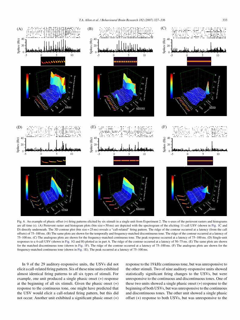
The majority of auditory-responsive units (20/29; 69%)esponded to both USVs with a call-related firing pattern ofither of two types. In 55% (11/20) of these units, the USVslicited a phasic onset (+) response to each call in the bout ofalls (Fig. 5A and D), as observed in Experiment 1 (compareith Fig. 4). In 45% (9/20) of these units, the USV elicitedphasic offset (+) response to each call in the bout of calls
Fig. 6A and D), a firing pattern that was not observed in Exper-
ment 1. This phasic offset (+) response to each call occurredn response to both of the USVs (Fig. 6A and D). The mostrominent difference in the firing patterns elicited by the twoSVs (Fig. 1C and G) was in the number of phasic (+) responses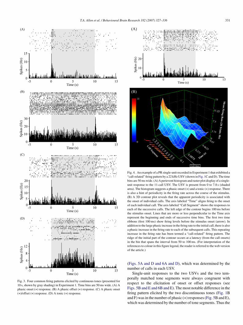
T.A. Allen et al. / Behavioural Brain Research 182 (2007) 327–336 331
Fig. 3. Four common firing patterns elicited by continuous tones (presented for10 s, shown by gray shading) in Experiment 1. Time bins are 50 ms wide. (A) Aphasic onset (+) response. (B) A phasic offset (+) response. (C) A phasic onset(+)/offset (+) response. (D) A tonic (+) response.
Fig. 4. An example of a PR single-unit recorded in Experiment 1 that exhibited a“call-related” firing pattern by a 22 kHz USV (shown in Fig. 1C and D). The timebins are 50 ms wide. (A) A perievent histogram and raster plot display of a single-unit response to the 11-call USV. The USV is present from 0 to 7.8 s (shadedarea). The histogram suggests a phasic onset (+) and a tonic (+) response. Thereis also a hint of periodicity in the firing rate across the course of the stimulus.(B) A 3D contour plot reveals that the apparent periodicity is associated withthe onset of individual calls. The axis labeled “Time” aligns firing to the onsetof each individual call. The axis labeled “Call Segment” shows the responses toeach of the successive calls. The left edge of the contour begins 100 ms beforethe stimulus onset. Lines that are more or less perpendicular to the Time axisrepresent the beginning and ends of successive time bins. The first two timeribbons (first 100 ms) show firing levels before the stimulus onset (arrow). Inaddition to the large phasic increase in the firing rate to the initial call, there is alsoa phasic increase in the firing rate to each of the subsequent calls. This repeatingincrease in the firing rate has been termed a “call-related” firing pattern. Theridge of the initial part of the contour occurs at a latency (from the call onsets)in the bin that spans the interval from 50 to 100 ms. (For interpretation of thero
(n
prFfiaw
eferences to colour in this figure legend, the reader is referred to the web versionf the article.)
Figs. 5A and D and 6A and D), which was determined by theumber of calls in each USV.
Single-unit responses to the two USVs and the two tem-orally matched tone segments were always congruent withespect to the elicitation of onset or offset responses (seeigs. 5B and E and 6B and E). The most notable difference in the
ring pattern elicited by the two discontinuous tones (Fig. 1Bnd F) was in the number of phasic (+) responses (Fig. 5B and E),hich was determined by the number of tone segments. Thus the
332 T.A. Allen et al. / Behavioural Brain Research 182 (2007) 327–336
Fig. 5. . An example of phasic onset (+) firing patterns elicited by six stimuli in a single unit from Experiment 2. The x-axes of the perievent rasters and histogramsare all time (s). (A) Perievent raster and histogram plots (bin size = 50 ms) are depicted with the spectrogram of the eliciting 11-call USV (shown in Fig. 1C and D)directly underneath. The 3D contour plot (bin size = 25 ms) reveals a “call-related” firing pattern. The ridge of the contour occurred at a latency (from the call onsets)of 25–50 ms. (B) The same plots are shown for the temporally and frequency-matched discontinuous tone (shown in Fig. 1B). The ridge of the contour occurred at alatency of 25–50 ms. (C) The analogous plots are shown for the frequency-matched continuous tone (shown in Fig. 1E). The peak occurred at a latency of 50–75 ms.The histogram suggests that the phasic onset (+) response might be followed by a tonic (−) response. However, the apparent tonic (−) component was not statisticallysignificant (p > 0.05). (D) Single-unit responses to a 4-call USV (shown in Fig. 1G and H) plotted as in part A. The ridge of the contour occurred at a latency of2 own ia n Fig
cit
bIc
coH
5–50 ms. (E) The same plots are shown for the matched discontinuous tone (shnalogous plots are shown for the frequency-matched continuous tone (shown i
all-related firing pattern elicited by the USVs was closely mim-cked by presentations of temporally and frequency-matchedone segments.
In general, single-unit responses to USVs were predictableased on responses to the frequency-matched continuous tones.n the majority of units that responded to the USVs with aall-related firing pattern (18/20; 90%), the frequency-matched
caup
n Fig. 1F). The ridge of the contour occurred at a latency of 25–50 ms. (F) The. 1E). The peak occurred at a latency of 25–50 ms.
ontinuous tones also elicited corresponding phasic onset orffset (+) responses (see Figs. 5C and F and 6C and F).owever, in 2 of the 20 units that responded to USVs with
all-related phasic (+) responses, the continuous tones producedtonic (+) response. In these two units, responses to contin-
ous tones clearly did not predict the USV call-related firingattern.
T.A. Allen et al. / Behavioural Brain Research 182 (2007) 327–336 333
Fig. 6. An example of phasic offset (+) firing patterns elicited by six stimuli in a single unit from Experiment 2. The x-axes of the perievent rasters and histogramsare all time (s). (A) Perievent raster and histogram plots (bin size = 50 ms) are depicted with the spectrogram of the eliciting 11-call USV (shown in Fig. 1C andD) directly underneath. The 3D contour plot (bin size = 25 ms) reveals a “call-related” firing pattern. The ridge of the contour occurred at a latency (from the calloffsets) of 75–100 ms. (B) The same plots are shown for the temporally and frequency-matched discontinuous tone. The ridge of the contour occurred at a latency of75–100 ms. (C) The analogous plots are shown for the frequency-matched continuous tone. The peak response occurred at a latency of 75–100 ms. (D) Single-unitresponses to a 4-call USV (shown in Fig. 1G and H) plotted as in part A. The ridge of the contour occurred at a latency of 50–75 ms. (E) The same plots are shownf ur ocf laten
eaeartn
rtsu
or the matched discontinuous tone (shown in Fig. 1F). The ridge of the contorequency-matched continuous tone (shown in Fig. 1E). The peak occurred at a
In 9 of the 29 auditory-responsive units, the USVs did notlicit a call-related firing pattern. Six of these nine units exhibitedlmost identical firing patterns to all six types of stimuli. Forxample, one unit produced a single phasic onset (+) response
t the beginning of all six stimuli. Given the phasic onset (+)esponse to the continuous tone, one might have predicted thathe USV would elicit a call-related firing pattern, but this didot occur. Another unit exhibited a significant phasic onset (+)tbao
curred at a latency of 75–100 ms. (F) The analogous plots are shown for thecy of 75–100 ms.
esponse to the 19 kHz continuous tone, but was unresponsive tohe other stimuli. Two of nine auditory-responsive units showedtatistically significant firing changes to the USVs, but werenresponsive to the continuous and discontinuous tones. One of
hese two units showed a single phasic onset (+) response to theeginning of both USVs, but was unresponsive to the continuousnd discontinuous tones. The other unit showed a single phasicffset (+) response to both USVs, but was unresponsive to the
3 rain
csm
4
pre2wevsmptsoft
olcctspiiFit
fr2cqmatsutvmroa
aiar
AtfiqoaUoapotbelat(t(
2PRePcHntsoU
(fcrmsbadpaic
bcccu
34 T.A. Allen et al. / Behavioural B
ontinuous and discontinuous tones. These two units suggest thatomething more than root frequency and temporal discontinuityay control the firing of some PR units.
. Discussion
Single-unit responses to auditory social signals have beenreviously studied in non-human primates [32,33], but not inodents, which offer certain experimental advantages. The twoxperiments described here examined single-unit responses to2 kHz USVs in rats. Experiment 1 was designed to explorehether, in experimentally naıve rats, PR neurons show prefer-
ntial responsiveness or distinguishable firing patterns to USVsersus other auditory stimuli that have been used as cues intudies of Pavlovian fear conditioning. This inquiry was partlyotivated by the theoretical possibility that auditory stimulus
rocessing in cortical circuits may have evolved to be selec-ively “tuned” to certain types of ethologically-important naturaltimuli. The results describe single-unit responses to three typesf stimuli that have been used as cues in studies of Pavlovianear conditioning: a 4 kHz continuous tone; a 22 kHz continuousone; and a 22 kHz USV.
A large proportion of PR units (59%) were responsive to oner more of the three auditory stimuli (see Table 1). The highevel of auditory responsiveness is consistent with the anatomi-al fact that rat PR receives a large afferent input from auditoryortex [11] as well as projections from auditory parts of thehalamus [16]. Unlike the continuous tones, the 22 kHz USVometimes elicited a clearly distinguishable “call-related” firingattern (Figs. 4–6). This pattern consisted of a transient increasen the firing frequency in response to each of the successive callsn a bout of calls; that is, a “phasic (+)” response to each call.rom a neurophysiological perspective, this repeating increase
n the firing frequency seemed potentially important for synapticransmission, neuronal plasticity, and stimulus coding.
Experiment 2 therefore focused on the stimulus requirementsor eliciting this repeating call-related firing pattern. Single-unitesponses were examined to six different stimuli: two different2 kHz USVs; two discontinuous tones that were matched to theorresponding USVs in terms of their on/off patterns, root fre-uency and total duration; and two continuous tones that wereatched to the corresponding USVs in terms of root frequency
nd total duration (Fig. 1). One USV stimulus was identical tohat used in Experiment 1 except that the loudness was increasedlightly to be more comparable to the continuous and discontin-ous tones. The second USV stimulus was specifically choseno differ from the first one in terms of root frequency (19 kHzersus 22 kHz), mean call duration (575 ms versus 1853 ms),ean inter-call interval (132 ms versus 213 ms), and mean call
epetition rate (1.50 Hz versus 0.55 Hz). There were also obvi-us differences between the two USVs in terms of frequencynd amplitude modulations (Fig. 1).
Overall, 44% of the units responded to one or more of the six
uditory stimuli. Both USVs elicited a call-related firing patternn 69% of the auditory-responsive units. The higher percent-ge of call-related firing patterns in the second experiment mayeflect the fact that the loudness of the USVs was increased.sd[c
Research 182 (2007) 327–336
lternatively, there may have been systematic differences inhe exact recording sites within PR. Most often, the call-relatedring pattern consisted of a transient increase in the firing fre-uency that was triggered by the onset of each call in the seriesf calls (Fig. 5A and D). This firing pattern was classified as“phasic onset (+)” response to each call. Less often, bothSVs elicited an increase in the firing frequency to the offsetf each call (Fig. 6A and D). This firing pattern was classifieds a “phasic offset (+)” response to each call. In general, theeak offset response occurred at a longer latency than the peaknset response (compare Figs. 5 and 6), when measured fromhe appropriate stimulus change. Thus the offset responses maye driven by more complex or possibly different circuitry. Inither case, the discontinuous tones always elicited a very simi-ar firing pattern (Figs. 5B and E and 6B and E). When the USVsnd discontinuous tones elicited onset responses, the continuousones also tended (in 10 of 11 cases) to elicit onset responsesFig. 5C and F). Similarly, when the USVs and discontinuousones elicited offset responses, the continuous tones also tendedin eight of nine cases) to elicit offset responses (Fig. 6C and F).
The results suggest that the call-related firing pattern to2 kHz USVs may emerge from a more basic tendency ofR neurons to respond to auditory stimulus onsets or offsets.esponsiveness to stimulus onsets or offsets appears to be a gen-ral characteristic of many auditory-responsive neurons [13,19].erhaps not surprisingly, therefore, the single-unit responses toontinuous tones were often predictive of responses to USVs.owever, there were clear cases in which tone-elicited firing wasot predictive of USV-elicited firing. For example, among unitshat responded to continuous tones with phasic (+) responses,ome responded to USVs with a call-related firing pattern andthers did not. Furthermore, two units responded to both of theSVs, but were unresponsive to the other four stimuli.The fact that the USVs and discontinuous tones usually
26/29; 89%) elicited similar firing patterns suggests that therequency modulations associated with individual calls do notritically determine the firing pattern or the overall level ofesponsiveness. Although USVs clearly do contain frequencyodulations (Fig. 1C and D), some investigators have empha-
ized the relative constancy of the principle frequency within aout of calls [6]. At the present, essentially nothing is knownbout which stimulus features are most important for rats iniscriminating among different USVs or for the recognition ofarticular USVs. Depending on the number of training trialsnd other acquisition conditions, the answer may turn out tonvolve different combinations of the root frequency, the meanall duration, and the mean call repetition rate [2,6].
The selection of PR as an initial region of interest was partlyased on the discovery that damage to this structure impairs fearonditioning to USVs [24,29] but has no significant effect ononditioning to continuous tones [10,24,29,34]. The fact thatortical processing is not necessary for conditioning to contin-ous tones is commonly interpreted to mean that activity in the
ubcortical projection from the auditory thalamus to the amyg-ala is sufficient to represent these relatively simple stimuli22,34,42,43]. Our working hypothesis has been that the dis-ontinuous nature of the USV is at least part of the reason why
rain
np
rdisbtdsoeua
tie2tcdoeo“cs2ttia
A
rw
R
[
[
[
[
[
[
[
[
[
[
[
[
[
[
[
[
[
[
[
[
[
[
[
T.A. Allen et al. / Behavioural B
ormal fear conditioning to this stimulus does require corticalrocessing.
Consistent with this hypothesis, we recently found that neu-otoxic PR lesions [29] also impair fear conditioning to aiscontinuous tone (the one shown in Fig. 1B). Cortical process-ng may be required to integrate these discontinuous auditorytimuli across time. Perhaps the auditory segments must beound together into a unitary representation, sometimes referredo as an “auditory object” [18,32], in order for normal fear con-itioning to occur. The obvious next step is to examine rat PRingle-unit responses before and after conditioning to USVs andther auditory stimuli. Ultimately, learning-related changes inlicited firing might be best understood by comparing single-nit responses in auditory cortex, perirhinal cortex, and themygdala.
One recent experiment by Endres and Fendt [17] suggestshat reactivity to 22 kHz USVs may depend on previous learn-ng. More specifically, this study found no differences amongxperimentally naıve rats in the levels of freezing elicited by2 kHz USVs and continuous tones. Of course, it is possiblehat differences would have been observed in the presence ofonspecifics or certain other critical stimuli [4]. Any innateefensive responses to 22 kHz USVs could theoretically dependn the simultaneous presence of multiple stimuli and certainnvironmental conditions [4]. Regardless of the interpretationf the preceding results, there may be some degree of biologicalpreparedness” [15,25,28,36] for forming or maintaining asso-iations with USVs. In support of the latter possibility, the sametudy by Endres and Fendt [17] found that fear conditioning to2 kHz USVs is more resistant to extinction than fear condi-ioning to continuous tones. It will be interesting and importanto understand what types of experiences are necessary for form-ng and maintaining ethologically appropriate responses to thesend other types of USVs.
cknowledgments
This work was supported by the National Institute of Healthesearch grant MH058405 and Yale University. We thank Ash-ini Tankhiwale for reading the manuscript.
eferences
[1] Anderson JW. The production of ultrasonic sounds by laboratory rats andother mammals. Science 1954;119:808–9.
[2] Bang S, Allen TA, Jones LK, Boguszewski P, Brown TH. Asymmetri-cal generalization toward social alarm calls in rats given differential fearconditioning. Society for Neuroscience Abstracts; 2006. Program Number67.15.
[3] Barfield RJ, Geyer LA. Sexual behavior: ultrasonic postejaculatory songof the male rat. Science 1972;176:1349–50.
[4] Blanchard RJ, Blanchard DC, Agullana R, Weiss SM. Twenty-two kHzalarm cries to presentation of a predator, by laboratory rats living in visibleburrow systems. Physiol Behav 1991;50:967–72.
[5] Brudzynski SM. Ultrasonic vocalization induced by intracerebral carba-chol in rats: localization and a dose-response study. Behav Brain Res1994;63:133–43.
[6] Brudzynski SM. Principles of rat communication: quantitative parametersof ultrasonic calls in rats. Behav Genet 2005;35(1):85–92.
[
Research 182 (2007) 327–336 335
[7] Brudzynski SM, Bihari F, Ociepa D, Fu XW. Analysis of 22 kHz ultra-sonic vocalization in laboratory rats: long and short calls. Physiol Behav1993;54:215–21.
[8] Brudzynski SM, Holland G. Acoustic characteristics of air puff-induced22-kHz alarm calls in direct recordings. Neurosci Biobehav Rev2005;29:1169–80.
[9] Brudzynski SM, Ociepa D. Ultrasonic vocalization of laboratory rats inresponse to handling and touch. Physiol Behav 1992;52:655–60.
10] Bucci DJ, Phillips RG, Burwell RD. Contributions of postrhinal andperirhinal cortex to contextual information processing. Behav Neurosci2000;114(5):882–94.
11] Burwell RD. The parahippocampal region: corticocortical connectivity.Ann NY Acad Sci 2000;911:25–42.
12] Burwell RD. Borders and cytoarchitecture of the perirhinal and postrhinalcortices in the rat. J Comp Neurol 2001;437:17–41.
13] Chimoto S, Kitama T, Qin L, Sakayori S, Sato Y. Tonal response patterns ofprimary auditory cortex neurons in alert cats. Brain Res 2002;934:34–42.
14] Choi J-S, Brown TH. Central amygdala lesions block ultrasonic vocaliza-tion and freezing as conditional but not unconditional responses. J Neurosci2003;23:8713–21.
15] Domjan M, Cusato B, Krause M. Learning with arbitrary versus ecologicalconditioned stimuli: evidence from sexual conditioning. Psychon Bull Rev2004;11(2):232–46.
16] Doron NN, LeDoux JE. Cells in the posterior thalamus project to bothamygdala and temporal cortex: a quantitative retrograde double-labelingstudy in the rat. J Comp Neurol 2000;425(2):257–74.
17] Endres T, Fendt M. Learning and extinction of natural fear-inducing stimuli.Society for Neurosciences Abstracts; 2006. Program Number 215.1.
18] Griffiths TD, Warren JD. What is an auditory object? Nat Rev Neurosci2004;5:887–92.
19] Heil P. Auditory cortical onset responses revisited. I. First-spike timing. JNeurophysiol 1997;77:2616–41.
20] Knutson B, Burgdorf J, Panksepp J. Ultrasonic vocalizations as indices ofaffective states in rats. Psychol Bull 2002;128(6):961–77.
21] Koo JW, Han JS, Kim JJ. Selective neurotoxic lesions of basolateral andcentral nuclei of the amygdala produce differential effects on fear condi-tioning. J Neurosci 2004;24:7654–62.
22] LeDoux. Emotion circuits in the brain. Annu Rev Neurosci2000;23:155–84.
23] Lee HJ, Choi J-S, Brown TH, Kim JJ. Amygdalar NMDA receptors arecritical for the expression of multiple conditioned fear responses. J Neurosci2001;21:4116–24.
24] Lindquist D, Jarrard LE, Brown TH. Perirhinal cortex supports delay fearconditioning to rat ultrasonic social signals. J Neurosci 2004;24:3610–7.
25] Marler P. Innateness and the instinct to learn. An Acad Bras Cienc2004;76:189–200.
26] Murray EA, Bussey TJ. Perceptual-mnemonic functions of the perirhinalcortex. Trends Cogn Sci 1999;3:142–51.
27] Murray EA, Graham KS, Gaffan D. Perirhinal cortex and its neighbors inthe medial temporal lobe: contributions to memory and perception. Q J ExpPsychol B 2005;58:378–96.
28] Ohman A, Mineka S. Fears, phobias, and preparedness: toward anevolved module of fear and fear learning. Psychol Rev 2001;108(3):483–522.
29] Padlubnaya DB, Allen TA, Brown TH. Perirhinal lesion effects on delayfear conditioning depend on stimulus features of the conditional stimulus.Society for Neuroscience Abstracts; 2006. Program Number 67.14.
30] Paxinos G, Watson C. The rat brain. 4th ed. San Diego, CA: AcademicPress; 1998.
31] Pitkanen A, Pikkarainen M, Nurminen N, Ylinen A. Reciprocal connectionsbetween the amygdala and the hippocampal formation, perirhinal cortex,and postrhinal cortex in rat. Ann NY Acad Sci 2000;911:369–91.
32] Rauschecker JP, Tian B. Mechanisms and steams for processing of
“what” and “where” in auditory cortex. Proc Natl Acad Sci USA2000;97(22):11800–6.33] Romanski LM, Averbeck BB, Diltz M. Neural representation of vocal-ization in the primate ventrolateral prefrontal cortex. J Neurophysiol2005;93:734–47.

3 rain
[
[
[
[[
[
[
[
[
36 T.A. Allen et al. / Behavioural B
34] Romanski LM, LeDoux JE. Equipotentiality of thalamo-amygdala andthalamo-cortico-amygdala circuits in auditory fear conditioning. J Neurosci1992;12:4501–9.
35] Sales GD. Ultrasound and aggressive behaviour in rats and other smallmammals. Anim Behav 1972;20:88–100.
36] Seligman MEP. On the generality of the laws of learning. Psychol Rev1970;77(5):406–18.
37] Sewell GD. Ultrasound in adult rodents. Science 1967;215:512.38] Sokal RR, Rohlf FJ. Biometry: the principles and practice of statistics in
biological research. New York: W.H. Freeman; 1995, 887 pp.39] Squire LR, Stark CE, Clark RE. The medial temporal lobe. Annu Rev
Neurosci 2004;27:279–306.
[
Research 182 (2007) 327–336
40] van der Poel AM, Miczek KA. Long ultrasonic calls in male rats followingmating, defeat and aversive stimulation: frequency modulation and boutstructure. Behavior 1991;19:127–42.
41] Witter MP, Naber PA, van Haeften T, Machielsen WC, Rombouts SA,Barkhof F, et al. Cortico-hippocampal communication by way of parallelparahippocampal-subicular pathways. Hippocampus 2000;10(4):398–410.
42] Yaniv D, Schafe GE, LeDoux JE, Richter-Levin G. A gradient of plasticity
in the amygdala revealed by cortical and subcortical stimulation, in vivo.Neuroscience 2001;106(3):613–20.43] Yaniv D, Desmedt A, Jaffard R, Richter-Levin G. The amygdala andappraisal processes: stimulus and response complexity as an organizingfactor. Brain Res Rev 2004;44(2–3):179–86.