Simultaneous Cell Lysis and DNA Extraction from Whole ...
28
1 Simultaneous Cell Lysis and DNA Extraction from Whole Blood using 1 Magnetic Ionic Liquids 2 Miranda N. Emaus and Jared L. Anderson* 3 Department of Chemistry, Iowa State University, Ames, IA 50011 USA 4 Abstract 5 Conventional DNA sample preparation methods involve tedious sample handling steps that 6 require numerous inhibitors of the polymerase chain reaction (PCR) and instrumentation to 7 implement. These disadvantages limit the applicability of conventional cell lysis and DNA 8 extraction methods in high throughput applications, particularly in forensics and clinical 9 laboratories. To overcome these drawbacks, a series of nine hydrophobic magnetic ionic liquids 10 (MILs) previously shown to preconcentrate DNA were explored as cell lysis reagents. The MILs 11 were found to lyse white blood cells from whole blood, 2-fold diluted blood, and dry blood samples 12 while simultaneously extracting human genomic DNA. The identity of metal ion incorporated 13 within the MIL appears to cause hemolysis while the cationic component further reduces the cell’s 14 integrity. Over 500 pg of human genomic DNA was isolated from 50 µL of whole blood using the 15 trioctylbenzylammonium tris(hexafluoroacetylaceto)nickelate(II) ([N8,8,8,Bz + ][Ni(hfacac)3 - ]) MIL 16 and 800 pg DNA was isolated from a dry blood samples using the trihexyl(tetradecyl)phosphonium 17 tris(phenyltrifluoroacetylaceto)nickelate(II) ([P6,6,6,14 + ][Ni(Phfacac)3 - ]) MIL following a 1 min 18 vortex step. A rapid, one-step cell lysis and DNA extraction method from blood is ideal for settings 19 that seek high-throughput analysis while minimizing the potential for contamination. 20 21 Keywords: Nucleic acid; Extraction; White blood cell analysis; Cell lysis; Dry blood analysis 22 23 ------------ 24 *Corresponding Author: 25 Jared L. Anderson 26 Department of Chemistry 27 Iowa State University 28 1605 Gilman Hall 29 Ames, IA 50011 USA 30
-
Upload
khangminh22 -
Category
Documents
-
view
4 -
download
0
Transcript of Simultaneous Cell Lysis and DNA Extraction from Whole ...

1
Simultaneous Cell Lysis and DNA Extraction from Whole Blood using 1
Magnetic Ionic Liquids 2
Miranda N. Emaus and Jared L. Anderson* 3
Department of Chemistry, Iowa State University, Ames, IA 50011 USA 4
Abstract 5
Conventional DNA sample preparation methods involve tedious sample handling steps that 6
require numerous inhibitors of the polymerase chain reaction (PCR) and instrumentation to 7
implement. These disadvantages limit the applicability of conventional cell lysis and DNA 8
extraction methods in high throughput applications, particularly in forensics and clinical 9
laboratories. To overcome these drawbacks, a series of nine hydrophobic magnetic ionic liquids 10
(MILs) previously shown to preconcentrate DNA were explored as cell lysis reagents. The MILs 11
were found to lyse white blood cells from whole blood, 2-fold diluted blood, and dry blood samples 12
while simultaneously extracting human genomic DNA. The identity of metal ion incorporated 13
within the MIL appears to cause hemolysis while the cationic component further reduces the cell’s 14
integrity. Over 500 pg of human genomic DNA was isolated from 50 µL of whole blood using the 15
trioctylbenzylammonium tris(hexafluoroacetylaceto)nickelate(II) ([N8,8,8,Bz+][Ni(hfacac)3
-]) MIL 16
and 800 pg DNA was isolated from a dry blood samples using the trihexyl(tetradecyl)phosphonium 17
tris(phenyltrifluoroacetylaceto)nickelate(II) ([P6,6,6,14+][Ni(Phfacac)3
-]) MIL following a 1 min 18
vortex step. A rapid, one-step cell lysis and DNA extraction method from blood is ideal for settings 19
that seek high-throughput analysis while minimizing the potential for contamination. 20
21
Keywords: Nucleic acid; Extraction; White blood cell analysis; Cell lysis; Dry blood analysis 22
23
------------ 24
*Corresponding Author: 25
Jared L. Anderson 26
Department of Chemistry 27
Iowa State University 28
1605 Gilman Hall 29
Ames, IA 50011 USA 30

2
1. Introduction 31
Genomic DNA analysis from blood samples is highly important in forensic and clinical 32
applications. Nucleic acid (NA) testing protocols for human immunodeficiency virus (HIV) and 33
hepatitis B virus (HBV) target human genomic DNA, as the virus integrates genomic information 34
into the host’s white blood cells (WBCs).[1–3] These NA tests are capable of detecting the virus 35
prior to antibody formation and are necessary to rapidly screen blood and organ donations. 36
However, according to the American Red Cross, conventional NA tests for viruses require greater 37
technical skill and expensive equipment compared to antibody testing. In addition, the isolation of 38
human genomic DNA is commonly used during investigations to determine the source of a 39
bloodstain. In both forensic and clinical applications, there is a great need to rapidly analyze DNA 40
from blood. Sample preparation is often considered an overlooked bottleneck in NA analysis as 41
poor cell lysis and DNA extraction often limits the sensitivity of bioassays. Therefore, the 42
development of highly efficient yet simple lysis and DNA extraction procedures are needed. 43
The separation of WBCs from red blood cells (RBCs) through mechanical or chemical 44
means (centrifugation or selective red blood cell lysis) is often the first step in sampling NAs from 45
whole blood due to the low abundance of WBCs.[4, 5] Despite improved sensitivity, the isolation 46
of WBCs is challenging and time-consuming. Therefore, interest exists in extracting NAs from 47
whole blood to enhance sample throughput. Conventional methods for chemical cell lysis involve 48
detergents, such as Triton X-100, to solubilize the cell membrane and release intracellular 49
components.[6] Despite being cheap and simple to implement, chemical lysis methods typically 50
require additional purification steps as the lysis reagent often inhibit downstream detection.[7, 8] 51
A recent advancement in the chemical lysis of cells involves the use of ionic liquids (ILs) to lyse 52
gram-positive and gram-negative cells, as well as viruses.[9–11] ILs are molten salts with melting 53
3
points below 100 °C that exhibit a number of unique physiochemical properties.[12, 13] Although 54
ILs are effective at lysing cells, ILs often inhibit downstream detection. This often necessitates the 55
dilution of their lysate and the amount of purified DNA prior to analysis by quantitative 56
polymerase chain reaction (qPCR). 57
Human genomic DNA extractions from blood generally involve alkaline extraction, 58
phenol-chloroform extraction, or spin column-based extraction.[6, 14] These methods utilize large 59
volumes of toxic organic solvents and require numerous time-consuming sample handling steps 60
which limit their applicability. In particular, phenol and chloroform are known carcinogens and 61
harmful towards the environment and their use should be minimized. Methods involving 62
simultaneous cell lysis and DNA extraction have reported a reduction in the number of sample 63
handling steps. Nanayakkara et al. investigated chitosan microparticles in the lysis of WBCs via 64
bead beating.[15] Bead beating is a common mechanical cell lysis method that imparts mechanical 65
shear to the cells.[6, 16] The chitosan-modified microparticles were simultaneously used to extract 66
genomic DNA through electrostatic interactions. To improve sample throughput, the modified 67
magnetic beads were added directly into the reaction buffer. However, the chitosan-microparticles 68
were observed to significantly inhibit qPCR, limiting the sensitivity of the extraction method.[17] 69
Magnetic ionic liquids (MILs) have been shown to efficiently extract DNA from a number 70
of complex matrices.[18–20] MILs are a subclass of ILs that contain a paramagnetic component 71
in either the cation or anion allowing the solvent to respond to an external magnet, while still 72
possessing similar physiochemical properties to ILs.[21–23] In addition, MILs can be designed to 73
be qPCR compatible, permitting DNA-enriched MILs to be integrated into custom-designed qPCR 74
buffers to efficiently desorb DNA during amplification.[24–26] Thermal desorption during PCR 75
has been shown to reduce the overall sample preparation time without impacting the efficiency of 76
4
the reaction. However, the performance of DNA extraction is often secondary to the lysis 77
efficiency, as poor lysis efficiencies limit the availability of DNA to extract. 78
In this study, a series of nine MILs were investigated as cell lysis agents. The hydrophobic 79
MIL was dispersed in whole blood facilitating the lysis of WBCs, while simultaneously extracting 80
human genomic DNA. The type of ligand, metal, and cation was found to play an important role 81
in the amount of DNA extracted as well as the lysis efficiency. MILs containing a Ni(II) metal 82
center and aromatic moieties in either the cationic component or ligand were found to exhibit 83
superior extraction of human genomic DNA. However, MILs containing the Dy(III) or Gd(III) 84
metal centers extracted significantly more DNA when the MIL is dispersed in blood compared to 85
Tris buffer, suggesting that they are efficient at lysing cells but not extracting DNA. It was found 86
that over 500 pg and 800 pg of genomic DNA was extracted from 50 µL whole blood and dried 87
bloodstains, respectively, using MILs after only a 1 min vortex step. In comparison, commercial 88
spin-column kits required a much longer extraction procedure of over 60 min. Integrating MILs 89
into the qPCR buffer for thermal desorption did not have a deleterious effect on the amplification 90
efficiency but greatly improved sample throughput. These results suggest that MILs are highly 91
effective at rapidly lysing white blood cells and simultaneously extracting DNA for downstream 92
analysis. 93
2. Methods and Materials 94
Ammonium hydroxide (28-30% solution in water), 1,1,1,5,5,5-hexafluoroacetylacetone 95
(99%), and 1-phenyl-4,4,4-trifluoro-1,3-butanedione (99%) was purchased from Alfa Aesar (Ward 96
Hill, MA, USA). Anhydrous diethyl ether (99.0%) was purchased from Avantor Performance 97
Materials Inc. (Center Valley, PA, USA). The 1-methyl-3-octylimidazolium bromide (99%) 98
5
([OMIM+][Br-]) IL was purchased from IoLITec (Tuscaloosa, AL, USA). Gadolinium(III) 99
chloride hexahydrate (99.9%) and 1-tetradecanol (97%) was purchased from Beantown Chemicals 100
(Hudson, NH, USA). Trihexyl(tetradecyl)phosphonium ([P6,6,6,14+]) chloride (97.7%) and 101
dysprosium(III) chloride hexahydrate (99.9%) were purchased from Strem Chemicals 102
(Newburyport, MA, USA). Ethylenediaminetetraacetic acid (EDTA) (99.4-100.06%), magnesium 103
chloride hexahydrate (99.0-102.0%), lithium bis[(trifluoromethyl)sulfonyl]imide ([Li+][NTf2-]), 104
cobalt chloride (97%), benzenesulfonyl chloride (99%), bovine serum albumin (BSA) (≥96%), 105
deoxyribonucleic acid sodium salt from salmon testes (20, 000 bp), Wright stain solution, LC-MS 106
grade acetonitrile (ACN) (≥99.9%), 1-methylimidzaole (99%), and LC-MS grade methanol 107
(≥99.8%) were purchased from MilliporeSigma (St. Louis, MO, USA). SYBR Green I (10,000x) 108
was purchased from Life Technologies (Carlsbad, CA, USA). Proteinase K was purchased from 109
New England Biolabs (Ipswich, MA, USA). Agarose and Tris(hydroxymethyl)aminomethane 110
(Tris) hydrochloride (HCl) were purchased from P212121 (Ypsilanti, MI, USA). SsoAdvanced 111
Universal SYBR Green Supermix (2x) was purchased from Bio-Rad Laboratories (Hercules, CA, 112
USA). Primers (see Table S1) were purchased from Integrated DNA Technologies (Coralville, IA, 113
USA). Modified plasmids (3.9 Kbp) containing a 210 bp insert (see Table S1) were obtained from 114
Eurofin Genomics (Louisville, KY, USA). PCR caps, tube strips, potassium chloride (99.7%), 115
sodium phosphate dibasic anhydrous (99.8%), potassium phosphate monobasic (100.0%), 116
dimethylsulfoxide (DMSO) (>99.7%), nickel chloride (98%), pyridine (99.9%), 1,1,1-trifluoro-117
2,4-pentadione (98%), sodium chloride, fresh human whole blood, frosted glass slides, and P5 118
grade filter paper were purchased from Fisher Scientific (Waltham, MA, USA). Neodymium 119
magnets (0.2 T) were purchased from K&J Magnetics (Pipersville, PA, USA). Deionized water 120

6
(18.2 MΩ cm), obtained from a Milli-Q water purification system, was used to prepare all aqueous 121
solutions (Millipore, Bedford, MA, USA). 122
2.1. Magnetic Ionic Liquid and Ionic Liquid Synthesis 123
The structures of the MILs used in this study are shown in Figure 1. The [P6,6,6,14+] 124
tris(hexafluoroacetylaceto)nickelate(II) ([Ni(hfacac)3-]), [P6,6,6,14
+] 125
tris(hexafluoroacetylaceto)colbaltate(II) ([Co(hfacac)3-]), [P6,6,6,14
+] 126
tetrakis(hexafluoroacetylaceto)dysprosate(III) ([Dy(hfacac)4-]), [P6,6,6,14
+] 127
tetrakis(hexafluoroacetylaceto)gadolinate(III) ([Gd(hfacac)4-]), [P6,6,6,14
+] 128
tris(phenyltrifluoroacetylaceto)nickelate(II) ([Ni(Phtfacac)3-]), [P6,6,6,14
+] tris(1,1,1-129
trifluoroacetylacetylaceto)nickelate(II) ([Ni(tfacac)3-]), and trioctylbenzylammonium ([N8,8,8,Bz
+]) 130
[Ni(hfacac)3-] MILs were synthesized and characterized as previously reported.[25, 27, 28] The 131
[P6,6,6,14+][NTf2
-], [N8,8,8,Bz+][NTf2
-], 1-tetradecyl-3-methylimidazolium ([C14MIM+]) 132
benzylsulfonate ([BS-]), and ammonium ([NH4+]) [Ni(hfacac)3
-] salts were synthesized according 133
to previously published procedures.[25, 27, 28] The [OMIM+][Ni(hfacac)3-] MIL was synthesized 134
by mixing equimolar amounts of [NH4+][Ni(hfacac)3
-] and [OMIM+][Br-] overnight in 50 mL of 135
methanol. The [C14MIM+][Ni(hfacac)3-] MIL was synthesized by mixing equimolar amounts of 136
[NH4+][Ni(hfacac)3
-] and [C14MIM+][BS-] overnight in 50 mL of methanol. The products were 137
subsequently dried in a vacuum oven and purified using diethyl ether and water. 138
2.2. PCR assays and Conditions 139
A Bio-Rad CFX96 Touch Real-time PCR (Hercules, CA, USA) was utilized for qPCR 140
amplification of the human genomic DNA and 98 base-pair (bp) sequence (see Table S1) using 141
the following program: 2 min initial denaturation at 95 °C followed by 40 cycles comprised of a 5 142
7
s denaturation step at 95 °C and a 30 s annealing step. An optical detection step was performed 143
after the annealing step to track the progress of the reaction in real-time. 144
The β-actin gene in human genomic DNA was amplified in the absence of MIL in reaction 145
buffer using 1x SsoAdvanced Supermix, 5% DMSO, and 1 µM primers. The addition of 0.3 µL 146
of [P6,6,6,14+][Ni(hfacac)3
-], [P6,6,6,14+][Ni(Phtfacac)3
-], [N8,8,8,Bz+][Ni(hfacac)3
-], or 147
[OMIM+][Ni(hfacac)3-] MILs to the reaction buffer required 1x SsoAdvanced Supermix, 5% 148
DMSO, 1 µM primers, and an additional 1x SYBR Green I to achieve uninhibited qPCR 149
amplification. Quantitative PCR with 0.3 µL of [C14MIM+][Ni(hfacac)3-] MIL in the reaction 150
buffer required 1x SsoAdvanced Supermix, 5% DMSO, 1 µM primers, additional 1.25 mM MgCl2, 151
and an additional 1x SYBR Green I. The addition of 0.3 µL of [P6,6,6,14+][Ni(tfacac)3
-] MIL required 152
to the qPCR buffer 1x SsoAdvanced Supermix, 5% DMSO, and 1 µM primers for amplification. 153
The addition of 0.3 µL of [P6,6,6,14+][Co(hfacac)3
-] MIL in the reaction buffer required 1x 154
SsoAdvanced Supermix, 5% DMSO, 1 µM primers, and an additional 2x SYBR Green I. The 155
addition of 0.3 µL of [P6,6,6,14+][Dy(hfacac)4
-] MIL to the reaction buffer required 1x SsoAdvanced 156
Supermix, 5% DMSO, 1 µM primers, 6 mM EDTA, 7.5 mM MgCl2, 0.5 mg·mL-1 BSA, and an 157
additional 1x SYBR Green I. The addition of 0.3 µL of [P6,6,6,14+][Gd(hfacac)4
-] MIL to the qPCR 158
buffer required 1x SsoAdvanced Supermix, 5% DMSO, 1 µM primers, 6 mM EDTA, 6.5 mM 159
MgCl2, 1.5 mg·mL-1 BSA, and an additional 1x SYBR Green I. 160
Amplification of a 98 bp DNA sequence was achieved using 1x SsoAdvanced Supermix 161
and 1 µM primers. The addition of 0.3 µL of [P6,6,6,14+][Ni(hfacac)3
-], [P6,6,6,14+][Ni(Phtfacac)3
-], 162
[N8,8,8,Bz+][Ni(hfacac)3
-], [P6,6,6,14+][Co(hfacac)3
-], and [OMIM+][Ni(hfacac)3-] MILs to the reaction 163
buffer required 1x SsoAdvanced Supermix, 1 µM primers, and an additional 1x SYBR Green I to 164
achieve uninhibited qPCR amplification. Amplification with 0.3 µL of [C14MIM+][Ni(hfacac)3-] 165

8
MIL in the qPCR buffer required 1x SsoAdvanced Supermix, 1 µM primers, additional 1.25 mM 166
MgCl2, and an additional 1x SYBR Green I. Amplification with 0.3 µL of [P6,6,6,14+][Ni(tfacac)3
-] 167
MIL in the reaction buffer required 1x SsoAdvanced Supermix and 1 µM primers for 168
amplification. The addition of 0.3 µL of [P6,6,6,14+][Dy(hfacac)4
-] MIL in the reaction buffer 169
required 1x SsoAdvanced Supermix, 1 µM primers, 2 mM EDTA, 7.5 mM MgCl2, 1.5 mg·mL-1 170
BSA, and additional 1x SYBR Green I. The addition of 0.3 µL of [P6,6,6,14+][Gd(hfacac)4
-] MIL to 171
the reaction buffer required 1x SsoAdvanced Supermix, 5% DMSO, 1 µM primers, 2 mM EDTA, 172
6.5 mM MgCl2, 1.5 mg·mL-1 BSA, and additional 1x SYBR Green I. 173
2.3. Lysis and Extraction Conditions 174
DNA extractions were performed from a 50 µL sample of either 50 pg·µL-1 human 175
genomic DNA or 5 fg·µL-1 98 bp DNA. A 2 µL volume of MIL was dispersed for 1 min and 176
collected on a rod magnet (B = 0.2 T). Recovered MIL was washed with deionized water and a 177
0.3 µL aliquot of DNA-enriched MIL was added to the qPCR buffer. All extractions were 178
performed in triplicate. 179
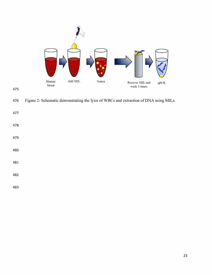
The general procedure used to simultaneously lyse and capture DNA from WBCs is shown 180
in Figure 2. An optimized volume of MIL was added to a 50 µL blood sample and dispersed using 181
a Barnstead/Thermolyne Type 16700 mixer (Dubuque, IA, USA) for an optimized length of time. 182
The MIL was collected using a rod magnet (B = 0.2 T) and washed three times with deionized 183
water. A 0.3 µL aliquot of DNA-enriched MIL was added to a qPCR tube for downstream 184
amplification and detection. All extractions were performed in triplicate. 185
Dry bloodstains were prepared by aliquoting 50 µL of the whole blood on P5 filter paper 186
(Fisher Scientific, Waltham, MA, USA). The blood was dried in a desiccator for 24 h. The filter 187
9
paper was subsequently placed in 100 µL of phosphate buffer saline (137 mM NaCl, 2.7 mM KCl, 188
10 mM Na2HPO4, and KH2PO4, pH = 7.4) (PBS) for 5 min to desorb cells. The optimized volume 189
of MIL was then dispersed for a specific amount of time to lyse WBCs and extract DNA. All 190
extractions from dried blood samples were performed in triplicate. 191
WBC lysis and DNA extractions using the QiaAMP DNA mini kit were performed as 192
specified by the manufacturer. Briefly, 50 µL of blood was diluted in PBS to 200 µL, and 16 units 193
of proteinase K were added to the diluted blood. A 1 mL volume of lysis buffer (AL buffer) was 194
added to the sample to lyse the cells for 10 min at 56 °C. After this, 1 mL of ethanol was added to 195
the sample and mixed. The lysate was added to a silica column and centrifuged for 1 min at 1.3 × 196
104 rpm. The flow-through was discarded. Next, 0.5 mL of wash buffer 1 (AW1 buffer) was added 197
to the column and centrifuged again for 1 min. The flow-through was discarded, and 0.5 mL of 198
wash buffer 2 (AW2 buffer) was placed in the column. The column was then centrifuged for 3 min 199
and the flow-through discarded. An additional 1 min centrifugation step was performed to ensure 200
that the wash buffers were thoroughly removed. Lastly, 200 µL of elution buffer (AE buffer) was 201
added to elute the purified DNA from the column. 202
2.4. Wright Staining Procedure 203
Wrights stains were developed based on the procedure described in Strober et al.[29] 204
Briefly, 3 µL of blood was placed on a clean microscope slide. The blood was spread across the 205
slide using a second slide. Once dry, 1 mL of methanol was placed on the dry blood to increase 206
the cell’s affinity for the stain. The slide was allowed to dry before 1 mL of Wright’s stain was 207
placed on the slide for 2 min. Then, 2 mL of PBS was placed on the slide for 4 min. Slides were 208
then rinsed with water to remove excess solution and allowed to air dry. Cells were visualized 209
under a Micromaster microscope (Fisher Scientific, Waltham, MA, USA). 210

10
3. Results and Discussion 211
Integrating DNA-enriched MILs into a qPCR assay allows DNA to desorb from the solvent 212
using the elevated temperatures required for PCR without impacting the amplification 213
efficiency.[25, 26] However, the elevated temperatures required for PCR may increase the 214
solubility of the MIL, potentially inhibiting the reaction. PCR inhibition caused by MILs can be 215
overcome by optimizing the amount of EDTA, SYBR Green I, BSA, and MgCl2. The custom-216
designed qPCR buffers for each MIL are summarized in Table S2. As shown in Figure S1, standard 217
curves were constructed by spiking human genomic DNA into the custom-designed qPCR buffers 218
and used to quantify the amount of DNA extracted by the MIL. Quantification of human genomic 219
DNA in the absence of MIL was carried out using the standard curve in Figure S2a. The standard 220
curve in Figure S2b was used to quantify the 98 bp DNA fragment extracted by the MILs. 221
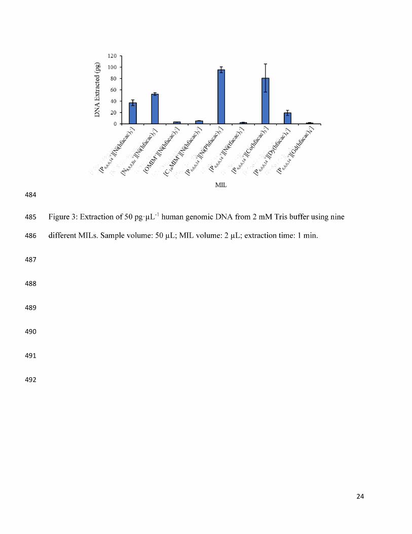
3.1. Extraction of Human Genomic DNA 222
To evaluate the ability of hydrophobic MILs to extract human genomic DNA, extractions 223
were initially performed from 2 mM Tris buffer. As shown in Figure 3, the nine MILs examined 224
all extracted human genomic DNA with the Ni(II)-based MILs containing an aromatic moiety 225
within either the cation or ligand structure generally extracted the most DNA. 226
The ability of the MIL to extract DNA from blood was studied by spiking a non-targeted 227
DNA sequence into the blood. As shown in Figure S3a, the spiked DNA could be recovered from 228
blood using all nine MILs, although several exhibited very low extraction efficiencies. However, 229
the blood matrix imparted a significant decrease in the amount of DNA extracted compared to 230
extractions from Tris buffer (Figure S3b). The [N8,8,8,Bz+][Ni(hfacac)3
-] and 231
[P6,6,6,14+][Ni(Phtfacac)3
-] MILs were found to extract the most DNA from the blood matrix. 232

11
3.2. Optimizing the Lysis and Extraction of Human Genomic DNA from WBCs 233
The volume of MIL and extraction time were optimized from 2-9 µL and 15-120 s, 234
respectively, to ensure the highest amount of DNA was extracted from 2-fold diluted blood. As 235
shown in Figure S4, 2 µL of MIL was optimum for the [P6,6,6,14+][Co(hfacac)3
-], 236
[OMIM+][Ni(hfacac)3-], [P6,6,6,14
+][Dy(hfacac)4-], and [P6,6,6,14
+][Gd(hfacac)4-] MILs. It was found 237
that a 3 µL volume of the [C14MIM+][Ni(hfacac)3-] MIL and 5 µL of the [N8,8,8,Bz
+][Ni(hfacac)3-] 238
MIL was optimum. For the [P6,6,6,14+][Ni(Phtfacac)3
-] MIL, 6 µL was optimum while 7 µL of the 239
[P6,6,6,14+][Ni(hfacac)3
-] and [P6,6,6,14+][Ni(tfacac)3
-] MILs was optimum. Extractions with higher 240
volumes of the [P6,6,6,14+][Dy(hfacac)4
-] and [P6,6,6,14+][Gd(hfacac)4
-] MILs were unsuccessful, and 241
the MIL could not be recovered. An optimized vortex time of only 30 s was required for the 242
[P6,6,6,14+][Gd(hfacac)4
-] MIL. The highest amount of DNA detected was achieved after a 1 min 243
vortex with the [P6,6,6,14+][Ni(hfacac)3
-], [P6,6,6,14+][Ni(Phtfacac)3
-], [N8,8,8,Bz+][Ni(hfacac)3
-], 244
[P6,6,6,14+][Co(hfacac)3
-], [OMIM+][Ni(hfacac)3-], [C14MIM+][Ni(hfacac)3
-], and 245
[P6,6,6,14+][Ni(tfacac)3
-] MILs, as shown in Figure S5. A vortex time of 90 s was optimum for the 246
[P6,6,6,14+][Dy(hfacac)4
-] MIL. 247
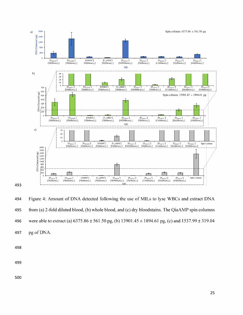
Simultaneous lysis of WBCs and extraction of human genomic DNA was evaluated using 248
the optimized extraction procedures for each MIL from 2-fold diluted blood, whole blood, and dry 249
bloodstains. As shown in Figure 4, the [N8,8,8,Bz+][Ni(hfacac)3
-] MIL was most successful at lysing 250
and extracting DNA from 2-fold diluted and whole blood. However, the [P6,6,6,14+][Ni(Phtfacac)3
-251
] MIL extracted more DNA from dry blood samples. It was found that commercial spin columns 252
extracted more DNA from 2-fold diluted blood, whole blood, and dry bloodstains compared to the 253
MILs. Interestingly, the Gd(III) and Dy(III) MILs recovered significantly more DNA from blood 254
compared to Tris buffer. It is possible that the Gd(III) and Dy(III) MILs were more successful at 255

12
lysing WBCs compared to the Ni(II) MIL, but their poor DNA extraction efficiencies hindered 256
their overall performance. Although the spin columns were able to extract more DNA, the MIL-257
based method required only 1 min whereas the kits require over 60 min as well as additional 258
instrumentation (centrifuge and water bath) and multiple reagents, making the extraction more 259
cumbersome.[30] 260
3.3. Evaluating of the Co-extraction of PCR Inhibitors 261
To evaluate the co-extraction of PCR inhibitors by the MIL from undiluted blood, serial 262
dilutions of a 98 bp DNA sequence not found in blood were spiked into the qPCR buffer to generate 263
standard curves. As shown in Figure 5, the amplification efficiencies associated with the 264
[P6,6,6,14+][Ni(hfacac)3
-], [P6,6,6,14+][Ni(Phtfacac)3
-], [P6,6,6,14+][Co(hfacac)3
-], 265
[OMIM+][Ni(hfacac)3-], [C14MIM+][Ni(hfacac)3
-], [P6,6,6,14+][Dy(hfacac)4
-], and 266
[P6,6,6,14+][Gd(hfacac)4
-] MILs were between 90-110%. This suggests that DNA is being duplicated 267
with each cycle and that limited amounts of qPCR inhibitors are being extracted by the MILs. 268
However, amplification efficiencies associated with dispersing the [N8,8,8,Bz+][Ni(hfacac)3
-] and 269
[P6,6,6,14+][Ni(tfacac)3
-] MILs in whole blood were above 110%, suggesting the co-extraction of 270
PCR inhibitors by these MILs. Interestingly, decreasing the MIL volume dispersed in blood from 271
the optimized volume (5 and 7 µL for the [N8,8,8,Bz+][Ni(hfacac)3
-] and [P6,6,6,14+][Ni(tfacac)3
-] 272
MILs, respectively) to 2 µL caused the amplification efficiencies to drop to 96.48% and 103.66% 273
for the [N8,8,8,Bz+][Ni(hfacac)3
-] and [P6,6,6,14+][Ni(tfacac)3
-] MILs, respectively, while developing 274
standard curves with the 98 bp sequence (see Figure S6). This suggests that PCR inhibitors are co-275
extracted more readily with larger volumes of MIL, likely due to the increased surface area 276
facilitating mass transfer with PCR inhibitors.[13] 277
13
The amount of DNases co-extracted by the MIL from whole blood was evaluated by 278
allowing the DNA-enriched MILs to incubate for up to 48 h at 25 °C prior to qPCR amplification. 279
All nine MILs were found to preserve DNA extracted from Tris buffer for 48 h (see Figure S7a). 280
This suggests that the hexafluoroacetylacetonate-based MILs do not degrade DNA over time. As 281
shown in Figure S7b, there was no significant change in the amount of DNA detected using the 282
[P6,6,6,14+][Ni(Phtfacac)3
-] and [N8,8,8,Bz+][Ni(hfacac)3
-] MILs after 48 h. However, a significant 283
drop in the amount of DNA was observed using the Student t-test (p < 0.05) after 48 h with the 284
[P6,6,6,14+][Gd(hfacac)4
-] MIL and 24 h with the [P6,6,6,14+][Ni(hfacac)3
-], [P6,6,6,14+][Ni(hfacac)3
-], 285
[C14MIM+][Ni(hfacac)3-], and [P6,6,6,14
+][Dy(hfacac)4-] MILs. The [OMIM+][Ni(hfacac)3
-] and 286
[P6,6,6,14+][Ni(tfacac)3
-] MILs were found to preserve DNA for only 6 h. The drop in the amount 287
of DNA detected suggests nucleases are being extracted by the [P6,6,6,14+][Ni(hfacac)3
-], 288
[P6,6,6,14+][Ni(hfacac)3
-], [C14MIM+][Ni(hfacac)3-], [P6,6,6,14
+][Gd(hfacac)4-], 289
[P6,6,6,14+][Dy(hfacac)4
-], [OMIM+][Ni(hfacac)3-], and [P6,6,6,14
+][Ni(tfacac)3-] MILs. However, 290
DNA was stable within the MIL for several hours after the extraction, possibly due to the 291
hydrophobic microenvironment of the MIL limiting the activity of DNase.[31] 292
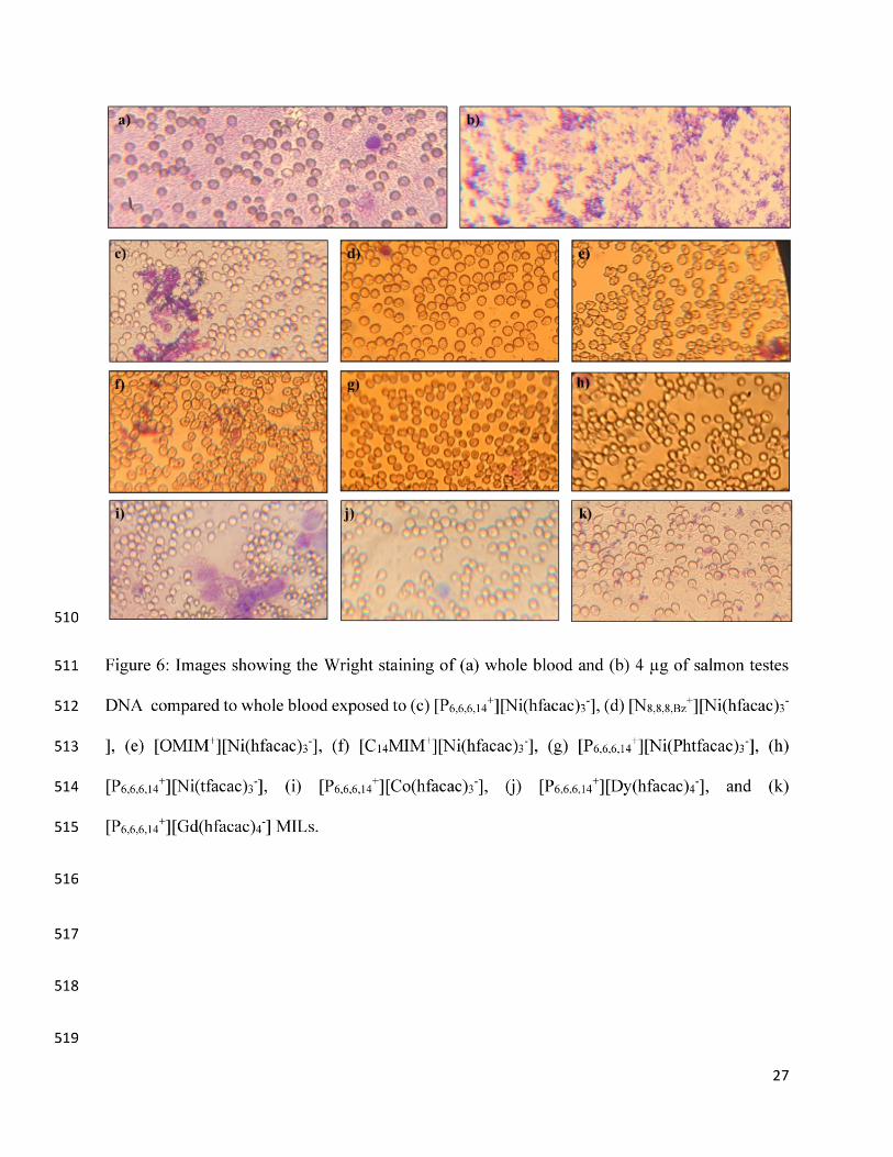
3.4. Insight into the Mechanism of Blood Cell Lysis by MILs 293
To investigate cell integrity after dispersing MILs in blood, blood smears were developed 294
using Wright’s stain. Wright’s stain is comprised of two colorimetric stains: (1) eosin, which stains 295
proteins in the cytosol red, and (2) methylene blue, which stains NAs in WBCs blue.[29] As shown 296
in Figure 6, exposing blood cells to MILs (Figures 6c-k) caused a significant reduction in the 297
central pallor of the RBCs compared to whole blood (Figure 6a). This suggests that proteins, such 298
as hemoglobin, are not present in the cytoplasm due to the lysis of cells. SpursSmall spikes 299
(sometimes referred to as spurs) were also noted on RBCs exposed to the [N8,8,8,Bz+][Ni(hfacac)3
-300
14
], [OMIM+][Ni(hfacac)3-], and [C14MIM+][Ni(hfacac)3
-] MILs. Spurs cells are a morphological 301
abnormality of RBCs associated with several diseases, such as carcinoma and liver disease.[32, 302
33] They form due to disturbances in the lipid composition of the cell membrane.[32, 34, 35] RBCs 303
exposed to the [P6,6,6,14+][Gd(hfacac)4
-] MILs no longer exhibited a circular shape and appeared 304
deformed. Few intact WBCs and free DNA was observed in the slides. 305
The effect that the MIL cation, ligand, and metal ion has on the lysis of WBCs was 306
evaluated by spiking each component into 50 µL of blood. As shown in Figure S8, the eosin stain 307
was not retained in RBCs exposed to nickel(II) chloride, cobalt(II) chloride, dysprosium(III) 308
chloride, or gadolinium(III) chloride. This may be linked to hemolysis generated through oxidative 309
stress by the metal ion.[36] As shown in Figure S9, the ligand may also play a role in cell lysis. 310
No cells were observed after exposing the blood to hexafluoroacetylacetone, the chemical 311
precursor used to form the metal hexafluoroacetylateonate-based MILs. However, non-viable 312
RBCs were noted with phenyltrifluoroacetone or 1,1,1-trifluoroacetylacetone; no intact WBCs 313
were noted. As shown in Figure S10, the cation appears to also affect cell viability. The 314
[P6,6,6,14+][NTf2
-] IL appears to completely lyse WBC and RBCs. RBCs exposed to the 315
[N8,8,8,Bz+][NTf2
-] IL were noted to have spurs. Previously, it was reported that a lipophilic cationic 316
component imparts surfactant-like properties to the MIL.[25] RBCs form spurs in the presence of 317
surfactants due to the intercalation of the surfactant molecules into the cell membrane.[37, 38] The 318
interaction of the surfactant then leads to solubilization of the cell membrane and consequently 319
cell lysis. 320
4. Conclusions 321

15
The simultaneous lysing of WBCs and extraction of DNA shows great potential for 322
consolidating sample preparation and achieving high throughput analysis. A short vortex step of 1 323
min was required to extract over 500 pg and 800 pg of human genomic DNA from 50 µL whole 324
blood and dry blood, respectively, whereas commercial methods require an hour to lyse, extract, 325
and recover NAs. From lysis to detection, MIL-based WBC lysis and genomic DNA extraction 326
was substantially faster than the spin column method. All nine MILs could be integrated into the 327
qPCR buffer without inhibiting the reaction, allowing for a chemical lysis method that did not 328
inhibit downstream detection. Thermal desorption during qPCR greatly reduced the sample 329
preparation time needed for isolating human genomic DNA. The qPCR efficiency was not affected 330
by dispersing small volumes of MIL in whole blood suggesting that the MILs co-extracted minimal 331
amounts of qPCR inhibitors. Wright staining revealed a noticeable lack of WBCs, with the 332
remaining RBCs appearing to have been lysed. The MIL cation, chelated metal ion, and ligand 333
play a significant role in the ability of the MIL to lyse cells and extract NAs. MILs containing an 334
aromatic component within the chemical structure were found to extract more DNA. The Gd(III) 335
and Dy(III) MILs appeared to be more efficient at lysing cells. The utilization of MILs to 336
chemically lyse cells and extract DNA can be highly advantageous in nucleic acid analysis since 337
the method has the potential to be fully automated. Finally, the simple 1 min sample preparation 338
step is ideal for high throughput analysis. 339
5. Acknowledgements 340
M. N. E. would like to thank Paul M. Emaus for his support. J. L. A acknowledges funding 341
from the Chemical Measurement and Imaging Program at the National Science Foundation (CHE-342
1709372). 343
16
Compliance with ethical standards. This article contains studies involving human blood samples 344
that were purchased commercially. All studies and procedures have been approved by the 345
Institutional Biosafety Committee (IBC) at Iowa State University. 346
Conflicts of interest. The authors declare no conflicts of interest. 347
348
Bibliography 349
1. Stramer SL, Wend U, Candotti D, Foster GA, Hollinger FB, Dodd RY, Allain J-P, Gerlich 350
W (2011) Nucleic acid testing to detect HBV infection in blood donors. N Engl J Med 351
364:236–247 . https://doi.org/10.1056/NEJMoa1007644 352
2. Gonçalves J, Moreira E, Sequeira IJ, Rodrigues AS, Rueff J, Brás A (2016) Integration of 353
HIV in the Human Genome: Which Sites Are Preferential? A Genetic and Statistical 354
Assessment. Int J Genomics 2016:8–13 . https://doi.org/10.1155/2016/2168590 355
3. Choi J, Hyun JC, Yang S (2015) On-chip Extraction of Intracellular Molecules in White 356
Blood Cells from Whole Blood. Sci Rep 5:1–12 . https://doi.org/10.1038/srep15167 357
4. Albariño CG, Romanowski V (1994) Phenol extraction revisited: A rapid method for the 358
isolation and preservation of human genomic DNA from whole blood. Mol. Cell. Probes 359
8:423–427 360
5. Chen X, Cui D, Liu C, Li H, Chen J (2007) Continuous flow microfluidic device for cell 361
separation, cell lysis and DNA purification. Anal Chim Acta 584:237–243 . 362
https://doi.org/10.1016/j.aca.2006.11.057 363
6. Ali N, Rampazzo RDCP, Costa ADiT, Krieger MA (2017) Current Nucleic Acid 364

17
Extraction Methods and Their Implications to Point-of-Care Diagnostics. Biomed Res Int 365
2017: . https://doi.org/10.1155/2017/9306564 366
7. Nan L, Jiang Z, Wei X (2014) Emerging microfluidic devices for cell lysis: A review. Lab 367
Chip 14:1060–1073 . https://doi.org/10.1039/c3lc51133b 368
8. Schrader C, Schielke A, Ellerbroek L, Johne R (2012) PCR inhibitors - occurrence, 369
properties and removal. J Appl Microbiol 113:1014–1026 . https://doi.org/10.1111/j.1365-370
2672.2012.05384.x 371
9. Fuchs-Telka S, Fister S, Mester PJ, Wagner M, Rossmanith P (2017) Hydrophobic ionic 372
liquids for quantitative bacterial cell lysis with subsequent DNA quantification. Anal 373
Bioanal Chem 409:1503–1511 . https://doi.org/10.1007/s00216-016-0112-x 374
10. Fister S, Fuchs S, Mester P, Kilpeläinen I, Wagner M, Rossmanith P (2015) The use of 375
ionic liquids for cracking viruses for isolation of nucleic acids. Sep Purif Technol 155:38–376
44 . https://doi.org/10.1016/j.seppur.2015.03.035 377
11. Martzy R, Bica-Schröder K, Pálvölgyi ÁM, Kolm C, Jakwerth S, Kirschner AKT, 378
Sommer R, Krska R, Mach RL, Farnleitner AH, Reischer GH (2019) Simple lysis of 379
bacterial cells for DNA-based diagnostics using hydrophilic ionic liquids. Sci Rep 9:1–10 380
. https://doi.org/10.1038/s41598-019-50246-5 381
12. Trujillo-Rodriguez MJ, Nan H, Varona M, Emaus MN, Souza ID, Anderson JL (2019) 382
Advances of Ionic Liquids in Analytical Chemistry. Anal Chem 91:505–531 . 383
https://doi.org/10.1021/acs.analchem.8b04710 384
13. Trujillo-Rodríguez MJ, Rocío-Bautista P, Pino V, Afonso AM (2013) Ionic liquids in 385

18
dispersive liquid-liquid microextraction. TrAC - Trends Anal Chem 51:87–106 . 386
https://doi.org/10.1016/j.trac.2013.06.008 387
14. Koshy L, Anju AL, Harikrishnan S, Kutty VR, Jissa VT, Kurikesu I, Jayachandran P, 388
Jayakumaran Nair A, Gangaprasad A, Nair GM, Sudhakaran PR (2017) Evaluating 389
genomic DNA extraction methods from human whole blood using endpoint and real-time 390
PCR assays. Mol Biol Rep 44:97–108 . https://doi.org/10.1007/s11033-016-4085-9 391
15. Nanayakkara IA, Cao W, White IM (2017) Simplifying Nucleic Acid Amplification from 392
Whole Blood with Direct Polymerase Chain Reaction on Chitosan Microparticles. Anal 393
Chem 89:3773–3779 . https://doi.org/10.1021/acs.analchem.7b00274 394
16. Berasaluce A, Matthys L, Mujika J, Antoñana-Díez M, Valero A, Agirregabiria M (2015) 395
Bead beating-based continuous flow cell lysis in a microfluidic device. RSC Adv 396
5:22350–22355 . https://doi.org/10.1039/c5ra01251a 397
17. Pandit KR, Nanayakkara IA, Cao W, Raghavan SR, White IM (2015) Capture and Direct 398
Amplification of DNA on Chitosan Microparticles in a Single PCR-Optimal Solution. 399
Anal Chem 87:11022–11029 . https://doi.org/10.1021/acs.analchem.5b03006 400
18. Clark KD, Nacham O, Yu H, Li T, Yamsek MM, Ronning DR, Anderson JL (2015) 401
Extraction of DNA by magnetic ionic liquids: Tunable solvents for rapid and selective 402
DNA analysis. Anal Chem 87:1552–1559 . https://doi.org/10.1021/ac504260t 403
19. Marengo A, Cagliero C, Sgorbini B, Anderson JL, Emaus MN, Bicchi C, Bertea CM, 404
Rubiolo P (2019) Development of an innovative and sustainable one ‑ step method for 405
rapid plant DNA isolation for targeted PCR using magnetic ionic liquids. Plant Methods 406
1–11 . https://doi.org/10.1186/s13007-019-0408-x 407

19
20. Bowers AN, Trujillo-Rodríguez MJ, Farooq MQ, Anderson JL (2019) Extraction of DNA 408
with magnetic ionic liquids using in situ dispersive liquid–liquid microextraction. Anal 409
Bioanal Chem 411:7375–7385 . https://doi.org/10.1007/s00216-019-02163-9 410
21. Clark KD, Nacham O, Purslow JA, Pierson SA, Anderson JL (2016) Magnetic ionic 411
liquids in analytical chemistry: A review. Anal Chim Acta 934:9–21 . 412
https://doi.org/10.1016/j.aca.2016.06.011 413
22. Del Sesto RE, McCleskey TM, Burrell AK, Baker G a, Thompson JD, Scott BL, Wilkes 414
JS, Williams P (2008) Structure and magnetic behavior of transition metal based ionic 415
liquids. Chem Commun 447–449 . https://doi.org/10.1039/b711189d 416
23. Hayashi S, Hamaguchi H (2004) Discovery of a Magnetic Ionic Liquid [bmim]FeCl4. 417
Chem Lett 33:1590–1591 . https://doi.org/10.1246/cl.2004.1590 418
24. Clark KD, Yamsek MM, Nacham O, Anderson JL (2015) Magnetic ionic liquids as PCR-419
compatible solvents for DNA extraction from biological samples. Chem Commun 420
51:16771–16773 . https://doi.org/10.1039/C5CC07253K 421
25. Emaus MN, Anderson JL (2020) Allelic discrimination between circulating tumor DNA 422
fragments enabled by a multiplex-qPCR assay containing DNA-enriched magnetic ionic 423
liquids. Anal Chim Acta. https://doi.org/10.1016/j.aca.2020.04.078 424
26. Emaus MN, Clark KD, Hinners P, Anderson JL (2018) Preconcentration of DNA using 425
magnetic ionic liquids that are compatible with real-time PCR for rapid nucleic acid 426
quantification. Anal Bioanal Chem 410:4135–4144 427
27. Pierson SA, Nacham O, Clark KD, Nan H, Mudryk Y, Anderson JL (2017) Synthesis and 428
20
characterization of low viscosity hexafluoroacetylacetonate-based hydrophobic magnetic 429
ionic liquids. New J Chem 41:5498–5505 . https://doi.org/10.1039/c7nj00206h 430
28. Farooq MQ, Chand D, Odugbesi GA, Varona M, Mudryk Y, Anderson JL (2019) 431
Investigating the Effect of Ligand and Cation on the Properties of Metal Fluorinated 432
Acetylacetonate Based Magnetic Ionic Liquids. New J Chem 43:11334–11341 . 433
https://doi.org/10.1039/C9NJ02595B 434
29. Strober W (2000) Wright‐Giemsa and Nonspecific Esterase Staining of Cells. Curr Protoc 435
Cytom 11:A.3D.1-A.3D.4 . https://doi.org/10.1002/0471142956.cya03ds11. 436
30. Whitehouse CA, Hottel HE (2007) Comparison of five commercial DNA extraction kits 437
for the recovery of Francisella tularensis DNA from spiked soil samples. Mol Cell Probes 438
21:92–96 . https://doi.org/10.1016/j.mcp.2006.08.003 439
31. Clark KD, Sorensen M, Nacham O, Anderson JL (2016) Preservation of DNA in 440
nuclease-rich samples using magnetic ionic liquids. RSC Adv 6:39846–39851 . 441
https://doi.org/10.1039/C6RA05932E 442
32. Silber R (1969) Of Acanthocytes, Spurs, Burrs, and Membranes. Blood 34:111–114 443
33. Owen JS, Brown DJC, Harry DS, McIntyre N, Beaven GH, Isenberg H, Gratzer WB 444
(1985) Erythrocyte echinocytosis in liver disease. Role of abnormal plasma high density 445
lipoproteins. J Clin Invest 76:2275–2285 . https://doi.org/10.1172/JCI112237 446
34. Salvioli G, Rioli G, Lugli R, Salati R (1978) Membrane lipid composition of red blood 447
cells in liver disease: Regression of spur cell anaemia after infusion of polyunsaturated 448
phosphatidylcholine. Gut 19:844–850 . https://doi.org/10.1136/gut.19.9.844 449

21
35. Beck JS (1978) Echinocyte formation: A test case for mechanisms of cell shape changes. J 450
Theor Biol 71:515–524 . https://doi.org/10.1016/0022-5193(78)90322-3 451
36. Ribarov SR, Benov LC (1981) Relationship between the hemolytic action of heavy metals 452
and lipid peroxidation. BBA - Biomembr 640:721–726 . https://doi.org/10.1016/0005-453
2736(81)90102-4 454
37. Shalel S, Streichman S, Marmur A (2002) The mechanism of hemolysis by surfactants: 455
Effect of solution composition. J Colloid Interface Sci 252:66–76 . 456
https://doi.org/10.1006/jcis.2002.8474 457
38. Manaargadoo-Catin M, Ali-Cherif A, Pougnas JL, Perrin C (2016) Hemolysis by 458
surfactants - A review. Adv Colloid Interface Sci 228:1–16 . 459
https://doi.org/10.1016/j.cis.2015.10.011 460
461
462
463
464
465
466
467


28
TOC Image 520
521
522