Signals from the IL-9 Receptor Are Critical for the Early Stages of Human Intrathymic T Cell...
8
of June 9, 2015. This information is current as Cell Development for the Early Stages of Human Intrathymic T Signals from the IL-9 Receptor Are Critical Jean-Christophe Renauld, Jacques Van Snick and Jean Plum Dominique Vanhecke, Evelien Naessens, Georges Leclercq, Magda De Smedt, Bruno Verhasselt, Tessa Kerre, http://www.jimmunol.org/content/164/4/1761 doi: 10.4049/jimmunol.164.4.1761 2000; 164:1761-1767; ; J Immunol References http://www.jimmunol.org/content/164/4/1761.full#ref-list-1 , 21 of which you can access for free at: cites 35 articles This article Subscriptions http://jimmunol.org/subscriptions is online at: The Journal of Immunology Information about subscribing to Permissions http://www.aai.org/ji/copyright.html Submit copyright permission requests at: Email Alerts http://jimmunol.org/cgi/alerts/etoc Receive free email-alerts when new articles cite this article. Sign up at: Print ISSN: 0022-1767 Online ISSN: 1550-6606. Immunologists All rights reserved. Copyright © 2000 by The American Association of 9650 Rockville Pike, Bethesda, MD 20814-3994. The American Association of Immunologists, Inc., is published twice each month by The Journal of Immunology by guest on June 9, 2015 http://www.jimmunol.org/ Downloaded from by guest on June 9, 2015 http://www.jimmunol.org/ Downloaded from
Transcript of Signals from the IL-9 Receptor Are Critical for the Early Stages of Human Intrathymic T Cell...

of June 9, 2015.This information is current as
Cell Developmentfor the Early Stages of Human Intrathymic T Signals from the IL-9 Receptor Are Critical
Jean-Christophe Renauld, Jacques Van Snick and Jean PlumDominique Vanhecke, Evelien Naessens, Georges Leclercq, Magda De Smedt, Bruno Verhasselt, Tessa Kerre,
http://www.jimmunol.org/content/164/4/1761doi: 10.4049/jimmunol.164.4.1761
2000; 164:1761-1767; ;J Immunol
Referenceshttp://www.jimmunol.org/content/164/4/1761.full#ref-list-1
, 21 of which you can access for free at: cites 35 articlesThis article
Subscriptionshttp://jimmunol.org/subscriptions
is online at: The Journal of ImmunologyInformation about subscribing to
Permissionshttp://www.aai.org/ji/copyright.htmlSubmit copyright permission requests at:
Email Alertshttp://jimmunol.org/cgi/alerts/etocReceive free email-alerts when new articles cite this article. Sign up at:
Print ISSN: 0022-1767 Online ISSN: 1550-6606. Immunologists All rights reserved.Copyright © 2000 by The American Association of9650 Rockville Pike, Bethesda, MD 20814-3994.The American Association of Immunologists, Inc.,
is published twice each month byThe Journal of Immunology
by guest on June 9, 2015http://w
ww
.jimm
unol.org/D
ownloaded from
by guest on June 9, 2015
http://ww
w.jim
munol.org/
Dow
nloaded from

Signals from the IL-9 Receptor Are Critical for the EarlyStages of Human Intrathymic T Cell Development1
Magda De Smedt,* Bruno Verhasselt,* Tessa Kerre,* Dominique Vanhecke,*Evelien Naessens,* Georges Leclercq,* Jean-Christophe Renauld,† Jacques Van Snick,† andJean Plum2*
Highly purified human CD341 hemopoietic precursor cells differentiate into mature T cells when seeded in vitro in isolated fetalthymic lobes of SCID mice followed by fetal thymus organ culture (FTOC). Here, this chimeric human-mouse FTOC was used toaddress the role of IL-9 and of thea-chain of the IL-9 receptor (IL-9R a) in early human T cell development. We report thataddition of the mAb AH9R7, which recognizes and blocks selectively the human high affinitya-chain of the IL-9R, results in aprofound reduction of the number of human thymocytes. Analysis of lymphoid subpopulations indicates that a highly reducednumber of cells undergo maturation from CD341 precursor cells toward CD41CD32CD82CD11 progenitor cells and subse-quently toward CD41CD81 double positive (DP) thymocytes. Addition of IL-9 to the FTOC resulted in an increase in cell number,without disturbing the frequencies of the different subsets. These data suggest that IL-9Ra signaling is critical in early T lymphoiddevelopment. The Journal of Immunology,2000, 164: 1761–1767.
T he in vitro growth and differentiation of human T cellprecursors requires a thymic microenvironment providingthe epithelial cells, extracellular matrix components, and
close range-acting cytokines. Direct evidence for the cytokinedependency of human T cell development relies on the immuno-logical abnormalities in patients suffering from X-linked SCID(X-SCID)3. Affected boys have no or markedly reduced T cells,indicating a block in T cell differentiation (1). It has been shownthat X-SCID is caused by mutations in the commong-chain(gc)(2). In these patients, there is a combined inability to respondto IL-2, IL-4, IL-7, IL-9, and IL-15 since the receptors for thosecytokines include thegc-chain as a functional component (3, 4).Analysis of gene-disrupted mice for those cytokines led to theconclusion that their importance in terms of T cell development isdifferent. Based on the dramatically diminished T cell develop-ment in IL-7-deficient (5) or IL-7Ra-deficient mice (6), yet normalT cell development in mice deficient in either IL-2Ra (7), IL-2Rb
(8), IL-2 (9), IL-4 (10, 11), or both IL-2 and IL-4 (12), it seemslikely that most of the defect in T cell development in patients withX-SCID is due to defective IL-7 signaling (13). Recently, it wasshown that mice deficient in IL-15Ra had;25% fewer cells in thethymus than the control littermates, indicating that the role of IL-15a may partially contribute to, but is dispensable for, the devel-opment of thymocytes (14). Mice deficient in IL-9 are not avail-able at this time. Another way to address the relative importance ofthe cytokines in human T cell development is the human-mousefetal thymus organ culture (FTOC) model. We have shown thathuman CD341 fetal liver hemopoietic precursor cells are able todifferentiate in a mouse thymic microenvironment (15, 16). Thischimeric human-mouse FTOC provides us with a tool to examinethe critical factors involved in the human T cell differentiationprocess. In a previous study, we have reported that IL-7 plays anessential role in the differentiation of human T cells (17). This wasshown by either blocking IL-7 activity with neutralizing Absagainst IL-7 or by blocking the IL-7Ra. A direct evidence for theessential role of IL-7 has now been given by coming across uponpatients with a defective IL-7Ra expression, who suffer from aT2B1NK1 SCID (18). To date, humans or mice lacking IL-9 orIL-9Ra have not been reported. However, the analysis of suchdefect will be of interest, in view of the possibility that IL-9 mightpartially contribute to thymic development, given the responsive-ness of fetal thymocytes to IL-9 (19) and the development of thy-mic lymphomas in IL-9 transgenic mice (20).
IL-9 was originally described as a murine T cell growth factor(21). Human and murine IL-9 are 126 amino acids long (21, 22).IL-9 is produced by activated T cells and supports the growth ofTh clones but not cytolytic clones (23, 24). In the mouse, IL-9 hasbeen reported to exert effects on erythroid progenitors, B cells,mast cells, and fetal thymocytes (19). Regarding mast cells, IL-9has been shown to be identical to mast cell growth-enhancing ac-tivity, a factor present in conditioned medium derived from spleno-cytes (25). IL-9 also can synergize with IL-3 for maximal prolif-eration of mast cells (24). The action of IL-9 on thymocytes invitro is interesting in view of the development of thymic lympho-mas in IL-9 transgenic mice coupled to the observation that IL-9 is
*Department of Clinical Chemistry, Microbiology and Immunology, University ofGhent, University Hospital of Ghent, Ghent, Belgium; and†Ludwig Institute forCancer Research, Brussels Branch and Experimental Medicine Unit, Catholic Uni-versity of Louvain, Brussels, Belgium
Received for publication August 25, 1999. Accepted for publication December3, 1999.
The costs of publication of this article were defrayed in part by the payment of pagecharges. This article must therefore be hereby markedadvertisementin accordancewith 18 U.S.C. Section 1734 solely to indicate this fact.1 This work was supported by grants from the Gezamenlijk Overlegde Actie, Uni-versity of Ghent; the Fund for Scientific Research-Flanders (Belgium); and theFlanders Interuniversity Institute of Biotechnology. B.V. and T.K. are research as-sistants, D.V. is a postdoctoral research fellow, and G.L. is a research director of theFund for Scientific Research-Flanders (Belgium). E.N. is a VIB employee. J.-C.R. andJ.V.S. are supported by the Belgian Federal Service for Scientific, Technical andCultural Affairs, and by the Actions de Recherches Concertees, Communaute Fran-caise de Belgique, Direction de la Recherche Scientifique. J.-C.R. is a research as-sociate with the Fonds National de la Recherche Scientifique, Belgium.2 Address correspondence and reprint requests to Dr. Jean Plum, Department of Clin-ical Chemistry, Microbiology and Immunology, University of Ghent, University Hos-pital of Ghent, 4Blok A, De Pintelaan 185, B-9000 Ghent, Belgium. E-mail address:[email protected] Abbreviations used in this paper: X-SCID, X-linked SDID; FTOC, fetal thymusorgan culture; DP, double positive;gc, common g-chain; PI, propidium iodide;HPRT, hypoxanthin phosphoribosyltransferase.
Copyright © 2000 by The American Association of Immunologists 0022-1767/00/$02.00
by guest on June 9, 2015http://w
ww
.jimm
unol.org/D
ownloaded from

a major anti-apoptotic factor for thymic lymphomas (26). Al-though murine IL-9 is active on human cells, human IL-9 is notbiologically active on murine cells. IL-9 binds to the 64-kDa IL-9Ra binding protein, which is similar in size togc (27). The func-tional IL-9 receptor, which binds IL-9 with aKd of 100 pM, con-sists of IL-9Ra plus gc (28, 29).
In this study we addressed the role of IL-9 by comparing intra-thymic human T cell development of CD341CD382Lin2 hemo-poietic progenitors from fetal liver or CD341CD381Lin2 cordblood in FTOC that were treated with mAbs that block IL-9Ra.Our data indicate that blocking this receptor severely affects hu-man T cell differentiation and is consistent with the notion thatsignaling through IL-7Ra andgc is not sufficient for normal T celldevelopment in man.
Materials and MethodsAnimals
C.B.-17 SCID mice were obtained from our own specific pathogen-freebreeding facility. For timed pregnancies, females were housed separatelyfrom the males until mating. The appearance of vaginal plugs after over-night mating was labeled as day 0 of pregnancy. Fourteen- to 15-day preg-nant mice were sacrificed to obtain the embryos for preparation of thethymic lobes.
Antibodies
The mAb used were rat anti-mouse CD45 (CD45-cychrome, 30F1 1.1,PharMingen, San Diego, CA), and the following mouse anti-human mAbfrom Becton Dickinson Immunocytometry Systems (Mountain View, CA):CD3 (Leu-4 FITC or APC), CD4 (Leu-3a FITC, or APC), CD7 (Leu-9-FITC), CD8 (Leu2a FITC), CD19 (Leu-12), CD34 (HPCA-2 PE or APC),CD45 (HLe-1 FITC), and HLA-DR (PE). The following mAbs were fromOrtho (Raritan, NJ): CD38 (OKT10 FITC or PE). The following were fromCoulter (Miami, Fl): CD1a, (T6 RD1), and TCR Panab (BMA031, PE).Monoclonal Abs to the human IL-9 receptor were obtained from DBA/2mice immunized with P815 mastocytoma cells transfected with a pEF-BOSplasmid encoding the human IL-9 receptor. The Abs used in the presentwork included AH9R2 (IgG2a), AH9R4 (IgG2a), and AH9R7 (IgG2b). Allspecifically bind to the human IL-9 receptor but have different inhibitoryactivities. AH9R4 shows little or no inhibition whereas AH9R2 andAH9R7 have weak and strong inhibitory activities, respectively. Half-max-imal inhibition of 100 pg/ml human IL-9 in a TS1H9RA3 assay (30) re-quires 10mg/ml AH9R2 and only 0.05mg/ml AH9R7 (30). In this assay,the murine helper T cell line TS1 (30) has been transfected with humanIL-9 receptora and cultured (3000 cells/100ml) in the presence of 100pg/ml human IL-9 with or without the indicated Abs. Cell growth wasevaluated by measuring hexosaminidase activity after 3 days. IgG2b,mouse mAb (IgG2b MOPC 195/k MOPC141/k) a mixture of Igs from twodifferent tumor sources was used as IgG2b control (ICN, Biomedicals,Irvine, CA)
Preparation of human fetal liver cells, cord blood cells, orthymocytes
Human fetal liver tissues from spontaneous termination of pregnancy orcord blood and human thymus tissue from children undergoing cardiacsurgery were obtained and used following the guidelines of the MedicalEthical Commission of the University Hospital of Ghent. Human fetal livercells were isolated by gentle disruption of the tissue in complete medium(IMDM medium/10% FCS, Life Technologies, Paisley, Scotland), fol-lowed by density gradient centrifugation over Lymphoprep (Nyegaard,Oslo, Norway). Umbilical cord blood was obtained from full-term, healthynewborns and used within 24 h after collection for isolation of mononu-clear cells as described before (31). Cells were washed and resuspended in90% FCS/10% DMSO and cryopreserved in liquid nitrogen until use. Forthe preparation of thymocytes, the thymic tissue was cut into small piecesof 0.5 cm3 0.5 cm. One piece was extensively teased apart with cataractknives in serum-free RPMI 1640 at 4°C, and the freed cells were washedand either used for further purification or washed in serum-free RPMI 1640medium (4003 g for 5 min at 4°C) and finally resuspended in 1 ml Trizol(Life Technologies) for PCR analysis.
Purification of CD341 fetal liver, cord blood stem cells, andhuman thymocyte subsets
After thawing and washing the cells, fetal liver cells were labeled withglycophorin A and CD19 and immunofluorescently labeled with CD1,CD3, CD4, CD7, CD8, and CD38; cord blood cells were labeled withglycophorin A or CD19, and FITC-labeled CD7. Freshly prepared thymo-cytes were labeled with FITC-labeled CD3 and CD8. For immunomagneticdepletion, the cells were resuspended in 0.5 ml cold PBS/2% FCS andmixed with 0.5 ml prewashed (to remove the preservative) sheep anti-mouse Ig-coated Dynabeads (Dynal, Oslo, Norway) to obtain a ratio ofcells/Dynabeads of 1:5. After 30 min at 4°C, the suspension was diluted bycarefully adding 5 ml PBS/2% FCS, and the rosettes of cells with Dyna-beads were removed by placing the tube on a magnetic particle concen-trator (Dynal). The supernatant containing the unlabeled and weaklystained cells was removed and centrifuged (5003 g, 6 min). The cells fromfetal liver or cord blood were resuspended in 0.5 ml PBS/2% FCS, labeledrespectively with CD34-PE or with FITC-labeled CD1, CD3, CD4, CD8,and PE-labeled CD34. Finally, cells were sorted for, respectively,CD341CD382Lin2 fetal liver or CD341Lin2 cord blood stem cells on aFACS Vantage (Becton Dickinson). The human thymocytes were labeledwith either CD4-PE and CD34-APC to allow FACS sorting of CD4 im-mature single positive or with CD34-APC and CD1-PE to allow sorting ofCD341CD11 and CD341CD12 thymocytes. Sorted cells were checked forpurity, which was always at least 99.0%.
Fetal thymic organ cultures
Thymic lobes were isolated from fetal day 14–15 SCID mice. Hangingdrops were prepared in Terasaki plates by adding in each well 25ml ofcomplete medium containing 10,000 sorted CD341CD382Lin2 fetal liverstem cells or CD341Lin2 cord blood stem cells or sorted CD4 immaturesingle positive or CD341CD11 or CD341CD12 thymocytes to one thymiclobe. The plates were immediately inverted to form hanging drops andincubated during 48 h in a humidified incubator (7.5% CO2 in air, 37°C)(32). After incubation, the lobes were removed from the hanging drop,washed, and put on a nuclepore filter (Nuclepore, Costar, Cambridge, MA)resting on a Gelfoam sponge (Upjohn, Kalamazoo, MI). mAbs reactingwith IL-9Ra were used at a concentration of 50mg/ml. The Abs were givenfrom the start of the hanging drop culture throughout the FTOC.
Flow cytometry
Before labeling, cells were recovered from thymic lobes, suspended inPBS/1% BSA-0.1% NaN3, and the Fc receptors of the mouse thymocyteswere blocked by preincubation for 15 min with saturating amounts of anti-FgRII/III mAb (clone 2.4.G2) (33) to avoid the aspecific binding of Abs bythe murine thymocytes. Subsequently, the cells were stained with a panelof mAb, as indicated. All mAbs against human Ags were checked fornegative staining on SCID thymocytes after blocking with 2.4G2 mAb.Isotype controls were also included in most staining series and were foundto be negative. The cells were analyzed on a FACScalibur (Becton Dick-inson) with an argon-ion laser tuned at 488 nm and red-diode laser at 635nm. Forward light scattering, orthogonal scattering, and four fluorescencesignals were determined on 10–20,000 cells and stored in list mode datafiles. Data acquisition and analysis were done with CellQuest software(Becton Dickinson). Viable human cells were gated by exclusion of mouseCD45-cychrome-positive cells and propidium iodide (PI)-positive cells.
RT-PCR and Southern blotting
Trizol (Life Technologies) was added to sorted cells or tissue fragments,and RNA was extracted according to the manufacturer’s instructions.cDNA was synthesized with oligo(dT) as a primer using the Superscript kit(Life Technologies). Oligonucleotides used for RT-PCR and hybridizationwere as follows: for human IL-9primary PCR, AAGTGCCACTGCAGTGCTAATGTGAC (sense primer), TGCATGGCTGTTCACAGGAAAAATAT (antisense primer); and for the nested IL-9 PCR, CTTGTTGTTCTTTAGTACTTCAACTG (antisense primer) and TGTTTGGGCATTCCCTCTGACA (sense primer); for hIL-9R PCR, ACCTGCCTCACCAACAACATTCTCA (sense primer) and ACGCTCCTCCTCTACCACATCATCC(antisense primer); and for hypoxanthin phosphoribosyltransferase (HPRT),AATTATGGACAGGACTGAACGT (sense primer) and TCAAATCCAACAAAGTCTGGCTTA (antisense primer). PCR amplification was performedusing a 96-well thermocycler (Omnigene,Hybaid Teddington, U.K.) with 1cycle at 94°C for 2 min, 35 cycles of 94°C for 0.5 min, 55°C for 0.5 min,and 72°C for 1 min for IL-9 and HPRT and with 1 cycle at 94°C for 2 min,35 cycles of 94°C for 0.5 min, 61°C for 0.5 min, and 72°C for 1 min forIL-9Ra. All primer pairs amplified human cDNA only. For semiquantita-tive RT-PCR, three 4-fold dilutions of each cDNA sample were amplified.
1762 ESSENTIAL ROLE FOR IL-9Ra IN HUMAN T CELL MATURATION
by guest on June 9, 2015http://w
ww
.jimm
unol.org/D
ownloaded from

ResultsBlocking IL-9 Ra results in a drastic reduction in the cellularityin chimeric human-mouse FTOC
To establish the role of IL-9Ra in human T cell development, weinvestigated whether mAbs that bind to the human IL-9Ra are ableto inhibit the proliferation and/or differentiation of human thymo-cytes in chimeric human-mouse FTOC. These Abs were added tothe SCID thymic lobi when these were seeded with highly purifiedCD341 precursor cells as well as during the FTOC thereafter.Addition of AH9R7 resulted in a dramatic decrease in the cellrecovery from the thymus seeded with fetal liver stem cells (TableI). By 14–16 days of FTOC, at least a 90% reduction in humancellularity was observed. The drop was at least 70% by day 28.The reduction in cellularity of the human cells was at least 85% byday 17 and 96% by day 27 in FTOC initiated with cord blood stemcells.
The partially reduced or normal human cellularity observed inFTOC, seeded with fetal liver CD341CD382Lin2 stem cells,treated with either AH9R2 or AH9R4, was in perfect agreementwith the intermediate inhibition and lack thereof, observed withthese Abs in a TS1H9RA3 proliferation assay (30), respectively(Fig. 1). Only the numbers of human thymocytes were decreased,whereas the numbers of murine thymocytes were unaffected.These findings strongly argue against a nonspecific effect, wherebyprecursor cells are eliminated through a cytotoxic effect of themAbs, and are in favor for a causative relationship with the neu-tralization of the IL-9Ra binding capacity.
Blocking IL-9Ra results in a developmental block of the humanhemopoietic precursor cells in chimeric human-mouse FTOC
As shown in Fig. 2Ain the anti-IL-9Ra-treated FTOC, the fre-quency of CD341 precursor cells is similar to that of the controlculture and the frequency of the immature CD41 progenitor cellsis reduced. At that time point of the culture, all the CD41 cells areCD32 and belong to the CD41 immature single positive subset(15, 16). As the absolute number of human cells is dramaticallyreduced (Table I), the net result is that both populations are re-duced in absolute numbers in the anti-IL-9Ra-treated FTOC.However, the reduction is more pronounced for the CD41 cellpopulation. This indicates that especially the early steps of differ-entiation are affected by blocking IL-9. The block is not completeas a small proportion of the cells undergoes similar phenotypicchanges as the control culture. Therefore, these data suggest thatIL-9 plays a role in the maintenance and expansion of early T cell
precursors at a stage between the transition of CD341 cells towardCD41 cells.
After 27 days of FTOC (Fig. 2B), the frequency of CD4CD8double positive thymocytes and thymocytes expressing high den-sity of CD3, or TCR-ab, which defines thymocytes at a furtherstage of differentiation, were decreased. Taken together with thereduction in absolute numbers, these data show that a reducednumber of cells is able to differentiate, when the IL-9Ra isblocked. Nevertheless, since precursor cells were able to differen-tiate, the block is not complete.
Additional evidence for an early block in the T cell develop-mental process was found by studying the influence of neutralizinganti-IL-9Ra Abs on the differentiation of different starting popu-lations. Therefore, three populations were sorted from human thy-mus that are known to present successive steps in differentiation :CD341CD12, CD341CD11, and CD342CD41CD32 cells. Asshown in Fig. 3, the first two populations were strongly inhibited(.80%) by the treatment of the FTOC with the mAb, whereas thisreduction was only 40% for the CD41CD32 population. Thisshows again that the cells in early phases of differentiation aremore dependent on IL-9Ra signaling. The differences in cell num-ber between these populations are a consequence of the more rapid
FIGURE 1. Comparative analysis of the inhibition in cell number bymAbs binding IL-9Ra, which differ in neutralizing capacity. FTOC seededwith fetal liver stem cells was performed as described inMaterials andMethods, in the presence of the mAbs binding IL-9Ra that are non inhib-itory (AH9R4), weakly inhibitory (AH9R2), strongly inhibitory (AH9R7),or in the presence of a nonreactive control mouse monoclonal IgG2b Ab(control IgG2b) or without Ab (control). FTOC were examined after 16days of culture.
Table I. Influence of anti-human IL-9Ra treatment on the development of human cells in chimeric human-mouse FTOCa
Source of Cells Treatment
Length of Culture
14 days 28 days
Fetal liver CD341 cellsb Control 48,7876 22,984 92,8266 5,904Anti-human IL9Ra (H7) 3,5246 1,544 13,0816 4,014% Control 7.86 1.8 17.86 9.2
17 days 27 days
Cord blood CD341 cellsb Control 74,4436 17,223 28,6076 11,894Anti-human IL9Ra (H7) 7,4746 1,874 5766 12% Control 10.06 0.2 2.46 1.0
a Cells were obtained as described inMaterials and Methods, and numbers of human cells were calculated by multiplying the total number of viablecells counted under the microscope by the fraction of human CD45-FITC positive cells determined by FACS analysis. Numbers of cells are given as themean for groups of 2–4 separate experiments (6SD) wherein a pool of at least three single lobes per experiment were analyzed.
b Differences between cell number in control and anti-human IL9Ra-treated FTOC were statistically significant, withp , 0.05 for 14 and 28 days ofculture of CD341 fetal liver cells andp , 0.05 for 17 and 27 days of culture of CD341 cord blood cells by a pairedt test on In-transformed values.
1763The Journal of Immunology
by guest on June 9, 2015http://w
ww
.jimm
unol.org/D
ownloaded from

kinetic of differentiation of the CD342CD41CD32 population,because these cells are already at a later stage of differentiation anddifferentiate more rapidly into CD41CD81 cells, which is accom-panied with a significant cell increase. Finally, we addressed
whether the addition of anti-IL9Ra-inhibiting mAbs is requiredduring the full length of culture time of the FTOC, or only duringpart of the experimental procedure. The inhibition was less pro-nounced in the FTOC that were treated only in the hanging drop.Treatment after hanging drop resulted in a drastically reduced cellnumber and T cell development and was comparable to the inhi-bition that was obtained after continuous treatment during hangingdrop and the FTOC afterwards (Table II).
Anti-IL-9R inhibition is restored by IL-9
To verify whether IL-9 was able to counteract the inhibition me-diated by anti-IL-9R mAb, both reagents were added simulta-neously to the FTOC. The mAb was used in different concentra-tions, and IL-9 was added at 10 ng/ml in the FTOC. As shown inFig. 4, IL-9 prevented the inhibitory activity of AH9R7. These dataargue in favor of an effect mediated by inhibition of the IL-9Raand strongly argue against a nonspecific mechanism affecting thecell viability by the treatment with the mAbs.
IL-9 augments the number of human cells in FTOC
Since the block of the IL-9Ra receptor inhibits early thymocytegeneration, we investigated whether addition of exogenous Il-9would increase cellularity in FTOC. Addition of human recombi-nant IL-9 (10 ng/ml) resulted in a significant increase in cell num-ber without clearly affecting the frequencies of the cell populations
FIGURE 2. Four-color FACS staining of thymo-cytes in control (IgG2b) and anti-IL-9Ra (AHR7)-treated FTOC initiated with fetal liver stem cells.A,Cells were labeled as described inMaterials andMethods with anti-moCD45, anti-huCD45, anti-huCD4, anti-huCD34, anti-huCD3, anti-huCD8, andanti-huHLA-DR. Results of analysis are shown forone representative control (IgG2b) and anti-IL-9Ra-treated FTOC after 16 days of culture. Numbers cor-respond to the frequency of the cells found in theindicated region.B, Cells were labeled as describedin Materials and Methodswith anti-moCD45, anti-huCD45,anti-huCD34,anti-huCD3,anti-huCD4,anti-huCD8, and anti-huTCR-ab. Results of analysis areshown for one representative control (IgG2b) and anti-IL-9Ra mAb-treated FTOC after 27 days of culture.Numbers correspond to the frequency of cells foundin the indicated region.
FIGURE 3. Comparative analysis of the inhibition in cell number bymAbs binding IL-9Ra on FTOC initiated with thymocyte subpopulationsthat differ in maturation degree. Thymocytes were prepared and sorted andput in FTOC as described inMaterials and Methods. FTOC were per-formed as described inMaterials and Methods, in the presence or absenceof the mAb as indicated. FTOC were examined after 11 days of culture.Note the 10-fold difference in scale of they-axis of theleft boxand rightbox, respectively.
1764 ESSENTIAL ROLE FOR IL-9Ra IN HUMAN T CELL MATURATION
by guest on June 9, 2015http://w
ww
.jimm
unol.org/D
ownloaded from

(Table III). In addition, the influence of IL-9 was studied on thedifferentiation of three populations from human thymus that areknown to present successive steps in differentiation: CD341CD12,CD341CD11, and CD342CD41CD32 cells. The three popula-tions were increased in cell number by the treatment of the FTOCwith IL-9 (data not shown). This effect was most pronounced withthe CD341CD11 population with an increase of 165%. Thisshows that the cells in early phases of differentiation are responsiveto IL-9.
IL-9Ra mRNA is present in human stem cells and IL-9 mRNA isproduced intrathymically
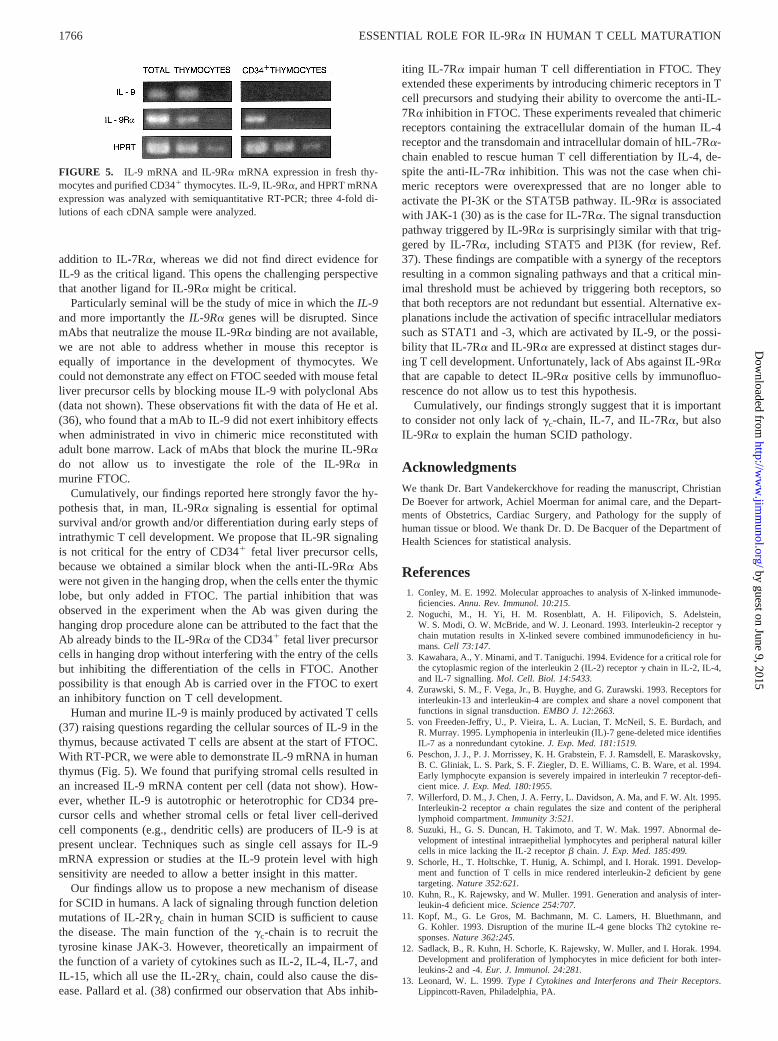
The ability of the SCID thymic lobes to support human T celldifferentiation, which can be blocked by neutralizing anti-IL-9RaAbs, raises the question about the source of IL-9. Previously, pres-ence of IL-9 in mouse thymus was shown by in situ hybridization(34). Because both murine and human IL-9 interact with the hu-man IL-9Ra (35), IL-9 could be produced in our model either byhuman thymocyte precursors or by murine stromal cells. Welooked whether IL-9 is produced in human thymus by a sensitiveand specific RT-PCR for detection of RNA message for humanIL-9. As shown in Fig. 5, mRNA was detected in the human thy-mus. Finally, we were able to demonstrate that mRNA for IL-9Rais present on CD341 thymocytes (Fig. 5).
DiscussionTo our knowledge, our findings are the first direct proof that, inhuman, IL-9Ra signaling is important for T cell development.Four types of experiments indicate that IL-9Ra is critical for earlyT cell development.
First, treating the FTOC with a mouse mAb blocking humanIL-9Ra decreased the number of human cells that develop in the
mouse SCID thymus when seeded with CD341 hemopoietic pre-cursor cells from fetal liver. The inhibition appeared to be at thetransition between CD341 to CD342CD11CD41CD32 immaturethymocytes.
Second, the results of using different mouse mAbs that all bindto human IL-9Ra, but differ in their neutralizing capacity, nicely fitwith their effect on the cell number when added in FTOC. Thisindicates that the mAbs do not exert cytopathic effects on IL-9Ra-bearing cells in vitro, but that the neutralization of the bindingcapacity of the receptor is essential to interfere with the normal Tcell developmental process.
Third, the block that is mediated by the mAb can be overcomeby exogenous IL-9. This finding argues strongly against a toxiceffect of the mAb or an aspecific inhibition mediated by the mAbpreparation.
Fourth, addition of exogenous IL-9 to the FTOC augments thecell number of the human cells obtained in FTOC, which signifiesthat IL-9Ra triggering favors either cell growth or cell survival.
A model to explain the essential role of IL-9Ra signalingevolves from the comparison with the results obtained with IL-7.We have previously shown that in FTOC neutralizing IL-7Ra re-sulted in a similar inhibition.
Here, however, the means by which IL-9Ra signals was notapparent from blocking studies of IL-9. Neutralization of eithermouse or human IL-9 or both did not result in the inhibition of Tcell development in FTOC (data not shown). This quandary iscompatible with the view that the experimental approach, in whichneutralizing mAbs or polyclonal Abs against IL-9 were used, wasnot powerful enough to result in a significant neutralization of thecytokine.
Cumulatively, our findings suggest that we must add IL-9Ra asan essential signaling receptor for T lymphocyte differentiation in
FIGURE 4. Restoration of IL-9Ra inhibition by exogenous IL-9. FTOCwere performed as described inMaterials and Methods, in the presence ofdifferent concentration of mAbs and with 10 ng/ml IL-9 (dashed line) orwithout IL-9 (solid line) as indicated. FTOC were examined after 14 daysof culture.
Table III. Influence of IL-9 on the development of human cells in chimeric human-mouse FTOCa
TreatmentFTOC Day128Number of cells
Frequency of Thymocyte Subsets
DN DP CD4 CD8
Control 99,0136 42,952 96 4.4 56.36 16.6 34.06 19.4 0.76 0.4IL-9 303,8126 32,085b 2.96 0.4 77.26 5.3 19.76 5.6 0.66 0.16
a Human CD341 fetal liver cells were obtained and cultured in FTOC for 27 days as described inMaterials and Methods,and numbers of human cells were calculated by multiplying the total number of viable cells counted under the microscope bythe fraction of human CD45-FITC positive cells determined by FACS analysis. The frequency of thymocyte subsets wasobtained by FACS analysis. Numbers of cells are given as the mean for groups of three separate experiments (1SD) whereina pool of at least three single lobes per experiment were analyzed.
b Differences between cell number in control and IL-9Ra-treated FTOC were statistically significant, withp , 0.05 for 28days of culture of CD341 fetal liver cells by a pairedt test on In-transformed values.
Table II. Comparative analysis of the influence of anti-human IL-9Ratreatment on the development of human cells in chimeric human-mouseFTOC in function of time of addition of the neutralizing Absa
Treatment
Ab Present in:Number of Human
Cells per Lobe After14 days of FTOCa
Hangingdrop FTOC
Control No No 67,760Control IgG2b Yes Yes 69,440Control IgG2b No Yes 100,912Anti-IL9Ra (AH9R7) Yes Yes 3,696Anti-IL9Ra (AH9R7) No Yes 4,437Anti-IL9Ra (AH9R7) Yes No 34,950
a Cells were obtained from fetal liver as described inMaterials and Methods, andnumbers of human cells were calculated by multiplying the total number of viablecells counted under the microscope by the fraction of human CD45-FITC positivecells determined by FACS analysis.
1765The Journal of Immunology
by guest on June 9, 2015http://w
ww
.jimm
unol.org/D
ownloaded from
addition to IL-7Ra, whereas we did not find direct evidence forIL-9 as the critical ligand. This opens the challenging perspectivethat another ligand for IL-9Ra might be critical.
Particularly seminal will be the study of mice in which theIL-9and more importantly theIL-9Ra genes will be disrupted. SincemAbs that neutralize the mouse IL-9Ra binding are not available,we are not able to address whether in mouse this receptor isequally of importance in the development of thymocytes. Wecould not demonstrate any effect on FTOC seeded with mouse fetalliver precursor cells by blocking mouse IL-9 with polyclonal Abs(data not shown). These observations fit with the data of He et al.(36), who found that a mAb to IL-9 did not exert inhibitory effectswhen administrated in vivo in chimeric mice reconstituted withadult bone marrow. Lack of mAbs that block the murine IL-9Rado not allow us to investigate the role of the IL-9Ra inmurine FTOC.
Cumulatively, our findings reported here strongly favor the hy-pothesis that, in man, IL-9Ra signaling is essential for optimalsurvival and/or growth and/or differentiation during early steps ofintrathymic T cell development. We propose that IL-9R signalingis not critical for the entry of CD341 fetal liver precursor cells,because we obtained a similar block when the anti-IL-9Ra Abswere not given in the hanging drop, when the cells enter the thymiclobe, but only added in FTOC. The partial inhibition that wasobserved in the experiment when the Ab was given during thehanging drop procedure alone can be attributed to the fact that theAb already binds to the IL-9Ra of the CD341 fetal liver precursorcells in hanging drop without interfering with the entry of the cellsbut inhibiting the differentiation of the cells in FTOC. Anotherpossibility is that enough Ab is carried over in the FTOC to exertan inhibitory function on T cell development.
Human and murine IL-9 is mainly produced by activated T cells(37) raising questions regarding the cellular sources of IL-9 in thethymus, because activated T cells are absent at the start of FTOC.With RT-PCR, we were able to demonstrate IL-9 mRNA in humanthymus (Fig. 5). We found that purifying stromal cells resulted inan increased IL-9 mRNA content per cell (data not show). How-ever, whether IL-9 is autotrophic or heterotrophic for CD34 pre-cursor cells and whether stromal cells or fetal liver cell-derivedcell components (e.g., dendritic cells) are producers of IL-9 is atpresent unclear. Techniques such as single cell assays for IL-9mRNA expression or studies at the IL-9 protein level with highsensitivity are needed to allow a better insight in this matter.
Our findings allow us to propose a new mechanism of diseasefor SCID in humans. A lack of signaling through function deletionmutations of IL-2Rgc chain in human SCID is sufficient to causethe disease. The main function of thegc-chain is to recruit thetyrosine kinase JAK-3. However, theoretically an impairment ofthe function of a variety of cytokines such as IL-2, IL-4, IL-7, andIL-15, which all use the IL-2Rgc chain, could also cause the dis-ease. Pallard et al. (38) confirmed our observation that Abs inhib-
iting IL-7Ra impair human T cell differentiation in FTOC. Theyextended these experiments by introducing chimeric receptors in Tcell precursors and studying their ability to overcome the anti-IL-7Ra inhibition in FTOC. These experiments revealed that chimericreceptors containing the extracellular domain of the human IL-4receptor and the transdomain and intracellular domain of hIL-7Ra-chain enabled to rescue human T cell differentiation by IL-4, de-spite the anti-IL-7Ra inhibition. This was not the case when chi-meric receptors were overexpressed that are no longer able toactivate the PI-3K or the STAT5B pathway. IL-9Ra is associatedwith JAK-1 (30) as is the case for IL-7Ra. The signal transductionpathway triggered by IL-9Ra is surprisingly similar with that trig-gered by IL-7Ra, including STAT5 and PI3K (for review, Ref.37). These findings are compatible with a synergy of the receptorsresulting in a common signaling pathways and that a critical min-imal threshold must be achieved by triggering both receptors, sothat both receptors are not redundant but essential. Alternative ex-planations include the activation of specific intracellular mediatorssuch as STAT1 and -3, which are activated by IL-9, or the possi-bility that IL-7Ra and IL-9Ra are expressed at distinct stages dur-ing T cell development. Unfortunately, lack of Abs against IL-9Rathat are capable to detect IL-9Ra positive cells by immunofluo-rescence do not allow us to test this hypothesis.
Cumulatively, our findings strongly suggest that it is importantto consider not only lack ofgc-chain, IL-7, and IL-7Ra, but alsoIL-9Ra to explain the human SCID pathology.
AcknowledgmentsWe thank Dr. Bart Vandekerckhove for reading the manuscript, ChristianDe Boever for artwork, Achiel Moerman for animal care, and the Depart-ments of Obstetrics, Cardiac Surgery, and Pathology for the supply ofhuman tissue or blood. We thank Dr. D. De Bacquer of the Department ofHealth Sciences for statistical analysis.
References1. Conley, M. E. 1992. Molecular approaches to analysis of X-linked immunode-
ficiencies.Annu. Rev. Immunol. 10:215.2. Noguchi, M., H. Yi, H. M. Rosenblatt, A. H. Filipovich, S. Adelstein,
W. S. Modi, O. W. McBride, and W. J. Leonard. 1993. Interleukin-2 receptorgchain mutation results in X-linked severe combined immunodeficiency in hu-mans.Cell 73:147.
3. Kawahara, A., Y. Minami, and T. Taniguchi. 1994. Evidence for a critical role forthe cytoplasmic region of the interleukin 2 (IL-2) receptorg chain in IL-2, IL-4,and IL-7 signalling.Mol. Cell. Biol. 14:5433.
4. Zurawski, S. M., F. Vega, Jr., B. Huyghe, and G. Zurawski. 1993. Receptors forinterleukin-13 and interleukin-4 are complex and share a novel component thatfunctions in signal transduction.EMBO J. 12:2663.
5. von Freeden-Jeffry, U., P. Vieira, L. A. Lucian, T. McNeil, S. E. Burdach, andR. Murray. 1995. Lymphopenia in interleukin (IL)-7 gene-deleted mice identifiesIL-7 as a nonredundant cytokine.J. Exp. Med. 181:1519.
6. Peschon, J. J., P. J. Morrissey, K. H. Grabstein, F. J. Ramsdell, E. Maraskovsky,B. C. Gliniak, L. S. Park, S. F. Ziegler, D. E. Williams, C. B. Ware, et al. 1994.Early lymphocyte expansion is severely impaired in interleukin 7 receptor-defi-cient mice.J. Exp. Med. 180:1955.
7. Willerford, D. M., J. Chen, J. A. Ferry, L. Davidson, A. Ma, and F. W. Alt. 1995.Interleukin-2 receptora chain regulates the size and content of the peripherallymphoid compartment.Immunity 3:521.
8. Suzuki, H., G. S. Duncan, H. Takimoto, and T. W. Mak. 1997. Abnormal de-velopment of intestinal intraepithelial lymphocytes and peripheral natural killercells in mice lacking the IL-2 receptorb chain.J. Exp. Med. 185:499.
9. Schorle, H., T. Holtschke, T. Hunig, A. Schimpl, and I. Horak. 1991. Develop-ment and function of T cells in mice rendered interleukin-2 deficient by genetargeting.Nature 352:621.
10. Kuhn, R., K. Rajewsky, and W. Muller. 1991. Generation and analysis of inter-leukin-4 deficient mice.Science 254:707.
11. Kopf, M., G. Le Gros, M. Bachmann, M. C. Lamers, H. Bluethmann, andG. Kohler. 1993. Disruption of the murine IL-4 gene blocks Th2 cytokine re-sponses.Nature 362:245.
12. Sadlack, B., R. Kuhn, H. Schorle, K. Rajewsky, W. Muller, and I. Horak. 1994.Development and proliferation of lymphocytes in mice deficient for both inter-leukins-2 and -4.Eur. J. Immunol. 24:281.
13. Leonard, W. L. 1999.Type I Cytokines and Interferons and Their Receptors.Lippincott-Raven, Philadelphia, PA.
FIGURE 5. IL-9 mRNA and IL-9Ra mRNA expression in fresh thy-mocytes and purified CD341 thymocytes. IL-9, IL-9Ra, and HPRT mRNAexpression was analyzed with semiquantitative RT-PCR; three 4-fold di-lutions of each cDNA sample were analyzed.
1766 ESSENTIAL ROLE FOR IL-9Ra IN HUMAN T CELL MATURATION
by guest on June 9, 2015http://w
ww
.jimm
unol.org/D
ownloaded from

14. Lodolce, J. P., D. L. Boone, S. Chai, R. E. Swain, T. Dassopoulos, S. Trettin, andA. Ma. 1998. IL-15 receptor maintains lymphoid homeostasis by supporting lym-phocyte homing and proliferation.Immunity 9:669.
15. Plum, J., M. De Smedt, M. P. Defresne, G. Leclercq, and B. Vandekerckhove.1994. Human CD341 fetal liver stem cells differentiate to T cells in a mousethymic microenvironment.Blood 84:1587.
16. Plum, J., M. De Smedt, B. Verhasselt, F. Offner, T. Kerre, D. Vanhecke,G. Leclercq, and B. Vandekerckhove. 1999. In vitro intrathymic differentiationkinetics of human fetal liver CD341 CD382 progenitors reveals a phenotypicallydefined dendritic/T-NK precursor split.J. Immunol. 162:60.
17. Plum, J., M. De Smedt, G. Leclercq, B. Verhasselt, and B. Vandekerckhove.1996. Interleukin 7 is a critical growth factor in early human T cell development.Blood 88:4239.
18. Puel, A., S. F. Ziegler, R. H. Buckley, and W. J. Leonard. 1998. Defective IL7Rexpression in T(-)B(1)NK(1) severe combined immunodeficiency.Nat. Genet.20:394.
19. Suda, T., R. Murray, M. Fischer, T. Yokota, and A. Zlotnik. 1990. Tumor ne-crosis factor-a and P40 induce day 15 murine fetal thymocyte proliferation incombination with IL-2.J. Immunol. 144:1783.
20. Renauld, J. C., N. van der Lugt, A. Vink, M. van Roon, C. Godfraind, G. Warnier,H. Merz, A. Feller, A. Berns, and J. Van Snick. 1994. Thymic lymphomas ininterleukin 9 transgenic mice.Oncogene 9:1327.
21. Van Snick, J., A. Goethals, J. C. Renauld, E. Van Roost, C. Uyttenhove,M. R. Rubira, R. L. Moritz, and R. J. Simpson. 1989. Cloning and characteriza-tion of a cDNA for a new mouse T cell growth factor (P40).J. Exp. Med. 169:363.
22. Yang, Y. C., S. Ricciardi, A. Ciarletta, J. Calvetti, K. Kelleher, and S. C. Clark.1989. Expression cloning of cDNA encoding a novel human hematopoieticgrowth factor: human homologue of murine T-cell growth factor P40.Blood74:1880.
23. Renauld, J. C., F. Houssiau, C. Uyttenhove, A. Vink, and J. Van Snick. 1993.Interleukin-9: a T-cell growth factor with a potential oncogenic activity.CancerInvest. 11:635.
24. Renauld, J. C., A. Kermouni, A. Vink, J. Louahed, and J. Van Snick. 1995.Interleukin-9 and its receptor: involvement in mast cell differentiation and T celloncogenesis.J. Leukocyte Biol. 57:353.
25. Reisbach, G., J. Sindermann, J. P. Kremer, L. Hultner, H. Wolf, and P. Dormer.1989. Macrophage colony-stimulating factor (CSF-1) is expressed by spontane-ously outgrown EBV-B cell lines and activated normal B lymphocytes.Blood74:959.
26. Renauld, J. C., A. Vink, J. Louahed, and J. Van Snick. 1995. Interleukin-9 is amajor anti-apoptotic factor for thymic lymphomas.Blood 85:1300.
27. Renauld, J. C., C. Druez, A. Kermouni, F. Houssiau, C. Uyttenhove,E. Van Roost, and J. Van Snick. 1992. Expression cloning of the murine andhuman interleukin 9 receptor cDNAs.Proc. Natl. Acad. Sci. USA 89:5690.
28. Kimura, Y., T. Takeshita, M. Kondo, N. Ishii, M. Nakamura, J. Van Snick, andK. Sugamura. 1995. Sharing of the IL-2 receptorg chain with the functional IL-9receptor complex.Int. Immunol. 7:115.
29. Russell, S. M., J. A. Johnston, M. Noguchi, M. Kawamura, C. M. Bacon,M. Friedmann, M. Berg, D. W. McVicar, B. A. Witthuhn, O. Silvennoinen, et al.1994. Interaction of IL-2Rb andg c chains with Jak1 and Jak3: implications forXSCID and XCID.Science 266:1042.
30. Demoulin, J., C. Uyttenhove, E. Van Roost, B. de Lestre, D. Donckers,J. Van Snick, and J.-C. Renauld. 1996. A single tyrosine of the interleukin-9(IL-9) receptor is required for STAT activation, anti-apoptotic activity andgrowth regulation by IL-9.Mol. Cell. Biol. 16:4710.
31. Verhasselt, B., M. De Smedt, R. Verhelst, E. Naessens, and J. Plum. 1998. Ret-rovirally transduced CD34(11) human cord blood cells generate T cells ex-pressing high levels of the retroviral encoded green fluorescent protein marker invitro. Blood 91:431.
32. De Smedt, M., G. Leclercq, B. Vandekerckhove, and J. Plum. 1994. Human fetalliver cells differentiate into thymocytes in chimeric mouse fetal thymus organculture.Adv. Exp. Med. Biol. 355:27.
33. Unkeless, J. 1979. Characterization of a monoclonal antibody against mousemacrophage and lymphocyte Fc receptors.J. Exp. Med. 150:580.
34. Deman, J. 1996. Role des cytokines dans le controle paracrine du developpementdes cellules T dans le thymus foetal et dans le thymus en re´generation apres uneirradiation subletale. Dortoral dissertation, Universite de Liege, Liege, Belgium.
35. Renauld, J.-C., A. Goethals, F. Houssiau, E. Van Roost, and J. Van Snick. 1990.Cloning and expression of a cDNA for the human homolog of mouse T cell andmast cell growth factor P40.Cytokine 2:9.
36. He, Y. W., H. Nakajima, W. J. Leonard, B. Adkins, and T. R. Malek. 1997. Thecommong-chain of cytokine receptors regulates intrathymic T cell developmentat multiple stages.J. Immunol. 158:2592.
37. Renauld, J.-C., and J. Van Snick. 1998.Interleukin-9. Academic Press, Inc.,New York.
38. Pallard, C., A. Stegmann, T. van Kleffens, F. Smart, A. Venkitaraman, andH. Spits. 1999. Distinct Roles of the phosphatidylinositol 3-kinase and STAT5pathways in IL-7-mediated development of human thymocyte precursors.Immu-nity 10:525.
1767The Journal of Immunology
by guest on June 9, 2015http://w
ww
.jimm
unol.org/D
ownloaded from