detailed questionnaire for the meat and poultry products ...
Upload
khangminh22Category
view
0download
0
coatings
Article
Shelf Life Assessment of Fresh Poultry MeatPackaged in Novel Bionanocomposite ofChitosan/Montmorillonite Incorporated withGinger Essential Oil
Victor G. L. Souza 1 ID , João R. A. Pires 1 ID , Érica T. Vieira 1, Isabel M. Coelhoso 2 ID ,Maria P. Duarte 1 and Ana L. Fernando 1,* ID
1 MEtRiCS, Departamento de Ciências e Tecnologia da Biomassa, Faculdade de Ciências e Tecnologia, FCT,Universidade Nova de Lisboa, Campus de Caparica, 2829-516 Caparica, Portugal;[email protected] (V.G.L.S.); [email protected] (J.R.A.P.);[email protected] (É.T.V.); [email protected] (M.P.D.)
2 LAQV-REQUIMTE, Departamento de Química, Faculdade de Ciências e Tecnologia,Universidade Nova de Lisboa, Campus de Caparica, 2829-516 Caparica, Portugal; [email protected]
* Correspondence: [email protected]; Tel./Fax: +351-212-948-543
Received: 31 March 2018; Accepted: 3 May 2018; Published: 5 May 2018�����������������
Abstract: Active packaging incorporated with natural extracts is a promising technology to extendshelf life of perishable food. Therefore, this study aimed to produce a bionanocomposite based onchitosan reinforced with sodium montmorillonite (MMT) and incorporated with ginger essentialoil (GEO). In vitro activity was assessed through migration assay and antimicrobial study againstfoodborne bacteria. Phenolic compounds were diffused within 48 h of contact, and retained some oftheir antioxidant activity. Films demonstrated antimicrobial activity against both Gram-positive and-negative bacteria tested. The effect on the shelf life of fresh poultry meat was determined on sampleswrapped in the biopolymers and stored under refrigeration for 15 days, through physicochemicaland microbiological analyses. Compared to unwrapped poultry meat, samples wrapped in thebionanocomposites showed a reduction in microorganisms count of 1.2–2.6 log CFU/g, maintainedcolor and pH values and thiobarbituric acid reactive substances (TBARS) index increased at a lowerrate, extending fresh poultry meat shelf life. The incorporation of GEO enhanced the biopolymeractivity, by reducing lipid oxidation and microbiological growth of the poultry meat. In contrast,reinforcement with MMT imprisoned the active compounds in the polymeric chain, hindering itsactivity. In conclusion, the bionanocomposites tested represent promising substitutes to commercialand unsustainable plastic films.
Keywords: active packaging; antioxidant activity; antimicrobial activity; bionanocomposites; naturalpreservatives; migration; shelf life extension; Zingiber officinale
1. Introduction
Poultry meat is considered a healthy source of high-quality protein, vitamins, minerals andessential polyunsaturated fatty acids (PUFAs), especially the omega (n)-3 fatty acids [1], and stillremains one of the cheapest sources of protein [2]. The OECD-FAO agriculture outlook 2016–2025forecasts an expansion of meat production by 1.8 Mt annually by 2025, mainly due to pork andpoultry products [2]. Moreover, the poultry sector is possibly the fastest growing and most flexibleof all livestock sectors [1]. Due to its intrinsic composition (i.e., high protein and moisture content)and high pH, the growth of spoilage and pathogenic microorganisms is favored; therefore chicken
Coatings 2018, 8, 177; doi:10.3390/coatings8050177 www.mdpi.com/journal/coatings

Coatings 2018, 8, 177 2 of 17
meat is considered a high perishable food [3,4]. The losses through the production chain are aneconomic burden to the producer; consequently, food industries are continuously searching for noveltechnologies/methods to increase the shelf life and overall safety/quality of the poultry products [3].
Biodegradable bioactive food packaging has aroused considerable interest to extend shelf lifeand guarantee safety of perishable food susceptible to microbial alteration [5]. Through this approach,the industry is capable of overcoming its three currently big problems or concerns: reduce foodspoilage and food poisoning [5]; attend to the consumer’s demand for “healthier” and less chemicallymodified products [3]; and reduce the disposal problem created through the growth of demand anduse of petroleum-based plastic materials [6], as biodegradable biopolymers are a more environmentallyfriendly alternative. In active packaging technology, bioactive substances are incorporated into thesystem to enhance the quality and to extend the shelf life of products, e.g., meat [7], through threedifferent methods, namely: by direct incorporation of active compounds into the polymeric matrix;as edible films and coatings with bioactive substances; and as activated sachet, patch or tablet [8].
Antimicrobial active packaging with incorporation of essential oils (EOs) has been studied in thepast decades as an alternative to traditional technologies to preserve food products [9–11]. Despite theirgood bioactivity, EOs when directly incorporated into food matrices have two main disadvantages:their costs and intense aroma that can change the organoleptic properties of the product, impactingthe sensory perception of applied food [5,12]. By incorporating these powerful extracts into polymers,this limitation can be overcome, once small amounts are necessary, enabling these natural additives tobe used in the food industry as active packaging [12]. Arkoun and collaborators [5] extended the shelflife of fresh meat by one weak with the use of chitosan-based nanofibers. Fish fillets and chicken breastrecorded a higher shelf life with the use of edible films based in chitosan [13] and cellulose acetate [14]incorporated with rosemary essential oil, respectively.
Chitosan (Ch) is the second most abundant polysaccharide in nature after cellulose and consideredas a natural biopolymer [15]. It has the advantages of being derived from renewable sources(i.e., found in the exoskeleton of crustaceans and in the cell wall of microorganisms), is biocompatible,biodegradable, non-toxic, able to form membranes, films, gels, and fibers and also exhibits intrinsicantimicrobial properties [9,16]. Biopolymers, including chitosan, are also known for their good oxygenbarrier properties [17]—a positive characteristic that plays and important role in the preservation offoodstuffs. Still, being a hydrophilic compound, chitosan films have high permeability to water [18]and, when compared to traditional plastic material, weaker mechanical properties [19]. To enhancesuch characteristics, nanofillers are generally added [6]. Through the interaction with the polymericchain, a more tortuous path and stronger structures are created, improving both mechanical and barrierproperties [20,21]. Montmorillonite (MMT)—a layered silicate mineral clay—is the most researchedand used nanomaterial for this purpose due to its low cost, abundancy, mechanical resistance, swellingand plasticizer ability [22].
Ginger is a spice obtained from the rhizomes of Zingiber officinale (Rosc.), family Zingiberaceae,typically consumed as a fresh paste, slices, dried powder, in candies or as flavoring in culinaryproducts or tea [23]. Essential oils and oleoresins extracted from the species Z. officinale are rich indiverse chemical compounds, such as zingiberene, α-curcumene, monoterpenes camphene, geranial,linalool, 1,8-cineole, which confer bioactivity as antioxidants and anti-inflammatory or antimicrobialextract [24]. Currently, ginger essential oil (GEO) is being used in studies aiming the development ofnovel active packaging materials to be used in the food industry: chitosan–carboxymethyl cellulosefilms incorporated with GEO and cinnamon EO [25]; nanofilms based on Tilapia fish skin gelatin andZnO nanoparticles incorporated with GEO for meat packaging application [26]; nanoemulsion-basededible sodium caseinate coatings applied in fresh meat [27], edible coatings based on hydroxypropylmethylcellulose, beeswax, nanoclay and ginger oil applied in fresh mango cuts [28].
However, to the best of our knowledge, to date, the combination of chitosan, montmorillonite andginger essential oil have never been produced and tested in fresh poultry meat. Moreover, despite thenumber of published papers on the development and characterization of chitosan-based films, only a
Coatings 2018, 8, 177 3 of 17
very limited number of studies assessed the antimicrobial potential of chitosan in real food systems [5].Thus, this work aimed to develop a bionanocomposite on chitosan/MMT and GEO and to evaluate itsactivity by in vitro and in situ assays.
2. Materials and Methods
2.1. Materials and Reagents
Food grade classification ginger (Zingiber officinale Roscoe) essential oil (Biover, Nazareth,Belgium) was purchased in local market. The biopolymer (chitosan-75% of deacetylation andhigh molecular weight (31–37 kDa)), ethanol absolute, 1,1,3,3-tetraethoxypropane (TEP) and2,2-diphenyl-1-picrylhydrazyl (DPPH) were acquired from Sigma-Aldrich (Steinheim, Germany).Folin-Ciocalteu Reagent, trichloroacetic acid (TCA), 2-thiobarbituric acid (TBA) and sodium carbonateanhydrous were obtained from PanReac (Barcelona, Spain), while glacial acetic acid, glycerol, sodiumhydroxide (NaOH), and tween 80 from Alfa Aesar (Kandel, Germany). Dimethyl sulfoxide (DMSO)was purchased from Fisher Scientific (Pittsburgh, PA, USA). All microbiological reagents werepurchased from Biokar (Allonne, Beauvais, France): Tripto-casein soy broth (TSB), tripto-caseinagar (TSA), plate count agar (PCA), brilliant green lactose bile broth and mueller hinton agar (MHA).The nanoclay used (sodium montmorillonite (Cloisite®Na+)), in this paper will be referred as MMT,was kindly donated by BYK Additives & Instruments (Wesel, Germany). The MMT is a commercialmontmorillonite on its natural state (with sodium interlayer cation), available as a solid powder withoff-white color and odorless, 2.9 g/cm3 density, pH of 9.0 in 2% aqueous dispersion and an interplanardistance d001 = 1.17 nm [29]. All water used was purified using Milli-Q system (Millipore, Billerica,MA, USA) and all chemicals were of analytical reagent grade.
2.2. Films Production
Bionanocomposites were prepared according to Souza et al. [30] and Dias et al. [31]. Chitosan(1.5%, w/v) film form dispersion (FFD) in 1% (v/v) of glacial acetic acid solution was prepared withcontinuous agitation overnight at room temperature. A plasticizer, glycerol, was added to all samples(30% w/w of polymer). In order to achieve good miscibility and dispersion of the 2.5% (w/w ofchitosan) of MMT into chitosan chains an exfoliation process consisting of three agitation cycles (5 minagitation with the ultraturrax (15,000 rpm) (IKA® T18, Staufen, Germany) followed by 15 min (360 W)in an ultrasound bath (Selecta, Barcelona, Spain)) was carried out. Before the last agitation cycleGEO (0.5%; 1% and 2% v/v in FFD) and tween 80 (0.2% w/v in GEO) were added. Biofilms withoutthe nanoreinforcement were produced without the two first agitation cycles. Also films withoutincorporation of MMT nor GEO were produced in the same conditions. The resulting composite wascasted in glass molds (18 cm × 25 cm) or in glass Petri Dish (10 cm diameter) and dried for 72 h at roomtemperature (relative humidity approximately 50%). Dried films were peeled and stored protectedfrom light at 25 ◦C until evaluation.
2.3. In Vitro Active Characterization
2.3.1. Migration Assays
Migration assays were performed to determine the diffusion of the active compounds (totalphenolic compounds present in the GEO) incorporated into the film towards a fatty food simulant(95% ethanol solution) at 37 ± 2 ◦C during 10 days [32]. Periodically, total phenol content present inthe simulant media and its antioxidant activity were determined by Folin–Ciocalteu method [33] andDPPH assays [34], respectively:
Total phenol content (TPC): 1 mL of simulant was mixed with 3 mL of water and 0.25 mL ofFolin–Ciocalteu reagent. After 5 min incubation at the dark, 0.75 mL of sodium carbonate solution5% (w/v) was added and the system incubated for more 60 min in the same conditions. Absorbance

Coatings 2018, 8, 177 4 of 17
at 760 nm was measured using spectrophotometer UV/Vis (Spekol 1500, Analytikjena, Germany).Calibration curve with known concentration (0–120 mg·L−1) of gallic acid solutions was used toquantify the TPC. Results were expressed in mg gallic acid equivalent (GAE)·L−1 of simulant.
DPPH free radical scavenging assay: antioxidant activity of the active compounds diffused tothe simulant media was determined with DPPH assay. Briefly, 1 mL of simulant was mixed with3 mL of 60 µmol·L−1 DPPH ethanolic solution and incubated in dark for 20 min. The absorbance wasread at 517 nm using UV/VIS spectrophotometer and the percentage of inhibition calculated usingEquation (1):
Radical scavenging (%) =(Initial absorbance − Sample absorbance)
Initial absorbance× 100 (1)
In a complementary manner, the diffusion coefficient (D) of phenolic compounds was calculatedfrom the plot of MF,t/MP,0 versus t0.5 using initial migration data according to the model based on theFick’s second law described in Equation (2) [35]:
MF,t
MP,0=
2δ
(Dtπ
)0.5(2)
where MF,t (mg GAE) is the total phenol content in the food simulant at time t, MP,0 (mg GAE) is theinitial TPC in the packaging film, D (m2·s−1) is the diffusion coefficient of TPC in the packaging filmand δ (m) is the thickness of the packaging film.
2.3.2. Antimicrobial Studies
In vitro antimicrobial activity of pure GEO and films was studied by agar diffusion method [36]against Gram-positive bacteria (Bacillus cereus (ATCC11778), Enterococcus faecalis (ATCC29212),Listeria monocytogenes (ATCC15313), Staphylococcus aureus (ATCC6538)); Gram-negative bacteria(Escherichia coli (ATCC8739), Pseudomonas aeruginosa (ATCC9027), Salmonella enterica (ATCC10708));and one yeast Candida albicans (ATCC10231). Firstly, glycerol stock cultures stored at −80 ◦C inultra-low temperature freezer (New Brunswick Scientific, St. Albans, Hertfordshire, UK) wereinoculated in TSA or MHA and incubated at 30 ◦C for B. cereus and C. albicans or at 37 ◦C for the othermicroorganisms during 16–24 h for bacteria and 48 h for the yeast. Petri dish (8.5 cm of diameter)containing Mueller Hinton Agar were inoculated with a suspension containing 1 × 10⁸ CFU/mL forthe bacteria or 1 × 10⁶ CFU/mL for the yeast (both previously adjusted to match a 0.5 McFarlandturbidity standard using a Mc-Farland densitometer (Model Den-1B, Grant Instruments, Cambridge,UK)). For the assessment of pure GEO, wells with 6 mm diameter were cut in the MHA and filledwith 50 µL of the pure essential oil or also diluted in DMSO (1:1) for C. albicans. Wells filled withDMSO were used as negative control. For the films, disks (6 mm diameter) were cut and placed ontoinoculated plate surface and incubated during 24 h in the fridge (5 ± 2 ◦C) to allow the migrationof the active compounds to MHA, prior to the incubation at 37 ◦C or 30 ◦C (depending on themicroorganism) for 20 h or 48 h (yeast). Inhibition zone below films disks was also considered aspositive antimicrobial activity.
Antimicrobial activity of bionanocomposites was also assessed by viable cell colony count(CFU) method [36,37] against Gram-positive (B. cereus (ATCC11778)) and Gram-negative (S. enterica(ATCC10708)) foodborne bacteria. An amount of 0.2 g of each film was immersed in 4 mL of TSBcontaining ~106 CFU/mL of the tested bacteria. Subsequently, the tubes were kept shaking (150 RPM)incubated at 37 ◦C (S. enterica) or 30 ◦C (B. cereus) during 24 h. Tubes without films were used ascontrol. One hundred microliters of serial dilutions were spread onto TSA plates, incubated for 16–24 hand the number of viable microorganism colonies was then manually counted. Results were expressedas the number of log reductions calculated according to Equation (3):

Coatings 2018, 8, 177 5 of 17
number of log reductions = log B − log A (3)
where B and A are the mean number of bacteria (CFU/mL) in the control samples and treated samplesafter 24 h incubation, respectively.
2.4. In Situ Active Characterization
Fresh poultry minced meat was purchased in local market and packaged in the biopolymersproduced. Approximately 30 g of meat were wrapped on films specimens (5 cm × 18 cm) and storedinto plastic boxes with screw cap, under refrigeration (5 ± 2 ◦C) for 15 days. Meat unwrappedand wrapped in commercial adherent films (polyvinyl chloride (PVC)) were also evaluated ascontrols. Periodically, meat was characterized in terms of the lipid oxidation, physicochemical andmicrobiological quality at 0, 3, 7, 10 and 15 days of storage.
2.4.1. Physicochemical Characterization
Physicochemical characterization was evaluated in terms of pH, titratable acidity and moistureaccording to AOAC method [38]. Color was also determined by measurement of CIE-L*a*b* coordinates(L = 0 (black) to L = 100 (white), −a (greenness) to +a (redness), and −b (blueness) to +b (yellowness))on the surface of the meat using a CR 410 colorimeter (Minolta Co., Tokyo, Japan) with D65 lightsource, and visual angle of 10◦. Hue angle was calculated according to Equation (4) [32]:
Hue angle = tan(
b∗
a∗
)−1(4)
where b* and a* are the coordinates measured from the samples.
2.4.2. Thiobarbituric Acid Reactive Substances (TBARS) Index
Lipid oxidation was assessed by TBARS assay [39]. Ten grams of each meat were mixed with 20 mLof trichloroacetic acid (TCA) 7.5% (w/v) and agitated for one hour to extract the malonaldehyde (MDA).Subsequently, 5 mL of the filtrate resulted was combined with 5 mL of 0.02 M 2-thiobarbituric acid(TBA) and heated (95 ◦C/30 min) in water bath (Memmert, Germany). After cooling, the absorbancewas measured at 530 nm in UV/VI spectrophotometer. Calibration curve using known concentrationsof MDA (from TEP solution) was used to calculate the TBARS index. Results were expressed as mg ofMDA/kg of meat.
2.4.3. Microbiological Growth
Total mesophilic aerobic bacteria (TMAB) and total coliforms were used to evaluate themicrobiological quality of the meat packaged. The determination was performed in accordanceto ISO 4833-1:2013 [40] and ISO 4831:2006 [41], respectively. Total mesophilic aerobic bacteria countswere performed in Plate Count Agar after incubation at 30 ◦C for 72 h. Total coliforms counts wereperformed in brilliant green lactose bile broth after incubation at 30 ◦C for 48 h to calculate the mostprobable number (MPN). Results were expressed as log CFU (colony forming units)/g of meat (TMAB)or log MPN/g of meat (total coliforms).
2.5. Statistical Analysis
All experiments were conducted using a completely randomized design with three replications.Statistical analysis of data was performed through a one-way analysis of variance (ANOVA) usingSoftware OriginLab (version 8.5), and when ANOVA was significant (p < 0.05) differences amongmean values were processed by the Tukey test. Significance was defined at p < 0.05.
Coatings 2018, 8, 177 6 of 17
3. Results and Discussion
3.1. In Vitro Active Characterization
3.1.1. Migration Assay
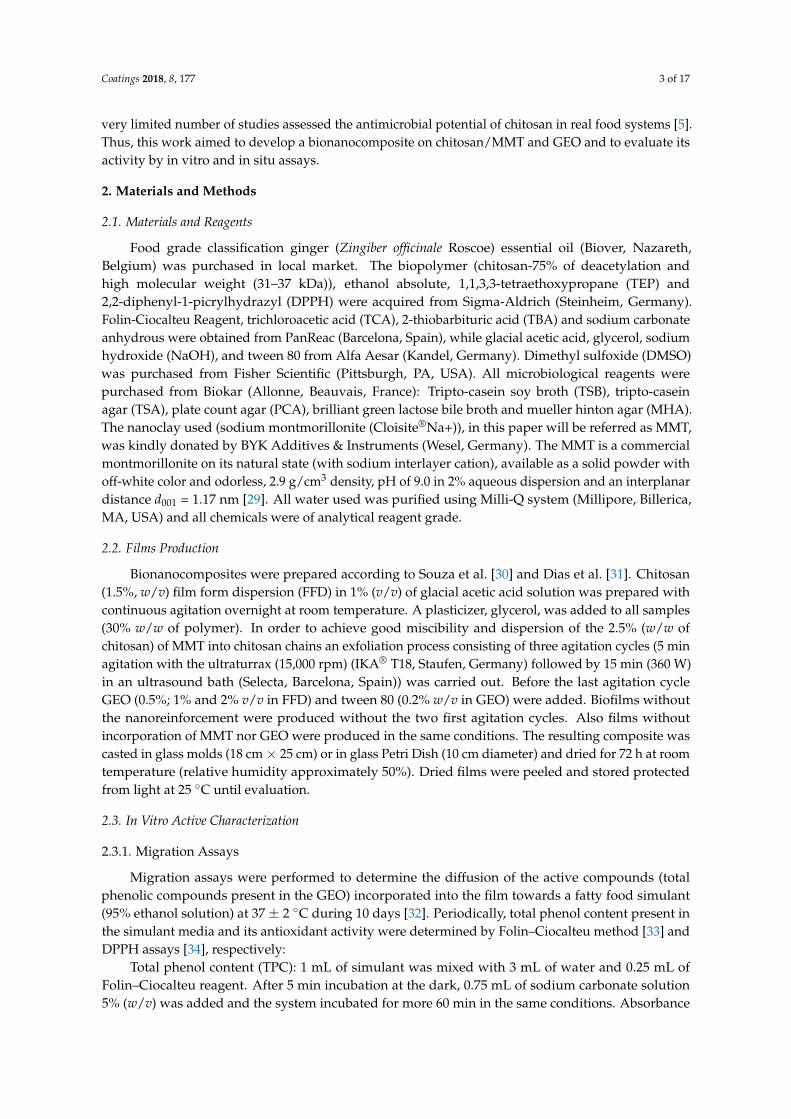
Diffusion coefficient of TPC, maximum TPC released ratio (maximum total phenol contentquantified in the simulant/total phenol content incorporated into the chitosan film) and percentageof radical scavenging are shown in Table 1. The release of phenolic compound towards the 95%ethanol followed an “exponential growth to a maximum” migration pattern, reaching the equilibriumafter 24–48 h of contact with the simulant (Figure 1). Similar behavior was reported in the migrationof TPC from green tea extract incorporated in ethylene–vinyl alcohol copolymer (EVOH) films to95% ethanol [42] or from coca extract incorporated in EVOH films to water [43]. The diffusioncoefficient did not statistically differ among the bionanocomposites tested (p > 0.05) except Chitosan+ MMT + 1% GEO, which showed a significantly higher diffusion coefficient than the ones obtainedwith the two samples incorporated with 2% GEO (p < 0.05). Films with one percent of GEO andMMT presented the faster release of TPC towards the simulant media. It is also important topoint out that the presence of the nanofiller accelerated the diffusion process when 0.5% and 1%of GEO was incorporated, while retarded with 2% GEO. In food systems, the big advantage of activepackaging is the possibility to extend the protection time through the gradual release of the bioactivecompounds [44]. Therefore, samples with the highest amount of GEO and MMT, from this in vitromigration assay, would be highlighted as the most promising film. However, in comparison withother diffusion coefficient values for similar EOs and films, this study findings are one order ofmagnitude higher than those obtained for chitosan films incorporated with rosemary essential oil [45],which suggests a smaller release time or lower retention of phenolic compounds from GEO insidechitosan film.
Coatings 2018, 8, x FOR PEER REVIEW 6 of 18
ethanol followed an “exponential growth to a maximum” migration pattern, reaching the equilibrium
after 24–48 h of contact with the simulant (Figure 1). Similar behavior was reported in the migration
of TPC from green tea extract incorporated in ethylene–vinyl alcohol copolymer (EVOH) films to 95%
ethanol [42] or from coca extract incorporated in EVOH films to water [43]. The diffusion coefficient
did not statistically differ among the bionanocomposites tested (p > 0.05) except Chitosan + MMT +
1% GEO, which showed a significantly higher diffusion coefficient than the ones obtained with the
two samples incorporated with 2% GEO (p < 0.05). Films with one percent of GEO and MMT
presented the faster release of TPC towards the simulant media. It is also important to point out that
the presence of the nanofiller accelerated the diffusion process when 0.5% and 1% of GEO was
incorporated, while retarded with 2% GEO. In food systems, the big advantage of active packaging
is the possibility to extend the protection time through the gradual release of the bioactive
compounds [44]. Therefore, samples with the highest amount of GEO and MMT, from this in vitro
migration assay, would be highlighted as the most promising film. However, in comparison with
other diffusion coefficient values for similar EOs and films, this study findings are one order of
magnitude higher than those obtained for chitosan films incorporated with rosemary essential oil
[45], which suggests a smaller release time or lower retention of phenolic compounds from GEO
inside chitosan film.
(a) (b)
Figure 1. Migration assay–total phenol content diffused from films to simulant over time: (a) Films
incorporated with GEO in different concentrations; (b) Films incorporated with MMTNa and GEO in
different concentrations. GEO (ginger essential oil), MMT (sodium montmorillonite), GAE (gallic acid
equivalent).
The phenolic compounds released to the simulant media kept some of their antioxidant activity;
however, it was not correlated to the TPC present in the simulant, as it was expected and previously
reported in the literature [32]. Abdollahi et al. [46] reported a good interaction between chitosan
functional groups and rosemary essential oil, furthermore, in the presence of MMT these bonding
were enhanced. Such interactions between chitosan phenolic compounds, and chitosan phenolic
compounds MMT may also have occurred with present films and can explain the existence of no
correlation between TPC in the simulant and the antioxidant activity of compounds released to the
simulant. In vitro studies are important to evaluate the activity of novel materials once these can
predict the behavior in real situation. However, the food matrix is much more complex than the
simulant media, thus in real scenarios the results may be different [32]. Therefore, it is important to
also investigate in situ activity of the films.
3.1.2. Antimicrobial Studies
Essential oils are well known for their good antimicrobial activity [47], and extensive research
and reviews have focused on this matter [48–51]. In the present study, pure GEO only demonstrated
antimicrobial activity against the Gram-positive bacteria (B. cereus, S. aureus and L. monocytogenes)
(Figure 2), and no inhibition zone was observed for E. faecalis or any of the Gram-negative foodborne
bacteria tested (data are not shown). Regarding the C. albicans, pure GEO led to the complete
Figure 1. Migration assay–total phenol content diffused from films to simulant over time: (a) Filmsincorporated with GEO in different concentrations; (b) Films incorporated with MMTNa and GEOin different concentrations. GEO (ginger essential oil), MMT (sodium montmorillonite), GAE (gallicacid equivalent).
The phenolic compounds released to the simulant media kept some of their antioxidant activity;however, it was not correlated to the TPC present in the simulant, as it was expected and previouslyreported in the literature [32]. Abdollahi et al. [46] reported a good interaction between chitosanfunctional groups and rosemary essential oil, furthermore, in the presence of MMT these bondingwere enhanced. Such interactions between chitosan × phenolic compounds, and chitosan × phenoliccompounds × MMT may also have occurred with present films and can explain the existence of no

Coatings 2018, 8, 177 7 of 17
correlation between TPC in the simulant and the antioxidant activity of compounds released to thesimulant. In vitro studies are important to evaluate the activity of novel materials once these canpredict the behavior in real situation. However, the food matrix is much more complex than thesimulant media, thus in real scenarios the results may be different [32]. Therefore, it is important toalso investigate in situ activity of the films.
3.1.2. Antimicrobial Studies
Essential oils are well known for their good antimicrobial activity [47], and extensive researchand reviews have focused on this matter [48–51]. In the present study, pure GEO only demonstratedantimicrobial activity against the Gram-positive bacteria (B. cereus, S. aureus and L. monocytogenes)(Figure 2), and no inhibition zone was observed for E. faecalis or any of the Gram-negative foodbornebacteria tested (data are not shown). Regarding the C. albicans, pure GEO led to the complete inhibitionof the yeast growth, and when diluted (1:1) an inhibition zone of approximately 20 mm was recorded(Figure 2). This is in accordance with what was previously reported for two ginger essential oilsextracted from the plant’s leaves or rhizomes, which in general exhibited better antibacterial activityagainst the Gram-positive bacteria than against the Gram-negative bacteria [52], probably due tothe protection conferred by the lipopolysaccharide layer of the outer membrane of Gram-negativebacteria [53]. Singh et al. [54] found good antifungal activity (average 60% of mycelial growth) ofGEO against several Aspergillus spp. and Fusarium moniliforme, which is in agreement with the strongantifungal activity against C. albicans.
The antimicrobial activity of EOs from spices and herbs are believed to be due to their rich contentin phenolic compounds [47,54], and the higher susceptibility of Gram-positive bacteria compared toGram negative bacteria suggests that the microbial targets of oil is the cell wall [24]. Singh et al. [54],Trajano et al. [55] and López et al. [24] also studied the antimicrobial activity of Zingiber officinaleessential oil with contradictory conclusions. Trajano et al. [55] only found antibacterial activity of GEOagainst S. aureus, and no inhibition against L. monocytogens, E. coli, B. cereus, S. enterica, P. aeruginosa andYersinia enterocolitica, while Singh et al. [54] observed good antimicrobial activity against E. coli, S. aureus,P. aeruginosa and Klebsiella pneumoniae, similar to what was observed by López et al. [24]. The differencesfrom present results and the ones cited may be attributed to the characteristic composition of eachGEO tested, once the type and concentration of phenolic compounds are directly correlated to theantimicrobial activity [52]. Also, the extraction procedure applied may have interfered in the finalcomposition of the GEO and consequently on its biological activity.
The films, on the other hand, only presented activity against two of the bacteria tested: B. cereusand S. aureus, and only underneath the disk specimens of the samples incorporated with 1% and 2% ofGEO, in the case of B. cereus, and with 0.5%, 1% and 2% of GEO against S. aureus (Figure 2). No activitywas observed against Gram negative bacteria or the yeast tested.
Despite its well-known antimicrobial activity, pristine chitosan films did not present an inhibitionzone for any of the microorganisms assessed. According to Hafsa et al. [56], the antimicrobialproperties of chitosan may become negligible when the polysaccharide is in the form of insolublefilms. Similar results were also reported with chitosan film incorporated with propolis extract againstseveral Gram-positive and Gram-negative bacteria, where only the samples incorporated with propolispresented inhibition underneath the disk films and pristine chitosan film did not show antimicrobialactivity [57].
Ginger essential oil when incorporated in the films had its antimicrobial activity reduced, probablydue to a partial loss of volatile essential oil in the film by evaporation [58] and to the good interactionbetween the phenolic compounds and the films that entrapped the active compounds in the polymericchain, lowering its diffusion towards the MHA, and therefore limiting its activity only underneath thedisk films [57].
Coatings 2018, 8, 177 8 of 17
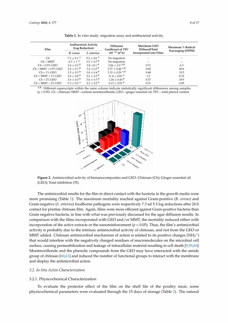
Table 1. In vitro study: migration assay and antibacterial activity.
Film
Antibacterial Activity(Log Reduction)
DiffusionCoefficient of TPC
(10−11 m2/s)
Maximum GEODiffused/Total
Incorporated into Films
Maximum % RadicalScavenging (DPPH)
B. cereus S. enterica
Ch 7.3 ± 0.1 A 5.3 ± 0.6 A No migration – –Ch + MMT 6.7 ± 1 A 3.5 ± 0.7 B No migration – –
Ch + 0.5% GEO 3.4 ± 0.2 B 3.8 ±0.1 B 2.06 ± 0.2 A,B 0.71 6.3Ch + MMT + 0.5% GEO 2.3 ± 0.1 B 3.2 ±1.0 B 3.77 ± 0.48 A,B 0.82 40.6
Ch + 1% GEO 1.5 ± 0.3 B 3.8 ± 0.4 B 3.15 ± 0.29 A,B 0.68 10.5Ch + MMT + 1% GEO 3.6 ± 0.8 B 3.2 ± 0.3 B 11.4 ± 0.63 A 1.0 8.32
Ch + 2% GEO 3.6 ± 0.3 B 3.6 ± 0.3 B 1.24 ± 0.43 B 0.37 18.9Ch + MMT + 2% GEO 5.3 ± 0.0 A 2.3 ± 0.2 B 0.13 ± 0.01 B 0.31 6.05
A,B: Different superscripts within the same column indicate statistically significant differences among samples(p < 0.05). Ch—chitosan; MMT—sodium montmorillonite; GEO—ginger essential oil; TPC—total phenol content.Coatings 2018, 8, x FOR PEER REVIEW 8 of 18
Figure 2. Antimicrobial activity of bionanocomposites and GEO. Chitosan (Ch); Ginger essential oil
(GEO); Total inhibition (TI).
Ginger essential oil when incorporated in the films had its antimicrobial activity reduced,
probably due to a partial loss of volatile essential oil in the film by evaporation [58] and to the good
interaction between the phenolic compounds and the films that entrapped the active compounds in
the polymeric chain, lowering its diffusion towards the MHA, and therefore limiting its activity only
underneath the disk films [57].
The antimicrobial results for the film in direct contact with the bacteria in the growth media were
more promising (Table 1). The maximum mortality reached against Gram-positive (B. cereus) and
Gram-negative (S. enterica) foodborne pathogens were respectively 7.3 ad 5.3 log reductions after 24 h
contact for pristine chitosan film. Again, films were more efficient against Gram-positive bacteria
than Gram negative bacteria, in line with what was previously discussed for the agar diffusion results.
In comparison with the films incorporated with GEO and/or MMT, the mortality reduced either with
incorporation of the active extracts or the nanoreinforcement (p < 0.05). Thus, the film s antimicrobial
activity is probably due to the intrinsic antimicrobial activity of chitosan, and not from the GEO or
MMT added. Chitosan antimicrobial mechanism of action is related to its positive charges (NH3+) that
would interfere with the negatively charged residues of macromolecules on the microbial cell surface,
causing permeabilization and leakage of intracellular material resulting in cell death [9,59,60].
Montmorillonite and the phenolic compounds from the GEO may have interacted with the amide
group of chitosan [60,61] and reduced the number of functional groups to interact with the membrane
and display the antimicrobial action.
3.2. In Situ Active Characterization
3.2.1. Physicochemical Characterization
To evaluate the protector effect of the film on the shelf life of the poultry meat, some
physicochemical parameters were evaluated through the 15 days of storage (Table 2). The natural
deterioration process resulted in increased pH value, with reduction of the acidity, changes on the
color (discoloration process) and increase of the moisture content, as can be observed for the meat
without packaging (unwrapped). Such changes were less significant for the other samples: samples
wrapped in the commercial PVC showed less change than unwrapped samples, but higher changes
than samples wrapped with Ch, thus indicating that the biocomposites produced are promising
substitutes to commercial and unsustainable plastic films. The incorporation of GEO in the chitosan
matrix enhanced significantly the activity of the biopolymers once the samples wrapped in those
films showed minimum changes (Table 2). The magnitude of the changes observed in samples
Figure 2. Antimicrobial activity of bionanocomposites and GEO. Chitosan (Ch); Ginger essential oil(GEO); Total inhibition (TI).
The antimicrobial results for the film in direct contact with the bacteria in the growth media weremore promising (Table 1). The maximum mortality reached against Gram-positive (B. cereus) andGram-negative (S. enterica) foodborne pathogens were respectively 7.3 ad 5.3 log reductions after 24 hcontact for pristine chitosan film. Again, films were more efficient against Gram-positive bacteria thanGram negative bacteria, in line with what was previously discussed for the agar diffusion results. Incomparison with the films incorporated with GEO and/or MMT, the mortality reduced either withincorporation of the active extracts or the nanoreinforcement (p < 0.05). Thus, the film’s antimicrobialactivity is probably due to the intrinsic antimicrobial activity of chitosan, and not from the GEO orMMT added. Chitosan antimicrobial mechanism of action is related to its positive charges (NH3
+)that would interfere with the negatively charged residues of macromolecules on the microbial cellsurface, causing permeabilization and leakage of intracellular material resulting in cell death [9,59,60].Montmorillonite and the phenolic compounds from the GEO may have interacted with the amidegroup of chitosan [60,61] and reduced the number of functional groups to interact with the membraneand display the antimicrobial action.
3.2. In Situ Active Characterization
3.2.1. Physicochemical Characterization
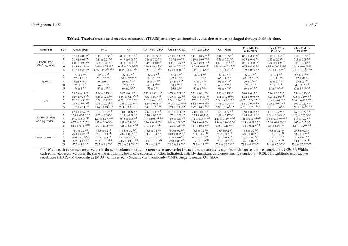
To evaluate the protector effect of the film on the shelf life of the poultry meat, somephysicochemical parameters were evaluated through the 15 days of storage (Table 2). The natural

Coatings 2018, 8, 177 9 of 17
deterioration process resulted in increased pH value, with reduction of the acidity, changes on thecolor (discoloration process) and increase of the moisture content, as can be observed for the meatwithout packaging (unwrapped). Such changes were less significant for the other samples: sampleswrapped in the commercial PVC showed less change than unwrapped samples, but higher changesthan samples wrapped with Ch, thus indicating that the biocomposites produced are promisingsubstitutes to commercial and unsustainable plastic films. The incorporation of GEO in the chitosanmatrix enhanced significantly the activity of the biopolymers once the samples wrapped in those filmsshowed minimum changes (Table 2). The magnitude of the changes observed in samples wrapped inthe biopolymers with MMT, with or without GEO, were in between samples wrapped with Ch andsamples wrapped with Ch + GEO.
In terms of color, the meat was evaluated through the calculation of the hue angle (h*) (Table 2).This angle converts the CIE-L*a*b* coordinates a* and b* into angles representing color tonalitiesstarting at 0◦ and with increasing values rotating counterclockwise: h* of 0◦ represents the color red,90◦ yellow, 180◦ green and 270◦ blue [62]. Initial h* observed was 57◦, i.e., reddish color, over timethe hue angle increased, especially for the control meat and the sample wrapped with the commercialplastic (Table 2). Higher values of hue indicate that the meat color tended to a more yellowish tone,losing its reddish characteristic color. In comparison with the meat protected with the active films,control sample and the meat wrapped in PVC presented higher value of h* in the last day of assessment(p < 0.05). Between the active films incorporated with GEO, despite the amount of essential oil added,the hue angle did not change statistically (p > 0.05), and the meat showed h* values around 55◦–60◦.Chitosan film and Ch + MMT film also slowed down the discoloration process. Moreover, samplepackaged in any of the chitosan films preserved the h* between the day zero and the day 15 (p > 0.05).Thus, the films maintained the original color of the meat, which is a key factor to boost the acceptanceof such products by consumer, once color is a valued sensory attribute when accepting or rejectingfoodstuff [63]. The decrease of the meat redness, mainly observed in the unwrapped samples and inthe samples wrapped with PVC, is directly related to the lipid oxidation process and the degradationof heme molecules with formation of metmyoglobin [14,64,65]. Therefore, chitosan films succeeded inpreventing the discoloration process. Two routes can explain the activity of the films: the antioxidantactivity of the phenolic compounds that retarded the lipid oxidation together with the protectionrelated to the metal chelator ability of chitosan. Present results are in line with the further TBARS andmicrobiological study discussed in Sections 3.2.2 and 3.2.3.
Regarding the pH, control sample reached a pH value around 8.17 after 15 days of refrigeratedstorage (Table 2). Similar to the results of the color, pH changed less in the wrapped samples. At day 15,the smallest pH value was 5.71 recorded for the meat wrapped in Ch + 1%GEO. The other twoconcentrations of GEO also kept pH values low. In fact, for those three samples, the pH value betweenday zero and day 15 did not differed much, and except for Ch + 2% GEO, the change was notstatistically significant (p > 0.05). For all the other films, the pH increased along 15 days of storage,and among the samples the value did not change statistically (p > 0.05). Thus, pH values of samplesprotected with PVC were the same as the pH of the samples protected with Ch, Ch + MMT, or Ch +MMT + GEO. The reduction of the activity of the active films with the incorporation of the MMT isprobably due to the diminish migration of the phenolic compounds from GEO to the meat, thereforereducing the protection against microbial development and lipid oxidation, in line with the findingspresented in the Sections 3.2.2 and 3.2.3. The increase in pH values may be attributed to the growth ofmicroorganisms that produce volatile basic components [14]; therefore, the maintenance of the pHindicates the efficiency of the films in the extension of the shelf life of the products. Similar resultswere reported in literature for chitosan films incorporated with rosemary essential oil applied in fishfillets [66] or for chitosan films incorporated with GEO applied in barracuda fish [67]. In agreement,titratable acidity reduced over the storage time (p < 0.05) (Table 2), which is in line with the increase ofpH observed. The highest drop on the titratable acidity occurred for the unwrapped meat, while thesamples protected showed less significant changes. Similar to the discussion for pH, the changes in

Coatings 2018, 8, 177 10 of 17
this parameter are attributed to the growth of microorganisms that, through the degradation of theproteins, produce amines that neutralize the acids reducing the titratable acidity [14].
The moisture content of the meat packaged with the chitosan films showed a tendency to maintainor decrease over time (Table 2), especially the meat wrapped with films incorporated with GEO(and with no MMT) (p < 0.05). Such behavior may be attributed to the hydrophilic character of chitosanfilms that are able to absorb water and exhibit a high water vapor permeability [30], explaining thereduction in the water content of the meat packaged. In a previous work, poultry meat packagedin chitosan films incorporated with rosemary essential oil and montmorillonite also showed similarbehavior [45], corroborating the present results.
3.2.2. Thiobarbituric Acid Reactive Substances Index
The oxidation status of the meat was assessed through the measurement of the TBARS index.Change in the secondary products of lipid oxidation was statistically different over time for alltreatments (p < 0.05), except for the meat protected with chitosan film incorporated with GEO atthe levels of 0.5%, 1% and 2% (p > 0.05) (Table 2). The presence of montmorillonite diminished theprotection of the active films, probably due to the good interaction between the nanofiller and thepolymer matrix and the phenolic compounds present in the GEO. Thus, the release of the bioactivecompounds was diminished, resulting in a weaker protection effect, although a smaller oxidation ifcompared to the unprotected meat.
Together with the microbial growth, lipid oxidation is one of the most detrimental processes infoodstuffs [66] and the threshold of off-odor perception by consumers corresponds to a TBARS value of0.5 mg MDA/kg sample [67]. Analyzing present results, up to the seventh day of refrigerated storage,only the meat unwrapped had exceeded this TBARS value threshold and, therefore, would be rejectedby the consumers. At day 10 of storage, meat wrapped in PVC, chitosan + MMT, chitosan + MMT +0.5% and 1% of GEO exceeded the limit. Finally, at the last day of assessment (15th day of storage),meat packaged with chitosan + MMT + 2% GEO overpassed the 0.5 mg MDA/kg meat, while thesamples protected with the chitosan film and with chitosan + GEO (0.5%, 1% and 2%) still had notreached the rejection point. This result confirms the potential of the films to be used as preservativesfor fresh poultry meat products, once pristine chitosan films and the biopolymers incorporated onlywith GEO (in all concentrations tested) extended the shelf life of the meat by at least eight days interms of lipid oxidation.
The protection effect of the films may be attributed either to the good oxygen barrier and thechelator ability of the chitosan [66] and/or to the GEO through the ability of its phenolic compounds todonate electrons and stabilize free radicals acting as antioxidants [14]. Other biopolymers incorporatedwith different EOs have demonstrated similar results in food matrices [14,66,68,69].
3.2.3. Microbiological Growth
Changes in the total mesophilic aerobic bacteria and total coliforms of unwrapped and wrappedminced poultry meat are presented in Table 3. The meat presented initial TMAB of 5.1 log CFU/gmeat, bellow the limit of 6.69 log CFU/g meat established in the Regulation (EC) No 2073/2005for minced meat [70]. Over time the total mesophilic aerobic bacteria increased for all treatments(p < 0.05), reaching a final counting of 10.1 log CFU/g meat for unwrapped meat and a reductionvarying between 1.2 and 2.6 log for the samples protected in the active films. Such results represent anincrease in the shelf life time of the product and highlight the films as a tool to preserve the qualityand safety of meat products. Moreover, the biopolymers reduced the microbiological growth morethan the commercial one, which only showed a reduction of 0.5 log compared to unwrapped samples.
Coatings 2018, 8, 177 11 of 17
Table 2. Thiobarbituric acid reactive substances (TBARS) and physicochemical evaluation of meat packaged though shelf life time.
Parameter Day Unwrapped PVC Ch Ch + 0.5% GEO Ch + 1% GEO Ch + 2% GEO Ch + MMT Ch + MMT +0.5% GEO
Ch + MMT +1% GEO
Ch + MMT +2% GEO
TBARS (mgMDA/kg meat)
0 0.11 ± 0.05 a,C 0.11 ± 0.05 a,B 0.11 ± 0.05 a,B 0.11 ± 0.05 a,A 0.11 ± 0.05 a,A 0.11 ± 0.05 a,A,B 0.11 ± 0.05 a,B 0.11 ± 0.05 a,C 0.11 ± 0.05 a,C 0.11 ± 0.05 a,B
3 0.13 ± 0.06 b,C 0.11 ± 0.01 b,B 0.19 ± 0.06 b,B 0.16 ± 0.02 b,A 0.07 ± 0.0 b,B 0.10 ± 0.06 b,A,B 0.34 ± 0.02 a,B 0.15 ± 0.01 b,C 0.15 ± 0.03 b,C 0.18 ± 0.09 b,B
7 0.88 ± 0.20 a,B 0.07 ± 0.01 c,B 0.12 ± 0.02 c,B 0.19 ± 0.10 c,A 0.05 ± 0.02 c,B 0.06 ± 0.02 c,A,B 0.49 ± 0.03 b,A,B 0.17 ± 0.04 c,C 0.16 ± 0.02 c,C 0.13 ± 0.02 c,B
10 1.48 ± 0.33 a,A 0.65 ± 0.25 b,c,A 0.23 ± 0.06 d,e,A,B 0.20 ± 0.02 d,e,A 0.04 ± 0.01 e,B 0.02 ± 0.01 e,B 0.58 ± 0.04 b,c,d,A,B 0.78 ± 0.00 b,B 0.57 ± 0.00 b,c,d,B 0.28 ± 0.03 c,d,e,B
15 1.97 ± 0.28 a,A 0.65 ± 0.02 b,c,d,A 0.42 ± 0.12 c,d,A 0.25 ± 0.0 c,d,A 0.20 ± 0.04 d,A 0.15 ± 0.06 d,A 1.11 ± 0.54 b,A 1.09 ± 0.20 b,A 0.83 ± 0.12 b,c,A 0.51 ± 0.12 b,c,d,A
Hue (◦)
0 57 ± 1 a,E 57 ± 1 a,C 57 ± 1 a,A 57 ± 1 a,B 57 ± 1 a,A 57 ± 1 a,A 57 ± 1 a,A 57 ± 1 a,A 57 ± 1 a,B 57 ± 1 a,B,C
3 62 ± 0 a,b,D 61 ± 1 a,b,c,B 59 ± 2 b,c,d,A 56 ± 1 d,e,B 52 ± 1 e,A 53 ± 1 e,B 62 ± 0 a,b,A 60 ± 2 a,b,c,A 58 ± 1 c,d,B 62 ± 0 a,A
7 64 ± 0 a,b,C 67 ± 0 a,A 59 ± 1 b,c,A 56 ± 1 c,d,B 57 ± 4 c,d,A 57 ± 2 c,d,A 62 ± 3 b,c,A 59 ± 1 b,c,A 64 ± 0 a,b,A 53 ± 2 d,C
10 67 ± 0 a,B 66 ± 1 a,A 59 ± 1 c,A 64 ± 1 a,b,A 58 ± 2 c,A 60 ± 1 b,c,A 61 ± 2 b,c,A 59 ± 1 c,A 60 ± 0 b,c,B 61 ± 2 b,c,A,B
15 70 ± 1 a,A 67 ± 1 a,b,A 60 ± 2 c,d,A 55 ± 0 e,B 55 ± 2 e,A 57 ± 1 d,e,A 62 ± 0 b,c,A 60 ± 0 c,d,A 57 ± 4 c,d,e,B 60 ± 3 c,d,e,A,B
pH
0 5.87 ± 0.11 a,C 5.96 ± 0.15 a,C 5.87 ± 0.11 a,D 5.72 ± 0.02 a,A,B 5.71 ± 0.21 a,A 5.71 ± 0.21 a,B,C 5.96 ± 0.15 a,B 5.96 ± 0.15 a,A 5.96 ± 0.15 a,B 5.96 ± 0.15 a,B
3 6.09 ± 0.35 a,C 6.19 ± 0.08 a,C 6.01 ± 0.20 a,C,D 5.57 ± 0.07 b,B 5.38 ± 0.02 b,B 5.45 ± 0.05 b,C 6.01 ± 0.00 a,B 6.12 ± 0.05 a,A 6.03 ± 0.02 a,B 5.96 ± 0.09 a,b,B
7 7.16 ± 0.30 a,B 6.50 ± 0.12 b,B 6.33 ± 0.02 b,c,B,C 5.62 ± 0.01 f,A,B 5.74 ± 0.04 e,f,A 5.76 ± 0.10 e,f,B 5.84 ± 0.02 d,f,B 6.02 ± 0.04 c,d,e,A 6.10 ± 0.04 c,d,B 5.94 ± 0.08 d,e,f,B
10 7.55 ± 0.02 a,B 6.79 ± 0.04 b,B 6.51 ± 0.21 b,c,B 5.58 ± 0.02 e,B 5.60 ± 0.03 e,A,B 5.52 ± 0.04 e,B,C 6.01 ± 0.04 d,B 6.14 ± 0.20 d,A 6.29 ± 0.07 c,d,B 6.00 ± 0.20 d,B
15 8.17 ± 0.14 a,A 7.26 ± 0.13 b,c,A 7.14 ± 0.23 b,c,A 5.85 ± 0.2 d,e,A 5.71 ± 0.09 e,A 6.22 ± 0.01 d,e,A 7.27 ± 0.36 b,c,A 6.50 ± 0.55 c,d,e,A 7.33 ± 0.36 b,A 6.61 ± 0.24 b,c,d,A
Acidity (% oleicacid equivalent)
0 1.80 ± 0.39 a,A 1.68 ± 0.22 a,A 1.80 ± 0.39 a,A 2.12 ± 0.11 a,A 2.12 ± 0.11 a,A 2.12 ± 0.11 a,A 1.68 ± 0.22 a,A 1.68 ± 0.22 a,A 1.68 ± 0.22 a,A 1.68 ± 0.22 a,A
3 1.24 ± 0.37 b,A,B 1.32 ± 0.08 b,B 1.13 ± 0.01 b,B 1.70 ± 0.02 a,B 1.75 ± 0.08 a,B 1.75 ± 0.03 a,B 1.19 ± 0.17 b,B 1.04 ± 0.19 b,B 1.04 ± 0.09 b,C,D 1.26 ± 0.05 b,A,B
7 0.42 ± 0.14 f,C 1.27 ± 0.07 c,d,B 0.85 ± 0.05 e,B 1.47 ± 0.01 a,b,B,C 1.55 ± 0.04 a,C 1.41 ± 0.03 a,b,c,C 1.49 ± 0.00 a,b,A,B 1.22 ± 0.00 c,d,A,B 1.33 ± 0.13 b,c,d,B,C 1.16 ± 0.02 d,B
10 0.73 ± 0.15 c,B,C 1.15 ± 0.06 b,B,C 1.13 ± 0.18 b,c,B 1.30 ± 0.20 a,b,C 1.46 ± 0.09 a,b,C 1.36 ± 0.04 a,b,C 1.46 ± 0.10 a,b,A,B 1.58 ± 0.23 a,A,B 1.52 ± 0.06 a,b,A,B 1.65 ± 0.15 a,A
15 0.66 ± 0.14 b,B,C 0.87 ± 0.02 a,b,C 1.22 ± 0.30 a,A,B 0.73 ± 0.11 a,b,D 1.22 ± 0.03 a,D 1.11 ± 0.08 a,b,D 0.78 ± 0.15 a,b,C 1.24 ± 0.34 a,A,B 0.76 ± 0.09 a,b,D 1.13 ± 0.29 a,b,B
Water content (%)
0 75.5 ± 0.2 a,B 75.5 ± 0.2 a,B 75.5 ± 0.2 a,A 75.5 ± 0.2 a,A 75.5 ± 0.2 a,A 75.5 ± 0.2 a,A 75.5 ± 0.2 a,A 75.5 ± 0.2 a,A 75.5 ± 0.2 a,A 75.5 ± 0.2 a,A
3 75.4 ± 0.2 a,b,B 75.8 ± 0.4 a,B 73.4 ± 0.1 c,B,C 74.7 ± 0.4 b,A 73.3 ± 0.0 c,A,B 73.6 ± 0.2 c,B 73.8 ± 0.2 c,B 73.1 ± 0.4 c,B 73.4 ± 0.2 c,B 73.0 ± 0.2 c,C
7 76.0 ± 0.2 a,A,B 75.1 ± 0.4 a,B 72.5 ± 0.1 b,C 73.2 ± 0.3 b,B 72.6 ± 0.6 b,B 72.8 ± 0.5 b,B,C 73.2 ± 0.3 b,B 73.1 ± 0.3 b,B 72.8 ± 0.0 b,B 72.9 ± 0.7 b,C
10 76.2 ± 0.4 a,A,B 75.6 ± 0.3 a,b,B 74.5 ± 0.3 b,c,A,B 74.4 ± 0.5 c,A,B 72.0 ± 0.1 d,B 70.7 ± 0.7 e,C,D 73.6 ± 0.2 c,B 74.1 ± 0.2 c,B 73.4 ± 0.6 c,B 74.1 ± 0.2 c,B
15 77.3 ± 1.0 a,A 76.7 ± 0.1 a,b,A 73.4 ± 0.8 c,d,e,B,C 71.6 ± 0.8 e,C 72.0 ± 2.0 d,e,B 71.3 ± 0.4 e,D 75.9 ± 0.6 a,b,c,A 74.2 ± 0.9 b,c,d,B 74.8 ± 0.2 a,b,c,A 73.6 ± 0.2 c,d,e,B,C
A–D: Within each parameter, mean values in the same column not sharing upper case superscript letters indicate statistically significant differences among samples (p < 0.05); a–f: Withineach parameter, mean values in the same line not sharing lower case superscript letters indicate statistically significant differences among samples (p < 0.05). Thiobarbituric acid reactivesubstances (TBARS), Malonaldehyde (MDA), Chitosan (Ch), Sodium Montmorillonite (MMT), Ginger Essential Oil (GEO).

Coatings 2018, 8, 177 12 of 17
Table 3. Microbiological study of meat packaged though shelf life time.
Parameter Day Unwrapped PVC Ch Ch + 0.5% GEO Ch + 1% GEO Ch + 2% GEO Ch + MMT Ch + MMT +0.5% GEO
Ch + MMT +1% GEO
Ch + MMT +2% GEO
TMAB (logCFU/g meat)
0 5.1 ± 0.1 a,C 5.1 ± 0.1 a,C 5.1 ± 0.1 a,C 5.1 ± 0.1 a,C 5.1 ± 0.1 a,B 5.1 ± 0.1 a,B 5.1 ± 0.1 a,C 5.1 ± 0.1 a,C 5.1 ± 0.1 a,B 5.1 ± 0.1 a,B
3 8.8 ± 0.1 a,B 8.7 ± 0.0 a,B 7.1 ± 0.3 b,B 7.1 ± 0.4 b,B 6.7 ± 0.2 b,A 6.8 ± 0.2 b,A 6.7 ± 0.0 b,B 7.1 ± 0.3 b,B 5.3 ± 0.2 c,B 5.9 ± 0.0 c,B
7 10.2 ± 0.1 a,A 9.5 ± 0.3 a,b,A 8.5 ± 0.2 c,d,A 7.4 ± 0.0 e,f,B 7.8 ± 0.1 d,f,A 8.1 ± 0.1 c,d,e,A 7.2 ± 0.7 f,A,B 8.7 ± 0.1 b,c,A 8.7 ± 0.1 c,A 8.3 ± 0.2 c,d,A
10 10.1 ± 0.0 a,A 9.6 ± 0.1 a,b,A 8.7 ± 0.5 c,A 8.3 ± 0.2 c,d,A 7.5 ± 0.1 e,A 7.7 ± 0.5 d,e,A 8.5 ± 0.0 c,A 8.8 ± 0.0 c,A 8.9 ± 0.1 b,c,A 8.8 ± 0.0 c,A
Total coliforms(log MPN/g meat)
0 0.8 ± 0.4 a,C 0.8 ± 0.4 a,C 0.8 ± 0.4 a,C 0.8 ± 0.4 a,B 0.8 ± 0.4 a,C 0.8 ± 0.4 a,B 0.8 ± 0.4 a,C 0.8 ± 0.4 a,C 0.8 ± 0.4 a,D 0.8 ± 0.4 a,C
3 3.0 ± 0.4 b,c,B,C 3.4 ± 0.0 a,b,B 2.8 ± 0.5 b,c,B 3.2 ± 0.2 a,b,c,A 4.0 ± 0.0 a,A 2.8 ± 0.4 b,c,A 2.9 ± 0.5 b,c,B 2.9 ± 0.5 b,c,B 2.2 ± 0.2 c,C 2.2 ± 0.2 c,B
7 4.5 ± 1.7 a,A,B 3.9 ± 0.5 a,b,c,B 4.0 ± 0.9 a,b,A,B 3.4 ± 0.3 a,b,c,A 2.3 ± 0.00 b,c,B 2.1 ± 0.1 c,A 3.0 ± 0.3 a,b,c,B 3.6 ± 0.4 a,b,c,B 3.7 ± 0.4 a,b,c,B 2.4 ± 0.1 b,c,B
10 6.7 ± 0.6 a,A 6.4 ± 0.0 a,A 5.3 ± 1.0 a,A 3.2 ± 0.0 b,A 2.6 ± 0.6 b,B 2.2 ± 0.8 b,A 5.9 ± 0.2 a,A 6.2 ± 0.2 a,A 5.9 ± 0.5 a,A 5.9 ± 0.5 a,A
A–D: Within each parameter, mean values in the same column not sharing upper case superscript letters indicate statistically significant differences among samples (p < 0.05); a–f: Withineach parameter, mean values in the same line not sharing lower case superscript letters indicate statistically significant differences among samples (p < 0.05). Total mesophilic aerobicbacteria (TMAB), Colony forming units (CFU), Chitosan (Ch), Sodium Montmorillonite (MMT), Most probable number (MPN) Ginger Essential Oil (GEO).

Coatings 2018, 8, 177 13 of 17
Fresh poultry meat is considered not acceptable for organoleptic (sensory) evaluation at countsgreater than 7 log CFU/g [71]. In the present study, the unprotected meat and the samples wrapped incommercial film exceeded this counting since the third day of storage, while the samples protectedwith the active packaging were either in this limit or below of it. Ginger essential oil enhanced theprotection effect of chitosan film, while MMT limited the activity of the films. As previously discussed,the presence of the nanofiller may interfere in the diffusion process of the active compounds and blockthe functional group of chitosan (amide group), reducing its antimicrobial action. Similar results wereobserved in fresh chicken breast fillets coated with sodium caseinate incorporated with a nanoemulsionof ginger essential oil [27]. The authors attributed the reduction in the microbial growth to the presenceof GEO in the edible coating, and also observed smaller counting with the increase of EO concentration.The protective effect of the bio-based films produced can also be related to the good oxygen barrierproperties of chitosan film, once reduced exposure of packaged meat products to high O2 concentrationreduces the growth of aerobic microorganisms and oxidation of lipids and myoglobin [7].
The results for total coliforms were in line with the ones reported for TMAB (Table 3). Unwrappedmeat presented the higher counting, followed by samples wrapped with PVC. Samples wrapped withthe active films showed the lower counting. Again, it was observed that the presence of MMT reducedthe antimicrobial activity of the chitosan film (without statistical significance, p > 0.05) and, the presenceof GEO enhanced it (with statistical significance, p < 0.05). Although the differences between the totalcoliform counting for the meat protected with chitosan + 0.5%, 1% or 2% GEO were not statisticallydifferent (p > 0.05), a dose response could be observed once the smallest contamination at the 10th dayof storage was found for the highest amount of GEO incorporated. Moreover, in comparison to themeat unwrapped or wrapped in PVC, the samples packaged in chitosan film + 2% GEO presented areduction of 4.5 and 4.2 log in the total coliforms count, i.e., a good antimicrobial action. The phenols,monoterpenes, sesquiterpenes and aldehydes present in EO may be responsible for the results reported.The possible mechanism of action of these oxygenated bioactive compounds may be through thedisruption and penetration in the lipid structure of the bacteria cell membrane with further damage ofthe microorganism enzyme systems [27]. Although GEO did not show antimicrobial activity againstGram-negative bacteria in the in vitro assay, its application in the film caused reduction in the coliformcounting in the meat. This is probably due to a synergistic effect of the chitosan and GEO, as discussedby Khanjari, Karabagias and Kontominas [72], that reported lower microbial growth in the raw chickenfillet packaged with N,O-carboxymethyl chitosan incorporated with oregano essential oil, comparedto samples packaged in pristine polymer or samples with added pure oregano essential oil. Moreover,the type of food matrix (high moisture content and presence of lipids) facilitated the diffusion of theGEO from the packaging enabling its active action.
4. Conclusions
The preparation of bio-based films demonstrated their potential to be used as primary packagingmaterial to fresh poultry meat, being capable of retarding deterioration process by antimicrobial andantioxidant mechanisms and extending its shelf life time. Although the in vitro results were notas positive as expected, when in direct contact with the food tested, the films demonstrated goodefficiency, highlighting the remarkable potential to extend the shelf life of poultry meat products.The incorporation of montmorillonite, at the level tested, diminished the bioactivity of the filmsproduced, while the incorporation of GEO potentialized it. When no mechanical improvements aredemanded, the use of MMT is unnecessary. In fact, chitosan film with one percent of GEO is a formulathat optimizes the protection effect with an intermediate amount of essential oil, minimizing thenegative effects of cost and flavor alterations related to the use of essential oils. Yet, future studiesare needed to identify a balanced formula that can optimize the bioactivity of the bionanocompositeswith the level of montmorillonite added, for applications that also require mechanical improvements.Further, more research studies with EOs or individual compounds, such as some phenolic compounds,combined with reinforcements, such as MMT, in biopolymers will help to clarify the molecular

Coatings 2018, 8, 177 14 of 17
mechanisms associated with the entrapment/release of the bioactive compounds, helping to definefuture pathways for the production of bionanocomposites.
Author Contributions: V.G.L.S., A.L.F. and M.P.D. conceived and designed the experiments. V.G.L.S., J.R.A.P.and É.T.V. performed the experiments and analyzed the data. V.G.L.S. wrote the paper with contributions fromthe remaining authors. A.L.F., M.P.D. and I.M.C. supervised the execution of analyses and revised the data andthe manuscript.
Funding: This work was supported by CNPq/Brazil [grant number 200790/2014-5]; and MEtRiCS throughFCT/MCTES [UID/SEM/04077/2013]; This work was also supported by the Associate Laboratory for GreenChemistry LAQV which is financed by national funds from FCT/MCTES (UID/QUI/50006/2013) and co-financedby the ERDF under the PT2020 Partnership Agreement (POCI-01-0145-FEDER-007265).
Acknowledgments: The authors acknowledge BYK Additives & Instruments for donating the sodiummontmorillonite (Cloisite®Na+).
Conflicts of Interest: The authors declare no conflict of interest. The founding sponsors had no role in the designof the study; in the collection, analyses, or interpretation of data; in the writing of the manuscript, and in thedecision to publish the results.
References
1. FAO. Poultry Development Review; FAO: Rome, Italy, 2013; ISBN 978-92-5-108067-2.2. OECD-FAO Agricultural Outlook 2016–2025; OECD Publishing: Paris, France, 2016; ISBN 9789264253223.3. Petrou, S.; Tsiraki, M.; Giatrakou, V.; Savvaidis, I.N. Chitosan dipping or oregano oil treatments, singly or
combined on modified atmosphere packaged chicken breast meat. Int. J. Food Microbiol. 2012, 156, 264–271.[CrossRef] [PubMed]
4. Latou, E.; Mexis, S.F.; Badeka, V.; Kontakos, S.; Kontominas, M.G. Combined effect of chitosan and modifiedatmosphere packaging for shelf life extension of chicken breast fillets. LWT Food Sci. Technol. 2014, 55,263–268. [CrossRef]
5. Arkoun, M.; Daigle, F.; Holley, R.A.; Heuzey, M.C.; Ajji, A. Chitosan-based nanofibers as bioactive meatpackaging materials. Packag. Technol. Sci. 2018, 31, 185–195. [CrossRef]
6. Souza, V.G.L.; Fernando, A.L. Nanoparticles in food packaging: Biodegradability and potential migration tofood—A review. Food Packag. Shelf Life 2016, 8, 63–70. [CrossRef]
7. Lee, K.T. Quality and safety aspects of meat products as affected by various physical manipulations ofpackaging materials. Meat Sci. 2010, 86, 138–150. [CrossRef] [PubMed]
8. Prasad, P.; Kochhar, A. Active packaging in food industry: A Review. IOSR J. Environ. Sci. Toxicol.Food Technol. 2014, 8. [CrossRef]
9. Dutta, P.K.; Tripathi, S.; Mehrotra, G.K.; Dutta, J. Perspectives for chitosan based antimicrobial films in foodapplications. Food Chem. 2009, 114, 1173–1182. [CrossRef]
10. Otoni, C.G.; Espitia, P.J.P.; Avena-Bustillos, R.J.; McHugh, T.H. Trends in antimicrobial food packagingsystems: Emitting sachets and absorbent pads. Food Res. Int. 2016, 83, 60–73. [CrossRef]
11. Malhotra, B.; Keshwani, A.; Kharkwal, H. Antimicrobial food packaging: Potential and pitfalls.Front. Microbiol. 2015, 6, 611. [CrossRef] [PubMed]
12. Yuan, G.; Chen, X.; Li, D. Chitosan films and coatings containing essential oils: The antioxidant andantimicrobial activity, and application in food systems. Food Res. Int. 2016, 89, 117–128. [CrossRef] [PubMed]
13. Alboofetileh, M.; Rezaei, M.; Hosseini, H.; Abdollahi, M. Antimicrobial activity of alginate/claynanocomposite films enriched with essential oils against three common foodborne pathogens. Food Control2014, 36, 1–7. [CrossRef]
14. De Melo, A.A.; Geraldine, R.M.; Silveira, M.F.; Torres, M.C.; e Rezende, C.S.; Silva, C.; Fernandes, T.H.;de Oliveira, A.N. Microbiological quality and other characteristics of refrigerated chicken meat in contactwith cellulose acetate-based film incorporated with rosemary essential oil. Braz. J. Microbiol. 2012, 43,1419–1427. [CrossRef] [PubMed]
15. Zivanovic, S.; Chi, S.; Draughon, A. Antimicrobial activity of chitosan. Science 2005, 70, 45–51. [CrossRef]16. Darder, M.; Colilla, M.; Ruiz-Hitzky, E. Biopolymer-clay nanocomposites based on chitosan intercalated in
montmorillonite. Chem. Mater. 2003, 15, 3774–3780. [CrossRef]
Coatings 2018, 8, 177 15 of 17
17. Ferreira, A.R.V.; Torres, C.A.V.; Freitas, F.; Sevrin, C.; Grandfils, C.; Reis, M.A.M.; Alves, V.D.; Coelhoso, I.M.Development and characterization of bilayer films of FucoPol and chitosan. Carbohydr. Polym. 2016, 147,8–15. [CrossRef] [PubMed]
18. Srinivasa, P.C.; Ramesh, M.N.; Tharanathan, R.N. Effect of plasticizers and fatty acids on mechanical andpermeability characteristics of chitosan films. Food Hydrocoll. 2007, 21, 1113–1122. [CrossRef]
19. Azeredo, H.M.C.; Mattoso, L.H.C.; Avena-Bustillos, R.J.; Filho, G.C.; Munford, M.L.; Wood, D.; McHugh, T.H.Nanocellulose reinforced chitosan composite films as affected by nanofiller loading and plasticizer content.J. Food Sci. 2010, 75, N1–N7. [CrossRef] [PubMed]
20. Mihindukulasuriya, S.D.F.; Lim, L.-T. Nanotechnology development in food packaging: A review. Trends FoodSci. Technol. 2014, 40, 149–167. [CrossRef]
21. Souza, V.G.L.; Pires, J.R.A.; Rodrigues, P.F.; Lopes, A.A.S.; Fernandes, F.M.B.; Duarte, M.P.; Coelhoso, I.M.;Fernando, A.L. Bionanocomposites of chitosan/montmorillonite incorporated with Rosmarinus officinalisessential oil: Development and physical characterization. Food Packag. Shelf Life 2018, 16, 148–156. [CrossRef]
22. Coelho, A.C.V.; Santos, P.D.S.; Santos, H.D.S. Argilas especiais: Argilas quimicamente modificadas-umarevisão. Quim. Nova 2007, 30, 1282–1294. [CrossRef]
23. El-Ghorab, A.H.; Nauman, M.; Anjum, F.M.; Hussain, S.; Nadeem, M. A comparative study on chemicalcomposition and antioxidant activity of ginger (Zingiber officinale) and cumin (Cuminum cyminum). J. Agric.Food Chem. 2010, 58, 8231–8237. [CrossRef] [PubMed]
24. López, E.I.C.; Balcázar, M.F.H.; Mendoza, J.M.R.; Ortiz, A.D.R.; Melo, M.T.O.; Parrales, R.S.; Delgado, T.H.Antimicrobial activity of essential oil of Zingiber officinale Roscoe (Zingiberaceae). Am. J. Plant Sci. 2017, 8,1511–1524. [CrossRef]
25. Noshirvani, N.; Ghanbarzadeh, B.; Gardrat, C.; Rezaei, M.R.; Hashemi, M.; Le Coz, C.; Coma, V.Cinnamon and ginger essential oils to improve antifungal, physical and mechanical properties ofchitosan-carboxymethyl cellulose films. Food Hydrocoll. 2017, 70, 36–45. [CrossRef]
26. Zhang, L.; Liu, A.; Wang, W.; Ye, R.; Liu, Y.; Xiao, J.; Wang, K. Characterisation of microemulsion nanofilmsbased on Tilapia fish skin gelatine and ZnO nanoparticles incorporated with ginger essential oil: Meatpackaging application. Int. J. Food Sci. Technol. 2017, 52, 1670–1679. [CrossRef]
27. Noori, S.; Zeynali, F.; Almasi, H. Antimicrobial and antioxidant efficiency of nanoemulsion-based ediblecoating containing ginger (Zingiber officinale) essential oil and its effect on safety and quality attributes ofchicken breast fillets. Food Control 2018, 84, 312–320. [CrossRef]
28. Klangmuang, P.; Sothornvit, R. Active hydroxypropyl methylcellulose-based composite coating powder tomaintain the quality of fresh mango. LWT Food Sci. Technol. 2018, 91, 541–548. [CrossRef]
29. BYK. Cloisite®Na+ Technical Data Sheet. Available online: http://www.byk.com/en/additives/additives-by-name/cloisite-na.php (accessed on 5 February 2018).
30. Souza, V.G.L.; Fernando, A.L.; Pires, J.R.A.; Rodrigues, P.F.; Lopes, A.A.S.S.; Fernandes, F.M.B. Physicalproperties of chitosan films incorporated with natural antioxidants. Ind. Crops Prod. 2017, 107, 565–572.[CrossRef]
31. Dias, M.V.; Machado Azevedo, V.; Borges, S.V.; Soares, N.D.F.F.; de Barros Fernandes, R.V.; Marques, J.J.;Medeiros, É.A.A. Development of chitosan/montmorillonite nanocomposites with encapsulated α-tocopherol.Food Chem. 2014, 165, 323–329. [CrossRef] [PubMed]
32. Souza, V.G.L.; Rodrigues, P.F.; Duarte, M.P.; Fernando, A.L. Antioxidant migration studies in chitosan filmsincorporated with plant extracts. J. Renew. Mater. 2018. [CrossRef]
33. Singleton, V.L.; Orthofer, R.; Lamuela-Raventós, R.M. Analysis of total phenols and other oxidation substratesand antioxidants by means of Folin-ciocalteu. Methods Enzymol. 1999, 299, 152–178. [CrossRef]
34. Brand-Williams, W.; Cuvelier, M.E.; Berset, C. Use of a free radical method to evaluate antioxidant activity.LWT Food Sci. Technol. 1995, 28, 25–30. [CrossRef]
35. Chungy, D.; Papadakis, S.E.; Yamy, K.L. Simple models for assessing migration from food-packaging films.Food Addit. Contam. 2002, 19, 611–617. [CrossRef] [PubMed]
36. Nouri, A.; Yaraki, M.T.; Ghorbanpour, M.; Agarwal, S.; Gupta, V.K. Enhanced antibacterial effect of chitosanfilm using montmorillonite/CuO nanocomposite. Int. J. Biol. Macromol. 2017, 109, 1219–1231. [CrossRef][PubMed]
37. ASTM E2149 Standard Test Methods for Determining the Antimicrobial Activity of Immobilized AntimicrobialAgents Under Dynamic Contact Conditions; ASTM International: West Conshohocken, PA, USA, 2001.

Coatings 2018, 8, 177 16 of 17
38. AOAC. Official Methods of Analysis of the Association of Official Analytical Chemists, 20th ed.; AOAC International:Gaithersburg, MD, USA, 2016.
39. Rosmini, M.R.; Perlo, F.; Pérez-Alvarez, J.A.; Pagán-Moreno, M.J.; Gago-Gago, A.; López-Santoveña, F.;Aranda-Catalá, V. TBA test by an extractive method applied to “paté”. Meat Sci. 1996, 42, 103–110. [CrossRef]
40. ISO 4833-1:2013 Microbiology of the Food Chain—Horizontal Method for the Enumeration of Microorganisms—Part 1:Colony Count at 30 Degrees C by the Pour Plate Technique; International Organization for Standardization:Geneva, Switzerland, 2013.
41. ISO 4831:2006(en) Microbiology of Food and Animal Feeding Stuffs—Horizontal Method for the Detection andEnumeration of Coliforms—Most Probable Number Technique; International Organization for Standardization:Geneva, Switzerland, 2006.
42. Lopez de Dicastillo, C.; Nerin, C.; Alfaro, P.; Catala, R.; Gavara, R.; Hernandez-Munoz, P. Development ofnew antioxidant active packaging films based on ethylene vinyl alcohol copolymer (EVOH) and green teaextract. J. Agric. Food Chem. 2011, 59, 7832–7840. [CrossRef] [PubMed]
43. Calatayud, M.; López-de-Dicastillo, C.; López-Carballo, G.; Vélez, D.; Hernández Muñoz, P.; Gavara, R.Active films based on cocoa extract with antioxidant, antimicrobial and biological applications. Food Chem.2013, 139, 51–58. [CrossRef] [PubMed]
44. Bolumar, T.; Andersen, M.L.; Orlien, V. Antioxidant active packaging for chicken meat processed by highpressure treatment. Food Chem. 2011, 129, 1406–1412. [CrossRef]
45. Souza, V.G.L.; Pires, J.R.A.; Vieira, E.T.; Coelhoso, I.M.; Duarte, M.P.; Fernando, A.L. Activity ofchitosan-montmorillonite bionanocomposites incorporated with rosemary essential oil: From in vitro assaysto aplication in fresh poultry meat (unpublished, manuscript in preparation).
46. Abdollahi, M.; Rezaei, M.; Farzi, G. A novel active bionanocomposite film incorporating rosemary essentialoil and nanoclay into chitosan. J. Food Eng. 2012, 111, 343–350. [CrossRef]
47. Ribeiro-Santos, R.; Andrade, M.; de Melo, N.R.; Sanches-Silva, A. Use of essential oils in active foodpackaging: Recent advances and future trends. Trends Food Sci. Technol. 2017, 61, 132–140. [CrossRef]
48. Regnier, T.; Combrinck, S.; Du Plooy, W. Essential oils and other plant extracts as food preservatives.In Progress in Food Preservation; Bhat, R., Alias, A.K., Paliyath, G., Eds.; Wiley-Blackwell: Oxford, UK, 2012;pp. 539–579.
49. Teixeira, B.; Marques, A.; Ramos, C.; Neng, N.R.; Nogueira, J.M.F.; Saraiva, J.A.; Nunes, M.L. Chemicalcomposition and antibacterial and antioxidant properties of commercial essential oils. Ind. Crops Prod. 2013,43, 587–595. [CrossRef]
50. Singh, P.; Singh, S.; Kapoor, I.P.S.; Singh, G.; Isidorov, V.; Szczepaniak, L. Chemical composition andantioxidant activities of essential oil and oleoresins from Curcuma zedoaria rhizomes, part-74. Food Biosci.2013, 3, 42–48. [CrossRef]
51. López, P.; Sánchez, C.; Batlle, R.; Nerín, C. Solid- and vapor-phase antimicrobial activities of six essential oils:Susceptibility of selected foodborne bacterial and fungal strains. J. Agric. Food Chem. 2005, 53, 6939–6946.[CrossRef] [PubMed]
52. Sivasothy, Y.; Chong, W.K.; Hamid, A.; Eldeen, I.M.; Sulaiman, S.F.; Awang, K. Essential oils of Zingiberofficinale var. rubrum Theilade and their antibacterial activities. Food Chem. 2011, 124, 514–517. [CrossRef]
53. Shariatinia, Z.; Fazli, M. Mechanical properties and antibacterial activities of novel nanobiocomposite filmsof chitosan and starch. Food Hydrocoll. 2015, 46, 112–124. [CrossRef]
54. Singh, G.; Kapoor, I.P.S.; Singh, P.; de Heluani, C.S.; de Lampasona, M.P.; Catalan, C.A.N. Chemistry,antioxidant and antimicrobial investigations on essential oil and oleoresins of Zingiber officinale.Food Chem. Toxicol. 2008, 46, 3295–3302. [CrossRef] [PubMed]
55. Trajano, V.N.; Lima, O.; Leite, E.; Travassos, R. Propriedade antibacteriana de óleos essenciais de especiariassobre bactérias contaminantes de alimentos. 2009, 29, 542–545. [CrossRef]
56. Hafsa, J.; Smach, M.; Ben Khedher, M.R.; Charfeddine, B.; Limem, K.; Majdoub, H.; Rouatbi, S. Physical,antioxidant and antimicrobial properties of chitosan films containing Eucalyptus globulus essential oil.LWT Food Sci. Technol. 2016, 68, 356–364. [CrossRef]
57. Siripatrawan, U.; Vitchayakitti, W. Improving functional properties of chitosan films as active food packagingby incorporating with propolis. Food Hydrocoll. 2016, 61, 695–702. [CrossRef]
58. Abdollahi, M.; Rezaei, M.; Farzi, G. Original article Improvement of active chitosan film properties withrosemary essential oil for food packaging. Food Sci. Technol. 2012, 47, 847–853. [CrossRef]

Coatings 2018, 8, 177 17 of 17
59. Verlee, A.; Mincke, S.; Stevens, C.V. Recent developments in antibacterial and antifungal chitosan and itsderivatives. Carbohydr. Polym. 2017, 164, 268–283. [CrossRef] [PubMed]
60. Siripatrawan, U.; Harte, B.R. Physical properties and antioxidant activity of an active film from chitosanincorporated with green tea extract. Food Hydrocoll. 2010, 24, 770–775. [CrossRef]
61. Lavorgna, M.; Piscitelli, F.; Mangiacapra, P.; Buonocore, G.G. Study of the combined effect of both clay andglycerol plasticizer on the properties of chitosan films. Carbohydr. Polym. 2010, 82, 291–298. [CrossRef]
62. MINOLTA. Precise Color Communication: Color Control from Perception to Instrumentation; Minolta Co., Ltd.:Osaka, Japan, 2007.
63. Rojas, M.C.; Brewer, M.S. Effect of natural antioxidants on oxidative stability of frozen, vacuum-packagedbeef and pork. J. Food Qual. 2008, 31, 173–188. [CrossRef]
64. Contini, C.; Álvarez, R.; O’Sullivan, M.; Dowling, D.P.; Gargan, S.Ó.; Monahan, F.J. Effect of an activepackaging with citrus extract on lipid oxidation and sensory quality of cooked turkey meat. Meat Sci. 2014,96, 1171–1176. [CrossRef] [PubMed]
65. Ghaderi-Ghahfarokhi, M.; Barzegar, M.; Sahari, M.A.; Ahmadi Gavlighi, H.; Gardini, F. Chitosan-cinnamonessential oil nano-formulation: Application as a novel additive for controlled release and shelf life extensionof beef patties. Int. J. Biol. Macromol. 2017, 102, 19–28. [CrossRef] [PubMed]
66. Abdollahi, M.; Rezaei, M.; Farzi, G. Influence of chitosan/clay functional bionanocomposite activated withrosemary essential oil on the shelf life of fresh silver carp. Int. J. Food Sci. Technol. 2014, 49, 811–818.[CrossRef]
67. Remya, S.; Mohan, C.O.; Bindu, J.; Sivaraman, G.K.; Venkateshwarlu, G.; Ravishankar, C.N. Effect of chitosanbased active packaging film on the keeping quality of chilled stored barracuda fish. J. Food Sci. Technol. 2016,53, 685–693. [CrossRef] [PubMed]
68. Yang, F.; Hu, S.; Lu, Y.; Yang, H.; Zhao, Y.; Li, L. Effects of coatings of polyethyleneimine and thyme essentialoil combined with chitosan on sliced fresh channa argus during refrigerated storage. J. Food Process Eng.2015, 38, 225–233. [CrossRef]
69. Atarés, L.; Pérez-Masiá, R.; Chiralt, A. The role of some antioxidants in the HPMC film properties and lipidprotection in coated toasted almonds. J. Food Eng. 2011, 104, 649–656. [CrossRef]
70. European Commission. Commmission Regulation (EC) No 2073/2005 of 15 November 2005 onmicrobiological criteria for foodstuffs. Off. J. Eur. Union 2005, L338, 1–19. Available online: https://www.fsai.ie/uploadedFiles/Reg2073_2005%281%29.pdf (accessed on 5 February 2018).
71. Economou, T.; Pournis, N.; Ntzimani, A.; Savvaidis, I.N. Nisin-EDTA treatments and modified atmospherepackaging to increase fresh chicken meat shelf-life. Food Chem. 2009, 114, 1470–1476. [CrossRef]
72. Khanjari, A.; Karabagias, I.K.; Kontominas, M.G. Combined effect of N,O-carboxymethyl chitosan andoregano essential oil to extend shelf life and control Listeria monocytogenes in raw chicken meat fillets.LWT Food Sci. Technol. 2013, 53, 94–99. [CrossRef]
© 2018 by the authors. Licensee MDPI, Basel, Switzerland. This article is an open accessarticle distributed under the terms and conditions of the Creative Commons Attribution(CC BY) license (http://creativecommons.org/licenses/by/4.0/).
Copyright © 2022 FDOKUMEN