Setting Aside Cells for Future Use in Development and Evolution
27
-
Upload
khangminh22 -
Category
Documents
-
view
2 -
download
0
Transcript of Setting Aside Cells for Future Use in Development and Evolution
3
1 Deferred-Use Cells in Development and EvolutionA Life History Perspective
Cory D. BishopSt. Francis Xavier University
Andreas HeylandUniversity of Guelph
Jason HodinUniversity of Washington
1.1 ONTOGENY AS A TIME-STRUCTURED PROCESS OF CELLULAR INTERACTION THAT CULMINATES WITH REPRODUCTION
The cell is evolution’s most magnificent achievement and embryonic development is merely a baroque elaboration.
Lewis Wolpert (1999, p. 1)
CONTENTS
1.1 Ontogeny as a Time-Structured Process of Cellular Interaction That Culminates with Reproduction .........................................................................3
1.2 Development: A Balancing Act between Proliferation, Pluripotency, and Function .....................................................................................................5
1.3 Deferred Development in the Context of Complex Life Cycle Evolution ........71.4 Ontologies of Deferred Development and Deferred-Use Cells ...................... 121.5 Independent Evolution of Extreme Patterns in Deferred Development ......... 18
1.5.1 Nemerteans ......................................................................................... 191.5.2 Echinoderms ....................................................................................... 211.5.3 Insects .................................................................................................22
1.6 Summary and Conclusion ...............................................................................25Acknowledgements ..................................................................................................25References ................................................................................................................25
4 Deferring Development
Multicellular life has resulted in the evolution of ontogenies that generally begin from a single cell every generation (Buss 1987; Grosberg and Strathmann 1998; Grosberg and Strathmann 2007). Plausible reasons for this commonality are that a cyclical return to a unicellular state evolved independently in multicellular taxa to purge cytoplasmic or genetic parasites (Grosberg and Strathmann 1998) and muta-tions (Muller 1964). The evolution of this broadly shared pattern of reproductive mode also invokes the evolution of the following:
i. Mechanisms for specifying divergent fates. If only gametes carry genes across generations, then a key specification event in a nascent obligate multicellular organism was “germ” versus “not-germ” (Swartz and Wessel 2015), where germ refers to cells having the developmental potential to form gametes. Such not-germ cells would function in resource acquisition, motility, as a defensive structure, or some other nonreproductive function sensu stricto.
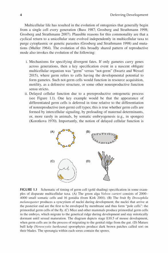
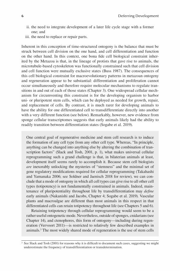
ii. Delayed cellular function due to a prereproductive ontogenetic process (see Figure 1.1). One key example would be that the appearance of differentiated germ cells is deferred in time relative to the differentiation of nonreproductive (not-germ) cell types; this is true whether germ cells are formed by intercellular signaling, by preloading of maternal determinants, or, more rarely in animals, by somatic embryogenesis (e.g., in sponges) (Korotkova 1970). Importantly, the notion of delayed cellular function is
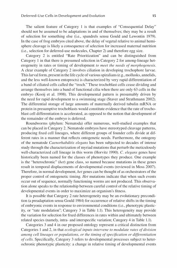
FIGURE 1.1 Schematic of timing of germ cell (gold shading) specification in some exam-ples of disparate multicellular taxa. (A) The green alga Volvox carteri consists of 2000–4000 small somatic cells and 16 gonidia (from Kirk 2001). (B) The fruit fly Drosophila melanogaster produces a syncytium of nuclei during development; the nuclei that arrive at the posterior end are the first to be enveloped by membrane and thus form “pole cells”: the primordial germ cells of the fly. (C) Mice and other mammals produce primordial germ cells in the embryo, which migrate to the genetical ridge during development and stay mitotically dormant until sexual maturation. The diagram depicts stage E10.5 of mouse development, when germ cells are in the process of migrating to the genital ridge from the gut. (D) Mature bull kelp (Nereocystis luetkeana) sporophytes produce dark brown patches called sori on their blades. The sporangia within each sorus contain the spores.
5Deferred-Use Cells in Development and Evolution
true for any functional distinction among cell types, “germ versus not-germ” or otherwise.
Therefore, cell division in multicellular organisms must be linked through gene regulatory mechanisms with the establishment of cell identities, morphogenesis, growth, and regeneration. Organisms that have somatic prereproductive growth—such as multicellular plants, kelp, fungi, and some animals such as sponges and cnidarians—diverge from most animals, in having no specific cells whose func-tion is specified as germ early and then differentiate later (Extavour and Akam 2003). Nevertheless, nonanimal multicellular taxa do share the general character-istics of deferred reproduction with most animals: formation of spores, pollen, or ovules after an often-lengthy period of purely somatic growth. Furthermore, germ cells in these nonanimal groups are typically embedded in and supported by somatic reproductive tissues: sori, fruiting bodies, flowers, gametangia, etc.
Taken together then, a uniting feature of multicellular ontogenies may be usefully described as “a plan within a plan,” in which the ultimate plan, reproduction, is embedded within the chronologically prior plan: ontogeny and maintenance of somatic and germ cells to support reproduction. The subsequent evolution of com-plex life cycles, in which a larva or some other intervening, distinct multicellular life stage—such as the encrusting stage in some upright algae (e.g., Santelices and Alvarado 2006)—fits naturally into the above expression of ontogeny, as a “plan within a plan within a plan.” Although this phrasing is unwieldy,1 it places the evolution of complex life cycles naturally in the same conceptual framework as the evolution of multicellularity: ontogeny as a time-structured process of cellular interaction that culminates with reproduction.2
In this introductory chapter, we place deferred developmental programs, and the cells at their foundation, in the context of complex life cycles and, to a lesser extent, regeneration. While we focus mainly on animals and their life cycles, we endeavor to note how the concepts we emphasize could apply across kingdoms, and hence, across independent origins of multicellularity and ontogeny. We intentionally avoid discussion of processes and mechanisms, deferring to other chapters in this volume for those topics.
1.2 DEVELOPMENT: A BALANCING ACT BETWEEN PROLIFERATION, PLURIPOTENCY, AND FUNCTION
Whether simple or complex, all ontogenies consist of the following sequentially structured processes that function over several different levels of organization (i.e., cellular, molecular, and biochemical):
i. the hierarchical and stepwise fashion with which multicellular bodies develop from one or a small number of cells;
1 All the more so for organisms, such as parasitic animals with derived “hypermetamorphic” life histories (see Truman and Riddiford 2002), and different types of algae with all manner of hypercom-plex life cycles (see, e.g., Lee 2008).
2 Of course, ontogeny may continue after reproduction commences until the organism dies.
6 Deferring Development
ii. the need to integrate development of a later life cycle stage with a former one; and
iii. the need to replace or repair parts.
Inherent in this conception of time-structured ontogeny is the balance that must be struck between cell division on the one hand, and cell differentiation and function on the other hand. In this context, one bona fide cell biological constraint inher-ited by the Metazoa is that, in the lineage of protists that gave rise to animals, the microtubule-based cytoskeleton was functionally constrained such that cell division and cell function were mutually exclusive states (Buss 1987). The consequences of this cell biological constraint for macroevolutionary patterns in metazoan ontogeny and regeneration appear to be substantial: differentiation and proliferation cannot occur simultaneously and therefore require molecular mechanisms to regulate tran-sitions in and out of each of those states (Chapter 3). One widespread cellular mech-anism for circumventing this constraint is for the developing organism to harbor uni- or pluripotent stem cells, which can be deployed as needed for growth, repair, and replacement of cells. By contrast, it is much rarer for developing animals to have the ability for one differentiated cell to transdifferentiate directly into another with a very different function (see below). Remarkably, however, new evidence from sponge cellular transcriptomes suggests that early animals likely had the ability to readily transition between differentiation states (Sogabe et al. 2019).
3 See Slack and Tosh (2001) for reasons why it is difficult to document such cases, suggesting we might underestimate the frequency of transdifferentiation or transdetermination.
One central goal of regenerative medicine and stem cell research is to induce the formation of any cell type from any other cell type. Whereas, “In principle, anything can be changed into anything else by altering the combination of tran-scription factors” (Slack and Tosh, 2001, p. 1), what makes universal cellular reprogramming such a grand challenge is that, in bilaterian animals at least, development itself seems rarely to accomplish it. Because stem cell biologists are inexorably unlocking the mysteries of “stemness” and the minimal set of gene regulatory modifications required for cellular reprogramming (Takahashi and Yamanaka 2006; see Soldner and Jaenisch 2018 for review), we can con-clude that a mode of ontogeny in which all cell types can give rise to all other cell types (totipotency) is not fundamentally constrained in animals. Indeed, main-tenance of pluripotentiality throughout life by transdifferentiation may define early animals (Nakanishi and Jacobs, Chapter 4; Sogabe et al. 2019). Vascular plants and macroalgae are different than most animals in this respect in that differentiated cells can retain totipotency throughout life (see Chapters 5 and 6).
Retaining totipotency through cellular reprogramming would seem to be a rather useful ontogenetic mode. Nevertheless, outside of sponges, cnidarians (see Chapter 14), and ctenophores, this form of ontogeny—including during regen-eration (Vervoort 2011)—is restricted to relatively few described examples in animals.3 The most widely shared mode of regeneration is the use of stem cells
7Deferred-Use Cells in Development and Evolution
Thus, if one considers ontogeny a time-structured process where different cell types differentiate at different times, then the notion of deferred development and deferred-use cells is both an emergent phenomenon of ontogeny itself and an evo-lutionary strategy for maintaining developmental potential after embryogenesis. Furthermore, regeneration can be considered a recapitulation of a time-structured ontogeny (not necessarily of the specific developmental processes themselves), once again involving cells whose potentials are greater than their fates; in this case, those potentials having been deferred until a portion of the organism is lost and requires healing and regrowth.
In the subsequent sections, we explore the concept of deferred development and deferred-use cells in the context of life cycles. Due to the diversity of perspectives contained in other chapters in this volume, we have not attempted to be broadly inclusive of the remarkably diverse life cycles in nonanimal multicellular organ-isms, nor even of those in animals. Rather, our goal is to use selected examples from animal development to both (i) identify similarities and differences in how deferred developmental programs and the cells at their foundation build bodies in a time-structured manner and (ii) explore how ontogenies produce a later, phenotypically distinct body from an earlier one.
1.3 DEFERRED DEVELOPMENT IN THE CONTEXT OF COMPLEX LIFE CYCLE EVOLUTION
The price of metaphor is eternal vigilance
-attributed to Norbert Weiner and Arturo Rosenbluth (Lewontin 2001, p. 1)
Multicellular bodies at reproductive size vary by ~5 orders of magnitude in linear dimensions, by over 10 orders of magnitude in volume, and by over 14 orders of mag-nitude in mass. In all cases other than for very small bodied adults (e.g., meiofauna and nematodes in animals), ontogeny must therefore contend with the challenge of patterning growing bodies. Because of selection on final size at reproductive maturity, ontogeny must also scale with evolutionary increases in body size. Werner (1988) pointed out that scaling relationships define absolute size limits over which a
(Lai and Aboobaker 2018), which in many ways are comparable to germ cells (e.g., germline multipotency program, Juliano et al. 2010). The cell biological constraint in development identified by Buss (1987) and the universal require-ment of pluripotent cells for regeneration purposes pointed out by Alvorado (2000) both converge on the notion that, in animal and perhaps all multicellular life, a source of pluripotent cells is an essential commodity. The hypothesis that postembryonic transdifferentiation potential may have been higher in basal animals and subsequently “traded in” for deferred-use cells (e.g., stem cells or neoblasts in planarians) among descendants suggests an undefined trade-off in modes by which pluripotency is maintained in ontogeny. See Chapter 12 for a treatment of the relationship between “set-aside stem cells” and cancer.
8 Deferring Development
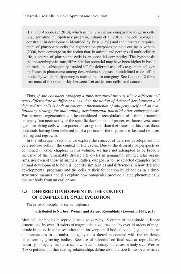
particular ecological niche can be exploited by a particular body design. Therefore, if performance requirements during ontogeny are not the same as those of the size of an organism at reproductive maturity, then life history strategies must evolve to accommodate the mismatch. Cohen (1985) identified three evolutionary responses that animals display, in various combinations, to contend with how to make bodies that can be orders of magnitude larger than their embryos (Figure 1.2):
i. coloniality (e.g., bryozoans and hard corals) where the same body unit is multiplied to create more mass;
ii. increased maternal provisioning (e.g., via evolutionary increases in egg size or other types of postzygotic provisioning) increases the size of the initial free-living ontogenetic stage compared to the ancestral state; or
iii. evolution of complex life cycles.
In animals, this latter tactic is the most widespread (Thorson 1950). Whereas the more commonly known examples of complex life cycles in animals are terrestrial—tadpole to frog and caterpillar to butterfly—the most dazzling array of complex life histories among animals are surely found in the ocean. Coastal marine waters especially are replete with diverse larval forms that disperse for a time in the plankton
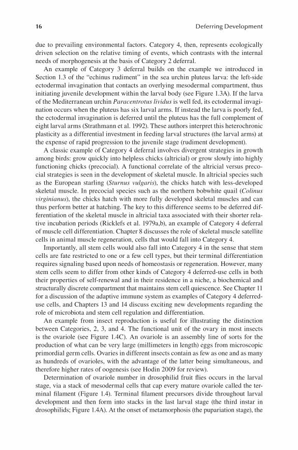
FIGURE 1.2 Three evolutionary strategies to make larger bodies in metazoans. (A) Evolution of coloniality, exemplified by solitary and colonial bryozoans; (B) evolutionary increase of maternal investment, exemplified here by the evolution of nonfeeding develop-ment in echinoids (Ht, Heliocidaris tuberculata; He, Heliocidaris erythrogramma; see Raff 1996); (C) evolution of complex life histories, exemplified by the veliger to juvenile sea slug transition; (D) schematic representation of size increase as a function of time for larval and colonial life histories. The arrow indicates the transition either to a juvenile or a colonial state.
9Deferred-Use Cells in Development and Evolution
before undergoing metamorphoses into what are often very different looking adults in the benthos. In general, the maximum size of these larval forms (and hence the size of the young juveniles that emerge after metamorphosis) is much smaller than that of the corresponding reproductive adults. How did ontogeny evolve in response to the evolution of large-bodied descendants from small-bodied ancestors?
A prominent attempt to place the evolution of large-bodied animals in the context of developmental innovations is the set-aside cell hypothesis of Eric Davidson, Kevin Peterson, and Andrew Cameron (Peterson et al. 1997). Prior to the formal elabora-tion of this hypothesis, Davidson et al. (1995) proposed the term “maximal indirect development” to reflect not only the significant mismatch in phenotypes between larvae of some marine invertebrates and their adult forms but also their nearly tem-porally mutually exclusive ontogenies. Larval forms in taxa with proposed maximal indirect development are small, generally less than 1 mm in length, but their adults could be orders of magnitude larger.
The set-aside cell hypothesis suggested a singular and bold resolution to this apparent paradox, involving a key evolutionary novelty in a hypothesized proto- animal with a small-bodied adult. This key novelty was the proposed origin of groups of cells or tissues that are formed in the embryo but are “set aside” as a rudiment of tissues that remain undifferentiated until the juvenile stage. In other words, the set-aside cell hypothesis posited that the evolution of large complex animals from small ancestors was facilitated by the developmental innovation of what the authors termed “set-aside cells,” which (along with attendant genomic regulatory mechanisms) pat-terned these rudimentary tissues on a regional, as opposed to a cell-by-cell basis.
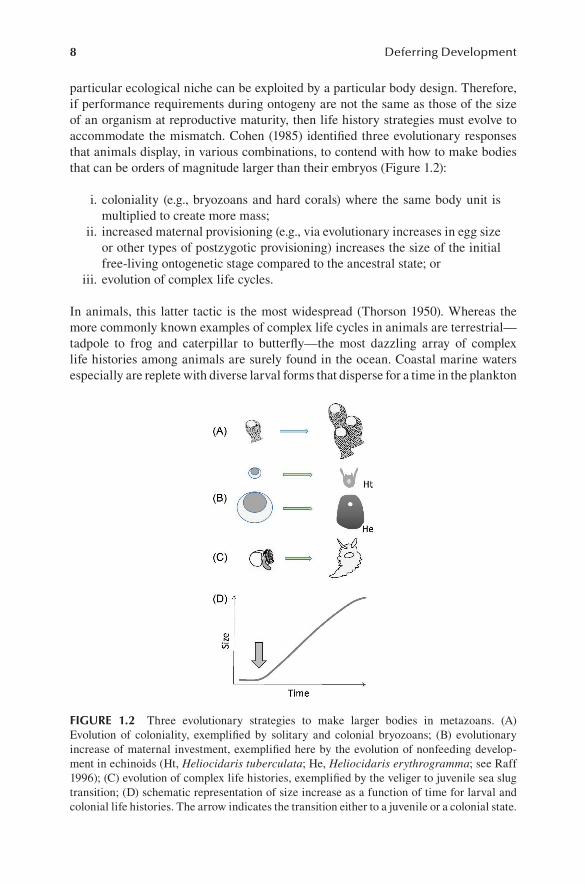
Of particular concern to Davidson and colleagues were marine animal phyla characterized by complex life histories involving a larva undergoing a radical meta-morphosis into a very different looking adult, such as the sea urchin and ribbon worm depicted in Figure 1.3. Davidson and colleagues considered the ancestral small-bodied adults to be homologous to modern larvae and concluded that the mac-roscopic adult forms that define the modern phyla arose later and were thus tacked on to the end of ontogeny. Hence the set-aside cell idea was essentially a macroevo-lutionary hypothesis that spanned innovations in cell behavior and function to novel complex genomic regulatory processes to the origin of large animals themselves.
The set-aside cell hypothesis and the related body of work from which it was derived was a bold attempt at a synthesis of comparative developmental biology and macroevolution, at once accounting for similarities in larval forms among many phyla, the shared use of patterning mechanisms to build adult bodies and the Cambrian explosion itself. Nevertheless, varied critiques soon followed, including, but not limited to, the failure of the set-aside cell hypothesis to meet the criterion of Darwinian plausibility (Wolpert 1999); to infer the likely ancestral metazoan life cycle and critically evaluate typological characterizations of larvae (Jenner 2000); to accurately depict the capacity of larval cells for extended cell division under dif-ferent conditions, such as phenotypic plasticity (Strathmann 2000); and to contend with the conclusion that adult body plans and possibly the patterning mechanisms that build them (e.g., Hox genes) must per force be homoplasies (Raff 2008). Each of these critiques represent significant challenges to the set-aside cell hypothesis as an explanation for broad patterns in animal evolution and development.
10 Deferring Development
Here, we emphasize an additional and perhaps more fundamental critique: the term “set-aside cell” did not adhere to an ontology consistent with cell or developmental biology. The definition of a set-aside cell is clear enough: “We term the specific patches of cells from which the juvenile arises in maximal indirect development ‘set-aside cells,’ because they are in some manner withheld from the differentiation processes that in the late embryo generate the structure of the larva per se.” (Peterson et al. 1997, p. 624). However, the cells and tissues to which this term was applied do not share a consistent set of properties. To make this point, we briefly reanalyze an example that Davidson and colleagues held up as an archetype for both maximal indirect development and the set-aside cell hypothesis itself: juvenile rudiment for-mation in sea urchins. Towards the end of the chapter (Section 1.5), we will reintro-duce sea urchin development in the context of an exploration in parallel patterns in the evolution of complex life histories in diverse animals via deferred development that employs rudiments.
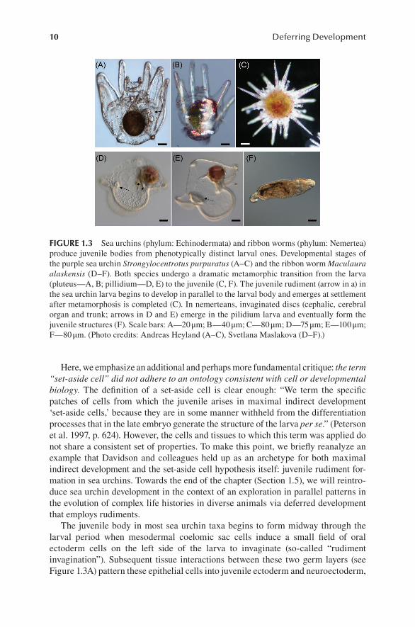
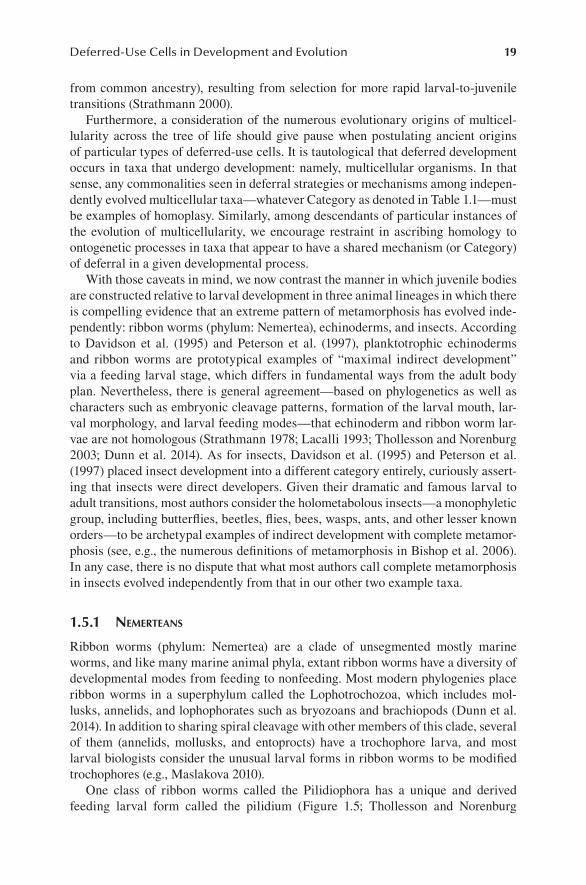
The juvenile body in most sea urchin taxa begins to form midway through the larval period when mesodermal coelomic sac cells induce a small field of oral ectoderm cells on the left side of the larva to invaginate (so-called “rudiment invagination”). Subsequent tissue interactions between these two germ layers (see Figure 1.3A) pattern these epithelial cells into juvenile ectoderm and neuroectoderm,
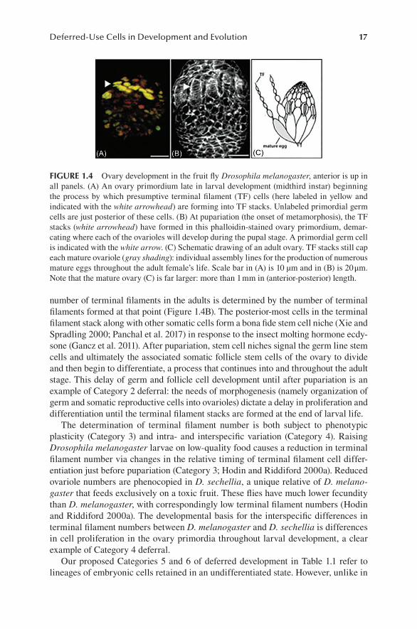
FIGURE 1.3 Sea urchins (phylum: Echinodermata) and ribbon worms (phylum: Nemertea) produce juvenile bodies from phenotypically distinct larval ones. Developmental stages of the purple sea urchin Strongylocentrotus purpuratus (A–C) and the ribbon worm Maculaura alaskensis (D–F). Both species undergo a dramatic metamorphic transition from the larva (pluteus—A, B; pillidium—D, E) to the juvenile (C, F). The juvenile rudiment (arrow in a) in the sea urchin larva begins to develop in parallel to the larval body and emerges at settlement after metamorphosis is completed (C). In nemerteans, invaginated discs (cephalic, cerebral organ and trunk; arrows in D and E) emerge in the pilidium larva and eventually form the juvenile structures (F). Scale bars: A—20 μm; B—40 μm; C—80 μm; D—75 μm; E—100 μm; F—80 μm. (Photo credits: Andreas Heyland (A–C), Svetlana Maslakova (D–F).)
11Deferred-Use Cells in Development and Evolution
and coelomic cells into juvenile mesoderm and germ cells (Chia and Burke 1978; Campanale et al. 2014).
Peterson et al. (1997) considered both the ectodermal invagination and the coelomic mesoderm as examples of set-aside cells. However, this characterization is not accurate for either cell type. Prior to their invagination and subsequent interac-tion with mesoderm, the larval epithelial cells are morphologically and functionally indistinguishable from adjacent, differentiated larval epithelial cells. They are not “set aside”; they undergo a change in function. Similarly, in the sense intended by Peterson et al., the somatic mesodermal cells are not set aside either. For example, the hydrocoel functions in larvae as a differentiated excretory organ (Ruppert and Balser 1986) and is later remodeled to produce the water vascular system of the juve-nile, among other things.
Beyond the particular characteristics of these purported “set-aside cells” them-selves, the term “set aside” itself is problematic for the comparison of development strategies used by different lineages of organisms, as it has two distinct connota-tions according to the Cambridge English Dictionary. One definition is “to save for a particular purpose.” It is this connotation that Davidson and colleagues were employing, as they envisioned embryos literally segregating cells or cell potential in a certain physical portion of the embryo. However, the second definition of the term “set aside” conveys the opposite meaning: “to decide not to consider something…to state that [ something] is no longer in effect.” In the former definition, something is saved for future use; in the latter definition, something is removed from future consideration! To make matters more confusing still, this second connotation of “set aside” precedes the set-aside cell hypothesis in the scientific literature with respect to future cell fate.
Buss (1987) used the term to refer to germ cells, as having been set aside relative to cells having a somatic function. In an opposite usage, Bell and Koufopanou (1991) refers to the replicative potential of flagellated somatic cells in volvocalean (colonial green algal) life cycles (see Figure 1.1A) as having been set aside to ensure ongoing motility of the colony while germ cells undergo mitosis. Pehrson and Cohen (1986), reported that descendants of small micromeres of sea urchin embryos come to reside in coelomic sacs and are thus set aside. These authors used the term with some precision, in which these micromere descendant cells were kept in a mitotically qui-escent state.4 Later, Truman and Riddiford (1999) in their paper on the evolution of larvae and metamorphosis in holometabolous insects refer to cells that form the imaginal discs as having been set aside and then later refer to cells/tissues that form the juvenile as having been developmentally deferred. Thus, they used the terms interchangeably.
To conclude:
i. what were identified as set-aside cells in the hypothesis of that name do not have a consistent set of properties; and
4 It was later shown (Yajima and Wessel 2011; Campanale et al. 2014; Wessel et al. 2014) that descendants of the small micromeres give rise to primordial germ cells; in this sense in particular, Pehrson & Cohen’s usage was post hoc consistent with Buss (1987).
12 Deferring Development
ii. opposing connotations exist in the use of “set aside” in the English language and by extension in the wider literature.
Given the varying uses in the literature for ‘set-aside’ the term ‘deferred development’ or ‘deferred-use’ cells’ seems more precise, and here we advocate it in place of ‘set aside.’
The presumed goal of Davidson and colleagues in proposing their set-aside cell hypothesis is one that we share: to gain an understanding of broader issues in the evolution of animals and their life histories through an examination of deferred developmental programs in disparate organisms. Unfortunately, the ambiguities inherent in their analyses that we outline above undermined this worthy goal. In the next section, we set aside (second meaning!) the evolutionary theoretical dimensions of the set-aside cell hypothesis and propose an ontology of deferred development in the service of resolving such ambiguities.
1.4 ONTOLOGIES OF DEFERRED DEVELOPMENT AND DEFERRED-USE CELLS
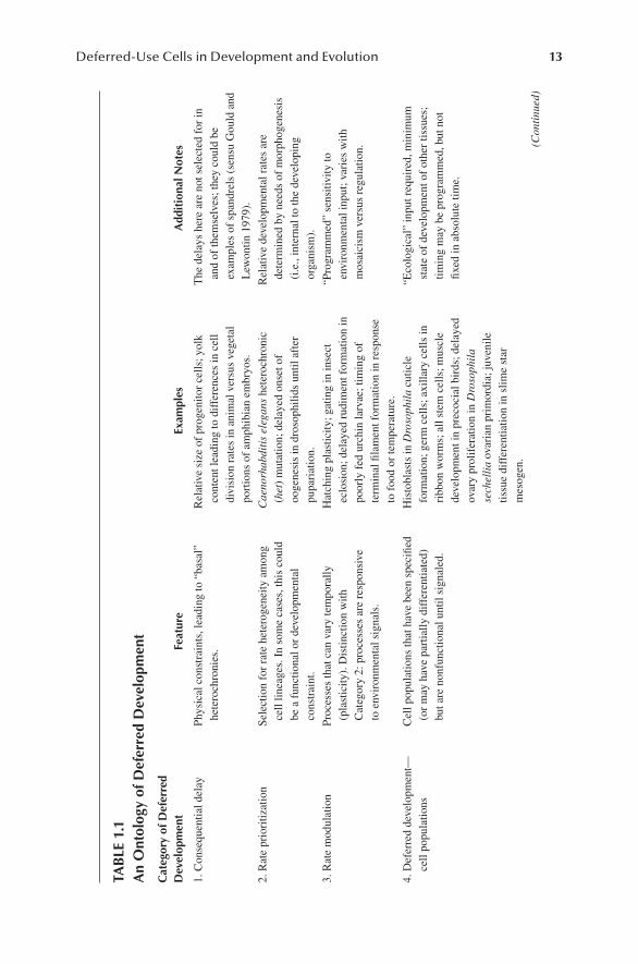
The concept of deferred development implies that specification and terminal differ-entiation occur relatively later for some populations of cells compared to others. This idea is axiomatic to developmental biologists and thus may seem to need no elabora-tion. Nevertheless, our basic claim is that both the reasons and the phenomenology for these delays differ within and among taxa, and therefore, for the purpose of com-parisons, it may be useful to devise a systematic ontology of deferred development (Table 1.1). In this section, we attempt to distinguish instances in which deferred development occurs as a by-product of something else (Category 1 in Table 1.1) from instances in which
i. ecological requirements of life history stages have resulted in heterogene-ity indevelopmental rates of distinct ontogenetic processes (Categories 2 and 3); or
ii. selection has generated cells or entire tissues whose developmental program is occurring in the context of a functioning earlier stage (Categories 4–6).
Before proceeding, we note that all Categories (1–6) could occur in a single organ-ism, and therefore, there is no implied grade of complexity or hierarchy among these categories. Furthermore, we are mindful that accelerated development of one part of an embryo could be interpreted as the deferred development of another part.
The first category of among-lineage rate heterogeneity is called “Consequential Delay” (Category 1 in Table 1.1), and it is driven by physical constraints. For exam-ple, the vegetal hemispheres of many frog embryos have very high yolk content, such that cleavage furrows cannot easily proceed through them. One result of this animal-vegetal disparity in yolk is that cleavage proceeds more rapidly in the animal hemisphere, and there are thus more cells in the animal hemisphere at any given time during early cleavage stages (Barresi and Gilbert 2016).
13Deferred-Use Cells in Development and Evolution
TABL
E 1.
1A
n O
ntol
ogy
of D
efer
red
Dev
elop
men
t
Cat
egor
y of
Def
erre
d D
evel
opm
ent
Feat
ure
Exam
ples
Add
ition
al N
otes
1. C
onse
quen
tial d
elay
Phys
ical
con
stra
ints
, lea
ding
to “
basa
l”
hete
roch
roni
es.
Rel
ativ
e si
ze o
f pro
geni
tor c
ells
; yol
k co
nten
t lea
ding
to d
iffer
ence
s in
cel
l di
visi
on ra
tes
in a
nim
al v
ersu
s ve
geta
l po
rtio
ns o
f am
phib
ian
embr
yos.
The
del
ays
here
are
not
sel
ecte
d fo
r in
and
of th
emse
lves
; the
y co
uld
be
exam
ples
of s
pand
rels
(sen
su G
ould
and
L
ewon
tin 1
979)
.2.
Rat
e pr
iori
tizat
ion
Sele
ctio
n fo
r rat
e he
tero
gene
ity a
mon
g ce
ll lin
eage
s. In
som
e ca
ses,
this
cou
ld
be a
func
tiona
l or d
evel
opm
enta
l co
nstr
aint
.
Cae
norh
abdi
tis e
lega
ns h
eter
ochr
onic
(h
et) m
utat
ion;
del
ayed
ons
et o
f oo
gene
sis
in d
roso
phili
ds u
ntil
afte
r pu
pari
atio
n.
Rel
ativ
e de
velo
pmen
tal r
ates
are
de
term
ined
by
need
s of
mor
phog
enes
is
(i.e
., in
tern
al to
the
deve
lopi
ng
orga
nism
).3.
Rat
e m
odul
atio
nPr
oces
ses
that
can
var
y te
mpo
rally
(p
last
icity
). D
istin
ctio
n w
ith
Cat
egor
y 2:
pro
cess
es a
re re
spon
sive
to
env
iron
men
tal s
igna
ls.
Hat
chin
g pl
astic
ity; g
atin
g in
inse
ct
eclo
sion
; del
ayed
rudi
men
t for
mat
ion
in
poor
ly fe
d ur
chin
larv
ae; t
imin
g of
te
rmin
al fi
lam
ent f
orm
atio
n in
resp
onse
to
food
or t
empe
ratu
re.
“Pro
gram
med
” se
nsiti
vity
to
envi
ronm
enta
l inp
ut; v
arie
s w
ith
mos
aici
sm v
ersu
s re
gula
tion.
4. D
efer
red
deve
lopm
ent—
cell
popu
latio
nsC
ell p
opul
atio
ns th
at h
ave
been
spe
cifie
d (o
r may
hav
e pa
rtia
lly d
iffer
entia
ted)
bu
t are
non
func
tiona
l unt
il si
gnal
ed.
His
tobl
asts
in D
roso
phila
cut
icle
fo
rmat
ion;
ger
m c
ells
; axi
llary
cel
ls in
ri
bbon
wor
ms;
all
stem
cel
ls; m
uscl
e de
velo
pmen
t in
prec
ocia
l bir
ds; d
elay
ed
ovar
y pr
olif
erat
ion
in D
roso
phila
se
chel
lia o
vari
an p
rim
ordi
a; ju
veni
le
tissu
e di
ffer
entia
tion
in s
lime
star
m
esog
en.
“Eco
logi
cal”
inpu
t req
uire
d, m
inim
um
stat
e of
dev
elop
men
t of o
ther
tiss
ues;
tim
ing
may
be
prog
ram
med
, but
not
fix
ed in
abs
olut
e tim
e.
(Con
tinue
d)

14 Deferring Development
TABL
E 1.
1 (C
ontin
ued
)A
n O
ntol
ogy
of D
efer
red
Dev
elop
men
t
Cat
egor
y of
Def
erre
d D
evel
opm
ent
Feat
ure
Exam
ples
Add
ition
al N
otes
5. D
efer
red
deve
lopm
ent—
dela
yed
life
hist
ory
shif
tD
efer
ral i
s no
w a
t the
leve
l of e
ntir
e tis
sues
or o
rgan
s an
d is
ass
ocia
ted
with
a
life
hist
ory
tran
sfor
mat
ion
such
as
in
met
amor
phic
life
his
tori
es.
Post
erio
r gro
wth
zon
e in
mar
ine
anne
lids;
fli
ght i
n he
mim
etab
olou
s in
sect
s;
rudi
men
t for
mat
ion
in p
enci
l urc
hins
(c
idar
oids
); ju
veni
le ti
ssue
di
ffer
entia
tion
in s
ea c
ucum
bers
.
Sim
ilar t
o C
ateg
ory
4 bu
t per
tain
s to
en
tire
tissu
es o
r org
ans.
6. D
efer
red
deve
lopm
ent—
rudi
men
ts a
llow
rapi
d de
ploy
men
t of d
efer
red
func
tion
in li
fe h
isto
ries
Initi
ally
, non
func
tiona
l cel
ls/o
rgan
s de
velo
p in
par
alle
l with
func
tiona
l la
rval
cel
ls. P
arad
oxic
ally
, les
s “d
efer
red”
in a
ppea
ranc
e th
an
Cat
egor
y 5,
but
we
hypo
thes
ize
that
C
ateg
ory
6 is
evo
lutio
nari
ly d
eriv
ed
rela
tive
to C
ateg
ory
5 an
d th
at th
e fu
nctio
n its
elf i
s st
ill d
efer
red.
Ech
inod
erm
rudi
men
ts; i
mag
inal
dis
cs in
ri
bbon
wor
m a
nd h
olom
etab
olou
s in
sect
s; ru
dim
ents
in o
wen
iid
poly
chae
tes;
pre
coci
ous
form
atio
n of
ju
veni
le b
odie
s w
ithin
com
poun
d as
cidi
an ta
dpol
es.
Mor
phog
enet
ic a
spec
ts o
f met
amor
phos
is
are
deco
uple
d fr
om th
e ha
bita
t tr
ansi
tion.
The
def
erre
d tis
sues
and
or
gans
are
seg
rega
ted
in ru
dim
ents
.
15Deferred-Use Cells in Development and Evolution
The salient feature of Category 1 is that examples of “Consequential Delay” should not be assumed to be adaptations in and of themselves; they may be a result of selection for something else (i.e., spandrels sensu Gould and Lewontin 1979). In the case of frog embryos cited above, the delay of vegetal relative to animal hemi-sphere cleavage is likely a consequence of selection for increased maternal nutrition (i.e., selection for deferred-use molecules, Chapter 2) and therefore egg size.
Category 2 is entitled “Rate Prioritization” and can be distinguished from Category 1 in that there is presumed selection in Category 2 for among-lineage het-erogeneity in rates or timing of development to meet the needs of morphogenesis. A clear example of Category 2 involves ciliation in developing trochophore larvae. This larval form, present in the life cycle of various spiralians (e.g., mollusks, annelids, and the less well-known entoprocts) is characterized by very rapid differentiation of a band of ciliated cells called the “troch.” These trochoblast cells cease dividing and arrange themselves into a band of functional cilia when there are only 63 cells in the embryo (Kooij et al. 1998). This developmental pattern is presumably driven by the need for rapid development to a swimming stage (Staver and Strathmann 2002). The differential storage of large amounts of maternally derived tubulin mRNA or protein in presumptive trochoblasts would constitute evidence that the rate of trocho-blast cell differentiation is accelerated, as opposed to the notion that development of the remainder of the embryo is deferred.
Roundworms (phylum: Nematoda) offer numerous, well-studied examples that can be placed in Category 2. Nematode embryos have stereotyped cleavage patterns, producing fixed cell lineages, where different groups of founder cells divide at dif-ferent rates in a manner that reflects ontogenetic needs. Furthermore, the ontogeny of the nematode Caenorhabditis elegans has been subjected to decades of intense study through the characterization of myriad mutations that perturb the meticulously well-characterized cell lineage in this worm (Horvitz 1990). C. elegans genes have historically been named for the classes of phenotypes they produce. One example is the “heterochronic” (het) gene class, so named because mutations in these genes result in temporal displacements of developmental events (reviewed in Moss 2007). Therefore, in normal development, het genes can be thought of as orchestrators of the proper control of ontogenetic timing. Het mutations indicate that when such events occur out of sequence, normally functioning worms are not produced. This observa-tion alone speaks to the relationship between careful control of the relative timing of developmental events in order to maximize an organism’s fitness.
It is possible that Category 2 rate heterogeneity may be an evolutionary precondi-tion (a preadaptation sensu Gould 1984) for occurrence of relative shifts in the timing of embryonic events in response to environmental conditions (i.e., phenotypic plastic-ity, or “rate modulation”; Category 3 in Table 1.1). This heterogeneity may provide the variation for selection for fixed differences in rates within and ultimately between related species (namely, intra- and interspecific variation; Category 4 in Table 1.1).
Categories 3 and 4 in our proposed ontology represent a critical distinction from Categories 1 and 2, in that ecological inputs intervene to modulate rates of division among cell lineages or populations, or the timing of specification or differentiation of cells. Specifically, Category 3 refers to developmental processes subject to heter-ochronic phenotypic plasticity: a change in relative timing of developmental events
16 Deferring Development
due to prevailing environmental factors. Category 4, then, represents ecologically driven selection on the relative timing of events, which contrasts with the internal needs of morphogenesis at the basis of Category 2 deferral.
An example of Category 3 deferral builds on the example we introduced in Section 1.3 of the “echinus rudiment” in the sea urchin pluteus larva: the left-side ectodermal invagination that contacts an overlying mesodermal compartment, thus initiating juvenile development within the larval body (see Figure 1.3A). If the larva of the Mediterranean urchin Paracentrotus lividus is well fed, its ectodermal invagi-nation occurs when the pluteus has six larval arms. If instead the larva is poorly fed, the ectodermal invagination is deferred until the pluteus has the full complement of eight larval arms (Strathmann et al. 1992). These authors interpret this heterochronic plasticity as a differential investment in feeding larval structures (the larval arms) at the expense of rapid progression to the juvenile stage (rudiment development).
A classic example of Category 4 deferral involves divergent strategies in growth among birds: grow quickly into helpless chicks (altricial) or grow slowly into highly functioning chicks (precocial). A functional correlate of the altricial versus preco-cial strategies is seen in the development of skeletal muscle. In altricial species such as the European starling (Sturnus vulgaris), the chicks hatch with less-developed skeletal muscle. In precocial species such as the northern bobwhite quail (Colinus virginianus), the chicks hatch with more fully developed skeletal muscles and can thus perform better at hatching. The key to this difference seems to be deferred dif-ferentiation of the skeletal muscle in altricial taxa associated with their shorter rela-tive incubation periods (Ricklefs et al. 1979a,b), an example of Category 4 deferral of muscle cell differentiation. Chapter 8 discusses the role of skeletal muscle satellite cells in animal muscle regeneration, cells that would fall into Category 4.
Importantly, all stem cells would also fall into Category 4 in the sense that stem cells are fate restricted to one or a few cell types, but their terminal differentiation requires signaling based upon needs of homeostasis or regeneration. However, many stem cells seem to differ from other kinds of Category 4 deferred-use cells in both their properties of self-renewal and in their residence in a niche, a biochemical and structurally discrete compartment that maintains stem cell quiescence. See Chapter 11 for a discussion of the adaptive immune system as examples of Category 4 deferred-use cells, and Chapters 13 and 14 discuss exciting new developments regarding the role of microbiota and stem cell regulation and differentiation.
An example from insect reproduction is useful for illustrating the distinction between Categories, 2, 3, and 4. The functional unit of the ovary in most insects is the ovariole (see Figure 1.4C). An ovariole is an assembly line of sorts for the production of what can be very large (millimeters in length) eggs from microscopic primordial germ cells. Ovaries in different insects contain as few as one and as many as hundreds of ovarioles, with the advantage of the latter being simultaneous, and therefore higher rates of oogenesis (see Hodin 2009 for review).
Determination of ovariole number in drosophilid fruit flies occurs in the larval stage, via a stack of mesodermal cells that cap every mature ovariole called the ter-minal filament (Figure 1.4). Terminal filament precursors divide throughout larval development and then form into stacks in the last larval stage (the third instar in drosophilids; Figure 1.4A). At the onset of metamorphosis (the pupariation stage), the
17Deferred-Use Cells in Development and Evolution
number of terminal filaments in the adults is determined by the number of terminal filaments formed at that point (Figure 1.4B). The posterior-most cells in the terminal filament stack along with other somatic cells form a bona fide stem cell niche (Xie and Spradling 2000; Panchal et al. 2017) in response to the insect molting hormone ecdy-sone (Gancz et al. 2011). After pupariation, stem cell niches signal the germ line stem cells and ultimately the associated somatic follicle stem cells of the ovary to divide and then begin to differentiate, a process that continues into and throughout the adult stage. This delay of germ and follicle cell development until after pupariation is an example of Category 2 deferral: the needs of morphogenesis (namely organization of germ and somatic reproductive cells into ovarioles) dictate a delay in proliferation and differentiation until the terminal filament stacks are formed at the end of larval life.
The determination of terminal filament number is both subject to phenotypic plasticity (Category 3) and intra- and interspecific variation (Category 4). Raising Drosophila melanogaster larvae on low-quality food causes a reduction in terminal filament number via changes in the relative timing of terminal filament cell differ-entiation just before pupariation (Category 3; Hodin and Riddiford 2000a). Reduced ovariole numbers are phenocopied in D. sechellia, a unique relative of D. melano-gaster that feeds exclusively on a toxic fruit. These flies have much lower fecundity than D. melanogaster, with correspondingly low terminal filament numbers (Hodin and Riddiford 2000a). The developmental basis for the interspecific differences in terminal filament numbers between D. melanogaster and D. sechellia is differences in cell proliferation in the ovary primordia throughout larval development, a clear example of Category 4 deferral.
Our proposed Categories 5 and 6 of deferred development in Table 1.1 refer to lineages of embryonic cells retained in an undifferentiated state. However, unlike in
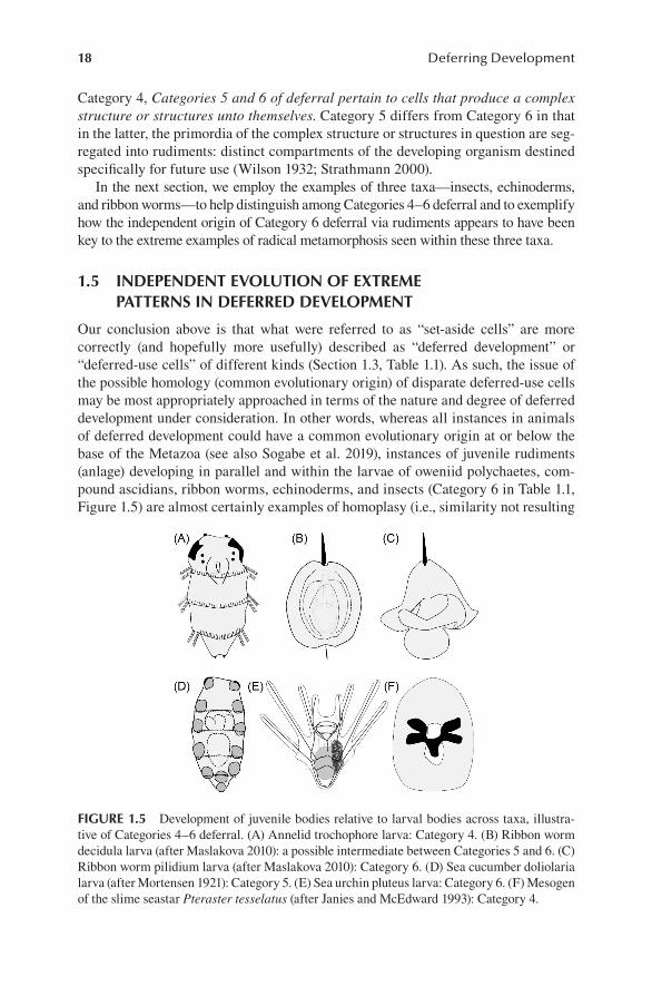
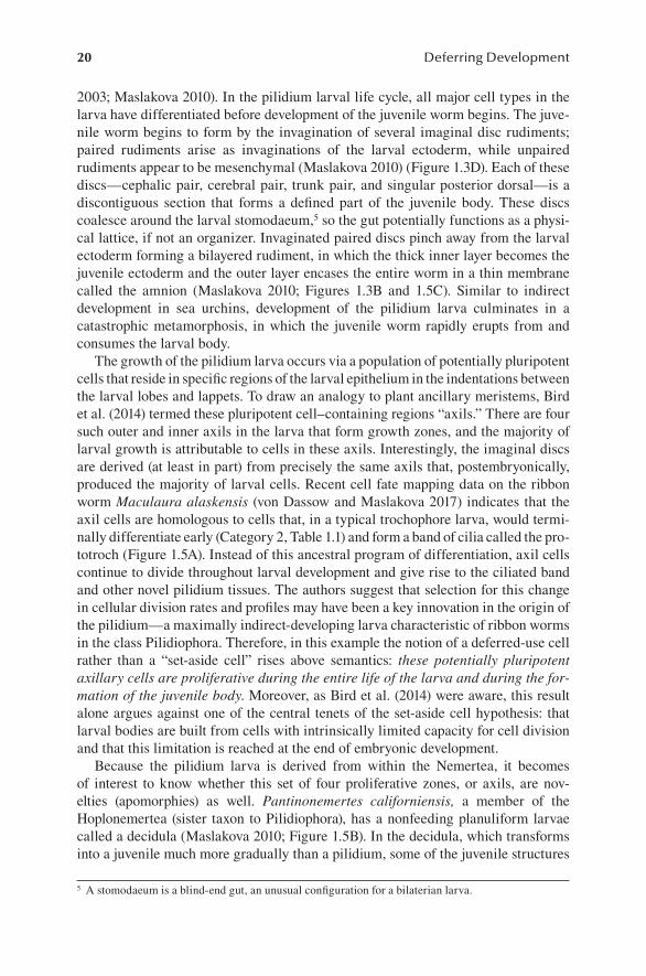
FIGURE 1.4 Ovary development in the fruit fly Drosophila melanogaster, anterior is up in all panels. (A) An ovary primordium late in larval development (midthird instar) beginning the process by which presumptive terminal filament (TF) cells (here labeled in yellow and indicated with the white arrowhead) are forming into TF stacks. Unlabeled primordial germ cells are just posterior of these cells. (B) At pupariation (the onset of metamorphosis), the TF stacks (white arrowhead) have formed in this phalloidin-stained ovary primordium, demar-cating where each of the ovarioles will develop during the pupal stage. A primordial germ cell is indicated with the white arrow. (C) Schematic drawing of an adult ovary. TF stacks still cap each mature ovariole (gray shading): individual assembly lines for the production of numerous mature eggs throughout the adult female’s life. Scale bar in (A) is 10 µm and in (B) is 20 μm. Note that the mature ovary (C) is far larger: more than 1 mm in (anterior-posterior) length.
18 Deferring Development
Category 4, Categories 5 and 6 of deferral pertain to cells that produce a complex structure or structures unto themselves. Category 5 differs from Category 6 in that in the latter, the primordia of the complex structure or structures in question are seg-regated into rudiments: distinct compartments of the developing organism destined specifically for future use (Wilson 1932; Strathmann 2000).
In the next section, we employ the examples of three taxa—insects, echinoderms, and ribbon worms—to help distinguish among Categories 4–6 deferral and to exemplify how the independent origin of Category 6 deferral via rudiments appears to have been key to the extreme examples of radical metamorphosis seen within these three taxa.
1.5 INDEPENDENT EVOLUTION OF EXTREME PATTERNS IN DEFERRED DEVELOPMENT
Our conclusion above is that what were referred to as “set-aside cells” are more correctly (and hopefully more usefully) described as “deferred development” or “deferred-use cells” of different kinds (Section 1.3, Table 1.1). As such, the issue of the possible homology (common evolutionary origin) of disparate deferred-use cells may be most appropriately approached in terms of the nature and degree of deferred development under consideration. In other words, whereas all instances in animals of deferred development could have a common evolutionary origin at or below the base of the Metazoa (see also Sogabe et al. 2019), instances of juvenile rudiments (anlage) developing in parallel and within the larvae of oweniid polychaetes, com-pound ascidians, ribbon worms, echinoderms, and insects (Category 6 in Table 1.1, Figure 1.5) are almost certainly examples of homoplasy (i.e., similarity not resulting
FIGURE 1.5 Development of juvenile bodies relative to larval bodies across taxa, illustra-tive of Categories 4–6 deferral. (A) Annelid trochophore larva: Category 4. (B) Ribbon worm decidula larva (after Maslakova 2010): a possible intermediate between Categories 5 and 6. (C) Ribbon worm pilidium larva (after Maslakova 2010): Category 6. (D) Sea cucumber doliolaria larva (after Mortensen 1921): Category 5. (E) Sea urchin pluteus larva: Category 6. (F) Mesogen of the slime seastar Pteraster tesselatus (after Janies and McEdward 1993): Category 4.
19Deferred-Use Cells in Development and Evolution
from common ancestry), resulting from selection for more rapid larval-to-juvenile transitions (Strathmann 2000).
Furthermore, a consideration of the numerous evolutionary origins of multicel-lularity across the tree of life should give pause when postulating ancient origins of particular types of deferred-use cells. It is tautological that deferred development occurs in taxa that undergo development: namely, multicellular organisms. In that sense, any commonalities seen in deferral strategies or mechanisms among indepen-dently evolved multicellular taxa—whatever Category as denoted in Table 1.1—must be examples of homoplasy. Similarly, among descendants of particular instances of the evolution of multicellularity, we encourage restraint in ascribing homology to ontogenetic processes in taxa that appear to have a shared mechanism (or Category) of deferral in a given developmental process.
With those caveats in mind, we now contrast the manner in which juvenile bodies are constructed relative to larval development in three animal lineages in which there is compelling evidence that an extreme pattern of metamorphosis has evolved inde-pendently: ribbon worms (phylum: Nemertea), echinoderms, and insects. According to Davidson et al. (1995) and Peterson et al. (1997), planktotrophic echinoderms and ribbon worms are prototypical examples of “maximal indirect development” via a feeding larval stage, which differs in fundamental ways from the adult body plan. Nevertheless, there is general agreement—based on phylogenetics as well as characters such as embryonic cleavage patterns, formation of the larval mouth, lar-val morphology, and larval feeding modes—that echinoderm and ribbon worm lar-vae are not homologous (Strathmann 1978; Lacalli 1993; Thollesson and Norenburg 2003; Dunn et al. 2014). As for insects, Davidson et al. (1995) and Peterson et al. (1997) placed insect development into a different category entirely, curiously assert-ing that insects were direct developers. Given their dramatic and famous larval to adult transitions, most authors consider the holometabolous insects—a monophyletic group, including butterflies, beetles, flies, bees, wasps, ants, and other lesser known orders—to be archetypal examples of indirect development with complete metamor-phosis (see, e.g., the numerous definitions of metamorphosis in Bishop et al. 2006). In any case, there is no dispute that what most authors call complete metamorphosis in insects evolved independently from that in our other two example taxa.
1.5.1 NEMERTEANS
Ribbon worms (phylum: Nemertea) are a clade of unsegmented mostly marine worms, and like many marine animal phyla, extant ribbon worms have a diversity of developmental modes from feeding to nonfeeding. Most modern phylogenies place ribbon worms in a superphylum called the Lophotrochozoa, which includes mol-lusks, annelids, and lophophorates such as bryozoans and brachiopods (Dunn et al. 2014). In addition to sharing spiral cleavage with other members of this clade, several of them (annelids, mollusks, and entoprocts) have a trochophore larva, and most larval biologists consider the unusual larval forms in ribbon worms to be modified trochophores (e.g., Maslakova 2010).
One class of ribbon worms called the Pilidiophora has a unique and derived feeding larval form called the pilidium (Figure 1.5; Thollesson and Norenburg
20 Deferring Development
2003; Maslakova 2010). In the pilidium larval life cycle, all major cell types in the larva have differentiated before development of the juvenile worm begins. The juve-nile worm begins to form by the invagination of several imaginal disc rudiments; paired rudiments arise as invaginations of the larval ectoderm, while unpaired rudiments appear to be mesenchymal (Maslakova 2010) (Figure 1.3D). Each of these discs—cephalic pair, cerebral pair, trunk pair, and singular posterior dorsal—is a discontiguous section that forms a defined part of the juvenile body. These discs coalesce around the larval stomodaeum,5 so the gut potentially functions as a physi-cal lattice, if not an organizer. Invaginated paired discs pinch away from the larval ectoderm forming a bilayered rudiment, in which the thick inner layer becomes the juvenile ectoderm and the outer layer encases the entire worm in a thin membrane called the amnion (Maslakova 2010; Figures 1.3B and 1.5C). Similar to indirect development in sea urchins, development of the pilidium larva culminates in a catastrophic metamorphosis, in which the juvenile worm rapidly erupts from and consumes the larval body.
The growth of the pilidium larva occurs via a population of potentially pluripotent cells that reside in specific regions of the larval epithelium in the indentations between the larval lobes and lappets. To draw an analogy to plant ancillary meristems, Bird et al. (2014) termed these pluripotent cell–containing regions “axils.” There are four such outer and inner axils in the larva that form growth zones, and the majority of larval growth is attributable to cells in these axils. Interestingly, the imaginal discs are derived (at least in part) from precisely the same axils that, postembryonically, produced the majority of larval cells. Recent cell fate mapping data on the ribbon worm Maculaura alaskensis (von Dassow and Maslakova 2017) indicates that the axil cells are homologous to cells that, in a typical trochophore larva, would termi-nally differentiate early (Category 2, Table 1.1) and form a band of cilia called the pro-totroch (Figure 1.5A). Instead of this ancestral program of differentiation, axil cells continue to divide throughout larval development and give rise to the ciliated band and other novel pilidium tissues. The authors suggest that selection for this change in cellular division rates and profiles may have been a key innovation in the origin of the pilidium—a maximally indirect-developing larva characteristic of ribbon worms in the class Pilidiophora. Therefore, in this example the notion of a deferred-use cell rather than a “set-aside cell” rises above semantics: these potentially pluripotent axillary cells are proliferative during the entire life of the larva and during the for-mation of the juvenile body. Moreover, as Bird et al. (2014) were aware, this result alone argues against one of the central tenets of the set-aside cell hypothesis: that larval bodies are built from cells with intrinsically limited capacity for cell division and that this limitation is reached at the end of embryonic development.
Because the pilidium larva is derived from within the Nemertea, it becomes of interest to know whether this set of four proliferative zones, or axils, are nov-elties ( apomorphies) as well. Pantinonemertes californiensis, a member of the Hoplonemertea (sister taxon to Pilidiophora), has a nonfeeding planuliform larvae called a decidula (Maslakova 2010; Figure 1.5B). In the decidula, which transforms into a juvenile much more gradually than a pilidium, some of the juvenile structures
5 A stomodaeum is a blind-end gut, an unusual configuration for a bilaterian larva.
21Deferred-Use Cells in Development and Evolution
arise from spatially segregated invaginations of larval epidermis (Hiebert et al. 2010); gene expression studies are consistent with the hypothesis of homology between ecto-dermal invaginations and pilidium imaginal discs (Hiebert and Maslakova 2015). The larval epidermis is shed or resorbed during development of the decidula larva (Hiebert et al. 2010), representing a minimal kind of metamorphosis. Hiebert and Maslakova (2015) suggested that the juvenile epidermal invaginations and epidermal shedding could represent a strategy to minimize or eliminate interruptions to larval ciliation patterns, which would otherwise reduce swimming performance.
Several groups within Lophotrochozoa (annelids, mollusks, and the less well-known entoprocts) have a trochophore larva, characterized by the presence of the preoral transverse ciliary band called the prototroch, which is derived from the same early-differentiating trochoblast cell lineage referred to above and serves as the primary swimming organ in the larva. Classic trochophore development is exemplified in annelids, in which the larval anterior-posterior axis is coaxial with the adult one (Figure 1.5A); larval growth involves additions of segments at the posterior, lengthening the larval into a more recognizably annelid appearance. The presumed ancestral annelid-like condition can be considered Category 5 in our ontology: deferral of posterior segments relative to formation of the feeding trochophore larva. By contrast, the pilidium with its imaginal discs would be an example of Category 6: deferral via the formation of juvenile rudiment structures (the imaginal discs) early in larval development, alongside—but at orthogonal axes to—the growing pilidium larva (Figure 1.5C). Although the polarity remains uncertain, the Hoplonemertean nonfeeding decidula larva (Figure 1.5B) may represent an intermediate between Categories 5 and 6 in that there are populations of cells that form juvenile parts inter-posed heterogeneously with the larval epidermis, but those rudiments are coaxial with the larva, and the metamorphosis is thus less radical.
1.5.2 ECHINODERMS
Echinoderms (sea urchins, sea stars, brittle stars, sea cucumbers, and sea lilies) have among the most radical metamorphoses described for animals (Figures 1.3A–C, 1.5D–F). The canonical echinoderm larva is a bilateral feeding form, which metamorphoses into a pentamerally symmetric adult (Chia and Burke, 1978). These larvae can do so in dramatic fashion: within as little as 15 min, the swimming plank-tonic form (Figure 1.3E) degrades, and a bottom-dwelling locomotory juvenile emerges (Figure 1.3F).
To accomplish this rapid transition, echinoderms employ a similar tactic described above for the pilidium larva: they initiate juvenile development early in the larval period through development of a segregated rudiment. In Section 1.3, we described the early stages of rudiment development in echinoids (sea urchins and sand dol-lars): via an invagination of a portion of larval ectoderm underlying the mesodermal hydrocoel. Together—and along with additional coelomic participants—the invagi-nated ectoderm and the hydrocoel form the oral field of the pentameral juvenile, with tube feet and spines developing within a functioning, feeding larva (Figures 1.3A, B, 1.5F). In the oldest living group of echinoids, the pencil urchins (order: Cidaroida), this process is less extreme. Cidaroids differ from typical echinoids in that they do
22 Deferring Development
not have a rudiment invagination, they do not develop definitive “adult-type” spines until the juvenile stage, and they retain much of the larval epidermis as juveniles (Emlet 1988). In this sense, they have a less radical metamorphic process, more akin to that seen in the sea cucumbers (Figure 1.5D), which likewise retains their larval epidermis into the juvenile stage (Chia and Burke 1978).
With respect to our proposed ontology, noncidaroid echinoids (subclass: Euechinoidea) such as the purple urchin Strongylocentrotus purpuratus exhibit Category 6 deferral of juvenile development via rudiment structures formed early in the larval period, in contrast to the more Category 5-like cidaroids and sea cucum-bers. In addition to these broader patterns, echinoderms demonstrate numerous evolutionary transitions from feeding to nonfeeding larval development. And these derived life history modifications are accompanied by simpler ciliated larval forms that exhibit less extreme metamorphic transitions, which we might call a reversion to a more “Category 4-like” state. In the most extreme example known, develop-ment in the slime sea star Pteraster tesselatus exhibits minimal traces of a bilateral larva. In P. tesselatus, the embryo develops directly to the juvenile via an abbre-viated nonfeeding dispersive stage referred to as a mesogen, in which pentameral symmetry emerges early in development via five circumferential outpocketings of the gut along the animal-vegetal axis of the embryo (Janies and McEdward 1993; Figure 1.5F). The manner in which the mesogen transitions into a juvenile slime star is thus analogous to the annelid example described above (Figure 1.5A). However, in annelids, the pattern of deferral is the seeming ancestral (plesiomorphic) condition, whereas in slime stars, Category 4 deferral is clearly derived (an apomorphy) from Categories 5–6 sea star ancestor (Janies and McEdward 1993).
1.5.3 INSECTS
Our third and final comparison is insects, a group that Davidson et al. (1995) placed in a separate category of development from the majority of animals due to their derived early nuclear division patterns without complete cleavage.6 And although much of embryogenesis in insects is indeed unique relative to even their sister group, the Crustacea, a broader look at the evolution of insect life histories reveals a striking parallel to our descriptions of ribbon worm and echinoderm metamorphosis above.
Like all arthropods, insects grow between molts, and the stages of development are denoted by the number and type of molts having occurred. The most ancient liv-ing insect lineage is a primitively wingless group referred to as the Ametabola, which have no distinct metamorphosis (Figure 1.6). Juvenile Ametabola such as silverfish look nearly identical to adult silverfish except that they are not yet reproductively mature (Figure 1.6A). In addition, silverfish have indeterminate molting, as they con-tinue to molt after reaching reproductive maturity. By contrast, all of the more derived, winged insects have determinate molting. With the sole exception of mayflies, insects with wings cannot molt, and the adult stage is the final stage (Figure 1.6B, C).
Among the winged insects are two main groups characterized by distinct life histories. The hemimetabolous insects (Figure 1.6B)—such as true bugs (Hemiptera),
6 An interesting example of deferred development in its own right: deferral of cytokinesis.
23Deferred-Use Cells in Development and Evolution
grasshoppers, cockroaches, and mantids—are so-called because they have a subtle metamorphosis between their nymphal stages that lack functional wings and reproductive structures and their adult stage that has these features. The second main group of winged insects are the Holometabola (Figure 1.6C), so-called because they have a famously dramatic metamorphosis. Examples of the holometabolous insects are flies, beetles, and butterflies, each of which has a larval form (maggot, grub, and caterpillar) that passes through a metamorphic pupal stage before emerging as what is generally a strikingly different looking, winged adult.
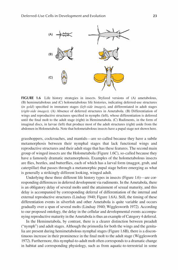
Underlying these three different life history types in insects (Figure 1.6)—are cor-responding differences in deferred development via rudiments. In the Ametabola, there is an obligatory delay of several molts until the attainment of sexual maturity, and this delay is accompanied by corresponding deferral of differentiation of the internal and external reproductive structures (Lindsay 1940; Figure 1.6A). Still, the timing of these differentiation events in silverfish and other Ametabola is quite variable and occurs gradually over a span of several molts (Lindsay 1940; Wigglesworth 1972). According to our proposed ontology, the delay in the cellular and developmental events accompa-nying reproductive maturity in the Ametabola is thus an example of Category 4 deferral.
In the Hemimetabola, by contrast, there is a clearer distinction between preadult (“nymph”) and adult stages. Although the primordia for both the wings and the genita-lia are present during hemimetabolous nymphal stages (Figure 1.6B), there is a discon-tinuous increase in their prominence in the final molt to the adult stage (Wigglesworth 1972). Furthermore, this nymphal-to-adult molt often corresponds to a dramatic change in habitat and corresponding physiology, such as from aquatic-to-terrestrial in some
FIGURE 1.6 Life history strategies in insects. Stylized versions of (A) ametabolous, (B) hemimetabolous and (C) holometabolous life histories, indicating deferred-use structures (in gold) specified in immature stages (left-side images), and differentiated in adult stages (right-side images). (A) Absence of deferred structures in Ametabola. (B) Differentiation of wings and reproductive structures specified in nymphs (left), whose differentiation is deferred until the final molt to the adult stage (right) in Hemimetabola. (C) Rudiments, in the form of imaginal discs, in larvae (left) that produce most of the adult structures (right) aside from the abdomen in Holometabola. Note that holometabolous insects have a pupal stage not shown here.
24 Deferring Development
hemimetabolous insects, including stoneflies (order: Plecoptera). This metamorphic life history shift from nymph to adult in hemimetabolous insects, and the associated greater distinction between larval and juvenile structures when compared to the Ametabola, thus qualifies as an example of Category 5 deferral in our proposed ontology.
The monophyletic Holometabola share several features that correspond to a more dramatic life history shift than seen in the Hemimetabola. First, as previously mentioned, the morphology of holometabolous preadult (“larval”) stages are morphologically quite distinct from the adults (Figure 1.6C). Second, there is a bona fide metamorphic life stage in holometabolous insects—the pupa or chrysalis—which intervenes between larva and adult. And third, as in echinoderms and ribbon worms, we see a trend toward early segregation of the primordia of adult structures in the form of ectodermal invaginations (Figure 1.6C). These rudiment structures, as in the ribbon worms, are called imaginal discs, and the cells that form them are specified in the embryo and then proliferate during larval stages before differentiating in the pupa.
As we observed in the trochophore-to-pilidium derivation, here we again see evidence for an evolutionary progression toward increased reliance on rudiments, with correspondingly greater portions of the adult being specified by imaginal discs in crown group Holometabola. A phylogenetic analysis by Truman and Riddiford (1999) indicates that early specification and proliferation in imaginal discs arose independently in several different holometabolous lineages. Specifically, these authors document a reiterated progression from Category 5 to Category 6 deferral in what appears to be a minimum of six different holometabolous lineages, in response to hypothesized selection for more rapid metamorphic transitions.
We want to reiterate that the categories in our proposed ontology are not hierarchi-cal in nature, nor do they automatically imply an evolutionary series, as exemplified by adult cuticle formation in the holometabolous insects. Whereas the imaginal discs in the Holometabola generate the adult limbs, head, and dorsal thorax (Figure 1.6C), their adult abdominal cuticle is derived from small nests of histoblast cells. These cells make larval and pupal cuticle, proliferate during metamorphosis, and then produce adult abdominal cuticle before eclosion into the adult stage (Ninov et al. 2009). In this sense, the deferral of histoblast proliferation and formation of adult cuticle until the metamorphic signal is received is an example of Category 4 deferral in the Holometabola, as the histoblasts are specialized to only produce abdominal cuticle. Meanwhile, during the same deferral period when histoblast cells are either proliferating or producing larval or pupal cuticle, the majority of the adult body is forming via imaginal disc rudiments of the Category 6 variety.
And again, in insects, there are examples where Category 5 or 6 deferral is evo-lutionarily lost. Gall midges (Diptera: Cecidomyiidae) are typical holometabolous insects, with larval maggots that metamorphose through a pupal stage into winged adults. One group of gall midges are mushroom pests in the maggot stage. When a female finds a mushroom on which to oviposit, she lays female-determined eggs that will hatch into “paedogenetic” maggots: their ovaries mature during larval develop-ment, far earlier than in typical flies (see Section 1.4); the eggs activate parthenoge-netically and hatch inside the mother maggot; and the baby maggots consume the mother and then emerge to continue feeding on the mycelium (reviewed in Went 1979). In this extreme abbreviated life cycle, metamorphosis and all signs of adult
25Deferred-Use Cells in Development and Evolution
development are skipped entirely, and development of the ovaries is accelerated via early activation of ecdysone responsiveness particularly in the ovary primordium (Hodin and Riddiford 2000b). When the mushroom patch dies off, then the larvae will proceed through metamorphosis to produce male and female winged adults that will mate and disperse to find a new mushroom patch to exploit. An analogous case is seen in aphids with telescoped generations (see Dixon 1985). These examples, as we discussed above with the insect abdominal histoblasts and the slime star mesogen (Figure 1.5F), exemplify how our six proposed categories of deferred development should not be considered a unidirectional hierarchical progression.
1.6 SUMMARY AND CONCLUSION
Nearly all multicellular life invokes a cyclical need to integrate cell division with ontogenetic processes so that divergent cell functions arise in the right place at the right time. Additional demands of multicellular ontogeny are to make the bodies of later life cycle stages from those of earlier ones and to repair or replace parts. Collectively, these elements of ontogeny have converged upon the need for mecha-nisms to maintain and then express developmental potential in a temporally and spatially structured manner. Pluripotentiality is thus an essential commodity of multicellular life that can be retained by either physically segregating populations of cells whose terminal developmental programs are deferred to varying degrees until signaled, or initiating regulated program of re- or transdifferentiation. In metazoans, the prevailing mode of development relies on deferred-use developmental programs, rather than transdifferentiation; this may be an evolutionarily derived state. Because there are multiple ways in which animal ontogeny is time-structured, using examples of development in selected animals, we have attempted to formalize an ontology of deferred development and deferred-use cells. We hope that this exercise engenders comparisons among taxa about how different lineages employ deferred development and deferred-use cells to contend with the demands of ontogeny.
ACKNOWLEDGEMENTS
The authors thank members of Journal Club at Friday Harbor Laboratories, and L. Riddiford, R.R. Strathmann and Rachel Merz in particular, for numerous insight-ful comments that improved an earlier draft of this chapter. We thank Svetlana Maslakova for providing pictures of ribbon worms and for reviewing and comment-ing upon Section 1.5A. JH acknowledges support from a University of Washington Royalty Research Fund.
REFERENCESAlvorado, A.S. 2000. Regeneration in the metazoans: why does it happen? Bioessays 22:
578–590.Barresi, M.J.F., and S.F. Gilbert. 2016. Developmental Biology (11th ed.). Sinauer: New York.Bell, G., and V. Koufopanou. 1991. The architecture of the life cycle in small organisms. Phil.
Trans. R. Soc. Lond. B 332: 81–89.Bird, A.M., G. von Dassow, and S.A. Maslakova. 2014. How the pilidium grows. Evodevo
5: 13.
26 Deferring Development
Bishop, C.D., D.F. Erezyilmaz, T. Flatt, C. Georgiou, M. Hadfield, A. Heyland, J. Hodin, M.W. Jacobs, S. Maslakova, A. Pires, A.M. Reitzel, S. Santagata, K. Tanaka, and J.H. Youson. 2006. What is metamorphosis? Int. Comp. Biol. 46: 655–661.
Buss, L.W. 1987. The Evolution of Individuality. Princeton University Press: Princeton.Campanale, J.P., T. Gökirmak, J.A. Espinoza, N. Oulhen, G.M. Wessel, and A. Hamdoun.
2014. Migration of sea urchin primordial germ cells. Dev. Dyn. 243: 917–927.Chia, F.S., and R.D. Burke. 1978. Echinoderm metamorphosis: fate of larval structures.
In Settlement and Metamorphosis of Marine Invertebrate Larvae, (eds.) F.S. Chia and M.E. Rice, pp. 219–234. Elsevier: New York.
Cohen, J. 1985. Metamorphosis: Introduction, usages, and evolution. In Metamorphosis, (eds.) M. Bulls and M. Brownes. Clarendon: Oxford.
Davidson, E.H., K.J. Peterson, and R.A. Cameron. 1995. Origin of bilaterian body plans: evolution of developmental regulatory mechanisms. Science 270: 1319–1325.
Dixon, A.F.G. 1985. Aphid Ecology. Chapman and Hall: New York.Dunn, C.W., G. Giribet, G.D. Edgecombe, and A. Hejnol. 2014. Animal phylogeny and its
evolutionary implications. Annu. Rev. Ecol. Evol. Syst. 45: 371–395.Emlet, R.B. 1988. Larval form and metamorphosis of a “primitive” sea urchin, Eucidaris
thouarsi (Echinodermata: Echinoida: Cidaroida), with implications for developmental and phylogenetic studies. Biol. Bull. 174: 4–19.
Extavour, C.G., and M. Akam. 2003. Mechanisms of germ cell specification across the metazoans: epigenesis and preformation. Development 130: 5869–5884.
Gancz, D., T. Lengil, and L. Gilboa. 2011. Coordinated regulation of niche and stem cell precursors by hormonal signaling. PLoS Biol. 9: e1001202.
Gould, S.J. 1984. Challenges to neo-darwinism and their meaning for a revised view of human consciousness. In The Tanner Lectures on Human Values, (ed.) S.M. McMurrin, Vol. 6, pp. 53–74. Cambridge University Press: Cambridge.
Gould, S.J., and R.C. Lewontin. 1979. The spandrels of San Marco and the Panglossian paradigm: a critique of the adaptationist programme. Proc. R. Soc. Lond. B Biol. Sci. 205: 581–598.
Grosberg, R.K., and R.R. Strathmann. 1998. One cell, two cell, red cell, blue cell: the persistence of a unicellular stage in multicellular life histories. Trends Ecol. Evol. 13: 112–116.
Grosberg, R.K., and R.R. Strathmann. 2007. The evolution of multicellularity: a minor major transition. Annu. Rev. Ecol. Evol. Syst. 38: 621–654.
Hiebert, L.S., G. Gavelis, G. von Dassow, and S.A. Maslakova. 2010. Five invaginations and shed-ding of the larval epidermis during development of the hoplonemertean Pantinonemertes californiensis (Nemertea: Hoplonemertea). J. Nat. Hist. 44(37–40): 2231–2347.
Hiebert, L.S., and S.A. Maslakova. 2015. Expression of Hox, Cdx, and Six3/6 genes in the hoplonemertean Pantinonemertes californiensis offers insight into the evolution of maximally indirect development in the phylum Nemertea. Evodevo 6: 26. doi:10.1186/s13227-015-0021-7.
Hodin, J. 2009. Chapter 11: She shapes events as they come: plasticity in insect reproduc-tion. In Phenotypic Plasticity of Insects: Mechanisms and Consequences, (eds.) D. Whitman and T.N. Ananthakrishnan, pp. 423–521. Science Publishers, Inc. Enfield: New Hampshire.
Hodin, J., and L.M. Riddiford. 2000a. Different mechanisms underlie phenotypic plastic-ity and interspecific variation for a reproductive character in drosophilids (Insecta: Diptera). Evolution 54: 1638–1653.
Hodin, J. and L.M. Riddiford. 2000b. Parallel alterations in the timing of ovarian ecdysone receptor and ultraspiracle expression characterize the independent evolution of larval reproduction in two species of gall midges (Diptera: Cecidomyiidae). Dev. Genes Evol. 210: 358–372.
Horvitz, H.R. 1990. Genetic control of Caenorhabditis elegans cell lineage. Harvey Lect. 84: 65–77.
27Deferred-Use Cells in Development and Evolution
Janies, D.A., and L.R. McEdward. 1993. Highly derived coelomic and water-vascular morphogenesis in a starfish with pelagic direct development. Biol. Bull. 185: 56–76.
Jenner, R.A. 2000. Evolution of animal body plans: the role of metazoan phylogeny at the interface between pattern and process. Evol. Dev. 2: 208–221.
Juliano, C.E., S.Z. Swartz, and G.M. Wessel. 2010. A conserved germline multipotency program. Development 137: 4113–4126.
Kirk, D.L. 2001. Germ-soma differentiation in Volvox. Dev. Biol. 238: 213–223.Kooij, A., C.P.W.M. van der Veraart, and A.E. van Loon. 1998. Cyclin A, cyclin B and string-
like are regulated separately in cell cycle arrested trochoblasts of Patella vulgata embryos. Dev. Genes Evol. 207: 524–534.
Korotkova, G.P. 1970. Regeneration and somatic embryogenesis in sponges. In Biology of the Porifera: Symp. Zool. Soc. London No. 25, (ed.) W.G. Fry, pp. 423–436. Academic Press: New York.
Lacalli, T.C. 1993. Ciliary bands in echinoderm larvae: evidence for structural homologies and a common plan. Acta Zool. 74: 127–133.
Lai, A.G., and A. Boobaker. 2018. Time to uncover deep conservation or convergence of adult stem cell evolution and regenerative processes. Devl. Biol. 433: 118–131.
Lee, R.E. 2008. Phycology (5th ed.). Cambridge University Press: New York.Lewontin, R.C. 2001. In the beginning was the word. Science 291: 1263–1264.Lindsay, E. 1940. The biology of the silverfish, Ctenolepisma longicaudata Esch., with
particular reference to its feeding habits. Royal Soc. Vic. 52: 35–83.Maslakova, S.A. 2010. The invention of the pilidium larva in an otherwise perfectly good
spiralian phylum Nemertea. Int. Comp. Biol. 50: 734–743.Mortensen, T.H. 1921. Studies of the Development and Larval Forms of Echinoderms. G.E.C.
Gad, Copenhagen, Denmark.Moss, E.G. 2007. Heterochronic genes and the nature of developmental time. Curr. Biol. 17:
R425–R434.Muller, H.J. 1964. The relation of recombination to mutational advance. Mutat. Res. 106: 2–9.Ninov, N., C. Manjón, and E. Martín-Blanco. 2009. Dynamic control of cell cycle and growth
coupling by ecdysone, EGFR, and PI3K signaling in Drosophila histoblasts. PLoS Biol. 7: e1000079. doi:10.1371/journal.pbio.1000079.
Panchal, T., X. Chen, E. Alchits, Y. Oh, J. Poon, J. Kouptsova, F.A. Laski, and D. Godt. 2017. Specification and spatial arrangement of cells in the germline stem cell niche of the Drosophila ovary depend on the Maf transcription factor Traffic jam. PLoS Genet. 13: e1006790.
Pehrson, J.R., and L.H. Cohen. 1986. The fate of the small micromeres in sea urchin development. Dev. Biol. 113: 522–526.
Peterson, K.J., R.A. Cameron, and E.H. Davidson. 1997. Set-aside cells in maximal indirect development: evolutionary and developmental significance. Bioessays 19: 623–631.
Raff, R.A. 1996. The Shape of Life. University of Chicago Press: Chicago.Raff, R. 2008. Origins of the other metazoan body plans: the evolution of larval forms. Phil.
Trans. R. Soc. 363: 1473–1479.Ricklefs, R.E. 1979a. Adaptation, constraint, and compromise in avian postnatal
development. Biol. Rev. Cambr. Phil. Soc. 54: 269–290.Ricklefs, R.E. 1979b. Patterns of growth in birds. V. A comparative study of development in
the starling, common tern and Japanese quail. Auk 96: 10–30.Ruppert, E.E., and E.J. Balser. 1986. Nephridia in the larvae of hemichordates and
echinoderms. Biol. Bull. 171: 188–196.Santelices, B., and J. Alvarado. 2006. Applying the concept of metamorphosis to the
crustose-to-erect thallus transition of macroalgae. Int. Comp. Biol. 46: 713–718.Slack, J.M.W., and D. Tosh. 2001. Transdifferentiation and metaplasia—switching cell types.
Curr. Opinion Genet. Dev. 11: 581–586.

28 Deferring Development
Sogabe, S., W.L. Hatleberg, K.M. Kocot, T.E. Say, D.S. Stoupin, K.E. Roper, L. Fernandez-Valverde, S.M. Degnan and B.M. Degnan. 2019. Pluripotency and the origin of animal multicellularity. Nature 570: 519–522. doi:10.1038/s41586-019-1290-4.
Soldner, F., and R. Jaenisch. 2018. Stem cells, genome editing, and the path to translational medicine. Cell 175: 615–632.
Staver, J.M., and R.R. Strathmann. 2002. Evolution of fast development of planktonic embryos to early swimming. Biol. Bull. 203: 58–69.
Strathmann, R.R. 1978. The evolution and loss of feeding larval stages of marine inverte-brates. Evolution 32: 894–906.
Strathmann, R.R. 2000. Functional design in the evolution of embryos and larvae. Semin. Cell Dev. Biol. 11: 395–402.
Strathmann, R.R., L. Fenaux, and M.F. Strathmann. 1992. Heterochronic developmental plasticity in larval sea urchins and its implications for evolution of nonfeeding larvae. Evolution 46: 972–986.
Swartz, S.Z., and G.M. Wessel. 2015. Germ line versus soma in the transition from egg to embryo. In The Maternal-to-Zygotic Transition, (ed.) H. Lipshitz, Current Topics in Developmental Biology, Vol. 113. Elsevier Press: Amsterdam.
Takahashi, K., and S. Yamanaka. (2006). Induction of pluripotent stem cells from mouse embryonic and adult fibroblast cultures by defined factors. Cell 126: 663–676.
Thollesson, M., and J.L. Norenburg. 2003. Ribbon worm relationships – a phylogeny of the phylum Nemertea. Proc. R. Soc. London, Ser. B 270: 407–415.
Thorson, G. 1950. Reproductive and larval ecology of marine bottom invertebrates. Biol. Rev. 25: 1–157.
Truman, J.W., and L.M. Riddiford. 1999. The origins of insect metamorphosis. Nature 401: 447–452.
Truman, J.W., and L.M. Riddiford. 2002. Endocrine insights into the evolution of metamor-phosis in insects. Annu. Rev. Entomol. 47: 467–500.
Vervoort, M. 2011. Regeneration and development in animals. Biol. Theory 6: 25–35.von Dassow, G., and S.A. Maslakova. 2017. The trochoblasts in the pilidium larva break an
ancient spiralian constraint to enable continuous larval growth and maximally indirect development. EvoDevo 8: 19. doi:10.1186/s13227-017-0079-5.
Went, D.F. 1979. Paedogenesis in the dipteran insect Heteropeza pygmaea: an interpretation. Int. J. Invertebr. Reprod. 1: 21–30.
Werner, E.E. 1988. Size, scaling and the evolution of complex life cycles. In Size-Structured Populations, (eds.) B. Ebenman and L. Persson, pp. 60–81. Springer-Verlag: Berlin.
Wessel, G.M., L. Brayboy, T. Fresques, E.A. Gustafson, N. Oulhen, I. Ramos, A. Reich, S.Z. Swartz, M. Yajima, and V. Zazueta. 2014. The biology of the germ line in echinoderms. Mol. Rep. Dev. 81: 679–711.
Wigglesworth, V.B. 1972. The Principles of Insect Physiology (7th ed.). Chapman & Hall: London.
Wilson, D.P. 1932. On the mitraria larva of Owenia fusiformis Delle Chiaje. Philos. Trans. R. Soc. Lond. B Biol. Sci. 221: 231–334.
Wolpert, L. 1999. From egg to adult to larva. Evol. Devl. 1: 3–4.Xie, T., and A.C. Spradling. 2000. A niche maintaining germ line stem cells in the Drosophila
ovary. Science 290: 328–330.Yajima, M., and G.M. Wessel. 2011. Small micromeres contribute to the germline in the sea
urchin. Development 138: 237–243.