A density functional study on dielectric properties of acrylic acid grafted polypropylene
Upload
independentCategory
view
7download
0
S�
WNSa
b
a
ARRAA
K�CQSM
1
thfsOwosiPphCsek
0d
International Journal of Biological Macromolecules 48 (2011) 589–595
Contents lists available at ScienceDirect
International Journal of Biological Macromolecules
journa l homepage: www.e lsev ier .com/ locate / i jb iomac
elf-aggregates formation and mucoadhesive property of water-soluble-cyclodextrin grafted with chitosan
arayuth Sajomsanga,∗, Pattarapond Gonil a, Uracha Rungsardthong Ruktanonchaia,uttaporn Pimphaa, Issara Sramalaa, Onanong Nuchuchuaa, Somsak Saesooa,aowaluk Chaleawlert-umpona, Satit Puttipipatkhachornb
Nanodelivery System Laboratory (NDS), National Nanotechnology Center (NANOTEC), National Science and Technology Development Agency (NSTDA), Pathumthani 12120, ThailandDepartment of Manufacturing Pharmacy, Faculty of Pharmacy, Mahidol University, Bangkok 10400, Thailand
r t i c l e i n f o
rticle history:eceived 4 November 2010eceived in revised form 9 December 2010ccepted 29 January 2011vailable online 12 February 2011
eywords:
a b s t r a c t
Water-soluble �-cyclodextrin grafted with chitosan (CD-g-CS) was carried out by quaternizing the CD-g-CS with glycidyltrimethyl ammonium chloride (GTMAC) under mild acidic condition, corresponding tothe quaternized CD-g-CS (QCD-g-CS). The degrees of substitution (DS) and quaternization (DQ), rangingfrom 5% to 23% and 66% to 80%, respectively, were determined by 1H NMR spectroscopy. Self-aggregatesformation of all QCD-g-CSs were investigated in water using dynamic light scattering (DLS), atomic forcemicroscopy (AFM), and transmission electron microscopy (TEM) techniques. The result revealed that all
-Cyclodextrinhitosanuaternizationelf-aggregatesucoadhesive property
QCD-g-CSs are able to form self-aggregates in water. Large particle sizes ranged from 800 to 3000 nmwere obtained by DLS while zeta-potentials were ranging from 25 to 40 mV. AFM and TEM depicted aspherical shape with particle sizes ranging from 100 to 900 nm. Mucoadhesive and cytotoxic propertiesof all QCD-g-CSs were evaluated using a mucin particle method and MTT assay compared to quaternizedchitosan (QCS). It was found that the mucoadhesive property increased with decreasing DS due to lessquaternary ammonium moiety into the chitosan backbone. On the other hand, the cytotoxicity increased
houg
with increasing DS even t. Introduction
It is well known that amphiphilic copolymers can self-assembleo form micelles. Basically, the amphiphilic copolymers consist ofydrophilic and hydrophobic moieties, therefore, micelles can be
ormed with the hydrophobic inner core and hydrophilic outerhell. Generally, micelle formation occurs as a result of two forces.ne is an attractive force that leads to the association of moleculeshile the other one, a repulsive force, prevents unlimited growth
f the micelles to a distinct macroscopic phase [1,2]. Today, nano-ystems assembled by macromolecular amphiphiles have gainedncreasing attention, particularly in drug delivery system (DDS).olymeric micelles are widely used as nanocarriers for drug,rotein, or gene delivery. In addition, in aqueous solutions carbo-ydrates and oligosaccharides self-associate to form aggregates [3].
yclodextrins (CDs), which are cyclic oligosaccharides consisting ofix or more a-l,4-linkedd-glucopyranose units, are used as enablingxcipients in numerous marketed drug formulations. The CDs arenown to form nanosized aggregates in aqueous solutions and thus∗ Corresponding author. Tel.: +66 02564 7100x6558; fax: +66 02564 6981.E-mail address: [email protected] (W. Sajomsang).
141-8130/$ – see front matter © 2011 Elsevier B.V. All rights reserved.oi:10.1016/j.ijbiomac.2011.01.028
h the DQ is decreased.© 2011 Elsevier B.V. All rights reserved.
have the potential to develop into sophisticated DDSs [4]. The CDscontain hydrophilic outer surface and a lipophilic central cavity thatcan accommodate a variety of lipophilic drugs due to hydrophobicinteractions [5–7]. The guest molecule must be able to fit insidethe cavity of the CD. Therefore, the CDs have been widely usedas host units to construct host–guest delivery carriers. Althoughthe CDs can include various guest molecules into their hydropho-bic cavity, allowing the solubilisation, stabilisation and transport ofhydrophobic drugs, disadvantage of the CDs is that their mucoadhe-sive property is neglected. In order to improve their mucoadhesiveproperties, the bioadhesive polymers must be introduced into theCDs.
In this study, chitosan (CS) was used as a bioadhesive polymersince the CS has non-toxic, biodegradable, and biocompatible prop-erties. The CS has shown to be one of mucoadhesive polycationicpolymers at acidic pH values. It composed of �-(1→4)-linked d-glucosamine (GlcN) and N-acetyl-d-glucosamine (GlcNAc) units.According to the advantages of both CD and CS, CD grafted with CS(CD-g-CS) has been synthesized by several research groups [8–10].
The main reason for the interest of CD-g-CS is on increased con-tact to the biological substrate and localisation in specified regionsto improve and enhance the bioavailability of the drug in DDS [11].However, main applications of the CD-g-CS can only be investigated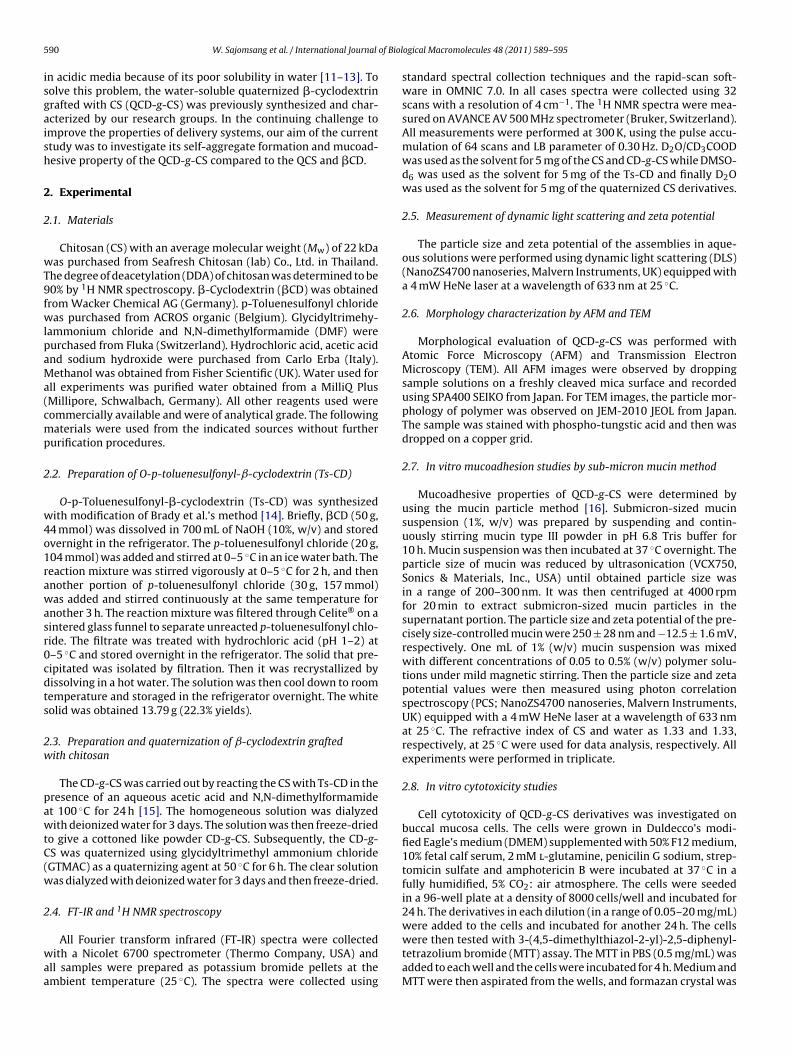
5 of Biol
isgaish
2
2
wT9fwlpaMa(cmp
2
w4o1rawasr0cdts
2w
pawtC(w
2
waa
90 W. Sajomsang et al. / International Journal
n acidic media because of its poor solubility in water [11–13]. Toolve this problem, the water-soluble quaternized �-cyclodextrinrafted with CS (QCD-g-CS) was previously synthesized and char-cterized by our research groups. In the continuing challenge tomprove the properties of delivery systems, our aim of the currenttudy was to investigate its self-aggregate formation and mucoad-esive property of the QCD-g-CS compared to the QCS and �CD.
. Experimental
.1. Materials
Chitosan (CS) with an average molecular weight (Mw) of 22 kDaas purchased from Seafresh Chitosan (lab) Co., Ltd. in Thailand.
he degree of deacetylation (DDA) of chitosan was determined to be0% by 1H NMR spectroscopy. �-Cyclodextrin (�CD) was obtainedrom Wacker Chemical AG (Germany). p-Toluenesulfonyl chlorideas purchased from ACROS organic (Belgium). Glycidyltrimehy-
ammonium chloride and N,N-dimethylformamide (DMF) wereurchased from Fluka (Switzerland). Hydrochloric acid, acetic acidnd sodium hydroxide were purchased from Carlo Erba (Italy).ethanol was obtained from Fisher Scientific (UK). Water used for
ll experiments was purified water obtained from a MilliQ PlusMillipore, Schwalbach, Germany). All other reagents used wereommercially available and were of analytical grade. The followingaterials were used from the indicated sources without further
urification procedures.
.2. Preparation of O-p-toluenesulfonyl-ˇ-cyclodextrin (Ts-CD)
O-p-Toluenesulfonyl-�-cyclodextrin (Ts-CD) was synthesizedith modification of Brady et al.’s method [14]. Briefly, �CD (50 g,
4 mmol) was dissolved in 700 mL of NaOH (10%, w/v) and storedvernight in the refrigerator. The p-toluenesulfonyl chloride (20 g,04 mmol) was added and stirred at 0–5 ◦C in an ice water bath. Theeaction mixture was stirred vigorously at 0–5 ◦C for 2 h, and thennother portion of p-toluenesulfonyl chloride (30 g, 157 mmol)as added and stirred continuously at the same temperature for
nother 3 h. The reaction mixture was filtered through Celite® on aintered glass funnel to separate unreacted p-toluenesulfonyl chlo-ide. The filtrate was treated with hydrochloric acid (pH 1–2) at–5 ◦C and stored overnight in the refrigerator. The solid that pre-ipitated was isolated by filtration. Then it was recrystallized byissolving in a hot water. The solution was then cool down to roomemperature and storaged in the refrigerator overnight. The whiteolid was obtained 13.79 g (22.3% yields).
.3. Preparation and quaternization of ˇ-cyclodextrin graftedith chitosan
The CD-g-CS was carried out by reacting the CS with Ts-CD in theresence of an aqueous acetic acid and N,N-dimethylformamidet 100 ◦C for 24 h [15]. The homogeneous solution was dialyzedith deionized water for 3 days. The solution was then freeze-dried
o give a cottoned like powder CD-g-CS. Subsequently, the CD-g-S was quaternized using glycidyltrimethyl ammonium chlorideGTMAC) as a quaternizing agent at 50 ◦C for 6 h. The clear solutionas dialyzed with deionized water for 3 days and then freeze-dried.
.4. FT-IR and 1H NMR spectroscopy
All Fourier transform infrared (FT-IR) spectra were collectedith a Nicolet 6700 spectrometer (Thermo Company, USA) and
ll samples were prepared as potassium bromide pellets at thembient temperature (25 ◦C). The spectra were collected using
ogical Macromolecules 48 (2011) 589–595
standard spectral collection techniques and the rapid-scan soft-ware in OMNIC 7.0. In all cases spectra were collected using 32scans with a resolution of 4 cm−1. The 1H NMR spectra were mea-sured on AVANCE AV 500 MHz spectrometer (Bruker, Switzerland).All measurements were performed at 300 K, using the pulse accu-mulation of 64 scans and LB parameter of 0.30 Hz. D2O/CD3COODwas used as the solvent for 5 mg of the CS and CD-g-CS while DMSO-d6 was used as the solvent for 5 mg of the Ts-CD and finally D2Owas used as the solvent for 5 mg of the quaternized CS derivatives.
2.5. Measurement of dynamic light scattering and zeta potential
The particle size and zeta potential of the assemblies in aque-ous solutions were performed using dynamic light scattering (DLS)(NanoZS4700 nanoseries, Malvern Instruments, UK) equipped witha 4 mW HeNe laser at a wavelength of 633 nm at 25 ◦C.
2.6. Morphology characterization by AFM and TEM
Morphological evaluation of QCD-g-CS was performed withAtomic Force Microscopy (AFM) and Transmission ElectronMicroscopy (TEM). All AFM images were observed by droppingsample solutions on a freshly cleaved mica surface and recordedusing SPA400 SEIKO from Japan. For TEM images, the particle mor-phology of polymer was observed on JEM-2010 JEOL from Japan.The sample was stained with phospho-tungstic acid and then wasdropped on a copper grid.
2.7. In vitro mucoadhesion studies by sub-micron mucin method
Mucoadhesive properties of QCD-g-CS were determined byusing the mucin particle method [16]. Submicron-sized mucinsuspension (1%, w/v) was prepared by suspending and contin-uously stirring mucin type III powder in pH 6.8 Tris buffer for10 h. Mucin suspension was then incubated at 37 ◦C overnight. Theparticle size of mucin was reduced by ultrasonication (VCX750,Sonics & Materials, Inc., USA) until obtained particle size wasin a range of 200–300 nm. It was then centrifuged at 4000 rpmfor 20 min to extract submicron-sized mucin particles in thesupernatant portion. The particle size and zeta potential of the pre-cisely size-controlled mucin were 250 ± 28 nm and −12.5 ± 1.6 mV,respectively. One mL of 1% (w/v) mucin suspension was mixedwith different concentrations of 0.05 to 0.5% (w/v) polymer solu-tions under mild magnetic stirring. Then the particle size and zetapotential values were then measured using photon correlationspectroscopy (PCS; NanoZS4700 nanoseries, Malvern Instruments,UK) equipped with a 4 mW HeNe laser at a wavelength of 633 nmat 25 ◦C. The refractive index of CS and water as 1.33 and 1.33,respectively, at 25 ◦C were used for data analysis, respectively. Allexperiments were performed in triplicate.
2.8. In vitro cytotoxicity studies
Cell cytotoxicity of QCD-g-CS derivatives was investigated onbuccal mucosa cells. The cells were grown in Duldecco’s modi-fied Eagle’s medium (DMEM) supplemented with 50% F12 medium,10% fetal calf serum, 2 mM l-glutamine, penicilin G sodium, strep-tomicin sulfate and amphotericin B were incubated at 37 ◦C in afully humidified, 5% CO2: air atmosphere. The cells were seededin a 96-well plate at a density of 8000 cells/well and incubated for24 h. The derivatives in each dilution (in a range of 0.05–20 mg/mL)
were added to the cells and incubated for another 24 h. The cellswere then tested with 3-(4,5-dimethylthiazol-2-yl)-2,5-diphenyl-tetrazolium bromide (MTT) assay. The MTT in PBS (0.5 mg/mL) wasadded to each well and the cells were incubated for 4 h. Medium andMTT were then aspirated from the wells, and formazan crystal was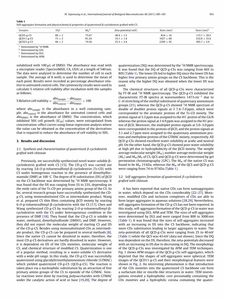
W. Sajomsang et al. / International Journal of Biological Macromolecules 48 (2011) 589–595 591
Table 1Self-aggregates formation and physicochemical properties of quaternized �-cyclodextrin grafted with CS.
Samples DQa Mwb Zeta potential (mV) Sizes (nm)c Sizes (nm)d
QCD5-g-CS 80 ± 2 75.67 40.4 ± 1.5 824 ± 16 115.7 ± 20.5QCD11-g-CS 73 ± 2 81.24 33.1 ± 1.4 1027 ± 60 232.8 ± 33.6QCD23-g-CS 66 ± 2 74.56 25.3 ± 3.4 2299 ± 45 305.7 ± 5.0
saTsetca
%
wpaictt
3
3g
cbCmowtfge6WcpwtoaSmiCowqwtplu
a Determined by 1H NMR.b Determined by GPC.c Determined by DLS.d Determined by AFM.
olubilized with 100 �L of DMSO. The absorbance was read withmicroplate reader (SpectroMAX, CA, USA) at a length of 540 nm.he data were analysed to determine the number of cell in eachample. The average of 8 wells is used to determine the mean ofach point. Results were recorded as percentage absorbance rela-ive to untreated control cells. The cytotoxicity results were used toalculate % relative cell viability after incubation with the sampless follows:
Relative cell viability = absSample − absDMSO
absControl − absDMSO× 100
here abssample is the absorbance in a well containing sam-le, abscontrol is the absorbance for untreated control cells andbsDMSO is the absorbance of DMSO. The concentration, whichnhibited 50% cell growth (IC50) values, were extrapolated fromoncentration–effect curves using linear regression analysis wherehe value can be obtained as the concentration of the derivativeshat is required to reduce the absorbance of cell viability to 50%.
. Results and discussion
.1. Synthesis and characterization of quaternized ˇ-cyclodextrinrafted with chitosan
Previously, we successfully synthesized novel water-soluble �-yclodextrin grafted with CS [15]. The CD-g-CS was carried outy reacting 3,6-O-p-toluenesulfonyl-�-cyclodextrin (Ts-CD) withS under homogenous reaction in the presence of dimethylfor-amide (DMF) at 100 ◦C. The degree of N-substitution (DS) of �CD
n the CS backbone was determined by 1H NMR spectroscopy. Itas found that the DS was ranging from 5% to 23%, depending on
he mole ratio of the Ts-CD per primary amino group of the CS. Soar, several research groups have successfully synthesized the CD--CS, using monosubstituted CDs as intermediate product. Wangt al. prepared CS thin films containing �CD moiety by reacting-O-p-toluenesulfonyl-�-cyclodextrin with the CS [17]. Chen andang synthesized CD-g-CS by reacting 2-O-p-toluenesulfonyl-�-
yclodextrin with the CS under heterogeneous condition in theresence of DMF [18]. They found that the CD-g-CS is soluble inater, methanol, dimethylsulfoxide (DMSO), and DMF. However,
hey did not report the molecular weight of starting CS and DSf the CD-g-CS. Besides using monosubstituted CDs as intermedi-te product, the CD-g-CS can be prepared in several methods [8].ince the native CS cannot be dissolved in water, therefore, theost CD-g-CS derivatives are hardly dissolved in water. However,
t is dependent on DS of the CDs moieties, molecular weight ofS, and chemical structure of modified CS. Quaternization is onef a popular method, resulting in a water-soluble CS derivativeith a wide pH range. In this study, the CD-g-CS was successfully
uaternized using glycidyltrimethylammonium chloride (GTMAC)
hich yielded quaternized CD-g-CS (QCD-g-CS). The reaction isaken place via a nucleophilic substitution by an attraction of freerimary amino groups of the CS to epoxide of the GTMAC. Simi-
ar reactions were done by reacting polysaccharides with GTMACnder the catalytic action of acid or base [19,20]. The degree of
quaternization (DQ) was determined by the 1H NMR spectroscopy.It was found that the DQ of QCD-g-CSs was ranging from 66% to80% (Table 1). The lower DS led to higher DQ since the lower DS hashigher free primary amino groups on the CS backbone. This is thereason why the higher DQ was obtained when the lower DS wasused.
The chemical structures of all QCD-g-CSs were characterizedby FT-IR and 1H NMR spectroscopy. The QCD-g-CS exhibited thecharacteristic FT-IR spectra at wavenumbers 1473 cm−1 due toC–H stretching of the methyl substituent of quaternary ammoniumgroups [21], whereas the QCD-g-CS showed 1H NMR spectrum ofdouble of doublet proton signals at ı 7.4–7.0 ppm, which werecorresponded to the aromatic protons of the Ts-CD moiety. Theproton signal at 5.3 ppm was assigned to the H1′ proton of the GlcNwhereas the proton signal at ı 4.9 ppm was assigned to the H1 pro-ton of �CD. Moreover, the multiplet proton signals at 5.0–3.0 ppmwere corresponded to the protons of �CD, and the proton signals at3.1 and 2.7 ppm were assigned to the quaternary ammonium pro-tons and methylene protons of the GTMAC moiety, respectively. AllQCD-g-CSs showed excellent water solubility at acidic and neutralpH. On the other hand, the QCD-g-CS showed poor water solubilityat high pH due to hydrophobicity of the �CD moiety. The weightaverage molecular weight (Mw), number average molecular weight(Mn) and Mw/Mn of CS, QCS and QCD-g-CS were determined by gelpermeation chromatography (GPC). The Mw of the native CS wasfound to be Mw 15 kDa, whereas the Mw of the QCS and QCD-g-CSwere ranging from 74 to 97 kDa (Table 1).
3.2. Self-aggregates formation of quaternized ˇ-cyclodextringrafted with chitosan
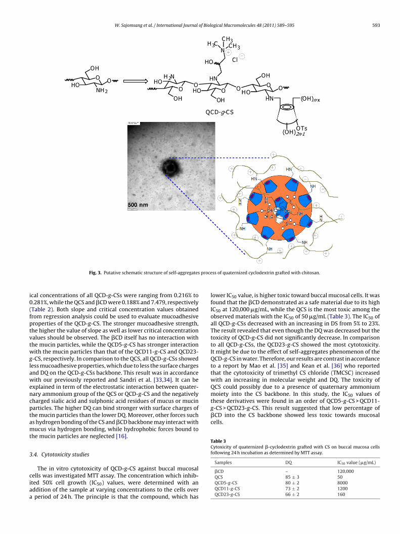
It has been reported that native CDs can form nanoaggregatesin water, which depend on the CDs considerably [22–27]. More-over, modified CDs and inclusion complexes of CDs are able toform larger aggregates in aqueous solutions [28,29]. Nevertheless,self-aggregates formation of the CD-g-CS has not been reported. Inthis study, self-aggregates formation of the QCD-g-CS in water wasinvestigated using DLS, AFM and TEM. The sizes of self-aggregateswere determined by DLS and were ranged from 800 to 3000 nm(Table 1). It was found that the sizes of self-aggregates increasedwith an increasing in DS into the CS backbone, indicating thatmore CDs substitution leading to larger aggregates in water. Thezeta-potentials of all QCD-g-CSs were ranging from 25 to 40 mV(Table 1) while the QCS was 43 mV (data not shown). Since the DQwas dependent on the DS, therefore, the zeta-potentials decreasedwith an increasing in DS due to decreasing in DQ. The morphologyof the QCD-g-CSs was investigated by AFM and TEM techniques.Fig. 1 shows AFM images of the QCD-g-CSs self-aggregates, whichdepicted that the shapes of self-aggregates were spherical. TEMimages of the QCD11-g-CS and their morphological features were
shown in Fig. 2. An interesting phenomenon is that introductionof the CDs moieties into the quaternized CS backbone can forma surfactant-like or micelle-like structures in water. TEM investi-gations revealed a hydrophobic core presumably containing theCDs moieties and a hydrophilic corona containing the quater-
592 W. Sajomsang et al. / International Journal of Biological Macromolecules 48 (2011) 589–595
F , a), quaternized cyclodextrin grafted with chitosan 11% (QCD11-g-CS, b) and quaternizedc
noitiordTdsp
3
mAdmcaoiCcss
Table 2Characteristics of the interaction between mucin particle and quaternized �-cyclodextrin grafted with CS.
Samples Equation R2 x% (w/v)
�CD y = −0.1333x + 0.9969 0.306 7.479QCS y = −5.1767x + 0.9714 0.9895 0.188QCD5-g-CS y = −6.5659x + 1.418 0.9440 0.216
ig. 1. AFM images of quaternized cyclodextrin grafted with chitosan 5% (QCD5-g-CSyclodextrin grafted with chitosan 23% (QCD23-g-CS, c).
ary ammoniums moieties. It is plausible that a small amountf the remaining p-toluenesulfonyl group onto �CD moiety maynduce host–guest inclusion complex formation. Therefore, puta-ive schematic structure of self-aggregates process of the QCD-g-CSs proposed by our group as shown in Fig. 3. The particle sizesbserved by AFM were ranging from 100 to 300 nm while TEM wereanging from 300 to 900 nm, and the values were smaller than theata determined by DLS obviously. It is noted that the AFM andEM gave the images of the particles in the dry state, while DLSepicted the value of the particle size in solution of the sample. Theize determined by DLS included hydrated layers surrounding thearticles, and was therefore larger than that in dry state.
.3. Mucoadhesion studies
The mucoadhesion of the QCD-g-CS was investigated by com-ercially available porcine mucin particles compared to the QCS.commercial lyophilized porcine (type III) was used in this work
ue to its less batch-to-batch variability and high reproducibility forucoadhesive test [30,31]. Basically, this method is based on the
hange of particle sizes and surface charges of commercially avail-ble porcine mucin particle upon the adhesion of various amountsf cationic polymers. Since the zeta-potential of the mucin particles
s negative value before interacted with the �CD, QCS, or QCD-g-S, therefore, the zeta-potential of the mucin particles is slowlyhanged from negative to positive values with an increasing in theample concentrations. It was expected that the strong mucoadhe-ive property of the QCD-g-CS would result in aggregation betweenFig. 2. TEM images of quaternized cyclodextrin graf
QCD11-g-CS y = −4.1937x + 1.0699 0.9824 0.255QCD23-g-CS y = −3.2673x + 0.9167 0.9618 0.281
x is the percentage of critical concentration (w/v).
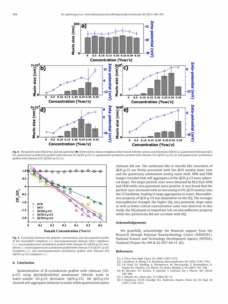
QCD-g-CS and mucin particles when the two species were mixed[16,32]. The change on both particle size and zeta potential of themucin particles were monitored at pH 6.8 in Tris buffer dependenton a titration of the QCD-g-CS concentrations. The particle size andzeta potential of the mucin particles were in a range of 250–300 nmand 10–13 mV, respectively before the mixing. A rapid increase inmucin particles size was found in QCD, QCD5-g-CS, QCD11-g-CS,and QCD23-g-CS while the �CD is constant (Fig. 4). Fig. 5 indicatesa correlation between the zeta-potential of the QCD-g-CS/mucincomplexes (ZPc)/the zeta-potential of the starting mucin particles(ZP0) ratios and the QCD-g-CS concentrations. Linear regressionanalysis was determined as shown in Table 2. The slope values of all
QCD-g-CSs were ranging from −3.2673 to −6.5659, while the QCSand �CD were −5.1767 and −0.1333, respectively. The percentageof critical concentration (x%) of the QCD-g-CS (w/v) was calculatedby providing the ZPC/ZP0 ratio equal to zero, which suggested thatneutralized mucin particles were formed. It was found that the crit-ted with chitosan 11% (QCD11-g-CS, a and b).
W. Sajomsang et al. / International Journal of Biological Macromolecules 48 (2011) 589–595 593
proce
i0(fptvtwglawencptamt
3
ciaa
these derivatives were found in an order of QCD5-g-CS > QCD11-g-CS > QCD23-g-CS. This result suggested that low percentage of�CD into the CS backbone showed less toxic towards mucosalcells.
Table 3Cytoxicity of quaternized �-cyclodextrin grafted with CS on buccal mucosa cellsfollowing 24 h incubation as determined by MTT assay.
Samples DQ IC50 value (�g/mL)
Fig. 3. Putative schematic structure of self-aggregates
cal concentrations of all QCD-g-CSs were ranging from 0.216% to.281%, while the QCS and �CD were 0.188% and 7.479, respectivelyTable 2). Both slope and critical concentration values obtainedrom regression analysis could be used to evaluate mucoadhesiveroperties of the QCD-g-CS. The stronger mucoadhesive strength,he higher the value of slope as well as lower critical concentrationalues should be observed. The �CD itself has no interaction withhe mucin particles, while the QCD5-g-CS has stronger interactionith the mucin particles than that of the QCD11-g-CS and QCD23-
-CS, respectively. In comparison to the QCS, all QCD-g-CSs showedess mucoadhesive properties, which due to less the surface chargesnd DQ on the QCD-g-CSs backbone. This result was in accordanceith our previously reported and Sandri et al. [33,34]. It can be
xplained in term of the electrostatic interaction between quater-ary ammonium group of the QCS or QCD-g-CS and the negativelyharged sialic acid and sulphonic acid residues of mucus or mucinarticles. The higher DQ can bind stronger with surface charges ofhe mucin particles than the lower DQ. Moreover, other forces suchs hydrogen bonding of the CS and �CD backbone may interact withucus via hydrogen bonding, while hydrophobic forces bound to
he mucin particles are neglected [16].
.4. Cytotoxicity studies
The in vitro cytotoxicity of QCD-g-CS against buccal mucosalells was investigated MTT assay. The concentration which inhib-ted 50% cell growth (IC50) values, were determined with anddition of the sample at varying concentrations to the cells overperiod of 24 h. The principle is that the compound, which has
ss of quaternized cyclodextrin grafted with chitosan.
lower IC50 value, is higher toxic toward buccal mucosal cells. It wasfound that the �CD demonstrated as a safe material due to its highIC50 at 120,000 �g/mL, while the QCS is the most toxic among theobserved materials with the IC50 of 50 �g/mL (Table 3). The IC50 ofall QCD-g-CSs decreased with an increasing in DS from 5% to 23%.The result revealed that even though the DQ was decreased but thetoxicity of QCD-g-CS did not significantly decrease. In comparisonto all QCD-g-CSs, the QCD23-g-CS showed the most cytotoxicity.It might be due to the effect of self-aggregates phenomenon of theQCD-g-CS in water. Therefore, our results are contrast in accordanceto a report by Mao et al. [35] and Kean et al. [36] who reportedthat the cytotoxicity of trimethyl CS chloride (TMCSC) increasedwith an increasing in molecular weight and DQ. The toxicity ofQCS could possibly due to a presence of quaternary ammoniummoiety into the CS backbone. In this study, the IC50 values of
�CD – 120,000QCS 85 ± 3 50QCD5-g-CS 80 ± 2 8000QCD11-g-CS 73 ± 2 1200QCD23-g-CS 66 ± 2 160
594 W. Sajomsang et al. / International Journal of Biological Macromolecules 48 (2011) 589–595
Fig. 4. The particle sizes (black bar) and zeta potential (�) of derivatives-mucin complexes(b), quaternized cyclodextrin grafted with chitosan 5% (QCD5-g-CS) (c), quaternized cyclografted with chitosan 23% (QCD23-g-CS) (e).
Fig. 5. Correlation between the polymer concentration and zeta-potential profileof the mucin/�CD complexes (♦), mucin/quaternized chitosan (QCS) complexes(pc(
4
gws
©), mucin/quaternized cyclodextrin grafted with chitosan 5% (QCD5-g-CS) com-lexes (�), mucin/quaternized cyclodextrin grafted with chitosan 11% (QCD11-g-CS)omplexes (�), and mucin/quaternized cyclodextrin grafted with chitosan 23%QCD23-g-CS) complexes (�).
. Conclusion
Quaternization of �-cyclodextrin grafted with chitosan (CD--CS) using glycidyltrimethyl ammonium chloride leads toater-soluble CD-g-CS derivative (QCD-g-CS). All QCD-g-CSs
howed self-aggregates behavior in water while quaternized native
when mixed with the various concentrations of �CD (a), quaternized chitosan (QCS)dextrin grafted with chitosan 11% (QCD11-g-CS) (d) and quaternized cyclodextrin
chitosan did not. The surfactant-like or micelle-like structures ofQCD-g-CS are firstly presented with the �CD moiety inner coreand the quaternary ammonium moiety outer shell. AFM and TEMimages revealed that self-aggregates of the QCD-g-CS were spheri-cal shape. The larger particle sizes were obtained by DLS than AFMand TEM while zeta-potentials were positive. It was found that theparticle sizes increased with an increasing in DS (�CD moiety) intothe CS backbone, leading to large aggregation in water. Mucoadhe-sive property of QCD-g-CS was dependent on the DQ. The strongermucoadhesive strength, the higher DQ, zeta-potential, slope valueas well as lower critical concentration value was observed. In thisstudy, the DQ played an important role on mucoadhesive propertywhile the cytotoxicity did not correlate with DQ.
Acknowledgements
We gratefully acknowledge the financial support from theResearch through National Nanotechnology Center (NANOTEC),National Science and Technology Development Agency (NSTDA),Thailand (Project No. NN-B-22-EN7-94-51-20).
References
[1] C. Price, Pure Appl. Chem. 55 (1983) 1563–1572.[2] I. Astafieva, X. Zhong, F.A. Eisenberg, Macromolecules 26 (1993) 7339–7352.[3] T.R. Patel, S.E. Harding, A. Ebringerova, M. Deszczynski, Z. Hromadkova, A.
Togola, B.S. Paulsen, G.A. Morris, A.J. Rowe, Biophys. J. 93 (2007) 741–749.[4] M. Messner, S.V. Kurkov, P. Jansook, T. Loftsson, Int. J. Pharm. 387 (2010)
199–208.[5] I. Tabushi, Acc. Chem. Res. 15 (1982) 66–72.[6] S. Anderson, T.D.W. Claridge, H.L. Anderson, Angew. Chem. Int. Ed. Engl. 36
(1997) 1310–1313.
of Biol
[
[
[[
[
[
[
[[[[
[[
[
[[
[[
[
[[
[
[
[bohydr. Polym. 78 (4) (2009) 945–952.
[34] G. Sandri, S. Rossi, M.C. Bonferoni, F. Ferrari, Y. Zambito, G. Di Colo, C. Caramella,
W. Sajomsang et al. / International Journal
[7] T. Loftsson, D. Duchêne, Int. J. Pharm. 329 (2007) 1–11.[8] M. Prabaharan, J.F. Mano, Carbohydr. Polym. 63 (2006) 153–166.[9] F. Manakker, T. Vermonden, C.F. Nostrum, W.E. Hennink, Biomacromolecules
10 (2009) 1357–3175.10] A. Binello, G. Cravotto, G.M. Nano, P. Spagliardi, Flavour Fragrance J. 19 (2004)
394–400.11] J.P. Venter, A.F. Kotzı̌e, R. Auzı̌ely-Velty, M. Rinaudo, Int. J. Pharm. 313 (2006)
36–42.12] M. Prabaharan, R. Jayakumar, Int. J. Biol. Macromol. 44 (2009) 320–325.13] X. Zhang, Z. Wub, X. Gao, S. Shu, H. Zhang, Z. Wang, C. Li, Carbohydr. Polym. 77
(2009) 394–401.14] B. Brady, N. Lynam, T. O′Sullivan, C. Ahern, R. Darcy, Org. Synth. Coll. 10 (2004)
686, Annual vol. 77, 220 (2000).15] P. Gonil, W. Sajomsang, U. Rungsardthong Ruktanonchai, N. Pimpha, I. Sra-
mala, O. Nuchuchua, S. Saesoo, S. Chaleawlert-umpon, S. Puttipipatkhachorn,Carbohydr. Polym. 83 (2) (2011) 905–913.
16] H. Takeuchi, J. Thongborisute, Y. Matsui, H. Sugihara, H. Yamamoto, Y.Kawashima, Adv. Drug Delivery Rev. 57 (2005) 1583–1594.
17] H. Wang, Y. Fang, L. Ding, L. Gao, D. Hu, Thin Solid Films 440 (2003) 255–260.18] S. Chen, Y. Wang, J. Appl. Polym. Sci. 82 (2001) 2414–2421.
19] G. Lang, H. Wendel, E. Konrad. U.S. Patent, Pat. No. 4 921 949, 1990.20] H. Li, Y. Du, X. Wu, H. Zhan, Colloids Surf. A: Physicochem. Eng. Aspects 242(2004) 1–8.21] E. Loubaki, M. Ourevitch, S. Sicsic, Eur. Polym. J. 27 (1991) 311–317.22] M. Bonini, S. Rossi, G. Karlsson, M. Almgren, P. Lo Nostro, P. Baglioni, Langmuir
22 (2006) 1478–1484.
[
[
ogical Macromolecules 48 (2011) 589–595 595
23] A.W. Coleman, I. Nicolis, J. Inclusion Phenom. Mol. Recognit. Chem. 13 (1992)139–143.
24] G.G. Gaitano, W. Brown, J. Phys. Chem. 101 (1997) 710–719.25] G. Gonzalez-Gaitano, P. Rodriguez, J.R. Isasi, M. Fuentes, G. Tardajos, M. Sanchez,
J. Inclusion Phenom. Macrocyclic Chem. 44 (2002) 101–105.26] T. Loftsson, M. Masson, M.E. Brewster, J. Pharm. Sci. 93 (2004) 1091–1099.27] S. Polarz, B. Smarsly, L. Bronstein, M. Antonietti, Angew. Chem. Int. Ed. Engl. 40
(2001) 4417–4421.28] R. Auzely-Velty, F. Djedaini-Pilard, S. Desert, B. Perly, T. Zemb, Langmuir 16 (8)
(2000) 3727–3734.29] J. Wang, M. Jiang, J. Am. Chem. Soc. 128 (11) (2006) 3703–3708.30] S. Rossi, F. Ferrari, M.C. Bonferoni, C. Caramella, Eur. J. Pharm. Sci. 10 (2000)
251–257.31] S. Rossi, F. Ferrari, M.C. Bonferoni, C. Caramella, Eur. J. Pharm. Sci. 12 (2001)
479–485.32] A. Jintapattanakit, S. Mao, T. Kissel, V.B. Junyaprasert, Eur. J. Pharm. Biopharm.
70 (2008) 563–571.33] W. Sajomsang, U. Rungsardthong Ruktanonchai, P. Gonil, O. Nuchuchua, Car-
Int. J. Pharm. 297 (2005) 146–155.35] S. Mao, X. Shuai, F. Unger, M. Wittmar, X. Xie, T. Kissel, Biomaterials 26 (2005)
6343–6356.36] T. Kean, S. Roth, M. Thanou, J. Controlled Release 103 (2005) 643–653.
Copyright © 2022 FDOKUMEN


![Chiral separation by a monofunctionalized cyclodextrin derivative: From selector to permethyl-[beta]-cyclodextrin bonded stationary phase](https://static.fdokumen.com/doc/165x107/63327b24576b626f850d70ad/chiral-separation-by-a-monofunctionalized-cyclodextrin-derivative-from-selector.jpg)