Selective crystallization of calcium salts by poly(acrylate)-grafted chitosan
8
Selective crystallization of calcium salts by poly(acrylate)-grafted chitosan Andrónico Neira-Carrillo a,b,c , Mehrdad Yazdani-Pedram c,d , Jaime Retuert b,c , Mario Diaz-Dosque c,e , Sebastien Gallois b , José L. Arias a,c,∗ a Faculty of Veterinary and Animal Sciences, University of Chile, Santiago, Chile b Faculty of Physics and Mathematics, University of Chile, Santiago, Chile c Center for Advanced Interdisciplinary Research in Materials (CIMAT), Santiago, Chile d Faculty of Chemistry and Pharmacy, University of Chile, Santiago, Chile e Faculty of Odontology, University of Chile, Santiago, Chile Abstract The biopolymer chitosan was chemically modified by grafting polyacrylamide or polyacrylic acid in a homogeneous aqueous phase using potassium persulfate (KPS) as redox initiator system in the presence of N,N -methylene-bis-acrylamide as a crosslinking agent. The influence of the grafted chitosan on calcium salts crystallization in vitro was studied using the sitting-drop method. By using polyacrylamide grafted chitosan as substrate, rosette-like CaSO 4 crystals were observed. This was originated by the presence of sulfate coming from the initiator KPS. By comparing crystallization on pure chitosan and on grafted chitosan, a dramatic influence of the grafted polymer on the crystalline habit of both salts was observed. Substrates prepared by combining sulfate with chitosan or sulfate with polyacrylamide did not produce similar CaSO 4 morphologies. Moreover, small spheres or donut-shaped CaCO 3 crystals on polyacrylic acid grafted chitosan were generated. The particular morphology of CaCO 3 crystals depends also on other synthetic parameters such as the molecular weight of the chitosan sample and the KPS concentration. Keywords: Chitosan; Crystallization; Calcium carbonate; Calcium sulfate; Polymorphs 1. Introduction In nature, biological organisms produce organo-inorganic polymeric hybrids as functional structural materials (egg- shells, seashells, and bone) [1]. Small amounts of acid-rich proteins and proteoglycans play a major role in forming these hybrids by influencing mineral crystal nucleation and growth [2–5]. Calcium carbonate is the major inorganic ma- terial in these systems, but biogenic CaSO 4 also occurs [6]. Calcium carbonate has three polymorph: calcite (hexago- nal), aragonite (orthorhombic), and vaterite (hexagonal). On the other hand, the calcium sulfate–H 2 O system is charac- * Corresponding author. Fax: +56(2)5416840. E-mail address: [email protected] (J.L. Arias). terized by different solid phases, which are differentiated by their degree of hydration: CaSO 4 ·2H 2 O (calcium sulfate dihydrate or gypsum), CaSO 4 ·(1/2)H 2 O (calcium sulfate hemihydrate), and CaSO 4 (calcium sulfate anhydrite) [7]. Therefore, understanding the mechanism of growth and inhi- bition during crystallization of calcium sulfate and calcium carbonate is of primary importance for biomedical and in- dustrial applications. An important factor for successfully coating an organic polymer substrate with crystals is to in- crease the charge density or polarity and solubility on the surface of the polymer. In this case, we have used flakes of chitosan (CHI) chem- ically modified by grafting as crystallization substrate. The CHI (poly-β -(1→4)-2-amino-2-deoxy-D-glucose) is ob- tained through partial deacetylation of chitin (poly-β (1→4)-
Transcript of Selective crystallization of calcium salts by poly(acrylate)-grafted chitosan

ase usingnce
e graftednitiatorrystallinet produceted.n sample
Selective crystallization of calcium salts bypoly(acrylate)-grafted chitosan
Andrónico Neira-Carrilloa,b,c, Mehrdad Yazdani-Pedramc,d, Jaime Retuertb,c,Mario Diaz-Dosquec,e, Sebastien Galloisb, José L. Ariasa,c,∗
a Faculty of Veterinary and Animal Sciences, University of Chile, Santiago, Chileb Faculty of Physics and Mathematics, University of Chile, Santiago, Chile
c Center for Advanced Interdisciplinary Research in Materials (CIMAT), Santiago, Chiled Faculty of Chemistry and Pharmacy, University of Chile, Santiago, Chile
e Faculty of Odontology, University of Chile, Santiago, Chile
Abstract
The biopolymer chitosan was chemically modified by grafting polyacrylamide or polyacrylic acid in a homogeneous aqueous phpotassium persulfate (KPS) as redox initiator system in the presence ofN,N -methylene-bis-acrylamide as a crosslinking agent. The influeof the grafted chitosan on calcium salts crystallization in vitro was studied using the sitting-drop method. By using polyacrylamidchitosan as substrate, rosette-like CaSO4 crystals were observed. This was originated by the presence of sulfate coming from the iKPS. By comparing crystallization on pure chitosan and on grafted chitosan, a dramatic influence of the grafted polymer on the chabit of both salts was observed. Substrates prepared by combining sulfate with chitosan or sulfate with polyacrylamide did nosimilar CaSO4 morphologies. Moreover, small spheres or donut-shaped CaCO3 crystals on polyacrylic acid grafted chitosan were generaThe particular morphology of CaCO3 crystals depends also on other synthetic parameters such as the molecular weight of the chitosaand the KPS concentration.
Keywords: Chitosan; Crystallization; Calcium carbonate; Calcium sulfate; Polymorphs
nicgg-
inganda-
go-On-
ted
inhi-min-llyin-the
em-
1. Introduction
In nature, biological organisms produce organo-inorgapolymeric hybrids as functional structural materials (eshells, seashells, and bone)[1]. Small amounts of acid-richproteins and proteoglycans play a major role in formthese hybrids by influencing mineral crystal nucleationgrowth[2–5]. Calcium carbonate is the major inorganic mterial in these systems, but biogenic CaSO4 also occurs[6].Calcium carbonate has three polymorph: calcite (hexanal), aragonite (orthorhombic), and vaterite (hexagonal).the other hand, the calcium sulfate–H2O system is charac
* Corresponding author. Fax: +56(2)5416840.E-mail address: [email protected](J.L. Arias).
terized by different solid phases, which are differentiaby their degree of hydration: CaSO4·2H2O (calcium sulfatedihydrate or gypsum), CaSO4·(1/2)H2O (calcium sulfatehemihydrate), and CaSO4 (calcium sulfate anhydrite)[7].Therefore, understanding the mechanism of growth andbition during crystallization of calcium sulfate and calciucarbonate is of primary importance for biomedical anddustrial applications. An important factor for successfucoating an organic polymer substrate with crystals is tocrease the charge density or polarity and solubility onsurface of the polymer.
In this case, we have used flakes of chitosan (CHI) ch
ically modified by grafting as crystallization substrate. TheCHI (poly-β-(1→4)-2-amino-2-deoxy-D-glucose) is ob-tained through partial deacetylation of chitin (poly-β(1→4)-
osttheso-on
-filmid)rm
ups
nc-cialtionaticrevi
seeretheirlts.
iumstal-ide
-
d toh,DH
ideaneenf theion.
edpre-
obopew-eter
byo-ulfate
er-
ong
iont toionsn0ne.cedplesin
aft-
nsid-ob-ted
rig-
hi-pec-
ion
hi-inyle00%.
A. Neira-Carrillo et al.
2-acetamido-2-deoxy-D-glucose), which is the second mabundant polysaccharide in nature. Although most ofwork on crystallization of these salts has been done inlution, only a few investigations have been carried outthe surfaces of films or gels[8–17]. Generally, the biopolymers chitin and chitosan are insoluble and do not formmaterials. However, when grafted with poly(acrylic acor polyacrylamide, they become insoluble, are able to foflakes with a nearly flat surface, and show functional grothat can influence crystal nucleation and growth[18–22]. Toour best knowledge, the spatial distribution of polar futional groups, that is, the local charge density, is of cruimportance for the nucleation and determines the orientaof crystals. The importance of the nonspecific electrosteffects such as charge density has been determined pously by Volkmer et al.[23].
Therefore, it is interesting to study the effect of thegrafted chains on the crystalline habit of calcium salts. Hwe report the preparation of these grafted polymers andapplication as substrates for crystallization of calcium saThe effect of these grafted chitosan matrix on the calcsalts crystallization is analyzed and compared to the crylization within an unmodified chitosan and polyacrylamgel as a control.
2. Materials and methods
2.1. Materials
Chitosan (CHI) samples of high molecular weight (Mw=350 KDa,>83% deacetylation) from Aldrich and low molecular weight (Mw= 70 KDa, >75% deacetylation) fromFluka were washed with acetone and methanol and drieconstant weight. Acrylamide and acrylic acid from Aldricpotassium persulfate (KPS) of analytical grade from BChemicals, andN,N -methylene-bis-acrylamide (N,N -bis-AAM) from Fluka were used as received. Calcium chlordihydrate, ethanol, and tris(hydroxymethyl)aminomethwere obtained from ACS-Merck and ammonium hydrogcarbonate was from J.T. Baker. These reagents were ohighest available grade and used without further purificat
2.2. Measurements
Fourier transform infrared spectra (FTIR) were obtainon a Bruker Vector 22 instrument. The samples werepared as potassium bromide pellets. The crystals wereserved in a Tesla BS 343 A scanning electron microsc(SEM). The diffraction spectrum was taken from a podered sample using a Siemens D-5000 X-ray diffractom
with CuKα radiation. The elemental analysis of grafted chi-tosan films was carried out by using an EAGER 200 (methodSSabr02.EAD).-
-
2.3. Graft copolymerization
The biopolymer chitosan was chemically modifiedgrafting with polyacrylamide or polyacrylic acid in a hmogeneous aqueous phase by using potassium persas a redox initiator in the presence ofN,N -methylene-bis-acrylamide as a crosslinking agent.
Grafting reactions were carried out in 100 ml polymization flasks by first dissolving 1 g of CHI in 40 ml of 2%acetic acid followed by the addition of monomer soluti(3.3 g of acrylic acid in 40 ml of 2% acetic acid and/or 2.4of acrylamide in 40 ml of 2% acetic acid). The concentratof N,N -methylene-bis-acrylamide was 0.25 wt% respecthe weight of the monomers used. Two KPS concentratof 1× 10⁻² and 2× 10⁻² M were used. The polymerizatioflask was closed and placed in a thermostated bath at 6◦Cfor 1 h. The reaction product was precipitated in acetoThe precipitate was filtered and then dried under redupressure to constant weight. The grafted chitosan samwere purified by exhaustive extraction with water for 24 horder to remove the homopolymer formed during the gring reaction and the nonreacted monomer[22]. After dryingunder reduced pressure the remaining product was coered to be a graft copolymer. Evidence of grafting wastained from comparison of IR of the grafted and nongrafchitosan.
2.4. Grafting parameters
The percentage of grafting (%G) and grafting efficiency(%E) was calculated from the increase in weight of the oinal CHI after grafting and extraction as follows[20–22],
(1)%G = w2 − w1
w1× 100,
(2)%E = w2 − w1
w3× 100,
wherew2, w1 andw3 represents the weights of grafted ctosan after extraction, initial chitosan, and monomer, restively.
An estimation of the average degree of polymerizatof grafted polyacrylic acid and/or polyacrylamide (R) wascalculated by using the equation
(3)R = (w2 − w1)MCHI
Mmw1,
whereMCHI andMm represent the molar mass of the ctosan monomer unit and the molar mass of grafted vmonomer respectively. TheR values are only approximatvalues since the chitosan used in this study was not 1deacetylated, i.e., has a degree of deacetylation of 83%
2.5. Crystallization method
Crystallization experiments were carried out in the pres-ence of grafted chitosan samples as structure-directing sub-strates using the sitting-drop method[24,25] (Fig. 1). The

as
con-ole
idekesas
mlr-
-
ent
0%per-to-
ingtal
san
san
nsnd
r-theons-
theeac-ly-
A. Neira-Carrillo et al.
Fig. 1. Experimental setup used for growing CaCO3 crystals in vitro. Theconcentration of CaCl2 (microbridge) and of ammonium bicarbonate w200 and 25 mM, respectively.
crystallization experiments were done using a chamber,sisting of an 85-mm plastic Petri dish having a central hin its bottom, glued to a plastic cylindrical vessel. Insthe chamber, 3–7 mg of chitosan or chitosan-grafted flawere put into a polystyrene microbridge. Thereafter it wfilled with 35 µl of 200 mM CaCl2 solution in 200 mMTRIS buffer pH 9. The cylindrical vessel contained 3of 25 mM (NH4)HCO3 solution. All experiments were caried out inside the Petri dish at pH 9.0 and 20◦C for 24 h.Precipitation of CaCO3 results from the diffusion of carbon dioxide (CO2) vapor into the buffered CaCl2 solution.The polymeric substrates with the crystals of the differpolymorphs of CaCO3 and/or CaSO4 crystals formed werecollected. Once rinsed with distilled water and on 50–10gradient ethanol solutions, they were dried at room temature and finally coated with gold using an EMS-550 aumated sputter coater and analyzed by SEM.
3. Results and discussion
The mineralization of CaCO3 and CaSO4 using thesitting-drop method, in the presence of different graftchitosan films, resulted in particles with different crys
structures and morphologies. The grafting of chitosans, usedFig. 2. Synthetic route for obtaining CHI-g-PAAM grafted
Table 1Grafting conditions and parameters for grafting of acrylamide onto chito(CHI-g-PAAM)
LMW chitosan HMW chitosan
Chitosana 1.0 g 1.0 g 1.0 g 1.0 gKPS 1× 10⁻² M 2 × 10⁻² M 1 × 10⁻² M 2 × 10⁻² MAAM 2.4 g 2.4 g 2.4 g 2.4 gCrosslinker(N,N -bis-AAM)
0.006 g 0.006 g 0.006 g 0.006 g
%G 129 125 127 73%E 53.8 52.1 52.9 30.6R 2.9 2.8 2.9 1.7
a Total volume of aqueous phase= 80 ml of 2% acetic acid.
Table 2Grafting conditions parameters for grafting of acrylic acid onto chito(CHI-g-PAA)
LMW chitosan HMW chitosan
Chitosana 1.0 g 1.0 g 1.0 g 1.0 gKPS 1× 10⁻² M 2 × 10⁻² M 1 × 10⁻² M 2 × 10⁻² MAA 3.330 g 3.330 g 3.330 g 3.330 gCrosslinker(N,N -bis-AAM)
0.008 g 0.008 g 0.008 g 0.008 g
%G 205 179 266 244%E 61.4 53.7 79.9 73.4R 4.6 4.0 6.0 5.5
a Total volume of aqueous phase= 80 ml of 2% acetic acid.
Tables 1 and 2show the percentage of grafting (%G),grafting efficiency (%E), and the length of the grafted chai(R) for the optimized grafting reactions of acrylamide aacrylic acid onto chitosan, respectively[20].
It can be seen fromTables 1 and 2that higher %E is ob-tained for a KPS concentration of 1× 10⁻² M for both sys-tems CHI-g-PAA and CHI-g-PAAM when low-moleculaweight chitosan was used. In the CHI-g-PAAM system,%E was slightly lower, probably due to the consumptiof monomer to form homopolymer. This favors the posible combination of the chitosan macroradicals withexisting excess of primary free radicals present in the rtion medium. The homopolymer formation and grafted po
acrylamide chains could be crucial for the interaction withOnce
as templates for crystallization studies, was carried out ac-cording to the following synthetic route (Fig. 2).
sulfate groups coming from KPS and allowed the CaS4crystals (gypsum) formation in this system. The prese
chitosan. The same procedure was used for CHI-g-PAA.

, re-estat
entromlar
fef-
and
fted
–de
g toted
nrular
ab-
S)
ofthe
wasftedndmu-d
rp-p
Mres-AMvedeareM
tentsdhi-en
Oe-
A. Neira-Carrillo et al.
Table 3Elemental analysis of CHI-g-PAAM and CHI-g-PAA films
Sample [KPS] (M) %N %C %H %S
CHI-g-PAAM(from LMW)
2× 10⁻² 13.8 41.2 7.9 1.2
CHI-g-PAAM(from HMW)
1× 10⁻² 12.2 39.7 7.4 1.5
CHI-g-PAA(from HMW)
1× 10⁻² 1.88 47.6 6.6 0.0
CHI-g-PAA(from LMW)
2× 10⁻² 1.68 46.8 6.3 0.0
of sulfur (Table 3) and the sulfate groups (Fig. 3) wereconfirmed by elemental analysis and FTIR spectroscopyspectively. In the case of CHI-g-PAA system, the high%E was found by using high molecular weight chitosan1× 10−2 M concentration.
The IR spectrum of the grafted chitosan is in agreemwith the proposed molecular structure. Infrared spectra fgrafted chitosan and chitosan of high and low molecuweights are shown inFigs. 3A and 3B. The comparison oIR spectra of chitosan and grafted chitosan show thatfectively the grafting has taken place. The absorption bat 3429 cm−1 is due to NH bonds of chitosan (Fig. 3A1and 3B1) and those at 1564 and 1651 cm−1 correspond toamide I and II bands respectively.
On the other hand, the spectrum of chitosan grawith polyacrylamide (PAAM) (Fig. 3A2) shows a strongabsorption band at 1673 cm−1 corresponding to aminecarbonyl absorption from PAAM chains. In polyacrylamihomopolymer this band is located at 1670 cm−1. Themost typical absorption bands of chitosan correspondinamide I and II bands, are not clearly visible in the graf
product since they are hidden by a strong and broad carbonyFig. 3. FTIR spectra for the chitosan and grafted chitosan. (A1) HMW chg-PAAM; (B3) CH-g-PAA.
shoulder at 1602 cm−1. The shift of the carbonyl absorptioband of PAAM grafted chitosan (CHI-g-PAAM) to lowefrequencies could be due to the inter-and/or intramolecinteractions through hydrogen bonding (Figs. 3A2 and 3B2).Both spectra for PAAM grafted chitosan show two newsorption bands at 1322 and 1087 cm−1 attributed to thepresence of sulfate groups coming from the initiator (KP[26,27].
The sulfate contained in the CHI-g-PAAM, in spitethe exhaustive extraction process with water, explainspresence of calcium sulfate crystals when this productused as a substrate for crystallization. For chitosan grawith polyacrylic acid (PAA) an absorption band at 1732 aat 1735 cm−1 were assigned to the carbonyl groups fropoly(acrylic acid)-grafted chitosan of high and low moleclar weight, respectively[22]. Both chitosan samples graftewith poly(acrylic acid) (CHI-g-PAA) also showed an absotion band at 1627 cm−1 corresponding to the amide grou(Figs. 3A3 and 3B3).
Table 3shows the elemental analysis of CHI-g-PAAand CHI-g-PAA. The results are in agreement with the pence and absence of the sulfate groups in CHI-g-PAand CHI-g-PAA, respectively, as was previously obserin the IR analysis (Fig. 3). The absence of sulfur in thCHI-g-PAA material indicates that charged PAA chainsnot able to coordinate sulfate ions at difference of PAAgrafted chitosan. The carbon and hydrogen relative conof CHI-g-PAAM and CHI-g-PAA products are similar anindependent of the molecular weight of the ungrafted ctosan. As expected, the CHI-g-PAAM has higher nitrogcontent that CHI-g-PAA.
The influence of the grafted chitosan films on the CaC3and CaSO4 crystallization in vitro was studied using thsitting-drop method (Fig. 4). Different Ca salts and mor
lphologies were obtained (Table 4). Fig. 4a shows the controlcal-y
absorption band of PAAM in this spectral region. However,the chitosan amide I absorption band could be observed as a
experiment carried out by using non-grafted CHI wherecite crystals were obtained.Fig. 4b shows the morpholog
itosan; (A2) CHI-g-PAAM; (A3) CH-g-PAA; (B1) LMW chitosan; (B2) CHI-

A. Neira-Carrillo et al.
Fig. 4. SEM pictures of crystals grown in the presence of chitosan and grafted chitosan samples. (a) Chitosan control, (b) CHI-g-PAAM (HMW, KPS1× 10−2 M), (c) CHI-g-PAAM (LMW, KPS 2× 10⁻² M), (d) CHI-g-PAA (HMW, KPS 1× 10⁻² M), (e, f) CHI-g-PAA (LMW, KPS 1× 10⁻² M).
A. Neira-Carrillo et al.
itosa
Min
nenceb-
CO
Ormsl-tast,led
chi-
fAdeeneh on
d in
e as
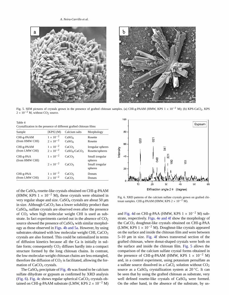
Fig. 5. SEM pictures of crystals grown in the presence of grafted ch2× 10−2 M, without CO2 source.
Table 4Crystallization in the presence of different grafted chitosan films
Sample [KPS] (M) Calcium salts Morphology
CHI-g-PAAM(from HMW CHI)
1× 10⁻² CaSO4 Rosette2× 10−2 CaSO4 Rosette
CHI-g-PAAM(from LMW CHI)
1× 10⁻² CaCO3 Irregular spheres2× 10−2 CaSO4/CaCO3 Rosette/spheres
CHI-g-PAA(from HMW CHI)
1× 10⁻² CaCO3 Small irregularspheres
2× 10⁻² CaCO3 Small irregularspheres
CHI-g-PAA(from LMW CHI)
1× 10⁻² CaCO3 Donuts2× 10−2 CaCO3 Donuts
of the CaSO4 rosette-like crystals obtained on CHI-g-PAA(HMW, KPS 1× 10⁻² M), these crystals were obtainedvery regular shape and size. CaSO4 crystals are about 50 µmin size. Although CaCO3 has a lower solubility product thaCaSO4, sulfate crystals are observed even after the presof CO2 when high molecular weight CHI is used as sustrate. In fact experiments carried out in the absence of2source showed the presence of CaSO4 with similar morphol-ogy as those observed inFigs. 4b and 5a. However, by usingsubstrates obtained with low molecular weight CHI, CaC3crystals are also formed. This could be rationalized in teof diffusion kinetics because all the Ca is initially in sufate form; consequently CO2 diffuses hardly into a compacstructure formed by the long chitosan chains. In contrthe low-molecular-weight chitosan chains are less entangtherefore the diffusion of CO2 is facilitated, allowing the for-mation of CaCO3 crystals.
The CaSO4 precipitate ofFig. 4b was found to be calcium
sulfate dihydrate or gypsum as confirmed by XRD analysis(Fig. 6). Fig. 4c shows regular spherical CaCO3 crystals ob-tained on CHI-g-PAAM substrate (LMW, KPS 2× 10⁻² M)n samples. (a) CHI-g-PAAM (HMW, KPS 1× 10⁻² M); (b) KPS-CaCl2, KPS
,
Fig. 6. XRD patterns of the calcium sulfate crystals grown on graftedtosan samples. CHI-g-PAAM (HMW, KPS 2× 10⁻² M).
andFig. 4d on CHI-g-PAA (HMW, KPS 1× 10⁻² M) sub-strate, respectively.Figs. 4e and 4fshow the morphology othe CaCO3 doughnut-like crystals obtained on CHI-g-PA(LMW, KPS 1× 10⁻² M). Doughnut-like crystals appeareon the surface and inside the chitosan film and were betw5–10 µm in size.Fig. 4f shows transversal section of thgrafted chitosan, where donut-shaped crystals were botthe surface and inside the chitosan film.Fig. 5 allows thecomparison of the calcium sulfate crystal forms obtainethe presence of CHI-g-PAAM (HMW, KPS 1× 10⁻² M)and, in a control experiment, using potassium persulfata sulfate source dissolved in a CaCl2 solution without CO2source as a CaSO4 crystallization system at 20◦C. It can
be seen that by using the grafted chitosan as substrate, verywell defined rosette-like crystals of CaSO4 were formed.On the other hand, in the absence of the substrate, by us-
ftedPS
e-
ueasthe
-
Son
n
t-as-
f
on-Sani-nt.
ine
nd
an,lline
ftedPS
ederi-al
es-htthari-
ofe, onentffer-In, it.theomthe
tioned
ga-
re-resslliza-f theakee-
A. Neira-Carrillo et al.
Fig. 7. XRD patterns of the calcium sulfate crystals grown on grachitosan samples. (a) HMW chitosan and (b) CHI-g-PAAM (HMW, K1× 10−2 M) after crystallization.
ing KPS as donor of sulfate groups, irregular CaSO4 crystalswere formed (Fig. 5b). We can clearly see how the surfacmodified substrate modulated the shape of CaSO4 crystals.
Powder X-ray diffraction is a more suitable techniqthan electron microscopy for solids identification, andsuch it was used in our crystallization assays to confirmelectron microscopic results.Fig. 7a shows the XRD patterns of the chitosan (HMW) andFig. 7b the XRD patternsof crystals grown on the grafted CHI-g-PAAM (HMW, KP1 × 10−2 M). It can be seen that the strongest diffractiintensity is the broad peak around 2θ = 17–23◦ due to thesubstrate chitosan. InFig. 7b the peaks at 2θ = 11.5◦, 20.8◦,22.4◦, 29◦, 32◦, 34◦, and 44◦ are ascribed to the diffractioof CaSO4·2H2O (gypsum).
In addition, Fig. 6 shows the fine powder XRD paterns of the grafted chitosan after the crystallizationsays using CHI-g-PAAM (HMW, KPS 2× 10⁻² M). It canclearly seen the peaks at 2θ = 11.6◦, 20.7◦, 20.7◦, 23.5◦,29.1◦, 31.1◦, 33.3◦, and 43.5◦, confirming the occurrence oCaSO4·2H2O.
In the case of SEM observations of calcium carbate crystals formed on a CHI-g-PAA surface (LMW, KP1 × 10−2 M), Figs. 4e and 4fshowed that they presentnovel and attractive donut-like form. Even at high magfication, no sign of defined crystal structure was evideThis was the first indication that this CaCO3 form mightcorrespond to an amorphous phase. Therefore we obtaa powder X-ray diffraction analysis of this sample (Fig. 8).
Fig. 8a shows the XRD patterns of the chitosan aFig. 8b the XRD pattern of donut CaCO3 particles grown onthe grafted CHI-g-PAA (LMW, KPS 1× 10⁻² M). This pat-tern is practically identical to that of the ungrafted chitoswithout the presence of peaks attributable to a crysta
phase what could indicate the presence of an amorphousCaCO3 phase. Transient stabilization of amorphous CaCO3phase by polycarboxylate additives (acrylic acid/maleic acidd
Fig. 8. XRD patterns of the calcium carbonate crystals grown on grachitosan samples. (a) LMW chitosan and (b) CHI-g-PAA (LMW, K1× 10−2 M) after crystallization.
copolymer) as a first stage of the precipitation was confirmby Rieger et al. using time-resolved X-ray scattering expments[28]. Table 4show a summary of the different crystforms by SEM analysis.
4. Summary
The mineralization of calcium salts done in the prence of chitosan of high (HMW) or low molecular weig(LMW), grafted in each case with either PAAM or PAA witwo concentrations of KPS, resulted in particles with a vety of crystalline structures and morphologies.
A dramatic influence of the particular compositiongrafted chitosan, used as a structure-directing substratthe crystalline habit of calcium salts was found. The differactive groups present in the grafted chains interact diently with the individual growing planes of the crystals.experiments done with CHI grafted with polyacrylamidewas found that rosette shaped CaSO4 crystals are formedThis indicates that, in spite of the exhaustive extraction ofgrafted copolymers with water, sulfate groups coming frthe KPS remains in the product, probably associated withgrafted polyacrylamide chains. In the case of crystallizaon CHI-g-PAA films small spheres or donuts-like shapCaCO3 particles were selectively formed. Further investition in the direction of the mechanism of the CaSO4 rosettecrystals growing, influence of acidic functional group steochemistry and crystal kinetic analysis are under progto achieve the understanding of heterogeneous crystation on grafted chitosans. Regarding the mechanism oCaSO4 crystals, we suggest that crystal growth could tplace inside the grafted CHI-g-PAAM films with subs
quent escalation and grown up to the surface. This kind ofmechanism was recently reported for some superstructure ofCaCO3 particles[29,30].
APand
iv.
a-
97)
61
530.
8)
em.
21.02)
14
ltry
ter.
A. Neira-Carrillo et al.
Acknowledgments
This research was supported by Project FOND11980002 granted by the Chilean Council for ScienceTechnology (CONICYT).
References
[1] S. Mann, G.A. Ozin, Nature 382 (1996) 313.[2] H.A. Lowenstam, S. Weiner, On Biomineralization, Oxford Un
Press, Oxford, 1989.[3] J.L. Arias, A. Neira-Carrillo, J.I. Arias, C. Escobar, M. Bodero, M. D
vid, M.S. Fernández, J. Mater. Chem. 14 (2004) 2154.[4] S. Mann, Biomineralization, Oxford Univ. Press, Oxford, 2001.[5] J.L. Arias, M.S. Fernandez, Mater. Charact. 50 (2003) 189.[6] H.C.W. Skinner, A.H. Jahren, Treatise Geochem. 8 (2003) 117.[7] R. Sheikholeslami, M. Ng, Ind. Eng. Chem. Res. 40 (2001) 3570.[8] F. Manoli, S. Koutsopoulus, E. Dalas, J. Cryst. Growth 189 (19
116.[9] T. Kato, T. Suzuki, T. Irie, Chem. Lett. 29 (2000) 186.
[10] N. Hosoda, T. Kato, Chem. Mater. 13 (2001) 688.[11] P. Liang, Y. Zhao, Q. Shen, D. Wang, D. Xu, J. Cryst. Growth 2
(2004) 571.
[12] O. Grassmann, G. Müller, P. Löbmann, Chem. Mater. 14 (2004) 4[13] S. Zhang, K.E. Gonsalves, J. Appl. Polym. Sci. 56 (1995) 687.[14] S. Zhang, K.E. Gonsalves, Langmuir 14 (1998) 6761.[15] A. Sugawara, T. Kato, Chem. Commun. 6 (2000) 487.[16] T. Kato, Adv. Mater. 12 (2000) 1543.[17] T. Kato, T. Suzuki, T. Amamiya, T. Irie, Supramol. Sci. 5 (199
411.[18] K. Kurita, Prog. Polym. Sci. 26 (2001) 1921.[19] E. Khor, L.Y. Lim, Biomaterials 24 (2003) 2339.[20] M. Yazdani-Pedram, A. Lagos, J. Retuert, J.M.S. Pure, Appl. Ch
A 32 (5) (1995) 1037.[21] M. Yazdani-Pedram, J. Retuert, J. Appl. Polym. Sci. 63 (1997) 13[22] M. Yazdani-Pedram, A. Lagos, J. Retuert, Polym. Bull. 48 (20
93.[23] D. Volkmer, M. Fricke, C. Agena, J. Mattay, J. Mater. Chem.
(2004) 2249.[24] J.M. Domínguez-Vera, J. Gautron, J.M. García-Ruiz, Y. Nys, Pou
Sci. 79 (1999) 901.[25] J.I. Arias, J.P. Wiff, M.S. Fernandez, V. Fuenzalida, J.L. Arias, Ma
Res. Soc. Symp. Proc. 711 (2002) 243.[26] S. Durmaz, O. Okay, Polymer 41 (2000) 3693.[27] O. Grassmann, P. Löbmann, Chem. Eur. J. 9 (2003) 1310.[28] J. Rieger, J. Thieme, C. Schmidt, Langmuir 16 (2000) 8300.[29] J. Rudloff, H. Cölfen, Langmuir 20 (2004) 991.[30] S.-H. Yu, H. Cölfen, J. Mater. Chem. 14 (2004) 2124.


![Electrografting of calix[4]arenediazonium salts to form ... - Nature](https://static.fdokumen.com/doc/165x107/6314eac3c72bc2f2dd0489bf/electrografting-of-calix4arenediazonium-salts-to-form-nature.jpg)