Selective Targeting of TRPV1 Expressing Sensory Nerve Terminals in the Spinal Cord for Long Lasting...
11
Selective Targeting of TRPV1 Expressing Sensory Nerve Terminals in the Spinal Cord for Long Lasting Analgesia Joseph A. Jeffry 1. , Shuang-Quan Yu 1. , Parul Sikand 1 , Arti Parihar 1 , M. Steven Evans 2 , Louis S. Premkumar 1 * 1 Department of Pharmacology, Southern Illinois University School of Medicine, Springfield, Illinois, United States of America, 2 Department of Neurology, Southern Illinois University School of Medicine, Springfield, Illinois, United States of America Abstract Chronic pain is a major clinical problem and opiates are often the only treatment, but they cause significant problems ranging from sedation to deadly respiratory depression. Resiniferatoxin (RTX), a potent agonist of Transient Receptor Potential Vanilloid 1 (TRPV1), causes a slow, sustained and irreversible activation of TRPV1 and increases the frequency of spontaneous excitatory postsynaptic currents, but causes significant depression of evoked EPSCs due to nerve terminal depolarization block. Intrathecal administration of RTX to rats in the short-term inhibits nociceptive synaptic transmission, and in the long-term causes a localized, selective ablation of TRPV1-expressing central sensory nerve terminals leading to long lasting analgesia in behavioral models. Since RTX actions are selective for central sensory nerve terminals, other efferent functions of dorsal root ganglion neurons can be preserved. Preventing nociceptive transmission at the level of the spinal cord can be a useful strategy to treat chronic, debilitating and intractable pain. Citation: Jeffry JA, Yu S-Q, Sikand P, Parihar A, Evans MS, et al. (2009) Selective Targeting of TRPV1 Expressing Sensory Nerve Terminals in the Spinal Cord for Long Lasting Analgesia. PLoS ONE 4(9): e7021. doi:10.1371/journal.pone.0007021 Editor: Hiroaki Matsunami, Duke Unviersity, United States of America Received June 8, 2009; Accepted August 10, 2009; Published September 15, 2009 Copyright: ß 2009 Jeffry et al. This is an open-access article distributed under the terms of the Creative Commons Attribution License, which permits unrestricted use, distribution, and reproduction in any medium, provided the original author and source are credited. Funding: This work was supported by grants from National Institutes of Health (NS042296 and DK065742) and EAM award from SIUSOM. The funders had no role in study design, data collection and analysis, decision to publish, or preparation of the manuscript. Competing Interests: The authors have declared that no competing interests exist. * E-mail: [email protected] . These authors contributed equally to this work. Introduction Transient receptor potential vanilloid 1 (TRPV1/VR1) is a nonselective cation channel with high calcium permeability expressed on the peripheral and central terminals of small- diameter sensory neurons. On the peripheral terminals it functions as a polymodal receptor [1,2,3]. On the central terminals it modulates synaptic transmission selectively at the first sensory synapse between dorsal root ganglion (DRG) or trigeminal ganglion (TG) neurons and dorsal horn (DH) or caudal spinal trigeminal nucleus (CSTN) neurons [4,5,6,7]. TRPV1 is activated by heat (.42uC), capsaicin (a pungent ingredient of hot chili peppers), resiniferatoxin (RTX), protons, anandamide, arachido- nic acid metabolites and N-arachidonyl dopamine (NADA) [1,2,8,9,10,11,12,13,14,15]. RTX is derived from latex of the cactus Euphorbium resinifera and is the most potent of all known natural and synthetic agonists for TRPV1 [16]. We have previously demonstrated that RTX is a potent and irreversible agonist of TRPV1 [17]. In DRG neurons, capsaicin-induced currents were readily reversible, whereas RTX-induced a sustained current. Furthermore, capsaicin-induced currents exhib- ited relatively fast activation and deactivation/desensitization phases as compared to RTX-induced currents, which exhibited significantly slower activation phase and deactivated/desensitized minimally even in the presence of extracellular Ca 2+ [1]. In single channel recordings, RTX induced a maximal activation of the receptor in a concentration-independent manner. Low concen- trations of RTX caused slow and sustained depolarization of the membrane potential and prevented action potential generation [17]. These studies suggest that RTX binds to TRPV1 with high affinity and does not readily desensitize the receptor. Intrathecal administration of capsaicin in rats caused long lasting loss of heat sensitivity [18,19,20]. In animal models of pain, RTX has also been found useful in inflammatory pain, painful conditions affecting joints, and bone cancer pain by eliminating TRPV1 expressing peripheral nerve terminals or DRG neurons [21,22,23,24]. In this study, we demonstrate that selective targeting of TRPV1 expressed in the central terminals of sensory neurons is sufficient to reduce inflammatory thermal hypersensitivity. The analgesic effects of RTX treatment arise from its ability to activate TRPV1 in a slow and sustained manner, leading to block of transmission at the first sensory synapse in the short-term and nerve terminal ablation in the long-term. Results Modulation of synaptic transmission by capsaicin and RTX The first sensory synapse at the spinal cord between DRG and dorsal horn (DH) neurons functions as a gain controller for painful inputs from the periphery. TRPV1 is expressed only at the sensory nerve terminals that form synapses with the second order DH neurons in spinal cord laminae I and II and in the caudal spinal trigeminal nucleus (CSTN) (Fig. S1). We have investigated TRPV1-mediated modulation of synaptic transmission at the first PLoS ONE | www.plosone.org 1 September 2009 | Volume 4 | Issue 9 | e7021
-
Upload
michiganstate -
Category
Documents
-
view
0 -
download
0
Transcript of Selective Targeting of TRPV1 Expressing Sensory Nerve Terminals in the Spinal Cord for Long Lasting...
Selective Targeting of TRPV1 Expressing Sensory NerveTerminals in the Spinal Cord for Long Lasting AnalgesiaJoseph A. Jeffry1., Shuang-Quan Yu1., Parul Sikand1, Arti Parihar1, M. Steven Evans2,
Louis S. Premkumar1*
1 Department of Pharmacology, Southern Illinois University School of Medicine, Springfield, Illinois, United States of America, 2 Department of Neurology, Southern Illinois
University School of Medicine, Springfield, Illinois, United States of America
Abstract
Chronic pain is a major clinical problem and opiates are often the only treatment, but they cause significant problemsranging from sedation to deadly respiratory depression. Resiniferatoxin (RTX), a potent agonist of Transient ReceptorPotential Vanilloid 1 (TRPV1), causes a slow, sustained and irreversible activation of TRPV1 and increases the frequency ofspontaneous excitatory postsynaptic currents, but causes significant depression of evoked EPSCs due to nerve terminaldepolarization block. Intrathecal administration of RTX to rats in the short-term inhibits nociceptive synaptic transmission,and in the long-term causes a localized, selective ablation of TRPV1-expressing central sensory nerve terminals leading tolong lasting analgesia in behavioral models. Since RTX actions are selective for central sensory nerve terminals, otherefferent functions of dorsal root ganglion neurons can be preserved. Preventing nociceptive transmission at the level of thespinal cord can be a useful strategy to treat chronic, debilitating and intractable pain.
Citation: Jeffry JA, Yu S-Q, Sikand P, Parihar A, Evans MS, et al. (2009) Selective Targeting of TRPV1 Expressing Sensory Nerve Terminals in the Spinal Cord forLong Lasting Analgesia. PLoS ONE 4(9): e7021. doi:10.1371/journal.pone.0007021
Editor: Hiroaki Matsunami, Duke Unviersity, United States of America
Received June 8, 2009; Accepted August 10, 2009; Published September 15, 2009
Copyright: � 2009 Jeffry et al. This is an open-access article distributed under the terms of the Creative Commons Attribution License, which permitsunrestricted use, distribution, and reproduction in any medium, provided the original author and source are credited.
Funding: This work was supported by grants from National Institutes of Health (NS042296 and DK065742) and EAM award from SIUSOM. The funders had no rolein study design, data collection and analysis, decision to publish, or preparation of the manuscript.
Competing Interests: The authors have declared that no competing interests exist.
* E-mail: [email protected]
. These authors contributed equally to this work.
Introduction
Transient receptor potential vanilloid 1 (TRPV1/VR1) is a
nonselective cation channel with high calcium permeability
expressed on the peripheral and central terminals of small-
diameter sensory neurons. On the peripheral terminals it functions
as a polymodal receptor [1,2,3]. On the central terminals it
modulates synaptic transmission selectively at the first sensory
synapse between dorsal root ganglion (DRG) or trigeminal
ganglion (TG) neurons and dorsal horn (DH) or caudal spinal
trigeminal nucleus (CSTN) neurons [4,5,6,7]. TRPV1 is activated
by heat (.42uC), capsaicin (a pungent ingredient of hot chili
peppers), resiniferatoxin (RTX), protons, anandamide, arachido-
nic acid metabolites and N-arachidonyl dopamine (NADA)
[1,2,8,9,10,11,12,13,14,15]. RTX is derived from latex of the
cactus Euphorbium resinifera and is the most potent of all known
natural and synthetic agonists for TRPV1 [16]. We have
previously demonstrated that RTX is a potent and irreversible
agonist of TRPV1 [17]. In DRG neurons, capsaicin-induced
currents were readily reversible, whereas RTX-induced a
sustained current. Furthermore, capsaicin-induced currents exhib-
ited relatively fast activation and deactivation/desensitization
phases as compared to RTX-induced currents, which exhibited
significantly slower activation phase and deactivated/desensitized
minimally even in the presence of extracellular Ca2+ [1]. In single
channel recordings, RTX induced a maximal activation of the
receptor in a concentration-independent manner. Low concen-
trations of RTX caused slow and sustained depolarization of the
membrane potential and prevented action potential generation
[17]. These studies suggest that RTX binds to TRPV1 with high
affinity and does not readily desensitize the receptor. Intrathecal
administration of capsaicin in rats caused long lasting loss of heat
sensitivity [18,19,20]. In animal models of pain, RTX has also
been found useful in inflammatory pain, painful conditions
affecting joints, and bone cancer pain by eliminating TRPV1
expressing peripheral nerve terminals or DRG neurons
[21,22,23,24].
In this study, we demonstrate that selective targeting of TRPV1
expressed in the central terminals of sensory neurons is sufficient to
reduce inflammatory thermal hypersensitivity. The analgesic
effects of RTX treatment arise from its ability to activate TRPV1
in a slow and sustained manner, leading to block of transmission at
the first sensory synapse in the short-term and nerve terminal
ablation in the long-term.
Results
Modulation of synaptic transmission by capsaicin andRTX
The first sensory synapse at the spinal cord between DRG and
dorsal horn (DH) neurons functions as a gain controller for painful
inputs from the periphery. TRPV1 is expressed only at the sensory
nerve terminals that form synapses with the second order DH
neurons in spinal cord laminae I and II and in the caudal spinal
trigeminal nucleus (CSTN) (Fig. S1). We have investigated
TRPV1-mediated modulation of synaptic transmission at the first
PLoS ONE | www.plosone.org 1 September 2009 | Volume 4 | Issue 9 | e7021
sensory synapse using DRG-DH co-cultures. We have shown
using DRG-DH co-cultures, that an increase in frequency of
synaptic currents in response to capsaicin or RTX occurred only
when the neurons form synapses between DRG-DH neurons (Fig.
S2), and not between DH-DH neurons. Furthermore, only the
excitatory synaptic events are affected, not the inhibitory synaptic
events [6]. These studies indicate that TRPV1 channels are
expressed only in the terminals of DRG neurons and modulate
excitatory synaptic transmission. In this study, we have recorded
spontaneous and evoked excitatory postsynaptic currents (s/eEPSCs)
from spinal cord and brain stem slices containing caudal spinal
trigeminal nucleus (CSTN). s/eEPSC were recorded at a holding
potential of 260 mV (close to ECl<255 mV) in the presence or
absence of bicuculline (20 mM) plus strychnine (2 mM), and APV
(20 mM) to block GABA plus glycine and NMDA channels,
respectively. We compared the effects of capsaicin (0.5–2 mM) and
RTX (50–200 nM) in spinal cord dorsal horn neurons. In previous
experiments in DRG neurons, these agonist concentrations produced
similar magnitude of depolarizations and ionic currents [17]. In
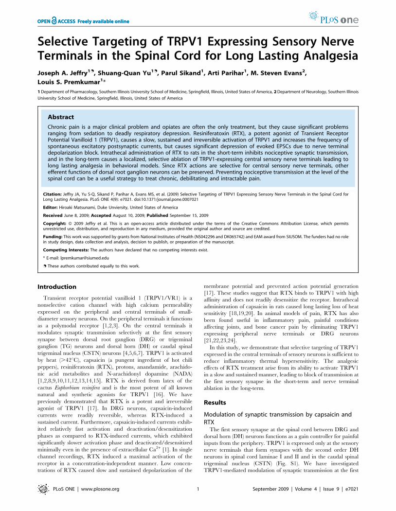
spinal cord slices, capsaicin (2 mM) increased the frequency of
sEPSCs in 53 out of 80 recordings; the mean increase was
9426200% (range 32–8621%, p,0.05, KS test) (Fig. 1A, C) without
affecting the amplitude significantly (Fig. 1D). RTX (200 nM)
increased the frequency of sEPSCs in 16 out of 55 recordings, with
a mean increase of 331687% (range 25–1200%, p,0.05, KS test)
(Fig. 2A, C) without affecting the amplitude (Fig. 2D). In similar
experimental conditons using brain stem slices that contained CSTN,
we observed similar effects on sEPSCs following capsaicin and RTX
application. Capsaicin (2 mM) increased the frequency of sEPSCs
in 12 out of 24 recordings; the mean increase was 6376184% (range
136–2428%, p,0.05 KS, test) (Fig. 3A, E) and RTX (100 nM)
increased the frequency of sEPSCs in 8 out of 23 recordings, with
a mean increase of 2776170% (range 55–1469%, p,0.05, KS test)
(Fig. 3B, G). We interpret the greater effects of capsaicin compared
to RTX as likely due to the slower depolarization RTX produces,
with less action potential firing, and therefore less activation of
presynaptic terminals [17]. Further analysis of the data showed that
the increase in sEPSC frequency caused by continuous application of
Figure 1. Modulation of synaptic transmission by capsaicin at the first sensory synapse in spinal cord. A. Application of capsaicin(2 mM) increased the frequency of sEPSCs in a reversible manner. The evoked responses are truncated. The synaptic events are shown at a higher timeresolution below (the regions denoted by asterisks). B. Superimposed traces (10) of evoked synaptic responses recorded at three different timepoints from the same neurons. In the presence of capsaicin eEPSCs either failed or exhibited a reduction in amplitude. C. Cumulative probability plotshowing decreased inter-event intervals representing increased frequency of sEPSCs (p,0.0001, KS test). D. The increase in frequency was notaccompanied by a significant change in amplitude. E. The reduction or failure of eEPSC amplitude partially reversed following washout.doi:10.1371/journal.pone.0007021.g001
RTX for Long Lasting Analgesia
PLoS ONE | www.plosone.org 2 September 2009 | Volume 4 | Issue 9 | e7021

capsaicin decreased with time (Fig. 3A, E), whereas RTX-induced
increase in the frequency of sEPSCs did not decrease with time
(Fig. 3B, G). The increase in frequency shows a large variability
because the extent of sensory input to the recording neurons is
variable and cannot be controlled [6]. The selectivity of action was
tested using a TRPV1 antagonist (BCTC 500 nM) that reversed the
RTX-induced increase in the frequency of sEPSCs (Fig. S3). These
results indicate that activation of TRPV1 increases transmitter release
by activating presynaptic receptors expressed in central sensory nerve
terminals.
We then studied evoked responses by stimulating the stump of the
dorsal root or dorsal root entry zone in spinal cord slices, or the
spinal trigeminal tract in CSTN slices. Intriguingly, following
application of capsaicin or RTX, we observed failures in evoked
responses and the evoked currents were significantly depressed at
the peak of their responses. Capsaicin (2 mM) depressed evoked
EPSCs in spinal cord slices (6767%, n = 12, p,0.05) (Fig. 1B, E)
and in CSTN slices (7465% n = 5, p,0.05) (Fig. 3A, C, F ).
Similarly, RTX (200 nM) depressed evoked EPSC in spinal cord
slices (43613%, n = 6, p,0.05) (Fig. 2 B, E) and in CSTN (38612%
n = 4, p,0.05) (Fig. 3 B, D, H). It appears the depression of evoked
currents is by a presynaptic mechanism, since the amplitude of
simultaneously-recorded sEPSCs did not decrease (Figs. 1D, 2D).
Possible explanations for the short-term effects of capsaicin and
RTX on eEPSCs include shunting of voltage-sensitive currents
by open TRPV1 channels, depolarization-induced inactivation
of voltage-gated sodium or calcium channels or depletion of readily
releasable vesicles. We propose that the failure of evoked responses
could be correlated to a blockade of nociceptive transmission.
Blockade of nociceptive transmission is likely to be more complete
with RTX, because we found that depression of eEPSC ampli-
tude partially recovered following desensitization or washout of
capsaicin, whereas RTX induced a sustained and irreversible
response.
Intrathecal administration of RTX-induced analgesiaHaving demonstrated the unique properties of RTX at the first
sensory synapse in the spinal cord and CSTN, we hypothesized
Figure 2. Modulation of synaptic transmission by RTX at the first sensory synapse in the spinal cord. A. Application of RTX (200 nM)increased the frequency of sEPSCs. RTX-induced response showed a slower onset and lesser deactivation/desensitization. The evoked responses aretruncated. The synaptic events are shown at a higher time resolution below (the regions denoted by asterisks). B. Superimposed traces (10) of evokedsynaptic responses recorded at three different time points from the same neurons. In the presence of RTX eEPSCs either failed or exhibited areduction in amplitude. C. Cumulative probability plot showing decreased inter-event intervals representing increased frequency of sEPSCs(p,0.0001, KS test). D. The increase in frequency was not accompanied by a significant change in the amplitude. E. The reduction or failure of eEPSCamplitude remained suppressed even after washout.doi:10.1371/journal.pone.0007021.g002
RTX for Long Lasting Analgesia
PLoS ONE | www.plosone.org 3 September 2009 | Volume 4 | Issue 9 | e7021

that RTX-induced sustained activation of TRPV1 at the
presynaptic terminal would cause analgesia by depression of
synaptic transmission in the short-term and by nerve terminal
ablation in the long-term. Therefore, we tested the effect of
intrathecal administration of RTX in adult rats. RTX (0.045–
1.9 mg/kg) was administered intrathecally in behavioral models of
pain. Paw withdrawal latency (PWL) to radiant heat was not
significantly affected following administration of RTX (control,
7.260.6 s, n = 8; RTX (1.9 mg/kg), 9.260.5 s, n = 8) (Fig. 4A).
However, when tested for nocifensive behavior by intraplantar
injection of capsaicin, a dramatic decrease in pain sensitivity was
observed as indicated by reduction in the duration and number of
guardings (Fig. 4B, C). The number of guardings decreased
significantly from 12.562.8 (n = 6) to 4.861. 5 (n = 11, p,0.05)
and the duration of guardings decreased significantly from
151.7630.1 s (n = 6) to 49619.9 s (n = 11, p,0.05) after RTX
treatment. We then tested whether RTX treatment could
selectively alleviate inflammatory thermal hypersensitivity. Inflam-
mation was induced by carrageenan (2%, 100 ml) in the left paw
and the right paw was used as a control. Following inflammation,
the PWL of control animals decreased significantly from 7.660.5 s
(n = 12) to 4.560.5 s, n = 6, p,0.05) (Fig. 4D). Intrathecal
administration of RTX prevented the reduction in PWL caused
by inflammation (control, 9.560.9 s (n = 10) and RTX, 8.560.4 s
(n = 12). These studies indicate that intrathecal administration of
RTX did not alter the acute thermal sensitivity but profoundly
reduced inflammation-induced thermal hypersensitivity.
We then tested for effects of intrathecal RTX administration on
mechanical sensitivity. The acute mechanical response elicited by
von Frey filaments was not affected by RTX injection (saline
24.8563.31 gms, RTX (1.9 mg/kg), 21.7961.6 gms) (Fig. 5A, B).
We tested the mechanical sensitivity following inflammation. The
PWL to von Frey filaments significantly decreased after carra-
geenan application (control, 27.562 gms, n = 9; after inflamma-
tion 13.960.44 gms, n = 11). Intrathecal administration of RTX
did not affect PWL to von Frey filaments after inflammation
Figure 3. Modulation of synaptic transmission by activation of TRPV1 at the first sensory synapse in the CSTN. A, B. Application ofcapsaicin (2 mM) or RTX (200 nM) increased the frequency of sEPSCs. Capsaicin-induced response desensitized with time, whereas RTX-inducedresponse was sustained. The evoked responses are truncated in A and B. The currents are shown at higher time resolutions below. C, D. Evokedsynaptic responses were recorded from the same neurons before and after administration of capsaicin or RTX. In the presence of both capsaicin andRTX, eEPSCs either failed or exhibited a reduction in amplitude. E, F. Capsaicin-induced increase in sEPSC frequency decreased with time and theeEPSC amplitude remained depressed, G, H. RTX-induced increase in sEPSC frequency remained elevated, but the amplitude of eEPSC remaineddepressed.doi:10.1371/journal.pone.0007021.g003
RTX for Long Lasting Analgesia
PLoS ONE | www.plosone.org 4 September 2009 | Volume 4 | Issue 9 | e7021

(control, 24.961.4 gms, n = 8; after inflammation 14.862.1 gms,
n = 12) (Fig. 5C). These results indicate that RTX treatment does
not affect mechanical hypersensitivity due to inflammation. Since
TRPV1 is selectively expressed in nonmyelinated peptidergic C
fibers and lightly myelinated Ad fibers, this observation is
consistent with the notion that mechanical sensitivity is carried
by a distinct set of nociceptors [25].
RTX caused selective ablation of TRPV1 expressing nerveterminals
The animals treated with intrathecal RTX that exhibited
analgesia, indicated by reduced nocifensive behavior and exhib-
iting no change in PWL following inflammation, were sacrificed
and TRPV1 levels were assessed in the spinal cord, DRG and paw
skin tissues using immunohistochemistry. Immunostaining was
performed at least in three different rats and 3–5 sections from
each rat were analyzed. There was a complete loss of TRPV1
labeling in the dorsal horn of the spinal cord (Fig. 6A, D). This is
likely to be due to TRPV1-mediated Ca2+ influx causing nerve
terminal death, or due to TRPV1 internalization. Central terminal
ablation was not caused by death of DRG neurons because there
was no difference in intensity of TRPV1 labeling or number of
neurons labeled in DRG in saline treated animals as compared to
RTX-treated animals (Fig. 6B). Similarly, there was no change in
TRPV1 expression in peripheral terminals, indicated by lack of
change in paw skin TRPV1 staining following intrathecal
administration of RTX (Fig. 6C). The peptide neurotransmitters
CGRP and SP are released by sensory nerve terminals of small
diameter TRPV1-containing neurons. Consistent with the loss of
TRPV1 expressing central nerve terminals, the immunoreactivity
of CGRP and SP associated with TRPV1 immunoreactivity was
also significantly reduced (p,0.001) (Fig. 6E, F). Furthermore,
RTX-induced loss of TRPV1 staining was localized to the lumbar
spinal segments closest to the level of the intrathecal injection
Figure 4. Intrathecal administration of RTX reduced pain behavior induced by capsaicin and inflammation. A. Effect of increasingconcentrations of RTX on PWL to radiant heat. B, C. nThe duration and the number of nocifensive behaviors after intraplantar capsaicin significantlydecreased after RTX treatment D. PWL to a thermal stimulus after injection of carrageenan is significantly reduced as compared to saline injectedanimals.doi:10.1371/journal.pone.0007021.g004
Figure 5. Intrathecal administration of RTX has no effect on mechanical sensitivity. A. There was no change in PWT with differentconcentrations of RTX. B. There was no change in PWT with time after intrathecal injection of RTX. C. Even after inflammation there was no change inPWT in response to mechanical stimulation.doi:10.1371/journal.pone.0007021.g005
RTX for Long Lasting Analgesia
PLoS ONE | www.plosone.org 5 September 2009 | Volume 4 | Issue 9 | e7021

Figure 6. Selective ablation of TRPV1 expressing nerve terminals in spinal cord by intrathecal administration of RTX.Immunofluroscent pictures from control animals (top panel) and animals treated with RTX (1.9 mg/kg) for 20 days (bottom panel). Representativepictures shown are from 3–5 sections stained at least from 3 rats for each group. A. Left panel shows complete loss of TRPV1 staining in spinal dorsalhorn after RTX treatment. Right panel shows that there was no change in staining for NeuN, a neuronal marker. B. RTX treatment did not affect thestaining or the number of DRG neurons stained. Immunofluroscent pictures of L4 DRG in control animals (top panel) and animals treated with RTX(bottom panel) show no difference in TRPV1 labeling between these two groups. C. TRPV1 staining of paw skin sections did not show a changebetween controls and following RTX treatment. Sections from another rat show intrathecal RTX eliminated TRPV1 staining (D), reduced CGRP stainingsignificantly (E) and reduced SP staining significantly (F), but did not alter TRPV4 staining in spinal cord (G). The corresponding histograms of analysisof gray value of the stained region are shown below. The scale bar is 100 mm except for C it is 50 mm.doi:10.1371/journal.pone.0007021.g006
RTX for Long Lasting Analgesia
PLoS ONE | www.plosone.org 6 September 2009 | Volume 4 | Issue 9 | e7021
(T12-L3). We interpret these data showing nerve terminal ablation
as due to TRPV1-mediated Ca2+ influx causing nerve terminal
death. The spinal cord nerve terminal arborization is selectively
affected near the site of injection, without detectable effect on
DRG somata or peripheral terminals. Consistent with this
observation, we have found that RTX selectively caused cell
death in small diameter DRG neurons in a dose-and time-
dependent manner (Fig. S4).
In order to determine the specificity of RTX action, we have
studied the expression of TRPV4, a putative mechanosensor. The
TRPV4 agonist 4a-PDD (4a-Phorbol-12,13-didecanoate) is able
to increase the frequency of mEPSCs without affecting their
amplitude [26], suggesting a presynaptic locus of action. The
spinal cord sections of RTX-treated animals that showed a
complete loss of TRPV1 immunostaining exhibited no change in
TRPV4 immunostaining (Fig. 6G). TRPV4 has been suggested to
mediate some forms mechanosensitivity. Its preservation in RTX-
treated animals is consistent with our data showing preservation of
behavioral measures of mechanosensitivity, and together these
results further confirm the specificity of RTX action.
Regeneration of nerve terminals following RTX treatmentIntrathecal RTX selectively targets central nerve terminals,
preserving DRG neurons and their peripheral terminals. A
potential consequence of selective targeting is that the terminals
may regenerate over time, avoiding permanent damage. We have
studied regeneration of peripheral and central terminals after
RTX administration. Following intraplantar injection of RTX
(10 mM, 10 ml), thermal hypersensitivity was determined in
response to intraplantar injection of capsaicin (100 mM, 10 ml).
A loss of capsaicin-induced thermal hypersensitivity accompanied
by a loss of TRPV1 staining in peripheral terminals was observed
within two days of RTX injection. However, capsaicin-induced
thermal hypersensitivity gradually recovered over time. In
confirmation of the nerve terminal regeneration, TRPV1 staining
partially recovered after 63 days (Fig. 7A). However, following
intrathecal administration of RTX, there was a complete loss of
capsaicin-induced nocifensive behavior, which did not recover
even after 5 months. Similarly, TRPV1 immunostaining in spinal
cord was not detected even after 5 months (Fig. 7B). In the same
intrathecal RTX treated animals there was no change in TRPV1
Figure 7. Loss and regeneration of TRPV1 expressing nerve terminals in the paw skin and spinal cord. A. Loss and recovery ofcapsaicin-induced nocifensive behavior within 60 days after intraplantar injection of RTX; TRPV1 staining 10 days and 2 months after RTX injection areshown. B. After intrathecal RTX injection, capsaicin-induced nocifensive behavior did not recover even after 5 months. TRPV1 staining after 10 daysand 5 months of RTX injection are shown. C. TRPV1 staining in DRG is not affected by intrathecal RTX. Asterisks denote significant change. The scalebar is 50 (middle row) or 100 mm.doi:10.1371/journal.pone.0007021.g007
RTX for Long Lasting Analgesia
PLoS ONE | www.plosone.org 7 September 2009 | Volume 4 | Issue 9 | e7021

staining intensity in DRG or the number of TRPV1-stained
neurons in DRG (Fig. 7C).
Discussion
From these studies, we have been able to demonstrate that
TRPV1 is selectively expressed in the sensory nerve terminals at
the DH of the spinal cord (laminae I and II) and CSTN. RTX
causes a sustained increase in sEPSCs as compared to capsaicin
which exhibits a desensitizing response. Evoked synaptic current
recordings show synaptic failures that are likely to be due to
depolarization block from sustained TRPV1 activation. We
propose that this effect causes reduced nociceptive transmission
and quick but short-term analgesia. RTX in the long-term leads to
ablation of TRPV1 expressing nerve terminals as a result of
sustained Ca2+ influx. Intrathecal administration of RTX reduced
inflammatory thermal hypersensitivity without altering acute
thermal sensitivity. This is due to selective ablation of TRPV1
expressing central nerve terminals in the dorsal horn that is
sufficient to reduce inflammatory thermal hypersensitivity without
affecting TRPV1 expressing DRG neurons or their peripheral
terminals. Immunohistochemical studies show that TRPV1 in
DRG neuronal cell bodies and peripheral terminals are preserved,
suggesting that sensory efferent functions such as TRPV1-
mediated CGRP and SP release at the peripheral nerve terminals
will not be affected. CGRP and SP are vasoactive peptides that
have been shown to be essential for control of the microvascular
circulation, which includes perineurial capillaries and coronary
vessels [27]. The specificity of intrathecal RTX action was shown
by the observation that RTX administration did not affect
mechanosensitivity and that staining for TRPV4, a putative
mechanosensor, was intact in the dorsal horn. We also observed
that following intrathecal administration of RTX, the central
terminals did not regenerate even after five months as compared to
the peripheral terminals that regenerated within two months of
intraplantar RTX injection. The inability of central terminals to
regenerate is an intriguing observation. In 1993, Goso et al.,
reported using receptor binding and neurogenic inflammatory
response that RTX-induced loss of binding and extravasation were
partly recovered in the bladder but the RTX binding was not
recovered in the spinal cord after intrathecal administration [28].
In earlier studies, a single intrathecal injection of capsaicin
depleted substance P from primary sensory neurons and caused a
prolonged increase in the thermal and chemical pain thresholds in
rats but there was no apparent change in responses to noxious
mechanical stimuli [19,20]. However, the literature mentions
conflicting reports of the appropriateness of using acute thermal
sensitivity to assess TRPV1 function in hot plate and tail flick tests
[18,19,29,30,31]. Furthermore, during intrathecal administration,
capsaicin may be distributed throughout the CSF and hence
effective concentrations may not have been achieved consistently.
Since RTX binds to TRPV1 irreversibly and with high affinity, we
propose this property will aid in its localization in a given segment
of the spinal cord by slow infusion using osmotic mini pumps. We
have found that slow infusion of RTX can target the lumbar
region selectively sparing the cervical and thoracic regions of the
spinal cord.
TRPV1 is found in the nerve terminals supplying the urinary
bladder and urothelium, indicating a role in urinary bladder
functions [32]. Human clinical trials of RTX are recent and have
so far been limited to treatment of bladder hyperreflexia, in which
it has been shown to be effective [33]. Following administration of
intravesical RTX, there was a reduction in TRPV1 immunore-
activity in the basal cell layer, which is similar to the loss of sensory
nerve fibers in the suburothelial layer [34] and leads to long lasting
reduction in bladder pain and incontinence. Interestingly,
intravesical administration of RTX, unlike capsaicin, does not
induce suprapubic discomfort [35]. We suggest that this property
may be due to the fact that depolarization block induced by RTX
is slow and sustained as compared to capsaicin [17].
Usefulness of the TRPV1 blockade has been demonstrated to be
beneficial in pain induced by Herpes zoster, diabetic peripheral
neuropathy, bone cancer, arthritis, inflammatory bowel disease
and migraine [36,37,38,39,40]. TRPV1 has been shown to be up-
regulated by TNFa in cancer-related thermal hyperalgesia in mice
[41]. Intrathecal administration of RTX has been used to
ameliorate painful conditions, which correlate with the destruction
of DRG neuronal cell bodies [21,22,23]. Intriguingly, bone cancer
induced by inoculation of carcinoma cells mainly results in altered
mechanosensitivity, yet TRPV1 antagonists have been found to be
useful [39,42].
TRPV1 is also involved in regulation of body temperature.
Subcutaneous injection of capsaicin decreases body temperature
by 2–3uC and permanently reduced the capacity of rats to
withstand a hot environment [43]. TRPV1 antagonists increase
the body temperature to the same extent [44,45,46]. Several
TRPV1 antagonists are in clinical trials and hyperthermia poses a
serious limitation to their usefulness. The promise of TRPV1
antagonists to treat painful conditions may not become a reality
because in phase I clinical trials, TRPV1 antagonists have been
shown to increase the body temperature significantly
[45,46,47,48]. Selective targeting of spinal segments may be
achieved by slow infusion of RTX using osmotic mini pumps may
spare thermoregulatory centers in the hypothalamus and avoid
hyperthermia.
Another advantage of RTX is that it appears to be selective.
Even when administered intraperitoneally it specifically ablates
TRPV1 expressing nociceptors. TRPV1 has been implicated in
diverse function such as release of the potent vasodilator CGRP
and maintaining microvascular circulation, including the coro-
naries and regulation of insulin secretion [49,50,51]. Therefore,
the approach described here may be superior to selectively
targeting TRPV1 expressed in peripheral nerve terminals and
prevent other unwanted effects resulting from the elimination of
the whole DRG neuron.
In summary, intrathecal administration of RTX ablates TRPV1
expressing central sensory nerve terminals, significantly reduces
nociceptive transmission and decreases TRPV1-mediated inflam-
matory thermal hypersensitivity. This approach is different from
previous studies in which RTX has been used for pain relief by
ablating DRG neuronal cell bodies. Our results indicate that
intrathecal administration of RTX or its analogues is a promising
method of achieving analgesia. Further study is needed of RTX
concentration-response relationships, since even lower concentra-
tions of RTX could cause a partial ablation of TRPV1 expressing
nerve terminals that may be sufficient for pain relief. Better
treatments for chronic intractable pain are urgently needed,
especially in terminally ill patients, in whom the best analgesic
option now available may be treatment with large doses of potent
opiate analgesics, which can cause mental clouding, respiratory
depression, and reduce quality of life.
Methods
All the procedures used especially in reference to experimental
animals not experiencing unnecessary discomfort, distress, pain or
injury have been approved by the Southern Illinois University
School of Medicine Institutional Animal Care and Use committee
RTX for Long Lasting Analgesia
PLoS ONE | www.plosone.org 8 September 2009 | Volume 4 | Issue 9 | e7021

review panel in accordance with the Panel of Euthanasia of
American Veterinary Medical Association.
ImmunohistochemistryFive week old Sprague-Dawley rats were anesthetized with
isoflurane and perfused with 4 % paraformaldehyde. Samples of
lumbar segments of the spinal cord, brain stem, DRG and paw skin
tissues were harvested and quickly frozen. The spinal cord/brain
stem and DRG were cut into 20 and 10 mm sections, respectively
(Leica CM 1850, Nussloch, Germany). The paw skin was cut into
40 mm sections. The sections were incubated with polyclonal rabbit
anti-TRPV1 antibody (Affinity BioReagents, PA1-747, 1:500), or
polyclonal rabbit anti-TRPV4 antibody (Alomone, ACC-034,
1:200), or monoclonal mouse anti-CGRP antibody (Sigma, C-
7113, 1:2500), or polyclonal guinea pig anti-SP antibody (abcam,
ab10353, 1:1000), or monoclonal mouse anti-NeuN antibody
(Chemicon, MAB377, 1:100) for 1 hour at room temperature, then
incubated with Rhodamine Red (TM)-X donkey anti-rabbit IgG
(Jackson 711-295-152, 1:100), or FITC donkey anti-mouse IgG
(Jackson, 715-095-151, 1:100), or Rhodamine Red (TM)-X donkey
anti-guinea pig IgG (Jackson, 706-295-148, 1:100) for 1 hour at
room temperature. Images were taken by a confocal microscope
(Olympus Fluoview). The intensity of TRPV1 staining was analyzed
by measuring the gray value of the stained region by using ImageJ
(Research Service Branch, NIMH). Immunohistochemistry was
performed at least in 3 rats from each group and 3–5 sections from
each animal were analyzed.
Synaptic current recording from spinal cord slicesSprague-Dawley rats were obtained from Harlan (Indianapolis)
for breeding locally. Horizontal CSTN and transverse spinal cord
slices (L4-L6) from 2 to 4 weeks old rats were prepared using
methods similar those previously described [52]. The rats were
deeply anesthetized with isoflurane (5%) and then decapitated.
The desired tissue, once isolated, was placed in cold (4uC),
oxygenated sucrose based physiological solution (in mM: sucrose
209, KCl 2, NaH2PO4 1.25, MgCl2 5, CaCl2 0.5, NaHCO3 26,
D-glucose 10) for 90 s, and then cut with a vibrating tissue slicer
(Precisionary Instruments, Greenville, NC, USA) into 300 mm
sections in 4uC physiological solution. Slices were allowed to
recover for 60 minutes in oxygenated extracellular solution at
room temperature. To record, slices were placed on the stage of an
upright near-infrared differential interference contrast microscope
(Olympus BX-50wi). Extracellular solution contained (in mM)
NaCl 126, KCl 2.5, MgCl2 1.2, dextrose 11, NaH2PO4 1.4, CaCl22.4, NaHCO3 25, at 32uC and was continually gassed with 95%
O2 5% CO2. Intracellular solution contained (in mM) CsCl 140,
CaCl2 2, EGTA 10, HEPES 5, MgATP 2, titrated to pH 7.3. The
lidocaine derivative (QX-314) was included to prevent action
potentials in the recording neuron. Electrodes were pulled from
thick-walled borosilicate glass (World Precision Instruments,
Sarasota, FL, USA). Electrode impedance was 4–6 MV.
Experiments were performed at room temperature, 24uC, and
the recording chamber was perfused at 4 ml/min.
In order to record excitatory synaptic currents, neurons were
voltage-clamped at -60 mV (EPC10, HEKA, Bellmore, New York,
USA). To obtain evoked EPSCs, a Grass Stimulator (S88) with
stimulus isolation unit PSIU 6 (Grass Technologies, West Warwick,
RI) triggered by a Master 8 (A.M.P.I., Jerusalem, Isreal) was used to
stimulate a concentric bipolar electrode (Rhodes Medical Instruments,
Tujunga, CA) placed on the sensory fiber tract. Stimulus duration was
100 ms, and half maximal stimulus intensity was used (less than
800 mA, usually 200–800 mA for C-fibers). Spontaneous and evoked
EPSCs were low-pass filtered at 2.5 kHz and digitized at 5 kHz. The
digitized signal was stored to hard drive on a PC compatible
computer. Fast and slow capacitance compensation was performed in
Pulse. Input resistance and series resistance were measured every 2–5
min in voltage clamp mode with three small (DV 10 mV, 150 ms)
hyperpolarizing voltage steps. Cells showing greater that 20% change
in series resistance were not included in analysis. Off-line data analysis
was done with the program Clampfit 9 (Molecular Devices,
Sunnyvale, CA). sEPSCs were analyzed using the Mini Analysis
Program (Synaptosoft, Decatur, GA) the threshold for event detection
(usually 10 pA) was at least 3 times baseline noise levels.
Intrathecal catheter implantation and intrathecalinjection
Intrathecal catheters were implanted in rats according to method
described by Yaksh et al. [19] with some modifications. Briefly, male
SD rats (225–250 g) were anesthetized with ketamine/xylazine (85/
5 mg/kg, i.p.). When they no longer responded to the tail pinch test,
the neck area was shaved and the skin was swabbed with betadine
followed by 70 % alcohol. A small incision was made in the skin and
the muscles were separated to expose the atlanto-occipital
membrane. A small incision was made in the membrane to allow
a polyethylene-10 catheter filled with 0.9 % sterile saline to be
inserted into the subarachnoid space. The catheter was threaded
through the space as far as the lumbar enlargement (approximately
7.5 cm). The catheter was then sutured in place with the muscles
and the incision closed. About 5 cm of catheter was exposed
externally to act a port for injections. The external port was sealed
with Parafilm to prevent flow of cerebrospinal fluid.
Rats were allowed to recover for 7 days after surgery. To prevent
infection, 10 mg/kg of kanamycin was injected subcutaneously
every day for 5 days during recovery. Drugs were administrated by
slow infusion into the subarachnoid space of anesthetized rats
Measurement of thermal sensitivityThermal nociceptive responses were determined using a plantar
test instrument (Ugo Basile, Camerio, Italy) as described
previously [53]. The rats were habituated to the apparatus that
consisted of three individual Perspex boxes on a glass table. A
mobile radiant heat source was located under the table and
focused onto the desired paw. Paw withdrawal latencies (PWLs)
were recorded three times for each hind paw and the average was
taken as the baseline value. A timer was automatically activated
with the light source, and response latency was defined as the time
required for the paw to show an abrupt withdrawal. The
apparatus has been calibrated to give a PWL of approximately
6–12 s. In order to prevent tissue damage a cut-off at 20 s was
used. Rats were accustomed to the test conditions 1 h per day for 5
days.
Measurement of nocifensive behaviorCapsaicin-evoked nocifensive behavior in rats was defined as
lifting (guarding), licking and shaking of the injected paw [54]. The
number of times the rat exhibited guarding, licking and shaking was
counted and the total duration of this behavior was measured over 5
min immediately after intraplantar administration of capsaicin (2
mM). Capsaicin-induced Inflammatory thermal hypersensitivity
was determined by subcutaneous injection of 100 mM of 50 ml
capsaicin into the plantar region of the rat left hind paw.
Carrageenan-induced thermal hyperalgesiaAfter obtaining baseline values of PWL to radiant heat, the
animals received an intraplantar injection of carrageenan (2%,
100 ml) into the left hind paw [55]. PWLs were determined 2 h after
RTX for Long Lasting Analgesia
PLoS ONE | www.plosone.org 9 September 2009 | Volume 4 | Issue 9 | e7021
carrageenan injection, a time point shown to produce reliable
readings, to confirm that hyperalgesia had developed. The PWL to
radiant heat stimulus was recorded at 2, 3, 4 and 5 hrs after
carrageenan injection. The data were compared with the uninjected
paw. These experiments were then repeated in intrathecal RTX-
injected animals and the PWL was determined again.
Measurement of mechanosensitivityMechanical nociceptive responses were assessed using a
dynamic plantar anesthesiometer instrument using von Frey Hairs
(Ugo Basile, Camerio, Italy)[56]. The rat was placed in a chamber
with a metal mesh floor. A 0.5 mm diameter von Frey probe was
applied to the plantar surface of the rat hind paw with pressure
increasing by 0.05 Newtons/s and the pressure at which a paw
withdrawal occurred was recorded and this was taken as PWT.
For each hind paw, the procedure was repeated 3 times and the
average pressure to produce withdrawal was calculated. Successive
stimuli were applied to alternating paws at 5 min intervals. Rats
were accustomed to the test conditions 1 h per day for 5 days.
ReagentsAll the chemicals used in this study were obtained from SIGMA
(St. Louis, MO) and BCTC was a gift from Glenmark Pharma-
ceuticals, Mumbai, India.
Statistical AnalysisKolmogorov-Smirnov (KS) test was used to compare the
cumulative probability curves for inter-event intervals and ampli-
tude between various treatment groups. Data are represented as
mean6SEM and expressed as percent of control, which is scaled to
100%. For evoked currents, Student’s paired t-test was used for
statistical comparisons and significance was considered at p,0.05.
For experiments that involved manipulation of one of the legs
(carrageenan or capsaicin injection), the data were normalized for
each animal as maximum possible effect (MPE). This value was
calculated as follows: MPE = (PDR2IBR)/(CBR2IBR), where
PDR is the postdrug response of the ipsilateral paw, IBR is the
ipsilateral paw baseline response, and CBR is the contralateral paw
baseline response. Accordingly, the individual values are reported as
the mean6SEM. Data obtained for the carrageenan or capsaicin
tests were subjected to a one-way ANOVA followed, when
significant, by post hoc Dunnett’s t tests. When comparing the
means of only two groups, Student’s t test was used. All comparisons
were analyzed separately for each time point. For all tests, a p value
lower than 0.05 (p,0.05) was considered significant.
Supporting Information
Figure S1 Expression of TRPV1 in spinal cord and CSTN. A.
Immunohistochemical labeling of TRPV1 is selectively seen only
in laminae I and II if the spinal dorsal horn (top panel). The
labeling of NeuN, a neuronal marker (middle) and the merged
images (bottom) are also shown. B. Immunohistochemical labeling
of TRPV1 in oral spinal trigeminal nucleus (OSTN), interpolar
spinal trigeminal nucleus (ISTN) and caudal spinal trigeminal
nucleus (CSTN). It is clear only CSTN shows TRPV1 labeling, a
region where trigeminal sensory neurons form synapses. TRPV1
labeling (left panel) NeuN labeling (middle panel) and merged
image (right panel) are shown.
Found at: doi:10.1371/journal.pone.0007021.s001 (3.32 MB TIF)
Figure S2 Enhancement of synaptic transmission by activation
of TRPV1 at the first sensory synapse in DRG and DH co-
cultures. A. Application of capsaicin (10 nM) increased the
frequency of mEPSCs in a reversible manner. The synaptic events
are shown at a higher time resolution below. B. Application of
RTX (10 nM) induced a sustained increase in the frequency of
mEPSCs. The synaptic events are shown in higher time resolution
below. C. F. Cumulative probability plots showing decreased inter-
event intervals representing increased frequency of mEPSCs in
presence of capsaicin and RTX. D. G. The increase in frequency
was not accompanied by a significant change in the amplitude. E.
H. Summary graphs showing capsaicin- and RTX-induced
increases in the frequency of mEPSCs were dose-dependent and
the enhancement of synaptic transmission by capsaicin application
was inhibited by TRPV1 antagonist, capsazepine (Cpz).
Found at: doi:10.1371/journal.pone.0007021.s002 (0.98 MB TIF)
Figure S3 RTX-induced increase in sEPSC frequency is
TRPV1-mediated. A. In spinal cord slices, RTX (200 nM)-
induced increase in the frequency of sEPSC was reversed by
application of BCTC (500 nM), a TRPV1 antagonist. Traces of
expanded time scale denoted by asterisks (*) are shown below. B.
Cumulative probability plot shows a decrease in inter-event
intervals representing increased frequency mEPSCs after RTX
(p,0.0001, KS test) and reversal after BCTC C. A plot shows the
change in frequency with time.
Found at: doi:10.1371/journal.pone.0007021.s003 (0.92 MB TIF)
Figure S4 RTX-induced cell death in DRG neurons were
identified using propidium iodide uptake assay. A. B. DRG
neurons were treated with different concentrations (0.3, 1 and
5 nM) of RTX for 24, 48 and 72 hrs. In control conditions, there
was a loss of 10 to 20% of both small and (,500 mm2) and large
(.500 mm2) neurons (n = 2614). Treatment with 300 pM RTX
caused significant increase in small diameter neuronal death
(44613 % after 24 hrs; 5267% after 48 hrs; 7064% after 72 hrs).
There was no change in the of large diameter neuronal death
(1164% after 24 hrs; 2065 after 48 hrs; 667 after 72 hrs).
Incubating the neurons with 1 nM RTX caused 62618% small
diameter neuron death after 24 hrs, 8767% after 48 hrs and
65614% after 72 hrs. The large diameter neurons showed no
difference as compared to controls (1264% after 24 hrs; 19610%
after 48 hrs; 762% after 72 hrs). Treatment with 5 nM RTX
caused maximal neuronal death (6769%) within 24 hrs and
remained the same after 48 hrs (7167%) and 72 hrs (7068%). As
seen with other concentrations the large diameter neurons showed
no significant change (1764% after 24 hrs; 2264% after 48 hrs;
3068% after 72 hrs).
Found at: doi:10.1371/journal.pone.0007021.s004 (0.20 MB TIF)
Author Contributions
Conceived and designed the experiments: MSE LSP. Performed the
experiments: JAJ SQY PS AP LSP. Analyzed the data: JAJ SQY PS AP
MSE LSP. Contributed reagents/materials/analysis tools: JAJ SQY PS AP
LSP. Wrote the paper: JAJ MSE LSP.
References
1. Caterina MJ, Schumacher MA, Tominaga M, Rosen TA, Levine JD, et al.
(1997) The capsaicin receptor: a heat-activated ion channel in the pain pathway.
Nature 389: 816–824.
2. Julius D, Basbaum AI (2001) Molecular mechanisms of nociception. Nature 413:
203–210.
3. Lazzeri M, Spinelli M, Beneforti P, Malaguti S, Giardiello G, et al. (2004)
Intravesical infusion of resiniferatoxin by a temporary in situ drug delivery
system to treat interstitial cystitis: a pilot study. Eur Urol 45: 98–102.
4. Nakatsuka T, Furue H, Yoshimura M, Gu JG (2002) Activation of central
terminal vanilloid receptor-1 receptors and alpha beta-methylene-ATP-sensitive
RTX for Long Lasting Analgesia
PLoS ONE | www.plosone.org 10 September 2009 | Volume 4 | Issue 9 | e7021
P2X receptors reveals a converged synaptic activity onto the deep dorsal horn
neurons of the spinal cord. J Neurosci 22: 1228–1237.5. Baccei ML, Bardoni R, Fitzgerald M (2003) Development of nociceptive
synaptic inputs to the neonatal rat dorsal horn: glutamate release by capsaicin
and menthol. J Physiol 549: 231–242.6. Sikand P, Premkumar LS (2007) Potentiation of glutamatergic synaptic
transmission by protein kinase C-mediated sensitization of TRPV1 at the firstsensory synapse. J Physiol 581: 631–647.
7. Yang K, Kumamoto E, Furue H, Yoshimura M (1998) Capsaicin facilitates
excitatory but not inhibitory synaptic transmission in substantia gelatinosa of therat spinal cord. Neurosci Lett 255: 135–138.
8. Zygmunt PM, Petersson J, Andersson DA, Chuang H, Sorgard M, et al. (1999)Vanilloid receptors on sensory nerves mediate the vasodilator action of
anandamide. Nature 400: 452–457.9. Hwang SW, Cho H, Kwak J, Lee SY, Kang CJ, et al. (2000) Direct activation of
capsaicin receptors by products of lipoxygenases: endogenous capsaicin-like
substances. Proc Natl Acad Sci U S A 97: 6155–6160.10. Premkumar LS, Ahern GP (2000) Induction of vanilloid receptor channel
activity by protein kinase C. Nature 408: 985–990.11. Caterina MJ, Julius D (2001) The vanilloid receptor: a molecular gateway to the
pain pathway. Annu Rev Neurosci 24: 487–517.
12. Chuang HH, Prescott ED, Kong H, Shields S, Jordt SE, et al. (2001) Bradykininand nerve growth factor release the capsaicin receptor from PtdIns(4,5)P2-
mediated inhibition. Nature 411: 957–962.13. De Petrocellis L, Harrison S, Bisogno T, Tognetto M, Brandi I, et al. (2001) The
vanilloid receptor (VR1)-mediated effects of anandamide are potently enhancedby the cAMP-dependent protein kinase. J Neurochem 77: 1660–1663.
14. Huang SM, Bisogno T, Trevisani M, Al-Hayani A, De Petrocellis L, et al. (2002)
An endogenous capsaicin-like substance with high potency at recombinant andnative vanilloid VR1 receptors. Proc Natl Acad Sci U S A 99: 8400–8405.
15. Szallasi A, Blumberg PM (1990) Resiniferatoxin and its analogs provide novelinsights into the pharmacology of the vanilloid (capsaicin) receptor. Life Sci 47:
1399–1408.
16. Appendino G, Szallasi A (1997) Euphorbium: modern research on its activeprinciple, resiniferatoxin, revives an ancient medicine. Life Sci 60: 681–696.
17. Raisinghani M, Pabbidi RM, Premkumar LS (2005) Activation of transientreceptor potential vanilloid 1 (TRPV1) by resiniferatoxin. J Physiol 567:
771–786.18. Nagy JI, Emson PC, Iversen LL (1981) A re-evaluation of the neurochemical
and antinociceptive effects of intrathecal capsaicin in the rat. Brain Res 211:
497–502.19. Yaksh TL, Farb DH, Leeman SE, Jessell TM (1979) Intrathecal capsaicin
depletes substance P in the rat spinal cord and produces prolonged thermalanalgesia. Science 206: 481–483.
20. Russell LC, Burchiel KJ (1984) Neurophysiological effects of capsaicin. Brain
Res 320: 165–176.21. Szabo T, Olah Z, Iadarola MJ, Blumberg PM (1999) Epidural resiniferatoxin
induced prolonged regional analgesia to pain. Brain Res 840: 92–98.22. Karai L, Brown DC, Mannes AJ, Connelly ST, Brown J, et al. (2004) Deletion of
vanilloid receptor 1-expressing primary afferent neurons for pain control. J ClinInvest 113: 1344–1352.
23. Brown DC, Iadarola MJ, Perkowski SZ, Erin H, Shofer F, et al. (2005)
Physiologic and antinociceptive effects of intrathecal resiniferatoxin in a caninebone cancer model. Anesthesiology 103: 1052–1059.
24. Kissin EY, Freitas CF, Kissin I (2005) The effects of intraarticular resiniferatoxinin experimental knee-joint arthritis. Anesth Analg 101: 1433–1439.
25. Scherrer G, Imamachi N, Cao YQ, Contet C, Mennicken F, et al. (2009)
Dissociation of the opioid receptor mechanisms that control mechanical andheat pain. Cell 137: 1148–1159.
26. Cao DS, Yu SQ, Premkumar LS (2009) Modulation of transient receptorpotential Vanilloid 4-mediated membrane currents and synaptic transmission by
protein kinase C. Mol Pain 5: 5.
27. Strecker T, Messlinger K, Weyand M, Reeh PW (2005) Role of different proton-sensitive channels in releasing calcitonin gene-related peptide from isolated
hearts of mutant mice. Cardiovasc Res 65: 405–410.28. Goso C, Piovacari G, Szallasi A (1993) Resiniferatoxin-induced loss of vanilloid
receptors is reversible in the urinary bladder but not in the spinal cord of the rat.Neurosci Lett 162: 197–200.
29. Davis JB, Gray J, Gunthorpe MJ, Hatcher JP, Davey PT, et al. (2000) Vanilloid
receptor-1 is essential for inflammatory thermal hyperalgesia. Nature 405:183–187.
30. Caterina MJ, Leffler A, Malmberg AB, Martin WJ, Trafton J, et al. (2000)Impaired nociception and pain sensation in mice lacking the capsaicin receptor.
Science 288: 306–313.
31. Russell LC, Burchiel KJ (1986) Effect of intrathecal and subepineural capsaicinon thermal sensitivity and autotomy in rats. Pain 25: 109–123.
32. Dinis P, Charrua A, Avelino A, Cruz F (2004) Intravesical resiniferatoxin
decreases spinal c-fos expression and increases bladder volume to reflexmicturition in rats with chronic inflamed urinary bladders. BJU Int 94: 153–157.
33. Kim JH, Rivas DA, Shenot PJ, Green B, Kennelly M, et al. (2003) Intravesical
resiniferatoxin for refractory detrusor hyperreflexia: a multicenter, blinded,randomized, placebo-controlled trial. J Spinal Cord Med 26: 358–363.
34. Apostolidis A, Brady CM, Yiangou Y, Davis J, Fowler CJ, et al. (2005) Capsaicinreceptor TRPV1 in urothelium of neurogenic human bladders and effect of
intravesical resiniferatoxin. Urology 65: 400–405.
35. Giannantoni A, Di Stasi SM, Stephen RL, Bini V, Costantini E, et al. (2004)Intravesical resiniferatoxin versus botulinum-A toxin injections for neurogenic
detrusor overactivity: a prospective randomized study. J Urol 172: 240–243.36. Petersen KL, Rice FL, Suess F, Berro M, Rowbotham MC (2002) Relief of post-
herpetic neuralgia by surgical removal of painful skin. Pain 98: 119–126.37. Lauria G, Morbin M, Lombardi R, Capobianco R, Camozzi F, et al. (2006)
Expression of capsaicin receptor immunoreactivity in human peripheral nervous
system and in painful neuropathies. J Peripher Nerv Syst 11: 262–271.38. Akerman S, Kaube H, Goadsby PJ (2004) Anandamide acts as a vasodilator of
dural blood vessels in vivo by activating TRPV1 receptors. Br J Pharmacol 142:1354–1360.
39. Ghilardi JR, Rohrich H, Lindsay TH, Sevcik MA, Schwei MJ, et al. (2005)
Selective blockade of the capsaicin receptor TRPV1 attenuates bone cancerpain. J Neurosci 25: 3126–3131.
40. Jones RC, 3rd, Xu L, Gebhart GF (2005) The mechanosensitivity of mousecolon afferent fibers and their sensitization by inflammatory mediators require
transient receptor potential vanilloid 1 and acid-sensing ion channel 3. J Neurosci25: 10981–10989.
41. Constantin CE, Mair N, Sailer CA, Andratsch M, Xu ZZ, et al. (2008)
Endogenous tumor necrosis factor alpha (TNFalpha) requires TNF receptor type2 to generate heat hyperalgesia in a mouse cancer model. J Neurosci 28:
5072–5081.42. Medhurst SJ, Walker K, Bowes M, Kidd BL, Glatt M, et al. (2002) A rat model
of bone cancer pain. Pain 96: 129–140.
43. Jancso-Gabor A, Szolcsanyi J, Jancso N (1970) Stimulation and desensitization ofthe hypothalamic heat-sensitive structures by capsaicin in rats. J Physiol 208:
449–459.44. Varga A, Nemeth J, Szabo A, McDougall JJ, Zhang C, et al. (2005) Effects of the
novel TRPV1 receptor antagonist SB366791 in vitro and in vivo in the rat.Neurosci Lett 385: 137–142.
45. Gavva NR, Treanor JJ, Garami A, Fang L, Surapaneni S, et al. (2008)
Pharmacological blockade of the vanilloid receptor TRPV1 elicits markedhyperthermia in humans. Pain 136: 202–210.
46. Lehto SG, Tamir R, Deng H, Klionsky L, Kuang R, et al. (2008)Antihyperalgesic effects of (R,E)-N-(2-hydroxy-2,3-dihydro-1H-inden-4-yl)-3-(2-
(piperidin-1-yl)-4-(tri fluoromethyl)phenyl)-acrylamide (AMG8562), a novel
transient receptor potential vanilloid type 1 modulator that does not causehyperthermia in rats. J Pharmacol Exp Ther 326: 218–229.
47. Immke DC, Gavva NR (2006) The TRPV1 receptor and nociception. SeminCell Dev Biol 17: 582–591.
48. Szallasi A, Cortright DN, Blum CA, Eid SR (2007) The vanilloid receptorTRPV1: 10 years from channel cloning to antagonist proof-of-concept. Nat Rev
Drug Discov 6: 357–372.
49. Razavi R, Chan Y, Afifiyan FN, Liu XJ, Wan X, et al. (2006) TRPV1+ sensoryneurons control beta cell stress and islet inflammation in autoimmune diabetes.
Cell 127: 1123–1135.50. Gram DX, Ahren B, Nagy I, Olsen UB, Brand CL, et al. (2007) Capsaicin-
sensitive sensory fibers in the islets of Langerhans contribute to defective insulin
secretion in Zucker diabetic rat, an animal model for some aspects of humantype 2 diabetes. Eur J Neurosci 25: 213–223.
51. Gram DX, Hansen AJ, Deacon CF, Brand CL, Ribel U, et al. (2005) Sensorynerve desensitization by resiniferatoxin improves glucose tolerance and increases
insulin secretion in Zucker Diabetic Fatty rats and is associated with reduced
plasma activity of dipeptidyl peptidase IV. Eur J Pharmacol 509: 211–217.52. Grudt TJ, Williams JT (1994) mu-Opioid agonists inhibit spinal trigeminal
substantia gelatinosa neurons in guinea pig and rat. J Neurosci 14: 1646–1654.53. Hargreaves K, Dubner R, Brown F, Flores C, Joris J (1988) A new and sensitive
method for measuring thermal nociception in cutaneous hyperalgesia. Pain 32:77–88.
54. Gilchrist HD, Allard BL, Simone DA (1996) Enhanced withdrawal responses to
heat and mechanical stimuli following intraplantar injection of capsaicin in rats.Pain 67: 179–188.
55. Winter CA, Risley EA, Nuss GW (1962) Carrageenin-induced edema in hindpaw of the rat as an assay for antiiflammatory drugs. Proc Soc Exp Biol Med
111: 544–547.
56. Chaplan SR, Bach FW, Pogrel JW, Chung JM, Yaksh TL (1994) Quantitativeassessment of tactile allodynia in the rat paw. J Neurosci Methods 53: 55–63.
RTX for Long Lasting Analgesia
PLoS ONE | www.plosone.org 11 September 2009 | Volume 4 | Issue 9 | e7021