
Selective naked-eye detection of Magnesium (II) ions using a coumarin-derived fluorescent probe
8
Sensors and Actuators B 207 (2015) 216–223 Contents lists available at ScienceDirect Sensors and Actuators B: Chemical jo u r nal homep age: www.elsevier.com/locate/snb Selective naked-eye detection of Magnesium (II) ions using a coumarin-derived fluorescent probe Vinod Kumar Gupta a,b,∗ , Naveen Mergu a , Lokesh Kumar Kumawat a , Ashok Kumar Singh a a Department of Chemistry, Indian Institute of Technology Roorkee, Roorkee 247 667, India b Department of Applied Chemistry, University of Johannesburg, Johannesburg, South Africa a r t i c l e i n f o Article history: Received 16 August 2014 Received in revised form 6 October 2014 Accepted 10 October 2014 Available online 22 October 2014 Keywords: Chemosensor Coumarin Fluorescence Naked-eye detection Binding mode a b s t r a c t A simple 4-Methyl-7-hydroxy-8-formyl Coumarin (CS) serves as a selective chemosensor for Mg 2+ in the presence of alkali and alkaline earth metal ions. It showed a significant fluorescence enhancement towards Mg 2+ . The receptor CS exhibited a good binding constant and lowest detection limit for Mg 2+ . The variation of emission signal exists via of reversible chelation enhanced fluorescence (CHEF) with this inherent quenching metal ion. © 2014 Elsevier B.V. All rights reserved. 1. Introduction Magnesium is the fourth most abundant cation in the human body, most abundant divalent cation within cells and plays an important physiological role in many of its functions [1]. Also magnesium found in the bone and plays an active role in bone remodelling and skeletal development [2,3]. On the other side, magnesium is the eighth most abundant element on earth crust [4]. Magnesium deficiency can result from a variety of causes includ- ing gastrointestinal and renal losses, can cause a wide variety of features including cardiac, hypokalaemia, hypocalcaemia and neu- rological manifestations. A number of chronic diseases, such as diabetes, osteoporosis, hypertension and coronary heart disease have been associated with chronic low magnesium [5,6]. Detec- tion of Mg 2+ in the presence of other alkali and alkaline earth metal ions, like as Ca 2+ , Na + and K + is of particular significance. Serum magnesium and the magnesium tolerance test are the most widely used. There are no easy and readily available methods to assess magnesium status. Paper presented at the 7th European conference on Optical Sensors and Biosen- sors, Athens, Greece, 13–16 April 2014. ∗ Corresponding author. Tel.: +91 1332285801; fax: +91 1332273560. E-mail addresses: [email protected], [email protected] (V.K. Gupta). The design of fluorescent chemosensors is an active field of research for analytical as well as environmental and biological problems [7–11]. Typically, chemosensors are small molecules and are able to bind selectively and reversibly the analyte of interest with a change in the sensing system property, such as absorption, emission and redox potentials, which may allow naked eye detec- tion of the analyte without resorting to the use of any expensive instruments. Of the various kinds of chemosensors, the fluorescent based chemosensors present many advantages. Fluorescence mea- surements are usually very sensitive, low cost, easily performed, capability of real-time detection and versatile [12–17]. Till now, various families of fluorescent probes for Mg 2+ have been developed. These probes have receptor groups based upon moieties including diaza-18-crown-6 [18], benzo-15-crown-5 [19], calix[4]arene [20], Benzo chromene [21], Imidazo-1,10- phenanthroline [22] and other [23–27]. Most of the reported fluorescent probes for Mg 2+ demonstrate poor selectivity for Mg 2+ over Ca 2+ and are useful only where the Mg 2+ ion concentrations are much higher than those of Ca 2+ ion. We now report the ability of 4-Methyl-7-hydroxy-8-formyl Coumarin (CS) as a fluorescent probe to serve as an effective chemosensor for Mg 2+ in the presence of other alkali and alka- line earth metal ions. Our particular interest is to investigate how Mg 2+ affects the fluorescent behaviour of fluorophore upon complexation. http://dx.doi.org/10.1016/j.snb.2014.10.044 0925-4005/© 2014 Elsevier B.V. All rights reserved.
-
Upload
johannesburg -
Category
Documents
-
view
1 -
download
0
Transcript of Selective naked-eye detection of Magnesium (II) ions using a coumarin-derived fluorescent probe
Sc
V
a
b
a
ARRAA
KCCFNB
1
bimrmMifrdhtimum
s
h0
Sensors and Actuators B 207 (2015) 216–223
Contents lists available at ScienceDirect
Sensors and Actuators B: Chemical
jo u r nal homep age: www.elsev ier .com/ locate /snb
elective naked-eye detection of Magnesium (II) ions using aoumarin-derived fluorescent probe�
inod Kumar Guptaa,b,∗, Naveen Mergua, Lokesh Kumar Kumawata, Ashok Kumar Singha
Department of Chemistry, Indian Institute of Technology Roorkee, Roorkee 247 667, IndiaDepartment of Applied Chemistry, University of Johannesburg, Johannesburg, South Africa
r t i c l e i n f o
rticle history:eceived 16 August 2014eceived in revised form 6 October 2014ccepted 10 October 2014vailable online 22 October 2014
a b s t r a c t
A simple 4-Methyl-7-hydroxy-8-formyl Coumarin (CS) serves as a selective chemosensor for Mg2+ inthe presence of alkali and alkaline earth metal ions. It showed a significant fluorescence enhancementtowards Mg2+. The receptor CS exhibited a good binding constant and lowest detection limit for Mg2+.The variation of emission signal exists via of reversible chelation enhanced fluorescence (CHEF) with thisinherent quenching metal ion.
eywords:hemosensoroumarinluorescenceaked-eye detectioninding mode
© 2014 Elsevier B.V. All rights reserved.
. Introduction
Magnesium is the fourth most abundant cation in the humanody, most abundant divalent cation within cells and plays an
mportant physiological role in many of its functions [1]. Alsoagnesium found in the bone and plays an active role in bone
emodelling and skeletal development [2,3]. On the other side,agnesium is the eighth most abundant element on earth crust [4].agnesium deficiency can result from a variety of causes includ-
ng gastrointestinal and renal losses, can cause a wide variety ofeatures including cardiac, hypokalaemia, hypocalcaemia and neu-ological manifestations. A number of chronic diseases, such asiabetes, osteoporosis, hypertension and coronary heart diseaseave been associated with chronic low magnesium [5,6]. Detec-ion of Mg2+ in the presence of other alkali and alkaline earth metalons, like as Ca2+, Na+ and K+ is of particular significance. Serum
agnesium and the magnesium tolerance test are the most widelysed. There are no easy and readily available methods to assessagnesium status.
� Paper presented at the 7th European conference on Optical Sensors and Biosen-ors, Athens, Greece, 13–16 April 2014.∗ Corresponding author. Tel.: +91 1332285801; fax: +91 1332273560.
E-mail addresses: [email protected], [email protected] (V.K. Gupta).
ttp://dx.doi.org/10.1016/j.snb.2014.10.044925-4005/© 2014 Elsevier B.V. All rights reserved.
The design of fluorescent chemosensors is an active field ofresearch for analytical as well as environmental and biologicalproblems [7–11]. Typically, chemosensors are small molecules andare able to bind selectively and reversibly the analyte of interestwith a change in the sensing system property, such as absorption,emission and redox potentials, which may allow naked eye detec-tion of the analyte without resorting to the use of any expensiveinstruments. Of the various kinds of chemosensors, the fluorescentbased chemosensors present many advantages. Fluorescence mea-surements are usually very sensitive, low cost, easily performed,capability of real-time detection and versatile [12–17].
Till now, various families of fluorescent probes for Mg2+ havebeen developed. These probes have receptor groups based uponmoieties including diaza-18-crown-6 [18], benzo-15-crown-5[19], calix[4]arene [20], Benzo chromene [21], Imidazo-1,10-phenanthroline [22] and other [23–27]. Most of the reportedfluorescent probes for Mg2+ demonstrate poor selectivity for Mg2+
over Ca2+ and are useful only where the Mg2+ ion concentrationsare much higher than those of Ca2+ ion.
We now report the ability of 4-Methyl-7-hydroxy-8-formylCoumarin (CS) as a fluorescent probe to serve as an effective
chemosensor for Mg2+ in the presence of other alkali and alka-line earth metal ions. Our particular interest is to investigatehow Mg2+ affects the fluorescent behaviour of fluorophore uponcomplexation.
V.K. Gupta et al. / Sensors and Actuators B 207 (2015) 216–223 217
Fig. 1. FT-IR Spectrum of 1 and CS.
HO OH+
H2SO 4
0-10 oC
O OHO (CH2)6N4 O OHOCHO
CH3COOHOEt
OO
(CS)(1)
ntheti
2
2
MItwnTT2r(fac
Scheme 1. Sy
. Experimental
.1. Reagents and apparatus
All the commercially available chemicals were purchased fromerck and Aldrich and used without further purification. The
R spectra were recorded on a Nexus FT-IR (Illinois, USA) spec-rometer in the range 4000–400 cm−1 with KBr. The NMR spectraere measured by using Bruker 500 MHz (USA), TMS as an inter-al standard, DMSO-d6, CDCl3 and CD3OD are taken as solvents.he mass spectra were recorded on a Bruker-micrOTOF II (USA).he UV–vis absorption spectra were obtained on a Shimadzu UV-450 spectrophotometer (Japan) and the Fluorescent spectra wereecorded by using Shimadzu RF-5301PC spectrofluorophotometer
Japan). Differential Pulse Voltammetric experiments were per-ormed using a CHI760E electrochemical workstation (USA) withconventional three-electrode configuration consisting of a glassyarbon working electrode, a platinum wire counter electrode, and
Fig. 2. 1H NMR Spectr
c route for CS.
an aqueous Ag/AgNO3 reference electrode. The pH was measuredwith a Eutech CyberScan pH 510 (Singapore).
2.2. Synthesis and characterisation
The synthetic route of Chemosensor (CS) was outlined inScheme 1. Chemosensor was prepared by following the literaturemethod [28,29] and the structure was characterised by FT-IR, 1HNMR and HRMS spectra (Figs. 1–3).
2.2.1. Synthesis of 4-Methyl-7-hydroxy Coumarin (1):Concentrated sulphuric acid (20 mL) was cooled at 0–5 ◦C in an
ice bath. A solution of resorcinol (20 mmol) in ethyl acetoacetate
(30 mmol) was added to sulphuric acid under constant stirringat 0–5 ◦C. The reaction mass was stirred about 2–3 h under thesame conditions and poured in to crushed ice under vigorousstirring. Obtained solid was filtered and recrystallised fromum of 1 and CS.

218 V.K. Gupta et al. / Sensors and Actuators B 207 (2015) 216–223
Fig. 3. HRMS Spectrum of CS.
m112J
2
araat1(
ethanol. Yield: 2.8 g (79%); color: White crystals; m.p.79–181 ◦C; FT-IR (KBr), �, cm−1: 3158 (O H), 1681 (C O),594 (C C), 1382, 1234 (C O); 1H NMR (DMSO-d6), ı, ppm (J, Hz):.38 (s, 3H), 6.14 (s, 1H), 6.72 (s, 1H), 6.81 (d, J = 8.5, 1H), 7.61 (d,
= 8.5, 1H), 10.54 (s, 1H).
.2.2. Synthesis of 4-Methyl-7-hydroxy-8-formyl Coumarin (CS):The mixture of 7-hydroxy-4-methyl-coumarin (10 mmol), hex-
methylene tetramine (30 mmol) in glacial acetic acid (20 mL) wasefluxed for 4–5 h in a water bath. Then, 20% HCl (25 mL) wasdded and further heated for 30 min, cooled to room temperature
nd extracted with diethyl ether. Solid was obtained on evapora-ion of solvent. Yield: 0.37 g (18%); color: pale yellow solid; m.p.75–177 ◦C; FT-IR (KBr), �, cm−1: 3433 (O H), 2924 (OC H), 1745HC O), 1642 (C O), 1589 (C = C), 1387, 1298 (C O); 1H NMR(CDCl3), ı, ppm (J, Hz): 2.43 (s, 3H), 6.20 (s,1H), 6.91 (d, J = 8.5, 1H),7.73 (d, J = 9.0, 1H), 10.62 (s, 1H), 12.22 (s, 1H). ESI calcd for C11H8O4(M + Na)+: 227.0320, found: 227.0356.
2.3. UV–vis and fluorescent studies
All measurements of UV–vis absorption and fluorescence emis-sion spectra were carried out in 1.0 cm path length quartz cuvettesin alcoholic medium (MeOH) at room temperature. Absorptionand emission spectra of the chemosensor in the presence of var-ious metal ions were measured in the concentration of 50 �M
(for absorption spectra), 10 �M (for emission spectra). Stoichiom-etry, binding constant of sensing probe–Mg2+ complex and limit ofdetection of Mg2+ were calculated by using spectrofluorophotome-ter. For all the measurements, excitation wavelength was 350 nm,
V.K. Gupta et al. / Sensors and Actuators B 207 (2015) 216–223 219
5004504003503002502000.00
0.25
0.50
0.75
1.00
Abs
orba
nce
Wavelength (nm)
Al3+, Cr3+
Cu2+, Co2+, Gd3+, Mg2+ ,
Mn2+, Nd3+ , Ni2+ , Zn2+
CS, Ba2+, Ca2+, Cd2+, Cs+,
Fe2+, Fe3+, Hg2+ , K+, Li+,Na+, Pb2+, Sr2+
Fv
ar
3
wa
3
sGZiaCspC
Fp
Inte
nsity
Fluo
resc
ence
3903600
110
220
330
440
550
660
770
880
990
1100
420 44 48050
Wavelength (nm)540510 600570
1.0 equiv.
0.0 equiv.
blue, which could easily be detected by the naked-eye (Fig. 6, inset).
ig. 4. UV–vis absorbance spectra of CS (50 �M) in the presence of 1.0 equiv. ofarious metal ions in MeOH.
nd both the excitation and emission slit widths were 3 and 5 nm,espectively.
. Results and discussion
The binding ability and mode of chemosensor towards Mg2+
ere investigated through absorption, emission, electrochemicalnd 1H NMR spectroscopic experiments.
.1. Absorption spectroscopic studies
The binding ability of probe (50 �M) against cations (50 �M),uch as Al3+, Ba2+, Ca2+, Cd2+, Co2+, Cr3+, Cs+, Cu2+, Fe2+, Fe3+,d3+, Hg2+, K+, Li+, Mg2+, Mn2+, Na+, Nd3+, Ni2+,Pb2+, Sr2+ andn2+ was carried out by UV–vis absorption spectroscopic studiesn methanol. The free ligand CS exhibited a single absorption bandt about 343 nm, hyperchromic shift was observed when added too2+, Cu2+, Gd3+, Mg2+, Mn2+, Nd3+, Ni2+ and Zn2+ ions (Fig. 4). It
howed a blue shift accompanied by a hyperchromic shift in theresence of Cr3+ and Al3+ metal ions. The other cations Ba2+, Ca2+,d2+, Cs+, Fe2+, Fe3+, Hg2+, K+, Li+, Na+, Pb2+ and Sr2+ did not show6005705405104804504203903600
100
200
300
400
500
600
700
800
900
1000
Al3+, Cu2+ , Co2+ , Fe2+ ,
Gd3+, Mn2+ , Nd3+
Ni2+, Cr3+
Fluo
resc
ence
Inte
nsity
Wavelength (nm)
Mg2+
Zn2+
CS, Ba2+, Ca2+ , Cd2+ ,
Cs+, Fe3+ , Hg2+ , K+,
Li+, Na+, Pb2+ , Sr2+
ig. 5. Fluorescence emission spectra (�ex = 340 nm) of receptor CS (10 �M) in theresence of 1.0 equiv. of various metal ions in MeOH.
Fig. 6. Fluorescent spectral changes of CS (10 �M) upon titration with MgCl2 inMeOH. Inset, changes in the fluorescence intensity of CS with Mg2+ under 365 nmUV-light.
any significant spectroscopic change even when added in excess(10 equiv.).
3.2. Fluorescence emission studies
The fluorescence emission spectral behaviour of sensor CS(10 �M) upon addition of various metal ions (10 �M) has beeninvestigated in methanol. Chemosensor alone showed a singleemission band at 473 nm with an excitation of 350 nm. CS showeda chelation enhanced fluorescence (CHEF) only with Mg2+, eventhough there was a relatively chelation enhanced fluorescentquenching (CHEQ) effect with Al3+, Co2+, Cr3+, Cu2+, Fe2+, Gd3+,Mn2+, Nd3+, Ni2+ and Zn2+ (Fig. 5).
The sequential addition of Mg2+ ions from 0 to 1.0 equiv. to thesolution of CS showed a gradual increase in emission accompaniedby a small red shift of 12 nm from 473 to 485 nm (Fig. 6). Under aUV lamp, the solution of CS in the presence of Mg2+ showed a dra-matic color change from dull fluorescent blue to bright fluorescent
Furthermore, to explore the binding mechanism, the Job’s plot offluorescence emission titration of Mg2+ was revealed in Fig. 7. Amaximum emission was observed when the molar fraction reached
0.00.20.40.60.81.0
0
200
400
600
800
1000
Fluo
resc
ence
Inte
nsity
Mole fraction of Mg2+
Fig. 7. Job’s plot, Fluorescence intensity at 485 nm was plotted as a function of themolar ratio.

220 V.K. Gupta et al. / Sensors and Actuators B 207 (2015) 216–223
1.000.750.500.250.00 2.252.001.75 1.501.250.0000
0.0053
0.0106
0.0159
0.0212
0.0265
0.0318
0.0371
0.0424
1/I-I
o
1/[Mg2+]/µM
y = 0.018x -0.0006R² = 0.9598
K = 5.7×10⁵ M-1
Fig. 8. Benesi–Hildebrand plot, fluorescence intensity at 485 nm.
1086420
550
660
770
880
990
Fluo
resc
ence
Inte
nsity
[Mg2+ ]/µM
y = 37.89x + 606.39R² = 0.8955
LOD = 4.28×10⁻⁷ M
Fc
0n
w
6005254503753000.0
0.5
1.0
0.0
0.5
1.0
Nor
mal
ized
Flu
ores
cenc
e In
tens
ity
Nor
mal
ized
Abs
orba
nce
Wavelength (nm)
CS (abs)
CS+Mg2+ (abs) CS (em)
CS+Mg2+ (em)
Fig. 11. UV–vis absorption and fluorescence emission spectra of CS and CS–Mg2+
recorded in methanol.
-0.20.00.20.40.60.81.01.21.41.6
3.6
3.0
2.4
1.8
1.2
0.6
0.0
Eox = 0.7
Cur
rent
/µA
Potential/V
CS
CS+Mg2+
Ferrocene
Eox = 0.656
ig. 9. The linear relation for fluorescent intensity of CS (10 �M) toward Mg2+ con-entration in the range of 0–10 �M.
.5, which is indicative of a 1:1 stoichiometry complexation for theewly formed species of CS–Mg2+.
From the Fluorescence titration profiles, linear relationshipas obtained for the plot measured [1/(I − I0)] at 485 nm as a
Fig. 10. Selectivity of the probe CS toward various metal ions (1.0 equiv.) in the a
Fig. 12. Differential pulse voltammograms recorded for the probe CS and the cor-responding Mg2+ addition product in methanol.
function of 1/[Mg2+] using the well-known linear Benesi– Hilde-
brand expression [30]. The binding constant of the newly formedcomplex (CS–Mg2+) in MeOH was determined as 5.7 × 10⁵ M−1(Fig. 8). The detection limit was calculated based on the fluores-cence titration of Mg2+ as 0.43 �M based on S/N = 3 (Fig. 9).
bsence and presence of Mg2+ (1.0 equiv.), fluorescence intensity at 485 nm.
d Actuators B 207 (2015) 216–223 221
iFpltSapaci
3
ga3a
rra[oc−
V.K. Gupta et al. / Sensors an
In addition, the selectivity of CS towards Mg2+ over other metalons was investigated by the competition experiment. As shown inig. 10, probe CS (10 �M) was treated with 1 equiv. of Mg2+ in theresence of other metal ions of the same concentration. Relatively
ow interference levels were observed for the detection of Mg2+ inhe presence of alkali (Li+, Na+, K+ and Cs+), alkaline earth (Ca2+,r2+ and Ba2+), p-block (Pb2+) and d-block (Fe3+, Mn2+, Zn2+, Cd2+
nd Hg2+) metal ions. The receptor CS responses for Mg2+ in theresence of Al3+, Co2+, Cr3+, Cu2+, Fe2+, Gd3+, Nd3+ and Ni2+ are rel-tively low. Thus probe CS can be used as a selective fluorescenthemosensor for Mg2+ in the presence of most competing metalons especially alkali and alkaline earth metal ions.
.3. Electrochemical measurement
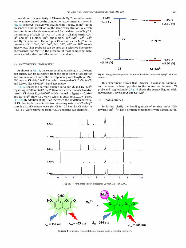
As shown in Fig. 11, the corresponding wavelength to the bandap energy can be calculated from the cross point of absorptionnd emission onset lines. The corresponding wavelength for CS is96 nm and CS + Mg2+ is 415 nm which are equal to 3.13 eV (for CS)nd 2.99 eV (for CS + Mg2+) band gap energy.
Fig. 12 shows the current–voltage curve for CS and CS + Mg2+
egarding to Differential Pulse Voltammetric experiments. Based onesults, CS shows Eox = 0.656 V which is equal to EHOMO = −5.46 eVnd CS + Mg2+ shows Eox = 0.7 V which is equal to EHOMO = −5.5 eV
31–42]. By addition of Mg2+ ion increased the oxidation potentialf CS, due to decrease in electron releasing nature of CS − Mg2+omplex. LUMO energy levels (for CS is −2.33 eV, for CS + Mg2+ is2.51 eV) were estimated from HOMO and band gap energies.
Fig. 14. 1H NMR titration plot of rec
Scheme 2. Schematic representation of b
Fig. 13. Energy level diagram of the probe CS and the corresponding Mg2+ additionproduct.
This experiment proves that, increase in oxidation potentialand decrease in band gap due to the interaction between CSprobe and magnesium ion. Fig. 13 shows the energy diagram withHOMO/LUMO levels of CS and CS + Mg2+.
3.4. 1H NMR titration
To further clarify the bonding mode of sensing probe (CS)towards Mg2+, 1H NMR titration experiments were carried out in
eptor CS with Mg2+ in CD3OD.
inding mode of receptor with Mg2+.
222 V.K. Gupta et al. / Sensors and ActuIn
tens
ityFl
uore
scen
ce
2-500
0
500
1000
1500
2000
2500
3000
3500
4 86
pH10 1412
CS
CS+Mg2+
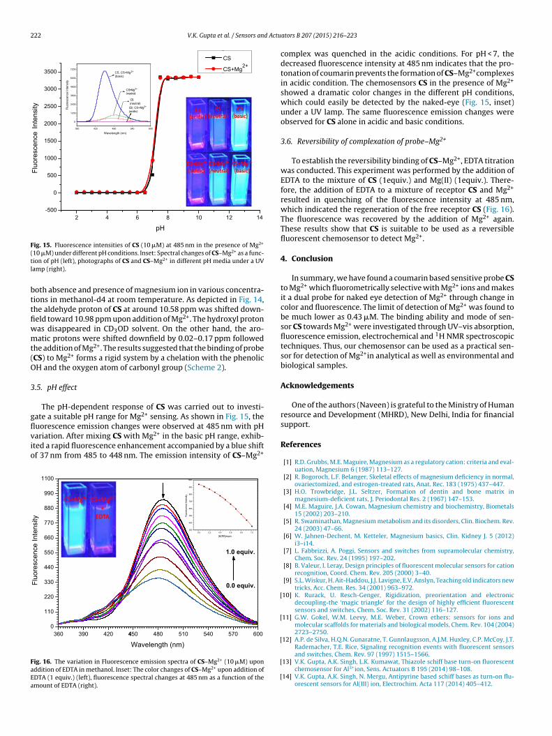
Fig. 15. Fluorescence intensities of CS (10 �M) at 485 nm in the presence of Mg2+
(tl
bttfiwmt(O
3
gflvio
FaEa
10 �M) under different pH conditions. Inset: Spectral changes of CS–Mg2+ as a func-ion of pH (left), photographs of CS and CS–Mg2+ in different pH media under a UVamp (right).
oth absence and presence of magnesium ion in various concentra-ions in methanol-d4 at room temperature. As depicted in Fig. 14,he aldehyde proton of CS at around 10.58 ppm was shifted down-eld toward 10.98 ppm upon addition of Mg2+. The hydroxyl protonas disappeared in CD3OD solvent. On the other hand, the aro-atic protons were shifted downfield by 0.02–0.17 ppm followed
he addition of Mg2+. The results suggested that the binding of probeCS) to Mg2+ forms a rigid system by a chelation with the phenolicH and the oxygen atom of carbonyl group (Scheme 2).
.5. pH effect
The pH-dependent response of CS was carried out to investi-ate a suitable pH range for Mg2+ sensing. As shown in Fig. 15, the
uorescence emission changes were observed at 485 nm with pHariation. After mixing CS with Mg2+ in the basic pH range, exhib-ted a rapid fluorescence enhancement accompanied by a blue shiftf 37 nm from 485 to 448 nm. The emission intensity of CS–Mg2+Inte
nsity
Fluo
resc
ence
3903600
110
220
330
440
550
660
770
880
990
1100
420 44 48050
Wavelength (nm)540510 600570
0.0 equiv.
1.0 equiv.
ig. 16. The variation in Fluorescence emission spectra of CS–Mg2+ (10 �M) uponddition of EDTA in methanol. Inset: The color changes of CS–Mg2+ upon addition ofDTA (1 equiv.) (left), fluorescence spectral changes at 485 nm as a function of themount of EDTA (right).
[
[
[
[
[
ators B 207 (2015) 216–223
complex was quenched in the acidic conditions. For pH < 7, thedecreased fluorescence intensity at 485 nm indicates that the pro-tonation of coumarin prevents the formation of CS–Mg2+complexesin acidic condition. The chemosensors CS in the presence of Mg2+
showed a dramatic color changes in the different pH conditions,which could easily be detected by the naked-eye (Fig. 15, inset)under a UV lamp. The same fluorescence emission changes wereobserved for CS alone in acidic and basic conditions.
3.6. Reversibility of complexation of probe–Mg2+
To establish the reversibility binding of CS–Mg2+, EDTA titrationwas conducted. This experiment was performed by the addition ofEDTA to the mixture of CS (1equiv.) and Mg(II) (1equiv.). There-fore, the addition of EDTA to a mixture of receptor CS and Mg2+
resulted in quenching of the fluorescence intensity at 485 nm,which indicated the regeneration of the free receptor CS (Fig. 16).The fluorescence was recovered by the addition of Mg2+ again.These results show that CS is suitable to be used as a reversiblefluorescent chemosensor to detect Mg2+.
4. Conclusion
In summary, we have found a coumarin based sensitive probe CSto Mg2+ which fluorometrically selective with Mg2+ ions and makesit a dual probe for naked eye detection of Mg2+ through change incolor and fluorescence. The limit of detection of Mg2+ was found tobe much lower as 0.43 �M. The binding ability and mode of sen-sor CS towards Mg2+ were investigated through UV–vis absorption,fluorescence emission, electrochemical and 1H NMR spectroscopictechniques. Thus, our chemosensor can be used as a practical sen-sor for detection of Mg2+in analytical as well as environmental andbiological samples.
Acknowledgements
One of the authors (Naveen) is grateful to the Ministry of Humanresource and Development (MHRD), New Delhi, India for financialsupport.
References
[1] R.D. Grubbs, M.E. Maguire, Magnesium as a regulatory cation: criteria and eval-uation, Magnesium 6 (1987) 113–127.
[2] R. Bogoroch, L.F. Belanger, Skeletal effects of magnesium deficiency in normal,ovariectomized, and estrogen-treated rats, Anat. Rec. 183 (1975) 437–447.
[3] H.O. Trowbridge, J.L. Seltzer, Formation of dentin and bone matrix inmagnesium-deficient rats, J. Periodontal Res. 2 (1967) 147–153.
[4] M.E. Maguire, J.A. Cowan, Magnesium chemistry and biochemistry, Biometals15 (2002) 203–210.
[5] R. Swaminathan, Magnesium metabolism and its disorders, Clin. Biochem. Rev.24 (2003) 47–66.
[6] W. Jahnen-Dechent, M. Ketteler, Magnesium basics, Clin. Kidney J. 5 (2012)i3–i14.
[7] L. Fabbrizzi, A. Poggi, Sensors and switches from supramolecular chemistry,Chem. Soc. Rev. 24 (1995) 197–202.
[8] B. Valeur, I. Leray, Design principles of fluorescent molecular sensors for cationrecognition, Coord. Chem. Rev. 205 (2000) 3–40.
[9] S.L. Wiskur, H. Ait-Haddou, J.J. Lavigne, E.V. Anslyn, Teaching old indicators newtricks, Acc. Chem. Res. 34 (2001) 963–972.
10] K. Rurack, U. Resch-Genger, Rigidization, preorientation and electronicdecoupling-the ‘magic triangle’ for the design of highly efficient fluorescentsensors and switches, Chem. Soc. Rev. 31 (2002) 116–127.
11] G.W. Gokel, W.M. Leevy, M.E. Weber, Crown ethers: sensors for ions andmolecular scaffolds for materials and biological models, Chem. Rev. 104 (2004)2723–2750.
12] A.P. de Silva, H.Q.N. Gunaratne, T. Gunnlaugsson, A.J.M. Huxley, C.P. McCoy, J.T.Rademacher, T.E. Rice, Signaling recognition events with fluorescent sensors
and switches, Chem. Rev. 97 (1997) 1515–1566.13] V.K. Gupta, A.K. Singh, L.K. Kumawat, Thiazole schiff base turn-on fluorescentchemosensor for Al3+ion, Sens. Actuators B 195 (2014) 98–108.
14] V.K. Gupta, A.K. Singh, N. Mergu, Antipyrine based schiff bases as turn-on flu-orescent sensors for Al(III) ion, Electrochim. Acta 117 (2014) 405–412.

d Actu
[
[
[
[
[
[
[
[
[
[
[
[
[
[
[
[
[
[
[
[
[
[
[
[
[
[
[
[
sion for almost 30 years. Presently, he is a Professor ofChemistry at Indian Institute of Technology Roorkee, Indiaand has more than 150 research publications to his credit.Prof. Singh works extensively in the field of macrocyclicchemistry
V.K. Gupta et al. / Sensors an
15] V.K. Gupta, N. Mergu, A.K. Singh, Fluorescent chemosensors for Zn2+ ions basedon flavonol derivatives, Sens. Actuators B 202 (2014) 674–682.
16] D. Kand, A.M. Kalle, S.J. Varma, P. Talukdar, A chromenoquinoline-based fluores-cent off–on thiol probe for bioimaging, Chem. Commun. 48 (2012) 2722–2724.
17] G. Zhang, Y. Wen, C. Guo, J. Xu, B. Lu, X. Duan, H. He, J. Yang, A cost-effectiveand practical polybenzanthrone-based fluorescent sensor for efficient deter-mination of palladium (II) ion and its application in agricultural crops andenvironment, Anal. Chim. Acta 805 (2013) 87–94.
18] G. Farruggia, S. Iotti, L. Prodi, M. Montalti, N. Zaccheroni, P.B. Savage, V. Tra-pani, P. Sale, F.I. Wolf, 8-hydroxyquinoline derivatives as fluorescent sensorsfor magnesium in living cells, J. Am. Chem. Soc. 128 (2006) 344–350.
19] H. Hama, T. Morozumi, H. Nakamura, Novel Mg2+-responsive flu-orescent chemosensor based on benzo-15-crown-5 possessing1-naphthaleneacetamide moiety, Tetrahedron Lett. 48 (2007) 1859–1861.
20] K.C. Song, M.G. Choi, D.H. Ryu, K.N. Kim, S.K. Chang, Ratiometric chemosensingof Mg2+ ions by a calix[4]arene diamide derivative, Tetrahedron Lett. 48 (2007)5397–5400.
21] H.M. Kim, P.R. Yang, M.S. Seo, J.S. Yi, J.H. Hong, S.J. Jeon, Y.G. Ko, K.J. Lee,B.R. Cho, Magnesium ion selective two-photon fluorescent probe based ona benzo[h]chromene derivative for in vivo imaging, J. Org. Chem. 27 (2007)2088–2096.
22] Y. Liu, Z.Y. Duan, H.Y. Zhang, X.L. Jiang, J.R. Han, Selective binding andinverse fluorescent behavior of magnesium ion by podand possessing plu-ral imidazo[4,5-f]-1,10-phenanthroline groups and its Ru(II) complex, J. Org.Chem. 70 (2005) 1450–1455.
23] B.J. Sanghavi, W. Varhue, J.L. Chavez, C.F. Chou, N.S. Swami, Elec-trokinetic preconcentration and detection of neuropeptides at patternedgraphene-modified electrodes in a nanochannel, Anal. Chem. 86 (2014)4120–4125.
24] B.J. Sanghavi, S. Sitaula, M.H. Griep, S.P. Karna, M.F. Ali, N.S. Swami, Real-timeelectrochemical monitoring of adenosine triphosphate in the picomolar tomicromolar range using graphene-modified electrodes, Anal. Chem. 85 (2013)8158–8165.
25] B.J. Sanghavi, S.M. Mobin, P. Mathur, G.K. Lahiri, A.K. Srivastava, Biomimeticsensor for certain catecholamines employing copper(II) complex and silvernanoparticle modified glassy carbon paste electrode, Biosens. Bioelectron. 39(2013) 124–132.
26] B.J. Sanghavi, A.K. Srivastava, Simultaneous voltammetric determination ofacetaminophen, aspirin and caffeine using an in situ surfactant-modifiedmultiwalled carbon nanotube paste electrode, Electrochim. Acta 55 (2010)8638–8648.
27] B.J. Sanghavi, A.K. Srivastava, Adsorptive stripping differential pulsevoltammetric determination of venlafaxine and desvenlafaxine employingnafion–carbon nanotube composite glassy carbon electrode, Electrochim. Acta56 (2011) 4188–4196.
28] H. Pechmann, C. Duisberg, Über die verbindungen der phenole mit acetes-sigäther, Ber. Dtsch. Chem. Ges. 16 (1883) 2119–2128.
29] A.D. Patel, M.S. Sharma, J.J. Vora, J.D. Joshi, Synthesis, characterization andantimicrobial activities of binary and ternary complexes of UO2II and ThIVcomplexes with 5-hydroxymethyl-8-quinolinol and 8-formyl-7-hydroxy-4-methyl-2H-1-benzopyran-2-one with aniline, J. Indian Chem. Soc. 74 (1997)287–288.
30] H.A. Benesi, J.H. Hildebrand, A spectrophotometric investigation of the inter-action of iodine with aromatic hydrocarbons, J. Am. Chem. Soc. 71 (1949)2703–2707.
31] J.L. Bredas, R. Silbey, D.S. Boudreux, R.R. Chance, Chain-length dependence ofelectronic and electrochemical properties of conjugated systems: polyacety-lene, polyphenylene, polythiophene, and polypyrrole, J. Am. Chem. Soc. 105(1983) 6555–6559.
32] V.K. Gupta, A.K. Jain, G. Maheshwari, Novel Aluminum (III) selective potentio-metric sensor based on morin in poly (vinyl chloride) matrix, Talanta 72 (4)(2007) 1469–1473.
33] V.K. Gupta, M.R. Ganjali, P. Norouzi, H. Khani, A. Nayak, S. Agarwal, Electro-chemical analysis of some toxic metals and drugs by ion selective electrodes,Crit. Rev. Anal. Chem. 41 (2011) 282–313.
34] R.N. Goyal, V.K. Gupta, S. Chatterjee, Voltammetric biosensors for the deter-mination of paracetamol at carbon nanotube modified pyrolytic graphiteelectrode, Sens. Actuators B. Chem. 149 (2010) 252–258.
35] V.K. Gupta, A.K. Jain, Shiva Agarwal, G. Maheshwari, An iron (III) ion-selective sensor based on a (bis (tridentate) ligand, Talanta 71 (2007)1964–1968.
36] R. Jain, V.K. Gupta, N. Jadon, K. Radhapyari, Voltammetric determination of
Cefixime in pharmaceuticals and biological fluids, Anal. Biochem. 407 (2010)79–88.37] V.K. Gupta, A.K. Singh, S. Mehtab, B. Gupta, ACobalt (II) selective PVC membranebased on a Schiff base complex of N, N′-bis (salicylidene)-3,4-diaminotoluene,Anal. Chim. Acta 566 (2006) 5–10.
ators B 207 (2015) 216–223 223
38] R.N. Goyal, V.K. Gupta, S. Chatterjee, Electrochemical oxidation of 2′ , 3′-dideoxyadenosine at pyrolytic graphite electrode, Electrochim. Acta 53 (2008)5354–5360.
39] V.K. Gupta, A.K. Singh, M. Al Khayat, B. Gupta, Neutral carriers based poly431meric membrane electrodes for selective determination of Mercury (II), Anal.Chim. Acta 590 (2007) 81–90.
40] V.K. Gupta, R. Prasad, R. Mangla, P. Kumar, New nickel (II) selective poten-tiometric sensor based on 5,7,12,14-tetramethyldibenzotetraazaannulene in apoly (vinyl chloride) matrix, Anal. Chim. Acta 420 (2000) 19–27.
41] R.N. Goal, V.K. Gupta, S. Chatterjee, Simultaneous determination of adenosineand inosine using single-wall carbon nanotubes modified pyrolytic graphiteelectrode, Talanta 76 (2008) 662–668.
42] V.K. Gupta, I. Ali, T.A. Saleh, A. Nayak, S. Agarwal, Chemical treatment technolo-gies for wastewater recycling–a review, RSC Adv. 2 (2012) 6380–6388.
Biographies
Vinod Kumar Gupta obtained his PhD degree in chem-istry from the University of Roorkee (now Indian Instituteof Technology Roorkee) Roorkee, India, in 1979. Sincethen he is pursuing research at the same Institute andpresently holding the position of Professor of ChemistryDepartment, at Indian Institute of Technology Roorkee,Roorkee. He worked as a post-doctoral fellow at Univer-sity of Regensburg, Germany, in 1993 as an EC fellow andwas DAAD visiting professor at University of Chemnitz andFreie University of Berlin in 2002. He has published morethan 400 paper with h-index of 110 and citation > 27000.His research interests include chemical sensors, wastewater treatment, environmental and electro analytical
chemistry. Dr. Gupta is an elected Fellow of the World Innovation Foundation (FIWF)since July 2004 and Fellow of the National Academy of Sciences (FNASc) since 2008.
Naveen Mergu, is a PhD student and pursuing researchon chemical sensors under the supervision of V.K. Guptaand A.K. Singh at IIT Roorkee. He did his M.Sc. in 2010 inChemistry at NIT Warangal.
Lokesh Kumar Kumawat is a Ph.D student and persuingresearch on chemical sensor under the supervision of V.K.Gupta and A. K. Singh at IIT Roorkee. He has completedhis master degree in chemistry from MLSU (Mohan LalSukhadia University) Udaipur, India in 2009
Ashok Kumar Singh is in teaching and research profes-