SCABP8/CBL10, a Putative Calcium Sensor, Interacts with the Protein Kinase SOS2 to Protect...
18
SCABP8/CBL10, a Putative Calcium Sensor, Interacts with the Protein Kinase SOS2 to Protect Arabidopsis Shoots from Salt Stress W OA Ruidang Quan, a Huixin Lin, a Imelda Mendoza, b Yuguo Zhang, a,c Wanhong Cao, a Yongqing Yang, a Mei Shang, a Shouyi Chen, c Jose ´ M. Pardo, b and Yan Guo a,1 a National Institute of Biological Sciences, Beijing 102206, China b Instituto de Recursos Naturales y Agrobiologı´a, Consejo Superior de Investigaciones Cientı ´ficas, Sevilla 41012, Spain c Institute of Genetics and Developmental Biology, Chinese Academy of Sciences, Beijing 100101, China The SOS (for Salt Overly Sensitive) pathway plays essential roles in conferring salt tolerance in Arabidopsis thaliana. Under salt stress, the calcium sensor SOS3 activates the kinase SOS2 that positively regulates SOS1, a plasma membrane sodium/ proton antiporter. We show that SOS3 acts primarily in roots under salt stress. By contrast, the SOS3 homolog SOS3-LIKE CALCIUM BINDING PROTEIN8 (SCABP8)/CALCINEURIN B-LIKE10 functions mainly in the shoot response to salt toxicity. While root growth is reduced in sos3 mutants in the presence of NaCl, the salt sensitivity of scabp8 is more prominent in shoot tissues. SCABP8 is further shown to bind calcium, interact with SOS2 both in vitro and in vivo, recruit SOS2 to the plasma membrane, enhance SOS2 activity in a calcium-dependent manner, and activate SOS1 in yeast. In addition, sos3 scabp8 and sos2 scabp8 display a phenotype similar to sos2, which is more sensitive to salt than either sos3 or scabp8 alone. Overexpression of SCABP8 in sos3 partially rescues the sos3 salt-sensitive phenotype. However, overexpression of SOS3 fails to complement scabp8. These results suggest that SCABP8 and SOS3 are only partially redundant in their function, and each plays additional and unique roles in the plant salt stress response. INTRODUCTION Soil salinity is a major abiotic stress that reduces plant growth and ranks among the leading factors limiting agricultural pro- ductivity. The sessile nature of plants has favored the evolution of mechanisms to cope with varying salt concentrations in the environment. Na þ is not essential for plant growth, and under salt stress, it hinders uptake of the important mineral nutrient K þ and competes for its enzyme binding sites. Maintaining a high K þ and low Na þ homeostasis in the cytoplasm is thus essential for plant salt tolerance. In Arabidopsis thaliana, an ionic homeostasis reg- ulatory pathway activated by salt stress has been identified through molecular and genetic characterization of several salt overly sensitive (sos) mutants that are defective in K þ /Na þ ho- meostasis (Liu and Zhu, 1998; Liu et al., 2000; Shi et al., 2000). The SOS pathway is known to be defined by three protein components, SOS1, SOS2, and SOS3. SOS3 encodes an EF hand–type calcium sensor with an N-myristoylation signal pep- tide (Liu and Zhu, 1998). Both calcium binding activity and N-myristoylation are essential for SOS3 function (Ishitani et al., 2000). In vitro, SOS3 physically interacts with and activates a Ser/Thr protein kinase, SOS2. Calcium is not required for the SOS2 and SOS3 interaction but was observed to enhance the phosphorylation of synthesized substrate p3 by the SOS2-SOS3 complex (Halfter et al., 2000). One of the downstream targets of the SOS3-SOS2 complex is SOS1, a plasma membrane– localized sodium/proton antiporter (Shi et al., 2000; Qiu et al., 2002; Quintero et al., 2002). Genetic evidence indicates that SOS3 and SOS2 function by activating SOS1. Mutations in SOS1, SOS2, or SOS3 all reduce the Na þ /H þ exchange activity, and a constitutively active SOS2 enhances Na þ /H þ exchange activity in a SOS1-dependent and SOS3-independent manner (Qiu et al., 2002). In yeast, SOS3 recruits SOS2 to the plasma membrane, and SOS1 increases NaCl tolerance in yeast mutants lacking Na þ transporters. Only when both SOS2 and SOS3 exist together does the system function to further increase SOS1-dependent NaCl resistance due to the phosphorylation of SOS1 by the SOS2-SOS3 complex (Quintero et al., 2002). However, evidence is lacking for an inter- action of SOS2 and SOS3 in planta. The growth inhibition of sos1 and sos2 mutants is more severe than that of sos3 under mild salt stress (Zhu et al., 1998). SOS1 and SOS2 are expressed in both root and shoot tissues, sug- gesting that they function in diverse tissue types (Liu et al., 2000; Shi et al., 2002). However, our examination of public microarray results (http://jsp.weigelworld.org/expviz/expviz.jsp) indicated that SOS3 is mainly expressed in the root tissue. It thus seems plau- sible that an additional calcium sensor(s) may exist to regulate SOS2 in shoot tissues as well. Here, we report that SOS3-LIKE 1 To whom correspondence should be addressed. E-mail guoyan@nibs. ac.cn; fax 86-10-80726671. The author responsible for distribution of materials integral to the findings presented in this article in accordance with the policy described in the Instructions for Authors (www.plantcell.org) is: Yan Guo ([email protected]). W Online version contains Web-only data. OA Open Access articles can be viewed online without a subscription. www.plantcell.org/cgi/doi/10.1105/tpc.106.042291 The Plant Cell, Vol. 19: 1415–1431, April 2007, www.plantcell.org ª 2007 American Society of Plant Biologists
-
Upload
universitasislamriau -
Category
Documents
-
view
1 -
download
0
Transcript of SCABP8/CBL10, a Putative Calcium Sensor, Interacts with the Protein Kinase SOS2 to Protect...

SCABP8/CBL10, a Putative Calcium Sensor, Interacts withthe Protein Kinase SOS2 to Protect Arabidopsis Shootsfrom Salt Stress W OA
Ruidang Quan,a Huixin Lin,a Imelda Mendoza,b Yuguo Zhang,a,c Wanhong Cao,a Yongqing Yang,a Mei Shang,a
Shouyi Chen,c Jose M. Pardo,b and Yan Guoa,1
a National Institute of Biological Sciences, Beijing 102206, Chinab Instituto de Recursos Naturales y Agrobiologıa, Consejo Superior de Investigaciones Cientıficas, Sevilla 41012, Spainc Institute of Genetics and Developmental Biology, Chinese Academy of Sciences, Beijing 100101, China
The SOS (for Salt Overly Sensitive) pathway plays essential roles in conferring salt tolerance in Arabidopsis thaliana. Under
salt stress, the calcium sensor SOS3 activates the kinase SOS2 that positively regulates SOS1, a plasma membrane sodium/
proton antiporter. We show that SOS3 acts primarily in roots under salt stress. By contrast, the SOS3 homolog SOS3-LIKE
CALCIUM BINDING PROTEIN8 (SCABP8)/CALCINEURIN B-LIKE10 functions mainly in the shoot response to salt toxicity.
While root growth is reduced in sos3 mutants in the presence of NaCl, the salt sensitivity of scabp8 is more prominent in
shoot tissues. SCABP8 is further shown to bind calcium, interact with SOS2 both in vitro and in vivo, recruit SOS2 to the
plasma membrane, enhance SOS2 activity in a calcium-dependent manner, and activate SOS1 in yeast. In addition, sos3
scabp8 and sos2 scabp8 display a phenotype similar to sos2, which is more sensitive to salt than either sos3 or scabp8
alone. Overexpression of SCABP8 in sos3 partially rescues the sos3 salt-sensitive phenotype. However, overexpression of
SOS3 fails to complement scabp8. These results suggest that SCABP8 and SOS3 are only partially redundant in their
function, and each plays additional and unique roles in the plant salt stress response.
INTRODUCTION
Soil salinity is a major abiotic stress that reduces plant growth
and ranks among the leading factors limiting agricultural pro-
ductivity. The sessile nature of plants has favored the evolution
of mechanisms to cope with varying salt concentrations in the
environment. Naþ is not essential for plant growth, and under salt
stress, it hinders uptake of the important mineral nutrient Kþ and
competes for its enzyme binding sites. Maintaining a high Kþ and
low Naþ homeostasis in the cytoplasm is thus essential for plant
salt tolerance. In Arabidopsis thaliana, an ionic homeostasis reg-
ulatory pathway activated by salt stress has been identified
through molecular and genetic characterization of several salt
overly sensitive (sos) mutants that are defective in Kþ/Naþ ho-
meostasis (Liu and Zhu, 1998; Liu et al., 2000; Shi et al., 2000).
The SOS pathway is known to be defined by three protein
components, SOS1, SOS2, and SOS3. SOS3 encodes an EF
hand–type calcium sensor with an N-myristoylation signal pep-
tide (Liu and Zhu, 1998). Both calcium binding activity and
N-myristoylation are essential for SOS3 function (Ishitani et al.,
2000). In vitro, SOS3 physically interacts with and activates a
Ser/Thr protein kinase, SOS2. Calcium is not required for the
SOS2 and SOS3 interaction but was observed to enhance the
phosphorylation of synthesized substrate p3 by the SOS2-SOS3
complex (Halfter et al., 2000). One of the downstream targets
of the SOS3-SOS2 complex is SOS1, a plasma membrane–
localized sodium/proton antiporter (Shi et al., 2000; Qiu et al.,
2002; Quintero et al., 2002).
Genetic evidence indicates that SOS3 and SOS2 function by
activating SOS1. Mutations in SOS1, SOS2, or SOS3 all reduce
the Naþ/Hþ exchange activity, and a constitutively active SOS2
enhances Naþ/Hþ exchange activity in a SOS1-dependent and
SOS3-independent manner (Qiu et al., 2002). In yeast, SOS3
recruits SOS2 to the plasma membrane, and SOS1 increases
NaCl tolerance in yeast mutants lacking Naþ transporters. Only
when both SOS2 and SOS3 exist together does the system
function to further increase SOS1-dependent NaCl resistance
due to the phosphorylation of SOS1 by the SOS2-SOS3 complex
(Quintero et al., 2002). However, evidence is lacking for an inter-
action of SOS2 and SOS3 in planta.
The growth inhibition of sos1 and sos2 mutants is more severe
than that of sos3 under mild salt stress (Zhu et al., 1998). SOS1
and SOS2 are expressed in both root and shoot tissues, sug-
gesting that they function in diverse tissue types (Liu et al., 2000;
Shi et al., 2002). However, our examination of public microarray
results (http://jsp.weigelworld.org/expviz/expviz.jsp) indicated that
SOS3 is mainly expressed in the root tissue. It thus seems plau-
sible that an additional calcium sensor(s) may exist to regulate
SOS2 in shoot tissues as well. Here, we report that SOS3-LIKE
1 To whom correspondence should be addressed. E-mail [email protected]; fax 86-10-80726671.The author responsible for distribution of materials integral to thefindings presented in this article in accordance with the policy describedin the Instructions for Authors (www.plantcell.org) is: Yan Guo([email protected]).W Online version contains Web-only data.OA Open Access articles can be viewed online without a subscription.www.plantcell.org/cgi/doi/10.1105/tpc.106.042291
The Plant Cell, Vol. 19: 1415–1431, April 2007, www.plantcell.org ª 2007 American Society of Plant Biologists
CALCIUM BINDING PROTEIN8 (SCABP8) interacts with and
recruits SOS2 to the plasma membrane in vivo and is required for
salt tolerance in shoots.
RESULTS
Expression of SOS2, SOS3, and SCABP8
Liu et al. (2000) and Shi et al. (2002) reported that SOS1 and
SOS2 were expressed in both root and shoot tissues in Arabi-
dopsis. By contrast, an examination of the microarray results in
the public domain (http://jsp.weigelworld.org/expviz/expviz.jsp)
indicated that SOS3 was strongly expressed in roots but only at
background level in shoots, suggesting that SOS3 mainly func-
tions in root. We therefore speculated that an SOS3 homolog or
SOS2-regulatory protein may function in shoots to regulate
SOS2. Interestingly, we noticed that of the other nine SOS3-
like calcium binding proteins, SCABP8 is the only one expressed
strongly in shoots but very weakly in roots in this microarray data
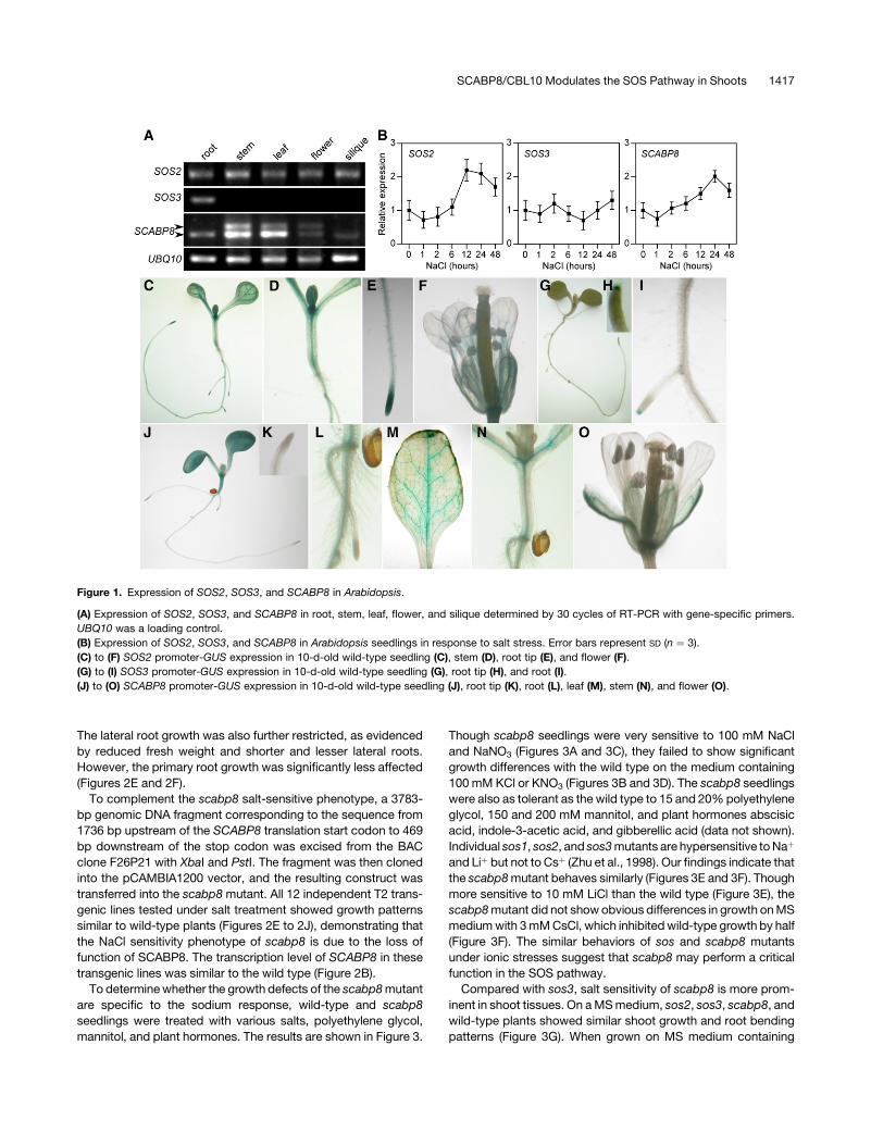
set. To confirm these results, RT-PCR analysis was performed.
The transcripts of SOS2 could be amplified from roots, stems,
leaves, flowers, and siliques, whereas SOS3 was only detected
in roots. SCABP8 was expressed at a high level in leaves and
stems but at a very low level in roots, flowers, and siliques (Figure
1A), consistent with microarray data in the public domain.
To examine whether expression of SOS2, SOS3, and SCABP8
is induced by NaCl treatment, total RNA was extracted from wild-
type plants treated with 100 mM NaCl for 0, 1, 2, 6, 12, 24, and
48 h. Consistent with previous results (Liu et al., 2000), the
expression of SOS2, but not SOS3, was induced by NaCl
treatment (Figure 1B). The transcript of SCABP8 was slightly
induced 6 h after treatment and decreased after 24 h (Figure 1B).
The SCABP8 gene contains nine exons and eight introns and
encodes a 246–amino acid protein with a predicted molecular
mass of 28.3 kD and isoelectric point of 4.3. Two SCABP8
cDNAs were amplified and sequenced. Two bands pertaining to
SCABP8 were amplified from aerial parts, whereas only the lower
band yielded in roots (Figure 1A). The shorter sequence is
identical to At4g33000.2 (http://www.arabidopsis.org), while
the longer retains its seventh intron, which contains a premature
stop codon that results in a truncated protein 194 amino acids
long. Overexpression of this mis-spliced SCABP8 cDNA could
not complement the scabp8 salt sensitivity phenotype (data not
shown).
To further determine the tissue specificity of these genes,
promoter regions of SOS2 (1.6 kb), SOS3 (1.4 kb), and SCABP8
(1.7 kb) were fused to a b-glucuronidase (GUS) reporter gene.
The resulting constructs were transferred into an Arabidopsis
Columbia (Col-0) background. For each construct, 12 indepen-
dent F2 transgenic lines were analyzed by a GUS staining assay.
Though the intensity of the GUS staining from each of the
independent lines was variable, the tissue-specific localization
was the same. As shown in Figures 1C to 1F, GUS driven by the
SOS2 promoter was expressed in the entire young seedling, leaf,
stem, and flower and was particularly strong in root tips. Similar
expression has been detected in SOS1 promoter-GUS trans-
genic plants (Shi et al., 2002). Consistent with microarray and RT-
PCR results, GUS expression driven by the SOS3 promoter was
only detected in root and very strongly in the root tip (Figures 1G
to 1I) but not other tissues. However, SCABP8-GUS was ex-
pressed only in the stem, leaf, flower, and weakly in root but not in
the root tip (Figures 1J to 1O). The combined pattern of SCABP8
and SOS3 expression overlaps with that of SOS2 and suggests
that SCABP8 and SOS3 may assume similar roles in different
tissue types to activate SOS2 in response to salt stress.
scabp8 Is Hypersensitive to Salt in Shoot Tissues
To test the hypothesis that SCABP8 is a tissue-specific activator
of SOS2, the Arabidopsis mutant line SALK_056042 with a
T-DNA insertion in the SCABP8 gene was obtained from the
ABRC (Alonso et al., 2003). The T-DNA insertion is located in the
seventh intron of the SCABP8 gene and was confirmed by PCR
using a SCABP8-specific primer and T-DNA left border primers
(data not shown; Figure 2A). The insertion was further confirmed
by a DNA gel blot probed with a neomycin phosphotransferase
coding sequence (nptII). Genomic DNA was extracted from
scabp8, digested by three restriction enzymes, EcoRI, HindIII,
and SacI, which are the enzymes with a single digesting site in the
pBIN-pROK2 vector, and subjected to DNA gel blot analysis.
Only one strongly hybridizing band from each digestion was
observed with different molecular sizes (Figure 2C). Two weaker
bands of similar sizes were detected in all three digestions,
suggesting they were nonspecific. These data suggest that there
was a single T-DNA insertion in the scabp8 mutant. To precisely
map the border sequences flanking the T-DNA insertion, PCR
reactions were performed with combinations of the primers an-
nealing upstream or downstream of the T-DNA insertion into the
SCABP8 gene and primers specific to the left border or right
border of the T-DNA. The PCR products were sequenced, and
the results indicated that the T-DNA insertion had generated an
18-bp deletion from 1264 to 1281 bp downstream of the SCABP8
translation start codon, in the seventh intron. This insertion point
matches the sequence shown on the SALK website. The ab-
sence of a full-length SCABP8 transcript in the scabp8 mutant
was confirmed by RT-PCR analysis (Figure 2B). However, a trun-
cated SCABP8 transcript corresponding to a fragment before
the T-DNA insertion, but not after, still existed in the scabp8
mutant (Figure 2B). Overexpression of this truncated SCABP8
transcript did not rescue the scabp8 salt phenotype (data not
shown).
To test the salt sensitivity of the scabp8 mutant, 5-d-old mu-
tant and wild-type plants were transferred to Murashige and
Skoog (MS) medium with or without NaCl. Indeed, scabp8 was
hypersensitive to NaCl, mainly in the shoot tissues, but devel-
oped normally when grown on MS medium lacking NaCl (Figure
2D). On MS medium containing 50 mM NaCl, the primary root
growth of scabp8 was indistinguishable from the wild type
(Figures 2D and 2F). The number and length of secondary roots
were reduced relative to the wild type, but the overall root fresh
weight was reduced only ;12% (Figures 2D, 2G, 2H, and 2J).
However, shoot growth was more severely inhibited (30% fresh
weight reduction) and leaves were smaller and darker in color
compared with the wild type (Figures 2D, 2E, and 2I). On 100 mM
NaCl medium, growth inhibition was more pronounced. The
shoot fresh weight of scabp8 was less than half of the wild type.
1416 The Plant Cell
The lateral root growth was also further restricted, as evidenced
by reduced fresh weight and shorter and lesser lateral roots.
However, the primary root growth was significantly less affected
(Figures 2E and 2F).
To complement the scabp8 salt-sensitive phenotype, a 3783-
bp genomic DNA fragment corresponding to the sequence from
1736 bp upstream of the SCABP8 translation start codon to 469
bp downstream of the stop codon was excised from the BAC
clone F26P21 with XbaI and PstI. The fragment was then cloned
into the pCAMBIA1200 vector, and the resulting construct was
transferred into the scabp8 mutant. All 12 independent T2 trans-
genic lines tested under salt treatment showed growth patterns
similar to wild-type plants (Figures 2E to 2J), demonstrating that
the NaCl sensitivity phenotype of scabp8 is due to the loss of
function of SCABP8. The transcription level of SCABP8 in these
transgenic lines was similar to the wild type (Figure 2B).
To determine whether the growth defects of the scabp8 mutant
are specific to the sodium response, wild-type and scabp8
seedlings were treated with various salts, polyethylene glycol,
mannitol, and plant hormones. The results are shown in Figure 3.
Though scabp8 seedlings were very sensitive to 100 mM NaCl
and NaNO3 (Figures 3A and 3C), they failed to show significant
growth differences with the wild type on the medium containing
100 mM KCl or KNO3 (Figures 3B and 3D). The scabp8 seedlings
were also as tolerant as the wild type to 15 and 20% polyethylene
glycol, 150 and 200 mM mannitol, and plant hormones abscisic
acid, indole-3-acetic acid, and gibberellic acid (data not shown).
Individual sos1, sos2, and sos3 mutants are hypersensitive to Naþ
and Liþ but not to Csþ (Zhu et al., 1998). Our findings indicate that
the scabp8 mutant behaves similarly (Figures 3E and 3F). Though
more sensitive to 10 mM LiCl than the wild type (Figure 3E), the
scabp8 mutant did not show obvious differences in growth on MS
medium with 3 mM CsCl, which inhibited wild-type growth by half
(Figure 3F). The similar behaviors of sos and scabp8 mutants
under ionic stresses suggest that scabp8 may perform a critical
function in the SOS pathway.
Compared with sos3, salt sensitivity of scabp8 is more prom-
inent in shoot tissues. On a MS medium, sos2, sos3, scabp8, and
wild-type plants showed similar shoot growth and root bending
patterns (Figure 3G). When grown on MS medium containing
Figure 1. Expression of SOS2, SOS3, and SCABP8 in Arabidopsis.
(A) Expression of SOS2, SOS3, and SCABP8 in root, stem, leaf, flower, and silique determined by 30 cycles of RT-PCR with gene-specific primers.
UBQ10 was a loading control.
(B) Expression of SOS2, SOS3, and SCABP8 in Arabidopsis seedlings in response to salt stress. Error bars represent SD (n ¼ 3).
(C) to (F) SOS2 promoter-GUS expression in 10-d-old wild-type seedling (C), stem (D), root tip (E), and flower (F).
(G) to (I) SOS3 promoter-GUS expression in 10-d-old wild-type seedling (G), root tip (H), and root (I).
(J) to (O) SCABP8 promoter-GUS expression in 10-d-old wild-type seedling (J), root tip (K), root (L), leaf (M), stem (N), and flower (O).
SCABP8/CBL10 Modulates the SOS Pathway in Shoots 1417
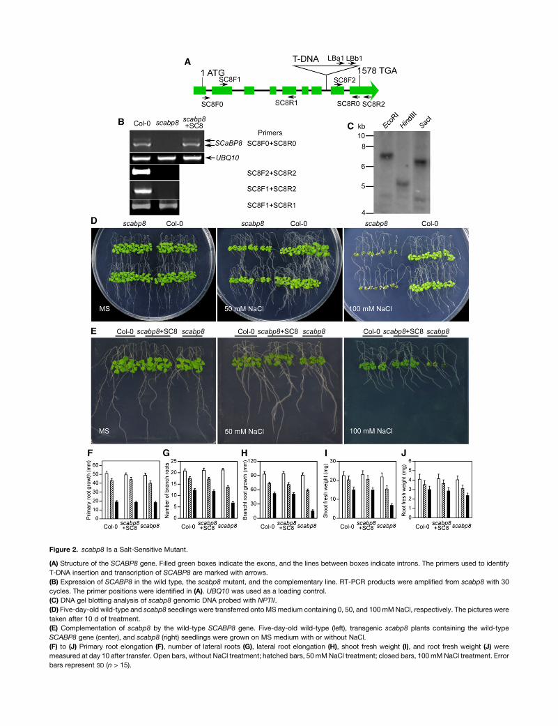
Figure 2. scabp8 Is a Salt-Sensitive Mutant.
(A) Structure of the SCABP8 gene. Filled green boxes indicate the exons, and the lines between boxes indicate introns. The primers used to identify
T-DNA insertion and transcription of SCABP8 are marked with arrows.
(B) Expression of SCABP8 in the wild type, the scabp8 mutant, and the complementary line. RT-PCR products were amplified from scabp8 with 30
cycles. The primer positions were identified in (A). UBQ10 was used as a loading control.
(C) DNA gel blotting analysis of scabp8 genomic DNA probed with NPTII.
(D) Five-day-old wild-type and scabp8 seedlings were transferred onto MS medium containing 0, 50, and 100 mM NaCl, respectively. The pictures were
taken after 10 d of treatment.
(E) Complementation of scabp8 by the wild-type SCABP8 gene. Five-day-old wild-type (left), transgenic scabp8 plants containing the wild-type
SCABP8 gene (center), and scabp8 (right) seedlings were grown on MS medium with or without NaCl.
(F) to (J) Primary root elongation (F), number of lateral roots (G), lateral root elongation (H), shoot fresh weight (I), and root fresh weight (J) were
measured at day 10 after transfer. Open bars, without NaCl treatment; hatched bars, 50 mM NaCl treatment; closed bars, 100 mM NaCl treatment. Error
bars represent SD (n > 15).
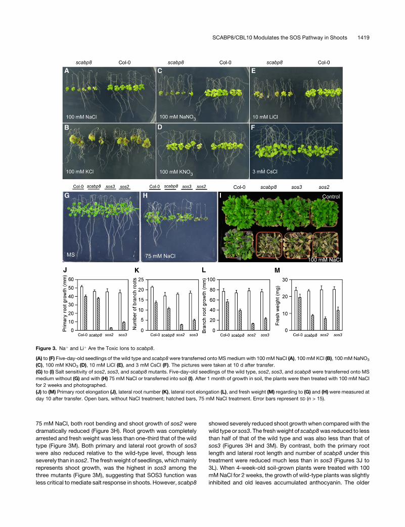
75 mM NaCl, both root bending and shoot growth of sos2 were
dramatically reduced (Figure 3H). Root growth was completely
arrested and fresh weight was less than one-third that of the wild
type (Figure 3M). Both primary and lateral root growth of sos3
were also reduced relative to the wild-type level, though less
severely than in sos2. The fresh weight of seedlings, which mainly
represents shoot growth, was the highest in sos3 among the
three mutants (Figure 3M), suggesting that SOS3 function was
less critical to mediate salt response in shoots. However, scabp8
showed severely reduced shoot growth when compared with the
wild type or sos3. The fresh weight of scabp8 was reduced to less
than half of that of the wild type and was also less than that of
sos3 (Figures 3H and 3M). By contrast, both the primary root
length and lateral root length and number of scabp8 under this
treatment were reduced much less than in sos3 (Figures 3J to
3L). When 4-week-old soil-grown plants were treated with 100
mM NaCl for 2 weeks, the growth of wild-type plants was slightly
inhibited and old leaves accumulated anthocyanin. The older
Figure 3. Naþ and Liþ Are the Toxic Ions to scabp8.
(A) to (F) Five-day-old seedlings of the wild type and scabp8 were transferred onto MS medium with 100 mM NaCl (A), 100 mM KCl (B), 100 mM NaNO3
(C), 100 mM KNO3 (D), 10 mM LiCl (E), and 3 mM CsCl (F). The pictures were taken at 10 d after transfer.
(G) to (I) Salt sensitivity of sos2, sos3, and scabp8 mutants. Five-day-old seedlings of the wild type, sos2, sos3, and scabp8 were transferred onto MS
medium without (G) and with (H) 75 mM NaCl or transferred into soil (I). After 1 month of growth in soil, the plants were then treated with 100 mM NaCl
for 2 weeks and photographed.
(J) to (M) Primary root elongation (J), lateral root number (K), lateral root elongation (L), and fresh weight (M) regarding to (G) and (H) were measured at
day 10 after transfer. Open bars, without NaCl treatment; hatched bars, 75 mM NaCl treatment. Error bars represent SD (n > 15).
SCABP8/CBL10 Modulates the SOS Pathway in Shoots 1419
leaves from all three mutants were bleached. A few young leaves
of sos3 and sos2 survived but none from scabp8 (Figure 3I).
We further tested whether expression of stress-responsive
genes is affected by scabp8 mutation in response to NaCl in an
RNA gel blot assay. These genes included RD29A, Cor15A,
Cor47, and Kin1. No significant difference was detected between
the mutant and the wild type (data not shown).
Genetic Interaction between SCABP8 and SOS2
The sos2 sos3 double mutant shows a salt-sensitive phenotype
similar to sos2 and greater than sos3, suggesting that SOS2 and
SOS3 genetically function in the same pathway and that SOS2
has additional functions in salt tolerance relative to SOS3 (Halfter
et al., 2000). To determine whether SCABP8 and SOS2 interact
genetically, the scabp8 T-DNA insertion line was crossed with
sos2 and sos3 mutants. From ;100 F2 plants of each cross, five
sos2 scabp8 and seven sos3 scabp8 double mutants were iden-
tified. The double mutants were then crossed into their parental
backgrounds to confirm the genotypes (the sos2-2 allele bears
an untagged 2-bp deletion that prevented PCR-based genotyp-
ing). We transferred 5-d-old wild-type, mutant, and double mu-
tant seedlings onto MS medium with or without 75 mM NaCl. In
the absence of NaCl treatment, none of the mutants showed
any significant growth differences compared with the wild type
(Figure 4A). As in the sos2 sos3 double mutant (Halfter et al.,
2000), sos2 scabp8 seedlings also displayed salt-sensitive fea-
tures similar to sos2, shown in shoot and root growth, and chlo-
rophyll and anthocynanin accumulation (Figures 4B to 4H). No
additional phenotypic difference was observed between sos2
and sos2 scabp8, suggesting that SOS2 and SCABP8 function in
the same pathway. Under 75 mM NaCl treatment, both root and
shoot growth of the sos3 scabp8 double mutant plants were
more severely reduced compared with the sos3 and scabp8
single mutants. The phenotype of sos3 scabp8 showed slightly
higher lateral root number than sos2 but otherwise resembled
sos2 (Figures 4B to 4H). These results further support a model in
which SCABP8 and SOS3 function together to modulate the
activity of SOS2.
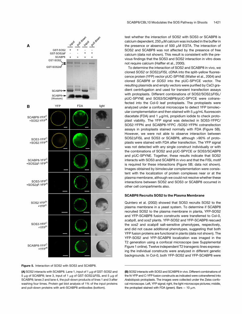
Interaction of SOS2 and SCABP8
If SCABP8 has partial functional redundancy with SOS3 and reg-
ulates SOS2 in the tissues that do not express SOS3, SCABP8
also would be expected to interact with and activate SOS2. This
was tested by cloning the SCABP8 cDNA into the pGEX-6P-1
vector containing a glutathione S-transferase (GST) tag, with sim-
ilar constructs made for SOS2 and SOS3. The FISL motif in the
SOS2 regulatory domain is necessary and sufficient for SOS3 in-
teraction (Guo et al., 2001). As a negative control, an SOS2DFISL
fragment (SOS2 with deletion of the FISL motif) was also gen-
erated by PCR mutagenesis and cloned into the pGEX-6P-1
vector. The GST-SCABP8, -SOS3, -SOS2, and -SOS2DFISL
fusion proteins were purified from Escherichia coli strain BL21(DE3)
using glutathione affinity chromatography. SOS3 and SCABP8
were digested from their GST fusion protein by PreScission
protease and subjected to in vitro pull-down assays. As ex-
pected, SOS3 was pulled down by GST-SOS2 but not by GST-
SOS2DFISL (data not shown). Similarly, the FISL motif was also
required for the interaction of SOS2 with SCABP8. SCABP8 was
pulled down by GST-SOS2 but not by GST-SOS2DFISL (Figure
5A). To confirm whether or not the pulled-down band was
SCABP8, 1% of the input and pull-down samples were subjected
to protein gel blot analysis with anti-SCABP8 antibodies. The
result showed that SCABP8 protein was only detected in GST-
SOS2 samples but not in GST-SOS2DFISL (Figure 5A). To further
Figure 4. Genetic Interaction of scabp8 with sos2 or sos3.
Five-day-old seedlings of the wild type, sos2, sos2 scabp8, scabp8,
scabp8 sos3, and sos3 were transferred onto MS medium without (A)
and with (B) 75 mM NaCl. The pictures were taken at day 10. Primary root
elongation (C), lateral root number (D), lateral root elongation (E), fresh
shoot weight (FW) (F), chlorophyll (G), and anthocynanin contents (H)
were measured at day 10 after transfer. Open bars, without NaCl
treatment; hatched bars, 75 mM NaCl treatment. Error bars represent
SD (n > 15).
1420 The Plant Cell
test whether the interaction of SOS2 with SOS3 or SCABP8 is
calcium dependent, 250 mM calcium was included in the buffer in
the presence or absence of 500 mM EGTA. The interaction of
SOS2 and SCABP8 was not affected by the presence of free
calcium (data not shown). This result is consistent with the pre-
vious findings that the SOS3 and SOS2 interaction in vitro does
not require calcium (Halfter et al., 2000).
To determine the interaction of SOS2 and SCABP8 in vivo, we
cloned SOS2 or SOS2DFISL cDNA into the split–yellow fluores-
cence protein (YFP) vector pUC-SPYNE (Walter et al., 2004) and
cloned SCABP8 or SOS3 into the pUC-SPYCE vector. The
resulting plasmids and empty vectors were purified by CsCl gra-
dient centrifugation and used for transient transfection assays
with protoplasts. Different combinations of SOS2/SOS2DFISL/
pUC-SPYNE and SOS3/SCABP8/pUC-SPYCE were cotrans-
fected into the Col-0 leaf protoplasts. The protoplasts were
analyzed under a confocal microscope to detect YFP bimolec-
ular complementation and then stained with 5 mg/mL fluorescein
diacetate (FDA) and 1 mg/mL propidium iodide to check proto-
plast viability. The YFP signal was detected in SOS3-YFPC/
SOS2-YFPN and SCABP8-YFPC /SOS2-YFPN cotransfection
assays in protoplasts stained normally with FDA (Figure 5B).
However, we were not able to observe interaction between
SOS2DFISL and SOS3 or SCABP8, although >90% of proto-
plasts were stained with FDA after transfection. The YFP signal
was not detected with any single construct individually or with
the combinations of SOS2 and pUC-SPYCE or SOS3/SCABP8
and pUC-SPYNE. Together, these results indicate that SOS2
interacts with SOS3 and SCABP8 in vivo and that the FISL motif
is required for these interactions (Figure 5B; data not shown).
Images obtained by bimolecular complementation were consis-
tent with the localization of protein complexes near or at the
plasma membrane, although we could not resolve whether these
interactions between SOS2 and SOS3 or SCABP8 occurred in
other cell compartments also.
SCABP8 Recruits SOS2 to the Plasma Membrane
Quintero et al. (2002) showed that SOS3 recruits SOS2 to the
plasma membrane in a yeast system. To determine if SCABP8
recruited SOS2 to the plasma membrane in planta, YFP-SOS2
and YFP-SCABP8 fusion constructs were transferred to Col-0,
scabp8, and sos2 plants. YFP-SOS2 and YFP-SCABP8 rescued
the sos2 and scabp8 salt-sensitive phenotypes, respectively,
and did not cause additional phenotypes, suggesting that both
YFP fusion proteins are functional in plants (data not shown). The
YFP-SOS2 and YFP-SCABP8 localization was imaged in the
T2 generation using a confocal microscope (see Supplemental
Figure 1 online). Twelve independent T2 transgenic lines express-
ing the individual constructs were analyzed in different genetic
backgrounds. In Col-0, both YFP-SOS2 and YFP-SCABP8 were
Figure 5. Interaction of SOS2 with SOS3 and SCABP8.
(A) SOS2 interacts with SCABP8. Lane 1, input of 1 mg of GST-SOS2 and
5 mg of SCABP8; lane 3, input of 1 mg of GST-SOS2DFISL and 5 mg of
SCABP8; lanes 2 and lane 4, the pull-down products of lines 1 and 3 after
washing four times. Protein gel blot analysis of 1% of the input proteins
and pull-down proteins with anti-SCABP8 antibodies (bottom).
(B) SOS2 interacts with SOS3 and SCABP8 in vivo. Different combinations of
the N-YFP and C-YFP fusion constructs as indicated were cotransferred into
Arabidopsis protoplasts. The images were collected under the Zeiss confo-
cal microscope. Left, YFP signal; right, the light microscope pictures; middle,
the protoplast stained with FDA (green). Bars ¼ 10 mm.
SCABP8/CBL10 Modulates the SOS Pathway in Shoots 1421
localized to the cell periphery, and a low level of YFP signal also
could be detected at the cytoplasm (see Supplemental Figures 1A
and 1C online). To exclude the possibility of cell wall association
of SOS2 and SCABP8, the transgenic plants were plasmolyzed
by mannitol treatment. Both YFP-SOS2 and YFP–SCABP8 sig-
nals were detached from the cell wall (see Supplemental Figures
1B and 1D online). Transgenic plants expressing YFP alone (see
Supplemental Figures 1G and 1H online), the cytoplasmic acidic
ribosomal green fluorescent protein (GFP) (see Supplemental
Figures 1I to 1J) (Cutler et al., 2000), and the plasma membrane
water channel protein (PIP2a)-GFP (see Supplemental Figures 1K
and 1L online) (Cutler et al., 2000) were used as controls. When
YFP-SOS2 was expressed in the scabp8 mutant, YFP-SOS2 was
detected more abundantly in the cytoplasm in comparison to the
wild-type background (see Supplemental Figure 1E online). After
mannitol treatment, YFP-SOS2 was clearly aggregated in the
cytosol (see Supplemental Figure 1F online). These determina-
tions were conducted in the upper part of seedling roots where
the analysis of promoter-GUS fusions demonstrated a significant
expression of SOS2 and SCABP8 genes, whereas that of SOS3
was undetectable (Figure 1). These results indicate that although
the localization of SOS2 and SCABP8 was not exclusively at the
plasma membrane, SCABP8 enhanced the fraction of SOS2
bound to the plasma membrane.
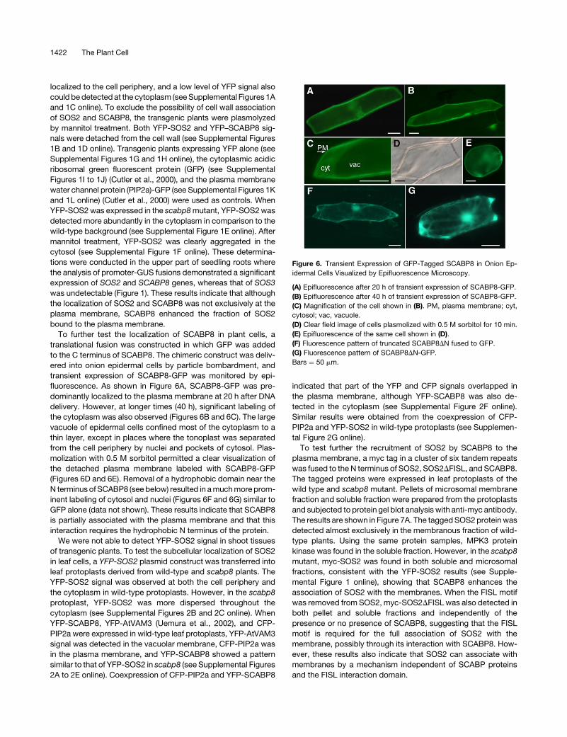
To further test the localization of SCABP8 in plant cells, a
translational fusion was constructed in which GFP was added
to the C terminus of SCABP8. The chimeric construct was deliv-
ered into onion epidermal cells by particle bombardment, and
transient expression of SCABP8-GFP was monitored by epi-
fluorescence. As shown in Figure 6A, SCABP8-GFP was pre-
dominantly localized to the plasma membrane at 20 h after DNA
delivery. However, at longer times (40 h), significant labeling of
the cytoplasm was also observed (Figures 6B and 6C). The large
vacuole of epidermal cells confined most of the cytoplasm to a
thin layer, except in places where the tonoplast was separated
from the cell periphery by nuclei and pockets of cytosol. Plas-
molization with 0.5 M sorbitol permitted a clear visualization of
the detached plasma membrane labeled with SCABP8-GFP
(Figures 6D and 6E). Removal of a hydrophobic domain near the
N terminus of SCABP8 (see below) resulted in a much more prom-
inent labeling of cytosol and nuclei (Figures 6F and 6G) similar to
GFP alone (data not shown). These results indicate that SCABP8
is partially associated with the plasma membrane and that this
interaction requires the hydrophobic N terminus of the protein.
We were not able to detect YFP-SOS2 signal in shoot tissues
of transgenic plants. To test the subcellular localization of SOS2
in leaf cells, a YFP-SOS2 plasmid construct was transferred into
leaf protoplasts derived from wild-type and scabp8 plants. The
YFP-SOS2 signal was observed at both the cell periphery and
the cytoplasm in wild-type protoplasts. However, in the scabp8
protoplast, YFP-SOS2 was more dispersed throughout the
cytoplasm (see Supplemental Figures 2B and 2C online). When
YFP-SCABP8, YFP-AtVAM3 (Uemura et al., 2002), and CFP-
PIP2a were expressed in wild-type leaf protoplasts, YFP-AtVAM3
signal was detected in the vacuolar membrane, CFP-PIP2a was
in the plasma membrane, and YFP-SCABP8 showed a pattern
similar to that of YFP-SOS2 in scabp8 (see Supplemental Figures
2A to 2E online). Coexpression of CFP-PIP2a and YFP-SCABP8
indicated that part of the YFP and CFP signals overlapped in
the plasma membrane, although YFP-SCABP8 was also de-
tected in the cytoplasm (see Supplemental Figure 2F online).
Similar results were obtained from the coexpression of CFP-
PIP2a and YFP-SOS2 in wild-type protoplasts (see Supplemen-
tal Figure 2G online).
To test further the recruitment of SOS2 by SCABP8 to the
plasma membrane, a myc tag in a cluster of six tandem repeats
was fused to the N terminus of SOS2, SOS2DFISL, and SCABP8.
The tagged proteins were expressed in leaf protoplasts of the
wild type and scabp8 mutant. Pellets of microsomal membrane
fraction and soluble fraction were prepared from the protoplasts
and subjected to protein gel blot analysis with anti-myc antibody.
The results are shown in Figure 7A. The tagged SOS2 protein was
detected almost exclusively in the membranous fraction of wild-
type plants. Using the same protein samples, MPK3 protein
kinase was found in the soluble fraction. However, in the scabp8
mutant, myc-SOS2 was found in both soluble and microsomal
fractions, consistent with the YFP-SOS2 results (see Supple-
mental Figure 1 online), showing that SCABP8 enhances the
association of SOS2 with the membranes. When the FISL motif
was removed from SOS2, myc-SOS2DFISL was also detected in
both pellet and soluble fractions and independently of the
presence or no presence of SCABP8, suggesting that the FISL
motif is required for the full association of SOS2 with the
membrane, possibly through its interaction with SCABP8. How-
ever, these results also indicate that SOS2 can associate with
membranes by a mechanism independent of SCABP proteins
and the FISL interaction domain.
Figure 6. Transient Expression of GFP-Tagged SCABP8 in Onion Ep-
idermal Cells Visualized by Epifluorescence Microscopy.
(A) Epifluorescence after 20 h of transient expression of SCABP8-GFP.
(B) Epifluorescence after 40 h of transient expression of SCABP8-GFP.
(C) Magnification of the cell shown in (B). PM, plasma membrane; cyt,
cytosol; vac, vacuole.
(D) Clear field image of cells plasmolized with 0.5 M sorbitol for 10 min.
(E) Epifluorescence of the same cell shown in (D).
(F) Fluorescence pattern of truncated SCABP8DN fused to GFP.
(G) Fluorescence pattern of SCABP8DN-GFP.
Bars ¼ 50 mm.
1422 The Plant Cell
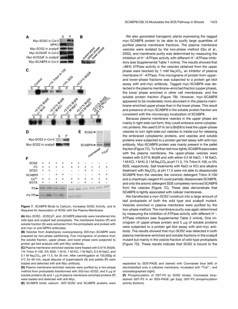
We also generated transgenic plants expressing the tagged
myc-SCABP8 protein to be able to purify large quantities of
purified plasma membrane fractions. The plasma membrane
vesicles were isolated by the two-phase method (Qiu et al.,
2002), and membrane purity was determined by measuring the
inhibition of Hþ-ATPase activity with different Hþ-ATPase inhib-
itors (see Supplemental Table 1 online). The results showed that
>88% ATPase activity in the vesicles obtained from the upper
phase were blocked by 1 mM Na3VO4, an inhibitor of plasma
membrane Hþ-ATPase. Five micrograms of protein from upper-
and lower-phase fractions was subjected to a protein gel blot
assay with anti-myc antibody. Tagged myc-SCABP8 was de-
tected in the plasma membrane–enriched fraction (upper phase),
the lower phase enriched in other cell membranes, and the
soluble protein fraction (Figure 7B). However, myc-SCABP8
appeared to be moderately more abundant in the plasma mem-
brane–enriched upper phase than in the lower phase. This result
and presence of myc-SCABP8 in the soluble protein fraction are
consistent with the microscopy localization of SCABP8.
Because plasma membrane vesicles in the upper phase are
mainly in right-side-out form, they could embrace some cytoplas-
mic proteins. We used 0.01% (w/v) Brij58 to treat the upper-phase
vesicles to turn right-side-out vesicles to inside-out for releasing
the embraced cytoplasmic proteins, and vesicles and soluble
proteins were subjected to a protein gel blot assay with anti-myc
antibody. Myc-SCABP8 protein was mainly present in the pellet
fraction (Figure 7C). To further test how tightly SCABP8 associates
with the plasma membrane, the upper-phase vesicles were
treated with 0.01% Brij58 and with either 0.5 M NaCl, 1 M NaCl,
1 M KCl, 1 M KI, 0.1 M Na2CO3 at pH 11.5, 1% Triton X-100, or 3%
SDS, respectively. Salt treatments with NaCl or KCl and alkaline
treatment with Na2CO3 at pH 11.5 were not able to disassociate
SCABP8 from the vesicles; the nonionic detergent Triton X-100
and a chaotropic reagent KI could partially disassociate SCABP8,
and only the anionic detergent SDS completely removed SCABP8
from the vesicles (Figure 7C). These data demonstrate that
SCABP8 is tightly associated with cellular membranes.
We transfected a myc-SOS2 construct into a large amount of
leaf protoplasts of both the wild type and scabp8 mutant.
Vesicles enriched in plasma membrane were purified by the
two-phase method. The membrane purity was again determined
by measuring the inhibition of ATPase activity with different Hþ-
ATPase inhibitors (see Supplemental Table 2 online). One mi-
crogram of upper-phase protein and 5 mg of soluble proteins
were subjected to a protein gel blot assay with anti-myc anti-
body. The results showed that myc-SOS2 was detected in both
plasma membrane–enriched and soluble fractions in the scabp8
mutant but mainly in the vesicle fraction of wild-type protoplasts
(Figure 7D). These results indicate that SOS2 is bound to the
Figure 7. SCABP8 Binds to Calcium, Increases SOS2 Activity, and Is
Required for Association of SOS2 with the Plasma Membrane.
(A) Myc-SOS2, -SOS2DF, and -SCABP8 plasmids were transferred into
wild-type and scabp8 leaf protoplasts. The membrane fraction (P) and
soluble fraction (S) were extracted from the protoplasts and probed with
anti-myc or anti-MPK3 antibodies.
(B) Vesicles from Arabidopsis overexpressing 35S:myc-SCABP8 were
prepared by two-phase partitioning. Five micrograms of proteins from
the soluble fraction, upper phase, and lower phase were subjected to
protein gel blot analysis with anti-Myc antibody.
(C) Plasma membrane–enriched vesicles were treated with 0.01% Brij58,
1% Triton X-100, 3% SDS, 1 M KI, 1 M KCl, 1 M NaCl, 0.5 M NaCl, and
0.1 M Na2CO3, pH 11.5, for 30 min. After centrifugation at 150,000g at
48C for 60 min, equal aliquots of supernatants (S) and pellets (P) were
loaded and detected with anti-Myc antibody.
(D) Plasma membrane–enriched vesicles were purified by a two-phase
method from protoplasts transformed with 35S:myc-SOS2, and 5 mg of
soluble proteins (S) and 1 mg of plasma membrane–enriched proteins (P)
were loaded and detected with anti-Myc.
(E) SCABP8 binds calcium. GST-SOS2 and SCABP8 proteins were
separated by SDS-PAGE and stained with Coomassie blue (left) or
electroblotted onto a cellulose membrane, incubated with 45Ca2þ, and
autoradiographed (right).
(F) Phosphorylation of GST-P3 by SOS2 kinase. Coomassie blue–
stained GST-P3 in an SDS-PAGE gel (top); GST-P3 phosphorylation
activity (bottom).
SCABP8/CBL10 Modulates the SOS Pathway in Shoots 1423
plasma membrane of leaf protoplasts and that it is partially
released in the absence of SCABP8.
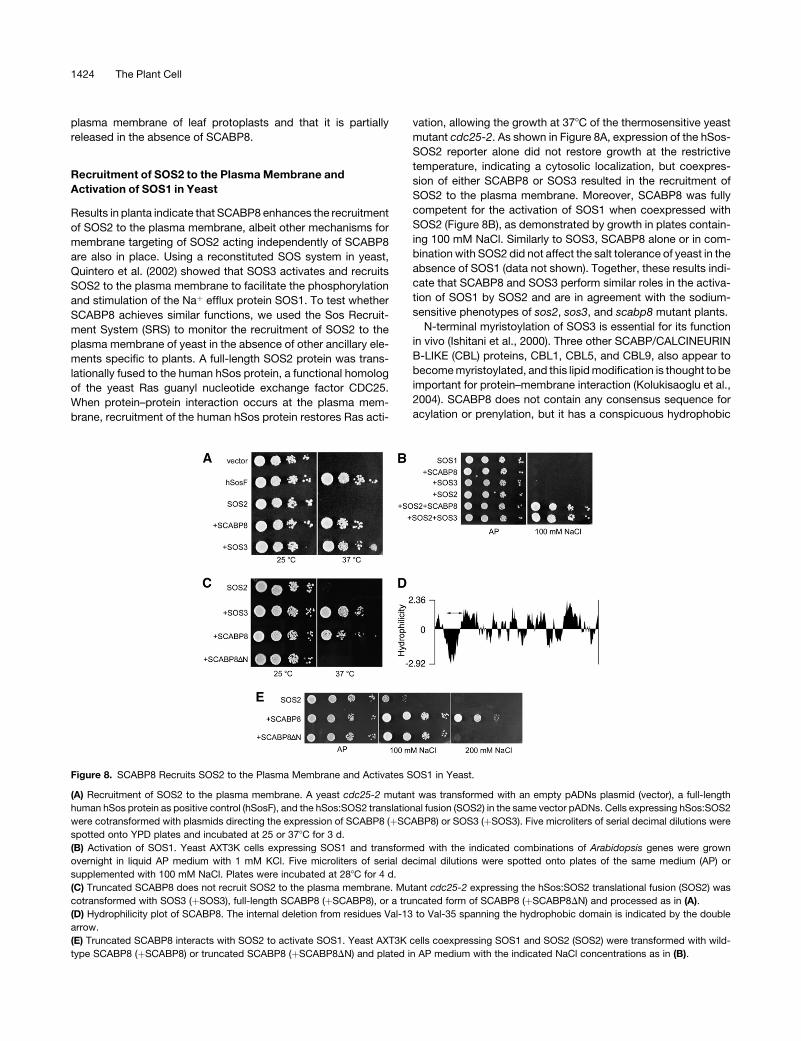
Recruitment of SOS2 to the Plasma Membrane and
Activation of SOS1 in Yeast
Results in planta indicate that SCABP8 enhances the recruitment
of SOS2 to the plasma membrane, albeit other mechanisms for
membrane targeting of SOS2 acting independently of SCABP8
are also in place. Using a reconstituted SOS system in yeast,
Quintero et al. (2002) showed that SOS3 activates and recruits
SOS2 to the plasma membrane to facilitate the phosphorylation
and stimulation of the Naþ efflux protein SOS1. To test whether
SCABP8 achieves similar functions, we used the Sos Recruit-
ment System (SRS) to monitor the recruitment of SOS2 to the
plasma membrane of yeast in the absence of other ancillary ele-
ments specific to plants. A full-length SOS2 protein was trans-
lationally fused to the human hSos protein, a functional homolog
of the yeast Ras guanyl nucleotide exchange factor CDC25.
When protein–protein interaction occurs at the plasma mem-
brane, recruitment of the human hSos protein restores Ras acti-
vation, allowing the growth at 378C of the thermosensitive yeast
mutant cdc25-2. As shown in Figure 8A, expression of the hSos-
SOS2 reporter alone did not restore growth at the restrictive
temperature, indicating a cytosolic localization, but coexpres-
sion of either SCABP8 or SOS3 resulted in the recruitment of
SOS2 to the plasma membrane. Moreover, SCABP8 was fully
competent for the activation of SOS1 when coexpressed with
SOS2 (Figure 8B), as demonstrated by growth in plates contain-
ing 100 mM NaCl. Similarly to SOS3, SCABP8 alone or in com-
bination with SOS2 did not affect the salt tolerance of yeast in the
absence of SOS1 (data not shown). Together, these results indi-
cate that SCABP8 and SOS3 perform similar roles in the activa-
tion of SOS1 by SOS2 and are in agreement with the sodium-
sensitive phenotypes of sos2, sos3, and scabp8 mutant plants.
N-terminal myristoylation of SOS3 is essential for its function
in vivo (Ishitani et al., 2000). Three other SCABP/CALCINEURIN
B-LIKE (CBL) proteins, CBL1, CBL5, and CBL9, also appear to
become myristoylated, and this lipid modification is thought to be
important for protein–membrane interaction (Kolukisaoglu et al.,
2004). SCABP8 does not contain any consensus sequence for
acylation or prenylation, but it has a conspicuous hydrophobic
Figure 8. SCABP8 Recruits SOS2 to the Plasma Membrane and Activates SOS1 in Yeast.
(A) Recruitment of SOS2 to the plasma membrane. A yeast cdc25-2 mutant was transformed with an empty pADNs plasmid (vector), a full-length
human hSos protein as positive control (hSosF), and the hSos:SOS2 translational fusion (SOS2) in the same vector pADNs. Cells expressing hSos:SOS2
were cotransformed with plasmids directing the expression of SCABP8 (þSCABP8) or SOS3 (þSOS3). Five microliters of serial decimal dilutions were
spotted onto YPD plates and incubated at 25 or 378C for 3 d.
(B) Activation of SOS1. Yeast AXT3K cells expressing SOS1 and transformed with the indicated combinations of Arabidopsis genes were grown
overnight in liquid AP medium with 1 mM KCl. Five microliters of serial decimal dilutions were spotted onto plates of the same medium (AP) or
supplemented with 100 mM NaCl. Plates were incubated at 288C for 4 d.
(C) Truncated SCABP8 does not recruit SOS2 to the plasma membrane. Mutant cdc25-2 expressing the hSos:SOS2 translational fusion (SOS2) was
cotransformed with SOS3 (þSOS3), full-length SCABP8 (þSCABP8), or a truncated form of SCABP8 (þSCABP8DN) and processed as in (A).
(D) Hydrophilicity plot of SCABP8. The internal deletion from residues Val-13 to Val-35 spanning the hydrophobic domain is indicated by the double
arrow.
(E) Truncated SCABP8 interacts with SOS2 to activate SOS1. Yeast AXT3K cells coexpressing SOS1 and SOS2 (SOS2) were transformed with wild-
type SCABP8 (þSCABP8) or truncated SCABP8 (þSCABP8DN) and plated in AP medium with the indicated NaCl concentrations as in (B).
1424 The Plant Cell
domain between amino acid residues Val-13 and Val-35, which
is long enough to span across a membrane once (Figure 8D). To
test whether this hydrophobic domain mediates the tight asso-
ciation of SCABP8 with membranes (Figure 7), an internal dele-
tion removing amino acids between Val-13 and Val-35 was
generated in SCABP8 by mutagenic PCR. The truncated protein
(SCABP8DN) became unable to recruit the hSos-SOS2 reporter
protein to the plasma membrane as evidenced by the SRS assay
(Figure 8C). This failure could not be attributed to lack of inter-
action with SOS2 because the truncated protein retained the
ability to activate SOS1 via SOS2 (Figure 8E). A SCABP8DN-hSos
translational fusion failed to show targeting to the plasma
membrane in contrast with wild-type SCABP8 (data not shown).
Thus, the differential salt tolerance conveyed by SCABP8 and
SCABP8DN likely results from the lack of plasma membrane
targeting of the later and reduced activation of SOS1 by a cyto-
plasmic SOS2 protein kinase (Quintero et al., 2002).
SCABP8 Is a Calcium Binding Protein, and Calcium
Enhances SOS2 Kinase Activity
The data suggest thatSCABP8 regulatesSOS2 activity. The cDNA
sequence of SCABP8 corresponds to The Arabidopsis Informa-
tion Resource accession At4g33000.2 and protein sequence
1009053152, which has three predicted EF hand domains. To
test whether SCABP8 could bind calcium, SOS2 and SCABP8
protein were mixed and resolved on a 10% SDS-PAGE gel, and
the proteins were transferred to a cellulose membrane. The mem-
brane was then used to perform a calcium binding assay. SCABP8
was able to bind calcium, but SOS2 did not (Figure 7E). The
interaction of SOS3 with SOS2 does not require calcium, but ac-
tivation of SOS2 by SOS3 is calcium dependent (Halfter et al.,
2000). To quantitate the activation of SOS2, a P3 peptide (Halfter
et al., 2000) was fused to GST in the pGEX-6P-1 vector. GST-
SOS2, GST-SCABP8, GST-SOS3, and GST-P3 proteins were
purified, and kinase activity assays were performed. The results
are shown in Figure 7F. All reactions contained 250 mM calcium.
The SOS2 kinase activity was significantly higher in the absence of
EGTA than with supplemental 500 mM EGTA, demonstrating that
calcium enhances SOS2 activity in the presence of SOS3 or
SCABP8. Neither SCABP8 nor SOS3 showed kinase activity.
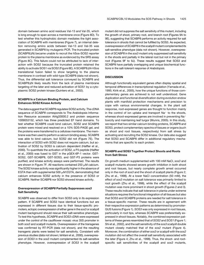
Overexpression of SCABP8 Partially Suppresses sos3
Salt Sensitivity
SCABP8 was observed to differ from SOS3 only in its expression
pattern. If SCABP8 and SOS3 have identical functions but are
expressed in different tissues due to their tissue-specific pro-
moters, ectopic overexpression of either gene in the sos3 or scabp8
mutant background should rescue their salt-sensitive phenotype.
To test this hypothesis, SCABP8 and SOS3 cDNA were expressed
under the control of the cauliflower mosaic virus 35S promoter in
both sos3 and scabp8 mutants. Overexpression of the transgenes
was confirmed by RT-PCR (data not shown), and the resulting
transgenic plants were tested for salt sensitivity. Consistent with
previous studies (data not shown; Ishitani et al., 2000), overexpres-
sion of SOS3 in the sos3 mutant complemented its salt-sensitive
phenotype. However, overexpression of SOS3 in the scabp8
mutant did not suppress the salt sensitivity of this mutant, including
the growth of shoot, primary root, and branch root (Figures 9A to
9E), suggesting that SCABP8 performs an activity required for salt
tolerance in shoots that cannot be fulfilled by SOS3. As expected,
overexpressionofSCABP8 in the scabp8mutantcomplemented its
salt-sensitive phenotype (data not shown). However, overexpres-
sion of SCABP8 in the sos3 mutant only suppressed salt sensitivity
in the shoots and partially in the lateral root but not in the primary
root (Figures 9F to 9J). These results suggest that SOS3 and
SCABP8 have partially overlapping and unique biochemical func-
tions in the salt-tolerant response of Arabidopsis.
DISCUSSION
Although functionally equivalent genes often display spatial and
temporal differences in transcriptional regulation (Yamada et al.,
1995; Kirik et al., 2005), how the unique functions of those com-
plementary genes are achieved is not well understood. Gene
duplication and functional diversification during evolution endow
plants with manifold protection mechanisms and precision to
cope with various environmental changes. In the plant salt
response, root-expressed genes are thought to function mainly
in the control of ion uptake and transport Naþ to the shoots,
whereas shoot-expressed genes are involved in preventing Naþ
toxicity and maintaining leaf turgor (Munns, 2005). In this study,
we report that two similar calcium binding proteins, SCABP8 and
SOS3, protect complementary spectra of tissues or organs (such
as shoot and root tissues, respectively) from salt stress by
activating and recruiting the SOS2 kinase. Our data also suggest
that SOS3 and SCABP8 differ in additional regulatory mecha-
nisms that are specific to each protein.
SCABP8 and SOS3 Together Protect Shoots and Roots
from Salt Stress
On growth medium supplemented with 100 mM NaCl, sos3 and
scabp8 mutants showed severe growth inhibition in both shoot
and root tissues, but nearly complete growth arrest occurred
only in the root of sos3 and the shoot of scabp8 plants (Figure 2;
Zhu et al., 1998). At a lower NaCl concentration (50 mM), the
effect of sos3 mutation on salt tolerance was primarily limited to
root growth (Zhu et al., 1998), while the effect of the scabp8
mutation was more prominent in shoot growth (Figures 2 and 3).
These results indicate that salt tolerance in planta under extreme
situations requires the functional integration of all tissues but also
that SOS3 and SCABP8 proteins are needed for salt tolerance in
a tissue-specific manner. These results are in agreement with
their respective expression patterns as determined by promoter-
GUS fusions (Figure 1). SOS3 was only expressed in root tissues,
particularly in root tips, whereas SCABP8 was preferentially ex-
pressed in shoot tissues. Notably, the combined expression pat-
terns of these genes resembled that of SOS2 and SOS1 (Figure 1;
Shi et al., 2002), and the salt sensitivity of the sos3 scabp8 double
mutant closely matched that of the sos2 mutant (Figure 4).
Moreover, the combination of either sos3 or scabp8 with the sos2
mutation did not affect the overall salt sensitivity associated with
the later (Figure 4; Zhu et al., 1998). Thus, the shoot- and root-
specific salt sensitivities of the scabp8 and sos3 mutants,
SCABP8/CBL10 Modulates the SOS Pathway in Shoots 1425
Figure 9. SCABP8 Partially Suppresses the sos3 Phenotype.
(A) to (E) Five-old-day seedlings of the wild type (left), overexpression of SOS3 in scabp8 (center), and scabp8 (right) were transferred onto MS medium
without (A) or with (B) 75 mM NaCl. Primary root elongation (C), lateral root elongation (D), and shoot fresh weight (E) were measured.
(F) to (J) Five-old-day seedlings of the wild type (left), overexpression of SCABP8 in sos3 (center), and sos3 (right) were transferred onto MS medium
without (F) or with (G) 75 mM NaCl. Primary root elongation (H), lateral root elongation (I), and shoot fresh weight (J) were measured. The pictures were
taken at day 15. Open bars, without NaCl treatment; hatched bars, 75 mM NaCl treatment. Error bars represent SD (n > 15).
1426 The Plant Cell
respectively, at least in part arise from the different expression
patterns of the SCABP8 and SOS3 genes. Genetic interactions
also indicate that SCABP8 and SOS3 regulate SOS2 coordinately.
SCABP8 Partially Associates to Plasma Membranes and
Recruits SOS2
Our results demonstrate that SCABP8 shares most of the bio-
chemical properties of SOS3. Like SOS3, SCABP8 encodes a
protein with EF hand domains that bind calcium in vitro, is
capable of binding to SOS2, and activates SOS2 in a calcium-
dependent manner (Figures 5A and 7). Two-phase partitioning of
microsomal membranes indicated that SCABP8 was associated
with the plasma membrane and cytosol. This bipartite localiza-
tion was corroborated by microscopy inspection of root cells and
leaf protoplasts expressing SCABP8 fused to YFP and its
colocalization with other cytological markers (see Supplemental
Figures 1 and 2 online) and by transient expression of SCABP8
fused to GFP in onion epidermal cells (Figure 6). Based on
biochemical evidence, a fraction of the SCABP8 protein pool was
tightly associated with cell membranes because it could only be
released by treating the membrane vesicles with detergents and
the chaotropic salt KI (Figure 7).
Although SOS3 is known to recruit SOS2 to the plasma
membrane in yeast (Quintero et al., 2002), this function had not
yet been demonstrated in plants. Bimolecular fluorescence
complementation of split-YFP modules fused to SCABP8 and
SOS2 indicated that these two proteins primarily interact at the
plasma membrane (Figure 5B). Consistent with a recruitment role
for SCABP8, SOS2 was also detected at the plasma membrane,
and this localization was partially dependent on the presence of
SCABP8 (see Supplemental Figure 1 online; Figure 7). In the
upper part of seedling roots, where the expression of SOS3 is
below detection (Figure 1), the relative amount of cytosolic YFP-
SOS2 was greater in the scabp8 mutant than in the wild type (see
Supplemental Figure 1 online). In protoplasts prepared from leaf,
where SOS3 promoter expression was also undetectable, tag-
ged myc-SOS2 fractionated with the microsomal pellet in the
wild type but became partly solubilized in the scabp8 mutant
(Figure 7). CBL1 is expressed in leaf tissue, interacts with SOS2,
and localizes to the plasma membrane (Cheong et al., 2003;
Batistic and Kudla, 2004; D’Angelo et al., 2006), which could
account for the SOS2 fraction that remained attached to micro-
somal membranes in the absence of SCABP8. The FISL motif is
required for the interaction of SOS2 with SOS3 and SCABP8
(Figure 5). Deletion of the FISL motif in SOS2 results in the
reduction of SOS2 plasma membrane localization (Figure 7A)
and disrupts SOS2 function in planta (Guo et al., 2004), demon-
strating that recruitment of SOS2 to the plasma membrane is
essential for salt tolerance. Notably, even without the FISL motif,
some of the SOS2DF protein was still bound to the microsomal
fraction in leaf protoplasts of the wild type and scabp8 mutant
(Figure 5A), suggesting that SOS2 can be recruited to cell
membranes independently of SCABP proteins. SOS2 interacts
with the vacuolar Ca2þ/Hþ antiporter CAX1 independently of
SOS3 (Cheng et al., 2004). Presumably, this protein interaction
occurs without involvement of the FISL domain. In summary,
recruitment of SOS2 to cell membranes seems to involve mul-
tiple proteins and mechanisms, both dependent and indepen-
dent of SCABP proteins, and our data clearly suggest that
SCABP8 is instrumental in this process.
The mechanism by which SCABP8 attaches itself to mem-
branes is novel among this class of proteins. SOS3 has an
N-terminal myristoylation signature sequence that is essential for
SOS3 function in salt tolerance, although it remains to be shown
whether this posttranslational modification is required for the
plasma membrane localization of SOS3 or binding to SOS2 in
plant cells (Ishitani et al., 2000; Quintero et al., 2002). CBL1 and
CBL9 also have N-myristoylation consensus sequences and
localize to the plasma membrane (D’Angelo et al., 2006). Inter-
estingly, SCABP8 does not have such a signature sequence for
N-myristoylation or any other recognizable acylation, but it is
nevertheless localized to the plasma membrane in plant and yeast
cells. Removal of an N-terminal hydrophobic domain in SCABP8
preserved the capacity for activation of SOS2 and SOS1 but
abrogated recruitment of SOS2 to the plasma membrane of yeast
(Figure 8). A truncated SCABP8DN protein lacking the N-terminal
hydrophobic domain fused to GFP showed a fluorescence pat-
tern similar to soluble GFP, indicating a predominant cytosolic
localization (Figure 6). These results, together with the tight asso-
ciation of SCABP8 to the plasma membrane (Figure 7), suggest
that the N-terminal part of this protein becomes immersed in the
plasma membrane. However, it still remains to be determined
whether a regulated process is involved in SCABP8 targeting to
the plasma membrane, how the SCABP8-SOS2 complex is
formed, and whether this complex includes the target substrate
for this protein kinase in a ternary conjugate.
Common and Differential Regulatory Mechanisms of
SCABP8 and SOS3
Although capable of performing similar functions in biochemical
and cellular tests, SCABP8 and SOS3 must fulfill distinct regu-
latory functions in the salt stress response of Arabidopsis plants.
SOS3 is expressed in root tissues only, particularly in the root
tips. SOS1, SOS2, and SOS3 are all strongly expressed in this
tissue, suggesting that the SOS cascade plays an important role
in root elongation in response to salt stress. On the other hand,
SCABP8 is mainly expressed in shoot tissues (Figure 1), and
expression of SCABP8 but not SOS3 is slightly induced by salt
stress, indicating that SOS3 and SCABP8 differ in their transcrip-
tional regulation in response to salinity. The Naþ/Hþ exchanger
SOS1 is preferentially expressed in the xylem parenchyma of
shoots and roots, where it is thought to control sodium loading in
the xylem sap (Shi et al., 2002). Activation of SOS2 and SOS1 in
shoot tissues requires components other than SOS3, and our
data suggest that SCABP8 is one of them. Functional analyses in
yeast, together with the specific sodium sensitivity of sos1, sos2,
sos3, and scabp8 mutants, suggest that SOS3 and SCABP8
have an overlapping function in the activation of SOS2 and its
downstream target SOS1. The shoot- and root-specific salt
sensitivities of the scabp8 and sos3 mutants may arise, at least in
part, from the different spatial expression patterns of the SCABP8
and SOS3 genes (Figure 1; Shi et al., 2002).
Like the sos2 and sos3 mutants, scabp8 is hypersensitive to Naþ
and Liþbut not to Csþ (Figure 2; Zhu et al., 1998). The sos2 scabp8,
SCABP8/CBL10 Modulates the SOS Pathway in Shoots 1427
sos2 sos3, and sos3 scabp8 double mutants displayed a sos2-like
phenotype, suggesting that SCABP8 and SOS3 perform similar
functions in regulating SOS2 (Figure 4; Halfter et al., 2000). One
critical difference, however, is that SCABP8 mainly protects shoot
tissues, whereas SOS3 protects root tissues from salt stress. Guo
et al. (2004) have shown that expression of an active form of SOS2,
T/DSOS2, which has a single amino acid Thr-168-to-Asp substi-
tution in the kinase activation loop, suppressed the salt sensitivity
of sos3 mutants only in the shoots, suggesting that both SOS2
kinase activity and precise localization facilitated by SCABP8 are
important for salt tolerance in shoot tissues. Accordingly, deletion
of the FISL domain necessary for interaction with SOS3 abrogated
suppression by T/DSOS2, indicating that activated SOS2 could
bypass the requirement for SOS3 if it retained the ability to bind to
alternative SCABP proteins. Our data indicate that SCABP8 could
possibly fulfill this role. Binding of SOS3 by SOS2 is necessary for
both protein kinase activation and recruitment to the plasma
membrane (Quintero et al., 2002; Guo et al., 2004), and we have
shown that SCABP8 can replace SOS3 in the reconstituted yeast
system (Figure 8), interacts with SOS2 primarily in the plasma
membrane of Arabidopsis protoplasts (Figure 5), and significantly
enhances the fraction of membrane-bound SOS2 (Figure 7).
The question then remains why SOS3 and SCABP8 could not
replace each other in genetic complementation tests (Figure 9).
Several SCABP proteins show preferential interaction with only a
subset of PKS/CIPK partners (Guo et al., 2001; Albrecht et al.,
2003; Kolukisaoglu et al., 2004). SOS3 interacts very specifically,
both biochemically and functionally, with SOS2 (Guo et al., 2001;
Albrecht et al., 2003), but SOS2 also binds SCABP8 (this work),
CBL1, and CBL9 (Kolukisaoglu et al., 2004). Mutants deficient in
SOS3, SCABP8, and CBL1, but not CBL9, are affected in salt
tolerance to various degrees. On the other hand, CBL1 defi-
ciency affects plant responses to drought, salt, and cold stresses
through its interaction with CIPK1, SOS2, and other unidentified
protein kinases, respectively (Albrecht et al., 2003; Batistic and
Kudla, 2004). Thus, different combinations of protein kinase
complexes may generate temporal and spatial specificity sig-
naling, connecting various stimuli to defined cellular responses
(Guo et al., 2001). Presumably, these alternative complexes also
exhibit different specificities toward their targets. Indeed, SOS2
has several known targets that are determinants of salt tolerance
that might involve diverse mechanisms of regulation and recruit-
ment of SOS2 to various subcellular compartments. Together
with SOS3, SOS2 activates SOS1 at the plasma membrane (Qiu
et al., 2002; Quintero et al., 2002). SOS2 also regulates the
activity of vacuolar cation/proton antiporters of the NHX family
(Qiu et al., 2004) and V-ATPase (J.-K. Zhu, personal communi-
cation); the SCABP subunit presumably involved in recruitment
of SOS2 to the tonoplast is presently unknown. Moreover, SOS2
regulates the vacuolar Ca2þ/Hþ exchanger CAX1 by direct
interaction, and no phosphorylation or ancillary SCABP subunit
seem to be required (Cheng et al., 2004). Finally, SOS2 interacts
with the protein phosphatase ABI2, a master regulation of
dehydration stress in plants (Ohta et al., 2003). Any of these
processes in which SOS2 is involved could have an effect on the
salt tolerance imparted by this protein kinase, and these diverse
functions could be regulated differently by SCABP8 and SOS3.
Hence, it is not entirely unexpected that alternative protein
kinase complexes SOS2-SOS3 and SOS2-SCABP8 may share
common target substrates, like SOS1, while affecting differen-
tially distinct targets and processes related to salt stress.
Detailed investigation of such tissue- and process-specific reg-
ulatory mechanisms involved in the salt response will be an
important area for future research.
METHODS
Plant Materials
The homozygous scabp8 mutant was identified from a SALK line (Alonso
et al., 2003) harboring a T-DNA insertion in the seventh intron of
At4g33000 (seed stock no. SALK_056042) using the gene-specific
primers 59-CAGGAGAAGACCGATTGTAAGG-39 and 59-GCGTCGACT-
CAGTCTTCAACCTCAGTG-39 and the T-DNA left border primers Lba1
59-TGGTTCACGTAGTGGGCCATCG-39 and LBb1 59-GCGTGGACCGC-
TTGCTGCAACT-39. Both of the sos2 and sos3 mutants were in the gl1/gl1
Col-0 background. The mutation gl1 did not affect the salt tolerance
phenotype. The sos2 scabp8 and sos3 scabp8 double mutants were
obtained by crossing scabp8 to sos2-2 and sos3, respectively. From F2
plants, scabp8 homozygotes were identified as described above. SOS3
gene-specific primers 59-TCTCATGAATTTGCAGTTGC-39 and 59-AAA-
CTGTTTAATCTGGAGGG-3 (Halfter et al., 2000) were used to amplify
a 112-bp DNA fragment from sos3 and a 121-bp fragment from the
wild type. For sos2-2, primers 59-CTATACGAGCAAGGGAAGAAG-39 and
59-AATGAAGGCGAAGCACAAAC-39 were used for PCR amplification.
The products were 86 bp from sos2-2 and 88 bp from the wild type. All of
the PCR products were separated on a 4% agarose gel.
Salt Sensitivity
Seeds of the mutants and wild-type Col-0 were sterilized in the solution
containing 20% sodium hypochlorite and 0.1% Triton X-100 for 10 min,
washed five times with sterilized water, and sown on MS medium with
0.6% Phytagel (Sigma-Aldrich). The plates were placed at 48C for 2 d, and
then the seeds were germinated vertically at 238C under continual
illumination. Four-day-old seedlings with a root length ;1.5 cm were
transferred onto MS medium with salt added as described.
scabp8 Phenotype Complementation
For scabp8 mutant complementation, a 3783-bp genomic DNA fragment
containing 1736 bp upstream of the translation start site and 469 bp
downstream of the stop codon of SCABP8 was obtained by the digestion
of BAC plasmid F26P21 (ABRC) with XbaI and PstI. The DNA fragment
corresponded to the sequence from 54,144 to 57,927 bp of the BAC
clone. This fragment was then cloned into the XbaI and PstI sites of
pCAMBIA1200 vector. The resultant plasmid 1200-SCABP8CP was
introduced into Agrobacterium tumefaciens GV3101 by electroporation
and transformed to scabp8 mutants by the flower dip method.
DNA Gel Blot Analysis
Genomic DNA was extracted from 6-week-old scabp8 plants using the
Wizard Genomic DNA extraction kit (Promega). For DNA gel blot analysis,
10 mg of genomic DNA was digested with EcoRI, HindIII, and SacI and
fractionated on agarose gel (0.8% [w/v]). The separated DNA was trans-
ferred to Hybond Nþ nucleic acid transfer membranes (Amersham). The
795-bp neomycin phosphotransferase coding sequence (NPTII CDS)
probe was gel purified and radiolabeled with 32P by a random primer
labeling kit (Takara). DNA gel blot hybridization was performed at 428C for
1428 The Plant Cell
16 h using hybridization solution (200 mM sodium phosphate buffer, pH
7.2, 1 mM EDTA, pH 8.0, 50% formamide, 10% BSA, and 7% SDS) with32P-labeled NPTII CDS probes, followed by washing at 658C in 23 SSC
and 0.5% SDS, 13 SSC and 0.5% SDS, and 0.13 SSC and 0.5% SDS for
15 min sequentially. The 32P-labeled membranes were exposed to a
phosphor screen (Amersham). After 12 h of exposure, signals were cap-
tured with a Typhoon 9410 phosphor imager (Amersham).
Promoter GUS Analysis
The plasmid 1200-SCABP8CP was digested with EcoRI and AccIII. The
resulting fragment from 35 to 1736 bp upstream of the translation start
site of SCABP8 was cloned into the EcoRI and XmaI sites of the
pCAMBIA1381Z vector. SOS2 promoter DNA fragment was amplified
with primers 59-CCCAAGCTTCATTAGGGTTCATGGGTTGAG-39 and
59-CGGGATCCTCTTTACAAACTTTTATCTG-39 and cloned into the HindIII
and BamHI sites of pBI101 vector. SOS3 promoter fragment was amplified
with primers 59-CCGCTCGAGAAATCATGTTGGGTCTGATTGG-39 and
59-CGGGATCCACAAACACACCCTTCTCTCAAC-39 and cloned into the
SalI and BamHI sites of pBI101 vector. The plasmids were introduced into
Arabidopsis thaliana by A. tumefaciens–mediated transformation. GUS
assays using transgenic T2 lines at different developmental stages were
performed as described by Jefferson et al. (1987).
RT-PCR Analysis
Total RNA from 10-d-old seedlings and from different organs of plants at
the six-week growth stage was extracted by the TRIzol method. Reverse
transcription was performed using 1 mg of total RNA and MLV reverse
transcriptase (Promega). The cDNA was then used for PCR amplification
with the following primers. For SCABP8, the primers SC8F0 59-CGG-
GATCCATGGAACAAGTTTCCTCTAG-39 and SC8R0 59-GCGTCG-
ACTCAGTCTTCAACCTCAGTG-39 were used. For SOS2, the primers
59-CGGGATCCATGACAAAGAAAATGAGAAG-39 and 59-GTTCCACA-
TGTGGTACGCAGAAGTTC-39 were used. For SOS3, the primers
59-CGGAATTCATGGGCTGCTCTGTATCG-39 and 59-CCGCTCGAGT-
TAGGAAGATACGTTTTGC-39 were used. For UBQ10, the primers
59-ACCGGAAAGACTATCACTTTG-39 and 59- AGAGATAACAGGAACG-
GAAAC-39 were used. To identify the level of SCABP8 transcription in the
SCABP8 mutant and the complementary transgenic plants by RT-PCR,
the primers used were SC8F1 59-GCGCCGTCTTTATACCTTTC-39,
SC8R1 59-ATCGGTCTTCTCCTGGATTG-39, SC8F2 59-GGATGAATGG-
AATGTCTATG-39, and SC8R2 59-AACAAAGCAAAGTGCTTGAC-39.
Real-time PCR was performed with the ABI 7500 real-time PCR system
using SYBR to monitor double-stranded DNA products. For SCABP8, the
primers were 59-AACAAATGGTTTCTGCTATTCTC-39 and 59-ATCTG-
CATCAGCAAATGTTTTATC-39. For SOS2, the primers were 59-CGA-
AACTTCAAGACAAGGCTC-39 and 59-GTGCCACCTCGTAAATCTC-
TATC-39. For SOS3, the primers were 59-GTAATGGTGGATAAGG-
CTTTCG-39 and 59-TGAGCGATGGATTCAAGGATAC-39. For ACT2,
used as an internal control, the primers were 59-GTCGTACAACC-
GGTATTGTG-39 and 59-GAGCTGGTCTTTGAGGTTTC-39. For UBQ10,
another internal control, the primers were 59-CCCTGATGAATAAGT-
GTTCTAC-39 and 59-ACGAAGCGATGATAAAGAAG-39. The relative gene
expression after NaCl treatment was calculated by comparison with that
of the seedlings without NaCl treatment that was defined as 1. SDS soft-
ware version 1.2 (Applied Biosystems) was used for analysis. Standard
deviations were calculated from three independent biological replicates.
Pigment Determination
Leaf chlorophyll was extracted with 80% acetone, the absorbance at 647
and 664 nm was determined by a DU 800 UV/visible spectrophotometer
(Beckman Coulter), and total chlorophyll was calculated (Inskeep and
Bloom, 1985). Anthocyanin was extracted in acid (1% HCl [w/v]) meth-
anol, the absorbance at 530 and 657 nm was determined, and the con-
centration of anthocynanin was calculated by A530-0.25A657 (Rabino
and Mancinelli, 1986).
Pull-Down, Kinase, and Calcium Binding Assays
The coding region of SCABP8 cDNA was amplified from Col-0 using the
primers 59-CGGGATCCATGGAACAAGTTTCCTCTAG-39 and 59-GCG-
TCGACTCAGTCTTCAACCTCAGTG-39, containing BamHI and SalI sites,
respectively. Two PCR bands were separated after electrophoresis. Both
PCR products were purified and digested with BamHI and SalI and cloned
into the pGEX-6P-1 vector. Sequence analysis revealed that the se-
quence of the smaller fragment was identical to CBL10 (Luan et al., 2002;
GenBank accession no. AF513507.2). The larger fragment was the same
as AF513507 except that the seventh intron of SCABP8 was not spliced
out. SOS3 was amplified using primers 59-CGGAATTCATGGGCTGCT-
CTGTATCG-39 and 59-CCGCTCGAGTTAGGAAGATACGTTTTGC-39 and
cloned into the EcoRI and XhoI sites of pGEX-6P-1 vector. SOS2 was
amplified using primers 59-CGGGATCCA TGACAAAGAAAATGAGAAG-39
and 59-GTTCCACATGTGGTACGCAGAAGTTC-39. To produce GST-
SOS2DF, two SOS2 cDNA fragments were amplified by the following
primer pairs: 59-CGGGATCCATGACAAAGAAAATGAGAAG-39 and
59-CAAAATCCTGTCGCAGGGGCCCTTCATCATTTCTC-39; 59-GAAGGGC-
CCCTGCGACAGGATTTTGTTAAAAG-39 and 59-GGAATTCTCAAAACG-
TGATTGTTCTGAG-39. The resulting PCR products were used in the
second round of PCR with primers 59-CGGGATCCATGACAAAGAAAAT-
GAGAAG-39 and 59-GGAATTCTCAAAACGTGATTGTTCTGAG-39 to pro-
duce the mutant SOS2 cDNA containing a deletion of 66 bp from 924 to
990 bp (SOS2DF). The SOS2DF was cloned into the pGEX-6P-1 vector at
the BamHI and EcoRI sites.
The GST fusion constructs were transformed into Escherichia coli
strain BL21 (DE3). The recombinant proteins were purified with glutathi-
one sepharose (Amersham Pharmacia) according to the manufacturer’s
protocol.
For the pull-down assay, 5 mg of SCABP8 or SOS3 was incubated with
1 mg each of GST-SOS2 or GST-SOS2DF on glutathione sepharose
beads for 30 min at room temperature in 100 mL of binding buffer (20 mM
Tris-HCl, pH 7.2, 10 mM MgCl2, and 2 mM DTT). After four washes with
binding buffer, the beads were resuspended in 100 mL of loading buffer,
20 mL were run on a 10% SDS-PAGE gel and stained with Commassie
Brilliant Blue R 250, and 1 mL was analyzed by protein gel blotting against
anti-SCABP8. Kinase reaction and calcium binding assays were de-
scribed by Halfter et al. (2000).
Interaction of SCABP8 and SOS2 in Plants (Split-YFP)
SCABP8 was amplified with primers 59-CGGGATCCATGGAACAAG-
TTTCCTCTAG-39 and 59-GCGTCGACGTCTTCAACCTCAGTGTTG-39 and
cloned into the BamHI and SalI sites of a pUC-SPYCE vector (Walter et al.,
2004). SOS2 and SOS2DFISL were amplified with primers 59-CGGGATC-
CATGACAAAGAAAATGAGAAG-39 and 59-GCGTCGACAAACGTGATT-
GTTCTGAGAATC-39 using GST-SOS2 and GST-SOS2DFISL plasmids
as templates, respectively, and then cloned into the BamHI and SalI sites
of a pUC-SPYNE vector. SOS3 was obtained by PCR using primers
59-GCTCTAGAATGGGCTGCTCTGTATCG-39 and 59-CCGCTCGAGG-
GAAGATACGTTTTGCAATTC-39 and cloned into the XbaI and XhoI sites
of pUC-SPYCE vector. These plasmids were purified by CsCl gradient
centri-fugation and transformed into Arabidopsis mesophyll protoplasts
(Sheen, 2001). The YFP fluorescence of protoplasts was assayed 10
to 12 h after transformation under a Zeiss LSM 510 META confocal
microscope.
SCABP8/CBL10 Modulates the SOS Pathway in Shoots 1429
Subcellular Localization of SCABP8 and SOS2
YFP-SCABP8 was constructed by excising the coding region of SCABP8
cDNA with BamHI and SalI from GST-SCABP8 and cloning it into the
binary vector pCAMBIA1205-YFP downstream of YFP. SOS2 was
digested with BamHI and EcoRI from GST-SOS2 and cloned into the
pCAMBIA1205-YFP vector. The resulting constructs were introduced
into A. tumefaciens GV3101 and transformed into Arabidopsis.
The coding sequences of SOS2, SOS2DF, and SCABP8 were trans-
lationally fused downstream of the c-myc tag. The plasmids were purified
by CsCl gradient centrifugation and transformed into Arabidopsis meso-
phyll protoplasts. After overnight incubation, the protoplasts were har-
vested at room temperature for 2 min at 100g, resuspended in 200 mL of
cold lysis buffer containing 20 mM Hepes-KOH, pH 7.1, 13.5% (w/v)
sucrose, 10 mM potassium acetate, 1 mM DTT, and 13 protease inhibitor
mixture (Sigma-Aldrich), and sonicated on ice in short bursts. The total
crude homogenate was further centrifuged for 20 min at 1000g at 48C,
and the supernatant was centrifuged for 60 min at 140,000g at 48C to
generate a total membrane pellet and a soluble protein fraction. The
resulting samples were then analyzed by SDS-PAGE and blotted onto a
polyvinylidene difluoride membrane (Millipore). The blots were probed
with primary antibody anti-cMyc (Sigma-Aldrich; M4439).
Plasma membrane–enriched vesicles were purified by the two-phase
method, and the purity of each fraction was determined as described by
Qiu et al. (2002). Seedlings overexpressing 35SP:myc-SCABP8 and
protoplasts transfected with 35SP:myc-SOS2 were used for two-phase
partitioning. Plasma membrane vesicles from seedlings overexpressing
35S:myc-SCABP8 were treated with 0.01% Brij58, 1% Triton X-100, 3%
SDS, 1 M KI, 1 M KCl, 1 M NaCl, 0.5 M NaCl, and 0.1 M Na2CO3, pH 11.5
(Overvoorde and Grimes, 1994). After centrifugation at 150,000g at 48C
for 60 min, aliquots of supernatants and pellets were loaded onto an
SDS-PAGE gel, and myc-SCABP8 and myc-SOS2 were detected with
anti-myc antibody.
The intracellular localization of SCABP8 was also determined by mon-
itoring the transient expression of a SCABP8-GFP translational fusion in
onion epidermal cells after DNA particle bombardment, essentially as
described by Yokoi et al. (2002). The coding regions of SCABP8 and
SCABP8DN in which the N-terminal hydrophobic domain had been
removed were amplified with primers 59-ACGTACCATGGAACAAGTT-
TCCTCTAGAT-39 and 59-ATAGCCCATGGCGTCTTCAACCTCAGTGTT-
GAA-39 that included NcoI restriction sites for subcloning into plasmid
pGFP-JS in frame with GFP (Sheen et al., 1995).
Overexpression of SCABP8 and Its Splicing Variant and
Overexpression of SOS3 in scabp8 and sos3
The SCABP8 open reading frame was digested from its GST fusion vector
with BamHI and SalI. To produce the truncated mis-spliced SCABP8
variant, the fragment was obtained by PCR with primers 59-CGGGA-
TCCTGGAACAAGTTTCCTCTAG-39 and 59-GCGTCGACACTAGTTCAA-
GAACTCACTTTATCAATAATC-39 harboring BamHI or SalI sites. Both of
the fragments were inserted into the pCAMBIA1205 vector under the
cauliflower mosaic virus 35S promoter, respectively. GST-SOS3 plasmid
DNA was digested with XhoI and then partially digested with BamHI and
cloned into the pCAMBIA1205 vector. These plasmids were introduced into
A. tumefaciens GV3101 and transformed into Arabidopsis.
Yeast Methods
Saccharomyces cerevisiae strains AXT3K (ena1::HIS3::ena4, nha1::
LEU2, and nhx1::KanMX4) and JP890 (ena1::HIS3::ena4, nha1::LEU2,
nhx1::KanMX, and CYC1::[PGK1:SOS1:CYC1]) have been described
previously (Quintero et al., 2002; Guo et al., 2004). Sodium tolerance
tests were performed in the alkali cation-free medium AP (8 mM phos-
phoric acid, 10 mM L-Arg, 2 mM MgSO4, 0.2 mM CaCl2, 2% glucose, plus
vitamins and trace elements, brought to pH 6.5 with Arg). Aliquots (5 mL)
were spotted onto AP plates supplemented with 1 mM KCl, with or
without 100 mM NaCl, and grown for 3 to 4 d at 288C. Plasmids for the
expression of SOS2 and SOS3 in yeast have been described elsewhere
(Quintero et al., 2002; Guo et al., 2004). Plasmid pSCABP8 was created
by subcloning a 0.7-kb BamHI-SalI fragment spanning the entire open
reading frame of SCABP8 into the BamHI-SalI sites of pYPGE15. An
internal deletion spanning from residues Val-13 to Val-35, both included,
was constructed in SCABP8 to remove an N-terminal hydrophobic
domain. First, the region upstream of the deleted fragment was amplified
by PCR with primers 59-CCTGGTATCTTTATAGTCCTG-39, annealing to
the vector plasmid, and 59- ATCGAAGCATTGTCCAGTGAGAGAGCTT-
GATCTAG-39. The downstream fragment was also amplified with primers
59-TCAAGCTCTCTCACTGGACAATGCTTCGATTGCCG-39 and 59-TCG-
AGGTCGACTCAGTCTTC-39, both annealing to SCABP8. Both PCR
products were combined and reamplified with the first and fourth primers
to render a read-through fragment bearing the Val-13–Val-35 deletion
in SCABP8. The resulting coding region, SCABP8DN, was cloned in
pYPGE15 as described above to produce plasmid pSCABP8DN. To
perform the SRS analysis, the S. cerevisiae strain cdc25-2 (MATalfa,
cdc25-2, ade2, his3, leu2, lys2, trp1, ura3) was transformed with plasmids
pSRS2-1 (hSos:SOS2), pSCABP8, pSCABP8DN, and pSOS3-2 (Quintero
et al., 2002; Guo et al., 2004). As controls, the empty vector pADNS and
pADNS-59SosF encoding a full-length human Sos protein were used
(Aronheim et al., 1997). Mutation cdc25-2 creates a conditional lethal
phenotype at 378C unless the reporter human protein hSos reaches the
plasma membrane. Cell viability at 378C was determined in YPD plates
(1% yeast extract, 2% peptone, and 2% glucose).
Accession Numbers
Sequence data from this article can be found in the GenBank/EMBL data
libraries under the following accession numbers: ACT2, At3g18780;
SCABP8, At4g33000; SOS1, At2g01980; SOS2, At5g35410; SOS3,
At5g24270; and UBQ10, At4g05320.
Supplemental Data
The following materials are available in the online version of this article.
Supplemental Figure 1. Enhanced Plasma Membrane Localization of
SOS2 by SCABP8.
Supplemental Figure 2. Transient Expression of Fluorescent Protein–
Tagged SCABP8, SOS2, PIP2a, and AtVAM3 Genes in Protoplasts.
Supplemental Table 1. Determining the Purity of Vesicles from
Leaves.
Supplemental Table 2. Determination the Purity of Vesicles from
Protoplasts.
ACKNOWLEDGMENTS
We thank Xing Wang Deng, Jianmin Zhou, and Valerie Karplus for
critical reading of the manuscript and stimulating discussions, Liqin Fu,
Cheng Zhan, and Jun Zhang for excellent technical assistance, Jijie Chai
for providing the GST-P3 plasmid, Jie Le for PIP2a-GFP and acidic
ribosomal protein-GFP transgenic seeds, and the ABRC (Ohio State
University) for the T-DNA insertion lines. This work was supported by
National Basic Research Program of China Grant 2006CB100100 and
National High Technology Research and Development Program of
China 863 Grant 2003AA210100 to Y.G. and S.C. and by Spanish
Ministry of Education and Science Grant BFU2006-06968 to J.M.P.
1430 The Plant Cell

Received March 14, 2006; revised March 18, 2007; accepted April 3,
2007; published April 20, 2007.
REFERENCES
Albrecht, V., Weinl, S., Blazevic, D., D’Angelo, C., Batistic, O.,
Kolukisaoglu, U., Bock, R., Harter, K., and Kudla, J. (2003). The
calcium sensor CBL1 integrates plant responses to abiotic stresses.
Plant J. 36: 457–470.
Alonso, J.M., et al. (2003). Genome-wide insertional mutagenesis of
Arabidopsis thaliana. Science 301: 653–657.
Aronheim, A., Zandi, E., Hennemann, H., Elledge, S.J., and Karin, M.
(1997). Isolation of an AP-1 repressor by a novel method for detecting
protein-protein interactions. Mol. Cell. Biol. 17: 3094–3102.
Batistic, O., and Kudla, J. (2004). Integration and channeling of calcium
signaling through the CBL calcium sensor/CIPK protein kinase net-
work. Planta 219: 915–924.
Cheng, N.-H., Pittman, J.K., Zhu, J.-K., and Hirschi, K.D. (2004). The
protein kinase SOS2 activates the Arabidopsis Hþ/Ca2þ antiporter
CAX1 to integrate calcium transport and salt tolerance. J. Biol. Chem.
279: 2922–2926.
Cheong, Y.H., Kim, K.N., Pandey, G.K., Gupta, R., Grant, J.J.,
and Luan, S. (2003). CBL1, a calcium sensor that differentially regu-
lates salt, drought, and cold responses in Arabidopsis. Plant Cell 15:
1833–1845.
Cutler, S.R., Ehrhardt, D.W., Griffitts, J.S., and Somerville, C.R.
(2000). Random GFP::cDNA fusions enable visualization of subcellular
structures in cells of Arabidopsis at a high frequency. Proc. Natl.
Acad. Sci. USA 97: 3718–3723.
D’Angelo, C., et al. (2006). Alternative complex formation of the Ca2þ-
regulated protein kinase CIPK1 controls abscisic acid-dependent and
independent stress responses in Arabidopsis. Plant J. 48: 857–872.
Guo, Y., Halfter, U., Ishitani, M., and Zhu, J.-K. (2001). Molecular
characterization of functional domains in the protein kinase SOS2 that
is required for plant salt tolerance. Plant Cell 13: 1383–1400.
Guo, Y., Qiu, Q., Quintero, F.J., Pardo, J.M., Ohta, M., Zhang, C.,
Schumaker, K.S., and Zhu, J.-K. (2004). Transgenic evaluation of
activated mutant alleles of SOS2 reveals a critical requirement for its
kinase activity and C-terminal regulatory domain for salt tolerance in
Arabidopsis thaliana. Plant Cell 16: 435–449.
Halfter, U., Ishitani, M., and Zhu, J.-K. (2000). The Arabidopsis
SOS2 protein kinase physically interacts with and is activated by
the calcium-binding protein SOS3. Proc. Natl. Acad. Sci. USA 97:
3735–3740.
Inskeep, W.P., and Bloom, P.R. (1985). Extinction coefficients of
chlorophyll a and b in N,N-dimethylformamide and 80% acetone.
Plant Physiol. 77: 483–485.
Ishitani, M., Liu, J., Halfter, U., Kim, C.-S., Wei, M., and Zhu, J.-K.
(2000). SOS3 function in plant salt tolerance requires myristoylation
and calcium binding. Plant Cell 12: 1667–1677.
Jefferson, R.A., Kavanagh, T.A., and Bevan, M.W. (1987). GUS
fusions: Beta-glucuronidase as a sensitive and versatile gene fusion
marker in higher plants. EMBO J. 6: 3901–3907.
Kirik, V., Lee, M.M., Wester, K., Herrmann, U., Zheng, Z., Oppenheimer,
D., Schiefelbein, J., and Hulskamp, M. (2005). Functional diversifica-
tion of MYB23 and GL1 genes in trichome morphogenesis and initiation.
Development 132: 1477–1485.
Kolukisaoglu, U., Weinl, S., Blazevic, D., Batistic, O., and Kudla, J.
(2004). Calcium sensors and their interacting protein kinases: Ge-
nomics of the Arabidopsis and rice CBL-CIPK signaling networks.
Plant Physiol. 134: 43–58.
Liu, J., Ishitani, M., Halfter, U., Kim, C.-S., and Zhu, J.-K. (2000). The
Arabidopsis thaliana SOS2 gene encodes a protein kinase that is
required for salt tolerance. Proc. Natl. Acad. Sci. USA 97: 3730–3734.
Liu, J., and Zhu, J.-K. (1998). A calcium sensor homolog required for
plant salt tolerance. Science 280: 1943–1945.
Luan, S., Kudla, J., Rodriguez-Concepcion, M., Yalovsky, S., and
Gruissem, W. (2002). Calmodulins and calcineurin B-like proteins:
Calcium sensors for specific signal response coupling in plants. Plant
Cell 14: S389–S400.
Munns, R. (2005). Genes and salt tolerance: Bringing them together.
New Phytol. 167: 645–663.
Ohta, M., Guo, Y., Halfter, U., and Zhu, J.-K. (2003). A novel domain in
the protein kinase SOS2 mediates interaction with the protein phos-
phatase 2C ABI2. Proc. Natl. Acad. Sci. USA 100: 11771–11776.
Overvoorde, P.J., and Grimes, H.D. (1994). Topographical analysis of
the plasma membrane-associated sucrose binding protein from soy-
bean. J. Biol. Chem. 269: 15154–15161.
Qiu, Q.S., Guo, Y., Dietrich, M.A., Schumaker, K.S., and Zhu, J.-K.
(2002). Regulation of SOS1, a plasma membrane Naþ/Hþ exchanger
in Arabidopsis thaliana, by SOS2 and SOS3. Proc. Natl. Acad. Sci.
USA 99: 8436–8441.
Qiu, Q.S., Guo, Y., Quintero, F.J., Pardo, J.M., Schumaker, K.S., and
Zhu, J.K. (2004). Regulation of vacuolar Naþ/Hþ exchange in Arabi-
dopsis thaliana by the SOS pathway. J. Biol. Chem. 279: 207–215.
Quintero, F.J., Ohta, M., Shi, H., Zhu, J.-K., and Pardo, J.M. (2002).
Reconstitution in yeast of the Arabidopsis SOS signaling pathway for
Naþ homeostasis. Proc. Natl. Acad. Sci. USA 99: 9061–9066.
Rabino, I., and Mancinelli, A.L. (1986). Light, temperature, and antho-
cyanin production. Plant Physiol. 81: 922–924.
Sheen, J. (2001). Signal transduction in maize and Arabidopsis meso-
phyll protoplasts. Plant Physiol. 127: 1466–1475.
Sheen, J., Hwang, S., Niwa, Y., Kobayashi, H., and Galbraith, D.W.
(1995). Green-fluorescent protein as a new vital marker in plant cells.
Plant J. 8: 777–784.
Shi, H., Ishitani, M., Kim, C.-S., and Zhu, J.-K. (2000). The Arabidopsis
thaliana salt tolerance gene SOS1 encodes a putative Naþ/Hþ anti-
porter. Proc. Natl. Acad. Sci. USA 97: 6896–6901.
Shi, H., Quintero, F.J., Pardo, J.M., and Zhu, J.-K. (2002). The putative
plasma membrane Naþ/Hþ antiporter SOS1 controls long-distance
Naþ transport in plants. Plant Cell 14: 465–477.
Uemura, T., Yoshimura, S.H., Takeyasu, K., and Sato, M.H. (2002).
Vacuolar membrane dynamics revealed by GFP-AtVam3 fusion pro-
tein. Genes Cells 7: 743–753.
Walter, M., Chaban, C., Schutze, K., Batistic, O., Weckermann, K.,
Nake, C., Blazevic, D., Grefen, C., Schumacher, K., Oecking, C.,
Harter, K., and Kudla, J. (2004). Visualization of protein interactions
in living plant cells using bimolecular fluorescence complementation.
Plant J. 40: 428–438.
Yamada, S., Katsuhara, M., Kelly, W.B., Michalowski, C.B., and
Bohnert, H.J. (1995). A family of transcripts encoding water channel
proteins: Tissue-specific expression in the common ice plant. Plant
Cell 7: 1129–1142.
Yokoi, S., Quintero, F.J., Cubero, B., Ruiz, M.T., Bressan, R.A.,
Hasegawa, P.M., and Pardo, J.M. (2002). Differential expression and
function of Arabidopsis thaliana NHX Naþ/Hþ antiporters in the salt
stress response. Plant J. 30: 529–539.
Zhu, J.-K., Liu, J., and Xiong, L. (1998). Genetic analysis of salt
tolerance in Arabidopsis: Evidence for a critical role of potassium
nutrition. Plant Cell 10: 1181–1192.
SCABP8/CBL10 Modulates the SOS Pathway in Shoots 1431

DOI 10.1105/tpc.106.042291; originally published online April 20, 2007; 2007;19;1415-1431Plant Cell
Shang, Shouyi Chen, José M. Pardo and Yan GuoRuidang Quan, Huixin Lin, Imelda Mendoza, Yuguo Zhang, Wanhong Cao, Yongqing Yang, Mei
Shoots from Salt StressArabidopsisSCABP8/CBL10, a Putative Calcium Sensor, Interacts with the Protein Kinase SOS2 to Protect
This information is current as of September 25, 2014
Supplemental Data http://www.plantcell.org/content/suppl/2007/04/16/tpc.106.042291.DC1.html
References http://www.plantcell.org/content/19/4/1415.full.html#ref-list-1
This article cites 35 articles, 26 of which can be accessed free at:
Permissions https://www.copyright.com/ccc/openurl.do?sid=pd_hw1532298X&issn=1532298X&WT.mc_id=pd_hw1532298X
eTOCs http://www.plantcell.org/cgi/alerts/ctmain
Sign up for eTOCs at:
CiteTrack Alerts http://www.plantcell.org/cgi/alerts/ctmain
Sign up for CiteTrack Alerts at:
Subscription Information http://www.aspb.org/publications/subscriptions.cfm
is available at:Plant Physiology and The Plant CellSubscription Information for
ADVANCING THE SCIENCE OF PLANT BIOLOGY © American Society of Plant Biologists