Morphological Identification of Edible Termites (Isoptera) in ...
Upload
independentCategory
view
4download
0
Molecular and Cellular Biochemistry 281: 197–209, 2006. c�Springer 2006
Safety and whole-body antioxidant potentialof a novel anthocyanin-rich formulationof edible berries
Debasis Bagchi,1,2 Sashwati Roy,3 Viren Patel,3 Guanglong He,4
Savita Khanna,3 Navdeep Ojha,3 Christina Phillips,3 Sumona Ghosh,3
Manashi Bagchi2 and Chandan K. Sen3
1Department of Pharmacy Sciences, School of Pharmacy and Health Professionals, Creighton University Medical Center,Omaha, NE; 2InterHealth Research Center, Benicia, CA; 3Department of Surgery, Davis Heart and Lung Research Institute,The Ohio State University Medical Center, Columbus, Ohio, USA; 4Department of Medicine, Davis Heart and Lung ResearchInstitute, The Ohio State University Medical Center, Columbus, Ohio, USA
Received 18 May 2005; accepted 18 July 2005
Abstract
Edible berry extracts rich in anthocyanins possess a broad spectrum of therapeutic, pharmacologic and anti-carcinogenic proper-ties. Six berry extracts (wild blueberry, bilberry, cranberry, elderberry, raspberry seeds and strawberry), singly and in combination,were studied in our laboratories for antioxidant efficacy, cytotoxic potential, cellular uptake and anti-angiogenic properties. Com-binations of edible berry extracts were evaluated to develop a synergistic formula, OptiBerry, which exhibited high oxygen radicalabsorbance capacity (ORAC) value, low cytotoxicity and superior anti-angiogenic properties compared to the other combinationstested. The current study sought to determine the broad spectrum safety and antioxidant potential of OptiBerry in vivo. Acute oralLD50 of OptiBerry was greater than 5 g/kg in rats. Acute dermal LD50 of OptiBerry was greater than 2 g/kg. No changes in thebody weight or adverse effects were observed following necropsy. Primary skin and eye irritation studies were conducted in NewZealand albino rabbits. OptiBerry was classified as slightly irritating to the skin (primary skin irritation index 0.3) and minimallyirritating to the eye (maximum mean total score 6.0). The antioxidant potential of OptiBerry was investigated in rats and mice byassessing GSH redox status in tissues as well as by a unique state-of-the-art electron paramagnetic resonance (EPR) imaging ofwhole-body redox status. A clinically relevant hyperbaric oxygen (HBO) exposure system (2 atm, 2 h) was employed to study theantioxidant properties of OptiBerry. OptiBerry feeding (8 weeks) significantly prevented HBO-induced GSH oxidation in the lungand liver of vitamin E-deficient Sprague Dawley rats. Furthermore, OptiBerry-fed mice, when exposed to HBO, demonstrated sig-nificant protection in whole-body HBO-induced oxidation compared to the unfed controls by EPR imaging. Taken together, theseresults indicate that OptiBerry is reasonably safe and possess antioxidant properties. (Mol Cell Biochem 281: 197–209, 2006)
Key words: berry anthocyanins, glutathione, hyperbaric oxygen, in vivo, OptiBerry, oxidative stress, safety studies
Introduction
Nutrition is a major tool in health preservation and diseaseprevention. The therapeutic property of edible berries has
Address for offprints: D. Bagchi, Department of Pharmacy Sciences, Creighton University Medical Center, 2500 California Plaza, Omaha, NE 68178, USA(E-mail: [email protected])
been long known [1]. More recently, it has been observedthat edible berries may have potent chemopreventive prop-erties [2–6]. Berries are rich in anthocyanins, flavonoid gly-cosides, responsible for the red, violet, purple and blue color
198
of the fruit. Dietary consumption of anthocyanin improvethe overall antioxidant defense status of human plasma [7].On one hand, the search is on for specific medical drugsthat would efficiently limit angiogenesis [8, 9]. On the otherhand, diet-based approaches to limit angiogenesis are be-ing actively explored [2, 3, 10–15]. Recently, we studied theanti-angiogenic properties of several edible berries and de-veloped a mixture (OptiBerry) of such extracts that exhibitpotent effects in vitro [16]. Next, we developed a novel invivo model to study the mechanisms regulating angiogenicoutcomes [17, 18]. OptiBerry proved to be clearly effectivein limiting angiogenesis in vivo by regulating the expres-sion of inducible MCP-1 [17–19]. These favourable devel-opments with OptiBerry led to the current study aimed attesting the safety and antioxidant efficacy of edible berriesin vivo.
Safety is a key concern for dietary supplements [20]. Al-though OptiBerry represents a novel formulation wholly de-rived from edible berries [16], we undertook to systematicallytest OptiBerry for safety in vivo. Edible berries are knownto possess potent antioxidant properties [21]. Beyond test-ing the safety of OptiBerry, we tested whether the berry mixwas able to protect against oxidant insult in vivo. Hyperbaricoxygen (HBO) therapy poses risk of oxygen toxicity [22].We employed a clinically relevant HBO exposure system toinvestigate the antioxidant properties of OptiBerry. The ef-fect of OptiBerry feeding for 8 weeks on HBO-induced GSHoxidation in the lung and liver of vitamin E deficient SpragueDawley rats was examined. We also employed a state-of-the-art electron paramagnetic resonance (EPR) imaging ap-proach to test the effect of HBO and OptiBerry feeding on thewhole body redox status of mice immediately after exposureto HBO.
Materials and methods
Chemicals
The anthocyanin-rich combination of berry extract,OptiBerry BX-600 (Lot no. 302019, InterHealth Nutraceuti-cals, Benicia, CA), used in this study is a standardised blendof wild blueberry, (Vaccinium angustifolium (fruit) extract),strawberry Fragaria chiloensis (fruit) powder, cranberry Vac-cinium macrocarpon (fruit) powder, wild bilberry Vacciniummyrtillus (fruit) extract, elderberry Sambucus nigra (fruit)extract and raspberry Rubus idaeus (seed) powder (patent-pending). Unless otherwise stated, all the chemicals andbiochemicals used in this study were obtained from SigmaChemical Company (St. Louis, MO). 3-carbamoyl-2,2,5,5-tetramethylpyrrolidine-N-oxyl (carbamoyl-PROXYL) waspurchased from Aldrich Chemical Co., Inc. (Milwaukee, WI).
Animals and treatment
All safety testing were conducted at the Product Safety Lab-oratories (East Brunswick, NJ, USA) in compliance with theGood Laboratory Practices (GLP) as defined in 21 CFR 58,1987: US Food and Drug Administration (FDA) GLP Stan-dards and in accordance with Organization for Economic Co-operation and Development (OECD) Guidelines for Testingof Chemicals, Procedures 402 (February, 1987), 404 (April,2002), 405 (April, 2002), 425 (December, 2001). Detailedanimal protocols are provided in individual toxicological as-sessment.
Acute oral toxicity
The acute oral toxicity evaluation (up and down proce-dure) was conducted with rats to determine the potential ofOptiBerry to produce acute oral toxicity from a single dosevia the oral route in compliance with the good laboratorypractices (GLP) as defined in 21 CFR 58, 1987: US FDAGLP Standards and in accordance with OECD guidelinesfor testing of chemicals, procedure 425, adopted December,2001. Three healthy young adult female, nulliparous and non-pregnant albino Sprague Dawley rats (aged 9–10 weeks old,initial body weight 188–197 g) were obtained from Ace Ani-mals, Inc. (Boyertown, PA), and singly housed in suspendedstainless steel cages with mesh floors which conform to thesize recommendations in the most recent Guide for the Careand Use of Laboratory Animals DHEW (NIH). Litter paperwas placed beneath the cage and was changed at least threetimes per week. The rats had free access to standard rat chow(Purina Rodent Chow# 5012) and filtered tap water ad libi-tum, and maintained at a controlled temperature (20–24 ◦C)and light cycle (12 h light/12 h dark). The animals were ac-climated to the laboratory conditions for at least 10–14 daysprior to initiation of dosing.
Before each dosing, rats were fasted overnight and exam-ined through the fasting period for health and weight (ini-tial). Individual doses were calculated based on initial bodyweights at a dose level of 5000 mg/kg. The test substancewas administered as a 25% w/w suspension in distilled waterusing a stainless steel ball-tipped gavage needle. Followingadministration, each animal was returned to its designatedcage and the feed was replaced 3–4 h after dosing. Individualbody weights were recorded again on days 7 and 14 (termina-tion) after dosing. The animals were observed for mortality,signs of gross toxicity and behavioural changes at least oncedaily for 14 days after dosing. Observations included grossevaluation of skin and fur, eyes and mucous membranes, res-piratory, circulatory, autonomic and central nervous systems,somatomotor activity and behavioral pattern. Particular at-tention was directed to observations of tremors, convulsions,
199
salivation, diarrhea and coma. All rats were euthanised byCO2 inhalation at the end of the 14-day observation periodand gross necropsies were performed on all animals. Tissuesand organs of the thoracic and abdominal cavities were ex-amined.
Acute dermal toxicity
The acute dermal toxicity evaluation was conducted in ratsto determine the potential for OptiBerry to produce toxic-ity from a single topical application in compliance with 21CFR 58, 1987: US FDA GLP Standards and in accordancewith OECD guidelines for testing of chemicals, procedure402, adopted February, 1987. Five healthy young adult al-bino Sprague Dawley male rats (aged 10–11 weeks old, initialbody weight 290–307 g) and five young adult female, nulli-parous and non-pregnant albino Sprague Dawley rats (aged10–11 weeks old, initial body weight 200–215 g) were ob-tained from Ace Animals, Inc. (Boyertown, PA), and singlyhoused in suspended stainless steel cages with mesh floorswhich conform to the size recommendations in the mostrecent Guide for the Care and Use of Laboratory AnimalsDHEW (NIH). Litter paper was placed beneath the cage andwas changed at least three times per week. The rats had freeaccess to standard rat chow (Purina Rodent Chow no. 5012)and filtered tap water ad libitum, and maintained at controlledtemperature (19–23 ◦C) and light cycle (12 h light/12 h dark).The animals were acclimated to the laboratory conditions for21 days.
On the day prior to application, the five male and five fe-male animals were prepared by clipping (Oster model no.A5-small) the dorsal area and the trunk. After clipping andprior to application, the animals were examined for healthand weight (initial) and the skin checked for any abnormal-ities. Individual doses were calculated based on the initialbody weights, taking into account the concentration of thetest mixture. Prior to application, OptiBerry was moistenedwith distilled water to achieve a dry paste by preparing a75% w/w mixture. OptiBerry (2 g/kg of body weight) wasthen applied to a 2 in. × 3 in., 4-ply gauze pad and placedon a dose area of approximately 2 in. × 3 in. (approximately10% of the body surface). The gauze pad and entire trunkof each animal were then wrapped with 3 in. Durapore tapeto avoid dislocation of the pad and to minimize loss of thetest substance (OptiBerry). The rats were then returned totheir designated cages. The day of application was consid-ered as day 0 of the study. After 24 h of exposure of the testsubstance (OptiBerry), the pads were removed and the testsites were gently cleansed of any residual test substance. In-dividual body weights of the animals were recorded prior toOptiBerry application (initial) and again on days 7 and 14 (ter-mination). The animals were observed for mortality, signs of
gross toxicity and behavioral changes during the first severalhours after application and at least once daily thereafter for14 days. Observations included gross evaluation of skin andfur, eyes and mucous membranes, respiratory, circulatory,autonomic and central nervous systems, somatomotor activ-ity and behavioral pattern. Particular attention was directedto observations of tremors, convulsions, salivation, diarrheaand coma. All the rats were euthanised via CO2 inhalationon day 14. Gross necropsies were performed on all animalsat terminal sacrifice. Tissues and organs of the thoracic andabdominal cavities were examined.
Primary skin irritation
The primary skin irritation test was conducted on two youngadult male New Zealand albino rabbits and one young nul-liparous non-pregnant female New Zealand albino rabbits(aged 13 weeks old, initial body weight range at receipt 2.3–2.4 kg, estimated body weight at the start of the experimentwas 2.8–3.2 kg) to determine the potential for OptiBerry tocause irritation after a single topical application in compli-ance with 21 CFR 58, 1987: US FDA GLP Standards and inaccordance with OECD guidelines for testing of chemicals,procedure 404, adopted in April of 2002. The rabbits wereobtained from Robinson Services, Inc. (Clemmons, NC), andsingly housed in suspended stainless steel cages with meshfloors which conform to the size recommendations in themost recent Guide for the Care and Use of Laboratory Ani-mals DHEW (NIH). Litter paper was placed beneath the cageand was changed at least three times per week. The rabbitswere allowed free access to lab chow (Purina Rabbit Chow no.5326, St. Louis, MO) and filtered tap water ad libitum, andmaintained at controlled temperature (20–22 ◦C) and lightcycle (12 h light/12 h dark). Animals were acclimated to thelaboratory conditions for a period of 28 days prior to initiationof dosing.
On the day before application, rabbits were prepared byclipping (Oster model no. A5-small) the dorsal area and thetrunk. On the day of dosing, but prior to application, the rab-bits were critically examined for health and the skin checkedfor any abnormalities, and three healthy rabbits without pre-existing skin irritation were selected for the test. Individualdoses were calculated based on the initial body weights, tak-ing into account the concentration of the test mixture. On theday of application (day 0), OptiBerry was moistened with dis-tilled water to achieve a dry paste by preparing a 75% w/wmixture. Five-tenths of a gram of OptiBerry (0.67 g of testmixture) was placed on a 1 in. × 1 in., 4-ply gauze pad andapplied to a 6 cm2 intact dose site on each rabbit. The pad andthe entire trunk of each rabbit were then wrapped with semi-occlusive 3 in. Micropore tape to avoid dislocation of the pad.Elizabethan collars were placed on each rabbit and they were
200
Table 1. Primary dermal irritation study of albino rabbits: Scoring criteria for dermal reactionsa [24]
Evaluation of dermal reactions
Value Erythema and eschar formation Edema formation
0 No erythema No edema
1 Very slight erythema (barely perceptible), edges of area not well defined Very slight edema (barely perceptible) edges of area not well defined
2 Slight erythema (pale red in color and edges definable) Slight edema (edges of area well defined by definite raising)
3 Moderate to severe erythema (defined in color and area well defined) Moderate edema (raised approximately 1 mm)
4 Severe erythema (beet to crimson red) to slight eschar formation (injuries Severe edema (raised more than 1 mm and extending beyond areain depth) of exposure)
4 Total possible erythema score Total possible edema score
aEight total possible primary irritation score.
returned to their designated cages. After 4 h of exposure toOptiBerry, the pads and collars were removed and the testsites were gently cleansed of any residual test substance.
Individual dose sites were scored according to the Draizescoring system (Table 1) [23, 24] at approximately 1, 24,48, and 72 h after patch removal. The classification of ir-ritancy was obtained by adding the average erythema andedema scores for the 1, 24, 48, and 72 h scoring intervals anddividing by the number of evaluation intervals (4). The re-sulting primary dermal irritation index (PDII) was classifiedas shown in Table 2.
The animals were also observed for signs of gross toxicityand behavioural changes at least once daily during the testperiod. Observations included gross evaluation of skin andfur, eyes and mucous membranes, respiratory, circulatory, au-tonomic and central nervous systems, somatomotor activityand behaviour pattern. Particular attention was directed toobservation of tremors, convulsions, salivation, diarrhea andcoma.
Primary eye irritation
The primary eye irritation test was conducted with rabbitsto determine the potential for OptiBerry to produce irritation
Table 2. Descriptive rating for mean primary dermal irritation indexa
Primary Dermal Irritation Index (PDII) Classification
0 No irritation
>0–2.0 Slight irritation
2.1–5.0 Moderate irritation
>5.0 Severe irritation
aUS EPA Addenum 3 on data reporting to pesticide assessment guidelines;dermal irritation, January 1988.
from a single installation via the ocular route in compliancewith 21 CFR 58, 1987: US FDA GLP Standards and in accor-dance with OECD guidelines for testing of chemicals, pro-cedure 405, adopted April, 2002. Three female, nulliparousand non-pregnant New Zealand albino rabbits (aged 13 weeksold, initial body weight range at receipt 2.0–2.3 kg, estimatedbody weight at the start of the experiment 2.6–3.0 kg) wereobtained from Robinson Services, Inc. (Clemmons, NC) andsingly housed in suspended stainless steel cages with meshfloors which conform to the size recommendations in themost recent Guide for the Care and Use of Laboratory Ani-mals DHEW (NIH). Litter paper was placed beneath the cageand was changed at least three times per week. The rabbitswere allowed free access to lab chow (Purina Rabbit Chow no.5326, St. Louis, MO) and filtered tap water ad libitum, andmaintained at controlled temperature (17–24 ◦C) and lightcycle (12 h light/12 h dark). Animals were acclimated to lab-oratory conditions for a period of 22 days prior to initiationof dosing.
Prior to instillation, both the eyes of rabbits were exam-ined using a fluorescein dye procedure. One drop of 2%ophthalmic fluorescein sodium was instilled into both theeyes of each rabbit. The eyes were rinsed with physiologi-cal saline (0.9% NaCl) approximately 30 s after installationof the fluorescein. Using an ultraviolet light source, the eyeswere checked for gross abnormalities according to the “Scalefor Scoring Ocular Lesions” (Table 3) [24]. Three healthyanimals without pre-existing ocular irritation were selectedfor the test. One-tenth of a milliliter (0.04 g) of OptiBerrywas instilled into the conjunctival sac of the right eye ofeach rabbit by gently pulling the lower lid away from theeyeball. The upper and lower lids were then gently held to-gether for about 1 s before releasing, to minimise the loss ofthe test substance. The left (control) eye of each animal re-mained untreated and served as a control. The rabbits werethen returned to their designated cages. Ocular irritation wasevaluated macroscopically using a high-intensity white light
201
Table 3. Scale for scoring ocular irritation [23]
I. Cornea
(A) Opacity-degree of density (area most dense taken for reading)
No ulceration or opacity 0
Dulling of normal luster, details of iris clearly visible 1b
Easily discernible translucent areas, details of iris slightly obscured 2b
Nacreous areas, no details of iris visible, size of pupil rarely discernible 3b
Opaque cornea, iris not discernible through the opacity 4b
(B) Area of cornea involved
No ulceration or opacity 0
One quarter or less but not zero 1
Greater than one quarter, but less than half 2
Greater than half, but less than three quarters 3
Greater than three quarters, up to whole area 4
Total maximum 80a
II. Iris
(A) Values
Normal 0
Markedly deepened rugae, congestion, swelling, circumcorneal injection (any or all of these or combination thereof), iris still reacting to light(sluggish reaction is positive)
1b
No reaction to light, hemorrhage, gross destruction (any or all of these) 2b
Total maximum 10c
III. Conjuctivae
(A) Redness (refers to palpebral and bulbar conjunctivae excluding cornea and iris)
Blood vessels normal 0
Some blood vessels definitely hyperemic (injected above) normal 1
Diffuse, deeper crimson color, individual vessels not easily discernible 2b
Diffuse beefy red 3b
(B) Chemosis: lids and/or nictitating membranes
No swelling 0
Any swelling above normal (includes nictitating membrane) 1
Obvious swelling with partial eversion of lids 2b
Swelling with lids about half closed 3b
Swelling with lids more than half closed 4b
(C) Discharge
No discharge 0
Any amount different from normal (does not include small amounts observed in inner canthus of normal animals) 1
Discharge with moistening of the lids and hairs just adjacent to the lids 2
Discharge with moistening of the lids and hair, and considerable area around the eye 3
Total maximum 20d
Total Maximum Score Possible 110e
aScore equals A × B × 5.bFigures indicate positive effect.cScore equals A × 5.dScore equals (A + B + C) × 2.eTotal Maximum Score Possible (110) represents the sum of all scores obtained for the cornea, iris and conjunctivae.
(MagLite) in accordance with Draize et al. at 1, 24, 48, and72 h post-instillation (Table 3). The fluorescein eye evalua-tion was used at 24 h to verify the absence of corneal damage.Individual irritation scores were recorded for each animal. In
addition to the observations of the cornea, iris and conjunc-tivae, any other lesions were noted. The average score for allrabbits at each scoring period was calculated to aid in datainterpretation. Time intervals with the highest mean score
202
Table 4. Descriptive rating of maximum mean total primary eye irritation scores [25]
Maximum meantotal score Classification Requirement for maintenance of classification
0.0–0.5 No irritation Up to 0.5 at 1 h with zeros at 24 h; otherwise increase one level
0.6–2.5 Practically no irritation With zeros at 24 h; otherwise increase one level
2.6–15.0 Minimal irritation With zeros at 48 h; otherwise increase one level
15.1–25.0 Mild irritation With zeros at 96 h; otherwise increase one level
25.1–50.0 Moderate irritation With 7 days mean ≤20 and individual total scores ≤10 in at least 60% of rabbits with no total score > 30;otherwise, increase one level
50.1–80.0 Severe irritation With 7 days mean ≤40 and individual total scores ≤30 in at least 60% of rabbits with no total score > 60;otherwise increase one level
80.1–100.0 Extreme irritation With 7 days mean ≤80 and individual total scores ≤60 in at least 60% of rabbits with no total score > 100;otherwise increase one level
100.1–110 Maximal irritation With 7 days mean >80 and individual total scores >60 in at least 60% of rabbits; otherwise decrease
one level
(maximum mean total score – MMTS) for all rabbits wereused to classify the test substance (OptiBerry) by the systemof Kay and Calandra (Table 4) [25].
The animals were also observed for signs of gross toxicityand behavioural changes at least once daily during the testperiod. Observations included gross evaluation of skin andfur, eyes and mucous membranes, respiratory, circulatory, au-tonomic and central nervous systems, somatomotor activityand behaviour pattern. Particular attention was directed to theobservation of tremors, convulsions, salivation, diarrhea andcoma.
Evaluation of antioxidant properties
Animals and dietary supplementation protocol for testing an-tioxidant properties. Male and female rats (aged 6 weeks)(Sprague-Dawley Harlan, Indianapolis, IN, USA) were di-vided into the following three groups: (i) E−/HBO (vita-min E deficient, placebo fed, exposed to HBO insult; HBO,24 h prior to tissue harvest exposed to pure O2 at 2 atmfor 2 h) group; male (n = 5) and female (n = 3) rats).IACUC (Institutional Animal Care and Use Committees) ap-proval for this experiment was obtained from the Ohio StateUniversity Medical Center (approved protocol no. 00A0173and 2005A0006). Rats were fed on a vitamin E deficientdiet (TD 88163, Harlan; α-tocopherol levels below detectionlimits). (ii) E−/OptiBerry/HBO group (vitamin E deficient,OptiBerry fed, exposed to HBO insult; male (n = 5) and fe-male (n = 3) rats) (iii) E+/control rats (vitamin E sufficientstandards laboratory diet, not exposed to HBO or not fedwith OptiBerry; male (n = 3) and female (n = 3) rats). TheOptiBerry-fed rats were supplemented with an OptiBerry sus-
pension (40 mg/ml in H2O, aliquoted and stored in −20 ◦C).Body weights were taken weekly and the animals gavagedaccordingly at 20 mg/kg dose for 8 weeks. Placebo rats weregavaged with H2O for 8 weeks as well. At 8 weeks of treat-ment (14 weeks of age) the OptiBerry and placebo treated ratswere exposed to HBO (hyperbaric oxygen). Tissue harvestfollowed 24 h later.
Glutathione assay
GSH was detected using an HPLC coulometric electrode ar-ray detector (CouloArray Detector Model 5600 with 12 chan-nels, ESA Inc., Chelmsford, MA). Sample preparation, mo-bile phase and column used for HPLC glutathione assay wereas previously described [26]. As an improvement to the pre-viously reported methods, the current method implementeda coulometric electrode array detector for the detection ofglutathione. This system uses multiple channels with differ-ent redox potentials [27]. Glutathione was detected at chan-nels set at the following potential: (i) 600, (ii) 700 and (iii)800 mV. Signals from the channel set at 800 mV were usedfor quantification.
Vitamin E extraction and analysis
Vitamin E extraction and analysis from the liver was per-formed as described previously using a HPLC-coulometricelectrode array detector (CouloArray Detector model 5600with 12 channels; ESA Inc., Chelmsford, MA, USA) [27,28]. This system uses multiple channels with different redox-potentials. α-Tocopherol was detected on a channel set at200 mV.
203
EPR imaging of whole-body redox status in mice
A unique, state-of-the-art technique was developed to eval-uate the antioxidant potential of OptiBerry against HBO-induced oxidative stress in mice in a whole-body scenario.Vitamin E deficient mice (aged 8 weeks) (Harlan, Indianapo-lis, IN, USA) were kept on an E-deficient diet as above for 7days before EPR imaging was performed. IACUC approvalfor this experiment was obtained from the Ohio State Uni-versity Medical Center. HBO treated group was subjectedto hyperbaric oxygen at 2 atm pressure for 2 h in a cham-ber before administration of anesthesia, whereas the controlgroup was kept in the room air all the time. Mice in eachgroup were anesthetised by intra-peritoneal injection of ke-tamine (90 mg/kg body weight) and xylazine (20 mg/kg). 3-Carbamoyl PROXYL solution (150 mg/kg of body weight)was injected into the tail vein and the mouse was placed inprone position in a quartz tube of 3.5 cm diameter. EPR spec-tra were immediately taken on the body of the mouse at reg-ular intervals of 4 min and 16 projections were taken for eachtime point for 2D imaging of redox status. The measurementswere done on a custom-built EPR spectrometer [29] with750 MHz microwave bridge unit and a 40 mm diameter loopgap re-entrant resonator. The microwave power was set at32 mW, and the modulation frequency was 100 kHz. Spectralacquisitions were performed using custom-developed dataacquisition software (SPEX) that was capable of automateddata acquisition and recording. For construction of 2D im-ages, 16 projections were taken and the spectral data wasdeconvoluted using the line shape of the zero-gradient spec-trum. Images were reconstructed from the deconvoluted databy filtered back projection [30].
Results
Acute oral toxicity
In the present study, a single oral administration of OptiBerrywas provided to female Sprague Dawley rats to assess itsacute toxicity following Up and Down procedure in accor-dance with the OECD guidelines for testing of chemicals,procedure 425 (adopted December 2001). OptiBerry, at thelimit dose of level of 5000 mg/kg body weight, did not causeany mortality and did not demonstrate any signs of gross tox-icity, adverse pharmacologic effects or abnormal behaviourin the treated female rats following dosing and during theobservation period of 14 days thereafter. All the animals sur-vived, gained normal body weight and appeared active andhealthy during the study. No gross abnormalities or patho-logical alterations were noted for any of the organs of the ratswhen necropsied at the conclusion of the 14-day observationperiod. Based on these results and under the conditions of
this study, the acute oral LD50 of OptiBerry is greater than5000 mg/kg of body weight in female rats.
Acute dermal toxicity
Acute dermal toxicity of OptiBerry was conducted in maleand female Sprague Dawley rats to determine the potentialfor OptiBerry to cause toxicity from a single topical appli-cation in accordance with the OECD guidelines for testingof chemicals, procedure 402 (adopted February 1987). Allanimals survived, gained normal body weight, and appearedactive and healthy during the study. There were no signs ofdermal irritation, gross toxicity, adverse pharmacologic ef-fects or abnormal behaviour. No gross abnormalities werenoted for any of the animals when necropsied at the conclu-sion of the 14-day observation period. Under the conditionsof this study, the single dose acute dermal LD50 of OptiBerryis greater than 2000 mg/kg of body weight in both male andfemale rats.
Primary skin irritation
Primary dermal irritation potential of OptiBerry was evalu-ated within male and female New Zealand albino rabbits toevaluate the potential of OptiBerry to produce irritation aftera single topical application in compliance with 21 CFR 58,1987: US FDA GLP Standards and in accordance with OECDguidelines for testing of chemicals, procedure 404 (adoptedApril, 2002). Following application of OptiBerry, all the an-imals appeared active and healthy. Apart from the dermalirritation noted below, there were no signs of gross toxicity,adverse pharmacologic effects or abnormal behaviour. Onehour after patch removal, very slight erythema was observedat all the three treated sites. The overall incidence and severityof irritation decreased with time. All animals were free fromdermal irritation within 24 h. A summary of Draize primarydermal irritation scoring criteria for dermal reactions is pre-sented in Table 1, while descriptive rating for mean primarydermal irritation index (PDII) is presented in Table 2.
Under the conditions of this study, the PDII for OptiBerrywas calculated to be 0.3, thus classifying OptiBerry to beslightly irritating to the skin (Table 5).
Primary eye irritation
A primary eye irritation test was conducted with New Zealandalbino rabbits to determine the potential for OptiBerry tocause irritation from a single instillation via the ocular routein compliance with 21 CFR 58, 1987: US FDA GLP Stan-dards and in accordance with OECD guidelines for testing of
204
Table 5. Primary dermal irritation scores in male and female New Zealandalbino rabbits after exposure to OptiBerrya
Incidence ofdermal irritation Total PDIb Total PDIIc
Time post
instillation (h) Erythema Edema Mean score Overall Index
1 1.0 0.0 1.0 0.3
24 0.0 0.0 0.0
48 0.0 0.0 0.0
72 0.0 0.0 0.0
aAverage values (n = 3).bPrimary dermal irritation (PDI) = average erythema + average edema.cPrimary dermal irritation index (PDII) = PDI for 1, 24, 48, and 72 h/4.
chemicals, procedure 405, adopted April, 2002. The DraizeScale for scoring eye lesions is presented in Table 3. The Kayand Calandra Scheme for classifying eye irritants are pre-sented in Table 4. All animals appeared active and healthy.There were no signs of gross toxicity, adverse pharmacologiceffects or abnormal behaviour. No corneal opacity or iritiswere observed in any of the treated eye during the study.One hour following OptiBerry instillation, all treated eyesexhibited conjunctivitis (Table 6). Individual eye irritationscores are presented in Table 6. The overall severity of irri-tation decreased with time. All animals were free of ocularirritation within 48 h. Under the conditions of this study, themaximum mean total score (MMTS) of OptiBerry powderis 6.0, classifying OptiBerry to be minimally irritating tothe eye.
Evaluation of antioxidant properties
Hyperbaric oxygen therapy (HBOT) delivers 100% O2 at2–3 atm pressure and patients typically receive 10–30 treat-ments, depending upon the diagnosis. These treatments areusually 60–120 min long, may be given several days a week
Table 6. Incidence, severity and reversibility of ocular irritation in NewZealand albino rabbits after exposure to OptiBerry
Incidence ofocular irritation Severity of irritation
Time post
instillation (h) Corneal opacity Iritis Conjunctivitis MMTa score
1 0/3 0/3 3/3 6.0
24 0/3 0/3 3/3 2.0
48 0/3 0/3 0/3 0.0
72 0/3 0/3 0/3 0.0
aMaximum mean total score (MMTS).
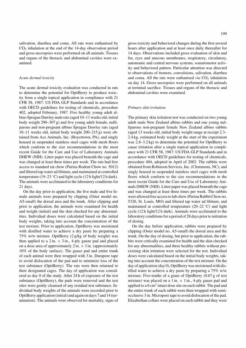
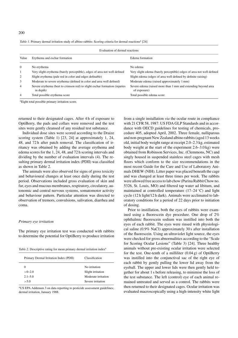
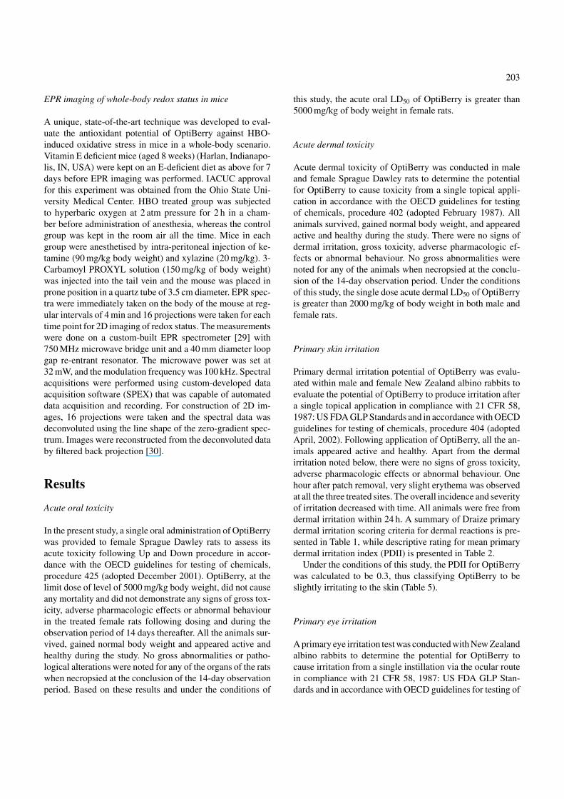
and performed in specialised chambers at facilities withphysician supervision. HBOT is capable of elevating arte-rial pO2 as high as 1200 mmHg. While HBOT may improvethe oxygenation status of several tissues, it poses a clear riskof oxygen toxicity [31, 32]. Like many other risk factorsincluding cigarette smoking, HBOT may not result in imme-diate manifestation of clinical abnormalities in most cases.However, it is general knowledge that exposure of biologicalcells and tissues to pure O2 may result in oxidative stressand genotoxicity [22]. We utilized this in vivo model systemto test the antioxidant efficacy of OptiBerry. The standardlaboratory feed for rodents is heavily enriched with the an-tioxidant vitamin E. Typical levels of vitamin E in the chowexceed the human recommended dietary allowance for vita-min E by over 15-folds. Excessive tissue vitamin E in thetissues of laboratory rodents may pose a problem to studyoxygen toxicity. As a result, we generated vitamin E deficientrats by feeding the rodents with vitamin E deficient diet (Fig.1). HBO treatment resulted in substantial increase in GSSGlevels in the rat lung. Such increase in lung GSSG level wassignificantly lower in OptiBerry-fed male rats compared to
Fig. 1. α-Tocopherol levels in rat liver. Rats were randomly divided intothe following four groups: (i) E+ group – fed on a standard rat chow that isenriched with α-tocopherol (∼200 nmol/g), (ii) E+ group – fed on a standardrat chow that is enriched with α-tocopherol (∼200 nmol/g) and exposed topure O2 at 2 atm for 2 h, (iii) E−; group – fed on a vitamin E deficientdiet treated with placebo and exposed to pure O2 at 2 atm for 2 h and (iv)E−; group – fed on a vitamin E deficient diet treated with OptiBerry andexposed to pure O2 at 2 atm for 2 h. Vitamin E analysis was performedusing HPLC. ∗ P < 0.05 significantly different compared between E+ HBOtreated and E−; HBO treated placebo. • P < 0.05 significantly differentcompared between male E+ control and female E+ control. ◦ P < 0.05significantly different compared between male E+ HBO treated and femaleE+ HBO treated.
205
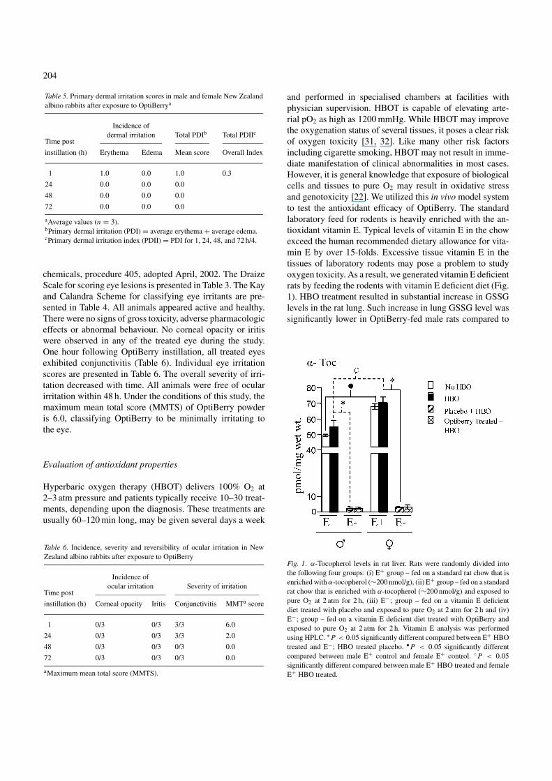
Fig. 2. (A) GSH levels in rat lung. (B) GSSG levels in rat lung. (C) GSH levels in rat liver. (D) GSSG levels on rat liver. Rats were randomly divided into thefollowing three groups: (i) control group – fed a standard rat chow that is enriched with (-tocopherol (∼200 nmol/g) (ii) E− group – fed a vitamin E deficientdiet, exposed to pure O2 at 2 atm for 2 h and treated with a placebo, (iii) E− group – fed a vitamin E deficient diet, exposed to pure O2 at 2 atm for 2 h andtreated with OptiBerry. Glutathione analysis was performed using HPLC. † p < 0.05 significantly different compared between placebo and OptiBerry treatedgroups.
that of placebo-fed controls (Fig. 2B). HBO exposure alsoresulted in substantial oxidation of hepatic GSH to GSSG. Inthe female rats, OptiBerry feeding significantly preventedHBO-induced GSH oxidation (Fig. 2D). These effects ofHBO on GSSG formation were studied 24 h after HBOexposure.
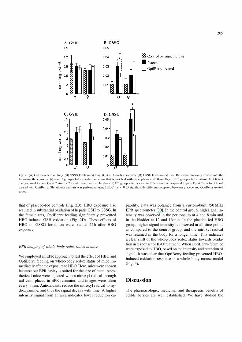
EPR imaging of whole-body redox status in mice
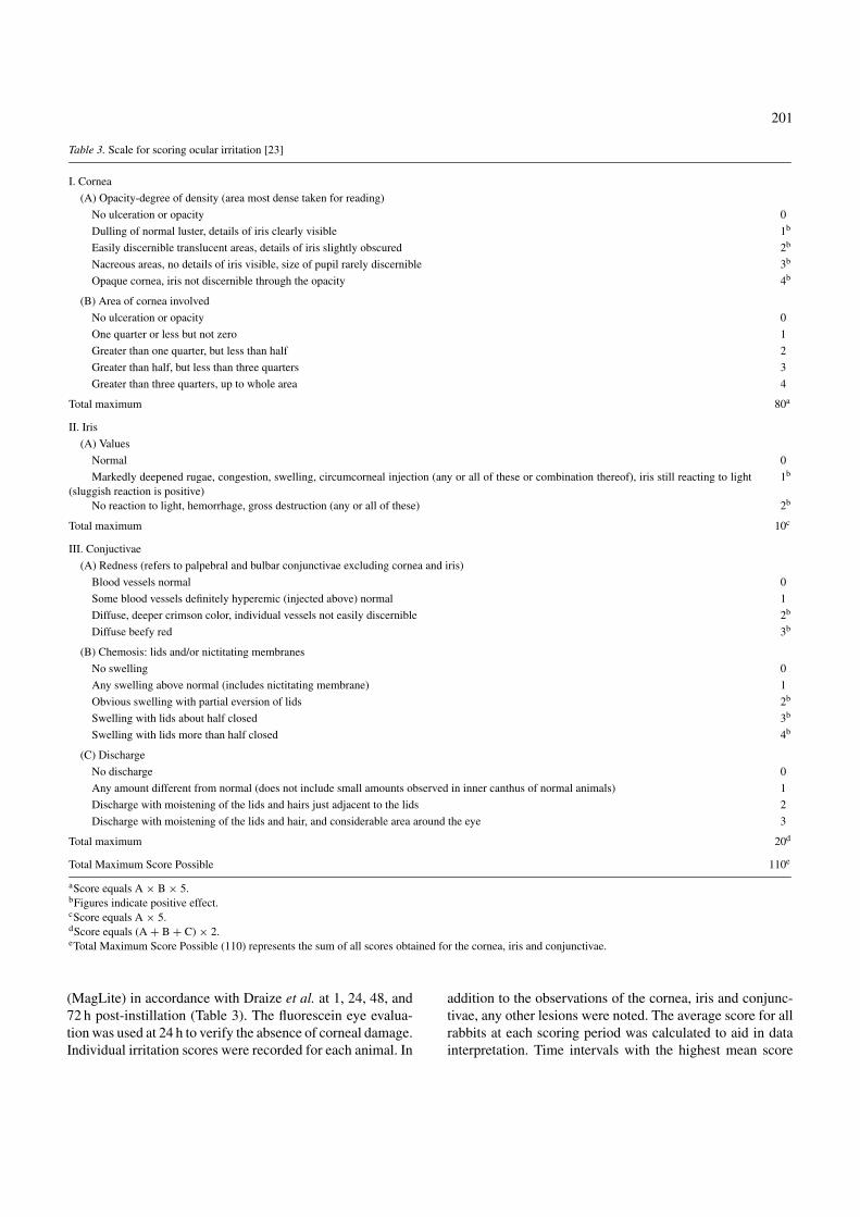
We employed an EPR approach to test the effect of HBO andOptiBerry feeding on whole-body redox status of mice im-mediately after the exposure to HBO. Here, mice were chosenbecause our EPR cavity is suited for the size of mice. Anes-thetized mice were injected with a nitroxyl radical throughtail vein, placed in EPR resonator, and images were takenevery 4 min. Antioxidants reduce the nitroxyl radical to hy-droxyamine, and thus the signal decays with time. A higherintensity signal from an area indicates lower reduction ca-
pability. Data was obtained from a custom-built 750 MHzEPR spectrometer [30]. In the control group, high signal in-tensity was observed in the peritoneum at 4 and 8 min andin the bladder at 12 and 16 min. In the placebo-fed HBOgroup, higher signal intensity is observed at all time pointsas compared to the control group, and the nitroxyl radicalwas retained in the body for a longer time. This indicatesa clear shift of the whole-body redox status towards oxida-tion in response to HBO treatment. When OptiBerry-fed micewere exposed to HBO, based on the intensity and retention ofsignal, it was clear that OptiBerry feeding prevented HBO-induced oxidation response in a whole-body mouse model(Fig. 3).
Discussion
The pharmacologic, medicinal and therapeutic benefits ofedible berries are well established. We have studied the
206
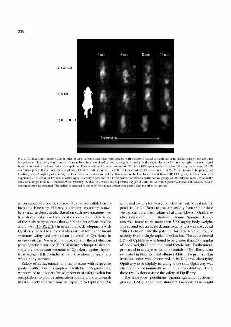
Fig. 3. Comparison of redox status in mice in vivo. Anesthetised mice were injected with a nitroxyl radical through tail vein, placed in EPR resonator, andimages were taken every 4 min. Antioxidants reduce the nitroxyl radical to hydroxyamine, and thus the signal decays with time. A higher intensity signalfrom an area indicates lower reduction capability. Data is obtained from a custom-built 750 MHz EPR spectrometer with the following parameters: 32 mWmicrowave power, 0.7 G modulation amplitude, 100 kHz modulation frequency, 80 ms time constant, 35G scan range and 750 MHz microwave frequency. (A)Control group: A high signal intensity is observed in the peritoneum at 4 and 8 min, and in the bladder at 12 and 16 min (B) HBO group: On treatment withhyperbaric O2 at 2 atm for 120 min, a higher signal intensity is observed at all time points as compared to the control group, and the nitroxyl radical stays in thebody for a longer time. (C) Treatment with Optiberry-fed diet for 2 weeks and hyperbaric oxygen at 2 atm for 120 min: Optiberry, a novel antioxidant, reducesthe signal intensity obtained. The radical is retained in the body for a much shorter time period than the other two groups.
anti-angiogenic properties of several extracts of edible berriesincluding blueberry, bilberry, elderberry, cranberry, straw-berry and raspberry seeds. Based on such investigations, wehave developed a novel synergistic combination, OptiBerry,of these six berry extracts that exhibit potent effects in vitroand in vivo [16, 19, 27]. These favourable developments withOptiBerry led to the current study aimed at testing the broadspectrum safety and antioxidant potential of OptiBerry inin vivo settings. We used a unique, state-of-the-art electronparamagnetic resonance (EPR) imaging technique to demon-strate the antioxidant potential of OptiBerry against hyper-baric oxygen (HBO)-induced oxidative stress in mice in awhole-body scenario.
Safety of nutraceuticals is a major issue with respect topublic health. Thus, in compliance with the FDA guidelines,we were led to conduct a broad spectrum of safety evaluationon OptiBerry to provide information on safety/toxicity/healthhazards likely to arise from an exposure to OptiBerry. An
acute oral toxicity test was conducted with rats to evaluate thepotential for OptiBerry to produce toxicity from a single dosevia the oral route. The median lethal dose (LD50) of OptiBerryafter single oral administration in female Sprague Dawleyrats was found to be more than 5000 mg/kg body weight.In a second set, an acute dermal toxicity test was conductedwith rats to evaluate the potential for OptiBerry to producetoxicity from a single topical application. The acute dermalLD50 of OptiBerry was found to be greater than 2000 mg/kgof body weight in both male and female rats. Furthermore,primary skin and eye irritation potentials of OptiBerry wereevaluated in New Zealand albino rabbits. The primary skinirritation index was determined to be 0.3, thus classifyingOptiBerry to be slightly irritating to the skin. OptiBerry wasalso found to be minimally irritating to the rabbit eye. Thus,these results demonstrate the safety of OptiBerry.
The tripeptide glutathione (gamma-glutamyl-cysteinyl-glycine; GSH) is the most abundant low-molecular-weight
207
thiol, and GSH/glutathione disulfide is the major redox cou-ple in animal cells. The synthesis of GSH from glutamate,cysteine and glycine is catalysed sequentially by two cytoso-lic enzymes, gamma-glutamylcysteine synthetase and GSHsynthetase. Compelling evidence shows that GSH synthesis isregulated primarily by gamma-glutamylcysteine synthetaseactivity, cysteine availability and GSH feedback inhibition.Animal and human studies demonstrate that adequate proteinnutrition is crucial for the maintenance of GSH homeosta-sis. In addition, enteral or parenteral cystine, methionine, N-acetyl-cysteine, and L-2-oxothiazolidine-4-carboxylate areeffective precursors of cysteine for tissue GSH synthesis.Glutathione plays important roles in antioxidant defense, nu-trient metabolism and regulation of cellular events (includinggene expression, DNA and protein synthesis, cell prolifera-tion and apoptosis, signal transduction, cytokine productionand immune response and protein glutathionylation). Oxida-tive stress can result from exposure to excessive amountsof endogenous and exogenous electrophiles. The concentra-tion of GSH, the most abundant intracellular non-proteinthiol and important antioxidant, declines with age and insome age-related diseases. The underlying mechanism, how-ever, is not clear. Glutathione deficiency contributes to ox-idative stress, which plays a key role in aging and the patho-genesis of many diseases (including Kwashiorkor, seizure,Alzheimer’s disease, Parkinson’s disease, liver disease, cysticfibrosis, sickle cell anemia, HIV, AIDS, cancer, heart attack,stroke and diabetes). New knowledge of the nutritional reg-ulation of GSH metabolism is critical for the developmentof effective strategies to improve health and to treat thesediseases [33].
HBO treatment is applied as a therapy for a wide varietyof diseases with symptoms caused by lack of oxygen in thetarget tissues. However, it is known that exposure to highconcentrations of oxygen may lead to oxidative stress andcause cell and tissue damage. Oxygen toxicity and possiblecancer-promoting effects of HBO therapy have been a mat-ter of serious concern. Although a cancer-inducing effect ofHBO was not found to date, recent studies clearly indicatedan induction of oxidative DNA damage in the blood cells ofhealthy subjects after HBO under therapeutic conditions. Thebiological significance of this finding has been investigatedin a series of in vitro and in vivo tests. These studies havebeen recently reviewed [30]. We employed a clinically rele-vant HBO exposure protocol to study the antioxidant proper-ties of OptiBerry. OptiBerry feeding prevented HBO-inducedGSSG formation in the lung and liver of vitamin E-deficientSprague Dawley rats. Accumulation of lung tissue oxidisedglutathione (GSSG) is recognised as a marker of oxidantinduced lung injury [34]. Oxygenation of the lung inducesGSSG formation [35] and such perturbation of GSH redoxstatus in the lung often accompanies lipid peroxidation [36].Thus, the observed protective effects of OptiBerry against
HBO-induced changes in the lung redox status is expected tohave direct bearing on the general health of this vital organ.
The liver is the central detoxifying organ and one of themost widely recognised function of GSH is its defense againsttoxic compounds, whether exogenous, such as electrophilicxenobiotics, or endogenous, such as reactive oxygen species,generated during normal oxidative metabolism and/or stress.However, another no less significant role of GSH, namely, itsfunction as a reservoir and vehicle for packaging and trans-port of cyst(e)ine has received significant attention. BecauseGSH is relatively more auto-oxidation resistant and stablethan cyst(e)ine (CYSH), it serves as the preferred form forstorage and transport of the latter, especially in the extracel-lular and relatively much less reduced (than intracellular) mi-lieu, where CYSH oxidises to cystine (CYSS) rapidly. Overthe past two decades, significant work has been going on todelineate the intra- and extrahepatic (interorgan) turnover,transport and disposal of GSH and define the quantitativerole of these processes in interorgan homeostasis of GSH,CYSH and CYSS. These studies have identified the liver asthe central organ of interorgan GSH homeostasis, with sinu-soidal GSH efflux as the major determinant of plasma GSH,CYSH, CYSS and thiol-disulfide status of plasma [37]. Akinto the scenario in the lung, levels of GSSG in the liver serve asan index of stress and injury [38]. Maintenance of appropriateglutathione redox homeostasis in the liver is central to over-all hepatic metabolism as well as immunity, which in turndirectly influence general health of the organism [39, 40].Thus, the observed protective effects of OptiBerry againstHBO-induced perturbation of GSH redox state in the liverare likely to be of significance to overall health.
Recently, He et al. [41] established the ability of EPRimaging to provide non-invasive in vivo mapping of theredox status of the skin of living rats. The redox statuswas measured using a topically applied nitroxyl spin probe,(15)N-PDT. Free radicals and other paramagnetic species,play an important role in the cellular injury and patho-physiology. EPR spectroscopy and imaging has emergedas an important tool for non-invasive in vivo measurementand spatial mapping of free radicals in biological tissues.Extensive applications have been performed in small animalssuch as mice and recently applications in humans havebeen performed. A variety of spatial, and spectral–spatialEPR imaging applications have been performed. Thesetechniques, along with the use of biocompatible paramag-netic probes, including particulate suspensions and solublenitroxide radicals, enable spatial imaging of the redox statein a variety of biomedical applications [42]. We employedthis novel EPR imaging approach to test the effect of HBOand OptiBerry feeding on whole-body redox status of mice,immediately after the exposure to HBO. This experimentalsystem provided data supporting the GSH/GSSG data tosubstantiate the antioxidant properties of OptiBerry. Taken
208
together, these results indicate that OptiBerry is safe andthat it may exhibit whole-body antioxidant properties whenconsumed orally for an extended period of time.
Acknowledgments
Acute oral and dermal toxicities, and primary skin and eyeirritation studies were done by Product Safety Labs, Day-ton, NJ. The authors thank Ms Shirley Zafra for technicalassistance.
References
1. Ofek I, Goldhar J, Zafriri D, Lis H, Adar R, Sharon N: Anti-Escherichiacoli adhesin activity of cranberry and blueberry juices. New Engl J Med324: 1599, 1991
2. Colic M, Pavelic K: Molecular mechanisms of anticancer activity ofnatural dietetic products. J Mol Med 78: 333–336, 2000
3. Kresty LA, Morse MA, Morgan C, Carlton PS, Lu L, Gupta A, Black-wood M, Stoner GD: Chemoprevention of esophageal tumorigenesis bydietary administration of lyophilized black raspberries. Cancer Res 61:6112–6119, 2001
4. McCarty MF: Current prospects for controlling cancer growth with non-cytotoxic agents – nutrients, phytochemicals, herbal extracts, and avail-able drugs. Med Hypotheses 56: 137–154, 2001
5. She QB, Bode AM, Ma WY, Chen NY, Dong Z: Resveratrol-inducedactivation of p53 and apoptosis is mediated by extracellular-signal-regulated protein kinases and p38 kinase. Cancer Res 61: 1604–1610,2001
6. Xue H, Aziz RM, Sun N, Cassady JM, Kamendulis LM, Xu Y, StonerGD, Klaunig JE: Inhibition of cellular transformation by berry extracts.[erratum appears in Carcinogenesis 22 (2001) 831–833]; Carcinogenesis22: 351–356, 2001
7. Cao G, Prior RL: Anthocyanins are detected in human plasma after oraladministration of an elderberry extract. Clin Chem 45: 574–576, 1999
8. Giavazzi R, Taraboletti G: Angiogenesis and angiogenesis inhibitors incancer. Forum 9: 261–272, 1999
9. Griffioen AW, Molema G: Angiogenesis: Potentials for pharmaco-logic intervention in the treatment of cancer, cardiovascular dis-eases, and chronic inflammation. Pharmacol Rev 52: 237–268,2000
10. Fotsis T, Pepper MS, Aktas E, Breit S, Rasku S, Adlercreutz H, WahalaK, Montesano R, Schweigerer L: Flavonoids, dietary-derived inhibitorsof cell proliferation and in vitro angiogenesis. Cancer Res 57: 2916–2921, 1997
11. Fotsis T, Pepper MS, Montesano R, Aktas E, Breit S, SchweigererL, Rasku S, Wahala K, Adlercreutz H: Phytoestrogens and inhibi-tion of angiogenesis. Baillieres Clin Endocrinol Metabol 12: 649–666,1998
12. Hayashi A, Gillen AC, Lott JR: Effects of daily oral administrationof quercetin chalcone and modified citrus pectin. Altern Med Rev 5:546–552, 2000
13. Hisa T, Kimura Y, Takada K, Suzuki F, Takigawa M: Shikonin, an in-gredient of Lithospermum erythrorhizon, inhibits angiogenesis in vivoand in vitro. Anticancer Res 18: 783–790, 1998
14. Jiang C, Agarwal R, Lu J: Anti-angiogenic potential of a cancer chemo-preventive flavonoid antioxidant, silymarin: Inhibition of key attributes
of vascular endothelial cells and angiogenic cytokine secretion bycancer epithelial cells. Biochem Biophys Res Commun 276: 371–378,2000
15. Paper DH: Natural products as angiogenesis inhibitors. Planta Medica64: 686–695, 1998
16. Roy S, Khanna S, Alessio HM, Vider J, Bagchi D, Bagchi M, Sen CK:Anti-angiogenic property of edible berries. Free Radic Res 36: 1023–1031, 2002
17. Gordillo G, Onat D, Stockinger M, Roy S, Atalay M, Beck F, SenC: A key angiogenic role of monocyte chemoattractant protein-1 inhemangioendothelioma proliferation. Am J Physiol Cell Physiol 287:C866–C873, 2004
18. Gordillo GM, Atalay M, Roy S, Sen CK: Hemangioma model for invivo angiogenesis: inducible oxidative stress and MCP-1 expression inEOMA cells. Methods Enzymol 352: 422–432, 2002
19. Atalay M, Gordillo G, Roy S, Rovin B, Bagchi D, Bagchi M, Sen CK:Anti-angiogenic property of edible berry in a model of hemangioma.FEBS Lett 544: 252–257, 2003
20. Knight J: Safety concerns prompt US ban on dietary supplement. Nature427: 90, 2004
21. Kahkonen MP, Hopia AI, Heinonen M: Berry phenolics and their an-tioxidant activity. J Agric Food Chem 49: 4076–4082, 2001
22. Speit G, Dennog C, Radermacher P, Rothfuss A: Genotoxicity of hy-perbaric oxygen. Mutation Res 512: 111–119, 2002
23. Draize J, Woodward G, Calvary H: Methods for the study of irritationand toxicity of substances applied topically to the skin and mucousmembranes. J Pharmacol Exp Ther 82: 377–390, 1944
24. Draize J: The appraisal of the safety of chemicals in foods. In: Drugs andCosmetics. Dermal Toxicity, Association of Food and Drug Officials ofthe US, Topeka, KA, 1965, pp 46–59
25. Kay J, Calandra J: Interpretation of eye irritation tests. J Soc Cos Chem13: 281–289, 1962
26. Sen CK, Khanna S, Babior BM, Hunt TK, Ellison EC, Roy S: Oxidant-induced vascular endothelial growth factor expression in human ker-atinocytes and cutaneous wound healing. J Biol Chem 277: 33284–33290, 2002
27. Roy S, Venojarvi M, Khanna S, Sen CK: Simultaneous detection of toco-pherols and tocotrienols in biological samples using HPLC-coulometricelectrode array. Methods Enzymol 352: 326–332, 2002
28. Khanna S, Roy S, Ryu H, Bahadduri P, Swaan PW, Ratan RR, SenCK: Molecular basis of vitamin E action: Tocotrienol modulates 12-lipoxygenase, a key mediator of glutamate-induced neurodegeneration.J Biol Chem 278: 43508–43515, 2003
29. He G, Shankar RA, Chzhan M, Samouilov A, Kuppusamy P, ZweierJL: Noninvasive instrument of anatomic structure and intraluminaloxygenation in the gastrointestinal tract of living mice with spatialand spectral EPR imaging. Proc Natl Acad Sci USA 96: 4586–4591,1999
30. He G, Petryakov S, Samouilov A, Chzhan M, Kuppusamy P, Zweier J:Development of a resonator with automatic tuning and coupling capa-bility to minimize sample motion noise for in vivo EPR spectroscopy.J Magn Reson 149: 218–227, 2001
31. Sen CK, Khanna S, Gordillo G, Bagchi D, Bagchi M, Roy S: Oxygen,oxidants, and antioxidants in wound healing: An emerging paradigm.Ann NY Acad Sci 957: 239–249, 2002
32. Gordillo GM, Sen CK: Revisiting the essential role of oxygen in woundhealing. Am J Surg 186: 259–263, 2003
33. Wu G, Fang YZ, Yang S, Lupton JR, Turner ND: Glutathionemetabolism and its implications for health. J Nutr 134: 489–492,2004
34. White CW, Mimmack RF, Repine JE: Accumulation of lung tissue ox-idized glutathione (GSSG) as a marker of oxidant induced lung injury.Chest 89: 111S–113S, 1986
209
35. Jenkinson SG, Marcum RF, Pickard JS, Orzechowski Z, Lawrence RA,Jordan JM: Glutathione disulfide formation occurring during hypoxiaand reoxygenation of rat lung. J Lab Clin Med 112: 471–480, 1988
36. Hammerschmidt S, Buchler N, Wahn H: Tissue lipid peroxidationand reduced glutathione depletion in hypochlorite-induced lung injury.Chest 121: 573–581, 2002
37. Ookhtens M, Kaplowitz N: Role of the liver in interorgan homeostasisof glutathione and cyst(e)ine. Semin Liver Dis 18: 313–329, 1998
38. Jaeschke H: Glutathione disulfide as index of oxidant stress in rat liverduring hypoxia. Am J Physiol 258: G499–G505, 1990
39. Mandl J, Banhegyi G: Role of glutathione in the regulation of livermetabolism. Biofactors 17: 21–26, 2003
40. Yamauchi A, Tsuyuki S, Inamoto T, Yamaoka Y: Liver immunity andglutathione. Antioxid Redox Signal 1: 245–253, 1999
41. He G, Kumar Kutala V, Kuppusamy P, Zweier JL: In vivo measurementand mapping of skin redox stress induced by ultraviolet light exposure.Free Radic Biol Med 36: 665–672, 2004
42. He G, Samouilov A, Kuppusamy P, Zweier JL: In vivo imaging of freeradicals: applications from mouse to man. Mol Cell Biochem 234–235:359–367, 2002
Copyright © 2022 FDOKUMEN