Contrat plus 2000 totalisant plus 25000 2016-2017 - Ville de ...
Upload
sorbonne-frCategory
view
0download
0
BRAINA JOURNAL OF NEUROLOGY
Riluzole treatment, survival and diagnostic criteriain Parkinson plus disorders: The NNIPPS StudyGilbert Bensimon,1 Albert Ludolph,2 Yves Agid,3 Marie Vidailhet,3 Christine Payan,1
P. Nigel Leigh4 and for the NNIPPS Study Group*
1 Departement de Pharmacologie Clinique, Hopital de la Pitie-Salpetriere, Assistance Publique Hopitaux de Paris & UPMC University Paris 06,
UMR 7087, Paris, France
2 Department of Neurology, University of Ulm, Oberer Eselsberg, Ulm, Germany
3 Federation de Neurologie, Hopital de la Pitie-Salpetriere, Assistance Publique Hopitaux de Paris & UPMC University Paris 06, Paris, France
4 MRC Centre for Neurodegeneration Research, King’s College London, Institute of Psychiatry, P0 41, Department of Clinical Neuroscience,
London, UK
�See appendix for the details on NNIPPS Study Group
Correspondence to: Prof. P. Nigel Leigh,
MRC Centre for Neurodegeneration Research,
King’s College London, PO 41, Institute of Psychiatry,
Camberwell, London SE5 8AF, UK
E-mail: [email protected]
Parkinson plus diseases, comprising mainly progressive supranuclear palsy (PSP) and multiple system atrophy (MSA) are rare
neurodegenerative conditions. We designed a double-blind randomized placebo-controlled trial of riluzole as a potential dis-
ease-modifying agent in Parkinson plus disorders (NNIPPS: Neuroprotection and Natural History in Parkinson Plus Syndromes).
We analysed the accuracy of our clinical diagnostic criteria, and studied prognostic factors for survival. Patients with an akinetic-
rigid syndrome diagnosed as having PSP or MSA according to modified consensus diagnostic criteria were considered for
inclusion. The psychometric validity (convergent and predictive) of the NNIPPS diagnostic criteria were tested prospectively
by clinical and pathological assessments. The study was powered to detect a 40% decrease in relative risk of death within PSP
or MSA strata. Patients were randomized to riluzole or matched placebo daily and followed up to 36 months. The primary
endpoint was survival. Secondary efficacy outcomes were rates of disease progression assessed by functional measures. A total
of 767 patients were randomized and 760 qualified for the Intent to Treat (ITT) analysis, stratified at entry as PSP (362 patients)
or MSA (398 patients). Median follow-up was 1095 days (range 249–1095). During the study, 342 patients died and 112 brains
were examined for pathology. NNIPPS diagnostic criteria showed for both PSP and MSA excellent convergent validity with the
investigators’ assessment of diagnostic probability (point-biserial correlation: MSA rpb = 0.93, P_ 0.0001; PSP, rpb = 0.95,
P_0.0001), and excellent predictive validity against histopathology [sensitivity and specificity (95% CI) for PSP 0.95 (0.88–
0.98) and 0.84 (0.77–0.87); and for MSA 0.96 (0.88–0.99) and 0.91 (0.86–0.93)]. There was no evidence of a drug effect on
survival in the PSP or MSA strata (3 year Kaplan–Meier estimates PSP-riluzole: 0.51, PSP-placebo: 0.50; MSA-riluzole: 0.53,
MSA-placebo: 0.58; P = 0.66 and P = 0.48 by the log-rank test, respectively), or in the population as a whole (P = 0.42, by the
stratified-log-rank test). Likewise, rate of progression was similar in both treatment groups. There were no unexpected adverse
effects of riluzole, and no significant safety concerns. Riluzole did not have a significant effect on survival or rate of functional
deterioration in PSP or MSA, although the study reached over 80% power to detect the hypothesized drug effect within strata.
The NNIPPS diagnostic criteria were consistent and valid. They can be used to distinguish between PSP and MSA with high
accuracy, and should facilitate research into these conditions relatively early in their evolution.
doi:10.1093/brain/awn291 Brain 2009: 132; 156–171 | 156
Received March 31, 2008. Revised September 10, 2008. Accepted October 13, 2008. Advance Access publication November 23, 2008
� 2008 The Author(s)
This is an Open Access article distributed under the terms of the Creative Commons Attribution Non-Commercial License (http://creativecommons.org/licenses/by-nc/
2.0/uk/) which permits unrestricted non-commercial use, distribution, and reproduction in any medium, provided the original work is properly cited.
by guest on July 27, 2016http://brain.oxfordjournals.org/
Dow
nloaded from

Keywords: progressive supranuclear palsy; multiple system atrophy; randomized controlled trial; riluzole; natural history
Abbreviations: ADL = activity of daily living; ALS = amyotrophic lateral sclerosis; ALT = alanine aminotransferase; AST = aspartateaminotransferase; CBD = corticobasal degeneration; CGI-ds = clinical global impression for disease severity;CGI-dysautonomia = clinical global impression for autonomic dysfunction; GCP = good clinical practice; IDMSC = independent datamonitoring and safety committee; IPD = idiopathic Parkinson’s disease; IRB = institutional review board; ITT = Intent-to-treat;MMSE = Mini-Mental State Examination; MSA-C = multiple system atrophy, Cerebellar form; MSA-P = multiple system atrophy,parkinsonian form; NINDS-SPSP = National Institute of Neurological Disorders and the Society for Progressive Supranuclear Palsy;PPPT = per protocol per treatment; SAE = serious adverse event; SEADL = Schwab and England activities of daily living scale;SMDS = Short Motor Disability Scale; VAS = Visual Analogue Scale
IntroductionProgressive supranuclear palsy (PSP) and multiple system atrophy
(MSA) are disabling and fatal neurodegenerative disorders for
which no disease-modifying treatment is available. For the major-
ity of patients with PSP and MSA, many of whom present with
an atypical Parkinsonian or akinetic-rigid syndrome (‘Parkinson
plus disorder’) the course is one of relentless progression, increasing
disability and death with a median survival of 5–10 years from
onset of symptoms (Litvan et al., 1996a; Testa et al., 1996, 2001;
Ben-Shlomo et al., 1997; Schrag et al., 1999, 2008; Litvan, 2003;
Golbe and Ohman-Strickland, 2007).
PSP and MSA have similar prevalence rates estimated at 2–7
per 100 000 person years (Golbe et al., 1988; Ben-Shlomo et al.,
1997; Bower et al., 1997; Schrag et al., 1999; Nath and Burn, 2000;
Nath et al., 2001; Vanacore et al., 2001a, b; Watanabe et al., 2002).
These are probably underestimates, however, because current diag-
nostic criteria are based on retrospective clinicopathological studies
(Litvan et al., 1996a, b, d, 2003; Litvan, 2003) and both delayed
diagnosis and mis-diagnosis are common. Although published con-
sensus diagnostic criteria are highly specific they are relatively insen-
sitive and a definite diagnosis can only be made through
histopathology (Litvan et al., 2003). Since it is likely that neuropro-
tective strategies are best tested at a relatively early stage of disease,
more sensitive diagnostic criteria are required for trials of potential
disease-modifying agents. Although PSP and MSA often present as
akinetic-rigid syndromes, each has distinctive pathological features.
In MSA, a key feature is glial cytoplasmic inclusions with accumula-
tion of �-synuclein in oligodendrocytes and neurons (Papp et al.,
1989; Lantos and Papp, 1994; Spillantini and Goedert, 2000)
whilst in PSP the hallmark is accumulation of abnormally phosphory-
lated microtubule-associated tau protein (�) in neurons and glia
(Hauw et al., 1994; Dickson et al., 2007). Although the pathogenic
mechanisms underlying MSA and PSP are unknown, there is evi-
dence that glutamate toxicity may contribute to neuronal damage
in these and other neurodegenerative diseases (Albin and
Greenamyre, 1992; Albers and Augood, 2001; Mattson, 2003;
Przedborski, 2005). The benzothiazole drug riluzole has a number
of pharmacological effects that contribute to neuroprotection in
experimental paradigms of neurodegenerative diseases including
anti-excitotoxic activity, blocking of voltage dependent sodium-
channels, free-radical scavenging, anti-apoptotic and neurotrophic
effects and inhibition of protein aggregation (Doble, 1999; Heiser
et al., 2002; Yoo et al., 2005; Caumont et al., 2006; Shortland et al.,
2006). In a rodent model of MSA, riluzole improved some measures
of neuronal damage (Diguet et al., 2005; Scherfler et al., 2005).
Riluzole (up to 200 mg daily) is well tolerated and prolongs
survival in amyotrophic lateral sclerosis (Bensimon et al., 1994;
Lacomblez et al., 1996; Miller et al., 2007). Thus far, riluzole remains
the only agent shown to modify disease progression in a human
neurodegenerative disorder.
In order to test the hypothesis that riluzole may slow disease
progression in PSP and MSA, we carried out a phase-III, random-
ized double-blind placebo controlled trial in 44 centres in France,
Germany and the UK. The study design incorporated ancillary
objectives including natural history, development and validation
of more sensitive diagnostic criteria and functional measures of
disease severity and progression, cognition, quality of life, health
economics, MRI changes, pathology and the establishment of
brain and DNA banks. We describe here the design and main
outcomes of the NNIPPS trial in terms of the efficacy and safety
of riluzole, the psychometric validity of the NNIPPS diagnostic
criteria in relation to clinic and pathology and the major factors
influencing prognosis for PSP and MSA.
MethodsNNIPPS was designed as a double-blind placebo-controlled, stratified
(by diagnosis of MSA or PSP, and by centre), parallel group, European
(France, Germany, United Kingdom) trial assessing the efficacy
and safety of riluzole at flexible dose (50–200 mg/day) (Fig. 1). The
primary objective of NNIPPS was to demonstrate the efficacy of
riluzole on survival (primary end-point) and rate of decline in motor
function (secondary end-points).
The number of patients required was determined for the primary
end-point survival and for each stratum (PSP, MSA). Assumptions
included mean disease duration of 3 years prior to entry and no loss
to follow-up. Assuming a 41% death rate at 3 years in the placebo
group (Litvan et al., 1996a; Testa et al., 1996, 2001; Ben-Shlomo
et al., 1997; Litvan, 2003; Golbe and Ohman-Strickland, 2007;
Schrag et al., 2007), 400 patients provide over 80% power to
detect a 40% decrease in the relative risk of death in the treated
group using the log-rank test with two-sided � risk set at 0.05.
With both strata combined (800 patients), the power reached 98%
to detect a 40% decrease in the risk of death with active treatment
compared to placebo, assuming that a minimum of 272 events would
be observed over the 36-month trial period.
Patients and treatmentFrom consensus criteria (Litvan et al., 1996a, d, 2003) we derived
simplified operational diagnostic criteria suitable for large-scale clinical
Riluzole in Parkinson-plus disorders Brain 2009: 132; 156–171 | 157
by guest on July 27, 2016http://brain.oxfordjournals.org/
Dow
nloaded from

trials, with the aim of maximizing sensitivity for recruitment. Strata
were defined according to these operational diagnostic criteria
(Table 1). The history of the condition in each patient was evaluated
at entry using systematic questionnaires recording the initial (pre-
senting) symptom, and current syndromes (entry and last visit).
Response to levodopa therapy was evaluated at entry. Additional
assessments related to ancillary studies including a new functional
scale (the Parkinson Plus scale), magnetic resonance imaging, neuro-
psychology, health economics and quality of life, genetic testing and
neuropathology, will be presented separately.
Patients were allocated to treatment according to a computer
generated randomization list, stratified for diseases (MSA or
PSP) and clinical centre, with a riluzole to placebo ratio of 1:1.
Riluzole (50 mg) or placebo was prepared as identical tablets (Sanofi-
Aventis, Antony-France). Packaging and labelling (LC2, Lentilly-France)
and treatment management (Cardinale, Corby-UK; Clindata,
Weilerswist-Germany; AGEPS-AP HP, Paris-France) was performed
so as to safeguard blinding to treatment allocation throughout
the trial duration.
Following randomization, a monthly dose-titration over 3 months
was used with increasing dosage from one, two and four tablets daily
during which time tolerance was assessed with monthly laboratory
tests for haematology (full blood count) and liver function (including
ALT and AST) and patient’s reporting of adverse events. Dose flexibility
(one to four tablets per day) according to tolerance was allowed
throughout the 36-month study period, with each dose-adjustment
recorded. Study treatment withdrawal was not considered as an ‘end
of study’ and the protocol required that patients should be followed
for the ITT analysis to the end of the planned double-blind period.
Treatments were delivered to patients every 3 months and the tablets
returned were counted to evaluate compliance.
AssessmentsTo assess the psychometric validity (see Statistical analysis section)
of the NNIPPS operational diagnostic criteria, investigators were
required to assign at entry a diagnostic probability (PSP or MSA) for
each patient using two 100 mm-visual analogue scales (VAS). These
clinical diagnostic assessments were completed at entry, every 12
months thereafter, and at the last visit. When possible, clinical diag-
nosis was compared to neuropathological diagnosis, which was
assessed blind to the clinical diagnosis at all stages of processing and
analysis of donated brains. The latter were processed according to
a standard protocol incorporating formalin fixation of one hemisphere
(randomly allocated by the UK, French or German coordinating
centres) with freezing (at –80�C) of the other hemisphere for banking.
Tissue sections were assessed against standard diagnostic criteria
(Lantos and Papp, 1994; Litvan et al., 1996d; Dickson, 1999) with
cross-examination and consensus scoring for each case.
The primary criterion of efficacy was defined as survival during
the 36-month double-blind period of the study (1095 days included)
or until the administrative cut-off date (November 30, 2004 included)
whichever came first. The primary end-point was defined as death
from any cause. All dates of death were documented with death
certificates. For all surviving patients, the date of last contact was
also documented.
Secondary end-points included standard assessments used in idio-
pathic Parkinson’s disease completed at entry and 6 monthly, using
the Hoehn and Yahr staging scale (Hoehn and Yahr, 1967), the
Schwab and England Activities of Daily Living scale (SEADL, Schwab
and England, 1969) as well as generic health scale assessments using
the Clinical Global Impression for disease severity completed by inves-
tigators (CGI-ds; Streiner and Norman, 2003) and a CGI adapted to
autonomic function assessment (CGI-dysautonomia). The Mini-Mental
State Examination (MMSE; Folstein et al., 1975) was completed by the
clinical observer at entry and every 12 months. In addition, a specific
functional measure assessing ambulation was developed (Short Motor
Disability Scale, SMDS) and performed at entry and 3 monthly
(Supplementary Table 1). Anticipated non-serious adverse events
of riluzole included dizziness, gastrointestinal symptoms, and fatigue
(Lacomblez et al., 1996). The main anticipated serious adverse
event (SAE) for riluzole was serious abnormality of liver function
tests defined as ALT 45� the upper limit of normal. Safety was eval-
uated through clinical examination, vital signs, routine laboratory tests
including haematology and transaminases (AST, ALT), concomitant
medication and patient reports of adverse events (at entry and
3 monthly). Adverse events, serious and non-serious were coded
using MedDRA� version 6.0 (MedDRA MSSO, Northrop Grumman
Corp., Reston, VA, USA). Weight and electrocardiogram were
assessed at entry and at the final visit.
Statistical analysisAn Independent Data Monitoring and Safety Committee (IDMSC)
was established for unblinded review of all SAEs during the trial and
to advise the Steering Committee at regular intervals regarding con-
tinuation of the trial according to predefined stopping rules. No interim
analysis for efficacy was planned but four safety analyses were per-
formed throughout the study to ensure that mortality in the treated
group was not in excess compared to the placebo group.
The detailed statistical plan was submitted to the French IRB prior
to unblinding. The primary analysis was conducted following the ITT
principles. The ITT population was defined as all randomized subjects
who received at least one dose of study medication and for whom
there were no major violations of GCP (ICH-6). Sensitivity analyses
population definition is described in Supplementary Text 1.
For the primary analysis, the diagnosis at inclusion was used to
define the PSP and MSA strata. In addition, the statistical plan defined
Months
PSP
Riluzole Placebo
MSA
0 1 2 3 6 9 12 15 18 21 24 27 30 33 36
Inclusion
MRI
Neuropsychology
Neurology
Dose titration
Termination
Assessments
Riluzole PlaceboRandomisation
Selection
Safety
Fig. 1 Trial Flow Chart. At the selection stage, patients were
assigned to either the MSA or PSP strata according to the
NNIPPS diagnostic criteria. Following Inclusion, patients within
each stratum were randomly allocated to either the riluzole
or placebo group on 1:1 ratio and followed-up 3 monthly for
36 months in double-blind fashion. Arrows indicate the time
of each assessment.
158 | Brain 2009: 132; 156–171 G. Bensimon et al.
by guest on July 27, 2016http://brain.oxfordjournals.org/
Dow
nloaded from

sensitivity analyses by diagnosis at the end of the study to allow
for the possibility of misdiagnosis.
The Safety Population comprised all randomized subjects who
received at least one dose of study medication. All analyses were
performed using SAS Software version 11. According to the guidelines
for the standard for educational and psychological testing (American
Psychological Association, 1985) two facets of the psychometric
validity of the diagnostic criteria were evaluated, the convergent and
the predictive validity. The convergent validity was assessed by the
degree of correlation between diagnostic classification according
to inclusion criteria and the investigator’s assessment of diagnostic
probability on the VAS. As inclusion criteria represent a nominal
variable with two modalities (MSA, PSP), we used the point biserial
coefficient which is the relevant method in this case (point-
biserial correlation, rpb; Nunnally and Bernstein, 1994). Predictive
criterion-related validity of the clinical diagnostic criteria was assessed
by calculating the specificity, sensitivity, percent correct classification
and positive likelihood (Attia, 2003), using pathological diagnosis as
the gold standard.
Descriptive analyses summarized the overall population, and the
population sub-divided by treatment group, by strata (PSP versus
MSA) and by treatment within strata. Categorical data were summar-
ized by frequency and percentage. The log-linear model was used
to compare distributions between factors including treatment,
strata, country and all interactions (chi-square tests of partial associa-
tion) (Bishop et al., 1975). Continuous data were summarized
by mean and standard deviation. Between treatment groups compar-
ison at entry was carried out using three-factor variance analyses
including treatment, strata, country and all interaction factors
(Winer, 1971).
Table 1 NNIPPS Inclusion and exclusion criteria
Inclusion criteria Exclusion criteria
BOTH STRATA All of the following:-Akinetic-rigid syndrome;-Age at disease onset 530 years;-Disease duration (12 months to 8 years);-Signed informed consent.
Any of the following:-Idiopathic Parkinson’s disease;-Evidence of any other neurological disease
that could explain signs;-History of repeated strokes with stepwise
progression of parkinsonian features;-History of major stroke;-Any history of severe or repeated head injury;-A history of encephalitis;-A history of neuroleptic use for a prolonged period
of time or within the past 6 months;-Street-drug related parkinsonism;-Significant other neurological disease on CT-scan/MRI;-Oculogyric crises;-Signs of corticobasal degeneration;-Signs of lewy body disease;-Other life-threatening disease likely to interfere
with the main outcome measure;-Any clinically significant laboratoryabnormality, with the exception of cholesterol,
triglyceride and glucose;-Renal failure (serum creatinine 4 300mM/l);-Transaminase elevation 4 2 time upper limit of normal;-Presence of contra-indicated treatments;-Any previous participation in a therapeutic trial
within 3 months prior to entry;-Patient likely to be non-compliant or not easily
reached in case of emergency;-Patient under legal guardianship (France only).
PSP All of the following:-Supranuclear ophthalmoplegia;-Postural instability or falls
(within 3 years from disease onset).
Any of the following:-Cerebellar ataxia;-Symptomatic autonomic dysfunction;-Tremor at rest.
MSA One or more of the following:-Symptomatic autonomic dysfunction;-Cerebellar ataxia;-Postural instability or falls
(within 3 years from disease onset);-Pyramidal signs.
Any of the following:-Supranuclear ophthalmoplegial-Signs of severe dementia.
According to the NNIPPPS standard operating procedures, for inclusion into the PSP stratum, supranuclear ophthalmoplegia required ‘definite slowness and/ormoderate to definite limitation of downward gaze’. For MSA, cerebellar ataxia required a moderate to severe ataxia of trunk and/or limbs. Less marked signs whichthe investigator nonetheless considered clinically significant were not considered as inclusion criteria but allowed investigators to report the presence or absence ofan oculomotor or cerebellar syndrome. The akinetic-rigid syndrome was defined as mild to severe rigidity or slowness of neck or limbs. Significant symptomaticautonomic dysfunction (not treatment induced) was defined as moderate to severe CGI-dysautonomia. A MMSE score of 420 was regarded as evidence of severe
dementia. Contraindicated treatments included glutamatergic drugs (e.g. amantadine, lamotrigine, dextrometorphan, gabapentin, glutamate containing drugs), freeradical scavengers (selegiline, vit-E/ or C at very high dose) or any drug given to treat the disease and not the symptoms; potentially hepatotoxic drugs (e.g. dantrolene);drugs interacting with riluzole metabolism (CYP1A2 inhibitors or inducers); and ropirinole (due to decreased levels of the drug induced by riluzole).
Riluzole in Parkinson-plus disorders Brain 2009: 132; 156–171 | 159
by guest on July 27, 2016http://brain.oxfordjournals.org/
Dow
nloaded from
For the primary end-point survival, between groups survival
curves were compared using the Mantel-Cox (log-rank) test (Mantel
and Haenszel, 1958). Treatment effect was assessed with the stratified
log-rank test (p� two-tailed test50.05). The Cox model (Cox, 1972)
including treatment, strata and interaction factors, was used to check
for treatment by strata (MSA, PSP) interaction.
The influence of demographic and clinical variables at entry on sur-
vival was tested with univariate and multivariate Cox proportional-
hazard analysis. Multivariate analysis used an automatic up and
down stepwise selection of variables (Allison, 1995).
Secondary end-point variables used for assessing disease progression
included the SMDS, the SEADL, the Hoehn and Yahr staging and
the CGI-ds. For each patient, repeated measurements were summar-
ized by slope of change over time using linear regression methods
(unweighted least square estimate; Wu, 1988). Comparisons of
slopes between the treatment groups were performed using three-
factor variance analyses, including strata, country, treatment and
interaction factors. Serious and non-serious adverse events were com-
pared between treatment groups with the Pearson’s chi-square test
or Fisher exact test where appropriate.
EthicsPrior to inclusion, patients gave their informed written consent to
participate in the study. Separate consent was obtained both for
DNA sampling and for post-mortem brain tissue donation. The pro-
tocol and subsequent amendments were approved by Ethics
Committees/Institutional Review Boards of each coordinating centre
in the three participating countries. The trial was conducted according
to International standards of Good Clinical Practice-(ICH guidelines
and the Helsinki Declaration). An internal audit of the study was
carried out at the end of the trial before unblinding by an independent
auditor (Qualilab, Olivet, France) in the nine largest centres (three
in France, three in Germany and three in UK) accounting for 43%
of the overall trial population, and the primary criteria survival was
audited and appropriate documentation certified as 100% complete
over the whole study population.
ResultsDuring the study period, April 2000 to December 2004, the
Independent Data Monitoring and Safety Committee performed
four safety analyses (last review, June 2004) with advice to con-
tinue the trial on each occasion.
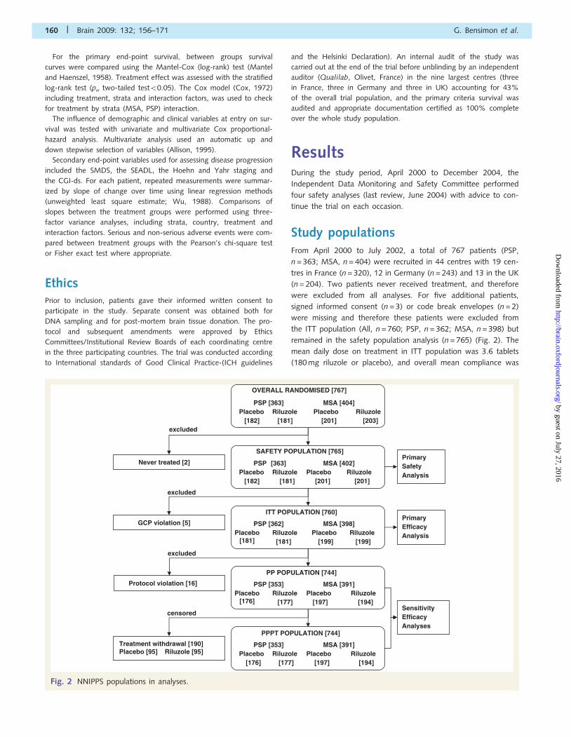
Study populationsFrom April 2000 to July 2002, a total of 767 patients (PSP,
n = 363; MSA, n = 404) were recruited in 44 centres with 19 cen-
tres in France (n = 320), 12 in Germany (n = 243) and 13 in the UK
(n = 204). Two patients never received treatment, and therefore
were excluded from all analyses. For five additional patients,
signed informed consent (n = 3) or code break envelopes (n = 2)
were missing and therefore these patients were excluded from
the ITT population (All, n = 760; PSP, n = 362; MSA, n = 398) but
remained in the safety population analysis (n = 765) (Fig. 2). The
mean daily dose on treatment in ITT population was 3.6 tablets
(180 mg riluzole or placebo), and overall mean compliance was
GCP violation [5]
Never treated [2]
Protocol violation [16]
Treatment withdrawal [190] Placebo [95] Riluzole [95]
excluded
excluded
censored
PrimaryEfficacyAnalysis
Sensitivity EfficacyAnalyses
PrimarySafetyAnalysis
OVERALL RANDOMISED [767]
PSP [363] MSA [404] Placebo Riluzole Placebo Riluzole
[182] [181] [201] [203]
SAFETY POPULATION [765]
PSP [363] MSA [402] Placebo Riluzole Placebo Riluzole
[182] [181] [201] [201]
ITT POPULATION [760]
PSP [362] MSA [398] Placebo
[181] Riluzole Placebo Riluzole
[181] [199] [199]
PP POPULATION [744]
PSP [353] MSA [391] Placebo
[176] Riluzole Placebo Riluzole
[177] [197] [194]
PPPT POPULATION [744]
PSP [353] MSA [391] Placebo Riluzole Placebo Riluzole
[176] [177] [197] [194]
excluded
Fig. 2 NNIPPS populations in analyses.
160 | Brain 2009: 132; 156–171 G. Bensimon et al.
by guest on July 27, 2016http://brain.oxfordjournals.org/
Dow
nloaded from

81.2% � 31.5. In the riluzole group, the maximum tolerated dose
of riluzole for the 284 (75%) patients who did not stop study
treatment until death or study cut-off was 200 mg in 237 patients
(83.4%), 150 mg in 7 patients (2.5%), 100 mg in 25 patients
(8.8%) and 50 mg in 15 patients (5.3%).
The sensitivity analysis population is described in Supplementary
Text 1.
Effect of riluzole in patients withPSP and MSATreatments were well balanced in the overall population and
within strata. There was no significant difference between treat-
ment groups in terms of demographic features, disease severity,
previous medical history or concomitant disease at entry in the
overall population or within strata (Table 2).
Follow-up at cut-off date was complete and documented in
all patients regardless of treatment compliance except for three
patients, two of whom withdrew consent in order to be included
in another trial and one who underwent medically assisted suicide.
At the cut-off date, the surviving patients had a mean (�SD)
follow-up time from randomization of 1055� 88 days with
81.1% having completed 3 years double-blind follow-up (1095
days). Overall 140 patients (placebo n = 71, riluzole n = 72) had
less then 3 years follow-up at the administrative cut-off date,
and three had to be censored at the time they withdrew from
the study, as mentioned above. The mean time in study for
these 143 patients (18.8%) was 979.4� 117.4 days (placebo:
970.5� 127.3 days; riluzole: 988.2� 106.9 days).
Overall 342 patients (45.0%) died during the double-blind
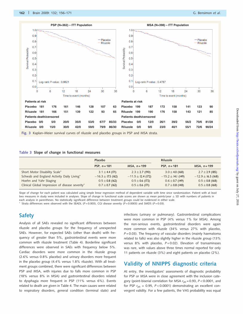
period, with no difference between the PSP and the MSA strata
[PSP, n = 171 (47.2%); MSA, n = 171 (43.0%); P = 0.21 by the
log-rank test]. With the strata combined, Kaplan–Meier survival
estimates at 36 months for riluzole and placebo groups were
52.6% and 54.9%, respectively. Comparison of survival curves
in treatment groups showed no statistically significant difference
either in the overall population (P = 0.42 by the stratified log-
rank test) or within strata (Kaplan–Meier estimates for PSP-
riluzole: 0.51, PSP-placebo: 0.50; and for MSA-riluzole: 0.53,
MSA-placebo: 0.58; P = 0.66 and P = 0.48 by the log-rank test,
respectively) (Fig. 3). Accordingly there was no statistically signi-
ficant treatment by strata interaction (P = 0.85, by a Cox model
analysis).
For secondary efficacy end-points of disease progression, 651
patients (86%) had at least two assessments allowing calculation
of a slope of change in scores for Hoehn and Yahr Staging, SEADL
and CGI for severity. For SMDS 714 (94%) patients had usable
data for calculating slope of change. All scales showed high sensi-
tivity to change with time (P50.0001). Strata differences were
evident with the SEADL, CGI for severity and SMDS (P = 0.003,
P = 0.0003, P = 0.03, respectively; Table 3), with the PSP group
showing more rapid progression compared to the MSA group.
As with the primary end-point, there was no statistically signifi-
cant difference between treatment groups in progression rate with
any of the scales, either in the overall population or within strata.
The PPPT analysis showed identical results for primary and sec-
ondary end-points (data not shown).
To test our initial assumptions, using the data acquired during
the study, we calculated the detectable difference in survival and
functional change. With power greater than 0.8, and � risk at 0.05
(two-tailed), the actual number of patients recruited and events
observed would have allowed us to detect a 40% decrease in
relative risk of death for PSP and 35% for MSA, consistent with
our initial hypothesis. For functional change, using the SEADL as
the most sensitive scale, we would have been able to detect diff-
erences in annual rate of progression of 32% for PSP and 36%
for MSA.
Table 2 ITT population characteristics at entry
Placebo Riluzole
PSP, n = 181 MSA, n = 199 PSP, n = 181 MSA, n = 199
Sex
Female (%) 40.3 44.2 44.8 46.2
Age at entry (years)� 67.7� 7.0 62.6� 8.1 68.0� 6.6 61.9� 8.5Age at onset (years)� 63.8� 7.0 58.2� 8.3 63.9� 7.0 57.3� 8.6Disease duration (years)� 3.9� 1.9 4.4� 2 4.1� 1.9 4.5� 1.9Short Motor Disability Scale (0–17) 6.4� 3.7 6.1� 3.9 6.7� 3.6 6.1� 3.6Frontal Assessment Battery (0–18)� 11.1� 4.3 14.6� 3.2 11.3� 4.1 14.3� 3.3Mini-Mental Status Examination (0–30)� 25.4� 4.4 27.8� 2.3 25.2� 4.4 27.6� 2.5Schwab & England Activity Daily Living (0–100)� 50.2� 24.5a 53.8� 24.8 48.3� 23.6 53.3� 24.3Hoehn and Yahr Staging (0–5)� 3.6� 1.0 3.4� 1.0 3.6� 0.9 3.5� 1.0Clinician Global Impression Disease severity (0–6)� 3.6� 1.0 3.6� 1.0 3.7� 1.0 3.6� 0.9Clinician Global Impression Dysautonomia (0–3)� 0.6� 0.6 1.8� 0.8 0.6� 0.6 1.8� 0.8
Quantitative variables were analysed using variance analysis and categorical data using a log-linear model. Factors included in models were strata, treatment, country
and strata by treatment interactions. Significance was set at P50.05 (two-tailed test) for each factor or interaction. No differences related to treatment group weredetected at entry for the whole population or within strata.All values are mean � SD.a n = 180 due to one missing value.� Differences between PSP and MSA strata (P50.05; two-tailed test) were found for all variables except for sex and the Short Motor Disability Scale score.
Riluzole in Parkinson-plus disorders Brain 2009: 132; 156–171 | 161
by guest on July 27, 2016http://brain.oxfordjournals.org/
Dow
nloaded from
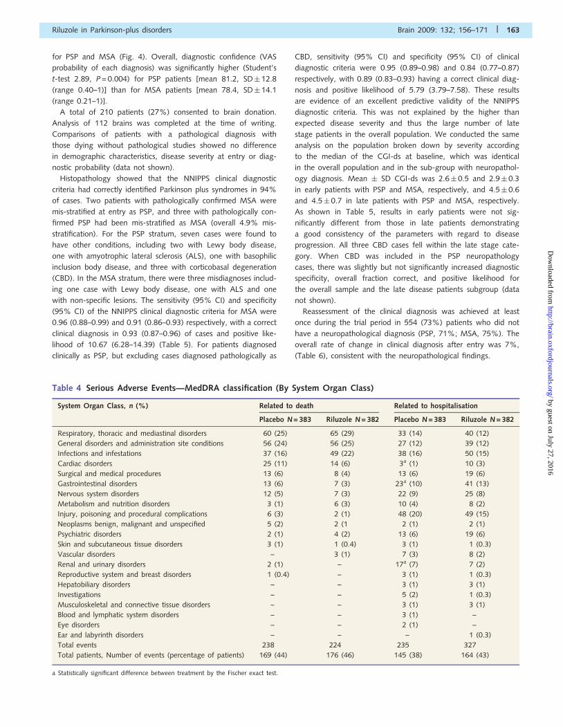
SafetyAnalysis of all SAEs revealed no significant differences between
riluzole and placebo groups for the frequency of unexpected
SAEs. However, for expected SAEs (other than death) with fre-
quency of greater than 5%, gastrointestinal events were more
common with riluzole treatment (Table 4). Borderline significant
differences were observed in SAEs with frequency below 5%.
Cardiac disorders were more common in the riluzole group
(2.6% versus 0.8% placebo) and urinary disorders more frequent
in the placebo group (4.4% versus 1.8% riluzole). With all treat-
ment groups combined, there were significant differences between
PSP and MSA, with injuries due to falls more common in PSP
(18% versus 8% in MSA) and gastrointestinal disorders related
to dysphagia more frequent in PSP (11% versus 6%). Events
related to death are given in Table 4. The main causes were related
to respiratory disorders, general condition (terminal state) and
infections (urinary or pulmonary). Gastrointestinal complications
were more common in PSP (4% versus 1% for MSA). Among
the non-serious events, gastrointestinal disorders were again
more common with riluzole (34% versus 27% with placebo,
P = 0.03). The frequency of vascular disorders (mainly haematoma
related to falls) was also slightly higher in the riluzole group (13%
versus 8% with placebo, P = 0.02). Elevation of transaminases
was rare, with values above three times normal reported for only
11 patients on riluzole (3%) and eight patients on placebo (2%).
Validity of NNIPPS diagnostic criteriaAt entry, the investigators’ assessments of diagnostic probability
for PSP or MSA were in close agreement with the inclusion cate-
gory (point-biserial correlation for MSA rpb = 0.93, P50.0001, and
for PSP rpb = 0.95, P50.0001) demonstrating an excellent con-
vergent validity. For a few patients, the VAS probability was equal
PSP (N=362) – ITT Population
Patients at risk
Placebo 181 176 161 146 128 107 63
Riluzole 181 166 151 139 122 93 65
Patients death/censored
Placebo 0/0 5/0 20/0 35/0 53/0 67/7 85/33
Riluzole 0/0 15/0 30/0 42/0 59/0 79/9 86/30
MSA (N=398) – ITT Population
Patients at risk
Placebo 199 187 172 158 141 123 90
Riluzole 199 190 176 158 143 121 85
Patients death/censored
Placebo 0/0 12/0 26/1 39/2 56/2 70/6 81/28
Riluzole 0/0 9/0 23/0 40/1 55/1 72/6 90/24
Fig. 3 Kaplan–Meier survival curves of riluzole and placebo groups in PSP and MSA strata.
Table 3 Slope of change in functional measures
Placebo Riluzole
PSP, n = 181 MSA, n = 199 PSP, n = 181 MSA, n = 199
Short Motor Disability Scale� 3.1� 4.4 (171) 2.3� 2.7 (190) 3.0� 4.0 (168) 2.7� 2.9 (185)Schwab and England Activity Daily Living� �16.3� 17.5 (162) �11.5� 12.4 (172) �15.2� 14.1 (149) �12.9� 16.5 (168)Hoehn and Yahr Staging 0.5� 0.8 (162) 0.5� 0.6 (172) 0.6� 0.7 (149) 0.5� 0.8 (168)Clinical Global Impression of disease severity� 0.7� 0.7 (162) 0.5� 0.6 (171) 0.7� 0.8 (148) 0.5� 0.8 (168)
Slope of change for each patient was calculated using simple linear regression method of dependent variable with time since randomization. Patient with at leasttwo measures in study were included in analyses. Slope of change in functional scale scores are shown as mean points/year � SD with numbers of patients ineach analysis in parentheses. No statistically significant difference between treatment groups could be evidenced in either scale.� Strata differences were observed with the SEADL (P = 0.003), CGI disease severity (P = 0.0003) and SMDS (P = 0.03).
162 | Brain 2009: 132; 156–171 G. Bensimon et al.
by guest on July 27, 2016http://brain.oxfordjournals.org/
Dow
nloaded from
for PSP and MSA (Fig. 4). Overall, diagnostic confidence (VAS
probability of each diagnosis) was significantly higher (Student’s
t-test 2.89, P = 0.004) for PSP patients [mean 81.2, SD� 12.8
(range 0.40–1)] than for MSA patients [mean 78.4, SD� 14.1
(range 0.21–1)].
A total of 210 patients (27%) consented to brain donation.
Analysis of 112 brains was completed at the time of writing.
Comparisons of patients with a pathological diagnosis with
those dying without pathological studies showed no difference
in demographic characteristics, disease severity at entry or diag-
nostic probability (data not shown).
Histopathology showed that the NNIPPS clinical diagnostic
criteria had correctly identified Parkinson plus syndromes in 94%
of cases. Two patients with pathologically confirmed MSA were
mis-stratified at entry as PSP, and three with pathologically con-
firmed PSP had been mis-stratified as MSA (overall 4.9% mis-
stratification). For the PSP stratum, seven cases were found to
have other conditions, including two with Lewy body disease,
one with amyotrophic lateral sclerosis (ALS), one with basophilic
inclusion body disease, and three with corticobasal degeneration
(CBD). In the MSA stratum, there were three misdiagnoses includ-
ing one case with Lewy body disease, one with ALS and one
with non-specific lesions. The sensitivity (95% CI) and specificity
(95% CI) of the NNIPPS clinical diagnostic criteria for MSA were
0.96 (0.88–0.99) and 0.91 (0.86–0.93) respectively, with a correct
clinical diagnosis in 0.93 (0.87–0.96) of cases and positive like-
lihood of 10.67 (6.28–14.39) (Table 5). For patients diagnosed
clinically as PSP, but excluding cases diagnosed pathologically as
CBD, sensitivity (95% CI) and specificity (95% CI) of clinical
diagnostic criteria were 0.95 (0.89–0.98) and 0.84 (0.77–0.87)
respectively, with 0.89 (0.83–0.93) having a correct clinical diag-
nosis and positive likelihood of 5.79 (3.79–7.58). These results
are evidence of an excellent predictive validity of the NNIPPS
diagnostic criteria. This was not explained by the higher than
expected disease severity and thus the large number of late
stage patients in the overall population. We conducted the same
analysis on the population broken down by severity according
to the median of the CGI-ds at baseline, which was identical
in the overall population and in the sub-group with neuropathol-
ogy diagnosis. Mean � SD CGI-ds was 2.6� 0.5 and 2.9� 0.3
in early patients with PSP and MSA, respectively, and 4.5� 0.6
and 4.5� 0.7 in late patients with PSP and MSA, respectively.
As shown in Table 5, results in early patients were not sig-
nificantly different from those in late patients demonstrating
a good consistency of the parameters with regard to disease
progression. All three CBD cases fell within the late stage cate-
gory. When CBD was included in the PSP neuropathology
cases, there was slightly but not significantly increased diagnostic
specificity, overall fraction correct, and positive likelihood for
the overall sample and the late disease patients subgroup (data
not shown).
Reassessment of the clinical diagnosis was achieved at least
once during the trial period in 554 (73%) patients who did not
have a neuropathological diagnosis (PSP, 71%; MSA, 75%). The
overall rate of change in clinical diagnosis after entry was 7%,
(Table 6), consistent with the neuropathological findings.
Table 4 Serious Adverse Events—MedDRA classification (By System Organ Class)
System Organ Class, n (%) Related to death Related to hospitalisation
Placebo N = 383 Riluzole N = 382 Placebo N = 383 Riluzole N = 382
Respiratory, thoracic and mediastinal disorders 60 (25) 65 (29) 33 (14) 40 (12)
General disorders and administration site conditions 56 (24) 56 (25) 27 (12) 39 (12)
Infections and infestations 37 (16) 49 (22) 38 (16) 50 (15)
Cardiac disorders 25 (11) 14 (6) 3a (1) 10 (3)
Surgical and medical procedures 13 (6) 8 (4) 13 (6) 19 (6)
Gastrointestinal disorders 13 (6) 7 (3) 23a (10) 41 (13)
Nervous system disorders 12 (5) 7 (3) 22 (9) 25 (8)
Metabolism and nutrition disorders 3 (1) 6 (3) 10 (4) 8 (2)
Injury, poisoning and procedural complications 6 (3) 2 (1) 48 (20) 49 (15)
Neoplasms benign, malignant and unspecified 5 (2) 2 (1 2 (1) 2 (1)
Psychiatric disorders 2 (1) 4 (2) 13 (6) 19 (6)
Skin and subcutaneous tissue disorders 3 (1) 1 (0.4) 3 (1) 1 (0.3)
Vascular disorders – 3 (1) 7 (3) 8 (2)
Renal and urinary disorders 2 (1) – 17a (7) 7 (2)
Reproductive system and breast disorders 1 (0.4) – 3 (1) 1 (0.3)
Hepatobiliary disorders – – 3 (1) 3 (1)
Investigations – – 5 (2) 1 (0.3)
Musculoskeletal and connective tissue disorders – – 3 (1) 3 (1)
Blood and lymphatic system disorders – – 3 (1) –
Eye disorders – – 2 (1) –
Ear and labyrinth disorders – – – 1 (0.3)
Total events 238 224 235 327
Total patients, Number of events (percentage of patients) 169 (44) 176 (46) 145 (38) 164 (43)
a Statistically significant difference between treatment by the Fischer exact test.
Riluzole in Parkinson-plus disorders Brain 2009: 132; 156–171 | 163
by guest on July 27, 2016http://brain.oxfordjournals.org/
Dow
nloaded from

Clinical features and natural historyof the NNIPPS cohortAnalysis of the systematic questionnaire on initial clinical signs
showed that the akinetic-rigid syndrome (with or without falls)
was a frequent presenting syndrome in the PSP stratum (70.2%)
and in the MSA stratum (61.8%). In the PSP stratum, oculomotor
abnormalities were an initial feature in only 7.7% of patients,
while 11.0% had presented with a behavioural or cognitive syn-
drome, and 5.8% had bulbar or pseudo-bulbar features. In MSA,
second to akinetic-rigid syndrome, the most common presenting
clinical features were cerebellar (22.1%) and genito-urinary
(9.1%). The date of onset of gait instability or falls was
documented in 93.3% of the population (92.5% PSP, 94.0%
MSA). Falls within the first year of disease onset, as incorporated
in the NINDS-SPSP criteria (Litvan et al., 2003) were present in
only 49.6% of the PSP patients, and were also present in 21.9%
of the MSA group. Similar results were observed in neuropatho-
logically confirmed cases (PSP 53.6%, MSA 27.9%). A similar
proportion of patients in the PSP and MSA strata had levodopa
therapy at entry [PSP, n = 307 (85%); MSA, n = 334 (84%)],
although the mean daily dose of levodopa was higher in the
MSA group (636 mg/day, range 50–2100 mg) compared to the
PSP group (509 mg/day, range 50–1600 mg) (P50.0001)
(Supplementary Table 2). Overall, most patients had a very poor
response to levodopa therapy. A greater than 50% response
to levodopa was reported for only 1.5% of MSA patients, and
none of the PSP group. A best-ever response to levodopa therapy
450% was reported more frequently for MSA patients (9.1%)
than for PSP patients (2.6%) (P = 0.0002). In the majority of
those who had experienced a good response to levodopa, the
duration of response was 51 year.
At time of entry in the study, the distribution of the various
syndromes is given for both diagnostic categories (Fig. 5).
Oculomotor abnormalities were present in all PSP patients
except one, but were also noted in 19% of MSA patients. PSP
patients showed a higher rate of cognitive and behavioural
syndromes. Dysautonomia was present in the majority of MSA
patients, with urinary symptoms (87%) more common than car-
diovascular symptoms (57%). Urinary symptoms were also
present in 48% of PSP patients. A cerebellar syndrome was
reported in 50% of MSA cases, but also a small number of PSP
patients (6%). Pyramidal signs were present in approximately
half the cases, slightly more in the MSA strata. A high frequency
of bulbar/pseudobulbar features was reported in the MSA patients
group (63%) and the PSP group (76%).
There were no differences between disease strata in terms of
gender, weight or height. MSA patients were younger than the
PSP patients at entry, younger at disease onset and had longer
disease duration prior to entry (Table 2). Assessments of disease
severity by the CGI-ds or the modified Hoehn and Yahr staging,
showed there were significantly fewer MSA patients in the most
severe stage (P = 0.024 and P = 0.048). Likewise, strata difference
in scores for the SEADL indicated less dependency for MSA than
for the PSP patients (P = 0.017). As expected, PSP patients scored
worse on cognitive functions as assessed with the MMSE
(P = 0.0001) (Table 2).
0
20
40
60
80
100
0 20 40 60 80 100
MSA Diagnos is probability (mm)
PSP
Dia
gnos
is p
roba
bilit
y (m
m)
Fig. 4 Convergent validity of NNIPPS Diagnostic Criteria with
Investigators’ Diagnostic Probability (VAS). At the inclusion
visit, following patients’ assignment to strata using the NNIPPS
diagnostic criteria, investigators were asked to evaluate the
probability of each diagnosis (PSP, MSA), using a 100 mm VAS.
All 760 patients are plotted on the graph according to
the probability score on each VAS (PSP-vertical axis, MSA-
horizontal axis). Solid diamonds represent patients included
in the PSP stratum; White circles represent patients included in
the MSA stratum. Convergent validity of the NNIPPS inclusion
criteria with the investigators’ assessment of diagnostic prob-
ability was tested with the point-biserial correlation. MSA,
rpb = 0.93 (P50.0001), PSP, rpb = 0.95, (P50.0001).
Table 5 Predictive validity of NNIPPS clinical diagnostic criteria in deceased patients with neuropathological diagnosis
CGI54 n = 39 CGI54 n = 73 ALL n = 112
PSP MSA PSP MSA PSP MSA
Sensitivity (95% CI) 0.95 (0.82–0.99)0.88 (0.73–0.95) 0.95 (0.86–0.98) 1.0 (0.91–1) 0.95 (0.88–0.98) 0.96 (0.88–0.99)
Specificity (95% CI) 0.84 (0.7–0.89) 0.91 (0.79–0.96) 0.83 (0.74–0.87) 0.91 (0.86–0.91) 0.84 (0.77–0.87) 0.91 (0.86–0.93)
Overall fraction correct (95% CI)0.90 (0.77–0.94)0.90 (0.77–0.96) 0.89 (0.80–0.93) 0.95 (0.88–0.95) 0.89 (0.83–0.93) 0.93 (0.87–0.96)
Positive likelihood (95% CI) 6.01 (2.83–8.60)9.71 (3.50–26.48)5.68 (3.35–7.71)11.25 (6.32–11.25)5.79 (3.79–7.58)10.67 (6.28–14.39)
Sensitivity, specificity, overall fraction correct and positive likelihood (95% CI) of NNIPPS diagnostic criteria in the overall population with neuropathology diagnosis, and
broken down by disease severity as defined by the CGI. Early patients were defined as those below the median (CGI, range 1–3); late patients were those equal or overthe median (CGI, range 4–6).
164 | Brain 2009: 132; 156–171 G. Bensimon et al.
by guest on July 27, 2016http://brain.oxfordjournals.org/
Dow
nloaded from

Demographics and clinical factors influencing survival were
assessed with univariate and multivariate Cox model analysis
(Supplementary material and Supplementary Table 7). In the uni-
variate analyses, only variables scoring disease severity were
predictive of survival. Accordingly, the stepwise multivariate
analysis selected first the SEADL, followed by the CGI scales for
disease severity and for dysautonomia, as strong predictors of
survival.
However, following adjustment on these variables, disease dura-
tion was selected in the model (RR = 0.923, P = 0.007) indicating
that at constant severity patients with a shorter disease duration
at entry had a worse prognosis (‘fast progressors’). To visualise the
discriminating accuracy of the combined variables, we constructed
a prognostic score for each patient using a Cox coefficient
weighted linear combination of the selected prognostic variables
(Supplementary Fig. 1A). Following adjustment on the prognostic
score as a variable, the strata factor became significant (RR =
0.657, P = 0.0002) indicating that PSP patients on the whole had
a worse prognosis compared to MSA patients at constant disease
severity and disease duration at entry (Supplementary Fig. 1B).
Testing for a treatment effect on survival following adjustment
on the prognostic variables did not change the results of the
log-rank analysis nor was there any significant interaction between
treatment and prognostic variables (Supplementary Table 3).
DiscussionOur results show that riluzole (up to 200 mg/day) is unlikely to
be effective as a disease-modifying agent in PSP or MSA. Riluzole
has a definite effect in ALS with an estimated 30–40% decrease in
relative risk at 12–18 months (Bensimon et al., 1994; Lacomblez
et al., 1996; Miller et al., 2007). NNIPPS was therefore designed
to detect an effect of similar size. Power calculation assumptions
for the placebo group were based largely on retrospective data
(Litvan et al., 1996a, c, 2003; Testa et al., 1996, 2001; Ben-
Shlomo et al., 1997), which underestimated the number of
events observed in NNIPPS by 2–6%. Hence the NNIPPS trial
achieved the power necessary to detect the hypothesized drug
effect. The failure of NNIPPS to detect that effect could be
due to heterogeneity in the trial population (loss of power),
uneven distribution of demographic and clinical prognostic factors
or comorbidity across treatment groups (randomization bias), poor
compliance, or simply a smaller pharmacological effect in these
conditions than the one used at the planning stage (effect size).
With regard to disease heterogeneity, a major difficulty in
designing trials of disease-modifying therapy in neurodegenerative
diseases is lack of diagnostic confidence, particularly at a stage
of disease evolution when intervention is likely to be most effec-
tive. Indeed, there are no prospective studies of clinical diagnostic
criteria assessed against the definitive criterion of histopathology
in PSP or MSA (Litvan et al., 2003). Such studies are not easy
to achieve, as long term follow-up and a high autopsy rate are
required. Until validated biomarkers are available for use in trials,
clinical criteria prospectively tested against pathology are essential
to understand heterogeneity, which is a potential source of bias
in estimating drug effects in Parkinson plus syndromes, as in idio-
pathic Parkinson’s disease. Furthermore, criteria for large scale
clinical trials in rare diseases such as PSP and MSA should be
robust and simple, so that they can be applied in international
multi-centre studies and not only in highly specialized centres.
The NNIPPS diagnostic criteria represent a simplification of existing
diagnostic criteria and we have shown them to have excellent
convergent and predictive validities in this study population,
Table 6 Change in clinical diagnosis during the trial showing predictive validity of NNIPPS clinical diagnostic criteria insurviving patients
Clinical diagnosis at inclusion Final clinical diagnosis All
PSP MSA Other�
PSP 246 (96%) 2 (1%) 8 (3%) 256
MSA 13 (4%) 270 (91%) 15 (5%) 298
All 259 272 23 554
� Other diagnosis: MSA (N = 15): IPD n = 7, ALS n = 2, CBD n = 3, LBD n = 1, Carbon dioxide intox n = 1, MPI multilacunar n = 1; PSP (N = 8): Mixed PSP/MSA n = 1,Lower body Parkinsonism – pseudo PSP n = 1, CBD n = 1, FTDP or Motor neuron disease n = 1, ophthalmoplegia n = 1, FTD n = 1, multiple cerebral infarct n = 1,unidentified n = 1.
0%
20%
40%
60%
80%
100%
Urinar
y
Cardio
vasc
ular
Cereb
ellar
Pyram
idal
Bulbar
Behav
ioral
Cognit
ive
Oculom
otor
Fig. 5 Syndrome profile of patients in PSP and MSA Strata.
At the inclusion visit, and based on the clinical neurological
assessments, investigators were asked to describe the syndrome
profile of the patients using a systematic (yes/no) question-
naire. Each bar represents the percentage of patients within
each stratum positive for a given syndrome. Black bars
represent the MSA stratum, grey bars the PSP stratum. The
akinetic rigid syndrome was a mandatory inclusion criterion
for both strata and therefore is not represented.
Riluzole in Parkinson-plus disorders Brain 2009: 132; 156–171 | 165
by guest on July 27, 2016http://brain.oxfordjournals.org/
Dow
nloaded from

despite variations in practice across three European countries and
44 centres. Although pathological examination was only possible
in 112 cases (15% of recruited patients), the pathological sample
is representative of those patients who died (45%) and, most
likely, of the population as a whole. Further analysis of these
diagnostic criteria in relation to MRI, DNA and neuropsychology
is ongoing. At this stage it can be concluded that the estimated
rate of misdiagnosis (about 6% overall, representing at most 46
patients in the ITT) is acceptably low and is unlikely to have biased
the estimate of the drug effect. We could not detect imbalance
in entry parameters with relevant prognostic significance for
survival or disease progression which could account for the lack
of treatment efficacy.
It is likely that a neuroprotective agent should be started in
the earliest phase of the disease in order to demonstrate efficacy.
Hence the impact of a neuroprotective agent is likely to be an
inverse function of the stage of disease progression. In support of
this assumption, riluzole did not show efficacy in ALS when tested
in a trial including late stage disease patients (Bensimon et al.,
2002). In the present trial, disease severity in the population
included was greater than anticipated and the death rate was
also higher than expected. Overall, about 50% of patients were
classified as severely disabled or wheelchair bound (i.e. in the
latest stages) by the Hoehn and Yahr staging and over 50% simi-
larly classified as markedly, severely or extremely ill by the CGI
scale for disease severity. However, adjustment for prognostic
factors with the Cox model did not show any trend for a treat-
ment effect, nor was there a significant treatment by prognostic
factor interaction, meaning that the treatment effect is constant
across the levels of the prognostic variables.
Even though treatment withdrawal rate was relatively high
(25%), and might have had an impact on the size of a treatment
effect, the overall mean dose was close to the maximum, and
compliance rate was good (81.2%) considering the length of the
trial, with a mean follow-up time close to the maximum planned
(3 years). Finally, the PPPT sensitivity analysis was consistent
with the ITT. Hence, the observed lack of treatment effect
in this Parkinson plus population seems relatively robust. Our
results are similar to those observed in Huntington’s disease
(Landwehrmeyer et al., 2007) and contrast with the findings in
ALS. This may indicate that the disease pathways targeted by
riluzole in ALS are more specific than previously thought.
At the time of designing the NNIPPS study there were no
validated instruments for measuring change in function in these
disorders. Since the priority was to detect a disease-modifying
effect of riluzole, we chose to use survival as a robust and
unambiguous endpoint for the primary analysis. There are several
reasons why prospectively acquired information on survival pro-
vides a unique tool for understanding these diseases and for devel-
oping new assessment instruments for clinical trials. First, it is
possible to achieve complete ascertainment of the endpoint, as
we have shown. Secondly, the high rate of death in PSP and
MSA limits the use of functional scales as measurements of disease
progression. Inevitably, the extent of missing functional data is
not random, so that in case of drug toxicity increasing death
rate, the remaining functional data in the active drug group is
misleading as regards efficacy (Wu, 1988; Wu and Bailey, 1988)
because they originate from selection of a biased sub-group. In
early patients, when the likelihood of informative censoring is
least, slope of change in functional measures might be appropriate
as an end point in a definitive phase III trial where using survival
as an endpoint would result in substantially increasing the number
of patients and/or length of the trial. The parameters provided in
this article allow power calculations for either context since linear-
ity is a basic assumption for slope of change and should hold true
early or late in the course of disease.
Third, survival data provide a unique insight into the natural
history of these disorders and help to validate functional assess-
ments in relation to prognosis. In this study, the median survival
from onset of symptoms is in keeping with our prior assumptions
based on retrospective (Litvan et al., 1996d; Testa et al., 1996,
2001; Vanacore et al., 2001a, b; Litvan et al., 2003) and prospec-
tive studies (Schrag et al., 2008). Following a step-wise Cox model
adjustment on prognostic factors which selected disease severity
scores (SEADL, CGI-ds, CGI-dysautonomia) and disease duration,
there was a significant difference between strata consistent with
the difference in the rate of disease progression between the two
strata. Finally, our data show that it is feasible to use survival as
a primary endpoint in phase III studies and that our strategy of
recruiting patients with PSP and MSA into a single stratified trial
is methodologically valid where a generic neuroprotective effect is
hypothesied. On the other hand, where the agent to be tested is
thought to act on disease-specific pathways (e.g. processing of tau
in PSP, or alpha-synuclein in MSA) a different strategy is likely to
be more appropriate. The demographic features of the NNIPPS
cohort clearly indicate that patients presenting with an akinetic-
rigid syndrome and categorized as having PSP or MSA with
reasonably high confidence have very poor prognosis, and no
significant response to levodopa therapy. Unfortunately, there
are no comparable prospective studies with which to compare
our cohort. The European MSA Study Group (EMSA-SG) has
reported preliminary data on a functional scale developed for
MSA based on assessments of 50 patients diagnosed on the
basis of the Gilman criteria (Gilman et al., 1998). A total of 412
patients with presumed MSA were included in a European registry
(Geser et al., 2005, 2006), but no information on survival is yet
available from the EMSA database. Nevertheless, demographic
features of the 50 patients reported from the EMSA are broadly
comparable to those in the NNIPPS MSA stratum (Geser et al.,
2006). In a cohort study of 162 PSP patients the median survival
since disease onset was 7.3 years, compared to 7.8 years in the
NNIPPS cohort (Golbe and Ohman-Strickland, 2007). As with
NNIPPS, functional scores predicted survival. However, multivari-
ate analysis of prognostic factors revealed that age of onset and
gender were predictors of survival. For the former, the apparent
discrepancy is likely to be related to differences in the method
of survival analysis since when survival is calculated from disease
onset in our cohort, age of onset becomes a significant predictor
of survival as well (data not shown). In relation to a gender effect,
the sex ratio in most studies, as in the NNIPPS cohort, shows
an equal proportion of men and women or a slight excess of
men (Golbe et al., 1988; Santacruz et al., 1998; Schrag et al.,
1999; Nath et al., 2001, 2003), whereas in the study of Golbe and
Ohman-Strickland (2007) the sex ratio was reversed, indicating
166 | Brain 2009: 132; 156–171 G. Bensimon et al.
by guest on July 27, 2016http://brain.oxfordjournals.org/
Dow
nloaded from
a potential sample bias. However, in MSA, demographic features
such as age of onset and sex ratio are similar in the NNIPPS cohort
to those recorded in previous natural history studies (Bower et al.,
1997; Schrag et al., 1999; Vanacore et al., 2001a, b; Schrag et al.,
2008).
In order to achieve a relatively homogeneous population of
patients in the PSP strata, ocumomotor abnormalities were
mandatory at inclusion. This increased the accuracy of diagnosis
(as confirmed pathologically) but did not take into account ‘PSP-
Parkinsonism’ patients without oculomotor abnormalities (Williams
et al., 2005, 2007) who may be confused with patients with PD
or with MSA-P. Likewise, in order to recruit MSA patients
with relatively early disease, we took a pragmatic approach so
as to avoid, as far as possible, recruiting patients with disorders
such as olivopontocerebellar atrophy (Berciano et al., 2006) and
idiopathic late-onset cerebellar ataxia, (Gilman and Quinn, 1996;
Gilman et al., 2000). We expected this to reduce the proportion
of patients with the cerebellar form of MSA (MSA-C) in our
cohort, but cerebellar features were noted at presentation in
20% of our patients, similar to the findings of a prospective
study (Schrag et al., 2008). Thus the NNIPPS population is reason-
ably representative of the whole (European and North American)
MSA population. In this context, we have clearly shown that
clinical features that have been regarded as characteristic of
each disorder, and which are of key importance in the different
consensus diagnostic criteria (Litvan et al., 1996a, b, d, 1997,
2003; Gilman et al., 1998) are common to both disorders. Thus
at entry urinary symptoms were present in over 85% of patients
in the MSA stratum but also in 50% in the PSP stratum (Fig. 5)
and oculomotor signs, required for inclusion in the PSP stratum,
were recorded in about 20% of patients in the MSA stratum.
However, this included any evidence of supranuclear ophthalmo-
plegia, whereas allocation to the PSP stratum required significant
supranuclear impairment of downward gaze. Likewise, behavioural
and cognitive abnormalities were judged as being common in
both PSP and MSA, in keeping with studies on cognitive function
in these disorders (Robbins et al., 1992, 1994; Burk et al., 2006;
Lyoo et al., 2008). Despite this overlap, we have shown that PSP
and MSA can be differentiated by neurologists at a relatively early
stage in the disease process using the NNIPPS diagnostic criteria.
A striking feature of the NNIPPS diagnostic criteria is the high
sensitivity at inclusion, although a direct comparison with pub-
lished diagnostic criteria is not possible as the timing of assess-
ments was different (Osaki et al., 2002, 2004; Litvan et al., 2003).
The NNIPPS diagnostic assessments were carried out on average
2 years before death in the pathologically confirmed cases
whereas in the published criteria the assessments were made at
the first (‘diagnostic’) visit when sensitivity is low, and at the
end of the disease (i.e., on the last visit before death) when sen-
sitivity is high. Studies of the sensitivity of the NNIPPS criteria
at the earliest stages of disease process are now needed, though
we provide evidence that the criteria still display acceptable quality
in the sub-group below the mid-range of disease progression.
Indeed, only because we studied both conditions in one cohort
could we discern the close similarities and the diagnostically impor-
tant differences between PSP and MSA.
We have shown that a large scale randomized trial with survival
as the primary endpoint is feasible using the Parkinson plus con-
cept to allow relatively early diagnosis and recruitment of a sur-
prisingly homogeneous population, as shown by the performance
of the NNIPPS diagnostic criteria informed by pathology. The rate
of death was higher than anticipated, allowing us to shorten the
trial slightly. We have learnt from clinical trials in ALS that survival
does not necessarily equate with functional change (Lacomblez
et al., 1996; Meininger, 2005). This has resulted in recommenda-
tions by the regulatory authorities (e.g. European Medicines
Agency, Committee for Proprietary Medicinal Products, CPMP/
EWP/565/98) and by the World Federation of Neurology
Committee on Research (Miller et al., 1999) that survival should
be the primary endpoint for definitive demonstration of a neuro-
protective drug effect. Future phase III studies on potential neu-
roprotective agents in these disorders should consider using
survival as a primary endpoint, and combining patients with pre-
sumed PSP and MSA in the same trial in order to improve our
understanding of both.
Supplementary materialSupplementary material is available at Brain online.
FundingNNIPPS was an academic-led study with core funding
from the European Union 5th Framework Programme (QLG1-
CT-2000-01262); support from French Health Ministry,
Programme Hospitalier de Recherche Clinique (AOM97073,
AOM01125) and from Sanofi-Aventis affiliates in the UK, France
and Germany providing an unconditional research grant and drug
supply throughout the study. Three academic institutions (Institute
of Psychiatry, King’s College London; Assistance Publique-
Hopitaux de Paris; and University of Ulm) were sponsors of the
study in each country, and jointly own the data. The protocol and
amendments were reviewed and approved by the Comite de
Protection des Personnes of Pitie-Salpetriere Hospital (France),
the UK Multicentre Research Ethics Committee (MREC), (UK),
Ethikkommission of the University of Ulm, (Germany), and by
local Institutional Review Boards (Ethics Committees) where
appropriate (UK, Germany).
AcknowledgementsWe thank the patients and their families for their commitment and
altruism, and The French and UK PSP Associations and the UK
Parkinson’s Disease Research Group for their help and support.
We are grateful to the many colleagues who were not formally
part of the NNIPPS consortium but whose support contributed
to the success of the study. The study protocol was filed in the
open clinical trial registry (www.clinicaltrials.gov) with ID number
NCT00211224.
Riluzole in Parkinson-plus disorders Brain 2009: 132; 156–171 | 167
by guest on July 27, 2016http://brain.oxfordjournals.org/
Dow
nloaded from
ReferencesAlbers DS, Augood SJ. New insights into progressive supranuclear palsy.
Trends Neurosci 2001; 24: 347–53.Albin RL, Greenamyre JT. Alternative excitotoxic hypotheses. Neurology
1992; 42: 733–8.Allison P. Survival Analysis using the SAS System – A Practical Guide. SAS
Institute Inc.; 1995.
American Psychological Association. Standard for educational and psy-
chological testing. Washington DC: American Psychological
Association ed.; 1985.Attia J. Moving beyond sensitivity and specificity: Using likelihood ratios
to help interpret diagnostic tests. Australian Prescriber 2003; 26:
111–13.Ben-Shlomo Y, Wenning GK, Tison F, Quinn NP. Survival of patients
with pathologically proven multiple system atrophy: a meta-analysis.
Neurology 1997; 48: 384–93.
Bensimon G, Lacomblez L, Delumeau JC, Bejuit R, Truffinet P, Meininger V.
A study of riluzole in the treatment of advanced stage or elderly patients
with amyotrophic lateral sclerosis. J Neurol 2002; 249: 609–15.
Bensimon G, Lacomblez L, Meininger V. A controlled trial of riluzole in
amyotrophic lateral sclerosis. ALS/Riluzole Study Group. N Engl J Med
1994; 330: 585–91.
Berciano J, Boesch S, Perez-Ramos JM, Wenning GK.
Olivopontocerebellar atrophy: toward a better nosological definition.
Mov Disord 2006; 21: 1607–13.Bishop Y, Fienberg S, Holland P. Discrete multivariate analysis: theory
and practice. Cambridge, MA: MIT Press; 1975.
Bower JH, Maraganore DM, McDonnell SK, Rocca WA. Incidence of
progressive supranuclear palsy and multiple system atrophy in
Olmsted County, Minnesota, 1976 to 1990. Neurology 1997; 49:
1284–8.
Burk K, Daum I, Rub U. Cognitive function in multiple system atrophy of
the cerebellar type. Mov Disord 2006; 21: 772–6.
Burn DJ, Lees AJ. Progressive supranuclear palsy: where are we now?
Lancet Neurol 2002; 1: 359–69.Caumont AS, Octave JN, Hermans E. Specific regulation of rat glial cell
line-derived neurotrophic factor gene expression by riluzole in C6
glioma cells. J Neurochem 2006; 97: 128–39.
Cox D. Regression models and life tables. J R Stat Soc 1972; B34:
187–220.Dickson DW. Neuropathologic differentiation of progressive supranuclear
palsy and corticobasal degeneration. J Neurol 1999; 246 (Suppl. 2):
II6–15.
Dickson DW, Rademakers R, Hutton ML. Progressive supranuclear palsy:
pathology and genetics. Brain Pathol 2007; 17: 74–82.Diguet E, Fernagut PO, Scherfler C, Wenning G, Tison F. Effects of
riluzole on combined MPTP + 3-nitropropionic acid-induced mild to
moderate striatonigral degeneration in mice. J Neural Transm 2005;
112: 613–31.
Doble A. The role of excitotoxicity in neurodegenerative disease: impli-
cations for therapy. Pharmacol Ther 1999; 81: 163–221.
Folstein MF, Folstein SE, McHugh PR. ‘‘Mini-mental state’’. A practical
method for grading the cognitive state of patients for the clinician. J
Psychiatr Res 1975; 12: 189–98.
Geser F, Seppi K, Stampfer-Kountchev M, Kollensperger M, Diem A,
Ndayisaba JP, et al. The European Multiple System Atrophy-Study
Group (EMSA-SG). J Neural Transm 2005; 112: 1677–86.Geser F, Wenning GK, Seppi K, Stampfer-Kountchev M, Scherfler C,
Sawires M, et al. Progression of multiple system atrophy (MSA): a
prospective natural history study by the European MSA Study Group
(EMSA SG). Mov Disord 2006; 21: 179–86.
Gilman S, Little R, Johanns J, Heumann M, Kluin KJ, Junck L, et al.
Evolution of sporadic olivopontocerebellar atrophy into multiple
system atrophy. Neurology 2000; 55: 527–32.Gilman S, Low P, Quinn N, Albanese A, Ben-Shlomo Y, Fowler C, et al.
Consensus statement on the diagnosis of multiple system atrophy.
American Autonomic Society and American Academy of Neurology.
Clin Auton Res 1998; 8: 359–62.
Gilman S, Quinn NP. The relationship of multiple system atrophy to
sporadic olivopontocerebellar atrophy and other forms of idiopathic
late-onset cerebellar atrophy. Neurology 1996; 46: 1197–9.
Golbe LI, Davis PH, Schoenberg BS, Duvoisin RC. Prevalence and natural
history of progressive supranuclear palsy. Neurology 1988; 38:
1031–4.Golbe LI, Ohman-Strickland PA. A clinical rating scale for progressive
supranuclear palsy. Brain 2007; 130: 1552–65.Hauw JJ, Daniel SE, Dickson D, Horoupian DS, Jellinger K,
Lantos PL, et al. Preliminary NINDS neuropathologic criteria for
Steele-Richardson-Olszewski syndrome (progressive supranuclear
palsy). Neurology 1994; 44: 2015–19.
Heiser V, Engemann S, Brocker W, Dunkel I, Boeddrich A, Waelter S, et al.
Identification of benzothiazoles as potential polyglutamine aggregation
inhibitors of Huntington’s disease by using an automated filter retarda-
tion assay. Proc Natl Acad Sci USA 2002; 99 (Suppl. 4): 16400–6.
Hoehn MM, Yahr MD. Parkinsonism: onset, progression and mortality.
Neurology 1967; 17: 427–42.
Lacomblez L, Bensimon G, Leigh PN, Guillet P, Meininger V. Dose-ran-
ging study of riluzole in amyotrophic lateral sclerosis. Amyotrophic
Lateral Sclerosis/Riluzole Study Group II. Lancet 1996; 347: 1425–31.
Landwehrmeyer GB, Dubois B, de Yebenes JG, Kremer B, Gaus W,
Kraus PH, et al. Riluzole in Huntington’s disease: a 3-year, randomized
controlled study. Ann Neurol 2007; 62: 262–72.Lantos PL, Papp MI. Cellular pathology of multiple system atrophy: a
review. J Neurol Neurosurg Psychiatry 1994; 57: 129–33.Litvan I. Update on epidemiological aspects of progressive supranuclear
palsy. Mov Disord 2003; 18 (Suppl. 6): S43–50.Litvan I, Agid Y, Calne D, Campbell G, Dubois B, Duvoisin RC, et al.
Clinical research criteria for the diagnosis of progressive supranuclear
palsy (Steele-Richardson-Olszewski syndrome): report of the NINDS-
SPSP international workshop. Neurology 1996a; 47: 1–9.
Litvan I, Agid Y, Jankovic J, Goetz C, Brandel JP, Lai EC, et al. Accuracy
of clinical criteria for the diagnosis of progressive supranuclear
palsy (Steele-Richardson-Olszewski syndrome). Neurology 1996b; 46:
922–30.Litvan I, Bhatia KP, Burn DJ, Goetz CG, Lang AE, McKeith I, et al.
Movement Disorders Society Scientific Issues Committee report: SIC
Task Force appraisal of clinical diagnostic criteria for Parkinsonian dis-
orders. Mov Disord 2003; 18: 467–86.
Litvan I, Goetz CG, Jankovic J, Wenning GK, Booth V, Bartko JJ, et al.
What is the accuracy of the clinical diagnosis of multiple system atro-
phy? A clinicopathologic study. Arch Neurol 1997; 54: 937–44.Litvan I, Hauw JJ, Bartko JJ, Lantos PL, Daniel SE, Horoupian DS, et al.
Validity and reliability of the preliminary NINDS neuropathologic cri-
teria for progressive supranuclear palsy and related disorders.
J Neuropathol Exp Neurol 1996c; 55: 97–105.
Litvan I, Hutton M. Clinical and genetic aspects of progressive supra-
nuclear palsy. J Geriatr Psychiatry Neurol 1998; 11: 107–14.
Litvan I, Lees AJ. Progressive supranuclear palsy. Adv Neurol 1999; 80:
341–5.
Litvan I, Mangone CA, McKee A, Verny M, Parsa A, Jellinger K, et al.
Natural history of progressive supranuclear palsy (Steele-Richardson-
Olszewski syndrome) and clinical predictors of survival: a clinicopatho-
logical study. J Neurol Neurosurg Psychiatry 1996d; 60: 615–20.
Lyoo CH, Jeong Y, Ryu YH, Lee SY, Song TJ, Lee JH, et al. Effects of
disease duration on the clinical features and brain glucose metabolism
in patients with mixed type multiple system atrophy. Brain 2008; 131:
438–46.
Mantel N, Haenszel W. Statistical aspects of the analysis of data from
retrospective studies of disease. J Natl Cancer Inst 1958; 22: 719–48.Mattson MP. Excitotoxic and excitoprotective mechanisms: Abundant
targets for the prevention and treatment of neurodegenerative disor-
ders. Neuromolecular Med 2003; 3: 65–94.
Meininger V. Clinical trials in ALS: what did we learn from recent trials in
humans? Neurodegener Dis 2005; 2: 208–14.
168 | Brain 2009: 132; 156–171 G. Bensimon et al.
by guest on July 27, 2016http://brain.oxfordjournals.org/
Dow
nloaded from

Miller RG, Mitchell JD, Lyon M, Moore DH. Riluzole for amyotrophic
lateral sclerosis (ALS)/motor neuron disease (MND). Cochrane
Database Syst Rev 2007; CD001447.
Miller RG, Munsat TL, Swash M, Brooks BR. Consensus guidelines
for the design and implementation of clinical trials in ALS. World
Federation of Neurology committee on Research. J Neurol Sci 1999;
169: 2–12.Nath U, Ben-Shlomo Y, Thomson RG, Lees AJ, Burn DJ. Clinical features
and natural history of progressive supranuclear palsy: a clinical cohort
study. Neurology 2003; 60: 910–16.
Nath U, Ben-Shlomo Y, Thomson RG, Morris HR, Wood NW,
Lees AJ, et al. The prevalence of progressive supranuclear palsy
(Steele-Richardson-Olszewski syndrome) in the UK. Brain 2001;
124: 1438–49.
Nath U, Burn DJ. The epidemiology of progressive supranuclear palsy
(Steele-Richardson-Olszewski syndrome). Parkinsonism Relat Disord
2000; 6: 145–53.Nunnally J, Bernstein I. Psychometric theory. New York: McGraw-Hill,
Inc; 1994.Osaki Y, Ben-Shlomo Y, Lees AJ, Daniel SE, Colosimo C, Wenning G,
et al. Accuracy of clinical diagnosis of progressive supranuclear palsy.
Mov Disord 2004; 19: 181–9.
Osaki Y, Ben-Shlomo Y, Wenning GK, Daniel SE, Hughes A, Lees AJ,
et al. Do published criteria improve clinical diagnostic accuracy in mul-
tiple system atrophy? Neurology 2002; 59: 1486–91.
Papp MI, Kahn JE, Lantos PL. Glial cytoplasmic inclusions in the CNS
of patients with multiple system atrophy (striatonigral degeneration,
olivopontocerebellar atrophy and Shy-Drager syndrome). J Neurol Sci
1989; 94: 79–100.Przedborski S. Pathogenesis of nigral cell death in Parkinson’s disease.
Parkinsonism Relat Disord 2005; 11 (Suppl. 1): S3–7.Robbins TW, James M, Lange KW, Owen AM, Quinn NP, Marsden CD.
Cognitive performance in multiple system atrophy. Brain 1992; 115:
271–91.
Robbins TW, James M, Owen AM, Lange KW, Lees AJ, Leigh PN, et al.
Cognitive deficits in progressive supranuclear palsy, Parkinson’s dis-
ease, and multiple system atrophy in tests sensitive to frontal lobe
dysfunction. J Neurol Neurosurg Psychiatry 1994; 57: 79–88.
Santacruz P, Uttl B, Litvan I, Grafman J. Progressive supranuclear palsy: a
survey of the disease course. Neurology 1998; 50: 1637–47.
Scherfler C, Sather T, Diguet E, Stefanova N, Puschban Z, Tison F, et al.
Riluzole improves motor deficits and attenuates loss of striatal neurons
in a sequential double lesion rat model of striatonigral degeneration
(parkinson variant of multiple system atrophy). J Neural Transm 2005;
112: 1025–33.
Schrag A, Ben-Shlomo Y, Quinn NP. Prevalence of progressive supra-
nuclear palsy and multiple system atrophy: a cross-sectional study.
Lancet 1999; 354: 1771–5.
Schrag A, Wenning GK, Quinn N, Ben-Shlomo Y. Survival in multiple
system atrophy. Mov Disord 2008; 23: 294–6.
Shortland PJ, Leinster VH, White W, Robson LG. Riluzole promotes cell
survival and neurite outgrowth in rat sensory neurones in vitro. Eur J
Neurosci 2006; 24: 3343–53.Spillantini MG, Goedert M. The alpha-synucleinopathies: Parkinson’s dis-
ease, dementia with Lewy bodies, and multiple system atrophy. Ann N
Y Acad Sci 2000; 920: 16–27.
Streiner D, Norman G. Health Measurement Scales: a practical guide to
their development and use. Oxford: Oxford University Press; 2003.
Testa D, Filippini G, Farinotti M, Palazzini E, Caraceni T. Survival in
multiple system atrophy: a study of prognostic factors in 59 cases. J
Neurol 1996; 243: 401–4.
Testa D, Monza D, Ferrarini M, Soliveri P, Girotti F, Filippini G.
Comparison of natural histories of progressive supranuclear palsy and
multiple system atrophy. Neurol Sci 2001; 22: 247–51.
Vanacore N, Bonifati V, Colosimo C, Fabbrini G, De Michele G,
Marconi R, et al. Epidemiology of progressive supranuclear palsy.
ESGAP Consortium. European Study Group on Atypical
Parkinsonisms. Neurol Sci 2001b; 22: 101–3.
Vanacore N, Bonifati V, Fabbrini G, Colosimo C, De Michele G,
Marconi R, et al. Epidemiology of multiple system atrophy. ESGAP
Consortium. European Study Group on Atypical Parkinsonisms.
Neurol Sci 2001a; 22: 97–9.
Watanabe H, Saito Y, Terao S, Ando T, Kachi T, Mukai E, et al.
Progression and prognosis in multiple system atrophy: an analysis of
230 Japanese patients. Brain 2002; 125: 1070–83.Williams DR, de Silva R, Paviour DC, Pittman A, Watt HC, Kilford L,
et al. Characteristics of two distinct clinical phenotypes in pathologi-
cally proven progressive supranuclear palsy: Richardson’s syndrome
and PSP-parkinsonism. Brain 2005; 128: 1247–58.
Williams DR, Holton JL, Strand C, Pittman A, de Silva R, Lees AJ, et al.
Pathological tau burden and distribution distinguishes progressive
supranuclear palsy-parkinsonism from Richardson’s syndrome. Brain
2007; 130: 1566–76.
Winer B. Design and analysis of factorial experiments in Statistical prin-
ciples in experimental design. New York: McGraw-Hill Inc; 1971.
p. 309–430.
Wu MC. Sample size for comparison of changes in the presence of right
censoring caused by death, withdrawal, and staggered entry. Control
Clin Trials 1988; 9: 32–46.Wu MC, Bailey K. Analysing changes in the presence of informative
right censoring caused by death and withdrawal. Stat Med 1988; 7:
337–46.
Yoo MH, Hyun HJ, Koh JY, Yoon YH. Riluzole inhibits VEGF-
induced endothelial cell proliferation in vitro and hyperoxia-induced
abnormal vessel formation in vivo. Invest Ophthalmol Vis Sci 2005;
46: 4780–7.
Appendix
The NNIPPS STUDY GROUPPrincipal Investigator: P.N. Leigh (London, UK)
Co-ordination: European and UK: P.N. Leigh (London, UK),
France: G. Bensimon (Paris, France), Germany: A.C. Ludolph
(Ulm, Germany)
Steering Committee: Chair: P.N. Leigh (London, UK), Members: Y.
Agid, G. Bensimon, M. Dib, L. Lacomblez, M. Vidailhet (Paris,
France), D. Burn (Newcastle, UK); B. Landwehrmeyer, A.C.
Ludolph (Ulm, Germany)
Independent Data Monitoring and Safety Committee: Chair: B.
Asselain (Paris-France), Members: H. Allain (Rennes, France), D.
Chadwick (Liverpool, UK), JE. Perret (Grenoble, France), C.
Warlow (Glasgow, UK)
Technical CommitteesClinical diagnostic criteria: Chair: D. Burn (Newcastle, UK),
Members: Y. Ben-Shlomo (Bristol, UK), AM. Bonnet, J.
Fermanian, C. Payan, M. Verny, M. Vidailhet (Paris, France), P.
Moore (Liverpool, UK), C. Tranchant (Strasbourg, France)
Motor Function, QoL & Health Service Research: Chair: C. Payan
(Paris, France), Members: M. Borg (Nice, France), P. McCrone
(London, UK), F. Durif (Clermont-Ferrand, France), A. Evans
(London, UK), J. Fermanian (Paris, France), F. Viallet (Aix en
Provence, France)
Riluzole in Parkinson-plus disorders Brain 2009: 132; 156–171 | 169
by guest on July 27, 2016http://brain.oxfordjournals.org/
Dow
nloaded from

Neuro-Imaging: Chair: M. Verin (Rennes, France), Members: N.
Deasy, J. Jarosz (London, UK), T.K. Hauser (Tubingen,
Germany), E. Kraft (Ulm, Germany), E. Broussolle (Lyon, France),
D. Dormont, C. Marsault, A. Tourbah (Paris, France), L. Defebvre,
L. Delmaire (Lille, France), Y. Roland (Rennes, France)
Neuro-Pathology: Chair: J.J. Hauw (Paris, France), Members: C.
Duyckaerts, D. Seilhean (Paris, France), S. Al-Sarraj, T. Revesz
(London, UK), B. Landwehrmeyer (Ulm, Germany), H. A.
Kretzschmar (Munich, Germany)
Neuropsychology: Chair: R. Brown (London, UK), Members: T.
Bak (Cambridge, UK), A. Danek (Munich, Germany), B. Dubois,
L. Lacomblez (Paris, France), RM. Marie (Caen, France), I. Uttner
(Ulm, Germany)
Genetics: Chair: A. Durr (Paris, France), Members: A. Al-Chalabi,
N. Wood (London, UK), A. Brice (Paris, France), W. Camu
(Montpellier, France), K. Morrison (Birmingham, UK)
Logistics, Treatments, Monitoring, DataManagement & Statistical analysisChair: G. Bensimon (Paris, France), European Project Manager: M.
Graf (Paris, France), Data Manager: C. Payan (Paris, France), Data
entry: P. Paillasseur (Theriamis – St Maur des Fosses, France),
Senior Statistician: C. Payan (Paris, France), Assistant Statistician:
H.P. Pham (Paris, France), Functional scales development: J.
Fermanian (Paris, France), Neuropsychology: R. Brown (London,
UK), Health economics: P. Mc Crone (London, UK), Clinical
Research Assistants: N. Dedise, C. Hermine, S. Sagnes, B.
Poıtre, C. Foucart (Paris, France), A. Dougherty, C. Murphy, H.
Mason (London, UK), T. Hermann, K. Klempp, A. Niess, V. Stange
(Ulm, Germany), Regulatory affairs France: A. Ouslimani (Paris,
France), Treatment Manufacturing: Sanofi-aventis (Antony,
France), LC2 (Lentilly, France), Treatment management: B.
Lehmann, A. Tibi, Fabreguette (Paris, France), Cardinal (UK),
Clindata (Germany)
Investigators within CountriesPrincipal Investigator (France/Germany/UK)
Centres (number of patients), Principal Investigators, Co-inves-
tigators (clinician/radiologist/psychologist)
FrancePrincipal Investigator France: Y. Agid (Paris, France)
Aix en Provence (n = 20): F. Viallet, C. Couratier, S. Arguillere
(clinicians), H. Payan-Cassin, G.M. Vassault (radiologists), P.
Henon, S. Gimeno (psychologists); Angers (5): F. Dubas, C.
Fressinaud (Clinicians), JY. Tanguy (radiologist), D. Legall (psychol-
ogist); Besanc�on (n = 7): L. Rumbach, E. Vidry (clinicians), J.
Kraehenbuhl, JF. Bonneville (radiologists), G. Chopard (psycholo-
gist); Caen (n = 13): F. LeDoze, G. Defer, F. Viader, R-M. Marie
(clinicians), H. Huet (radiologist), F. Daniel, C. Lalevee (psycholo-
gists); Clermont-Ferrand (n = 21): F. Durif, B. Debilly, Ph. Derost,
C. Tilignac (clinicians), J. Gabrillargues (radiologist), D. Lauvergne
Cregu, C. Rosiere (psychologists); Grenoble (n = 7): G. Besson,
C. Mallaret (clinicians), S. Grand (radiologist), H. Klinger, A.
Funkiewiez (psychologists); Lille (n = 8): A. Destee, L. Defebvre
(clinicians), C. Delmaire (radiologist), K. Dujardin (psychologist);
Limoges (n = 12): P. Couratier (clinician), MP Boncoeur-Martel
(radiologist), M. Chazot-Balcon (psychologists); Lyon (n = 11): E.
Broussole, H. Mollion (clinicians), M Hermier (radiologist),
M. Bouvard (psychologists); Marseille (n = 15): JP Azulay, T.
Witjas (clinicians), (radiologist cf Aix en Provence), M. Delfini (psy-
chologist); Montpellier (n = 16): W. Camu, F. Portet, J. Khoris,
N. Pageot, G. Garrigues (clinicians), B. Viaud (radiologist), K.
Martin, J. Bernard (psychologists); Nice (n = 8): M. Borg (clinician),
S. Chanalet (radiologist), B. Bailet (psychologist); Paris patient clin-
ical selection: M. Vidailhet, S. Sangla (Hopital St Antoine),
D. Ranoux (Hopital St Anne), J.P. Brandel (Hopital Leopold
Belland), T. De Broucker (Hopital St Denis), Y. Agid, B. Dubois,
Meininger, Verny (Hopital Pitie-Salpetriere), P. Cesaro (Hopital
Henri Mondor), G. Fenelon (Hopital Tenon); Paris CIC Pitie-
Salpetriere-inclusion and follow-up (n = 111): Y. Agid, F. Bloch,
A.M. Bonnet, L. Lacomblez, D. Maltete, A. Memin, Torni, ML.
Welter, J. Worbe (Clinicians), T. Lalam, A. Tourbah, C. Marsault,
Pr. D. Dormont (Radiologists), B. Pillon, V. Czernecki, A. Picard,
B. Passaquet (psychologists); Pointe a Pitre (n = 5) D. Caparros-
Lefebvre, A. Lannuzel (clinicians), (no radiologist), F. Verlut (psy-
chologist); Poitiers (n = 12): R. Gil, M. Bailbe, S. Venisse, H.
Moumy, V. Mesnage, J.L. Houeto, F. Petit (clinicians), P.
Vandermarcq (radiologist), V. Bonnaud, C. Ornon (psychologists);
Rennes (n = 13): M. Verin (clinician), Y. Rolland (radiologist),
P. Trebon, G. Salice (psychologists); Strasbourg (n = 16):
C. Tranchant, G. Steinmetz (clinicians), JL Dietemann (radiologist),
Cremel (psychologist); Toulouse (n = 8): O. Rascol, M. Galitzky, C.
Thalamas (clinicians), P. Manelfe (radiologist), S. Lemoal, M.C.
Deneuville, H. Delabaere (psychologists); Tours (n = 11): C.
Prunier, A. Autret, P. Corsia (clinicians), P. Cottier, S. Gallas (radi-
ologists), D. Beauchamp (psychologist).
GermanyPrincipal Investigator Germany: A. Ludolph (Ulm, Germany)
Aachen (n = 24): J. Noth, C. Kosinski, C. Geyer, M. Kronenburger,
C. Schlangen (clinicians), M. Doenges, S. Kemeny (radiologists),
M. Kronenburger (psychologist); Berlin (n = 37): K. Einhaeupl, PD
G. Arnold, B. Hauptmann, A. Lipp (clinicians), A. Villringer, A. Lipp
(radiologists), K. Fassdorf (psychologist); Bochum (n = 7): H.
Przuntek, T. Muller, G. Gagel-Schweibold, M. Siepmann, S. Benz
(clinicians), G. Schmid (radiologist), M. Finger, P. Klotz (psycholo-
gists); Dresden (n = 20): H. Reichmann, B. Herting (clinicians), R.
von Kummer, D. Mucha (radiologists), E. Reissner (psychologist);
Freiburg (n = 14): C. H. Lucking, I. Botefur, S. Braune,
C. Magerkurth, V. Mylius (clinicians), M. Schumacher, J. Spreer,
S. Ziyeh (radiologists), C. Magerkurth, V. Mylius (psychologists);
Halle (n = 7): S. Zierz, M. Kornhuber, T. Mueller, S. Neudecker, U.
Seifert (clinicians), C. Behrmann, A. Schlueter (radiologists), A.
Rockahr (psychologist); Hannover (n = 28): R. Dengler,
A. Hauswedell, H. Kolbe, T. Peschel, C. Schrader, S. Siggelkow,
J. Stewen, H.-H. Kapels, C. Winkler (clinicians), H. Heinze, G.
170 | Brain 2009: 132; 156–171 G. Bensimon et al.
by guest on July 27, 2016http://brain.oxfordjournals.org/
Dow
nloaded from

Kauffmann, M. Rotte (radiologists), C. Schrader (psychologist);
Magdeburg (n = 7): C. W. Wallesch, C. Bartels, M. Fork (clini-
cians), S. Reissberg (radiologist), M. Fork (psychologist);
Munchen (n = 21): T. Brandt, F. Asmus, M. Bauer, T. Gasser, S.
Maass, J. Velden, A. Viehover, D. Wassilowsky, K. Botzel (clini-
cians), T. Youssri, T. Wesemann, H. Bruckmann, R. Bruning (radi-
ologists), B. Baur, C. von Schlippenbach, G. Stenglein-Krapf
(psychologists); Regensburg (n = 11): U. Bogdahn, J. Klucken, Z.
Kohl, M. Lange, C. Thun, J. Winkler, B. Winner (clinicians), G.
Schuierer (radiologist), M. Lange (psychologist); Rostock (n = 12):
R. Benecke, D. Dressler, A. Wolters, G. Zegowitz (clinicians), G.
Grau (radiologist), A. Fister (psychologist); Tubingen (n = 23): J.
Dichgans, O. Eberhardt, K. Groschel, T. K. Hauser, J. B. Schulz
(clinicians), M. Skalej, T. K. Hauser (radiologists), T. K. Hauser
(psychologist); Ulm (n = 31): A. C. Ludolph, D. Ecker, A. Jung, B.
Kramer, G. B. Landwehrmeyer, A. Storch, S. D. Sussmuth (clini-
cians), E. Kraft, J. Kassubek (radiologists), I. Uttner (psychologist).
United KingdomPrincipal Investigator UK: PN Leigh (London, UK)
Belfast (n = 9): M. Gibson, R. Forbes (clinicians), C. Reynolds, C.
S. McKinstry (radiologists), A. Dick (psychologist); Birmingham –
City Hospital (n = 5): C. Clarke (clinician), S. Chavda (radiologist),
S. Dhariwal, R. Hornabrook, S. Colhoun, V. Barnfield (psycholo-
gists); Birmingham – Queen Elizabeth Hospital (n = 9): H. Pall,
D. Nicholls (clinicians), S. Chavda (radiologist), D. Nicholl (psychol-
ogist); Cambridge (n = 17): J. Hodges, T. Bak (clinicians), A.
Carpenter (radiologist), T. Bak, V. Hearn, L. Donald (psycholo-
gists); Liverpool (n = 17): P. Moore (clinician), T. Dixon (radiolo-
gist), G. Baker, L. Owen, I. O’Brien (psychologists); London, King’s
College London (n = 55): P.N. Leigh, K. R. Chaudhuri, D. Heaney,
C. Blain, S. Azam, V. Williams, J. Isaacs, C. Smallman, B. Stanton
(clinicians), J. Jarosz, N. Deasey (radiologists), R. Brown, A.
Dittner, C. Donnellan, D. Secker, A. Langman, C. Lomax (psychol-
ogists); C. Simeon (research nurse) London NHNN & Queen
Square Hospital (n = 29): A. Lees, N. Quinn, A. Evans,
T. Scaravilli, N. Russo, E. Trikouli, D. Paviour, Luke Massey (clin-
icians), C. Andrews, J. Stevens (radiologists), M. Jahanshahi, D.
Winterburn (psychologists); Middlesborough (n = 7): P. Newman,
Bathgate (clinicians), N. Bradey (radiologist), Z. Cowen (psycholo-
gist); Newcastle upon Tyne (n = 20): D. Burn, A. Zermansky, N.
Warren (clinicians), P. English, A. Gholkar (radiologists), J. Welch,
P. Welch (psychologists); Stafford (n = 9): B. Summers (clinician),
D. Steventon (radiologist), B. Summers, L. Silver (psychologists);
Aberdeen (n = 4): C. Counsell (clinician), A. Murray (radiologist),
J. Gordon, C. Harris, K. Perkins (psychologists); Guernsey (n = 9)
S. Bhaumick, S. Evans, G. Turner (clinicians), (no radiologist),
S. Adam (psychologist); Swansea (14): R. Weiser, C. Lawthom,
A. Lowman (clinicians), (no radiologist), G. Forwood, M. Moran,
L. Bastin (psychologists).
Riluzole in Parkinson-plus disorders Brain 2009: 132; 156–171 | 171
by guest on July 27, 2016http://brain.oxfordjournals.org/
Dow
nloaded from
Copyright © 2022 FDOKUMEN