Revisiting the outstanding questions in cancer nanomedicine ...
20
Revisiting the outstanding questions in cancer nanomedicine with a future outlook M. S. Sudheesh, * a K. Pavithran b and Sabitha M * a The field of cancer nanomedicine has been fueled by the expectation of mitigating the inefficiencies and life-threatening side effects of conventional chemotherapy. Nanomedicine proposes to utilize the unique nanoscale properties of nanoparticles to address the most pressing questions in cancer treatment and diagnosis. The approval of nano-based products in the 1990s inspired scientific explorations in this direction. However, despite significant progress in the understanding of nanoscale properties, there are only very few success stories in terms of substantial increase in clinical efficacy and overall patient survival. All existing paradigms such as the concept of enhanced permeability and retention (EPR), the stealth effect and immunocompatibility of nanomedicine have been questioned in recent times. In this review we critically examine impediments posed by biological factors to the clinical success of nanomedicine. We put forth current observations on critical outstanding questions in nanomedicine. We also provide the promising side of cancer nanomedicine as we move forward in nanomedicine research. This would provide a future direction for research in nanomedicine and inspire ongoing investigations. 1. A brief historical background The history of the evolution of drug delivery systems (DDSs) is a story of triumphs and failures as we moved from simple to complex drug delivery approaches. DDSs have been classied into different generations (Table 1). 1 Delivery systems of the rst generation (1G) were the most successful in terms of the number of commercial products. The success of the 1G DDSs is attributed to the temporal control of drug release by rate- controlled processes. The control on drug release was based on Dr M. S. Sudheesh joined as Associate Professor at the Department of Pharmaceutics, Amrita School of Pharmacy, in June 2019. He has more than 12 years of research experience. He pursued his B. Pharm., M. Pharm. and PhD at Dr H. S. Gour Central University, Sagar. M. P. He has a one-year post- doctoral experience at the Department of Pharmacoki- netics, Kyoto Pharmaceutical University, Japan, where he worked on dissolving microneedle- based skin vaccination. His area of interest includes the study of bio-nano interactions, the immunological consequences of nano- particles and the study of supersaturation and precipitation of BCS class II drugs. Dr K. Pavithran currently serves as Professor and Head of the Department of Medical Oncology and Hematology at Amrita Institute of Medical Sciences, Kochi. He pursued his MD in Internal Medicine at Calicut Medical College and DM in Medical Oncology at the Kid- wai Memorial Institute of Oncology, Bangalore. He is a Fellow of the Royal College of Physicians, London. He under- went training in hematology at the Royal Hallamshire Hospital, Sheffield, United Kingdom and gained experience in bone-marrow transplantation at the Fred Hutchinson Cancer Centre, Seattle, USA. He has to his credit 80 papers in international journals and 98 in national journals . a Dept. of Pharmaceutics, Amrita School of Pharmacy, Amrita Health Science Campus, Amrita Vishwa Vidyapeetham, Ponekkara, Kochi – 682041, India. E-mail: [email protected]; [email protected]; Tel: +91-9669372019 b Department of Medical Oncology, Amrita Institute of Medial Sciences and Research Centre, Amrita Health Science Campus, Amrita Vishwa Vidyapeetham, Ponekkara, Kochi – 682041, India Cite this: Nanoscale Adv., 2022, 4, 634 Received 15th November 2021 Accepted 22nd December 2021 DOI: 10.1039/d1na00810b rsc.li/nanoscale-advances 634 | Nanoscale Adv., 2022, 4, 634–653 © 2022 The Author(s). Published by the Royal Society of Chemistry Nanoscale Advances REVIEW Open Access Article. Published on 22 December 2021. Downloaded on 7/3/2022 3:02:36 PM. This article is licensed under a Creative Commons Attribution 3.0 Unported Licence. View Article Online View Journal | View Issue
-
Upload
khangminh22 -
Category
Documents
-
view
4 -
download
0
Transcript of Revisiting the outstanding questions in cancer nanomedicine ...
NanoscaleAdvances
REVIEW
Ope
n A
cces
s A
rtic
le. P
ublis
hed
on 2
2 D
ecem
ber
2021
. Dow
nloa
ded
on 7
/3/2
022
3:02
:36
PM.
Thi
s ar
ticle
is li
cens
ed u
nder
a C
reat
ive
Com
mon
s A
ttrib
utio
n 3.
0 U
npor
ted
Lic
ence
.
View Article OnlineView Journal | View Issue
Revisiting the ou
DADAJypPGMdDn
University, Japan, where he workbased skin vaccination. His area obio-nano interactions, the immunparticles and the study of supersatuclass II drugs.
aDept. of Pharmaceutics, Amrita School
Campus, Amrita Vishwa Vidyapeetham, Pon
[email protected]; mssudheesh@
Cite this: Nanoscale Adv., 2022, 4, 634
Received 15th November 2021Accepted 22nd December 2021
DOI: 10.1039/d1na00810b
rsc.li/nanoscale-advances
634 | Nanoscale Adv., 2022, 4, 634–
tstanding questions in cancernanomedicine with a future outlook
M. S. Sudheesh, *a K. Pavithranb and Sabitha M*a
The field of cancer nanomedicine has been fueled by the expectation of mitigating the inefficiencies and
life-threatening side effects of conventional chemotherapy. Nanomedicine proposes to utilize the
unique nanoscale properties of nanoparticles to address the most pressing questions in cancer
treatment and diagnosis. The approval of nano-based products in the 1990s inspired scientific
explorations in this direction. However, despite significant progress in the understanding of nanoscale
properties, there are only very few success stories in terms of substantial increase in clinical efficacy and
overall patient survival. All existing paradigms such as the concept of enhanced permeability and
retention (EPR), the stealth effect and immunocompatibility of nanomedicine have been questioned in
recent times. In this review we critically examine impediments posed by biological factors to the clinical
success of nanomedicine. We put forth current observations on critical outstanding questions in
nanomedicine. We also provide the promising side of cancer nanomedicine as we move forward in
nanomedicine research. This would provide a future direction for research in nanomedicine and inspire
ongoing investigations.
1. A brief historical background
The history of the evolution of drug delivery systems (DDSs) isa story of triumphs and failures as we moved from simple tocomplex drug delivery approaches. DDSs have been classied
r M. S. Sudheesh joined asssociate Professor at theepartment of Pharmaceutics,mrita School of Pharmacy, inune 2019. He has more than 12ears of research experience. Heursued his B. Pharm., M.harm. and PhD at Dr H. S.our Central University, Sagar.. P. He has a one-year post-octoral experience at theepartment of Pharmacoki-etics, Kyoto Pharmaceuticaled on dissolving microneedle-f interest includes the study ofological consequences of nano-ration and precipitation of BCS
of Pharmacy, Amrita Health Science
ekkara, Kochi – 682041, India. E-mail:
aims.amrita.edu; Tel: +91-9669372019
653
into different generations (Table 1).1Delivery systems of the rstgeneration (1G) were the most successful in terms of thenumber of commercial products. The success of the 1G DDSs isattributed to the temporal control of drug release by rate-controlled processes. The control on drug release was based on
Dr K. Pavithran currently servesas Professor and Head of theDepartment of MedicalOncology and Hematology atAmrita Institute of MedicalSciences, Kochi. He pursued hisMD in Internal Medicine atCalicut Medical College and DMin Medical Oncology at the Kid-wai Memorial Institute ofOncology, Bangalore. He isa Fellow of the Royal College ofPhysicians, London. He under-
went training in hematology at the Royal Hallamshire Hospital,Sheffield, United Kingdom and gained experience in bone-marrowtransplantation at the Fred Hutchinson Cancer Centre, Seattle,USA. He has to his credit 80 papers in international journals and98 in national journals .
bDepartment of Medical Oncology, Amrita Institute of Medial Sciences and Research
Centre, Amrita Health Science Campus, Amrita Vishwa Vidyapeetham, Ponekkara,
Kochi – 682041, India
© 2022 The Author(s). Published by the Royal Society of Chemistry
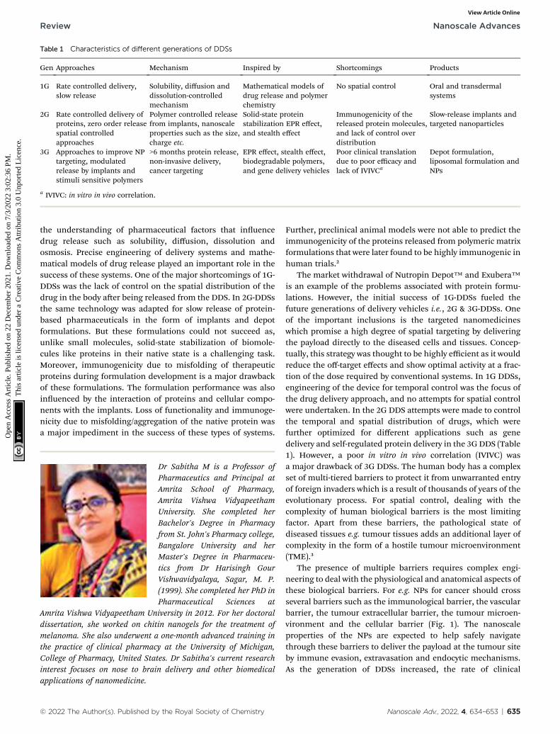
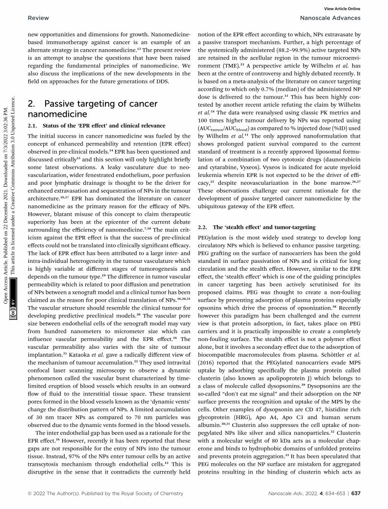
Table 1 Characteristics of different generations of DDSs
Gen Approaches Mechanism Inspired by Shortcomings Products
1G Rate controlled delivery,slow release
Solubility, diffusion anddissolution-controlledmechanism
Mathematical models ofdrug release and polymerchemistry
No spatial control Oral and transdermalsystems
2G Rate controlled delivery ofproteins, zero order releasespatial controlledapproaches
Polymer controlled releasefrom implants, nanoscaleproperties such as the size,charge etc.
Solid-state proteinstabilization EPR effect,and stealth effect
Immunogenicity of thereleased protein molecules,and lack of control overdistribution
Slow-release implants andtargeted nanoparticles
3G Approaches to improve NPtargeting, modulatedrelease by implants andstimuli sensitive polymers
>6 months protein release,non-invasive delivery,cancer targeting
EPR effect, stealth effect,biodegradable polymers,and gene delivery vehicles
Poor clinical translationdue to poor efficacy andlack of IVIVCa
Depot formulation,liposomal formulation andNPs
a IVIVC: in vitro in vivo correlation.
Review Nanoscale Advances
Ope
n A
cces
s A
rtic
le. P
ublis
hed
on 2
2 D
ecem
ber
2021
. Dow
nloa
ded
on 7
/3/2
022
3:02
:36
PM.
Thi
s ar
ticle
is li
cens
ed u
nder
a C
reat
ive
Com
mon
s A
ttrib
utio
n 3.
0 U
npor
ted
Lic
ence
.View Article Online
the understanding of pharmaceutical factors that inuencedrug release such as solubility, diffusion, dissolution andosmosis. Precise engineering of delivery systems and mathe-matical models of drug release played an important role in thesuccess of these systems. One of the major shortcomings of 1G-DDSs was the lack of control on the spatial distribution of thedrug in the body aer being released from the DDS. In 2G-DDSsthe same technology was adapted for slow release of protein-based pharmaceuticals in the form of implants and depotformulations. But these formulations could not succeed as,unlike small molecules, solid-state stabilization of biomole-cules like proteins in their native state is a challenging task.Moreover, immunogenicity due to misfolding of therapeuticproteins during formulation development is a major drawbackof these formulations. The formulation performance was alsoinuenced by the interaction of proteins and cellular compo-nents with the implants. Loss of functionality and immunoge-nicity due to misfolding/aggregation of the native protein wasa major impediment in the success of these types of systems.
Dr Sabitha M is a Professor ofPharmaceutics and Principal atAmrita School of Pharmacy,Amrita Vishwa VidyapeethamUniversity. She completed herBachelor's Degree in Pharmacyfrom St. John's Pharmacy college,Bangalore University and herMaster's Degree in Pharmaceu-tics from Dr Harisingh GourVishwavidyalaya, Sagar, M. P.(1999). She completed her PhD inPharmaceutical Sciences at
Amrita Vishwa Vidyapeetham University in 2012. For her doctoraldissertation, she worked on chitin nanogels for the treatment ofmelanoma. She also underwent a one-month advanced training inthe practice of clinical pharmacy at the University of Michigan,College of Pharmacy, United States. Dr Sabitha's current researchinterest focuses on nose to brain delivery and other biomedicalapplications of nanomedicine.
© 2022 The Author(s). Published by the Royal Society of Chemistry
Further, preclinical animal models were not able to predict theimmunogenicity of the proteins released from polymeric matrixformulations that were later found to be highly immunogenic inhuman trials.2
The market withdrawal of Nutropin Depot™ and Exubera™is an example of the problems associated with protein formu-lations. However, the initial success of 1G-DDSs fueled thefuture generations of delivery vehicles i.e., 2G & 3G-DDSs. Oneof the important inclusions is the targeted nanomedicineswhich promise a high degree of spatial targeting by deliveringthe payload directly to the diseased cells and tissues. Concep-tually, this strategy was thought to be highly efficient as it wouldreduce the off-target effects and show optimal activity at a frac-tion of the dose required by conventional systems. In 1G DDSs,engineering of the device for temporal control was the focus ofthe drug delivery approach, and no attempts for spatial controlwere undertaken. In the 2G DDS attempts were made to controlthe temporal and spatial distribution of drugs, which werefurther optimized for different applications such as genedelivery and self-regulated protein delivery in the 3G DDS (Table1). However, a poor in vitro in vivo correlation (IVIVC) wasa major drawback of 3G DDSs. The human body has a complexset of multi-tiered barriers to protect it from unwarranted entryof foreign invaders which is a result of thousands of years of theevolutionary process. For spatial control, dealing with thecomplexity of human biological barriers is the most limitingfactor. Apart from these barriers, the pathological state ofdiseased tissues e.g. tumour tissues adds an additional layer ofcomplexity in the form of a hostile tumour microenvironment(TME).3
The presence of multiple barriers requires complex engi-neering to deal with the physiological and anatomical aspects ofthese biological barriers. For e.g. NPs for cancer should crossseveral barriers such as the immunological barrier, the vascularbarrier, the tumour extracellular barrier, the tumour microen-vironment and the cellular barrier (Fig. 1). The nanoscaleproperties of the NPs are expected to help safely navigatethrough these barriers to deliver the payload at the tumour siteby immune evasion, extravasation and endocytic mechanisms.As the generation of DDSs increased, the rate of clinical
Nanoscale Adv., 2022, 4, 634–653 | 635

Fig. 1 Different levels of anatomical and physiological barriers in clinical translation of cancer nanomedicine. (TAMs, tumour associatedmacrophages; NPs nanoparticles; DCs, dendritic cells; FC fibroblast cell; IFP interstitial fluid pressure; MDSC, myeloid-derived suppressor cells;TME tumour microenvironment; ECM extracellular matrix).
Nanoscale Advances Review
Ope
n A
cces
s A
rtic
le. P
ublis
hed
on 2
2 D
ecem
ber
2021
. Dow
nloa
ded
on 7
/3/2
022
3:02
:36
PM.
Thi
s ar
ticle
is li
cens
ed u
nder
a C
reat
ive
Com
mon
s A
ttrib
utio
n 3.
0 U
npor
ted
Lic
ence
.View Article Online
translation reduced progressively mainly because of a poorunderstanding of the complex biological barriers.
The eld of nanomedicine represents the convergence of thecomplexity of the biological eld and the precise quantitativeapproaches used in the physical sciences. A lack of communi-cation between disciplines has been a major challenge. Thetendency to oversimplify and generalize the complexities andredundancy of biological systems by physical scientists and thelack of understanding of physical properties at the nanoscaleamong biologists have led to misperceptions and inappropriateconclusions.4 An overemphasis on the engineering aspect ofNPs and an under-appreciation for the complex biological andimmunological consequences of these delivery approaches arepartly responsible for the low success rate of these deliverysystems. The trends in publications also exemplify this asa large part of the work on cancer nanomedicine is published in
636 | Nanoscale Adv., 2022, 4, 634–653
materials science, chemical engineering and related elds andrelatively a low percentage of articles (17.3%) have been pub-lished in medical and biological science-related journals (basedon the data from Scopus by Salvioni et al., 2019).5
Opinions are divided for6 and against7,8 the efficiency andsuccess of nanomedicine in cancer therapeutics. Some of therecent discussions on the future of the eld have led to a lot ofskepticism with diametrically opposite views and opinions.9,10
Existing paradigms in nanomedicine are debated, discussedand questioned. New results in the eld are counterintuitive tothe existing dogmas including the underlying assumption thatsolid tumours could be targeted by the enhanced permeabilityand retention effect (EPR effect) and the long circulatory“stealth effect” of PEGylated NPs.9,11,12 These observations haveraised even more questions on the existing fundamentals ofnano-drug delivery. However, every new question also brings
© 2022 The Author(s). Published by the Royal Society of Chemistry
Review Nanoscale Advances
Ope
n A
cces
s A
rtic
le. P
ublis
hed
on 2
2 D
ecem
ber
2021
. Dow
nloa
ded
on 7
/3/2
022
3:02
:36
PM.
Thi
s ar
ticle
is li
cens
ed u
nder
a C
reat
ive
Com
mon
s A
ttrib
utio
n 3.
0 U
npor
ted
Lic
ence
.View Article Online
new opportunities and dimensions for growth. Nanomedicine-based immunotherapy against cancer is an example of analternate strategy in cancer nanomedicine.13 The present reviewis an attempt to analyse the questions that have been raisedregarding the fundamental principles of nanomedicine. Wealso discuss the implications of the new developments in theeld on approaches for the future generations of DDS.
2. Passive targeting of cancernanomedicine2.1. Status of the ‘EPR effect’ and clinical relevance
The initial success in cancer nanomedicine was fueled by theconcept of enhanced permeability and retention (EPR effect)observed in pre-clinical models.14 EPR has been questioned anddiscussed critically15 and this section will only highlight brieysome latest observations. A leaky vasculature due to neo-vascularization, wider fenestrated endothelium, poor perfusionand poor lymphatic drainage is thought to be the driver forenhanced extravasation and sequestration of NPs in the tumourarchitecture.16,17 EPR has dominated the literature on cancernanomedicine as the primary reason for the efficacy of NPs.However, blatant misuse of this concept to claim therapeuticsuperiority has been at the epicenter of the current debatesurrounding the efficiency of nanomedicine.7,18 The main crit-icism against the EPR effect is that the success of pre-clinicaleffects could not be translated into clinically signicant efficacy.The lack of EPR effect has been attributed to a large inter- andintra-individual heterogeneity in the tumour vasculature whichis highly variable at different stages of tumorogenesis anddepends on the tumour type.19 The difference in tumor vascularpermeability which is related to poor diffusion and penetrationof NPs between a xenogramodel and a clinical tumor has beenclaimed as the reason for poor clinical translation of NPs.16,20,21
The vascular structure should resemble the clinical tumour fordeveloping predictive preclinical models.20 The vascular poresize between endothelial cells of the xenogra model may varyfrom hundred nanometers to micrometer size which caninuence vascular permeability and the EPR effect.16 Thevascular permeability also varies with the site of tumourimplantation.21 Kataoka et al. gave a radically different view ofthe mechanism of tumour accumulation.22 They used intravitalconfocal laser scanning microscopy to observe a dynamicphenomenon called the vascular burst characterized by time-limited eruption of blood vessels which results in an outwardow of uid to the interstitial tissue space. These transientpores formed in the blood vessels known as the ‘dynamic vents’change the distribution pattern of NPs. A limited accumulationof 30 nm tracer NPs as compared to 70 nm particles wasobserved due to the dynamic vents formed in the blood vessels.
The inter endothelial gap has been used as a rationale for theEPR effect.16 However, recently it has been reported that thesegaps are not responsible for the entry of NPs into the tumourtissue. Instead, 97% of the NPs enter tumour cells by an activetranscytosis mechanism through endothelial cells.12 This isdisruptive in the sense that it contradicts the currently held
© 2022 The Author(s). Published by the Royal Society of Chemistry
notion of the EPR effect according to which, NPs extravasate bya passive transport mechanism. Further, a high percentage ofthe systemically administered (88.2–99.9%) active targeted NPsare retained in the acellular region in the tumour microenvi-ronment (TME).23 A perspective article by Wilhelm et al. hasbeen at the centre of controversy and highly debated recently. Itis based on a meta-analysis of the literature on cancer targetingaccording to which only 0.7% (median) of the administered NPdose is delivered to the tumour.11 This has been highly con-tested by another recent article refuting the claim by Wilhelmet al.24 The data were reanalysed using classic PK metrics and100 times higher tumour delivery by NPs was reported using(AUCtumor/AUCblood) as compared to% injected dose (%ID) usedby Wilhelm et al.11 The only approved nanoformulation thatshows prolonged patient survival compared to the currentstandard of treatment is a recently approved liposomal formu-lation of a combination of two cytotoxic drugs (daunorubicinand cytarabine, Vyxeos). Vyxeos is indicated for acute myeloidleukemia wherein EPR is not expected to be the driver of effi-cacy,25 despite neovascularization in the bone marrow.26,27
These observations challenge our current rationale for thedevelopment of passive targeted cancer nanomedicine by theubiquitous gateway of the EPR effect.
2.2. The ‘stealth effect’ and tumor-targeting
PEGylation is the most widely used strategy to develop longcirculatory NPs which is believed to enhance passive targeting.PEG graing on the surface of nanocarriers has been the goldstandard in surface passivation of NPs and is critical for longcirculation and the stealth effect. However, similar to the EPReffect, the ‘stealth effect’ which is one of the guiding principlesin cancer targeting has been actively scrutinised for itsproposed claims. PEG was thought to create a non-foulingsurface by preventing adsorption of plasma proteins especiallyopsonins which drive the process of opsonization.28 Recentlyhowever this paradigm has been challenged and the currentview is that protein adsorption, in fact, takes place on PEGcarriers and it is practically impossible to create a completelynon-fouling surface. The stealth effect is not a polymer effectalone, but it involves a secondary effect due to the adsorption ofbiocompatible macromolecules from plasma. Schottler et al.(2016) reported that the PEGylated nanocarriers evade MPSuptake by adsorbing specically the plasma protein calledclusterin (also known as apolipoprotein J) which belongs toa class of molecule called dysopsonins.29 Dysopsonins are theso-called “don't eat me signal” and their adsorption on the NPsurface prevents the recognition and uptake of the MPS by thecells. Other examples of dysopsonin are CD 47, histidine richglycoprotein (HRG), Apo A4, Apo C3 and human serumalbumin.30,31 Clusterin also suppresses the cell uptake of non-pegylated NPs like silver and silica nanoparticles.32 Clusterinwith a molecular weight of 80 kDa acts as a molecular chap-erone and binds to hydrophobic domains of unfolded proteinsand prevents protein aggregation.33 It has been speculated thatPEG molecules on the NP surface are mistaken for aggregatedproteins resulting in the binding of clusterin which acts as
Nanoscale Adv., 2022, 4, 634–653 | 637

Nanoscale Advances Review
Ope
n A
cces
s A
rtic
le. P
ublis
hed
on 2
2 D
ecem
ber
2021
. Dow
nloa
ded
on 7
/3/2
022
3:02
:36
PM.
Thi
s ar
ticle
is li
cens
ed u
nder
a C
reat
ive
Com
mon
s A
ttrib
utio
n 3.
0 U
npor
ted
Lic
ence
.View Article Online
a molecular chaperone by refolding proteins to their nativeconformation.29 Clusterin has a strong dysopsonin propertyregardless of the surface on which it is adsorbed. In contrast,the adsorption of opsonins such as immunoglobulins,complement proteins, brinogens etc. on NPs is responsible forrapid clearance of NPs by opsonization. Opsonization can alsohappen without protein adsorption as demonstrated in proteindepleted media which shows the existence of alternate mecha-nisms for cell uptake.29
Questions have been raised against the use of stealth NPs fortumour targeting.34 Long circulating PEGylated liposomesneither extravasate substantially to the tumour tissue nor theyare cleared by the MPS. It has been observed that long circula-tion is associated with skin deposition of PEGylated liposomaldoxorubicin (PLD), causing incidence of dermal toxicity, whichis not observed in non-PEGylated liposomes.35,36 The longcirculatory behaviour promotes the kinetically slow process ofextravasation into the skin.37 A comparison of commercialPEGylated (Doxil™) and non-PEGylated liposomes (Myocet™)shows that changes in pharmacokinetic parameters due toPEGylation does not contribute to efficacy.38 Doxil™ andMyocet™ differ in terms of the lipid composition, drug releaseand circulation half-life. Due to high drug release, only 10% ofthe drug is retained by Myocet™ aer 24 h, whereas even aer2–3 days of circulation 50% of the dose is retained in Doxil™,which in principle should promote EPR and enhance efficacy.In contrast, phase III trials show no difference in efficacy whiledermal side effects seen with Doxil™ are practically eliminatedwith Myocet™.39,40 Collectively, these pieces of evidence chal-lenge the currently held notion that long circulation will provideadditional time for NPs to extravasate into leaky tumour tissue,thereby contributing to the EPR effect.
PEGylation, on the one hand, increases the circulation timeof NPs by evading cell uptake by the MPS and, on the otherhand, it prevents the internalization and endocytosis of NPs bythe tumour cells resulting in limited therapeutic efficacy. This iscalled the “PEG dilemma”. A strategy to overcome this problemis to design a cleavable PEG corona in response to environ-mental stimuli. These stimuli include acidic pH, hypoxia andthe presence of certain enzymes in the TME.41,42However, due tointer- and intra-heterogeneity43,44 in the TME and the transientnature of the stimuli, these approaches suffer from variabilitysimilar to the variability of the EPR effect.
2.3. Safety and immunocompatibility of PEGylated carriers
The PEGylation strategy has been used for the past more thantwo and a half decades in commercial nanomedicine formula-tions with the introduction of the rst PEGylated liposomaldoxorubicin (PLD) Doxil™. However lately, it has been observedthat PEGylation is associated with mild to very strong immu-nological reactions which led to the clinical failure of somePEG-conjugated drugs. PEGylation has been linked to the deathof some patients during an infusion reaction which led to themarket withdrawal of PEGinesatide (a functional analog oferythropoietin).45 The failure in the clinical trial of a PEGylatedaptamer has been linked to PEGylation and anti-PEG
638 | Nanoscale Adv., 2022, 4, 634–653
antibodies.46 Following these incidences, the FDA has revisedthe guidelines and has recommended screening of anti-PEGantibodies in patients.
Anti-PEG antibodies are associated with mild to severe life-threatening infusion reactions (IR) and represent a translationalhurdle for NP-based products.47 The mechanism of these IRs ispoorly understood. The binding of anti-PEG IgM antibodies toPEGylated NPs has been reported to cause IRs by inducinga complement reaction and anaphylactoid shock.48 Despiteseveral studies, the cause-effect relationship of the physico-chemical attributes of NPs with complement activation, celluptake and cytokine release are still unclear.47 Differences invesicle-mediated complement response have been observed fortwo different brands of PEGylated liposomes (Doxil™ andCaelyx™) which are perceived to be similar.49 The role of plate-lets as an effector for a complement response is also being sus-pected.50,51 Complement activation-related pseudoallergy(CARPA) syndrome is one of the underlying mechanisms ofIR.52,53 Pre-existing anti-PEG antibody is regarded as one of themost critical factors for complement activation by PEGylatedNPs.54 The complement activation has been found to increaselinearly with the concentration of anti-PEG antibodies duringa second injection with PEG.55 Anti-PEG antibodies can inducea complement-dependent disruption of the liposomalmembrane resulting in rapid drug release from liposomes. PEGis not able to prevent the binding of complement fragment C5b-9on liposomes and the release of drug that is thought to happendue to the disruption of the proton and ammonium ion gradientin PLD used for passive drug loading.56 Anti-PEG antibodiescould be a factor responsible for the variable effect of PEGylatedliposomes. A concentration above the cut-off anti-PEG IgG titrerequired for liposomalmembrane disruption has been estimatedto be present in 5.5% of the normal population.56 A minimalphysiologically based pharmacokinetic (PBPK) model predictsthat a median concentration of 50 ng ml�1 of pre-existing anti-PEG antibody may only result in a 5–15% decrease in AUC rela-tive to patients having no anti-PEG antibody.57 A genetic basis forthe generation of anti-PEG antibodies has also been identiedsince only selective patients show the propensity to induce themwhich increases the risk of IR. A genome-wide association studyshows that the immunoglobulin heavy chain is the susceptiblelocus for an anti-PEG IgM response.58
The PEG-liposome–IgM complex is also responsible fora phenomenon called accelerated blood clearance (ABCs) bywhich NPs are rapidly removed from circulation aer a secondinjection of PLD.59 However, it is intriguing that the ABCphenomenon has not been observed in clinical studies, which iswell-established in different animal models. Two reasons forthis observation are proposed in the literature. First, theimmunological tolerance of PEG at a higher dose and second,the cytotoxic effect of PLD on macrophages.60 There is evidenceto both the claims. It is also reported that there is an inversecorrelation between the initial dose of PLD and the extent ofABC.61 The administration of a high dose (equivalent to theclinically recommended dose of PLD) results in the abrogationof the ABC phenomenon. This has been attributed to a state ofimmunological tolerance or clonal anergy of the marginal zone
© 2022 The Author(s). Published by the Royal Society of Chemistry
Review Nanoscale Advances
Ope
n A
cces
s A
rtic
le. P
ublis
hed
on 2
2 D
ecem
ber
2021
. Dow
nloa
ded
on 7
/3/2
022
3:02
:36
PM.
Thi
s ar
ticle
is li
cens
ed u
nder
a C
reat
ive
Com
mon
s A
ttrib
utio
n 3.
0 U
npor
ted
Lic
ence
.View Article Online
B cells (which trigger the generation of anti-PEG IgM).62,63 Thisoccurs at a high dose of PLD due to the lack of appropriate co-stimulatory signals. However, similar effects were not observedwith PEGylated polymeric carriers at a higher dose.64 Thehepatic clearance of the PEGylated liposome shows a sigmoidalrelationship with anti-PEG antibodies which is attributed to thecapacity-limited uptake by the Kupffer cells.55 In fact, thehepatic clearance of NPs and not the anti-PEG antibody-medi-ated complement activation is the limiting step in ABC.
Contrary to ABC observed in animals, human clinical studiesshow that the clearance of PLD was progressively reduced fromcycle 1 to cycle 3 of chemotherapy.65,66 There is a clear dose-dependent reduction of the monocyte count which is attributedto the cytotoxic effect of the drug, doxorubicin. Age and gender-related changes in clearance are also associated with a changein the MPS function.66 This also highlights the fact that study inrodent models is of limited value especially when immunolog-ical processes are responsible for the pharmacokinetic clear-ance of the NPs. A memory response mediated by the anti-PEGantibody is thought to be responsible for the rapid clearance ofPEG-based therapeutics.57 It is critical for physicians whoregularly prescribe PEGylated therapeutics to know the impli-cations of anti-PEG antibodies. An interesting survey suggeststhat only roughly one-quarter of the physicians who prescribedPEGylated therapeutics knew of the presence of PEG in theformulation and that this formulation can generate anti-PEGantibodies and its implications to patient safety.67
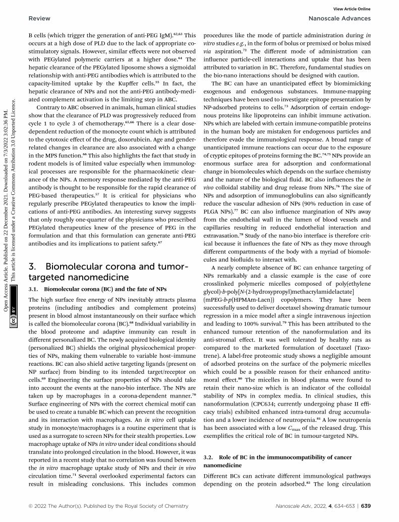
3. Biomolecular corona and tumor-targeted nanomedicine3.1. Biomolecular corona (BC) and the fate of NPs
The high surface free energy of NPs inevitably attracts plasmaproteins (including antibodies and complement proteins)present in blood almost instantaneously on their surface whichis called the biomolecular corona (BC).68 Individual variability inthe blood proteome and adaptive immunity can result indifferent personalized BC. The newly acquired biological identity(personalized BC) shields the original physicochemical proper-ties of NPs, making them vulnerable to variable host–immunereactions. BC can also shield active targeting ligands (present onNP surface) from binding to its intended target/receptor oncells.69 Engineering the surface properties of NPs should takeinto account the events at the nano-bio interface. The NPs aretaken up by macrophages in a corona-dependent manner.70
Surface engineering of NPs with the correct chemical motif canbe used to create a tunable BC which can prevent the recognitionand its interaction with macrophages. An in vitro cell uptakestudy in monocyte/macrophages is a routine experiment that isused as a surrogate to screen NPs for their stealth properties. Lowmacrophage uptake of NPs in vitro under ideal conditions shouldtranslate into prolonged circulation in the blood. However, it wasreported in a recent study that no correlation was found betweenthe in vitro macrophage uptake study of NPs and their in vivocirculation time.71 Several overlooked experimental factors canresult in misleading conclusions. This includes common
© 2022 The Author(s). Published by the Royal Society of Chemistry
procedures like the mode of particle administration during invitro studies e.g., in the form of bolus or premixed or bolusmixedvia aspiration.72 The different mode of administration caninuence particle-cell interactions and uptake that has beenattributed to variation in BC. Therefore, fundamental studies onthe bio-nano interactions should be designed with caution.
The BC can have an unanticipated effect by biomimickingexogenous and endogenous substances. Immune-mappingtechniques have been used to investigate epitope presentation byNP-adsorbed proteins to cells.73 Adsorption of certain endoge-nous proteins like lipoproteins can inhibit immune activation.NPs which are labeled with certain immune-compatible proteinsin the human body are mistaken for endogenous particles andtherefore evade the immunological response. A broad range ofunanticipated immune reactions can occur due to the exposureof cryptic epitopes of proteins forming the BC.74,75NPs provide anenormous surface area for adsorption and conformationalchange in biomolecules which depends on the surface chemistryand the nature of the biological uid. BC also inuences the invivo colloidal stability and drug release from NPs.76 The size ofNPs and adsorption of immunoglobulins can also signicantlyreduce the vascular adhesion of NPs (90% reduction in case ofPLGA NPs).77 BC can also inuence margination of NPs awayfrom the endothelial wall in the lumen of blood vessels andcapillaries resulting in reduced endothelial interaction andextravasation.78 Study of the nano-bio interface is therefore crit-ical because it inuences the fate of NPs as they move throughdifferent compartments of the body with a myriad of biomole-cules and biouids to interact with.
A nearly complete absence of BC can enhance targeting ofNPs remarkably and a classic example is the case of corecrosslinked polymeric micelles composed of poly(ethyleneglycol)-b-poly[N-(2-hydroxypropyl)methacrylamidelactate](mPEG-b-p(HPMAm-Lacn)) copolymers. They have beensuccessfully used to deliver docetaxel showing dramatic tumourregression in a mice model aer a single intravenous injectionand leading to 100% survival.79 This has been attributed to theenhanced tumour retention of the nanoformulation and itsanti-stromal effect. It was well tolerated by healthy rats ascompared to the marketed formulation of docetaxel (Taxo-trene). A label-free proteomic study shows a negligible amountof adsorbed proteins on the surface of the polymeric micelleswhich could be a possible reason for their enhanced antitu-moral effect.80 The micelles in blood plasma were found toretain their nano-size which is an indicator of the colloidalstability of NPs in complex media. In clinical studies, thisnanoformulation (CPC634; currently undergoing phase II effi-cacy trials) exhibited enhanced intra-tumoral drug accumula-tion and a lower incidence of neutropenia.81 A low neutropeniahas been associated with a low Cmax of the released drug. Thisexemplies the critical role of BC in tumour-targeted NPs.
3.2. Role of BC in the immunocompatibility of cancernanomedicine
Different BCs can activate different immunological pathwaysdepending on the protein adsorbed.82 The long circulation
Nanoscale Adv., 2022, 4, 634–653 | 639
Nanoscale Advances Review
Ope
n A
cces
s A
rtic
le. P
ublis
hed
on 2
2 D
ecem
ber
2021
. Dow
nloa
ded
on 7
/3/2
022
3:02
:36
PM.
Thi
s ar
ticle
is li
cens
ed u
nder
a C
reat
ive
Com
mon
s A
ttrib
utio
n 3.
0 U
npor
ted
Lic
ence
.View Article Online
behaviour of Apo E pre-adsorbed graphene/gold nanoparticleshas been attributed to the lack of complement activation.83
Many complement proteins act as opsonins by marking the NPsfor rapid clearance from circulation. The complement proteinadsorbed on NPs is an important determinant of recognitionand clearance by macrophages and therefore plays an impor-tant role in the biofate of NPs.70 The uptake of super-paramagnetic dextran iron oxide (SPIO) was reduced by 95% inC3 decient mice which demonstrates the role of complementactivation in opsonization.84 Complement mediated uptake ofSPIO was also blocked by EDTA (a complement inhibitor) inblood from healthy volunteers and cancer patients. There issignicant variability in complement reactivity towards NPs inthe general population which is found to be independent of ageand gender.85–87 Differential complement activation is a criticaldeterminant of the individual variation in innate immunity.The individual variability in complement and the componentsof innate immunity can result in variability in the biofate ofNPs, including its clearance and pharmacokinetics.
Natural antibodies in the blood adsorb on NPs and can act asan inducer of complement activation. The complement-medi-ated opsonization of iron oxide (SPIO) NPs by the thirdcomplement C3 is dependent on the natural antibody present inthe BC.88 The natural antibody in plasma was found to be thelink between BC and C3 mediated opsonization. Complementactivation and internalization via opsonization can be triggeredby the presence of very few antibodies on the surface of NPs. Theactivation process of complement by the three pathways (clas-sical, alternative and lectin) converges at a point where C3 iscleaved by the C3 convertases assembly into C3a, C3b and iC3b.The C3b and iC3b fragments bind and prime an activatingsurface like NPs which aid in cellular biorecognition via Fc andcomplement receptors for opsonization by immune cells.89 TheC3 adsorption on the surface and C3-mediated biorecognitionare key events that dictate the biofate of NPs. Natural antibodiesin blood critically inuence complement-mediated NP opsoni-zation predominantly by an alternative pathway.88 Variation inblood proteome can contribute to individual variability in thecell uptake of NPs. This has been demonstrated by a signicantincrease in the uptake of NPs at an articially elevated level ofIgG by the cells expressing IgG receptors.90 Doubling the IgGconcentration resulted in a 40-fold increase in the fraction ofantibodies in BC. This signicantly increases the cellular uptakeof NPs because the adsorbed IgG acts as an opsonin andpromotes opsonization via Fc receptors present in cells. Indi-vidual variability in the adaptive immunity may be one of thefactors for the life-threatening infusion reaction observed insome patients treated with PLD.
There are contradictory reports on the effect of surfaceadsorption of potential opsonins on NPs and their clearancefrom circulation. It has recently been reported that thecomplement system has a negligible inuence on the circula-tion time of both PEGylated liposomes and their non-PEGylatedcounterpart.91 Complement activation was unable to explain thepharmacokinetic clearance of liposomes in rodent models.Maintaining circulation stability by controlling the surfaceproperties is critical for the efficient delivery of NPs to the
640 | Nanoscale Adv., 2022, 4, 634–653
tumour tissue.92 Studies on C3�/� knock-out animals show thatthe complement proteins have no role in the clearance of PEG-coated or uncoated NPs with preadsorbed clusterin (Apo J).93
This has been attributed to the non-specic binding of anti-bodies. Antibodies that bind via specic epitopes on NPs areonly marked for opsonization.94 Screening for backgroundimmunity of patients and identication of specic biomarkerswhich inuence the immunological consequence of NPs aretherefore required for predictable outcomes in nanomedicine.
4. Pharmacokinetic,pharmacodynamic and therapeuticefficacy of nanomedicine4.1. Pharmacokinetic and pharmacodynamic (PK/PD)variability in the use of anticancer nanomedicine
One of the impediments in the clinical success of NPs is thehigh inter- and intra-variability in their PK/PD. In a PK meta-analysis study, signicant inter-patient variability was observedin the covariance coefficient (% CV) of the plasma concentra-tion-time AUC of a liposomal anticancer drug when comparedto a lipid-free formulation.95 The PK variability has been asso-ciated with factors such as the age, gender, body weight, cancertype and monocyte count.66,96,97 Signicant variability in PKtranslates into high variability in the efficacy and toxicity ofNPs.95,98,99 Clinically signicant variability in clearance of PLD(15.3-fold) has been observed which was not affected bya dosing schedule based on the body surface area.66
The clearance mechanism of PLD is different from that offree drugs. PLD is predominantly cleared by the MPS whereasfree drugs are cleared by metabolism in the liver. PEGylationsignicantly inuences clearance. Therefore, PK and PD aspectsof PLD are quite different from those of the free drug. Thecytotoxic effect of PLD on Kupffer cells, which are residentmacrophages in the liver, has been observed in a mice modelwhich results in a longer circulation time of a subsequentlyinjected PEG-liposome formulation.100 The change in circula-tion time was observed between day 3 and 14 aer the rst doseand subsequently, the effect was lower between days 21 and 28.In a clinical setting, however, this may not be observed due toa 3 week gap between the chemotherapy cycle for Doxil™. Thesignicantly delayed systemic clearance of bacteria alsoprovides indirect evidence for the loss of phagocytic activity ofmacrophages indicating the toxic effect of PLD on macro-phages.101 This is a PLD-specic phenomenon and is notattributed to bone marrow suppression (a common side effectof doxorubicin) as it is not observed in the lipid-free drug. Aninteresting meta-analysis study shows that there is an inverserelationship between the clearance of NPs and inter-patientvariability in PK which has important implications in thedevelopment of NPs.102 While targeted delivery aims to increasethe circulation time by reducing the clearance of NPs, however,this may result in interpatient variability in PK and PD (toxicityand response).
The clearance of NPs by cells of the MPS also depends on thenatural immunity of an individual. Across multiple species,
© 2022 The Author(s). Published by the Royal Society of Chemistry
Review Nanoscale Advances
Ope
n A
cces
s A
rtic
le. P
ublis
hed
on 2
2 D
ecem
ber
2021
. Dow
nloa
ded
on 7
/3/2
022
3:02
:36
PM.
Thi
s ar
ticle
is li
cens
ed u
nder
a C
reat
ive
Com
mon
s A
ttrib
utio
n 3.
0 U
npor
ted
Lic
ence
.View Article Online
a strong correlation has been observed between MPS function(both phagocytosis and ROS activity of blood monocytes anddendritic cells) and the clearance of PLD.103 Therefore, pheno-typic markers for measuring cellular functions can be used topredict the clearance and adjust the dose for personalizedmedicine. A need for dose adjustment is proposed for patientswith liver metastasis where a signicant NP clearance isobserved due to a high MPS activity.99 Therefore, there isa compelling need to adjust the dose based on MPS functionsfor positive clinical outcomes. This is in stark contrast to theactivity of free drugs which shows lower liver metabolism byhepatic enzymes during liver metastasis.104 The uptake of PLDby peritoneal macrophages has been reported to induce tol-erogenic M2 macrophages and cytokine release by thesemacrophages can lead to a secondary release of CCL2 (CeCmotif chemokine ligand 2 also called monocyte chemotacticprotein 1 [MCP-1]). Clinical observations show that in patientswith ovarian cancer, plasma clearance of NPs correlates with theplasma CCL2 and monocyte count.105 This is substantiated bythe fact that an altered clearance of PLD has been observed inCCL2 knock-out mice.106 The particulate nature of NPs isresponsible for the activation of the immune system whichresults in substantial variability in PK parameters depending onthe patients’ adaptive immunity which is also inuenced by thedisease state.
The pharmacokinetic characteristic of the carrier dictatesthe clearance and distribution of the encapsulated drug. It hasbeen demonstrated in a mouse model that PLD can preferen-tially accumulate in adipose tissue than in muscles. This resultsin a high volume of distribution and low plasma drug concen-tration and therefore a reduced efficacy due to poor tumouraccumulation.107 In human PK studies, a 10-fold difference inplasma exposure between obese patients as compared tonormal-weight patients has been observed.108 PK variabilityamong obese patients is attributed to faster clearance of NPs bythe MPS.109,110
Obese patients with cancer show an enhanced level ofhormones and chemokines which can inuence MPS func-tion.107 Patients with high estrone levels show higher MPSactivity and lower drug exposure of the encapsulated drug. Ahigh level of hormones and chemokines can modulate MPSfunction in obesity which is commonly observed in patientswith endometrial and ovarian cancer. In vitro studies show thatestrogen can stimulate the phagocytic activity of the MPS.111,112
Individualization of dose depending on the serum hormone
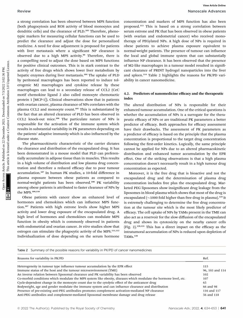
Table 2 Summary of the possible reasons for variability in PK/PD of can
Reasons for variability in PK/PD
Heterogeneity in tumour type inuence tumour accumulation by the EPRImmune status of the host and the tumour microenvironment (TME)An inverse relation between liposomal clearance and PK variability has beCo-morbid conditions which modulate the MPS system like obesity, diseaCycle-dependent change in the monocyte count due to the cytolytic effectBodyweight, age and gender modulate the immune system and can inuePresence of pre-existing anti-PEG antibodies promotes complement activaAnti-PEG antibodies and complement-mediated liposomal membrane dam
© 2022 The Author(s). Published by the Royal Society of Chemistry
concentration and markers of MPS function has also beenproposed.113 This is based on a strong correlation betweenserum estrone and PK that has been observed in obese patients(with ovarian and endometrial cancer) who received mono-therapy of PEGylated NPs. A high dose of NPs is required inobese patients to achieve plasma exposure equivalent tonormal-weight patients. The presence of tumour can inuencethe local and global immune system that can substantiallyinuence NP clearance. It has been observed that the presenceof M2-like macrophages in a tumour model resulted in signi-cant clearance of PRINT hydrogel nanoparticles into the liverand spleen.114 Table 2 highlights the reasons for PK/PD vari-ability in cancer nanomedicine.
4.2. Predictors of nanomedicine efficacy and the therapeuticindex
The altered distribution of NPs is responsible for theirenhanced tumour accumulation. One of the critical questions iswhether the accumulation of NPs is a surrogate for the thera-peutic efficacy of NPs or are traditional PK parameters a betterpredictor of efficacy. Both approaches for efficacy assessmenthave their drawbacks. The assessment of PK parameters asa predictor of efficacy is based on the principle that the plasmaconcentration is proportional to the target drug concentrationfollowing the rst-order kinetics. Logically, the same principlecannot be applied for NPs due to an altered pharmacokineticdistribution and enhanced tumor accumulation by the EPReffect. One of the striking observations is that a high plasmaconcentration doesn't necessarily result in a high tumour drugconcentration as expected.
Moreover, it is the free drug that is bioactive and not theencapsulated drug and the determination of plasma drugconcentration includes free plus the encapsulated drug. Mar-keted PEG liposomes show insignicant drug leakage from theliposomes in blood plasma which shows that most of the drug isencapsulated (�1000 fold higher than free drug in plasma).119 Itis extremely challenging to determine the free drug concentra-tion at the tumour site which is the most likely predictor ofefficacy. The cell uptake of NPs by TAMs present in the TME canalso act as a reservoir for the slow diffusion of the encapsulateddrug and shows its cytotoxicity on the nearby cancer cells(Fig. 2).120,121 This has a direct impact on the efficacy as theintratumoral accumulation of NPs is reduced upon depletion ofTAMs.120
cer nanomedicines
Ref.
effect 11596, 103 and 114
en observed 102ses which modulate the hormone level, etc. 107of the anticancer drug 65nce clearance and distribution 66 and 98tion-mediated NP clearance 116 and 117age and drug release 56 and 118
Nanoscale Adv., 2022, 4, 634–653 | 641
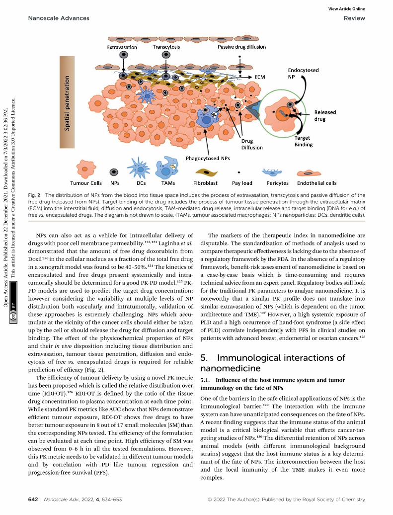
Fig. 2 The distribution of NPs from the blood into tissue space includes the process of extravasation, transcytosis and passive diffusion of thefree drug (released from NPs). Target binding of the drug includes the process of tumour tissue penetration through the extracellular matrix(ECM) into the interstitial fluid, diffusion and endocytosis, TAM-mediated drug release, intracellular release and target binding (DNA for e.g.) offree vs. encapsulated drugs. The diagram is not drawn to scale. (TAMs, tumour associated macrophages; NPs nanoparticles; DCs, dendritic cells).
Nanoscale Advances Review
Ope
n A
cces
s A
rtic
le. P
ublis
hed
on 2
2 D
ecem
ber
2021
. Dow
nloa
ded
on 7
/3/2
022
3:02
:36
PM.
Thi
s ar
ticle
is li
cens
ed u
nder
a C
reat
ive
Com
mon
s A
ttrib
utio
n 3.
0 U
npor
ted
Lic
ence
.View Article Online
NPs can also act as a vehicle for intracellular delivery ofdrugs with poor cell membrane permeability.122,123 Laginha et al.demonstrated that the amount of free drug doxorubicin fromDoxil™ in the cellular nucleus as a fraction of the total free drugin a xenogramodel was found to be 40–50%.124 The kinetics ofencapsulated and free drugs present systemically and intra-tumorally should be determined for a good PK-PD model.125 PK-PD models are used to predict the target drug concentration;however considering the variability at multiple levels of NPdistribution both vascularly and intratumorally, validation ofthese approaches is extremely challenging. NPs which accu-mulate at the vicinity of the cancer cells should either be takenup by the cell or should release the drug for diffusion and targetbinding. The effect of the physicochemical properties of NPsand their in vivo disposition including tissue distribution andextravasation, tumour tissue penetration, diffusion and endo-cytosis of free vs. encapsulated drugs is required for reliableprediction of efficacy (Fig. 2).
The efficiency of tumour delivery by using a novel PK metrichas been proposed which is called the relative distribution overtime (RDI-OT).126 RDI-OT is dened by the ratio of the tissuedrug concentration to plasma concentration at each time point.While standard PKmetrics like AUC show that NPs demonstrateefficient tumour exposure, RDI-OT shows free drugs to havebetter tumour exposure in 8 out of 17 small molecules (SM) thanthe corresponding NPs tested. The efficiency of the formulationcan be evaluated at each time point. High efficiency of SM wasobserved from 0–6 h in all the tested formulations. However,this PK metric needs to be validated in different tumour modelsand by correlation with PD like tumour regression andprogression-free survival (PFS).
642 | Nanoscale Adv., 2022, 4, 634–653
The markers of the therapeutic index in nanomedicine aredisputable. The standardization of methods of analysis used tocompare therapeutic effectiveness is lacking due to the absence ofa regulatory framework by the FDA. In the absence of a regulatoryframework, benet-risk assessment of nanomedicine is based ona case-by-case basis which is time-consuming and requirestechnical advice from an expert panel. Regulatory bodies still lookfor the traditional PK parameters to analyze nanomedicine. It isnoteworthy that a similar PK prole does not translate intosimilar extravasation of NPs (which is dependent on the tumorarchitecture and TME).127 However, a high systemic exposure ofPLD and a high occurrence of hand-foot syndrome (a side effectof PLD) correlate independently with PFS in clinical studies onpatients with advanced breast, endometrial or ovarian cancers.128
5. Immunological interactions ofnanomedicine5.1. Inuence of the host immune system and tumorimmunology on the fate of NPs
One of the barriers in the safe clinical applications of NPs is theimmunological barrier.129 The interaction with the immunesystem can have unanticipated consequences on the fate of NPs.A recent nding suggests that the immune status of the animalmodel is a critical biological variable that effects cancer-tar-geting studies of NPs.130 The differential retention of NPs acrossanimal models (with different immunological backgroundstrains) suggest that the host immune status is a key determi-nant of the fate of NPs. The interconnection between the hostand the local immunity of the TME makes it even morecomplex.
© 2022 The Author(s). Published by the Royal Society of Chemistry

Review Nanoscale Advances
Ope
n A
cces
s A
rtic
le. P
ublis
hed
on 2
2 D
ecem
ber
2021
. Dow
nloa
ded
on 7
/3/2
022
3:02
:36
PM.
Thi
s ar
ticle
is li
cens
ed u
nder
a C
reat
ive
Com
mon
s A
ttrib
utio
n 3.
0 U
npor
ted
Lic
ence
.View Article Online
One of the least known aspects of nanomedicine is itsinteraction with tumour immunological milieu. The physio-logical restructuring of the tumour immunological milieu inresponse to NP deposition can have a positive or negativeimpact on cancer therapy. On the one hand, it has the potentialfor NP-mediated cancer immunotherapy and, on the otherhand, NP-mediated immunosuppression can have a pro-tumoral effect.
The interaction between NPs and tumor-inltrating immunecells induces a cytokine milieu in the TME which determines thebiofate of nanomedicine.130 The tumour immunological milieu isbelieved to contribute to the suboptimal efficacy of therapeuticNPs. It has been reported that the suppression of antitumourimmunity is associated with the pro-tumoral effect of PEGylatedliposomal doxorubicin (PLD).131 PLD reduces IFN-g productionby TAMs and cytotoxic T lymphocytes which is essential for anti-tumour immunity. The mechanism is based on the uptake ofPLD by the TAMs in the tumour microenvironment and itspolarization from a tumour suppressive and inammatory M1macrophage to an anti-inammatory M2 phenotype and a globaldownregulation of inammatory cytokine secretion by T cells.
The notion that active targeting is due to the binding ofantibodies present on antibody-labeled NPs with the antigensoverexpressed on tumour cells has been challenged by a recentreport. A signicant accumulation of antibody-labelled NPs (incomparison to unlabeled NPs) in the tumour tissue was found tobe dependent on the immune cells in the tumour milieu and noton the antigen–antibody interaction observed in in vitrostudies.130 It was also reported recently that active targeting ofNPs shows extremely low targeting properties as very few NPs (14of 1 million NPs) interacted with cancer cells.23 This exempliesa signicant challenge in the delivery of NPs to cancer tissues.
The interaction of NPs with the immune system also pres-ents huge possibilities for immunotherapy. It has beenobserved that irrespective of the tumor retention potential, NPs(both plain and antibody-labeled) can induce a similar immuneresponse, T cell inltration and tumour inhibition.130 An anti-tumor immune response mediated by CD8+ T cells can beinduced by NPs without the requirement of any bioactivepayload. NPs have the potential to modulate both systemic andlocal immune effects on tumour growth inhibition which mayhave potential for cancer immunotherapy.
The TME has an immunosuppressive environment charac-terized by a hypoxia-induced cytokine cocktail which negativelyregulates tumor antigen presentation.132 The hypoxic environ-ment also restricts the CTL count; however, its cytolytic capacityis not compromised.133 The host immune status and the tumorimmunological milieu are critical variables for NP-based cancertherapy. TAMs, MDSC and Treg cells are the key effector cellsresponsible for an immunosuppressive TME which are thepossible target cells for immunomodulation.134,135
5.2. Immunological consequence of NPs may promote thepro-tumoral effect
NP-mediated immunological mechanisms that inhibit antitu-moral immunity have been demonstrated to cause the pro-
© 2022 The Author(s). Published by the Royal Society of Chemistry
tumoral effect of NPs. The polarization of TAMs from an anti-tumoral M1 phenotype to a pro-tumoral M2 phenotype byPEGylated liposomes has been reported.136 PEGylated lipo-somes have shown a pro-tumoral effect in an immunocompe-tent mice model, subcutaneously implanted with TC-1 cells.131
The pro-tumoral effect is attributed to the suppression of Th1cytokine (IFN gamma) and low CTLs in the tumour tissue ascompared to vehicle control. It was also found that the pro-tumoral effect depends on the type of implanted tumour andnot on the background immunity of the selected animal model.Liposomes not only increase the primary tumour, they alsoincrease the peritoneal metastasis of orthotopic implanted cells(ID8-VEGF-GFP) in a mice model.136 Complement activation hasalso been implicated in the pro-tumoral effect of NPs. Tumouraccumulation of empty poloxamine 908 coated polystyrene NPsand activation of intratumoral complement against long circu-latory NPs is thought to be responsible for the pro-tumoraleffect.137 The complement protein C5a induces an immuno-suppressive environment by the recruitment of MDSCs andsuppression of CD8+ T cells.138 Activated components ofcomplement can inuence various stages of carcinogenesis byevading immune recognition, promoting angiogenesis, cellmigration, and activating growth factors and preventingapoptosis.139 The pro-tumoral effect of NPs is a matter of greatconcern for the translation of NPs, which could partially explainthe lack of efficacy. In the above examples of the pro-tumoraleffect of NPs, studies were performed with placebo NPs withoutthe drug. The presence of drug may exert a cytotoxic effect onthe immune cells responsible for a tumour suppressive envi-ronment which may negate the inuence of an immunosup-pressive state and therefore further studies are warranted tofully establish the pro-tumoral consequence of NPs.
6. Relevance of preclinical models incancer nanomedicine6.1. Pre-clinical models to predict the targetability andefficacy of cancer nanomedicine
NPs show strong interactions with the host immune system.The interconnection between the host and the local immunityof the TME makes it even more complex. Questions have beenraised against the relevance of preclinical models in cancernanomedicine. Pre-clinical studies of NPs are oen performedon patient-derived xenogra models for which immunocom-promised animals are used due to a conducive immunosup-pressive environment for cross-tissue graing. The interactionof NPs with the host immune system has a bearing on the fate ofNPs which challenges the basic premise of tumour targetingstudies in immunocompromised models. The tumour growthand progression in the absence of immune pressure lacks thecomplexity of a clinical tumour. The process of immunoeditingwhich happens in clinical tumours might not happen in thepreclinical models. The ratio of tumour to body mass also variessignicantly between humans and rodent models. In mousemodels, the tumour volume may grow to a size of about 10% ofthe body mass.38 This can signicantly alter the body
Nanoscale Adv., 2022, 4, 634–653 | 643
Nanoscale Advances Review
Ope
n A
cces
s A
rtic
le. P
ublis
hed
on 2
2 D
ecem
ber
2021
. Dow
nloa
ded
on 7
/3/2
022
3:02
:36
PM.
Thi
s ar
ticle
is li
cens
ed u
nder
a C
reat
ive
Com
mon
s A
ttrib
utio
n 3.
0 U
npor
ted
Lic
ence
.View Article Online
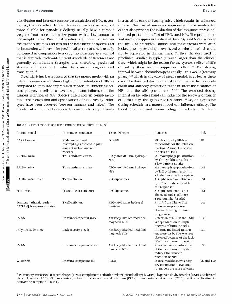
distribution and increase tumour accumulation of NPs, accen-tuating the EPR effect. Human tumours can vary in size, butthose eligible for nanodrug delivery usually have a tumourweight of not more than a few grams with a low tumour tobodyweight ratio. Preclinical studies are more focused ontreatment outcomes and less on the host immune system andits interaction with NPs. The preclinical testing of NPs is usuallyperformed in comparison to a drug monotherapy as a controlthat is clinically irrelevant. Current standards of treatment aregenerally combination therapies and therefore, preclinicalstudies add very little value to clinical progress andtranslation.15
Recently, it has been observed that the mouse model with anintact immune system shows high tumour retention of NPs ascompared to immunocompromised models.130 Tumour-associ-ated phagocytic cells also have a signicant inuence on thetumour retention of NPs. Species differences in complement-mediated recognition and opsonization of SPIO NPs by leuko-cytes have been observed between humans and mice.84 Thenumber of immune cells especially neutrophils is signicantly
Table 3 Animal models and their immunological effect on NPsa
Animal model Immune competence Tested
CARPA model PIMs are residentmacrophages present in pigsand not in humans androdents
Doxil™
C57BL6 mice Th1-dominant strains PEGylaNPs
BALB/c mice Th2-dominant strains PEGylaNPs
BALB/c nu/nu mice T cell-decient PEG-lip
SCID mice (T and B cell-decient) PEG-lip
Foxn1nu (athymic nude,C57BL/6J background) mice
T cell-decient PEGylaparticl
FVB/N Immunocompetent mice Antibomagne
Athymic nude mice Lack mature T cells Antibomagne
FVB/N Immune competent mice Antibomagne
Wistar rat Immune competent rat PLDs
a Pulmonary intravascular macrophages (PIMs), complement activation-reblood clearance (ABC); NP nanoparticle; enhanced permeability and retenonwetting templates (PRINT).
644 | Nanoscale Adv., 2022, 4, 634–653
increased in tumour-bearing mice which results in enhanceduptake. The use of immunocompromised mice models forcancer also prevents the evaluation of the immunosuppression-induced pro-tumoral effect of PEGylated NPs. The pro-tumoraland immunosuppressive nature of the PEGylated NPs was neverthe focus of preclinical studies and these factors were over-looked possibly resulting in overhyped conclusions which couldnot be replicated in clinical trials. Further, the dose used inpreclinical studies is typically much larger than the clinicaldose, which might be the reason for the cytotoxic effect of NPsoverriding their immunosuppressive effect.140 The dosinginterval between chemotherapy is usually 2 to 4 weeks (recoveryphase),141 which in the case of mouse models is as low as threedays. The dose and dosing interval can inuence the monocytecount and antibody generation that can affect the clearance ofNPs and the ABC phenomenon.61,105 The extended dosinginterval on the other hand can inuence the recovery of cancercells that may also gain drug resistance.142 So, an aggressivedosing schedule in a mouse model can inuence efficacy. Theblood proteome and hemorheology of rodents differ from
NP type Remarks Ref.
NP clearance by PIMs isresponsible for the infusionreaction. A model to assessthe risk of HSRs
48
ted 300 nm hydrogel M1macrophage polarizationby Th1 cytokines results ina low particle uptake
148
ted 300 nm hydrogel M2macrophage polarizationby Th2 cytokines results ina higher nanoparticle uptake
148
osomes ABC phenomenon observedby a T cell-independent Bcell response
151
osomes ABC phenomenon is notobserved and B cells area prerequisite for ABC
151
ted print hydrogeles
A shi from Th1 to Th2immune response wasobserved during tumourprogression
145
dy-labelled modiedtic NPs
Retention of NPs in the TMEis dependent on multiplelineages of immune cells
130
dy labelled modiedtic NPs
Immune-mediated tumoursuppression by NPs was notobserved because of the lackof an intact immune system
130
dy labelled modiedtic NPs
Pharmacological inhibitionof the host immune systemreduces the tumourretention of NPs
130
Mouse models show a verylow complement level andrat models are more relevant
56 and 150
lated pseudoallergy (CARPA), hypersensitivity reaction (HSR), acceleratedntion (EPR); tumour microenvironment (TME); particle replication in
© 2022 The Author(s). Published by the Royal Society of Chemistry

Fig. 3 Proposed rational for the design of fixed molar drug ratios fortherapy using Vyxeos (liposomal combination of cytarabine anddaunorubicin).
Review Nanoscale Advances
Ope
n A
cces
s A
rtic
le. P
ublis
hed
on 2
2 D
ecem
ber
2021
. Dow
nloa
ded
on 7
/3/2
022
3:02
:36
PM.
Thi
s ar
ticle
is li
cens
ed u
nder
a C
reat
ive
Com
mon
s A
ttrib
utio
n 3.
0 U
npor
ted
Lic
ence
.View Article Online
humans which can also inuence the BC-mediated biologicalinteraction of NPs.143,144
NP disposition is a function of its physicochemical charac-teristics and the global immune status of the host (Table 3). Thepresence of tumour can have a dramatic inuence on particleclearance due to a change in the local and global immune systemin preclinical models.145 The tumour burden can polarize theimmune system to a Th2 phenotype which can inuence thebiofate of NPs. The short plasma circulation and enhancedclearance of NPs by the MPS in tumour-bearing animals werereported to be due to the polarization of macrophages to a M2phenotype. This shi in the immune response which preventsa cell-mediated response in the TME has also been reported inhumans.146 Even among immunocompetent mice, the globalimmune status of the mouse strain can vary considerably.147 Amouse strain with a predominantly Th1 response (e.g. C57BL6)shows a signicantly slower rate of clearance than a Th2-pronemouse (e.g. BALB/c).148 This has been attributed to the polariza-tion ofmacrophages to aM1 phenotype by Th1 cytokines and thetolerant M2 phenotype by Th2 cytokines.149 However, the particleclearance by a Th1 biased strain is similar to that by a Th2 strainfollowing tumour induction which exhibits a shi in theimmune response with cancer progression that can dramaticallyinuence particle clearance.145 A deeper understanding of themolecular aspects of this shi in immune response will helpoptimize nanodrug delivery. The pivotal role of immune systemin the kinetics of NPs and tumour immunobiology is a criticalaspect that has oen been overlooked in preclinical studies.
Species-specic differences in complement activation affectscomplement-mediated liposomal membrane damage and therelease of drug from PLDs.56 Due to the low cytolytic activity ofanti-PEG antibodies in mice, they were not found to be suitablemodels to study complement-induced liposomal lysis. Thissuggests the role of interspecies difference in human trans-lational studies. Wistar rats were found to be efficient indemonstrating the complement-mediated change in pharma-cokinetics and toxicity of PLDs. The complement level ofcommon mice strains is very low relative to humans andtherefore complement related studies in mice models may notbe physiologically relevant.150
7. Future outlook7.1. Rationale for the design of a xed molar drug ratio inNPs: case study of Vyxeos (liposomal combination ofcytarabine and daunorubicin)
The theoretical basis for the use of a combined drug regimen incancer chemotherapy is to improve efficacy, reduce drug resis-tance and decrease toxicity by reducing the dose. The conven-tional standard of care treatment of acute myeloid leukemia(AML), a heterogeneous cancer is by a 7 + 3 regimen. This isoptimized combination therapy with two drugs developed bya trial-and-error method.152 The 7 + 3 regimen involves 7 days ofcontinuous infusion of cytarabine in combination with 3 days ofconcurrent intermittent dosing of daunorubicin.153 Ratiometricdelivery of drugs is based on the rationale that a combination ofmolar ratios of drugs can have synergistic action relative to
© 2022 The Author(s). Published by the Royal Society of Chemistry
using individual drugs at the maximum tolerable dose.154 Vyx-eos was developed with the idea that a xed molar drug ratio of5 : 1 packed in liposomal vesicles would provide better efficacyand tolerability to the drug administered historically by thestandard 7 + 3 regimen. The proposed advantage of usinga liposomal xed molar ratio of the drug is highlighted in Fig. 3.This ratio was found to maximize efficacy and reduce antago-nism during cytotoxic study in a panel of cell lines.155 Cytarabineis loaded passively in liposomes whereas daunorubicin isloaded actively using copper gluconate as a buffer to improvedrug retention in liposomes by metal complexation withcopper.155 The synergistic effect of the ratiometric delivery hasbeen observed in a preclinical model when compared to sepa-rate doses of liposomal cytarabine and daunorubicin thatshowed a signicantly lower antitumoral effect than Vyxeos.155
In the phase III studies, clinically signicant overall survival wasobserved in comparison to the standard 7 + 3 regimen(CLTR0310-301). Based on the relative molar ratio of the drugs,the action may vary from synergistic to antagonistic effects.Although the molar drug ratio of 5 : 1 shows maximum syner-gistic activity in vitro it is difficult to maintain in vivo ratiometryby traditional chemotherapy using iv infusion due to thedifference in the pharmacokinetics of the two drugs which mayeventually lead to an antagonistic dose ratio.154 Liposomes ata lipid ratio of 7 : 2 : 1 of distearoylphosphatidylcholine (DSPC),distearylphosphatidylglycerol (DSPG), and cholesterol providethe requisite biophysical properties for homing the drug in therequired 5 : 1 ratio to maintain the synergistic ratio for anextended duration that has been used to resolve the issue ofdelivering the synergistic combination to the target cells.156 It isa non-PEGylated liposome that remains in a gel state at body
Nanoscale Adv., 2022, 4, 634–653 | 645
Nanoscale Advances Review
Ope
n A
cces
s A
rtic
le. P
ublis
hed
on 2
2 D
ecem
ber
2021
. Dow
nloa
ded
on 7
/3/2
022
3:02
:36
PM.
Thi
s ar
ticle
is li
cens
ed u
nder
a C
reat
ive
Com
mon
s A
ttrib
utio
n 3.
0 U
npor
ted
Lic
ence
.View Article Online
temperature and provides the required in vivo stability.157 Themost important attribute of using a liposomal carrier is todeliver the required synergistic ratio, which brings predict-ability andminimizes antagonism between the two drugs whichis difficult with conventional chemotherapy.
It has been observed that 24 hours post-administration themolar ratio of the drugs was maintained between 5 : 1 to 9 : 1 (inthe synergistic range) whereas, within 15 minutes of adminis-tration, the ratio of the free drug was changed substantially fromthe synergistic ratio.155 It has also been claimed that liposomescan accumulate preferentially in the malignant myeloblast cellsand have demonstrated signicant cytotoxicity on leukemiaprecursors than normal hemopoietic precursors.158 However, ithas been observed in preclinical studies that liposomal formu-lation shows a lower nadir of all blood and precursor cells ascompared to the 7 + 3 regimen.159Clinical data also reveal the factthat there is a delayed recovery of platelets and neutrophilsfollowing a prolonged period of thrombocytopenia and neu-tropenia on the administration of liposomal formulation.160
7.2. Personalized drug therapy using NPs
The traditional method of designing NPs of dened physico-chemical properties as a ‘one-size-ts-all’ approach for allcancer types has yielded limited success. A disease driven-approach has been proposed for the rational design of NPs asagainst a formulation-driven approach used traditionally.161
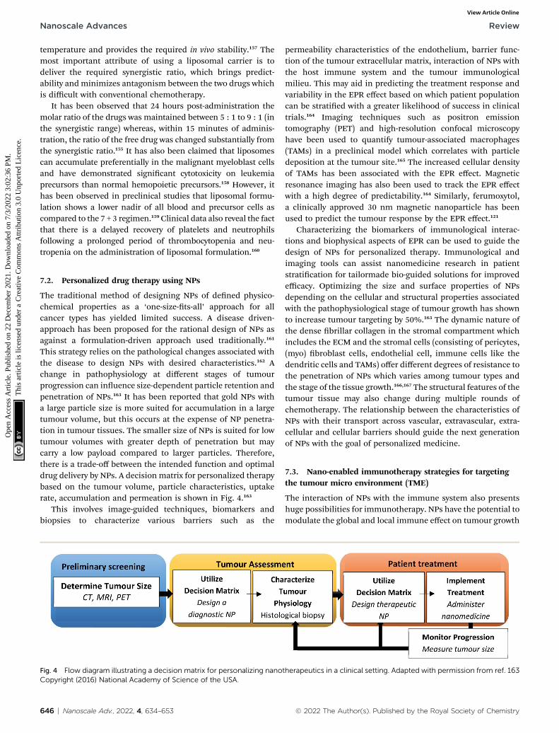
This strategy relies on the pathological changes associated withthe disease to design NPs with desired characteristics.162 Achange in pathophysiology at different stages of tumourprogression can inuence size-dependent particle retention andpenetration of NPs.163 It has been reported that gold NPs witha large particle size is more suited for accumulation in a largetumour volume, but this occurs at the expense of NP penetra-tion in tumour tissues. The smaller size of NPs is suited for lowtumour volumes with greater depth of penetration but maycarry a low payload compared to larger particles. Therefore,there is a trade-off between the intended function and optimaldrug delivery by NPs. A decision matrix for personalized therapybased on the tumour volume, particle characteristics, uptakerate, accumulation and permeation is shown in Fig. 4.163
This involves image-guided techniques, biomarkers andbiopsies to characterize various barriers such as the
Fig. 4 Flow diagram illustrating a decision matrix for personalizing nanoCopyright (2016) National Academy of Science of the USA.
646 | Nanoscale Adv., 2022, 4, 634–653
permeability characteristics of the endothelium, barrier func-tion of the tumour extracellular matrix, interaction of NPs withthe host immune system and the tumour immunologicalmilieu. This may aid in predicting the treatment response andvariability in the EPR effect based on which patient populationcan be stratied with a greater likelihood of success in clinicaltrials.164 Imaging techniques such as positron emissiontomography (PET) and high-resolution confocal microscopyhave been used to quantify tumour-associated macrophages(TAMs) in a preclinical model which correlates with particledeposition at the tumour site.165 The increased cellular densityof TAMs has been associated with the EPR effect. Magneticresonance imaging has also been used to track the EPR effectwith a high degree of predictability.164 Similarly, ferumoxytol,a clinically approved 30 nm magnetic nanoparticle has beenused to predict the tumour response by the EPR effect.121
Characterizing the biomarkers of immunological interac-tions and biophysical aspects of EPR can be used to guide thedesign of NPs for personalized therapy. Immunological andimaging tools can assist nanomedicine research in patientstratication for tailormade bio-guided solutions for improvedefficacy. Optimizing the size and surface properties of NPsdepending on the cellular and structural properties associatedwith the pathophysiological stage of tumour growth has shownto increase tumour targeting by 50%.163 The dynamic nature ofthe dense brillar collagen in the stromal compartment whichincludes the ECM and the stromal cells (consisting of pericytes,(myo) broblast cells, endothelial cell, immune cells like thedendritic cells and TAMs) offer different degrees of resistance tothe penetration of NPs which varies among tumour types andthe stage of the tissue growth.166,167 The structural features of thetumour tissue may also change during multiple rounds ofchemotherapy. The relationship between the characteristics ofNPs with their transport across vascular, extravascular, extra-cellular and cellular barriers should guide the next generationof NPs with the goal of personalized medicine.
7.3. Nano-enabled immunotherapy strategies for targetingthe tumour micro environment (TME)
The interaction of NPs with the immune system also presentshuge possibilities for immunotherapy. NPs have the potential tomodulate the global and local immune effect on tumour growth
therapeutics in a clinical setting. Adapted with permission from ref. 163
© 2022 The Author(s). Published by the Royal Society of Chemistry
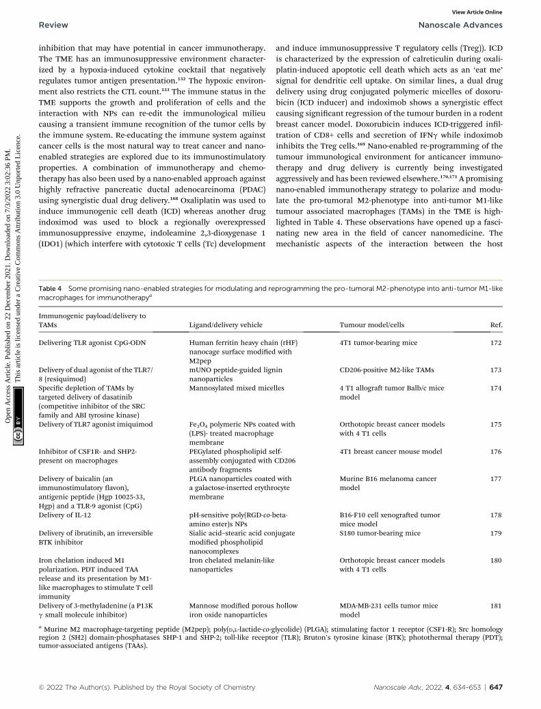
Review Nanoscale Advances
Ope
n A
cces
s A
rtic
le. P
ublis
hed
on 2
2 D
ecem
ber
2021
. Dow
nloa
ded
on 7
/3/2
022
3:02
:36
PM.
Thi
s ar
ticle
is li
cens
ed u
nder
a C
reat
ive
Com
mon
s A
ttrib
utio
n 3.
0 U
npor
ted
Lic
ence
.View Article Online
inhibition that may have potential in cancer immunotherapy.The TME has an immunosuppressive environment character-ized by a hypoxia-induced cytokine cocktail that negativelyregulates tumor antigen presentation.132 The hypoxic environ-ment also restricts the CTL count.133 The immune status in theTME supports the growth and proliferation of cells and theinteraction with NPs can re-edit the immunological milieucausing a transient immune recognition of the tumor cells bythe immune system. Re-educating the immune system againstcancer cells is the most natural way to treat cancer and nano-enabled strategies are explored due to its immunostimulatoryproperties. A combination of immunotherapy and chemo-therapy has also been used by a nano-enabled approach againsthighly refractive pancreatic ductal adenocarcinoma (PDAC)using synergistic dual drug delivery.168 Oxaliplatin was used toinduce immunogenic cell death (ICD) whereas another drugindoximod was used to block a regionally overexpressedimmunosuppressive enzyme, indoleamine 2,3-dioxygenase 1(IDO1) (which interfere with cytotoxic T cells (Tc) development
Table 4 Some promising nano-enabled strategies for modulating and remacrophages for immunotherapya
Immunogenic payload/delivery toTAMs Ligand/delivery vehicle
Delivering TLR agonist CpG-ODN Human ferritin heavy chainanocage surface modiedM2pep
Delivery of dual agonist of the TLR7/8 (resiquimod)
mUNO peptide-guided lignnanoparticles
Specic depletion of TAMs bytargeted delivery of dasatinib(competitive inhibitor of the SRCfamily and ABI tyrosine kinase)
Mannosylated mixed mice
Delivery of TLR7 agonist imiquimod Fe3O4 polymeric NPs coate(LPS)- treated macrophagemembrane
Inhibitor of CSF1R- and SHP2-present on macrophages
PEGylated phospholipid seassembly conjugated withantibody fragments
Delivery of baicalin (animmunostimulatory avon),antigenic peptide (Hgp 10025-33,Hgp) and a TLR-9 agonist (CpG)
PLGA nanoparticles coateda galactose-inserted erythrmembrane
Delivery of IL-12 pH-sensitive poly(RGD-co-bamino ester)s NPs
Delivery of ibrutinib, an irreversibleBTK inhibitor
Sialic acid–stearic acid conmodied phospholipidnanocomplexes
Iron chelation induced M1polarization. PDT induced TAArelease and its presentation by M1-like macrophages to stimulate T cellimmunity
Iron chelated melanin-likenanoparticles
Delivery of 3-methyladenine (a P13Kg small molecule inhibitor)
Mannose modied porousiron oxide nanoparticles
a Murine M2 macrophage-targeting peptide (M2pep); poly(D,L-lactide-co-gregion 2 (SH2) domain-phosphatases SHP-1 and SHP-2; toll-like receptotumor-associated antigens (TAAs).
© 2022 The Author(s). Published by the Royal Society of Chemistry
and induce immunosuppressive T regulatory cells (Treg)). ICDis characterized by the expression of calreticulin during oxali-platin-induced apoptotic cell death which acts as an ‘eat me’signal for dendritic cell uptake. On similar lines, a dual drugdelivery using drug conjugated polymeric micelles of doxoru-bicin (ICD inducer) and indoximob shows a synergistic effectcausing signicant regression of the tumour burden in a rodentbreast cancer model. Doxorubicin induces ICD-triggered inl-tration of CD8+ cells and secretion of IFNg while indoximobinhibits the Treg cells.169 Nano-enabled re-programming of thetumour immunological environment for anticancer immuno-therapy and drug delivery is currently being investigatedaggressively and has been reviewed elsewhere.170,171 A promisingnano-enabled immunotherapy strategy to polarize and modu-late the pro-tumoral M2-phenotype into anti-tumor M1-liketumour associated macrophages (TAMs) in the TME is high-lighted in Table 4. These observations have opened up a fasci-nating new area in the eld of cancer nanomedicine. Themechanistic aspects of the interaction between the host
programming the pro-tumoral M2-phenotype into anti-tumor M1-like
Tumour model/cells Ref.
n (rHF)with
4T1 tumor-bearing mice 172
in CD206-positive M2-like TAMs 173
lles 4 T1 allogra tumor Balb/c micemodel
174
d with Orthotopic breast cancer modelswith 4 T1 cells
175
lf-CD206
4T1 breast cancer mouse model 176
withocyte
Murine B16 melanoma cancermodel
177
eta- B16-F10 cell xenograed tumormice model
178
jugate S180 tumor-bearing mice 179
Orthotopic breast cancer modelswith 4 T1 cells
180
hollow MDA-MB-231 cells tumor micemodel
181
lycolide) (PLGA); stimulating factor 1 receptor (CSF1-R); Src homologyr (TLR); Bruton's tyrosine kinase (BTK); photothermal therapy (PDT);
Nanoscale Adv., 2022, 4, 634–653 | 647

Table 5 A list of outstanding issues in nanomedicine and our changing perspectives on their mechanistic understandinga
Issues innanomedicine Previous understanding Current understanding Remedial approaches
Biofate ofnanoparticles
It is a function of physicochemicalproperties such as the size, shape,charge, morphology, surfacechemistry, hydrophobicity etc. ofNPs
Type of BC dictates thedownstream effects of NPs.The BC ngerprint isdetermined by theproperties of NPs184,185
Engineering the surface chemistryfor predictive proteinadsorption184
Stealth Long circulation was thought tobe due to non-fouling surface
Adsorption of dysopsonine.g. clusterin is responsiblefor stealth29
Surface modication foradsorption of biocompatibleproteins like dysopsonins93,168
Biocompatibility PEGylation was thought to be non-immunogenic and bioinert
PEG can generate anti-PEGantibodies, cause infusionreaction, and complementactivation and ABC47,54
Screening of anti-PEG antibodiesbefore using PEGylated carriers34
Mechanism ofextravasation
Passive targeting due to leakyvasculature
Tumour uptake happens bytranscytosis12
Transcytosis pathway can beutilized for targeting186
NP clearance NPs are mainly taken up by thecells of the liver and spleen
A large fraction of NPs istaken up by TAMs in theTME
Particles taken up by TAMs can actas a depot for release of thedrug120,121
Pro-tumoral effect ofPEGylatedliposomes
PEGylated NPs are safe andbiocompatible
Pro-tumoral effect of NPsmay occur due to M2polarization of TAMs butmay be tumour specic131
NPs which can cause M1polarization of TAMs can be usedfor immunotherapy140
Altered toxicityprole
Side effects are reduced due toencapsulation and tumourspecic accumulation ascompared to the free drug
NPs can generate IR andcomplement activation(CARPA) and skinaccumulation causingdermal toxicity not foundin the free drug48,52
Identifying biomarkers for IR isrequired. Methods to enhance theEPR effect will reduce skinaccumulation47
EPR effect Thought to be the universalgateway for tumour targeting. Wasfound to be effective in preclinicalmodels
No clinical improvement inefficacy. Inter and intra-tumoral heterogeneity inthe EPR effect38,120
Stratication of patients based ondiagnostic imaging andbiomarkers163
a TAM, tumour associated macrophage; NP nanoparticle; EPR, enhanced permeability and retention; IR, infusion reaction; BC, biomolecularcorona; TME tumour microenvironment; ECM extracellular matrix; CARPA, complement activation-related pseudoallergy; ABC accelerated bloodclearance.
Nanoscale Advances Review
Ope
n A
cces
s A
rtic
le. P
ublis
hed
on 2
2 D
ecem
ber
2021
. Dow
nloa
ded
on 7
/3/2
022
3:02
:36
PM.
Thi
s ar
ticle
is li
cens
ed u
nder
a C
reat
ive
Com
mon
s A
ttrib
utio
n 3.
0 U
npor
ted
Lic
ence
.View Article Online
immune system and the tumour immunological milieu on thebiofate of tumour targeted NPs is still vague. Cancer immuno-therapy has the potential to radically change the landscape ofcancer therapy.
8. Conclusion
We have discussed the key outstanding issues in different areasof contemporary relevance in cancer nanomedicine. Identifyingthe possible causes of the lack of efficacy is critical as we moveforward in our efforts to translate safe and effective nano-medicine. Going back to basics and focusing on the funda-mental nano-bio interactions is required to address these issuesand to look beyond proof-of-concept studies of NPs.182
The fundamental interaction at the nanobio interface is thestarting point in the understanding of the biological conse-quences of NPs. Interactions occurring under static in vitroconditions oen vary from dynamic physiological conditions.76
Interference of NPs in in vitro cell-based assays and a lack ofstandardization of protocols oen result in false-positive andcontradictory results, making it difficult to predict the in vivo
648 | Nanoscale Adv., 2022, 4, 634–653
consequences of NPs.183 Fundamental interactions at thenano-bio interface and their inuence on the biofate andimmunological consequence of NPs are critical for theimmunotherapy-based applications of NPs. The interaction ofthe host immune system and tumour immunological milieuhas an impact on all aspects of targeting including longcirculation, clearance, extravasation, infusion reaction,tumour immunosuppression/activation and pro-tumoraleffect. The mechanistic understanding of the outstandingissues in cancer nanomedicine has radically changed in thelast decade which is highlighted in Table 5. The nano-biointeraction and the complex interdependency of the host andthe tumour immune response is by far the least understood ofall the problems in cancer nanomedicine. Understanding themechanism of nano-enabled immunotherapy is currently themost promising avenue in cancer research. Future research onthe nano-bio interactions and the immunological conse-quences of NPs at the fundamental level will be critical toreect on the most pressing questions in cancernanomedicine.
© 2022 The Author(s). Published by the Royal Society of Chemistry

Review Nanoscale Advances
Ope
n A
cces
s A
rtic
le. P
ublis
hed
on 2
2 D
ecem
ber
2021
. Dow
nloa
ded
on 7
/3/2
022
3:02
:36
PM.
Thi
s ar
ticle
is li
cens
ed u
nder
a C
reat
ive
Com
mon
s A
ttrib
utio
n 3.
0 U
npor
ted
Lic
ence
.View Article Online
Abbreviations
DDS
© 2022 The A
Drug delivery systems
BC Biomolecular corona TAM Tumour associated macrophage NP Nanoparticle DC Dendritic cell FC Fibroblast cell IFP Interstitial uid pressure MDSC Myeloid-derived suppressor cells TME Tumour microenvironment ECM Extracellular matrix NP Nanoparticle EPR Enhance permeability and retention PEG Polyethylene glycol IR Infusion reaction PIMs Pulmonary intravascular macrophages CARPA Complement activation-related pseudoallergy ICD Immunogenic cell death ABC Accelerated blood clearance PLD PEGylated liposomal doxorubicin CTL Cytotoxic T lymphocytes Treg T regulatory cells MPS Mononuclear phagocyte system AUC Area under the curveFunding
This research did not receive any specic grant from fundingagencies in the public, commercial, or not-for-prot sectors.
Conflicts of interest
Authors declare that they have no conict of interest or nancialconicts to disclose.
References
1 Y. H. Yun, B. K. Lee and K. Park, J. Controlled Release, 2015,219, 2–7.
2 H. J. Lee, G. Riley, O. Johnson, J. L. Cleland, N. Kim,M. Charnis, L. Bailey, E. Duenas, A. Shahzamani,M. Marian, A. J. S. Jones and S. D. Putney, J. Pharmacol.Exp. Ther., 1997, 281, 1431–1439.
3 H. Meng, W. Leong, K. W. Leong, C. Chen and Y. Zhao,Biomaterials, 2018, 174, 41–53.
4 R. L. Juliano, Sci. Public Policy, 2012, 39, 99–104.5 L. Salvioni, M. A. Rizzuto, J. A. Bertolini, L. Pandol,M. Colombo and D. Prosperi, Cancers, 2019, 11, 1855.
6 M. Germain, F. Caputo, S. Metcalfe, G. Tosi, K. Spring,A. K. O. Aslund, A. Pottier, R. Schiffelers, A. Ceccaldi andR. Schmid, J. Controlled Release, 2020, 326, 164–171.
7 K. Park, ACS Nano, 2013, 7, 7442–7447.8 K. Park, J. Controlled Release, 2019, 305, 221–222.9 S. Wilhelm, A. J. Tavares and W. C. W. Chan, Nat. Rev.Mater., 2016, 1, 16074.
uthor(s). Published by the Royal Society of Chemistry
10 S. E. McNeil, Nat. Rev. Mater., 2016, 1, 16073.11 S. Wilhelm, A. J. Tavares, Q. Dai, S. Ohta, J. Audet,
H. F. Dvorak and W. C. W. Chan, Nat. Rev. Mater., 2016,16014.
12 S. Sindhwani, A. M. Syed, J. Ngai, B. R. Kingston,L. Maiorino, J. Rothschild, P. MacMillan, Y. Zhang,N. U. Rajesh, T. Hoang, J. L. Y. Wu, S. Wilhelm,A. Zilman, S. Gadde, A. Sulaiman, B. Ouyang, Z. Lin,L. Wang, M. Egeblad and W. C. W. Chan, Nat. Mater.,2020, 19, 566–575.
13 P. Zhang, Y. Zhai, Y. Cai, Y. Zhao and Y. Li, Adv. Mater.,2019, 31(49), e1904156.
14 Y. Matsumura and H. Maeda, Cancer Res., 1986, 46, 6387–6392.
15 T. Lammers, J. Controlled Release, 2019, 294, 372–375.16 H. Hashizume, P. Baluk, S. Morikawa, J. W. McLean,
G. Thurston, S. Roberge, R. K. Jain and D. M. McDonald,Am. J. Pathol., 2000, 156, 1363–1380.
17 H. Maeda, Adv. Drug Delivery Rev., 2015, 91, 3–6.18 Y. Nakamura, A. Mochida, P. L. Choyke and H. Kobayashi,
Bioconjugate Chem., 2016, 27, 2225–2238.19 S. K. Golombek, J. N. May, B. Theek, L. Appold, N. Drude,
F. Kiessling and T. Lammers, Adv. Drug Delivery Rev.,2018, 130, 17–38.
20 F. Yuan, M. Dellian, D. Fukumura, M. Leunig, D. A. Berk,R. K. Jain and V. P. Torchilin, Cancer Res., 1995, 55, 3752–3756.
21 H. Sarin, A. S. Kanevsky, H. Wu, A. A. Sousa, C. M. Wilson,M. A. Aronova, G. L. Griffiths, R. D. Leapman and H. Q. Vo,J. Transl. Med., 2009, 7, 51.
22 Y. Matsumoto, J. W. Nichols, K. Toh, T. Nomoto, H. Cabral,Y. Miura, R. J. Christie, N. Yamada, T. Ogura, M. R. Kano,Y. Matsumura, N. Nishiyama, T. Yamasoba, Y. H. Bae andK. Kataoka, Nat. Nanotechnol., 2016, 11, 533–538.
23 Q. Dai, S. Wilhelm, D. Ding, A. M. Syed, S. Sindhwani,Y. Zhang, Y. Y. Chen, P. Macmillan and W. C. W. Chan,ACS Nano, 2018, 12, 8423–8435.
24 L. S. L. Price, S. T. Stern, A. M. Deal, A. V. Kabanov andW. C. Zamboni, Sci. Adv., 2020, 6, eaay9249.
25 S. A. Kusmartsev, Y. Li and S.-H. Chen, J. Immunol., 2000,165, 779–785.
26 A. Silva, A. Anderson and R. Gatenby, Math. Biosci. Eng.,2011, 8, 643–658.
27 A. Vacca and D. Ribatti, Leukemia, 2006, 20, 193–199.28 G. Caracciolo, S. Palchetti, V. Colapicchioni, L. Digiacomo,
D. Pozzi, A. L. Capriotti, G. La Barbera and A. Lagana,Langmuir, 2015, 31, 10764–10773.
29 S. Schottler, G. Becker, S. Winzen, T. Steinbach, K. Mohr,K. Landfester, V. Mailander and F. R. Wurm, Nat.Nanotechnol., 2016, 11, 372–377.
30 E. Papini, R. Tavano and F. Mancin, Front. Immunol., 2020,11, 567365.
31 P. A. Oldenborg, A. Zheleznyak, Y. F. Fang, C. F. Lagenaur,H. D. Gresham and F. P. Lindberg, Science, 2000, 288, 2051–2054.
Nanoscale Adv., 2022, 4, 634–653 | 649
Nanoscale Advances Review
Ope
n A
cces
s A
rtic
le. P
ublis
hed
on 2
2 D
ecem
ber
2021
. Dow
nloa
ded
on 7
/3/2
022
3:02
:36
PM.
Thi
s ar
ticle
is li
cens
ed u
nder
a C
reat
ive
Com
mon
s A
ttrib
utio
n 3.
0 U
npor
ted
Lic
ence
.View Article Online
32 M. Aoyama, K. Hata, K. Higashisaka, K. Nagano,Y. Yoshioka and Y. Tsutsumi, Biochem. Biophys. Res.Commun., 2016, 480, 690–695.
33 S. Poon, S. B. Easterbrook-Smith, M. S. Rybchyn, J. A. Carverand M. R. Wilson, Biochemistry, 2000, 39, 15953–15960.
34 T. J. Anchordoquy and D. Simberg, J. Controlled Release,2017, 262, 87–90.
35 J. I. Griffin, M. J. Benchimol and D. Simberg, J. ControlledRelease, 2017, 247, 167–174.
36 A. Gabizon, D. Goren, A. T. Horowitz, D. Tzemach, A. Lossosand T. Siegal, Adv. Drug Delivery Rev., 1997, 24, 337–344.
37 D. N. Waterhouse, P. G. Tardi, L. D. Mayer and M. B. Bally,Drug Saf., 2001, 24, 903–920.
38 J. W. Nichols and Y. H. Bae, J. Controlled Release, 2014, 190,451–464.
39 M. Amantea, M. S. Newman, T. M. Sullivan, A. Forrest andP. K. Working, Hum. Exp. Toxicol., 1999, 18, 17–26.
40 A. N. Gordon, J. T. Fleagle, D. Guthrie, D. E. Parkin,M. E. Gore and A. J. Lacave, J. Clin. Oncol., 2001, 19, 3312–3322.
41 X. Guan, Z. Guo, L. Lin, J. Chen, H. Tian and X. Chen, NanoLett., 2016, 16, 6823–6831.
42 H. Xiang, H. Lin, L. Yu and Y. Chen, ACS Nano, 2019, 13,2223–2235.
43 R. Somasundaram, G. Zhang, M. Fukunaga-Kalabis,M. Perego, C. Krepler, X. Xu, C. Wagner, D. Hristova,J. Zhang, T. Tian, Z. Wei, Q. Liu, K. Garg, J. Griss,R. Hards, M. Maurer, C. Hafner, M. Mayerhofer,G. Karanikas, A. Jalili, V. Bauer-Pohl, F. Weihsengruber,K. Rappersberger, J. Koller, R. Lang, C. Hudgens, G. Chen,M. Tetzlaff, L. Wu, D. T. Frederick, R. A. Scolyer,G. V. Long, M. Damle, C. Ellingsworth, L. Grinman,H. Choi, B. J. Gavin, M. Dunagin, A. Raj, N. Scholler,L. Gross, M. Beqiri, K. Bennett, I. Watson, H. Schaider,M. A. Davies, J. Wargo, B. J. Czerniecki, L. Schuchter,D. Herlyn, K. Flaherty, M. Herlyn and S. N. Wagner, Nat.Commun., 2017, 8, 607.
44 M. Kleppe and R. L. Levine, Nat. Med., 2014, 20, 342–344.45 K. U. Eckardt, Nat. Rev. Nephrol., 2013, 9, 192–193.46 N. J. Ganson, T. J. Povsic, B. A. Sullenger, J. H. Alexander,
S. L. Zelenkofske, J. M. Sailstad, C. P. Rusconi andM. S. Hersheld, J. Allergy Clin. Immunol., 2016, 137,1610–1613.
47 J. Szebeni, D. Simberg, A. Gonzalez-Fernandez,Y. Barenholz and M. A. Dobrovolskaia, Nat. Nanotechnol.,2018, 13, 1100–1108.
48 G. T. Kozma, T. Meszaros, I. Vashegyi, T. Fulop, E. Or,L. Dezsi, L. Rosivall, Y. Bavli, R. Urbanics, T. E. Mollnes,Y. Barenholz and J. Szebeni, ACS Nano, 2019, 13, 9315–9324.
49 P. P. Wibroe, D. Ahmadvand, M. A. Oghabian, A. Yaghmurand S. M. Moghimi, J. Controlled Release, 2016, 221, 1–8.
50 H. Beutier, B. Hechler, O. Godon, Y. Wang, C. M. Gillis,L. De Chaisemartin, A. Gouel-Cheron, S. Magnenat,L. E. Macdonald, A. J. Murphy, S. Chollet-Martin,D. Longrois, C. Gachet, P. Bruhns and F. Jonsson, Sci.Immunol., 2018, 3, eaan5997.
51 Z. Patko and J. Szebeni, Eur. J. Nanomed., 2015, 7, 233–244.
650 | Nanoscale Adv., 2022, 4, 634–653
52 J. Szebeni, Mol. Immunol., 2014, 61, 163–173.53 J. Szebeni, Drug Discovery Today, 2018, 23, 487–492.54 B. W. Neun, Y. Barenholz, J. Szebeni and
M. A. Dobrovolskaia, Molecules, 2018, 23, 1700.55 Y. Hashimoto, T. Shimizu, A. S. A. Lila, T. Ishida and
H. Kiwada, Biol. Pharm. Bull., 2015, 38, 417–424.56 E. Chen, B. M. Chen, Y. C. Su, Y. C. Chang, T. L. Cheng,
Y. Barenholz and S. R. Roffler, ACS Nano, 2020, 14, 7808–7822.
57 M. D. McSweeney, T. Wessler, L. S. L. Price, E. C. Ciociola,L. B. Herity, J. A. Piscitelli, W. C. Zamboni, M. G. Forest,Y. Cao and S. K. Lai, J. Controlled Release, 2018, 284, 171–178.
58 C. J. Chang, C. H. Chen, B. M. Chen, Y. C. Su, Y. T. Chen,M. S. Hersheld, M. T. M. Lee, T. L. Cheng, Y. T. Chen,S. R. Roffler and J. Y. Wu, Nat. Commun., 2017, 8, 522.
59 J. J. F. Verhoef, J. F. Carpenter, T. J. Anchordoquy andH. Schellekens, Drug Discovery Today, 2014, 19, 1945–1952.
60 P. Laverman, M. G. Carstens, O. C. Boerman, E. T. M. Dams,W. J. G. Oyen, N. Van Rooijen, F. H. M. Corstens andG. Storm, J. Pharmacol. Exp. Ther., 2001, 298, 607–612.
61 T. Ishida, K. Atobe, X. Y. Wang and H. Kiwada, J. ControlledRelease, 2006, 115, 251–258.
62 A. S. Abu Lila, H. Kiwada and T. Ishida, J. Controlled Release,2013, 172, 38–47.
63 T. Shimizu, T. Ishida and H. Kiwada, Immunobiology, 2013,218, 725–732.
64 T. Ishihara, M. Takeda, H. Sakamoto, A. Kimoto,C. Kobayashi, N. Takasaki, K. Yuki, K. I. Tanaka,M. Takenaga, R. Igarashi, T. Maeda, N. Yamakawa,Y. Okamoto, M. Otsuka, T. Ishida, H. Kiwada,Y. Mizushima and T. Mizushima, Pharm. Res., 2009, 26,2270–2279.
65 A. Gabizon, R. Isacson, O. Rosengarten, D. Tzemach,H. Shmeeda and R. Sapir, Cancer Chemother. Pharmacol.,2008, 61, 695–702.
66 N. M. La-Beck, B. A. Zamboni, A. Gabizon, H. Schmeeda,M. Amantea, P. A. Gehrig and W. C. Zamboni, CancerChemother. Pharmacol., 2012, 69, 43–50.
67 M. D. McSweeney, Z. C. Versfeld, D. M. Carpenter andS. K. Lai, Clin. Transl. Sci., 2018, 11, 162–165.
68 S. Tenzer, D. Docter, J. Kuharev, A. Musyanovych, V. Fetz,R. Hecht, F. Schlenk, D. Fischer, K. Kiouptsi,C. Reinhardt, K. Landfester, H. Schild, M. Maskos,S. K. Knauer and R. H. Stauber, Nat. Nanotechnol., 2013,8, 772–781.
69 A. Salvati, A. S. Pitek, M. P. Monopoli, K. Prapainop,F. B. Bombelli, D. R. Hristov, P. M. Kelly, C. Aberg,E. Mahon and K. A. Dawson, Nat. Nanotechnol., 2013, 8,137–143.
70 K. Saha, M. Rahimi, M. Yazdani, S. T. Kim, D. F. Moyano,S. Hou, R. Das, R. Mout, F. Rezaee, M. Mahmoudi andV. M. Rotello, ACS Nano, 2016, 10, 4421–4430.
71 Y. Zhang, A. T. Liu, Y. R. Cornejo, D. van Haute andJ. M. Berlin, PLoS One, 2020, 15, 1–16.
72 T. L. Moore, D. A. Urban, L. Rodriguez-Lorenzo,A. Milosevic, F. Crippa, M. Spuch-Calvar, S. Balog,
© 2022 The Author(s). Published by the Royal Society of Chemistry
Review Nanoscale Advances
Ope
n A
cces
s A
rtic
le. P
ublis
hed
on 2
2 D
ecem
ber
2021
. Dow
nloa
ded
on 7
/3/2
022
3:02
:36
PM.
Thi
s ar
ticle
is li
cens
ed u
nder
a C
reat
ive
Com
mon
s A
ttrib
utio
n 3.
0 U
npor
ted
Lic
ence
.View Article Online
B. Rothen-Rutishauser, M. Lattuada and A. Petri-Fink, Sci.Rep., 2019, 9, 1–9.
73 S. Lara, F. Alnasser, E. Polo, D. Garry, M. C. Lo Giudice,D. R. Hristov, L. Rocks, A. Salvati, Y. Yan andK. A. Dawson, ACS Nano, 2017, 11, 1884–1893.
74 A. A. Shemetov, I. Nabiev and A. Sukhanova, ACS Nano,2012, 6, 4585–4602.
75 I. Lynch, K. A. Dawson and S. Linse, Science's STKE, 2006,2006, pe14.
76 P. Jain, R. S. Pawar, R. S. Pandey, J. Madan, S. Pawar,P. K. Lakshmi and M. S. Sudheesh, Biotechnol. Adv., 2017,35, 889–904.
77 D. J. Sobczynski, P. Charoenphol, M. J. Heslinga,P. J. Onyskiw, K. Namdee, A. J. Thompson and O. Eniola-Adefeso, PLoS One, 2014, 9, e107408.
78 X. Duan and Y. Li, Small, 2013, 9, 1521–1532.79 Q. Hu, C. J. Rijcken, R. Bansal, W. E. Hennink, G. Storm and
J. Prakash, Biomaterials, 2015, 53, 370–378.80 I. Alberg, S. Kramer, M. Schinnerer, Q. Hu, C. Seidl, C. Leps,
N. Drude, D. Mockel, C. Rijcken, T. Lammers, M. Diken,M. Maskos, S. Morsbach, K. Landfester, S. Tenzer,M. Barz and R. Zentel, Small, 2020, 16, e1907574.
81 F. Atra, R. A. G. van Eerden, M. A. M. van Hylckama Vlieg,E. O. Hoop, P. de Bruijn, M. P. Lolkema, A. Moelker,C. J. Rijcken, R. Hanssen, A. Sparreboom,F. A. L. M. Eskens, R. H. J. Mathijssen andS. L. W. Koolen, Clin. Cancer Res., 2020, 26, 3537–3545.
82 G. S. Han and D.W. Domaille, ACS Cent. Sci., 2020, 6, 14–15.83 X. Lu, P. Xu, H. M. Ding, Y. S. Yu, D. Huo and Y. Q. Ma, Nat.
Commun., 2019, 10, 4520.84 S. Inturi, G. Wang, F. Chen, N. K. Banda, V. M. Holers,
L. P. Wu, S. M. Moghimi and D. Simberg, ACS Nano,2015, 9, 10758–10768.
85 C. T. N. Pham, D. G. Thomas, J. Beiser, L. M. Mitchell,J. L. Huang, A. Senpan, G. Hu, M. Gordon, N. A. Baker,D. Pan, G. M. Lanza and D. E. Hourcade, Nanomedicine,2014, 10, 651–660.
86 C. T. N. Pham, L. M. Mitchell, J. L. Huang,C. M. Lubniewski, O. F. Schall, J. K. Killgore, D. Pan,S. A. Wickline, G. M. Lanza and D. E. Hourcade, J. Biol.Chem., 2011, 286, 123–130.
87 H. Benasutti, G. Wang, V. P. Vu, R. Scheinman, E. Groman,L. Saba and D. Simberg, Bioconjugate Chem., 2017, 28, 2747–2755.
88 V. P. Vu, G. B. Gifford, F. Chen, H. Benasutti, G. Wang,E. V. Groman, R. Scheinman, L. Saba, S. M. Moghimi andD. Simberg, Nat. Nanotechnol., 2019, 14, 260–268.
89 D. Ricklin, G. Hajishengallis, K. Yang and J. D. Lambris,Nat. Immunol., 2010, 11, 785–797.
90 D. Prozeller, J. Pereira, J. Simon, V. Mailander, S. Morsbachand K. Landfester, Adv. Sci., 2019, 6, 1802199.
91 I. M. D. O. Viana, P. Grenier, J. Defrene, F. Barabe,E. M. Lima and N. Bertrand, Nanoscale, 2020, 12, 18875–18884.
92 J. Park, J. E. Park, V. E. Hedrick, K. V. Wood, C. Bonham,W. Lee and Y. Yeo, Small, 2018, 14, e1703670.
© 2022 The Author(s). Published by the Royal Society of Chemistry
93 N. Bertrand, P. Grenier, M. Mahmoudi, E. M. Lima,E. A. Appel, F. Dormont, J. M. Lim, R. Karnik, R. Langerand O. C. Farokhzad, Nat. Commun., 2017, 8, 777.
94 R. Rampado, S. Crotti, P. Caliceti, S. Pucciarelli andM. Agostini, Front. Bioeng. Biotechnol., 2020, 8, 166.
95 R. F. Schell, B. J. Sidone, W. P. Caron, M. D. Walsh,T. F. White, B. A. Zamboni, R. K. Ramanathan andW. C. Zamboni, Nanomedicine, 2014, 10, 109–117.
96 G. Song, T. K. Tarrant, T. F. White, D. A. Barrow,C. M. Santos, R. G. Timoshchenko, S. K. Hanna,R. K. Ramanathan, C. R. Lee, V. L. Bae-Jump, P. A. Gehrigand W. C. Zamboni, Nanomedicine, 2015, 11, 1797–1807.
97 W. C. Zamboni, S. Strychor, E. Joseph, D. R. Walsh,B. A. Zamboni, R. A. Parise, M. E. Tonda, N. Y. Yu,C. Engbers and J. L. Eiseman, Clin. Cancer Res., 2007, 13,7217–7223.
98 W. P. Caron, G. Song, P. Kumar, S. Rawal andW. C. Zamboni, Clin. Pharmacol. Ther., 2012, 91, 802–812.
99 H. Wu, R. K. Ramanathan, B. A. Zamboni, S. Strychor,S. Ramalingam, R. P. Edwards, D. M. Friedland,R. G. Stoller, C. P. Belani, L. J. Maruca, Y. J. Bang andW. C. Zamboni, J. Clin. Pharmacol., 2012, 52, 180–194.
100 J. Yahuafai, T. Asai, G. Nakamura, T. Fukuta, P. Siripong,K. Hyodo, H. Ishihara, H. Kikuchi and N. Oku, J.Controlled Release, 2014, 192, 167–173.
101 A. Yavlovich, A. Singh, R. Blumenthal and A. Puri, Biochim.Biophys. Acta, Biomembr., 2011, 1808, 117–126.
102 R. F. Schell, B. J. Sidone, W. P. Caron, M. D. Walsh,T. F. White, B. A. Zamboni, R. K. Ramanathan andW. C. Zamboni, Nanomedicine, 2014, 10, 109–117.
103 W. P. Caron, J. C. Lay, A. M. Fong, N. M. La-Beck, P. Kumar,S. E. Newman, H. Zhou, J. H. Monaco, D. L. Clarke-Pearson,W. R. Brewster, L. Van Le, V. L. Bae-Jump, P. A. Gehrig andW. C. Zamboni, J. Pharmacol. Exp. Ther., 2013, 347, 599–606.
104 C. F. Stewart, S. G. Arbuck, R. A. Fleming andW. E. Evans, J.Clin. Oncol., 1990, 8, 1874–1879.
105 C. Fotiadis, G. Zografos, K. Aronis, B. Dousaitou, M. Sechasand G. Skalkeas, Int. J. Surg., 1999, 1, 113–120.
106 L. Wang, C. Wang, J. Jiao, Y. Su, X. Cheng, Z. Huang, X. Liuand Y. Deng, Int. J. Nanomed., 2014, 9, 3645–3657.
107 B. R. Starling, P. Kumar, A. T. Lucas, D. Barrow, L. Farnan,L. Hendrix, H. Giovinazzo, G. Song, P. Gehrig, J. T. Bensenand W. C. Zamboni, Cancer Chemother. Pharmacol., 2019,83, 61–70.
108 W. C. Zamboni, S. Strychor, E. Joseph, D. R. Walsh,B. A. Zamboni, R. A. Parise, M. E. Tonda, N. Y. Yu,C. Engbers and J. L. Eiseman, Clin. Cancer Res., 2007, 13,7217–7223.
109 W. C. Zamboni, L. J. Maruca, S. Strychor, B. A. Zamboni,S. Ramalingam, R. P. Edwards, J. Kim, Y. Bang, H. Lee,D. M. Friedland, R. G. Stoller, C. P. Belani andR. K. Ramanathan, J. Liposome Res., 2011, 21, 158–165.
110 W. C. Zamboni, J. L. Eiseman, S. Strychor, P. M. Rice,E. Joseph, B. A. Zamboni, M. K. Donnelly, J. Shurer,R. A. Parise, M. E. Tonda, N. Y. Yu and P. H. Basse, J.Liposome Res., 2011, 21, 70–80.
Nanoscale Adv., 2022, 4, 634–653 | 651
Nanoscale Advances Review
Ope
n A
cces
s A
rtic
le. P
ublis
hed
on 2
2 D
ecem
ber
2021
. Dow
nloa
ded
on 7
/3/2
022
3:02
:36
PM.
Thi
s ar
ticle
is li
cens
ed u
nder
a C
reat
ive
Com
mon
s A
ttrib
utio
n 3.
0 U
npor
ted
Lic
ence
.View Article Online
111 T. Nicol, D. L. Bilbey, L. M. Charles, J. L. Cordingle andB. Vernon-Roberts, J. Endocrinol., 1964, 277–291.
112 B. Vernon-Roberts, Int. Rev. Cytol., 1969, 25, 131–159.113 B. R. Starling, P. Kumar, A. T. Lucas, D. Barrow, L. Farnan,
L. Hendrix, H. Giovinazzo, G. Song, P. Gehrig, J. T. Bensenand W. C. Zamboni, Cancer Chemother. Pharmacol., 2019,83, 61–70.
114 M. P. Kai, H. E. Brighton, C. A. Fromen, T. W. Shen,J. C. Lu, Y. E. Lu, A. W. Keeler, G. R. Robbins,J. P. Y. Ting, W. C. Zamboni, J. E. Bear andJ. M. DeSimone, ACS Nano, 2016, 10, 861–870.
115 G. Song, D. B. Darr, C. M. Santos, M. Ross, A. Valdivia,J. L. Jordan, B. R. Midkiff, S. Cohen, N. Nikolaishvili-Feinberg, C. R. Miller, T. K. Tarrant, A. B. Rogers,A. C. Dudley, C. M. Perou and W. C. Zamboni, Clin.Cancer Res., 2014, 20, 6083–6095.
116 B. M. Chen, T. L. Cheng and S. R. Roffler, ACS Nano, 2021,15, 14022–14048.
117 Q. Yang, T. M. Jacobs, J. D. McCallen, D. T. Moore,J. T. Huckaby, J. N. Edelstein and S. K. Lai, Anal. Chem.,2016, 88, 11804–11812.
118 A. Gabizon and J. Szebeni, ACS Nano, 2020, 14, 7682–7688.119 A. Gabizon, H. Shmeeda and Y. Barenholz, Clin.
Pharmacokinet., 2003, 42, 419–436.120 M. A. Miller, Y. R. Zheng, S. Gadde, C. Prschke, H. Zope,
C. Engblom, R. H. Kohler, Y. Iwamoto, K. S. Yang,B. Askevold, N. Kolishetti, M. Pittet, S. J. Lippard,O. C. Farokhzad and R. Weissleder, Nat. Commun., 2015,6, 8692.
121 J. Shi, P. W. Kantoff, R. Wooster and O. C. Farokhzad, Nat.Rev. Cancer, 2017, 17, 20–37.
122 P. Rajitha, D. Gopinath, R. Biswas, M. Sabitha andR. Jayakumar, Expert Opin. Drug Delivery, 2016, 13, 1177–1194.
123 V. Aparna, M. Shiva, R. Biswas and R. Jayakumar, Int. J. Biol.Macromol., 2018, 110, 2–6.
124 K. M. Laginha, S. Verwoert, G. J. R. Charrois and T. M. Allen,Clin. Cancer Res., 2005, 11, 6944–6949.
125 H. He, D. Yuan, Y. Wu and Y. Cao, Pharmaceutics, 2019, 11,110.
126 A. J. Madden, S. Rawal, K. Sandison, R. Schell,A. Schorzman, A. Deal, L. Feng, P. Ma, R. Mumper,J. DeSimone and W. C. Zamboni, J. Nanopart. Res., 2014,16, 2662.
127 K. Yokoi, D. Chan, M. Kojic, M. Milosevic, D. Engler,R. Matsunami, T. Tanei, Y. Saito, M. Ferrari andA. Ziemys, J. Controlled Release, 2015, 217, 293–299.
128 M. J. Boers-Sonderen, C. M. L. Van Herpen, W. T. A. Van DerGraaf, I. M. E. Desar, M. G. W. A. Van Der Logt, Y. M. DeBeer, P. B. Ottevanger and N. P. Van Erp, CancerChemother. Pharmacol., 2014, 74, 457–463.
129 T. J. Anchordoquy, Y. Barenholz, D. Boraschi, M. Chorny,P. Decuzzi, M. A. Dobrovolskaia, Z. S. Farhangrazi,D. Farrell, A. Gabizon, H. Ghandehari, B. Godin,N. M. La-Beck, J. Ljubimova, S. M. Moghimi, L. Pagliaro,J. H. Park, D. Peer, E. Ruoslahti, N. J. Serkova andD. Simberg, ACS Nano, 2017, 11, 12–18.
652 | Nanoscale Adv., 2022, 4, 634–653
130 P. Korangath, J. D. Barnett, A. Sharma, E. T. Henderson,J. Stewart, S. H. Yu, S. K. Kandala, C. T. Yang,J. S. Caserto, M. Hedayati, T. D. Armstrong, E. Jaffee,C. Gruettner, X. C. Zhou, W. Fu, C. Hu, S. Sukumar,B. W. Simons and R. Ivkov, Sci. Adv., 2020, 6, eaay1601.
131 M. K. Sabnani, R. Rajan, B. Rowland, V. Mavinkurve,L. M. Wood, A. A. Gabizon and N. M. La-Beck,Nanomedicine, 2015, 11, 259–262.
132 I. B. Barsoum, M. Koti, D. R. Siemens and C. H. Graham,Cancer Res., 2014, 74, 7185–7190.
133 R. Vuillefroy de Silly, P. Y. Dietrich and P. R. Walker,Oncoimmunology, 2016, 5, e1232236.
134 T. F. Gajewski, H. Schreiber and Y. X. Fu, Nat. Immunol.,2013, 14, 1014–1022.
135 D. Marvel and D. I. Gabrilovich, J. Clin. Invest., 2015, 125,3356–3364.
136 N. M. La-Beck, X. Liu and L. M. Wood, Front. Pharmacol.,2019, 10, 220.
137 S. M. Moghimi, J. Controlled Release, 2014, 190, 556–562.138 M. M. Markiewski, R. A. DeAngelis, F. Benencia,
S. K. Ricklin-Lichtsteiner, A. Koutoulaki, C. Gerard,G. Coukos and J. D. Lambris, Nat. Immunol., 2008, 9,1225–1235.
139 M. J. Rutkowski, M. E. Sughrue, A. J. Kane, S. A. Mills andA. T. Parsa, Mol. Cancer Res., 2010, 8, 1453–1465.
140 R. Rajan, M. K. Sabnani, V. Mavinkurve, H. Shmeeda,H. Mansouri, S. Bonkoungou, A. D. Le, L. M. Wood,A. A. Gabizon and N. M. La-Beck, J. Controlled Release,2018, 271, 139–148.
141 D. S. Alberts, F. M. Muggia, J. Carmichael, E. P. Winer,M. Jahanzeb, A. P. Venook, K. M. Skubitz, E. Rivera,J. A. Sparano, N. J. Dibella, S. J. Stewart, J. J. Kavanaghand A. A. Gabizon, Semin. Oncol., 2004, 31, 53–90.
142 J. Baker, J. Ajani, F. Scotte, D. Winther, M. Martin,M. S. Aapro and G. von Minckwitz, Eur. J. Oncol. Nurs.,2009, 13, 49–59.
143 A. Gessner, C. Olbrich, W. Schroder, O. Kayser andR. H. Muller, Int. J. Pharm., 2001, 214, 87–91.
144 K. Namdee, D. J. Sobczynski, P. J. Onyskiw and O. Eniola-Adefeso, Bioconjugate Chem., 2015, 26, 2419–2428.
145 M. P. Kai, H. E. Brighton, C. A. Fromen, T. W. Shen,J. C. Lu, Y. E. Lu, A. W. Keeler, G. R. Robbins,J. P. Y. Ting, W. C. Zamboni, J. E. Bear andJ. M. DeSimone, ACS Nano, 2016, 10, 861–870.
146 A. Sica and V. Bronte, J. Clin. Invest., 2007, 117, 1155–1166.147 H. Watanabe, K. Numata, T. Ito, K. Takagi and
A. Matsukawa, Shock, 2004, 22, 460–466.148 S. W. Jones, R. A. Roberts, G. R. Robbins, J. L. Perry,
M. P. Kai, K. Chen, T. Bo, M. E. Napier, J. P. Y. Ting,J. M. DeSimone and J. E. Bear, J. Clin. Invest., 2013, 123,3061–3073.
149 C. D. Mills, A. M. Hill, K. Kincaid, J. M. Alt, C. D. Mills,M. J. Heilman and A. M. Hill, J. Immunol. Res., 2018, 164,6166–6173.
150 G. L. Ong and M. J. Mattes, J. Immunol. Methods, 1989, 125,147–158.
© 2022 The Author(s). Published by the Royal Society of Chemistry
Review Nanoscale Advances
Ope
n A
cces
s A
rtic
le. P
ublis
hed
on 2
2 D
ecem
ber
2021
. Dow
nloa
ded
on 7
/3/2
022
3:02
:36
PM.
Thi
s ar
ticle
is li
cens
ed u
nder
a C
reat
ive
Com
mon
s A
ttrib
utio
n 3.
0 U
npor
ted
Lic
ence
.View Article Online
151 H. Koide, T. Asai, K. Hatanaka, S. Akai, T. Ishii, E. Kenjo,T. Ishida, H. Kiwada, H. Tsukada and N. Oku, Int. J.Pharm., 2010, 392, 218–223.
152 J. W. Yates, H. J. Wallace, R. R. Ellison and J. F. Holland,Cytosine arabinoside (NSC 63878) and daunorubicin (NSC83142) therapy in acute nonlymphocytic leukemia, https://pubmed.ncbi.nlm.nih.gov/4586956/, accessed 9October 2021.
153 M. F. Fey and C. Buske, Ann. Oncol., 2013, 24, vi138–vi143.154 A. Cafaro, M. B. Giannini, P. Silimbani, D. Cangini,
C. Masini, A. G. L. Di Rora, G. Simonetti, G. Martinelliand C. Cerchione, Minerva Med., 2020, 111, 455–466.
155 P. Tardi, S. Johnstone, N. Harasym, S. Xie, T. Harasym,N. Zisman, P. Harvie, D. Bermudes and L. Mayer, Leuk.Res., 2009, 33, 129–139.
156 A. Dicko, S. Kwak, A. A. Frazier, L. D. Mayer andB. D. Liboiron, Int. J. Pharm., 2010, 391, 248–259.
157 L. D. Mayer, P. Tardi and A. C. Louie, Int. J. Nanomed., 2019,14, 3819–3830.
158 H. P. Kim, B. Gerhard, T. O. Harasym, L. D. Mayer andD. E. Hogge, Exp. Hematol., 2011, 39, 741–750.
159 W. S. Lim, P. G. Tardi, N. Dos Santos, X. Xie, M. Fan,B. D. Liboiron, X. Huang, T. O. Harasym, D. Bermudesand L. D. Mayer, Leuk. Res., 2010, 34, 1214–1223.
160 W. S. Lim, P. G. Tardi, X. Xie, M. Fan, R. Huang, T. Ciofani,T. O. Harasym and L. D. Mayer, Leuk. Lymphoma, 2010, 51,1536–1542.
161 J. I. Hare, T. Lammers, M. B. Ashford, S. Puri, G. Storm andS. T. Barry, Adv. Drug Delivery Rev., 2017, 108, 25–38.
162 W. Poon, B. R. Kingston, B. Ouyang, W. Ngo andW. C. W. Chan, Nat. Nanotechnol., 2020, 15, 819–829.
163 E. A. Sykes, Q. Dai, C. D. Sarsons, J. Chen, J. V. Rocheleau,D. M. Hwang, G. Zheng, D. T. Cramb, K. D. Rinker andW. C. W. Chan, Proc. Natl. Acad. Sci. U. S. A., 2016, 113,E1142–E1151.
164 M. A. Miller, S. Gadde, C. Prschke, C. Engblom,M. M. Sprachman, R. H. Kohler, K. S. Yang,A. M. Laughney, G. Wojtkiewicz, N. Kamaly, S. Bhonagiri,M. J. Pittet, O. C. Farokhzad and R. Weissleder, Sci.Transl. Med., 2015, 7, 314ra183.
165 H. Y. Kim, R. Li, T. S. C. Ng, G. Courties, C. B. Rodell,M. Prytyskach, R. H. Kohler, M. J. Pittet, M. Nahrendorf,R. Weissleder and M. A. Miller, ACS Nano, 2018, 12,12015–12029.
166 E. Brown, T. McKee, E. DiTomaso, A. Pluen, B. Seed,Y. Boucher and R. K. Jain, Nat. Med., 2003, 9, 796–800.
167 K. Yokoi, M. Kojic, M. Milosevic, T. Tanei, M. Ferrari andA. Ziemys, Cancer Res., 2014, 74, 4239–4246.
© 2022 The Author(s). Published by the Royal Society of Chemistry
168 J. Lu, X. Liu, Y. P. Liao, F. Salazar, B. Sun, W. Jiang,C. H. Chang, J. Jiang, X. Wang, A. M. Wu, H. Meng andA. E. Nel, Nat. Commun., 2017, 8, 1811.
169 Z. Wan, J. Sun, J. Xu, P. Moharil, J. Chen, J. Xu, J. Zhu, J. Li,Y. Huang, P. Xu, X. Ma, W. Xie, B. Lu and S. Li, ActaBiomater., 2019, 90, 300–313.
170 Y. Xia, L. Rao, H. Yao, Z. Wang, P. Ning and X. Chen, Adv.Mater., 2020, 32, e2002054.
171 J. D. Martin, H. Cabral, T. Stylianopoulos and R. K. Jain,Nat. Rev. Clin. Oncol., 2020, 17, 251–266.
172 H. Shan, W. Dou, Y. Zhang and M. Qi, Nanoscale, 2020, 12,22268–22280.
173 P. Figueiredo, A. Lepland, P. Scodeller, F. Fontana,G. Torrieri, M. Tiboni, M. A. Shahbazi, L. Casettari,M. A. Kostiainen, J. Hirvonen, T. Teesalu andH. A. Santos, Acta Biomater., 2021, 133, 231–243.
174 X. Zhang, X. Zang, M. Qiao, X. Zhao, H. Hu and D. Chen,ACS Biomater. Sci. Eng., 2020, 6, 5675–5684.
175 L. Liu, Y. Wang, X. Guo, J. Zhao and S. Zhou, Small, 2020,16, e2003543.
176 A. Ramesh, S. Kumar, D. Nandi and A. Kulkarni, Adv.Mater., 2019, 31, e1904364.
177 S. Han, W. Wang, S. Wang, S. Wang, R. Ju, Z. Pan, T. Yang,G. Zhang, H. Wang and L. Wang, Nanoscale, 2019, 11,20206–20220.
178 Y. Wang, Y. X. Lin, S. L. Qiao, H. W. An, Y. Ma, Z. Y. Qiao,R. P. Y. J. Rajapaksha and H. Wang, Biomaterials, 2017, 112,153–163.
179 Q. Qiu, C. Li, Y. Song, T. Shi, X. Luo, H. Zhang, L. Hu,X. Yan, H. Zheng, M. Liu, M. Liu, M. Liu, S. Yang,X. Liu, G. Chen and Y. Deng, Acta Biomater., 2019, 92,184–195.
180 L. Rong, Y. Zhang, W. S. Li, Z. Su, J. I. Fadhil and C. Zhang,Biomaterials, 2019, 225, 119515.
181 K. Li, L. Lu, C. Xue, J. Liu, Y. He, J. Zhou, Z. Xia, L. Dai,Z. Luo, Y. Mao and K. Cai, Nanoscale, 2020, 12, 130–144.
182 D. Witzigmann, S. Hak and R. van der Meel, J. ControlledRelease, 2018, 290, 138–140.
183 N. A. Monteiro-Riviere, A. O. Inman and L. W. Zhang,Toxicol. Appl. Pharmacol., 2009, 234, 222–235.
184 M. Mahmoudi, S. Sheibani, A. S. Milani, F. Rezaee,M. Gauberti, R. Dinarvand and H. Vali, Nanomedicine,2015, 10, 215–226.
185 M. Schaffler, F. Sousa, A. Wenk, L. Sitia, S. Hirn, C. Schleh,N. Haberl, M. Violatto, M. Canovi, P. Andreozzi,M. Salmona, P. Bigini, W. G. Kreyling and S. Krol,Biomaterials, 2014, 35, 3455–3466.
186 X. Liu, J. Jiang and H. Meng, Theranostics, 2019, 9, 8018–8025.
Nanoscale Adv., 2022, 4, 634–653 | 653


![[Revised] Revisiting Verb Aspect in T'boli](https://static.fdokumen.com/doc/165x107/631ef9e50ff042c6110c9f71/revised-revisiting-verb-aspect-in-tboli.jpg)