Review of pulp sensibility tests. Part I: general information and thermal tests
25
REVIEW Review of pulp sensibility tests. Part I: general information and thermal tests H. Jafarzadeh 1 & P. V. Abbott 2 1 Department of Endodontics, Faculty of Dentistry and Dental Research Center, Mashhad University of Medical Sciences, Mashhad, Iran; and 2 School of Dentistry, University of Western Australia, Perth, Australia Abstract Jafarzadeh H, Abbott PV. Review of pulp sensibility tests. Part I: general information and thermal tests. International Endodontic Journal, 43, 738–762, 2010. A major, and essential, part of the diagnostic process for pulp disease is the use of pulp sensibility tests. When diagnosing pulp pain, these tests can be used to reproduce the symptoms reported by the patient to diagnose the diseased tooth as well as the disease state. However, a major shortcoming with these tests is that they only indirectly provide an indication of the state of the pulp by measuring a neural response rather than the vascular supply, so both false positive and false negative results can occur. The relevant literature on pulp sensibility tests in the context of endodontics up to January 2009 was reviewed using PubMed and MEDLINE database searches. This search identified papers published between November 1964 and January 2009 in all languages. Thermal tests have been used as an integral part of dental examinations. Two types of thermal tests are available, one uses a cold stimulus and the other uses a hot stimulus, and each has various methods of delivery. If these tests are used properly, injury to the pulp is highly unlikely. A review of the literature regarding the rationale, indications, limitations, and interpretation of thermal tests, the value of these diagnostic tests, as well as a discussion of the important points about each of these tests is presented. Keywords: cold, diagnosis, heat, pulp, sensibility test, thermal test. Received 5 June 2009; accepted 22 April 2010 Introduction Before commencing any treatment, one must first assemble all the available information regarding the symptoms and history of the disease. These should then be combined with the findings of the clinical examina- tion (the signs) and the results of relevant diagnostic tests. Once the information has been gathered and interpreted, the clinician can formulate a differential diagnosis made up of all possible disease entities that are consistent with the history, examination, and the results of the tests. For this purpose, the clinician must have a thorough knowledge of the examination proce- dures and tests as well as their limitations (Ingle et al. 2002). A major, and an essential, part of the diagnostic process for pulp disease is the use of pulp sensibility tests. When diagnosing pulp pain, these tests can be used to reproduce the symptoms reported by the patient to diagnose the diseased tooth as well as the disease state. There are several pulp tests available, and these can be grouped as either sensibility tests or vitality tests. Pulp sensibility tests include thermal tests (heat and cold stimuli), electric pulp tests (EPTs), and a test cavity. Pulp vitality tests include laser Doppler flowme- try (LDF), pulse oximetry, and tooth temperature measurement. These tests can be used in conjunction with other clinical tests such as periodontal probing, Correspondence: Hamid Jafarzadeh, Faculty of Dentistry and Dental Research Center, Mashhad University of Medical Sciences, PO Box: 91735-984, Vakilabad Blvd, Mashhad, Iran (Tel.: +98 511 8829501; fax: +98 511 7626058; e-mail: [email protected], [email protected]). doi:10.1111/j.1365-2591.2010.01754.x International Endodontic Journal, 43, 738–762, 2010 ª 2010 International Endodontic Journal 738
Transcript of Review of pulp sensibility tests. Part I: general information and thermal tests

REVIEW
Review of pulp sensibility tests. Part I: generalinformation and thermal tests
H. Jafarzadeh1 & P. V. Abbott2
1Department of Endodontics, Faculty of Dentistry and Dental Research Center, Mashhad University of Medical Sciences, Mashhad,
Iran; and 2School of Dentistry, University of Western Australia, Perth, Australia
Abstract
Jafarzadeh H, Abbott PV. Review of pulp sensibility tests.
Part I: general information and thermal tests. International
Endodontic Journal, 43, 738–762, 2010.
A major, and essential, part of the diagnostic process
for pulp disease is the use of pulp sensibility tests. When
diagnosing pulp pain, these tests can be used to
reproduce the symptoms reported by the patient to
diagnose the diseased tooth as well as the disease state.
However, a major shortcoming with these tests is that
they only indirectly provide an indication of the state of
the pulp by measuring a neural response rather than
the vascular supply, so both false positive and false
negative results can occur. The relevant literature on
pulp sensibility tests in the context of endodontics up to
January 2009 was reviewed using PubMed and
MEDLINE database searches. This search identified
papers published between November 1964 and
January 2009 in all languages. Thermal tests have
been used as an integral part of dental examinations.
Two types of thermal tests are available, one uses a cold
stimulus and the other uses a hot stimulus, and each
has various methods of delivery. If these tests are used
properly, injury to the pulp is highly unlikely. A review
of the literature regarding the rationale, indications,
limitations, and interpretation of thermal tests, the
value of these diagnostic tests, as well as a discussion of
the important points about each of these tests is
presented.
Keywords: cold, diagnosis, heat, pulp, sensibility
test, thermal test.
Received 5 June 2009; accepted 22 April 2010
Introduction
Before commencing any treatment, one must first
assemble all the available information regarding the
symptoms and history of the disease. These should then
be combined with the findings of the clinical examina-
tion (the signs) and the results of relevant diagnostic
tests. Once the information has been gathered and
interpreted, the clinician can formulate a differential
diagnosis made up of all possible disease entities that
are consistent with the history, examination, and the
results of the tests. For this purpose, the clinician must
have a thorough knowledge of the examination proce-
dures and tests as well as their limitations (Ingle et al.
2002).
A major, and an essential, part of the diagnostic
process for pulp disease is the use of pulp sensibility
tests. When diagnosing pulp pain, these tests can be
used to reproduce the symptoms reported by the patient
to diagnose the diseased tooth as well as the disease
state. There are several pulp tests available, and these
can be grouped as either sensibility tests or vitality
tests. Pulp sensibility tests include thermal tests (heat
and cold stimuli), electric pulp tests (EPTs), and a test
cavity. Pulp vitality tests include laser Doppler flowme-
try (LDF), pulse oximetry, and tooth temperature
measurement. These tests can be used in conjunction
with other clinical tests such as periodontal probing,
Correspondence: Hamid Jafarzadeh, Faculty of Dentistry and
Dental Research Center, Mashhad University of Medical
Sciences, PO Box: 91735-984, Vakilabad Blvd, Mashhad,
Iran (Tel.: +98 511 8829501; fax: +98 511 7626058; e-mail:
[email protected], [email protected]).
doi:10.1111/j.1365-2591.2010.01754.x
International Endodontic Journal, 43, 738–762, 2010 ª 2010 International Endodontic Journal738

percussion, palpation, mobility, transillumination, and
anaesthetic tests to aid the diagnostic process (Jafar-
zadeh et al. 2008, Jafarzadeh 2009, Jafarzadeh &
Rosenberg 2009, Udoye & Jafarzadeh 2010).
No single element of the diagnostic process should be
relied upon to make even what appears to be an
uncomplicated diagnosis. Hence, before commencing
any treatment, there should be at least two indepen-
dent diagnostic test results that correlate to indicate the
disease process. Pulp tests alone are usually not
adequate for establishing a diagnosis but can provide
useful information (Dachi et al. 1967, Fulling and
Andreasen 1976a,b). The purpose of this review is to
discuss the rationale, indications, limitations, and
interpretation of pulp sensibility tests, as well as the
value of these tests.
Search strategy
A literature search for relevant articles on pulp sensi-
bility tests in the context of endodontics up to January
2009 was performed using PubMed and MEDLINE
database searches. The search was performed using
different keywords (‘pulp test’ or ‘pulpal test’ or ‘pulpal
testing’ or ‘pulp testing’ or ‘electric pulp tester’ or
‘vitality test’ or ‘endodontic tests’ or ‘thermal test’ or
‘cold test’ or ‘heat test’ or ‘cavity test’ or ‘palpation test’
or ‘percussion test’ or ‘laser doppler’ or ‘pulse oximetry’).
This search identified papers published between Novem-
ber 1964 and January 2009 in all languages. After
removing duplicates, the remaining papers were
retrieved, and their reference lists were checked to
identify any other articles/textbooks relevant to the
topic, which might have provided additional informa-
tion.
Terminology
There appears to be considerable confusion amongst
dentists and in the literature regarding the terminology
used to describe pulp tests. It is common to see the use
of inappropriate terms, which suggest that the authors
or users of those terms do not fully understand the test
being used or described. If the nature of the test is not
understood, then the results cannot be accurately
determined or interpreted, which can lead to incorrect
treatment decisions. Hence, to clarify the terminology
used in this study, the following definitions are
provided.
Sensibility is defined as the ability to respond to a
stimulus (http://www.Dictionary.com) (Accessed online
on 13 March 2009). Tests such as thermal tests and
electric pulp tests are sensibility tests because they are
assessing whether the pulp’s nerve fibres can respond
to a stimulus when applied to the tooth. In clinical
situations, an assumption is then made on the basis of
the results of these tests regarding the state of the pulp,
whether it is alive or necrotic. That is, the results of
these tests are extrapolated to imply a state of health or
disease of the pulp. Thus, sensibility tests are used to
determine whether a pulp is likely to be alive (with
response) or necrotic (without response).
Sensitivity is a condition of being very responsive to a
stimulus. Thus, when testing a tooth to determine
whether it has pulpitis (a condition that is very
responsive to certain stimuli, as reported by the
patient), sensibility tests are used to test the sensitivity
of the pulp. However, they are still regarded as
sensibility tests.
The term ‘hypersensitivity’ implies a condition of
being even more responsive to a stimulus than a very
responsive (i.e. sensitive) tooth; it is subjective and
difficult to define or compare between patients. There-
fore, the term ‘hypersensitivity’ should not be used
because ‘sensitivity’ defines the state of the pulp and the
prefix ‘hyper-’ is not required.
Vitality implies that a blood supply is present within
the tissues. Hence, only a test that actually measures or
assesses pulp blood flow can be called a vitality test.
Currently, the only true vitality tests that have been
reported in the literature are the laser Doppler
flowmetry (LDF) and pulse oximetry tests. However,
these tests are not commonly used in dental practice
owing to the costs, time, and clinical procedures
required for use (LDF) (Jafarzadeh 2009) and uncertain
results (pulse oximetry) (Jafarzadeh & Rosenberg
2009).
Pulp sensibility tests
The common methods for pulp sensibility testing are
thermal stimulation (cold and heat), electrical stimula-
tion, or direct dentine stimulation (cavity test). These
tests indicate whether there is a neural response from
the pulp. A major shortcoming with these tests is that
they do not indicate the state of health of the pulp and
that in some situations, the responses might be
unreliable – such as when teeth have temporarily or
permanently lost their sensory function (for example,
traumatized teeth, immature teeth, or teeth that are in
proximity to moving jaw components in orthognathic
surgery) but still might have intact vasculature
Jafarzadeh & Abbott Pulp sensibility tests
ª 2010 International Endodontic Journal International Endodontic Journal, 43, 738–762, 2010 739

(Bhaskar & Rappaport 1973, Evans et al. 1999). Also,
the nerve tissue, being highly resistant to inflamma-
tion, might remain reactive long after the surrounding
pulp tissue has degenerated. Pulp sensibility tests also
do not indicate the vitality of the pulp, that is, whether
there is an adequate vascular circulation (Noblett et al.
1996, Radhakrishnan et al. 2002).
Some authors have reported inconsistencies between
pulp symptoms and responses when compared with the
histological findings. A criticism of these tests is that
they have only shown a correlation between the test
results and necrotic pulps or pulpless teeth (Seltzer et al.
1963a, Tyldesley & Mumford 1970, Dummer et al.
1980). These studies suggest that such tests should only
be used to assess whether a pulp is alive or necrotic
because they do not quantify disease or health and
should not be used to judge the degree of pulp disease.
However, when used appropriately, thermal pulp sen-
sibility tests are useful when patients report symptoms
of pulpitis because they can be used to identify the
diseased tooth by reproducing the symptoms.
Pulp testing schemes should be performed initially on
pain-free teeth, away from the area of the chief
complaint. The preferred sequence is to test disease-free
contralateral teeth first, opposing teeth second, then
presumably healthy teeth within the thermally painful
quadrant, and finally, the most suspicious tooth. This
strategy allows the clinician to appreciate the range of
normal responses exhibited by asymptomatic teeth in
that particular patient. It also allows the patient to learn
what to expect with the test. Importantly, performing
repetitive tests will tend to relax the patient, build
confidence, and reduce the probability of a false
response (Cohen & Hargreaves 2006). To improve
objectivity, the tests should be repeated after a recovery
period of 1 min, unless too much discomfort has been
caused (Ruddle 2002a, Pitt Ford & Patel 2004).
Thermal tests are highly subjective as they are
wholly dependent on the patient’s response to the
stimulus. There is no accurate or objective method of
assessing how responsive the tooth under investigation
is to any particular test or of comparing with a previous
measurement. In contrast, EPTs have numerical dis-
plays, which allow the report to be compared with
previous readings (Pitt Ford & Patel 2004) although
many clinicians do not believe these readings are
reproducible and simply look for a response or no
response (Cohen & Hargreaves 2006). No studies have
demonstrated whether the readings shown on the
numerical display have any particular meaning or
whether they are reproducible.
The ideal technique for the evaluation of pulp status
should be noninvasive, painless, standardized, repro-
ducible, reliable, inexpensive, easily completed, and
objective (Chambers 1982). However, pulp sensibility
tests that are currently used have the potential to
produce an unpleasant sensation, and therefore, there
will be a subjective element to their interpretation
(Bhaskar & Rappaport 1973, Klein 1978).
Rationale of the tests
The rationale for innervation of any structure in the
body is to provide a warning of damage that is
occurring or impending (Cohen & Hargreaves 2006).
With this understanding, sharp, nonlingering pain
with the application of thermal stimulation is normal
and a part of the patient’s protective defence system.
An exaggerated response can indicate a lowered
threshold (Cohen & Hargreaves 2006).
In general, the amount of stimulus required to
activate A-d afferent fibres is only 25% of that required
to activate C fibres; therefore, most clinical pulp testing
activates only the A-d fibres (Virtanen 1985, Harg-
reaves & Goodis 2002).
Thermal tests
Sensory response to thermal stimuli occurs before there
is a temperature change in the pulpo–dentinal junction
(PDJ) area, where sensory nerve endings are located
(Trowbridge et al. 1980, Ingle et al. 2008). Thus, it
appears that the sensory response is not initiated by
temperature changes in the receptors (Trowbridge et al.
1980). Instead, thermal tests activate hydrodynamic
movement of fluid within dentinal tubules, which
excites the A-d fibres (Cohen & Hargreaves 2006).
The C fibres are not activated by these tests unless they
produce injury to the pulp (Fuss et al. 1986). Another
reason for proving that the understanding of pain
responses to thermal stimuli is based on the hydrody-
namic theory is that there are no thermosensing nerve
endings in the pulp tissue (Cohen & Hargreaves 2006).
Thus, the pain sensation the patient experiences
requires some pulp tissue, including odontoblasts.
These odontoblasts should be intact for the function
of hydrodynamic mechanism (Berman & Hartwell
2006).
Cold stimulates the fast-conducting A-d fibres, which
produces a sharp localized pain (Ingle & Bakland
2002). Continued heat application, on the other hand,
will more likely stimulate the slower conducting C
Pulp sensibility tests Jafarzadeh & Abbott
International Endodontic Journal, 43, 738–762, 2010 ª 2010 International Endodontic Journal740
fibres, located deeper in the pulp, resulting in dull pain
of longer duration (Bender 2000). Thermal stimulation
provides a greater response when more extreme tem-
perature changes occur, causing more rapid and
stronger fluid movement within the dentinal tubules,
stimulating receptors, and exciting A-d fibres (Bender
2000, Ingle & Bakland 2002). However, gradual
temperature changes do not produce a rapid response
but will eventually produce a response by C fibres
(Mengel et al. 1993, Bender 2000).
Electric pulp test (EPT)
Electric pulp tests deliver a current sufficient to
overcome the resistance of enamel and dentine and
stimulate the A-d fibres. The nonmyelinated C fibres do
not respond to the conventional EPTs because signif-
icantly more current is needed to stimulate them
(Narhi et al. 1979). The pulp is assumed to be
responsive or at least partially alive if a sensation is
felt by the patient when a gradually increasing level of
electrical current is transmitted through the tooth. A
positive response to the EPT is the result of ionic shift in
dentinal fluid within the tubules causing local depo-
larization and subsequent generation of action poten-
tial from intact nerves (Pantera et al. 1993).
EPTs assess the integrity of the A-d fibres by briefly
applying the stimulus to the outer surface of the tooth.
If the A-d fibres are successfully stimulated, the patient
will respond by acknowledging a brief sharp sensation
or a tingling from the tooth. If there is no blood flow in
the pulp tissue, it will become anoxic and the A-d fibres
will cease to function (Pitt Ford & Patel 2004).
Indications for pulp sensibility tests
Indications for the use of pulp sensibility tests include:
1. When diagnosing pain in the trigeminal area,
including when referred pain is present, it is important
to assess the status of the pulp of individual teeth before
considering treatment (Mumford & Bjorn 1962, Harris
1973, Ehrmann 1977).
2. The pulp status is important when periodically
monitoring teeth that have been subjected to trauma
(Mumford & Bjorn 1962, Cooley et al. 1984). Pulp
sensibility tests have questionable predictive value of
the state of the pulp, so endodontic treatment should be
delayed following trauma to the teeth (Bhaskar &
Rappaport 1973). After trauma, the lack of a response
to pulp tests may not be indicative of the pulp’s blood
supply because of shock of the pulp, whereas a response
may have no prognostic value and may serve only as a
base line that can be compared with subsequent test
results during the follow-up period (Teitler et al. 1972).
Periods of 1–8 weeks can lapse before a normal
response can be elicited. However, longer observation
periods might be required (Andreasen & Andreasen
1994). The transition from obtaining no response to
having a response at a later time might be considered a
sign of a pulp that is recovering following trauma,
whilst the repetitious finding of a response might be
taken as a sign of a healthy pulp (Bhaskar & Rappaport
1973, Andreasen & Andreasen 1994). In contrast, the
transition from having a response to not obtaining a
response might be taken as an indication that the pulp
is undergoing degeneration, and the persistence of no
response would suggest that the pulp was necrotic, but
even this is not absolute and might be transient
(Bhaskar & Rappaport 1973). The EPT has been shown
to be reliable in differentiating between pulps that are
alive and those that are necrotic following trauma
(Ingle et al. 2002).
3. Before restorative dental procedures on a tooth, it is
essential to ascertain whether the pulp is healthy, using
sensibility tests (Bender & Seltzer 1961, Ehrmann
1977). Moreover, pulp status is important when
examining a potential prosthetic abutment tooth to
help assess its long-term prognosis (Cooley et al. 1984).
4. It is desirable to periodically test the pulp in teeth
that have undergone pulp preservation procedures,
such as a partial pulpotomy, and those that have had
extensive restorations (Mumford & Bjorn 1962, Cvek
1978).
5. Pulp tests are an integral part of the diagnostic
process when differentiating periapical radiolucencies
from normal landmarks and nonodontogenic lesions
(Hare 1969, Harris 1971, Ehrmann 1977, Cooley et al.
1984), although it is important to understand that
even if a radiograph reveals a normal periapical
appearance, a root canal system might be infected
because the cancellous bone might be lost without this
becoming apparent radiographically (Bender & Seltzer
1961, Ehrmann 1977).
6. Pulp sensibility tests can be a valuable tool in
predicting potential anaesthetic problems in restorative
dentistry (Certosimo & Archer 1996), as well as
endodontic procedures (Dreven et al. 1987, Cohen
et al. 1993, Hsiao-Wu et al. 2007). The lack of a
response to a pulp test is usually indicative of profound
anaesthesia, and it is then likely that the treatment will
be painless. A lack of response to a cold test is more
effective in assessing pulp anaesthesia compared to soft
Jafarzadeh & Abbott Pulp sensibility tests
ª 2010 International Endodontic Journal International Endodontic Journal, 43, 738–762, 2010 741

tissue signs of anaesthesia (Dreven et al. 1987, Cohen
et al. 1993, Certosimo & Archer 1996, Hsiao-Wu et al.
2007). When using these tests as an indicator of the
effectiveness of local anaesthesia, all teeth to be treated
should be pulp-tested preoperatively to determine
whether they respond. After injection of local anaes-
thetic, traditional parameters of anaesthesia such as lip
numbness are verified. The teeth should then be
retested with the same pulp test, and the results should
be recorded. Then, the teeth can be prepared for
endodontic therapy/restoration, and the patient’s level
of anaesthesia can be screened by other means such as
a visual analogue scale (Certosimo & Archer 1996).
Mumford & Bjorn (1962) noted that pulp tests
have also been used in studies to compare and
evaluate analgesic drugs. Also, Carnes et al. (1998)
compared the changes in pain threshold caused by
meperidine, naproxen sodium, acetaminophen, and
placebo using an EPT. They showed that acetamino-
phen may significantly elevate the pain threshold,
but no elevation of the threshold occurred with
narcotic drugs and nonsteroidal anti-inflammatory
drugs.
7. The EPT can be a suitable device for evaluating the
pulp status of transplanted teeth, especially if the EPT
responses subsequent to transplantation increase
numerically over time (Clark et al. 1955, Reade et al.
1973, Bolton 1974, Altonen et al. 1978, Hardy 1982,
Pogrel 1987, Robinson 1987, Andreasen et al. 1990,
Waikakul et al. 2002) (Table 1).
Table 1 The association between tooth transplantation and EPT responses
Authors/year
Type of
transplanted
teeth
Examination
period
Probability of
positive response Sample size Major findings
Clark et al. 1955 Mandibular third
molars
5 months
8 months
43%
85%
19 teeth
18 patients
A correlation existed
between the EPT response
and lamina dura formation
Reade et al. 1973 Maxillary canines >6 months 39% 50 teeth
37 patients
Autotransplanted canines
can survive for many years
Bolton 1974 All kinds of teeth 1–7.5 years 83% (Easy
transplant)
60% (Moderate
transplant)
21% (Difficult
transplant)
68 teeth
60 patients
If the transplantation process
is difficult, the possibility of
positive response to EPT is
decreased
Altonen et al. 1978 Maxillary canines 6–37 months 10.7% 28 teeth
22 patients
Significantly better results in
younger patients than in
older patients when assessing
the pulp response to EPTs
Hardy 1982 Maxillary canines 12–108 months 15% 132 teeth
110 patients
In the absence of reaction to the
EPT, a decrease in the size of
the coronal pulp chamber or
root canal on radiograph is a
more reliable sign of the pulp
survival
Pogrel 1987 Various kinds of
teeth
‡2 years 14% 416 teeth
368 patients
A success rate of over 70% was
reported
Robinson 1987 Maxillary canines N/A Monopolar
stimuli: 39%
Bipolar
stimuli: 45%
33 teeth Responses returned between
7 weeks and 26 months after
transplantation
Andreasen
et al. 1990
Premolars 8 weeks
6 months
1 year
2%
90%
95%
370 teeth
195 patients
Pulp responses in transplanted
teeth became normal with time
Waikakul
et al. 2002
Third molars 3 months
6 months
9 months
12 months
After 1 year
55%
82%
91%
95%
Unchanged
22 teeth
14 patients
No significant association
between the EPT response and
bone formation
Pulp sensibility tests Jafarzadeh & Abbott
International Endodontic Journal, 43, 738–762, 2010 ª 2010 International Endodontic Journal742
8. Sensibility tests, particularly the EPT, can be used
after Le Fort type fractures/osteotomies. Most teeth
return to normal responses around 7–11 months after
surgery (Roed-Petersen & Andreasen 1970, Tajima
1975).
Limitations of pulp sensibility tests
1. A major limitation of sensibility tests is that these
tests are subjective and measure only pulp nerve
responses and not pulp blood flow (Gazelius et al.
1986, Schnettler & Wallace 1991).
2. Thermal tests require dentinal tubules to be open to
allow fluid to flow according to the hydrodynamic
theory. Thus, these tests may not be effective in elderly
patients where it is more likely that teeth will have
closed tubules and substantial secondary dentine for-
mation (Reynolds 1966).
3. Electric pulp tests are less reliable in teeth with
immature apices because development of the Rasch-
kow’s plexus does not completely occur until the final
stages of root development (Fulling & Andreasen
1976a, Fuss et al. 1986). They are also unreliable
following traumatic injuries, when there may be no
response to cold and electric pulp tests even if blood
circulation is restored (Ohman 1965, Bhaskar &
Rappaport 1973). The traumatized immature tooth
with an open apex and thin dentinal walls is particu-
larly difficult from a diagnostic and treatment perspec-
tive because there is even more subjectivity in young
individuals to sensibility tests (Tronstad 1988, Yanpiset
et al. 2001).
4. Another limitation of pulp tests is the lack of
correlation with the histological status of the pulp.
Many studies have evaluated the results of sensibility
tests alongside the histological status of the pulp but
have found no clear correlation (Mumford & Bjorn
1962, Seltzer et al. 1963a,b, Dummer et al. 1980). In
addition, some studies have found a poor correlation
between clinical symptoms and histopathological
findings when the pulp diagnosis was subdivided into
specific categories (Baume 1970, Garfunkel et al.
1973, Iqbal et al. 2007). In contrast, Hill (1986)
found some relationship between the pulp state and
responses to ethyl chloride.
5. Sensibility tests are difficult to administer or
inconclusive when used with children (Peters et al.
1994). Children cannot always describe subjective
symptoms or a response to a stimulus (Ingle & Bakland
2002). False responses, particularly, occur if the dentist
asks the child a leading question (Cohen & Hargreaves
2006). Furthermore, these tests are perceived as
unpleasant, as children adapt their behaviour to avoid
a painful stimulus, and their ability to properly respond
to pulp testing is limited (Kennedy et al. 1987). Also, it
has been stated that sensibility to electrical stimulation
is directly related to the stage of root development
(Klein 1978). There are conflicting opinions about pulp
testing teeth in the primary dentition with some
authors stating that thermal tests are generally unre-
liable (Berman & Hartwell 2006). However, Asfour
et al. (1996), in a study on the maxillary canines of
100 children aged 7–10, reported that EPTs or ethyl
chloride were valid for pulp testing in the deciduous
dentition (in this study, 5% lignocaine paste was used
on the gingival margin to prevent gingival detection of
the stimulus).
6. The reduced neural components of aged pulps, their
reduced volume, and changes in character of the
ground substance create an environment that may
result in responses that are different to the stimulation
of younger pulps (Cohen & Hargreaves 2006). There
are fewer nerve branches in older pulps, which may be
attributable to the retrogressive changes resulting from
mineralization of the nerves and nerve sheaths (Ber-
man & Hartwell 2006, Cohen & Hargreaves 2006).
Consequently, the response to stimuli might be weaker
than in the more innervated younger pulp tissues
(Berman & Hartwell 2006).
7. Extensive restorations, pulp recession, and exces-
sive calcifications create limitations in both performing
and interpreting pulp test results (Pantera et al. 1992).
8. Patients might respond differently to sensibility tests
on different days and at different hours of the same day,
that is, they lack reproducibility (Reiss & Furedi 1933).
Interpretation of test results when
making a diagnosis
When performing a test, the clinician should evaluate
the immediacy, the intensity, and duration of the
response. The immediacy and intensity of a response
can vary substantially depending on many factors
including the individual patient’s response to any form
of stimulus, the depth of caries, the placement of a new
restoration, recent periodontal surgery, etc (Ingle &
Bakland 2002, Cohen & Hargreaves 2006). Impor-
tantly, it is the duration of the response, compared to
the baseline that has been established by testing other
teeth in the same patient, which may be the most
helpful aid to diagnosis, especially when testing a tooth
for pulpitis (Ingle & Bakland 2002).
Jafarzadeh & Abbott Pulp sensibility tests
ª 2010 International Endodontic Journal International Endodontic Journal, 43, 738–762, 2010 743

When a cold test is applied to a healthy pulp, it
usually results in a sharp localized sensation for the
duration of the applied test and for a few seconds after
removal of the stimulus (Cohen & Hargreaves 2006). A
pulp response that lingers for some time (although how
long is indeterminate for any particular patient and
pulp) after the stimulus has been removed is frequently
interpreted as indicating an irreversibly inflamed pulp.
No response from the tooth to such stimulation is
normally regarded as an indication of pulp necrosis or
that the tooth has become pulpless (Pitt Ford & Patel
2004, Cohen & Hargreaves 2006). The outcome of
such testing is never absolutely certain, and that is why
diagnosis must not rely on a single test (Pitt Ford &
Patel 2004). It should be emphasized that there is no
particular response to either heat or cold that is unique
to specific pathologic states of the pulp although there
are general trends (Seltzer et al. 1963a).
Clinically normal pulp
This condition is asymptomatic and produces a mild to
moderate transient response to cold and electrical
stimuli (Cohen & Hargreaves 2006). Such stimuli do
not cause the patient distress. When the stimulus is
removed, the response subsides within a few seconds
(Berman & Hartwell 2006). Clinically normal pulps do
not usually respond to heat tests (Cohen & Hargreaves
2006).
Reversible pulpitis (localized inflammation)
Thermal stimuli (usually cold) cause a sharp pain that
subsides as soon as the stimulus is removed or within a
few seconds (Berman & Hartwell 2006).
Irreversible pulpitis (advanced inflammation)
In this condition, the acutely inflamed pulp is usually
associated with severe symptoms, whereas the chron-
ically inflamed pulp is either asymptomatic or has only
mild symptoms from time to time (Seltzer et al. 1963a,
Ingle et al. 2008). The apical extent of the inflamma-
tion cannot be determined clinically until the peri-
odontal ligament is affected and the tooth becomes
tender to biting and/or percussion. Temperature
changes (usually cold) elicit a sharp pain followed by
a dull prolonged ache that might last up to an hour or
so in some cases. EPTs are of little value in the
diagnosis of this condition because the inflamed pulp is
still responsive to it and the intensity of the response
cannot be quantified or compared with clinically
normal pulps (Seltzer et al. 1963a, Rowe & Pitt Ford
1990). As long as the disease is still limited to the pulp,
the use of EPTs followed by thermal testing is a
commonly recommended sequence of testing (Seltzer
et al. 1963a, Dummer et al. 1980, Rowe & Pitt Ford
1990, Peters et al. 1994) although there seems little
point in using an EPT in these cases. The use of a
thermal test is most valuable because the dentist is able
to apply the stimulus that is reported by the patient as
causing pain and thereby the pain can be reproduced
and assessed (Cohen & Hargreaves 2006).
Pulp necrosis
The difficulty with the use of the term necrosis is that
the blood supply of a pulp cannot be determined with
electrical and thermal stimulation. Ideally, the use of a
test such as LDF would help to overcome this limitation
(Jafarzadeh 2009). It has been proposed that the EPT is
the instrument of choice for determining pulp necrosis
(Ingle et al. 2002), but other studies have shown cold
pulp tests, especially carbon dioxide snow, to be reliable
and useful (Linsuwanont et al. 2008).
Although it is not possible to determine the histo-
pathological status of the pulp on the basis of the pulp
sensibility tests alone (Reynolds 1966, Lundy & Stanley
1969), there is a significant relationship between the
lack of response to these tests and pulp necrosis
(Marshall 1979, Seltzer & Bender 1984). Because
nerve fibres of the pulp are relatively resistant to
necrosis and will be the last part of the pulp to die
(Mullaney et al. 1970, England et al. 1974), the
necrotic pulp may continue to respond to stimulation
for some time (Fuss et al. 1986). However, essentially
no response will be obtained with EPTs and thermal
tests from teeth with pulp necrosis. In addition, it must
be remembered that pulp tests are not able to indicate
whether the root canal system has become infected,
and therefore, the history and other clinical and
radiographic findings must be combined to complete
the diagnosis (Cohen & Hargreaves 2006).
Pulp necrobiosis
This condition is sometimes referred to as partial pulp
necrosis, but ‘Necrobiosis’ is more appropriate as it has
been defined as the condition when some of the pulp
has necrosed and become infected whilst the remainder
of the pulp is inflamed (i.e. has pulpitis, presumably
irreversible) (Ingle et al. 2002). Necrobiosis may be
Pulp sensibility tests Jafarzadeh & Abbott
International Endodontic Journal, 43, 738–762, 2010 ª 2010 International Endodontic Journal744

difficult to diagnose because the patient’s report of
symptoms suggests that he/she has pulpitis but the
pulp tests results may suggest pulp necrosis. In some
cases, a vague response to EPTs and/or cold tests may
be obtained (Ingle et al. 2002).
Acute apical periodontitis
This condition is usually a sequel to an infected root
canal system, and therefore, the use of pulp sensibility
tests is essential to the diagnostic process (Berman &
Hartwell 2006). In some patients, there may be acute
apical periodontitis associated with pulpitis, and there-
fore, a full and accurate assessment of the pulp status is
required before any treatment can be considered. The
pulp tests might have different results in this condition.
Acute apical abscess and acute lateral periodontal
abscess
The symptoms of a lateral periodontal abscess may
mimic those of the acute periradicular abscess
(Ehrmann 1977). However, these conditions can usu-
ally be differentially diagnosed from each other largely
through the use of appropriate pulp sensibility tests and
radiographs (Cohen & Hargreaves 2006). In the case of
a lateral periodontal abscess, the tooth would be
expected to respond to pulp sensibility tests (assuming
there is no concurrent pulp disease), and therefore, the
pulp can be diagnosed as being clinically normal (Ingle
et al. 2008). In contrast, a tooth with an acute apical
abscess would not be expected to respond to pulp
sensibility tests as they are a sequel to a necrotic and
infected pulp, a pulpless infected root canal system, or a
previously root filled tooth that has become infected
(Ehrmann 1977, Cohen & Hargreaves 2006).
Chronic apical periodontitis
This condition is usually a sequel to an infected root
canal system, and therefore, the use of pulp sensibility
tests in addition to the radiographs is essential to the
diagnostic process (Berman & Hartwell 2006).
False responses
False negative responses (teeth with normal pulps that
do not respond to the tests), associated with both
thermal tests and EPTs, are more often misleading than
a false positive response (Seltzer et al. 1965, Grossman
et al. 1988).
The patient is unlikely to respond to a cold pulp test
but may respond to an EPT if the pulp space has
significantly calcified (Klein 1978, Grossman et al.
1988). In these cases, more electric current is often
needed to elicit a response because there is an
increased dentine layer and a diminishing pulp cavity
or a fibrotic pulp (Klein 1978, Grossman et al. 1988).
However, Peters et al. (1994) showed that most teeth
with calcified pulp chambers and presumably atrophic
pulps responded to EPTs or cold tests. This correlated
with the findings of Seltzer et al. (1963a) who
reported that only 19% of atrophic pulps did not
respond to EPTs and only 3% did not respond to heat
and cold.
No response to a test might occur when the patient
has been pre-medicated with sedative, tranquillizing,
analgesic or anti-inflammatory medications, or alcohol
(Degering 1962, Grossman et al. 1988, Ruddle 2002a),
when the tooth has been involved in a recent episode of
trauma or when the tooth has an immature apex
(Mumford 1976, Grossman et al. 1988). Cold testing of
immature teeth with refrigerant spray or carbon
dioxide snow appears to be more reliable than EPTs
(Fulling & Andreasen 1976a, Fuss et al. 1986).
Teeth that have had root canal treatment would not
be expected to respond to pulp sensibility testing
although occasionally a response can be obtained,
especially to heat tests (Ruddle 2002a). A canal may be
missed during root canal treatment that might contain
inflamed or infected pulp tissue and so respond to the
pulp tests.
Other factors that might affect the response to pulp
tests include when extensive restorations and pulp-
protecting bases have been placed (Peters et al. 1994),
patients with unusual high pain threshold (Grossman
et al. 1988), following activation of fixed orthodontic
appliances (a lack of response was found to the EPT for
up to 2 months after activation of the appliances;
however, thermal testing appeared more reliable)
(Burnside et al. 1974, Sailus et al. 1987, McDonald &
Pitt Ford 1994, Hall & Freer 1998), and patients with
psychotic disorders (Cooley & Robison 1980). A defect
with the EPT device, discharged batteries, or a poor
electrical contact can also induce a false negative
response (Peters et al. 1994).
Some studies have shown a high incidence of false
positive results (i.e., teeth with necrotic pulps or
pulpless root canal systems responding to a pulp
sensibility test), particularly with EPTs (Seltzer et al.
1965, Dummer et al. 1980). Several explanations for
this phenomenon have been proposed:
Jafarzadeh & Abbott Pulp sensibility tests
ª 2010 International Endodontic Journal International Endodontic Journal, 43, 738–762, 2010 745
1. The response may be caused by conduction of the
current to the adjacent gingival or periodontal tissues
(Ziskin & Wald 1938, Ziskin & Zegarelli 1945, Green-
wood et al. 1972, Matthews & Searle 1974, Matthews
et al. 1974, Narhi et al. 1979, Cooley et al. 1984).
However, Bjorn (1946) reasoned that the risk involved
in the activation of non-pulp nerve fibres is minimal if
reasonable current strength and proper techniques are
used.
2. Canal moisture from putrescence of the pulp tissue
(also called moist gangrene) or the presence of inflamed
pulp tissue in a partially necrotic, infected pulp might
be a factor (Seltzer et al. 1965, Grossman et al. 1988).
3. The breakdown products associated with localized
necrosis might be capable of conducting electrical
current to adjacent inflamed pulp tissue (Dummer et al.
1980).
4. The calcified tooth structure might be capable of
conducting an electrical current to tissue apical to an
area of pulp necrosis (Ziskin & Wald 1938, Mumford
1976, Narhi et al. 1979).
5. The electrical current might be conducted to adja-
cent teeth through contacting Class II restorations,
especially if they are metallic (Elfenbaum 1968, Myers
1998). In such cases, a piece of rubber dam or a
celluloid strip can be placed between the teeth to avoid
electrical conductance (Cooley et al. 1984).
6. A multi-rooted tooth might have inflamed pulp
tissue in a canal, whilst the pulp chamber and other
canals might be necrotic and infected (Grossman et al.
1988).
7. Pulp sensibility tests are reliant on the patient’s
response, and therefore, a false positive response might
occur in anxious or young patients (Cooley & Robison
1980).
It is much rarer to have a false positive response to
cold tests than to the EPT (Peters et al. 1994). This
conforms to the theory of progressive breakdown and
necrosis occurring within the pulp. The ability to cause
a response to temperature changes via the tissue that
has already broken down is lost much sooner for cold
stimuli than for electric stimulation (Peters et al. 1994,
Ingle et al. 2008). A response to a cold test requires
more live tissue in the coronal aspect of the tooth than
does a response to an EPT, so cold stimulation is not
possible via fluids or necrotic pulp tissue (Peters et al.
1994).
Peters et al. (1994) reported that false positive
responses to EPTs were spread evenly throughout all
types of teeth, whereas false positive responses to cold
tests occurred in multi-rooted teeth only. It was
extremely rare to have false negative responses to EPTs
if more than one surface or part of each tooth was
tested carefully (Peters et al. 1994). With EPTs, the
incidence of obtaining a response when testing one part
of the tooth and another part not responding was not
high. However, when a cold test was applied and only
one area responded, it was significantly more fre-
quently the cervical area.
Value of diagnostic tests
A perfect diagnostic test would always provide a
response in the presence of disease and no response
in the absence of disease. However, false negative or
false positive results do occur (Ransohoff & Feinstein
1978).
Two classes of descriptors that can be used for
determining the value of diagnostic tests are variability
and precision. All tests have certain potential for
variability of results or for the lack of precision where
the precision of a test refers to the tendency of repeated
measurements on the same sample to yield the same
result. Possible causes of variability include differences
in test interpretation amongst clinicians or by the same
clinician at different times, equipment malfunction, and
subjective responses from the patient (Hyman & Cohen
1984, Petersson et al. 1999).
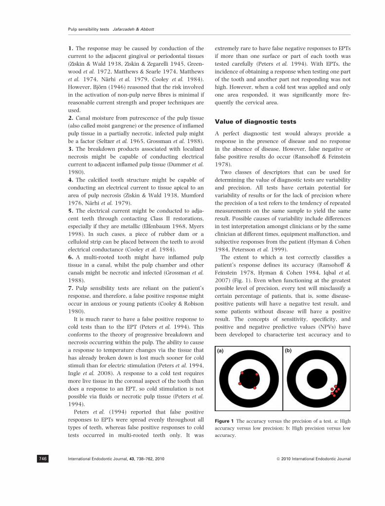
The extent to which a test correctly classifies a
patient’s response defines its accuracy (Ransohoff &
Feinstein 1978, Hyman & Cohen 1984, Iqbal et al.
2007) (Fig. 1). Even when functioning at the greatest
possible level of precision, every test will misclassify a
certain percentage of patients, that is, some disease-
positive patients will have a negative test result, and
some patients without disease will have a positive
result. The concepts of sensitivity, specificity, and
positive and negative predictive values (NPVs) have
been developed to characterize test accuracy and to
(a) (b)
Figure 1 The accuracy versus the precision of a test. a: High
accuracy versus low precision; b: High precision versus low
accuracy.
Pulp sensibility tests Jafarzadeh & Abbott
International Endodontic Journal, 43, 738–762, 2010 ª 2010 International Endodontic Journal746

describe the benefits of test usage (Fig. 2) (Ransohoff &
Feinstein 1978). As the calculations are based on
comparison of the test results and true disease status,
identification of this true status becomes an important
part of the evaluation of the status of the pulp (Hyman
& Cohen 1984, Petersson et al. 1999).
When a diagnostic test can be used as a gold
standard, determining the accuracy rates of other tests
can be determined (Ransohoff & Feinstein 1978,
Hyman & Cohen 1984, Iqbal et al. 2007). Unfortu-
nately, when diagnosing pulp status, a standard is not
available to determine the accuracy of other tests as
there is no consistently reported relationship between
clinical symptoms and histopathological findings (Selt-
zer et al. 1963a,b, Garfunkel et al. 1973, Dummer et al.
1980) and patients experiencing identical clinical
symptoms may exhibit different histological states
(Iqbal et al. 2007).
Sensitivity denotes the ability of a test to detect
disease in patients who actually have the disease, for
example, a test with a sensitivity of 0.60 has a 60%
chance of returning positive results when teeth with
pulp disease are tested. In contrast, specificity is the
ability of a test to detect the absence of disease. Hence, a
test with specificity of 0.80 has an 80% chance of
returning negative results when performed on teeth
without disease. The specificity and sensitivity of most
tests are inversely related. As the test is calibrated to
detect more disease (i.e. sensitivity is increased), it
returns a positive value in more disease-free persons
(i.e. specificity is decreased) (Lilienfeld & Lilienfeld
1980, Hyman & Cohen 1984).
Although sensitivity and specificity describe test
performance in relation to patients with known disease
states, the actual interest is in evaluating test responses
of patients with unknown disease states. This is
measured by predictive values. Positive predictive value
(PPV) is the probability that a positive test result
actually represents a disease-positive tooth, whereas
negative predictive value is the probability that a tooth
with a negative test result is actually free from disease.
PPV is more responsive to changes in specificity than in
sensitivity, whilst the reverse is true for NPV (Williams
1982, Hyman & Cohen 1984).
A few studies on the accuracy of pulp sensibility tests
were found in the literature (Fuss et al. 1986, Peters
et al. 1994, Evans et al. 1999, Petersson et al. 1999,
Gopikrishna et al. 2007) (Table 2). However, whereas
sensitivity and specificity are independent of disease
prevalence in the population, PPV is directly related to
the prevalence and NPV is inversely related, so the
predictive values cannot be directly compared in studies
with different disease prevalence (Hyman & Cohen
1984).
It should be emphasized that EPTs and thermal tests
are simple non-invasive tests, but they are not com-
pletely reliable. Heat has a relatively high sensibility but
is the least accurate overall of the three common pulp
tests owing to its low specificity (Petersson et al. 1999),
whereas the cold test is more accurate than the EPT
(Moody et al. 1989, Peters et al. 1994). Some studies
have indicated that if adult teeth with no history of
trauma do not respond to both EPTs and cold tests, then
there is a high probability that the tooth has a necrotic
pulp or it is pulpless (Seltzer et al. 1963a, Peters et al.
1994).
Thermal tests
The most common tests for assessing the status of the
pulp are thermal pulp sensibility tests. Stimulation of
the pulp with thermal tests is one of the oldest methods
to evaluate health of the pulp and its ability to respond
to external stimulation (Jack 1899). These tests have
often been called ‘vitality tests’, but this term is
inappropriate because these tests are unable to deter-
mine or demonstrate whether a tooth has a viable
blood supply (Jafarzadeh et al. 2008, Jafarzadeh 2009,
Jafarzadeh & Rosenberg 2009).
Two types of thermal tests are available, cold and hot
stimuli; although these tests may not be considered as
sophisticated as an EPT, they are often more reliable
Condition
Test outcomeTrue positive (TP) False positive (FP) Positive predictive value [TP/(TP+FP)]
False negative (FN) True negative (TN) Negative predictive value [TN/(TN+FN)]
Sensitivity [TP/(TP+FN)]
Specificity [TN/(TN+FP)]
Accuracy [(TP+TN)/(TP+TN+FP+FN)]
Clinicallynormal pulp
Positive
Negative
Necrotic pulp orpulpless tooth
Figure 2 Definition of the sensitivity, specificity, positive and negative predictive value, and accuracy of the pulp tests.
Jafarzadeh & Abbott Pulp sensibility tests
ª 2010 International Endodontic Journal International Endodontic Journal, 43, 738–762, 2010 747

(Weine 1996). However, neither cold nor heat is totally
reliable in all cases (Rowe & Pitt Ford 1990, Weine
1996). Each method of pulp testing has its place and
they are often complementary. A major advantage of
thermal tests over EPTs is that the equipment required
is usually inexpensive and easy to use (Rowe & Pitt
Ford 1990, Weine 1996), especially if the patient is
complaining of thermal sensitivity because the tests can
reproduce the patient’s pain.
It is recognized clinically that thermal sensitivity is
a sign of pulp inflammation; thus, the use of thermal
sensibility tests is a standard means of assessing the
state of the pulp (Rickoff et al. 1988). The presence of
thermal sensitivity indicates the development of pulp
disease, and the symptoms typically follow a consis-
tent scenario. As a rule, cold sensitivity is apparent
initially, whilst continuing pulp deterioration leads to
heat sensitivity, whereas cold sensitivity disappears.
Eventually, cold stimuli might relieve the heat-
induced pain (Sommer et al. 1962, Morse 1974).
The diseased pulp also usually demonstrates changes
in response to electrical stimulus (Udoye et al. 2010),
so a full diagnostic procedure will most likely utilize
both electrical and thermal testing (Selden 2000).
Selection of a cold test or a heat test should be based
on the patient’s chief complaint. If the patient does not
report any history of thermal pain, then, usually for
ease and reliability, a cold test is selected (Ruddle
2002a). However, it has been claimed that thermal
stimuli are of value only in teeth that are sensitive to
temperature changes (Degering 1962), but the
temperature and duration of thermal tests are not well
controlled, so thermal tests are somewhat variable,
with a degree of subjectivity in distinguishing a normal
from an abnormal response (Linsuwanont et al. 2008).
Thermal pulp tests rely on fluid flow through the
dentinal tubules (Trope & Debelian 2005). When cold
or heat is placed on a tooth surface, the fluid movement
in these tubules will result in irritation of the peripheral
tissues, including the A-d fibres, which results in a
sharp pain (Cohen & Hargreaves 2006). If the pulp is
severely inflamed, it is also likely that the expansion
from heat stimulation, or possibly the contraction from
cold stimulation, of the pulp tissue might induce
stimulation of the centrally placed C fibres (Trope &
Debelian 2005).
Damage to the soft and hard tissuesof the tooth
It has been reported that cold tests do not injure the
pulp (Ingram & Peters 1983, Rickoff et al. 1988),
whereas heat tests have a greater potential to cause
injury (Cohen & Hargreaves 2006). However, if tests
are used correctly, injuries are rare (Cohen & Harg-
reaves 2006).
Some studies have shown that cold tests can cause
pulp degeneration if tissue freezing occurs (Langeland
et al. 1969, Dowden et al. 1983). However, freezing
has been shown to result only when a cold probe
maintained a temperature lower than )10 �C for 5–
20 min (Frank et al. 1972). The mechanism of this
injury might involve intracellular ice crystal forma-
tion coupled with ischaemic necrosis resulting from
Table 2 The accuracy of pulp tests
Authors/year Tested teeth Kinds of tests Sensitivity Specificity
Positive
predictive
value
Negative
predictive
value Accuracy
Fuss et al. 1986 Premolars EPT
CO2 snow
DDM
Ethyl chloride
Ice stick
Not stated 0.90
0.97
0.99
0.49
0.33
Not stated Not stated Not stated
Peters et al. 1994 Complete dentition CO2 snow
EPT
0.94
0.66
Not stated Not stated Not stated Not stated
Petersson
et al. 1999
Various teeth Ethyl chloride
Hot gutta-percha
EPT
0.83
0.86
0.72
0.93
0.41
0.93
0.89
0.48
0.88
0.90
0.83
0.84
0.86
0.71
0.81
Evans et al. 1999 Anterior teeth Ethyl chloride
EPT
0.92
0.87
0.89
0.96
Not stated Not stated Not stated
Gopikrishna
et al. 2007
Single-rooted
incisors, canines,
and premolars
EPT
TFE
0.71
0.81
0.92
0.92
0.91
0.92
0.74
0.81
0.81
0.86
EPT, Electric pulp tester; DDM, Dichlorodifluoromethane spray; TFE, Tetrafluoroethane spray.
Pulp sensibility tests Jafarzadeh & Abbott
International Endodontic Journal, 43, 738–762, 2010 ª 2010 International Endodontic Journal748

vascular injuries (Frank et al. 1972, Dowden et al.
1983). Langeland et al. (1969) concluded that an
applied temperature of )22 �C, which lowered the
temperature of the pulp to 11 �C, caused no damage
to the pulp but that an application of an extreme
cold stimulus of )160 �C for 3 min caused extensive
pulp damage. Dowden et al. (1983) reported that
when temperatures below )80 �C are applied to teeth
for 1, 2, or 3 min, the temperature of the pulp was
lowered (ranged from 0 to )30 �C) and an increasing
degree of pulp damage occurred, but all of the pulps
remained alive and able to respond to thermal tests.
The response of the pulp was predictable and
characterized by a distinct layer of coronal secondary
dentine with odontoblast destruction, cellular inclu-
sions, and microvascular injuries. The pulp within
the root remained essentially healthy and without
inflammation, but some periodontal and root surface
damage was observed.
Barker et al. (1972) examined teeth before and after
application of a cold stimulus of )196 �C for two
periods of 5 min each. They saw no changes in the
surface status of the crown, but the extreme temper-
ature changes created new cracks in the teeth. More-
over, some other studies with the aid of permeating dye
and standardized fluorescence UV-photography have
demonstrated that dry ice and refrigerant sprays may
create new fissures in the enamel (Lutz et al. 1974,
Bachmann & Lutz 1976).
Some studies reported that dry ice applied to a tooth
did not jeopardize pulp health (Schiller 1937, Ingram &
Peters 1983, Rickoff et al. 1988). They revealed struc-
turally intact pulp tissue exhibiting no evidence of
injury. However, Dowden et al. (1983) showed that
minor pulp injury occurred but the tissue recovered
within 47–63 days.
Some have indicated that no new cracks or fissures
could be seen after application of dry ice. This proved
true even when used for significantly longer periods of
time than that normally used clinically (2 min vs.
<15 s). Clinically, most teeth respond to the cold test in
<5 s (Ingram & Peters 1983, Peters et al. 1983, 1986).
If a tooth is going to respond, it almost always occurs
within this time (Ingram & Peters 1983, Peters et al.
1983). These findings are in contrast with several other
studies, which determined that dry ice created fissures/
cracks (Lutz et al. 1974, Bachmann & Lutz 1976).
Also, it might cause surface pitting of porcelain when
applied for as little as 5.4 s (Krell et al. 1985). Some
delay in response of the patient to this test might be
considered as normal, and this might be explained by a
delayed cold transfer process (Linsuwanont et al.
2008).
This method must be used with caution on teeth
forming part of a bridge if the bridge includes metal
retainers. A cold stimulus applied to a tooth whose pulp
may itself be necrotic may be felt by an adjacent tooth
because the coldness is conducted through the metal
part of the bridge (Ehrmann 1977).
Some clinicians worry that a piece of dry ice that
falls inadvertently into the mouth could have a
deleterious effect on the mucosa with which it comes
into contact. However, it is fortunate that this is not
the case because when CO2 falls into the mouth it is
surrounded by an insulating layer of gaseous CO2,
which does not harm the mucosa. This is known as
‘film boiling’ or the ‘Leidenfrost phenomenon’ (Fig. 3)
(Ehrmann 1977). In contrast to this, some authors
believe that owing to the extremely cold temperature,
burns of the mucosa it contacts can occur (Cohen &
Hargreaves 2006), but these claims have not been
substantiated.
Cold tests
Records, including the Edwin Smith Surgical Papyrus,
have referred to the use of cold stimuli as a form of pulp
testing since 2500 BC (Rand et al. 1968). Since 1938,
experiments in hypothermia have shown that cold
temperatures cause minimal irreversible tissue effects
unless crystal formation occurs (Rand et al. 1968).
Frank et al. (1972) have confirmed this claim in respect
to pulp tissues.
It has been stated that cold tests, in general, are
more accurate methods of testing pulps than heat tests
(Ehrmann 1977, Shabahang 2005). However, a cold
test to determine the state of the pulp (e.g. pulp
necrosis versus a healthy pulp) is not always reliable
because teeth with calcified pulp spaces might have
normal and healthy pulps but the cold stimuli might
not be able to excite the nerve endings owing to the
insulating effect of the thicker layer of dentine, which
Hot surface
Floating drop
Vapor layer
Figure 3 Leidenfrost phenomenon.
Jafarzadeh & Abbott Pulp sensibility tests
ª 2010 International Endodontic Journal International Endodontic Journal, 43, 738–762, 2010 749

is a result of secondary and reactionary dentine
formation (Ehrmann 1977, Ingle et al. 2008). Calcifi-
cation of the pulp space can usually be identified by
radiographic examination, and this highlights the
essential need to always and only interpret pulp test
results in conjunction with viewing a periapical
radiograph. Cold tests can also be used to differentiate
between reversible and irreversible pulpitis (Cohen &
Hargreaves 2006). In general, the response to a cold
stimulus is measured as a positive or negative reaction
(Ehrmann 1977), but the quality of the response is also
important, particularly whether it reproduces the pain
reported by the patient. Typically, if the pulp is
inflamed, the patient will report short, sharp pain that
might disappear rapidly once the stimulus is removed
or it might become a dull ache that lingers after the
stimulus is removed. When the pain disappears
rapidly, this usually indicates reversible pulpitis,
whereas the dull lingering ache indicates irreversible
pulpitis (Cohen & Hargreaves 2006).
Cold tests are presumed to be more reliable than EPTs
in teeth with incomplete root formation. As thermal
tests are not 100% accurate, an EPT is especially
beneficial for confirming a questionable diagnosis
(Shabahang 2005). However, cold tests should be used
in conjunction with the EPT, so the results from one
test can verify the findings of the other test, and
radiographs must also be viewed.
The A-d fibres are neurons that are initially stimu-
lated during cold testing. At 22 �C, A-d transmission is
abolished, whereas C fibre sensitivity remains (Douglas
& Malcolm 1955). For dentinal pain, the PDJ must cool
to 29 �C (Naylor 1964, Pantera et al. 1993). If neurons
are directly stimulated by temperature changes, it is
possible to cause anaesthesia. However, the tempera-
ture needed for anaesthesia is much lower than that
needed to induce pain (Naylor 1964). Clinically, a cold
stimulus is removed when the patient feels the sensa-
tion, so it is unlikely that the PDJ temperature would
decrease to the level needed for anaesthesia (Naylor
1964, Pantera et al. 1993). Several methods such as ice
sticks, refrigerant sprays, carbon dioxide snow (CO2),
ethyl chloride, and cold water baths are available, with
the major difference between them being the tempera-
ture produced by each test (Pitt Ford & Patel 2004).
Ice sticks
Ice sticks have been used as a cold test. The ice
produces a temperature of 0 �C, but it is not considered
accurate, especially in adult teeth, posterior teeth, and
in teeth with severe deposition of secondary or repar-
ative dentine (Ehrmann 1977, Augsburger & Peters
1981). Also, the use of ice appears to be of limited value
when testing teeth under crowns and splints (Fulling &
Andreasen 1976b).
Ice sticks can be formed by freezing water in the
plastic covers of hypodermic needles or in used local
anaesthetic cartridges after sterilization. The ice stick
should be placed in gauze to prevent warmth from the
operator’s fingers from prematurely melting the ice
(Ruddle 2002a). It is placed towards the cervical
(Ruddle 2002a) or middle (Cohen & Hargreaves 2006)
third of the buccal or lingual aspect of the crown of the
tooth, or on any exposed metal surface and quickly
moved back and forth (Fig. 4) (Ruddle 2002a). The ice
stick should be kept in contact with the tooth for 5 s or
until the patient begins to feel pain (Cohen & Hargreaves
2006). Testing should begin with the most posterior
tooth and then advance towards the anterior teeth. This
prevents melting ice water from dripping in a posterior
direction and possibly touching a tooth that has not yet
been tested but that could give a false response. A cotton
pellet should be placed just distal to the tooth being
tested to prevent any cold water from contacting the
posterior teeth (Ruddle 2002a). The melting ice might
also stimulate nerves within the surrounding gingiva of
the tooth being tested or that of adjacent or nearby
teeth, which can result in false responses that may lead
to an incorrect diagnosis.
It has been indicated that the application of an ice
stick for a period of 5 s is a reliable and valid method
(Dachi et al. 1967), Under these situations, the error in
predicting which teeth will be bothersome to the
patient whilst eating cold food is reduced by 20–50%,
depending upon which tooth is involved (Dachi et al.
1967), A major disadvantage of the ice stick test is that
it is simply not cold enough and, hence, CO2 dry ice or
refrigerant sprays might be better choices (Dachi et al.
1967, Ehrmann 1977). A recent study showed that ice
can stimulate the pulp less effectively than other cold
tests (Linsuwanont et al. 2008).
Refrigerant sprays
A common method for cold testing is to use a
refrigerant spray (Ingle et al. 2008). It is probably the
most convenient and easiest to use and ranks just
behind CO2 dry ice for overall efficacy and accuracy
(White & Cooley 1977, Ingle et al. 2008). It can
provide reliable and reproducible results (White &
Cooley 1977). Different refrigerant sprays are available,
Pulp sensibility tests Jafarzadeh & Abbott
International Endodontic Journal, 43, 738–762, 2010 ª 2010 International Endodontic Journal750

and they are based on dichlorodifluoromethane (DDM),
tetrafluoroethane (TFE), or a propane–butane mixture
(PBM). There are also sprays used to clean and test
electronic circuits, which have the same composition as
those used for pulp tests (Ingle et al. 2008, de Morais
et al. 2008). The temperature decrease offered by such
products varies according to their composition and
ranges from )20 to )50 �C, according to the manu-
facturers (de Morais et al. 2008).
DDM (R-12), usually sold under the brand name
Freon-12, is a chlorofluorocarbon halomethane, which
has been shown to be an effective and uncomplicated
agent for cold testing of teeth (http://en.wikipedia.org/
wiki/Dichlorodifluoromethane) (Accessed November 4,
2007). It is commercially packaged for dental use as a
compressed spray (Endo-Ice) ()50 �C) (Coltene/Whal-
edent, Switzerland) (Fig. 5a) (Augsburger & Peters
1981, Fuss et al. 1986). The production of DDM was
prohibited by the Clean Air Act in the United States in
January 1996, because of environmental concerns
regarding its effect on the ozone layer of the atmo-
sphere. Moreover, it might act as an irritant if inhaled
by the patients, nurses, or the dentists. However, some
researchers feel that the amount used to test pulps
might not be of any real significance with respect to
these potential hazards (Cohen & Hargreaves 2006),
but others believe that DDM can create new fissures or
cracks in enamel (Bachmann & Lutz 1976).
White & Cooley (1977) showed that DDM can
produce a more rapid thermal change than either an
ice stick or a cold water bath. Fuss et al. (1986) also
showed that DDM and CO2 dry ice produced a greater
decrease in temperature than either an ice stick or ethyl
chloride. DDM produced the greatest reduction in
intrapulpal temperature when applied by a saturated
cotton pellet rather than via a cotton tip applicator
(Jones 1999).
DDM was less efficient than CO2 dry ice when testing
multiple teeth (Augsburger & Peters 1981). However,
one study showed that at least three teeth could be
tested with DDM before having to respray the cotton
pellets (Fuss et al. 1986).
DDM has been replaced by the manufacturer with
1,1,1,2-tetrafluoroethane (TFE) because of the above-
mentioned concerns. TFE is commercially available as
Green Endo-Ice ()26.2 �C) (Coltene/Whaledent)
(Fig. 5b) and has also been chemically called R-134a,
Freon 134a, Genetron 134a, or HFC-134a. It is a
haloalkane refrigerant without ozone-depletion poten-
tial and has thermodynamic characteristics similar to
DDM (http://en.wikipedia.org/wiki/Dichlorodifluoro-
methane) (Accessed November 4, 2007). It is easy to
use with rapid results (Ingle et al. 2008). The material is
sprayed onto a cotton pellet which is then applied to the
middle third of the facial/labial surface of the crown. It
should be kept in contact with the surface for 5 s or until
the patient begins to feel pain (Cohen & Hargreaves
2006).
Figure 4 Application of ice sticks for cold testing.
(b) (c)(a)
Figure 5 Endo-Ice for cold testing
(Reproduced with permission from
Johnson WT (2002) Color Atlas of End-
odontics. Philadelphia: W.B. Saunders)
(a), Green Endo-Ice for cold testing
(Courtesy of http://www.coltenewhale-
dent.biz) (b), and Endo-Frost for cold
testing (Courtesy of http://www.
coltenewhaledent.biz) (c).
Jafarzadeh & Abbott Pulp sensibility tests
ª 2010 International Endodontic Journal International Endodontic Journal, 43, 738–762, 2010 751

Jones et al. (2002) concluded that TFE and CO2 dry
ice were equivalent in producing a pulp response
regardless of the tooth type and the presence/absence of
restorations, but the response from TFE was faster.
Miller et al. (2004) reported that in intact teeth and
teeth restored with porcelain-fused-to-metal (PFM)
crowns or all-ceramic restorations, TFE produced a
more rapid and effective initial temperature reduction
than CO2 dry ice. Application of TFE on a saturated
cotton pellet was the most effective technique for
producing a temperature reduction in these cases.
PBM, commercially available as Endo-Frost ()50 �C)
(Coltene/Whaledent) (Fig. 5c), usually contains 30–
50% propane, 30–50% butane, and 10–20% isobu-
tane. It is a nontoxic cold spray, which is recommended
for freezing cotton pellets and small cotton rolls. A
recent study showed that PBM and TFE induced lower
temperatures than dichlorofluoroethane-containing
sprays when measured immediately after application
on a cotton swab. However, temperatures inside the
pulp chamber had similar decreases, no matter which
spray was used (de Morais et al. 2008).
Carbon dioxide snow (dry ice)
Carbon dioxide (CO2) is a chemical compound com-
posed of two oxygen atoms bonded to a single carbon
atom. It is a gas, but in its solid state, it is often called
‘dry ice’, and it is commonly used as a versatile cooling
agent (http://en.wikipedia.org/wiki/Carbon_dioxide)
(Accessed November 4, 2007). The low temperature
and direct sublimation to a gas at atmospheric pressure
make it an effective coolant, because it is colder than
ice and leaves no moisture as it changes state. Fischer
(1972) regarded it as the only satisfactory method of
pulp testing.
In 1835, the French chemist Charles Thilorier
(1835) published the first account of the use of dry
ice. Upon opening the lid of a large cylinder containing
liquid CO2, he noted much of the CO2 rapidly evapo-
rated leaving solid dry ice in its container. Dry ice was
first introduced into dentistry by Back (1936) whose
apparatus was modified by Obwegeser & Steinhauser
(1963) to collect the dry ice in a thin Plexiglas tube
that could then be used in a pencil-like form.
The temperature of dry ice has been reported to be
)78 �C (Bachmann & Lutz 1976); however, applying it
as used clinically directly to a temperature probe
produced a less cold reading of )56 �C (Augsburger &
Peters 1981), which is still effective for decreasing pulp
temperature and to elicit a painful response (Augs-
burger & Peters 1981, Fuss et al. 1986). The sensory
response to the application of dry ice is rapid, usually
being <2 s (Fuss et al. 1986).
Mechanism
Application of dry ice for 5 s can reduce the temper-
ature at the PDJ to <2 �C (Rickoff et al. 1988). This
further supports the hydrodynamic theory concerning
the sensory response of the teeth to thermal stimula-
tion. According to this theory, application of heat or
cold to a tooth results in rapid movement of dentinal
fluid that mechanically stimulates the sensory termi-
nals located in the region of the PDJ (Brannstrom &
Johnson 1970). However, a recent study reported that
thermal contraction and expansion of dentinal fluid
might not be the complete explanation for dentinal fluid
movement in intact teeth. Enamel might serve not only
as a temperature transfer medium, but it might also
expand or contract when subjected to thermal stimu-
lation (Linsuwanont et al. 2007).
Technique
For pulp sensibility testing, CO2 is released into a special
tube inside the Plexiglas container in which it forms the
‘snow’ (Figs 6 and 7). It is compacted with a plugger
and the ‘pencil’ or ‘stick’ of dry ice is expressed slightly
from the tube and then applied immediately to the
middle third of the facial surface of the crown of the
tooth and kept in contact for 2–5 s or until the patient
feels pain (Fig. 8) (Cohen & Hargreaves 2006).
Advantages
Clinically, testing with CO2 snow is accurate, reliable,
consistent, fast, and uncomplicated (Ehrmann 1977).
A complete dentition can be tested in 1–2 min
without any requirement for isolation of the teeth
(Obwegeser & Steinhauser 1963, Ehrmann 1977,
Augsburger & Peters 1981). It is preferred for pulp
testing because it does not affect the adjacent teeth
and because it gives an intense, reproducible response
(Augsburger & Peters 1981, Ingram & Peters 1983,
Peters et al. 1983, Fuss et al. 1986). It has been
shown that the accuracy of dry ice is greater than
that of EPTs (Back 1936, Obwegeser & Steinhauser
1963, Fulling & Andreasen 1976a). It is also effective
for testing teeth with full coverage crowns (Ehrmann
1977, Augsburger & Peters 1981, Grossman et al.
1988, Cohen & Hargreaves 2006). PFM crowns can
be tested by applying the dry ice to the metal part of
the crown (Ehrmann 1977). Although Riedel &
Fichter (1966) found CO2 to be less effective when
Pulp sensibility tests Jafarzadeh & Abbott
International Endodontic Journal, 43, 738–762, 2010 ª 2010 International Endodontic Journal752

testing through acrylic or porcelain restorations, it
can still be used when these restorations are present
as long as the clinician recognizes the potentially
different response to be expected. Notwithstanding
this, CO2 is considered to be more reliable than other
forms of pulp sensibility tests when these types of
restorations are present (Ingle et al. 2008).
When testing teeth after trauma for their response to
cold, the CO2 snow test is the most effective test (Fulling
& Andreasen 1976b, Ehrmann 1977, Ingle et al.
2008). It gives more accurate responses than ice
because the intense cold penetrates the tooth and any
covering splints or restorations and will reach the
deeper areas of the tooth/pulp, so it can be used on
individual teeth under splinted abutments (Fulling &
Andreasen 1976b, Ehrmann 1977). In the case of
splints and temporary crowns, dry ice can also provide
reliable responses, unless applied on the incisal edge
(Fulling & Andreasen 1976b).
Other advantages of CO2 snow include no false
positive readings in cases of pulp necrosis and in
cases of early pulpitis where there is a sustained or
lingering painful response, which is quite different
from that exhibited by normal pulps (Fulling &
Andreasen 1976b, Ehrmann 1977, Ingle et al.
2008). It also may be used to test teeth undergoing
orthodontic treatment using fixed appliances where
an EPT is not possible (Fulling & Andreasen 1976b,
Ehrmann 1977) because the metallic wires, brackets,
and bands might result in conduction of the electric
current to another tooth other than the one being
tested.
Disadvantages
One disadvantage of the dry ice test is that it is not
effective with calcified pulps or in elderly patients where
there has been considerable secondary dentine forma-
tion (Ehrmann 1977). Also, the apparatus is more
expensive to purchase than ethyl chloride or ice sticks
(Weine 1996).
Figure 8 Application of dry ice.
Figure 7 Dry ice stick with holder and plugger (Reproduced
with permission from Johnson WT (2002) Color Atlas of
Endodontics. Philadelphia: W.B. Saunders).
Figure 6 Dry ice maker.
Jafarzadeh & Abbott Pulp sensibility tests
ª 2010 International Endodontic Journal International Endodontic Journal, 43, 738–762, 2010 753

Comparative studies
Fuss et al. (1986) concluded that dry ice and DDM were
more dependable than ethyl chloride and ice in
producing a positive response in premolars and that
the rate of temperature decrease was greater for dry ice
and DDM than for either ethyl chloride or ice. Their
findings are in agreement with those of Fulling &
Andreasen (1976a) who found that 100% of the
incisors tested responded positively to dry ice, whereas
only 70% responded to the ice stick test. The lower
incidence of positive response in the study of Fuss et al.
(1986), compared with the study of Fulling & Andrea-
sen (1976a), may be attributable to the total thickness
of enamel and dentine being greater in premolars than
in incisors.
According to Fuss et al. (1986), the reaction to CO2
snow and DDM was rapid (i.e. only 1–2 s). Character-
istically, DDM evoked a sharp pain almost immediately,
whereas it usually took CO2 snow somewhat longer to
elicit a response. This difference may be owing to the
cotton pellets saturated with DDM covering a greater
surface area of enamel than that covered by the CO2
snow ‘pencil’. It has been hypothesized that that CO2
snow makes less contact with the tooth surface (Ingle
et al. 2008).
When comparing CO2 snow and DDM, Fuss et al.
(1986) found no difference between the young and
adult groups. This is in accordance with the results of
other studies (Fulling & Andreasen 1976a, Ehrmann
1977) where CO2 snow was reported to be more
reliable than an EPT in determining the pulp status of
immature teeth. Therefore, it seems that the statement
by Mumford (1967) that the EPT is more reliable than
thermal tests is correct with regard to tests using ice
and ethyl chloride but not to those utilizing CO2 snow
and DDM.
Linsuwanont et al. (2008) indicated that CO2 snow
and refrigerant sprays provided more consistent stimuli
when compared with cold water and ice, so these tests
might be considered as the stimulus/test of choice.
Ethyl chloride
Ethyl chloride (chloroethane), with a temperature of
)12.3 �C, is a colourless, flammable gas or refrigerated
liquid with a faintly sweet odour, available as a
compressed spray and commonly used in medicine as
a skin refrigerant (Fig. 9) (Augsburger & Peters 1981).
It is a prescription drug in the United States of America,
where it is supplied as a liquid in a spray bottle
propelled by its own vapour pressure. It acts as a mild
topical anaesthetic when sprayed on skin, such as
when removing splinters in a clinical setting. The heat
absorbed by the boiling liquid on tissues produces a
rapid and deep chill, but as the boiling point is well
above freezing, it usually presents no danger of
frostbite (http://en.wikipedia.org/wiki/Ethyl_chloride)
(Accessed November 4, 2007).
Like other chlorinated hydrocarbons, ethyl chloride
is a central nervous system depressant, albeit a less
potent one than many other similar compounds.
Breathing its vapours at <1% concentration in air
usually produces no symptoms; however, in higher
concentrations, victims usually exhibit symptoms
similar to those of alcohol intoxication. Breathing its
vapours at a concentration of ‡15% is often fatal
(http://en.wikipedia.org/wiki/Ethyl_chloride) (Accessed
November 4, 2007).
Moody et al. (1989), using ethyl chloride and
monopolar and bipolar EPTs, reported that the best
prediction of the pulp status was with the ethyl
chloride. Also, Mumford (1964) showed that the results
using ethyl chloride were better than hot Gutta-percha
for both consistency of the response and the lower
number of teeth that did not respond. However, he
showed that reproducible results could not be obtained
with ethyl chloride.
The use of ethyl chloride in pulp testing is no longer
recommended because it has been found to be less
effective than dry ice or DDM (Augsburger & Peters
1981). Although it provides a good source for lowering
Figure 9 Ethyl chloride spray (Courtesy of Gebauer Company,
Cleveland, Ohio, USA).
Pulp sensibility tests Jafarzadeh & Abbott
International Endodontic Journal, 43, 738–762, 2010 ª 2010 International Endodontic Journal754

tooth temperature, the temperature obtained is nor-
mally not sufficient enough for pulp stimulation in
teeth with very extensive restorations or full crown
coverage restorations (Cohen & Hargreaves 2006).
Cold water bath
For cold testing, a tooth or a group of teeth can be
isolated with a rubber dam, and iced water can be
syringed onto each tooth (Fig. 10). This cold water
bath can be an effective method of pulp testing because
it is the only technique that immerses the tooth in
chilled water. Although more time-consuming, this
kind of cold testing is effective at simultaneously
bathing the entire crown of the tooth, which is why
this is a reliable method for testing thermal sensitivity,
and stimulating an inflamed pulp (Cohen & Hargreaves
2006). It is clearly superior in its accuracy compared to
ice sticks. Even when the tooth has a full crown
restoration, sufficient contact between the tooth and
the cold water is made to allow cooling of the pulp. In
addition, it prevents damage to the tooth caused by
excessive temperature changes. This method is effective
for evaluating teeth with full coverage restorations,
either metal or porcelain. The sensory response of teeth
is refractory to repeated thermal stimulation. Another
benefit of this method is that it needs no armamentar-
ium except for rubber dam (Ruddle 2002a).
Heat tests
When a cold stimulus is applied to a tooth with a
normal pulp, it creates a response quickly but the
stimulus does not last long. If the pulp has only a mild
and local inflammation, the patient feels a sharper
reaction to it (Ingle et al. 2008). Heat application in
these cases produces a similar sensation, but more
slowly. Heat causes the fluid to expand, stimulating the
A-d fibres; however, when heat is applied to an
inflamed pulp, the pressure increase can stimulate the
C fibres and produce a long lasting pain (Grossman
1981). Because cold might relieve the pain in irrevers-
ibly inflamed pulps and because the response to cold is
by the A-d fibres and not the C fibres, heat testing might
give better results for true diagnosis (Cohen & Harg-
reaves 2006).
Pain precipitated by heat usually shows an acutely
inflamed or a partially necrotic pulp (Cohen & Harg-
reaves 2006). Necrotic tissue frequently harbours
bacteria that can produce gasses that potentially
expand against tissue encased inside the unyielding
dentinal walls. This may cause sensory nerve fibres to
transmit pain. Hence, in the case of a small pulp
abscess, heat can increase the symptoms, and cold can
relieve the pain (Seltzer et al. 1963a). It has also been
proposed that the heat might cause sufficient thermal
expansion to push infected material from the pulp space
into the periapical tissues (Mumford 1964, Mumford &
Jedynakiewicz 1988).
Heat tests have many methods of delivery, such as
heated Gutta-percha, warmed instruments, electrical
heat sources, frictional heat, and hot water baths.
However, heat tests are infrequently performed because
of the difficulties associated with tooth isolation and
obtaining a consistent heat stimulus (Ingle & Bakland
2002). As studies indicate that the diagnostic accuracy
of heat tests is low, heat should not be used as a single
test of the pulp status (Petersson et al. 1999, Bergen-
holtz et al. 2003). It has been shown that thermal
stimuli are highly variable in terms of the rate of
thermal transfer through the tooth, the stimulus
temperature, and the extent of temperature change
within the tooth structure (Linsuwanont et al. 2008).
Heated Gutta-percha (Grossman’s method)
Application of warmed sticks of gutta-percha (120–
140 �C) is one method of performing a heat test. To use
this technique, the tooth surfaces and the surrounding
area should firstly be dried with cotton rolls (Rosenberg
1991). The teeth to be tested should be protected with a
light coating of petroleum jelly to prevent the heated
gutta-percha from sticking to the tooth surface (Cohen
& Hargreaves 2006). The gutta-percha stick is then
warmed over a flame until it becomes soft and begins to
glisten, but not so that it slumps and becomes too limp
Figure 10 Cold water bath (Reproduced with permission from
Johnson WT (2002) Color Atlas of Endodontics. Philadelphia:
W.B. Saunders).
Jafarzadeh & Abbott Pulp sensibility tests
ª 2010 International Endodontic Journal International Endodontic Journal, 43, 738–762, 2010 755

for application (Grossman 1981, Tronstad 2003).
Mumford (1964) found that if the gutta-percha stick
was held in the flame for 2 s, only 14% of the anterior
teeth tested gave a positive response, whereas Trow-
bridge et al. (1980) found that all teeth gave a positive
response, generally within <2 s.
One disadvantage of heating gutta-percha over a
flame is the difficulty in controlling the temperature
obtained (Rickoff et al. 1988). As its temperature may
be as high as 76 �C when applied to the tooth surface
(Trowbridge et al. 1980), some concerns have been
expressed that such high temperatures might damage a
healthy pulp. However, one study indicated that it does
not jeopardize the pulp health as the 5-s application
only increased the temperature at the PDJ by <2 �C
(Rickoff et al. 1988). Special care should be taken not to
damage the pulp tissue with an excessive or prolonged
heat stimulus that could result in pulp damage
(Mumford 1964), because it will result in biphasic
stimulation, initially of the A-d fibres and then subse-
quently the C fibres, resulting in a lingering pain.
Therefore, heat tests should be applied to the tooth
surface for no more than 5 s. However, and in contrast,
inadequate heating of the gutta-percha stick might
result in a weak stimulus, which might not elicit a
response from the pulp (Narhi 1985).
Application to the middle third of the facial/labial
surface of the crown usually results in a response
within <2 s (Cohen & Hargreaves 2006) (Fig. 11). If
the patient complains that the heat test produces a
severe ache, the clinician must be ready to apply a cold
stimulus immediately to the tooth to relieve the pain
(Ingle & Bakland 2002).
Reproducible results cannot be obtained by testing
pulps with heated gutta-percha. Moreover, teeth that
fail to respond to this test are mainly canines, and this
is attributed to their greater bulk that permits only a
small change in temperature for the amount of heat
applied (Mumford 1964). The temperature at the site
where excitation occurs (either in the dentine or in the
pulp) depends not only on the amount of heat
transferred from the gutta-percha but also on the
amount of heat present in the gutta-percha to begin
with and furthermore on the area of contact with the
tooth. The latter in turn depends on the original
diameter of the gutta-percha stick, its plasticity, and the
force with which it is applied (Mumford 1964). In
addition, the thickness, thermal conductivity, and
thermal diffusivity of the enamel and dentine, and the
presence and extent of caries or restorations may
modify the final stimulus and response (Braden 1963,
Mumford 1964, Mumford & Jedynakiewicz 1988).
A recent study showed that heated gutta-percha is a
strong but less consistent stimulus in terms of both
initial temperature and the temperature change during
testing (Linsuwanont et al. 2008). Overall, the use of
heated gutta-percha appears to be of limited value in
sensibility testing of teeth under splints and temporary
crowns (Fulling & Andreasen 1976b). It may also be
difficult to use on posterior teeth because of the limited
access (Ehrmann 1977).
Warmed hand instruments
A popular but not very reliable and relatively poorly
assessed method for heat testing of the teeth is through
using a heated dental hand instrument such as a ball
burnisher. The instrument is heated over a flame and
then applied close to the buccal surface of the crown
without actually contacting the tooth, thus relying on
the radiant heat produced from the heated instrument
(Fig. 12). Because of the difficulty in controlling the
temperature of the heated instrument and the potential
safety problems associated with using heated instru-
ments in the patient’s mouth, this type of heat test is
not generally recommended. In addition, it cannot be
considered as being reliable or reproducible (Walton &
Torabinejad 2002).
Electrical heat sources
There are several devices that might be used to apply
a hot stimulus to teeth, including the Touch’N Heat
(SybronEndo, Orange, CA, USA) or the System B
devices (SybronEndo). Each of these devices has a
handpiece designed to receive various inserts such asFigure 11 Grossman’s method.
Pulp sensibility tests Jafarzadeh & Abbott
International Endodontic Journal, 43, 738–762, 2010 ª 2010 International Endodontic Journal756

the Hot Pulp Test Tip. Regardless of the device chosen,
the continuous heat mode is selected, and the
intensity is set at the manufacturer’s recommendation
for performing the heat test on the surface of the
tooth, provided it has been lubricated to prevent
sticking and burning of the enamel. As an example,
when using the System B device, the temperature is
set at 150 �F. This test can be performed without
damage to hard and soft tissues of the tooth (Ruddle
2002a, Ingle et al. 2008).
Frictional heat
Frictional heat can be generated using a rubber cup,
such as those intended for prophylaxis, applied to the
buccal aspect of a tooth without prophylactic paste and
rotated within a dental handpiece to produce frictional
heat (Fig. 13). This method is considered to be the best,
easiest, and safest technique of performing a heat test
(Walton & Torabinejad 2002), especially when testing
a tooth with a gold crown (Weine 1996). Despite this,
frictional heat tests are seldom used today (Cohen &
Hargreaves 2006).
Hot water bath
The simplest method of testing for heat sensitivity is to
bathe the tooth in hot water (Trope & Debelian 2005).
This is performed by placing the patient in a supine
position and applying rubber dam on the most
posterior tooth in the quadrant using a single-tooth
isolation technique. The tooth is then bathed in very
hot water syringed onto it for 5 s or until the patient
begins to feel pain. If the patient’s chief complaint is
pain associated with heat, then the temperature can
be gradually increased if no response is obtained,
rather than producing unnecessary pain by beginning
with excessively hot liquid. If this test does not
reproduce the patient’s symptoms, the rubber dam is
removed and then placed on the next tooth anterior to
the first tooth tested and the hot water test is repeated
using fresh hot water. This protocol is repeated on one
tooth at a time, moving further anteriorly until the
patient’s presenting pain is replicated (Ruddle 2002b,
Trope & Debelian 2005). This technique produces a
greater thermal change than heated gutta-percha, a
warmed hand instrument, or a rotating rubber cup
(White & Cooley 1977) because the entire crown of
the tooth is bathed with the hot water.
The advantage of this method is attributable to the
hot water instantaneously bathing the entire clinical
crown, which improves conductivity and more closely
replicates the way heat naturally contacts the tooth
during drinking of hot liquids (Ruddle 2002a). Also,
because the hot water is contained within the rubber
dam, the response is limited to the one tooth being
tested (Grossman et al. 1988) although this will be
dependent on the thickness of the rubber dam (i.e. thick
or heavy body dam is more effective than thinner
grades of dam) and on how well the tooth is isolated
because any marginal gaps between the dam and the
tooth may allow the hot water to diffuse through the
gaps and then stimulate the adjacent tooth or gingivae.
Heat testing by hot water bathing may be more
effective in penetrating PFM crowns, and it may also
prevent damage to the tooth caused by excessive
temperature changes (Ingle et al. 2008).
The disadvantage of this test is the cooperation
required to comfortably place a rubber dam clamp on
Figure 12 Application of warmed hand instruments for heat
testing.
Figure 13 Application of frictional heat for heat testing.
Jafarzadeh & Abbott Pulp sensibility tests
ª 2010 International Endodontic Journal International Endodontic Journal, 43, 738–762, 2010 757
one tooth at a time and the time required to perform
this test on several teeth (Ruddle 2002a). Although this
method is time-consuming, it is superior in its accuracy
compared to other heat tests (Cohen & Hargreaves
2006, Ingle et al. 2008).
Important technical points about
thermal pulp tests
Before initiating thermal tests, it is important to inform
the patient of the nature of the test(s) and what is to be
achieved. The patient must be aware of when the
operator requires them to respond and how they can
indicate their response; hand signals are an effective
means of achieving this, but it is essential to establish a
reliable report. The patient should be instructed to raise
his/her hand when they first feel the sensation from the
thermal stimulus in the tooth, to keep their hand up as
long as this sensation lingers and to lower their hand
when the sensation dissipates. In the event that the
patient does not perceive any sensation in the tooth
after 5–6 s, then the stimulus should be removed.
Because some pulpally involved teeth may not be
stimulated immediately by the thermal test, the oper-
ator should wait for several seconds after the stimulus
is removed from the tooth to see whether any pain is
elicited before placing the stimulus on the next tooth.
Some teeth with irreversible pulpitis require repeated
stimuli to reach the threshold that provokes pain
(Mumford & Jedynakiewicz 1988, Trope & Debelian
2005, Cohen & Hargreaves 2006, Ingle et al. 2008).
Also, it should be remembered that once a pulp is tested
with cold, there is a refractory period of several minutes
before a second cold or heat stimulation can be
accurately conducted on that same tooth (Mumford &
Jedynakiewicz 1988, Trope & Debelian 2005).
Thermal tests are considered to be more accurate
when applied to the cervical aspect of a tooth and as
close as possible to the gingival margin. This location
represents the thinnest aspect of enamel or a restora-
tion and the closest distance to the pulp chamber
(Peters et al. 1994, Ruddle 2002a). However, some
studies have used the middle third of the buccal/labial
or palatal/lingual aspect of the crown for thermal
testing (Cohen & Hargreaves 2006). In contrast, others
believe that for anterior teeth, the cold stimulus should
be placed on the incisal third, and for posterior teeth on
the incisal aspect of the mesiobuccal cusp. These areas
approximate the pulp horns, where the pulp innerva-
tion is most plentiful (Trope & Debelian 2005). It
should be remembered that the size of the pulp
chamber is the most important factor in determining
the thermal response, with small chambers making
stimulation of the pulp difficult (Reynolds 1966).
Notwithstanding all these opinions, the most important
consideration is whether the tooth responds to a test,
and this might be dependent on the history of caries,
restorations, cracks, calcifications, etc in the tooth.
Hence, teeth should ideally be tested on several surfaces
to gain as much information as possible about the state
of the pulp.
Finally, several adjacent, opposing, and contralateral
teeth should be tested before testing the tooth in
question, so the ‘normal’ response for that particular
patient can be determined. The response to a stimulus
can vary considerably between patients, and this
largely depends on the individual’s perception of what
constitutes pain, discomfort, or a normal feeling. The
dentist should also use care not to bias a patient’s
response by indicating either a normal or suspect tooth
is being tested (Rowe & Pitt Ford 1990).
Summary
Pulp tests include sensibility and vitality tests. Sensi-
bility tests are the most commonly performed in clinical
practice owing to their simplicity and low cost. It is
essential that clinicians understand the limitations of
these tests and their usefulness. They are important
diagnostic aids; however, their results must be inter-
preted in conjunction with consideration of a detailed
history, the symptoms, the clinical findings, and
radiographic observations. A diagnosis of the state of
the pulp can only be reached once all the information
has been gathered and assessed. This clinical pulp
diagnosis might not correspond to the histological state
of the pulp tissues.
References
Altonen M, Haavikko K, Malmstrom M (1978) Evaluation of
autotransplantations of completely developed maxillary
canines. International Journal of Oral Surgery 7, 434–41.
Andreasen JO, Andreasen FM (1994) Textbook and Color Atlas
of Traumatic Injuries to the Teeth, 3rd edn. Copenhagen:
Munksgaard.
Andreasen JO, Paulsen HU, Yu Z, Bayer T, Schwartz O (1990)
A long-term study of 370 autotransplanted premolars. Part
II. Tooth survival and pulp healing subsequent to trans-
plantation. European Journal of Orthodontics 12, 14–24.
Asfour MA, Millar BJ, Smith PB (1996) An assessment of the
reliability of pulp testing deciduous teeth. International
Journal of Paediatric Dentistry 6, 163–6.
Pulp sensibility tests Jafarzadeh & Abbott
International Endodontic Journal, 43, 738–762, 2010 ª 2010 International Endodontic Journal758

Augsburger RA, Peters DD (1981) In vitro effects of ice, skin
refrigerant, and CO2 snow on intrapulpal temperature.
Journal of Endodontics 7, 110–6.
Bachmann A, Lutz F (1976) Cracks in the dental enamel
caused by sensitivity testing with CO2 snow and dichloro-
difluoromethane–a comparative in vivo study. Schweizeri-
sche Monatsschrift Fur Zahnheilkunde 86, 1042–59.
Back R (1936) Vitalitaetspruefung der zaehne mittels kohlen-
saeureschnee. Praktischer Zahnarzt 4, 309.
Barker RE Jr, Rafoth RF, Ward RW (1972) Thermally induced
stresses and rapid temperature changes in teeth. Journal of
Biomedical Materials Research 6, 305–25.
Baume LJ (1970) Diagnosis of diseases of the pulp. Oral
Surgery, Oral Medicine, and Oral Pathology 29, 102–16.
Bender IB (2000) Pulpal pain diagnosis – a review. Journal of
Endodontics 26, 175–9.
Bender IB, Seltzer S (1961) Roentgenographic and direct
observation of experimental lesions in bone: I. Journal of the
American Dental Association 62, 152–60.
Bergenholtz G, Hørsted-Bindslev P, Reit C (2003) Textbook of
Endodontology. Oxford: Blackwell Munksgaard.
Berman LH, Hartwell GR (2006) Diagnosis. In: Cohen S,
Hargreaves KM, eds. Pathways of the Pulp, 9th edn. St. Louis:
Mosby, pp. 16–20.
Bhaskar SN, Rappaport HM (1973) Dental vitality tests and
pulp status. Journal of the American Dental Association 86,
409–11.
Bjorn H (1946) Electrical excitation of teeth. Swedish Dental
Journal 39, 6–10.
Bolton R (1974) Autogenous transplantation and replantation
of teeth: report on 60 treated patients. British Journal of Oral
Surgery 12, 147–65.
Braden M (1963) Heat conduction in teeth and efficiency of
lining materials. Journal of Dental Research 42, 1084.
Brannstrom M, Johnson G (1970) Movements of the dentine
and pulp liquids on application of thermal stimuli. An in
vitro study. Acta Odontologica Scandinavica 28, 59–70.
Burnside RR, Sorenson FM, Buck DL (1974) Electric vitality
testing in orthodontic patients. Angle Orthodontics 44, 213–7.
Carnes PL, Cook B, Eleazer PD, Scheetz JP (1998) Change in
pain threshold by meperidine, naproxen sodium, and
acetaminophen as determined by electric pulp testing.
Anesthesia Progress 45, 139–42.
Certosimo AJ, Archer RD (1996) A clinical evaluation of the
electric pulp tester as an indicator of local anesthesia.
Operative Dentistry 21, 25–30.
Chambers IG (1982) The role and methods of pulp testing in oral
diagnosis: a review. International Endodontic Journal 15, 1–15.
Clark HB Jr, Tam JC, Mitchell DF (1955) Transplantation of
developing teeth. Journal of Dental Research 34, 322–8.
Cohen S, Hargreaves KM (2006) Pathways of the Pulp, 9th edn.
Louis: Mosby.
Cohen HP, Cha BY, Spangberg LS (1993) Endodontic anes-
thesia in mandibular molars: a clinical study. Journal of
Endodontics 19, 370–3.
Cooley RL, Robison SF (1980) Variables associated with
electric pulp testing. Oral Surgery, Oral Medicine, and Oral
Pathology 50, 66–73.
Cooley RL, Stilley J, Lubow RM (1984) Evaluation of a digital
pulp tester. Oral Surgery, Oral Medicine, and Oral Pathology
58, 437–42.
Cvek M (1978) A clinical report on partial pulpotomy and
capping with calcium hydroxide in permanent incisors with
complicated crown fracture. Journal of Endodontics 4, 232–7.
Dachi SF, Haley JV, Sanders JE (1967) Standardization of a test
for dental sensitivity to cold. Oral Surgery, Oral Medicine, and
Oral Pathology 24, 687–92.
Degering CI (1962) Physiologic evaluation of dental-pulp
testing methods. Journal of Dental Research 41, 695–700.
Douglas WW, Malcolm JL (1955) The effect of localized
cooling on conduction in cat nerves. Journal of Physiology
130, 53–71.
Dowden WE, Emmings F, Langeland K (1983) The pulpal
effect of freezing temperatures applied to monkey teeth. Oral
Surgery, Oral Medicine, and Oral Pathology 55, 408–18.
Dreven LJ, Reader A, Beck M, Meyers WJ, Weaver J (1987) An
evaluation of an electric pulp tester as a measure of
analgesia in human vital teeth. Journal of Endodontics 13,
233–8.
Dummer PMH, Hicks R, Huws D (1980) Clinical signs and
symptoms in pulp disease. International Endodontic Journal
13, 27–35.
Ehrmann EH (1977) Pulp testers and pulp testing with
particular reference to the use of dry ice. Australian Dental
Journal 22, 272–9.
Elfenbaum A (1968) The electric pulp vitality test. Dental
Digest 74, 168–71.
England MC, Pellis EG, Michanowicz AE (1974) Histopatho-
logic study of the effect of pulpal disease upon nerve fibers of
the human dental pulp. Oral Surgery, Oral Medicine, and Oral
Pathology 38, 783–90.
Evans D, Reid J, Strang R, Stirrups D (1999) A comparison of
laser Doppler flowmetry with other methods of assessing the
vitality of traumatised anterior teeth. Endodontics and Dental
Traumatology 15, 284–90.
Fischer CH (1972) Endodontie. Berlin: Die Quintessenz.
Frank U, Freundlich J, Tansy MF, Chaffee RB Jr, Weiss RC,
Kendall FM (1972) Vascular and cellular responses of teeth
after localized controlled cooling. Cryobiology 9, 526–33.
Fulling HJ, Andreasen JO (1976a) Influence of maturation
status and tooth type of permanent teeth upon electrometric
and thermal pulp testing. Scandinavian Journal of Dental
Research 84, 286–90.
Fulling HJ, Andreasen JO (1976b) Influence of splints and
temporary crowns upon electric and thermal pulp-testing
procedures. Scandinavian Journal of Dental Research 84, 291–
6.
Fuss Z, Trowbridge H, Bender IB, Rickoff B, Sorin S (1986)
Assessment of reliability of electrical and thermal pulp
testing agents. Journal of Endodontics 12, 301–5.
Jafarzadeh & Abbott Pulp sensibility tests
ª 2010 International Endodontic Journal International Endodontic Journal, 43, 738–762, 2010 759

Garfunkel A, Sela J, Ulmansky M (1973) Dental pulp pathosis.
Clinicopathologic correlations based on 109 cases. Oral
Surgery, Oral Medicine, and Oral Pathology 35, 110–7.
Gazelius B, Olgart L, Edwall B, Edwall L (1986) Non-invasive
recording of blood flow in human dental pulp. Endodontics
and Dental Traumatology 2, 219–21.
Gopikrishna V, Tinagupta K, Kandaswamy D (2007) Com-
parison of electrical, thermal, and pulse oximetry methods
for assessing pulp vitality in recently traumatized teeth.
Journal of Endodontics 33, 531–5.
Greenwood F, Horiuchi H, Matthews B (1972) Electrophysi-
ological evidence on the types of nerve fibres excited by
electrical stimulation of teeth with a pulp tester. Archives of
Oral Biology 17, 701–9.
Grossman LI (1981) Endodontic Practice, 10th edn. Philadel-
phia: Lea & Febiger.
Grossman LI, Oliet S, Del Rio CE (1988) Endodontic Practice,
11th edn. Philadelphia: Lea & Febiger.
Hall CJ, Freer TJ (1998) The effects of early orthodontic force
application on pulp test responses. Australian Dental Journal
43, 359–61.
Hardy P (1982) The autogenous transplantation of maxillary
canines. British Dental Journal 153, 183–6.
Hare GC (1969) Diagnostic value of vitality tests. Journal of the
Canadian Dental Association 35, 495.
Hargreaves KM, Goodis HE (2002) Seltzer and Bender’s Dental
Pulp, 3rd edn. Chicago: Quintessence Pub Co.
Harris WE (1971) Pseudoendodontic sinus tract: report of
case. Journal of the American Dental Association 83, 165–7.
Harris WE (1973) Endodontic pain referred across the midline:
report of case. Journal of the American Dental Association 87,
1240–3.
Hill CM (1986) The efficacy of transillumination in vitality
tests. International Endodontic Journal 19, 198–201.
Hsiao-Wu GW, Susarla SM, White RR (2007) Use of the cold
test as a measure of pulpal anesthesia during endodontic
therapy: a randomized, blinded, placebo-controlled clinical
trial. Journal of Endodontics 33, 406–10.
Hyman JJ, Cohen ME (1984) The predictive value of end-
odontic diagnostic tests. Oral Surgery, Oral Medicine, and Oral
Pathology 58, 343–6.
Ingle JI, Bakland LK (2002) Endodontics, 5th edn. London: BC
Decker Inc.
Ingle JI, Heithersay GS, Hartwell GR et al. (2002) Endodontic
diagnostic procedures. In: Ingle JI, Bakland LK, eds.
Endodontics, 5th edn. London: BC Decker Inc, pp. 203–17.
Ingle JI, Bakland LK, Baumgartner JC (2008) Endodontics, 6th
edn. Hamilton: BC Decker Inc.
Ingram TA, Peters DD (1983) Evaluation of the effects of
carbon dioxide used as a pulpal test. Part 2. In vivo effect on
canine enamel and pulpal tissues. Journal of Endodontics 9,
296–303.
Iqbal M, Kim S, Yoon F (2007) An investigation into
differential diagnosis of pulp and periapical pain: a
PennEndo database study. Journal of Endodontics 33, 548–
51.
Jack L (1899) Observation of the relation of thermal irritation
of the teeth to their treatment. Dental Cosmos 41, 1–6.
Jafarzadeh H (2009) Laser Doppler flowmetry in endodontics:
a review. International Endodontic Journal 42, 476–90.
Jafarzadeh H, Rosenberg PA (2009) Pulse oximetry: review of
a potential aid in endodontic diagnosis. Journal of Endodon-
tics 35, 329–33.
Jafarzadeh H, Udoye CI, Kinoshita J (2008) The application of
tooth temperature measurement in endodontic diagnosis: A
review. Journal of Endodontics 34, 1435–40.
Jones DM (1999) Effect of the type carrier used on the results
of dichlorodifluoromethane application to teeth. Journal of
Endodontics 25, 692–4.
Jones VR, Rivera EM, Walton RE (2002) Comparison of carbon
dioxide versus refrigerant spray to determine pulpal respon-
siveness. Journal of Endodontics 28, 531–3.
Kennedy DC, Kiely MC, Keating PJ (1987) Efficacy of
electrical pulp testing. Journal of Irish Dental Association
33, 41–6.
Klein H (1978) Pulp responses to an electric pulp stimulator in
the developing permanent anterior dentition. ASDC Journal
of Dentistry for Children 45, 199–202.
Krell KV, Madison S, Jordan RD (1985) The effects of CO2 ice
on PFM restorations. Journal of Endodontics 11, 146.
Langeland K, Tobon G, Eda S, Langeland LK (1969) The effect
of lowered temperatures on dental tissues. Journal of
Connecticut State Dental Association 43, 10–26.
Lilienfeld AM, Lilienfeld DE (1980) Foundations of Epidemiology,
2nd edn. New York: Oxford University Press.
Linsuwanont P, Palamara JE, Messer HH (2007) An investi-
gation of thermal stimulation in intact teeth. Archives of Oral
Biology 52, 218–27.
Linsuwanont P, Palamara JE, Messer HH (2008) Thermal
transfer in extracted incisors during thermal pulp sensitivity
testing. International Endodontic Journal 41, 204–10.
Lundy T, Stanley HR (1969) Correlation of pulpal histopa-
thology and clinical symptoms in human teeth subjected to
experimental irritation. Oral Surgery, Oral Medicine, and Oral
Pathology 27, 187–201.
Lutz F, Mormann W, Lutz T (1974) Enamel cracks caused by
vitality tests with carbon dioxide snow. Schweizerische
Monatsschrift Fur Zahnheilkunde 84, 709–25.
Marshall FJ (1979) Planning endodontic treatment. Dental
Clinics of North America 23, 495–518.
Matthews B, Searle BN (1974) Some observations on pulp
testers. British Dental Journal 137, 307–12.
Matthews B, Searle BN, Adams D, Linden R (1974) Thresholds
of vital and non-vital teeth to stimulation with electric pulp
testers. British Dental Journal 137, 352–5.
McDonald F, Pitt Ford TR (1994) Blood flow changes in
permanent maxillary canines during retraction. European
Journal of Orthodontics 16, 1–9.
Pulp sensibility tests Jafarzadeh & Abbott
International Endodontic Journal, 43, 738–762, 2010 ª 2010 International Endodontic Journal760

Mengel MK, Stiefenhofer AE, Jyvasjarvi E, Kniffki KD (1993)
Pain sensation during cold stimulation of the teeth: differ-
ential reflection of A delta and C fibre activity? Pain 55,
159–69.
Miller SO, Johnson JD, Allemang JD, Strother JM (2004) Cold
testing through full-coverage restorations. Journal of End-
odontics 30, 695–700.
Moody AB, Browne RM, Robinson PP (1989) A comparison of
monopolar and bipolar electrical stimuli and thermal stimuli
in determining the vitality of human teeth. Archives of Oral
Biology 34, 701–5.
de Morais CA, Bernardineli N, Lima WM, Cupertino RR,
Guerisoli DM (2008) Evaluation of the temperature of
different refrigerant sprays used as a pulpal test. Australian
Endodontic Journal 34, 86–8.
Morse DR (1974) Clinical Endodontology. Springfield: Charles C.
Thomas.
Mullaney TP, Howell RM, Petrich JD (1970) Resistance of
nerve fibers to pulpal necrosis. Oral Surgery, Oral Medicine,
and Oral Pathology 30, 690–3.
Mumford JM (1964) Evaluation of gutta-percha and ethyl
chloride in pulp-testing. British Dental Journal 116, 338–42.
Mumford JM (1967) Thermal and electrical stimulation of
teeth in the diagnosis of pulpal and periapical disease.
Proceedings of the Royal Society of Medicine 60, 197–200.
Mumford JM (1976) Stimulus-evoked pain in teeth. Frontiers of
Oral Physiology 2, 51–77.
Mumford JM, Bjorn H (1962) Problems in electrical pulp-
testing and dental algesimetry. International Dental Journal
12, 161–79.
Mumford JM, Jedynakiewicz NM (1988) Principles of Endodon-
tics. London: Quintessence Pub Co.
Myers JW (1998) Demonstration of a possible source of error
with an electric pulp tester. Journal of Endodontics 24, 199–
200.
Narhi MV (1985) The characteristics of intradental sensory
units and their responses to stimulation. Journal of Dental
Research 64, 564–71.
Narhi M, Virtanen A, Kuhta J, Huopaniemi T (1979) Electrical
stimulation of teeth with a pulp tester in the cat.
Scandinavian Journal of Dental Research 87, 32–8.
Naylor MN (1964) Studies on sensation to cold stimulation in
human teeth. British Dental Journal 117, 482–6.
Noblett WC, Wilcox LR, Scamman F, Johnson WT, Diaz-
Arnold A (1996) Detection of pulpal circulation in vitro by
pulse oximetry. Journal of Endodontics 22, 1–5.
Obwegeser H, Steinhauser E (1963) Ein neues gerat zur
vitalitatsprufung der zahne mit kohlensaureschnee. Schwei-
zerische Monatsschrift Fur Zahnheilkunde 73, 1001–12.
Ohman A (1965) Healing and sensitivity to pain in young
replanted human teeth. An experimental, clinical and
histological study. Odontologisk Tidskrift 73, 166–227.
Pantera EA Jr, Anderson RW, Pantera CT (1992) Use of dental
instruments for bridging during electric pulp testing. Journal
of Endodontics 18, 37–8.
Pantera EA Jr, Anderson RW, Pantera CT (1993) Reliability of
electric pulp testing after pulpal testing with dichlorodiflu-
oromethane. Journal of Endodontics 19, 312–4.
Peters DD, Lorton L, Mader CL, Augsburger RA, Ingram TA
(1983) Evaluation of the effects of carbon dioxide used as a
pulpal test. 1. In vitro effect on human enamel. Journal of
Endodontics 9, 219–27.
Peters DD, Mader CL, Donnelly JC (1986) Evaluation of the
effects of carbon dioxide used as a pulpal test. 3. In vivo
effect on human enamel. Journal of Endodontics 12, 13–
20.
Peters DD, Baumgartner JC, Lorton L (1994) Adult pulpal
diagnosis. I. Evaluation of the positive and negative
responses to cold and electrical pulp tests. Journal of
Endodontics 20, 506–11.
Petersson K, Soderstrom C, Kiani-Anaraki M, Levy G (1999)
Evaluation of the ability of thermal and electrical tests to
register pulp vitality. Endodontics and Dental Traumatology
15, 127–31.
Pitt Ford TR, Patel S (2004) Technical equipment for
assessment of dental pulp status. Endodontic Topics 7, 2–
13.
Pogrel MA (1987) Evaluation of over 400 autogenous tooth
transplants. Journal of Oral and Maxillofacial Surgery 45,
205–11.
Radhakrishnan S, Munshi AK, Hegde AM (2002) Pulse
oximetry: a diagnostic instrument in pulpal vitality testing.
Journal of Clinical Pediatric Dentistry 26, 141–5.
Rand RW, Rinfret AP, von Leden H (1968) Cryosurgery.
Springfield, IL: Charles C Thomas.
Ransohoff DF, Feinstein AR (1978) Problems of spectrum and
bias in evaluating the efficacy of diagnostic tests. The New
England Journal of Medicine 299, 926–30.
Reade P, Mansour A, Bowker P (1973) A clinical study of the
autotransplantation of unerupted maxillary canines. Aus-
tralian Dental Journal 18, 273–80.
Reiss HL, Furedi A (1933) Significance of the pulp test as
revealed in microscopic study of the pulps of 130 teeth.
Dental Cosmos 75, 272–83.
Reynolds RL (1966) The determination of pulp vitality by
means of thermal and electrical stimuli. Oral Surgery, Oral
Medicine, and Oral Pathology 22, 231–40.
Rickoff B, Trowbridge H, Baker J, Fuss Z, Bender IB (1988)
Effects of thermal vitality tests on human dental pulp.
Journal of Endodontics 14, 482–5.
Riedel H, Fichter J (1966) Ein Beitrag zur systematischen
Befunder-heberg. Deutscher Zahnarzte 21, 511–3.
Robinson PP (1987) A comparison of monopolar and bipolar
electrical stimuli and thermal stimuli in determining the
vitality of autotransplanted human teeth. Archives of Oral
Biology 32, 191–4.
Roed-Petersen B, Andreasen JO (1970) Prognosis of perma-
nent teeth involved in jaw fractures. A clinical and
radiographic follow-up study. Scandinavian Journal of Dental
Research 78, 343–52.
Jafarzadeh & Abbott Pulp sensibility tests
ª 2010 International Endodontic Journal International Endodontic Journal, 43, 738–762, 2010 761

Rosenberg RJ (1991) Using heat to assess pulp inflammation.
Journal of the American Dental Association 122, 77–8.
Rowe AH, Pitt Ford TR (1990) The assessment of pulpal
vitality. International Endodontic Journal 23, 77–83.
Ruddle CJ (2002a) Endodontic diagnosis. Dentistry Today 21,
90–2.
Ruddle CJ (2002b) Ruddle on Clean•Shape•Pack. 2-tape video
series. Studio 2050. Santa Barbara: Advanced endodontics.
Sailus J, Trowbridge HO, Greco M, Emling R (1987) Sensitivity
of teeth subjected to orthodontic forces. Journal of Dental
Research 66, 556.
Schiller F (1937) Ist die vitalitatsprufung mit kohlensauresch-
nee unschadlich? Ost Ztschr Stomatology 35, 1056–63.
Schnettler JM, Wallace JA (1991) Pulse oximetry as a
diagnostic tool of pulpal vitality. Journal of Endodontics 17,
488–90.
Selden HS (2000) Diagnostic thermal pulp testing: a tech-
nique. Journal of Endodontics 26, 623–4.
Seltzer S, Bender IB (1984) The Dental Pulp: Biologic Consid-
erations in Dental Procedures, 3rd edn. Philadelphia: J. B.
Lippincott.
Seltzer S, Bender IB, Ziontz M (1963a) The dynamics of pulp
inflammation: correlations between diagnostic data and
actual histologic findings in the pulp. Oral Surgery, Oral
Medicine, and Oral Pathology 16, 846–71.
Seltzer S, Bender IB, Ziontz M (1963b) The dynamics of pulp
inflammation: correlations between diagnostic data and
actual histologic findings in the pulp. Oral Surgery, Oral
Medicine, and Oral Pathology 16, 969–77.
Seltzer S, Bender IB, Nazimov H (1965) Differential diagnosis
of pulp conditions. Oral Surgery, Oral Medicine, and Oral
Pathology 19, 383–91.
Shabahang S (2005) State of the art and science of endodon-
tics. Journal of the American Dental Association 136, 41–52.
Sommer RF, Ostrander FD, Crowley MC (1962) Clinical
Endodontics, 2nd edn. Philadelphia: WB Saunders.
Tajima S (1975) A longitudinal study on electrical pulp testing
following Le Fort type osteotomy and Le Fort type fracture.
Journal of Maxillofacial Surgery 3, 74–80.
Teitler D, Tzadik D, Eidelman E, Chosack A (1972) A clinical
evaluation of vitality tests in anterior teeth following
fracture of enamel and dentin. Oral Surgery, Oral Medicine,
and Oral Pathology 34, 649–52.
Thilorier C (1835) Solidification de l’Acide carbonique. Comp-
tes Rendus 1, 194.
Tronstad L (1988) Root resorption – etiology, terminology and
clinical manifestations. Endodontics and Dental Traumatology
4, 241–52.
Tronstad L (2003) Clinical Endodontics: A Textbook, 2nd edn.
Stuttgart: Thieme.
Trope M, Debelian G (2005) Endodontics Manual for the General
Dentist. London: Quintessence Publishing Co.
Trowbridge HO, Franks M, Korostoff E, Emling R (1980)
Sensory response to thermal stimulation in human teeth.
Journal of Endodontics 6, 405–12.
Tyldesley WR, Mumford JM (1970) Dental pain and the
histological condition of the pulp. The Dental Practitioner and
Dental Record 20, 333–6.
Udoye CI, Jafarzadeh H (2010) Xeroradiography: stagnated
after a promising beginning? A historical review European
Journal of Dentistry 4, 95–9.
Udoye CI, Jafarzadeh H, Okechi UC, Aguwa EN (2010)
Appropriate electrode placement site for electric pulp testing
of anterior teeth in Nigerian adults: a clinical study. Journal
of Oral Science 52, 287–92.
Virtanen AS (1985) Electrical stimulation of pulp nerves –
comparison of monopolar and bipolar electrode coupling.
Pain 23, 279–88.
Waikakul A, Kasetsuwan J, Punwutikorn J (2002) Response of
autotransplanted teeth to electric pulp testing. Oral Surgery,
Oral Medicine, Oral Pathology, Oral Radiology, and Endodontics
94, 249–55.
Walton RE, Torabinejad M (2002) Principles and Practice of
Endodontics, 3rd edn. Philadelphia: WB Saunders.
Weine FS (1996) Endodontic Therapy, 5th edn. St. Louis:
Mosby.
White JH, Cooley RL (1977) A quantitative evaluation of
thermal pulp testing. Journal of Endodontics 3, 453–7.
Williams BT (1982) Computer Aids to Clinical Decisions. Boca
Raton: CRC Press.
Yanpiset K, Vongsavan N, Sigurdsson A, Trope M (2001)
Efficacy of laser Doppler flowmetry for the diagnosis of
revascularization of reimplanted immature dog teeth. Dental
Traumatology 17, 63–70.
Ziskin DE, Wald A (1938) Observations on electrical pulp
testing. Journal of Dental Research 17, 79–89.
Ziskin DE, Zegarelli EV (1945) The pulp testing problem: the
stimulus threshold of the dental pulp and the periodontal
membrane as indicated by electrical means. Journal of the
American Dental Association 32, 1439–49.
Pulp sensibility tests Jafarzadeh & Abbott
International Endodontic Journal, 43, 738–762, 2010 ª 2010 International Endodontic Journal762