Review : LATERAL FLOW TEST STRIP APPROACHES FOR RAPID DETECTION OF FOOD CONTAMINANTS, RESIDUES, AND...
27
2810 AMERICAN RESEARCH THOUGHTS ISSN: 2392 – 876X Impact Factor: 2.0178 (UIF) Volume 1 │ Issue 12 │ October 2015 Available online at: www.researchthoughts.us http://dx.doi.org/10.6084/m9.figshare.1572171 LATERAL FLOW TEST STRIP APPROACHES FOR RAPID DETECTION OF FOOD CONTAMINANTS AND PATHOGENS: REVIEW Wenpu Chen 1, ‡ , Md Ramim Tanver Rahman 1, 2, ‡,* , Raiya Adiba Antora 3 , Md. Nazmus Saqib 3 , Md Pavel Hossain 3 , Jun Zhang 1, 4 , Nabil Qaid M Al-Hajj 1, 2 , Zaixiang Lou 1, 2 1 State Key Laboratory of Food Science and Technology, School of Food Science and Technology, Jiangnan University, Wuxi 214122, P.R. China 2 National Engineering Research Center of Functional Food, Jiangnan University, Wuxi 214122, P.R China 3 Department of Food Technology and Rural industries, Bangladesh Agricultural University, Mymensingh-2202, Bangladesh 4 Research Center of Food Nutrition & Functional Factors, Jiangnan University, Wuxi 214122, P.R. China Abstract: Lateral flow tests, also known as immune chromatographic strip (ICS) tests, are point of care tests that have reduced the time spent waiting for test results from hours to minutes utilizing immunochromatographic assy. It requires no specialized equipment, less technical training for operators and has reduced the cost of device development as well as care. Recent progress in the laboratory has been a result of improvements in rapid analytical techniques. An update of the applications for lateral flow tests (also called immunochromatographic assay or test strip) is presented in this review manuscript. The paper reviews the current trend of investigation on technology in the detection of a variety of food contaminants, residues, and chemical constituents. It includes outstanding data, such as sample treatment, sensitivity, specificity, accuracy and reproducibility. Lateral flow tests provide advantages on simplicity and rapidity when compared to the conventional detection methods and interact with his foreign customers and clients across geographical boundaries while socially and culturally connecting their significant on-work and off-work people. i ‡ Contributed equally. *Corresponding author: Md Ramim Tanver Rahman; [email protected]; +8618800591830; +8801710504050
Transcript of Review : LATERAL FLOW TEST STRIP APPROACHES FOR RAPID DETECTION OF FOOD CONTAMINANTS, RESIDUES, AND...
2810
AMERICAN RESEARCH THOUGHTSISSN: 2392 – 876XImpact Factor: 2.0178 (UIF)
Volume 1 │ Issue 12 │October 2015Available online at: www.researchthoughts.us
http://dx.doi.org/10.6084/m9.figshare.1572171
LATERAL FLOW TEST STRIP APPROACHES FOR RAPID DETECTION OF FOOD
CONTAMINANTS AND PATHOGENS: REVIEW
Wenpu Chen1, ‡, Md Ramim Tanver Rahman 1, 2, ‡,*, Raiya Adiba Antora3, Md. Nazmus Saqib3, Md Pavel Hossain3, Jun Zhang1, 4,
Nabil Qaid M Al-Hajj1, 2, Zaixiang Lou1, 2
1State Key Laboratory of Food Science and Technology, School of Food Science and Technology, Jiangnan University, Wuxi 214122, P.R. China
2National Engineering Research Center of Functional Food, Jiangnan University, Wuxi 214122, P.R China
3Department of Food Technology and Rural industries, Bangladesh Agricultural University, Mymensingh-2202, Bangladesh
4Research Center of Food Nutrition & Functional Factors, Jiangnan University, Wuxi 214122, P.R. China
Abstract: Lateral flow tests, also known as immune chromatographic strip (ICS) tests, are point of care tests that have reduced the time spent waiting for test results from hours to minutes utilizing immunochromatographic assy. It requires no specialized equipment, less technical training for operators and has reduced the cost of device development as well as care. Recent progress in the laboratory has been a result of improvements in rapid analytical techniques. An update of the applications for lateral flow tests (also called immunochromatographic assay or test strip) is presented in this review manuscript. The paper reviews the current trend of investigation on technology in the detection of a variety of food contaminants, residues, and chemical constituents. It includes outstanding data, such as sample treatment, sensitivity, specificity, accuracy and reproducibility. Lateral flow tests provide advantages on simplicity and rapidity when compared to the conventional detection methods and interact with his foreign customers and clients across geographical boundaries while socially and culturally connecting their significant on-work and off-work people.
i‡ Contributed equally.*Corresponding author: Md Ramim Tanver Rahman;[email protected]; +8618800591830; +8801710504050
Wenpu Chen, Md Ramim Tanver Rahman, Raiya Adiba Antora, Md. Nazmus Saqib, Md Pavel Hossain, Jun Zhang, Nabil Qaid M Al-Hajj, Zaixiang Lou- LATERAL FLOW TEST STRIP APPROACHES FOR RAPID DETECTION OF FOOD CONTAMINANTS AND PATHOGENS: REVIEW
2811AMERICAN RESEARCH THOUGHTS- Volume 1 │ Issue 12│2015
Key Words: Lateral flow test strip, immune chromatographic strip, food contaminants,pathogens
Abbreviations:AIV: Avian Influenza Virus; CFU: Colony-forming unit; DNA: Deoxyribonucleic acid; ELISA: Enzyme-linked immune sorbant assay; GC-MS: Gas chromatography-mass spectrometry; HPLC: High performance Liquid Chromatography;
ICA: Immuno chromatographic assay; LFT: Lateral Flow Test strip; NALFIA: Nucleic Acid Lateral Flow Immunoassay; NALFT: Nucleic Acid Lateral Flow Test strip; RT-PCR: Reverse Transcription-Polymerase Chain Reaction; WHO: World Health Organization
Introduction
In the past decade, advances in technologies have driven a new era in the analysis of both biological agents and chemical contaminants. For instance, various new diagnostictools available for detecting bio-threat agents are based on genetic techniques (e.g. RT-PCR, hybridization, microarrays etc.) and immune sensor techniques [1,2]. Formonitoring residue contaminants such as food contaminant, an analytical strategy has been recommended using two different methods.
This strategy comprises: (i) screening with a first method optimized to prevent false negative results, with a high sample throughout (e.g. ELISA), an acceptable percentage of false positive results and low cost, and (ii) confirmation with an independent second method optimized to prevent false positive results [3–5]. Confirmatory methods are generally separate techniques coupledwith various detectors such as HPLC and GC–MS.
These analytical techniques have been described and reviewed extensively in the literature [6–13]. Chromatography methods are sensitive and specific, but suffer from being time consuming, laborious and multi-complex. In addition, these technologies are unaffordable to the farmers and some laboratories in the developing countries. There have been therefore emergent needs for developing highly accurate, rapid and cheap analytical tools. To achieve this goal, many attempts have been focused on the development of rapid point-of-care (POC) testing such as lateral flow tests.

Wenpu Chen, Md Ramim Tanver Rahman, Raiya Adiba Antora, Md. Nazmus Saqib, Md Pavel Hossain, Jun Zhang, Nabil Qaid M Al-Hajj, Zaixiang Lou- LATERAL FLOW TEST STRIP APPROACHES FOR RAPID DETECTION OF FOOD CONTAMINANTS AND PATHOGENS: REVIEW
1224AMERICAN RESEARCH THOUGHTS- Volume 1 │ Issue 12│2015
Immuno chromatographic assay (ICA), lateral flow tests (LFT) or test strips has been a well-established diagnostic tool in laboratory. This technology offers additionaladvantages when compared to the conventional detection methods: rapid, simple and cost-effective. The format in LFT is similar to ELISA, and the base substrate isnitrocellulose membrane in which is immobilized capture binding protein, usually an antibody or antigen. Labels such as latex, colloidal gold, carbon, and recently up-converting phosphorus technology have been employed in LFT development [14–18].
The first application of strip assay was the pregnancy test with the detection of human chorionic gonadotropin (HCG) [19]. The speed observation of results are directly by the naked-eyes and the utilization of a membrane strip as the immune sorbent provided an analytical platform that permits one-step, rapid and low-costanalysis. To date, this technology has reached many fields of research such as veterinary diagnostic, food monitoring and environment. Recently, a review paper describing LFT,its strengths, weaknesses, opportunities and threats has been presented by Trumpie et al. 2009 [20]. Here, we aimed to present a wide range of reports published in these last ten years and regarding the application of LFT for the control of pathogen bacteria and related toxins in food. The principle and assay formats on LFT development are firstly emphasized in this chapter.
LFT Formation and Procedure
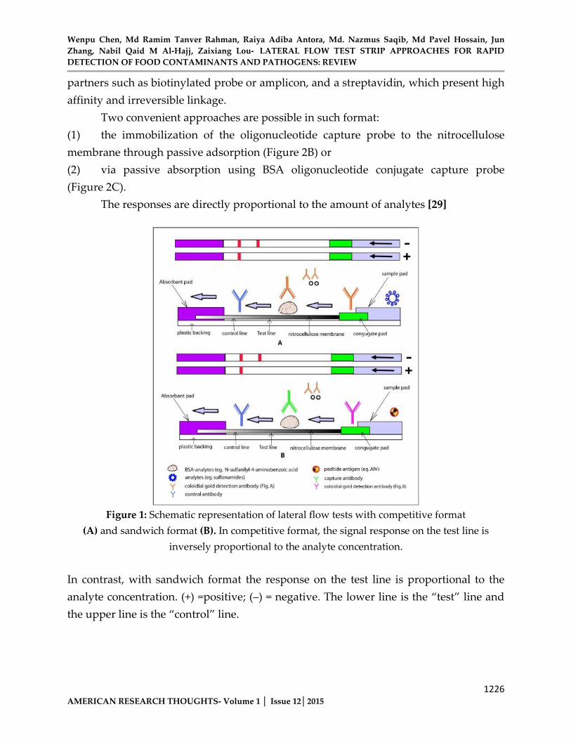
The two kinds of format frequently use dare competitive assay and sandwich assay. Competitive format is employed most often when testing analytes with low molecular weight or presenting single antigenic determinant [21]. A simple LFT device consists of four sections (sample pad, conjugate pad, nitrocellulose membrane and absorbent pad)which are laminated into a sheet of plastic backing orderly. There are various possible formats depending on the type of target analyte [22]. In a competitive format (Figure1A), an analyte-protein conjugate (e.g. N-sulfanilyl-4-aminobenzoicacid-Bovine Serum Albumin (BSA)) coated on the test zone of a nitrocellulose membrane captures a labeled anti-analyte monoclonal antibody complex, allowing color particle (e.g. colloidal gold) to concentrate and form a visible line on the test zone. Another specific antibody coated on the control line allows the capture of the excess antibody complex. One band color will therefore be visible in the control zone regardless of the presence of target analytes (e.g. sulfonamides), confirming correct test development. Conversely, a negative sample
Wenpu Chen, Md Ramim Tanver Rahman, Raiya Adiba Antora, Md. Nazmus Saqib, Md Pavel Hossain, Jun Zhang, Nabil Qaid M Al-Hajj, Zaixiang Lou- LATERAL FLOW TEST STRIP APPROACHES FOR RAPID DETECTION OF FOOD CONTAMINANTS AND PATHOGENS: REVIEW
1225AMERICAN RESEARCH THOUGHTS- Volume 1 │ Issue 12│2015
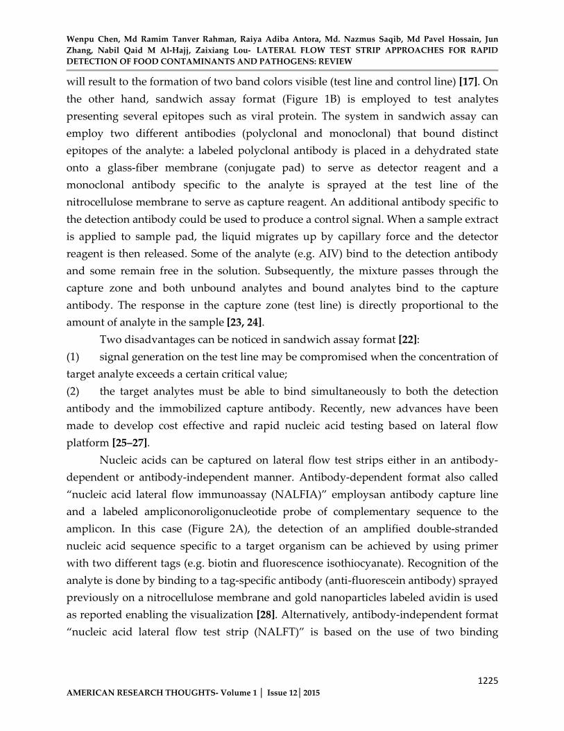
will result to the formation of two band colors visible (test line and control line) [17]. On the other hand, sandwich assay format (Figure 1B) is employed to test analytes presenting several epitopes such as viral protein. The system in sandwich assay can employ two different antibodies (polyclonal and monoclonal) that bound distinct epitopes of the analyte: a labeled polyclonal antibody is placed in a dehydrated state onto a glass-fiber membrane (conjugate pad) to serve as detector reagent and a monoclonal antibody specific to the analyte is sprayed at the test line of thenitrocellulose membrane to serve as capture reagent. An additional antibody specific to the detection antibody could be used to produce a control signal. When a sample extractis applied to sample pad, the liquid migrates up by capillary force and the detector reagent is then released. Some of the analyte (e.g. AIV) bind to the detection antibody and some remain free in the solution. Subsequently, the mixture passes through the capture zone and both unbound analytes and bound analytes bind to the capture antibody. The response in the capture zone (test line) is directly proportional to the amount of analyte in the sample [23, 24].
Two disadvantages can be noticed in sandwich assay format [22]: (1) signal generation on the test line may be compromised when the concentration of target analyte exceeds a certain critical value; (2) the target analytes must be able to bind simultaneously to both the detection antibody and the immobilized capture antibody. Recently, new advances have been made to develop cost effective and rapid nucleic acid testing based on lateral flowplatform [25–27].
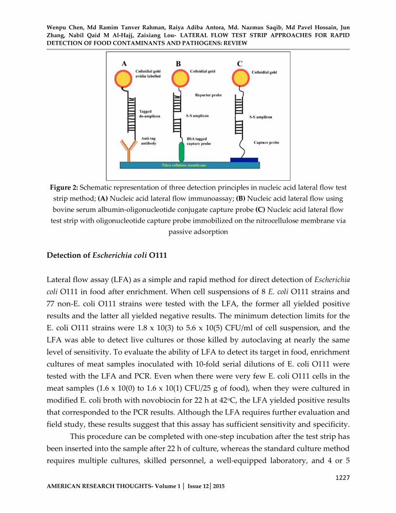
Nucleic acids can be captured on lateral flow test strips either in an antibody-dependent or antibody-independent manner. Antibody-dependent format also called“nucleic acid lateral flow immunoassay (NALFIA)” employsan antibody capture line and a labeled ampliconoroligonucleotide probe of complementary sequence to theamplicon. In this case (Figure 2A), the detection of an amplified double-stranded nucleic acid sequence specific to a target organism can be achieved by using primer with two different tags (e.g. biotin and fluorescence isothiocyanate). Recognition of the analyte is done by binding to a tag-specific antibody (anti-fluorescein antibody) sprayed previously on a nitrocellulose membrane and gold nanoparticles labeled avidin is used as reported enabling the visualization [28]. Alternatively, antibody-independent format “nucleic acid lateral flow test strip (NALFT)” is based on the use of two binding
Wenpu Chen, Md Ramim Tanver Rahman, Raiya Adiba Antora, Md. Nazmus Saqib, Md Pavel Hossain, Jun Zhang, Nabil Qaid M Al-Hajj, Zaixiang Lou- LATERAL FLOW TEST STRIP APPROACHES FOR RAPID DETECTION OF FOOD CONTAMINANTS AND PATHOGENS: REVIEW
1226AMERICAN RESEARCH THOUGHTS- Volume 1 │ Issue 12│2015
partners such as biotinylated probe or amplicon, and a streptavidin, which present high affinity and irreversible linkage.
Two convenient approaches are possible in such format: (1) the immobilization of the oligonucleotide capture probe to the nitrocellulose membrane through passive adsorption (Figure 2B) or (2) via passive absorption using BSA oligonucleotide conjugate capture probe (Figure 2C).
The responses are directly proportional to the amount of analytes [29]
Figure 1: Schematic representation of lateral flow tests with competitive format (A) and sandwich format (B). In competitive format, the signal response on the test line is
inversely proportional to the analyte concentration.
In contrast, with sandwich format the response on the test line is proportional to the analyte concentration. (+) =positive; (–) = negative. The lower line is the “test” line andthe upper line is the “control” line.
Wenpu Chen, Md Ramim Tanver Rahman, Raiya Adiba Antora, Md. Nazmus Saqib, Md Pavel Hossain, Jun Zhang, Nabil Qaid M Al-Hajj, Zaixiang Lou- LATERAL FLOW TEST STRIP APPROACHES FOR RAPID DETECTION OF FOOD CONTAMINANTS AND PATHOGENS: REVIEW
1227AMERICAN RESEARCH THOUGHTS- Volume 1 │ Issue 12│2015
Figure 2: Schematic representation of three detection principles in nucleic acid lateral flow test strip method; (A) Nucleic acid lateral flow immunoassay; (B) Nucleic acid lateral flow using bovine serum albumin-oligonucleotide conjugate capture probe (C) Nucleic acid lateral flow
test strip with oligonucleotide capture probe immobilized on the nitrocellulose membrane via passive adsorption
Detection of Escherichia coli O111
Lateral flow assay (LFA) as a simple and rapid method for direct detection of Escherichia coli O111 in food after enrichment. When cell suspensions of 8 E. coli O111 strains and 77 non-E. coli O111 strains were tested with the LFA, the former all yielded positive results and the latter all yielded negative results. The minimum detection limits for the E. coli O111 strains were 1.8 x 10(3) to 5.6 x 10(5) CFU/ml of cell suspension, and the LFA was able to detect live cultures or those killed by autoclaving at nearly the same level of sensitivity. To evaluate the ability of LFA to detect its target in food, enrichment cultures of meat samples inoculated with 10-fold serial dilutions of E. coli O111 were tested with the LFA and PCR. Even when there were very few E. coli O111 cells in the meat samples (1.6 x 10(0) to 1.6 x 10(1) CFU/25 g of food), when they were cultured in modified E. coli broth with novobiocin for 22 h at 42oC, the LFA yielded positive results that corresponded to the PCR results. Although the LFA requires further evaluation and field study, these results suggest that this assay has sufficient sensitivity and specificity.
This procedure can be completed with one-step incubation after the test strip has been inserted into the sample after 22 h of culture, whereas the standard culture method requires multiple cultures, skilled personnel, a well-equipped laboratory, and 4 or 5
Wenpu Chen, Md Ramim Tanver Rahman, Raiya Adiba Antora, Md. Nazmus Saqib, Md Pavel Hossain, Jun Zhang, Nabil Qaid M Al-Hajj, Zaixiang Lou- LATERAL FLOW TEST STRIP APPROACHES FOR RAPID DETECTION OF FOOD CONTAMINANTS AND PATHOGENS: REVIEW
1228AMERICAN RESEARCH THOUGHTS- Volume 1 │ Issue 12│2015
days. The speed and simplicity of this LFA make it suitable for use as part of routine screening assays in the food industry. [30]
Cd2+ detection in drinking waters
A lateral flow immune-sensor device (LFID) for Cd2+ determination in drinking and tap waters using the Cd-EDTA-BSA-AuNP conjugate as signal producer tool is introduced. The principle of working is based on competitive reaction between the Cd-EDTA-BSA-AuNP conjugate deposited on the conjugation pad strip and the Cd-EDTA complex formed in the analysis sample for the same binding sites of the 2A81G5 monoclonal antibody, specific to Cd-EDTA but not Cd2+ free, which is immobilized onto the test line. The device has a large response range within 0.4 to 2000 ppb, being the linear response between 0.4 and 10 ppb. The quantification and detection limits of 0.4 and 0.1 ppb, respectively, represent the lowest ones reported so far, for paper based metal sensors. The obtained detection limit is 50 times lower than the maximum contamination level required for drinking water. Here we also show a new option for increasing the sensibility in the LFDs with competitive format, through the decreasing in concentrations of the Cd-EDTA-BSA-AuNP conjugate deposited in the conjugation strip and the mAbs deposited in the test and control zones until to reach optimized concentrations. It is an important result take into account that the increase in sensibility is one of the challenges in the field of LFD sensors, where are focused many of the ongoing researches. In addition, a specificity study of the device for several metal interferences, where potential metal interferences are masked with the use of the EDTA and OVA optimized concentrations, is presented too. [31]
The rapid screening of domoic acid from shellfish extracts
A lateral flow immune assay (LFIA) has been developed and fully validated to detect the primary amnesic shellfish poisoning (ASP) toxin, domoic acid (DA). The performance characteristics of two versions of the test were investigated using spiked and naturally contaminated shellfish (mussels, scallops, oysters, clams, and cockles).
The tests provide a qualitative result, to indicate the absence or presence of DA in extracts of shellfish tissues, at concentrations that are relevant to regulatory limits. The new rapid assay (LFIA version 2) was designed to overcome the performance

Wenpu Chen, Md Ramim Tanver Rahman, Raiya Adiba Antora, Md. Nazmus Saqib, Md Pavel Hossain, Jun Zhang, Nabil Qaid M Al-Hajj, Zaixiang Lou- LATERAL FLOW TEST STRIP APPROACHES FOR RAPID DETECTION OF FOOD CONTAMINANTS AND PATHOGENS: REVIEW
1229AMERICAN RESEARCH THOUGHTS- Volume 1 │ Issue 12│2015
limitations identified in the first version of the assay. The improved test uses an electronic reader to remove the subjective nature of the generated results, and the positive cut-off for screening of DA in shellfish was increased from 10ppm (version 1) to 17.5ppm (version 2). A simple extraction and test procedure was employed, which required minimal equipment and materials; results were available 15min after sample preparation. Stability of the aqueous extracts at room temperature (22°C) at four time points (up to 245 min after extraction) and across a range of DA concentrations was 100.31.3% and 98.82.4% for pre- and post-buffered extracts, respectively. The assay can be used both within laboratory settings and in remote locations. The accuracy of the new assay, to indicate negative results at or below 10ppm DA, and positive results at or above 17.5ppm, was 99.5% (n=216 tests). Validation data were obtained from a 2-day, randomized, blind study consisting of multiple LFIA lots (n=3), readers (n=3) and operators (n=3), carrying out multiple extractions of muscle tissue (n=3) at each concentration (0, 10, 17.5, and 20ppm). No matrix effects were observed on the performance of the assay with different species (mussels, scallops, oysters, clams, and cockles). There was no impact on accuracy or interference from other phycotoxins, glutamic acid or glutamine with various strip incubations (8, 10, and 12min).
The accuracy of the assay, using naturally contaminated samples to indicate negative results at or below 12.5ppm and positive results at or above 17.5ppm, was 100%. Variability between three LFIA lots across a range of DA concentrations,expressed as coefficient of variation (% CV), was 1.10.4% (n=2 days) based on quantitative readings from the electronic reader. During 8 weeks of stability study, accuracy of the method with test strips stored at various temperatures (6, 22, 37 and 50°C) was 100%. Validation for both versions included comparisons with results obtained using reference LC-UV methods. [32]
Staphylococcus aureus
Staphylococcus aureus is a non-motile, spherical, Gram-positive microscopic bacterium. Food poisoning can be also caused by the consumption of food containing staphylococcal enterotoxins. For the toxic levels of enterotoxin to occur, extensive multiplication (growth) of staphylococci cells generally needs to have taken place in the food [33, 34]. AnICA was developed by Huang et al. (2007) [35] for detectingStaphylococcus aureus. The developed assay was based on sandwich format using anti-

Wenpu Chen, Md Ramim Tanver Rahman, Raiya Adiba Antora, Md. Nazmus Saqib, Md Pavel Hossain, Jun Zhang, Nabil Qaid M Al-Hajj, Zaixiang Lou- LATERAL FLOW TEST STRIP APPROACHES FOR RAPID DETECTION OF FOOD CONTAMINANTS AND PATHOGENS: REVIEW
1230AMERICAN RESEARCH THOUGHTS- Volume 1 │ Issue 12│2015
protein A (specific product of S. Aureus, 99%) IgG with two distinct specificities. The studyset-up comprised the identification of the production of protein A by 51 strains and an inoculation experiment. After comparison with the conventional method [36], the sensitivity and specificity of the test were 100 and 93.0–100% for 28 Staphylococcus aureus strains and 23 non Staphylococcus aureus strains, respectively. A total of eleven processed foods (pork, beef, and fried chicken) artificially inoculated with S. aureus at level <25 CFU. g−1 yielded positive results by the test strip. The test was however not as sensitive as the conventional culture procedures in detecting S. aureus in raw foods.
Salmonella
Salmonella are genuses of Gram-negative that are usually associated with animals, both wild and domestic. Food contaminations by Salmonella strains can infect humans bycausing severe gastroenteritis [37, 38]. Seoet al. (2003) [39] presented an ICA to detect Salmonella enteritidis in raw eggs. The test strip device manufactured by Neogen, Lansing, MI was based on sandwich format. The sample pretreatments comprised three steps: (1) disinfection in 70%of iso-propanol,(2) homogenization of egg contents (10 eggs) for 30 second sunder a stomacher and (3) panel dilutions followed by extraction (centrifugation at 10 000 × g) of the antigen with a mixture of fatty acids or phosphate buffer saline (PBS).
The detection limit of the test kit increased more than tenfold up to106–105 CFU. mL−1 in whole egg contents using the acid extraction technique compared with tenfold PBS dilution. The minimum concentration of Salmonella enteritidis to generate a positive band on the test panel was approximately107CFU. mL−1 in pure culture, and no cross-reactivity was observed with two other Salmonella serovars – Salmonella kentucky, Salmonella Typhimurium in pure culture. Campylobactersare aerobic Gram-negative, motile helical bacteria. Campylobacter species differ from other foodborne pathogens in that they do not multiply within the food. Most frequently, poultry and cattle are the sources of human infection. Some species including Campylobacter jejuni, Campylobacter coli, Campylobacter lari and Campylobacter upsaliens have been reported to be associated with human gastroenteritis [40–42]. Kawatsu et al. (2008) [43] reported a rapid and simple ICA to detect Campylobacter jejuni and Campylobacter coli in human stool samples.
Wenpu Chen, Md Ramim Tanver Rahman, Raiya Adiba Antora, Md. Nazmus Saqib, Md Pavel Hossain, Jun Zhang, Nabil Qaid M Al-Hajj, Zaixiang Lou- LATERAL FLOW TEST STRIP APPROACHES FOR RAPID DETECTION OF FOOD CONTAMINANTS AND PATHOGENS: REVIEW
1231AMERICAN RESEARCH THOUGHTS- Volume 1 │ Issue 12│2015
The ICA method used a monoclonal antibody against cell surface protein (15-kDa) of Campylobacter jejuni.
The study comprised two steps as follows: (1) the identification of cell extracts of Campylobacter (cell suspension of 86 C. jejunistrains, 27 C. coli strains, and strains of the 4 other Campylobacter species) and 26 non-Campylobacter species and(2) the testing of 222 stool specimens obtained from different patients with acute gastroenteritis.
For detecting Campylobacter species from cell extracts and stool samples by the ICA, a suspension of test strains at various cell concentrations were prepared by using PBS. The detection limit of Campylobacter jejuni and Campylobacter coli by the test strip ranged 1.8×10⁴ to 8.2×10⁵CFU. mL−1 and 1.4×10⁵ to 4.6×10⁶CFU. mL−1, respectively. Detection limits of5.3×105 and 5.0×10⁵CFU. mL−1of cell suspensions was obtained with other Campylobacter species including C. lari and C. Upsaliensis. Of the 86 C. jejunistrains and 27 C. coli strains tested, all yielded positive results with the ICA. Negative results were obtained with the other non-Campylobacter species. In the analysis of stool samples, the ICA showed a sensitivity of 84.8% which was comparable to those of some other commercial available enzyme immunoassays (ProSpecT Campylobacter microplate assay; Alexon-Trend, Ramsey, MN).
Botulism neurotoxin
Botulinum neurotoxins (BoNT) produced by the bacteriaClostridium botulinum are the most potent poisonous that causes paralytic illness in human. ICA had offered a suitable tool for BoNT detection. Klewitzet al. (2006) [44] reported an ICA against botulism neurotoxin D (BoNT/D). The test was based on double sandwich format using a gold-anti BoNT/D nmonoclonal antibody conjugates (detector reagent) and an anti-BoNT/D chicken polyclonal antibody. Fecal samples or standard samples spiked with various concentrations of BoNT/D were treated in 28 mM gelatin-phosphate buffer,vortexed and stored at temperature ranging 5 to 8°C for overnight. 2.0μg.mL−1 of polyclonal chicken anti-BoNT/DIgG and 0.35µg.mL−1 of gold conjugated monoclonalantibody was added to the sample extracts and incubated at 37 °C for 3 hours. Then, 50μL of pre-treated samples was applied to the test strip. The results indicated that the binding affinity of the investigated antibodies for BoNT/D and the kinetics of the
Wenpu Chen, Md Ramim Tanver Rahman, Raiya Adiba Antora, Md. Nazmus Saqib, Md Pavel Hossain, Jun Zhang, Nabil Qaid M Al-Hajj, Zaixiang Lou- LATERAL FLOW TEST STRIP APPROACHES FOR RAPID DETECTION OF FOOD CONTAMINANTS AND PATHOGENS: REVIEW
1232AMERICAN RESEARCH THOUGHTS- Volume 1 │ Issue 12│2015
complex formation were the most determining factors for the test time. Signal intensity of the background signal (no specific interaction) with blank samples increased with theincrease of sample volume, the incubation time and the amount of gold conjugated antibody that flows through the test line zone. Optimal conditions were found byapplying an incubation time at 2–4 hours. Importantly, the higher the average signal intensity of the blank sample (long incubation time), the lower the determination level as far as no BoNT/D could be determined. The lower limit of determination of BoNT/D by the test strip was 50 pg.mL−1. This concentration was found very satisfactorycompared to the mouse bioassay method (50pg.mL−1).Another study conducted by Chiao et al. (2006) [45] developed an ICA against Botulism neurotoxin B (BoNT/B). The detection limit was 50mg/ml in both PBS standard sample and diluted urine samples (1:1 in PBS). Using a silver enhancement technique, the detection limit was improved at 50ng.mL−1. The test strip showed no cross-reactivity to BoNT/A andBoNT/E at 1µg.mL−1. Authors of the same group have also investigated an ICA against BoNT/A [46]. The assay was based on sandwich format using monoclonal antibodies (MAbs) of two distinct specificities. The assay allowed a detection of 50ng.mL−1 of BoNT/A with an assay time of less than 10 min. interestingly, a detection limit of 1ng.mL−1 was obtained when using silver enhancement. The ICA also showed no cross-reactivity to type B neurotoxin (BoNT/B) and type E neurotoxin (BoNT/E).
Staphylococcus enterotoxin B
Staphylococcus aureus enterotoxin B (SEB) is a highly heat resistant enteric toxin with a potential as a bio threat agent. A rapid and sensitive method for the detection of staphylococcal enterotoxins is needed for food safety and food defense monitoring. An ICA against SEB was presented by Shyu et al. (2010) [47]. The detection limit of the SEB toxin in PBS was achieved at 1ng.mL−1 within 5–10 min. using a silver enhancer reagent, the sensitivity of the assay was improved to 10 pg. mL−1. Simulated samples (SEB toxin diluted inhuman urine, serum and cow milk powder) were also analyzed by the SEB test strips. The detection limit of SEB in the simulated urine and milk samples was 10ng.mL−1, and it was 100ng.mL−1 in serum samples. An interesting study using fluorescent immunoliposome as label has been described by Khreich et al.(2008) [48] for the detection of SEB in tap water, surface water, apple juice, raw milk, ham and cheese. The ICA was based on sandwich immunoassay using labeled monoclonal antibodies
Wenpu Chen, Md Ramim Tanver Rahman, Raiya Adiba Antora, Md. Nazmus Saqib, Md Pavel Hossain, Jun Zhang, Nabil Qaid M Al-Hajj, Zaixiang Lou- LATERAL FLOW TEST STRIP APPROACHES FOR RAPID DETECTION OF FOOD CONTAMINANTS AND PATHOGENS: REVIEW
1233AMERICAN RESEARCH THOUGHTS- Volume 1 │ Issue 12│2015
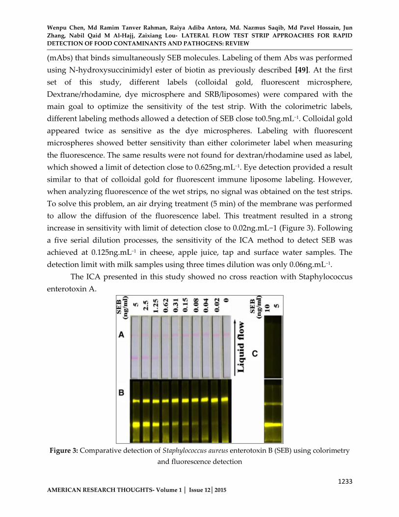
(mAbs) that binds simultaneously SEB molecules. Labeling of them Abs was performed using N-hydroxysuccinimidyl ester of biotin as previously described [49]. At the first set of this study, different labels (colloidal gold, fluorescent microsphere,Dextrane/rhodamine, dye microsphere and SRB/liposomes) were compared with the main goal to optimize the sensitivity of the test strip. With the colorimetric labels,different labeling methods allowed a detection of SEB close to0.5ng.mL−1. Colloidal gold appeared twice as sensitive as the dye microspheres. Labeling with fluorescent microspheres showed better sensitivity than either colorimeter label when measuring the fluorescence. The same results were not found for dextran/rhodamine used as label, which showed a limit of detection close to 0.625ng.mL−1. Eye detection provided a result similar to that of colloidal gold for fluorescent immune liposome labeling. However, when analyzing fluorescence of the wet strips, no signal was obtained on the test strips. To solve this problem, an air drying treatment (5 min) of the membrane was performed to allow the diffusion of the fluorescence label. This treatment resulted in a strong increase in sensitivity with limit of detection close to 0.02ng.mL−1 (Figure 3). Following a five serial dilution processes, the sensitivity of the ICA method to detect SEB was achieved at 0.125ng.mL−1 in cheese, apple juice, tap and surface water samples. The detection limit with milk samples using three times dilution was only 0.06ng.mL−1.
The ICA presented in this study showed no cross reaction with Staphylococcus enterotoxin A.
Figure 3: Comparative detection of Staphylococcus aureus enterotoxin B (SEB) using colorimetry and fluorescence detection
Wenpu Chen, Md Ramim Tanver Rahman, Raiya Adiba Antora, Md. Nazmus Saqib, Md Pavel Hossain, Jun Zhang, Nabil Qaid M Al-Hajj, Zaixiang Lou- LATERAL FLOW TEST STRIP APPROACHES FOR RAPID DETECTION OF FOOD CONTAMINANTS AND PATHOGENS: REVIEW
1234AMERICAN RESEARCH THOUGHTS- Volume 1 │ Issue 12│2015
The same strips corresponding to serial dilutions of SEB (5 to 0.02 ng. mL−1) assayed with the immunochromatographic test using SRB encapsulated immunoliposomes as tracer were successively analyzed by colorimetric detection (A; scanned image obtained with an Epsonexpression 1640XL scanner) and fluorescent detection (B; picturesobtained with Kodak station 2000 MM). The control and test lines are on the top and the bottom of the strips, respectively. The effect of air-drying on the fluorescence of the immunoliposomes is illustrated (C; pictures obtained with Kodak Station 2000 MM before and after air-drying), with two strips, corresponding to 10 and 5ng.mL−1 of SEB (reproduced with permission from Khreichet al. (2008) [48])
Sulphonamides
Sulphonamides (SAs) are broad-spectrum antimicrobials used in both humans and animals. Residues in food are of concern because of the potential carcinogenic nature of these compounds [50]. To protect consumers, the maximum residue limits (MRLs) for the total sulphonamide concentrations in edibles tissues have been established at 100μg.kg−1 in EU and China. Wang et al. (2007) [17] proposed a simple and rapidextraction method for the detection of four sulphonamides (sulfamonomethoxine (SMM), sulfamethoxydiazine, (SMD)sulfadimethoxine (SDM) and sulfadiazine (SDZ)) in eggs and chicken muscles by ICA.
In this study, the extractions of the sulphonamides from matrice samples were realized by ethyl acetate. Briefly, muscle or egg samples homogenized in ethyl acetate (2 mL.g−1) were vortexed for 3 min and then centrifuged at 2000 g for 10 min. Resultant supernatants (300μL) were evaporated to dryness in a 60°C water bath under a gentle flow of nitrogen. The residues obtained were then resuspended in 150μLof phosphate buffer (pH7.4) for analysis by ICA test strip. With that method, recoveries of SMM, SMD and SDM ranged 85–95.6% in both chicken muscle and eggs samples spiked at concentration 20–100ng.g−1. On the other hand, low recovery was obtained with SDZ(44.8–60.9%). The differences of recovery have been explained by the authors. Indeed, the sensitivity limits of the test strip were 20 ng.mL−1 for three sulphonamides (sulfamonomethoxine, sulfamethoxydiazine, and sulfadimethoxine) and 40 ng.mL−1 for sulfadiazine. Using HPLC as confirmatory method, the validation of the ICA test was achieved by analyzing SMM in 27 egg samples and 28 chicken muscle samples from animal experiment.
Wenpu Chen, Md Ramim Tanver Rahman, Raiya Adiba Antora, Md. Nazmus Saqib, Md Pavel Hossain, Jun Zhang, Nabil Qaid M Al-Hajj, Zaixiang Lou- LATERAL FLOW TEST STRIP APPROACHES FOR RAPID DETECTION OF FOOD CONTAMINANTS AND PATHOGENS: REVIEW
1235AMERICAN RESEARCH THOUGHTS- Volume 1 │ Issue 12│2015
The result of comparison indicated that the differences between the two methods were from 0.8 to 11.2% for egg samples and from 2.2 to 34% for chicken muscle samples for the quantitative detection. The agreement rates between test strips and HPLC were 100%. In the study conducted by Zhang et al. (2008) [51], a sensitivity limit at 15 ng.mL−1
was obtained when screening SMM in swine serum, and the recovery in sera spiked at20 ng.mL−1 and 30 ng.mL−1 ranged 94–103%.Wang et al. (2008) [52] presented a combination of ICA test and ELISA method to detect sulfamethazine residues in milk, pig muscle and fish. To obtain a more sensitive immunoassay, the authors investigated the influence of various analytical parameters including the incubation time, matrix effects, buffer components and immune reagent concentrations. Pretreatment of milk samples was performed with acetone, and that of pig muscle and fish samples was realized in methanol. Removal of the matrix effects was achieved at 10–20 fold dilution in PBS (30 mmol.L−1, pH7.2) containing 0.05%tween. The ICA presented in this study detected efficiently SMZ at concentration 20 μg.kg−1 in milk, pig muscle and fish. The detection time was accomplished in less than 10 min.
Results from these studies proved the reliability of LFT technology for analyzing sulphonamides in food producing animals. Nevertheless, there are still some limitations to find a novel test strip that could detect simultaneously all sulphonamide compounds.Anabolic steroids in the EU, the use of steroid and β-agonist to enhance animal growth is prohibited according to the council directive 96/22/EC [53]. A set of minimum performance characteristics that have to be fulfilled by the methods to be used for growth promoting compounds has been defined by the EU and laid down in pertinent Commission decisions [54]. Monitoring anabolic steroids in meat-producing animals therefore is a challenging task. It implies the development of rapid and sensitive analytical method able to detect and identify sub-μg.kg–1 residue levels in complex biological n matrices such as meat, urine, or milk. An ICA test for screening 19-nortestosterone (19-NT) in swine urine was reported by Liqiang et al. (2007) [55].
The test based on competitive assay format employed a colloidal goldpolyclonalantibody (pAb) conjugate against 19-NT (detector reagent) and 19-NT-ovalbumin conjugate (capture reagent) immobilized on a nitrocellulose membrane. The detection limit of the test strip to 19-NT was 200 ng.mL−1 within10 min. Results of the parallel analysis of urine samples spiked with 19-NT at 0, 100, 200, 400, 800 and 1600 ng.mL−1 showed good agreement rate between LC/MS/MS method and the ICA. However, significant cross-reactivities were observed with five other anabolic steroid
Wenpu Chen, Md Ramim Tanver Rahman, Raiya Adiba Antora, Md. Nazmus Saqib, Md Pavel Hossain, Jun Zhang, Nabil Qaid M Al-Hajj, Zaixiang Lou- LATERAL FLOW TEST STRIP APPROACHES FOR RAPID DETECTION OF FOOD CONTAMINANTS AND PATHOGENS: REVIEW
1236AMERICAN RESEARCH THOUGHTS- Volume 1 │ Issue 12│2015
including19-norandrostendione (30%), 19-norandrostandione (30%), trenbolone (10%), 17 α-trenbolone (10%), 17 α–19–nortestosterone (10%). In our opinion, the developed ICA cannot be applied for specific detection of 19-nortestosterone, and it would be more convenient to develop a monoclonal antibody against 19-nortestosterone. Geertruida et al. (2008) [56] proposed attest strip against progesterone (PA). The authors investigated the influence of several aspects such as the kinetic interaction of antibody-conjugate with various capture antigens. The influence of several commonly used blocking agents (BSAOVA, β-Lactoglobulin, lactoferrin, casein hydrosylate, polyvinylpyrrolidone (30 kDa), poly-methyl-vinylether-10-maleic anhydride and teleostan gelatin) has been assessed in this study. Results showed that blocking agents like casein hydrolysate, polyvinylpyrrolidone and poly (methylvinylether-10-maleic anhydride) affected the performance of the assay. The addition of OVA, lactoferrinor β-Lactoglobulin as blocking agents was found detrimental in progesterone-specific ICA test. Indeed, the overall charge of lactoferrin in the incubation buffer was positive instead of negative, as was the case for the other blocking agents. The authors did not explain how these charge differences contributed to the absence of signal in the blank sample. Finally, BSA provided the most superior blocking agents, and a half-maximal inhibition concentration (IC50) at 0.6–1.2 μg.mL−1 was obtained for progesterone detection in bovine milk by the ICA. A validation study using a reference method should be performed before routine use.
Aflatoxin
Aflatoxins (AFT) are a group of widely researched mycotoxins that are produced by fungi A. flavus and A. parasiticus. The four major types of AFT are B1, B2, G1, and G2. Among these AFT, B1 and G1 had been considered as potential human carcinogens by the International Agency for Research on Cancer [57]. An ICA for the detection of AFT B1 in foods was proposed by Xiulan et al. (2006) [58]. The assay used gold-labeled polyclonal antibodies as detector reagent against AFT B1, and AFT B1-BSA as capture reagents. In the first step of the study, the efficacy of the test strip was evaluated through analysis of AFT B1 in food sample extracts (rice, corn and wheat flour). Briefly, 50 g of finely ground samples were blended with 250 mL methanol/water (6:4, v/v) containing 4 gr. of NaCl at high-speed in a blender for 2 min and followed by filtration.
The solution was then diluted six times with PBS to a final methanol
Wenpu Chen, Md Ramim Tanver Rahman, Raiya Adiba Antora, Md. Nazmus Saqib, Md Pavel Hossain, Jun Zhang, Nabil Qaid M Al-Hajj, Zaixiang Lou- LATERAL FLOW TEST STRIP APPROACHES FOR RAPID DETECTION OF FOOD CONTAMINANTS AND PATHOGENS: REVIEW
1237AMERICAN RESEARCH THOUGHTS- Volume 1 │ Issue 12│2015
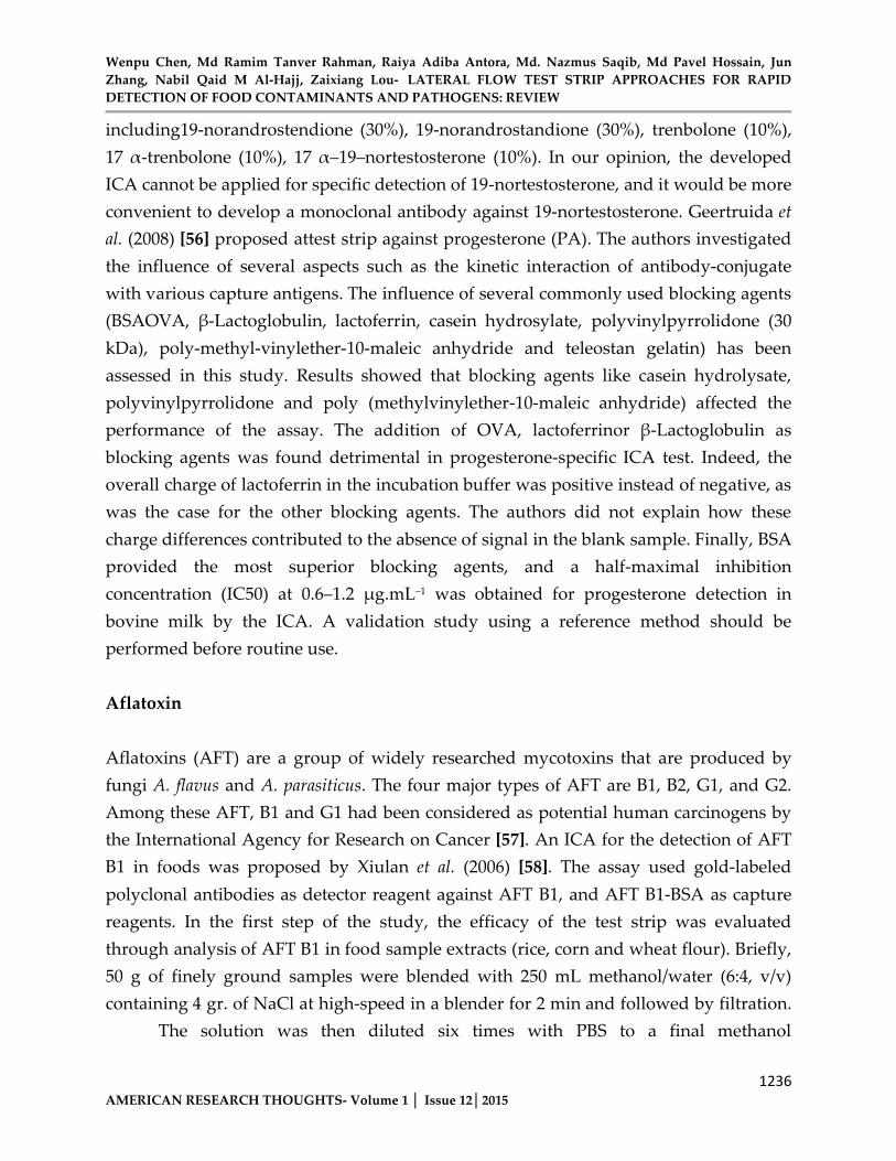
concentration of 10%. Different levels of AFT B1 (2, 5, 10, 20, 50 ng.mL−1) were added to the blank extract for analysis by the ICA. With this method, the limit of detection of AFTB1 by visual observation was 2.5 ng.mL−1, and it ranged 0.05–0.150 ng.mL−1 when using photometric strip reader. The analytical recoveries for AFB1 in sample extract (rice, corn, and wheat) spiked at 2–50 μg.kg−1 were from80.79–110.56% with mean co-efficient of variations ranging 6.27–14.63%. Validation of the assay was carried out by analyzing naturally contaminated samples including rice, corn and wheat (n=67), and the results showed good correlation (r2=0.93) between the ICA and a competitive ELISA method. In all assay, the analysis of AFT B1 in one sample was completed within 10 min. More recently, a novel membrane-based lateral-flow immunodipstick assay was developed by Tang et al. (2009) [70] for the fast screening of aflatoxin B2 (AFT B2) in food samples. In this study, the detector reagent consisted of magnetic nano gold microspheres (MnGMs) with nano-Fe2O3 particles as core and gold nanoparticles as shell, and bio-functionalized with monoclonal anti-AFT B2 antibodies. Manually spotted AFT B2–bovine serum albumin conjugates (AFT B2–BSA) and goat anti-mouse IgG on nitrocellulose membrane had been used as test and control lines, respectively.
Result showed that the cutoff value of conventional strip with pure gold nanoparticles as detection reagent was 3.1 ng.mL−1 AFT B2, while the cutoff value with MnGMs as detection reagent was 3 times lower at 0.9 ng.mL−1 AFT B2 (Figure 4). The reliability of the assay was evaluated in an inter laboratory study with the analysis ofspiked peanut samples and naturally contaminated samples (n=24). HPLC has been used as reference test. The assay showed sensitivity rate of 100% and a specificity rate of75%. This study demonstrates a new approach to develop higher sensitive ICA which makes use of bio-functionalized magnetic nano gold microspheres as detection reagent.
The developed method would be convenient for use to detect other toxins and residue contaminants.
Wenpu Chen, Md Ramim Tanver Rahman, Raiya Adiba Antora, Md. Nazmus Saqib, Md Pavel Hossain, Jun Zhang, Nabil Qaid M Al-Hajj, Zaixiang Lou- LATERAL FLOW TEST STRIP APPROACHES FOR RAPID DETECTION OF FOOD CONTAMINANTS AND PATHOGENS: REVIEW
1238AMERICAN RESEARCH THOUGHTS- Volume 1 │ Issue 12│2015
Figure 4: Lateral-flow test immunodipsticks using different detector reagents for aflatoxin B2 (AFT B2): (a) anti-AFT B2–MnGMs, (b) anti-AFT B2–nano gold particles and (c) electron
microscopy (SEM) image of the prepared anti-AFT B2MnGMs. The immobilized amount of AFT B2–BSA of 1.0 μg.cm−1 was identical in the two immunodipstick formats (reproduced with
permission from Tang et al. [59])
Pesticides
Pesticides contribute significantly to the contamination of the environment due to their extended use in modern agricultural production. Because of the toxicity risks to human and the ecosystem, legislations have been established on the hazard assessment to control pesticides in water [60, 61]. As mentioned previously, conventional methods available (e.g. HPLC) for monitoring pesticide contaminants are complex and time-consuming. There has therefore a great demand for developing rapid and reliable analytical techniques to control multiple pesticides in foods, agriculture and environmental samples. For this purpose, several cheminulescence based detection strategies have been proposed in the literature [60-62]. Amongst the modern immunochemical techniques available, ICA had known successful applications [63]. Up to now, researches in ICA methods have included the development of more extended formats that permit a one-site detection of multiple pesticides. Guo et al. (2009) [64, 65-69] proposed two gold based ICAs to detect simultaneously carbofuran and triazophos in water samples (tap water, surface water and ground water). In Thai study, one test strip format employed abi-specific monoclonal antibody against both carbofuran and
Wenpu Chen, Md Ramim Tanver Rahman, Raiya Adiba Antora, Md. Nazmus Saqib, Md Pavel Hossain, Jun Zhang, Nabil Qaid M Al-Hajj, Zaixiang Lou- LATERAL FLOW TEST STRIP APPROACHES FOR RAPID DETECTION OF FOOD CONTAMINANTS AND PATHOGENS: REVIEW
1239AMERICAN RESEARCH THOUGHTS- Volume 1 │ Issue 12│2015
triazophos. The second test format was based on two different monoclonal antibodies raised specifically against the two pesticides. Gold-monoclonal antibody conjugates were applied on the conjugate pads of the developed test strips to serve as detector reagents. The capture reagents consisted of two test lines (T1 and T2) coated with carbofuran-ovalbumin conjugate and triazophos-ovalbumin conjugate, respectively. Water samples were treated by centrifugation (3000 × g for 10 min), and the supernatants were spiked with carbofuran or triazophos at concentrations ranging 0–132 μg.mL−1. The visual detection limits of the pesticides by the two ICA formats were from 4 μg.mL−1 to 62 μg.mL−1. The strip showed low cross-reaction to other structurally related pesticide compounds including carbosulfan, carbofuran phenol, isoprocarb, chlorpyrifos, parathion and diazinon. Kranthi et al. (2009) [70] also investigated a single test strip format for one-site verification ofactive ingredients in commercial insecticide formulations of endosulfan and three pyrethryroids (cypermethrin, deltamethrinand fenvalerate). Colloidal gold were conjugated with two antisera raised against endosulfan and cypermethrin. The developed test strip consisted of two capture test lines coated separately with endosulfan-BSA conjugate or cypermethrin-BSA conjugate. Finally, the test strip was designed to allow the detection of standardized concentrations of endosulfan (1800 mg. mL−1), cypermethrin (800 mg. mL−1), deltamethrin (1000 mg. mL−1) and fenvalerate (1400 mg. mL−1).
These concentrations were found sufficient to inhibit color development on the test lines. The test strip developed here is very useful for the farmers, and it could have potential applications in environmental testing [71].
Wenpu Chen, Md Ramim Tanver Rahman, Raiya Adiba Antora, Md. Nazmus Saqib, Md Pavel Hossain, Jun Zhang, Nabil Qaid M Al-Hajj, Zaixiang Lou- LATERAL FLOW TEST STRIP APPROACHES FOR RAPID DETECTION OF FOOD CONTAMINANTS AND PATHOGENS: REVIEW
1240AMERICAN RESEARCH THOUGHTS- Volume 1 │ Issue 12│2015
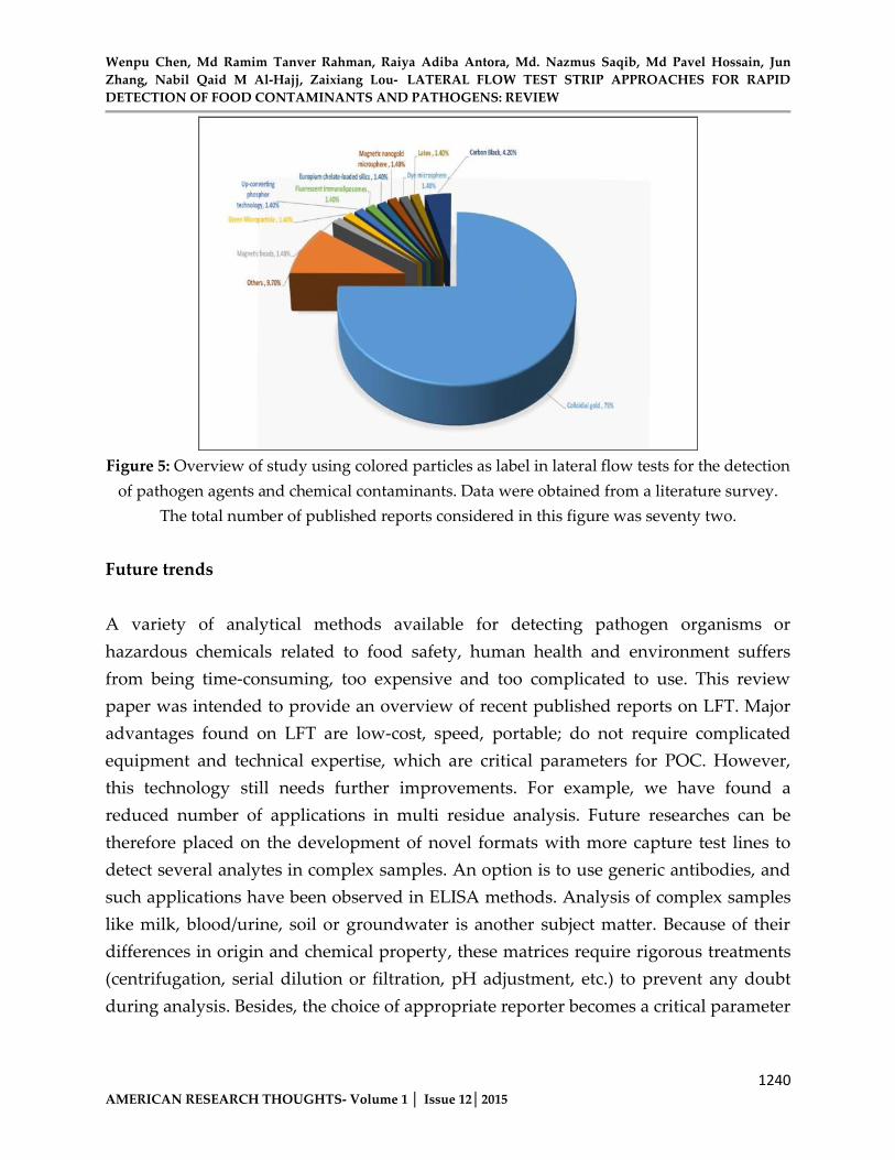
Figure 5: Overview of study using colored particles as label in lateral flow tests for the detection of pathogen agents and chemical contaminants. Data were obtained from a literature survey.
The total number of published reports considered in this figure was seventy two.
Future trends
A variety of analytical methods available for detecting pathogen organisms or hazardous chemicals related to food safety, human health and environment suffers from being time-consuming, too expensive and too complicated to use. This review paper was intended to provide an overview of recent published reports on LFT. Major advantages found on LFT are low-cost, speed, portable; do not require complicated equipment and technical expertise, which are critical parameters for POC. However, this technology still needs further improvements. For example, we have found a reduced number of applications in multi residue analysis. Future researches can be therefore placed on the development of novel formats with more capture test lines to detect several analytes in complex samples. An option is to use generic antibodies, and such applications have been observed in ELISA methods. Analysis of complex samples like milk, blood/urine, soil or groundwater is another subject matter. Because of their differences in origin and chemical property, these matrices require rigorous treatments (centrifugation, serial dilution or filtration, pH adjustment, etc.) to prevent any doubt during analysis. Besides, the choice of appropriate reporter becomes a critical parameter
Wenpu Chen, Md Ramim Tanver Rahman, Raiya Adiba Antora, Md. Nazmus Saqib, Md Pavel Hossain, Jun Zhang, Nabil Qaid M Al-Hajj, Zaixiang Lou- LATERAL FLOW TEST STRIP APPROACHES FOR RAPID DETECTION OF FOOD CONTAMINANTS AND PATHOGENS: REVIEW
1241AMERICAN RESEARCH THOUGHTS- Volume 1 │ Issue 12│2015
in LFT development. In our literature survey, we found that seventy-five percent of published reports on LFT employed colloidal gold particle as label (Figure 5).
Only a study in which fluorescent immunoliposome was used as label for the detection of SEB investigated the alternative use of other types of label including fluorescent microsphere, dextranerhod amine and dye microsphere. Fluorescent immunoliposome allowed a fifteen-fold increase insensitivity compared with that for visual detection of colored labels. Quality control is essentially establishing adequate performance characteristics (sensitivity, specificity, negative predictive value, positive predictive value, cross reactivity, etc.) of a given test. With the globalization, new generations of analytical devices have to be accurate, speed, simple and cost-effective. Lateral flow test strip technology could provide a promising approach for this purpose.
Acknowledgements
This work was supported by the China Scholarship Council (CSC) and Project 31201433 of The National Natural Science Foundation of PR China.
References
1. Naravaneni R, Jamil K (2005). Rapid detection of food-borne pathogens by using molecular techniques. Journal of Medical Microbiology 54:51–54
2. Ricci F, Volpe G, Micheli L, Palleschi G (2007). A review on novel developmentsand applications of immunosensors in food analysis. AnalyticaChimicaActa605:111–129
3. Companyo R, Granados M, Guiteras J, Prat MD (2009). Antibiotics in food: Legislation and validation of analytical methodologies. Analytical and Bioanalytical Chemistry395:877–891
4. Kaufmann A (2009). Validation of multiresidue methods for veterinary drug residues; related problems and possible solutions. AnaliticaChimicaActa 637:144–155
5. Stolker AAM, Brinkman UATh (2005) Journal for Chromatography A 1067:15–53
Wenpu Chen, Md Ramim Tanver Rahman, Raiya Adiba Antora, Md. Nazmus Saqib, Md Pavel Hossain, Jun Zhang, Nabil Qaid M Al-Hajj, Zaixiang Lou- LATERAL FLOW TEST STRIP APPROACHES FOR RAPID DETECTION OF FOOD CONTAMINANTS AND PATHOGENS: REVIEW
1242AMERICAN RESEARCH THOUGHTS- Volume 1 │ Issue 12│2015
6. Aprea C, Colosio C, Mammone T, Minoia C, Maroni M (2002). Biological monitoring of pesticide exposure: a review of analytical methods. Journal forChromatography B. Analyt. Technol. Biomed. Life Sci. 769:191–219
7. Goryacheva IY, Rusanova TY, Burmistrova NA, De Saeger S(2009). Immunochemical methods for the determination of mycotoxins. Journal of Analytical Chemistry 64:768–785
8. Maragos CM (2009). Recent advances in the development of novel materials for mycotoxin analysis. Analytical Bioanalytical Chemistry 395:1205–1213
9. Pikkemaat MG (2009). Microbial screening methods for detection of antibiotic residues in slaughter animals. Analytical and Bioanalytical Chemistry395:893–905
10. Pasto-Navaro N, Maquieira A, Puchades R (2009). Review on immunoanalytical determination of tetracycline and sulfonamide residues in edible products. Analytical and Bioanalytical Chemistry 395:907–920
11. Prieto-Simon B, Noguer T, Campa M (2007). Emerging biotools for assessment of mycotoxins in the past decade. Trends in Analytical Chemistry26:689–702
12. Reig M, Toldra F (2008). Veterinary drug residues in meat: Concerns and rapid methods for detection. Meat Science 78:60–67
13. Turner NWT, Subrahmanyam S, Piletsky SA (2009).Analytical methods for determination of mycotoxins: A review.AnalyticaChimicaActa 632:168–180
14. Blaskoza M, Koets M, Wichers JH, Amerongen AV, Fukal L,Rauch P (2009).Nucleic Acid Lateral Flow Immunoassay for the Detection of Pathogenic Bacteria from Food. Czech J, Food Sci 27:S350–S353
15. Hampl J, Hall M, Mufti NA, Yao YMD, MacQueen D, WrightWH, Cooper DE (2001). Upconverting Phosphor Reporters in Immunochromatographic Assays. Analytical Biochemistry 288:176–187
16. Takanashi S, Okame M, Shiota T, Takagi M, Yagyu F, Tung PG,Nishimura S, Katsumata N, Igarashi T, Okitsu S, Ushijima H(2008).Development of a rapid immunochromatographic test for noroviruses genogroups I and II. Journal of Virological Methods 148:1–8
17. Wang X, Li K, Shi D, Xiong N, Jin X, Yi D, Bi D (2007).Development of an Immunochromatographic Lateral-Flow Test Strip for Rapid Detection of Sulfonamides in Eggs and Chicken Muscles. Journal of Agricultural and Food Chemistry 55:2072–2078
Wenpu Chen, Md Ramim Tanver Rahman, Raiya Adiba Antora, Md. Nazmus Saqib, Md Pavel Hossain, Jun Zhang, Nabil Qaid M Al-Hajj, Zaixiang Lou- LATERAL FLOW TEST STRIP APPROACHES FOR RAPID DETECTION OF FOOD CONTAMINANTS AND PATHOGENS: REVIEW
1243AMERICAN RESEARCH THOUGHTS- Volume 1 │ Issue 12│2015
18. Yan Z, Zhou L, Zhao Y, Wang J, Huang L, Hu K, Liu H, WangH, Guo Z, Song Y, Huang H, Yang R (2006).Rapid quantitative detection of Yersinia pestis by lateral-flow immunoassay and up-converting phosphor technology-based biosensor. Sensors and Actuators B: Chemical119:656–663
19. Leuvering JHW, Thal PJHM, Van der Waart M, Schuurs AHWM (1980).Sol Particle Immunoassay (SPIA). Journal of Immunoassay 1:77–91
20. Posthuma-Trumpie G, Korf J, Amerongen AV (2009).Lateral flow (immuno) assay: its strengths, weaknesses, opportunities and threats. A literature survey. Analytical and Bioanalytical Chemistry393:569–582
21. Qian S, Bau HH (2004).Analysis of lateral flow biodetectors: competitive format. Analytical Biochemistry 326:211–224
22. Peak S-H, Lee S-H, Cho J-H, Kim Y-S (2000).Development of Rapid One-Step Immunochromatographic Assay. Methods 22:53–60
23. Peng F, Wang Z, Zhang S, Wu R, Hu S, Li Z, Wang X, Bi D(2008).Development of an Immunochromatographic Strip for Rapid Detection of H9 Subtype Avian Influenza Viruses. Clinical and Vaccine Immunology 15:569–574
24. Peng DP, Hu SS, Hua Y, Xiao YC, Li ZL, Wang X, Bi D (2007).Comparison of a new gold-immunochromatographic assay for the detection of antibodies against avian influenza virus with hemagglutination inhibition and agar gel immunodiffusion assays. Journal of Veterinary Immunology and Immunopathology 117:17–25
25. Blaskoza M, Koet M, Rauch P, Amerogen AV (2009).Development of a nucleic acid lateral flow immunoassay for simultaneous detection of Listeria spp. and Listeriamonocytogenes in food. Europian Food Research and Technology 229:867–874
26. Carter DJ, Cary RB (2007) Lateral flow microarrays: a novelplatform for rapid nucleic acid detection based on miniaturized lateral flow chromatography. Nucleic Acids Research 35(10): e74-.doi:10.1093/nar/gkm269
27. Edwards KA, Baeumner EJ (2008).Liposome-Enhanced Lateral-Flow Assays for the Sandwich-Hybridization Detection of RNA. Method in Molcular Biology 504:185–215
28. Amerongen AV, Koets M (2005). Simple and rapid bacterial protein and DNA diagnostic methods based on signal generation with colloidal carbon particles. In: Amerongen AV, Barug D, Lauwaars M (editors). Rapid methods for

Wenpu Chen, Md Ramim Tanver Rahman, Raiya Adiba Antora, Md. Nazmus Saqib, Md Pavel Hossain, Jun Zhang, Nabil Qaid M Al-Hajj, Zaixiang Lou- LATERAL FLOW TEST STRIP APPROACHES FOR RAPID DETECTION OF FOOD CONTAMINANTS AND PATHOGENS: REVIEW
1244AMERICAN RESEARCH THOUGHTS- Volume 1 │ Issue 12│2015
biological and chemical contaminants in food and feed. Wageningen Academic Publishers, Wageningen, pp. 105–126
29. Corstjens PLAM, Zuiderwijk M, Nilson M, Feindt H, NiedbalaRS, Tanke HJ (2003).Lateral-flow and up-converting phosphor reporters to detect single-stranded nucleic acids in a sandwich-hybridization assay. Analytical Biochemistry 312:191–200
30. Terao Y, Yonekita T, Morishita N, Fujimura T, Matsumoto T, Morimatsu F (2013). Potential Rapid and Simple Lateral Flow Assay for Escherichia coli O111.Journal of food protection 76:755-761
31. Adaris M. Lopez-Marzo, Pons J, Diane AB, MerkociA(2013).High sensitive gold-nanoparticle based lateral flow Immuno device for Cd2+ detection in drinking waters. Biosensors & Bioelectronics47:190-198
32. Jawaid W, Meneely JP, Campbell K, Hooper M, Melville K(2013) Development and validation of the first high performance-lateral flow immunoassay (HP-LFIA) for the rapid screening of domoic acid from shellfish extracts. Talanta116: 663-669
33. Halpin-Dohnalek, M.I. and Marth, E.H. (1989) Staphylococcus aureus: production of extracellular compounds and behavior in foods—a review. Journal of Food Protection 52:267–282
34. Portocarrero SM, Newman M, Mikel B (2002). Staphylococcus aureus survival, staphylococcal enterotoxin production and shelf stability of country-cured hams manufactured under different processing procedures. Meat Science 62:267–273
35. Huang S-H, Wei H-C, Lee Y-C (2007).One-step immunochromatographic assay for the detection of Staphylococcus aureus. Food Control 18:893–897
36. Bennett RW, Lancette GA (1992). Staphylococcus aureus. InBacteriological analytical manual, 7th edn. Association of Official Analytical Chemists, Arlington, pp 161–166
37. Centers for Disease Control and Prevention (CDC) (2000) Outbreaks of Salmonella serotype Enteritidis infection associate dwith eating raw or undercooked shell eggs–United States, 1996–1998. MMWR 49:73–79
38. Humphrey TJ (1994).Contamination of egg shell and contents with Salmonella enteritidis: A review. International Journal of Food Microbiology 2:31–40
Wenpu Chen, Md Ramim Tanver Rahman, Raiya Adiba Antora, Md. Nazmus Saqib, Md Pavel Hossain, Jun Zhang, Nabil Qaid M Al-Hajj, Zaixiang Lou- LATERAL FLOW TEST STRIP APPROACHES FOR RAPID DETECTION OF FOOD CONTAMINANTS AND PATHOGENS: REVIEW
1245AMERICAN RESEARCH THOUGHTS- Volume 1 │ Issue 12│2015
39. Seo K-H, Holt PS, Gast RK, Stone HD (2003).Simple and rapid methods for detecting Salmonella enteritidis in raw eggs. International Journal of FoodMicrobiology 87:139–144
40. Broczyk A, Thompson S, Smith D, Lior H (1987). Water-borne outbreak of Campylobacter laridis-associated gastroenteritis. The Lancet 329:164–165
41. Goossens H, Vlaes L, De Boeck M, Pot B, Kersters K, Levy J,De Mol P, Butzler JP, Vandamme P (1990).Is "Campylobacter upsaliensis" an unrecognized cause of human diarrhoea? The Lancet 335:584–586
42. Healing TD, Greenwood MH, Pearson AD (1992). Campylobacters and enteritis. Reviews I MediclaMicrobiology 3:159–167
43. Kawatsu K, Kumeda Y, Taguchi M, Yamazaki-Matsume W,Kanki M, Inoue K (2008).Development and Evaluation of Immunochromatographic Assay for Simple and Rapid Detection of Campylobacter jejuni and Campylobacter coli in Human Stool Specimens. Journal of Clinical Microbiology 46:1226–1231
44. Klewitz T, Gressler F, Hans B, Pflanz K, Scheper T (2006).Immunochromatographic assay for determination of botulinum neurotoxin type D. Sensors and Actuators B: Chemical 113:582–589
45. Chiao D-J, Shyu R-H, Hu C-S, Chiang H-Y, Tang S-S (2004).Colloidal gold-based immunochromatographic assay for detection of botulinum neurotoxin type B. Journal of Chromatography B 809:37–41
46. Chiao D-J, Wey J-J, Shyu R-H, Tang S-S (2008).Monoclonal Antibody-Based Lateral Flow Assay for Detection of Botulinum Neurotoxin Type A. Hybridoma27:31–35
47. Shyu R-H, Tang S-S, Chiao D-J, Hung Y-W (2010).Gold nanoparticle-based lateral flow assay for detection of staphylococcal enterotoxin B. Food Chemistry118:462–466
48. Khreich N, Lamourette P, Boutal H, Deveilliers K, Creminon C,Vollad H (2008).Detection of Staphylococcus enterotoxin B using fluorescent immunoliposomes as label for immunochromatographic testing. Analytical Biochemistry 377:182–188
49. Grassi J, Frobert Y, Lamourette P, Lagoutte B (1998).Screening of monoclonal antibodies using antigens labeled with acetylcholinesterase: Application to the peripheral proteins of photosystem 1. Analytical Biochemistry 168:436–450
Wenpu Chen, Md Ramim Tanver Rahman, Raiya Adiba Antora, Md. Nazmus Saqib, Md Pavel Hossain, Jun Zhang, Nabil Qaid M Al-Hajj, Zaixiang Lou- LATERAL FLOW TEST STRIP APPROACHES FOR RAPID DETECTION OF FOOD CONTAMINANTS AND PATHOGENS: REVIEW
1246AMERICAN RESEARCH THOUGHTS- Volume 1 │ Issue 12│2015
50. Cribb AE, Lee BL, Trepanier LA, Spielberg SP (1996). Adverse reactions to sulphonamide and sulphonamide-trimethoprim antimicrobials: clinical syndromes and pathogenesis. Adverse Drug Reactions and Toxicological Reviews Journal 15:9–50
51. Zhang G, Wang X, Zhi A, Bao Y, Yang Y, Qu M, Luo J, Li Q, Guo J, Wang Z, Yang J, Xing G, Chai S, Shi T, Liu Q (2008). Development of a lateral flow immunoassay strip for screening of sulfamonomethoxine residues. Food Additives and Contaminants: Part A 25:413–423
52. Wang L, Wang S, Zhan J, Liu J, Zhang Y (2008).Enzyme-linked immunosorbent assay and colloidal gold immunoassay for sulphamethazine residues in edible animal foods: investigation of the effects of the analytical conditions and the sample matrix on assay performance. Analytical Bioanalytical Chemistry 390:1619–1627
53. European Union L12523/05/1996, Official Journal, (1996) Council Directive 96/22/EC of 29 April 1996 concerning the prohibition on the use in stock farming of certain substances having a hormonal or thyrostatic action and of beta-agonists, and repealing Directives 81/602/EEC, 88/146/EEC and 88/299/EEC,Brussels, Belgium pp 3–9
54. European Union Commission (1993) Commission Decision (93/256/EEC) of 14 April 1993 laying down the methods to be used for detecting residues of substances having a hormonal or a thyrostatic action. Official Journal of the European Communities L118:64–74
55. Liqiang L, Chifang P, Zhengyu J, Chuanlai X (2007). Development and evaluation of a rapid lateral flow immunochromatographic strip assay for screening 19-nortestosterone. Journal of Biomedical Chromatography 21:861–866
56. Geertruida A, Posthuma T, Jakob K, Amerogen AV (2008).Development of a competitive lateral flow immunoassay for progesterone: influence of coating conjugates and buffer components. Analytical and bioanalytical Chemistry 392:1215–1223
57. International Agency for Research on Cancer (IARC), Geneva(1993), 56, p48958. Xiulan S, Xiaolian Z, Jian T, Xiaohong G, Jun Z, Chu FS (2006).Development of an
immunochromatographic assay for detection of aflatoxin B1 in foods.Food Control 17:256–262

Wenpu Chen, Md Ramim Tanver Rahman, Raiya Adiba Antora, Md. Nazmus Saqib, Md Pavel Hossain, Jun Zhang, Nabil Qaid M Al-Hajj, Zaixiang Lou- LATERAL FLOW TEST STRIP APPROACHES FOR RAPID DETECTION OF FOOD CONTAMINANTS AND PATHOGENS: REVIEW
1247AMERICAN RESEARCH THOUGHTS- Volume 1 │ Issue 12│2015
59. Tang D, Sauceda JC, Lin Z, Basova SOE, Goryacheva I, BiselliS, Lin J, Niessner R, Knopp D (2009).Magnetic nanogold microspheres-based lateral-flow immunodipstick for rapid detection of aflatoxin B2 in food. Biosensors and Bioelectronics 25:514–518
60. European commission (2000) Directive 2000/60/EC of the European Parliament and of the Council of 23 October 2000 establishing a framework for Community action in the field of water policy. Official Journal of the European Communities L 327:1–72
61. European commission (1998) Council Directive (98/83/EC) of 3 November 1998 relating to the water quality of water intended for human consumption. Official Journal of the European Communities L 330:32–54
62. Gamiz-Gracia L, Garcia-Campana AM, Soto-Chincilla JJ,Huertas-Perez JF, Gongzalez-Casado A (2005).Analysis of pesticides by chemiluminescence detection in the liquid phase. Trends in Analytical Chemistry 24:927–942
63. Mauriz E, Calle A, Manclus JJ, Montaya A, Lechuga LM (2007).Multi-analyte SPR immunoassays for environmental biosensing of pesticides. Analytical and Bioanalytical Chemistry 387:1449–1458
64. Suri CR, Boro R, Nangia Y, Gandhi S, Sharma P, Wangoo N, Rajesh K, Shekhawat GS (2009). Immunoanalytical techniques for analyzing pesticides in the environment. Trends in Analytical Chemistry28:29–39
65. Blazkova M, Mickova-Holubova B, Rauch P, Fukal L (2009). Immunochromatographic colloidal carbon-based assay for detection of methiocarb in surface water. Biosensors and Bioelectronics 25:753–758
66. Chenggang S, Suqing Z, Kun Z, Guobao H, Zhenyu Z (2008). Preparation of colloidal gold immunochromatography strip for detection of methamidophos residue. Journal of Environmental Science 20:1392–1397
67. Guo Y-R, Liu S-Y, Gui W-J, Zhu G-N (2009).Gold immunochromatographic assay for simultaneous detection of carbofuran and triazophos in water samples. Analytical Biochemistry389:32–39
68. Kaur J, Singh V, Boro R, Thampi KR, Raje M, Varshney GC,Suri CR (2007).Immunochromatographic Dipstick Assay Format Using Gold Nanoparticles Labeled Protein−Hapten Conjugate for the Detection of Atrazine. Environmental Science and Technology 41:5028–5036
Wenpu Chen, Md Ramim Tanver Rahman, Raiya Adiba Antora, Md. Nazmus Saqib, Md Pavel Hossain, Jun Zhang, Nabil Qaid M Al-Hajj, Zaixiang Lou- LATERAL FLOW TEST STRIP APPROACHES FOR RAPID DETECTION OF FOOD CONTAMINANTS AND PATHOGENS: REVIEW
1248AMERICAN RESEARCH THOUGHTS- Volume 1 │ Issue 12│2015
69. Lyubavina IA, Zinchenko AA, Salomatina IS, Zherdev AV,Dzantiev BB (2004).An Immunochromatographic Assay of 2,4-Dichlorophenoxyacetic Acid and Simazine Using Monoclonal Antibodies Labeled with Colloidal Gold. Russian Journal of Bioorganic Chemistry 30:178–183
70. Kranthi KR, Davis M, Mayee CD, Russell DA, Shukla RM,Satija U, Kshirsagar M, Shiware D, Kranthi S (2009).Development of a colloidal-gold based lateral-flow immunoassay kit for ‘quality-control’ assessment of pyrethroid and endosulfan formulations in a novel single strip format. Crop Protection 28:428–434
71. Sajid M, Kawde AN, Daud M. ( 2014 ) Designs, formats and applications of lateral flow assay: A literature review, Journal of Saudi Chemical Society, doi:10.1016/j.jscs.2014.09.001