Resource partitioning to male and female flowers of Spinacia ...
14
Journal of Experimental Botany, Vol. 62, No. 12, pp. 4323–4336, 2011 doi:10.1093/jxb/err148 Advance Access publication 12 May, 2011 This paper is available online free of all access charges (see http://jxb.oxfordjournals.org/open_access.html for further details) RESEARCH PAPER Resource partitioning to male and female flowers of Spinacia oleracea L. in relation to whole-plant monocarpic senescence Diane E. Sklensky* and Peter J. Davies † Department of Plant Biology, Plant Science Building, Cornell University, Ithaca, NY 14853-5908, USA * Present address: Department of Biology, Lane College, Jackson, TN 38301, USA. y To whom correspondence should be addressed. E-mail: [email protected] Received 10 December 2010; Revised 25 March 2011; Accepted 13 April 2011 Abstract Male plants of spinach (Spinacea oleracea L.) senesce following flowering. It has been suggested that nutrient drain by male flowers is insufficient to trigger senescence. The partitioning of radiolabelled photosynthate between vegetative and reproductive tissue was compared in male (staminate) versus female (pistillate) plants. After the start of flowering staminate plants senesce 3 weeks earlier than pistillate plants. Soon after the start of flowering, staminate plants allocated several times as much photosynthate to flowering structures as did pistillate plants. The buds of staminate flowers with developing pollen had the greatest draw of photosynthate. When the staminate plants begin to show senescence 68% of fixed C was allocated to the staminate reproductive structures. In the pistillate plants, export to the developing fruits and young flowers remained near 10% until mid-reproductive development, when it increased to 40%, declining to 27% as the plants started to senesce. These differences were also present on a sink-mass corrected basis. Flowers on staminate spinach plants develop faster than pistillate flowers and have a greater draw of photosynthate than do pistillate flowers and fruits, although for a shorter period. Pistillate plants also produce more leaf area within the inflorescence to sustain the developing fruits. The 14 C in the staminate flowers declined due to respiration, especially during pollen maturation; no such loss occurred in pistillate reproductive structures. The partitioning to the reproductive structures correlates with the greater production of floral versus vegetative tissue in staminate plants and their more rapid senescence. As at senescence the leaves still had adequate carbohydrate, the resources are clearly phloem-transported compounds other than carbohydrates. The extent of the resource redistribution to reproductive structures and away from the development of new vegetative sinks, starting very early in the reproductive phase, is sufficient to account for the triggering of senescence in the rest of the plant. Key words: Carbohydrates, dioecious, female, flowering, flowers, fruits, male, monocarpic, photosynthate partitioning, pistillate, nitrogen, reproduction, respiration, resource allocation, senescence, sink strength, Spinacea oleracea, spinach, staminate, whole plant. Introduction Many plants possess the characteristic of living more or less indefinitely until some agent or stress intervenes. However, some plants have evolved the monocarpic growth habit in which the entire plant undergoes a programmed cycle of organ degradation after reproduction, followed by the death of the plant. Two hypotheses have been advanced to explain this correlative phenomenon: (i) that allocation of nutrients to reproductive tissue results in the degradation of the vegetative structures or (ii) that a factor, probably hormonal, is produced by fruits and is then translocated to the leaves and stem apex, triggering their senescence. Literature supporting and challenging these proposals has ª 2011 The Author(s). This is an Open Access article distributed under the terms of the Creative Commons Attribution Non-Commercial License (http://creativecommons.org/licenses/by- nc/2.5), which permits unrestricted non-commercial use, distribution, and reproduction in any medium, provided the original work is properly cited. Downloaded from https://academic.oup.com/jxb/article/62/12/4323/486534 by guest on 10 January 2022
-
Upload
khangminh22 -
Category
Documents
-
view
0 -
download
0
Transcript of Resource partitioning to male and female flowers of Spinacia ...

Journal of Experimental Botany, Vol. 62, No. 12, pp. 4323–4336, 2011doi:10.1093/jxb/err148 Advance Access publication 12 May, 2011This paper is available online free of all access charges (see http://jxb.oxfordjournals.org/open_access.html for further details)
RESEARCH PAPER
Resource partitioning to male and female flowers of Spinaciaoleracea L. in relation to whole-plant monocarpicsenescence
Diane E. Sklensky* and Peter J. Davies†
Department of Plant Biology, Plant Science Building, Cornell University, Ithaca, NY 14853-5908, USA
* Present address: Department of Biology, Lane College, Jackson, TN 38301, USA.y To whom correspondence should be addressed. E-mail: [email protected]
Received 10 December 2010; Revised 25 March 2011; Accepted 13 April 2011
Abstract
Male plants of spinach (Spinacea oleracea L.) senesce following flowering. It has been suggested that nutrient drain
by male flowers is insufficient to trigger senescence. The partitioning of radiolabelled photosynthate betweenvegetative and reproductive tissue was compared in male (staminate) versus female (pistillate) plants. After the start
of flowering staminate plants senesce 3 weeks earlier than pistillate plants. Soon after the start of flowering,
staminate plants allocated several times as much photosynthate to flowering structures as did pistillate plants. The
buds of staminate flowers with developing pollen had the greatest draw of photosynthate. When the staminate
plants begin to show senescence 68% of fixed C was allocated to the staminate reproductive structures. In the
pistillate plants, export to the developing fruits and young flowers remained near 10% until mid-reproductive
development, when it increased to 40%, declining to 27% as the plants started to senesce. These differences were
also present on a sink-mass corrected basis. Flowers on staminate spinach plants develop faster than pistillateflowers and have a greater draw of photosynthate than do pistillate flowers and fruits, although for a shorter period.
Pistillate plants also produce more leaf area within the inflorescence to sustain the developing fruits. The 14C in the
staminate flowers declined due to respiration, especially during pollen maturation; no such loss occurred in pistillate
reproductive structures. The partitioning to the reproductive structures correlates with the greater production of
floral versus vegetative tissue in staminate plants and their more rapid senescence. As at senescence the leaves still
had adequate carbohydrate, the resources are clearly phloem-transported compounds other than carbohydrates.
The extent of the resource redistribution to reproductive structures and away from the development of new
vegetative sinks, starting very early in the reproductive phase, is sufficient to account for the triggering ofsenescence in the rest of the plant.
Key words: Carbohydrates, dioecious, female, flowering, flowers, fruits, male, monocarpic, photosynthate partitioning, pistillate,
nitrogen, reproduction, respiration, resource allocation, senescence, sink strength, Spinacea oleracea, spinach, staminate, wholeplant.
Introduction
Many plants possess the characteristic of living more or less
indefinitely until some agent or stress intervenes. However,
some plants have evolved the monocarpic growth habit in
which the entire plant undergoes a programmed cycle of
organ degradation after reproduction, followed by the
death of the plant. Two hypotheses have been advanced to
explain this correlative phenomenon: (i) that allocation of
nutrients to reproductive tissue results in the degradation of
the vegetative structures or (ii) that a factor, probably
hormonal, is produced by fruits and is then translocated to
the leaves and stem apex, triggering their senescence.
Literature supporting and challenging these proposals has
ª 2011 The Author(s).
This is an Open Access article distributed under the terms of the Creative Commons Attribution Non-Commercial License (http://creativecommons.org/licenses/by-nc/2.5), which permits unrestricted non-commercial use, distribution, and reproduction in any medium, provided the original work is properly cited.
Dow
nloaded from https://academ
ic.oup.com/jxb/article/62/12/4323/486534 by guest on 10 January 2022

been reviewed in Kelly and Davies (1988b) and Sklensky
and Davies (1993). In either case, expression of senescence-
promoting and senescence-retarding genes interacts with the
plant’s environmental and internal conditions to regulate
the active senescence process (Nooden et al., 1997).
Molisch (1938) first proposed that a drain of nutrients to
the fruits was the mechanism for the induction of whole plant
senescence. An objection to this hypothesis emerged fromwork involving dioecious spinach plants (Leopold et al.,
1959; Janick and Leopold, 1961), which normally undergo
senescence following flowering similar to any other mono-
carpic species. However, as the removal of the very small
pollen-producing flowers, which were assumed to be negligi-
ble as a nutrient sink, resulted in a delay of senescence it was
suggested that these flowers must act in some other fashion to
induce senescence. One such possibility could be by theproduction of a senescence factor (Kelly and Davies, 1988b),
but experiments in peas failed to provide any support for the
idea of a senescence factor (Hamilton and Davies, 1988a, b),
supplying instead evidence that senescence is regulated by
a shift in nutrient allocation from the vegetative to the
reproductive tissue that occurs very early in the reproductive
phase (Kelly and Davies, 1986, 1988a, b).
Resource diversion and the induction of senescence
The explanation for whole plant senescence that best fits the
evidence currently available is that a physiological transition
is initiated at flowering and results in a resource allocationthat is detrimental to the maintenance of vegetative tissues in
comparison to the uninduced state. Many studies have
demonstrated that alterations in the source–sink relations of
vegetative and reproductive tissues can affect the course of
senescence. Senescence can be prevented in pea genotypes
with a slower rate of reproductive growth that enables
continued resource allocation to vegetative growth (Kelly
and Davies, 1986, 1988a). The endogenous auxin andgibberellin content of floral and vegetative tissue within the
apical buds of these peas correlated with this resource
allocation (Zhu and Davies, 1997): a higher gibberellin
content of the apical vegetative tissues within the apical bud
was associated with vigorous vegetative growth, slower floral
development, and continued growth, whereas the greater rate
of floral bud growth, which precedes senescence, was
associated with a higher indoleacetic acid content in the floralbuds. Senescence of soybean was also delayed by the
mechanical prevention of pod expansion (Crafts-Brandner
and Egli, 1987; Miceli et al., 1995). Such physical restrictions
to soybean seed growth did not alter the time of initiation of
senescence but decreased the rate of leaf senescence as judged
by changes in photosynthesis (Crafts-Brandner and Egli,
1987), indicating that senescence was initiated at the same
time regardless of sink size but that the rate of senescence wasmodulated by sink size. Restricting soybean pod growth did
not affect total plant dry matter and N accumulations during
seed-filling because there were proportional increases in the
partitioning of assimilates into stems and leaves (Miceli et al.,
1995). Similarly, pea plants bearing the alleles ar and n, which
have smaller seeds and lower total seed yield, showed
a resurgence of growth after apical growth had initially
stopped, which is when normal plants undergo senescence.
Only after further growth did the plants undergo full
senescence (Murfet, 1985). The recessive alleles ar and n were
proposed to impose a lower metabolic drain per reproductive
node as a consequence of their restrictive effects on hilum
anatomy and pod morphology, respectively, leading toa reduction in sink capacity. As a consequence the developing
seed crop fails to cause plant senescence and death at the
usual developmental time. Similarly, in maize, crop manipu-
lations leading to kernel set restrictions enhanced post-
flowering assimilate availability and reduced leaf senescence
(Borras et al., 2003), whereas the acceleration of senescence
by the imposition of water stress increased the rate of grain-
filling in wheat and rice (Yang et al., 2001a, b; 2003).The initiation of the senescence programme appears to
occur early in the flowering period, and, in most cases,
senescence can only be delayed, not prevented, by surgery or
hormone application. Sterile and other mutants of Arabidop-
sis with delayed senescence did not exhibit prolonged leaf life,
but did show an extended production of leaves and flowering
stalks (Nooden and Penney, 2001). Following soybean pod
removal the leaves did not show the dramatic visual yellow-ing associated with senescence, but function, as measured in
terms of the rate of photosynthesis or the activity of ribulose
bisphosphate carboxylase, was inhibited (Mondal et al., 1978;
Wittenbach, 1982, 1983a, b). Delayed senescence in ‘stay-
green’ genotypes of sorghum did appear to result in
prolonged photosynthetic capability, but this seemed to be
most closely correlated with greater nitrogen assimilation
during the grain-filling period (Borrell et al., 2001).Sinks have profound effects on the photosynthesis in
source tissue (Paul and Foyer, 2001). Partitioning enzymes
in sink tissue, such as sucrose phosphate synthase, showed
both short- and long-term regulation by sugar concen-
trations (Huber and Huber, 1996). In addition to affecting
partitioning enzymes, sugars also play a key role in the
transcription regulation of such key photosynthetic genes
as rbcS, cab, and atpD (nuclear genes encoding the smallsubunit of Rubisco, the chlorophyll a/b binding protein,
and the D subunit of a thylakoid ATPase, respectively)
(Krapp and Stitt, 1995). The interrelatedness of these
facets of carbohydrate metabolism suggests that measure-
ment of translocation between source and sink under
conditions of varying senescence would provide useful
information on the events leading to senescence.
In light of the clear results in peas that senescence isregulated by a shift in nutrient resource allocation early in
reproductive development (Kelly and Davies, 1986, 1988a, b),
the regulation of monocarpic senescence in spinach has been
re-examined. The prior assumption was that the utilization of
carbohydrate resources by staminate spinach flowers is
negligible and would have no effect on senescence regulated
by nutrient ‘exhaustion’. This has led to an investigation of
photosynthate allocation in both male (staminate) and female(pistillate) spinach plants as an indication of overall phloem-
derived resource allocation in order to determine whether the
4324 | Sklensky and DaviesD
ownloaded from
https://academic.oup.com
/jxb/article/62/12/4323/486534 by guest on 10 January 2022
nutrient demand by staminate spinach flowers is insignificant,
as has been previously assumed. It is demonstrated below
that staminate flowers have a nutrient demand exceeding the
rate of import of pistillate flowers, and thus could be
a determining factor in bringing about monocarpic, whole-
plant senescence, even in male plants.
Materials and methods
Below are the abbreviated Materials and methods. Completedetails can be found in the Supplementary material at JXB online.
Growing conditions
Comparison among three different cultivars of spinach (Spinaciaoleracea L.: Packer, Asgrow XPH 1450 and Asgrow XPH 1510) ledto the selection of Asgrow XPH 1510, due to its retention ofa vegetative state under short days and its tendency to flower rapidlyunder long-day conditions. Seeds of cv. Asgrow XPH 1510 (AsgrowSeed Company, Gonzales, CA), were sown in flats of peat–vermiculite mix under greenhouse conditions and grown in an 18 hphotoperiod (long-day inductive conditions), with daylight supple-mented by overhead lights. Seedlings emerged from the soil in 7–10d and were subsequently transplanted to 12.5 cm pots. The firstflower buds were just visible about 18 d later, designated ‘onset offlowering’. Thereafter, treatments and sampling were undertakenweekly. Sampling of staminate plants ceased after the fifth weekfrom the start of flowering because, at that time, they senesced,whereas that of pistillate plants continued through the seventh week.
Radioactive assimilate distribution
Plants were moved to growth chambers, with 18 h day lengths of 175lE m�2 s�1, 19 �C day and 17 �C night. Three to four hours after thestart of the light period, individual plants were exposed to 14CO2 for1 h. A thick, transparent polyethylene bag (10320 cm or 10340 cm),containing a 3 ml plastic vial, was placed over a single leaf or over theentire young inflorescence region, respectively. Unless otherwisenoted, the labelled leaf was located immediately below theinflorescence region, designated L–1. Leaves within the flower-bearingportion are indicated by positive L numbers, with the lowest leaf thatsubtends an axillary raceme being referred to as L1. The bags weresealed around the petioles with adhesive paper tape and petroleumjelly, whereas Cling florists’ adhesive sealed the inflorescence regionsin an air-tight manner. A syringe was used to inject 1 lCi ofNaH14CO3 in 20 ll of water into the vial, followed by 200 ll of 0.6M HCl to release the 14CO2. The sealed bag contained approximately1.5 lmol CO2, including 0.0182 lmol 14CO2. The injection hole wassealed with Scotch transparent adhesive tape. The plant was left toassimilate the 14CO2 for 1 h followed by a variable chase period(usually 3 h).To determine the utilization of carbon over a longer term, both
staminate and pistillate plants in the third week of floraldevelopment were left for chase periods of 1, 2, 3, or 4 d followinglabelling of the leaf immediately below the inflorescence region(L–1) for 1 h.Following the post-labelling chase period, plants were dissected,
weighed, frozen on dry ice, oven-dried for 2 d at 60 �C, reweighed,and combusted in a sample oxidizer. The resultant 14CO2 wascaptured by an absorbent solution, and this solution counted ina scintillation counter with quench correction. Recoveries were90% or better, as measured by the combustion of standards.Because of variation in the total amount fixed and exported, themeasure of the carbon demand of a group of structures was thepercentage of the total export that was measured in that structure;this provides an indication of the relative importance of a givenstructure as a sink for the particular leaf labelled. Total export was
calculated by a summation of the total recovered radioactivityfrom all harvested plant parts, except the labelled leaf. A mass-independent measure of sink strength, designated ‘relative specificactivity’ (RSA), was calculated by dividing the dpm g�1 dry weightof an individual organ by the average dpm g�1 dry weight for theplant. At least three plants of each type were tested in eachexperiment and each experiment was repeated at least twice,usually several times.
Analysis of respiration and photosynthesis
Photosynthesis and respiration rates were measured using a Li-Cor6200 (Lincoln, NE) infrared gas analyser. Photosynthesis was recordedusing four leaves individually, from L–1 to L3. Respiration of theinflorescence was measured by CO2 evolution in darkness: 3.5 cmregions of the inflorescence, with the leaves removed, were enclosed inthe cuvette, with sealing around the stem. This was repeatedfollowing removal of the flowers and fruits.
Carbohydrate analysis
Concentrations of glucose, fructose, and sucrose were determinedcolorimetrically using sequential enzymatic reactions (Cairns,1987). Starch analysis was carried out as in Ober et al. (1991).
Results
Shoot morphology and leaf area during reproduction inspinach
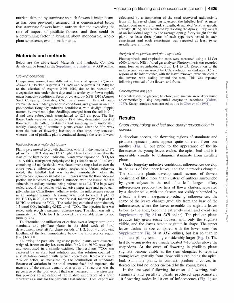
A dioecious species, the flowering regions of staminate and
pistillate spinach plants appear quite different from one
another (Fig. 1), but prior to the appearance of the in-florescence the young leaves enclose the apical bud and it is
impossible visually to distinguish staminate from pistillate
plants.
Under long-day inductive conditions, inflorescences develop
in the axils of the upper leaves, and the internodes elongate.
The staminate plants develop small racemes of flowers
consisting of little more than clusters of anthers surrounded
by green calyxes in the axil of a leaf. Larger axillaryinflorescences produce two tiers of flower clusters, separated
by a slender stalk, with the clusters not visibly subtended by
a leaf. In these male-gamete-producing inflorescences, the
shape of the leaves changes gradually from the base of the
inflorescence, where the leaves resemble the sagittate leaves
below, to the apex, becoming extremely small and ovoid (see
Supplementary Fig. S1 at JXB online). The pistillate plants
produce tiny green sessile flowers, with only the stigmatavisible, and the leaves remain sagittate in shape. The upper
leaves decline in size compared with the lower ones (see
Supplementary Fig. S1 at JXB online), but less so than in
staminate plants, remaining considerably larger (Fig. 1). The
first flowering nodes are usually located 7–10 nodes above the
cotyledons. At the onset of flowering in pistillate plants
stigmata become visible as the stem elongates to separate
young leaves spatially from those still surrounding the apicalbud. Staminate plants, in contrast, produce a convex in-
florescence bud no longer enclosed by the young leaves.
In the first week following the onset of flowering, both
staminate and pistillate plants produced approximately
10 flowering nodes in 10 cm of inflorescence (Fig. 1; see
Resource partitioning and senescence in spinach | 4325D
ownloaded from
https://academic.oup.com
/jxb/article/62/12/4323/486534 by guest on 10 January 2022
Supplementary Fig. S2 and Table S1 at JXB online).Stigmata protruded from the axils of leaves in pistillate
plants, but the anthers remained enclosed in the calyxes
of staminate plants. By the end of the second week after
the start of flowering some anthesis and pollen shedding
had begun in staminate plants, the inflorescences of both
types having attained about 25 cm in length, with up to
30 flowering nodes. Minute fruits began to appear in the
lower leaf axils of the inflorescences of the pistillateplants. In the third week of flowering, plants of both
sexes had 50 or more flowering nodes. The staminate
plants shed copious amounts of pollen and pistillate
plants had several large pointed fruits in the axils of
lower leaves, although, near the apex, fruits remained
small or indistinguishable from the flowers. At this stage,
in many plants of both sexes, age-related senescence of
leaves below the inflorescence may have begun. By week
4, the staminate plants showed yellowing nearly up to the
level of the inflorescence, and the apical bud had
converted into flower buds. The pistillate plants either
continued production of flowering nodes or ceased node
initiation in week 4, with development of the fruits
occurring in either case. The staminate plants exhibited
signs of the late, degradative stages of senescence in week5, with overall yellowing, and sampling of these plants
ceased before this age. Pistillate plants continued green,
with the fruits enlarging, until the seventh week after the
onset of flowering, although the lowest leaves often
yellowed and withered with age. The overall senescence
stage of the pistillate plants in week 8 corresponded to
a similar stage of staminate plants in week 5.
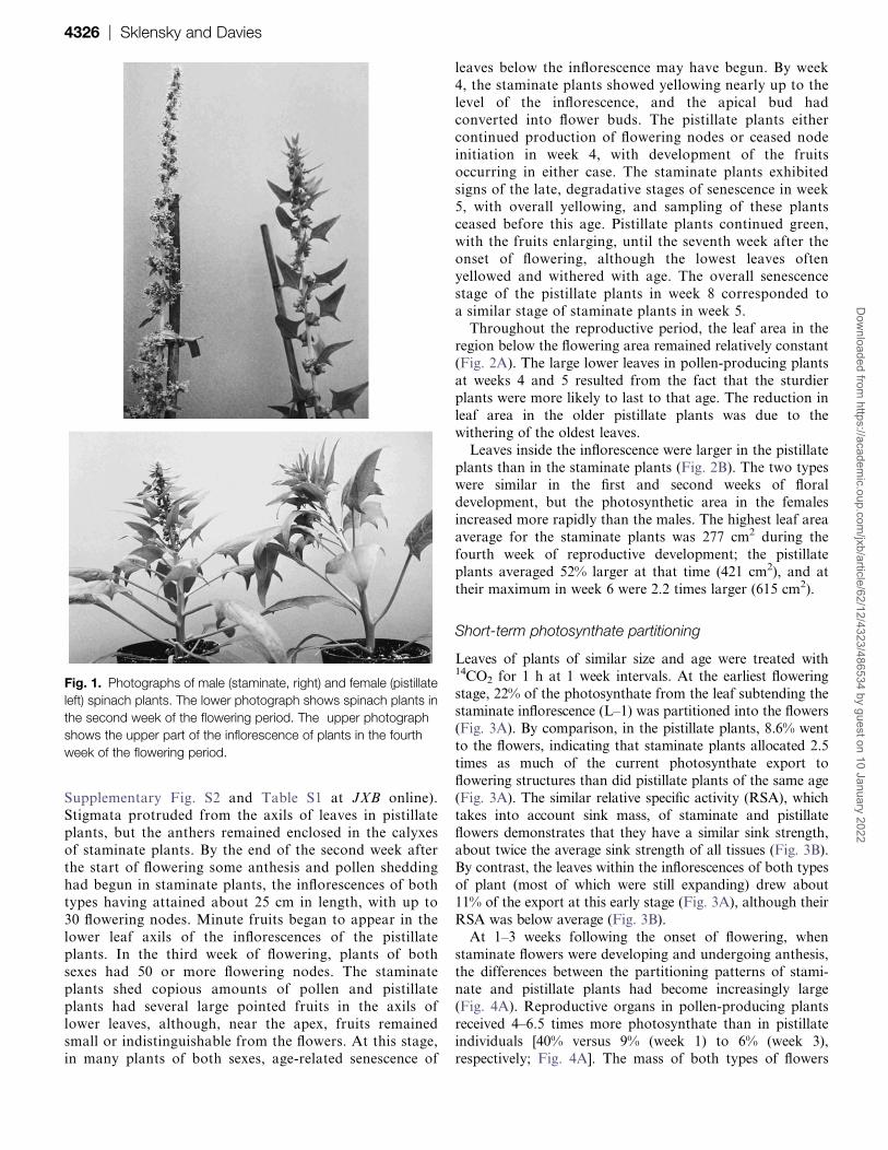
Throughout the reproductive period, the leaf area in theregion below the flowering area remained relatively constant
(Fig. 2A). The large lower leaves in pollen-producing plants
at weeks 4 and 5 resulted from the fact that the sturdier
plants were more likely to last to that age. The reduction in
leaf area in the older pistillate plants was due to the
withering of the oldest leaves.
Leaves inside the inflorescence were larger in the pistillate
plants than in the staminate plants (Fig. 2B). The two typeswere similar in the first and second weeks of floral
development, but the photosynthetic area in the females
increased more rapidly than the males. The highest leaf area
average for the staminate plants was 277 cm2 during the
fourth week of reproductive development; the pistillate
plants averaged 52% larger at that time (421 cm2), and at
their maximum in week 6 were 2.2 times larger (615 cm2).
Short-term photosynthate partitioning
Leaves of plants of similar size and age were treated with14CO2 for 1 h at 1 week intervals. At the earliest flowering
stage, 22% of the photosynthate from the leaf subtending thestaminate inflorescence (L–1) was partitioned into the flowers
(Fig. 3A). By comparison, in the pistillate plants, 8.6% went
to the flowers, indicating that staminate plants allocated 2.5
times as much of the current photosynthate export to
flowering structures than did pistillate plants of the same age
(Fig. 3A). The similar relative specific activity (RSA), which
takes into account sink mass, of staminate and pistillate
flowers demonstrates that they have a similar sink strength,about twice the average sink strength of all tissues (Fig. 3B).
By contrast, the leaves within the inflorescences of both types
of plant (most of which were still expanding) drew about
11% of the export at this early stage (Fig. 3A), although their
RSA was below average (Fig. 3B).
At 1–3 weeks following the onset of flowering, when
staminate flowers were developing and undergoing anthesis,
the differences between the partitioning patterns of stami-nate and pistillate plants had become increasingly large
(Fig. 4A). Reproductive organs in pollen-producing plants
received 4–6.5 times more photosynthate than in pistillate
individuals [40% versus 9% (week 1) to 6% (week 3),
respectively; Fig. 4A]. The mass of both types of flowers
Fig. 1. Photographs of male (staminate, right) and female (pistillate
left) spinach plants. The lower photograph shows spinach plants in
the second week of the flowering period. The upper photograph
shows the upper part of the inflorescence of plants in the fourth
week of the flowering period.
4326 | Sklensky and DaviesD
ownloaded from
https://academic.oup.com
/jxb/article/62/12/4323/486534 by guest on 10 January 2022

also rose rapidly (see Supplementary Table S1 at JXB
online). The RSA for the pistillate reproductive structures
continued to be similar to that of the staminate flowers
through the first week (Fig. 4B), but fell to less than half of
the comparable value in the second week and dropped even
lower in later weeks.The bulk of the current carbon fixed by the leaf just
below the inflorescence was distributed to the tissue below
the inflorescence region, being 63% of the exported radioac-
tivity in the earliest stage of flowering in pistillate plants
(Fig. 3A) and more than 50% for pollen-producing plants.
However, the proportions become more disparate at later
stages. At the time when partitioning to the shoot below the
inflorescence declined rapidly for staminate plants (valuesrange from 17–31% after the initial stage) (Fig. 4C),
distribution to the comparable region in seed-bearing
individuals stayed between 1.3 and 3 times higher, only
falling to the 30% level well after the staminate plants had
senesced. When the allocation to the lower region of the
shoot was considered on a mass-corrected basis, the
contrast remained: for staminate plants RSAs were between
0.4 and 1 during the period of flowering (Fig. 4D), whereasthe RSAs in pistillate plants were larger at every stage,
reaching a value of over 5 in the fifth week.
The percentage exported to the apical bud, which in-
cluded both leaf and flower primordia, ranged from
a fraction of 1% to 3.5% (Fig. 4E), but was, after the first
week, higher in the staminate plants than the pistillate
plants. The young leaves of the pistillate plants were large
enough, relative to the flowers, to enclose the bud, while
those of the staminate plants were not. Because of the small
size of this structure, even the very small amount allocatedresulted in a high RSA, but the values in staminate and
pistillate plants were comparable at most stages. However,
by week 7, after the staminate plants have senesced, the
RSA for the apical buds of the pistillate plants exceeded 12
(Fig. 4F). At this stage, the bud included several large fruits.
By the fourth week of flowering, the staminate plants had
begun to show the yellowing that is characteristic of whole
plant senescence, and the export to the reproductive struc-tures reached 68% (Fig. 4A). The pistillate plants were still
robust, although individual older leaves had begun to
senesce, and export from the leaf just below the inflorescence
to the developing fruits and young flowers remained near
10% (Fig. 4A). Vegetative structures continued to receive the
larger proportion of the exported photosynthate in the
pistillate plants, including 51% to the lower vegetative tissue
(Fig. 4C). However, in weeks 5, 6, and 7, the allocation to
Fig. 2. Area of leaves of staminate (black bars) and pistillate plants
(grey bars). (A) Area of leaves below the inflorescence; (B) area of
leaves within the inflorescence. After week 5, the staminate plants
have senesced. Bars represent 6SE.
Fig. 3. Allocation of radiolabelled photosynthate to different
structures from the leaf just below the inflorescence (L-1) to each
importing structure at the onset of flowering, within a few days of
flowers becoming visible on the plants. (A) As a percentage of total
export; (B) relative specific activity (RSA), calculated on a per gram
basis. The dpm g�1 for each organ was divided by the average
dpm g�1 of all the importing organs in that plant. Labelling was for
1 h followed by a 3 h chase. Bars represent 6SE.
Resource partitioning and senescence in spinach | 4327D
ownloaded from
https://academic.oup.com
/jxb/article/62/12/4323/486534 by guest on 10 January 2022

the fruits increased (Fig. 4A), while that to the lower portion
of the plant declined. In week 7, the pistillate plants showed
signs of overall senescence, and by week 8, most of thepistillate plants were dead.
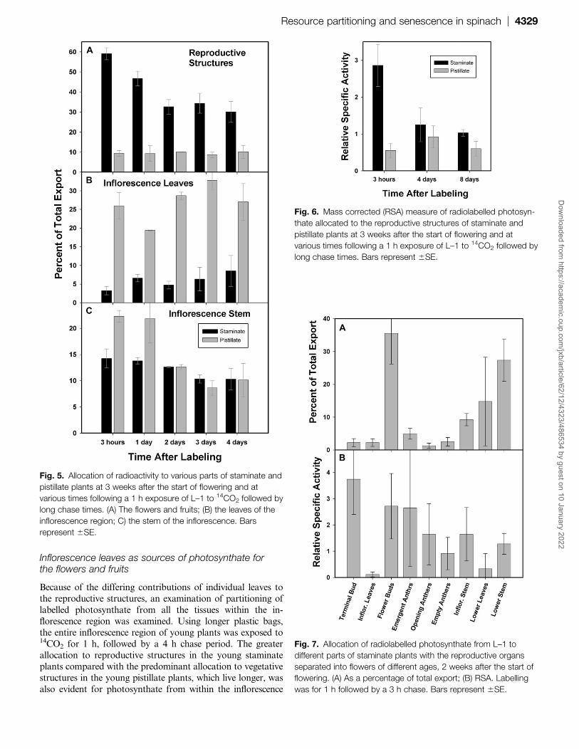
Long-term photosynthate partitioning
Three weeks after the start of flowering, a full day ofpartitioning after 14CO2 labelling of L–1 for 1 h produced
a similar photosynthate allocation pattern as did a 3 h
partitioning period in both staminate and pistillate plants.
The staminate flowers retained 47% of the radioactive
carbon, while pistillate reproductive structures had 9% (Fig.
5A). However, whereas the amount in the pistillate flowers
remained constant over the next 3 d, the proportion in the
pollen-producing flowers had declined appreciably by day 2and thereafter remained stable over the next 2 d. Whereas
less than 9% of exported carbon went to the leaves within the
staminate inflorescence, inflorescence leaves in the pistillate
plants received 20–33% (Fig. 5B), with no consistent changes
with the time. The amount of carbon located in inflorescence
stem tissue was greater in the pistillate plants (22%) than the
staminate plants for the first day (14%), but the difference
disappeared in subsequent days (Fig. 5C). Even at the end ofthe four days, levels of total recovered radioactivity were
similar to values for the 3 h partitioning period, suggesting
that any respired carbon was immediately re-fixed.
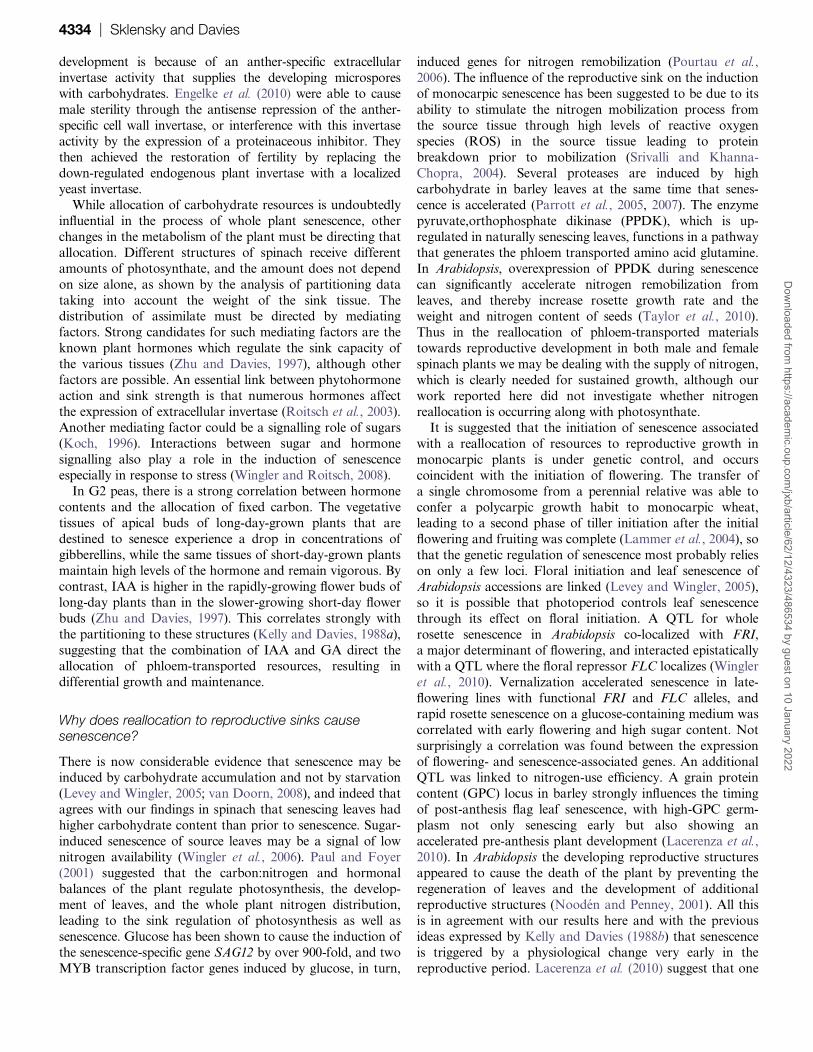
The RSA of the staminate flowers still showed a sub-
stantial drop over the long chase period (Fig. 6). The RSAs
of the pistillate flowers and fruits remained similar during
partitioning over 8 d, with values of less than 1, indicating
a below-average sink activity drawing from the leafimmediately below the inflorescence.
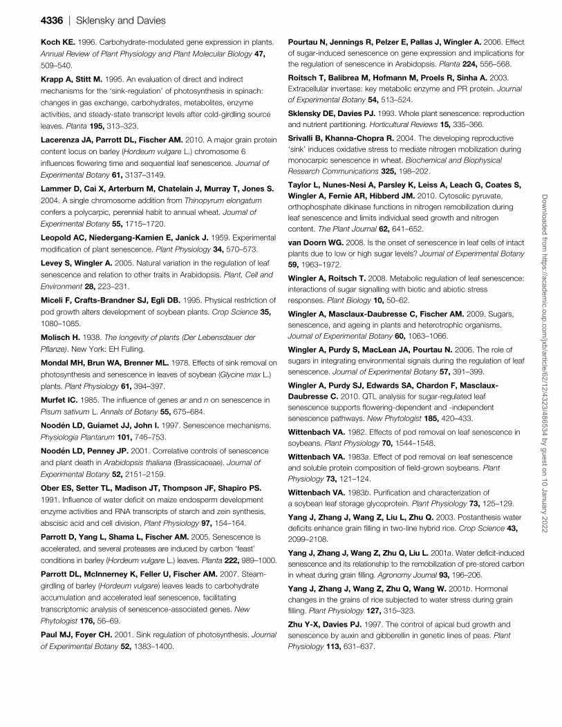
Staminate flowers as sinks
To determine the stage of maximum photosynthate import
into the staminate flowers, the flowers of staminate plants in
the second week of floral development were each separated
into four categories following labelling of the leaf just belowthe inflorescence for 1 h and a chase period of 3 h. The four
categories were: bud, being those flowers and flower parts
still held within the axils; extruded, which had extruded
anthers; anthesis, where anthers had begun to open; and
old, where the anthers were withered and had largely shed
their pollen. The flowers in the bud stage received the
largest amount of radioactivity, 35%, compared with less
than 5% at other stages (Fig. 7A), but they also had thelargest mass. The buds were the only stage containing
developing pollen, as determined by microscopic examina-
tion; in all the other stages, all of the pollen was mature.
RSA differences between the staminate floral stages were
not significant (Fig. 7B). The RSA for the empty anthers
was just below 1, indicating less than average but not
a cessation of import, despite the completion of develop-
ment. However, as the flowers of the older stages were fewin number, their total drain was relatively small.
Fig. 4. Allocation of radiolabelled photosynthate from the leaf just below the inflorescence (L–1), over time, from the start of
flowering: (A, B) to the flowers or fruits; (C, D) to the leaves and stem below the inflorescence; (E, F) to the apical bud region. Week
1 represents the time 1 week after flowers were first visible on the plant, with subsequent samples occurring weekly afterwards. (A,
C, E) As a percentage of total export; (B, D, F) relative specific activity (RSA), calculated on a per gram basis. The dpm g�1 for each
organ was divided by the average dpm g�1 of all the importing organs in that plant. Labelling was for 1 h followed by a 3 h chase.
Bars represent 6SE. By week 5, the staminate plants have senesced.
4328 | Sklensky and DaviesD
ownloaded from
https://academic.oup.com
/jxb/article/62/12/4323/486534 by guest on 10 January 2022
Inflorescence leaves as sources of photosynthate forthe flowers and fruits
Because of the differing contributions of individual leaves to
the reproductive structures, an examination of partitioning of
labelled photosynthate from all the tissues within the in-
florescence region was examined. Using longer plastic bags,
the entire inflorescence region of young plants was exposed to14CO2 for 1 h, followed by a 4 h chase period. The greater
allocation to reproductive structures in the young staminateplants compared with the predominant allocation to vegetative
structures in the young pistillate plants, which live longer, was
also evident for photosynthate from within the inflorescence
Fig. 5. Allocation of radioactivity to various parts of staminate and
pistillate plants at 3 weeks after the start of flowering and at
various times following a 1 h exposure of L–1 to 14CO2 followed by
long chase times. (A) The flowers and fruits; (B) the leaves of the
inflorescence region; C) the stem of the inflorescence. Bars
represent 6SE.
Fig. 7. Allocation of radiolabelled photosynthate from L–1 to
different parts of staminate plants with the reproductive organs
separated into flowers of different ages, 2 weeks after the start of
flowering. (A) As a percentage of total export; (B) RSA. Labelling
was for 1 h followed by a 3 h chase. Bars represent 6SE.
Fig. 6. Mass corrected (RSA) measure of radiolabelled photosyn-
thate allocated to the reproductive structures of staminate and
pistillate plants at 3 weeks after the start of flowering and at
various times following a 1 h exposure of L–1 to 14CO2 followed by
long chase times. Bars represent 6SE.
Resource partitioning and senescence in spinach | 4329D
ownloaded from
https://academic.oup.com
/jxb/article/62/12/4323/486534 by guest on 10 January 2022

(Fig. 8). This difference showed clearly at week 1, when the
staminate flowers received 2.5 times more photosynthate than
pistillate flowers, but disappeared by week 2. This contrasts
with the situation when a leaf below the inflorescence was
labelled, when the difference persisted throughout the life of
the staminate plants.
Photosynthesis and respiration in the light and in thedark
Photosynthesis rates in the leaves of the staminate plantswere constant during the first 3 weeks of flowering (Fig. 9A).
The photosynthetic rates of the leaves declined substantially
in week 4 and even further, essentially to zero, in week 5. The
pistillate plants, on the other hand, showed a slight reduction
in activity from week 1 to week 5, but thereafter displayed
noticeable loss of function in weeks 6, 7, and 8 (Fig. 9B).
As the different regions of the inflorescence were at
different stages of development, respiration was measuredin consecutive 3.5 cm segments, numbering 1 at the apex
down to the mature regions of the inflorescence, following
the removal of the leaves from the inflorescence. Plants
from the second week of flowering were measured at four
consecutive segments, with the number of measurements
increasing with the larger plants. The staminate plants,
stripped of their leaves, had 3–3.5-fold greater rates of
respiration at week 2 than did the pistillate plants near the
inflorescence apices (Fig. 10A). This difference still existed,
but with less magnitude, further down the axis in young
plants. At this time, some anthers had begun to shed pollen,
although very few in the first two segments. In older plants,
at week 4, flowers existed in a mixture of pre-and post-anthesis stages: the majority of the staminate flowers had
undergone anthesis while the region nearest the tip showed
the fewest open anthers. At this time the respiration rates
for the pistillate plants had risen to levels comparable with
that of the staminate plants (Fig. 10B). The upper two
segments of the pistillate inflorescence had very small flower
clusters until week 4, at which time some of those in
segment 2 had full-sized fruits.As the pistillate flowers were initially extremely small, the
degree to which the initial disparity in activity results from
size was determined based on a calculation of the re-
spiratory rate per gram of flower or fruit. To determine the
respiration of the reproductive structures, carbon dioxide
evolution of the stem regions was measured with both
leaves and reproductive structures removed. This value was
subtracted from the rate of respiration of the stem with
Fig. 8. Allocation to organs of staminate and pistillate plants when
the entire inflorescence was exposed to 14CO2 for 1 h with a 4 h
chase period. (A) 1 week after the start of flowering. (B) Plants two
weeks after the start of flowering. Bars represent 6SE.
Fig. 9. Photosynthetic rates of leaves of staminate and pistillate
spinach plants measured at various times following the start of
flowering. Measurements occurred at mid-morning with 175 lE
m�2 of light. (A) Leaves of staminate plants; (B) Leaves of pistillate
plants. Bars represent 6SE.
4330 | Sklensky and DaviesD
ownloaded from
https://academic.oup.com
/jxb/article/62/12/4323/486534 by guest on 10 January 2022

flowers and fruits, and the resulting value divided by the
fresh weight of the flowers removed from the segment,
producing an estimate of the respiratory activity of the
reproductive tissue on a tissue fresh weight basis. In the
apical region, all of the tissues were very active in
respiration, with the youngest plants showing the highest
level of activity on a per gram basis (Fig. 10C). Below this
region, the respiration of the staminate flowers declined, butstill tended to be more than in the pistillate flowers. The
older fruits showed a reduced respiration on a per gram
basis compared with the young pistillate flowers (Fig. 10D).
Carbohydrate content
The concentration of total non-structural carbohydrates
(glucose+fructose+sucrose+starch) in the senescing stami-
nate flowers at week 5 was as high as the highest level in
pistillate fruits, which occurred close to fruit maturity at
week 7 (Fig. 11A). These non-structural carbohydrates
showed a continuous increase in leaf L3 of the staminate
plants until their senescence (Fig. 11B), while in pistillate
plants a very low level was maintained until the fifth week,when the amount rose, peaking 1 week later.
Discussion
How senescence is triggered remains an enigma (Wingler
et al., 2009), and even more so in monocarpic plants
(Lacerenza et al., 2010), but here some light has been shed
on the degree to which the diversion of nutrient flow from
vegetative organs to reproductive organs is involved in the
process. In this study, the examination of carbon partition-
ing throughout the flowering phase of spinach has demon-
strated the importance of the reproductive structures of
both staminate and pistillate plants to the pattern of
resource allocation. The argument against nutrient-drain to
the reproductive tissues triggering the senescence of spinachplants, namely that staminate flowers represent an insignif-
icant photosynthate drain on the plant, is clearly refuted.
Pollen-producing flowers demand, from an early age, much
of the photosynthate of the plant. In fact, at many stages
during the reproductive period, leaves on staminate spinach
plants export larger quantities of photosynthate to stami-
nate flowers than is provided to the pistillate flowers from
a comparable leaf on a pistillate plant. The senescence-delaying removal of flowers from staminate plants (Leopold
et al., 1959) could, in fact, affect the nutritional status of the
plant in a similar manner to removing fruits from pistillate
plants, thus influencing the senescence of the entire plant.
The different time-course of senescence in staminate and
pistillate plants, and differences in the size of inflorescence
leaves, indicate that the rate of reproductive development
and the amount of source tissue may also factor in theregulation of senescence of the plants. It is therefore
possible to account for the senescence of staminate spinach
plants merely on the basis of resource diversion, without
invoking any fruit-derived hormonal senescence factor. This
is not to say that photosynthate per se is the responsible
factor, but rather that the diversion of any required
Fig. 10. Rates of respiration per segment (A, B) of 3.5 cm segments of the inflorescence, without leaves, and rates of respiration, per
unit fresh weight, of the reproductive structures (C, D) from 3.5 cm segments within the inflorescence, both numbered downwards from
the apex of the plant. (A, C) 2 weeks; (B, D) 4 weeks after the start of flowering. Respiration was determined by CO2 evolution in
darkness. Values in (C) and (D) were obtained by subtraction of the CO2 evolution of stems alone from that of stems with reproductive
structures, divided by the fresh weight of the reproductive structures. Bars represent 6SE.
Resource partitioning and senescence in spinach | 4331D
ownloaded from
https://academic.oup.com
/jxb/article/62/12/4323/486534 by guest on 10 January 2022

components, such as nitrogenous materials or hormonesthat are moving with the photosynthate in the phloem, may
also have a role in the regulation of senescence.
Partitioning to vegetative and reproductive organsthroughout the reproductive phase
At the very early stages of reproduction, more fixed carbon
was apportioned to the pollen-producing flowers than to the
tiny flowers of the pistillate plants. The larger mass of the
staminate racemes only partially explains the greater draw onassimilate at this stage. This higher resource allocation to
flowers continued throughout the lives of the staminate
plants. From an early stage, the fruit-producing plants
designated a higher proportion of their carbon intake to the
maintenance and production of vegetative tissue to support
their reproductive stage than did the shorter-lived staminate
plants. While the combined area of the leaves below the
inflorescence is similar for each type of plant, the leaf areawithin the inflorescence becomes substantially larger in the
seed-producing plants, providing them with a larger amount
of photosynthetically-active tissue in close proximity to the
developing sinks. A lower allocation of photosynthate to the
stems and leaves developing in the staminate inflorescence
results in a reduced growth of these organs, leading to
a smaller total of source tissue. The leaves within the
pistillate inflorescence stay green and retain photosynthetic
activity, as the fruits develop, for weeks after the staminate
plants have senesced, and thus contribute to the longer
lifespan of the pistillate plants.
Two weeks after the start of flowering the terminal bud
of the staminate plants is tightly compacted and receiveda higher proportion of assimilate than at other times. At
this time flowers close to the apical region are undergoing
anthesis, with those in the bud nearly ready to do so. The
apical buds of the pistillate plants include larger
developing leaves that enclose their flowers than do the
pollen-producing plants with their exposed flower buds.
The contrast in the senescence programmes of staminate
and pistillate plants lies primarily in the timing. Late indevelopment, after the age at which staminate plants have
senesced, the fruits began to draw higher quantities of
photosynthate (and any other phloem-transported resour-
ces) from the lower leaves. This distinction in timing reflects
the different roles of the gametophytes produced on each
plant, namely the early development of pollen on the
staminate plants versus the later development of fruits and
seeds. As the fruits develop, the lower leaves allotted a veryhigh proportion of their current photoassimilate to the
developing fruits, though this amount fell somewhat
towards the very end. By this time, large younger leaves
within the fruit-bearing inflorescence have developed, and
served as source tissue for the fruits after the oldest leaves
have senesced. However, the leaf just below the inflores-
cence continued to designate a fairly large proportion of the
resources to vegetative tissue production and maintenance,demonstrating the importance of the position of the source
in consideration of the allocation of assimilates.
Male/female differences in role and partitioning
Numerous differences exist between staminate and pistillate
plants in the long-term fate of the carbohydrate resources in
vegetative and reproductive tissues. These differences are
explicable as a consequence of their different functions and
developmental timing leading to different rates and
intensities of energy use.
The pollen-producing flowers had a relatively high degree
of metabolic activity, losing substantial quantities of pre-fixed carbon. This rapid rate of respiration is responsible for
at least part of the increased greater import of photosyn-
thate into the pollen-producing flowers. By contrast, the
pistillate flowers and fruits showed essentially constant
levels of radiolabel.
In the shorter lifespan of the staminate plants, the
allocation of resources to the flowers producing and
shedding pollen indicates the time of greatest metabolicactivity in these organs. The staminate flower buds in the
leaf axils drew by far the highest proportion of label.
Flowers at later stages, in which the anthers are extruded
from the calyxes, following dehiscence at anthesis, received
much smaller portions of assimilate. However, the
Fig. 11. Changes over time in the total non-structural carbohy-
drate content of staminate and pistillate plants. (A) Reproductive
structures 15–18.5 cm below the tip of the inflorescence; (B) leaf
L3 (third leaf, counting from the base, within the inflorescence).
Bars represent 6SE.
4332 | Sklensky and DaviesD
ownloaded from
https://academic.oup.com
/jxb/article/62/12/4323/486534 by guest on 10 January 2022

allocation of radioactivity to different stages was similar on
a mass-normalized basis, suggesting that the respiration of
pollen and anthers continued beyond the earliest stages of
flower development.
While staminate plants achieve, at most, a ratio of 2:1
for the contribution of photosynthetic tissue within and
below the inflorescence respectively, pistillate plants more
commonly have ratios of 4–5:1, possessing far larger,young, active leaves in the inflorescence region by the
third week of flowering. The younger leaves were the
most important as source tissue whereas the lower leaves
of both staminate and pistillate plants exhibited an age-
related decline in photosynthesis. However, even these
older leaves maintained some function for a longer period
of time in the pistillate plants. Comparable rates of
carbon dioxide uptake per unit area were seen in bothsexes for both the leaf just below the inflorescence and
each of the three lower leaves of the flowering region,
until the leaves of the staminate plants cease to photo-
synthesize in week 5. The larger size of these younger
leaves in the pistillate plants indicates that more assimi-
late is being produced than in the staminate plants.
Young pollen-producing inflorescences are relatively large
and respired at a far greater rate than did their muchsmaller pistillate counterparts. Even on a weight basis, the
young staminate flowers respired somewhat more rapidly
than did the flowers or fruits of the pistillate plants. Mid-
age inflorescences were more comparable to one another,
with the staminate flowers, which are mostly at, near, or
post-anthesis, respiring at a much slower rate. As the fruits
enlarged, their respiration increased slightly, but the per
gram rate of respiration declined as the fresh weight rose.Staminate plants make an early, highly energetic contribu-
tion to reproduction, with no necessity to prolong function
by producing large source leaves later in development.
Pistillate plants maintain function longer, requiring an
investment in source tissue that will continue to produce
assimilate throughout the development of embryos and
fruits.
An increase in the carbohydrate concentration in thefruits or pollen-producing flowers up to a peak probably
reflects the maturation of those sinks. Starch concentration
continued to increase in anthers in the last week of
staminate flower development, after pollen development
has ceased and pollen been shed from most of the anthers,
and little purpose remains for carbohydrate allocation to
the staminate flowers. Clearly once the shift in resources
has occurred, the allocation pattern continues despitea lack of obvious function for the photosynthate at this
stage.
The carbohydrate content of leaves directly contradicts
any suggestion that the leaves senesce because of carbohy-
drate starvation. However, the pistillate plants maintained
low total carbohydrate concentrations for a leaf in the
inflorescence through the fourth week, whereas in the fifth
and sixth weeks the carbohydrate levels showed a dramaticincrease, at the time of the drop in photosynthesis for the
leaves on the pistillate plants. Similarly, the same leaf of
the staminate plants showed a gradual but substantial
increase in total non-structural carbohydrate throughout
the reproductive period, until it senesced in week 5. This
evidence strongly suggests that lack of total carbohydrate
does not cause leaf senescence.
To the extent to which fruits serve as an explanation for
the senescence of pistillate plants because of the distribution
of resources to these organs, the different timing, the greaterrespiration rate of pollen-producing flowers, and contrast-
ing inflorescence morphology provides ample cause for the
senescence of staminate plants.
The effect of source–sink manipulations on monocarpicsenescence
The work of Leopold et al. (1959) that examined the effect of
flower removal on the senescence of staminate spinach plants
has been consistently misinterpreted as a counter-example to
the nutrient drain hypothesis. By showing the different
morphology, timing of senescence, and high rate of respiratory
activity of staminate spinach plants, the current studiesdemonstrate that excision of pollen-producing flowers results
in the loss of a very significant sink for photosynthetic carbon,
and thus a sink for any other phloem-transported resource
such as nitrogen or hormonal compound. In unpollinated
plants senescence did proceed even though it was delayed
(Leopold et al., 1959). The removal of unpollinated pistillate
flowers also delayed senescence and the removal of flowers at
a younger stage resulted in a greater delay of senescence. Ourresults show that the young staminate flowers draw large
allocations of photoassimilate. By contrast, young pistillate
flowers are a relatively small sink, but, because of their size,
they are active sinks on a per gram basis. The lack of the fruits
as a large sink in the unpollinated or de-flowered plants
examined by Leopold et al. (1959) may have caused an
alteration in the pattern of resource allocation, but the early
shift to support the reproductive process was apparentlysufficient to lead to eventual senescence.
G2 peas allocate less photosynthate to their vegetative
buds in long days, when they senesce after flowering, than in
short-days when the plants continue to flower without
senescing, showing the importance of resource partitioning in
the mediation of senescence phenomena (Kelly and Davies,
1988a). The flowers and pods of the pre-senescent long-day
plants develop far more rapidly, correlating with the greaterresource allocation to reproduction, well before senescence
symptoms are visible (Kelly and Davies, 1986), also illustrat-
ing the early initiation of the regulation senescence.
Causes of nutrient diversion
During senescence it has been shown that phloem-transported
compounds are diverted from vegetative to reproductive sinks.
This indicates that there are possible changes in theserespective sinks, and also in source leaves as they transfer
from being a source of photosynthate to one of remobilized
compounds. Extracellular invertases are important for apo-
plastic phloem unloading and are key enzymes in determining
sink strength (Roitsch et al., 2003). The sink activity for pollen
Resource partitioning and senescence in spinach | 4333D
ownloaded from
https://academic.oup.com
/jxb/article/62/12/4323/486534 by guest on 10 January 2022
development is because of an anther-specific extracellular
invertase activity that supplies the developing microspores
with carbohydrates. Engelke et al. (2010) were able to cause
male sterility through the antisense repression of the anther-
specific cell wall invertase, or interference with this invertase
activity by the expression of a proteinaceous inhibitor. They
then achieved the restoration of fertility by replacing the
down-regulated endogenous plant invertase with a localizedyeast invertase.
While allocation of carbohydrate resources is undoubtedly
influential in the process of whole plant senescence, other
changes in the metabolism of the plant must be directing that
allocation. Different structures of spinach receive different
amounts of photosynthate, and the amount does not depend
on size alone, as shown by the analysis of partitioning data
taking into account the weight of the sink tissue. Thedistribution of assimilate must be directed by mediating
factors. Strong candidates for such mediating factors are the
known plant hormones which regulate the sink capacity of
the various tissues (Zhu and Davies, 1997), although other
factors are possible. An essential link between phytohormone
action and sink strength is that numerous hormones affect
the expression of extracellular invertase (Roitsch et al., 2003).
Another mediating factor could be a signalling role of sugars(Koch, 1996). Interactions between sugar and hormone
signalling also play a role in the induction of senescence
especially in response to stress (Wingler and Roitsch, 2008).
In G2 peas, there is a strong correlation between hormone
contents and the allocation of fixed carbon. The vegetative
tissues of apical buds of long-day-grown plants that are
destined to senesce experience a drop in concentrations of
gibberellins, while the same tissues of short-day-grown plantsmaintain high levels of the hormone and remain vigorous. By
contrast, IAA is higher in the rapidly-growing flower buds of
long-day plants than in the slower-growing short-day flower
buds (Zhu and Davies, 1997). This correlates strongly with
the partitioning to these structures (Kelly and Davies, 1988a),
suggesting that the combination of IAA and GA direct the
allocation of phloem-transported resources, resulting in
differential growth and maintenance.
Why does reallocation to reproductive sinks causesenescence?
There is now considerable evidence that senescence may beinduced by carbohydrate accumulation and not by starvation
(Levey and Wingler, 2005; van Doorn, 2008), and indeed that
agrees with our findings in spinach that senescing leaves had
higher carbohydrate content than prior to senescence. Sugar-
induced senescence of source leaves may be a signal of low
nitrogen availability (Wingler et al., 2006). Paul and Foyer
(2001) suggested that the carbon:nitrogen and hormonal
balances of the plant regulate photosynthesis, the develop-ment of leaves, and the whole plant nitrogen distribution,
leading to the sink regulation of photosynthesis as well as
senescence. Glucose has been shown to cause the induction of
the senescence-specific gene SAG12 by over 900-fold, and two
MYB transcription factor genes induced by glucose, in turn,
induced genes for nitrogen remobilization (Pourtau et al.,
2006). The influence of the reproductive sink on the induction
of monocarpic senescence has been suggested to be due to its
ability to stimulate the nitrogen mobilization process from
the source tissue through high levels of reactive oxygen
species (ROS) in the source tissue leading to protein
breakdown prior to mobilization (Srivalli and Khanna-
Chopra, 2004). Several proteases are induced by high
carbohydrate in barley leaves at the same time that senes-
cence is accelerated (Parrott et al., 2005, 2007). The enzyme
pyruvate,orthophosphate dikinase (PPDK), which is up-
regulated in naturally senescing leaves, functions in a pathway
that generates the phloem transported amino acid glutamine.
In Arabidopsis, overexpression of PPDK during senescence
can significantly accelerate nitrogen remobilization from
leaves, and thereby increase rosette growth rate and the
weight and nitrogen content of seeds (Taylor et al., 2010).
Thus in the reallocation of phloem-transported materials
towards reproductive development in both male and female
spinach plants we may be dealing with the supply of nitrogen,
which is clearly needed for sustained growth, although our
work reported here did not investigate whether nitrogen
reallocation is occurring along with photosynthate.
It is suggested that the initiation of senescence associated
with a reallocation of resources to reproductive growth in
monocarpic plants is under genetic control, and occurs
coincident with the initiation of flowering. The transfer of
a single chromosome from a perennial relative was able to
confer a polycarpic growth habit to monocarpic wheat,leading to a second phase of tiller initiation after the initial
flowering and fruiting was complete (Lammer et al., 2004), so
that the genetic regulation of senescence most probably relies
on only a few loci. Floral initiation and leaf senescence of
Arabidopsis accessions are linked (Levey and Wingler, 2005),
so it is possible that photoperiod controls leaf senescence
through its effect on floral initiation. A QTL for whole
rosette senescence in Arabidopsis co-localized with FRI,a major determinant of flowering, and interacted epistatically
with a QTL where the floral repressor FLC localizes (Wingler
et al., 2010). Vernalization accelerated senescence in late-
flowering lines with functional FRI and FLC alleles, and
rapid rosette senescence on a glucose-containing medium was
correlated with early flowering and high sugar content. Not
surprisingly a correlation was found between the expression
of flowering- and senescence-associated genes. An additionalQTL was linked to nitrogen-use efficiency. A grain protein
content (GPC) locus in barley strongly influences the timing
of post-anthesis flag leaf senescence, with high-GPC germ-
plasm not only senescing early but also showing an
accelerated pre-anthesis plant development (Lacerenza et al.,
2010). In Arabidopsis the developing reproductive structures
appeared to cause the death of the plant by preventing the
regeneration of leaves and the development of additionalreproductive structures (Nooden and Penney, 2001). All this
is in agreement with our results here and with the previous
ideas expressed by Kelly and Davies (1988b) that senescence
is triggered by a physiological change very early in the
reproductive period. Lacerenza et al. (2010) suggest that one
4334 | Sklensky and DaviesD
ownloaded from
https://academic.oup.com
/jxb/article/62/12/4323/486534 by guest on 10 January 2022

of these GPC genes may be a functional homologue of
Arabidopsis glycine-rich RNA-binding protein 7, which has
previously been implicated in the promotion of flowering.
We may, therefore, be on the verge of a more detailed
analysis of the interactions between the physiological and
molecular networks controlling monocarpic senescence.
Conclusion
Resource redistribution regulates senescence
Our results clearly show an early reallocation of phloem-
transported fixed carbon to reproductive development, so
the nutrient-diversion hypothesis can account for the
induction of senescence in vegetative tissues. However, the
crucial compound is clearly not carbohydrate but a part of
a global shift in the hormonal and/or nutrient balance,
probably including nitrogen, resulting from flowering. This
would then lead to changes in gene expression associatedwith the cessation of growth and the development of the
senescence syndrome in the vegetative tissues.
The observed diminution of the leaves in the inflorescence
of spinach (which occurs more rapidly in the staminate
plants), and in the apical senescence in peas (Kelly and
Davies, 1988a; Zhu and Davies, 1997), can be explained by
this shift. The changes in allocation, including the proximity
of the apical meristem to floral sinks, which may equal orexceed the apical meristem in sink strength, must affect the
meristem itself. The meristem may then decline in size (and
thus produce smaller leaves) and eventually often either
senesces or converts to a flower primordium. The loss of the
apical bud leads to a number of physiological changes, and
the inevitable senescence of the whole plant, due to the
inability to produce new organs. When the balance of
carbohydrates is again altered by cessation of development inthe floral sinks, the resultant feed-back inhibition could cause
not only a repression of photosynthesis, but leaf senescence.
Thus, it is not the drain to the reproductive sinks per se, but
the permanent diversion away from the development of
further new vegetative sinks that may be responsible for some
of the observed phenomena in whole-plant senescence. As
noted by Leopold et al. (1959), once flowering is initiated,
even if flowers are removed or if pistillate flowers remainunpollinated, senescence will surely follow.
Supplementary data
Supplementary data can be found at JXB online.
Supplementary material. A full version of the Materials
and methods.
Supplementary Table S1. Descriptions and average dry
weight of flowering structures for plants of the given
flowering stage in weeks following the onset of flowering6SE.
Supplementary Fig. S1. Shapes and sizes of leaves from
staminate and pistillate spinach plants in ascending order:
leaf L1, L11, L21, L31, L41, L51, numbering from the base
of the inflorescence.
Supplementary Fig. S2. Photographs of staminate (A–E)
and pistillate (F–J) spinach plants at various stages of
development.
Acknowledgements
This work was supported, in part, by Hatch fundsadministered by the New York State College of Agriculture
and Life Sciences at Cornell University. DES was sup-
ported, in part, by a National Science Foundation Fellow-
ship. We thank Jeffrey Melkonian, David Wolfe, Marvin
Pritts, Tim Setter, and Brian Flanagan for assistance and
the use of their equipment, and the Asgrow Seed Company
for donation of the spinach seed.
References
Borras L, Maddonni GA, Otegui ME. 2003. Leaf senescence in
maize hybrids: plant population, row spacing and kernel set effects.
Field Crops Research 82, 13–26.
Borrell A, Hammer G, Van Oosterom E. 2001. Stay-green:
a consequence of the balance between supply and demand for
nitrogen during grain filling? Annals of Applied Biology 138, 91–95.
Cairns AJ. 1987. Colorimetric microtiter plate assay of glucose and
fructose by enzyme-linked formazan production applicability to the
measurement of fructosyltransferase activity in higher plants. Analytical
Biochemistry 167, 270–278.
Crafts-Brandner SJ, Egli DB. 1987. Modification of seed growth in
soybean by physical restraint effect on leaf senescence. Journal of
Experimental Botany 38, 2043–2049.
Engelke T, Hirsche J, Roitsch T. 2010. Anther-specific
carbohydrate supply and restoration of metabolically engineered male
sterility. Journal of Experimental Botany 61, 2693–2706.
Hamilton DA, Davies PJ. 1988a. Export of organic materials from
developing fruits of pea and its possible relation to apical senescence.
Plant Physiology 86, 951–955.
Hamilton DA, Davies PJ. 1988b. Sucrose and malic acid as the
compounds exported to the apical bud of pea following 14CO2 labeling
of the fruit. No evidence for a senescence factor. Plant Physiology 88,
466–472.
Huber SC, Huber JL. 1996. Role and regulation of sucrose-
phosphate synthase in higher plants. Annual Review of Plant
Physiology and Plant Molecular Biology 47, 431–444.
Janick J, Leopold AC. 1961. A distinction between bolting and
flowering effects on senescence. Nature 192, 887–888.
Kelly MO, Davies PJ. 1986. Genetic and photoperiodic control of the
relative rates of reproductive and vegetative development in peas.
Annals of Botany 58, 13–21.
Kelly MO, Davies PJ. 1988a. Photoperiodic and genetic control of
carbon partitioning in peas and its relationship to apical senescence.
Plant Physiology 86, 978–982.
Kelly MO, Davies PJ. 1988b. The control of whole plant senescence.
CRC Critical Reviews in Plant Science 7, 139–173.
Resource partitioning and senescence in spinach | 4335D
ownloaded from
https://academic.oup.com
/jxb/article/62/12/4323/486534 by guest on 10 January 2022
Koch KE. 1996. Carbohydrate-modulated gene expression in plants.
Annual Review of Plant Physiology and Plant Molecular Biology 47,
509–540.
Krapp A, Stitt M. 1995. An evaluation of direct and indirect
mechanisms for the ‘sink-regulation’ of photosynthesis in spinach:
changes in gas exchange, carbohydrates, metabolites, enzyme
activities, and steady-state transcript levels after cold-girdling source
leaves. Planta 195, 313–323.
Lacerenza JA, Parrott DL, Fischer AM. 2010. A major grain protein
content locus on barley (Hordeum vulgare L.) chromosome 6
influences flowering time and sequential leaf senescence. Journal of
Experimental Botany 61, 3137–3149.
Lammer D, Cai X, Arterburn M, Chatelain J, Murray T, Jones S.
2004. A single chromosome addition from Thinopyrum elongatum
confers a polycarpic, perennial habit to annual wheat. Journal of
Experimental Botany 55, 1715–1720.
Leopold AC, Niedergang-Kamien E, Janick J. 1959. Experimental
modification of plant senescence. Plant Physiology 34, 570–573.
Levey S, Wingler A. 2005. Natural variation in the regulation of leaf
senescence and relation to other traits in Arabidopsis. Plant, Cell and
Environment 28, 223–231.
Miceli F, Crafts-Brandner SJ, Egli DB. 1995. Physical restriction of
pod growth alters development of soybean plants. Crop Science 35,
1080–1085.
Molisch H. 1938. The longevity of plants (Der Lebensdauer der
Pflanze). New York: EH Fulling.
Mondal MH, Brun WA, Brenner ML. 1978. Effects of sink removal on
photosynthesis and senescence in leaves of soybean (Glycine max L.)
plants. Plant Physiology 61, 394–397.
Murfet IC. 1985. The influence of genes ar and n on senescence in
Pisum sativum L. Annals of Botany 55, 675–684.
Nooden LD, Guiamet JJ, John I. 1997. Senescence mechanisms.
Physiologia Plantarum 101, 746–753.
Nooden LD, Penney JP. 2001. Correlative controls of senescence
and plant death in Arabidopsis thaliana (Brassicaceae). Journal of
Experimental Botany 52, 2151–2159.
Ober ES, Setter TL, Madison JT, Thompson JF, Shapiro PS.
1991. Influence of water deficit on maize endosperm development
enzyme activities and RNA transcripts of starch and zein synthesis,
abscisic acid and cell division. Plant Physiology 97, 154–164.
Parrott D, Yang L, Shama L, Fischer AM. 2005. Senescence is
accelerated, and several proteases are induced by carbon ‘feast’
conditions in barley (Hordeum vulgare L.) leaves. Planta 222, 989–1000.
Parrott DL, McInnerney K, Feller U, Fischer AM. 2007. Steam-
girdling of barley (Hordeum vulgare) leaves leads to carbohydrate
accumulation and accelerated leaf senescence, facilitating
transcriptomic analysis of senescence-associated genes. New
Phytologist 176, 56–69.
Paul MJ, Foyer CH. 2001. Sink regulation of photosynthesis. Journal
of Experimental Botany 52, 1383–1400.
Pourtau N, Jennings R, Pelzer E, Pallas J, Wingler A. 2006. Effect
of sugar-induced senescence on gene expression and implications for
the regulation of senescence in Arabidopsis. Planta 224, 556–568.
Roitsch T, Balibrea M, Hofmann M, Proels R, Sinha A. 2003.
Extracellular invertase: key metabolic enzyme and PR protein. Journal
of Experimental Botany 54, 513–524.
Sklensky DE, Davies PJ. 1993. Whole plant senescence: reproduction
and nutrient partitioning. Horticultural Reviews 15, 335–366.
Srivalli B, Khanna-Chopra R. 2004. The developing reproductive
‘sink’ induces oxidative stress to mediate nitrogen mobilization during
monocarpic senescence in wheat. Biochemical and Biophysical
Research Communications 325, 198–202.
Taylor L, Nunes-Nesi A, Parsley K, Leiss A, Leach G, Coates S,
Wingler A, Fernie AR, Hibberd JM. 2010. Cytosolic pyruvate,
orthophosphate dikinase functions in nitrogen remobilization during
leaf senescence and limits individual seed growth and nitrogen
content. The Plant Journal 62, 641–652.
van Doorn WG. 2008. Is the onset of senescence in leaf cells of intact
plants due to low or high sugar levels? Journal of Experimental Botany
59, 1963–1972.
Wingler A, Roitsch T. 2008. Metabolic regulation of leaf senescence:
interactions of sugar signalling with biotic and abiotic stress
responses. Plant Biology 10, 50–62.
Wingler A, Masclaux-Daubresse C, Fischer AM. 2009. Sugars,
senescence, and ageing in plants and heterotrophic organisms.
Journal of Experimental Botany 60, 1063–1066.
Wingler A, Purdy S, MacLean JA, Pourtau N. 2006. The role of
sugars in integrating environmental signals during the regulation of leaf
senescence. Journal of Experimental Botany 57, 391–399.
Wingler A, Purdy SJ, Edwards SA, Chardon F, Masclaux-
Daubresse C. 2010. QTL analysis for sugar-regulated leaf
senescence supports flowering-dependent and -independent
senescence pathways. New Phytologist 185, 420–433.
Wittenbach VA. 1982. Effects of pod removal on leaf senescence in
soybeans. Plant Physiology 70, 1544–1548.
Wittenbach VA. 1983a. Effect of pod removal on leaf senescence
and soluble protein composition of field-grown soybeans. Plant
Physiology 73, 121–124.
Wittenbach VA. 1983b. Purification and characterization of
a soybean leaf storage glycoprotein. Plant Physiology 73, 125–129.
Yang J, Zhang J, Wang Z, Liu L, Zhu Q. 2003. Postanthesis water
deficits enhance grain filling in two-line hybrid rice. Crop Science 43,
2099–2108.
Yang J, Zhang J, Wang Z, Zhu Q, Liu L. 2001a. Water deficit-induced
senescence and its relationship to the remobilization of pre-stored carbon
in wheat during grain filling. Agronomy Journal 93, 196–206.
Yang J, Zhang J, Wang Z, Zhu Q, Wang W. 2001b. Hormonal
changes in the grains of rice subjected to water stress during grain
filling. Plant Physiology 127, 315–323.
Zhu Y-X, Davies PJ. 1997. The control of apical bud growth and
senescence by auxin and gibberellin in genetic lines of peas. Plant
Physiology 113, 631–637.
4336 | Sklensky and DaviesD
ownloaded from
https://academic.oup.com
/jxb/article/62/12/4323/486534 by guest on 10 January 2022