Development and Ultrastructure of Cucurbita pepo Nectaries of Male Flowers
10
Annals of Botany 78 : 95–104, 1996 Development and Ultrastructure of Cucurbita pepo Nectaries of Male Flowers M. NEPI*, F. CIAMPOLINI and E. PACINI Department of Enironmental Biology, Botany Section, Siena Uniersity, Via P.A. Mattioli 4, 53110 Siena, Italy Received : 28 July 1995 Accepted : 22 January 1996 The development of the nectary of the male flower of Cucurbita pepo L. was studied from 5 d before to 2 d after anthesis. The nectary consists of parenchyma that stores starch in the presecretory stages, and epidermis. An hour before nectar secretion begins, the starch is hydrolyzed. The nectar exudes from the stomata and forms a continuous layer on the nectary surface. During anthesis the nectar may all be collected by pollinators or some or all of it may remain in the nectary and be successively resorbed. The nectary parenchyma stores material for synthesizing the sugar component of nectar and stores similar material again after nectar resorption. It is also responsible for nectar production and secretion. The epidermis is actively involved in the reabsorption process. The resorption of nectar is a phenomenon that allows the plant to recover invested energy. Few observations on this phenomenon have hitherto been published. # 1996 Annals of Botany Company Key words : Amyloplasts, Cucurbita pepo L., courgette, nectaries, nectar resorption, plastid, secretion, starch. INTRODUCTION The term ‘ nectary ’ does not refer to a well defined anatomical structure, it may vary in type and anatomical origin. It is of ecological significance because it is where substances involved in interactions with the animal world are situated. During evolution, nectaries appeared at the same time as interactions between plants and animals (Lawton and Heads, 1984). Nectar is not a catabolic product that is eliminated from the plant but a secretion produced specifically for interaction with animals : insects, birds, mammals (Fahn, 1979). Nectaries usually consist of an epidermis and specialized parenchyma (Fahn, 1979, 1988). In some cases, nectary stomata have lost the capacity to close and are therefore regarded as modified stomata (Davis and Gunning, 1992). The tissue of which the nectary is made is known as ‘ nectariferous ’ or nectar-producing. The cells of this tissue, especially those of the parenchyma, are often small with a relatively large nucleus. Vascular bundles consisting of xylem and phloem or only phloem are found in the parenchyma (Fahn, 1979, 1988). From an ultrastructural point of view, the usual features of the nectariferous tissue are abundance of ribosomes and mitochondria, amyloplasts and in some cases invagination of the plasma membrane (Durkee, 1982; Fahn, 1988). There may be little endoplasmic reticulum or it may be the principal cytoplasmic organelle ; the same is true of dictyosomes (Figueiredo and Pais, 1992). The walls of the parenchyma cells contain many plasmo- desmata (Fahn, 1988). The vacuole is small before secretion and increases in volume in the postsecretory stages (Fahn, 1988). When secretion begins, amyloplasts may occur in variable number, however in some cases they are completely absent. In many extrafloral nectaries, for example, chloro- * For correspondence. plasts are present (Baker, Hall and Thorpe, 1978; Durkee, 1982; Eleftheriou and Hall, 1983; Fahn, 1987; Grout and Williams, 1980; Galetto and Bernardello, 1992; Vinoth and Yash, 1992). The general anatomy of the nectary of Cucurbita pepo (epidermis with stomata and nectar-producing parenchyma) is common to other angiosperms (Rachmilevitz and Fahn, 1973; Fahn, 1979, Durkee, Gaal and Reisner, 1981; Davis and Gunning, 1992; Zer and Fahn, 1992). The stomata of the nectary are of the aquiferous type. They are regarded as modified because they have lost the capacity to close (Davis and Gunning, 1992). According to these authors, this is why they do not regulate the flow of nectar. When nectaries have stomata, the nectar secreted by the parenchyma exudes through them ; in other cases, the nectar is transported from the parenchyma to the epidermal cells and comes out through the cuticle, which may be permeable, provided with pores or ready to burst at the time of secretion (Fahn, 1988). Irrespective of the manner in which the nectar emerges, it may remain near the site of secretion or flow into secondary receptacles, often in the form of spurs as in certain Scrophulariaceae (Faegri and van der Pijl, 1979). The nectar can be regarded as an aqueous solution of sugars, with some amino acids and proteins and traces of many other substances that vary from species to species (Baker and Baker, 1983) and also among different plants of the same species and different flowers of the same plant according to changes of environmental parameters (Fahn, 1979; Cruden, Hermann and Peterson, 1983 ; Marden, 1984; Freeman and Head, 1990; Wyatt, Broyles and Derda, 1992) and physiological factors (Gottsberger, Arnold and Liskens, 1990). Here we study the ultrastructure of the floral nectary of Cucurbita pepo L. in relation to qualitative and quantitative changes in the organelles of the cells of the nectariferous tissue. In this species, nectar is secreted for about 6 h, which 0305-7364}96}070095›10 $18.00}0 # 1996 Annals of Botany Company by guest on January 5, 2016 http://aob.oxfordjournals.org/ Downloaded from
-
Upload
independent -
Category
Documents
-
view
2 -
download
0
Transcript of Development and Ultrastructure of Cucurbita pepo Nectaries of Male Flowers

Annals of Botany 78 : 95–104, 1996
Development and Ultrastructure of Cucurbita pepo Nectaries of Male Flowers
M. NEPI*, F. CIAMPOLINI and E. PACINI
Department of En�ironmental Biology, Botany Section, Siena Uni�ersity, Via P.A. Mattioli 4, 53110 Siena, Italy
Received: 28 July 1995 Accepted: 22 January 1996
The development of the nectary of the male flower of Cucurbita pepo L. was studied from 5 d before to 2 d afteranthesis. The nectary consists of parenchyma that stores starch in the presecretory stages, and epidermis. An hourbefore nectar secretion begins, the starch is hydrolyzed. The nectar exudes from the stomata and forms a continuouslayer on the nectary surface. During anthesis the nectar may all be collected by pollinators or some or all of it mayremain in the nectary and be successively resorbed. The nectary parenchyma stores material for synthesizing the sugarcomponent of nectar and stores similar material again after nectar resorption. It is also responsible for nectarproduction and secretion. The epidermis is actively involved in the reabsorption process. The resorption of nectar isa phenomenon that allows the plant to recover invested energy. Few observations on this phenomenon have hithertobeen published. # 1996 Annals of Botany Company
Key words : Amyloplasts, Cucurbita pepo L., courgette, nectaries, nectar resorption, plastid, secretion, starch.
INTRODUCTION
The term ‘nectary’ does not refer to a well definedanatomical structure, it may vary in type and anatomicalorigin. It is of ecological significance because it is wheresubstances involved in interactions with the animal worldare situated. During evolution, nectaries appeared at thesame time as interactions between plants and animals(Lawton and Heads, 1984). Nectar is not a catabolicproduct that is eliminated from the plant but a secretionproduced specifically for interaction with animals : insects,birds, mammals (Fahn, 1979).
Nectaries usually consist of an epidermis and specializedparenchyma (Fahn, 1979, 1988). In some cases, nectarystomata have lost the capacity to close and are thereforeregarded as modified stomata (Davis and Gunning, 1992).The tissue of which the nectary is made is known as‘nectariferous’ or nectar-producing. The cells of this tissue,especially those of the parenchyma, are often small with arelatively large nucleus. Vascular bundles consisting ofxylem and phloem or only phloem are found in theparenchyma (Fahn, 1979, 1988). From an ultrastructuralpoint of view, the usual features of the nectariferous tissueare abundance of ribosomes and mitochondria, amyloplastsand in some cases invagination of the plasma membrane(Durkee, 1982; Fahn, 1988). There may be little endoplasmicreticulum or it may be the principal cytoplasmic organelle ;the same is true of dictyosomes (Figueiredo and Pais, 1992).The walls of the parenchyma cells contain many plasmo-desmata (Fahn, 1988). The vacuole is small before secretionand increases in volume in the postsecretory stages (Fahn,1988). When secretion begins, amyloplasts may occur invariable number, however in some cases they are completelyabsent. In many extrafloral nectaries, for example, chloro-
* For correspondence.
plasts are present (Baker, Hall and Thorpe, 1978; Durkee,1982; Eleftheriou and Hall, 1983; Fahn, 1987; Grout andWilliams, 1980; Galetto and Bernardello, 1992; Vinoth andYash, 1992).
The general anatomy of the nectary of Cucurbita pepo(epidermis with stomata and nectar-producing parenchyma)is common to other angiosperms (Rachmilevitz and Fahn,1973; Fahn, 1979, Durkee, Gaal and Reisner, 1981; Davisand Gunning, 1992; Zer and Fahn, 1992). The stomata ofthe nectary are of the aquiferous type. They are regarded asmodified because they have lost the capacity to close (Davisand Gunning, 1992). According to these authors, this is whythey do not regulate the flow of nectar. When nectaries havestomata, the nectar secreted by the parenchyma exudesthrough them; in other cases, the nectar is transported fromthe parenchyma to the epidermal cells and comes outthrough the cuticle, which may be permeable, provided withpores or ready to burst at the time of secretion (Fahn, 1988).Irrespective of the manner in which the nectar emerges, itmay remain near the site of secretion or flow into secondaryreceptacles, often in the form of spurs as in certainScrophulariaceae (Faegri and van der Pijl, 1979).
The nectar can be regarded as an aqueous solution ofsugars, with some amino acids and proteins and traces ofmany other substances that vary from species to species(Baker and Baker, 1983) and also among different plants ofthe same species and different flowers of the same plantaccording to changes of environmental parameters (Fahn,1979; Cruden, Hermann and Peterson, 1983; Marden, 1984;Freeman and Head, 1990; Wyatt, Broyles and Derda, 1992)and physiological factors (Gottsberger, Arnold and Liskens,1990).
Here we study the ultrastructure of the floral nectary ofCucurbita pepo L. in relation to qualitative and quantitativechanges in the organelles of the cells of the nectariferoustissue. In this species, nectar is secreted for about 6 h, which
0305-7364}96}07009510 $18.00}0 # 1996 Annals of Botany Company
by guest on January 5, 2016http://aob.oxfordjournals.org/
Dow
nloaded from

96 Nepi et al.—Cucurbita pepo Nectaries
corresponds to the period of anthesis (Nepi and Pacini,1993a). If all the nectar is collected by insects, the nectarybegins to degenerate at the end of anthesis. If some nectarremains in the nectary, it is reabsorbed before the nectarydegenerates (Nepi and Pacini, 1993b). The anatomical,chemical and ecological characterization of the nectaries ofboth male and female flowers of Cucurbita pepo is discussedin another paper (Nepi, Pacini and Willemse, 1996). Aforthcoming paper will be concerned with the method ofnectar production and secretion. Since Cucurbita pepo is amonoecious species, this ultrastructural study was onlyperformed on the nectaries of male flowers, becausepreliminary observations showed that the nectaries offlowers of both sexes are the same from an anatomical andcytological point of view, although the volume andcomposition of the nectar is different (Nepi et al., 1996).
MATERIALS AND METHODS
Courgette plants (Cucurbita pepo L. cv. Greyzini) weregrown in the botanical gardens of Siena University in thesummer of 1993. The stages were identified by preliminarymicroscope observations of the presence}absence andabundance of starch in the nectary parenchyma andepidermis.
Light microscopy and histochemistry
Nectary specimens (small fragments of nectariferoustissue) were taken from male flowers from 5 d before to 2 dafter anthesis. At anthesis, some flowers were bagged toexclude pollinators. At 1200 h on the day of anthesis, all thenectar was swabbed from some of these flowers using cottonwool. The specimens were fixed in 4% paraformaldehyde inphosphate buffer at pH 7±2, dehydrated in an ethanol seriesand embedded in LR white (London Resin Co. Ltd.).Sections of 2–4 µm were stained as follows: (a) PAS forinsoluble polysaccharides (O’Brien and McCully, 1981) ; (b)bromophenol blue for total proteins (Pearse, 1968) ; (c)auramine O for cuticle (Heslop-Harrison, 1977) ; (d) tol-uidine blue (O’Brien and McCully, 1981) as a general stain.
The parenchyma and epidermis were tested for starch bystaining hand cut sections with Lugol (Johansen, 1940) orby observation with polarized light for starch birefringence.
The number of stomata per unit cross-sectional area wasdetermined by making a replica with a layer of nail polish(Hilu and Randall, 1984). The layer was removed withtweezers when dry and placed between a microscope slideand cover slip. The stomata per unit area were countedunder the microscope.
Scanning electron microscopy
Fresh fragments of nectaries taken 5 d before and at theend of anthesis were observed with a Philips 501 scanningelectron microscope at 3±6 kV.
Transmission electron microscopy
Small fragments of nectariferous tissue from male flowerswere taken from 5 d before until 2 d after anthesis and
processed as follows: (1) fixation in 3% glutaraldehyde in0±06 cacodylate buffer for 2 h; (2) postfixation in 1%osmium tetroxide for 1 h; (3) dehydration in an ethanolseries ; (4) embedding in Spurr low viscosity resin; (5)cutting with an LKB III ultramicrotome; (6) staining withuranyl acetate and lead citrate ; (7) observation with a JEOLJEM 100 electron microscope at 80 kV.
Quantitati�e cytology
Outlines of cytoplasm, plastids (and starch, if any),mitochondria and vacuoles were traced from transmissionelectron micrographs of the developmental stages using alight pen on the digitizing tablet of a VideoPlan computerimage analysis system (Carl Zeiss, Oberkochen, Germany).Profile areaswere determined using the programme supplied.Means and standard errors were calculated from ten countsof five different specimens at the same stage. The resultswere expressed as a percentage of cell cross-sectional area.
OBSERVATIONS
At stage 0 (5 d before anthesis), the epidermis andunderlying parenchyma could be distinguished. Figure 1shows the percentage of cell cross-sectional area occupied
100
0
Stages
% o
f ce
ll c
ross
-sec
tion
al a
rea
80
60
40
20
0 1 2 3 4 5 6 7 8 9
B
100
0
Stages
% o
f ce
ll c
ross
-sec
tion
al a
rea
80
60
40
20
0 1 2 3 4 5 6 7 8 9
A
F. 1. Percentage cell cross-sectional area occupied by mitochondria+, plastid stroma 8, starch and 9 vacuoles * in epidermal and (A)parenchyma (B) cells during nectary development and nectar resorption.In the epidermis, the vacuole is the main cell component except in stage6 when mitochondria predominate. In the parenchyma, the vacuole isthe predominant component only after stage 5. In the presecretorystages, undifferentiated plastids initially predominate and later amylo-plasts. Mitochondria are at a maximum in stage 4 which corresponds
to the start of anthesis. Bars represent standard error.
by guest on January 5, 2016http://aob.oxfordjournals.org/
Dow
nloaded from

Nepi et al.—Cucurbita pepo Nectaries 97
16
00
Stages
Nu
mbe
r of
pla
stid
san
d m
itoc
ondr
ia p
er 5
0 µm
2
5
B
8
1 2 3 4 6 7 8 9
40
0
20
10
30
Sta
rch
gra
in n
um
ber
×pl
asti
d cr
oss-
sect
ion
al a
rea
16
00
Stages
Nu
mbe
r of
pla
stid
san
d m
itoc
ondr
ia p
er 5
0 µm
2
5
A
8
1 2 3 4 6 7 8 9
40
0
20
10
30
Sta
rch
gra
in n
um
ber
×pl
asti
d cr
oss-
sect
ion
al a
rea
F. 2. Number of plastids (+) and mitochondria (D) per unit cross-sectional area and number of starch grains per plastid section (_) inepidermis (A) and parenchyma (B) during nectary development. Bars
represent standard error.
by mitochondria, plastids and vacuoles. Figure 2 shows thenumber of organelles per unit cross-sectional area and thenumber of starch grains per plastid.
Stage 0 (5 d before anthesis)
The stomata were open (Fig. 3). The outer walls of theepidermal cells were thicker than the radial walls (Fig. 4). Inthis and all subsequent stages, no cuticle was detectable withAuramine O, but electron microscopy revealed a cuticle-likelayer over the guard cells. This layer was thinner on adjacentepidermal cells (Fig. 5).
The parenchyma cells had a larger cross section and moreirregular shape than those of the epidermis. Some paren-chyma cells had just divided (Fig. 6). Intercellular spaceswere reduced and plasmodesmata connected the paren-chyma cells (Fig. 6). Starch grains were present inparenchyma cells just under the substomatal cavity (Fig. 4).
Stage 1 (3 d before anthesis)
In the epidermis, vacuoles were the predominant cellcomponent (Figs 1 and 7) Starch grains appeared in theplastids but were fewer in number than in the parenchymalplastids (Fig. 2). Plastid morphology in guard cells (Fig. 5)
was different from that in other epidermal cells because theywere bigger and contained more starch grains. The internalperipheral and radial walls contained plasmodesmataarranged in pits (Fig. 7). The plasma membrane of the outerperipheral wall surface was invaginated (Fig. 7).
In the parenchyma, many small vacuoles were observed.Plastids now occupied a higher percentage of cell cross-sectional area than those of the epidermis (Fig. 1) and hadalready differentiated into amyloplasts, each with a numberof starch grains (Figs 2 and 8). Intercellular spaces hadincreased (Fig. 8) due to the increase in cell cross-sectionalarea and volume. Plasmodesmata disposed in groups wereobserved (Fig. 8).
Stage 2 (2 d before anthesis)
In the epidermal cells, the cross-sectional area occupiedby vacuoles increased (Fig. 1). Plastids were very elongatedwith few starch grains (Figs 2 and 9). Vesicles were visiblein contact with the plasma membrane of the outer peripheraland radial walls (Fig. 9).
In the parenchyma cells, amyloplasts were the pre-dominant organelle (Figs 1 and 10). They contained manyroundish starch grains. A few lysosomes were observed(Fig. 10).
Stage 3 (1 d before anthesis)
In the epidermal cells, maximum starch storage wasattained in respect to the previous stages (Fig. 1). Amylo-plasts occupied less cytoplasmic cross-sectional area than inthe parenchyma (Figs 1, 11 and 12) and were roundish.They contained starch grains which were smaller on averagethan those in the parenchyma (Fig. 11). The percentage ofcytoplasm occupied by the vacuole had increased.
In the parenchyma, starch storage reached its maximum(Figs 1, 2, 11 and 12). Amyloplasts were more numerousand larger and contained more starch grains than in theepidermal cells (Figs 1 and 2). The starch grains werepolyhedral in shape and closely juxtaposed (Fig. 11). Thestarch stained black with Lugol and showed doublerefraction with polarized light.
Stage 4 (0600 h of the day of anthesis)
The starch stored previously was hydrolysed. In theepidermis, each cell had a single large vacuole (Fig. 13). Thenumber of mitochondria was more or less the same inrespect to the previous stages (Figs 1 and 2).
Starch hydrolysis had also begun in the parenchyma andsigns of ‘erosion’ of the starch grains in the amyloplastswere evident. The starch grains had an irregular profile andwere electron-opaque (Fig. 13). The mitochondria, arrangedaround the amyloplasts, had greatly increased in number(Fig. 2) and showed clear cristae (Fig. 14). Spherosomeswere observed (Fig. 14). There was a sharp rise in thevacuole component (Fig. 1). Hydrolysis first involved thestarch in the proximal part of the nectariferous parenchymanearest to the epidermis. It then spread to involve the distalpart as well. Nectar was secreted through the stomata (Fig.
by guest on January 5, 2016http://aob.oxfordjournals.org/
Dow
nloaded from

98 Nepi et al.—Cucurbita pepo Nectaries
3 4
5
7
86
*
*
**
ma
v
m
mma
aa
Egc
a
a
F. 3. SEM image of nectary surface 5 d before anthesis (stage 0). The stomatal pores are open. Bar¯ 40 µm.F. 4. Section of PAS-stained nectary 5 d before anthesis (stage 0) showing layer of epidermal cells with stomata and larger parenchyma cells.The external peripheral walls of the epidermal cells (arrows) are thicker than the internal ones. The parenchyma cells just under the stomata
already contain starch grains (asterisk) and guard cells are evident. Bar¯ 30 µm.F. 5. Part of a guard cell (stage 1). An electron-opaque layer covers the guard cell (gc) and is thinner (arrow) on the adjacent epidermal cell(E). The layer is not a true cuticle because it does not stain with Auramine O. Two amyloplasts (a) containing many starch grains can be seen
in the guard cell. Bar¯ 1 µm.
by guest on January 5, 2016http://aob.oxfordjournals.org/
Dow
nloaded from

Nepi et al.—Cucurbita pepo Nectaries 99
9
a
*
*
a
a
a
10
Ea
a
a
a
a
11 12
P
F. 9. Part of two nectary epidermal cells 2 d before anthesis (stage 2). The amyloplasts (a) contain few starch grains. Some vesicles are in contactwith the plasma membrane adjacent to the radial and external peripheral walls (arrowheads). Bar¯ 1±5 µm.
F. 10. Part of the nectary parenchyma 2 d before anthesis (stage 2). The amyloplasts (a) contain more starch grains than in the previous stage.Lysosomes (*). Bar¯ 2 µm.
F. 11. Part of the nectary the day before anthesis (stage 3). Both the epidermis (E) and the parenchyma (P) reach maximum starch storage. Theamyloplasts (a) of the parenchyma cells occupy a greater cross-sectional area and contain larger starch grains than those of the epidermis.
Bar¯ 2±5 µm.F. 12. Section of a PAS-stained nectary the day before anthesis (stage 3). Amyloplasts are evident in the parenchyma and epidermal cells, but
are much more abundant in the former. Bar¯ 30 µm.
F. 6. Part of a nectary parenchyma 5 d before anthesis (stage 0). The parenchyma cells are still dividing and new walls are being built (arrows).The cells have undifferentiated cytoplasm with many ribosomes and few organelles. The cell walls are pierced by plasmodesmata (arrowheads).
Bar¯ 0±8 µm.F. 7. Part of two nectary epidermal cells 3 d before anthesis (stage 1). A large vacuole is visible (v). The amyloplasts (a) contain fewer starchgrains than those in parenchyma cells (see Fig. 9). The plasma membrane has invaginations (arrows) in the external peripheral wall. The radial
wall contains some plasmodesmata (arrowheads) ; mitochondria (m). Bar¯ 1 µm.F. 8. Part of the nectary parenchyma 3 d before anthesis (stage 1). Amyloplasts (a) each containing several starch grains, have differentiated.
The radial cell walls have plasmodesmata (arrowheads) grouped in pits. Intercellular spaces (asterisk) are evident. Bar¯ 1 µm.
by guest on January 5, 2016http://aob.oxfordjournals.org/
Dow
nloaded from

100 Nepi et al.—Cucurbita pepo Nectaries
14
15
16
a
a
a
s m
m
am
mm
s
13
F. 13. Part of a nectary at 0600 h on the day of anthesis (stage 4). Some of the starch in the epidermal and parenchyma cells has already beenhydrolysed. Only electronopaque residues of starch grains are observed in the amyloplasts (a). Bar¯ 4 µm.
F. 14. The same stage as Fig. 13 at higher magnification. The mitochondria (m) have many cristae and are arranged around the amyloplasts(a) with which they are sometimes in contact. Spherosomes (s) and rare tubular cisterns of rough endoplasmic reticulum (arrows) are visible.
Bar¯ 0±5 µm.F. 15. Section of a PAS-stained nectary at 0600 h on the day of anthesis (stage 4). Some of the starch has already been hydrolysed (cf. Fig. 13).
Droplets of nectar are visible on the surface of the nectary and in the substomatal chambers (arrows) ; ¬340.F. 16. Part of a nectary observed by a stereomicroscope at 0600 h on the day of anthesis (stage 4). Many droplets of nectar are visible on the
surface. As secretion proceeds, the droplets coalesce to form a continuous layer of nectar. Bar¯ 500 µm.
15). Drops of nectar were secreted onto the surface outsideeach stoma and coalesced (Fig. 16) to form a uniform layer.
Stage 5 (1200 h, end of anthesis)
The stomata had dilated pores (Fig. 17). The amyloplastsof the epidermis and parenchyma were both empty and themitochondria no longer had cristae (Figs 18 and 19).
Stage 6 (1800 h of the day of anthesis)
At this stage the nectary took one of two paths, accordingto whether all the nectar had been collected by pollinators
or not. If nectar was collected nectary degeneration starts :large vacuoles appear in the parenchyma cells (Fig. 20) andlater lacerations in the nectariferous tissue. If nectar was notcollected degeneration was postponed and the changesobserved in the nectariferous tissue are described below.
The plastids of the epidermis showed concentric internalmembranes (Fig. 21). Mitochondria increased slightly innumber and had many cristae (Figs 2 and 21). The cross-sectional area occupied by vacuoles decreased (Fig. 1).Dictyosomes were visible. The external peripheral andradial walls often showed invaginations (Fig. 21).
In the parenchyma, the plastids contained electron-opaque stroma and some of them were lobed (Fig. 22). They
by guest on January 5, 2016http://aob.oxfordjournals.org/
Dow
nloaded from

Nepi et al.—Cucurbita pepo Nectaries 101
17
s
s
a
m
m
m
m
18
19 20
m
m
a
*
F. 17. Nectary surface at the end of anthesis (stage 5). The stomatal fissure is completely dilated. Bar¯ 15 µm.F. 18. Part of an epidermal cell at the end of anthesis (stage 5). The amyloplasts (a) no longer contain starch grains. Many mitochondria (m)
without cristae are evident, together with spherosomes (s). Bar¯ 1 µm.F. 19. Part of a nectary parenchyma cell at the end of anthesis at 1200 h (stage 5). The amyloplasts (a) have been completely emptied of starch.
The mitochondria (m) are without cristae. Bar¯ 0±5 µm.F. 20. Section of a PAS-stained nectary from which all the nectar has been removed, at 1800 h on the day of anthesis (stage 6). The cells contain
large vacuoles. Small lacerations in the parenchyma are evident (asterisk). Bar¯ 100 µm.
contained concentric membrane systems that were moredeveloped than those of the epidermis.
Stage 7 (0900 h of the day after anthesis)
In the epidermis the number of mitochondria decreased(Fig. 2) and they showed well developed cristae. Starch hadbegun to accumulate in the plastids (Fig. 23). At this stagethe number of starch grains per amyloplast was greater thanin the parenchyma (Fig. 2).
In the parenchyma, the plastids had the same appearance
as in the previous stage and began to accumulate smallstarch grains (Fig. 23).
Stage 8 (1800 h of the day after anthesis)
In the epidermis, the starch had almost completelydisappeared, the number of mitochondria had decreasedand the cross-sectional area occupied by the vacuole hadincreased (Figs 1 and 2).
In the plastids of the parenchyma cells, maximum starchstorage was attained (Fig. 24). The amyloplasts were larger
by guest on January 5, 2016http://aob.oxfordjournals.org/
Dow
nloaded from

102 Nepi et al.—Cucurbita pepo Nectaries
21 22
24
p
p
p
p
m
m
m a
a2
23 25
a1
F. 21. Two epidermal cells of a nectary from which the nectar was not removed, at 1800 h on the day of anthesis (stage 6). The plasma membranealong the radial walls is invaginated (arrows). Bar¯ 1±5 µm.
F. 22. Part of the parenchyma of a nectary, the nectar of which was not collected, at 1800 h on the day of anthesis (stage 6). The plastids (p)have electron-opaque stroma and are lobed or racket-shaped. They contain concentric membranes. Bar¯ 2±5 µm.
F. 23. Part of a nectary the morning after anthesis (0900 h) (stage 7). Amyloplasts (a1) with many starch grains have differentiated in theepidermal cells. The amyloplasts (a2) of the parenchyma contain fewer starch grains. The concentric membranes are still visible. Bar¯ 2±5 µm.F. 24. Part of a nectary parenchyma cell in the afternoon of the day after anthesis (stage 8). Maximum starch storage is attained. There is agreater number of starch grains per amyloplast (a) but they are smaller and rounder than in stage 3 (cf Fig. 11). Many mitochondria (m) with
electron opaque matrix are visible. Bar¯ 2 µm.
by guest on January 5, 2016http://aob.oxfordjournals.org/
Dow
nloaded from
Nepi et al.—Cucurbita pepo Nectaries 103
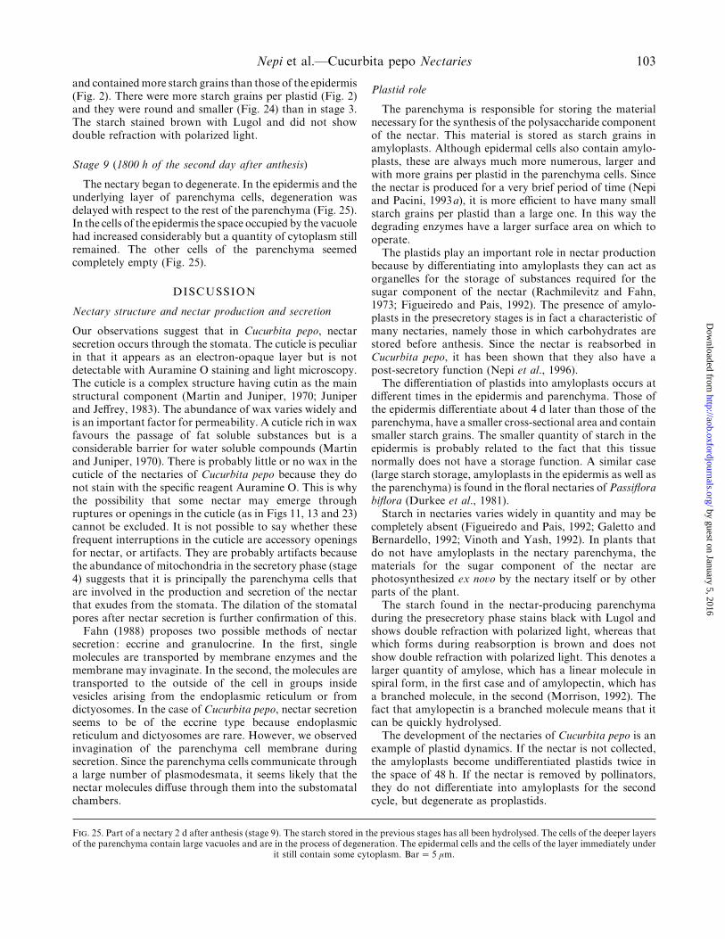
and contained more starch grains than those of the epidermis(Fig. 2). There were more starch grains per plastid (Fig. 2)and they were round and smaller (Fig. 24) than in stage 3.The starch stained brown with Lugol and did not showdouble refraction with polarized light.
Stage 9 (1800 h of the second day after anthesis)
The nectary began to degenerate. In the epidermis and theunderlying layer of parenchyma cells, degeneration wasdelayed with respect to the rest of the parenchyma (Fig. 25).In the cells of the epidermis the space occupied by the vacuolehad increased considerably but a quantity of cytoplasm stillremained. The other cells of the parenchyma seemedcompletely empty (Fig. 25).
DISCUSSION
Nectary structure and nectar production and secretion
Our observations suggest that in Cucurbita pepo, nectarsecretion occurs through the stomata. The cuticle is peculiarin that it appears as an electron-opaque layer but is notdetectable with Auramine O staining and light microscopy.The cuticle is a complex structure having cutin as the mainstructural component (Martin and Juniper, 1970; Juniperand Jeffrey, 1983). The abundance of wax varies widely andis an important factor for permeability. A cuticle rich in waxfavours the passage of fat soluble substances but is aconsiderable barrier for water soluble compounds (Martinand Juniper, 1970). There is probably little or no wax in thecuticle of the nectaries of Cucurbita pepo because they donot stain with the specific reagent Auramine O. This is whythe possibility that some nectar may emerge throughruptures or openings in the cuticle (as in Figs 11, 13 and 23)cannot be excluded. It is not possible to say whether thesefrequent interruptions in the cuticle are accessory openingsfor nectar, or artifacts. They are probably artifacts becausethe abundance of mitochondria in the secretory phase (stage4) suggests that it is principally the parenchyma cells thatare involved in the production and secretion of the nectarthat exudes from the stomata. The dilation of the stomatalpores after nectar secretion is further confirmation of this.
Fahn (1988) proposes two possible methods of nectarsecretion: eccrine and granulocrine. In the first, singlemolecules are transported by membrane enzymes and themembrane may invaginate. In the second, the molecules aretransported to the outside of the cell in groups insidevesicles arising from the endoplasmic reticulum or fromdictyosomes. In the case of Cucurbita pepo, nectar secretionseems to be of the eccrine type because endoplasmicreticulum and dictyosomes are rare. However, we observedinvagination of the parenchyma cell membrane duringsecretion. Since the parenchyma cells communicate througha large number of plasmodesmata, it seems likely that thenectar molecules diffuse through them into the substomatalchambers.
F. 25. Part of a nectary 2 d after anthesis (stage 9). The starch stored in the previous stages has all been hydrolysed. The cells of the deeper layersof the parenchyma contain large vacuoles and are in the process of degeneration. The epidermal cells and the cells of the layer immediately under
it still contain some cytoplasm. Bar¯ 5 µm.
Plastid role
The parenchyma is responsible for storing the materialnecessary for the synthesis of the polysaccharide componentof the nectar. This material is stored as starch grains inamyloplasts. Although epidermal cells also contain amylo-plasts, these are always much more numerous, larger andwith more grains per plastid in the parenchyma cells. Sincethe nectar is produced for a very brief period of time (Nepiand Pacini, 1993a), it is more efficient to have many smallstarch grains per plastid than a large one. In this way thedegrading enzymes have a larger surface area on which tooperate.
The plastids play an important role in nectar productionbecause by differentiating into amyloplasts they can act asorganelles for the storage of substances required for thesugar component of the nectar (Rachmilevitz and Fahn,1973; Figueiredo and Pais, 1992). The presence of amylo-plasts in the presecretory stages is in fact a characteristic ofmany nectaries, namely those in which carbohydrates arestored before anthesis. Since the nectar is reabsorbed inCucurbita pepo, it has been shown that they also have apost-secretory function (Nepi et al., 1996).
The differentiation of plastids into amyloplasts occurs atdifferent times in the epidermis and parenchyma. Those ofthe epidermis differentiate about 4 d later than those of theparenchyma, have a smaller cross-sectional area and containsmaller starch grains. The smaller quantity of starch in theepidermis is probably related to the fact that this tissuenormally does not have a storage function. A similar case(large starch storage, amyloplasts in the epidermis as well asthe parenchyma) is found in the floral nectaries of Passiflorabiflora (Durkee et al., 1981).
Starch in nectaries varies widely in quantity and may becompletely absent (Figueiredo and Pais, 1992; Galetto andBernardello, 1992; Vinoth and Yash, 1992). In plants thatdo not have amyloplasts in the nectary parenchyma, thematerials for the sugar component of the nectar arephotosynthesized ex no�o by the nectary itself or by otherparts of the plant.
The starch found in the nectar-producing parenchymaduring the presecretory phase stains black with Lugol andshows double refraction with polarized light, whereas thatwhich forms during reabsorption is brown and does notshow double refraction with polarized light. This denotes alarger quantity of amylose, which has a linear molecule inspiral form, in the first case and of amylopectin, which hasa branched molecule, in the second (Morrison, 1992). Thefact that amylopectin is a branched molecule means that itcan be quickly hydrolysed.
The development of the nectaries of Cucurbita pepo is anexample of plastid dynamics. If the nectar is not collected,the amyloplasts become undifferentiated plastids twice inthe space of 48 h. If the nectar is removed by pollinators,they do not differentiate into amyloplasts for the secondcycle, but degenerate as proplastids.
by guest on January 5, 2016http://aob.oxfordjournals.org/
Dow
nloaded from

104 Nepi et al.—Cucurbita pepo Nectaries
Nectar resorption
Amyloplasts form in the epidermis at stage 6 and then inthe parenchyma at stage 7. In these stages the mitochondriaincrease in the epidermis and decrease in the parenchyma.This is why we consider that in the process of resorption, itis the epidermis that plays a principal role whereas theparenchyma acts as a temporary storage site for materialderived from the sugar component of the nectar. This is alsoconfirmed by the presence of invaginations of the plasmamembrane of the epidermal cells in stage 6, a feature typicalof transfer cells. Transfer cells are involved in the processesof secretion and absorption (Pate and Gunning, 1972).
Nectar production involves a large expenditure of energyfor the plant (Southwick, 1984; Pyke, 1991). Despite thisinvestment, it may happen that the pollinators do notcollect the nectar (e.g. due to bad weather) or at least not allof it. In this case, the plant could lose all or part of theenergy invested. The process of reabsorption enables thisenergy to be recovered.
ACKNOWLEDGEMENTS
This research was supported by a grant from Ministerodell’Universita' e della Ricerca Scientifica e Tecnologica(M.U.R.S.T.) 40%, Italy.
LITERATURE CITED
Baker DA, Hall JL, Thorpe JR. 1978. A study of the extrafloralnectaries of Ricinus communis. New Phytologist 81 : 129–137.
Baker HG, Baker I. 1983. A brief historical review of the chemistry offloral nectar. In: Bentley B, Elias T, eds. The biology of nectaries.New York: Columbia University Press, 126–152
Cruden RW, Hermann SM, Peterson S. 1983. Patterns of nectarproduction and plant animal coevolution. In: Bentley B, Elias T,eds. The biology of nectaries. New York: Columbia UniversityPress, 126–152.
Davis AR, Gunning BES. 1992. The modified stomata of the floralnectary of Vicia faba L. 1. Development, anatomy and ultra-structure. Protoplasma 166 : 134–152.
Durkee LT. 1982. The floral and extra-floral nectaries of Passiflora. II.The extra-floral nectary. American Journal of Botany 69 : 1420–1428.
Durkee LT, Gaal DJ, Reisner WH. 1981. The floral and extra-floralnectaries of Passiflora. I.The floral nectary. American Journal ofBotany 68 : 453–462.
Eleftheriou EP, Hall JL. 1983. The extra-floral nectaries of cotton. I.Fine structure of the secretory papillae. Journal of ExperimentalBotany 34 : 103–119.
Faegri K, van der Pijl L. 1979. The principles of pollination ecology. 3rdrevised edition. Oxford: Pergamon Press.
Fahn A. 1979. Secretory tissues in plants. London: Academic Press.Fahn A. 1987. Extrafloral nectaries of Sambucus nigra L. Annals of
Botany 60 : 299–308.
Fahn A. 1988. Secretory tissues in vascular plants. New Phytologist 108 :229–257.
Figueiredo CAS, Pais SM. 1992. Ultrastructural aspects of the nectaryspur of Limodorum aborti�um (L) Sw. (Orchidaceae). Annals ofBotany 70 : 325–331.
Freeman CE, Head KC. 1990. Temperature and sucrose composition offloral nectars in Ipomopsis longiflora under field conditions.Southwestern Naturalist 35 :423–426.
Galetto L, Bernardello LM. 1992. Extrafloral nectaries that attract antsin Bromeliaceae: structure and nectar composition. CanadianJournal of Botany 70 : 1101–1106.
Gottsberger G, Arnold T, Liskens HF. 1990. Variation in floral nectaramino acids with aging of flowers, pollen contamination, andflower damage. Israel Journal of Botany 39 : 167–176.
Grout BWW, Williams A. 1980. Extrafloral nectaries of Dioscorearotundata Poir. : their structure and secretions. Annals of Botany46 : 255–258.
Heslop-Harrison Y. 1977. The pollen stigma interaction: pollen tubepenetration in Crocus. Annals of Botany 41 : 913–922.
Hilu KW, Randall JL. 1984. Convenient method for studying grass leafepidermis. Taxon 33 : 413–415.
Johansen DA. 1940. Plant microtechnique. New York: McGraw-Hill.Juniper BE, Jeffrey CE. 1983. Plant surfaces. London: Edward Arnold.Lawton JH, Heads PA. 1984. Bracken, ants and extrafloral nectaries. I.
The components of the system. Journal of Animal Ecology 53 :995–1014.
Marden JH. 1984. Intrapopulation variation in nectar secretion inImpatiens capensis. Oecologia 63 : 418–422.
Martin JT, Juniper BE. 1970. The cuticles of plants. Edinburgh: EdwardArnold.
Morrison WR 1992. Analysis of cereal starches in seed analysis. In:Linskens HF, Jackson JF, eds. Modern methods of plant analysis.Volume 14. Berlin: Springer-Verlag: 129–215.
Nepi M, Pacini E. 1993a. Pollination, pollen viabilty and pistilreceptivity in Cucurbita pepo. Annals of Botany 72 : 527–536.
Nepi M, Pacini E. 1993b. First observations on nectaries and nectar ofCucurbita pepo. Giornale Botanico Italiano 127 : 1208–1210.
Nepi M, Pacini E, Willemse MTM. 1996. Nectary biology of Cucrbitapepo : ecophysiological aspects. Acta Botanica Neerlandica 45 :41–54.
O’Brien TP, McCully ME. 1981. The study of plant structure—principlesand selected methods. Melbourne: Thermacarphi Pty. Ltd.
Pate JS, Gunning BES. 1972. Transfer cells. Annual Re�iew of PlantPhysiology 23 : 173–196.
Pearse AGE. 1968. Histochemistry—theoretical and applied. I. London:Churchill.
Pyke GH. 1991. What does it cost a plant to produce floral nectar?Nature 350 : 58–59.
Rachmilevitz T, Fahn A. 1973. Ultrastructure of nectaries of Vinca roseaL., Vinca major L. and Citrus sinensis Osbeck cv. Valencia and itsrelation to the mechanism of nectar secretion. Annals of Botany37 : 1–9.
Southwick EE. 1984. Photosynthate allocation to floral nectar : aneglected energy investment. Ecology 65 : 1775–1779.
Vinoth T, Yash D. 1992. Structure and biology of nectaries in Tabebuiaserratifolia Nichols (Bignoniaceae). Botanical Journal of theLinnean Society 109 : 395–400.
Wyatt R, Broyles SB, Derda GS. 1992. Enviromental influences onnectar production in milkweeds (Asclepias syriaca and A. exaltata).American Journal of Botany 79 : 636–642.
Zer H, Fahn A. 1992. Floral nectaries of Rosmarinus officinalis L.Structure, ultrastructure and nectar secretion. Annals of Botany70 : 391–397.
by guest on January 5, 2016http://aob.oxfordjournals.org/
Dow
nloaded from