Renal Medullary Nitric Oxide Deficit of Dahl S Rats Enhances Hypertensive Actions of Angiotensin II
25
R-00461-2001.R1 Renal Medullary Nitric Oxide Deficit of Dahl S Rats Enhances Hypertensive Actions of Angiotensin II Mátyás Szentiványi Jr.*, Ai-Ping Zou, David L. Mattson, Paulo Soares, Carol Moreno, Richard J. Roman, and Allen W. Cowley Jr. *Clinical Research Department-2 nd Institute of Physiology, Semmelweis University of Medicine, Budapest, Hungary; Department of Physiology, Medical College of Wisconsin, Milwaukee, Wisconsin Szentiványi Jr.: Impaired renal medullary NO in Dahl S rats Correspondence: Allen W. Cowley Jr., Ph.D. Department of Physiology, Medical College of Wisconsin 8701 Watertown Plank Rd. P.O.B. 26509 Milwaukee WI 53226-0509 Fax: (414) 456-6546 Phone: (414) 456-8266 e-mail: [email protected] Copyright 2002 by the American Physiological Society. AJP-Regu Articles in PresS. Published on April 4, 2002 as DOI 10.1152/ajpregu.00461.2001
Transcript of Renal Medullary Nitric Oxide Deficit of Dahl S Rats Enhances Hypertensive Actions of Angiotensin II

R-00461-2001.R1
Renal Medullary Nitric Oxide Deficit of
Dahl S Rats Enhances Hypertensive Actions of Angiotensin II
Mátyás Szentiványi Jr.*, Ai-Ping Zou, David L. Mattson, Paulo Soares,
Carol Moreno, Richard J. Roman, and Allen W. Cowley Jr.
*Clinical Research Department-2nd Institute of Physiology, Semmelweis University of Medicine, Budapest, Hungary; Department of Physiology, Medical College of Wisconsin, Milwaukee,
Wisconsin
Szentiványi Jr.: Impaired renal medullary NO in Dahl S rats
Correspondence:Allen W. Cowley Jr., Ph.D. Department of Physiology, Medical College of Wisconsin 8701 Watertown Plank Rd. P.O.B. 26509 Milwaukee WI 53226-0509 Fax: (414) 456-6546 Phone: (414) 456-8266 e-mail: [email protected]
Copyright 2002 by the American Physiological Society.
AJP-Regu Articles in PresS. Published on April 4, 2002 as DOI 10.1152/ajpregu.00461.2001
2
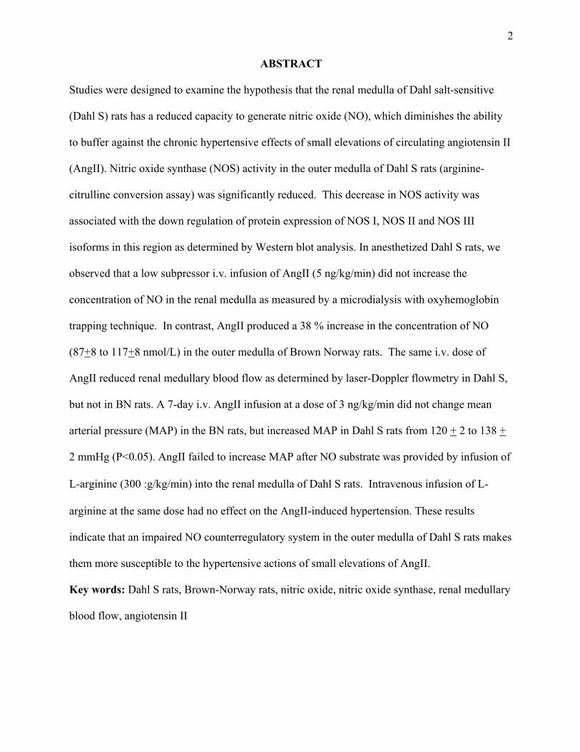
ABSTRACT
Studies were designed to examine the hypothesis that the renal medulla of Dahl salt-sensitive
(Dahl S) rats has a reduced capacity to generate nitric oxide (NO), which diminishes the ability
to buffer against the chronic hypertensive effects of small elevations of circulating angiotensin II
(AngII). Nitric oxide synthase (NOS) activity in the outer medulla of Dahl S rats (arginine-
citrulline conversion assay) was significantly reduced. This decrease in NOS activity was
associated with the down regulation of protein expression of NOS I, NOS II and NOS III
isoforms in this region as determined by Western blot analysis. In anesthetized Dahl S rats, we
observed that a low subpressor i.v. infusion of AngII (5 ng/kg/min) did not increase the
concentration of NO in the renal medulla as measured by a microdialysis with oxyhemoglobin
trapping technique. In contrast, AngII produced a 38 % increase in the concentration of NO
(87+8 to 117+8 nmol/L) in the outer medulla of Brown Norway rats. The same i.v. dose of
AngII reduced renal medullary blood flow as determined by laser-Doppler flowmetry in Dahl S,
but not in BN rats. A 7-day i.v. AngII infusion at a dose of 3 ng/kg/min did not change mean
arterial pressure (MAP) in the BN rats, but increased MAP in Dahl S rats from 120 + 2 to 138 +
2 mmHg (P<0.05). AngII failed to increase MAP after NO substrate was provided by infusion of
L-arginine (300 g/kg/min) into the renal medulla of Dahl S rats. Intravenous infusion of L-
arginine at the same dose had no effect on the AngII-induced hypertension. These results
indicate that an impaired NO counterregulatory system in the outer medulla of Dahl S rats makes
them more susceptible to the hypertensive actions of small elevations of AngII.
Key words: Dahl S rats, Brown-Norway rats, nitric oxide, nitric oxide synthase, renal medullary
blood flow, angiotensin II

3
There has been persistent evidence, albeit indirect, that Dahl salt-sensitive (Dahl S) rats
may have a reduced ability to produce nitric oxide (NO) in response to different stimuli such as a
high salt diet and vasoconstrictors (5,6,14,16,27,33). These findings are consistent with
observations that chronic oral or i.v. administration of L-arginine (L-arg) prevents salt-induced
hypertension in Dahl S rats and normalizes the pressure-natriuresis relationship (31). There is
evidence of a reduced ability of NO to block chloride reabsorption in the thick ascending limb in
Dahl S rats, which could account in part for the salt-sensitivity of blood pressure in this strain
(13). Consistent with these findings, it was observed that chronic infusion of L-arg into the renal
medullary interstitial space alone was able to prevent salt-induced hypertension in Dahl S rats at
doses that have no effect when infused intravenously (23,24). Taken together, these data suggest
that the renal medulla of Dahl S rats may have both a reduced ability to produce NO in response
to various stimuli as well as with a reduced responsiveness to NO, but this has not been directly
ascertained.
NO production is known to play an important role in the regulation of medullary blood
flow (MBF) and arterial blood pressure (8,22,26). Several vasoconstrictors, such as angiotensin
II (AngII), arginine vasopressin (AVP), and norepinephrine can stimulate the release of
medullary NO, which in turn buffers the vascular actions of these agonists (30,37,42,46).
Chronic AngII infusion in captopril treated rats was shown to increase NOS III protein levels in
homogenates of whole kidney of Sprague Dawley rats (15). A moderate reduction of NOS
activity within the renal medulla resulting from the infusion of a subpressor dose of L-NAME
into the renal medullary interstitial space, was shown to enhance the medullary vasoconstrictor
effects to small subpressor elevations of plasma AngII (46). Chronic i.v. administration of
AngII or AVP administered at a dose that normally does not raise arterial pressure, produced a
sustained hypertension in rats with blunted medullary NOS activity (36,37).

4
Recently, we found that mRNA expression of NOS I and NOS III in the outer medulla of the
kidney was significantly less in Dahl S rats compared to BN rats (41). Furthermore, these Dahl S rats
exhibited sustained hypertension in response to small, normally subpressor elevations of plasma
AVP. It was these observations that motivated the design of the present study to more completely
characterize the production of NO.
Therefore, studies were carried out to determine whether Dahl S rats: 1) exhibit a reduced
medullary expression of NOS protein and NOS enzyme activity; 2) exhibit a reduced elevation of
medullary NO in response to AngII administration; 3) exhibit an exaggerated reduction of MBF in
the face of small subpressor elevations of circulating AngII; and 4) would respond to AngII
chronically like Brown Norway (BN) rats if L-arg was administered into the renal medullary
interstitial space (e.g. Would AngII-induced hypertension be prevented?).
In these studies, inbred strains of Dahl S rats were compared to an inbred strain of
normotensive, salt-insensitive BN rats. These two strains were chosen because extensive genetic and
functional data have now been obtained from genetic linkage studies using an intercross of these two
strains of rats, both of which were inbred in our department (7,34). Although comparisons between
Dahl S and Dahl R strains could have served the same purpose, the aim of the present study was to
compare a rat strain in which the renal medulla had a reduced ability to produce NO to a strain in
which administration of AngII resulted in a clear elevation of medullary NO concentrations.
Both rat strains were maintained on a low salt (0.4 %) diet throughout all studies. AngII was infused
intravenously for a week to Dahl S and BN rats at 3 ng/kg/min, a dose that was shown to be non-
pressor in Sprague Dawley rats (36). The ability of systemic administration of ANGII administered
intravenously at a subpressor dose to change MBF and stimulate medullary NO production was
determined first in anesthetized rats (46). Chronically instrumented rats were then studied to
determine whether blunted medullary NO production in Dahl S rats would sensitize these animals to

5
the hypertensive actions of small elevations of circulating AngII (3ng/kg/min infused i.v. for one
week) and whether a continuous administration of L-arg into the renal medulla of Dahl S rats would
return AngII hyper-responsiveness to normal.
METHODS
Twelve week old male inbred Dahl S(SS/JrHsdMcw) and BN(BN/SsMcw) rats obtained from
colonies maintained at the Medical College of Wisconsin (7) were studied. Rats were given free
access to tap water and maintained throughout the study on low salt rat chow (0.4% NaCl; Dyets,
Inc., Bethlehem, PA).
NOS enzyme activity in the renal cortex, outer and inner medulla of Dahl S (N=7) and BN
(N=7) rats. NOS activity in renal homogenates was determined by measuring the conversion of L-
[3H]-arginine to citrulline using a modification of the radioactive HPLC method originally described
by Carlberg (1) and modified by Wu et al. (39). The kidneys from Dahl S and BN rats were rapidly
removed from pentobarbital anesthetized rats, immersed in ice-cold saline, and the cortex, outer
medulla and inner medulla separated. The tissue was homogenized in 3 volumes of a 20 mM HEPES
buffer containing 1 mM EDTA, 1 mM DTT, 2 mM pepstatin, 1 mM leupeptin and 0.1 mM PMSF.
The homogenate was centrifuged at 3,000 g for 5 minutes and 11,000 g for 15 minutes. The
supernatant was mixed (2:1) with a suspension of Dowex-50W resin in water at pH 5.5 (HCR-W2,
ionic form: Sigma Chemical Corporation, St Louis, MO) for 10 minutes followed by a brief
centrifugation (3000 g) to remove endogenous L-arginine from the homogenate (25). This procedure
removed >97 % of 3H-arginine counts added to the samples to monitor the efficiency of the arginine
removal. It is important to note that the Dowex-50W resin had to be extensively washed with water
to remove residual acidity prior to use. Aliquots of the Dowex-treated homogenates (100 g protein
for inner medulla, 250 g for outer medulla and cortex) were incubated with L-[3H]-arginine (0.2
Ci, 20 M) in 100 l of 20 mM HEPES buffer containing 2 mM, CaCl2, 1 mM NADPH, 1.5 g/ml

6
calmodulin, 2.5 M FAD, 1 M FMN, 20 M tetrahydrobiopterin and 0.4 mM L-proline to inhibit
arginase activity (2). Inner medullary samples were incubated for 30 minutes at 37o C, outer medulla
for 60 minutes and cortical tissue incubated for 90 minutes. The reactions were stopped by the
addition of 50 l of 20 mM EGTA. The conversion of L-arginine to L-citrulline after derivatization
of the sample with 2 volumes of o-pthalaldehyde reagent (P-0532) was determined by RP-HPLC
using a 4.6 X 25cm LC-18-DB column (Supelco) which was eluted isocratically at a rate of 1.5
ml/min using 11.5% methanol, 11.5% acetonitrile, 1% tetrahydrofuran and 0.1 M KH2PO4 (pH 5.9)
as the mobile phase. Products were monitored using a radioactive flow detector (Radiomatic
Instruments, Tampa, FL) and the activity determined from the ratio of counts in the citrulline peak to
the sum of the counts in the arginine and citrulline peaks.
NOS protein expression in the outer and inner medulla of Dahl S (N=5) and BN rats (N=5).
Expression of NOS protein was measured in homogenates of renal inner and outer medulla. Aliquots
of 50 g of the renal outer medullary protein, and 25 g of inner medullary homogenate protein
were loaded into each lane and size separated by electrophoresis through an 8% SDS-PAGE gel. The
proteins were transferred onto a nitrocellulose membrane (Bio-Rad), blocked overnight at 4 C, then
incubated with a monoclonal anti-NOS I antibody (which cross reacts with NOS II; Transduction
Labs) and a monoclonal anti-NOS III antibody (Transduction Labs) for 2 hours at a 1:1000 dilution
as we have previously described (21). Bound primary antibodies were detected with a horseradish
peroxidase labeled secondary antibody (goat anti-mouse IgG; 1:1000; 2 hr) and enhanced
chemiluminescence (SuperSignal, Pierce Chemical). The band intensities were quantitated using
densitometry (Scion Image, Scion Corporation).
Comparison of renal medullary [NO], MAP, and MBF responses to AngII (5 ng/kg/min) in
anesthetized Dahl S (N=6) and BN (N=6) rats. Rats were anesthetized with Inactin (50 mg/kg i.p.)
and ketamine (33 mg/kg i.m.) and surgically prepared for measurement of renal medullary NO

7
concentration using the microdialysis/hemoglobin trapping technique described in our previous
studies (43,46). The microdialysis probe was perfused at a rate of 2.3 l/min during a 2 hour
equilibration period followed by two 30-minute control collection periods (70 l/30 min). Then
AngII was infused i.v. at a rate of 5 ng/kg/min. for a one hour equilibration period and dialysate fluid
was collected during experimental periods. NO-dependent conversion of oxyhemoglobin to
methemoglobin in the dialysate samples was determined using a wavelength scanning mode of a DU-
640 Beckman spectrophotometer (Beckman Instruments, Inc.). MBF was measured using an optical
fiber (500 micron; Edmund Scientific) implanted into the renal medulla to a depth of 5.5 millimeters.
MBF changes were determined using a Perimed PeriFlux PF3 Flowmeter (Perimed, Sweden) as
described in previous studies (20,24,26).
Chronic measurement of MAP in Dahl S (N=6) and BN rats (N=6). Rats were anesthetized with
xylazine (2 mg/kg i.p.) and ketamine (30 mg/kg i.p.) for implantation of indwelling arterial and
venous catheters which were tunneled subcutaneously to the back of the neck, and exited through a
flexible spring for attachment to a swivel over the home cage as described in detail previously (7,23).
Rats received a continuous saline i.v. infusion at a rate of 0.25 ml/hour, the rate of delivery
subsequently used for the AngII. One week following surgery, daily 2 hr. measurements of MAP
were begun using an on-line data collection and analysis system (7). After 3 days of stable and
reproducible MAP measurements, AngII (Sigma Chemical) was infused i.v. at a dose of 3 ng/kg/min.
Infusion of AngII was continued for 7 days with daily MAP measurements made throughout and 2
days following the cessation of AngII. Previous studies in our laboratory have demonstrated that this
dose does not elevate arterial pressure when infused i.v. for one week in uninephrectomized Sprague
Dawley rats (36).
Effect of medullary versus intravenous L-arginine (L-arg) administration on AngII-induced
hypertension in Dahl S (N=5) rats. To assess whether a deficiency in the formation of NO in the

8
renal medulla sensitizes Dahl S rats to the chronic hypertensive effects of AngII, a protocol was
designed in which medullary NO production was enhanced by local renal medullary infusion of L-
arg. Rats were uninephrectomized to avoid interactions with the contralateral kidney. One week later,
femoral arterial and venous catheters as well as a renal medullary interstitial catheter were implanted
under anesthesia (22,23,24). Following another week of recovery, MAP was measured for 2 hours
daily. Following three stable control days, the renal medullary interstitial infusion of L-arg was
begun at a dose of 300 g/kg/min in a volume of 8 l/min. This dose of L-arg administered into the
renal medulla was shown previously to increase medullary [NO] (43) while having no effect on basal
MBF (23,24). After 3 days of L-arg infusion, the i.v. AngII infusion (3 ng/kg/min.) was started and
continued for 7 days followed by 2 post-control days.
Another instrumented group of uninephrectomized Dahl S (N=5) rats was studied as a control
group to determine whether the renal medullary infusion of L-arg alone could be exerting anti-
hypertensive effects by escape from the kidney and recirculation in the systemic vasculature. In this
group of Dahl S rats, L-arg was infused intravenously at the same rate (300 g/kg/min) while
isotonic saline was administered into the medullary interstitial space. On the third day of i.v. L-arg
infusion, AngII (3 ng/kg/min) was added to the i.v. infusate for a 7 day period followed by 2 post
control days.
Statistics. Data are presented as mean values standard error of the mean (SEM). For statistical
comparisons of the chronic experiments, one-way analysis of variance (ANOVA) for repeated
measures was used and Tukey's multiple range test as a post hoc analysis was carried out. One-way
ANOVA with Tukey’s multiple range test was used for all other comparisons. All statistical analyses
were performed on the raw data. P<0.05 was considered as statistically significant.
RESULTS
NOS enzyme activity in the renal cortex, outer and inner medulla of Dahl S and BN rats. As

9
seen in Figure 1, the NOS enzyme activity of the Dahl S rats was nearly one-third that of the BN rats
in the outer medulla (1.8+0.4 and 11.6+1 pmoles citrulline/min-1.mg protein). No differences between
the two strains were found in NOS activity in inner medullary homogenates (60.0+5.6 in BN vs.
49.0+3.0 in Dahl S, pmoles citrulline/min-1.mg protein). NOS activity in aliquots of renal cortex
homogenates was similar in the two strains (0.9+0.1. vs. 0.9+0.1 pmoles citrulline/min-1.mg protein
in Dahl S and BN respectively).
NOS protein expression in the outer and inner medulla of Dahl S and BN rats. As can be
seen in Figure 2, the levels of NOS I, NOS II and NOS III protein expressed was significantly lower
in the outer medulla of Dahl S than in BN rats, while no significant differences between the strains
were observed in the inner medulla. Previous studies have indicated little or no expression of any of
the NOS isoforms in homogenates of cortex (21).
Comparison of renal medullary [NO], MAP, and MBF responses to AngII in
anesthetized Dahl S and BN rats. As seen in Figure 3, i.v. AngII infusion (5 ng/kg/min) given
acutely produced small but consistent reductions of the MBF signal (~10 %) in anesthetized Dahl S
rats (P < 0.05), but not in BN rats. These changes were consistent with the results of the
microdialysis studies presented in Figure 3 (lower panel) indicating that AngII had no effect on
medullary [NO] in Dahl S rats, while BN rats exhibited a 38 % [NO] increase (p<0.05). There was no
difference in the resting medullary [NO] levels of Dahl S and BN rats (89 3 vs. 87 8 nMol/L,
respectively), suggesting that the deficit was due to the inability of Dahl S rats to increase [NO] with
AngII stimulation. The low dose of AngII used in this study did not change MAP in either strain of
rat in these acute experiments (MAP: 131 3 vs.134 4 in Dahl S; 114 3 vs. 118 4 mmHg in BN).
Chronic measurement of MAP in i.v. AngII-infused Dahl S and BN rats. A comparison
of the pressor effects of a chronic i.v. infusion of AngII (3 ng/kg/min) in Dahl S and BN rats is
presented in Figure 4. Ang II had no effect on MAP over 7 days in BN rats but increased MAP

10
significantly in Dahl S rats. The pressure increase was sustained throughout the AngII infusion,
reaching its highest level after 7 days at which time MAP was 18 mmHg higher than during the
control days. MAP returned to within control level 2 days following cessation of the infusion of
AngII.
Effect of medullary versus intravenous L-arg administration on AngII-induced
hypertension in Dahl S rats. As seen on Figure 5, a renal medullary interstitial infusion of L-arg to
increase NO levels completely blocked the pressor response to AngII in Dahl S rats. The same dose
of L-arg infused intravenously did not prevent this AngII-induced hypertension in this strain of rats.
DISCUSSION
The present study advances our understanding of the role of renal medullary NO in the
development of salt-induced hypertension in the Dahl S rat in several ways. First, it provides direct
evidence that in a naturally occurring animal model, the reduced ability of the renal medulla to
produce NO can lead to the enhancement of the vasoconstrictor actions of AngII. Second, this study
has demonstrated that if this NO deficit was corrected in the Dahl S rat by the local administration of
L-arg, the hypertensive actions of subpressor amounts of AngII converted the Dahl S phenotype to
that of the BN rat strain.
Evidence and consequences of reduced medullary NOS activity and reduced ability to generate
NO responses to AngII stimulation in Dahl S rats. We have shown previously that lowering of
medullary NOS activity with a pharmacological inhibitor (L-NAME) in Sprague Dawley rats led to
hypersensitivity of the medullary vasculature to AngII (46) and to a salt-sensitive form of
hypertension (21). It was also shown that salt-induced hypertension in Dahl S rats could be
prevented by medullary L-arg administration, suggesting indirectly that NO production may be
reduced in the Dahl S rat (24). The present study, however, provides direct evidence that, with NOS
mRNA reduced in the Dahl S rat (41), there is also a reduction in the protein expression of all three

11
NOS isoforms most notably in the renal outer medulla. This is turn was accompanied by a
significant reduction in NOS enzyme activity and medullary NO tissue concentrations in response to
AngII.
A blunted ability of the renal medulla of Dahl S rats to generate NO in response to AngII
stimulation is of particular interest because this would be expected to reduce the excretion of sodium
and thereby shift the chronic pressure relationship (21) through two mechanisms. First, by reducing
sodium reabsorption in the medullary thick ascending limb (mTAL) as shown by Oritz and Garvin
(28). NO reduction may indeed account in part for observations that sodium reabsorption by the
thick ascending limb is elevated in Dahl S rats (18,32,46). This, in turn, may be related to the
production of 20-HETE in these epithelial cells since this cytochrome P450 product of arachadonic
acid metabolism has vasoconstrictor actions and reduces sodium reabsorption in the medullary thick
ascending limb of Henle (44). This same study showed that Dahl S rats have a reduced capacity to
produce 20-HETE in the outer medulla (44). Since NO binds to heme in P4504A enzymes and
inhibits the formation of 20-HETE (35), some of the observed response differences between Dahl S
and BN could be influenced by these complex interactions. Preliminary studies in our laboratory also
suggest that AngII increases oxygen free radical production in the outer medulla that would, in part,
buffer the actions of AngII stimulated NO production (12). NADH-oxidase levels are higher in the
medulla of Sprague Dawley rats compared to the cortex (45) and it is possible that Dahl S rats exhibit
greater levels of oxidative stress than BN rats. Thus, there appears to exist several important
pathways whereby the reduction of NOS activity in the outer medulla of Dahl S rats could modify the
vasoconstrictor effects of plasma AngII.
The second mechanism leading to decreased sodium excretion could be by the reduction of
medullary blood flow via enhanced vasoconstrictor effects of AngII in the presence of reduced levels
of NO. The rich density of contractile pericytes surrounding the vasa recta vessels in the outer
12
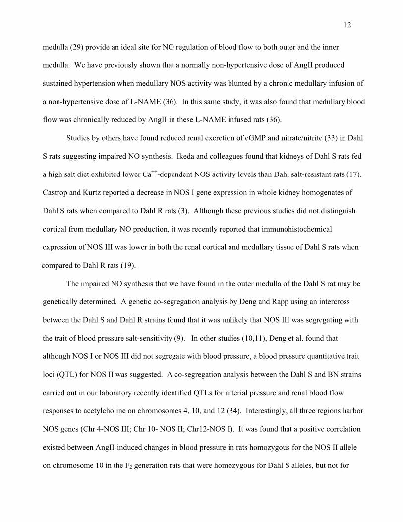
medulla (29) provide an ideal site for NO regulation of blood flow to both outer and the inner
medulla. We have previously shown that a normally non-hypertensive dose of AngII produced
sustained hypertension when medullary NOS activity was blunted by a chronic medullary infusion of
a non-hypertensive dose of L-NAME (36). In this same study, it was also found that medullary blood
flow was chronically reduced by AngII in these L-NAME infused rats (36).
Studies by others have found reduced renal excretion of cGMP and nitrate/nitrite (33) in Dahl
S rats suggesting impaired NO synthesis. Ikeda and colleagues found that kidneys of Dahl S rats fed
a high salt diet exhibited lower Ca++-dependent NOS activity levels than Dahl salt-resistant rats (17).
Castrop and Kurtz reported a decrease in NOS I gene expression in whole kidney homogenates of
Dahl S rats when compared to Dahl R rats (3). Although these previous studies did not distinguish
cortical from medullary NO production, it was recently reported that immunohistochemical
expression of NOS III was lower in both the renal cortical and medullary tissue of Dahl S rats when
compared to Dahl R rats (19).
The impaired NO synthesis that we have found in the outer medulla of the Dahl S rat may be
genetically determined. A genetic co-segregation analysis by Deng and Rapp using an intercross
between the Dahl S and Dahl R strains found that it was unlikely that NOS III was segregating with
the trait of blood pressure salt-sensitivity (9). In other studies (10,11), Deng et al. found that
although NOS I or NOS III did not segregate with blood pressure, a blood pressure quantitative trait
loci (QTL) for NOS II was suggested. A co-segregation analysis between the Dahl S and BN strains
carried out in our laboratory recently identified QTLs for arterial pressure and renal blood flow
responses to acetylcholine on chromosomes 4, 10, and 12 (34). Interestingly, all three regions harbor
NOS genes (Chr 4-NOS III; Chr 10- NOS II; Chr12-NOS I). It was found that a positive correlation
existed between AngII-induced changes in blood pressure in rats homozygous for the NOS II allele
on chromosome 10 in the F2 generation rats that were homozygous for Dahl S alleles, but not for

13
those homozygous for the BN allele. These data indicated that at least one NOS loci (NOS II) could
confer a distinct pressure phenotype to these rats (34). Chen et al. (4) recently identified a single
nucleotide transversion in the NOS II gene that produced an amino acid substitution (S714P) between
the FAD and FMN binding sites and a restriction fragment length polymorphism, which was present
only in Dahl S rats. In subsequent studies from this group, expression of this mutation in COS-7 cells
showed a decrease in the Vmax but no change in Km of the enzyme for L-arg of the mutant NOS II
gene. Within the physiological concentration range of L-arg, the amount of NO produced was
reduced in cells transfected with the mutant NOSII compared to the wildtype. Accelerated
degradation of the mutant gene also appeared to contribute to the diminished NO production in cells
expressing the Dahl S NOSII gene (40).
Restoration of medullary [NO] by L-arginine prevents subpressor AngII-induced hypertension
in Dahl S rats. It is interesting that the basal NO concentrations within the renal medullary tissue,
did not differ between the Dahl S and BN rat strains as determined by the oxyhemoglobin
microdialysis method. This would suggest that there exists sufficient L-arg substrate and NOS
enzyme activity to maintain resting vascular tone in the renal medulla.
However, administration of L-arg into the renal medulla prevented AngII-induced
hypertension in the Dahl S rats. These results are consistent with the conclusion that reduced
medullary NO production in Dahl S rats contributed importantly to the AngII hypersensitivity. The
antihypertensive effects of the L-arg administration were almost certainly due to the intrarenal effects
of L-arg since intravenous administration of the same dose of L-arg failed to reduce the hypertensive
effects of AngII. The dose of L-arg administered in the present study has been shown to produce an
elevation of medullary NO concentration in Sprague Dawley rats (44).
Studies by others have found that salt-induced hypertension in Dahl S rats could be prevented
by high dose oral or systemic administration of L-arg (5,6,16,31). Miyata et al. (23,24) demonstrated

14
the importance of the renal medullary impairment of NO production in salt-induced hypertension of
Dahl S rats. Specifically, renal medullary interstitial infusion of L-arg in the same low doses used in
the present study, protected Dahl S rats from high salt-induced hypertension whereas D-arg did not, nor
did intravenous administration of the same dose of L-arg.
In summary, the present results indicate that there exists an inherited defect in the ability of the
Dahl S rat to produce NO within the outer medulla of the kidney. This defect was manifested by
reduced protein expression of all three NOS isoforms, a reduction of NOS enzyme activity, and a
failure of medullary NO concentrations to increase in response to AngII in Dahl S rats. As a
consequence, hypertension occurred in Dahl S rats with small elevations of circulating AngII that had
no effect in the Brown Norway strain.
ACKNOWLEDGMENTS
This work was supported by the National Institute of Health grant HL-29587. The authors thank
Meredith M. Skelton for careful review of the manuscript and Ryan Hagemeier and Robert Klum for
their careful analyses of NOS by HPLC. Mátyás Szentiványi, Jr. was a visiting postdoctoral fellow
from Semmelweis University of Medicine, Budapest, Hungary with support from OKTA T030245.
Paulo Soares was a visiting fellow from the Dept. Internal Medicine, State Univ. of Campenas, Brasil.
Carol Moreno is a visiting postdoctoral fellow supported by Fundacion Seneca in Spain.

15
1.
2.
3.
4.
5.
6.
7.
8.
9.
10.
REFERENCES
Carlberg M. Assay of neuronal nitric oxide synthase by HPLC determination of citrulline.
J. Neurosci. Methods. 52: 165-167, 1994.
Carajal N and Cederbaum SD. Kinetics of inhibition of rat liver and kidney arginases by
proline and branched-chain amino acids. Biochem. Biophy. Ceta. 870: 181-184, 1986.
Castrop H and Kurtz A. Differential nNOS gene expression in salt-sensitive and salt-resistant
Dahl rats. J. Hypertens. 19: 1223-1231, 2001.
Chen, PY, Gladish RD, Sanders PW. Vascular smooth muscle nitric oxide synthase anomolies
in Dahl/Rapp salt-sensitive rats. Hypertension. 31: 918-924, 1998.
Chen PY, Sanders PW. L-arginine abrogates salt-sensitive hypertension in Dahl/Rapp rats. J
Clin Invest. 88: 1559-1567, 1991.
Chen PY, Sanders PW. Role of nitric oxide synthesis in salt-sensitive hypertension in
Dahl/Rapp rats. Hypertension. 22: 812-818, 1993.
Cowley AW Jr, Stoll M, Kaldunski M, Kurth TM, Greene AS, Roman RJ. Genetically
defined risk of salt sensitivity in an intercross of Brown Norway and Dahl S rats. Physiol.
Genomics. 2: 107-115, 2000.
Cowley AW Jr. Role of the renal medulla in volume and arterial pressure regulation. Am J
Physiol. 273: R1-R15, 1997.
Deng AY, Rapp JP. Absence of linkage for "endothelial" nitric oxide synthase locus to blood
pressure in Dahl rats. Hypertension. 29: 49-52, 1997.
Deng AY, Rapp JP. Locus for the inducible, but not a constitutive, nitric oxide synthase
cosegregates with blood pressure in the Dahl salt-sensitive rat. J Clin Invest. 95: 2170-2177,
1995.

1611.
12.
13.
14.
15.
16.
17.
18.
19.
20.
Deng AY. Is the nitric oxide system involved in genetic hypertension in Dahl rats?
Kidney Int. 53: 1501-1511, 1998.
Dickhout J, Mori T, and Cowley AW Jr. Calcium, nitric oxide (NO) and superoxide
responses of outer medullary vasa recta to Angiotensin II (AII). FASEB J., in press,
2002. [Abstract]
García NH, Plato CF, Stoos BA, Garvin JL. Nitric oxide–induced inhibition of
transport by thick ascending limbs from Dahl salt-sensitive rats. Hypertension. 34: 508-
513, 1999.
He H, Kimura S, Fujisawa Y, Tomohiro A, Kiyomoto K, Aki Y, Abe Y. Dietary L-
arginine supplementation normalizes regional blood flow in Dahl-Iwai salt-sensitive rats.
Am J Hypertens. 10: 89S-93S, 1997.
Hennington BS, Zhang H, Miller TM, Granger JP, Reckelhoff JF. Angiotensin II
stimulates synthesis of endothelial nitric oxide synthase. Hypertension. 31: 283-288.
Hu L, Manning DR Jr. Role of nitric oxide in regulation of long-term pressure-
natriuresis relationship in Dahl S rats. Am J Physiol. 268: H2375-H2383, 1995.
Ikeda Y, Saito K, Kim JL, Yokoyama M. Nitric oxide synthase isoform activities in
kidney of Dahl salt-sensitive rats. Hypertension. 26: 1030-1034, 1995.
Ito O and Roman RJ. Role of 20-HETE in elevating chloride transport in the thick
ascending limb of Dahl SS/Jr rats. Hypertension. 33: 419-423, 1999.
Johnson RJ, Gordon, KL, Giachelli C, Kurth T, Skelton MM, and Cowley AW, Jr.
Tubuloinsterstitial injury and loss of nitric oxide synthases parallel the development of
hypertension in the Dahl-SS rat. J. Hyperten. 18: 1497-1505.
Lu S, Mattson DL, Roman RJ, Becker CG, Cowley AW Jr. Assessment of changes in
intrarenal blood flow in conscious rat using laser-Doppler flowmetry. Am. J. Physiol.
246: F956-F962, 1993.

1721.
22.
23.
24.
25.
26.
27.
28.
29.
30.
Mattson DL, Higgins DJ. Influence of dietary sodium intake on renal medullary nitric
oxide synthase. Hypertension. 27: 688-692, 1996a.
Mattson DL, Lu S, Nakanishi K, Papanek E, Cowley AW Jr. Effect of chronic renal
medullary nitric oxide inhibition on blood pressure. Am J Physiol. 266: H1918-H1926,
1994.
Miyata N, Cowley AW Jr. Renal intramedullary infusion of L-arginine prevents
reduction of medullary blood flow and hypertension in Dahl salt-sensitive rats.
Hypertension. 33: 446-450, 1999.
Miyata N, Zou AP, Mattson DL, Cowley AW Jr. Renal medullary interstitial infusion
of L-arginine prevents hypertension in Dahl salt-sensitive rats. Am J Physiol. 275:
R1667-R1673, 1998.N
Morindani, BA and Kline RL. Effect of endogenous L-arginine on the measurement of
nitric oxide synthase activity in the rat kidney. Can. J. Physiol. Pharmacol. 74: 1210-
1214, 1996.
Nakanishi K, Mattson DL, Cowley AW Jr. Role of renal medullary blood flow in the
development of L-NAME hypertension in rats. Am J Physiol. 268: R317-R323, 1995.
Nishida Y, Ding J, Zhou MS, Chen QH, Murakami H, Wu XZ, Kosaka H. Role of
nitric oxide in vascular hyper-responsiveness to norepinephrine in hypertensive Dahl rats.
J Hypertens. 16: 1611-1618, 1998.
Oritz PA, Garvin JL. Interaction of O2- and NO in the thick ascending limb.
Hypertension. 39 [part2]: 591-596, 2002.
Pallone TL. Vasoconstriction of outer medullary vasa recta by angiotensin II is
modulated by prostaglandin E2. Am J Physiol. 266: F850-F857, 1994.
Park F, Zou AP, Cowley AW Jr. Arginine vasopressin–mediated stimulation of nitric
oxide within the rat renal medulla. Hypertension. 32: 896-901, 1998.

1831.
32.
33.
34.
35.
36.
37.
38.
39.
40.
Patel AR, Layne S, Watts D, Kirchner KA. L-arginine administration normalizes
pressure natriuresis in hypertensive Dahl rats. Hypertension. 22: 863-869, 1993.
Roman RJ and Kaldunski ML. Enhanced chloride reabsorption in the loop of Henle in
Dahl salt-sensitive rats. Hypertension. 17: 1018-1024, 1991.
Simchon S, Manger W, Blumberg G, Brensilver J, Cortell S. Impaired renal
vasodilation and urinary cGMP excretion in Dahl salt-sensitive rats. Hypertension. 27:
653-657, 1996.
Stoll M, Cowley AW Jr, Tonellato PJ, Greene AS, Kaldunski ML, Roman RJ,
Dumas P, Schork NJ, Wang Z, Jacob HJ. A genomics-system biology map for
cardiovascular function. Science. 294 (5547): 1723-1726, 2001.
Sun CW, Alonso-Galicia M, Taheri MR, Falck FJ, Harder DR, Roman RJ. Nitric
oxide-20-hydroxyeicosatetraenoic acid interactin in the regulation of K+ channel activity
and vascular tone in renal arterioles. Circ Res. 83: 1069-1079, 1998.
Szentiványi M Jr, Maeda CY, Cowley AW Jr. Local renal medullary L-NAME
infusion enhances the effect of long-term angiotensin II treatment. Hypertension. 33:
440-445, 1999a.
Szentiványi M Jr, Park F, Maeda CY, Cowley AW Jr. Nitric oxide in the renal
medulla protects from vasopressin-induced hypertension. Hypertension. 35: 740-745,
2000.
Tan DY, Meng S, Manning DR Jr. Role of neuronal nitric oxide synthase in Dahl Salt-
Sensitive hypertension. Hypertension. 33: 456-461, 1999.
Wu F, Park F, Cowley AW Jr, Mattson DL. Quantification of nitric oxide synthase
activity in microdissected segments of the rat kidney. Am J Physiol. 276: F874-F881,
1999.
Ying WZ, Zia H, Sanders PW. Nitric oxide synthase (NOS2) mutation in Dahl/Rapp

19
41.
42.
43.
44.
45.
46.
rats decreases enzyme stability. Circ. Res. 89: 317-322, 2001.
Yuan B and Cowley AW, Jr. Evidence that reduced renal medullary nitric oxide
activity of Dahl S rats renables small elevations of arginine vasopressin to produce
sustained hypertension. Hypertension. 37: 524-528, 2001.
Zou AP, Cowley AW Jr. Alpha-2-Adrenergic receptor-mediated increase in NO
production buffers renal medullary vasoconstriction. Am J Physiol. 279: R769-R777,
2000.
Zou AP, Cowley Jr AW. Nitric oxide in renal cortex and medulla: An in vivo
microdialysis study. Hypertension. 29: 194-198, 1997.
Zou AP, Drummond HA, Roman RJ Role of 20-HETE in elevating loop chloride
reabsorption in Dahl SS/Jr rats. Hypertension 27[part 1]: 631-635, 1996.
Zou AP, Li N, and Cowley AW Jr. Production and actions of superoxide in the renal
medulla. Hypertension. 37: 547-553, 2001.
Zou AP, Wu F, Cowley AW Jr. Protective effect of angiotensin II-induced increase in
nitric oxide in the renal medullary circulation. Hypertension. 31: 271-276, 1997.

20
FIGURE LEGENDS
Figure 1. Comparison of nitric oxide synthase (NOS) activity in the inner and medulla and the cortex
of the kidney of Dahl S ( DS; black bars; N=6) and Brown Norway (BN;N=6) rats. * significant
difference from BN (P<0.05).
Figure 2. Densitometric analysis of the Western blots of the three isoforms: NOS I, NOS II, and NOS
III, using proteins isolated from homogenates of the outer medulla (lower panel) and the inner medulla
(upper panel) for the Dahl S (white bar; N=5) and Brown Norway (black bar; BN; N=5) rats.
*significant difference from BN (P<0.05).
Figure 3. Comparison of changes in medullary blood flow (MBF) and changes in medullary nitric
oxide concentration ([NO]) following one hour i.v. administration of Angiotensin II (AngII) in Dahl S
(N=6) and Brown Norway (BN; N=6) rats. * significant difference from control (P<0.05).
Figure 4: Average changes in mean arterial pressure (MAP) with i.v. infusion of Angiotensin II (AngII;
3.0 ng/kg/min) in Brown Norway rats (BN; closed circles) and in Dahl S rats (DS; open circles). *
indicates significant difference from the control days; P<0.05.
Figure 5: Average daily changes in mean arterial pressure (MAP) with i.v. infusion of angiotensin II
(AngII: 3.0 ng/kg/min) in Dahl S rats infused with L-arginine (L-Arg) administered intravenously (i.v.;
closed circles) or into the renal interstitium (r.i.; open circles). * significant difference from control;
P<0.05)

21
Figure 1:
0
20
40
60
80
100
0
2
4
6
8
10
12
NO
S ac
tivity
(pm
oles
citr
ullin
e fo
rmed
/m
in. m
g pr
otei
n)
Outer MedullaInner Medulla
BN BNDS DS
*0
2
4
6
8
10
12
BNDS
Cortex

22
Figure 2:
Outer MedullaNOS I NOS II NOS III
DS DSDSBN BNBN
Inner MedullaNOS I NOS II NOS III
DS DSDSBN BNBNDen
sito
met
ic U
nits
Den
sito
met
ic U
nits
*
* **
2500
2000
1500
1000
500
0
2500
2000
1500
1000
500
0
Outer MedullaNOS I NOS II NOS III
DS DSDSBN BNBNDS DSDSBN BNBN
Inner MedullaNOS I NOS II NOS III
DS DSDSBN BNBNDen
sito
met
ic U
nits
Den
sito
met
ic U
nits
*
* **
2500
2000
1500
1000
500
0
2500
2000
1500
1000
500
0
2500
2000
1500
1000
500
0

23
Figure 3:
Dahl-S BNM
BF
(%)
*
60
70
80
90
100
60
70
80
90
100
Med
ulla
ry[N
O]
(nm
ol/L
)
40
60
80
100
120
140
Control AngII40
60
80
100
120
140
Control AngII
A.
*B.
Dahl-S BNDahl-S BNM
BF
(%)
*
60
70
80
90
100
60
70
80
90
100
Med
ulla
ry[N
O]
(nm
ol/L
)
40
60
80
100
120
140
Control AngII40
60
80
100
120
140
Control AngII
A.
*B.

24
Figure 4:
Time (days)
MAP(mmHg)
1 2 3 4 5 6 7 8 9 10 11 12
90100110120130140150
AngII 3 ng/kg/min i.v.
* * * * ** *

25
Figure 5:
Time (days)
MAP(mmHg)
1 2 3 4 5 6 7 8 9 10 11 12 13 14 15
110120130140150160170
AngII 3 ng/kg/min i.v.
* * * * *
L-Arg 300 ug/kg/min.


![3 - 3 - Sobre a Democracia Robert Dahl[1]](https://static.fdokumen.com/doc/165x107/6313a5f15cba183dbf073154/3-3-sobre-a-democracia-robert-dahl1.jpg)