Reflection measurement and visual evaluation of the luminosity of skin coated with powder foundation
11
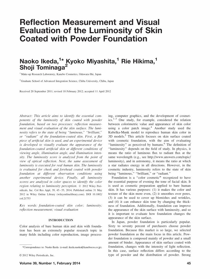
Reflection Measurement and Visual Evaluation of the Luminosity of Skin Coated with Powder Foundation Naoko Ikeda, 1 * Kyoko Miyashita, 1 Rie Hikima, 1 Shoji Tominaga 2 1 Make-up Research Laboratory, Kanebo Cosmetics, Odawara-Shi, Japan 2 Graduate School of Advanced Integration Science, Chiba University, Chiba, Japan Received 20 September 2011; revised 18 February 2012; accepted 11 April 2012 Abstract: This article aims to identify the essential com- ponents of the luminosity of skin coated with powder foundation, based on two processes: reflection measure- ment and visual evaluation of the skin surface. The lumi- nosity refers to the state of being ‘‘luminous,’’ ‘‘brilliant,’’ or ‘‘radiant’’ of the foundation-coated skin. First, a flat piece of artificial skin is used, and an experimental device is developed to visually evaluate the appearance of the foundation-coated artificial skin at different conditions of viewing angle, illumination angle, and illumination inten- sity. The luminosity score is analyzed from the point of view of optical reflection. Next, the same assessment of luminosity is executed for real human skin. The luminosity is evaluated for cheek and forehead coated with powder foundation at different observation conditions using another experimental device. Finally, all luminosity scores are analyzed in color spaces to identify the color region relating to luminosity perception. Ó 2012 Wiley Peri- odicals, Inc. Col Res Appl, 39, 45 – 55, 2014; Published online 31 May 2012 in Wiley Online Library (wileyonlinelibrary.com). DOI 10.1002/ col.21753 Key words: foundation-coated skin color; luminosity; reflection measurement; visual evaluation INTRODUCTION Color analysis of bare human skin and skin with founda- tion has been an extremely popular research topic in many fields including color reproduction, image process- ing, computer graphics, and the development of cosmet- ics. 1–3 One study, for example, considered the relation between colorimetric value and appearance of skin color using a color patch image. 4 Another study used the Kubelka-Munk model to reproduce human skin color in 3D models. 5 This article focuses on skin surface coated with cosmetic foundation, with the aim of evaluating ‘‘luminosity’’ as perceived by humans. 6 The definition of ‘‘luminosity’’ depends on the field of study. In physics, it means the ratio of luminous flux to radiant flux at the same wavelength (e.g., see http://www.answers.com/topic/ luminosity), and in astronomy, it means the ratio at which a star radiates energy in all directions. However, in the cosmetic industry, luminosity refers to the state of skin being ‘‘luminous,’’ ‘‘brilliant,’’ or ‘‘radiant.’’ Foundation is a ‘‘color cosmetic’’ recognized to have the essential purpose of evening the tone of facial skin. It is used as cosmetic preparation applied to bare human skin. It has various purposes: (1) it makes the color and texture of the skin more even, (2) it smoothes over pores, (3) it can be used to cover up blemishes and wrinkles, and (4) it can enhance skin tone by changing the thick- ness of foundation. Additionally, foundation can improve the appearance of the skin surface with luminosity, and so it is important to evaluate how foundation changes the appearance of the skin surface. In Japan, powder foundation is particularly popular. Sixty to seventy percent of purchasers choose powder foundation. Because this market is so large, we selected powder foundation as the main focus in this article. Pow- der foundation is composed mainly of powder and a small amount of binder. Appearance of skin surface coated with foundation, changes with the intensity of light reflection. In this case, surface reflection differs according to the type of powder and the distribution of powder. Strong *Correspondence to: Naoko Ikeda (e-mail: [email protected]). V V C 2012 Wiley Periodicals, Inc. Volume 39, Number 1, February 2014 45
-
Upload
independent -
Category
Documents
-
view
1 -
download
0
Transcript of Reflection measurement and visual evaluation of the luminosity of skin coated with powder foundation
Reflection Measurement and VisualEvaluation of the Luminosity of SkinCoated with Powder Foundation
Naoko Ikeda,1* Kyoko Miyashita,1 Rie Hikima,1
Shoji Tominaga2
1 Make-up Research Laboratory, Kanebo Cosmetics, Odawara-Shi, Japan
2 Graduate School of Advanced Integration Science, Chiba University, Chiba, Japan
Received 20 September 2011; revised 18 February 2012; accepted 11 April 2012
Abstract: This article aims to identify the essential com-ponents of the luminosity of skin coated with powderfoundation, based on two processes: reflection measure-ment and visual evaluation of the skin surface. The lumi-nosity refers to the state of being ‘‘luminous,’’ ‘‘brilliant,’’or ‘‘radiant’’ of the foundation-coated skin. First, a flatpiece of artificial skin is used, and an experimental deviceis developed to visually evaluate the appearance of thefoundation-coated artificial skin at different conditions ofviewing angle, illumination angle, and illumination inten-sity. The luminosity score is analyzed from the point ofview of optical reflection. Next, the same assessment ofluminosity is executed for real human skin. The luminosityis evaluated for cheek and forehead coated with powderfoundation at different observation conditions usinganother experimental device. Finally, all luminosityscores are analyzed in color spaces to identify the colorregion relating to luminosity perception. � 2012 Wiley Peri-
odicals, Inc. Col Res Appl, 39, 45 – 55, 2014; Published online 31 May
2012 in Wiley Online Library (wileyonlinelibrary.com). DOI 10.1002/
col.21753
Key words: foundation-coated skin color; luminosity;reflection measurement; visual evaluation
INTRODUCTION
Color analysis of bare human skin and skin with founda-
tion has been an extremely popular research topic in
many fields including color reproduction, image process-
ing, computer graphics, and the development of cosmet-
ics.1–3 One study, for example, considered the relation
between colorimetric value and appearance of skin color
using a color patch image.4 Another study used the
Kubelka-Munk model to reproduce human skin color in
3D models.5 This article focuses on skin surface coated
with cosmetic foundation, with the aim of evaluating
‘‘luminosity’’ as perceived by humans.6 The definition of
‘‘luminosity’’ depends on the field of study. In physics, it
means the ratio of luminous flux to radiant flux at the
same wavelength (e.g., see http://www.answers.com/topic/
luminosity), and in astronomy, it means the ratio at which
a star radiates energy in all directions. However, in the
cosmetic industry, luminosity refers to the state of skin
being ‘‘luminous,’’ ‘‘brilliant,’’ or ‘‘radiant.’’
Foundation is a ‘‘color cosmetic’’ recognized to havethe essential purpose of evening the tone of facial skin. It
is used as cosmetic preparation applied to bare humanskin. It has various purposes: (1) it makes the color and
texture of the skin more even, (2) it smoothes over pores,
(3) it can be used to cover up blemishes and wrinkles,and (4) it can enhance skin tone by changing the thick-
ness of foundation. Additionally, foundation can improvethe appearance of the skin surface with luminosity, and so
it is important to evaluate how foundation changes the
appearance of the skin surface.
In Japan, powder foundation is particularly popular.
Sixty to seventy percent of purchasers choose powder
foundation. Because this market is so large, we selected
powder foundation as the main focus in this article. Pow-
der foundation is composed mainly of powder and a small
amount of binder. Appearance of skin surface coated with
foundation, changes with the intensity of light reflection.
In this case, surface reflection differs according to the
type of powder and the distribution of powder. Strong
*Correspondence to: Naoko Ikeda (e-mail: [email protected]).
VVC 2012 Wiley Periodicals, Inc.
Volume 39, Number 1, February 2014 45
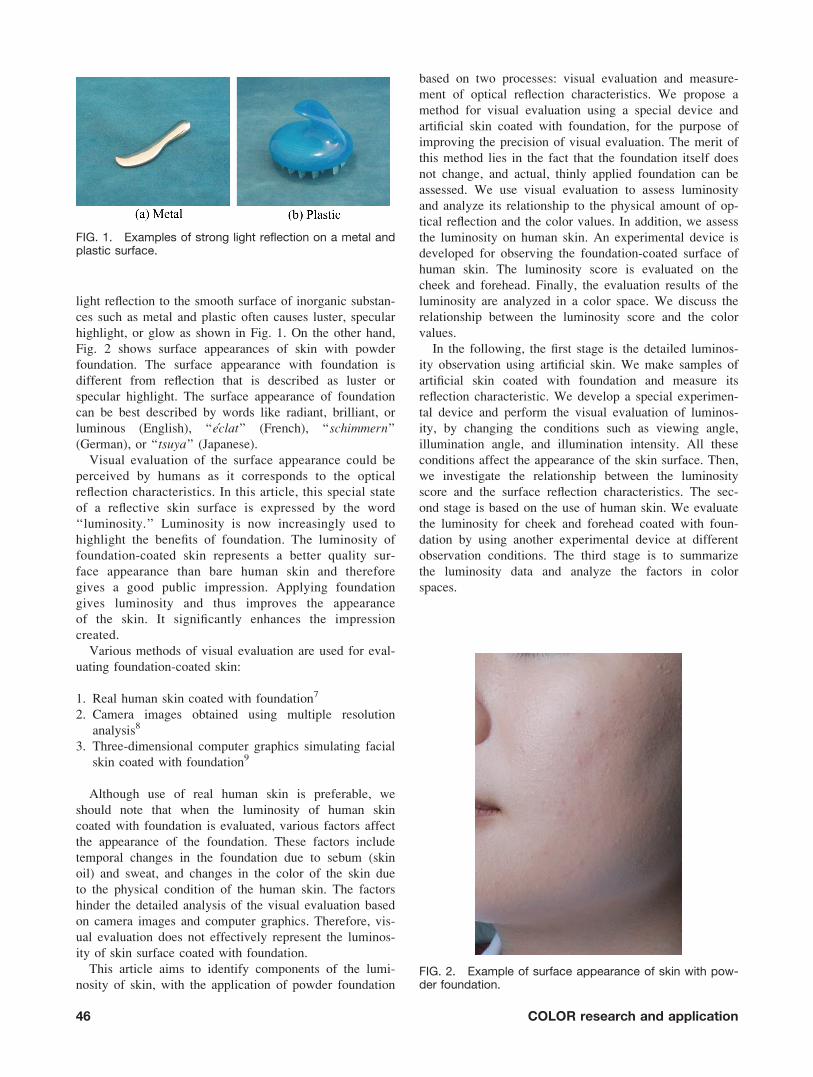
light reflection to the smooth surface of inorganic substan-
ces such as metal and plastic often causes luster, specular
highlight, or glow as shown in Fig. 1. On the other hand,
Fig. 2 shows surface appearances of skin with powder
foundation. The surface appearance with foundation is
different from reflection that is described as luster or
specular highlight. The surface appearance of foundation
can be best described by words like radiant, brilliant, or
luminous (English), ‘‘eclat’’ (French), ‘‘schimmern’’
(German), or ‘‘tsuya’’ (Japanese).
Visual evaluation of the surface appearance could be
perceived by humans as it corresponds to the optical
reflection characteristics. In this article, this special state
of a reflective skin surface is expressed by the word
‘‘luminosity.’’ Luminosity is now increasingly used to
highlight the benefits of foundation. The luminosity of
foundation-coated skin represents a better quality sur-
face appearance than bare human skin and therefore
gives a good public impression. Applying foundation
gives luminosity and thus improves the appearance
of the skin. It significantly enhances the impression
created.
Various methods of visual evaluation are used for eval-
uating foundation-coated skin:
1. Real human skin coated with foundation7
2. Camera images obtained using multiple resolution
analysis8
3. Three-dimensional computer graphics simulating facial
skin coated with foundation9
Although use of real human skin is preferable, we
should note that when the luminosity of human skin
coated with foundation is evaluated, various factors affect
the appearance of the foundation. These factors include
temporal changes in the foundation due to sebum (skin
oil) and sweat, and changes in the color of the skin due
to the physical condition of the human skin. The factors
hinder the detailed analysis of the visual evaluation based
on camera images and computer graphics. Therefore, vis-
ual evaluation does not effectively represent the luminos-
ity of skin surface coated with foundation.
This article aims to identify components of the lumi-
nosity of skin, with the application of powder foundation
based on two processes: visual evaluation and measure-
ment of optical reflection characteristics. We propose a
method for visual evaluation using a special device and
artificial skin coated with foundation, for the purpose of
improving the precision of visual evaluation. The merit of
this method lies in the fact that the foundation itself does
not change, and actual, thinly applied foundation can be
assessed. We use visual evaluation to assess luminosity
and analyze its relationship to the physical amount of op-
tical reflection and the color values. In addition, we assess
the luminosity on human skin. An experimental device is
developed for observing the foundation-coated surface of
human skin. The luminosity score is evaluated on the
cheek and forehead. Finally, the evaluation results of the
luminosity are analyzed in a color space. We discuss the
relationship between the luminosity score and the color
values.
In the following, the first stage is the detailed luminos-
ity observation using artificial skin. We make samples of
artificial skin coated with foundation and measure its
reflection characteristic. We develop a special experimen-
tal device and perform the visual evaluation of luminos-
ity, by changing the conditions such as viewing angle,
illumination angle, and illumination intensity. All these
conditions affect the appearance of the skin surface. Then,
we investigate the relationship between the luminosity
score and the surface reflection characteristics. The sec-
ond stage is based on the use of human skin. We evaluate
the luminosity for cheek and forehead coated with foun-
dation by using another experimental device at different
observation conditions. The third stage is to summarize
the luminosity data and analyze the factors in color
spaces.
FIG. 1. Examples of strong light reflection on a metal andplastic surface.
FIG. 2. Example of surface appearance of skin with pow-der foundation.
46 COLOR research and application
REFLECTION MEASUREMENT AND VISUAL
EVALUATION USING ARTIFICIAL SKIN
Sample Preparation of Artificial Skin
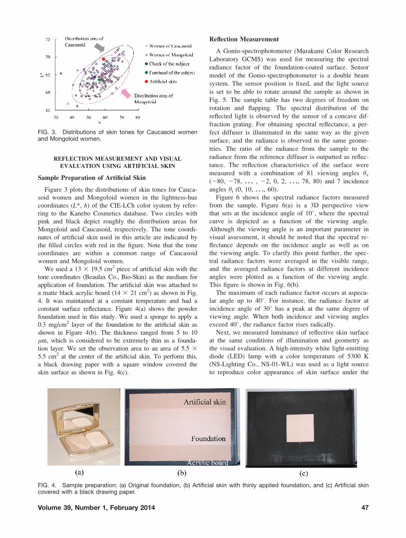
Figure 3 plots the distributions of skin tones for Cauca-
soid women and Mongoloid women in the lightness-hue
coordinates (L*, h) of the CIE-LCh color system by refer-
ring to the Kanebo Cosmetics database. Two circles with
pink and black depict roughly the distribution areas for
Mongoloid and Caucasoid, respectively. The tone coordi-
nates of artificial skin used in this article are indicated by
the filled circles with red in the figure. Note that the tone
coordinates are within a common range of Caucasoid
women and Mongoloid women.
We used a 13 3 19.5 cm2 piece of artificial skin with the
tone coordinates (Beaulax Co., Bio-Skin) as the medium for
application of foundation. The artificial skin was attached to
a matte black acrylic board (14 3 21 cm2) as shown in Fig.
4. It was maintained at a constant temperature and had a
constant surface reflectance. Figure 4(a) shows the powder
foundation used in this study. We used a sponge to apply a
0.3 mg/cm2 layer of the foundation to the artificial skin as
shown in Figure 4(b). The thickness ranged from 5 to 10
lm, which is considered to be extremely thin as a founda-
tion layer. We set the observation area to an area of 5.5 3
5.5 cm2 at the center of the artificial skin. To perform this,
a black drawing paper with a square window covered the
skin surface as shown in Fig. 4(c).
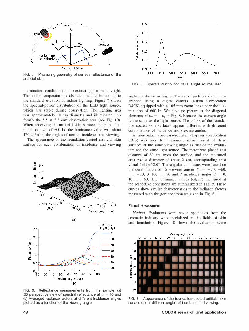
Reflection Measurement
A Gonio-spectrophotometer (Murakami Color Research
Laboratory GCMS) was used for measuring the spectral
radiance factor of the foundation-coated surface. Sensor
model of the Gonio-spectrophotometer is a double beam
system. The sensor position is fixed, and the light source
is set to be able to rotate around the sample as shown in
Fig. 5. The sample table has two degrees of freedom on
rotation and flapping. The spectral distribution of the
reflected light is observed by the sensor of a concave dif-
fraction grating. For obtaining spectral reflectance, a per-
fect diffuser is illuminated in the same way as the given
surface, and the radiance is observed in the same geome-
tries. The ratio of the radiance from the sample to the
radiance from the reference diffuser is outputted as reflec-
tance. The reflection characteristics of the surface were
measured with a combination of 81 viewing angles hv
(280, 278, . . . , 22, 0, 2, . . ., 78, 80) and 7 incidence
angles hi (0, 10, . . ., 60).
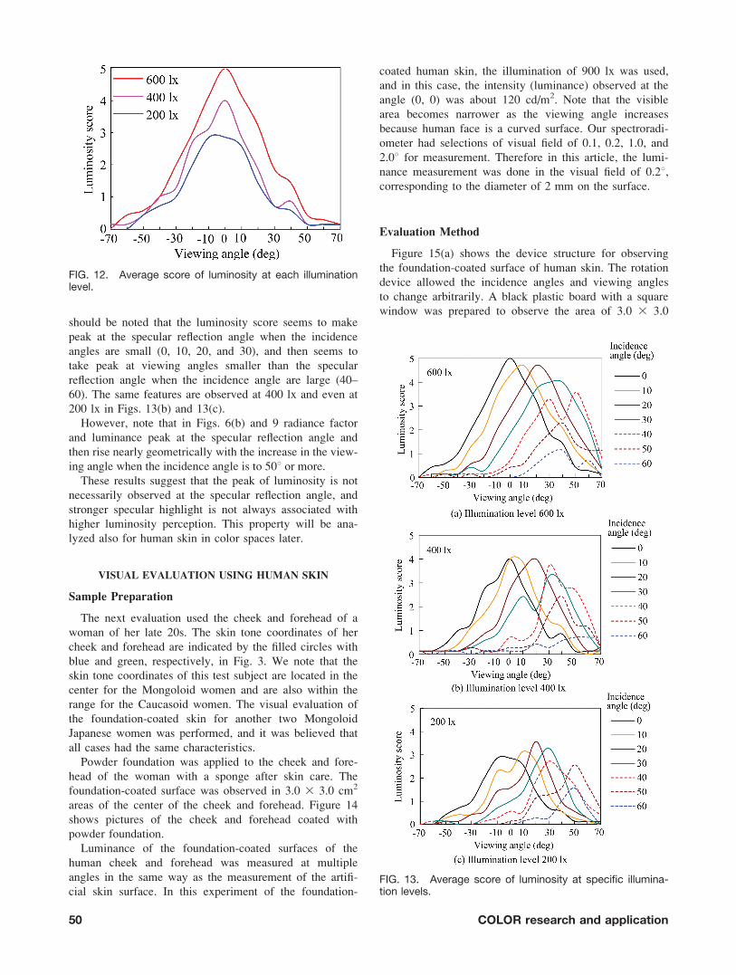
Figure 6 shows the spectral radiance factors measured
from the sample. Figure 6(a) is a 3D perspective view
that sets at the incidence angle of 108, where the spectral
curve is depicted as a function of the viewing angle.
Although the viewing angle is an important parameter in
visual assessment, it should be noted that the spectral re-
flectance depends on the incidence angle as well as on
the viewing angle. To clarify this point further, the spec-
tral radiance factors were averaged in the visible range,
and the averaged radiance factors at different incidence
angles were plotted as a function of the viewing angle.
This figure is shown in Fig. 6(b).
The maximum of each radiance factor occurs at aspecu-
lar angle up to 408. For instance, the radiance factor at
incidence angle of 308 has a peak at the same degree of
viewing angle. When both incidence and viewing angles
exceed 408, the radiance factor rises radically.
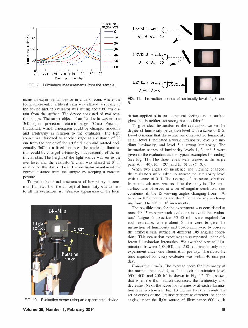
Next, we measured luminance of reflective skin surface
at the same conditions of illumination and geometry as
the visual evaluation. A high-intensity white light-emitting
diode (LED) lamp with a color temperature of 5300 K
(NS-Lighting Co., NS-01-WL) was used as a light source
to reproduce color appearance of skin surface under the
FIG. 3. Distributions of skin tones for Caucasoid womenand Mongoloid women.
FIG. 4. Sample preparation: (a) Original foundation, (b) Artificial skin with thinly applied foundation, and (c) Artificial skincovered with a black drawing paper.
Volume 39, Number 1, February 2014 47
illumination condition of approximating natural daylight.
This color temperature is also assumed to be similar to
the standard situation of indoor lighting. Figure 7 shows
the spectral-power distribution of the LED light source,
which was stable during observation. The lighting area
was approximately 10 cm diameter and illuminated uni-
formly the 5.5 3 5.5 cm2 observation area (see Fig. 10).
When observing the artificial skin surface under the illu-
mination level of 600 lx, the luminance value was about
120 cd/m2 at the angles of normal incidence and viewing.
The appearance of the foundation-coated artificial skin
surface for each combination of incidence and viewing
angles is shown in Fig. 8. The set of pictures was photo-
graphed using a digital camera (Nikon Corporation
D40X) equipped with a 105 mm zoom lens under the illu-
mination of 600 lx. We have no picture at the diagonal
elements of hv ¼ 2hi in Fig. 8, because the camera angle
is the same as the light source. The colors of the founda-
tion-coated skin surfaces appear different with different
combinations of incidence and viewing angles.
A noncontact spectroradiometer (Topcon Corporation
SR-3) was used for luminance measurement of these
surfaces at the same viewing angle as that of the evalua-
tors and the same light source. The meter was placed at a
distance of 60 cm from the surface, and the measured
area was a diameter of about 2 cm, corresponding to a
visual field of 2.08. The angular conditions were based on
the combination of 15 viewing angles hv ¼ 270, 260,
. . ., 210, 0, 10, . . ., 70 and 7 incidence angles hi ¼ 0,
10, . . ., 60. The luminance values (cd/m2) measured at
the respective conditions are summarized in Fig. 9. These
curves show similar characteristics to the radiance factors
measured with the goniophotometer given in Fig. 6.
Visual Assessment
Method. Evaluators were seven specialists from the
cosmetic industry who specialized in the fields of skin
and foundation. Figure 10 shows the evaluation scene
FIG. 5. Measuring geometry of surface reflectance of theartificial skin.
FIG. 6. Reflectance measurements from the sample: (a)3D perspective view of spectral reflectance at yi ¼ 10 and(b) Averaged radiance factors at different incidence anglesplotted as a function of the viewing angle.
FIG. 7. Spectral distribution of LED light source used.
FIG. 8. Appearance of the foundation-coated artificial skinsurface under different angles of incidence and viewing.
48 COLOR research and application
using an experimental device in a dark room, where the
foundation-coated artificial skin was affixed vertically to
the device and an evaluator was sitting about 60 cm dis-
tant from the surface. The device consisted of two rota-
tion stages. The target object of artificial skin was on one
360-degree precision rotation stage (Chuo Precision
Industrial), which orientation could be changed smoothly
and arbitrarily in relation to the evaluator. The light
source was fastened to another stage at a distance of 30
cm from the center of the artificial skin and rotated hori-
zontally 3608 at a fixed distance. The angle of illumina-
tion could be changed arbitrarily, independently of the ar-
tificial skin. The height of the light source was set to the
eye level and the evaluator’s chair was placed at 08 in
relation to the skin surface. The evaluator maintained the
correct distance from the sample by keeping a constant
posture.
To make the visual assessment of luminosity, a com-
mon framework of the concept of luminosity was defined
to all the evaluators as: ‘‘Surface appearance of the foun-
dation applied skin has a natural feeling and a surface
gloss that is neither too strong nor too faint.’’
To give clear instruction to the evaluators, we set the
degree of luminosity perception level with a score of 0–5:
Level 0 means that the evaluators observed no luminosity
at all, level 1 indicated a weak luminosity, level 3 a me-
dium luminosity, and level 5 a strong luminosity. The
instruction scenes of luminosity levels 1, 3, and 5 were
given to the evaluators as the typical examples for coding
(see Fig. 11). The three levels were created at the angle
pairs (0, 240), (0, 220), and (5, 0) of (hi, hv).
When two angles of incidence and viewing changed,
the evaluators were asked to answer the luminosity level
with a score of 0–5. The average of the scores obtained
from all evaluators was used for the analysis. The same
surface was observed at a set of angular conditions that
combines all the 15 viewing angles changing from 270
to 70 in 108 increments and the 7 incidence angles chang-
ing from 0 to 608 in 108 increments.
The possible time for the experiment was considered at
most 40–45 min per each evaluator to avoid the evalua-
tors’ fatigue. In practice, 35–40 min were required for
each evaluator, where about 5 min were to give the
instruction of luminosity and 30–35 min were to observe
the artificial skin surface at different 105 angular condi-
tions. This evaluation experiment was repeated under dif-
ferent illumination intensities. We switched vertical illu-
mination between 600, 400, and 200 lx. There is only one
experiment under one illumination per day. Therefore, the
time required for every evaluator was within 40 min per
day.
Evaluation results. The average score for luminosity at
the normal incidence hi ¼ 0 at each illumination level
(600, 400, and 200 lx) is shown in Fig. 12. This shows
that when the illumination decreases, the luminosity also
decreases. Next, the score for luminosity at each illumina-
tion level is shown in Fig. 13. Figure 13(a) represents the
set of curves of the luminosity score at different incidence
angles under the light source of illuminance 600 lx. ItFIG. 10. Evaluation scene using an experimental device.
FIG. 9. Luminance measurements from the sample.
FIG. 11. Instruction scenes of luminosity levels 1, 3, and5.
Volume 39, Number 1, February 2014 49
should be noted that the luminosity score seems to make
peak at the specular reflection angle when the incidence
angles are small (0, 10, 20, and 30), and then seems to
take peak at viewing angles smaller than the specular
reflection angle when the incidence angle are large (40–
60). The same features are observed at 400 lx and even at
200 lx in Figs. 13(b) and 13(c).
However, note that in Figs. 6(b) and 9 radiance factor
and luminance peak at the specular reflection angle and
then rise nearly geometrically with the increase in the view-
ing angle when the incidence angle is to 508 or more.
These results suggest that the peak of luminosity is not
necessarily observed at the specular reflection angle, and
stronger specular highlight is not always associated with
higher luminosity perception. This property will be ana-
lyzed also for human skin in color spaces later.
VISUAL EVALUATION USING HUMAN SKIN
Sample Preparation
The next evaluation used the cheek and forehead of a
woman of her late 20s. The skin tone coordinates of her
cheek and forehead are indicated by the filled circles with
blue and green, respectively, in Fig. 3. We note that the
skin tone coordinates of this test subject are located in the
center for the Mongoloid women and are also within the
range for the Caucasoid women. The visual evaluation of
the foundation-coated skin for another two Mongoloid
Japanese women was performed, and it was believed that
all cases had the same characteristics.
Powder foundation was applied to the cheek and fore-
head of the woman with a sponge after skin care. The
foundation-coated surface was observed in 3.0 3 3.0 cm2
areas of the center of the cheek and forehead. Figure 14
shows pictures of the cheek and forehead coated with
powder foundation.
Luminance of the foundation-coated surfaces of the
human cheek and forehead was measured at multiple
angles in the same way as the measurement of the artifi-
cial skin surface. In this experiment of the foundation-
coated human skin, the illumination of 900 lx was used,
and in this case, the intensity (luminance) observed at the
angle (0, 0) was about 120 cd/m2. Note that the visible
area becomes narrower as the viewing angle increases
because human face is a curved surface. Our spectroradi-
ometer had selections of visual field of 0.1, 0.2, 1.0, and
2.08 for measurement. Therefore in this article, the lumi-
nance measurement was done in the visual field of 0.28,corresponding to the diameter of 2 mm on the surface.
Evaluation Method
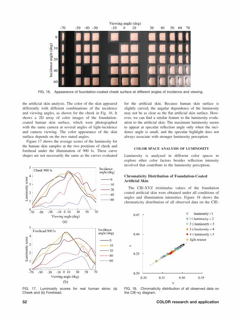
Figure 15(a) shows the device structure for observing
the foundation-coated surface of human skin. The rotation
device allowed the incidence angles and viewing angles
to change arbitrarily. A black plastic board with a square
window was prepared to observe the area of 3.0 3 3.0
FIG. 13. Average score of luminosity at specific illumina-tion levels.
FIG. 12. Average score of luminosity at each illuminationlevel.
50 COLOR research and application
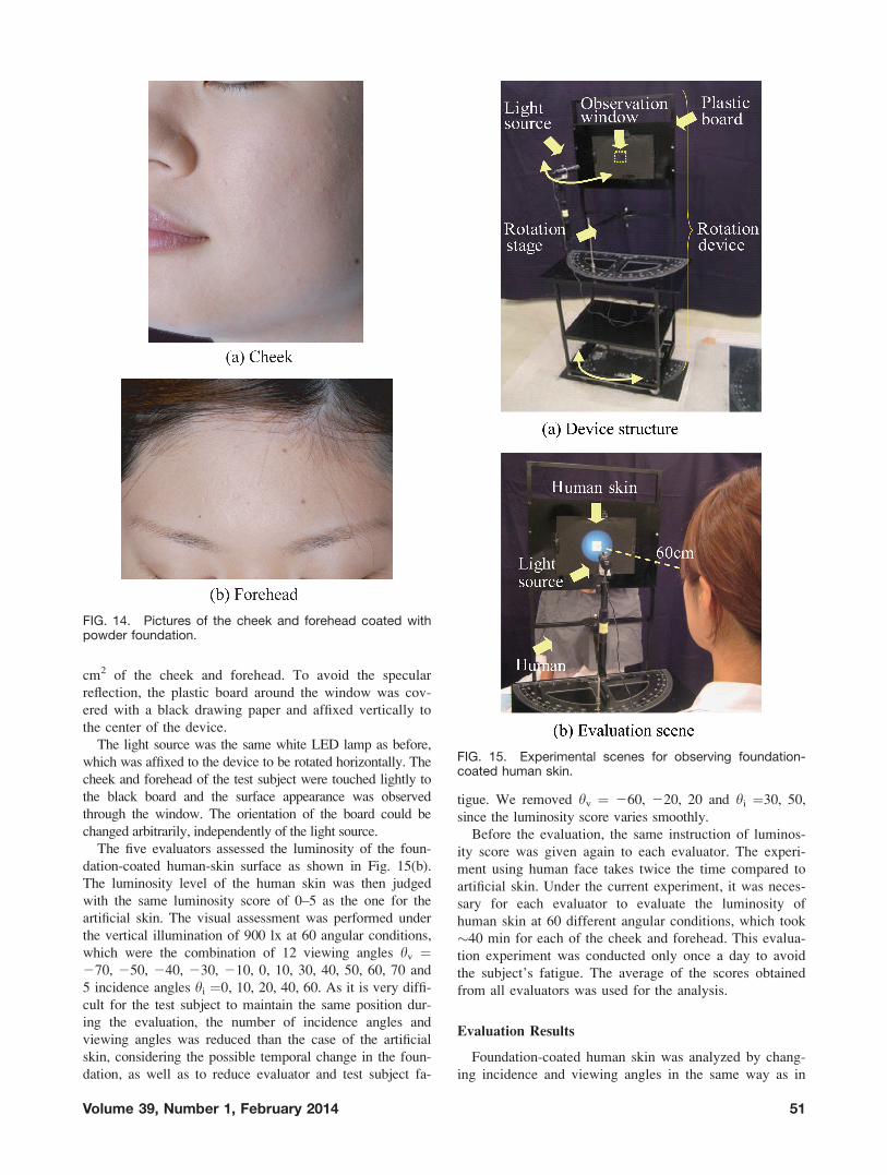
cm2 of the cheek and forehead. To avoid the specular
reflection, the plastic board around the window was cov-
ered with a black drawing paper and affixed vertically to
the center of the device.
The light source was the same white LED lamp as before,
which was affixed to the device to be rotated horizontally. The
cheek and forehead of the test subject were touched lightly to
the black board and the surface appearance was observed
through the window. The orientation of the board could be
changed arbitrarily, independently of the light source.
The five evaluators assessed the luminosity of the foun-
dation-coated human-skin surface as shown in Fig. 15(b).
The luminosity level of the human skin was then judged
with the same luminosity score of 0–5 as the one for the
artificial skin. The visual assessment was performed under
the vertical illumination of 900 lx at 60 angular conditions,
which were the combination of 12 viewing angles hv ¼270, 250, 240, 230, 210, 0, 10, 30, 40, 50, 60, 70 and
5 incidence angles hi ¼0, 10, 20, 40, 60. As it is very diffi-
cult for the test subject to maintain the same position dur-
ing the evaluation, the number of incidence angles and
viewing angles was reduced than the case of the artificial
skin, considering the possible temporal change in the foun-
dation, as well as to reduce evaluator and test subject fa-
tigue. We removed hv ¼ 260, 220, 20 and hi ¼30, 50,
since the luminosity score varies smoothly.
Before the evaluation, the same instruction of luminos-
ity score was given again to each evaluator. The experi-
ment using human face takes twice the time compared to
artificial skin. Under the current experiment, it was neces-
sary for each evaluator to evaluate the luminosity of
human skin at 60 different angular conditions, which took
�40 min for each of the cheek and forehead. This evalua-
tion experiment was conducted only once a day to avoid
the subject’s fatigue. The average of the scores obtained
from all evaluators was used for the analysis.
Evaluation Results
Foundation-coated human skin was analyzed by chang-
ing incidence and viewing angles in the same way as in
FIG. 15. Experimental scenes for observing foundation-coated human skin.
FIG. 14. Pictures of the cheek and forehead coated withpowder foundation.
Volume 39, Number 1, February 2014 51
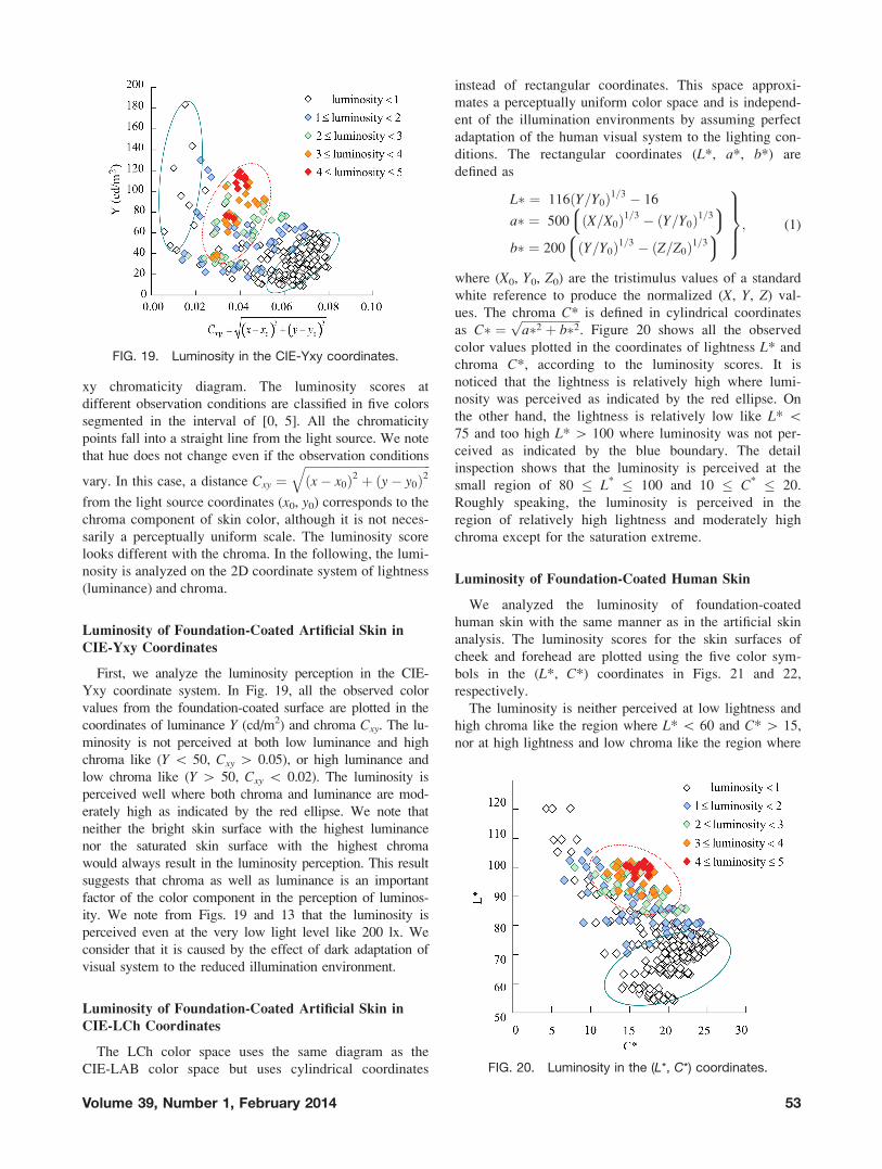
the artificial skin analysis. The color of the skin appeared
differently with different combinations of the incidence
and viewing angles, as shown for the cheek in Fig. 16. It
shows a 2D array of color images of the foundation-
coated human skin surface, which were photographed
with the same camera at several angles of light-incidence
and camera viewing. The color appearance of the skin
surface depends on the two stated angles.
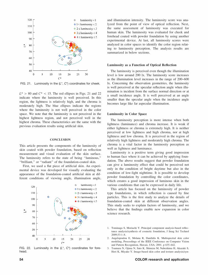
Figure 17 shows the average scores of the luminosity for
the human skin samples at the two positions of cheek and
forehead under the illumination of 900 lx. These curve
shapes are not necessarily the same as the curves evaluated
for the artificial skin. Because human skin surface is
slightly curved, the angular dependence of the luminosity
may not be as clear as the flat artificial skin surface. How-
ever, we can find a similar feature to the luminosity evalu-
ation to the artificial skin. The maximum luminosity seems
to appear at specular reflection angle only when the inci-
dence angle is small, and the specular highlight does not
always associate with stronger luminosity perception.
COLOR SPACE ANALYSIS OF LUMINOSITY
Luminosity is analyzed in different color spaces to
explore other color factors besides reflection intensity
involved that contribute to the luminosity perception.
Chromaticity Distribution of Foundation-Coated
Artificial Skin
The CIE-XYZ tristimulus values of the foundation
coated artificial skin were obtained under all conditions of
angles and illumination intensities. Figure 18 shows the
chromaticity distribution of all observed data on the CIE-
FIG. 16. Appearance of foundation-coated cheek surface at different angles of incidence and viewing.
FIG. 17. Luminosity scores for real human skins: (a)Cheek and (b) Forehead.
FIG. 18. Chromaticity distribution of all observed data onthe CIE-xy diagram.
52 COLOR research and application
xy chromaticity diagram. The luminosity scores at
different observation conditions are classified in five colors
segmented in the interval of [0, 5]. All the chromaticity
points fall into a straight line from the light source. We note
that hue does not change even if the observation conditions
vary. In this case, a distance Cxy ¼ffiffiffiffiffiffiffiffiffiffiffiffiffiffiffiffiffiffiffiffiffiffiffiffiffiffiffiffiffiffiffiffiffiffiffiffiffiffiffiffiffiffix� x0ð Þ2 þ y� y0ð Þ2
q
from the light source coordinates (x0, y0) corresponds to the
chroma component of skin color, although it is not neces-
sarily a perceptually uniform scale. The luminosity score
looks different with the chroma. In the following, the lumi-
nosity is analyzed on the 2D coordinate system of lightness
(luminance) and chroma.
Luminosity of Foundation-Coated Artificial Skin in
CIE-Yxy Coordinates
First, we analyze the luminosity perception in the CIE-
Yxy coordinate system. In Fig. 19, all the observed color
values from the foundation-coated surface are plotted in the
coordinates of luminance Y (cd/m2) and chroma Cxy. The lu-
minosity is not perceived at both low luminance and high
chroma like (Y \ 50, Cxy [ 0.05), or high luminance and
low chroma like (Y [ 50, Cxy \ 0.02). The luminosity is
perceived well where both chroma and luminance are mod-
erately high as indicated by the red ellipse. We note that
neither the bright skin surface with the highest luminance
nor the saturated skin surface with the highest chroma
would always result in the luminosity perception. This result
suggests that chroma as well as luminance is an important
factor of the color component in the perception of luminos-
ity. We note from Figs. 19 and 13 that the luminosity is
perceived even at the very low light level like 200 lx. We
consider that it is caused by the effect of dark adaptation of
visual system to the reduced illumination environment.
Luminosity of Foundation-Coated Artificial Skin in
CIE-LCh Coordinates
The LCh color space uses the same diagram as the
CIE-LAB color space but uses cylindrical coordinates
instead of rectangular coordinates. This space approxi-
mates a perceptually uniform color space and is independ-
ent of the illumination environments by assuming perfect
adaptation of the human visual system to the lighting con-
ditions. The rectangular coordinates (L*, a*, b*) are
defined as
L� ¼ 116 Y=Y0ð Þ1=3 � 16
a� ¼ 500 X=X0ð Þ1=3 � Y=Y0ð Þ1=38: 9;
b� ¼ 200 Y=Y0ð Þ1=3 � Z=Z0ð Þ1=38: 9;
9>>=>>;; (1)
where (X0, Y0, Z0) are the tristimulus values of a standard
white reference to produce the normalized (X, Y, Z) val-
ues. The chroma C* is defined in cylindrical coordinates
as C� ¼ffiffiffiffiffiffiffiffiffiffiffiffiffiffiffiffiffiffiffiffia�2 þ b�2p
. Figure 20 shows all the observed
color values plotted in the coordinates of lightness L* and
chroma C*, according to the luminosity scores. It is
noticed that the lightness is relatively high where lumi-
nosity was perceived as indicated by the red ellipse. On
the other hand, the lightness is relatively low like L* \75 and too high L* [ 100 where luminosity was not per-
ceived as indicated by the blue boundary. The detail
inspection shows that the luminosity is perceived at the
small region of 80 � L* � 100 and 10 � C* � 20.
Roughly speaking, the luminosity is perceived in the
region of relatively high lightness and moderately high
chroma except for the saturation extreme.
Luminosity of Foundation-Coated Human Skin
We analyzed the luminosity of foundation-coated
human skin with the same manner as in the artificial skin
analysis. The luminosity scores for the skin surfaces of
cheek and forehead are plotted using the five color sym-
bols in the (L*, C*) coordinates in Figs. 21 and 22,
respectively.
The luminosity is neither perceived at low lightness and
high chroma like the region where L* \ 60 and C* [ 15,
nor at high lightness and low chroma like the region where
FIG. 19. Luminosity in the CIE-Yxy coordinates.
FIG. 20. Luminosity in the (L*, C*) coordinates.
Volume 39, Number 1, February 2014 53
L* [ 80 and C* \ 15. The red ellipses in Figs. 21 and 22
indicate where the luminosity is well perceived. In this
region, the lightness is relatively high, and the chroma is
moderately high. The blue ellipses indicate the regions
where the luminosity is not well perceived in the color
space. We note that the luminosity is not perceived in the
highest lightness region, and not perceived well in the
highest chroma. These characteristics are the same with the
previous evaluation results using artificial skin.
CONCLUSION
This article presents the components of the luminosity of
skin coated with powder foundation, based on reflection
measurement and visual evaluation of the skin surface.
The luminosity refers to the state of being ‘‘luminous,’’
‘‘brilliant,’’ or ‘‘radiant’’ of the foundation-coated skin.
First, we used a flat piece of artificial skin. An experi-
mental device was developed for visually evaluating the
appearance of the foundation-coated artificial skin at dif-
ferent conditions of viewing angle, illumination angle,
and illumination intensity. The luminosity score was ana-
lyzed from the point of view of optical reflection. Next,
the same assessment of luminosity was executed for
human skin. The luminosity was evaluated for cheek and
forehead coated with powder foundation by using another
experimental device. At last, all luminosity scores were
analyzed in color spaces to identify the color region relat-
ing to luminosity perception. The analysis results are
summarized in below sections.
Luminosity as a Function of Optical Reflection
The luminosity is perceived even though the illumination
level is low around 200 lx. The luminosity score increases
as the illumination level increases in the range of 200–600
lx. Concerning the observation geometries, the luminosity
is well perceived at the specular reflection angle when illu-
mination is incident from the surface normal direction or at
a small incidence angle. It is well perceived at an angle
smaller than the specular angle when the incidence angle
becomes large like far aspecular illumination.
Luminosity in Color Space
The luminosity perception is more intense when both
lightness (luminance) and chroma increase. It is weak if
either lightness or chroma is extremely high. It is neither
perceived at low lightness and high chroma, nor at high
lightness and low chroma. It is perceived in the region of
relatively high lightness and moderately high chroma. The
chroma is a vital factor in the luminosity perception as
well as lightness and luminance.
Luminosity is a positive term giving good impression
to human face where it can be achieved by applying foun-
dation. The above results suggest that powder foundation
can give a luminosity effect that could be perceived not
only in the condition of bright daytime but also in the
condition of low-light nighttime. It is possible to develop
powder foundation by controlling the color coordinates,
which creates a good impression of luminous skin in the
various conditions that can be expressed in daily life.
This article has focused on the luminosity of powder
type foundations, in which reflection is caused by fine
particles. This is the first study to analyze the details of
foundation-coated skin at different observation angles.
This study seeks to explain factors of luminosity, and we
believe that the findings enable new expansion in color
science research.
1. Tominaga S, Moriuchi Y. Principal component analysis-based reflec-
tance analysis/synthesis of cosmetic foundation, J Imag Sci Technol
2009;53:1–8.
2. Angelopoulou E, Molana R, Danilidis K. Multispectral skin color
modeling, Proceedings of the IEEE Conference on Computer Vision
and Pattern Recognition, Hawaii, USA; 2001. p 635–642.
3. Tsumura N, Ojima N, Sato K, Shimizu M, Nabeshima H, Akazaki S,
Hori K, Miyake Y. Image-based skin color and texture analysis/syn-
FIG. 21. Luminosity in the (L*, C*) coordinates for cheek.
FIG. 22. Luminosity in the (L*, C*) coordinates for fore-head.
54 COLOR research and application

thesis by extracting hemoglobin and melanin information in the
skin, ACM Trans Graphics; 2003;22:770–779.
4. Yoshikawa H, Kikuchi K, Kim J, Nishikawa S, Yaguchi H, Mizo-kami Y. Effect of the chromatic components on the whiteness evalu-ation of skin, Midterm Meeting of Congress of International ColourAssociation (AIC); 2007. p 203–206.
5. Doi M, Tanaka N, Tominaga S. Spectral reflectance estimation of
human skin and its application to image rendering. J Imag Sci Tech-
nol 2005;49:574–582.
6. Ikeda N, Hikima R, Tanno O, Tominaga S. Measurement of reflec-
tion and visual evaluation of the luminosity of skin with foundation
applied, Proceedings of the Congress of International Colour Associ-
ation (AIC), Sydney, Australia; 2009. p 1–8.
7. Nishimura H, Takasuka Y, Yamamoto M. Optical properties of skin
gloss and development of mizumizushii-looking makeup foundation,
Proceedings of the International Federation of Society of Cosmetic
Chemists (IFSCC) International Congress, Orlando, USA; 2004. p
307–311.
8. Fujii M, Misaki Y, Sasaki I. Application of image processing tech-
nique for facial gloss evaluation, Proceedings of the International
Federation of Society of Cosmetic Chemists (IFSCC) International
Congress, Orlando, USA; 2004. p 302–306.
9. Minami K, Kaneko T, Nabeshima H, Iwamoto H, Ojima N, Nagatani
N, Hori K. Evaluation of glossy appearance of facial make-up using
CG images. Int J Cosmetic Sci 2005;27:251–252.
Volume 39, Number 1, February 2014 55