Redescriptions of the Nematodes Litomosoides patersoni (Mazza, 1928) (Onchocercidae) and...
10
BioOne sees sustainable scholarly publishing as an inherently collaborative enterprise connecting authors, nonprofit publishers, academic institutions, research libraries, and research funders in the common goal of maximizing access to critical research. Redescriptions of the Nematodes Litomosoides patersoni (Mazza, 1928) (Onchocercidae) and Stilestrongylus stilesi Freitas, Lent, And Almeida, 1937 (Heligmonellidae) Parasites of Holochilus chacarius (Rodentia, Cricetidae) From Salta, Argentina Author(s): Juliana Notarnicola, María Celina Digiani, and Pablo Martín López Source: Journal of Parasitology, 96(5):993-1001. 2010. Published By: American Society of Parasitologists DOI: http://dx.doi.org/10.1645/GE-2448.1 URL: http://www.bioone.org/doi/full/10.1645/GE-2448.1 BioOne (www.bioone.org ) is a nonprofit, online aggregation of core research in the biological, ecological, and environmental sciences. BioOne provides a sustainable online platform for over 170 journals and books published by nonprofit societies, associations, museums, institutions, and presses. Your use of this PDF, the BioOne Web site, and all posted and associated content indicates your acceptance of BioOne’s Terms of Use, available at www.bioone.org/page/terms_of_use . Usage of BioOne content is strictly limited to personal, educational, and non-commercial use. Commercial inquiries or rights and permissions requests should be directed to the individual publisher as copyright holder.
-
Upload
independent -
Category
Documents
-
view
1 -
download
0
Transcript of Redescriptions of the Nematodes Litomosoides patersoni (Mazza, 1928) (Onchocercidae) and...
BioOne sees sustainable scholarly publishing as an inherently collaborative enterprise connecting authors, nonprofit publishers, academic institutions, researchlibraries, and research funders in the common goal of maximizing access to critical research.
Redescriptions of the Nematodes Litomosoides patersoni (Mazza, 1928)(Onchocercidae) and Stilestrongylus stilesi Freitas, Lent, And Almeida, 1937(Heligmonellidae) Parasites of Holochilus chacarius (Rodentia, Cricetidae) FromSalta, ArgentinaAuthor(s): Juliana Notarnicola, María Celina Digiani, and Pablo Martín LópezSource: Journal of Parasitology, 96(5):993-1001. 2010.Published By: American Society of ParasitologistsDOI: http://dx.doi.org/10.1645/GE-2448.1URL: http://www.bioone.org/doi/full/10.1645/GE-2448.1
BioOne (www.bioone.org) is a nonprofit, online aggregation of core research in the biological, ecological, andenvironmental sciences. BioOne provides a sustainable online platform for over 170 journals and books publishedby nonprofit societies, associations, museums, institutions, and presses.
Your use of this PDF, the BioOne Web site, and all posted and associated content indicates your acceptance ofBioOne’s Terms of Use, available at www.bioone.org/page/terms_of_use.
Usage of BioOne content is strictly limited to personal, educational, and non-commercial use. Commercial inquiriesor rights and permissions requests should be directed to the individual publisher as copyright holder.
REDESCRIPTIONS OF THE NEMATODES LITOMOSOIDES PATERSONI (MAZZA, 1928)
(ONCHOCERCIDAE) AND STILESTRONGYLUS STILESI FREITAS, LENT, AND ALMEIDA,
1937 (HELIGMONELLIDAE) PARASITES OF HOLOCHILUS CHACARIUS (RODENTIA,
CRICETIDAE) FROM SALTA, ARGENTINA
Juliana Notarnicola, Marıa Celina Digiani*, and Pablo Martın Lopez�Centro de Estudios Parasitologicos y de Vectores–CEPAVE–CCT La Plata-CONICET, Calle 2 #584 (1900) La Plata, Argentina.e-mail: [email protected]
ABSTRACT: Two nematode species are redescribed from the type host species Holochilus chacarius Thomas (Rodentia, Cricetidae,Sigmodontinae) and from the type locality of 1 of them, i.e., Ingenio San Martın de Tabacal, Salta Province, Argentina. Rodents weredeposited at the Coleccion Mamıferos Lillo, Tucuman, Argentina. Litomosoides patersoni (Mazza, 1928) (Onchocercidae) possesses abuccal capsule with irregular external walls, a buccal cavity smooth, becoming thinner near the oral opening, a complete set of headpapillae, 3–6 pairs of cloacal papillae, and the ‘‘sigmodontis’’ type of spicules. Filarioids were found in 3 of 17 examined hosts.Stilestrongylus stilesi Freitas, Lent, and Almeida, 1937 (Heligmonellidae), whose description was based on male specimens, was foundin all 17 of the examined hosts. Here, we describe the female and the synlophe of both sexes. Females are characterized by a shortuterus with less than 25 eggs, short ovejector, short and conical tail, and the posterior extremity strongly invaginated in a cuticularexpansion usually harboring 1 to several eggs. The synlophe is characterized by 29–31 sub-equal cuticular ridges at the mid-body, withsingle (in males) or double (in females) axis of orientation of the ridges. The present work validates and enlarges the originaldescriptions of both species and assigns the specimens from L. patersoni, recovered from the type locality and the type host species, asneotypes.
In 1928, Mazza described a new filarioid species, Filaria
patersoni, from several chacoan marsh rats, Holochilus chacarius
Thomas, 1906 (originally regarded as H. vulpinus Brants, 1827)
from the Ingenio San Martın de Tabacal, a sugar cane plantation
in Salta Province, Argentina (Mazza, 1928). The systematic
position of this species was complex and synonymyzed several
times (Vogel and Gabaldon, 1932; Chitwood, 1933; Vaz, 1934;
Esslinger, 1973; Bain et al., 1989). It was considered as
Vestibulosetaria patersoni (Mazza, 1928) by Vogel and Gabaldon
(1932) within a set of filarioid specimens parasitizing Rattus
norvegicus (Berkenhout, 1769) from Caracas, Venezuela (Vogel
and Gabaldon, 1932) and Sigmodon hispidus (Say and Ord, 1825)
from Jalisco and Michoacan, Mexico (Ochoterena and Caballero,
1932); as Litomosoides patersoni (Mazza, 1928) by Chitwood
(1933), who validated the genus Litomosoides Chandler, 1931; and
as L. carinii (Travassos, 1919) by Vaz (1934) and Esslinger (1973)
within a set of filarioids from Nectomys squamipes (Brants, 1827)
from Sao Pablo, Brazil (Vaz, 1934), and Melanomys caliginosus
(Tomes, 1860) from Valle, Colombia (Esslinger, 1973). Bain et al.
(1989) reexamined the material of most of these sets, with the
exception of that from Mazza, which is lost, and clarified the
systematic position from L. carinii and L. sigmodontis Chandler,
1931; they also created a new species based on the material of Vaz
(1934), i.e., L. kohnae (Vaz, 1934), and considered L. patersoni a
valid species.
In 1934, Mazza sent some specimens of the chacoan marsh rat
to Dr. F. Werneck of the Instituto Oswaldo Cruz, Brazil, who
kindly gave the intestines to Dr J. F. T. Freitas. Years later,
Freitas et al. (1937) described a new genus and species of
heligmonellid nematode from the intestines of these hosts, i.e.,
Stilestrongylus stilesi Freitas, Lent, and Almeida, 1937, plus 3
other new species, i.e., Hassalstrongylus argentinus (Freitas, Lent,
and Almeida, 1937) (5Longistriata argentina), Hassalstrongylus
mazzai (Freitas, Lent, and Almeida, 1937) (5Heligmonoides
mazzai), and Longistriata fortuita Freitas, Lent, and Almeida,
1937 (Heligmosomoidea); all were reported from a single H.
chacarius (syn. Holochilus balnearum Thomas, 1906) from Salta.
The original descriptions of most of these nematodes are
incomplete, particularly the Heligmosomoidea, for which the
publications prior to 1964 do not include the synlophe;
additionally, females were typically poorly described. Currently,
Stilestrongylus includes 24 species, and the synlophe of 21 of them
is known, but that of the nominal species, S. stilesi, remains
undescribed. The type material of S. stilesi is available from the
Helminthological Collection of the Instituto Oswaldo Cruz
(CHIOC), Rio de Janeiro, Brazil. However, its state of
preservation precludes making a correct redescription of the
species (M. C. Durette-Desset, pers. comm.). In contrast,
specimens from L. patersoni were not deposited in any collection.
In the present paper, we provide redescriptions of L. patersoni
and S. stilesi, based on specimens colleted in H. chacarius from
the Ingenio San Martın de Tabacal, and designate neotypes for
the filarioid species, considering that the type material is lost.
MATERIALS AND METHODS
Seventeen H. chacarius Thomas, deposited in the Coleccion MamıferosLillo (CML), Tucuman, Argentina, were examined for parasites. Theywere captured in the Ingenio San Martın de Tabacal, Departamento Oran,Salta Province, in August and September 1990. Rodents were fixed in 10%formalin and stored in 70% ethanol.
Filarial worms were recovered from the abdominal cavity andheligmonellids from the small intestine. Nematodes were preserved in70% ethanol. A transverse section posterior to the vulva was made in afilarioid female, and microfilariae were isolated (Notarnicola et al., 2000).Synlophe was studied, following the method of Durette-Desset (1985).Measurements are given in micrometers, except otherwise stated, as therange followed by the mean in parentheses. Classification used above thefamily Heligmonellidae level follows Durette-Desset and Chabaud (1993).The nomenclature and synonymy of the host species follows Wilson andReeder (2005). Parasites were deposited in the Helminthological Collec-tions of the Museo de La Plata, La Plata, Argentina (CHMLP), and of theMuseum national d’Histoire naturelle, Paris, France (MNHN).
Received 3 February 2010; revised 8 June 2010; accepted 11 June 2010.*Division Zoologıa Invertebrados, Museo de La Plata and CONICET.
Paseo del Bosque s/n (1900) La Plata, Argentina.{Facultad de Ciencias Naturales y Museo, Universidad Nacional de La
Plata. Av. 60 y 122, 1900 La Plata, Argentina.DOI: 10.1645/GE-2448.1
J. Parasitol., 96(5), 2010, pp. 993–1001
F American Society of Parasitologists 2010
993

RESULTS
REDESCRIPTIONSLitomosoides patersoni (Mazza, 1928)
(Figs. 1–21; Tables I, II)
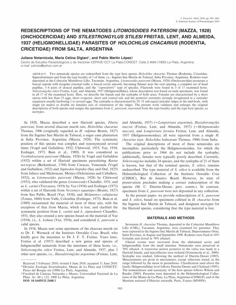
General: Males about 2.5 times shorter than females. Cephalic
extremity attenuated. Mouth small. Four minute cephalic papillae
placed in rectangle stretched laterally; 4 labial papillae surround
oral opening (Fig. 4); amphids lateral, not salient. Buccal capsule
embedded in esophagus, with irregular external walls (Figs. 2, 3);
buccal cavity smooth, becoming thinner near oral opening.
Esophagus muscular, slightly glandular posteriorly. Tail attenu-
ated. Measurements given in Tables I and II.
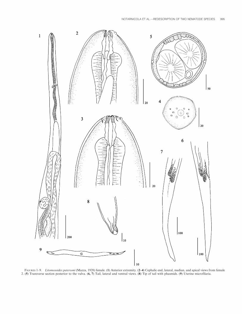
Males (based on 4 specimens): Posterior region coiled. Left
spicule with handle shorter than blade; blade constituted by
membranous folded ala and terminal filament (Fig. 14). Right
spicule with poorly cuticularized heel (Fig. 15). Number of cloacal
papillae variable, from 3 to 6 pairs. One male with 1 pair of
adcloacal, 1 pair of lateral postcloacal papillae, and 1 papilla in
median ventral line (Fig. 18); other male with 1 precloacal pair, 1
adcloacal pair, and 4 postcloacal pairs (Figs. 19–21). Area rugosa
composed of transverse ridges of small longitudinal crests,
extending through coiled region (Figs. 16, 17).
Females (based on 4 specimens): Vulva far away from
esophagus-intestine junction. Vagina globular, ovejector muscular
directed posteriad (Fig. 1). Small divergent phasmids. In cross
section, at mid-body, lateral chords flattened and expanded
laterally; internal cuticular ridges square-shaped (Fig. 5).
Microfilariae: Body fusiform. Stout microfilariae. Anterior
extremity with small visible hook, tail attenuated with nuclei near
the tip tail. Sheath not visible (Fig. 9). Measurements based on
uterine microfilariae from female 2: 35 3 6 mm; 44 3 6 mm.
Taxonomic summary
Host: Three H. chacarius Thomas (Rodentia, Cricetidae)
deposited at Coleccion Mamıferos Lillo, numbers CML 5810,
CML 5813, CML 5825.
Material studied: Three males, 4 female anterior extremities,
and 1 posterior extremity deposited at CHMLP, numbers 5995
and 6007; 1 male deposited at MNHN, number 591 MQ.
Site of infection: Body cavity, between the intestinal mesentery
and testis. In host CML 5810, 1 male in small intestine.
Locality: Ingenio San Martın de Tabacal (23u169S, 64u159W),
lote Milagros, Departamento Oran, Salta, Argentina.
Prevalence and mean intensity: Three of 17 hosts infected
(17.6%); 2.6 (1–5) worms per host.
Remarks
Litomosoides patersoni belongs to the ‘‘sigmodontis’’ group of
species (Bain et al., 1989; Notarnicola et al., 2000; Bain et al.,
2003) based on the morphological characteristics of both spicules.
The left spicule has a handle shorter than the blade and the blade
is divided in anterior membranous folded ala, with a terminal
filament; heel of right spicule not heavily cuticularized. Mazza
(1928) reported smaller length for the spicules; moreover,
Figure 4 in his paper clearly shows the ‘‘sigmodontis’’ type of
spicules. One of the males described herein possesses a similar
arrangement of the cloacal papillae as stated by Mazza (1928), but
others display a greater number of papillae (see Figs. 18–21).
Microfilariae are shorter and more stout compared with other
species of Litomosoides, which was also reported by Mazza (1928)
for microfilariae from blood smears.
Litomosoides patersoni can be differentiated from L. navonae
Notarnicola, 2005, a parasite of H. chacarius from Chaco and
Formosa Provinces, Argentina, by the shape of the buccal
capsule, the number and disposition of the cloacal and the head
papillae (Notarnicola, 2005). The 4 postcloacal papillae in L.
patersoni are mostly symmetrical, while in L. navonae there are 5–
6 asymmetric papillae. Moreover, the microfilariae in L. patersoni
are shorter (35–44 3 6 mm vs. 70 3 3.8 mm) and the sheaths are
not visible as in L. navonae (Notarnicola, 2005). Moreover, L.
patersoni can be differentiated from other species of Litomosoides
from cricetid rodents because of the shape of the buccal capsule,
the stout microfilariae, and the complete presence of the head
papillae.
Stilestrongylus stilesi Freitas, Lent, and Almeida, 1937
(Figs. 22–34)
General: Medium-sized nematodes, usually coiled sinistrally
along ventral side, in different degrees. Coiling varying from
tightly and completely coiled in up to 6 spirals, partly coiled with
up to 3 spirals in anterior half, loosely coiled with 1–2 spirals at
anterior end, or uncoiled. Excretory pore within 74–81% of
esophagus length in males; 65–78% in females. Deirids small,
situated at level of excretory pore or slightly posterior (Fig. 22).
Ratio uterus length/body length 10–20%. Cephalic vesicle present.
In apical view, triangular buccal opening surrounded by thin ring.
Two amphids, 6 externo-labial papillae, and 4 submedian cephalic
papillae (Fig. 23).
Synlophe (2 males and 2 females): In both sexes, cuticle with
longitudinal, uninterrupted ridges appearing mainly on left side
posterior to cephalic vesicle, disappearing just anterior to caudal
bursa in males and extending up to end of terminal cuticular
dilatation in female. At level of esophagus-intestinal junction: 24
ridges in male, 25 in female (Fig. 24); 29–31 ridges in both sexes at
mid-body; unequal in size, ridges on ventral right quadrant,
smaller. In female, double axis of orientation, right axis inclined
at 60u to sagittal axis, left axis sub-frontal. In males, right axis
inclined at 63u, left axis at 54u to sagittal axis (Figs. 25, 27).
Within distal third of body length; in male, 29 ridges at 150 mm
anterior to caudal bursa, in female, 26 ridges at mid-length of
uterus (Figs. 26–28); ridges sub-equal in size in both sexes. Most
ridges with similar orientation than at mid-body.
Males (20 specimens, except otherwise stated): 2.62–3.73 (3.12)
mm long and 80–130 (100) wide at mid-body. Cephalic vesicle 50–
62 (57) long and 22–32 (28) wide. Nerve ring not observed.
Excretory pore situated at 200–270 (228) from apex (n 5 9).
Deirids, when observed (n 5 4), at same level than excretory pore.
Esophagus 260–310 (263) long (n 5 10).
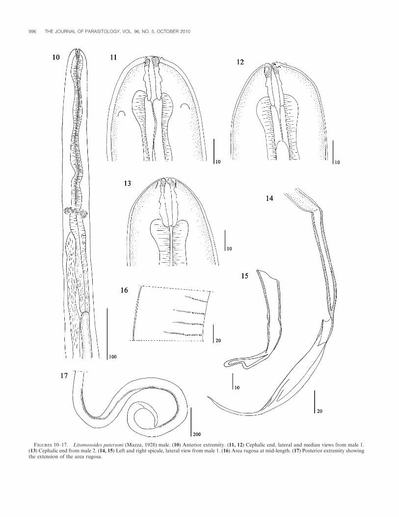
Caudal bursa asymmetrical, with right lobe more developed
than left (Figs. 29, 30). Prebursal papillae not observed. Right
lobe: pattern of type 2–3; rays 2 and 3 joined proximally,
diverging at half of their length, stout and of similar length; rays
4–6 with long common trunk; ray 5 stout, with strongly reinforced
margins; rays 4 and 6 thinner, divergent from ray 5 at distal and
proximal third of its length, respectively (Fig. 29). Left lobe:
pattern of type 2-2-1; rays 2 and 3 joined proximally, diverging at
distal third of their length, stout and of similar length; rays 4 and
994 THE JOURNAL OF PARASITOLOGY, VOL. 96, NO. 5, OCTOBER 2010
FIGURES 1–9. Litomosoides patersoni (Mazza, 1928) female. (1) Anterior extremity. (2–4) Cephalic end, lateral, median, and apical views from female2. (5) Transverse section posterior to the vulva. (6, 7) Tail, lateral and ventral views. (8) Tip of tail with phasmids. (9) Uterine microfilaria.
NOTARNICOLA ET AL.—REDESCRIPTION OF TWO NEMATODE SPECIES 995
FIGURES 10–17. Litomosoides patersoni (Mazza, 1928) male. (10) Anterior extremity. (11, 12) Cephalic end, lateral and median views from male 1.(13) Cephalic end from male 2. (14, 15) Left and right spicule, lateral view from male 1. (16) Area rugosa at mid-length. (17) Posterior extremity showingthe extension of the area rugosa.
996 THE JOURNAL OF PARASITOLOGY, VOL. 96, NO. 5, OCTOBER 2010

FIGURES 18–21. Litomosoides patersoni (Mazza, 1928) male. (18) Posterior end showing the spicules and papillae from male 1. (19–21) Tail from male2, lateral views from left and right sides and ventral view, respectively.
TABLE I. Measurements of males of Litomosoides patersoni (Mazza, 1928).
Measurements
CML5813 CML5810
M1 M2 M3 M4*
Body length 19.305 mm 15.85 mm 17.3 mm 12 mm
Maximum width 120 120 135 85
Buccal capsule (L 3 W) 18 3 7.5 22 3 8 19 3 7 17 3 9
Esophagus length 510 580 540 425
Nerve ring 340 360 340 –
Tail length 275 180 146 195
Left spicule 246 – 247 265
Handle/lamina 100/146 – 107/140 115/150
Right spicule 80 87 84 80
Area rugosa length 1,150 1,740 1,500 400
Area rugosa beginning at 500–1700 from
tip of tail 550–1,700 760–2,500 770–2,270 260–660
* Specimen found in the small intestine of the host.
TABLE II. Measurements of females of Litomosoides patersoni (Mazza, 1928).
Measurements
CML5813 CML5825
F1 ae + Tail F2 ae F3* F4 ae
Length of pieces 28.71 + 15.345 mm – ,31.6 mm –
Maximum width 250 300 160 260
Width at vulva 170 200 150 175
Buccal capsule (L 3 W) 24 3 9 24 3 9 26 3 9 26.5 3 10
Esophagus length 780 650 700 690
Nerve ring 490 260 470 600
Tail length 500 – – –
Vulva to apex 1,520 2,050 1,350 1,600
Vulva to e-i junction{ 720 1,400 610 1,000
ae, anterior extremity.* Female with the tail broken.{ Distance of the vulva to the esophagus-intestine junction.
NOTARNICOLA ET AL.—REDESCRIPTION OF TWO NEMATODE SPECIES 997

5 divergent at extremities; ray 6 short and thin, diverging from
common trunk at same level than the group formed by rays 2–3
(Fig. 30). Rays 8 thin, arising asymmetrically from proximal third
of dorsal ray. Dorsal ray divided at about distal third into 2
branches. These branches are not bifurcated at their distal end,
indicating that rays 9 and 10 probably are merged (Fig. 31).
Genital cone well developed, markedly conical, 100–140 (122)
long by 50–60 (58) wide at base (n 5 10). Papillae on genital cone
not observed. Spicules subequal, alate, ending in simple, pointed
tips. Length of spicules 740–970 (865), representing 23.3–33% of
body length. Gubernaculum quadrangular, 20–50 (36) long and
15–30 (23) wide (Fig. 29).
Females (14 specimens, except otherwise stated): 3.08–5.10
(3.83) mm long and 80–120 (98) wide at mid-body. Cephalic
vesicle 55–70 (60) long and 25–35 (29) wide. Nerve ring, excretory
pore, and deirids situated at 125–160 (141) (n 5 5), 190–260 (231)
(n 5 7), and 200–270 (240) (n 5 4) from apex, respectively.
Esophagus 280–400 (325) long (n 5 10). Monodelphic. Vulva
situated at 65–70 from caudal extremity (n 5 7). Vagina vera 25–
35 (31) long (n 5 8), vestibule 55–90 (68) long (n 5 11), sphincter
40–50 (44) long and 35–55 (46) wide, infundibulum 60–120 (82) (n
5 11). Uterus 400–750 (560) long, taking up 9.8–20.5% (14.8%) of
body length, containing 7–24 (16) eggs. Eggs at 2–8 blastomeres
stage, 55–80 long and 30–45 wide. Tail 20 (n 5 5) long. Posterior
FIGURES 22–28. Stilestrongylus stilesi Freitas, Lent and Almeida, 1937. (22) Female, anterior extremity, right lateral view. (23) Female, head, apicalview. (24–28) Transverse sections of the body. (24) Female at esophageal level (220 mm from apex). (25) Female, at mid-body (53% of body length). (26)Female at mid-length of uterus (400 mm from posterior end). (27) Male at mid-body (50% of body length). (28) Male at 150 mm anterior to caudal bursa.Abbreviations: R, right; V, ventral.
998 THE JOURNAL OF PARASITOLOGY, VOL. 96, NO. 5, OCTOBER 2010
FIGURES 29–34. Stilestrongylus stilesi Freitas, Lent and Almeida, 1937. (29–31) Male, caudal bursa. (29) Entire bursa, ventral view. (30) Detail of leftlobe, ventral view (spicules omitted). (31) Detail of dorsal lobe, ventral view. (32–34) Female, posterior extremity in 3 different specimens. (32, 33) Rightlateral view. (34) Left lateral view, showing eggs within cuticular dilatation.
NOTARNICOLA ET AL.—REDESCRIPTION OF TWO NEMATODE SPECIES 999

extremity, from vestibular level, invaginated into a cuticular
dilatation, 155–310 (187) long. Seven of 20 females harbored 1–8
eggs within this cuticular dilatation (Figs. 32–34).
Taxonomic summary
Host: Seventeen H. chacarius Thomas (Rodentia, Cricetidae)
deposited at Coleccion Mamıferos Lillo (numbers CML 5810–
5826).
Material studied: Males and females deposited as CHMLP
numbers 5993, 5994, 5996–6006, 6008, 6009, and 6096; males and
females deposited at MNHN (numbers 592 MQ to 594 MQ).
Site of infection: Small intestine.
Locality: Ingenio San Martın de Tabacal (23u169S, 64u159W),
lote Milagros, Departamento Oran, Salta, Argentina.
Prevalence and mean intensity: Seventeen hosts examined (100%
infected); 108 (4–826) worms per host.
Remarks
Freitas et al. (1937) based their description of S. stilesi, the
nominal species, on an unspecified number of males, plus 1 female
in the same work. The illustrations of the male (caudal bursa,
spicules, and gubernaculum) made by Freitas et al. (1937) are very
accurate, allowing confirmation of our specimens as S. stilesi. The
preservation of type material, available from CHIOC, precludes a
redescription of the species because some specimens are mounted
on slides and others preserved in ethanol are too fragile to permit
a description of the synlophe (M. C. Durette-Desset, pers.
comm.). The finding of voucher specimens parasitizing the same
host species, and probably from the same locality, permitted us to
make a complete description of S. stilesi.
Stilestrongylus was originally defined based mainly on the
characters of the caudal bursa, which is markedly asymmetrical
(Freitas et al., 1937). However, Durette-Desset (1971) redefined
the genus based on 5 species with known synlophe (Stilestrongy-
lus barusi Durette-Desset, 1970; S. dessetae Yoyotte-Vado, 1972;
S. freitasi Durette-Desset, 1968; S. inexpectatus Durette-Desset
and Tcheprakoff, 1969; and S. renaudae Durette-Desset, 1970)
and 4 species with unknown synlophe (S. stilesi, S. aculeata
[Travassos, 1918], S. eta [Travassos, 1937], and S. riberoi
[Travassos, 1937]). She established Stilestrongylus as having a
strongly asymmetrical caudal bursa, a hypertrophied genital
cone, and synlophe with more than 24 cuticular ridges that are
small and sub-equal, with axis of orientation of ridges from right
ventral to left dorsal quadrant. Since the synlophe observed in the
present specimens is coincident with the described characters, this
redescription also validates the redefinition of the genus by
Durette-Desset (1971).
In the material studied herein, some internal structures of the
anterior extremity are difficult to observe; in females, the caudal
structures are frequently hidden by the folds of the posterior
cuticular dilatation or the presence of eggs within it. The
inclination of the left axis of orientation differs between males
and females, but this should not be interpreted as a specific
character. The inclination of the axis on the left side is difficult to
evaluate, due to the opacity of the cuticle (orientation of ridges
unclear) and the frequent deformation of the cuticle on this side of
the body. Despite these minor constraints, the most important
characters can be observed and interpreted for both sexes. We
clarify in this work the asymmetry of the caudal bursa in having
the right lobe more developed than the left one, in contrast to the
affirmation by Freitas et al. (1937) where the left lobe is the most
developed.
DISCUSSION
Both species, L. patersoni and S. stilesi, are redescribed herein
with new morphological data. Currently, specimens of the
chacoan marsh rat distributed in the Yungas of Salta are referred
to H. chacarius Thomas, 1906 (Cirignoli et al., 2006). Mazza
(1928) misidentified the specimens from Salta, which he assigned
to H. vulpinus, a synonym of H. brasiliensis (Desmarest, 1819)
distributed in eastern Argentina (Pardinas and Galliari, 1998a,
1998b). However, Freitas at al. (1937) were correct in that the
hosts were H. balnearum, which is a subspecies of H. chacarius
(Cirignoli et al., 2006).
The site of infection for Litomosoides spp. is typically the
thoracic and/or abdominal cavity of the host. The present finding
of 1 male of L. patersoni in the intestine is unusual. However,
other authors have reported living filarioids in the intestine of
their host, i.e., Litomosa filaria (van Beneden) in bats (Desportes,
1946). Considering that this male worm was smaller than the
other collected in the body cavity, the intestine could be a
transitory organ in the migratory route.
Since the type specimens of L. patersoni have been lost, we
assigned the specimens from this survey (recovered from the type
locality and the type host species) as neotypes, according with the
Articles 75.3.4, 75.3.5, and 75.3.6 from the International Code of
Zoological Nomenclature (ICZN, 1999). For the heligmosomoid
S. stilesi, we validated and completed the description from Freitas
et al. (1937) and provided voucher specimens.
ACKNOWLEDGMENTS
We thank Dr. R. Barquez, curator of the Coleccion Mamıferos Lillo,and Dr. M. M. Dıaz, both from Tucuman, Argentina, who kindly madethe rodents available for parasitological examination; we also thank Dr.M.C. Durette-Desset from MNHN, Paris, France, for helpful commentson the manuscript, and Marıa Cristina Estivariz from CEPAVE for thedrawings. Funds came from Project N11/541 from SeCyT (UNLP,Argentina). J. Notarnicola and M.C. Digiani are members of CONICET.
LITERATURE CITED
BAIN, O., R. GUERRERO, B. RODRIGUEZ, S. BABAYAN, AND N. JOVEMENT.2003. Examination of type material of two species of Litomosoides(Filarioidea: Onchocercidae), parasites from bats; taxonomic conse-quences. Parasite 210: 211–218.
———, G. PETIT, AND M. DIAGNE. 1989. Etude de quelques Litomosoidesparasites de rongeurs: Consequences taxonomiques. Annales deParasitologie Humaine et Comparee 64: 268–289.
CHITWOOD, B. C. 1933. A note on the status of Vestibulosetaria Vogel ansGabaldon, 1932. Proceedings of the Helminthological Society. InJournal of Parasitology 19: 253.
CIRIGNOLI, S., P. TETA, U. F. J. PARDINAS, AND G. D’ELIA. 2006. TribuOryzomyni Vorontsov, 1959 (sensu Voss and Carleton, 1993). InMamıferos de Argentina. Sistematica y distribucion, R. M. Barquez,M. M. Dıaz, and R. A. Ojeda (eds.). Sociedad Argentina para elEstudio de los Mamıferos SAREM, Mendoza, Argentina, p. 166–175.
DESPORTES, C. 1946. Des filaires dans le tube digestif. Annales deParasitologie Humaine et Comparee 21: 138–141.
DURETTE-DESSET, M. C. 1971. Essai de classification des NematodesHeligmosomes. Correlation avec la paleobiogeographie des hotes.Memoires du Museum National de Histoire NaturelleSerie A Zoologie 49: 1–126.
1000 THE JOURNAL OF PARASITOLOGY, VOL. 96, NO. 5, OCTOBER 2010
———. 1985. Trichostrongyloid nematodes and their vertebrate hosts:Reconstruction of the phylogeny of a parasitic group. Advances inParasitology 24: 239–306.
———, AND A. G. CHABAUD. 1993. Note sur la nomenclature suprafa-miliale des Strongylida. Annales de Parasitologie Humaine etComparee 68: 11–12.
ESSLINGER, J. H. 1973. The genus Litomosoides Chandler, 1931 (Filarioi-dea: Onchocercidae) in Colombian bats and rats. Journal ofParasitology 59: 225–246.
FREITAS, J. F. T., H. LENT, AND J. L. ALMEIDA. 1937. Pequena contribuicaoao estudo da fauna helminthologica da Argentina. Memorias doInstituto Oswaldo Cruz 32: 195–209.
ICZN. 1999. International Code of Zoological Nomenclature. Interna-tional Trust for Zoological Nomenclature (eds.). London, U.K.
MAZZA, S. 1928. Filarideo n. sp. de la cavidad peritonal de la rata de loscanaverales de Tabacal, Salta. 4 Reunion de la Sociedad Argentina dePatologıa Regional del Norte: 628–632.
NOTARNICOLA, J. 2005. Description of adult and fourth-stage larva ofLitomosoides navonae n. sp. (Nematoda: Onchocercidae), a parasiteof five species of sigmodontine rodents from northeastern Argentina.Systematic Parasitology 62: 171–183.
———, G. T. NAVONE, AND O. BAIN. 2000. Two new species ofLitomosoides (Nematoda: Filarioidea) in sigmodontines (Rodentia:
Muridae) from Rıo de la Plata marshland, Argentina. Journal ofParasitology 86: 1318–1325.
OCHOTERENA, I., AND E. CABALLERO. 1932. Filaria parasita de las ratas decampo, Micropleura sigmodoni spec. nov. Anales del Instituto deBiologıa, Universidad Nacional de Mexico 3: 123–125.
PARDINAS, U. F. J., AND C. GALLIARI. 1998a. Comentario sobre el trabajo‘‘Los mamıferos del Parque Biologico Sierra San Javier, Tucuman,Argentina: Observaciones sobre su sistematica y distribucion’’Capllonch et al., 1997 (Mastozoologıa Neotropical, 4: 49–71).Mastozoologıa Neotropical 5: 61–62.
———, AND ———. 1998b. Sigmodontinos (Rodentia, Muridae) delHoloceno inferior de Bolivia. Revista Espanola de Paleontologıa 13:17–23.
VAZ, Z. 1934. Ackertia gen. nov. for Litomosa burgosi De La Barrera,1926, with notes on the synonymy and morphological variations ofLitomosoides carinii (Travassos, 1919). Annales of Tropical Medicineand Parasitology 28: 143–149.
VOGEL, H., AND A. GABALDON. 1932. Vestibulosetaria, eine neueFilariengattung aus Rattenarten. Zentralblatt fur Bakteriologie,Parasitenkunde und Infektionskrankhleiten 126: 119–124.
WILSON, D. E., AND D. A. M. REEDER. 2005. Mammal Species of theWorld. A taxonomic and geographic reference. Johns HopkinsUniversity Press, Baltimore, Maryland, 2142 p.
NOTARNICOLA ET AL.—REDESCRIPTION OF TWO NEMATODE SPECIES 1001