Factors influencing the level of circulating procoagulant microparticles in acute pulmonary embolism
Upload
independentCategory
view
7download
0
C E L L P H Y S I O L O G Y
Red blood cell microparticles show altered inflammatorychemokine binding and release ligand upon interaction
with platelets_2861 610..621
Zeyu Xiong, John Cavaretta, Lirong Qu, Donna Beer Stolz, Darrell Triulzi, and Janet S. Lee
BACKGROUND: Storage of red blood cells (RBCs)under standard blood bank conditions results inreduced structural integrity leading to membranebudding and release of microparticles. Microparticlesexpress the blood group Duffy antigen known to bindmultiple inflammatory chemokines, but the functionalchemokine binding properties of microparticles are notknown.STUDY DESIGN AND METHODS: We determinedwhether storage-induced microparticles show inflamma-tory chemokine binding through the expression of theDuffy antigen, comparing the binding properties to intactRBCs, and assessed microparticle interactions withplatelets (PLTs) that release chemokines upon activa-tion.RESULTS: Intact RBCs retained similar equilibrium dis-sociation constants for CCL2 (Kd = 7.4 � 0.9 nmol/L),CXCL8 (Kd = 7.9 � 1.0 nmol/L), and CXCL1 (Kd =4.4 � 1.0 nmol/L) throughout storage. In contrast,microparticles increased in relative counts with storage,showed higher percentages of surface phosphati-dylserine, and demonstrated impaired Duffy-dependentchemokine binding affinity with wider variability in disso-ciation constant for CXCL1(Kd = 362 � 328 nmol/L;range, 0.6-2000 nmol/L). The altered chemokine bindingaffinity of RBC microparticles was associated with apropensity to release ligand upon incubation with PLTs.Relative quantification of microparticles, based on crite-ria of glycophorin A expression and size, underesti-mated particle numbers with functional chemokinebinding, suggesting that glycophorin A–negative par-ticles and nanoparticles contribute to overall chemokinebinding capacity.CONCLUSION: Microparticle burden in transfusates, asdetermined by functional chemokine binding, is consid-erable. Altered membrane properties of RBC micropar-ticles enhance PLT interactions to increaseinflammatory chemokine bioavailability in vitro.
During storage, red blood cells (RBCs) losetheir membrane stability leading to hemoly-sis and microparticle formation.1,2 The RBCskeletal framework is essential for mem-
brane stability and consists of a filamentous meshworkof proteins created by a spectrin-spectrin dimer back-bone anchored to the lipid bilayer at junctional pointsthrough interaction with actin and protein 4.1R as well asinteractions with ankyrin.3,4 Storage reduces the forma-tion of the spectrin-actin-protein 4.1 complex that corre-lates tightly with reductions in total RBC membranephospholipid content.3 Storage also shifts the redoxpotential toward oxidative stress.1 Indeed, spectrin oxida-tion correlates strongly with membrane microparticleformation.5 It is also notable that microparticles shedduring storage generally lack the skeletal protein spec-trin.6 This is in contrast to Band 3, a membrane integralprotein that provides linkage between the lipid bilayerand the underlying skeletal framework, found in
ABBREVIATIONS: PMA = phorbol 12-myristate 13-acetate;
PPP = platelet-poor plasma; PRP = platelet-rich plasma.
From the Division of Pulmonary, Allergy, and Critical Care
Medicine, Department of Medicine; the Department of
Pathology; the Center for Biologic Imaging, Department of Cell
Biology and Physiology; and the Vascular Medicine Institute,
University of Pittsburgh, Pittsburgh, Pennsylvania.
Address reprint requests to: Janet S. Lee, Division of Pulmo-
nary, Allergy, Critical Care Medicine, NW628 Montefiore Hospi-
tal, 3459 Fifth Avenue, Pittsburgh, PA 15213; e-mail: leejs3@
upmc.edu.
This work was supported by HL086884, Hemostasis and
Vascular Biology Grant, and the Department of Medicine Junior
Scholar Award.
Received for publication May 6, 2010; revision received July
6, 2010, and accepted July 12, 2010.
doi: 10.1111/j.1537-2995.2010.02861.x
TRANSFUSION 2011;51:610-621.
610 TRANSFUSION Volume 51, March 2011
microparticles.6 Normally sequestered to the inner leafletof the membrane, anionic phospholipids such as phos-phatidylserine also interact with protein 4.1R and spec-trin to strengthen mechanical stability.7-9 However,microparticles lose phospholipid asymmetry and bearphosphatidylserine on their surface.10 Thus, the anchor-age points where membrane regions attach to the under-lying cytoskeleton may represent focal sites that favorbudding and microparticle formation.10
The Duffy antigen is a transmembrane protein andminor blood group antigen that interacts with the protein4.1R–based macromolecular complex—a complex thatincludes other transmembrane proteins such as Band 3,Rh, Kell, XK, and glycophorin C.11 Historically known asthe RBC receptor for merozoite invasion by malarial para-site Plasmodium vivax,12-15 Duffy antigen demonstrateshigh-affinity binding to multiple inflammatory CXC andCC chemokines such as CXCL1, CXCL7, CCL2, and CCL5but not CCL3 or CCL4 with receptor binding sites esti-mated at 1000 to 9000/RBC surface.12,16-20 The role of Duffyantigen in inflammatory states has remained elusive, inpart, due to its unique position as a nonsignaling proteinon circulating RBCs. However, emerging evidence supportthe role of RBC Duffy antigen as a modifier and regulatorof systemic and local inflammatory chemokinebioavailability.21-23
We have recently shown that transfusion of storedRBCs amplifies existing lung inflammation and promoteslung injury in a murine model of systemic endotoxemia.22
We also demonstrate that increased duration of RBCstorage can promote lung injury and implicate the reduc-tion of RBC chemokine scavenging as one consequence ofthe storage lesion. Studies have previously shown bloodgroup antigenicity on RBC microparticles,24 in particularthe presence of the Duffy antigen.10 We have previouslyshown reductions in surface Duffy antigen expression onintact RBCs with storage and this loss from the RBCsurface may result from membrane alterations and subse-quent microparticle formation. Whether or not Duffyprotein expressed on microparticles shows altered func-tional properties remains unknown.
Exposure of phosphatidylserine on the surface ofthe microparticle membrane provides a procoagulantsurface that facilitates thrombin generation.25-28 Throm-bin, a main agonist of platelet (PLT) activation,29 caninduce the release of a-granule contents such asP-selectin and chemokines,30 possibly further amplifyinginflammatory processes. However, the interactionbetween RBC microparticles and PLTs in perpetuatinginflammation is largely unknown. The purpose of thestudy was to determine the expression of Duffy antigenon the surface of microparticles obtained from RBC unitsunder standard blood bank conditions, determine itsfunctional properties in terms of chemokine binding,and assess its interaction with PLTs.
MATERIALS AND METHODS
Human subjectsAdsol-preserved leukoreduced and nonleukoreduced RBCunits were obtained from the Institute of TransfusionMedicine, Central Blood Bank of Pittsburgh, Pennsylvaniabased upon their availability. The majority of the studieswere conducted with nonleukoreduced units, but selectexperiments were performed using leukoreduced unitsonly as indicated. For RBC radioligand binding experi-ments and assays requiring PLT-poor plasma (PPP) or PLT-rich plasma (PRP), we obtained whole blood from healthyvolunteers after obtaining written informed consent. TheInstitutional Review Board of University of Pittsburghapproved the studies.
Reagents125I-CCL2, 125I-CXCL8, and 125I-CXCL1/GRO-a were pur-chased from Perkin-Elmer, Inc. (Waltham, MA). Recombi-nant human proteins CCL2, CXCL8, CXCL1, CXCL5, andCXCL7 were obtained from PeproTech, Inc. (Rocky Hill,NJ). Mouse immunoglobulin (Ig)G1 anti-human Duffy Fy6antibody (clone 2C3), glycophorin A–phycoerythrin (PE),and annexin V–fluorescein isothiocyanate (FITC) wereobtained from BD Biosciences (San Jose, CA). MouseIgG2b, k-PE was purchased from BD PharMingen (SanJose, CA). Mouse IgG1 isotype control antibody wasobtained from R&D Systems (Minneapolis, MN).
Isolation and purification of RBCs andmicroparticlesRBCs were purified from RBC units or from anticoagu-lated whole blood of healthy volunteers using a methodwe have previously described.22,31 The method for isola-tion and purification of RBC microparticles is a modifica-tion of the techniques previously described.32-34 Briefly,banked RBC units (90 mL) were centrifuged at 2000 ¥ g for20 minutes at 4°C. The pellet was removed and the super-natant was centrifuged at 3000 ¥ g for 10 minutes at 4°C.The supernatant was immediately filtered through asterile 0.8-mm pore size syringe-driven polycarbonatefilter units (Millipore, Billerica, MA) and examined underthe microscope to ensure the absence of RBCs. The super-natant was ultracentrifuged at 37,000 ¥ g at 4°C for 1 hour.The pellet containing microparticles was resuspended inphosphate-buffered saline (PBS) and ultracentrifugedtwice under the same conditions, resuspended in 300 mLof final volume of PBS, and maintained at 4°C. The ratio offinal resuspension volume to original volume of transfu-sate was maintained at 1:300, and experiments were con-ducted within 24 hours of isolation. A small aliquot of theresuspended microparticle pellet was examined under themicroscope to confirm the absence of RBCs and visible
RBC MICROPARTICLE CHEMOKINE BINDING
Volume 51, March 2011 TRANSFUSION 611

RBC fragments. Microparticles (5 mL) were labeled withglycophorin A-PE and annexin V-FITC and counted usingflow cytometry absolute counting standard microbeads(Bangs Laboratories, Inc., Fishers, IN) as detailed below.Microparticle preps were devoid of PLTs or PLT micropar-ticles, as determined by assaying for the constitutivePLT-specific marker CD42a by flow cytometry (datanot shown).
Flow cytometric analysis and relativequantification of RBC microparticlesRBC counts were obtained from each unit bag by manualcounting using a hemocytometer. The final supernatantvolume examined from each unit bag was normalized to108 RBCs. The supernatant was labeled with glycophorinA as indicated above. The forward scatter variable was setto log scale to identify microparticle, RBC, and fluores-cent absolute count beads. The flow was maintained atslow and stable rate, and events were acquired aftersamples were running for 2 minutes. To reduce thepossibility of carryover, cleaning with 10% bleach anddeionized water was performed between samples. Micro-particle counting was performed in duplicate or tripli-cate for each sample. Relative quantification ofmicroparticle counts from each RBC unit bag wasobtained on the same flow cytometer (FACSCalibur, BDBiosciences) with identical settings on the same day.Acquired events showed a coefficient of variation (1.34%-18.42%; mean, 10.94%). Microparticles were defined asglycophorin A positive and annexin V events at sizeapproximately 70 to 700 nm.
Immunoelectron microscopyIsolated microparticles were fixed in suspension with 2%paraformaldehyde in PBS and adsorbed to formvar-coated 100-mesh grids. Grids underwent several wash-ings with PBS and then PBS supplemented with 0.5%bovine serum albumin and 0.15% glycine. Microparticleswere blocked in 5% normal goat serum and incubatedwith a 1:50 dilution of mouse IgG1 anti-human Duffy Fy6antibody (Clone 2C3) and sheep anti-human hemoglo-bin (Hb), affinity-purified polyclonal antibody (BethylLaboratories, Inc., Montgomery, TX). Grids were incu-bated with goat anti-sheep biotinylated antibody (1:100,Jackson ImmunoResearch Laboratories, West Grove, PA)followed by goat anti-mouse 5-nm gold-conjugated sec-ondary antibody (1:25, Amersham, Piscataway, NJ) and10-nm gold-conjugated streptavidin (1:25, Amersham).Grids were fixed in 2.5% glutaraldehyde, poststained in2% neutral uranyl acetate, and finally embedded in1.25% methylcellulose. Labeling was observed on a elec-tron microscope (JEM 1011, JEOL, Peabody, MA) at 80 kV.Control grids were incubated with biotinylated anti-
sheep, goat anti-mouse 5-nm gold, and streptavidin10-nm gold reagents in the absence of primary antibody.No gold staining was observed in these samples.
Radioligand competition binding assays125I-Chemokine at 0.25 nmol/L was incubated with eithermicroparticles (approx. 0.5 ¥ 106) or RBCs (1 ¥ 108) in thepresence or absence of increasing molar concentrationsof unlabeled or “cold” homologous chemokine as com-petitor. Nonspecific binding was defined as 1 mmol/Lcold competitor. The mixture was incubated on ice for 60minutes. The reaction was terminated by centrifugingthe mixture through a sucrose cushion. The supernatantand pellet were measured in a gamma counter. Each datapoint was performed in duplicate or triplicate. Data wereanalyzed using a nonlinear regression curve fit program.A comparison of fits was used to define the preferredmodel for one-site-specific binding (Prism 5 v5.01,GraphPad, La Jolla, CA). For the quick chemokinebinding assays, 125I-chemokine at 0.25 nmol/L was incu-bated with either microparticles or RBCs in the presenceor absence of 3 ¥ 10-7 mol/L unlabeled homologouscompetitor.
PLT assaysWhole blood was drawn into acid-citrate-dextrose at aratio of 1:6 anticoagulant as previously described.35-37 Theblood was immediately centrifuged at 100 ¥ g for 15minutes at room temperature and the supernatant wasseparated and collected as PRP. The remaining bloodsample was centrifuged at 1880 ¥ g for 20 minutes andthe supernatant was separated and collected as PPP. Theabsence of RBCs and white blood cells were confirmedby light microscopy, and PLT counts from samples weremanually counted using a hemocytometer. PRP andPPP from the same individual served as their owncomparison.
Phorbol 12-myristate 13-acetate (PMA; Sigma, StLouis, MO) at 200 ng/mL was added to fixed volumes ofthe PRP or PPP, incubated for 10 minutes at room tem-perature. Purified RBC microparticles at a fixed volumewere incubated with 125I-CXCL1 (0.25 nmol/L) and thenadded to PRP or PPP. The entire mixture was incubated at4°C for 60 minutes. The reaction was terminated by cen-trifuging the mixture through a sucrose cushion. Countsper minute for the supernatant and the pellet fractionswere obtained using a gamma counter. Each data pointwas performed in triplicate.
Statistical analysisA paired t test was performed to compare two matchedgroups. A Mann-Whitney nonparametric test was applied
XIONG ET AL.
612 TRANSFUSION Volume 51, March 2011

to compare the distributions of twounmatched groups. For comparisonsinvolving more than two groups, analy-sis of variance (ANOVA) was performedand a posttest analysis to compareamong two groups. A p value of less than0.05 was considered significant. Corre-lation was calculated with Spearmancorrelation coefficient and p value. Sta-tistics were performed using computersoftware (Prism 5, GraphPad). Data infigures are presented as mean � SEM.
RESULTS
Storage does not alter the bindingaffinity of intact RBCs forchemokines CCL2 and CXCL8To determine whether in-storage timeinduces alterations of the chemokinebinding pocket and, hence, the bind-ing affinity for ligand, we performedhomologous competitive binding as-says on fresh and stored RBCs using125I-CCL2 to calculate the equilibriumdissociation constants (Kd) for CCL2.Fresh RBCs showed saturable and spe-cific binding in the presence of increas-ing molar concentrations of unlabeledor “cold” competitor CCL2 (data notshown). Similar to reports by others,20
the Kd was estimated at 6 nmol/L for arepresentative sample (Fig. 1A). We alsocompared the Kd of fresh RBCs to that ofstored RBCs ranging from Day 1 to Day41 and show no significant difference inbinding affinities across storage time(Fig. 1C).
The Duffy antigen is a promiscuouschemokine binding protein that bindsboth inflammatory CC- and CXC-chemokines. We determined whetherstorage alters RBC binding affinity forCXCL8, the canonical inflammatoryCXC chemokine. Homologous competition binding assaysusing 125I-CXCL8 in the presence of increasing concentra-tions of unlabeled competitor CXCL8 of fresh RBCs fromhealthy volunteers also showed saturable and specificbinding (data not shown). The estimated Kd for RBCs froma representative healthy volunteer was 15 nmol/L(Fig. 1B). The Kd of fresh RBCs was also compared to thatof stored RBCs ranging from Day 1 to Day 37 and showedno significant difference in binding affinities acrossstorage time (Fig. 1D). We also performed heterologouscompetitive radioligand binding assays and show the
ability of various CXC ligands to compete off bound 125I-CCL2 and 125I-CXCL1 (Fig. 1E and 1F). Storage did notappreciably alter the ability of heterologous ligands tocompete off-bound chemokine (data not shown). Notably,PLT chemokines CXCL5 and CXCL7 showed dissociationconstants for competitor binding (Ki) between that ofCXCL1 and CXCL8, whereas Duffy null (Fya–b–) RBCs didnot show specific binding to either CC or CXC ligands(Figs. 1E and 1F). Thus, the findings show that the bindingaffinity for inflammatory chemokines is not altered onintact RBCs with increasing duration of storage.
Kd= 6 nmol/L
Bound CCL2 (nmol/L)
Log unlabeled ligand (mol/L) Log unlabeled ligand (mol/L)
Kd (
nm
ol/L)
Kd (
nm
ol/L)
Bound CXCL8 (nmol/L)
CXCL1 Ki = 9 nmol/L
CXCL5 Ki = 13 nmol/L
CXCL7 Ki = 42 nmol/L
CXCL8 Ki = 55 nmol/L
CCL2 Fynull Ki = NA
CXCL1 Kd = 3 nmol/L
CXCL5 Ki = 56 nmol/L
CXCL7 Ki = 118 nmol/L
CXCL8 Ki = 74 nmol/L
CXCL8 Fynull Ki = NA
Kd= 15 nmol/L
Fig. 1. CCL2 and CXCL8 binding affinity of fresh versus stored RBCs. (A) Equilibrium
dissociation constant (Kd) for CCL2 presented in Scatchard plot form, using repre-
sentative fresh RBCs from a volunteer. (B) Scatchard plot data for CXCL8, using
representative fresh RBCs from a volunteer. (C) Fresh versus stored RBCs (Days
1-41) show similar CCL2 binding affinity, a comparison of Kd’s. Fresh RBCs,
Kd = 7.3 � 1.9 nmol/L (n = 4); stored RBCs, Kd = 7.1 � 0.9 nmol/L (n = 13). (D) Fresh
versus stored RBCs (Days 1-37) show similar CXCL8 binding affinity. Fresh RBCs,
Kd = 9.0 � 3.4 nmol/L (n = 5); stored RBCs, Kd = 7.9 � 1.0 nmol/L (n = 5). Data were
obtained from Adsol-preserved leukoreduced and nonleukoreduced RBC units, puri-
fied and isolated in the same manner as fresh RBCs, and performed in triplicates.
(E, F). Heterologous competitive binding assays show percentage of total 125I-CCL2 or125I-CXCL1 binding as a function of increasing molar concentrations of unlabeled
CXCL1, CXCL5, CXCL7, and CXCL8. For 125I-CCL2 binding, representative Ki’s for
fresh RBCs are shown. For 125I-CXCL1 binding, representative Ki’s for stored RBCs
are shown (n = 2-5 for each ligand tested comparing fresh to older stored RBCs,
Days 20-32). The dissociation constant for competitor binding (Ki) is provided for
each ligand. Fynull RBCs do not bind chemokine.
RBC MICROPARTICLE CHEMOKINE BINDING
Volume 51, March 2011 TRANSFUSION 613

Fya–b+ RBCs show reduced surface Duffy antigenexpression and chemokine binding sites butbinding affinity to CCL2 is not altered andindependent of storage durationWe examined whether the two major alloantigens of Duffyprotein (Fya+b–, Fya–b+) showed differences in RBCinflammatory chemokine binding with storage duration.Fya and Fyb antigens clinically define four phenotypes ofthe minor blood group Duffy antigen: Fya+b–, Fya–b+,Fya+b+, and Fya–b– RBCs. These phenotypes are the prod-ucts of codominant alleles comprising genotypes FyA/FyA, FyB/FyB, FyA/FyB, and FyB*/FyB*, respectively.38 TheFya/Fyb alloantigen system is due to a single polymor-phism in Codon 42 that encodes a glycine residue in Fyaand aspartic acid in Fyb39 and appears to show no knownbiologic consequence,38 although the inflammatorychemokine binding properties have not been systemati-cally compared. We observed a reduction in Duffy antigen(Fy) surface expression among Fya–b+ RBCs comparedwith Fya+b– and Fya+b+ RBCs (Fig. 2A). Fya–b+ RBCs alsoshowed reduced surface 125I-CCL2 binding after competi-tion with excess unlabeled competitor CCL2 (Fig. 2B). Thisreduction in surface radioligand binding can be explainedby either reduced numbers of binding sites available orreduced binding affinity. Fya+b– RBCs showed similarCCL2 binding affinity compared to Fya–b+ RBCs(Kd = 6.5 � 1.1 nmol/L vs. 8.7 � 1.5 nmol/L, respectively)that was independent of storage duration (Fig. 2C), sug-gesting that reduced number of binding sites account forthe reduced surface 125I-CCL2 binding observed in Fya–b+RBCs (Fig. 2B).
Based upon the Bmax, the calculated estimates ofreceptor sites available per cell surface were similarbetween the two groups (receptor sites 4080 � 1480/cellsurface vs. 4030 � 1500/cell surface, respectively). Thisestimate is in agreement with previous published reportsfor RBCs,16 but is unlikely to discriminate between small
differences that require larger size sampling. Thus, Fya–b+RBCs possess reduced surface Fy expression compared toFya+b– RBCs independent of storage duration; however,the binding affinity of the available numbers of surface Fyfor CCL2 is not appreciably different between the twophenotypes.
RBC microparticle counts increase with storageduration and express surface Duffy antigenMicroparticles express components of the RBC mem-brane such as glycophorin A32 and are distinguished bytheir forward scatter properties (or size) relative to RBCs.We employed 7-mm-sized fluorescent beads and esti-mated the size of RBC microparticles at approximately 70to 700 nm by flow cytometry (Fig. 3A). Our findings aresimilar to those of published reports using scanning elec-tron microscopy.33 A high percentage of microparticles(57%) expressed surface phosphatidylserine at Day 40 ofstorage, indicating significant phospholipid membraneredistribution but only a minority of RBCs from the sameunit bag (3%) showed this rearrangement (Fig. 3B).
We initially examined leukoreduced RBC units for theamount of microparticle formation as a function ofstorage time. Microparticles were identified by size andexpression of glycophorin A and quantified by assayingthe numbers proportional to a known amount of fluores-cent microbeads added into each sample. By normalizingto RBC counts in each of the unit bags assayed, we wereable to quantify the amount of microparticles in a RBCunit relative to other RBC units assayed. Microparticlecounts increased from Day 13 to Day 29 (data not shown),as did the percentage of annexin V–positive microparticleswith storage duration (Fig. 3C, bottom left panel). Exam-ining the data differently, we showed that total numbers ofannexin V–positive microparticles also increased fromStorage Day 13 to Storage Day 29 (p < 0.05; Fig. 3C, top
Fig. 2. Comparison of Fy serotypes according to Fy surface expression, percentage of 125I-CCL2 bound, and binding affinity for
CCL2. (A) RBCs from Adsol-preserved leukoreduced RBC units matched for storage duration were tested for Duffy surface expres-
sion by flow cytometry using monoclonal antibody Fy6. The mean fluorescence intensity (MFI) as a function of Fy serologic status.
(B) Percentage of 125I-CCL2 RBC surface binding following competition with 200 nmol/L CCL2 (n = 10 units, each point reflecting
RBCs from a different RBC unit). (C) CCL2 binding affinity: Fya+b– RBCs, Kd = 6.5 � 1.1 nmol/L (n = 3, performed in triplicates);
Fya–b+ RBCs, Kd = 8. 7 � 1.5 nmol/L (n = 5, performed in triplicates). Mann-Whitney, p < 0.05.
XIONG ET AL.
614 TRANSFUSION Volume 51, March 2011

right panel). Thus, relative quantification of microparticlecounts is possible and, regardless of the method of analy-sis, microparticles increased with storage duration and asignificant proportion of these microparticles showedrearrangement of membrane phospholipids.
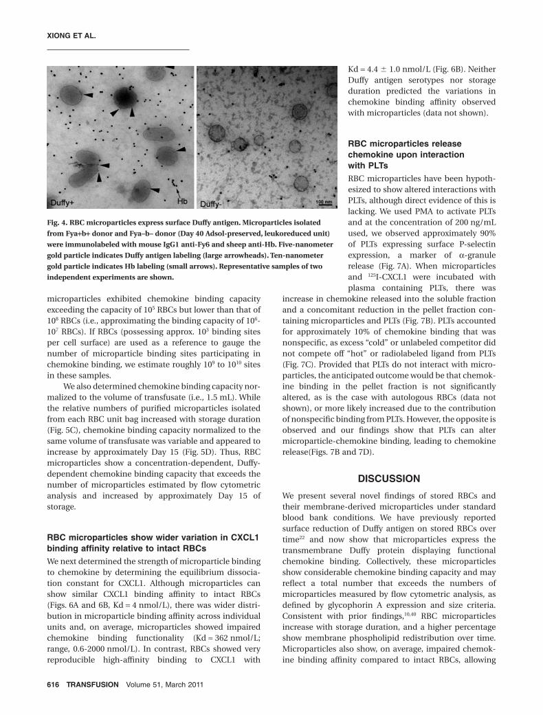
Others have previously determined that blood groupantigens are expressed on microparticles using indirectagglutination testing24 and by Western blotting.10 Byimmunoelectron microscopy, purified Fya+b+ micropar-ticles express surface Duffy antigen but Fya–b– micropar-
ticles do not (Fig. 4). Relative to the free Hb observedwithin the pellet fraction, Duffy expression on any givenFya+b+ microparticle is observed attached to the mem-brane surface. Based on this observation, we were able torule out the possibility of significant clustering of Duffymonomeric chains during the process of microparticleformation.
RBC microparticles show Duffy-dependentfunctional chemokine binding that increasesduring storageWe determined whether RBC microparticles are potentialreservoirs for inflammatory chemokine binding by ini-tially examining microparticles from expired RBC units.Microparticles, estimated at approximately 106 countsbased on predefined criteria of glycophorin A expressionand size by flow cytometry, showed 125I-CXCL1 bindingthat was concentration dependent, as serial dilutions ofmicroparticles resulted in progressive reductions inbound chemokine in the microparticle fraction andincreased within the soluble fraction (Fig. 5A).
To confirm Duffy-dependent chemokine binding, wepurified microparticles from the four different Fy sero-types between Day 22 and Day 27 of storage, comparingthem to the parent RBCs. Chemokine binding was Duffyantigen dependent as bound 125I-CXCL1 on Fya+b–,Fya+b+, Fya–b+ microparticles could be competed offwith excess cold competitor CXCL1 whereas Fya–b–microparticles showed no appreciable binding (Fig. 5B).Adjusted for approximately 105 relative counts per sample,
100
101
102
103
104
100
101
102
103
104
100
101
102
103
104
100
101
102
103
104
epyt
osi b2
GgI es
uo
M
A nir
oh
pocyl
G
Forward Scatter
AMicroParticles
RBCs
sdae
B
Day 13 Day 290
20
40
60
80
100 *
% A
nn
exi
n V
+ m
icro
part
icle
s
100
101
102
103
104
100
101
102
103
104
0.32% 1.82%
2.14%
Day 13 Day 29
102
103
104
105
101
*
count/10
8 R
BC
s
A nir
oh
pocyl
G
C
Annexin V
selcitrap
orciM
V ni xe
nn
A
+
A nir
oh
pocyl
G
Annexin V
BMicroparticles RBCs
100
101
102
103
104
100
101
102
103
104
0.15%0.02%
3.26%96.57%
100
101
102
103
104
100
101
102
103
104
4.75% 6.38%
82.61% 6.26%
�Fig. 3. Relative quantification of RBC microparticles obtained
from RBC units (Adsol-preserved, leukoreduced). (A) RBCs
from Day 40 RBC unit were washed, counted, and stained with
glycophorin A (right panel) or isotype control antibody (left
panel). Fluorescent microbeads (approx. 7 mm in dimension)
that were added are the same size as glycophorin A–positive
RBCs. Note the forward scatter is in log scale, and micropar-
ticles are approximately 70 to 700 nm in size. (B) A signifi-
cantly higher percentage of glycophorin A–positive
microparticles express surface phosphatidylserine, indicated
by annexin V binding (approx. 57% of total glycophorin A
events) compared with intact Day 40 parent RBCs. (C) Relative
quantification of microparticles. Supernatants from Day 13
or Day 29 RBC units were labeled with glycophorin A and
annexin V (top left panel). The volume of supernatant assayed
from each unit bag was normalized to 108 RBC counts. The
numbers of annexin V–positive microparticles per 108 RBCs
were higher in Day 29 versus Day 13 RBC units (p < 0.05; top
right panel). The percentage of annexin V–positive micropar-
ticles were also higher in Day 29 versus Day 13 RBC units
(p < 0.05; bottom left panel), with each point representing
individual RBC units.
RBC MICROPARTICLE CHEMOKINE BINDING
Volume 51, March 2011 TRANSFUSION 615
microparticles exhibited chemokine binding capacityexceeding the capacity of 105 RBCs but lower than that of108 RBCs (i.e., approximating the binding capacity of 106-107 RBCs). If RBCs (possessing approx. 103 binding sitesper cell surface) are used as a reference to gauge thenumber of microparticle binding sites participating inchemokine binding, we estimate roughly 109 to 1010 sitesin these samples.
We also determined chemokine binding capacity nor-malized to the volume of transfusate (i.e., 1.5 mL). Whilethe relative numbers of purified microparticles isolatedfrom each RBC unit bag increased with storage duration(Fig. 5C), chemokine binding capacity normalized to thesame volume of transfusate was variable and appeared toincrease by approximately Day 15 (Fig. 5D). Thus, RBCmicroparticles show a concentration-dependent, Duffy-dependent chemokine binding capacity that exceeds thenumber of microparticles estimated by flow cytometricanalysis and increased by approximately Day 15 ofstorage.
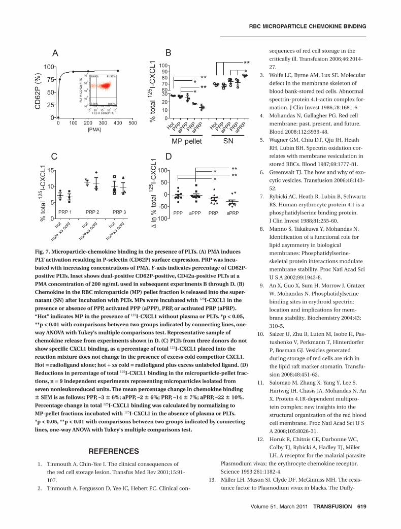
RBC microparticles show wider variation in CXCL1binding affinity relative to intact RBCsWe next determined the strength of microparticle bindingto chemokine by determining the equilibrium dissocia-tion constant for CXCL1. Although microparticles canshow similar CXCL1 binding affinity to intact RBCs(Figs. 6A and 6B, Kd = 4 nmol/L), there was wider distri-bution in microparticle binding affinity across individualunits and, on average, microparticles showed impairedchemokine binding functionality (Kd ª 362 nmol/L;range, 0.6-2000 nmol/L). In contrast, RBCs showed veryreproducible high-affinity binding to CXCL1 with
Kd = 4.4 � 1.0 nmol/L (Fig. 6B). NeitherDuffy antigen serotypes nor storageduration predicted the variations inchemokine binding affinity observedwith microparticles (data not shown).
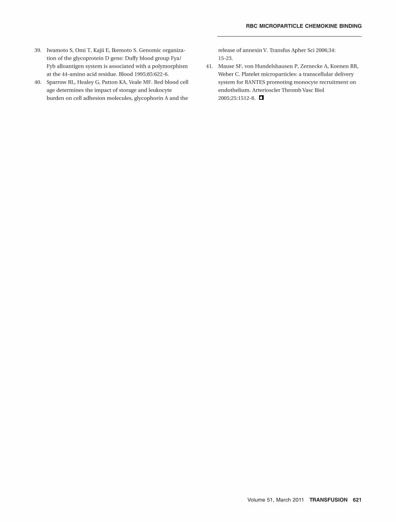
RBC microparticles releasechemokine upon interactionwith PLTsRBC microparticles have been hypoth-esized to show altered interactions withPLTs, although direct evidence of this islacking. We used PMA to activate PLTsand at the concentration of 200 ng/mLused, we observed approximately 90%of PLTs expressing surface P-selectinexpression, a marker of a-granulerelease (Fig. 7A). When microparticlesand 125I-CXCL1 were incubated withplasma containing PLTs, there was
increase in chemokine released into the soluble fractionand a concomitant reduction in the pellet fraction con-taining microparticles and PLTs (Fig. 7B). PLTs accountedfor approximately 10% of chemokine binding that wasnonspecific, as excess “cold” or unlabeled competitor didnot compete off “hot” or radiolabeled ligand from PLTs(Fig. 7C). Provided that PLTs do not interact with micro-particles, the anticipated outcome would be that chemok-ine binding in the pellet fraction is not significantlyaltered, as is the case with autologous RBCs (data notshown), or more likely increased due to the contributionof nonspecific binding from PLTs. However, the opposite isobserved and our findings show that PLTs can altermicroparticle-chemokine binding, leading to chemokinerelease(Figs. 7B and 7D).
DISCUSSION
We present several novel findings of stored RBCs andtheir membrane-derived microparticles under standardblood bank conditions. We have previously reportedsurface reduction of Duffy antigen on stored RBCs overtime22 and now show that microparticles express thetransmembrane Duffy protein displaying functionalchemokine binding. Collectively, these microparticlesshow considerable chemokine binding capacity and mayreflect a total number that exceeds the numbers ofmicroparticles measured by flow cytometric analysis, asdefined by glycophorin A expression and size criteria.Consistent with prior findings,10,40 RBC microparticlesincrease with storage duration, and a higher percentageshow membrane phospholipid redistribution over time.Microparticles also show, on average, impaired chemok-ine binding affinity compared to intact RBCs, allowing
Fig. 4. RBC microparticles express surface Duffy antigen. Microparticles isolated
from Fya+b+ donor and Fya–b– donor (Day 40 Adsol-preserved, leukoreduced unit)
were immunolabeled with mouse IgG1 anti-Fy6 and sheep anti-Hb. Five-nanometer
gold particle indicates Duffy antigen labeling (large arrowheads). Ten-nanometer
gold particle indicates Hb labeling (small arrows). Representative samples of two
independent experiments are shown.
XIONG ET AL.
616 TRANSFUSION Volume 51, March 2011

for greater propensity to release ligand upon interactionwith PLTs.
Stored RBCs undergo oxidative modification such asspectrin oxidation that predisposes toward membranemicroparticle formation.5 Our findings indicate that intactRBCs do not show functional alterations in CCL2, CXCL8,or CXCL1 binding affinity. Thus, the chemokine bindingpocket created by the tertiary structure of the Duffyprotein shows relatively preserved binding on intact RBCsduring storage with predictable Kd of approximately4 nmol/L for CXCL1. This is in contrast to the impaired
chemokine binding affinity observedfor microparticles. We speculate thatthe phospholipid redistribution of themicroparticle membrane may alter thestability of Duffy-ligand binding.
We also observed small differencesin surface chemokine binding capacitybetween Fya+b– and Fya–b+ serotypes,with Fya–b+ RBCs showing bothreduced surface Fy expression andreduced 125I-CCL2 binding after compe-tition with excess cold competitor.Although there was wider variation inCCL2 binding affinity using Fya–b+RBCs, the differences were not signifi-cant and appeared to be storage inde-pendent. We conclude that differencesin chemokine binding affinity betweenthe Fy serotypes may be distinct butonly uncovered with larger populationstudies.
Indeed, in a recent genomewideassociation study involving three inde-pendent cohorts (n = 9598), the stron-gest association regulating circulatingCCL2 concentrations was the nonsyn-onymous polymorphism Asp42Gly inthe Duffy antigen gene.23 The Asp42Glypolymorphism results in the Fya–b+(A/A genotype, Asp) and Fya+b– (G/Ggenotype, Gly) serotypes.39 The A/Agenotype (corresponding to Fya–b+serotype) was associated with higherserum CCL2 concentrations but notplasma concentrations, leading theauthors to postulate that serum CCL2concentrations represented the combi-nation of plasma CCL2 and additionalCCL2 released from RBC Duffy antigenthrough the process of clotting. Consis-tent with our observations, the studyalso showed a similar reduction insurface Fy expression in individualsexpressing the Fya–b+ (Asp, A/A)23 sero-
type, although the authors concluded that this finding didnot represent a meaningful difference to account for thehigher systemic CCL2 concentrations. However, the pos-sibility exists that reduced surface binding sites can resultin higher soluble chemokine concentrations in vitro dueto the equilibrium dissociation kinetics of ligand to theDuffy protein and that clotting suggests an interactionbetween PLTs and RBC chemokine binding.
Although relative microparticle counts increasedwith storage duration, the relationship between chemok-ine binding capacity and storage was not linear. The
Fig. 5. RBC microparticle show inflammatory chemokine binding that is Duffy
dependent. Purified microparticle pellets were assayed by flow cytometry using gly-
cophorin A expression and size criteria to quantify the relative numbers. (A) Micro-
particles (MP) show concentration-dependent 125I-CXCL1 binding. Serial dilutions of
microparticles (approx. 106 counts) isolated from expired RBC unit (Adsol-preserved,
Fya+b+ serotype) at 1:1, 1:2, 1:4, and 1:8 were prepared and incubated with
0.25 nmol/L 125I-CXCL1. Progressive reduction in percentage of 125I-CXCL1 binding in
microparticle pellet is associated with increases in radiolabeled ligand found in the
supernatant (SN). A representative sample of n = 3 individual units examined. (B)125I-CXCL1 binding of microparticles (denoted by Fy serotype) at approximately
1 ¥ 105 relative counts and parent RBCs. Incubation with radioligand alone ( ) or
radioligand plus excess unlabeled ligand (�) are indicated. The amount of chemok-
ine binding in counts per minute was expressed as percentage of total 125I-CXCL1
added in the absence of microparticles or RBCs. Samples were obtained from leuko-
reduced units and performed in triplicate (n = 12 samples examined, four represen-
tative samples shown). (C) Relative microparticle counts from RBC units correlate
with storage duration. Spearman correlation coefficient r = 0.8375, p < 0.0001. (D)
Chemokine binding capacity of microparticles from RBC units correlates with
storage duration. Spearman correlation coefficient r = 0.5365, p = 0.04. Each point
shown in C and D represent microparticles isolated from a different RBC unit,
n = 18.
RBC MICROPARTICLE CHEMOKINE BINDING
Volume 51, March 2011 TRANSFUSION 617

increase in microparticle-chemokine binding occurredby Day 15 and remained fairly constant throughout theshelf life of blood (Day 42) although expired bloodshowed higher chemokine binding. In addition, a limita-tion of relative quantification was the definition ofmicroparticles. Strictly defining microparticles by glyco-phorin A expression and size criteria precluded thepossibility of including either glycophorin A–negativeparticles or particles of smaller dimensions with func-tional chemokine binding. Because we made no attemptto separate out nanoparticles from microparticles duringthe ultracentrifugation steps, it is reasonable to concludethat our microparticle counts underestimate the amountof particles participating in chemokine binding. Indeed,the chemokine binding capacity attributable to micro-particles is consistent with this hypothesis given thelimited numbers of Duffy antigen identified per micro-particle surface by immunoelectron microscopy. If thenumbers of predicted chemokine binding sites are anindication of actual particle counts, this suggests thatloss of RBC Duffy antigen by microparticle formationpoint to a region of membrane vulnerability. Based onDuffy’s location within the 4.1R macromolecular com-plex,11 this is a region of the membrane where anchorageto the skeletal framework may be compromised duringstorage.3
RBC microparticles have been shown to facilitatethrombin generation in vitro10 and this is consistent withsurface exposure of phosphatidylserine on microparticles.Our findings show that PLTs attenuate microparticlechemokine binding and activation of PLTs may accentuatethis process. One possible explanation for this finding isthat PLTs may physically bind microparticles and subse-quently reduce the number of binding sites available forchemokines. Physical interaction between PLTs andmicroparticles may also activate PLTs, resulting in further
release of chemokines from alpha granules that cancompete off microparticle-bound chemokines. Indeed,PLT chemokines such as CXCL5 and CXCL7 show theability to compete off both microparticle-bound CCL2and CXCL1 with Kis between those of CXCL1 or CXCL8.Others have shown that PLT microparticles can provide amechanism for chemokine deposition to activated endot-helium leading to monocyte arrest.41 We speculate thatRBC microparticles show a dynamic relationship withPLTs leading to the binding and release of chemokinesthat may alter local inflammatory microenvironmentsin vivo.
In summary, RBC microparticles under standardstorage conditions express transmembrane Duffy proteinwith impaired chemokine binding functionality. While theDuffy protein does not appear to significantly cluster onthe microparticle surface, its collective chemokinebinding capacity is considerable and suggests that relativeestimation of microparticle numbers by flow cytometricanalysis using glycophorin A expression and size criterialikely underestimates the true particle count. Moreover,altered membrane property of microparticles canenhance interactions with PLTs to release chemokinebound on the microparticle surface and alter soluble con-centrations of ligand.
ACKNOWLEDGMENTS
The authors thank Mr Frank Cornell at the Institute for Transfu-
sion Medicine for providing red blood cells and Ming Sun for
immunoTEM preparation.
CONFLICT OF INTEREST
The authors declare that they have no conflicts of interest rel-
evant to the manuscript submitted to TRANSFUSION.
Kd= 4 nmol/L
Total ligand in well (nmol/L)
MicroparticleTotal ligand in well (nmol/L)
RBCs
RBCs
Specific
125I-
CX
CL1
Bound (
nm
ol/L)
Specific
125I-
CX
CL1
Bound (
nm
ol/L)
Kd (
nm
ol/L)
Kd= 4 nmol/L
Microparticle
Fig. 6. Saturation curve, Scatchard plot, and comparisons of CXCL1 binding affinity of microparticles and stored RBCs. (A) Micro-
particles show saturable and specific 125I-CXCL1 binding. Inset data in Scatchard plot form, Kd = 4 nmol/L. (B) RBCs show saturable
and specific 125I-CXCL1, Kd = 4 nmol/L. (C) Wide variation in microparticle binding affinity for CXCL1, Kd = 362 � 328 nmol/L
(range, 0.6-2000 nmol/L) compared with RBC binding. Each point represents microparticles or RBCs isolated from individual RBC
units, assayed in duplicate or triplicates.
XIONG ET AL.
618 TRANSFUSION Volume 51, March 2011
REFERENCES
1. Tinmouth A, Chin-Yee I. The clinical consequences of
the red cell storage lesion. Transfus Med Rev 2001;15:91-
107.
2. Tinmouth A, Fergusson D, Yee IC, Hebert PC. Clinical con-
sequences of red cell storage in the
critically ill. Transfusion 2006;46:2014-
27.
3. Wolfe LC, Byrne AM, Lux SE. Molecular
defect in the membrane skeleton of
blood bank-stored red cells. Abnormal
spectrin-protein 4.1-actin complex for-
mation. J Clin Invest 1986;78:1681-6.
4. Mohandas N, Gallagher PG. Red cell
membrane: past, present, and future.
Blood 2008;112:3939-48.
5. Wagner GM, Chiu DT, Qju JH, Heath
RH, Lubin BH. Spectrin oxidation cor-
relates with membrane vesiculation in
stored RBCs. Blood 1987;69:1777-81.
6. Greenwalt TJ. The how and why of exo-
cytic vesicles. Transfusion 2006;46:143-
52.
7. Rybicki AC, Heath R, Lubin B, Schwartz
RS. Human erythrocyte protein 4.1 is a
phosphatidylserine binding protein.
J Clin Invest 1988;81:255-60.
8. Manno S, Takakuwa Y, Mohandas N.
Identification of a functional role for
lipid asymmetry in biological
membranes: Phosphatidylserine-
skeletal protein interactions modulate
membrane stability. Proc Natl Acad Sci
U S A 2002;99:1943-8.
9. An X, Guo X, Sum H, Morrow J, Gratzer
W, Mohandas N. Phosphatidylserine
binding sites in erythroid spectrin:
location and implications for mem-
brane stability. Biochemistry 2004;43:
310-5.
10. Salzer U, Zhu R, Luten M, Isobe H, Pas-
tushenko V, Perkmann T, Hinterdorfer
P, Bosman GJ. Vesicles generated
during storage of red cells are rich in
the lipid raft marker stomatin. Transfu-
sion 2008;48:451-62.
11. Salomao M, Zhang X, Yang Y, Lee S,
Hartwig JH, Chasis JA, Mohandas N, An
X. Protein 4.1R-dependent multipro-
tein complex: new insights into the
structural organization of the red blood
cell membrane. Proc Natl Acad Sci U S
A 2008;105:8026-31.
12. Horuk R, Chitnis CE, Darbonne WC,
Colby TJ, Rybicki A, Hadley TJ, Miller
LH. A receptor for the malarial parasite
Plasmodium vivax: the erythrocyte chemokine receptor.
Science 1993;261:1182-4.
13. Miller LH, Mason SJ, Clyde DF, McGinniss MH. The resis-
tance factor to Plasmodium vivax in blacks. The Duffy-
Fig. 7. Microparticle-chemokine binding in the presence of PLTs. (A) PMA induces
PLT activation resulting in P-selectin (CD62P) surface expression. PRP was incu-
bated with increasing concentrations of PMA. Y-axis indicates percentage of CD62P-
positive PLTs. Inset shows dual-positive CD62P-positive, CD42a-positive PLTs at a
PMA concentration of 200 ng/mL used in subsequent experiments B through D. (B)
Chemokine in the RBC microparticle (MP) pellet fraction is released into the super-
natant (SN) after incubation with PLTs. MPs were incubated with 125I-CXCL1 in the
presence or absence of PPP, activated PPP (aPPP), PRP, or activated PRP (aPRP).
“Hot” indicates MP in the presence of 125I-CXCL1 without plasma or PLTs. *p < 0.05,
**p < 0.01 with comparisons between two groups indicated by connecting lines, one-
way ANOVA with Tukey’s multiple comparisons test. Representative sample of
chemokine release from experiments shown in D. (C) PLTs from three donors do not
show specific CXCL1 binding, as a percentage of total 125I-CXCL1 placed into the
reaction mixture does not change in the presence of excess cold competitor CXCL1.
Hot = radioligand alone; hot + xs cold = radioligand plus excess unlabeled ligand. (D)
Reductions in percentage of total 125I-CXCL1 binding in the microparticle-pellet frac-
tions, n = 9 independent experiments representing microparticles isolated from
seven nonleukoreduced units. The mean percentage change in chemokine binding
� SEM is as follows: PPP, –3 � 6%; aPPP, –2 � 6%; PRP, –14 � 7%; aPRP, –22 � 10%.
Percentage change in total 125I-CXCL1 binding was calculated by normalizing to
MP-pellet fractions incubated with 125I-CXCL1 in the absence of plasma or PLTs.
*p < 0.05, **p < 0.01 with comparisons between two groups indicated by connecting
lines, one-way ANOVA with Tukey’s multiple comparisons test.
RBC MICROPARTICLE CHEMOKINE BINDING
Volume 51, March 2011 TRANSFUSION 619

blood-group genotype, FyFy. N Engl J Med 1976;295:
302-4.
14. Miller LH, Mason SJ, Dvorak JA, McGinniss MH, Rothman
IK. Erythrocyte receptors for (Plasmodium knowlesi)
malaria: Duffy blood group determinants. Science 1975;
189:561-3.
15. Miller LH, McGinniss MH, Holland PV, Sigmon P. The
Duffy blood group phenotype in American blacks infected
with Plasmodium vivax in Vietnam. Am J Trop Med Hyg
1978;27:1069-72.
16. Darbonne WC, Rice GC, Mohler MA, Apple T, Hebert CA,
Valente AJ, Baker JB. Red blood cells are a sink for interleu-
kin 8, a leukocyte chemotaxin. J Clin Invest 1991;88:1362-9.
17. Horuk R, Wang ZX, Peiper SC, Hesselgesser J. Identification
and characterization of a promiscuous chemokine-binding
protein in a human erythroleukemic cell line. J Biol Chem
1994;269:17730-3.
18. Neote K, Darbonne W, Ogez J, Horuk R, Schall TJ. Identifi-
cation of a promiscuous inflammatory peptide receptor on
the surface of red blood cells. J Biol Chem 1993;268:
12247-9.
19. Neote K, Mak JY, Kolakowski LF, Schall TJ. Functional and
biochemical analysis of the cloned Duffy antigen: identity
with the red blood cell chemokine receptor. Blood 1994;84:
44-52.
20. Szabo MC, Soo KS, Zlotnik A, Schall TJ. Chemokine class
differences in binding to the Duffy antigen-erythrocyte
chemokine receptor. J Biol Chem 1995;270:25348-51.
21. Lee JS, Wurfel MM, Matute-Bello G, Frevert CW, Rosengart
MR, Ranganathan M, Wong VW, Holden T, Sutlief S, Rich-
mond A, Peiper S, Martin TR. The Duffy antigen modifies
systemic and local tissue chemokine responses following
lipopolysaccharide stimulation. J Immunol 2006;177:8086-
94.
22. Mangalmurti NS, Xiong Z, Hulver M, Ranganathan M, Liu
XH, Oriss T, Fitzpatrick M, Rubin M, Triulzi D, Choi A, Lee
JS. Loss of red cell chemokine scavenging promotes
transfusion-related lung inflammation. Blood 2009;113:
1158-66.
23. Schnabel RB, Baumert J, Barbalic M, Dupuis J, Ellinor PT,
Durda P, Dehghan A, Bis JC, Illig T, Morrison AC, Jenny NS,
Keaney JF, Jr, Gieger C, Tilley C, Yamamoto JF, Khuseyi-
nova N, Heiss G, Doyle M, Blankenberg S, Herder C,
Walston JD, Zhu Y, Vasan RS, Klopp N, Tracy RP, et al.
Duffy antigen receptor for chemokines (Darc) polymor-
phism regulates circulating concentrations of monocyte
chemoattractant protein-1 and other inflammatory media-
tors. Blood 2010;115:5289-99.
24. Oreskovic RT, Dumaswala UJ, Greenwalt TJ. Expression of
blood group antigens on red cell microvesicles. Transfu-
sion 1992;32:848-9.
25. Kuypers FA. Phospholipid asymmetry in health and
disease. Curr Opin Hematol 1998;5:122-31.
26. Martin DW, Jesty J. Calcium stimulation of procoagulant
activity in human erythrocytes. ATP dependence and the
effects of modifiers of stimulation and recovery. J Biol
Chem 1995;270:10468-74.
27. Valles J, Santos MT, Aznar J, Martinez M, Moscardo A,
Pinon M, Broekman MJ, Marcus AJ. Platelet-erythrocyte
interactions enhance alpha(IIb)beta(3) integrin receptor
activation and P-selectin expression during platelet
recruitment: down-regulation by aspirin ex vivo. Blood
2002;99:3978-84.
28. Zwaal RF, Bevers EM, Comfurius P, Rosing J, Tilly RH, Ver-
hallen PF. Loss of membrane phospholipid asymmetry
during activation of blood platelets and sickled red cells;
mechanisms and physiological significance. Mol Cell
Biochem 1989;91:23-31.
29. Ruggeri ZM. Platelets in atherothrombosis. Nat Med 2002;
8:1227-34.
30. Gleissner CA, von Hundelshausen P, Ley K. Platelet
chemokines in vascular disease. Arterioscler Thromb Vasc
Biol 2008;28:1920-7.
31. Beutler E, West C, Blume KG. The removal of leukocytes
and platelets from whole blood. J Lab Clin Med 1976;88:
328-33.
32. Westerman M, Pizzey A, Hirschman J, Cerino M, Weil-
Weiner Y, Ramotar P, Eze A, Lawrie A, Purdy G, Mackie I,
Porter J. Microvesicles in haemoglobinopathies offer
insights into mechanisms of hypercoagulability, haemoly-
sis and the effects of therapy. Br J Haematol 2008;142:126-
35.
33. Kriebardis AG, Antonelou MH, Stamoulis KE, Economou-
Petersen E, Margaritis LH, Papassideri IS. RBC-derived
vesicles during storage: ultrastructure, protein composi-
tion, oxidation, and signaling components. Transfusion
2008;48:1943-53.
34. Rubin O, Crettaz D, Canellini G, Tissot JD, Lion N.
Microparticles in stored red blood cells: an approach using
flow cytometry and proteomic tools. Vox Sang 2008;95:288-
97.
35. Pula G, Crosby D, Baker J, Poole AW. Functional interac-
tion of protein kinase Calpha with the tyrosine kinases Syk
and Src in human platelets. J Biol Chem 2005;280:7194-
205.
36. van der Meijden PE, Schoenwaelder SM, Feijge MA, Cose-
mans JM, Munnix IC, Wetzker R, Heller R, Jackson SP,
Heemskerk JW. Dual P2Y 12 receptor signaling in
thrombin-stimulated platelets - involvement of phosphoi-
nositide 3-kinase beta but not gamma isoform in Ca2+mobilization and procoagulant activity. FEBS J 2008;275:
371-85.
37. Villagra J, Shiva S, Hunter LA, Machado RF, Gladwin MT,
Kato GJ. Platelet activation in patients with sickle disease,
hemolysis-associated pulmonary hypertension, and nitric
oxide scavenging by cell-free hemoglobin. Blood 2007;110:
2166-72.
38. Hadley TJ, Peiper SC. From malaria to chemokine receptor:
the emerging physiologic role of the Duffy blood group
antigen. Blood 1997;89:3077-91.
XIONG ET AL.
620 TRANSFUSION Volume 51, March 2011
39. Iwamoto S, Omi T, Kajii E, Ikemoto S. Genomic organiza-
tion of the glycoprotein D gene: Duffy blood group Fya/
Fyb alloantigen system is associated with a polymorphism
at the 44-amino acid residue. Blood 1995;85:622-6.
40. Sparrow RL, Healey G, Patton KA, Veale MF. Red blood cell
age determines the impact of storage and leukocyte
burden on cell adhesion molecules, glycophorin A and the
release of annexin V. Transfus Apher Sci 2006;34:
15-23.
41. Mause SF, von Hundelshausen P, Zernecke A, Koenen RR,
Weber C. Platelet microparticles: a transcellular delivery
system for RANTES promoting monocyte recruitment on
endothelium. Arterioscler Thromb Vasc Biol
2005;25:1512-8.
RBC MICROPARTICLE CHEMOKINE BINDING
Volume 51, March 2011 TRANSFUSION 621
Copyright © 2022 FDOKUMEN