Apparent environmental synergism drives the dynamics of Amazonian forest fragments
Journal of General Virology (2001), 82, 655–665. Printed in Great Britain. . . . . . . . . . . . . . . . . . . . . . . . . . . . . . . . . . . . . . . . . . . . . . . . . . . . . . . . . . . . . . . . . . . . . . . . . . . . . . . . . . . . . . . . . . . . . . . . . . . . . . . . . . . . . . . . . . . . . . . . . . . . . . . . . . . . . . . . . . . . . . . . . . . . . . . . . . . . . . . . . . . . . . . . . . . . . . . . . . . . . . . . . . . . . . . . . . . . . . . . . . . . . . . . . . . . . . . . . . . . . . . . . . . . . . . . . . . . . . . . . . . . . . . . . . . . . . . . . . . . . . . . . . . . . . . . . . .
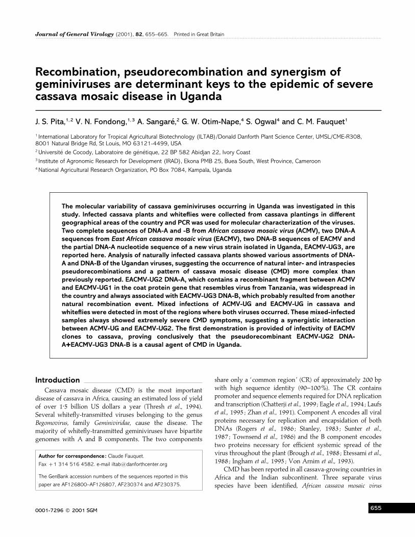
Recombination, pseudorecombination and synergism ofgeminiviruses are determinant keys to the epidemic of severecassava mosaic disease in Uganda
J. S. Pita,1, 2 V. N. Fondong,1, 3 A. Sangare! ,2 G. W. Otim-Nape,4 S. Ogwal4 and C. M. Fauquet1
1 International Laboratory for Tropical Agricultural Biotechnology (ILTAB)/Donald Danforth Plant Science Center, UMSL/CME-R308,8001 Natural Bridge Rd, St Louis, MO 63121-4499, USA2 Universite! de Cocody, Laboratoire de ge! ne! tique, 22 BP 582 Abidjan 22, Ivory Coast3 Institute of Agronomic Research for Development (IRAD), Ekona PMB 25, Buea South, West Province, Cameroon4 National Agricultural Research Organization, PO Box 7084, Kampala, Uganda
The molecular variability of cassava geminiviruses occurring in Uganda was investigated in thisstudy. Infected cassava plants and whiteflies were collected from cassava plantings in differentgeographical areas of the country and PCR was used for molecular characterization of the viruses.Two complete sequences of DNA-A and -B from African cassava mosaic virus (ACMV), two DNA-Asequences from East African cassava mosaic virus (EACMV), two DNA-B sequences of EACMV andthe partial DNA-A nucleotide sequence of a new virus strain isolated in Uganda, EACMV-UG3, arereported here. Analysis of naturally infected cassava plants showed various assortments of DNA-A and DNA-B of the Ugandan viruses, suggesting the occurrence of natural inter- and intraspeciespseudorecombinations and a pattern of cassava mosaic disease (CMD) more complex thanpreviously reported. EACMV-UG2 DNA-A, which contains a recombinant fragment between ACMVand EACMV-UG1 in the coat protein gene that resembles virus from Tanzania, was widespread inthe country and always associated with EACMV-UG3 DNA-B, which probably resulted from anothernatural recombination event. Mixed infections of ACMV-UG and EACMV-UG in cassava andwhiteflies were detected in most of the regions where both viruses occurred. These mixed-infectedsamples always showed extremely severe CMD symptoms, suggesting a synergistic interactionbetween ACMV-UG and EACMV-UG2. The first demonstration is provided of infectivity of EACMVclones to cassava, proving conclusively that the pseudorecombinant EACMV-UG2 DNA-AMEACMV-UG3 DNA-B is a causal agent of CMD in Uganda.
IntroductionCassava mosaic disease (CMD) is the most important
disease of cassava in Africa, causing an estimated loss of yieldof over 1±5 billion US dollars a year (Thresh et al., 1994).Several whitefly-transmitted viruses belonging to the genusBegomovirus, family Geminiviridae, cause the disease. Themajority of whitefly-transmitted geminiviruses have bipartitegenomes with A and B components. The two components
Author for correspondence: Claude Fauquet.
Fax 1 314 516 4582. e-mail iltab!danforthcenter.org
The GenBank accession numbers of the sequences reported in this
paper are AF126800–AF126807, AF230374 and AF230375.
share only a ‘ common region ’ (CR) of approximately 200 bpwith high sequence identity (90–100%). The CR containspromoter and sequence elements required for DNA replicationand transcription (Chatterji et al., 1999 ; Eagle et al., 1994 ; Laufset al., 1995 ; Zhan et al., 1991). Component A encodes all viralproteins necessary for replication and encapsidation of bothDNAs (Rogers et al., 1986 ; Stanley, 1983 ; Sunter et al.,1987 ; Townsend et al., 1986) and the B component encodestwo proteins necessary for efficient systemic spread of thevirus throughout the plant (Brough et al., 1988 ; Etessami et al.,1988 ; Ingham et al., 1995 ; Von Arnim et al., 1993).
CMD has been reported in all cassava-growing countries inAfrica and the Indian subcontinent. Three separate virusspecies have been identified, African cassava mosaic virus
0001-7296 # 2001 SGM GFF
J. S. Pita and othersJ. S. Pita and others
(ACMV), East African cassava mosaic virus (EACMV) and Indiancassava mosaic virus (ICMV) (Hong et al., 1993 ; Swanson &Harrison, 1994). Recently, a fourth virus species was isolated inSouth Africa (Berrie et al., 1998) with a proposed name of SouthAfrican cassava mosaic virus (SACMV). CMD was first detectedin Uganda in 1928 (Martin, 1928). Severe epidemics werereported between 1933 and 1944, although they weresuccessfully controlled by the use of resistant cassava varietiesand by sanitation of infected plants (Otim-Nape et al., 1997).The situation remained stable until 1988, when an extremelysevere epidemic of CMD developed, advancing from the northto the south of the country at a rate of approximately 20–25km per year. This aggressive form of the disease has devastatedcassava fields and caused food shortages and famine in anumber of districts where the crop was the major staple (Otim-Nape et al., 1996). A correlation exists between the presence ofa new virus, recombinant between ACMV and EACMV, andthe epidemic (Deng et al., 1997 ; Zhou et al., 1997). In order tohave a better understanding of the dynamics of this epidemic,we performed a large sampling in Uganda in February 1997.
Based on observations of cassava grown in the field and incontrolled growth-room conditions and results of PCR witholigonucleotide primers for genes encoded by both com-ponents A and B of cassava-infecting geminiviruses, we reporthere the molecular variability of cassava geminiviruses oc-curring in Uganda in 1997 and discuss the importance and roleof recombination, pseudorecombination and synergism in theUgandan CMD epidemic.
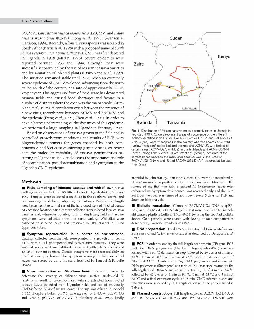
Methods+ Field sampling of infected cassava and whiteflies. Cassavacuttings were collected from 80 different sites in Uganda during February1997. Samples were collected from fields in the southern, central andnorthern regions of the country (Fig. 1). Cuttings 25–30 cm in lengthwere taken from the central part of the hardwood stem of infected plants.At each field location, samples were collected from infected local cassavavarieties and, whenever possible, cuttings displaying mild and severesymptoms were collected from the same variety. Whiteflies werecollected on infected leaves and preserved in 80% ethanol in 1±5 mlEppendorf tubes.
+ Symptom reproduction in a controlled environment.Cuttings collected from the field were planted in a growth chamber at24 °C with a 14 h photoperiod and 70% relative humidity. They werewatered twice a week and fertilized once a week with Peter’s professional15-16-17 nutrient solution. Disease symptoms were recorded daily onthe first emerging leaves. The symptom severity on fully expandedleaves was scored by using the scale described by Fauquet & Fargette(1988).
+ Virus inoculation on Nicotiana benthamiana. In order todetermine the severity of different virus isolates, 30-day-old N.benthamiana seedlings were inoculated with sap extracted from infectedcassava leaves collected from Ugandan fields and sap of previouslyCMD-infected N. benthamiana leaves. The sap was diluted in ice-cold0±1 M phosphate buffer, pH 7±0. One µg each of DNA-A (pCLV1.3A)and DNA-B (pCLV2B) of ACMV (Klinkenberg et al., 1989), kindly
Fig. 1. Distribution of African cassava mosaic geminiviruses in Uganda inFebruary 1997. Colours represent areas of occurrence of the differentisolates identified in this study. EACMV-UG2/Svr DNA-A and EACMV-UG3DNA-B (red) were widespread in the country whereas EACMV-UG2/Mld(yellow) was confined to isolated pockets and ACMV-UG was limited tocertain areas : ACMV-UG/Svr (blue) in the highlands and ACMV-UG/Mld(green) along Lake Victoria. Mixed infections (orange) occurred at thecontact zones between the main virus species, ACMV and EACMV.EACMV-UG1 DNA-A and -B and EACMV-UG3 DNA-A occurred at isolatedsites (stars).
provided by John Stanley, John Innes Centre, UK, were also inoculated toN. benthamiana as a positive control. Inoculum was rubbed onto thesurface of the first two fully expanded N. benthamiana leaves withcarborundum. Symptom development was recorded daily and the thirdleaf from the apex was removed and frozen every 5 days for PCR andSouthern blot analysis.
+ Biolistic inoculation. Clones of EACMV-UG2 DNA-A (pJSP-EA2) and EACMV-UG3 DNA-B (pJSP-EB3) were inoculated to 3-week-old cassava plantlets (cultivar TMS 60444) by using the Bio-Rad biolisticdevice. Gold particles were coated with 200 ng of each component asdescribed by Garzo! n-Tiznado et al. (1993).
+ DNA preparation. Total DNA was extracted from whiteflies andfrom cassava and N. benthamiana leaves as described by Dellaporta et al.(1983).
+ PCR. In order to amplify the full-length coat protein (CP) gene, PCRwith Taq DNA polymerase (Life Technologies}Gibco-BRL) was per-formed with a 94 °C denaturation step followed by 20 cycles of 1 min at94 °C, 1 min at 50 °C and 2 min at 72 °C and an extension cycle of10 min at 72 °C. A mixture of Taq DNA polymerase and cloned PfuDNA polymerase (Stratagene) at a ratio of 15 :1 was used to amplify thefull-length viral DNA-A and -B with a first cycle of 4 min at 94 °Cfollowed by 40 cycles of 1 min at 94 °C, 1 min at 58 °C and 3 min at72 °C and a final extension cycle of 15 min. CMD-infected plants andwhiteflies were screened by PCR amplification with the primers listed inTable 1.
+ Plasmid construction. Full-length copies of ACMV-UG DNA-Aand -B, EACMV-UG2 DNA-A and EACMV-UG3 DNA-B were
GFG
Cassava mosaic disease in UgandaCassava mosaic disease in Uganda
Table 1. Primers used for PCR characterization and amplification of geminiviruses occurring in cassava plants andwhitefly
Notation for degenerate oligonucleotides : R, A}G; Y, C}T; N, A}C}T}G; K, T}G; W, A}T. f.l. ; Full-length. BV1 is the DNA-B virion-sensegene ; BC1 is the DNA-B complementary-sense gene.
Primer Sequence (5«!3«) Target region
JSP001 ATGTCGAAGCGACCAGGAGAT 5« ACMV}EACMV CPJSP002 TGTTTATTAATTGCCAATACT 3« ACMV CPJSP003 CCTTTATTAATTTGTCACTGC 3« EACMV}SACMV CPJSP004 ATCTCCTGGTCGCTTCGACAT ACMV-UG DNA-A f.l.JSP005 ATCATCATTTCCACTCCAGGC ACMV-UG DNA-A f.l.JSP006 TGGTAGAATTCTTTGAAGTGTTTGAGGTA EACMV-UG2 DNA-A f.l.JSP007 GATTTGAATTCATGGGGGCCCAAAGGGAC EACMV-UG2 DNA-A f.l.JSP008 GAATTCAGCCTCAGAGGTATACTG ACMV-UG DNA-B f.l.JSP009 GAATTCCCACTTAGAGGGCTACAC ACMV-UG DNA-B f.l.JSP010 AAGACGGATTCTTGTTAGAAGTGGAGAAA EACMV-UG1}3 DNA-B f.l.JSP011 CCACGGAATTCACTCCCGACTGCTTCCTA EACMV-UG1}3 DNA-B f.l.JSP012 GTCCATATAGGTAARGTNATG 5« ICMV CPJSP013 CCTGCTCCTTGCTNGCYTART 3« ICMV CPJSP014 GCTGTCCCCATTGTCCARGGN 5« SACMV CPJSP015 TGCCCAAAGTCTTGGGGCGCATTG EACMV-UG2 recombined frag.JSP016 TGTCCAAAATCTTGAGGACTCGAA EACMV-TZ recombined frag.JSP017 TCCTCCTATTAATTGGAGACATTA ACMV BC1JSP018 CCTATATATAATACATAAACAATT ACMV BC1MP 16 CCTCTAGATAATATTACYKRWKGRCC Universal primerMP 82 CGGAATTCYTGNACYTTRCANGGNCCYTCR Universal primer
Primers used for PCR discrimination of EACMV-UG1 and EACMV-UG2 DNA-BJSP019 TAGACTTCCATTCAAAATTTGAAT EACMV-UG1 CRJSP020 CTCAATGAATGTGAAAGGCAAACG EACMV-UG3 CR-BJSP021 TGCCTTTCACATTCATTGAGGN EACMV-UG3 CR-BJSP022 CCTATTTACACATATGCCATTGGG EACMV-UG1 CRJSP023 TACATCGGCCTTTGAGTCGCATGG EACMV-UG1}3 BC1JSP024 CAGACACCCTCAAGCTTAATA EACMV-UG1}3 BV1
amplified by PCR and cloned into pBluescript II KS (). Unique BamHIand HindIII sites were used respectively for cloning ACMV-UG DNA-Aand EACMV-UG2 DNA-A and EcoRI for ACMV-UG DNA-B andEACMV-UG3 DNA-B. Head-to-tail partial repeats of these clones wereconstructed as described by Von Arnim & Stanley (1992).
+ Southern blot analysis. Total DNA (4 µg) was electrophoresedon a 1% agarose gel without ethidium bromide and transferred to nylonmembranes. Viral DNA was detected by using specific radiolabelledprobes of ACMV-UG DNA-A and -B, EACMV-UG2 DNA-A andEACMV-UG3 DNA-B.
+ Sequence determination and analysis. The complete DNAsequences of ACMV-UG DNA-A and -B, EACMV-UG2 DNA-A andEACMV-UG3 DNA-B from mild and severely CMD-affected cassavaplants were sequenced by the dideoxynucleotide chain-terminationmethod on an ABI automatic sequencer. In addition, part of the EACMV-UG3 DNA-A, cloned using the TA cloning kit (Invitrogen), and the full-length EACMV-UG1 DNA-B were also sequenced as described above.Sequences obtained were compared by using the cluster option of themultiple sequence alignment (MegAlign) program within the DNASTARpackage for Apple Macintosh computers. An identical consensus tree wasobtained when phylogenetic analysis was performed by a cladisticparsimony method using the computer program PAUP 3.1.1 (Swofford,
1993). Ten thousand bootstrap replications were performed to placeconfidence estimates on the groups contained in the tree.
ResultsSample collection
The route followed to collect cuttings of CMD-infectedcassava plants in Uganda is shown in Fig. 1. Samplesrepresenting the south-western, central and part of thenorthern regions were obtained. The extreme north of thecountry could not be prospected as it was inaccessible at thetime of collection in February 1997.
More than 90% of the cassava plants showed severe CMDsymptoms in almost all of the fields in all regions visited.However, mild symptoms could also be observed in someplants scattered throughout some fields. The northern borderof Lake Victoria represented an exception, as generally onlymild CMD symptoms were found in that area. A total of ninecultivars were collected, with Kameza in the south-western andEbwanatereka in the south-eastern region being the most
GFH
J. S. Pita and othersJ. S. Pita and others
strongly represented (38 and 27% of the collection, re-spectively).
Establishing cassava cuttings in growth chambers
Symptoms of infected cassava samples collected in the fieldwere reproduced in controlled conditions to assess symptomvariability linked to the environment. From a total of 183cuttings collected, 129 (70±5%) were successfully established inthe growth chamber. Of the 54 cuttings that failed to grow,55% were from plants severely affected by CMD in the fieldand 45% were from plants that displayed mild symptoms.Therefore, failure of the cuttings to establish in the growthchamber was not related to disease severity. Some cuttingsshowed symptom variations in the growth chamber comparedwith the field-assessed symptoms. Consequently, only plantsshowing clearly reproducible symptoms on two differentcuttings after 70 days in the growth chamber were selected forsubsequent analysis.
CMD symptoms on cassava plants
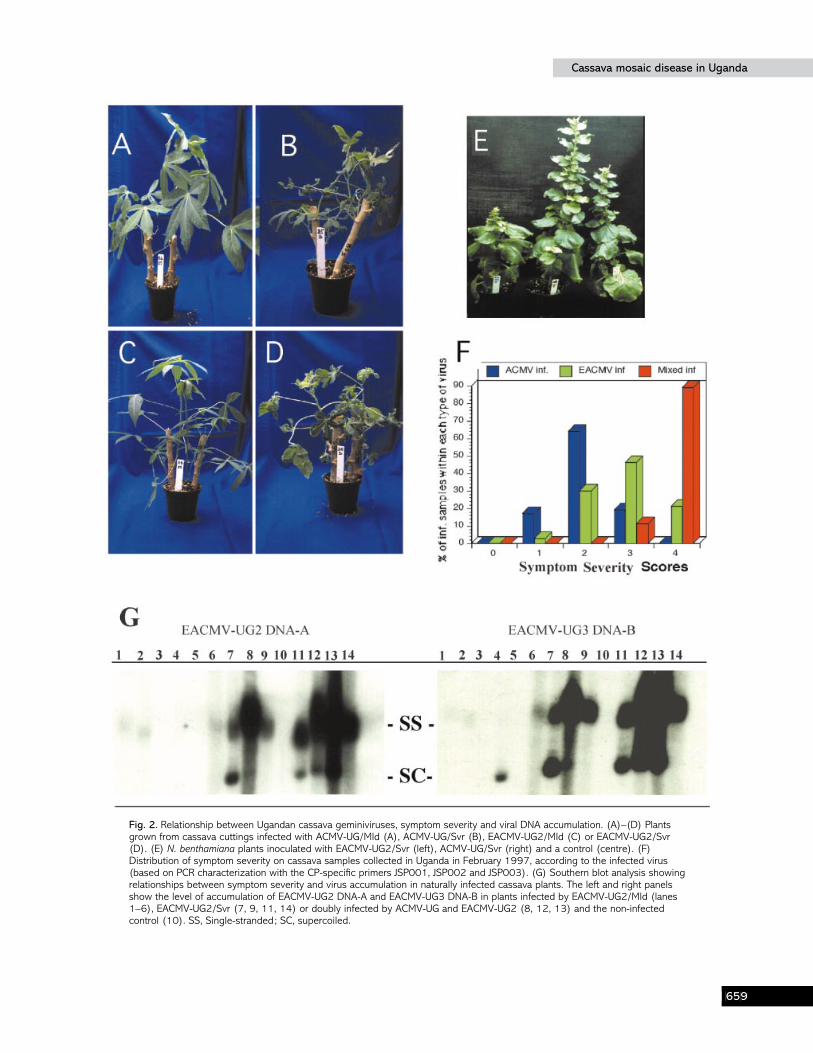
Fig. 2 shows the different types of disease symptomsobserved on cassava plants growing in the field and in thegrowth chamber. In both cases, cassava plants expressing mildsymptoms developed normally with leaves showing mild,light-green mosaic symptoms. In contrast, plants expressingsevere symptoms displayed extreme shrinking of the leaves,along with distortion at the bases of the leaflets and distinctchlorosis. In very severe cases, the leaves became desiccatedand the young plants died. The proportion of disease-relateddeath of cassava plants was estimated periodically for thepredominant cultivars in the collection. Cultivar Ebwanaterekawas very susceptible to CMD, as 32% of cuttings of thatcultivar died 90 days after establishment in the growthchamber. Other cultivars were less susceptible, with approxi-mately 15% of the Ssenyonjo and Kameza cuttings dyingwithin 90 days, while only 8% of Machunde cuttings had diedduring the same period.
Molecular characterization of cassava geminiviruses inUganda
Based on the published nucleotide sequences of ACMV-KE(Stanley & Gay, 1983), EACMV-TZ and EACMV-UG2 DNA-A (Deng et al., 1997 ; Zhou et al., 1997), specific and degenerateprimers (Table 1) were employed to characterize the Ugandancassava collection. Using discriminating primers to amplify theCP gene, 48% of the cassava samples were found to be infectedby EACMV-UG2 alone, 28% by ACMV-UG alone and 7%with both ACMV-UG and EACMV-UG2 and 8% were triple-infected with ACMV-UG, EACMV-UG1 (a non-recombinedsequence similar to EACMV from Tanzania) and EACMV-UG2. For 9% of the samples, no signal was detected for any of
the primers pairs employed. None of the cassava samplestested in this study was infected by EACMV-UG1 alone.
Cassava samples that failed to produce a signal with thespecific primers (JSP001, 002, 003) showed mild symptoms inthe field and no symptoms in the greenhouse. These sampleswere then tested with the degenerate primers MP16 and MP82(Padidam et al., 1995). A fragment of approximately 500 bpwas amplified, stretching from the stem–loop of the CR to the5« end of the core region of the CP. This suggested thepresence of an as-yet undescribed geminivirus, which we shallcall provisionally EACMV-UG3. Only a partial DNA-Asequence of this virus was obtained in this study, stretchingfrom 150 nt upstream of the nonanucleotide to the 5« end ofthe core region of the CP.
None of the CMD-affected plants from our collectionyielded PCR products with CP primers specific for ICMV orSACMV (Table 1), suggesting that neither ICMV nor SACMVis present in Uganda.
The relationship between Ugandan cassavageminiviruses, CMD symptom severity and virus DNAaccumulation
As described previously, cassava plants displaying mildsymptoms could be found in fields where the severe conditionwas also prominent. In order to understand this situationbetter, a relationship was established between the virus type,ACMV or EACMV, found in the plants by PCR analysis andsymptom severity (Fig. 2F). Eighty per cent of ACMV-UG-positive plants exhibited very mild symptoms and 20%showed intermediate symptoms. Sixty-seven per cent and33% of EACMV-UG2-infected plants were respectivelyseverely and moderately affected. Noticeably, all samplesfound to be infected by both ACMV-UG and EACMV-UG2were very severely affected (10% scored 3 and 90% scored 4),suggesting a synergistic interaction between these two virusspecies. The suffix -UG}Mld or -UG}Svr was used todiscriminate between the putative viruses present in thedifferent inocula.
Cassava plants of the cultivar Ebwanatereka displayingmild and severe symptoms were selected for further study.Crude sap was extracted from infected leaves and tested for itsability to transmit the viruses to N. benthamiana. In the case ofplants with mild symptoms, only areas of the leaves showingsymptoms were used for crude sap preparation. Using dilutedcrude sap solutions (1 g leaf tissue ground in 10 ml buffer),infection of N. benthamiana could be obtained with bothACMV-UG}Svr and EACMV-UG2}Svr. A more concen-trated sap solution of at least 5 g}10 ml was necessary toobtain symptoms with ACMV-UG}Mld. For sap extractedfrom cassava infected by EACMV-UG2}Mld, symptoms werebarely detectable in N. benthamiana, regardless of the inoculumconcentration employed. However, specific PCR productsattesting to the presence of the respective viruses could be
GFI
Cassava mosaic disease in UgandaCassava mosaic disease in Uganda
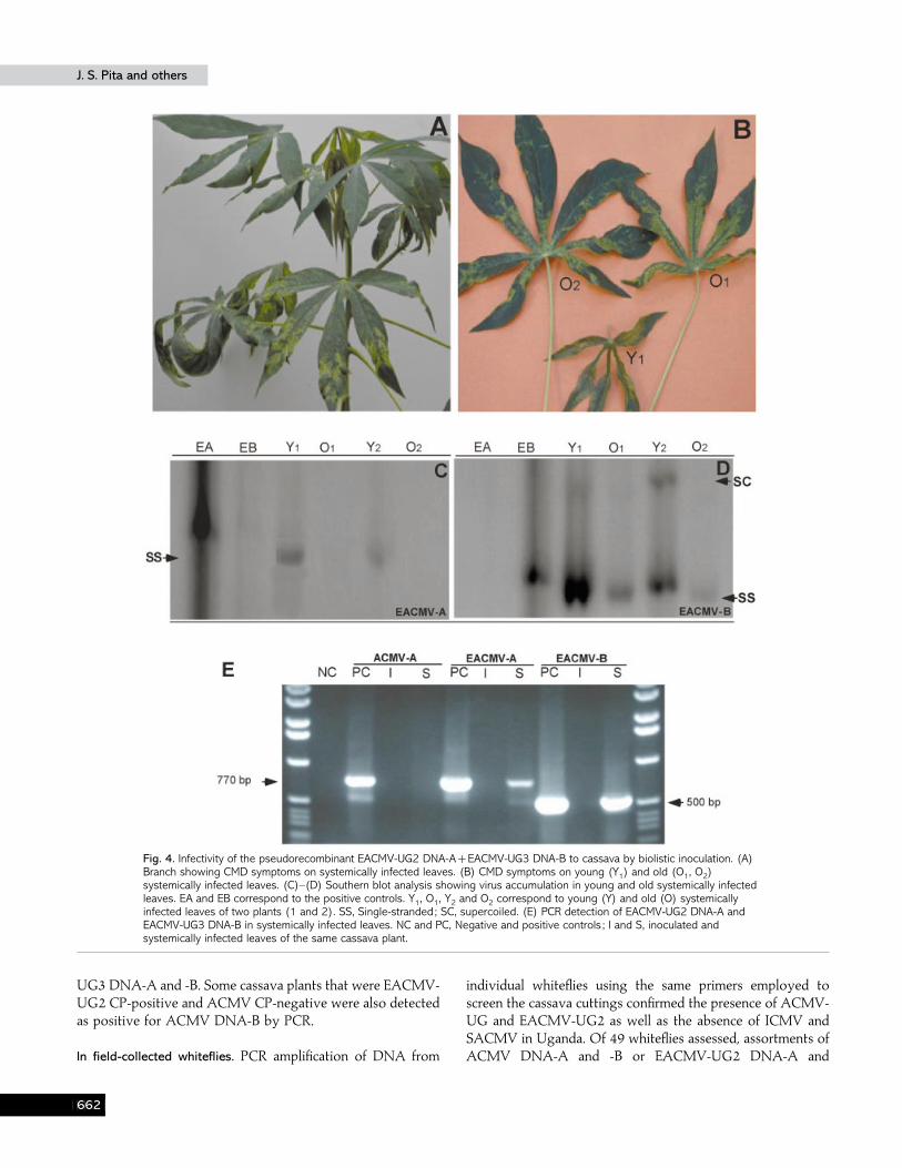
Fig. 2. Relationship between Ugandan cassava geminiviruses, symptom severity and viral DNA accumulation. (A)–(D) Plantsgrown from cassava cuttings infected with ACMV-UG/Mld (A), ACMV-UG/Svr (B), EACMV-UG2/Mld (C) or EACMV-UG2/Svr(D). (E) N. benthamiana plants inoculated with EACMV-UG2/Svr (left), ACMV-UG/Svr (right) and a control (centre). (F)Distribution of symptom severity on cassava samples collected in Uganda in February 1997, according to the infected virus(based on PCR characterization with the CP-specific primers JSP001, JSP002 and JSP003). (G) Southern blot analysis showingrelationships between symptom severity and virus accumulation in naturally infected cassava plants. The left and right panelsshow the level of accumulation of EACMV-UG2 DNA-A and EACMV-UG3 DNA-B in plants infected by EACMV-UG2/Mld (lanes1–6), EACMV-UG2/Svr (7, 9, 11, 14) or doubly infected by ACMV-UG and EACMV-UG2 (8, 12, 13) and the non-infectedcontrol (10). SS, Single-stranded; SC, supercoiled.
GFJ
J. S. Pita and othersJ. S. Pita and others
obtained on systemically affected leaves of all inoculatedplants at 10 days post-inoculation for ACMV-UG andapproximately 30 days post-inoculation for EACMV-UG2.
ACMV-UG and EACMV-UG2 induced different types ofsymptoms in N. benthamiana (Fig. 2E). ACMV-UG-infectedplants did not show the typical mosaic and severe leaf-curlingsymptoms of ACMV-KE (Klinkenberg et al., 1989) charac-teristic in N. benthamiana plants, but caused moderate leafcurling at the base of systemically infected leaves and}orchlorotic spots on inoculated leaves. EACMV-UG2-infectedplants were reduced in height compared with ACMV-UG-infected plants or control, non-infected plants (Fig. 2E).Systemic leaves in EACMV-UG2}Svr-infected plants showedan approximately 40% reduction in size compared with similarleaves of wild-type N. benthamiana and chlorotic patches ratherthan the mosaic typical of ACMV were present on olderleaves.
The relationship between virus accumulation and symptomseverity was investigated by using Southern blot analysis.Cassava samples infected by EACMV-UG2}Mld, EACMV-UG2}Svr and mixed-infected by ACMV-UG and EACMV-UG2 were used. Four µg total DNA extracted from infectedleaves of cultivar Ebwanatereka was used for Southernhybridization. A positive correlation between symptomseverity and virus accumulation was obvious (Fig. 2 G).EACMV-UG2}Mld was barely detectable compared withEACMV-UG2}Svr, whereas samples with mixed infectionsgave stronger signals, indicating probable synergistic in-teraction between ACMV-UG and EACMV-UG2.
Sequence analysis and comparison with selectedviruses
Coat protein (CP). The CP gene sequences of the four strainsidentified in our study were compared to published sequences.Results indicated that the CP nucleotide sequences fromACMV-UG}Mld, ACMV-UG}Svr, EACMV-UG2}Mld andEACMV-UG2}Svr were nearly identical within each virusspecies. In order to test whether the CP gene sequences of allEACMV-UG2 isolates found in our collection were similar tothe published sequence of the recombinant EACMV-UG (Denget al., 1997 ; Zhou et al., 1997), primers specific to therecombinant fragment (JSP015 and JSP016) were designed toamplify selectively the CP of EACMV-UG1-like or EACMV-UG2-like molecules. All EACMV-UG2 isolates, irrespective ofsymptom severity, were found to contain the same ACMVrecombinant fragment in their CP. Of the 129 samples tested,EACMV-UG1-like CP sequences were amplified from onlyfive samples that were also infected by ACMV-UG, EACMV-UG2 and EACMV-UG3.
Complete DNA-A sequences. Complete nucleotide sequences ofthe DNA-A molecules of the different Ugandan ACMV andEACMV strains were compared with published sequences
Table 2. Nucleotide sequence identity (%) of completeDNA-A sequences of selected cassava geminiviruses
Values over 80% are shown in bold.
Virus isolate 1 2 3 4 5 6 7 8 9
1. ACMV-UG}Mld – 97 97 96 69 68 64 64 662. ACMV-UG}Svr – 97 97 69 69 64 64 663. ACMV-KE – 96 68 68 63 64 664. ACMV-NG – 69 68 64 64 665. EACMV-UG2}Mld – 99 91 90 856. EACMV-UG2}Svr – 92 91 857. EACMV-KE – 94 868. EACMV-TZ – 859. EACMV-MW –
Table 3. Nucleotide and amino acid sequence identities(%) of BC1 and BV1 of selected cassava geminiviruses
Numbers in parentheses correspond to percentage amino acidsequence identities. The part of the matrix above the diagonal showsBC1 comparisons and the part below the diagonal shows BV1comparisons. Values over 80% are in bold.
Virus isolate 1 2 3 4 5
1. ACMV-KE – 97 (96) 96 (97) 46 (53) 46 (53)2. ACMV-NG 95 (92) – 97 (98) 45 (54) 46 (53)3. ACMV-UG 96 (96) 96 (94) – 45 (53) 46 (53)4. EACMV-UG1 37 (34) 35 (34) 35 (34) – 98 (97)5. EACMV-UG3 37 (35) 36 (36) 36 (35) 99 (98) –
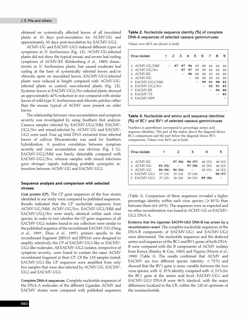
(Table 2). Comparison of these sequences revealed a higherpercentage identity within each virus species (& 85%) thanbetween them (63–69%). The sequences were as expected andno other recombination was found in ACMV-UG or EACMV-UG2 DNA-A.
Evidence that the Ugandan EACMV-UG3 DNA-B has arisen by arecombination event. The complete nucleotide sequences of theDNA-B components of EACMV-UG1 and EACMV-UG3were determined. The nucleotide sequences and the deducedamino acid sequences of the BC1 and BV1 genes of both DNA-B were compared with the B components of ACMV isolatesfrom Kenya (Stanley & Gay, 1983) and Nigeria (Morris et al.,1990) (Table 3). The results confirmed that ACMV andEACMV are two different species (identity ! 75%) andshowed that the BV1 gene is more variable between the twovirus species, with % 35% identity compared with % 53% forthe BC1 gene at the amino acid level. EACMV-UG1 andEACMV-UG3 DNA-B were 96% identical, with the majordifferences localized in the CR, within the 120 nt upstream ofthe nonanucleotide.
GGA
Cassava mosaic disease in UgandaCassava mosaic disease in Uganda
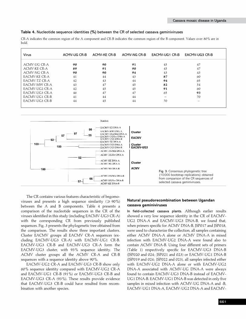
Table 4. Nucleotide sequence identities (%) between the CR of selected cassava geminiviruses
CR-A indicates the common region of the A component and CR-B indicates the common region of the B component. Values over 80% are inbold.
Virus ACMV-UG CR-B ACMV-KE CR-B ACMV-NG CR-B EACMV-UG1 CR-B EACMV-UG3 CR-B
ACMV-UG CR-A 90 90 91 45 47ACMV-KE CR-A 89 91 90 45 47ACMV-NG CR-A 90 90 94 43 43EACMV-KE CR-A 41 44 43 87 60EACMV-TZ CR-A 42 43 44 94 65EACMV-MW CR-A 43 47 45 82 54EACMV-UG2 CR-A 42 45 45 91 60EACMV-UG3 CR-A 46 47 47 65 93EACMV-UG1 CR-B 41 44 44 – 70EACMV-UG3 CR-B 44 45 44 70 –
Fig. 3. Consensus phylogenetic tree(10000 bootstrap replications) obtainedfrom comparison of the CR sequences ofselected cassava geminiviruses.
The CR contains various features characteristic of begomo-viruses and presents a high sequence similarity (& 90%)between the A and B components. Table 4 presents acomparison of the nucleotide sequences in the CR of theviruses identified in this study (including EACMV-UG3 CR-A)with the corresponding CR from previously publishedsequences. Fig. 3 presents the phylogenetic tree obtained fromthe comparison. The results show three important clusters.Cluster EACMV groups all EACMV CR-A sequences (ex-cluding EACMV-UG3 CR-A) with EACMV-UG1 CR-B.EACMV-UG3 CR-B and EACMV-UG3 CR-A form theEACMV-UG3 cluster, with 93% sequence identity. TheACMV cluster groups all the ACMV CR-A and CR-Bsequences with a sequence identity above 90%.
EACMV-UG2 CR-A and EACMV-UG3 CR-B show only60% sequence identity compared with EACMV-UG2 CR-Aand EACMV-UG1 CR-B (91%) or EACMV-UG3 CR-B andEACMV-UG3 CR-A (93%). These results provide evidencethat EACMV-UG3 CR-B could have resulted from recom-bination with another species.
Natural pseudorecombination between Ugandancassava geminiviruses
In field-collected cassava plants. Although earlier resultsshowed a very low sequence identity in the CR of EACMV-UG2 DNA-A and EACMV-UG3 DNA-B, we found that,when primers specific for ACMV DNA-B, JSP017 and JSP018,were used to characterize the collection, all samples containingeither ACMV DNA-A alone or ACMV DNA-A in mixedinfection with EACMV-UG2 DNA-A were found also tocontain ACMV DNA-B. Using four different sets of primers(Table 1) respectively specific for EACMV-UG3 DNA-B(JSP020 and 024 ; JSP021 and 023) or EACMV-UG1 DNA-B(JSP019 and 024 ; JSP022 and 023), all samples infected eitherwith EACMV-UG2 DNA-A alone or with EACMV-UG2DNA-A associated with ACMV-UG DNA-A were alwaysfound to contain EACMV-UG3 DNA-B instead of EACMV-UG1 DNA-B. EACMV-UG1 DNA-B was detected in only fivesamples in mixed infection with ACMV-UG DNA-A and -B,EACMV-UG1 DNA-A, EACMV-UG2 DNA-A and EACMV-
GGB
J. S. Pita and othersJ. S. Pita and others
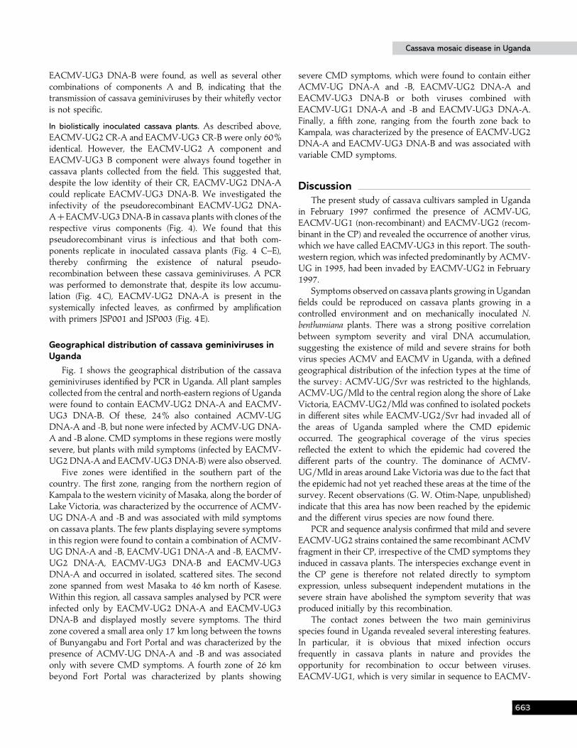
Fig. 4. Infectivity of the pseudorecombinant EACMV-UG2 DNA-AEACMV-UG3 DNA-B to cassava by biolistic inoculation. (A)Branch showing CMD symptoms on systemically infected leaves. (B) CMD symptoms on young (Y1) and old (O1, O2)systemically infected leaves. (C)–(D) Southern blot analysis showing virus accumulation in young and old systemically infectedleaves. EA and EB correspond to the positive controls. Y1, O1, Y2 and O2 correspond to young (Y) and old (O) systemicallyinfected leaves of two plants (1 and 2). SS, Single-stranded; SC, supercoiled. (E) PCR detection of EACMV-UG2 DNA-A andEACMV-UG3 DNA-B in systemically infected leaves. NC and PC, Negative and positive controls ; I and S, inoculated andsystemically infected leaves of the same cassava plant.
UG3 DNA-A and -B. Some cassava plants that were EACMV-UG2 CP-positive and ACMV CP-negative were also detectedas positive for ACMV DNA-B by PCR.
In field-collected whiteflies. PCR amplification of DNA from
individual whiteflies using the same primers employed toscreen the cassava cuttings confirmed the presence of ACMV-UG and EACMV-UG2 as well as the absence of ICMV andSACMV in Uganda. Of 49 whiteflies assessed, assortments ofACMV DNA-A and -B or EACMV-UG2 DNA-A and
GGC
Cassava mosaic disease in UgandaCassava mosaic disease in Uganda
EACMV-UG3 DNA-B were found, as well as several othercombinations of components A and B, indicating that thetransmission of cassava geminiviruses by their whitefly vectoris not specific.
In biolistically inoculated cassava plants. As described above,EACMV-UG2 CR-A and EACMV-UG3 CR-B were only 60%identical. However, the EACMV-UG2 A component andEACMV-UG3 B component were always found together incassava plants collected from the field. This suggested that,despite the low identity of their CR, EACMV-UG2 DNA-Acould replicate EACMV-UG3 DNA-B. We investigated theinfectivity of the pseudorecombinant EACMV-UG2 DNA-AEACMV-UG3 DNA-B in cassava plants with clones of therespective virus components (Fig. 4). We found that thispseudorecombinant virus is infectious and that both com-ponents replicate in inoculated cassava plants (Fig. 4 C–E),thereby confirming the existence of natural pseudo-recombination between these cassava geminiviruses. A PCRwas performed to demonstrate that, despite its low accumu-lation (Fig. 4C), EACMV-UG2 DNA-A is present in thesystemically infected leaves, as confirmed by amplificationwith primers JSP001 and JSP003 (Fig. 4E).
Geographical distribution of cassava geminiviruses inUganda
Fig. 1 shows the geographical distribution of the cassavageminiviruses identified by PCR in Uganda. All plant samplescollected from the central and north-eastern regions of Ugandawere found to contain EACMV-UG2 DNA-A and EACMV-UG3 DNA-B. Of these, 24% also contained ACMV-UGDNA-A and -B, but none were infected by ACMV-UG DNA-A and -B alone. CMD symptoms in these regions were mostlysevere, but plants with mild symptoms (infected by EACMV-UG2 DNA-A and EACMV-UG3 DNA-B) were also observed.
Five zones were identified in the southern part of thecountry. The first zone, ranging from the northern region ofKampala to the western vicinity of Masaka, along the border ofLake Victoria, was characterized by the occurrence of ACMV-UG DNA-A and -B and was associated with mild symptomson cassava plants. The few plants displaying severe symptomsin this region were found to contain a combination of ACMV-UG DNA-A and -B, EACMV-UG1 DNA-A and -B, EACMV-UG2 DNA-A, EACMV-UG3 DNA-B and EACMV-UG3DNA-A and occurred in isolated, scattered sites. The secondzone spanned from west Masaka to 46 km north of Kasese.Within this region, all cassava samples analysed by PCR wereinfected only by EACMV-UG2 DNA-A and EACMV-UG3DNA-B and displayed mostly severe symptoms. The thirdzone covered a small area only 17 km long between the townsof Bunyangabu and Fort Portal and was characterized by thepresence of ACMV-UG DNA-A and -B and was associatedonly with severe CMD symptoms. A fourth zone of 26 kmbeyond Fort Portal was characterized by plants showing
severe CMD symptoms, which were found to contain eitherACMV-UG DNA-A and -B, EACMV-UG2 DNA-A andEACMV-UG3 DNA-B or both viruses combined withEACMV-UG1 DNA-A and -B and EACMV-UG3 DNA-A.Finally, a fifth zone, ranging from the fourth zone back toKampala, was characterized by the presence of EACMV-UG2DNA-A and EACMV-UG3 DNA-B and was associated withvariable CMD symptoms.
DiscussionThe present study of cassava cultivars sampled in Uganda
in February 1997 confirmed the presence of ACMV-UG,EACMV-UG1 (non-recombinant) and EACMV-UG2 (recom-binant in the CP) and revealed the occurrence of another virus,which we have called EACMV-UG3 in this report. The south-western region, which was infected predominantly by ACMV-UG in 1995, had been invaded by EACMV-UG2 in February1997.
Symptoms observed on cassava plants growing in Ugandanfields could be reproduced on cassava plants growing in acontrolled environment and on mechanically inoculated N.benthamiana plants. There was a strong positive correlationbetween symptom severity and viral DNA accumulation,suggesting the existence of mild and severe strains for bothvirus species ACMV and EACMV in Uganda, with a definedgeographical distribution of the infection types at the time ofthe survey : ACMV-UG}Svr was restricted to the highlands,ACMV-UG}Mld to the central region along the shore of LakeVictoria, EACMV-UG2}Mld was confined to isolated pocketsin different sites while EACMV-UG2}Svr had invaded all ofthe areas of Uganda sampled where the CMD epidemicoccurred. The geographical coverage of the virus speciesreflected the extent to which the epidemic had covered thedifferent parts of the country. The dominance of ACMV-UG}Mld in areas around Lake Victoria was due to the fact thatthe epidemic had not yet reached these areas at the time of thesurvey. Recent observations (G. W. Otim-Nape, unpublished)indicate that this area has now been reached by the epidemicand the different virus species are now found there.
PCR and sequence analysis confirmed that mild and severeEACMV-UG2 strains contained the same recombinant ACMVfragment in their CP, irrespective of the CMD symptoms theyinduced in cassava plants. The interspecies exchange event inthe CP gene is therefore not related directly to symptomexpression, unless subsequent independent mutations in thesevere strain have abolished the symptom severity that wasproduced initially by this recombination.
The contact zones between the two main geminivirusspecies found in Uganda revealed several interesting features.In particular, it is obvious that mixed infection occursfrequently in cassava plants in nature and provides theopportunity for recombination to occur between viruses.EACMV-UG1, which is very similar in sequence to EACMV-
GGD
J. S. Pita and othersJ. S. Pita and others
TZ within its CP gene, was always found mixed with ACMV-UG, EACMV-UG2 and EACMV-UG3, and therefore could beone of the putative parents of the recombinant EACMV-UG2.This was not obvious from previous reports, where samplingdid not allow the detection of EACMV-UG1 in Uganda(Harrison et al., 1997).
Forty-five per cent of EACMV-UG2-infected cassava plantspresented very severe CMD symptoms. These samples werePCR-positive for the presence of EACMV-UG2 DNA-A,EACMV-UG3 DNA-B and ACMV-UG DNA-B, but negativefor ACMV-UG DNA-A. This suggests a natural pseudo-recombination between EACMV-UG2 DNA-A and ACMV-UG DNA-B, in a manner already demonstrated for severalbipartite geminiviruses (Hill et al., 1998 ; Hou et al., 1998).Pseudorecombination was confirmed in biolistically inoculatedcassava plants using infectious clones, proving that EACMV-UG2 DNA-A can replicate EACMV-UG3 DNA-B despite thelow identity of their CR.
The intensity of CMD symptoms could be correlated withthe synergistic interaction observed between ACMV-UG andEACMV-UG2. All samples doubly infected with ACMVDNA-A and -B and EACMV DNA-A and -B within ourUgandan cassava collection produced extremely severesymptoms compared with plants infected with only one virusspecies. To date, we have not tried to reproduce this synergisticinteraction with infectious clones. However, Southern blotanalysis of naturally CMD-infected cassava plants showed agreater accumulation of EACMV-UG2 DNA-A and EACMV-UG3 DNA-B in mixed-infected plants compared with singlyinfected plants. Such a synergistic interaction between ACMVand EACMV has been suggested by Harrison et al. (1997) anddemonstrated by Fondong et al. (2000) with infectious clonesof ACMV-CM and EACMV-CM isolated from Cameroon.
Another important result described in this paper is theevidence for interspecies recombination provided by EACMV-UG3 DNA-B. This DNA species is highly similar to theEACMV-UG1 DNA-B component with the exception of theCR, which contains considerable differences that may haveresulted from recombination with another geminivirus species.Despite the low nucleotide sequence identity in their CR(60%), we have shown that the pseudorecombinant virusEACMV-UG2 DNA-AEACMV-UG3 DNA-B is infectiousto biolistically inoculated cassava plants and is the combinationfound most frequently in nature, probably due to its tendencyto accumulate more in the youngest leaves (Fig. 4C) that aretargetted preferentially by the whitefly vector. The closeassociation between EACMV-UG2 DNA-A and EACMV-UG3 DNA-B and the fact that these two molecules are themost widespread in Uganda provides evidence of theiressential role in the severe CMD epidemic in Uganda. Itappears that the association of EACMV-UG2 DNA-A andEACMV-UG3 DNA-B is privileged over the homologouscombination and this could be a key to the CMD epidemic, asthe pseudorecombinant might replicate more, accumulate more
DNA and consequently cause more severe symptoms. Thishypothesis could be supported by the fact that we observed ahigher level of EACMV-UG3 DNA-B accumulation in cassavaleaves compared with EACMV-UG2 DNA-A (Fig. 4C). Thisobservation is consistent with the severe symptoms thatdevelop in the young systemic leaves of infected plants. We donot currently understand the significance of the dominance ofEACMV-UG3 DNA-B accumulation over EACMV-UG2DNA-A, but this will be investigated in future work.
Specific or preferential transmission of the recombinant(CP) strain compared with EACMV-UG1 could explain theinvasion of Uganda by EACMV-UG2 and the disappearanceof the non-recombinant EACMV-UG1. Spread of EACMV-UG2 within areas where cassava plants were already infectedby ACMV would then have been enhanced through itssynergistic interaction with ACMV, leading to developmentof the CMD epidemic.
In summary, a combination and}or the sequential oc-currence of several phenomena could explain the UgandanCMD epidemic. (i) Synergism between members of the twospecies ACMV and EACMV is certainly a major possiblefactor, as it increases DNA accumulation of both viruses andtherefore their capacity to be transmitted by whiteflies. (ii) Thefact that the most prevalent virus in the epidemic (EACMV-UG2) is a recombinant for CP with ACMV-UG is certainly nota coincidence. However, the reason for its success is notevident, in view of the fact that we found a very mild strain ofEACMV-UG2 in Uganda that possessed the same recom-bination. The recombinant CP could have the same level ofreplication as the non-recombinant but might possess similarwhitefly transmission-properties to those of ACMV. This,however, remains to be proven. Subsequent mutations andselection by farmers of plants that displayed mild symptomscould easily explain the occurrence of mild EACMV-UG2isolates. (iii) For the first time, we demonstrate that it is possiblein nature to find stable pseudorecombinants between the A andB components of strains of the same species (EACMV-UG2DNA-A and EACMV-UG3 DNA-B) or between A and Bcomponents of different species (ACMV and EACMV). Thissystem offers an additional possibility of survival for theviruses and another source of virus biodiversity.
These biological findings show that more than one event isprobably necessary to start such CMD epidemics. However,more experimentation will be necessary to understand betterthe dynamics of this complex biological situation. Infectiousclones of ACMV and EACMV that are now available will beused for this purpose.
This work was funded by the Rockefeller Foundation, the NationalAgricultural Research Organization (NARO) of Uganda and the FrenchInstitute for Research and Development (IRD). The authors would like tothank Dr Nigel Taylor and Dr Victor Masona for critical reading andcorrecting the manuscript and Dr Isabel Ordiz for her technical assistance.This is manuscript DDPSC 00-018.
GGE
Cassava mosaic disease in UgandaCassava mosaic disease in Uganda
ReferencesBerrie, L. C., Palmer, K. E., Rybicki, E. P. & Rey, M. E. C. (1998).Molecular characterisation of a distinct South African cassava infectinggeminivirus. Archives of Virology 143, 2253–2260.
Brough, C. L., Hayes, R. J., Morgan, A. J., Coutts, R. H. A. & Buck,K. W. (1988). Effects of mutagenesis in vitro on the ability of clonedtomato golden mosaic virus DNA to infect Nicotiana benthamiana plants.Journal of General Virology 69, 503–514.
Chatterji, A., Padidam, M., Beachy, R. N. & Fauquet, C. M. (1999).Identification of replication specificity determinants in two strains oftomato leaf curl virus from New Delhi. Journal of Virology 73, 5481–5489.
Dellaporta, S. L., Wood, J. & Hicks, J. B. (1983). A plant DNA minipreparation : version II. Plant Molecular Biology Reporter 1, 19–21.
Deng, D., Otim-Nape, G. W., Sangare, A., Ogwal, S., Beachy, R. N. &Fauquet, C. M. (1997). Presence of a new virus closely related to EastAfrican cassava mosaic geminivirus, associated with cassava mosaicoutbreak in Uganda. African Journal of Root and Tuber Crops 2, 23–28.
Eagle, P. A., Orozco, B. M. & Hanley-Bowdoin, L. (1994). A DNAsequence required for geminivirus replication also mediates trans-criptional regulation. Plant Cell 6, 1157–1170.
Etessami, P., Callis, R., Ellwood, S. & Stanley, J. (1988). Delimitationof essential genes of cassava latent virus DNA 2. Nucleic Acids Research16, 4811–4829.
Fauquet, C. M. & Fargette, D. (editors) (1988). Proceedings of theInternational Seminar : African Cassava Mosaic Disease and its Control. Ede,Netherlands : CTA}ORSTOM.
Fondong, V. N., Pita, J. S., Rey, M. E. C., de Kochko, A., Beachy, R. N.& Fauquet, C. M. (2000). Evidence of synergism between Africancassava mosaic virus and a new double-recombinant geminivirus infectingcassava in Cameroon. Journal of General Virology 81, 287–297.
Garzo! n-Tiznado, J. A., Torres-Pacheco, I., Ascencio-Iban4 ez, J. T.,Herrera-Estrella, L. & Rivera-Bustamante, R. F. (1993). Inoculation ofpeppers with infectious clones of a new geminivirus by a biolisticprocedure. Phytopathology 83, 514–521.
Harrison, B. D., Zhou, X., Otim-Nape, G. W., Lui, Y. & Robinson, D. J.(1997). Role of a novel type of double infection in the geminivirus-induced epidemic of severe cassava mosaic in Uganda. Annals of AppliedBiology 131, 437–448.
Hill, J. E., Strandberg, J. O., Hiebert, E. & Lazarowitz, S. G. (1998).Asymmetric infectivity of pseudorecombinants of cabbage leaf curl virusand squash leaf curl virus : implications for bipartite geminivirus evolutionand movement. Virology 250, 283–292.
Hong, Y. G., Robinson, D. J. & Harrison, B. D. (1993). Nucleotidesequence evidence for the occurrence of three distinct whitefly-transmitted geminiviruses in cassava. Journal of General Virology 74,2437–2443.
Hou, Y.-M., Paplomatas, E. J. & Gilbertson, R. L. (1998). Hostadaptation and replication properties of two bipartite geminiviruses andtheir pseudorecombinants. Molecular Plant–Microbe Interactions 11,208–217.
Ingham, D. J., Pascal, E. & Lazarowitz, S. G. (1995). Both bipartitegeminivirus movement proteins define viral host range, but only BL1determines viral pathogenicity. Virology 207, 191–204.
Klinkenberg, F. A., Ellwood, S. & Stanley, J. (1989). Fate of Africancassava mosaic virus coat protein deletion mutants after agroinoculation.Journal of General Virology 70, 1837–1844.
Laufs, J., Traut, W., Heyraud, F., Matzeit, V., Rogers, S. G., Schell, J. &Gronenborn, B. (1995). In vitro cleavage and joining at the viral origin
of replication by the replication initiator protein of tomato yellow leafcurl virus. Proceedings of the National Academy of Sciences, USA 92,3879–3883.
Martin, E. F. (1928). Report of the mycologist. Annual Report of theDepartment of Agriculture, Uganda, p. 31. Entebbe : Ugandan Government.
Morris, B., Coates, L., Lowe, S., Richardson, K. & Eddy, P. (1990).Nucleotide sequence of the infectious cloned DNA components ofAfrican cassava mosaic virus (Nigerian strain). Nucleic Acids Research 18,197–198.
Otim-Nape, G. W., Thresh, J. M. & Fargette, D. (1996). Bemisia tabaciand cassava mosaic virus disease in Africa. In Bemisia 1995 : Taxonomy,Biology, Damage, Control and Management, pp. 319–350. Edited by D.Gerling & R. T. Meyer. Andover, UK: Intercept.
Otim-Nape, G. W., Bua, A., Baguma, Y. & Thresh, J. M. (1997).Epidemic of severe cassava mosaic disease in Uganda and efforts tocontrol it. African Journal of Root and Tuber Crops 2, 42–43.
Padidam, M., Beachy, R. N. & Fauquet, C. M. (1995). Classification andidentification of geminiviruses using sequence comparisons. Journal ofGeneral Virology 76, 249–263.
Rogers, S. G., Bisaro, D. M., Horsch, R. B., Fraley, R. T., Hoffmann,N. L., Brand, L., Elmer, J. S. & Lloyd, A. M. (1986). Tomato goldenmosaic virus A component DNA replicates autonomously in transgenicplants. Cell 45, 593–600.
Stanley, J. (1983). Infectivity of the cloned geminivirus genome requiressequences from both DNAs. Nature 305, 643–645.
Stanley, J. & Gay, M. R. (1983). Nucleotide sequence of cassava latentvirus DNA. Nature 301, 260–262.
Sunter, G., Gardinier, W. E., Rushing, A. E., Rogers, S. G. & Bisaro,D. M. (1987). Independent encapsidation of tomato golden mosaicvirus A component DNA in transgenic plants. Plant Molecular Biology 8,477–486.
Swanson, M. M. & Harrison, B. D. (1994). Properties, relationships anddistribution of cassava mosaic geminiviruses. Tropical Science 34, 15–25.
Swofford, D. L. (1993). PAUP: phylogenetic analysis using parsimony,version 3.1.1. Champaign, IL : Illinois Natural History Survey.
Thresh, J. M., Fishpool, L. D. C., Otim-Nape, G. W. & Fargette, D.(1994). African cassava mosaic virus disease : an underestimated andunsolved problem. Tropical Science 34, 3–14.
Townsend, R., Watts, J. & Stanley, J. (1986). Synthesis of viral DNAforms in Nicotiana plumbaginifolia protoplasts inoculated with cassavalatent virus (CLV) ; evidence for the independent replication of onecomponent of CLV genome. Nucleic Acids Research 14, 1253–1265.
Von Arnim, A. & Stanley, J. (1992). Determinants of tomato goldenmosaic virus symptom development located on DNA B. Virology 186,286–293.
Von Arnim, A., Frischmuth, T. & Stanley, J. (1993). Detection andpossible functions of African cassava mosaic virus DNA B gene products.Virology 192, 264–272.
Zhan, X., Haley, A., Richardson, K. & Morris, B. (1991). Analysis of thepotential promoter sequences of African cassava mosaic virus by transientexpression of the β-glucuronidase gene. Journal of General Virology 72,2849–2852.
Zhou, X., Liu, Y., Calvert, L., Munoz, C., Otim-Nape, G. W., Robinson,D. J. & Harrison, B. D. (1997). Evidence that DNA-A of a geminivirusassociated with severe cassava mosaic disease in Uganda has arisen byinterspecific recombination. Journal of General Virology 78, 2101–2111.
Received 21 July 2000; Accepted 21 November 2000
GGF
Copyright © 2022 FDOKUMEN