Real-time support vector classification and feedback of multiple emotional brain states
13
Real-time support vector classification and feedback of multiple emotional brain states Ranganatha Sitaram a, ⁎ ,1 , Sangkyun Lee a,b, ⁎ ,1 , Sergio Ruiz a,b,d , Mohit Rana a,b , Ralf Veit a , Niels Birbaumer a,c a Institute of Medical Psychology and Behavioral Neurobiology, University of Tübingen, Gartenstr. 29, 72074 Tübingen, Germany b Graduate School of Neural & Behavioural Sciences, International Max Planck Research School, 72074 Tübingen, Germany c Ospedale San Camillo, Istituto di Ricovero e Cura a Carattere Scientifico, Venezia, Lido, Italy d Department of Psychiatry, Faculty of Medicine, Pontificia Universidad Católica de Chile, Santiago, Chile abstract article info Article history: Received 25 February 2010 Revised 31 July 2010 Accepted 3 August 2010 Available online xxxx An important question that confronts current research in affective neuroscience as well as in the treatment of emotional disorders is whether it is possible to determine the emotional state of a person based on the measurement of brain activity alone. Here, we first show that an online support vector machine (SVM) can be built to recognize two discrete emotional states, such as happiness and disgust from fMRI signals, in healthy individuals instructed to recall emotionally salient episodes from their lives. We report the first application of real-time head motion correction, spatial smoothing and feature selection based on a new method called Effect mapping. The classifier also showed robust prediction rates in decoding three discrete emotional states (happiness, disgust and sadness) in an extended group of participants. Subjective reports ascertained that participants performed emotion imagery and that the online classifier decoded emotions and not arbitrary states of the brain. Offline whole brain classification as well as region-of-interest classification in 24 brain areas previously implicated in emotion processing revealed that the frontal cortex was critically involved in emotion induction by imagery. We also demonstrate an fMRI-BCI based on real- time classification of BOLD signals from multiple brain regions, for each repetition time (TR) of scanning, providing visual feedback of emotional states to the participant for potential applications in the clinical treatment of dysfunctional affect. © 2010 Elsevier Inc. All rights reserved. Introduction Prediction of emotional states from brain activity constitutes a major scope of affective neuroscience and would solve several pressing clinical problems such as the assessment of affect in verbally incompetent people with dementia, minimally conscious state and locked-in-syndrome, and the detection of deception. Recent advances in multivariate pattern classification of functional magnetic resonance imaging (fMRI) signals are especially important due to the high spatial resolution, whole brain coverage and non-invasiveness of fMRI. It has been pointed out (Haynes and Rees, 2006) that multivariate approaches are more sensitive in decoding brain states as they integrate spatial and temporal information from different regions of the brain, in contrast to univariate statistical parametric mapping (SPM) which analyses each brain location in isolation. Studies on pattern classification of fMRI signals can be grouped into three major themes. Firstly, a number of methodological studies aimed to incorporate and adapt the existing methods in the field of machine learning to the classification of fMRI signals (LaConte et al., 2003, 2005; Martinez-Ramon et al., 2006; Shaw et al., 2003; Strother et al., 2004). LaConte and colleagues examined the classification of block-design fMRI data using linear discriminant analysis (LDA) (LaConte et al., 2003) and support vector machines (SVM) (LaConte et al., 2005) in contrast to canonical variates analysis (CVA). Mourao-Miranda and colleagues (2005) compared SVM and the Fisher linear discriminant (FLD) classifier and demonstrated that SVM outperforms FLD in prediction accuracy as well as in robustness of the spatial maps obtained. Shaw and colleagues (2003) showed that preprocessing strategies such as spatial and temporal smoothing and classification parameters could be derived in a subject-specific manner to result in optimal prediction. Martinez- Ramon and colleagues (2006) later developed an approach for multi- class classification by segmenting partially normalized activation maps into functional areas using a neuroanatomical atlas, classified each area separately with local classifiers, and finally performed a weighted aggregation of the multiclass outputs. A second topic of research pertains to the application of pattern classification for obtaining greater insight into spatial and temporal NeuroImage xxx (2010) xxx–xxx ⁎ Corresponding authors. Institute of Medical Psychology and Behavioural Neurobi- ology, Eberhard-Karls-University of Tübingen, Gartenstr. 29, D-72074 Tuebingen, Germany. Fax: + 49 7071 295956. E-mail addresses: [email protected] (R. Sitaram), [email protected] (S. Lee). 1 Authors contributed equally. YNIMG-07543; No. of pages: 13; 4C: 1053-8119/$ – see front matter © 2010 Elsevier Inc. All rights reserved. doi:10.1016/j.neuroimage.2010.08.007 Contents lists available at ScienceDirect NeuroImage journal homepage: www.elsevier.com/locate/ynimg Please cite this article as: Sitaram, R., et al., Real-time support vector classification and feedback of multiple emotional brain states, NeuroImage (2010), doi:10.1016/j.neuroimage.2010.08.007
-
Upload
independent -
Category
Documents
-
view
5 -
download
0
Transcript of Real-time support vector classification and feedback of multiple emotional brain states

NeuroImage xxx (2010) xxx–xxx
YNIMG-07543; No. of pages: 13; 4C:
Contents lists available at ScienceDirect
NeuroImage
j ourna l homepage: www.e lsev ie r.com/ locate /yn img
Real-time support vector classification and feedback of multiple emotionalbrain states
Ranganatha Sitaram a,⁎,1, Sangkyun Lee a,b,⁎,1, Sergio Ruiz a,b,d, Mohit Rana a,b, Ralf Veit a, Niels Birbaumer a,c
a Institute of Medical Psychology and Behavioral Neurobiology, University of Tübingen, Gartenstr. 29, 72074 Tübingen, Germanyb Graduate School of Neural & Behavioural Sciences, International Max Planck Research School, 72074 Tübingen, Germanyc Ospedale San Camillo, Istituto di Ricovero e Cura a Carattere Scientifico, Venezia, Lido, Italyd Department of Psychiatry, Faculty of Medicine, Pontificia Universidad Católica de Chile, Santiago, Chile
⁎ Corresponding authors. Institute of Medical Psychoology, Eberhard-Karls-University of Tübingen, GartenGermany. Fax: +49 7071 295956.
E-mail addresses: [email protected]@gmail.com (S. Lee).
1 Authors contributed equally.
1053-8119/$ – see front matter © 2010 Elsevier Inc. Aldoi:10.1016/j.neuroimage.2010.08.007
Please cite this article as: Sitaram, R., etNeuroImage (2010), doi:10.1016/j.neuroim
a b s t r a c t
a r t i c l e i n f oArticle history:Received 25 February 2010Revised 31 July 2010Accepted 3 August 2010Available online xxxx
An important question that confronts current research in affective neuroscience as well as in the treatment ofemotional disorders is whether it is possible to determine the emotional state of a person based on themeasurement of brain activity alone. Here, we first show that an online support vector machine (SVM) canbe built to recognize two discrete emotional states, such as happiness and disgust from fMRI signals, inhealthy individuals instructed to recall emotionally salient episodes from their lives. We report the firstapplication of real-time head motion correction, spatial smoothing and feature selection based on a newmethod called Effect mapping. The classifier also showed robust prediction rates in decoding three discreteemotional states (happiness, disgust and sadness) in an extended group of participants. Subjective reportsascertained that participants performed emotion imagery and that the online classifier decoded emotionsand not arbitrary states of the brain. Offline whole brain classification as well as region-of-interestclassification in 24 brain areas previously implicated in emotion processing revealed that the frontal cortexwas critically involved in emotion induction by imagery. We also demonstrate an fMRI-BCI based on real-time classification of BOLD signals from multiple brain regions, for each repetition time (TR) of scanning,providing visual feedback of emotional states to the participant for potential applications in the clinicaltreatment of dysfunctional affect.
logy and Behavioural Neurobi-str. 29, D-72074 Tuebingen,
n.de (R. Sitaram),
l rights reserved.
al., Real-time support vector classificationage.2010.08.007
© 2010 Elsevier Inc. All rights reserved.
Introduction
Prediction of emotional states from brain activity constitutes amajor scope of affective neuroscience and would solve severalpressing clinical problems such as the assessment of affect in verballyincompetent people with dementia, minimally conscious state andlocked-in-syndrome, and the detection of deception. Recent advancesin multivariate pattern classification of functional magnetic resonanceimaging (fMRI) signals are especially important due to the high spatialresolution, whole brain coverage and non-invasiveness of fMRI. It hasbeen pointed out (Haynes and Rees, 2006) that multivariateapproaches are more sensitive in decoding brain states as theyintegrate spatial and temporal information from different regions ofthe brain, in contrast to univariate statistical parametric mapping(SPM) which analyses each brain location in isolation.
Studies on pattern classification of fMRI signals can be grouped intothree major themes. Firstly, a number of methodological studies aimedto incorporate and adapt the existing methods in the field of machinelearning to the classification of fMRI signals (LaConte et al., 2003, 2005;Martinez-Ramon et al., 2006; Shaw et al., 2003; Strother et al., 2004).LaConte and colleagues examined the classification of block-design fMRIdata using linear discriminant analysis (LDA) (LaConte et al., 2003) andsupport vector machines (SVM) (LaConte et al., 2005) in contrast tocanonical variates analysis (CVA). Mourao-Miranda and colleagues(2005) compared SVM and the Fisher linear discriminant (FLD)classifier and demonstrated that SVM outperforms FLD in predictionaccuracy aswell as in robustness of the spatialmaps obtained. Shawandcolleagues (2003) showed that preprocessing strategies such as spatialand temporal smoothing and classification parameters could be derivedin a subject-specific manner to result in optimal prediction. Martinez-Ramon and colleagues (2006) later developed an approach for multi-class classification by segmenting partially normalized activation mapsinto functional areas using a neuroanatomical atlas, classified each areaseparately with local classifiers, and finally performed a weightedaggregation of the multiclass outputs.
A second topic of research pertains to the application of patternclassification for obtaining greater insight into spatial and temporal
and feedback of multiple emotional brain states,
2 R. Sitaram et al. / NeuroImage xxx (2010) xxx–xxx
patterns of brain activity during cognitive, affective and perceptualstates, represented by the following recent studies: neural antecedentsof voluntary movement (Soon et al., 2008), visual processing (Kamitaniand Tong, 2005), memory recall (Polyn et al., 2005), detection ofdeception (Davatzikos et al., 2005) and emotion perception (Pessoa andPadmala, 2005).
A third class of studies in pattern classification of brain signals, towhich thepresentwork belongs, is related to the rapidly advancingfieldof brain–computer interfaces (BCIs) and neurorehabilitation. A series ofstudies (Caria et al., 2006; deCharmset al., 2004, 2005; Posse et al., 2003;Rota et al., 2006; Sitaram et al., 2005; Veit et al., 2006; Weiskopf et al.,2003, 2004; Yoo et al., 2004; Yoo and Jolesz, 2002) with fMRI-BCIsdemonstrated that healthy individuals as well as patients can learnvoluntary regulation of localized brain regions and presented evidencefor behaviouralmodifications that accompany self-regulation training. Ifthe neurobiological basis of a disorder is known in terms of abnormalactivity in certain regions of the brain, fMRI-BCI can be potentiallytargeted to modify activity in those regions with high specificity fortreatment. However, a disadvantage of the existing fMRI-BCIs is in therestriction to one single region of interest (ROI) for extracting signalsand providing feedback of activation to the participant. A majorargument for moving away from deriving feedback signals from singleROIs is that perceptual, cognitive or emotional states generally recruit adistributed network of brain regions rather than single locations.Training subjects to merely increase or decrease BOLD signals in singleROIs may not completely model the network dynamics of the targetbrain state. Applyingmultivariate pattern classification presents itself asa potential solution to this problem as it does not make priorassumptions about functional localization and cognitive strategy.However, due to the limited computational time and resources availableduring online decoding, much of the offline classification methodsdeveloped so far might not be directly applicable to real-time fMRI.LaConte and colleagues (2007) reported the first real-time fMRI systemwith multivariate classification. Using SVM, the authors showed thefeasibility of online decoding and feedback from single (repetition time,TR) fMRI data during block-design left- and right-hand motor imageryand further demonstrated the classifier's ability todecode other formsofcognitive and emotional states. The present study is in line with theabove work and extends by demonstrating robust online classificationand feedback of multiple emotional states.
fMRI-BCI is a promising tool for affective neuroscience and hasshown potential for neurorehabilition to alleviate emotional disorderssuch as anxiety, sociopathy, chronic pain and schizophrenia and braincommunication in the locked-in syndrome (Caria et al., 2007;deCharms et al., 2005; Rota et al., 2008; Ruiz et al., 2008). However,Phan et al. (2002) in a meta-analysis indicated that emotions such ashappiness, sadness, fear, anger and disgust activate networks ofseveral brain regions such as insula, amygdala, hippocampus,thalamus, medial and lateral prefrontal cortex, orbitofrontal cortexand anterior and posterior cingulate. In consideration of the aboveevidence and the limitations of single ROI feedback, we intended toimplement a high performance real-time SVM classifier that coulddecode as well as provide feedback of emotional states of the brainfrom fMRI signals. It would be beneficial if the classifier could learn togeneralize across intra-subject variations of emotion regulation.
The purpose of the present study is to demonstrate that multiplediscrete emotional states (e.g., happy, disgust and sad) could beclassified and the information could be fed back to the participant inreal time. We first demonstrate two-class classification between happyanddisgust emotions as these twoemotions are quite distinct fromeachother in emotional valence. Further, these emotions are interesting fromthe point of view of clinical applications—namely, rehabilitation inpatients with dysfunctional affect. A novelty of the present work is thedemonstration of the generalization of classification by training theclassifier on one type of imagery or scenario and testing it on anothertype of imagery. In other words, we show that the classifier is able to
Please cite this article as: Sitaram, R., et al., Real-time support vectNeuroImage (2010), doi:10.1016/j.neuroimage.2010.08.007
distinguish between two different imageries, e.g., winning a lottery andplaying with a pet, as the same emotion, i.e., happy.
However, performing two-class (happy vs. disgust) classificationalone may raise the question whether the classification is betweenpositive and negative emotions in general, or whether specificdiscrete emotions could indeed be classified. To answer this question,we further aimed to demonstrate multiclass classification of 3 discreteemotional states, namely, happy, disgust and sad. We chose sad as thethird emotion to show that the classifier can distinguish between twoemotions that are similar in valence (e.g., negative), namely, disgustand sad.
We first demonstrate in 12 healthy volunteers, a high performance,real-time, two-class prediction of positive (happy) and negative(disgust) emotions and visual feedback of the predicted brain statesfor each TR. We show that robust classification performance could beobtained even with limited computational time by the application ofpreprocessing (realignment, detrending and spatial smoothing) and amethod of feature selection called Effect mapping. Our previous work(Lee et al., 2010) has already reported results of rigorous comparisonsbetween the Effect mappingmethod and some of the existingmethods,showing clearly that the Effect mapping method fares better inclassification performance.
Further to the two-class classification, we illustrate for the firsttime a multiclass (happiness, disgust and sadness) prediction andneurofeedback on 4 more healthy volunteers. To test whetherclassifier-based feedback training can help participants to improveemotion regulation, we trained two additional healthy participants forthree sessions of feedback training. Finally, to obtain greater insightinto the brain regions activated in emotion imagery and regulation,we carried out the following offline analyses: (1) SVM classificationand Effect mapping of whole brain fMRI signals and (2) ROIclassification of fMRI signals extracted from 24 brain regionspreviously implicated in emotion imagery and regulation.
Methods
Our aim was to develop a system that could train a classificationmodel based on an initial set of fMRI data and brain state labelsdetermined by the experimental protocol (training data set),thereafter to use the classification model to predict brain statesfrom every volume of brain images (at the end of each repetition time,TR) that are acquired from the scanner, and to subsequently updatethe visual feedback based on the classifier's prediction.
Real-time Implementation
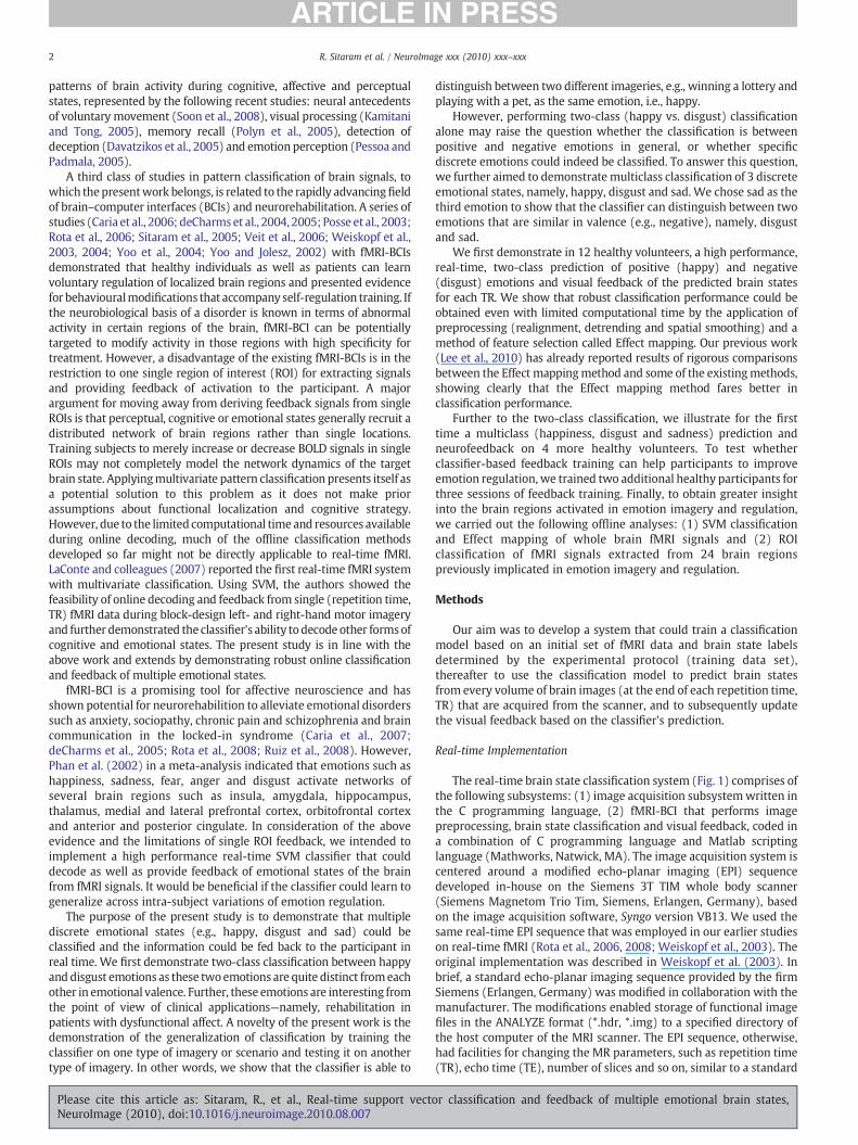
The real-time brain state classification system (Fig. 1) comprises ofthe following subsystems: (1) image acquisition subsystemwritten inthe C programming language, (2) fMRI-BCI that performs imagepreprocessing, brain state classification and visual feedback, coded ina combination of C programming language and Matlab scriptinglanguage (Mathworks, Natwick, MA). The image acquisition system iscentered around a modified echo-planar imaging (EPI) sequencedeveloped in-house on the Siemens 3T TIM whole body scanner(Siemens Magnetom Trio Tim, Siemens, Erlangen, Germany), basedon the image acquisition software, Syngo version VB13. We used thesame real-time EPI sequence that was employed in our earlier studieson real-time fMRI (Rota et al., 2006, 2008; Weiskopf et al., 2003). Theoriginal implementation was described in Weiskopf et al. (2003). Inbrief, a standard echo-planar imaging sequence provided by the firmSiemens (Erlangen, Germany) was modified in collaboration with themanufacturer. The modifications enabled storage of functional imagefiles in the ANALYZE format (*.hdr, *.img) to a specified directory ofthe host computer of the MRI scanner. The EPI sequence, otherwise,had facilities for changing the MR parameters, such as repetition time(TR), echo time (TE), number of slices and so on, similar to a standard
or classification and feedback of multiple emotional brain states,
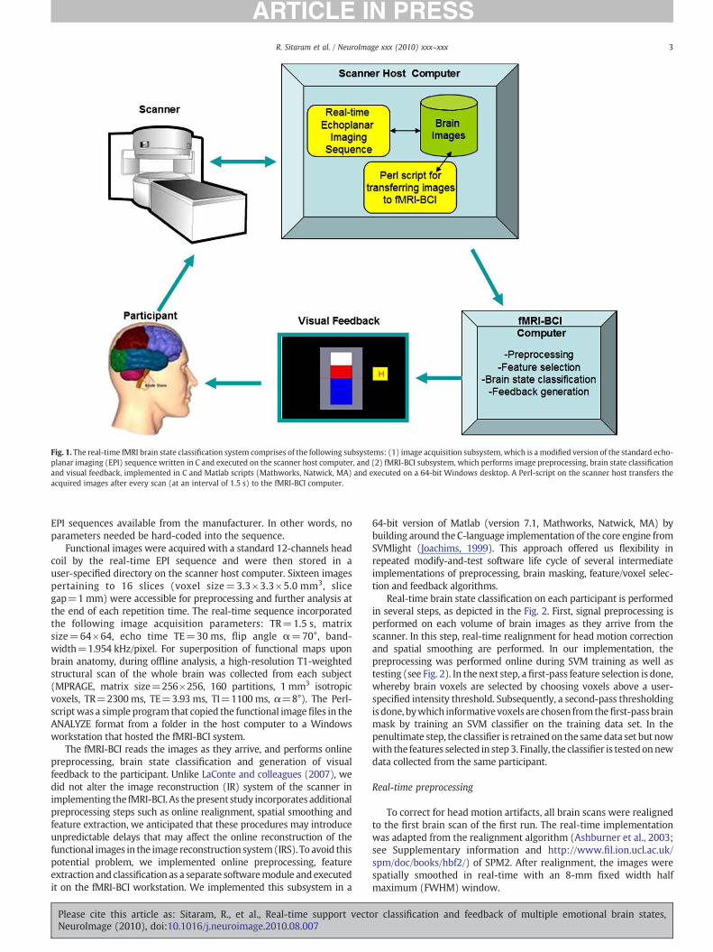
Fig. 1. The real-time fMRI brain state classification system comprises of the following subsystems: (1) image acquisition subsystem, which is a modified version of the standard echo-planar imaging (EPI) sequence written in C and executed on the scanner host computer, and (2) fMRI-BCI subsystem, which performs image preprocessing, brain state classificationand visual feedback, implemented in C and Matlab scripts (Mathworks, Natwick, MA) and executed on a 64-bit Windows desktop. A Perl-script on the scanner host transfers theacquired images after every scan (at an interval of 1.5 s) to the fMRI-BCI computer.
3R. Sitaram et al. / NeuroImage xxx (2010) xxx–xxx
EPI sequences available from the manufacturer. In other words, noparameters needed be hard-coded into the sequence.
Functional images were acquired with a standard 12-channels headcoil by the real-time EPI sequence and were then stored in auser-specified directory on the scanner host computer. Sixteen imagespertaining to 16 slices (voxel size=3.3×3.3×5.0 mm3, slicegap=1 mm) were accessible for preprocessing and further analysis atthe end of each repetition time. The real-time sequence incorporatedthe following image acquisition parameters: TR=1.5 s, matrixsize=64×64, echo time TE=30 ms, flip angle α=70°, band-width=1.954 kHz/pixel. For superposition of functional maps uponbrain anatomy, during offline analysis, a high-resolution T1-weightedstructural scan of the whole brain was collected from each subject(MPRAGE, matrix size=256×256, 160 partitions, 1 mm3 isotropicvoxels, TR=2300 ms, TE=3.93 ms, TI=1100 ms, α=8°). The Perl-scriptwas a simple program that copied the functional image files in theANALYZE format from a folder in the host computer to a Windowsworkstation that hosted the fMRI-BCI system.
The fMRI-BCI reads the images as they arrive, and performs onlinepreprocessing, brain state classification and generation of visualfeedback to the participant. Unlike LaConte and colleagues (2007), wedid not alter the image reconstruction (IR) system of the scanner inimplementing the fMRI-BCI. As thepresent study incorporates additionalpreprocessing steps such as online realignment, spatial smoothing andfeature extraction, we anticipated that these procedures may introduceunpredictable delays that may affect the online reconstruction of thefunctional images in the image reconstruction system(IRS). To avoid thispotential problem, we implemented online preprocessing, featureextraction and classification as a separate softwaremodule andexecutedit on the fMRI-BCI workstation. We implemented this subsystem in a
Please cite this article as: Sitaram, R., et al., Real-time support vectoNeuroImage (2010), doi:10.1016/j.neuroimage.2010.08.007
64-bit version of Matlab (version 7.1, Mathworks, Natwick, MA) bybuilding around the C-language implementation of the core engine fromSVMlight (Joachims, 1999). This approach offered us flexibility inrepeated modify-and-test software life cycle of several intermediateimplementations of preprocessing, brain masking, feature/voxel selec-tion and feedback algorithms.
Real-time brain state classification on each participant is performedin several steps, as depicted in the Fig. 2. First, signal preprocessing isperformed on each volume of brain images as they arrive from thescanner. In this step, real-time realignment for head motion correctionand spatial smoothing are performed. In our implementation, thepreprocessing was performed online during SVM training as well astesting (see Fig. 2). In the next step, a first-pass feature selection is done,whereby brain voxels are selected by choosing voxels above a user-specified intensity threshold. Subsequently, a second-pass thresholdingis done, bywhich informative voxels are chosen fromthefirst-passbrainmask by training an SVM classifier on the training data set. In thepenultimate step, the classifier is retrained on the samedata set but nowwith the features selected in step 3. Finally, the classifier is testedonnewdata collected from the same participant.
Real-time preprocessing
To correct for head motion artifacts, all brain scans were realignedto the first brain scan of the first run. The real-time implementationwas adapted from the realignment algorithm (Ashburner et al., 2003;see Supplementary information and http://www.fil.ion.ucl.ac.uk/spm/doc/books/hbf2/) of SPM2. After realignment, the images werespatially smoothed in real-time with an 8-mm fixed width halfmaximum (FWHM) window.
r classification and feedback of multiple emotional brain states,

Fig. 2. Flow chart for fMRI signal preprocessing and classification. Brain state classification is performed in the following steps: (1) signal preprocessing for online realignment andspatial smoothing, (2) first-pass feature selection for selecting brain voxels by applying an intensity threshold resulting in a brain mask, (3) second-pass thresholding for selectinginformative voxels from the first-pass brain mask by the method of Effect mapping resulting in the final brain mask, (3) classifier retraining based on the brain mask obtained in step3, and (4) real-time classifier testing on new data using the second-pass brain mask.
4 R. Sitaram et al. / NeuroImage xxx (2010) xxx–xxx
Support vector classification
To classify an individual scan of fMRI data, all brain voxels orselected brain voxels from each repetition time (TR) can be composedof an input vector xj. SVM determines a scalar class label Yi (Yi=sgn(yi=wTxi+b)=±1, i=1,…, N, where N is the number of inputvectors, T is the transpose of a vector, b is a constant value, sgn(.) is asignum function, sgn(x)=+1, 0, −1 if xN0, x=0, xb0, respectively)from the input vector.
When the input vectors xi and the designed labels YiL are takenfrom the training data set, the weight vector w of SVM is obtained byminimizing objective function L of Eq. (5) with constraintsEqs. (6) and (7),
L =12w
Tw + C ∑
N
i=1ξi; ð5Þ
withYiLðwT
xi + bÞ≥1−ξi; ð6Þ
andξi≥0 ð7Þ
where slack variable ξi is introduced to describe a non-separable case,i.e., data that cannot be separated without classification error, Cdenotes the weighting on the slack variable, i.e., the extent to whichmisclassification is allowed, and a value of C=1 was used in ourimplementation because of the following reasons. Firstly, modelselection to determine the C value is hard to perform in the context ofreal-time classification due to limitations of time available for SVMtraining. What is important is that real-time, online classificationshould work robustly in the majority of participants and sessions. Inmany previous fMRI classification studies (Haynes et al., 2007;LaConte et al., 2005, 2007; Mourao-Miranda et al., 2005, 2007,2006; Soon et al., 2008), C=1 was successfully used. Furthermore,
Please cite this article as: Sitaram, R., et al., Real-time support vectNeuroImage (2010), doi:10.1016/j.neuroimage.2010.08.007
LaConte et al. (2005) showed that prediction accuracy does not varymuch with the selection of C.
Feature selection and SVM training
The performance of a pattern classifier could be improved by aprocedure called feature selection, where informative features from thedata are extracted for input to the classifier. We performed featureselection in 2 steps: (1) a first-pass intensity thresholding and (2) asecond-pass voxel selection using Effect mapping (Lee et al., 2010). Forthe first-pass intensity thresholding, an image of the brain, calledintensity thresholded mask, was created by removing voxels below acertain BOLD intensity value in a mean image of all the brain scans. Forthe second-pass feature selection, training data from 2 runs weredivided into 5 sequential subsets. That is, each subset consisted of 4.5blocks (for example, in the case of binary classification: 2.5 blocks ofcondition happy, 2 blocks of condition disgust or 2.5 blocks of conditiondisgust, 2 blocks of conditionhappy) froma total numberof 24blocks. Z-normalization (z-value: (x-mean(x))/standard deviation(x), x: sam-ples) was applied across all the time-course signals at each voxel toaccount for the variability of BOLD signals across runs. Z-normalizationwas performed to correct for the variability of the BOLD signal and toconvert it to a signal of zero mean and unit variance. BOLD values frombrain voxels of the subsetswere used to create input vectors for trainingSVMs and subsequently to generate five Effect maps (please seeSection 3.5 for a description of the Effectmaps). Informative voxelswereselected by applying a threshold on themean of five Effectmaps (Fig. 2).The online SVM classification software written in Matlab allows forspecifying user-specified threshold. The threshold that was incorporat-ed in this study was 0.5 for all participants and sessions. In ourexperience, this threshold provided a good balance between reductionof the size of the data inputwhilemaintaining good prediction accuracy.Finally, an SVM was trained from the BOLD values of the feature-selected voxels after Z-normalization across all the training samples.
or classification and feedback of multiple emotional brain states,

5R. Sitaram et al. / NeuroImage xxx (2010) xxx–xxx
Ideally, SVM output yi should be centered about zero, so that whenthe output is greater than zero the brain state is assigned to oneemotion (e.g., happy), andwhen it is less than zero it is assigned to theother emotion (e.g., disgust). However, due to participants' headmovements and other systemic changes, a gradual drift in theclassifier output can be expected (LaConte et al., 2007). To removethis bias during online classification, we subtracted the mean of theSVM outputs during the rest condition from each SVM output duringthe active condition (i.e., happy or disgust).
Effect mapping
Based on the parameters of the trained SVM model, we analyzedthe fMRI data with a newmeasure called effect value (Lee et al., 2010)which estimates the effect of the BOLD activation at each voxel on theclassifier output by first computing the mutual information betweenthe voxel and the output, and then multiplying it with the weightvalue of the voxel as estimated by SVM. Mutual information (MI) isderived from relative entropy or the Kullback–Leibler divergence,which is defined as the amount of information that one randomvariable contains about another random variable (Cover and Thomas,1991). Hence, the effect value (EV) Ek of a voxel k is defined as:
Ek = wkIðxk; yÞ; k = 1; ⋯;M ð8Þ
where I(xk; y) is the mutual information between the voxel and theoutput, y is the SVMoutput after excluding the sign function,wk and xkare weight value and activation at voxel k, respectively. To reduce thevariability of EVs from Eq. (8) dependent on data set, normalization ofEk from Eq. (9) was applied as follows:
lEk = sgnðEkÞ logð1 + stdðjEjÞÞ; k = 1; ⋯;M ð9Þ
where sgn(.) is a signum function, and std(|E|) is standard deviationacross all the brain voxels. For ease of voxel selection based on the EVs,we have used Eq. (9) for generating the Effect maps at the contrast:happy vs. disgust in binary classification, and happy vs. disgusted,happy vs. sad, and disgusted vs. sad, in our multiclass classification,respectively. Such maps can be thresholded at a suitable level byvisual inspection by the experimenter and the resulting map can beused as a brain mask. This brain mask is subsequently applied tofunctional images arriving at each time point (TR), to reduce the datadimension and to choose the most important voxels for classification.
Generation of visual feedback
Visual feedback was provided in the form of a graphicalthermometer whose bars increase or decrease in number from thebaseline value (represented by the middle red dashed line of thethermometer) proportional to the classifier output after each TR. Thethermometer could be operated in two modes by selection in ourMatlab toolbox: (1) feedback mode and (2) non-feedback mode. Inthe non-feedback mode, the thermometer was displayed without anybars for the whole duration of the experiment. In the feedback mode,the graphical thermometer is shown in an animated fashion byincreasing or decreasing the number of bars. The classifier output aftercorrection of the classifier drift was used to generate the visualfeedback. Depending on the classification result, i.e., +1 or −1, thethermometer bars were updated with respect to the previous TR inthe positive or negative direction, respectively. The thermometer wasdesigned to indicate not only the correctness of classification (interms of the bar changing in the positive or negative direction) at theend of each TR, but also the cumulative performance of the past trialsin terms of the height of the bars above or below the baseline. Thisway the participant receives feedback about his instantaneous
Please cite this article as: Sitaram, R., et al., Real-time support vectoNeuroImage (2010), doi:10.1016/j.neuroimage.2010.08.007
(defined by the length of the TR) brain state as well as his pastperformance.
Multiclass classification
The objective of the multiclass classification experiment was totest whether the online classifier is able to classify three discreteemotions, namely, happy, sad and disgust. The multiclass problemwas formulated as a problem of finding the best classification amongthree trained binary classifiers (i.e., classifier 1: happy vs. disgust,classifier 2: happy vs. sad, and classifier 3: disgust vs. sad) based onthe selected voxels from an Effect map of each combination asdescribed above.
Multiclass classificationwas performed based on the framework oferror correcting output code (ECOC) from 3 binary classifications(Mourao-Miranda et al., 2006). The ECOC method assigns a uniquebinary string of length n, called the “code word”. Subsequently, nbinary classifiers are trained, one for each bit position of the binarystring. During the classifier testing, new data are classified byevaluating each of the binary classes to arrive at an n-bit string,which is then compared to all the different codewords. The test data isfinally assigned to the code word that is the closest based on adistance measure. In our approach to the ECOC, each class has its owncode mi as follows: m1=[1 1 0] for classifier 1 (happy vs. disgust),m2=[−1 1 0] for classifier 2 (happy vs. sad), m3=[0 −1 −1] forclassifier 3 (disgust vs. sad). Let p be a vector of predictions from the 3classifiers. Then, the final decision was made by selecting the closestcode from the prediction vector r = minarg i = 13distðmi;pÞ. We usedthe Euclidian distance measure in our implementation.
Experimental paradigm
Our experiment (Fig. 3) was divided into three parts: experiment 1for investigating real-time binary classification, experiment 2 forfurther feasibility testing of multiclass prediction, and experiment 3for assessing the effect of extended feedback training with a real-timeclassifier. Twelve healthy students from the Department of Medicine,University of Tuebingen, Germany, participated in experiment 1 andfour more students participated in experiment 2. The participantswere aged in the range of 22–26 years, with a mean age of 25 years.All participants signed a written informed consent, and the study wasapproved by the local institutional review board.
Experiment 1 (see Fig. 3) was envisioned in 4 succeeding stages,each stage prepared as a block-design protocol with alternating blocksof happy and disgust imagery of 24-s duration, interleaved with restblocks of 4.5-s duration. In each run, there were 6 blocks of happyimagery, 6 blocks of disgust imagery and 12 blocks of rest. In total, 233scanswere collected in a run including the 5 initial scans that were notused in the analysis, due to magnetic equilibration effects that couldpotentially distort the data. The rest blocks were incorporated for thesole purpose of avoiding cognitive and emotional overload due tosudden changes of imagery. During classifier training (stage 1)containing 2 runs of the above protocol, there were 12 blocks ofhappy imagery, 12 blocks of disgust imagery and 24 blocks of rest.During each type of the emotional imagery block, an emptythermometer (without feedback) was shown at the centre of thescreen with a letter beside it indicating the participant to performhappy (‘H’) or disgust (‘D’) mental imagery. The purpose of the emptythermometer during the SVM training stage was to maintainconsistency in visual stimulation with the testing stage duringwhich the thermometer could be updated for visual feedback of thebrain state. Two runs of the stage 1 protocol were performed to collectsufficient amount of data for training the SVM classifier (stage 2).
Participants were instructed well in advance of the experiment toidentify one or more emotional episodes from their personal lives foreach type of emotion (e.g., happy, sad or disgust) that they were
r classification and feedback of multiple emotional brain states,

Fig. 3. Experimental protocol. Both binary (happy and disgust) and multiclass (happy, sad and disgust) classification experiments were performed in 4 succeeding stages, each stageprepared as a block-design protocol with alternating blocks of emotion imagery each of 24-s duration, interleaved with rest blocks of 4.5-s duration. During SVM training, an emptythermometer was shown at the centre of the screen with a letter beside it indicating the participant to perform happy (‘H’), disgust (‘D’) or sad (‘S’) mental imagery. Two runs of thestage 1 protocol were performed to collect data for training the SVM classifier (stage 2). Stages 3 and 4 were performed to test the classifier without and with feedback, in that order.
6 R. Sitaram et al. / NeuroImage xxx (2010) xxx–xxx
required to recall during the experimental blocks. To maintainconsistency between classifier training and testing conditions weshowed participants pre-selected pictures as a reminder of the specifictype of emotion imagery that they need to use in each session. Towardsthis end, participants were asked to identify one picture from theInternational Affective Picture System(IAPS) (Langet al., 1997) that bestepitomised each type of emotional episode. At the beginning of atraining or testing run, participants were shown each selected picturefor a duration of 30 s to remind and strengthen their emotional recallstrategies. We could have alternately displayed text to describe andremind them of the specific imagery to use, but we found that picturescapturedmore succinctly the complexity of anemotional recall scenario.However, to avoid a potential problem that subjectsmight just recall theimages rather than the emotion, we specifically instructed subjects touse the IAPS images asmere references to their imagery and to elaborateon the images to invoke the required discrete emotion (happy, disgustor sad emotion). We later confirmed the type and level of emotionimagery they performed with an interview and subjective rating (seeTable 1 and Supplementary Table 2).
Please cite this article as: Sitaram, R., et al., Real-time support vectNeuroImage (2010), doi:10.1016/j.neuroimage.2010.08.007
Stages 3 and 4 were meant to test the classifier without and withfeedback, respectively. These two tests were performed to comparereal-time classification performance in the absence and presence offeedback to evaluate the eventual application of real-time brain statedecoding for BCI and clinical rehabilitation. At the end of each run,participants were asked, over the scanner audio system, to complete aself-assessment scale (1–9, 1 indicating poor performance and 9indicating best performance) of their level of emotion regulation foreach type of emotion. The purpose of this self-report was to performan offline analysis of the correlation between subjective report andonline classifier performance.
Four out of the 12 participants in experiment 1 underwent anadditional run of the stage 4 protocol to test whether the classifiercould robustly predict emotion states even when participants do notuse the same emotion inducing strategies that were used during SVMtraining runs. To assess whether feedback training helps participantsto improve emotion recall, we recruited two more participants andtrained them on two-class (happy vs. disgust) classification for 3sessions (in addition to 2 sessions for collection of training samples).
or classification and feedback of multiple emotional brain states,

Table 1PANAS scores, subjective ratings and strategies for binary and multiclass emotion imagery.
Subject Mean positiveaffect sub-scoreof PANAS
Mean negativeaffect sub-scoreof PANAS
Subjective ratings of emotion imagery (scales 1–9, 1=worst, 9=best)* Strategies for happy (H),disgust (D) and sad (S)emotions
Training run 1 Training run 2 Test run (no feedback) Test run (with feedback)
Binary classification with same strategies for training and testingS1 30 24 7.5 7.5 6 6.5 H: romantic love
D. toilet/fecesS2 41 21 6 6 6 5 H: family reunion
D: eating insectsS3 26 12 8 7.5 7.5 7.5 H: good marks in exams
D: blood in an open woundS4 26 13 6 6.5 6 7 H: graduation day
D: a dirty toiletS5 27 26 6.5 5 4.5 4.5 H: riding a bike
D: infected skin woundS6 20 23 2.5 3 4 3.5 H: romantic love
D: fecesS7 28 30 6 6 5.5 6.5 H: romantic love
D: a dirty toiletS8 28 18 9 6 6 7 H: baby
D: a dirty toiletS9 29 22 6.5 6 5 6 H: friends gathering
D: a dirty toiletS10 34 10 7 7 7 9 H: cats
D: an used toiletS11 23 16 7 8 9 H: friends gathering
D: dirty and sick peopleS12 4 8 9 9 H: falling in love
D: spiders
Binary classification with different strategies for training and testingS9 28 13 7 6.5 6.5 5.5 Training runs:
H: romantic loveD: baby with fecesTesting runs:H: family reunionD: dirty bathroom
S10 14 17 6 7 6 7.5 Training runs:H: romantic loveD: spidersTesting runs:H: party with friendsD: dirty bathroom
S11 39 10 8.5 8.5 8 9 Training runs:H: romantic loveD: toiletTesting runs:H: playing the guitarD: rotten food
S12 43 11 8 8.5 8 6.5 H: getting the visaD: a dirty laboratoryTesting runs:H: romantic loveD: toilet
Multiclass classification with same strategies for training and testingS13 31 10 7.7 8.4 8 7.7 H: pets
D: toiletS: braking up with boyfriend
S14 24 14 7.7 7.8 7.7 7.8 H: graduation dayD: toiletS: death of a friend
S15 27 11 6.2 8 8 9 H: a boat tripD: bloody car accidentS: braking up with boyfriend
S16 14 16 6.8 7.3 6.3 7.8 H: romantic loveD: spidersS: view of blind beggars
*Values correspond to the mean between happy and disgust score in binary classification, or between happy, disgust and sad scores in the multiclass classification.
7R. Sitaram et al. / NeuroImage xxx (2010) xxx–xxx
To adapt the classifier to the changes in the participant's brainactivation with thermometer feedback and learned regulation, weretrained the classifier after each feedback session on a combined dataset of the training data set (collected in the absence of feedback) andthe feedback data set (collected in the presence of feedback).
Please cite this article as: Sitaram, R., et al., Real-time support vectoNeuroImage (2010), doi:10.1016/j.neuroimage.2010.08.007
Experiment 2 was designed to test real-time multiclass classifica-tion, both with and without feedback. Four more student participants(mean age=25) were recruited for this experiment. Conceptually,experiment 2 was designed similar to experiment 1, except thatadditional blocks pertaining to a third emotion (sadness) was added
r classification and feedback of multiple emotional brain states,

8 R. Sitaram et al. / NeuroImage xxx (2010) xxx–xxx
to train and test a three-class classifier. That is, each run consists of 4blocks of each emotion with 12 blocks of rest condition. The blocklength is exactly same as the binary case. The total duration of eachrun of experiment 2 was also 233 scans (5.825 min).
Experiment 3 was conducted to answer the question: How can apattern classifier provide contingent feedback in a self-regulationapplication where participants are normally expected to improvetheir ability with training to induce themselves into desired brainstates? One way to approach this would be by adapting the classifierto participant's learning by incrementally retraining the classifier withnew samples at the end of each session of feedback training. We haveadapted this method in an extended experiment in two participants toincrementally enhance the classifier's ability to recognize changes inbrain activity with self-regulation learning. We trained participantson 2-class (happy vs. disgust) classification, with 3 sessions offeedback training (in addition to 2 sessions for collection of trainingsamples) in a day. We retrained the classifier after each feedbacksession by combining the previous training set with the new test data.Thermometer feedback was provided as described in section 3.6.Participants were instructed to perform emotion recall in order toincrease the bars of the thermometer. They were told that thermom-eter bars going up meant that they were doing the task right and barsgoing down meant that they were probably not doing it right.
Offline whole brain and ROI analysis with classification
To assess the level of involvement of different brain regions, twoadditional types of SVMclassificationswere performed offline: (1) single-subject whole brain analysis with the E-map (Lee et al., 2010) (binaryclassificationon12 subjects andmulticlass classificationon1 subject), and(2) ROI analysis (binary classification on 12 subjects). We expected thatBOLD signals from brain regions known to be involved in emotionprocessing would show higher decoding accuracy for the discreteemotions under consideration (i.e., happy and disgust) than in otherregions. Our ROI analysis is based on the expectation that classificationaccuracy will be higher in ROIs that have been previously implicated inemotion regulation and induction (Amodio andFrith, 2006;Ochsner et al.,2005, 2004; Phan et al., 2002). Towards this end, fMRI images for allparticipants were preprocessed with the following procedures: realign-ment, normalization to MNI space, and spatial smoothing with 8 mmGaussian FWHM. First, single-subject whole brain classification wasperformed wherein BOLD signals from all brain voxels for each TR werecollected into an input vector. For the ROI classification, 24 masks (seeSupplementary Fig. 2) from brain regions previously reported to beinvolved in emotion imagery, recall and regulation (Amodio and Frith,2006; Ochsner et al., 2005, 2004; Phan et al., 2002) were applied to thepreprocessed data to extract BOLD signals as input to separate classifiers.Z-normalization was performed on the BOLD values at each voxel acrossthe whole time course to correct for the variance of BOLD signals ofdifferent runs and different participants. The normalized BOLD valuesfrom each ROI of each TR were then collected into an input vector. Datafrom2 training sessionsand1 testing sessionwereused inboth the single-subject whole brain and ROI classifications. Linear SVM, with theregularization parameter C=1, based on freely available SVM librarySVMLight (Joachims, 1999) was used to perform both single-subject andROI classifications. The single-subjectwhole brain analysiswasperformedbased on the E-maps obtained after training a separate SVMmodel fromeach participant's data. The ROI classification performance was evaluatedthrough 12-fold cross validation (CV). In each fold, the data of 11participants were used to train the classifier, and then the data of 1remaining participant were used to test the classifier.
Affect scores and subjective ratings of emotion recall
Before thebeginningof fMRIdata collection,weaskedeachparticipantto fill out the Positive Affect Negative Affect Schedule (PANAS), which is a
Please cite this article as: Sitaram, R., et al., Real-time support vectNeuroImage (2010), doi:10.1016/j.neuroimage.2010.08.007
psychometric scale developed to measure the independent constructs ofpositive and negative affect both as states and traits (Watson et al., 1988).PANAS contains a list of 10 descriptors for positive scale: attentive,interested, alert, excited, enthusiastic, inspired, proud, determined, strongand active; and 10 descriptors for negative scale: distressed, upset-distressed; hostile, irritable-angry; scared, afraid-fearful; ashamed, guilty;nervous, and jittery. The PANAS has been found to exhibit the followingcharacteristics: (1) a significant level of stability in every time frame; (2)no consistent sex differences; (3) inter-correlations and internalconsistency reliabilities are all acceptably high (ranging from 0.86 to0.90 for PA and 0.84–0.87 for NA); (4) the reliability of the scales isunaffected by the time instructions used; and (5) has high scale validityand high item validity.
At the end of each SVM training or testing run, participants wereinstructed to rate their degree of success in being able to recall eachtype of emotion (i.e., happiness, disgust or sadness) for each block ofthe run on a scale of 1–9, where 1 represented the worst and 9represented the best regulation (see Table 1).
Results
PANAS scores and subjective reports of emotion imagery
Participants used several emotion recall scenarios and imagerystrategies for inducing themselves into happy, disgust and sad (seeTable 1).
A negative correlation was found between the mean of thenegative subscale of the PANAS and participants' ratings of theirperformance in emotion imagery tasks of session 3 (rs=−.74, pb .01)and 4 (rs=−.72, pb .01). A similar trend was also found for session 2(rs=−.56, p=.07, two-tailed). No significant correlations werefound between positive subscale of the PANAS and the participants'ratings of their performance across the sessions of emotion imagerytask. Also, no significant correlations were found between theprediction accuracy either with the PANAS scores, or with partici-pants' ratings of their performance in emotion imagery.
In addition, we collected subjective reports of the intensity of theemotion attained, attention to the imagery task and the consistency of themental strategywithin each session, in a 9-point scale (1—worst, 9—best).Due to lack of scanning time, these data could be collected only in a subsetofparticipants: 4participants in thebinaryclassificationand4participantsin the multiclass classification (see Supplementary Table 2).
Real-time binary and multiclass classification
Fig. 4 shows the prediction accuracies of the online classifier for allparticipants for SVM testing runs, with and without feedback, forbinary classification (see Figs. 4a and b) and for multiclassclassification (see Fig. 4c). Fig. 4a shows the performance of thebinary classifier when participants used the same type of emotionimagery both during SVM training and testing runs (e.g., a participantused imageries of family reunion for happy emotion and eating insectsfor disgust emotion, respectively). Fig. 4b shows the performance ofthe binary classifier when participants used different types of emotionimagery during SVM training and testing runs (e.g. a participant usedimageries of romantic love for happy emotion and baby with feces fordisgust emotion for SVM training; and family reunion for happyemotion and dirty bathroom for disgust emotion for SVM testing). Theonline classifier showed the following classification accuracies acrossparticipants, indicated by mean and standard deviation of correctclassification: (1) for binary classification (chance accuracy=50%)testing with same emotion imagery, without feedback (92%±6%) andwith feedback (80%±13%) (see Fig. 4a); (2) for binary classificationtesting with different emotion imagery, without feedback (80%±10%) and with feedback (65%±18%) (see Fig. 4b); and (3) formulticlass classification (chance accuracy=33%) testing without
or classification and feedback of multiple emotional brain states,

Fig. 4. Online SVM classification performance. (a) Binary classification (happy vs. disgust) accuracies (chance level=50%) in 12 participants who used the same emotion imageriesduring both SVM training and testing sessions. (b) Binary classification accuracies in 4 participants who used different emotion imageries between SVM training and testing sessions.(c) Multiclass classification (happy vs. disgust vs. sad) accuracies (chance level=33%) in 4 participants who used same emotion imageries in SVM training and testing sessions. (d)Classification accuracies (chance level=50%) of extended feedback training on 2 participants on two-class (happy vs. disgust) classification.
9R. Sitaram et al. / NeuroImage xxx (2010) xxx–xxx
feedback (62%±14%) and with feedback (60%±16%). Classificationperformance indicates the beneficial effects of online realignment,feature selection and optimized experimental procedures. Robustprediction accuracies were seen even when participants wereintentionally instructed to use different strategies between SVMtraining and testing runs, indicating the ability of the classifier togeneralize across varied emotion imagery strategies and memoryrecall scenarios.
Significantly higher accuracies of the SVM classifier were found inthe sessions without feedback (median=88.5) compared to thesessions with feedback (median=76), z=−2.02, pb .05, r=−.37(Wilcoxon signed-rank test). To assess the effect of extended feedbacktraining on participants' performance, we trained 2 new participantsfor 3 sessions of emotion recall in the presence of feedback andincremental retraining of the classifier. Our results show thatclassification performance improves already in the second feedbacksession and classification accuracy continues to be maintained in thethird session (see Fig. 4d). However, by the end of 5 training sessions,participants were unable to continue due to tiredness and hencefeedback training had to be stopped. Nevertheless, our data suggeststhat feedback training with incrementally retrained classifier couldenhance participant's performance.
SVM outputs for binary and multiclass classification
Supplementary Figs. 3a and b show the online SVM output for aparticipant before and after bias removal. Examination of the SVMoutputs across different participants for multiclass classificationshowed robust classifier performance on a block-by-block basiswithin each testing session (see Supplementary Fig. 3c).
Effect maps and ROI classification performance
Extensive results of the single-subject whole brain classificationand Effect maps generated thereof (in an offline analyses performed
Please cite this article as: Sitaram, R., et al., Real-time support vectoNeuroImage (2010), doi:10.1016/j.neuroimage.2010.08.007
after the real-time experiments to reveal the discriminating brainregions) have been documented in the Supplementary information(Supplementary Sections 2.2 and 2.3, including supplementaryTable 3 (a–k), Supplementary Fig. 1 (a–k) and Table 4 (a–c)). TheSupplementary Tables also provide, for each brain region, the effectvalue (EV) and the cluster size, which together indicate the degree ofimportance of the cluster in discriminating between the emotions.Much inter-subject variability was observed in brain activations,possibly due to the diverse emotion imageries employed by theparticipants (see Table 1 for list of emotion imageries). The followingbrain regions were also observed as commonly activated among theparticipants: orbital medial frontal cortex (oMFC), anterior rostralmedial frontal cortex (arMFC) and posterior rostral medial frontalcortex (prMFC) (these anatomical segregation were based on Amodioand Frith (2006)); superior and lateral frontal cortices, anteriorcingulate cortex (ACC), insula, superior temporal gyrus (STG),primary and association areas of the visual cortex, posterior cingulatecortex (PCC) and precuneus. In terms of Brodmann areas, thefollowing areas were commonly observed: BA10, 11, 14 and 25belonging to the oMFC, BA32 to arMFC, BA8, 9 to the prMFC; andBA17, 18 and 19 belonging to the visual cortex. Fig. 5 showsexemplary Effect maps depicting the 3 contrasts of (happy vs.disgust), (happy vs. sad) and (disgust vs. sad) emotions.
To better identify the most important brain regions for emotionimagery and regulation, we performed offline ROI classifications,based on fMRI signals extracted from 24 different brain masks (seeSupplementary Fig. 2) previously implicated in emotion processing(Amodio and Frith, 2006; Ochsner et al., 2005, 2004; Phan et al.,2002). This analysis was conducted separately on 12 subjects who hadperformed happy and disgust imagery (see Fig. 3, Table 1). Our results(see Fig. 5d) reveal that high classification performance (N75%) wasobtained in the following brain regions: middle frontal gyrus (MiFG),superior frontal gyrus (SFG), ventrolateral prefrontal cortex (VLPFC),STG and precuneus. Other ROIs that showed high prediction ratesincluded dorsolateral prefrontal cortex (DLPFC), ACC and the insula.
r classification and feedback of multiple emotional brain states,

Fig. 5. Effect maps generated from single-subject whole brain SVM classification showing discriminating voxels for: (a) happy vs. disgust classification (for participant S1), (b) happyvs. sad classification and (c) disgust vs. sad classification (for participant S13). Second level threshold appliedwas 0.5, slices shown are in the range, Z(MNI coordinates)=−11 to+10in steps of 3 units. Brain regions: oMFC—orbital medial frontal cortex, arMFC—anterior rostral MFC (based on Amodio and Frith, 2006); OFC—orbitofrontal cortex, ACC—anteriorcingulate cortex, PCC—posterior cingulate cortex. (d) Prediction accuracy of 24 regions of interest (ROIs) in a single-subject ROI classification from 12 participants performing happyand disgust motor imagery. ROIs: Subgenual—subgenual cingulate (BA25), Nac—nucleus accumbens, PVC—primary visual cortex, DLPFC—dorsolateral prefrontal cortex, LiG—lingualgyrus, VLPFC—ventrolateral prefrontal cortex, STG—superior temporal gyrus, MiTG—middle temporal gyrus, MFG—medial frontal gyrus, SFG—superior frontal gyrus, MiFG—middlefrontal gyrus.
10 R. Sitaram et al. / NeuroImage xxx (2010) xxx–xxx
Discussion
The present implementation of brain state decoding and fMRI-BCIis based on real-time SVM classification of fMRI signals after onlineheadmotion correction, spatial smoothing and feature selection based
Please cite this article as: Sitaram, R., et al., Real-time support vectNeuroImage (2010), doi:10.1016/j.neuroimage.2010.08.007
on our new approach called Effect mapping (EM) (Lee et al., 2010).EM is a method of generating functional activations by multiplyingtwo types of information at each voxel of the brain: (1) SVM trainedweight value at the voxel, and (2) and the mutual information (MI)between the BOLD value at the voxel and the SVM output. Activation
or classification and feedback of multiple emotional brain states,

11R. Sitaram et al. / NeuroImage xxx (2010) xxx–xxx
maps so derived represent a hybrid of univariate and multivariatefunctional mapping, resulting at once in spatially distributed activa-tions as well as in large clusters centered about brain regionspresumably important for discriminating between the brain states.In the present study, EM was used as the basis for feature selection toreduce the multidimensionality of fMRI data, and to choose the mostdiscriminating clusters of voxels for classification. Our results showthat the application of feature selection results in consistently goodpredictions of emotions for binary classification as well as formulticlass classification.
Subjective reports collected from participants ascertained thatparticipants performed emotion imagery and that the online classifierdecoded emotions and not arbitrary states of the brain. This study alsoshows a relationship between the participants' affect scores asmeasured by PANAS and the subjective ratings of their performancein the emotion imagery task, indicating that participants who reportgreater negative affect rate themselves relatively lower on theimagery tasks.
One may expect that higher subjective reports of success inrecalling emotions should correlate with higher prediction accuracies.However, one should note that pattern classification works well andshows higher accuracy based on how similar the samples in the testset are in comparison to the training set. The greater the similarity, thegreater is the accuracy. However, if the test set has different brainsignals generated due to higher success in recall than in the trainingset, then the similarity of patterns is reduced and so is theclassification accuracy. This means that improved success in recallingemotions may not result in greater decoding accuracy. This is incontrast to the situation with single ROI real-time feedback experi-ments where improved success in recall may correlate with higher %BOLD in the target region of interest.
This may then lead one to ask the question: How then can apattern classifier provide contingent feedback in a self-regulationapplication where participants, including patients, are normallyexpected to improve their ability (with more training) to inducethemselves into desired brain states? One way to approach this wouldbe by adapting the classifier to participant's learning by incrementallyretraining the classifier with new samples at the end of each session offeedback training. Another way could be to carry out feedbacktraining of patients or beginners using a brain state classifier whichhas been previously trained on data from experts or individuals whoare already good at self-regulation. In this approach, the classifierwould show improved performance as the trainees achieve brainstates similar to the experts. In such a classifier, one can expectimproved prediction accuracy with greater success in recall orregulation. In any method, the experimenter has the prerogative toascertain whether the participant is performing self-regulation asdesired and whether the relevant brain regions are activated byscrutinizing the multivariate functional activations.
In our experiments, the performance of the classifier in the presenceof feedback was lower than in its absence although a few participantsperformed better in the presence of feedback than in its absence. Thislower accuracyof the classifierwhen the feedbackwas introduced couldbe due to as yet unlearned ability of the participants to performconsistent emotion imagery while simultaneously monitoring thethermometer. These data are in line with self-reports by participantsafter scanning sessions, about the difficulties they had in performingemotion regulation when feedback was introduced for the first time. Ithas been known that the introduction of feedback initially creates a taskconflict between attention to the imagery task and the visual feedback(Rota et al., 2008), which could be overcomewith longer training (Cariaet al., 2007; deCharms, 2008; Rota et al., 2008). A second reason for thereduction in classification performance could be the extra activation ofthevisual cortexdue to the graphical animationof the thermometer thatwas previously absent during the classifier training. This additionalBOLD activation could act as noise or confound to the classifier input,
Please cite this article as: Sitaram, R., et al., Real-time support vectoNeuroImage (2010), doi:10.1016/j.neuroimage.2010.08.007
reducing its prediction accuracy. To overcome the above limitations andto assess the effect of extended feedback training on participants'performance, we recruited two more participants and trained them ontwo-class (happy vs. disgust) classification, with 3 sessions of feedbacktraining by incorporating an incrementalmethod of classifier retraining.Our results show that participants did learn to improve theirperformance in the presence of feedback. An elaborate study on theeffects of feedback training is beyond the scope of this work. Futurestudies could be dedicated to the systematic evaluation of the effect offeedback training on self-regulation performance, behaviour andchanges in relevant neural network.
We performed additional offline pattern classification and multi-variate statistical mapping based on the EM (Lee et al., 2010)approach to identify brain regions for discriminating emotion underconsideration. Our results are in line with previous findings. Firstly,the most observed involvement of the medial frontal cortex in ourclusters of activation and Effect maps is of interest. A theoreticalreview of the involvement of medial frontal cortex (MFC) by Amodioand Frith (2006) delineates the functional roles of the 3 regions ofMFC, namely, arMFC, prMFC and oMFC. The authors associated theactivation of the arMFC (the most anterior part of the frontal cortexincluding BA10, BA32 and rostral ACC) with the monitoring of one'sown emotional state, such as rating one's emotions in response topictures of varying valence. Themore posterior region, the rostral MFC(prMFC), including BA8, BA9 and dorsal ACC, is activated by cognitivetasks such as action monitoring and attention. The orbital MFC(oMFC), including BA10, BA14 and BA25, was linked to themonitoringof task outcomes associatedwith punishments or rewards. Activationsof the prefrontal cortex, ACC and insula found in our analyses are inline with an influential meta-analysis by (Phan and colleagues (2002)which states that the MFC has a general role in emotional processingand that induction by emotion imagery recruits the anterior cingulateand insula. Our offline analyses serve to confirm that participantsperformed emotion imagery and that the SVM results are reliable.Additionally, to our knowledge, our study provides the first objectivecomparison, through pattern classification, of the degree of involve-ment of different brain regions (see Fig. 5 and Supplementary Table 4)in emotion imagery and regulation.
In our real-time implementation of the SVM classifier, weprogrammed the online SVM classifier and the BCI system in acombination of C programs and Matlab scripts on a dedicatedworkstation. This design decision was beneficial in the followingways: (1) it allowed us greater flexibility in the repeated softwarecycle of source code modification and testing, (2) it helped us avoidinterfering with the normal operation of the scanner and (3) itresulted in faster SVM training and online SVM online classification.This modular approach to software design has an added advantage inthat when future versions of the scanner operating software (e.g.,Syngo version VB15) are implemented, it is no longer necessary for theuser to modify, recompile and relink the SVM classification sourcecode.
Our decision to use was SVM based on recent developments in thepattern classification of neuroimaging data (LaConte et al., 2003,2005; Martinez-Ramon et al., 2006; Shaw et al., 2003; Strother et al.,2004). SVM has been shown to have certain advantages in theclassification of fMRI signals in comparison to other methods such aslinear discriminant analysis (LaConte et al., 2005) and multilayerneural network. SVM is less sensitive to preprocessing (LaConte et al.,2005), better capable of handling large data sizes (Haynes and Rees,2006), techniques developed to perform multiclass classification andmost importantly produces unique optimal solutions (Collobert et al.,2006). Although, SVM model training is computationally moreintensive, current availability of faster yet cheaper processors morethan compensate for this drawback (LaConte et al., 2007).
Onlinemulticlass brain state decoding has potential applications inlie detection (critical lies, non-critical lies and truths), BCIs (detecting
r classification and feedback of multiple emotional brain states,

12 R. Sitaram et al. / NeuroImage xxx (2010) xxx–xxx
multiple movement states and thoughts) for communication andcontrol, to name a few. The majority of the present implementationsof brain state decoding including real-time classification, however,have two main drawbacks. Firstly, the present methods work basedon spatial pattern of brain activity alone as input in discriminatingdifferent states of the brain and ignore the temporal pattern or timeevolution of brain activity. Considering that brain states may differ intheir temporal signature, in terms of time evolution of activity in oneregion and also in terms of its dynamic interaction with other regionsof the brain, spatial classification of brain activity alone is largelylimiting. For example, emotion regulation involves the dynamic,temporal evolution of BOLD activity connecting different parts of thelimbic region that includes insula, amygdala, anterior cingulatedcortex and medial prefrontal cortex. Hence, a pattern classificationsystem that uses both spatial and temporal information but does so ina computationally efficient way so that it could be used in real-timewould be of great practical importance and should be the focus offuture research.
Another limitation of our present work and possible topic of futureresearch is the lack of an implementation for real-time, subject-independent classification of brain states. Existing methods areoptimized for each participant because SVM training is carried outon subject-specific data. However, these methods have two majordisadvantages: (1) one needs to collect initial data for classifiertraining that consumes time and participant's attention and involve-ment, and (2) this approach is not suitable for certain applicationssuch as real-time lie detection for forensics and security on personsfrom whom data for SVM training might not be available. An adaptiveclassifier needs to be developed similar to those previously developedin the pattern recognition field for subject-independent speech andcharacter recognition. Such a subject-independent brain state classi-fier could then be applied to new subject data without prior classifiertraining, and in addition, could be adapted to the idiosyncrasies ofevery individual's brain size, shape and activation patterns. Anessential technical improvisation to be achieved in this regard is thereal-time coregistration and normalization of functional images to astandard brain so that inter-subject variations in brain size, shape andactivations could be overcome. We anticipate that a subject-independent classifier could find use in clinical rehabilitation, wherepatients with brain abnormalities pertaining to motor, cognitive oremotion processing could be retrained to achieve normal level offunctioning by providing feedback from a real-time pattern classifierthat is trained on healthy subjects. By repeated operant training withcontingent reward from the classifier, patients could perhaps learn tomimic brain activation of healthy individuals in order to amelioratetheir problem.
Acknowledgment
This work was supported by grants from the SFB 437 “Kriegser-fahrungen”, DFG BI 195/59-1 and DFG BI 195/56-1 of the DeutscheForschungsgemeinschaft (DFG) and DFG-grant KU 1453/3-1. AuthorS. L. is grateful to DAAD (German Academic Exchange Service) forsupporting this research. Author S.R is thankful to the Faculty ofMedicine, Pontificia Universidad Católica de Chile, and Conycit forsupporting this research.
Appendix A. Supplementary data
Supplementary data to this article can be found online atdoi:10.1016/j.neuroimage.2010.08.007.
References
Amodio, D.M., Frith, C.D., 2006. Meeting of minds: the medial frontal cortex and socialcognition. Nat. Rev. Neurosci. 7, 268–277.
Please cite this article as: Sitaram, R., et al., Real-time support vectNeuroImage (2010), doi:10.1016/j.neuroimage.2010.08.007
Ashburner, J., Friston, K., Penny, W., 2003. Human Brain Function, 2nd ed. .Caria, A., Sitaram, R., Veit, R., Gaber, T., Kübler, A., Birbaumer, N., 2006. Can we learn to
increase our emotional involvement? Real-Time fMRI of Anterior Cingulate CortexDuring Emotional Processing. Organization for Human Brain Mapping. Florence,Italy.
Caria, A., Veit, R., Sitaram, R., Lotze, M., Weiskopf, N., Grodd, W., Birbaumer, N., 2007.Regulation of anterior insular cortex activity using real-time fMRI. Neuroimage 35,1238–1246.
Collobert, R., Sinz, F., Weston, J., Bottou, L., 2006. Trading convexity for scalability.Proceedings of the 23rd international conference on Machine learning. ACM NewYork. NY, USA.
Cover, T.M., Thomas, J.A., 1991. Elements of information theory. Wiley, New York.Davatzikos, C., Ruparel, K., Fan, Y., Shen, D.G., Acharyya, M., Loughead, J.W., Gur, R.C.,
Langleben, D.D., 2005. Classifying spatial patterns of brain activity with machinelearning methods: application to lie detection. Neuroimage 28, 663–668.
deCharms, R.C., 2008. Applications of real-time fMRI. Nat. Rev. Neurosci. 9, 720–729.deCharms, R.C., Christoff, K., Glover, G.H., Pauly, J.M., Whitfield, S., Gabrieli, J.D., 2004.
Learned regulation of spatially localized brain activation using real-time fMRI.Neuroimage 21, 436–443.
deCharms, R.C., Maeda, F., Glover, G.H., Ludlow, D., Pauly, J.M., Soneji, D., Gabrieli, J.D.,Mackey, S.C., 2005. Control over brain activation and pain learned by using real-time functional MRI. Proc. Natl. Acad. Sci. U. S. A. 102, 18626–18631 (Epub 12005Dec 18613).
Haynes, J.D., Rees, G., 2006. Decoding mental states from brain activity in humans. Nat.Rev. Neurosci. 7, 523–534.
Haynes, J.D., Sakai, K., Rees, G., Gilbert, S., Frith, C., Passingham, R.E., 2007. Readinghidden intentions in the human brain. Curr. Biol. 17, 323–328.
Joachims, T., 1999. Making large-scale SVM learning practical. In: Schölkopf, B., Burges,C., Smola, A. (Eds.), Advances in Kernel Methods—Support Vector Learning. MIT-Press.
Kamitani, Y., Tong, F., 2005. Decoding the visual and subjective contents of the humanbrain. Nat. Neurosci. 8, 679–685.
LaConte, S., Anderson, J., Muley, S., Ashe, J., Frutiger, S., Rehm, K., Hansen, L.K., Yacoub,E., Hu, X., Rottenberg, D., Strother, S., 2003. The evaluation of preprocessing choicesin single-subject BOLD fMRI using NPAIRS performance metrics. Neuroimage 18,10–27.
LaConte, S., Strother, S., Cherkassky, V., Anderson, J., Hu, X., 2005. Support vectormachines for temporal classification of block design fMRI data. Neuroimage 26,317–329.
LaConte, S.M., Peltier, S.J., Hu, X.P., 2007. Real-time fMRI using brain-state classification.Hum. Brain Mapp. 28, 1033–1044.
Lang, P.J., Bradley, M.M., Cuthbert, B.N., 1997. International Affective Picture System(IAPS): Technical Manual and Affective Ratings. The Center for Research inPsychophysiology, University of Florida, Gainesville.
Lee, S., Halder, S., Kubler, A., Birbaumer, N., Sitaram, R., 2010. Effective functionalmapping of fMRI data with support-vector machines. Hum. Brain Mapp. (Epubahead of print).
Martinez-Ramon, M., Koltchinskii, V., Heileman, G.L., Posse, S., 2006. fMRI patternclassification using neuroanatomically constrained boosting. Neuroimage 7, 7.
Mourao-Miranda, J., Bokde, A.L., Born, C., Hampel, H., Stetter, M., 2005. Classifyingbrain states and determining the discriminating activation patterns: supportvector machine on functional MRI data. Neuroimage 28, 980–995 (Epub 2005Nov 2004).
Mourao-Miranda, J., Friston, K.J., Brammer, M., 2007. Dynamic discrimination analysis:a spatial-temporal SVM. Neuroimage 36, 88–99.
Mourao-Miranda, J., Reynaud, E., McGlone, F., Calvert, G., Brammer, M., 2006. Theimpact of temporal compression and space selection on SVM analysis of single-subject and multi-subject fMRI data. Neuroimage 33, 1055–1065.
Ochsner, K.N., Beer, J.S., Robertson, E.R., Cooper, J.C., Gabrieli, J.D., Kihsltrom, J.F.,D'Esposito, M., 2005. The neural correlates of direct and reflected self-knowledge.Neuroimage 28, 797–814.
Ochsner, K.N., Ray, R.D., Cooper, J.C., Robertson, E.R., Chopra, S., Gabrieli, J.D., Gross, J.J.,2004. For better or for worse: neural systems supporting the cognitive down- andup-regulation of negative emotion. Neuroimage 23, 483–499.
Pessoa, L., Padmala, S., 2005. Quantitative prediction of perceptual decisions duringnear-threshold fear detection. Proc. Natl. Acad. Sci. U. S. A. 102, 5612–5617.
Phan, K.L., Wager, T., Taylor, S.F., Liberzon, I., 2002. Functional neuroanatomy ofemotion: a meta-analysis of emotion activation studies in PET and fMRI. Neuro-image 16, 331–348.
Polyn, S.M., Natu, V.S., Cohen, J.D., Norman, K.A., 2005. Category-specific cortical activityprecedes retrieval during memory search. Science 310, 1963–1966.
Posse, S., Fitzgerald, D., Gao, K., Habel, U., Rosenberg, D., Moore, G.J., Schneider, F., 2003.Real-time fMRI of temporolimbic regions detects amygdala activation duringsingle-trial self-induced sadness. Neuroimage 18, 760–768.
Rota, G., Sitaram, R., Veit, R., Erb, M., Weiskopf, N., Dogil, G., Birbaumer, N., 2008. Self-regulation of regional cortical activity using real-time fMRI: the right inferiorfrontal gyrus and linguistic processing. Hum. Brain Mapp. 30 (5), 1605–1614.
Rota, G., Sitaram, R., Veit, R., Weiskopf, N., Birbaumer, N., Dogil, G., 2006. fMRI-neurofeedback for operant conditioning and neural plasticity investigation: a studyon the physiological self-induced regulation of the BA 45. Proceedings of theCognitive Neuroscience conference, San Francisco.
Ruiz, S., Sitaram, R., Lee, S., Caria, A., Soekadar, S., Veit, R., Birbaumer, N., 2008. Learnedcontrol of insular activity using fMRI brain computer interface in schizophrenia. 1stSchizophrenia International Research Society Conference, Venice, Italy.
Shaw, M.E., Strother, S.C., Gavrilescu, M., Podzebenko, K., Waites, A., Watson, J.,Anderson, J., Jackson, G., Egan, G., 2003. Evaluating subject specific preprocessing
or classification and feedback of multiple emotional brain states,

13R. Sitaram et al. / NeuroImage xxx (2010) xxx–xxx
choices in multisubject fMRI data sets using data-driven performance metrics.Neuroimage 19, 988–1001.
Sitaram, R., Caria, A., Veit, R., Gaber, T., Kuebler, A., Birbaumer, N., 2005. Real-time fMRIbased brain–computer Interface enhanced by interactive virtual worlds. 45thAnnual Meeting Society for Psychophysiological Research, Lisbon, Portugal.
Soon, C.S., Brass, M., Heinze, H.J., Haynes, J.D., 2008. Unconscious determinants of freedecisions in the human brain. Nat. Neurosci. 11, 543–545.
Strother, S., La Conte, S., Kai Hansen, L., Anderson, J., Zhang, J., Pulapura, S., Rottenberg,D., 2004. Optimizing the fMRI data-processing pipeline using prediction andreproducibility performance metrics: I. A preliminary group analysis. Neuroimage23 (Suppl 1), S196–207.
Veit, R., Lotze, M., Caria, A., Gaber, T., Sitaram, R., Birbaumer, N., 2006. Real-time fMRIof human anterior insula during emotional processing. Human Brain Mapping(Florenece, Italy).
Please cite this article as: Sitaram, R., et al., Real-time support vectoNeuroImage (2010), doi:10.1016/j.neuroimage.2010.08.007
Watson, D., Clark, L.A., Tellegen, A., 1988. Development and validation of brief measures ofpositive and negative affect: the PANAS scales. J. Pers. Soc. Psychol. 54, 1063–1070.
Weiskopf, N., Scharnowski, F., Veit, R., Goebel, R., Birbaumer, N., Mathiak, K., 2004. Self-regulation of local brain activity using real-time functional magnetic resonanceimaging (fMRI). J. Physiol. Paris 98, 357–373.
Weiskopf, N., Veit, R., Erb, M., Mathiak, K., Grodd, W., Goebel, R., Birbaumer, N., 2003.Physiological self-regulation of regional brain activity using real-time functionalmagnetic resonance imaging (fMRI): methodology and exemplary data. Neuro-image 19, 577–586.
Yoo, S.S., Fairneny, T., Chen, N.K., Choo, S.E., Panych, L.P., Park, H., Lee, S.Y., Jolesz, F.A.,2004. Brain–computer interface using fMRI: spatial navigation by thoughts.Neuroreport 15, 1591–1595.
Yoo, S.S., Jolesz, F.A., 2002. Functional MRI for neurofeedback: feasibility study on ahand motor task. Neuroreport 13, 1377–1381.
r classification and feedback of multiple emotional brain states,