Rea BINGULA
287
UMR 1019 INRA – Université Clermont Auvergne THÈSE présentée par : Rea BINGULA Soutenue le : 20 Décembre 2019 En vue de l’obtention du grade de Docteur de l’Université Clermont Auvergne Spécialité : Génétique, Physiologie, Pathologies, Nutrition, Microbiologie, Santé, Innovation Non-small cell lung cancer, immunity and microbiota: laying ground for the gut–lung–lung cancer axis in human subjects Thèse dirigée par : Mme Edith FILAIRE Professeur des Universités, CIAMS EA4532 Université Paris Sud - Université d’Orléans, UMR 1019 INRA - Université Clermont Auvergne M Marc FILAIRE Professeur des Universités – Praticien Hospitalier, UMR 1019 INRA - Université Clermont Auvergne, Centre Jean Perrin Présidente du jury : Mme Annick BERNALIER-DONADILLE Directrice de recherche, UMR 0454 MEDiS, INRA – Université Clermont Auvergne Rapporteurs : M Philippe LANGELLA Directeur de recherche, Micalis Institute, INRA – Université Paris-Saclay Mme Chantal PICHON Professeur des Universités, Université d’Orléans, CBM UPR4301 CNRS Mme Muriel THOMAS Directrice de recherche, Micalis Institute, INRA – Université Paris-Saclay Examinateurs : Mme Aude REMOT Chargé de recherche, UMR ISP – 311, INRA – Université de Tours M Julien SCANZI Chargé de recherche – Practicien hospitalier, UMR INSERM 1107 – NEURO-DOL, Université Clermont Auvergne, CHU Estaing Membre invité: M Jean-Yves BERTHON Docteur en biologie, PDG de la société Greentech Laboratoires d’accueil : Unité de Nutrition Humaine, équipe ECREIN, UMR1019 INRA – Université Clermont Auvergne UMR0454 MEDiS (INRA Centre Auvergne Rhône Alpes) SA Greentech École Doctorale des Sciences de la Vie, Santé, Agronomie, Environnement
-
Upload
khangminh22 -
Category
Documents
-
view
2 -
download
0
Transcript of Rea BINGULA
UMR 1019 INRA – Université Clermont Auvergne
THÈSE présentée par :
Rea BINGULA
Soutenue le : 20 Décembre 2019
En vue de l’obtention du grade de Docteur de l’Université Clermont Auvergne
Spécialité : Génétique, Physiologie, Pathologies, Nutrition, Microbiologie, Santé, Innovation
Non-small cell lung cancer, immunity and microbiota: laying ground for
the gut–lung–lung cancer axis in human subjects
Thèse dirigée par :
Mme Edith FILAIRE Professeur des Universités, CIAMS EA4532 Université Paris Sud - Université d’Orléans,
UMR 1019 INRA - Université Clermont Auvergne
M Marc FILAIRE Professeur des Universités – Praticien Hospitalier, UMR 1019 INRA - Université Clermont
Auvergne, Centre Jean Perrin
Présidente du jury :
Mme Annick BERNALIER-DONADILLE Directrice de recherche, UMR 0454 MEDiS, INRA – Université
Clermont Auvergne
Rapporteurs :
M Philippe LANGELLA Directeur de recherche, Micalis Institute, INRA – Université Paris-Saclay
Mme Chantal PICHON Professeur des Universités, Université d’Orléans, CBM UPR4301 CNRS
Mme Muriel THOMAS Directrice de recherche, Micalis Institute, INRA – Université Paris-Saclay
Examinateurs :
Mme Aude REMOT Chargé de recherche, UMR ISP – 311, INRA – Université de Tours
M Julien SCANZI Chargé de recherche – Practicien hospitalier, UMR INSERM 1107 – NEURO-DOL,
Université Clermont Auvergne, CHU Estaing
Membre invité:
M Jean-Yves BERTHON Docteur en biologie, PDG de la société Greentech
Laboratoires d’accueil :
Unité de Nutrition Humaine, équipe ECREIN, UMR1019 INRA – Université Clermont Auvergne
UMR0454 MEDiS (INRA Centre Auvergne Rhône Alpes)
SA Greentech
École Doctorale des Sciences de la Vie,
Santé, Agronomie, Environnement

Acknowledgments
“People are guests in our story, the same way we are guests in theirs. But we all meet each other
for a reason because every person is a personal lesson waiting to be told.” – Lauren Klarfeld
First, I would like to thank the French Government, Région Auvergne, European Research and
Development Fonds and SA Greentech for their financial support of this project and their
involvement in fellowship CIFRE (Industrial Agreement of Training through Research). I
would equally like to acknowledge the Centre Jean Perrin, University Clermont Auvergne and
INRA Centre Auvergne Rhône Alpes for hosting this research project in their facilities. In
particular, I would like to thank Imagery platform of University Clermont Auvergne, and
Pathology and Thoracic Surgery Departments of Centre Jean Perrin for their reception and
hospitality. Special acknowledgement goes to Clinical Research associates Ms Emilie Thivat
and Ms Margaux Marcoux, providing irreplaceable role in dialogue with patients and in project
administration, and to Ms Ioana Molnar and Mr Fabrice Kwiatkowski for their help in statistical
planning and analysis. Last but not the least, to all patients who accepted to take part in this
protocol despite living through difficult moments, without you, this work would be impossible,
so please take my sincere appreciation.
I would like to express my gratitude to both Ms Aude Remot and Ms Muriel Thomas for
accepting to be a part of the jury and for being in my Thesis Committee, providing me with
always lively and valuable discussions. These gave me confidence in my scientific reasoning
and have importantly influenced this work, along with the fact that I thoroughly enjoyed our
exchange! Another “thank you” to Ms Thomas for giving me the opportunity to perform certain
analyses within her respectful team. I would also like to sincerely thank Ms Chantal Pichon, Mr
Philippe Langella, and Mr Julien Scanzi for having accepted to be a part of the jury and for
providing their suggestions and respectful insight into this work as reporters and as an examiner,
respectively.
I would like to acknowledge the two directors of this thesis, Mrs Edith Filaire and Mr Marc
Filaire, who I came to know during my Master as a part of project collaboration. Since the
beginning of the thesis, they entrusted me with their full confidence, with envying liberty to
shape and build this project, making me grow as a scientist and as a person. Mrs Filaire, thank
you for sharing your “nose” for science with me, by involving me into the extremely dynamic

Acknowledgments
and multidisciplinary project where all we can do is only go forward each day. Mr Filaire, thank
you for sharing with me the role of the “doctor”, letting me infiltrate all the aspects of hospital
life, from surgery to consultations, while pondering over sterility of microbiota isolation! Thank
you both sincerely for your help, support and given opportunities!
To Mr Jean-Yves Berthon, thank you for accepting to trust in my capabilities and to support
our research by providing both resources and scientific opinion, and for including me as a part
of Greentech. The time I spent in your facilities enabled me to learn more about different yet
shared fields of science, which was of high value both to development of my competences and
to the project. One big thank you to my Greentech colleagues at the time: Sandie Gervason,
Alban Dherbet, Emilie Bony, Carine Boutot, Hedvige Ranouille, and others, thank you for all
the shared moments and your scientific and personal support. Special appreciation dedicated to
my first “microbiology teacher” and rare I-am-French-but-I-speak-great-English friend,
Mathieu Bey. You are the proof that work can be excellently done while not seeing the time
pass by and enjoying the art of discussion in all fields of science and life!
To Mrs Marie-Paule Vasson, thank you for including me as a part of the team, for always
finding time for my questions and providing your valuable suggestions to improve our work.
To my present and past laboratory and teaching colleagues, Aicha Demidem, Marie-Chantal
Farges, Marie Goepp, Nicolas Goncalves-Mendes, Delphine Le Guennec, Adrien Rossary,
Stéphanie Rougé, Stéphane Walrand, and all other members of team ECREIN, thank you for
your company, for your support in every-day life and at work, and for shown trust, providing
me more peaceful end of the thesis. Special gratitude goes to Jérémie Talvas, without whom
flow cytometry would be a bit more complicated (ok, impossible), for always being there when
I needed, and for helping me achieve what I did not think I could!
To Mrs Annick Bernalier-Donadille, thank you for embracing me as one of your
microbiologists, being always supportive and eager to answer my questions while passionate
by the possibilities, for enabling me to learn all the “secrets” in understanding our little friends,
providing me with crucial knowledge to succeed in this project. To my former and present
INRA colleagues, Floriane Serre (especially for the help in this vast project), Chloé Habouzit,
Julien Daniel, Raphäelle Gresse, Yacine Lebbaoui, Maria-Gloria Lopes, Gregory Jubelin,
Frederique Durand, Evelyne Forano, Pascale Mosoni, and others, thank you for our exchange
and moments shared.

Acknowledgments
I would like to express my sincere gratitude to Ms Nina Radošević-Robin, both for our always-
fruitful and inspiring multidisciplinary discussions filled with scientific enthusiasm that have
gravely influenced this work, as well as for our “Balkan” moments making us feel a bit closer
to home.
I am indebted to thank Lucie Pigeon, who shared her passion for science with me during my
Master, finally enabling me to earn a privilege to continue to this thesis project. I plan to catch
up on that promise of mojitos!
The one without whom I would never speak French (ever!), the one helping me understand
France, expressions, customs, and of course administration, making me feel less lost and daily
comforted through our endless talks, is my friend and 1-year labo-roommate, Carmen Dupuis.
What more can I say than thank you, and we finally did it!
To my « French family », Aurelie Ameilbonne, Laureen Crouzet, Lysianne Duniere, Laurie
Guillot, Christophe Del’Homme, thank you for being there for me making me never feel alone,
for helping me in all the ways imaginable, for your advices, for your care, for your time, for
your corrections of everything possible in French, for all our laughs (and we count some!). One
special “thank you!” to Laureen, for your dynamics, knowledge and endless ambition to always
do better, you are an inspiration to follow.
Each in her time and each in her way, Petra Gospodnetić, Dejana Orešić and Eve Delmas, you
have been the light on my way when the sky seemed peach black and the road felt never ending.
Discussions, arguments, reasoning, comfort, encouragement, presence, awareness, everything
that makes a human feel truly alive, you made me shape my soul and you made me stand here
today as I am. Thank you…
Finally, I would like to thank my grandparents Vesna, Inka and Pero, cousins Pia, Jan, Mateja
and Silvana, aunts and uncles, Darija, Andrea, Katica, Matea, Neda, Tihomir, Davor, Toni,
Darko. Each of you had and still has, in one way or another, an important role in my life, making
me who I am, and therefore, this achievement is yours as it is mine.
As the sugar on the top, to those bearing with me all my life (literally) while giving me their
selflessness support in all my choices, I thank from the bottom of my heart to my sisters
Franciska and Irma, brother Avelin, and parents Sanja and Avelin. Your names should be
written as synonyms for “family” in the dictionary, but also for devotion, help, trust and
unconditional love. Hvala vam!

Thesis context
Born and raised in Zagreb, Croatia, I obtained double Master diploma in Biochemistry,
Biotechnology and Molecular Biology from joint collegium of University of Zagreb and
University of Orléans in 2015. In January 2016, I began the thesis project in Clermont Ferrand,
France, under codirection of Prof Edith Filaire and Prof Marc Filaire in the scope of the
fellowship CIFRE with SA Greentech (Saint-Beauzire, France).
Due to its multidisciplinarity (microbiology, thoracic surgery, immunology, oncology), the
thesis project was ongoing in multiple sites and involved interactions with different teams and
specialties. The following non-exaustive list summarises the main collaborating institutions and
the person in charge:
Centre Jean Perrin: Thoracic Surgery Department (Prof Marc Filaire, MD), Clinical
Research (Mrs Emilie Thivat, PhD), Pathology Department (Dr Nina Radosevic-Robin,
MD),
University Clermont Auvergne: UMR1019 INRA/UCA – Laboratory of Biochemistry,
Molecular Biology and Nutrition (Prof Marie-Paule Vasson, PhD),
INRA Centre Auvergne Rhône Alpes – UMR0454 MEDiS (Mrs Annick Bernalier-
Donadille, PhD),
SA Greentech (Mr Jean-Yves Berthon, PhD).

Abstract
Abstract
Lung cancer is the main cause of death by cancer worldwide. Despite the variety of available
treatments, including surgery, chemotherapy, radiotherapy, and immune therapy, the average
5-year survival is 60%. One of the underlying reasons is a very high variability in patients’
susceptibility to treatment, explained by genetic background and since recently – our
microbiota. The term microbiota includes bacteria, archaea, fungi, viruses and protists that
inhabit our organism. The studies in animal models show that the gut microbiota (focused on
bacteria) has a crucial role in host’s responsiveness to therapy through the stimulation of
immune system. In this light, several “communication axes” between the gut and distal tumour
sites have started to develop, including the “gut-lung” axis. However, the resident microbiota
in the lungs that could directly influence the tumour response and interact with the gut
microbiota has been scarcely characterised. To enable further development of the idea of the
“gut-lung-lung cancer” axis, we included 18 non-small cell lung cancer (NSCLC) patients
eligible for surgery and analysed the microbiota from four different lung samples (non-
malignant, peritumoural and tumour tissue and bronchoalveolar lavage fluid; BAL), saliva and
faeces by high-throughput sequencing. We also analysed several immune markers, as
lymphocytic tumour infiltrate, Th and neutrophil profiles and cytokines in BAL and blood, and
inflammatory markers in faeces along with short-chain fatty acids. Focusing first on the lungs,
we show that BAL microbiota represents a significantly distinct community compared to lung
tissue microbiota by providing detailed characterisation of the four different lung samples.
Since tumours in lower lobes are reported as the ones with the worse prognosis, we investigated
how the lobe location affected the microbiota composition. Peritumoural tissue and BAL
microbiota were identified as the most affected in both abundance and diversity, and tumour as
the least affected. However, phylum Firmicutes, previously reported as elevated in chronic
obstructive pulmonary disease compared to controls, was found more abundant in microbiota
from lower lung lobes. Therefore, we propose that both increase in Firmicutes and extensive
changes in peritumoural tissue could be associated to increased aggressiveness of the lower
lobe tumours. Next, we show that the presence of metastatic lymph nodes (LN), negative
prognostic marker in NSCLC, significantly influence the local tissue microbiota in relation to
its respiratory profile. We reported that anaerobic bacteria were more abundant within the
tumour in the presence of metastatic LN, and aerobic bacteria within the one without it.

Abstract
Moreover, exactly inverse was observed for the same bacteria in extratumoural tissues. Along
with migratory hypothesis depending on the bacterial preference for growth conditions shaped
by tumour’s features, we propose several biomarkers for detection of metastatic LN that might
facilitate their detection without imposing LN biopsy. Finally, we showed that BAL microbiota
is the most associated to the local immune response and independent of the presence of
metastatic LN. Future research will focus on the exploration of the interaction between the lung
microbiota, systemic immunity and the gut microbiota.
Key words: non-small cell lung cancer, lung microbiota, immune response, metastatic
lymph nodes, tumour lobe
Résumé
Résumé
Le cancer du poumon est la principale cause de décès par cancer dans le monde. En dépit de la
variété de traitements disponibles, tels que la chirurgie, la chimiothérapie, la radiothérapie et
l’immunothérapie, la survie moyenne à 5 ans est de 60 %. L’une des raisons sous-jacente est
une très grande variabilité de réponse au traitement, expliquée par les antécédents génétiques
du patient et depuis peu par son microbiote. Le terme « microbiote » regroupe les bactéries, les
archées, les champignons, les virus et les protistes qui colonisent notre organisme. Des études
utilisant des modèles animaux montrent que le microbiote intestinal joue un rôle crucial dans
la réponse de l’hôte au traitement, via la stimulation du système immunitaire. Dans ce contexte,
plusieurs « axes de communication » entre le site intestinal et les sites tumoraux distaux
commencent à émerger, y compris l’axe « intestin-poumon ». Cependant, le microbiote
pulmonaire, qui pourrait directement influencer la réponse tumorale et interagir avec le
microbiote intestinal, est pour l’heure peu caractérisé. Afin de développer cette idée d’un axe
« intestin, poumon et cancer du poumon », nous avons inclus dans notre étude 18 patients
atteints d’un cancer du poumon non à petites cellules (CBNPC) admissibles à la chirurgie. Nous
avons analysé leurs microbiotes par séquençage à haut débit à partir de quatre échantillons
différents de poumon (tissu sain, tissus péritumoral et tumoral et fluide de lavage broncho-
alvéolaire LBA) mais également à partir d’échantillons de salive et de fèces. Nous avons
également analysé plusieurs marqueurs immunitaires (infiltration lymphocytaire des tumeurs,
profils Th et neutrophiles, cytokines dans le LBA et le sang), des marqueurs inflammatoires et
enfin les acides gras à chaînes courtes dans les fèces. Une caractérisation détaillée de ces quatre
types d’échantillons de poumons nous a permis de montrer que le microbiote du LBA présente
une communauté nettement distincte de celle du tissu pulmonaire. Les tumeurs des lobes
inférieurs prédisant le plus mauvais pronostic, nous avons décidé d’étudier le lien entre
l’emplacement des tumeurs et la composition du microbiote. Les microbiotes du tissu
péritumoral et du LBA ont été identifiés comme étant les plus impactés en terme d’abondance
et de diversité ; la tumeur est quant à elle moins impactée. Cependant nous avons observé que
le phylum des Firmicutes, décrit comme étant élevé dans les maladies pulmonaires obstructives
chroniques, est plus abondant dans le microbiote des lobes inférieurs du poumon. Par
conséquent, nous pouvons émettre l’hypothèse que l’augmentation des Firmicutes et les
variations importantes du microbiote dans le tissu péritumoral pourraient être associés à une
agressivité accrue des tumeurs du lobe inférieur. Nous avons ensuite démontré que la présence
Résumé
de ganglions lymphatiques (GL) métastatiques, marqueur d’un pronostic négatif dans le
NSCLC, influence considérablement le microbiote local de par le profil respiratoire du
tissu. Nous avons en effet observé que les bactéries anaérobies étaient plus abondantes dans les
tumeurs en présence de LN métastatiques. Les bactéries aérobies sont quant à elles plus
représentées dans les tumeurs sans GL métastatiques. Nous avons cependant observé la
situation inverse dans les tissus extratumoraux. L’hypothèse avancée est celle d’une migration
bactérienne en fonction des préférences de conditions de croissance, directement liées aux
caractéristiques de la tumeur. Ceci nous permet de proposer plusieurs biomarqueurs pour la
détection de GL métastatique, facilitant ainsi leur détection sans imposer de biopsie. Enfin, nous
montrons que le microbiote du LBA est d’avantage associé à la réponse immunitaire locale et
est indépendant de la présence de GL métastatique. Les recherches à venir porteront sur
l’exploration de l’interaction entre le microbiote pulmonaire, l’immunité systémique et le
microbiote intestinal.
Mots clés : cancer du poumon non à petites cellules, microbiote pulmonaire, réponse
immunitaire, ganglions lymphatiques métastatiques, lobe tumoral

Table of contents
Abbreviations ........................................................................................................ 1
List of publications and communications ............................................................. 3
Publications associated to thesis project ................................................................................ 3
Communications associated to thesis project ......................................................................... 3
Other publications .................................................................................................................. 4
List of illustrations and tables ............................................................................... 5
Introduction ........................................................................................................... 6
Literature overview ............................................................................................... 9
1 Lung cancer ...................................................................................................................... 10
1.1 Epidemiology ............................................................................................................. 10
1.2 Lung cancer types ...................................................................................................... 10
1.2.1 Adenocarcinoma ................................................................................................. 11
1.2.2 Squamous cell carcinoma (epidermoid carcinoma) ........................................... 11
1.2.3 Large cell carcinoma .......................................................................................... 12
1.3 Lung cancer staging ................................................................................................... 12
1.4 Tumour and immunity ............................................................................................... 12
1.5 Risk factors for lung cancer ....................................................................................... 15
1.5.1 Smoking ............................................................................................................. 15
1.5.2 Chronic inflammation ........................................................................................ 16
2 Microbiota and connection to lung cancer ....................................................................... 17
2.1 What is microbiota? ................................................................................................... 17
2.2 Gut microbiota – a role model ................................................................................... 17
2.2.1 Diet and immunity .............................................................................................. 18
2.2.2 Gut microbiota for cancer................................................................................... 20
2.2.3 Gut microbiota against cancer ............................................................................ 22
2.3 Lung microbiota in sickness and health .................................................................... 27
2.3.1 Environment of the lower airways ..................................................................... 27
2.3.2 Lungs are not sterile ........................................................................................... 27
2.3.3 Resident lung immunity ..................................................................................... 28
2.3.4 Infection and inflammation ................................................................................ 30

2.3.5 Age ..................................................................................................................... 31
2.3.6 Vices: smoking and alcohol ............................................................................... 31
2.3.7 Microaspiration and pneumotypes ..................................................................... 31
2.3.8 Chronic obstructive pulmonary disease (COPD) ............................................... 32
2.3.9 Lung cancer and microbiota ............................................................................... 34
3 Review: The gut-lung axis and lung cancer ..................................................................... 38
Thesis objectives and hypotheses ........................................................................ 64
Materials and methods ......................................................................................... 70
Article: Study protocol………………………………………………………………...71
Results ............................................................................................................... 102
Article 1: Characterisation of the four lung microbiota……………………………...103
Article 2: Lung microbiota and metastatic lymph nodes………………………….…142
General discussion, conclusion and perspectives .............................................. 176
1 Discussion ...................................................................................................................... 177
2 Conclusion ...................................................................................................................... 184
3 Perspectives .................................................................................................................... 186
References ........................................................................................................................... 188
Annex ................................................................................................................................... 206
Annex 1 ...................................................................................................................... 207
Annex 2 ...................................................................................................................... 219
Annex 3 ...................................................................................................................... 234

Abbreviations
1
Abbreviations
A
ADK – adenocarcinoma
APC – antigen-presenting cell
B
BAL – bronchoalveolar lavage
Bf – Bacteroides fragilis
BPT – background predominant taxa
C CCR – chemokine receptor
COPD – chronic obstructive pulmonary
disease
CRC – colorectal cancer
CTLA-4 – cytotoxic T lymphocyte antigen
4
CTX – cyclophosphamide
CXCL – chemokine (C-X-C motif) ligand
D
DAMPs – damage-associated molecular
patters
DC – dendritic cells
F
FOXP3 – fork-head box protein 3
G
GATA3 – GATA binding protein 3
GF – germ-free
GM-CSF – granulocyte-macrophage
colony-stimulating factor
GPR – G-protein coupled receptor
H
HIF-1 – hypoxia-inducing factor 1
HMP – Human Microbiome Project
I
IBD – inflammatory bowel disease
IEL – intraepithelial lymphocytes
IEC – intestinal epithelial cells
IL – interleukin
IFN – interferon
K KRAS - Kirsten ras oncogene homolog
L
LC – lung cancer
LN – lymph nodes
LPS – lipopolysaccharide
LT – larger tumour
LUNG.DP – non-malignant distant
piece/specimen
LUNG.PT – peritumoural tissue
LUNG.T - tumour
M
MDSC – myeloid-derived suppressor cells
N
NF-kB – kappa-light-chain-enhancer of
activated B cells
NK – natural killer
NSCLC – non-small cell lung cancer
ntHI – nontypeable Haemophilus influenza
O
OTU – outer taxonomic unit
P
PAMPs – pathogen-associated molecular
patterns
PD-1 – programmed cell death protein 1
PD-L1 – programmed cell death protein 1
ligand
PRR – pattern recognition receptors
R ROC – receiver operation characteristic
RORγt - Retinoic Acid-Related Orphan
Receptor gamma t
ROS – reactive oxygen species
S
SCC – squamous cell carcinoma
SCFA – short-chain fatty acids
SCLC – small-cell lung cancer
SCT – supraglottic-characteristic taxa
sIgA – secretory immunoglobulin A
SPF – specific-pathogen free
ST – smaller tumour

Abbreviations
2
STAT3 – signal transducer and activator of
transcription 3
T
TAM – tumour associated macrophage
Tc – cytotoxic CD8+ T lymphocytes
TGFβ – transforming growth factor β
Th – T helper lymphocyte
TIGIT – T-cell immunoreceptor with
immunoglobulin and immunoreceptor
tyrosine-based inhibitory motif domains
TLR – Toll-like receptor
TME – tumour microenvironment
TNFα – tumour necrosis factor α
Treg – regulatory helper T lymphocyte
V
VEGF – vascular endothelial growth factor
W
WHO – World Health Organisation

List of publications and communications
3
List of publications and communications
This thesis project has been funded by Auvergne Region (France), European Regional
Development Fund (ERDF), SA Greentech and fellowship CIFRE (French Government).
Publications associated to thesis project
Bingula R, Filaire E, Molnar I, Delmas E, Berthon JY, Vasson MP, Bernalier-Donadille A,
Filaire M (2019) Characterisation of microbiota in saliva, bronchoalveolar lavage fluid, non-
malignant, peritumoural and tumour tissue in non-small cell lung cancer patients: cross-
sectional clinical trial. Submitted to BMC Respiratory Research the 5th December 2019
Bingula R, Filaire E, Talvas J, Berthon JY, Vasson MP, Bernalier-Donadille A, Radosevic-
Robin N, Filaire M (2019) Inverse abundance pattern of tumour and extratumoural lung
microbiota between non-small cell lung cancer patients with or without metastatic lymph nodes:
a cross-sectional clinical study. Submitted to BMC Microbiome the 25th September 2019
Bingula R, Filaire M, Radosevic-Robin N, Berthon JY, Bernalier-Donadille A, Vasson MP,
Thivat E, Kwiatkowski F, Filaire E (2018) Characterisation of gut, lung, and upper airways
microbiota in patients with non-small cell lung carcinoma: Study protocol for case-control
observational trial. Medicine 97(50): e13676. doi: 10.1097/MD.0000000000013676 (Annex 1)
Bingula R, Filaire M, Radosevic-Robin N, Bey M, Berthon JY, Bernalier-Donadille A, Vasson
MP, Filaire E (2017) Desired Turbulence? Gut-Lung Axis, Immunity, and Lung Cancer.
Journal of Oncology. doi: 10.1155/2017/5035371 (Annex 2)
Communications associated to thesis project
Bingula R. Etude MICA: le microbiote, le système immunitaire et le cancer de poumon - l'idée,
le concept et les premiers résultats. Animation scientifique UMR 1019 INRA-UCA (8th
January 2019) (oral communication)
Bingula R. Le microbiote pulmonaire : pour l’étudier, il faut d’abord le « trouver ». Journées
de l’Ecole Doctorale des Sciences de la Vie, Santé, Agronomie, Environnement (n°21,
Clermont-Ferrand, 14th-15th June 2018) (oral communication)
List of publications and communications
4
Bingula R. Commensal Bifidobacterium promotes antitumor immunity and facilitates
anti-PD-L1 efficacy: Sivan et al. 2015. Journal Club of INRA de Theix (5th April 2017) (oral
communication)
Bingula R. Microbiota, nutrition and lung cancer. Journée d'unité de la nutrition humaine –
flashposter « Ma Thèse en 180 seconds » (15th June 2016) (oral communication)
Other publications
Bingula R, Dupuis C, Pichon C, Berthon J-Y, Filaire M, Pigeon L, Filaire E (2016) The
antitumour effects of betaine and/or C-phycocyanin on the lung cancer A549 cells in vitro and
in vivo. Journal of Oncology: e8162952. doi: 10.1155/2016/8162952 (Annex 3)
List of illustrations and tables
5
List of illustrations and tables
Figure 1 Estimated number of deaths in 2018 worldwide, all cancers, both sexes, all ages
(adapted from International Agency for Research on Cancer 2019)........................................ 10
Figure 2 Prevalence of various histological types of lung cancer (adapted from LUNGevity
Foundation site) ........................................................................................................................ 11
Figure 3 Hallmarks of cancer (adapted from Hanahan and Weinberg, 2011) ......................... 13
Figure 4 Overview of the basic Th profiles and their stimulating/producing cytokines (adapted
from Russ et al. 2013). ............................................................................................................. 14
Figure 5 The factors with a direct influence on the gut microbiota (adapted from Cerdá et al.
2016) ......................................................................................................................................... 17
Figure 6 Modulation of hallmarks of cancer by microbial-derived signals (adapted from
Fulbright, Ellermann, and Arthur 2017) ................................................................................... 19
Figure 7 Homeostasis and dysbiosis in the lung epithelium (adapted from Mao et al. 2018). 29
Figure 8 Vicious circle hypothesis in chronic obstructive pulmonary disease (adapted from
Sethi and Mammen 2017) ........................................................................................................ 32
Figure 9 Synthetic overview of the study protocol .................................................................. 72
Figure 10 Modification of the extracellular matrix (ECM) promotes cancer progression
(adapted from Altinay 2016) .................................................................................................. 180
Table 1 The studies identifying a significant difference in abundance of lung microbiota in lung
cancer (adapted from Mao et al., 2018)……………………………………………………….35

Introduction
6
Introduction
According to the World Health Organisation (WHO), lung cancer (LC) is the world’s first cause
of death by cancer with approximately 1,761,000 registered deaths and 2,093,876 new cases in
2018 (International Agency for Research on Cancer 2019). It is classified into two main groups:
small-cell lung cancer (SCLC) and non-small cell lung cancer (NSCLC). Even though SCLC
has worse prognosis and very high lethality, it accounts for ~5% of LC cases. NSCLC is
therefore more frequent, with prognosis dependent on underlying mutations, histological type
and degree of aggressiveness (Molina et al. 2008). In NSCLC, the leading prognostic tool is
tumour staging based on TNM classification, dividing tumours into different stages depending
on their size (T), metastatic changes on lymph nodes (N) and distant metastasis (M) (Goldstraw
et al. 2016). More advanced stages, especially if including metastatic changes, are associated
with worse outcome and significantly shorter overall survival (Planchard et al. 2019).
So-called “escape” mutations were considered as the major way of cancer to evade host’s
immune response. This means that cancer cells accumulate various mutations due to high
turnover, lack of tumour suppressors and exterior mutagenic factors (e.g. smoke, alcohol) until
they become unrecognisable to host’s immune system (Dunn et al. 2002). However recently,
this exclusively intrinsic approach has begun to loosen up, and cancer research has adopted
more of a systemic approach. Several factors are now recognised to influence cancer
surveillance, such as nutrition, physical activity, life style (Molina et al. 2008), to the most
recent one – microbiota. Microbiota represents a consortium of bacteria, fungi, viruses and
protozoa (Marsland, Trompette, and Gollwitzer 2015), but is most often used to refer only to
bacteria (in this manuscript as well). Except in the gastrointestinal system, as the most studied,
microbiota resides on the skin and within other host’s cavities (urogenital tract, oropharyngeal
area, etc.). With the increasing recognition of functions performed by the gut microbiota and
their crucial role for the host, the gut microbiota is frequently considered as a “forgotten organ”.
With the bacterial mass of 2 kg, 1-10-fold more cells and 100-fold more genes than in human
cells, this “organ” assures host’s homeostasis by nutriment degradation, precursor and vitamin
synthesis, but also by stimulation of the immune system (Bashiardes et al. 2017; Flint et al.
2012; García-Castillo et al. 2016; Kamada et al. 2013; Kau et al. 2011; Sender, Fuchs, and Milo
2016; Zeng et al. 2016). Gut microbiota was recognised as the underlying factor of immune
system development in early age (Gensollen et al. 2016; Thaiss et al. 2014), and its dysbiosis
was associated to several immune disorders, such as asthma (Kalliomäki and Isolauri 2003). It

Introduction
7
is now known that commensal bacterial cells constantly prime the immune system, maintaining
the “stand by” state that will respond more readily to foreign antigens, but also maintaining the
“cool-down” mechanisms that will prevent overreaction (Curotto de Lafaille, Lafaille, and
Graça 2010; Mazmanian et al. 2005; Noverr and Huffnagle 2004). This feature of commensal
microbiota was recognised in the cancer treatment, leading to emerging of “gut-lymph” theory
(Samuelson, Welsh, and Shellito 2015). It suggests that commensal bacteria in the intestines
could stimulate anti-tumour immunity not only locally, but also systemically. This could happen
through migration of bacterial cells, their products or primed immune cells to distal location
through the lymphatic system or circulation. At the distal location, e.g. tumour bed, these factors
could either stimulate anti-tumour response (same as in the gut), or already primed immune
cells could directly exert their anti-tumour effect. The basis of this theory lies in several studies
in animal models, where animals raised in germ-free (GF) conditions or treated with antibiotics
did not respond to classical chemotherapy with cyclophosphamide or neoadjuvant immune
checkpoint inhibitor therapies (Daillère et al. 2016; Routy, Le Chatelier, et al. 2018; Sivan et
al. 2015; Viaud et al. 2013). On the contrary, introduction of selected bacterial strains in these
animals restored response to therapy and even had anti-tumour effect if administered without
chemotherapy (Sivan et al. 2015). These studies opened a huge field of interest into
oncomicrobiotics, bacteria that could be used as an anti-cancer drug and improve immune
surveillance (Routy, Gopalakrishnan, et al. 2018).
The increased interest in the features of microbiota and interaction with the host led to
establishment of the Human Microbiome Project (HMP), aiming to obtain a complete
characterisation of the human microbiome by joint forces of the worldwide scientists. However,
not all microbiota were initially included, as for example microbiota of the lungs (Proctor 2011),
decreasing the general interest in its study. Another inconvenience to exploration of the lung
microbiota has been the difficulty of its sampling due to techniques’ invasiveness. This based
most of the studies on bronchoalveolar lavage fluid (BAL) obtained by the bronchoscopy, but
with accompanying risk of contamination by the upper airways due to the passage of
bronchoscope (Bassis et al. 2015; Beck, Young, and Huffnagle 2012; Charlson et al. 2011). So
far, most of the studies investigating lung microbiota consider chronic-obstructive pulmonary
disease (COPD) (Banerjee, Khair, and Honeybourne 2004; Einarsson et al. 2016; Erb-
Downward et al. 2011; Moghaddam 2011; Sze et al. 2015), asthma (Gollwitzer and Marsland
2014; Huang et al. 2011), and cystic fibrosis (Fodor et al. 2012; Garg et al. 2017) (often routine
bronchoscopy that simplifies sampling). But finally, the past few years were marked with the

Introduction
8
first studies on lung cancer microbiota and its association with environmental factors (Yu et al.
2016) or increased inflammation in the lower airways (Segal et al. 2016). Even though certain
studies already evoked the “magical” influence of the gut microbiota on response to lung cancer
(Schuijt et al. 2016), the scientific community warned that it is first necessary to study the effect
of the local lung microbiota before being able to evoke the new-established “gut-lung” axis
(Dickson and Cox 2017).
In an attempt to be the first to provide a more complete vision of the lung microbiota in lung
cancer, its association to local and systemic immunity, and finally, with the gut microbiota, we
performed a clinical trial in NSCLC patients with and without neoadjuvant chemotherapy
before surgery. In the scope of this thesis, only a group without neoadjuvant therapy will be
addressed.
Our work has provided the first results on composition of the lung microbiota in NSCLC
patients based on samples with different origin (BAL, non-malignant tissue, peritumoural
tissue, and tumour), their difference between upper and lower lobes, but also their
connexion to local immune response and presence of metastatic lymph nodes.
We have shown that BAL microbiota consists of a unique microbiota and that the bias in
sampling of either BAL or tissue microbiota has been justified. Next, we have shown that the
varying characteristics between the three tissues are visible only when putting the analysis “into
perspective”, such as the factor of tumour lobe’s location or presence of metastatic lymph nodes
(LN). Moreover, we have shown that tumour and extratumoural tissues have inverse
abundances of aerobic and anaerobic genera depending on the metastatic status of LN. Finally,
we have shown that BAL is the only sample whose genera are associated with protumoural and
antitumoural markers of both immune cell phenotypes in BAL and with tumour infiltrating
lymphocytes.
The manuscript begins with bibliographic background organised in four parts. First part
introduces the problematic of lung cancer (types, staging), its connection to local and systemic
immune response as well as risk factors. Since gut microbiota is the most explored and
characterised microbiota in the terms of host interactions and the basis of microbiota-against-
cancer approach, second part provides the essential knowledge on this matter. The third part
introduces lung microbiota and its so-far known characteristics and interaction with immune
system. Final part discusses the gut-lung axis in the form of the published review. Following
the bibliographic overview, current results are presented as articles that answer to hypotheses
and objectives. The manuscript ends with general discussion of the results and perspectives.

9
Literature overview

Literature overview Lung cancer
10
1 Lung cancer
1.1 Epidemiology
Lung cancer (LC) is a leading cause of death by cancer worldwide, responsible for 1,761,007
or 18.4% deaths in 2018 according to the World Health Organisation (WHO) (Figure 1)
(International Agency for Research on Cancer 2019). It is also the most frequent cancer in men
and the third most frequent in women (International Agency for Research on Cancer (IARC)
2014). Its poor survival rates post-diagnosis are due to its late and very often accidental
detection, when the success rate of clinical intervention is significantly reduced. In average, the
5-year relative survival rate for non-small cell lung cancer, as the most common type, (NSCLC)
is about 60% (Goldstraw et al. 2016).
1.2 Lung cancer types
LC exists in many histological types (Figure 2), each linked to various aetiology, developmental
patterns and prognoses (Mur et al. 2018). Three major groups of LC are small cell lung cancer
(SCLC) (10-15% of lung cancers), NSCLC (80-85%) and lung carcinoid tumours (fewer than
5%)(American Cancer Society 2016). Since SCLC were not included in this study due to the
dispersive nature of this type of cancer, the focus will be mostly on NSCLC.
Figure 1 Estimated number of deaths in 2018 worldwide, all cancers, both sexes, all ages
(adapted from International Agency for Research on Cancer 2019)

Literature overview Lung cancer
11
There are three major types of non-small cell lung cancer:
Adenocarcinoma
Squamous cell lung carcinoma (epidermoid carcinoma)
Large cell lung carcinoma
Other (sarcomatoid carcinoma/salivary gland tumour/unclassified carcinomas)
1.2.1 Adenocarcinoma
Adenocarcinoma (ADK) accounts for about 40% of LC (Zappa and Mousa 2016) and is the
most common histological type of NSCLC (Pallis and Syrigos 2013). It develops from early
versions of “gland” cells (the name “adeno”), and can often (but not always) be distinguished
from other tumour types by excessive mucus production at tumour site. It is often localised in
the outer parts of the lung and tends to grow slower than other tumour types. It is more common
in women than in men (Nagy-Mignotte et al. 2011), and is the most common type of lung cancer
seen in non-smokers (American Cancer Society 2016; Nagy-Mignotte et al. 2011).
1.2.2 Squamous cell carcinoma (epidermoid carcinoma)
Squamous cell carcinoma (SCC) accounts for 25-30% of LC (Zappa and Mousa 2016). Unlike
adenocarcinoma, SCC origins from early versions of squamous cells, forming the thin
monolayer on the inside of the airways that separates alveolar lumen from blood vessels. These
tumours are mostly located in the central part of the lungs, near the main bronchus. It is often
found in men of age and the history of smoking is strongly related to its development. SCC is
a slow-growing tumour that develops late metastasis, which makes it suitable for treatment by
surgical resection (Popper 2016). However, this tumour type does not show high sensitivity to
chemoradiotherapy (Xue et al. 2016).
Figure 2 Prevalence of various histological types of lung cancer (adapted from LUNGevity
Foundation site)

Literature overview Lung cancer
12
1.2.3 Large cell carcinoma
Large cell carcinoma accounts for 5-10% of LC (Zappa and Mousa 2016). It is characterised
by large, poorly differentiated cells that tend to grow and spread quickly. Certain subtypes can
be similar to small cell LC, e.g. large cell neuroendocrine carcinoma. It can appear anywhere
in the lung without site-specific affinity (American Cancer Society 2016).
1.3 Lung cancer staging
Staging system involves the assessment of cancer spreading by attributing different degrees to
three main characteristics: size and extent of the main tumour (T), the spread to nearby lymph
nodes (N), and the spread (metastasis) to distant sites (M). Therefore, the staging system is also
called the TNM system, and is defined by the American Joint Committee on Cancer (Goldstraw
et al. 2016).
Stages range from 0 to IV, 0 being the earliest stage (also called the carcinoma in situ).
Likewise, within the stage an earlier letter/number designates a lower sub-stage. Interestingly,
cancer stage seems to be more important than its proper type, since same stages of different
cancer types show similar outlook and are often treated in the same way (American Cancer
Society 2016; Novello et al. 2016). For example, 5-years survival rate following surgery is
~90% for stage IA1 as the least aggressive stage, and ~12% for stage IIIC as the more advanced
(Asamura et al. 2008; Goldstraw et al. 2016).
1.4 Tumour and immunity
In 2001, with a profounded update in 2011, Hanahan and Weinberg (Hanahan and Weinberg
2011) presented an intuitive descriptive of a multistep initiation, transformation and progression
of normal cells towards a tumorigenic profile under the name “Hallmarks of cancer” (Figure
3). Our immune system is designed to meet up to these changes by close surveillance of our
body (by e.g. dendritic cells (DC)) that can elicit anti-tumour response (so called “elimination”
phase). However, certain tumour cells are able to evade this “cleaning” phase, either by reduced
surface recognition molecules or by producing immunosuppressive cytokines (Dunn et al.
2002). These surviving cells begin to expand in a dynamic equilibrium with the immune system;
cells with genetic changes that result in recognisable epitopes are eliminated by the immune
system, others continue to propagate (Bashiardes et al. 2017; Hanahan and Weinberg 2011).
When tumour is no more recognised by immune system, it enters the “escape” phase (Dunn et

Literature overview Lung cancer
13
al. 2002) characterised also by its accentuated immunosuppressive nature: recruitment of
regulatory T cells (Tregs) and myeloid-derived suppressor cells (MDSC) to cancer site
(Bashiardes et al. 2017).
NSCLC also belongs to this group of tumours that create an immune-privileged
microenvironment based on immunosuppression. The production of immunosuppressive
cytokines by tumour cells induces an increase in the frequency and suppressive capacity of
Tregs (Ju et al. 2009; Koyama et al. 2008), but also the expression of suppressive surface
molecules. One such molecule is the programmed cell death-1 (PD-1) receptor on CD8+ T cells,
which binds its programmed cell death protein 1 ligand (PD-L1) in the tumour bed (Yannelli et
al. 2009; Zhang et al. 2010), leading to the abrogation of anti-tumour response. Furthermore,
NSCLC tumour cells have a possibility to affect DC cells, “upstream” of the final response, by
the inhibition of their maturation and hence the induction of the adequate immune reaction
(Perrot et al. 2007; Tabarkiewicz et al. 2008). These DCs produce higher amounts of
transforming growth factor β (TGFβ) and are therefore strong inducers of Tregs (Dumitriu et
al. 2009).
Interleukin (IL)-17 was recognised as another important factor in a progression of NSCLC.
Secreted by CD4+ T helper (Th) Th17 cells, macrophages and CD8+ T cells (Rouvier et al.
1993), IL-17 is shown to stimulate production of, among others, IL-6, IL-8, IL-18, tumour
necrosis factor α (TNFα) and vascular endothelial growth factor (VEGF) (Numasaki et al. 2003;
Tartour et al. 1999). These cytokines favour Th2 immune profile that in tumour environment
stimulates angiogenesis and tumour progression (Nam et al. 2008; Numasaki, Lotze, and Sasaki
Figure 3 Hallmarks of cancer (adapted from Hanahan and Weinberg, 2011)
Literature overview Lung cancer
14
2004; Wang et al. 2009). In addition, IL-6 has been identified to play essential role in lung
cancer by promoting chronic obstructive pulmonary disease (COPD)-like inflammation (Ochoa
et al. 2011). The role of IL-17 (and the whole Th17 set) in the lung tumour immunology was
subject for a long and a complex debate, since several studies reported positive effect on effector
cytotoxic T cell generation and induction of an anti-tumour response (Kryczek et al. 2009;
Martin-Orozco et al. 2009; Muranski et al. 2008). However, the recent metastudy analysing the
results of 6 lung cancer cohorts (total nb. of participants = 479) associated elevated IL-17
concentrations with significantly reduced overall and disease free survival, respectively, siding
increased production of IL-17 and Th17 profile with negative effect on lung tumour progression
(Wang et al. 2017).
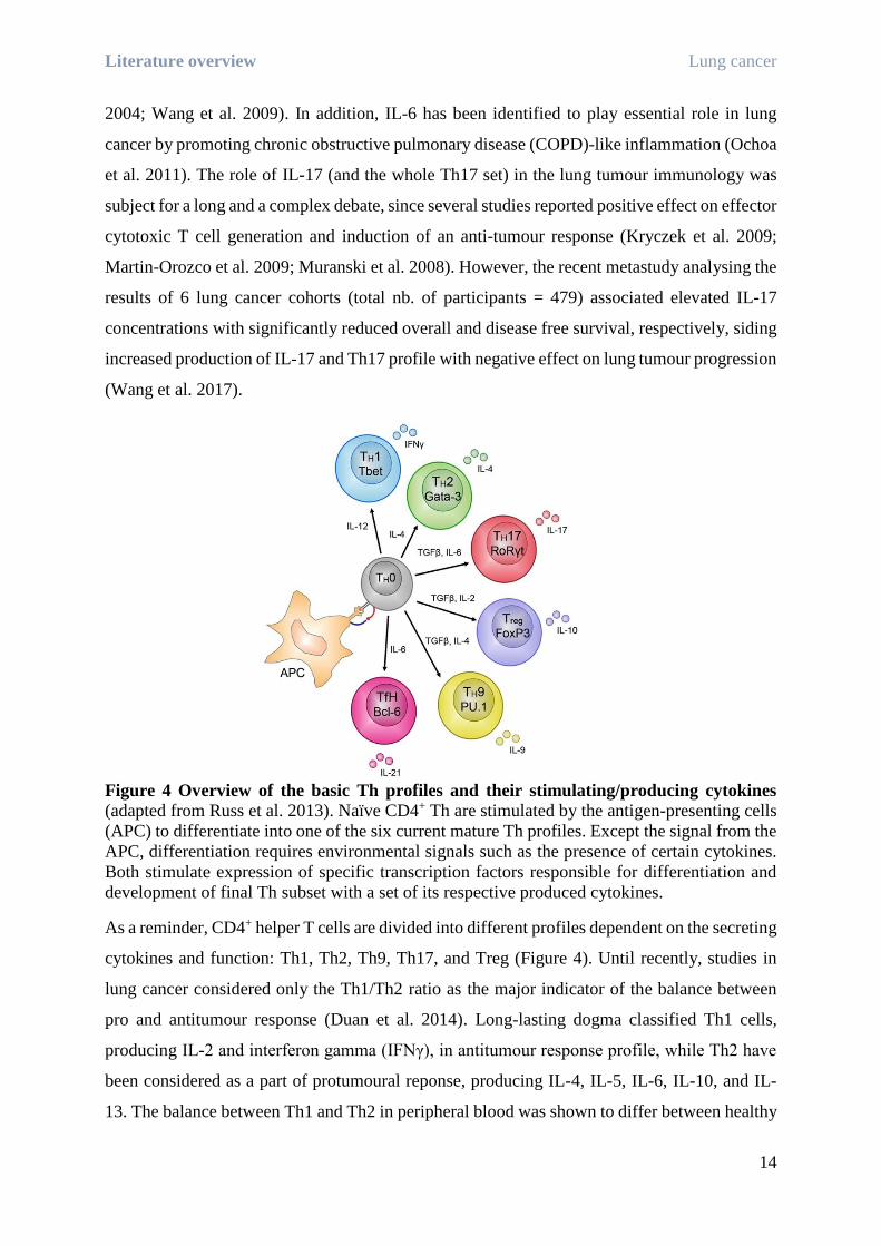
Figure 4 Overview of the basic Th profiles and their stimulating/producing cytokines
(adapted from Russ et al. 2013). Naïve CD4+ Th are stimulated by the antigen-presenting cells
(APC) to differentiate into one of the six current mature Th profiles. Except the signal from the
APC, differentiation requires environmental signals such as the presence of certain cytokines.
Both stimulate expression of specific transcription factors responsible for differentiation and
development of final Th subset with a set of its respective produced cytokines.
As a reminder, CD4+ helper T cells are divided into different profiles dependent on the secreting
cytokines and function: Th1, Th2, Th9, Th17, and Treg (Figure 4). Until recently, studies in
lung cancer considered only the Th1/Th2 ratio as the major indicator of the balance between
pro and antitumour response (Duan et al. 2014). Long-lasting dogma classified Th1 cells,
producing IL-2 and interferon gamma (IFNγ), in antitumour response profile, while Th2 have
been considered as a part of protumoural reponse, producing IL-4, IL-5, IL-6, IL-10, and IL-
13. The balance between Th1 and Th2 in peripheral blood was shown to differ between healthy

Literature overview Lung cancer
15
controls and lung cancer patients (Ito et al. 2005). The increase of concentration of Th2
cytokines was suggested to shift balance into immunosuppressive profile (Becker 2006; Pinto
et al. 2006), which as a consequence creates more tumorigenic environment (Green et al. 2010;
Krohn et al. 2011) and higher relapse rate (Hong et al. 2013; Liang et al. 2011; Wei et al. 2003).
However, the ratio Th17/Treg started to increase attention, due to the reported tumour-
influencing nature of these two cell sets (Marshall et al. 2016). First, it is important to say that
both sets share common progenitor, expressing both retinoic acid-related orphan receptor
gamma t (RORγt) and fork-head box protein 3 (FOXP3) genes, and chemokine receptors CCR6
and CCR4 (Duan et al. 2014). TGFβ was found as a central element that differentiates the two
cell types. The presence of IL-1β, IL-6 and low dose of TGFβ stimulates common progenitor
to differentiate into Th17 cell set by stimulating the expression of RORγt genes. The
differentiation itself is stimulated by DCs activated by microbes (Crome et al. 2009). On the
other hand, the presence of retinoic acid stimulates the production of TGFβ, and both have the
potential to inhibit RORγt expression (i.e. Th17 development), favouring FOXP3 expression
and differentiation towards Treg cell set (Peck and Mellins 2010). However, it has been reported
that Tregs can convert to Th17 in the presence of inflammatory signals, such as IL-1β, IL-6,
IL-21, IL-23 (Duan et al. 2014).
Th17/Treg balance is particularly interesting in lung cancer due to overexpression of TGFβ by
lung cancer cells. Therefore, they favour Treg differentiation, associated with immune
suppression and decreased anti-tumour response (Ju et al. 2009). This is also supported by the
studies reporting that higher Th17/Treg ratio was negatively correlated with the tumour stages,
and higher TGFβ with the cases of metastases (Duan et al. 2014; Roberts and Wakefield 2003).
1.5 Risk factors for lung cancer
Even though genetic predisposition to mutations and consequentially development to lung
cancer were described first, other factors like pollution, tobacco, alcohol, infections, hormones
and immune competence of the host are now also recognized as factors that can increase the
risk of lung cancer development (Plottel and Blaser 2011; Zong, Cao, and Wang 2012).
1.5.1 Smoking
Smoking is still recognized as the primary risk factor for lung cancer development. Its
malefaction lies in the components of the smoke that act as DNA adducts, favouring
carcinogenesis (Ahrendt et al. 2000; Lim et al. 2016; Nagy-Mignotte et al. 2011). Another

Literature overview Lung cancer
16
consequence of smoke particle inhalation and extensive heat production is the development of
chronic inflammation that is thought to be an underlying cause of 25% of lung cancers (Balkwill
and Mantovani 2012; Hanahan and Weinberg 2011; Mur et al. 2018; Walser et al. 2008). As
reported in the study of Berger et al. (2016) on smokers without COPD, the elevated forced
oscillation (as a measurement of respiratory function) was elevated in smokers and significantly
associated with two-fold higher lymphocyte and neutrophil counts and higher pro-inflammatory
markers (IL-8, eotaxin, fractalkine). This distal airway dysfunction lines up with similar
observations reported in patients with established COPD and could add to understanding of
higher risk of COPD development in smokers and its progression to cancer (Cho et al. 2011;
Palucka and Coussens 2016).
1.5.2 Chronic inflammation
Although in general population smoking is considered, and presented, as one of the most
influential factors to lung cancer development, it is interesting that only 10-15% of smokers
develop cancer (but 90% of cancer cases consider smokers) (García-Castillo et al. 2016;
Houghton 2013; Pevsner-Fischer et al. 2016). Chronic inflammation is considered to be a true
culprit, already shown in the clinical research as the one increasing the risk of neoplasm (Greer
and O’Keefe 2011; Louis, Hold, and Flint 2014). Proinflammatory cytokines in the tumour bed
induce production of reactive oxygen species (ROS) by macrophages, which damage DNA,
obstruct repair mechanisms, stimulate pro-tumorigenic pathways such as mediated by nuclear
factor kappa-light-chain-enhancer of activated B cells (NF-kB) transcription factor
(Francescone, Hou, and Grivennikov 2014; Klaunig, Kamendulis, and Hocevar 2010). All this
leads to the increased genetic instability (Elinav et al. 2013) and creates a favourable
environment for tumour progression.

Literature overview Microbiota and connection to lung cancer
17
2 Microbiota and connection to lung cancer
2.1 What is microbiota?
Although most often used as a synonym for “bacteria”, “microbiota” is a much broader term
that includes bacteria, archaea, fungi, viruses and protists that inhabit our organism in different
types of commensal relationship (Hassan, El-khattouti, and Tandon 2013; Plottel and Blaser
2011). The gather of their genes is therefore called the “microbiome” (Clemente et al. 2012).
Its impressive size that surpasses human genome up to 100 times and is closely related to host’s
homeostasis is appropriately called the “second genome” or “the forgotten organ” (Arslan 2014;
Dietert and Dietert 2015; Schwabe and Jobin 2013).
2.2 Gut microbiota – a role model
Gut microbiota is without a doubt the most extensively studied microbiota in humans.
Containing more than 100 trillion microbes, it is one of the most complex and the most abundant
ecosystems. In its balanced state, Firmicutes and Bacteroidetes are the two dominant phyla,
while other less abundant are Actinobacteria, Fusobacteria and Verrucomicrobia (Belizário
and Napolitano 2015). However, their balance can be gravely different between individuals,
depending on their diet, life habits, origin, genetics, which has been already extensively studied
(Figure 5) (Arumugam et al. 2011; Bäckhed et al. 2012; Eckburg et al. 2005; Segata et al. 2012;
Zoetendal, Rajilic-Stojanovic, and de Vos 2008).
Figure 5 The factors with a direct influence on the gut microbiota (adapted from Cerdá et
al. 2016)

Literature overview Microbiota and connection to lung cancer
18
2.2.1 Diet and immunity
Gut microbiota is known to have many different roles essential for its host’s homeostasis;
hydrolysis of dietary compounds, synthesis of vitamins, stimulation of immune system, control
of pathogen colonization, development of intestinal barrier, and regulation of fat storage as only
some of them (Bashiardes et al. 2017; Flint et al. 2012; García-Castillo et al. 2016; Kamada et
al. 2013; Kau et al. 2011; Zeng et al. 2016).
Following the studies on the mucosal immunity (Fujimura et al. 2014; Noverr et al. 2004, 2005),
gut microbiota was found to be responsible for the maturation of the overall immune system.
In GF animals, the immune system was underdeveloped and with pronounced
immunosuppressive character. This was however reversible by colonization with conventional
microbiota (Chung et al. 2012). Even though certain genera or species were correlated to the
production of cytokines, regulation of systemic inflammation or metabolic pathways, it was
suggested that the microbial functionary groups sharing same metabolic roles could have much
greater importance than e.g. genera with similar taxonomical classification (Blander et al. 2017;
Schirmer et al. 2016).
Anaerobic fermentation of non-digestible polysaccharides, such as dietary fibre, is one of the
most extensively studied roles of gut microbiota and also its importance in maintaining gut-host
homeostasis (Zoetendal et al. 2008). Through fermentation and carbohydrate hydrolysis,
colonic bacteria produce carbon dioxide, methane, hydrogen and short-chain fatty acids (SCFA)
that can affect gut barrier stability, inflammation and gut hormone regulation (Slavin 2013).
Among fermentation products, it is important to emphasize the role of three SCFAs: acetate,
propionate and butyrate. In human caecum, they are found in 70:20:10 ratio, respectively (Lloyd
and Marsland 2017). They are produced in majority from the members in the genera
Bacteroides, Bifidobacterium, Lactobacillus, Coprococcus, Methanobrevibacter, and families
Clostridiaceae and Lactobacillaceae. SCFAs, and especially butyrate, are used by colonocytes
as energy source and thus are extremely important for the maintenance of the gut barrier
(Meijer, de Vos, and Priebe 2010). Furtherly, they are shown to have anti-inflammatory
properties, to inhibit biofilm formation and pathogens activity, and decrease tendency of type-
2-diabetes development (Belizário and Napolitano 2015; Le Chatelier et al. 2013; Corrêa-
Oliveira et al. 2016). SCFAs are also reported to influence function, maturation and fate of
immune cells (Atarashi et al. 2013; Le Poul et al. 2003; Vinolo et al. 2011). They are ligands
of surface receptors found on immune cells, G-protein coupled receptor 41 (GPR41), GPR43

Literature overview Microbiota and connection to lung cancer
19
and GPR109a (Maslowski et al. 2009; Smith et al. 2013; Trompette et al. 2014). Moreover,
butyrate was found to be a very potent inhibitor of histone deacetylase, which stimulates
maturation of Treg cells via expression of FOXP3 gene, exerting anti-inflammatory properties
(Kim et al. 2007). Also, uniquely butyrate showed to inhibit proliferation of intestinal stem cells
and progenitor cells during mucosal injury, which might prevent tumorigenic transformation
under inflammatory conditions (Kaiko et al. 2016). Since butyrate is approximately used as
80% of energy source for colonocytes, it is not a surprise that it promotes colonic oxygen
consumption by stabilizing transcription factor hypoxia-inducing factor 1 (HIF-1), which is
responsible for transcription of a set of anti-tumorigenic genes (Kelly et al. 2015).
A variety of other bacterial metabolites are found to be similar to metabolites produced by
human cells (Wikoff et al. 2009) and are thought to be one of the key components for
microbiota-host interaction. This chemical mimicry, especially of signalling molecules, could
represent one of the bases for future therapeutic usage (microbiome-biosynthetic gene therapy)
(Cohen et al. 2017).
Gut microbiota has also been implied in the direct interaction with cancer (Figure 6), both in
the preventive and promoting aspect. Those will be discussed in the following two chapters,
with the accent on the proposed and established mechanisms.
Figure 6 Modulation of hallmarks of cancer by microbial-derived signals (adapted from
Fulbright, Ellermann, and Arthur 2017)

Literature overview Microbiota and connection to lung cancer
20
2.2.2 Gut microbiota for cancer
2.2.2.1 High-protein and unbalanced diet
Except fibres, high-protein diet also increases colonic fermentation (Louis et al. 2014).
Fermentation of aromatic amino acids produces phenols, indoles, p-cresol and phenylacetic acid
that could act as bioactive metabolites (Rajilić-Stojanović 2013; Schwabe and Jobin 2013).
Ammonium, also a product of protein fermentation, has been proven to be carcinogenic in low
concentration, as well as N-nitroso compounds inducing nitrogen alkylation of the DNA (Louis
et al. 2014).
In anaerobic conditions, nitrate, sulphates and organic compounds replace oxygen as electron
acceptor. Therefore, sulphate-reducing bacteria that compete for hydrogen have an important
impact on cross-species interaction and changes in diversity. In normal population, they are
found in low concentrations, but their higher abundance can be harmful (Louis et al. 2014;
Rajilić-Stojanović 2013). The product of sulphate reduction is hydrogen sulphide, which
inhibits oxidation of butyrate. It is toxic for colonocytes, proinflammatory, inhibits mucus
production and is genotoxic (generation of free radicals) (Louis et al. 2014; Ridlon, Kang, and
Hylemon 2006).
2.2.2.2 Disruption of barrier
Intestinal barrier with its mucosal layer, tight junctions between colonocytes, constant microbe
monitoring by M-cells and secretion of multiple anti-microbial molecules represents a real
anatomical separation with intestinal lumen (Schwabe and Jobin 2013). Its disruption leads to
translocation of microbes or their products into systemic circulation and could trigger
inflammatory response (Belizário and Napolitano 2015; Garrett 2015; Logan, Jacka, and
Prescott 2016). The disruption could be caused by local inflammation, infection or defect genes
(Garrett 2015; Schwabe and Jobin 2013). The deficiency of dietary fibres could also create a
dysbiosis with proliferation of mucus-degrading bacteria. This showed to lead to thinning of
the mucus layer with increased adhesion of the intestinal bacteria to colonocytes, and increased
susceptibility to different pathogen colonisation, such as Citrobacter infection (Desai et al.
2016). In already established tumours, this secondary inflammation was shown to enhance
intestinal tumour progression (Grivennikov et al. 2012; Huber et al. 2012) or increase a risk of
its development in inflammatory bowel disease (IBD) patients (Ekbom 1991).

Literature overview Microbiota and connection to lung cancer
21
2.2.2.3 Microbial products
To find their place within this vast intestinal ecosystem, many microbes have developed various
mechanisms that would give them selective advantage in competition and survival. One of these
mechanisms are the DNA-damaging proteins, which cause genome instability and lead to
mutations (Garrett 2015). There are many examples of bacteria with oncogenic potential,
amongst others genotoxic Escherichia coli, Enterococcus faecalis, Bacteroides fragilis,
Fusobacterium nucleatum (Arthur et al. 2012; Francescone et al. 2014; Kostic et al. 2013;
Ohtani 2015; Schwabe and Jobin 2013; Sheflin, Whitney, and Weir 2014; Wu et al. 2009).
Fusobacterium nucleatum, a commensal from the oral cavity (Han and Wang 2013) already
linked with certain pathogenic conditions (Lagier et al. 2012; Témoin et al. 2012), was recently
connected to pathogenesis of colon adenocarcinoma. On its surface, F. nucleatum expresses
adhesin FadA, which engages with E-cadherin on epithelial cells and stimulates their
proliferation (Rubinstein et al. 2013), therefore is a potent stimulator of tumour cell growth.
The other mechanism is realised through the effect on immune cells. Interaction of Fap2 protein
of F. nucleatum with T-cell immunoreceptor with immunoglobulin and immunoreceptor
tyrosine-based inhibitory motif domains (TIGIT), which is an inhibitory receptor on natural
killer (NK) and cytotoxic T (Tc) cells, inhibits tumour-killing properties of this two cell sets
essential for identifying and destroying precancerous and malignant cells (Bashiardes et al.
2017; Gur et al. 2015). In animal models and clinical studies, F. nucleatum was also positively
correlated to enrichment with tumour-associated macrophages (TAMs) and MDSCs, both with
immunosuppressive and protumorigenic roles (Kostic et al. 2013).
2.2.2.4 Loss of diversity
High diversity of the gut microbiota is the virtue of a healthy, balanced intestinal environment.
Loss of diversity was proposed to reflect the state of dysbiosis (Bäckhed et al. 2012) and was
shown to be directly connected to inflammation, as seen in IBD, Crohn’s disease and colorectal
cancer (CRC) (Brown et al. 2013; Clemente et al. 2012; Fulbright et al. 2017; Sartor and
Mazmanian 2012).
2.2.2.5 Neoangiogenesis
Gut microbiota was found essential for normal development of the intestinal vascular system
(Stappenbeck, Hooper, and Gordon 2002). Interestingly, it was seen that during infection, toll-
like receptors (TLRs) activated by bacterial lipopolysaccharide (LPS) promote angiogenesis,
with the effect even more pronounced in the presence of damage-associated molecular patterns
(DAMPs). Both are proposed to be a possible reason of microbial contribution to

Literature overview Microbiota and connection to lung cancer
22
neoangiogenesis seen in the tumour microenvironment, either directly by the colonising
bacteria or pathogen-associated molecular patterns (PAMPs) and DAMPs within the
microenvironment (Fulbright et al. 2017; Osherov and Ben-Ami 2016).
2.2.2.6 Microbiota-induced Th17 inflammation
Th17 immune response is crucial in maintenance of the homeostasis of intestinal epithelium,
and is driven by commensal microbiota and its products (reviewed in Bingula et al. 2017).
However, it is also associated with protumorigenic activity and worse prognosis in CRC
(Grivennikov et al. 2012). CRC murine models showed that colonisation with enterotoxigenic
Bacteroides fragilis stimulates Th17-driven inflammation and cancer development (Housseau
et al. 2016; Wu et al. 2009). Intestinal commensal Alistipes showed similar effect by enhancing
the production of IL-6 and signal transducer and activator of transcription 3 (STAT3) activation
(Moschen et al. 2016). Adversely, inhibition of IL-17 signalling axis reduced STAT3 promotion
of inflammation and tumorigenesis (Housseau et al. 2016; Wu et al. 2009), confirming the
application of Th17 signalling in CRC development and progression (Fulbright et al. 2017).
2.2.3 Gut microbiota against cancer
Over several decades, the cancer treatment gravitated towards more case-adapted and
personalised approach. The currently used treatment includes surgery, radiotherapy,
chemotherapy, and since recently immunotherapy and hormonal therapy (Bashiardes et al.
2017). Each of these is focused on “rectification” of immune response or eradication of the
consequences of its failed activity. While some can truly bring instant salvation in certain cases
(e.g. surgery), others are not always applicable or silently fail after first success, often leaving
only palliative treatment. Furthermore, in certain cases the applied therapies have no effect
whatsoever (e.g. chemotherapy, immunotherapy), leaving the physicians with no explication or
solutions (Haslam and Prasad 2019; Prelaj et al. 2019).
Recently, certain studies started to search the answer in the giant ecosystem within our
intestines – the gut microbiota. Even though as early as in 1920s, the intravesicular injection of
bacillus Calmette-Guérin (derived from strain Mycobacterium bovis) into patients with
superficial bladder cancer resulted in antitumour response and increased survival (Herr and
Morales 2008), it was not until the 21st century that the answer to this irregularity in response
to cancer therapy was connected and partially explained by our lifetime companions.

Literature overview Microbiota and connection to lung cancer
23
2.2.3.1 How does it work?
The proposed mechanism by which various microbiota elicit antitumour response lies in
activation and stimulation of the immune system and possible similarity between bacterial and
tumour epitopes, causing cross-reactivity (Zitvogel et al. 2016). LPS, flagellin, peptidoglycans
or other microbial molecules that could serve as PAMPs are the ones behind priming and
activation of the immune cells (Mogensen 2009; Ranf 2016).
Different bacteria could therefore indirectly induce antitumour response as seen in Clostridia
spores found within solid tumours (Morrissey, O’Sullivan, and Tangney 2010). Those or alike
could induce TNFα secretion, which vasoactive effect was shown to facilitate further entry of
the bacteria into the tumour environment, led to CD8+ T cell activation and enhanced tumour
surveillance (Leschner et al. 2009; Stern et al. 2015). Tumour colonisation by bacteria was also
suggested as a result of the gut barrier disruption and bacterial translocation (Balzan et al. 2007).
Further, several studies in animal models investigated the role of balanced or dysbiotic gut
microbiota on chemotherapy effectiveness, investigating the reason behind the effect of non-
responding patients. Their results have opened a new area on importance of the gut microbiota
– host relation through the interaction with the immune system, and have provided a new way
of thinking in clinical therapeutic design. The following paragraphs summarise the most
important discoveries of these studies.
2.2.3.2 Gut microbiota in chemotherapy
Chemotherapy is a non-selective cancer treatment, most often based on alkylating agents that
act as DNA adducts, which are genotoxic and induce apoptosis in fast proliferating cells
(tumour cells, but also some types of normal cells). Following apoptosis or necrosis, a liberation
of tumour DNA and other internal proteins serves as immunogenic marker, stimulating T cell
immunity and antitumour response (Alcindor and Beauger 2011). The following studies
addressed the most commonly used drugs: platinum-based compound (still the most common
in LC treatment) oxaliplatin, and cyclophosphamide (CTX) (Bracci et al. 2007). CTX was also
shown to affect tumour environment by inducing reduction in Treg, and stimulating Th1 and
Th17 (Routy, Le Chatelier, et al. 2018; Schiavoni et al. 2011).
The study of Iida et al. (2013) examined the effect of oxaliplatin on subcutaneous tumour (EL4
lymphoma, MC38 colon carcinoma and B16 melanoma) in different murine models.
Conventional mice showed tumour regression and had improved survival, while in GF or
antibiotic-treated mice this effect was much weaker. They found that TLR agonists, coming
Literature overview Microbiota and connection to lung cancer
24
from the commensal microbiota, promote ROS generation by the tumour-infiltrating myeloid
cells causing cell death. Oxaliplatin alone functions on the same principle, but as explained,
without microbiota the effect was more modest. Conventional mice with defect TLR signalling
had the same response as GF/antibiotic-treated mice, and the other way around, GF mice
administered with LPS showed a full response. The study also looked at the efficacy of the
combination of intratumour CpG-oligonucleotides and anti-IL-10R antibody. Conventional
mice showed retarded tumour growth and prolonged survival by rapid induction of intra-
tumoral haemorrhagic necrosis dependent on TNFα, while this mechanism was significantly
impaired in antibiotic-treated mice. In conventional mice, the therapy also induced higher
production of IL-12 by tumour-infiltrating myeloid cells and IFNγ in tumour-infiltrating T and
NK cells. These factors negatively correlated with the genus Lactobacillus and positively with
Gram-negative bacteria (genera Alistipes, Ruminococcus etc.). In the genus Lactobacillus, there
were L. murinum, intestinalis and fermentum, previously reported for their anti-inflammatory
effects. Gavage of the mice with L. fermentum attenuated TNF production, while Alistipes
shahii reconstituted TNF response in antibiotic-treated mice.
Viaud et al. (2013) evaluated the potential connection between gut microbiota and CTX in anti-
tumour response stimulation. They demonstrated that treatment with CTX disrupted the
intestinal barrier and induced a severe reduction within the Firmicutes phylum (Clostridium
cluster XIVa, Roseburia, Coprococcus and unclassified Lachnospiraceae, facilitating the
translocation of the certain Gram-positive bacteria into mesenteric lymph nodes and spleen.
Several cultivated bacteria included L. johnsonii, L. murinus and Enterococcus hirae. These
bacteria induced “priming” of specific subset of “pathogenic” Th17, normally responsible for
the anti-bacterial response, and activation of Th1 memory cells that exerted antitumor
responses. GF or antibiotic-treated mice did not show the priming effect of Th17 in the spleen
and therefore had impaired antitumor responses, and adoptive transfer of mature Th17 cells
only partially restored the initial antitumor effect.
Following this discovery, Daillère et al. (2016) investigated the effect of oral supplementation
with these bacteria, positively correlating with productive antitumor response (Viaud et al.
2013), in sarcoma bearing mice treated with CTX. They found that Enterococcus hirae
translocated from the small intestine to secondary lymph organs increased intratumour
CD8/Treg ratio thus stimulating tumour eradication, failed by other species like L. johnsonii or
other. Additionally, they identified another species from the Porphyromonadaceae family,
Barnesiella intestinihominis, which accumulated in the colon, were able to induce

Literature overview Microbiota and connection to lung cancer
25
polyfunctional CD4+ Th1 and CD8+ Tc1 effector cells and stimulate infiltration of IFNγ-
producing γδT cells into tumour. Both were addressed as potent “oncobiotics” that could
ameliorate standard chemotherapeutical treatment.
2.2.3.3 Gut microbiota in immunotherapy
The big success in the understanding of the tumour – immune system interaction in cancer
treatment were the antibodies that “unleashed” the suppressed immune system by inhibiting the
immune checkpoints, to let the immune system fight the cancer on its own. The two currently
used therapies include anti-PD-1/anti-PD-L1 antibodies, preventing inactivation of antitumor
CD8+ cytotoxic T lymphocytes within the tumour, and anti-CTLA-4 (cytotoxic T lymphocyte
antigen 4), which enables priming and activation of antitumor cells in lymph nodes (Brahmer
and Pardoll 2013). Despite the initial overwhelmingness by this new approach, the percent of
non-responders and developed resistance has been very high (~80%) as well as the number of
autoimmune responses (Prelaj et al. 2019).
Sivan et al. (2015) looked at efficacy of anti-PD-L1 therapy on melanoma in genetically
identical C57BL/6 mice coming from two different facilities. First, in two mice groups the
spontaneous tumour growth was significantly different, but cohousing or faecal transfer
eliminated this effect. Sequencing of faecal microbiota identified significantly different
microbial population between two mice groups. 257 taxa were different between groups, but
genus Bifidobacterium was the only significantly correlated with productive antitumor T cell
responses. Secondly, they tested the effect of anti-PD-L1 therapy in both mice groups, with
larger tumours (LT) and smaller tumours (ST). The group ST, naturally enriched with
Bifidobacterium, completely inhibited tumour growth with therapy administration.
Interestingly, the ST group without treatment had almost equal tumour size as the LT group
with treatment. Further, the authors showed that in LT group either treatment with anti-PD-L1
or oral gavage with Bifidobacterium equally reduced tumour growth. Moreover, the
combination of two had been significantly the most efficient, with the effect similar to ST group
with only anti-PD-L1. The closer immunological analysis showed that the main effect was not
exerted on effector cells, but on the more upstream level, i.e. DC priming.
Matson et al. (2018) looked at microbiota in stool of patients with metastatic melanoma before
the anti-PD-1 therapy and correlated their clinical response with microbiota composition. They
identified Bifidobacterium longum, Collinsella aerofaciens and Enterococccus faecium as more
abundant in responders. The ratio of total nb. of “beneficial” and “non-beneficial” OTUs was
calculated for each patient, and they showed that all patients with the ratio over 1.5 had

Literature overview Microbiota and connection to lung cancer
26
belonged to responders. Further, they showed that GF mice with stool transferred from
responders had improved tumour surveillance mediated by elevated tumour antigen specific
CD8+ T cell response and more efficient anti-PD-1 therapy. Conversely, in mice with the stool
from non-responders the therapy was completely ineffective. Their findings reinforced previous
studies and suggested a new, simple way of possible use of “beneficial”-“non-beneficial”
bacteria ratio in prediction of PD-1 efficacy in treatment of metastasis.
Vétizou et al. (2015) addressed different type of immune checkpoint inhibitor, the therapy with
anti-CTLA-4 antibodies, ipilimumab. They tested ipilimumab efficacy on established sarcoma
in specific-pathogen free (SPF) and GF mice and observed that in GF mice therapy had no
effect. Antibiotics administered to SPF mice however abrogated the ipilimumab efficacy,
pointing to the responsibility of the gut microbiota. Ipilimumab induced inflammatory changes
in the intestines, observed as increased cell death and proliferation of intestinal epithelial cells
(IEC). Authors identified CD4+ and CD8+ T cells (including intraepithelial lymphocytes (IEL))
responsible for IEC apoptosis in the presence of microbial TLR agonists, and concluded that
ipilimumab compromised the IEC-IEL balance. Ipilimumab also induced rapid
underrepresentation of Bacteroidales and Burkholderiales and relative increase of
Clostridiales. Further, oral gavage of antibiotic-treated SPF mice with qPCR identified species
Bacteroides thetaiotaomicron, Bacteroides fragilis (Bf) and Burkholderia cepacia, or
combination of the last two recovered the response to anti-CTLA-4 antibody. In GF mice, Bf
induced Th1 response in tumour-draining lymph nodes and promoted maturation of DCs, both
restoring anti-CTLA-4 response. Moreover, these bacteria reconstituted intestinal barrier
damaged by anti-CTLA-4 therapy in antibiotic-treated mice. This supported the fact that overall
efficacy of therapy was highly dependent on microbiota composition, affecting IL-12 and IL-
10 levels, reported the authors. To conclude, they also performed faecal transfer from patients
before therapy and followed tumour growth in mice, which corresponded to patient’s response
to therapy.

Literature overview Microbiota and connection to lung cancer
27
2.3 Lung microbiota in sickness and health
2.3.1 Environment of the lower airways
Vocal cords have been considered as a limit between upper and lower airways, and even though
they are characterised by continuous mucosal layer, these two parts are both anatomically and
functionally very different (Kronenberger and Meyers 1994; Shaker and Hogan 2000; Wu and
Segal 2017). Pulmonary epithelium is composed of one thin layer of squamous cells, among
which there are ciliated columnar mucus-secreting goblet cells and cells secreting surfactants,
mutually connected in tight junctions. In healthy lungs, mucus production is at low rate and
form a protective layer enough to maintain homeostasis (disables microbial adherence to cells)
(Radicioni et al. 2016). On their surface, cells have pattern recognition receptors (PRRs) such
as TLR that serve to recognize PAMPs from viruses, bacteria, fungi, protozoa, and intracellular
parasites (Lambrecht and Hammad 2012). As a response, the cells excrete multiple
antimicrobial peptides such as surfactant protein A, lysozyme, lactoferrin, defensins,
complement components (Iwasaki, Foxman, and Molony 2017; Rogan et al. 2006). Another
role of the epithelium lies in cilia that with their synchronised movements facilitate the
clearance of particulates towards upper airways, where they are terminally removed by
coughing (Lloyd and Marsland 2017; Radicioni et al. 2016).
2.3.2 Lungs are not sterile
The biggest common worldwide project for the research of the microbiota, The Human
Microbiome Project (HMP), included nares and oral cavity as the sites of the research interest,
but interestingly, not the lower airways (Proctor 2011). Therefore, until today the knowledge
about lung microbial composition and its interaction with the host’s physiology and immune
system remains scarcely explored (Huang et al. 2015; Segal et al. 2013, 2016; Sze et al. 2015).
There have been also other inconveniences, as the difficulty of sampling, low biomass of the
samples and consequentially, the high risk of contamination by upper airways while sampling
(100 to 10,000 times less of 16S rRNA gene copies in lungs) (Bassis et al. 2015; Charlson et
al. 2011). The three sample types are most commonly found across the studies of lung
microbiota: tissue, bronchoalveolar lavage fluid (BAL) and sputum. The one the least ethically
acceptable (so, the rarest) but representing the lowest contamination risk is the tissue, initially
obtained from lung transplantation patients. Sputum is the sample with the highest
contamination risk but the easiest to obtain. Therefore, the most studies are realised on BAL
that represents an acceptable compromise; direct sampling of the lower airways lung microbiota
Literature overview Microbiota and connection to lung cancer
28
by lobe washing, but with bronchoscope passing through the airways (Bassis et al. 2015;
Bernasconi et al. 2016; Dickson et al. 2016; Dickson, Martinez, and Huffnagle 2014; Hilty et
al. 2010).
Lungs are an extremely voluminous hollow system of connected branching tubes with constant
changes in gradients of oxygen and carbon dioxide tension. This creates rather selective
pressure on microbial communities that might exist in lower airways (Lieberman et al. 2013).
Under healthy conditions, there is a constant turnover of bacterial communities in the lower
airways, as reported both by culture-dependant and independent techniques (Venkataraman et
al. 2015). The theory of adapted island model suggests that the upper airways are the source of
microbial diversity of lower airways, and that this diversity decreases with furthering from the
central bronchus towards more distant lung parts. It was brought up by Dickson et al. (2015) in
the study including 15 healthy volunteers whose microbiota was sampled by BAL in different
lung parts. In these subjects, the BAL microbiota of the upper lobes resembled more the
microbiota of the upper respiratory tract than the BAL microbiota from lower lobes. Further,
the authors have suggested that lung microbiota in healthy subjects is more influenced by the
rate of immigration and elimination than by the local growth conditions. Those, they say, have
more importance in the cases of advanced lung diseases.
Lung microbiota by its composition was clearly distinct from other microbiota such as oral,
nasal, skin, vagina or stool microbiota (Yu et al. 2016). Likewise, Proteobacteria was reported
as the dominant phylum in the tissues with average relative abundance of 60%. Although lung
is the highly aerated organ, the reports of co-occurring anaerobes and aerobes in culture-
independent studies suggest that biofilms might play a very important role in the lower airways
microbiota (Costerton et al. 1994; J W Costerton, Stewart, and Greenberg 1999) and also in
potential pathogenicity as seen in the case of Pseudomonas aeruginosa (Whitchurch et al.
2002).
2.3.3 Resident lung immunity
Postnatal exposure to different air microbes by either inhalation or aspiration is irreplaceable
for proper education and maturation of lung immune system. It is also important for proper
structure development as seen in germ-free (GF) animals that had smaller lungs and decreased
number of matured alveoli than conventional animals (Ho Man, de Steenhuijsen Piters, and
Bogaert 2017).

Literature overview Microbiota and connection to lung cancer
29
As effective gas exchange is essential for human life, the maintenance of immune balance in
the lung is of exceptional importance (Ho Man et al. 2017). This homeostasis is maintained by
tissue resident cells that at the same time create tolerance and ensure rapid responses against
invading pathogens. This includes members of both adaptive and innate immunity, such as
alveolar macrophages, DCs, memory T lymphocytes but also Tregs and γδT cells (Lloyd and
Marsland 2017). Following the encounter with antigen, the additional antigen-specific
lymphocytes can be recruited to the lung through expression of lung-homing chemokine
receptors 4 (CCR4) and CCR8 (on e.g. Th2 cells) (Cho et al. 2016; Lloyd et al. 2000).
Figure 7 Homeostasis and dysbiosis in the lung epithelium (adapted from Mao et al. 2018). Proposed homeostasis (left part of the figure) includes dynamic interplay between the commensal microbiota and
local immunity (alveolar macrophage, lung DCs, several lymphocytic subtypes), maintaining a state of tolerance
by decreasing inflammation and controlling local bacterial population through sIgA. A non-commensal microbiota
introduces dysbiotic state, activating the PRRs and the NF-κB cascade, inducing inflammation. Here, SCFA
produced by local bacteria or delivered through circulation (produced e.g. in the gut) might have an anti-
inflammatory role. CXCLs – chemokine (C-X-C motif) ligands, DC – dendritic cell, GPR43 – G-protein coupled
receptor 43, LPS –lipopolysaccharide, SCFA – short-chain fatty acid, TNFα – tumour necrosis factor α
Commensal airway microbiota stimulates production of secretory immunoglobulin A (sIgA)
that maintains constant dynamic balance between eliminated and colonised microbiota. Its
deficiency leads to increased inflammation against commensal microbiota that could induce
tissue damage (Richmond et al. 2016). Commensal microbiota is also directly responsible for
phenotype of certain immune cells. For example, the stimulation of PD-1/PD-L1 expression by
microbiota is directly connected to formation of inducible Tregs (Gollwitzer et al. 2014). These
cells reside in the lung since birth, and are crucial in the maintenance of lung immune
homeostasis (Lloyd and Hawrylowicz 2009). They are defined as CD4+CD25+FOXP3+ cells,
and even though their major secretion product is immunosuppressive IL-10, their phenotype is
influenced by the local cytokine milieu and infection status (Curotto de Lafaille et al. 2010;
Krishnamoorthy et al. 2012; Morita et al. 2015). In allergy, their production of IL-10 is

Literature overview Microbiota and connection to lung cancer
30
responsible for amelioration of symptoms due to their immunosuppressive effect (Kearley et
al. 2005; Lewkowich et al. 2005). However, in the case of the local infection by respiratory
syncytial virus, they were reported to express GATA binding protein 3 (GATA3) gene (marker
of Th2) and to produce Th2 cytokines. Therefore, instead of their immunosuppressive activity,
Tregs contributed to Th2 mediated response and pathology in asthma (Krishnamoorthy et al.
2012). All this characterises lung homeostasis as a fine tuned environment of delicate
equilibrium between resident microbiota and immunity (Figure 7).
2.3.4 Infection and inflammation
In health, lungs are a poor source of nutrients and therefore harbour rather low number of
bacteria (Dickson et al. 2016). However, when our natural barriers, described earlier, do not
suffice to eliminate potentially harmful particles or microbes, we talk about infection. Infection
is accompanied by inflammatory response that, even though it clears its cause, leaves the tissue
in the changed state by influencing local gas tensions, temperature or pH and could, therefore,
create more favourable growth conditions for various bacteria (Dickson, Erb-Downward, and
Huffnagle 2014; Mur et al. 2018). For example, mucus production is a common co-occurrence
of inflammation, serving as a potential nutrient source (Dickson et al. 2016).
Furthermore, the immune system finds itself in immunocompromised state, meaning that
surface markers for PAMP recognition (such as TLRs on e.g. macrophages or epithelial cells)
or different genetic modification within cells might not return immediately to the same
expression level as before the infection (Didierlaurent et al. 2008; Netea et al. 2016). This leads
to the higher susceptibility of the lungs for recurrent infection. To illustrate, in the mouse model
of COPD (model of inflammatory lung state that could mimic post-infection), exposure to LPS
(mimics a new infection by bacteria) induced tissue damage and further altered microbiome
causing diversity decrease (Yadava et al. 2016). Another example considers Mycobacterium
tuberculosis, the pathogen responsible for pulmonary tuberculosis. The studies showed that
persistent infection induced the production of TNFα and IFNγ, leading to inflammation and
pulmonary fibrosis (Dheda et al. 2005). Furthermore, in certain cases lung cancer developed
from this infection site “scars”, identifying the infection with M. tuberculosis as a risk factor
for the development of lung cancer (Cukic 2017; Pallis and Syrigos 2013).
Although having a curative objective, the administration of antibiotics can also have a
potentially indesirable effect on the community balance and infection. Its disturbance of the
microbial communites, while clearing the target organisms, can lead to propagation of certain

Literature overview Microbiota and connection to lung cancer
31
surviving commensals that could become opportunistic pathogens or can clear a niche for
pathogens already residing in the lung. Both ends up in the same process of infection and
induces inflammatory response (Lloyd and Marsland 2017). Hospital pneumonia lies on this
mechanism (Rello et al. 2009).
2.3.5 Age
Although at the moment very little is known about how does immune senescence affect lung
responses, it is certain that lung habitat changes with age (Marsland and Gollwitzer 2014). The
lungs become more susceptible to infections and inflammatory responses, as shown by the
studies on COPD or pulmonary fibrosis (Lloyd and Marsland 2017). The role of the resident
lung microbiota, as well as its “aging”, remains unknown for the moment.
2.3.6 Vices: smoking and alcohol
Previously, both alcohol and smoking have been acknowledged as strong risk factors for head
and neck cancer (Vogtmann and Goedert 2016), as well as their synergistic effect for increasing
the risk of lung cancer (Ahrendt et al. 2000). However, little is known about their interaction
with lung microbiota.
Interestingly, even though smoking stays one of the main risks for lung cancer development, at
the moment it seems that it does not directly influence the microbiota of the lower airways
(Morris et al. 2013; Segal et al. 2013), unlike the one of the upper airways (Charlson et al. 2010;
Wu et al. 2016). Nevertheless, more research is necessary, and future studies might identify
smoking as a cofactor that will influence the disease and consequentially, microbial
composition, rather than the cause of this modification.
Another vice causing local damage and inflammation is alcohol. The malice lies in the enzyme
alcohol dehydrogenase, which converts alcohol to carcinogen acetaldehyde (Ahrendt et al.
2000). This enzyme and its high activity were noted in several microbial groups, amongst other
in Neisseria, bacterial genus from the class Betaproteobacteria, a commensal found in oral
mucosa (Muto et al. 2000). Therefore, high alcohol consummation could lead to overproduction
of this and other carcinogenic metabolites that could descend into the lung and induce tissue
damage and subclinical inflammation.
2.3.7 Microaspiration and pneumotypes
Microaspiration occurs in all individuals but is elevated in certain diseases such as COPD,
asthma or lung infections, inducing the subclinical lung inflammation (Cvejic et al. 2011;

Literature overview Microbiota and connection to lung cancer
32
Huang et al. 2015; Koh et al. 2007; Morse et al. 2004; Segal et al. 2013; Segal, Rom, and
Weiden 2014; Sze et al. 2012, 2015; Sze, Hogg, and Sin 2014; Teramoto et al. 1999). It has
been suggested that the cause of inflammation lies in the enrichment of the lower airways with
supraglottic taxa. In the study of BAL microbiota from 29 asymptotic healthy subjects, Segal
et al. (2013) detected two distinct pneumotypes. Pneumotype BPT (background predominant
taxa) was characterised by low community richness and rRNA gene concentration, and
associated with decreased production of proinflammatory cytokines, as TNFα, IL-6, and
granulocyte-macrophage colony-stimulating factor (GM-CSF). On the other hand, pneumotype
SCT (supraglottic-characteristic taxa) had higher relative abundance of SCT such as Veillonella
and Prevotella, significantly higher lymphocyte and neutrophil count and produced NO
quantities. Other studies found similar results, with addition of Rothia, Streptococcus, and
Porphyromonas to SCT pneumotype (Erb-Downward et al. 2011; Morris et al. 2013; Segal et
al. 2013, 2016; Sze et al. 2012).
2.3.8 Chronic obstructive pulmonary disease (COPD)
COPD is a collective term for multisystemic inflammatory state that includes chronic bronchitis
and emphysema (airway obstruction with disappearance of small airway sacs in the peripheral
regions) (Houghton 2013). In 2016, there were 251 million cases worldwide with 3.17 million
deaths the year before (WHO 2019). The underlying factor for COPD development in the
western world is smoking, but also pollution and exposure to various particles.
Figure 8 Vicious circle hypothesis in chronic obstructive pulmonary disease (adapted from
Sethi and Mammen 2017)
Unlike smoking (Morris et al. 2013; Segal et al. 2013), COPD exerts distinct changes on
microbial composition of the lower airways. One of the reasons is certainly its effect on

Literature overview Microbiota and connection to lung cancer
33
underlying lung architecture and activity of mucociliary surfaces (Wu and Segal 2017). As
discussed in 2.3.4 Infection and inflammation, airways obstruction with increased mucus
production could introduce pockets of increased temperature and decreased oxygen tension,
which selectively favour the growth of microbes (Schmidt et al. 2014; Worlitzsch et al. 2002).
Although results between studies tend to be inconsistent due to different sampling methods
(tissue, BAL, sputum) (Sze et al. 2014), there is a consensus that acute exacerbations (episodes
of crisis that worsen patient’s state) are in 75% of cases connected to viral or bacterial infection,
or a combination (Han et al. 2012). This intertwined cause-consequence relation between
inflammation and bacterial colonisation has been explained by the vicious cycle hypothesis
(Figure 8) (Mammen and Sethi 2017). Culture-dependent analyses have identified some of
these bacteria that are already known as pathogens: Streptococcus pneumoniae, Haemophilus
influenzae and Moraxella catarrhalis (Hirschmann 2000). Culture-independent studies based
on BAL samples found that lungs of COPD patients were enriched by genera Pseudomonas,
Corynebacterium, Prevotella, Staphylococcus, Streptococcus, Veillonella, Haemophilus and
Neisseria (Erb-Downward et al. 2011; Sze et al. 2014). Sputum also showed to be indicative,
since COPD severity correlated with differences in abundance of Streptococcus pneumonia.
The difference was also significant in the overall genus Streptococcus when comparing healthy
subjects and ones with COPD (Cameron et al. 2016).
2.3.8.1 Chronic inflammation to lung cancer
Even though a role of inflammation in lung cancer does not naturally come first to mind, the
development of lung cancer is closely related to chronic inflammation. In the situation of lung
tissue injury or recognition of the foreign epitopes, there is an infiltration of inflammatory cells
(Schmidt et al. 2014). However, for one reason or another, there is no resolution of
inflammation, and the prolonged excretion of proinflammatory factors in the end leads to
excessive tissue destruction. In the same time, the factors responsible for tissue remediation, as
cytokines and chemokines that stimulate cell proliferation, angiogenesis and tissue remodelling
in response to tissue injury, send survival signals to damaged or deregulated cells. All this
creates a perfect environment for mutagenesis and metastasis (Cho et al. 2011; Palucka and
Coussens 2016). Several studies already established the link between COPD and lung cancer
development (Houghton 2013; Melkamu et al. 2013). Therefore, the role of lung microbiota in
lung cancer was suggested as one of the important issues for consideration (Mur et al. 2018).

Literature overview Microbiota and connection to lung cancer
34
2.3.9 Lung cancer and microbiota
2.3.9.1 Egg or chicken
The question is simple – is the change in lung microbiota the cause of lung cancer development,
or does the lung cancer development cause the change in surrounding microbiota. The answer
is less simple, and it will probably rest highly dependent on each case (Blumberg and Powrie
2012; Francescone et al. 2014).
The colonisation of tumours by bacteria has already been suggested (Chanudet et al. 2007;
Hooper et al. 2007). Growing, solid tumours stimulate neo-angiogenesis that results in leaky
and irregular organisation, possibly enabling bacterial entry into the tumour to stimulate
immune response (Bashiardes et al. 2017). However, in the same time the defective blood
supply compromises drug delivery (Dang et al. 2002) and in itself is also not sufficient for
oxygen needs of rapidly proliferating tumour cells (Vaupel, Mayer, and Höckel 2004). This
leads to the creation of necrotic and hypoxic environment in the centre with increased activity
of lactate dehydrogenase, which lowers pH by lactate production (Wouters et al. 2003). These
particular conditions might furtherly favour colonisation and replication of anaerobic
acidophilic bacteria (Baban et al. 2010). In addition, genetic transformation of tumour cells
might stimulate the expression of certain epitopes that could serve as bacterial adhesion points
not present in normal cells (García-Castillo et al. 2016; Garrett 2015; Khan, Shrivastava, and
Khurshid 2012), facilitating tumour colonisation.
Both negative effect of this colonisation with direct role in carcinogenesis have already been
well documented in other sites (e.g. Helicobacter pylori, Fusobacterium nucleatum) (Gur et al.
2015; Wroblewski, Peek, and Wilson 2010) and in different models (Schwabe and Jobin 2013;
Sears and Garrett 2014), as well as the immune-stimulating properties of e.g. anaerobic
Clostridium spores, enhancing tumour surveillance (Van Dessel, Swofford, and Forbes 2015).
However, for the moment the involvement of lung bacteria in lung cancer development or
suppression remains a mystery.
2.3.9.2 Microbiota in lung cancer
Until today, microbiota in lung cancer patients has been poorly studied. Unlike in asthma or
COPD, routine bronchoscopy that could give direct possibility of microbiota assessment is not
a common practice. Moreover, as discussed in the chapter 2.3.8.1 Chronic inflammation to lung
cancer, until recently lung cancer was not associated with an inflammatory state nor was it
treated as an outcome of infection with specific pathogen, therefore no true need was seen to

Literature overview Microbiota and connection to lung cancer
35
examine the microbiota within the tumour, or in the tumour environment. Similarly, the
sampling methods that remain invasive and therefore difficult to realise (lung tissue or tumour
samples) contributed to small number of studies of this type (discussed in the chapter 2.3.2
Lungs are not sterile).
Table 1 The studies identifying a significant difference in abundance of lung microbiota
in lung cancer (adapted from Mao et al., 2018)). ↑: Microbiota increases in LC compared to
controls, ↓: Microbiota decrease in LC compared to controls.
The studies performed on microbiota from BAL remain the most common (Table 1). Even
though for the moment without a direct connection with the disease, certain pathogenic bacteria
belonging to Gram-negative bacilli (H. influenzae, Enterobacter sp., Escherichia coli) and
Gram-positive Mycobacteria were identified in LC patients (Laroumagne et al. 2011). Another
study compared BAL microbiota between LC patients and patients with benign biomasses as
controls (Lee et al. 2016). They identified two phyla, Firmicutes and TM7, and two genera,
Veillonella and Megasphaera, as more abundant in LC patients. The authors showed that these
two genera together can predict LC with accuracy of 0.888. What is interesting is that TM7 was
also reported as elevated in certain studies on COPD, making it a potential microbial candidate
for transition of COPD to LC (Mao et al. 2018). The authors also observed that the ratio between
Firmicutes and Bacteroidetes was significantly higher in smokers than in non-smokers,
suggesting that smoking might be the cofactor of microbial modification during the already
established disease. Comparing BAL microbiota between the tumour lobe and contralateral
lobe, Liu et al. (2018) noted significant decrease in diversity in the tumour lobe, as well as
increased abundance of genera Streptococcus and Neisseria.
At the moment, only two studies have analysed microbiota of the non-malignant tissue paired
tumour (Peters et al. 2019; Yu et al. 2016). Yu et al. (2016) defined the core microbiota found
in 80% of non-malignant tissue samples as members of the phyla Proteobacteria, Firmicutes,
Bacteroidetes and Actinobacteria. Interestingly, at the genus level, five genera determined as
core microbiota belonged to the phylum Proteobacteria: Acinetobacter, Pseudomonas,
Ralstonia and two other from families Comamonadacea and Oxalobacteraceae. When
comparing with tumour tissue, the observed alpha diversity in non-malignant samples was

Literature overview Microbiota and connection to lung cancer
36
higher than in tumour tissue. The analysis by tumour histology indicated that adenocarcinoma
had significantly increased phylogenetic diversity compared to squamous cell carcinoma, the
increase in abundance of genus Thermus and the decrease in Ralstonia. Interestingly, Peters et
al. (2019) associated higher diversity in non-malignant tissue with decreased recurrence-free
and disease-free survival, while no link was seen with tumour microbiota.
Lung microbiota was also analysed in sputum samples of healthy subjects and LC patients. LC
patients showed significant decrease in the abundance of phyla Synergistetes and Spirochaetes
compared to healthy controls (Hosgood et al. 2015). Another retrospective study identified
Streptococcus viridis as the only significantly more abundant in patients who developed lung
cancer (Cameron et al. 2017)
In 2015, a paper was published on salivary microbiota demonstrating that LC patients had the
increased abundances of genera Capnocytophaga, Selenomonas, Veillonella and Neisseria
compared to healthy controls. Results on Capnocytophaga and Veillonella were furtherly
confirmed by qPCR. Moreover, the combination of these two genera yielded a receiver
operation characteristic (ROC) value of 0.86 and 0.80 in distinguishing healthy subjects from
patients with squamous cell and adenocarcinoma, respectively.
Regardless of the sample source, certain bacterial genera appear significant in several studies,
such as Granulicatella, Streptococcus and Veillonella (Cameron et al. 2017; Hosgood et al.
2015; Lee et al. 2016; Liu et al. 2018; Yan et al. 2015). In addition, genera Streptococcus and
Granulicatella are facultative anaerobes, while Veillonella is an obligate anaerobe (Mur et al.
2018), which could suggest the existence of biofilms and their potential role in LC. These
findings confirm the hypothesis that lung cancer is accompanied by the specific changes in local
microbiota. However, further studies are necessary to clarify this interplay and also to better
characterise the changes depending on the location of sampling (tumour, adjacent tissue, non-
malignant tissue, alveolar lumen, sputum, saliva).
2.3.9.3 Lung microbiota, Th17 and tumorigenesis
The ambiguous role of IL-17 was already addressed in the section “Tumour immunology” from
the immune aspect. When observing its relation with microbiota, the implication in both
protumoral inflammation and antitumoral response were described and were depending on the
models of lung cancer (Chang et al. 2014; Cheng et al. 2014; Jin et al. 2019; Segal et al. 2016)

Literature overview Microbiota and connection to lung cancer
37
It was already evidenced in healthy subjects that the presence of supraglottic taxa in the lower
airways was associated to subclinical inflammation marked by higher presence of Th17
phenotype and upregulation of IL-17 (Segal et al. 2016).
In COPD patients, nontypeable Haemophilus influenza (ntHI) induced secretion of epithelial
IL-17C that increased neutrophilic infiltration and inflammation, promoting tumorigenesis
(Jungnickel et al. 2017). Same authors demonstrated that exposure to combination of smoking
and ntHI induced translocation of bacterial factors that promoted proliferation of LC and
metastatic growth (Jungnickel et al. 2015). Similarly but this time directly with cytokine
producers, another study showed that ntHI-induced Th17 cells could promote angiogenesis and
LC cell proliferation (Chang et al. 2014).
Study of Jin et al. (Jin et al. 2019) investigated the interaction between LC and host microbiota
using the mouse model with activated KRAS (Kirsten ras oncogene homolog) gene expression
and loss of P53 gene to simulate mutation frequently associated with ADK. They showed that
local commensal microbiota stimulated production of IL-1b and IL-23 from myeloid cells,
which induced proliferation and activation of γδT cells and their secretion of inflammatory
cytokines including IL-17, to promote tumour inflammation and progression. Adversely, SPF
mice treated with antibiotics showed suppressed tumour growth in both early and late stages.
Likewise, antibody neutralisation of IL-17A significantly decreased neutrophil influx, tumour
growth and IL-1b expression.
Completely opposite results were seen in the study of Cheng et al. (Cheng et al. 2014) on mouse
model, where intact commensal microbiota were responsible for γδT cell response against LC.
Mice treated with antibiotics were more prone to develop both B16/F10 melanoma and Lewis
lung carcinoma, with more numerous and larger tumour foci in the lungs, and showing a
defective γδT cell response. Transfer of normal γδT cells or injection of IL-17 rectified the
aberrant antitumour response.

Literature overview Review: The gut-lung axis and lung cancer
38
3 Review: The gut-lung axis and lung cancer
Review Article
Desired turbulence? Gut-lung axis, immunity and lung cancer
Rea Bingula,1 Marc Filaire,1,2 Nina Radosevic-Robin,3 Mathieu Bey,4 Jean-Yves Berthon,4
Annick Bernalier-Donadille,5 Marie-Paule Vasson,1,6 Edith Filaire1,7,8
Published in Journal of Oncology, 17 September 2017
The importance of gut microbiota in modulating host’s immune and cancer response (see
chapter 2.2.3 Gut microbiota against cancer) yielded in extensive research of communication
between gut microbiota and different organs, called axes. Recently, a new axis has been
proposed, named the “gut-lung axis”. Based on multiple indices that modification of a diet or
exposure to certain particles changes the immune response in the lungs, and vice versa, the axis
theory proposed the communication between these two compartments through lymphatic and
blood circulation.
To approach these two very different microbiota, discuss their interaction with and through the
immune system, and address its potential application in lung cancer, we wrote a review article
entitled “Desired Turbulence? Gut-Lung Axis, Immunity, and Lung Cancer”. Its objective was
to provide an overview of the current knowledge of the matter, which would then serve to guide
the thesis research in the most pertinent direction for the scientific community.
The review addressed the current knowledge in the composition of the gut, rhino-oropharyngeal
and lung microbiota in healthy subjects, and reported changes in COPD and lung cancer for
lung microbiota. Next, it addressed the interaction between the mucosal immune system and
the local microbiota, taking into account the influence of both bacterial cells and their products.
Finally, the review considered the bidirectional concept of the gut-lung axis in the context of
cancer, based on previously proposed lymph theory and the theory of the cancer-immunity
cycle.

Literature overview Review: The gut-lung axis and lung cancer
39
Review Article
Title: Desired turbulence? Gut-lung axis, immunity and lung cancer
Rea Bingula,1 Marc Filaire,1,2 Nina Radosevic-Robin,3 Mathieu Bey,4 Jean-Yves Berthon,4
Annick Bernalier-Donadille,5 Marie-Paule Vasson,1,6 Edith Filaire1,7,8
1University of Clermont-Auvergne, UMR 1019 INRA-UCA, UNH (Human Nutrition Unit),
ECREIN Team, 63000 Clermont-Ferrand, France.
2Jean Perrin Comprehensive Cancer Centre, Thoracic Surgery Unit, 63011 Clermont-Ferrand,
France.
3University of Clermont-Auvergne, Jean Perrin Comprehensive Cancer Centre – Department
of Pathology, INSERM U1240 Molecular Imaging and Theranostic Strategies, 63000
Clermont-Ferrand, France.
4Greentech SA, Biopôle Clermont-Limagne, 63360 Saint-Beauzire, France.
5INRA, UR454 Microbiology Unit, Clermont-Ferrand/Theix Research Centre, 63122 Saint-
Genès-Champanelle, France.
6Jean Perrin Comprehensive Cancer Centre, CHU Gabriel-Montpied, UNH (Human Nutrition
Unit), CRNH Auvergne, 63000 Clermont-Ferrand, France.
7CIAMS, University Paris-Sud, University Paris-Saclay, 91405 Orsay Cedex, France.
8CIAMS, University of Orléans, 45067 Orléans, France.
Correspondence should be addressed to Edith Filaire; CIAMS, University of Orléans, 2 Allée
du Château, 45067 Orléans, France. Email contact: [email protected]
Conflict of Interest
The authors hereby declare that no conflict of interest exists with respect to the publication of
this article.

Literature overview Review: The gut-lung axis and lung cancer
40
Abstract
The microbiota includes different microorganisms consisting of bacteria, fungi, viruses and
protozoa distributed over many human body surfaces including the skin, vagina, gut, and
airways, with the highest density found in the intestine. The gut microbiota strongly influences
our metabolic, endocrine, and immune systems, as well as both the peripheral and central
nervous systems. Recently, a dialogue between the gut and lung microbiota has been
discovered, suggesting that changes in one compartment could impact the other compartment,
whether in relation to microbial composition or function. Further, this bidirectional axis is
evidenced in an, either beneficial or malignant, altered immune response in one compartment
following changes in the other compartment. Stimulation of the immune system arises from the
microbial cells themselves, but also from their metabolites. It can be either direct or mediated
by stimulated immune cells in one site impacting the other site. Additionally, this interaction
may lead to immunological boost, assisting the innate immune system in its anti-tumour
response. Thus, this review offers an insight into the composition of these sites, the gut and the
lung, their role in shaping the immune system and, finally, their role in the response to lung
cancer.
Key words: Intestinal microbiota, lung microbiota, dysbiosis, immunotherapy, probiotics

Literature overview Review: The gut-lung axis and lung cancer
41
1. Introduction
The microbiota is a consortium of different microorganisms that includes bacteria (microbiota),
fungi (mycobiota), viruses and protozoa [1] residing on the skin, and in the oral, pulmonary,
urogenital and gastrointestinal (GI) cavities – with the gastrointestinal having the highest
density of microbiota. Weighing approximately 1.5 kg, microbial residents in the GI tract
outnumber human cells 10-fold and genome size 100-fold. Their functional importance for the
host is undeniable involving functions that range from breaking down complex dietary
polysaccharides [2] to competing with pathogens, and modulating the mucosal and immune
system in general [3]. Gut dysbiosis is now considered to be an underlying cause for a wide
range of GI diseases, and an emerging number of non-GI conditions such as obesity and
cardiovascular disease, as well as a range of psychiatric disease [4,5]. Recent studies have also
reported the effects that the intestinal microbiota exerts on the lungs. This has been referred to
as the ‘‘gut-lung axis”, which in most cases is mediated by inflammation involving the
translocation of bacteria and bacterial products across the GI tract barrier and into blood vessels
[6]. However, data on this topic is scarce. Studies of the lung microbiota and its interconnection
with other systems and organisms are an emerging field, which is rapidly accumulating
evidence to demonstrate that the lungs are not in fact sterile, but contain distinct microbial
communities [7]. Moreover, it appears that chronic lung diseases such as cancer are linked to a
dysbiotic airway microbiota, and commonly occur alongside GI disorders [8,9]. Likewise,
individuals with irritable bowel syndrome sometimes have impaired lung function [10]. This
leads us to the conclusion that the axis between lung and gut can be considered bidirectional.
In this review we give an overview of the composition of both the gut and the lung, and describe
the interaction between the immune system and microbiota using the intestinal tract as an
example. In the case of the lungs we are still only able speculate about any similarity. We also
examine immune stimulation of the gut to observe the effects on lung immunity, inflammation
and lung cancer, and finally, we discuss how these two sites might “cooperate” to achieve a
productive immune and anti-cancer response.
2. Gut microbiota
The evolution of an individual’s microbiota begins at birth, with its composition becoming
relatively stable after the age of two and remaining so throughout life. The GI tract is populated
by more than 1,000 bacterial species. At the level of the phylum, the composition of the
microbiota is similar in most healthy people. Over 90% of bacterial cells are Firmicutes and
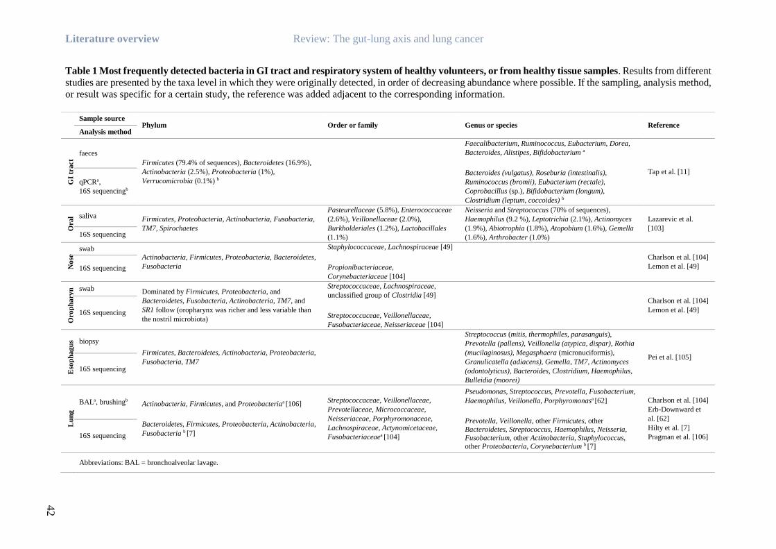
Literature overview Review: The gut-lung axis and lung cancer
42
Table 1 Most frequently detected bacteria in GI tract and respiratory system of healthy volunteers, or from healthy tissue samples. Results from different
studies are presented by the taxa level in which they were originally detected, in order of decreasing abundance where possible. If the sampling, analysis method,
or result was specific for a certain study, the reference was added adjacent to the corresponding information.
Sample source
Phylum Order or family Genus or species Reference Analysis method
GI
tract
faeces
Firmicutes (79.4% of sequences), Bacteroidetes (16.9%),
Actinobacteria (2.5%), Proteobacteria (1%),
Verrucomicrobia (0.1%) b
Faecalibacterium, Ruminococcus, Eubacterium, Dorea,
Bacteroides, Alistipes, Bifidobacterium a
Bacteroides (vulgatus), Roseburia (intestinalis),
Ruminococcus (bromii), Eubacterium (rectale),
Coprobacillus (sp.), Bifidobacterium (longum),
Clostridium (leptum, coccoides) b
Tap et al. [11]
qPCRa,
16S sequencingb
Ora
l
cav
ity
saliva Firmicutes, Proteobacteria, Actinobacteria, Fusobacteria,
TM7, Spirochaetes
Pasteurellaceae (5.8%), Enterococcaceae
(2.6%), Veillonellaceae (2.0%),
Burkholderiales (1.2%), Lactobacillales
(1.1%)
Neisseria and Streptococcus (70% of sequences),
Haemophilus (9.2 %), Leptotrichia (2.1%), Actinomyces
(1.9%), Abiotrophia (1.8%), Atopobium (1.6%), Gemella
(1.6%), Arthrobacter (1.0%)
Lazarevic et al.
[103] 16S sequencing
No
se
swab
Actinobacteria, Firmicutes, Proteobacteria, Bacteroidetes,
Fusobacteria
Staphylococcaceae, Lachnospiraceae [49]
Propionibacteriaceae,
Corynebacteriaceae [104]
Charlson et al. [104]
Lemon et al. [49] 16S sequencing
Oro
ph
ary
n
x
swab Dominated by Firmicutes, Proteobacteria, and
Bacteroidetes, Fusobacteria, Actinobacteria, TM7, and
SR1 follow (oropharynx was richer and less variable than
the nostril microbiota)
Streptococcaceae, Lachnospiraceae,
unclassified group of Clostridia [49]
Streptococcaceae, Veillonellaceae,
Fusobacteriaceae, Neisseriaceae [104]
Charlson et al. [104]
Lemon et al. [49] 16S sequencing
Eso
ph
ag
us biopsy
Firmicutes, Bacteroidetes, Actinobacteria, Proteobacteria,
Fusobacteria, TM7
Streptococcus (mitis, thermophiles, parasanguis),
Prevotella (pallens), Veillonella (atypica, dispar), Rothia
(mucilaginosus), Megasphaera (micronuciformis),
Granulicatella (adiacens), Gemella, TM7, Actinomyces
(odontolyticus), Bacteroides, Clostridium, Haemophilus,
Bulleidia (moorei)
Pei et al. [105]
16S sequencing
Lu
ng
BALa, brushingb Actinobacteria, Firmicutes, and Proteobacteriaa [106]
Bacteroidetes, Firmicutes, Proteobacteria, Actinobacteria,
Fusobacteria b [7]
Streptococcaceae, Veillonellaceae,
Prevotellaceae, Micrococcaceae,
Neisseriaceae, Porphyromonaceae,
Lachnospiraceae, Actynomicetaceae,
Fusobacteriaceaea [104]
Pseudomonas, Streptococcus, Prevotella, Fusobacterium,
Haemophilus, Veillonella, Porphyromonasa [62]
Prevotella, Veillonella, other Firmicutes, other
Bacteroidetes, Streptococcus, Haemophilus, Neisseria,
Fusobacterium, other Actinobacteria, Staphylococcus, other Proteobacteria, Corynebacterium b [7]
Charlson et al. [104]
Erb-Downward et
al. [62]
Hilty et al. [7]
Pragman et al. [106] 16S sequencing
Abbreviations: BAL = bronchoalveolar lavage.
Literature overview Review: The gut-lung axis and lung cancer
43
Bacteroidetes, followed by Actinobacteria, Proteobacteria and Verrucomicrobia, together
constituting 99% of the overall commensal microbiota [11]. Around 60 species have been
identified as the “core” microbiota, mostly bacteria from the Bacteroides, Bifidobacterium,
Eubacterium, Ruminococcus, Faecalibacterium genera, as well as a few others [11]. A
summary overview of the most prevalent human GI microbiota is shown in Table 1.
Strongly correlating to long-term diet [12], different enterotypes can also be identified through
a variation in the levels of one of the three most abundant genera: Bacteroides (enterotype 1),
Prevotella (enterotype 2) and Ruminococcus (enterotype 3), which is often less clearly
distinguished [13].
The impact of the GI microbiota on host mucosal immunity
The microbiota is now considered key to the proper development, maturation and reactivity of
the immune system [6,14]. Microorganisms serve as an inexhaustible source of microorganism-
associated molecular patterns (MAMPs) as well as pathogen-associated molecular patterns
(PAMPs). The two are recognizable on the host’s cells through pattern-recognition receptors
(PRRs), which include toll-like receptors (TLRs) and nucleotide-binding receptors (NODs)
[15]. TLRs are conserved receptors of the innate immune system that recognize MAMPs and
PAMPs among other molecules, evoking different immunological reactions depending on the
type of the cell, ligand and the receptor itself (some of the most common pairs are shown in
Figure 1A). TLRs, which are in direct contact with the intestinal lumen, are found not only in
intestinal epithelial cells (IECs) but also on immune cells within the lamina propria, such as
macrophages, dendritic cells (DCs), B cells, T cells and stromal cells. In IECs, TLR activation
by microbial ligands results in epithelial cell proliferation and the expression of antimicrobial
peptides, and secretion of immunoglobulin A (IgA) produced by plasma cells in lamina propria,
into the gut lumen [16], as well as the expression of antimicrobial peptides. All of the above
lead to enhanced intestinal barrier function and limit the possibility of microbial breach.
Interestingly, some of the TLRs, such as TLR2 and TLR4, are inhibited by IEC’s intracellular
Toll-interacting protein (TOLLIP) (Figure 1B) when the signal comes from the intestinal lumen,
which suggests a selective inflammatory response reserved to microbes that have breached the
intestinal barrier [16]. NOD-like receptors, or nucleotide-binding domain, leucine-rich repeat
containing proteins (NLRs), are cytoplasmatic equivalents of TLRs that detect bacterial PAMPs
entering the mammalian cell (Figure 1B). They are especially important in tissues where TLRs
are expressed at low levels e.g. in GI tract epithelial cells where the cells are in constant contact

Literature overview Review: The gut-lung axis and lung cancer
44
with the microbiota, and the expression of TLRs must be down-regulated to avoid over-
stimulation [17].
Figure 1 Interaction of the microbiota and intestinal mucosa. A) Microorganisms in the
intestine provide pathogen-associated molecular patterns (PAMPs) that serve as ligands for different
Toll-like receptors (TLRs) on the luminal or basolateral surface of the intestinal epithelial cells (IECs).
B) TLRs stimulation activates a signalling cascade resulting in transcription factor activation and gene
transcription, enhancing the cell barrier and further stimulating the immunological cells in the lamina
propria. This cascade can be inhibited by toll-interacting protein (TOLLIP). *Inhibition seen only for
TLR2 and TLR4 [16]. C) Commensal bacteria and their derivatives (e.g. short-chain fatty acids
(SCFAs)) directly stimulate IECs (B) or can be phagocytosed by DCs and macrophages in lamina
propria, carried to mesenteric lymph nodes (MLN) where they prime naïve B and T cells to mature and
differentiate. B cells become plasma cells and produce IgA that is secreted into the intestinal lumen
(sIgA). T cells profile into Th17 and Th1, with proinflammatory tendency, activating additional
effectory cells as neutrophils, resulting in bacterial clearance. There is also differentiation to Treg cells
having anti-inflammatory properties and controlling inflammation.
Commensal microorganisms can enter intestinal lamina propria in several ways: through an
opening in the barrier as a result of injury, or through active sampling by DCs or M cells. In
any case, microorganisms in the lamina propria are either phagocytosed and eliminated by
macrophages [18], or engulfed by DCs (along with B cells, both are considered the
“professional” antigen presenting cells (APCs)) and are carried live to the mesenteric lymph
nodes (Figure 1C). Recognition of infected apoptotic cells and bacteria results in the
upregulation of interleukin 6 (IL-6), which drives the differentiation of proinflammatory T-
helper-IL-17-producing (Th17) cells. Th17 cells primarily produce two main members of the

Literature overview Review: The gut-lung axis and lung cancer
45
IL-17 family, IL-17A and IL-17F which are involved in the recruitment, activation, and
migration of neutrophils [19], with granulocytes playing an important role in bacterial
clearance. Commensal-bearing DCs also induce the production of protective secretory
immunoglobulin A (sIgA) in activated B cells (Figure 1C) [20], i.e. plasma cells. This sIgA is
then distributed across all mucosal surfaces through the recirculation of activated B and T cells.
Commensal bacteria also directly promote the expression of factors involved in the induction
of IgA+ B cells (Figure 1C) [21], their survival and, interestingly, production of the more
resistant form of sIgA, exerting its stability and antimicrobial properties [22]. Through this
constant “priming” across several layers, microbiota maintains the immune system’s readiness
and reactivity, making it more capable of a quick and effective response when needed.
Specific populations of commensal bacteria e.g. Bacteroides fragilis, Bifidobacterium infantis,
Clostridium cluster IV and XIVa, also induce so-called regulatory T cells (Tregs), a subset of
forkhead box P3 positive (FoxP3+) CD4+ T-helper lymphocytes that maintain gut homeostasis
by stimulating the production of anti-inflammatory cytokine IL-10 [23]. These cells serve as a
kind of counterbalance to Th17 response, controlling the scope of its reaction and
proinflammatory cytokine production. Thus, depletion of Tregs leads to the abnormal
expansion of CD4+ Th cells, resulting in robust IL-17 and interferon gamma (IFN) responses
in the colonic lamina propria, to produce intestinal inflammation.
To understand more precisely which commensal microbiota has this immunostimulatory effect,
the well-known Bifidobacterium probiotic group was tested for its ability to induce the full
maturation of human peripheral blood mononuclear cells (PBMCs) into DCs. Twelve
Bifidobacterium strains descending from 4 probiotic species (Bifidobacterium longum, B.
breve, B. bifidum, and B. animalis subsp. lactis) were tested, and each was induced to full
maturation into DCs but using different Th preferences (Th1 or Th17), depending on the type
of Bifidobacterium strain with which they were incubated. Also, cell-free culture supernatants
were poor inducers of maturation [24], leading us to conclude that live bacterial cells are
necessary to induce efficient maturation and antigen presentation in DCs with this type of
bacteria.
Likewise, introducing probiotic strains such as Bifidobacterium lactis into healthy elderly
volunteers with fully developed immune systems, resulted in a significant increase in the
proportion of mononuclear leukocytes, the phagocytic capacity of mononuclear and
polymorphonuclear phagocytes, and the tumoricidal activity of NK cells [25], highlighting the

Literature overview Review: The gut-lung axis and lung cancer
46
importance of this specific immune-boosting species whose presence directly impacts immune
status.
Immunity and inflammation are not necessarily affected by bacterial cells, but may be
influenced by bacterial products. Bacterial products that have a significant effect on overall host
status surely included short chain fatty acids (SCFAs), by-products of the microbial
fermentation of dietary fibre. Amongst others, Bacteroides, Bifidobacterium,
Propionibacterium, Eubacterium, Lactobacillus, Clostridium, Roseburia, Prevotella are all
remarkable SCFA producers [26]. Fermentation and SCFA production are thought to inhibit
the growth of pathogenic organisms by reducing luminal pH [27]. The most abundant SCFAs
are acetate, propionate (found mostly in the small and large intestines) and butyrate (found
mostly in the caecum and colon), which are primarily derived from carbohydrates [28]. SCFAs
have their specific receptors both on leukocytes and on endothelial cells. Known as two
formerly orphan G protein-coupled receptors, GPR41 is found in a wide range of tissues
including in neutrophils, while GPR43 is shown to be highly expressed in immune cells [29].
GPR109a, the third receptor found in the colon epithelium and immune cells, is butyrate-
specific, and closely associated with the anti-inflammatory effect [30]. Butyrate is one of the
most important SCFAs with members of the Firmicutes phylum as major butyrate producers,
harbouring genes for the acetyl-CoA pathways. Other than as the main energy source for the
intestinal epithelium and its role in barrier integrity [31,32] following selective transport into
the colon epithelium, butyrate manifests broad anti-inflammatory activities such as immune cell
activation, proliferation, migration, adhesion, cytokine expression, and cancer cell apoptosis
[14,30]. These are mostly attributed to its function as a histone deacetylase (HDAC) inhibitor.
HDAC inhibition influences the acetylation not only of histones (FoxP3 locus in Tregs,
important for their maturation) [33], but also of major transcription factors such as nuclear
factor kappa-light-chain-enhancer for activated B cells (NF-κB), or signal transducer and
activator of transcription 3 (Stat3) [34,35], major proinflammatory pathway factors affecting
the proinflammatory cytokine secretion profile in immune cells [36] and decreasing the
proliferation and apoptosis of tumour cells [37].
As mentioned previously, changes to the gut microbiota are related to, for example, changes in
diet, antibiotic administration, chemotherapy, and a person’s general immune status. Whether
with a transient or permanent effect, these changes often lead to dysbiosis, with an altered ratio
of beneficial bacterial species (e.g. Lactobacillus sp., Faecalibacterium prausnitzii, etc.) and/or
an overgrowth or population shift of other species [14]. In this event, expanded indigenous

Literature overview Review: The gut-lung axis and lung cancer
47
microorganisms (now potential opportunistic pathogens), may also produce DNA-damaging
superoxide radicals and genotoxins in significant concentrations and induce innate immune
mediated proinflammatory pathways [38], directly damaging cells, and promoting malignant
transformation by inducing chromosomal and microsatellite instability, CpG island
methylation, epigenetic alterations, and post-translational modifications, which weaken the
immune response and increase the risk of cancer [39].
Apart from the direct pathological effect, an absence of the appropriate microbial composition
in the immune system’s early development has more far-reaching effects. This is evident from
studies on mice reared in germ-free (GF) conditions. These animals have impaired GI-driven
immune development, characterized by smaller Peyer’s patches, fewer CD8αβ intraepithelial
lymphocytes, underdeveloped isolated lymphoid follicles, a lack of primed T cells, lower levels
and impaired production of mucosal IgA antibodies, and active IL-10-mediated inflammatory
hyporesponsiveness [40,41]. Also, mice with colitis-associated cancer (CAC) that lacked
microbiota were unable to process pro–IL-1β and pro–IL-18 (interleukins in this case necessary
for a desirable inflammatory reaction) into their mature forms, resulting in increased tumour
burden [42]. That said, we can see that the composition of “healthy” microbiota is crucial to the
proper development of the immune system’s basic structures. Unless fully developed, their
ability to exchange information and their reactions to the “outside” world are compromised.
This state could be characterised as similar to anergy: the signal is present but the immune
system does not respond, as exemplified in the case of CAC mice.
3. Lung microbiota
The human respiratory tract is the primary and continuous entry portal for numerous
microorganisms and particles, such as viruses, bacteria, or fungi. These are primarily airborne,
but can also be transferred through saliva. Below the vocal cords, the human airways harbour
bacteria and other microbes in rich surroundings [43] that are distinct in composition from the
microbiota seen at other sites (in the nasal and oral cavity, gut, skin, and vagina). Despite being
less populated compared to the GI tract, the lung microbiota includes a range of
microorganisms, mostly seen through the use of bronchoalveolar lavage (BAL) fluid, or tissue
samples. Since lung microbiota exploration has rather young and non-uniform protocols, it is
crucial to bear in mind that the type of sample (lavage, tissue), sampling method, and possibility
of cross-contamination during sampling between distinct parts of the airway, influence final
results [44,45]. Therefore, due to the paucity of overall studies in this field, one must be careful
Literature overview Review: The gut-lung axis and lung cancer
48
to take detailed methodology and its possible advantages and disadvantages into account.
Studies that analyse lung tissue acquired through sterile surgical explant have also been carried
out, and they all report that the lower respiratory tract contains a microbiome that is distinct
from, but related to, that of the upper airways [46].
In 2014 Dickson et al. [47] proposed an adapted island model for lung biogeography, suggesting
that more distal lung bacterial communities are less rich and more dissimilar to their upper
respiratory tract source community. Here, microbial composition is determined by the rate of
microbial immigration into the airways, their rate of elimination (e.g. by coughing or immune
defences), and the rate at which different community members are reproduced [48].
Temperature, oxygen tension, pH, nutrient density, local anatomy, and host defence are
spatially heterogeneous across the airways and the lungs, all of which affect local
microbiological growth conditions.
Starting with the upper respiratory airways, the nostril is dominated by Firmicutes and
Actinobacteria; Firmicutes, Proteobacteria, and Bacteroidetes are prevalent in the oropharynx
[49]. In the lung the most common phyla consistently observed are Bacteroidetes, Firmicutes
and Proteobacteria. The nasal microbiota seems to more closely resemble that of the skin than
that of the lungs and contributes little to lung communities [50]. The coming years are likely to
bring the development of new methods able to minimize cross-contamination (as seen for
endoscopy [51]), which is the biggest problem in lower respiratory tract sampling that, along
with metagenomic analysis (of both cultivable and non-cultivable bacteria), will yield more
precise results in terms of the bacterial population found at certain airway depths. An overview
of different studies investigating the composition of the respiratory system microbiota is
presented in Table 1.
The ecological determinants of the lung microbiota (immigration, elimination, and regional
growth conditions) change during acute and chronic lung disease, as seen in chronic obstructive
pulmonary disease (COPD) (often a precancerous inflammatory state) and lung cancer (Table
2) [7,44]. Whether the observed dysbiosis is a cause, consequence or simply a co-evolving
factor, still needs to be elucidated, but its likely role will be individually connected to the
pathology and its aetiology. However, it is known that smouldering inflammation (caused by
lung injury, pathogen colonization, or intrinsic factors) is often a common starting point that
leads to subsequent cancer development [52].

Literature overview Review: The gut-lung axis and lung cancer
49
The microbial factors that may be responsible for lung cancer development are still not well
known, unlike the many genetic predispositions and mutations that underlie the different types
of lung cancer [53]. This is why, for now, it is possible to correlate the non-genetic development
of lung cancer with the well characterized development of COPD, a chronic inflammatory state
where initial lung injury, whatever its cause, creates an opening for microbial dysbiosis and
colonization, thus worsening the overall condition and often leading to the cancer state.
Inflammation of the lung is associated with a loss of epithelial integrity and results in the
“leakage” of serum proteins into the airways [54]. Formyl peptides, cleavage products of
bacterial or mitochondrial proteins, as well as other bacterial products, serve as powerful
chemoattractants for both the neutrophils and monocytes that emigrate from alveoli [55].
Although essential in pathogen clearance and having a tumoricidal effect [56], neutrophil influx
and degranulation in the airways and lung parenchyma contribute to chronic inflammation,
parenchymal lung damage, and progressive small airway obstruction, due to the loss of alveolar
attachments and lung elasticity [57], as explained by Sethi’s vicious circle hypothesis [58]. In
vitro, their enzymes, serine proteinases (elastase, cathepsin G and proteinase 3), and defensins
markedly affect the integrity of the epithelial layer, decreasing the frequency of the ciliary beat,
increasing mucus secretion, and inducing the synthesis of epithelium-derived mediators such
as neutrophil chemoattractant chemokine IL-8 from respiratory epithelial cells [59].
Obstruction of the lumina with mucus introduces pockets of increased temperature and
decreased oxygen tension, selectively favouring the growth of well-known disease-associated
microbes [60,61]. This dysbiotic shift can be characterized by a move away from the
Bacteroidetes phylum, often to Proteobacteria (e.g. Pseudomonas aeruginosa, Haemophilus
influenza, Moraxella catarrhalis) [62,63], and sometimes to Firmicutes (e.g. Streptococcus
pneumoniae, Staphylococcus aureus) [44]. The same growth effect was observed with a
generation of intra-alveolar catecholamines and inflammatory cytokines [64]. If airway
colonization becomes persistent, it further promotes chronic inflammatory response, affecting
the elastase-anti-elastase balance in the lung, which was shown to vary 80-fold with changes to
the airway bacterial load [65]. A higher bacterial load consequently induces higher overall IL-
8 and other blood circulating inflammatory cytokine levels, greater inflammation, oxidative
stress and greater forced expiration volume (FEV) decline [66].
Lung microbiota was also shown to vary according to clinical endpoints. In non-malignant lung
tissue from advanced stage cancer, alpha diversity had increased, whilst it had decreased in the
tumour lung tissue. Also, the interaction between the upper and lower airways involving the

Literature overview Review: The gut-lung axis and lung cancer
50
Table 2 Most frequently detected bacteria in the lung of patients suffering from their respective diseases. Results from different studies were
presented by the taxa level in which they were originally detected, in order of decreasing abundance where possible. If the sampling, analysis
method, or result was specific for a certain study, the reference was added adjacent to the corresponding information.
Lung
Disease
Sample
source Phylum Order or family Genus or species Reference
Analysis
method
COPD
BALa,
sputumb, lung tissuec
Proteobacteria (44%) and Firmicutes
(16%) followed by Actinobacteria (13%), with Bacteroidetes, Fusobacteria,
Tenericutes, SR1 incertae, TM7, and Synergistetes identified in lower
proportions (<3%) b,d [107]
Increase in Proteobacteria (sometimes Firmicutes), decrease in Bacteroidetes and
Firmicutes c,d [44]
In moderate COPD mostly Actinobacteria and Proteobacteria
In severe COPD mostly Actinobacteria and
Firmicutesa,d [106]
Proteobacteria, Bacteroidetes, Firmicutes,
Actinobacteria, Fusobacteriab,d [7]
Haemophilus influenza, Pseudomonas aeruginosa, Moraxella catarrhalis, Streptococcus pneumoniae b,f [107]
Significant increases of Streptococcus pneumoniae, Klebsiella
pneumoniae and Pseudomonas aeruginosa b,e [63]
Streptococcus, Abiotrophia, Rothia, Tropheryma, Actinomyces,
Peptostreptococcus, Serratia, Capnocytophaga, Leptotrichia,
Kingella, Dysgonomonasa,d [106]
Significant abundance of Pseudomonas and Haemophilus a,d [62]
Prevotella, Haemophilus, Streptococcus, Neisseria, other
Proteobacteria, Veillonella, other Firmicutes, other Bacteroidetes, Fusobacterium, other Actinobacteria,
Staphylococcus b,d [7]
Erb-Downward et
al. [62] Garcia-Nuñez et al.
[107]
Hilty et al. [7] Sze et al. [44]
Wu et al. [63]
Pragman et al. [106] 16S
sequencingd,
qPCR/DGGEe, bacterial
culturef
Lung
cancer
Sputuma,
salivab
Flavobacteriales, Burkholderiales,
Campylobacterales, Spirochaetales
(more abundant) and Bacteroidales
(less abundant)b,d [67]
Veillonellaceae significantly overexpressed whereas
Lachnospiraceae underexpressedb,d
[67]
Actinomyces spp., Peptostreptococcus spp., Eubacterium lentum,
Veillonella parvula, Prevotella spp., Bacteroides spp., Lactobacillus jenseniia,c [108]
Capnocytophaga, Selenomonas, and Veillonella were found to
be more abundant in both SCC and ACb,d [67]
Rybojad et al. [108]
Yan et al. [67] anaerobic culturec,
16S
sequencingd
Abbreviations: COPD = chronic obstructive pulmonary disease; BAL = bronchoalveolar lavage; DGGE = PCR-denaturing gradient gel electrophoresis; SCC = small cell carcinoma; AC = adenocarcinoma.

Literature overview Review: The gut-lung axis and lung cancer
51
microbial population present in lung cancer is clearly shown in the study carried out by Yan et
al. [67]. The study showed that there is a high degree of specificity in patients with either small
cell carcinoma (SCC) or adenocarcinoma (AC), compared to controls based on bacteria isolated
from saliva (Table 2).
As mentioned above, IL-6 and IL-8 are cytokines that become elevated during inflammatory
stress. They are involved in tumorigenesis by acting directly on lung epithelial cells to stimulate
the NF-κB-1 pathway [68]. Additionally, IL-6 and IL-8 are expressed by premalignant or
senescent lung cancer cells [69]. They may act in an autocrine and/or paracrine fashion to
stimulate cancer cell proliferation [70], migration, and invasion [71]. In bronchoalveolar
carcinoma, tumour cells were a main source of IL-8 and the presence of an increased number
of neutrophils in BAL fluid was correlated with the IL-8 level in BAL, and associated with a
poor outcome [72]. In a case study carried out by Pine et al. [73], increased levels of both serum
IL-6 and IL-8 were associated with lung cancer, but only the IL-8 level was associated with
lung cancer risk several years prior to diagnosis.
To summarize, the appearance of dysbiosis or malignancy is likely the product of a dynamic
interaction between various immune, microbial, and environmental factors. At least one of these
acts as an initiator but others often readily follow. This is why it remains difficult to reach any
conclusions regarding the true aetiology of disease and what might be the best intervention and,
more importantly, prevention approach.
4. A bidirectional concept of the gut-lung axis
Recently, we have reached a greater understanding of microbial influence on the complex and
interconnected axis between gut and lung. This stems from the simple fact that ingested
microorganisms can access both sites – from gastrointestinal tract microbiota that enter the lung
through aspiration [74] to the more “internal” influence, which shows improved lung function
and pathogen clearance following the transplantation of faecal microbiota [6].
This interaction can be mediated in different ways – by the microbiota and its products, or via
immune cells (Figure 2). According to the “gut-lymph” theory of Samuelson et al. [14], there
are sufficient macrophages and other immune cells in the intestinal submucosa or the
mesenteric lymph nodes that contain a majority of translocating bacteria. Surviving bacteria,
cell wall fragments, or the protein parts of dead bacteria escaping with the cytokines and
chemokines produced in the gut, travel along the mesenteric lymphatic system to the cisterna

Literature overview Review: The gut-lung axis and lung cancer
52
chyli, and subsequently enter the systemic circulation. Access to pulmonary circulation may
lead to DC and macrophage activation as well as the priming of T cells and their differentiation.
Figure 2 Proposed pathways of the gut-lung interaction. Microbiota and its products that enter
intestinal mucosa (blue arrows) are phagocytosed and transferred to mesenteric lymph nodes (MLN) by
antigen presenting cells (APC), where they stimulate priming of the T and B cells. Once activated, with
the expression of proper homing receptors, these cells can migrate back to the original site (intestinal
mucosa) (black dashed arrow), or to distal locations such as the lung epithelium and lung nodes through
lymphatic and blood circulation. There, they can directly act on their target or continue to stimulate
other immune cells. On the other hand, bacterial products from the intestinal mucosa or surviving
bacteria can also reach the lung by blood or lymphatics to stimulate the immune system in the same way
as they would have done in the intestinal tract. Depending on the tissue pre-stimulation, type of stimulus,
and local and general immunological status, the result can be either positive, as effective bacterial
clearance or anti-tumour activity, or over-inflammatory response, promoting further tissue damage,

Literature overview Review: The gut-lung axis and lung cancer
53
pathogen colonization, and tumour progression. The same schema is proposed in the other sense,
beginning with the lung mucosa and finishing with distal effect on the gut. Although not known for the
moment, there is also a possibility that bacterial products of the lung microbiota can exert their effect in
the intestinal mucosa, being delivered in the same way as explained above.
Another way to influence the pulmonary region is through the migration of immunological
cells. As previously mentioned, translocated microorganisms and their parts within the lamina
propria are transferred to the mesenteric lymph nodes by antigen presenting cells (APCs), and
used for priming naive B and T cells. Activated B cells capable of producing antigen-specific
immunoglobulins, i.e. plasma cells, will not only produce immunoglobulins in situ, but will
reach draining lymph nodes and other mucosal tissues, thereby spreading immunological
“information”. The constitutive entry of antigen at steady-state stimulates inflammasome
conversion of pro-IL-1β and pro-IL-18 into active form, in other words it switches off our innate
ability to produce IL-10 and other anti-inflammatory molecules leading to DC migration to
local lymph nodes, and the priming and differentiation of T cells. The latter can subsequently
migrate out of the gut-associated lymphatic tissue (GALT) and reach both mucosal and
peripheral non-mucosal tissues, including the bronchial epithelium, thus modifying the
immunological response which is dependent on the induced cell profile (to Th1, Th2, etc.)
[75,76], and improving the immunological response against pulmonary pathogens [77].
Although this theory explains the unilateral interaction, it is reasonable to speculate that this
axis works in precisely the same way when it originates in the lung mucosa and lung lymph
nodes (Figure 2). Further, lung DCs in vitro have the option to imprint the expression of gut-
homing integrin α4β7 and CCR9 (lung-homing integrin is CCR4 [78]) on co-cultured T cells
in vitro, and on adoptively transferred cells in vivo, which guides their migration to the GI tract
[79].
4.1. Influence of the gut microbiota on the lung
The composition of “healthy”, or rather balanced, gut microbiota is shown to have a serious
influence on the effectiveness of lung immunity. GF mice, devoid of their intestinal microbiota
during the development of their immune system, show impaired pathogen clearance in the lung,
which results in their growth and dissemination [41]. At this stage, it is also important to
consider that the lungs of these mice are also germ-free [80], devoid of all microbiota that might
normally play a role in stimulating lung immunity. Modified alveolar architecture also results,
and thus both factors modify the response to pathogen infection. The same observations were
made as with the infection of GF animals, when wild phenotypes were treated with antibiotics,
thus disturbing the intestinal homeostasis, after they had been challenged with bacterial or viral

Literature overview Review: The gut-lung axis and lung cancer
54
microorganisms [81,82]. If animals had been boosted with lipopolysaccharide (LPS) following
antibiotic treatment, they were better able to cope with the lung infection and consequently had
reduced mortality. Population studies followed these findings with respect to the importance of
preserving gut microbiota, showing that increased use of penicillins, cephalosporins,
macrolides and quinolones correlated with an increased risk of lung cancer in humans [83].
Here we can speculate that various antibiotic treatments eradicate the bacterial populations
required for effectively priming T lymphocytes with anti-tumour properties, while at the same
time making room for other opportunistic pathogens to colonize both the gut and the lung.
Interestingly, modified gut microbiota not necessarily characterized as dysbiotic may also
influence immune response efficiency, as seen in obese mice. These mice had an impaired
expression of cytokines in their lungs (IFNα, IFNβ, IL-6, TNFα) and significantly decreased
mRNA of IFNγ, interleukin 2 receptor subunit beta (IL-2RB) and perforin 1 (Prf1). All the
criteria were improved following daily supplementation with a probiotic strain of Lactobacillus
gasseri [84].
Nutrition may also impact microbial development and the composition of our respiratory tract
microbiota [85]. A high-fibre diet in mice has been shown to increase SCFA circulating blood
levels (but no traces in the lung itself), and has been shown to be responsible for increased
protection against allergic inflammation in the lung (reduced inflammatory cell infiltration),
followed by a change in the intestinal and, to a lesser extent, the airway microbiota [86].
The above-mentioned findings clearly show how important the overall composition of the
intestinal microbiota is for a productive immunological response in the lung. Lack of an
appropriate stimulus during the developmental phase, as during infection, will disable a quick
and effective immune reaction, resulting in pathogenic colonisation, increased susceptibility to
infection, damage, the possible development of cancer, and increased mortality. At the same
time, just one single strain, bacterial part or product can turn the tables and provide the boost
needed to stimulate the correct immune response.
4.2. Influence of the lung microbiota on the gut
Unlike the local and systemic influence of intestinal microbiota, the influence of lung
microbiota, its products, and their circulation are yet to be properly assessed. One study reported
that non-absorbable tracer deposited into the nasal cavity of mice can be found in the GI tract a
short time later [87]. Also, Sze et al. [43] showed that, in mice, even acute exposure to a single
dose of intratracheal LPS disrupts the airway microbiota, leading to translocation of these
Literature overview Review: The gut-lung axis and lung cancer
55
bacteria into the bloodstream. Within 24 hours the caecal microbiota is also disturbed, leading
to a sharp increase in the total bacterial load. It still remains unclear as to whether this effect is
due to the direct interaction of translocated pulmonary and residential intestinal microbiota via
immune cell or cytokine mediation, or to microbial products that reach the gut.
4.3. Microbiota and cancer via the immune system
Due to a number of genetic alterations resulting in the loss of normal cellular regulatory
processes, cancer cells express neoantigens that are tumour-specific and distinguish tumour
cells from healthy cells. The importance of the gut microbiota in anti-cancer response has been
described by Chen et al. [88] through the concept of a cancer-immunity cycle. The cancer-
immunity cycle starts with the capture of neoantigens from cancer cells by DCs. For an anti-
cancer response to take place, this must be accompanied by another signal, such as
proinflammatory cytokines, factors released by dying cancer cells or by gut microbiota
components. The goal here is to reduce peripheral tolerance to tumour antigens. Following
processing, DCs present captured neoantigens to T cells, thus resulting in their priming and
activation to create effector T cells against cancer-specific antigens. At this point, the balance
between T effector and T regulatory cells is crucial in determining the nature of the immune
response. The now activated T effector cells travel to the tumour site, invade the tumour bed
and, by recognizing specific tumour antigens, bind and kill cancer cells. Problems arise when
tumour antigens are not detected, meaning that DCs and T cells treat antigens as ‘self’ rather
than foreign. In this case, a Treg response rather than an effectory response results. Homing of
T cells to tumour may not be correct either. T cells may be inhibited from infiltrating the tumour,
or (more importantly) factors in the tumour microenvironment may suppress any effector cells
that are produced. There are two main negative regulators of T cell responses: checkpoints in
lymphoid organs (CTLA-4), and immunostats within the tumour beds (PD-L1:PD-1).
Programmed cell death ligand 1 (PD-L1) is a molecule expressed on tumour cells or on tumour-
infiltrating immune cells, which binds programmed cell death protein 1 (PD-1) expressed on
effector CD8+ T lymphocytes, blocking the secretion or production of the cytotoxic mediators
needed to kill tumour cells within the tumour beds. Cytotoxic T-lymphocyte-associated
protein 4 (CTLA-4), expressed on Tregs, acts as the major negative regulator of the priming
and activation of effector CD8+ T cells inside the lymphoid organs, by binding its CD80 and
CD86 ligands on APCs. The presence of these suppressive factors explains the limited activity
of previous immune-based therapies. The goal of current cancer immunotherapy, using anti-
PD-L1:PD-1 and anti-CTLA-4 antibodies, is to initiate or reinitiate a self-sustaining cycle of
Literature overview Review: The gut-lung axis and lung cancer
56
cancer immunity, enabling it to amplify and propagate without creating an unrestrained
response.
To create a higher response to neoantigens, the immune system’s peripheral tolerance must be
reduced. It is already known that the commensal microbiota induces the generation of CD4+ T
cells against their own antigens [89] thereby limiting the systemic dissemination of commensal
bacteria [90]. The same antigen cross-reactivity effect, or super antigen-driven response,
accounts for T cell-dependent tumour regression. As suggested by Viaud et al. [36], and based
on recent studies by Iida et al. [91] and Viaud et al. [92] in mice, Th17 cells and memory Th1
cells elicited against commensal bacteria might preferentially accumulate in an inflammatory
tumour microenvironment, already primed by bacterial products or ligands for PRRs. Based on
these studies, Zitvogel et al. [93] explain the long-range effect of microbiota through two signal
hypotheses. Signal 1 hypothesis suggests a phenomenon of antigen mimicry or cross-reactivity.
That is, certain microbial antigens from the bacterial species that pass the intestinal barrier and
are used for T cell priming could closely resemble tumour antigens, thus promoting better
immune system reactivity and anti-tumour response, i.e. immunosurveillance. In signal 2
hypothesis, by interacting with PRRs after passing the intestinal barrier, microbiota can
stimulate the production of a diverse palette of cytokines and interferons, and determine
whether it will elicit a proinflammatory, immunostimulatory or immunosuppressive response.
Also, there is some evidence that commensal-specific Tregs are capable of switching to effector
inflammatory Th17 cells after sensing the disruption of the mucosal barrier. Because microbial
products, metabolites, effectory cells, and cytokines are able to travel, this stimulation is not
necessarily confined to just the gut.
Observing these effects, it is interesting to speculate that at least a transient disruption of
intestinal barrier functions and microbiota translocation is a primary factor in shaping the
relationship between the gut microbiome, the immune system, and cancer.
4.4. Probiotics and the lung
Probiotics, best known in nutritional therapy, are defined as “live microorganisms, which, when
administered in adequate amounts, confer a health benefit on the host” [94]. In the intestine
they mainly refer to the genera Lactobacillus and Bifidobacterium, and include many different
strains such as L. paracasei, L. rhamnosus, L. acidophilus, L. johnsonii, L. fermentum, L.
reuteri, L. plantarum, B. longum, B. breve, B. bifidum, B. animalis subsp. lactis, etc. The same
genus does not necessarily involve the same characteristics, due to the great genomic
differences between species and also within the different strains of the same species [95].

Literature overview Review: The gut-lung axis and lung cancer
57
Although the first evidence of probiotic influence on lung cancer was seen in 1985 [96],
probiotics only recently re-emerged in the field of lung oncology as a possible new therapy, and
is already showing highly promising results. Using different mouse lung cancer models,
conventional therapies (a combination of platinum-based agents with paclitaxel, gemcitabine,
vinorelbine, or docetaxel, which all have high toxicity [97]) were combined with specific
probiotic strains or, conversely, the antibiotic eradication of microbiota, to assess the effect of
chemotherapy.
In a lung adenocarcinoma viral model, when vancomycin was used to eradicate gram-positive
bacteria it compromised the efficacy of cyclophosphamide (CTX)-based chemotherapy, and
correlated with a reduced intratumoural CD8+ T effector/ FoxP3+ regulatory T cell ratio [98].
Likewise, in mice treated with cisplatin combined with an antibiotic cocktail of vancomycin,
ampicillin, and neomycin, tumour size was larger than that found in mice receiving a single
treatment of cisplatin [99]. Taking these examples, we can readily conclude that the presence
of conventional intact microbiota is crucial for effective chemotherapy.
On the other hand, feeding mice orally with L. acidophilus (Lewis lung cancer model) treated
with cisplatin decreases the size of tumours and improves the survival rate. Enhanced anti-
tumour response is also achieved through upregulation of IFNγ, granzyme B (GzmB), and Prf1
expression [99] following probiotic supplementation.
Recently, there was considerable interest in evaluating the role of gut microbiota in lung cancer
therapy using immune checkpoint inhibitors. One of the first studies of this principle was done
using a mouse melanoma model, but is readily applicable to other cancer types, as shown in the
study. Here, oral administration of a Bifidobacterium cocktail (B. bifidum, B. longum, B. lactis
and B. breve) on its own improved tumour control to the same degree as PD-L1–specific
antibody therapy (checkpoint blockade) [100]. When the two treatments were combined it
virtually abolished tumour outgrowth. Improvement was seen in immune responses upstream
of T cells, at the level of host DCs. Their augmented function enhanced CD8+ T cell priming
and accumulation in the tumour microenvironment. The percentage of MHC IIhi DCs was also
increased. With Bifidobacterium treatment, 760 genes were upregulated, including cytokine-
cytokine receptor interaction, CD8+ T cell activation and costimulation, DC maturation, antigen
processing and cross presentation, the chemokine-mediated recruitment of immune cells to the
tumour microenvironment, and type I interferon signalling.
Literature overview Review: The gut-lung axis and lung cancer
58
Vétizou et al. [101] reported that the oral feeding of GF mice with Bacteroides fragilis induced
Th1 immune responses in tumour-draining lymph nodes and promoted the maturation of
intratumoural DCs. This was, as the authors suggest, due to the cross-reactivity of the bacterial
and tumour epitopes, which led to the restoration of the therapeutic response of GF tumour
bearers to CTLA-4 antibody treatment. Antibody therapies such as this prevent inactivation of
the CD8+ T cells by binding to the CTLA-4. Daillère et al. [102] noted that Enterococcus hirae
and Barnesiella intestinihominis specific-memory Th1 cell immune responses selectively
predicted longer progression-free survival in advanced lung cancer patients treated with chemo-
immunotherapy. Both strains represent valuable "oncomicrobiotics" improving the efficacy of
the most common alkylating immunomodulatory compound.
To summarize, as scientists delve deeper, the beneficial effects of probiotics on the immune
system continue to emerge. As seen, certain strains have the power and ability to stimulate anti-
tumour response or to simply stimulate the immune system to show lower tolerance, thus
promoting higher reactivity and tumour eradication. The future objective is to find the optimum
probiotic cocktail that may one day completely substitute conventional therapies, thereby
obtaining equal or better success, and lowering toxicity, one of the biggest problems in cancer
treatment.
5. Conclusion
The importance of the gut microbiota and its composition has long been recognized, for
digestion as well as for overall wellbeing. Recently, the presence of the lung microbiota and the
role it plays, in health and in disease, has been receiving attention. The lung and gut microbiota,
both continually reseeded through interaction with the environment, modulate our local and
systemic immunity. More than simply two distinct microbiota, they are now seen as functioning
in dialogue, altering previous ideas of airway sterility and the existence of a “barrier” between
the two compartments, due to their perceived distance or functional differences. By providing
stimulating signals through its epitopes or products (such as SCFA butyrate), the gut microbiota
directly enhances the intestinal barrier. Likewise, it stimulates the priming and maturation of T
and B cells, ensuring improved microbial clearance and mucosal protection through antibodies.
This effect is not only retained in the intestinal system but is spread along other mucosal
surfaces by means of lymphatic and blood circulation, influencing distal site immune response.
So, even though the antigen was introduced in the gut, an immunological response can also be
elicited in the lung, although there was no direct prior contact with the antigen, and vice versa.
Literature overview Review: The gut-lung axis and lung cancer
59
Bacteria and their products that go through the first immunological barrier also reach distal sites
through the lymphatic system and blood, and modulate the immune response at the remote site.
The site where the first encounter between the immune system and microbial antigens took
place is also important, since it influences reactivity and the influx of these cells into other
tissues. Applying prebiotics to target a specific microbial group could be a good way to restore
“healthy” microbial composition, which will consequently increase intestinal barrier function
and stimulate the immune system. The relevance of natural microbial support in chemotherapy
effectiveness or replacement has already been demonstrated. In future, further discoveries will
surely be made in this new and exciting area of research, adding to the complexity of, but also
clarifying the reasons behind, this axis; opening ideas to new or enhanced therapies based on
the natural behaviour of the organism; increasing longevity; and decreasing therapeutic side-
effects or the effects of disease itself.
6. References
[1] B.J. Marsland, A. Trompette, E.S. Gollwitzer, “The Gut-Lung Axis in Respiratory Disease.,” Annals of
the American Thoracic Society, vol. 12 Suppl 2, pp. S150-6, 2015.
[2] A.M. Brownawell, W. Caers, G.R. Gibson, et al., “Prebiotics and the health benefits of fiber: Current
regulatory status, future research, and goals,” The Journal of Nutrition, vol. 124, pp. 962–974, 2012.
[3] Y. Belkaid, T.W. Hand, “Role of the microbiota in immunity and inflammation,” Cell, vol. 157, pp. 121–
141, 2014.
[4] S. Carding, K. Verbeke, D.T. Vipond, B.M. Corfe, L.J. Owen, “Dysbiosis of the gut microbiota in
disease.,” Microbial Ecology in Health and Disease, vol. 26, pp. 26191, 2015.
[5] P. Forsythe, W. Kunze, J. Bienenstock, et al., “Moody microbes or fecal phrenology: what do we know
about the microbiota-gut-brain axis?,” BMC Medicine, vol. 14, pp. 58, 2016.
[6] T.J. Schuijt, J.M. Lankelma, B.P. Scicluna, et al., “The gut microbiota plays a protective role in the host
defence against pneumococcal pneumonia.,” Gut, pp. 1–9, 2016.
[7] M. Hilty, C. Burke, H. Pedro, et al., “Disordered Microbial Communities in Asthmatic Airways,” PLoS
ONE, vol. 5, pp. e8578, 2010.
[8] H. Wang, J.-S. Liu, S.-H. Peng, et al., “Gut-lung crosstalk in pulmonary involvement with inflammatory
bowel diseases,” World J Gastroenterol, vol. 19, pp. 6794–6804, 2013.
[9] T. Jess, E. Horváth-Puhó, J. Fallingborg, H.H. Rasmussen, B.A. Jacobsen, “Cancer Risk in Inflammatory
Bowel Disease According to Patient Phenotype and Treatment: A Danish Population-Based Cohort
Study,” The American Journal of Gastroenterology, vol. 108, pp. 1869–1876, 2013.
[10] S. Keely, N.J. Talley, P.M. Hansbro, “Pulmonary-intestinal cross-talk in mucosal inflammatory disease.,”
Mucosal Immunology, vol. 5, pp. 7–18, 2012.
[11] J. Tap, S. Mondot, F. Levenez, et al., “Towards the human intestinal microbiota phylogenetic core,”
Environmental Microbiology, vol. 11, pp. 2574–2584, 2009.
[12] G.D. Wu, J. Chen, C. Hoffmann, et al., “Linking long-term dietary patterns with gut microbial
enterotypes.,” Science, vol. 334, pp. 105–8, 2011.
[13] M. Arumugam, J. Raes, E. Pelletier, et al., “Enterotypes of the human gut microbiome,” Nature, vol. 12,
pp. 174–180, 2011.
[14] D.R. Samuelson, D.A. Welsh, J.E. Shellito, “Regulation of lung immunity and host defense by the
intestinal microbiota,” Frontiers in Microbiology, vol. 6, pp. 1–14, 2015.
[15] I.I. Ivanov, K. Honda, “Intestinal commensal microbes as immune modulators,” Cell Host Microbe, vol.
12, pp. 496–508, 2012.
[16] M.T. Abreu, “Toll-like receptor signalling in the intestinal epithelium: how bacterial recognition shapes
intestinal function.,” Nature Reviews. Immunology, vol. 10, pp. 131–44, 2010.
[17] A. Hakansson, G. Molin, “Gut microbiota and inflammation,” Nutrients, vol. 3, pp. 637–687, 2011.
[18] B. Kelsall, “Recent progress in understanding the phenotype and function of intestinal dendritic cells and
macrophages,” Mucosal Immunology, vol. 1, pp. 460–469, 2008.

Literature overview Review: The gut-lung axis and lung cancer
60
[19] T. Nakajima, V. Palchevsky, D.L. Perkins, J.A. Belperio, P.W. Finn, “Lung transplantation : infection ,
inflammation , and the microbiome,” Seminars in Immunopathology, vol. 33, pp. 135–156, 2011.
[20] A.J. Macpherson, T. Uhr, “Induction of protective IgA by intestinal dendritic cells carrying commensal
bacteria,” Science, vol. 303, pp. 1662–1665, 2004.
[21] H. Tezuka, Y. Abe, J. Asano, et al., “Prominent Role for Plasmacytoid Dendritic Cells in Mucosal T Cell-
Independent IgA Induction,” Immunity, vol. 34, pp. 247–257, 2011.
[22] E. Castigli, S.A. Wilson, S. Scott, et al., “TACI and BAFF-R mediate isotype switching in B cells.,” The
Journal of Experimental Medicine, vol. 201, pp. 35–9, 2005.
[23] N. Kamada, S.-U. Seo, G.Y. Chen, G. Núñez, “Role of the gut microbiota in immunity and inflammatory
disease,” Nature Reviews Immunology, vol. 13, pp. 321–35, 2013.
[24] P. López, M. Gueimonde, A. Margolles, A. Suárez, “Distinct Bifidobacterium strains drive different
immune responses in vitro,” International Journal of Food Microbiology, vol. 138, pp. 157–165, 2010.
[25] H.S. Gill, K.J. Rutherfurd, M.L. Cross, P.K. Gopal, “Enhancement of immunity in the elderly by dietary
supplementation with the probiotic Bifidobacterium lactis HN019.,” The American Journal of Clinical
Nutrition, vol. 74, pp. 833–9, 2001.
[26] G.T. Macfarlane, S. Macfarlane, “Bacteria, colonic fermentation, and gastrointestinal health.,” Journal of
AOAC International, vol. 95, pp. 50–60, 2012.
[27] D.L. Topping, P.M. Clifton, “Short-chain fatty acids and human colonic function: roles of resistant starch
and nonstarch polysaccharides.,” Physiological Reviews, vol. 81, pp. 1031–64, 2001.
[28] G.T. Macfarlane, S. Macfarlane, “Fermentation in the human large intestine: its physiologic consequences
and the potential contribution of prebiotics.,” Journal of Clinical Gastroenterology, vol. 45 Suppl, pp.
S120-7, 2011.
[29] E. Le Poul, C.C. Loison, S. Struyf, et al., “Functional Characterization of Human Receptors for Short
Chain Fatty Acids and Their Role in Polymorphonuclear Cell Activation,” Journal of Biological
Chemistry, vol. 278, pp. 25481–25489, 2003.
[30] N. Singh, A. Gurav, S. Sivaprakasam, et al., “Activation of Gpr109a, Receptor for Niacin and the
Commensal Metabolite Butyrate, Suppresses Colonic Inflammation and Carcinogenesis,” Immunity, vol.
40, pp. 128–139, 2014.
[31] N.I. McNeil, “The contribution of the large intestine to energy supplies in man.,” The American Journal
of Clinical Nutrition, vol. 39, pp. 338–42, 1984.
[32] S. Krishnan, N. Alden, K. Lee, “Pathways and functions of gut microbiota metabolism impacting host
physiology,” Current Opinion in Biotechnology, vol. 36, pp. 137–145, 2015.
[33] Y. Furusawa, Y. Obata, S. Fukuda, et al., “Commensal microbe-derived butyrate induces the
differentiation of colonic regulatory T cells,” Nature, vol. 504, pp. 446–450, 2013.
[34] L. Chen Lf, W. Fischle, E. Verdin, et al., “Duration of nuclear NF-kappaB action regulated by reversible
acetylation.,” Science, vol. 293, pp. 1653–7, 2001.
[35] Z.-L. Yuan, Y.-J. Guan, D. Chatterjee, Y.E. Chin, “Stat3 dimerization regulated by reversible acetylation
of a single lysine residue.,” Science, vol. 307, pp. 269–73, 2005.
[36] S. Viaud, R. Daillère, I.G. Boneca, et al., “Gut microbiome and anticancer immune response: really hot
Sh*t!,” Cell Death and Differentiation, vol. 22, pp. 199–214, 2015.
[37] H.M. Hamer, D. Jonkers, K. Venema, S. Vanhoutvin, F.J. Troost, R.J. Brummer, “Review article: The role
of butyrate on colonic function,” Alimentary Pharmacology and Therapeutics, vol. 27, pp. 104–119, 2008.
[38] E. Cario, “Microbiota and innate immunity in intestinal inflammation and neoplasia,” Current Opinion in
Gastroenterology, vol. 29, pp. 85–91, 2013.
[39] S. Dulal, T.O. Keku, “Gut microbiome and colorectal adenomas.,” Cancer Journal, vol. 20, pp. 225–31,
2015.
[40] L. V Hooper, D.R. Littman, A.J. Macpherson, “Interactions between the microbiota and the immune
system,” Science, vol. 336, pp. 1268–1273, 2012.
[41] C.T. Fagundes, F.A. Amaral, A.T. Vieira, et al., “Transient TLR Activation Restores Inflammatory
Response and Ability To Control Pulmonary Bacterial Infection in Germfree Mice,” The Journal of
Immunology, vol. 188, pp. 1411–1420, 2012.
[42] I.C. Allen, E.M. Tekippe, R.T. Woodford, et al., “The NLRP3 inflammasome functions as a negative
regulator of tumorigenesis during colitis-associated cancer.,” The Journal of Experimental Medicine, vol.
207, pp. 1045–1056, 2010.
[43] M.A. Sze, M. Tsuruta, S.-W.J. Yang, et al., “Changes in the Bacterial Microbiota in Gut, Blood, and Lungs
following Acute LPS Instillation into Mice Lungs,” PLoS ONE, vol. 9, pp. e111228, 2014.
[44] M.A. Sze, P.A. Dimitriu, S. Hayashi, et al., “The Lung Tissue Microbiome in Chronic Obstructive
Pulmonary Disease,” American Journal of Respiratory and Critical Care Medicine, vol. 185, pp. 1073–
1080, 2012.
[45] M.A. Sze, P.A. Dimitriu, M. Suzuki, et al., “Host Response to the Lung Microbiome in Chronic

Literature overview Review: The gut-lung axis and lung cancer
61
Obstructive Pulmonary Disease,” American Journal of Respiratory and Critical Care Medicine, vol. 192,
pp. 438–445, 2015.
[46] A.F. Goddard, B.J. Staudinger, S.E. Dowd, et al., “Direct sampling of cystic fibrosis lungs indicates that
DNA-based analyses of upper-airway specimens can misrepresent lung microbiota.,” Proceedings of the
National Academy of Sciences of the United States of America, vol. 109, pp. 13769–74, 2012.
[47] R.P. Dickson, J.R. Erb-Downward, G.B. Huffnagle, “Towards an ecology of the lung: New conceptual
models of pulmonary microbiology and pneumonia pathogenesis,” The Lancet Respiratory Medicine, vol.
2, pp. 238–246, 2014.
[48] R.P. Dickson, F.J. Martinez, G.B. Huffnagle, “The Role of the Microbiome in Exacerbations of Chronic
Lung Diseases,” Lancet August, vol. 231016, pp. 691–702, 2014.
[49] K.P. Lemon, V. Klepac-Ceraj, H.K. Schiffer, E.L. Brodie, S. V Lynch, R. Kolter, “Comparative analyses
of the Bacterial microbiota of the human nostril and oropharynx,” mBio, vol. 1, 2010.
[50] A. Venkataraman, C.M. Bassis, J.M. Beck, et al., “Application of a neutral community model to assess
structuring of the human lung microbiome.,” mBio, vol. 6, 2015.
[51] E.R. Shanahan, L. Zhong, N.J. Talley, M. Morrison, G. Holtmann, “Characterisation of the gastrointestinal
mucosa-associated microbiota: a novel technique to prevent cross-contamination during endoscopic
procedures,” Alimentary Pharmacology & Therapeutics, vol. 43, pp. 1186–1196, 2016.
[52] S. Raviv, K.A. Hawkins, M.M. DeCamp, R. Kalhan, “Lung Cancer in Chronic Obstructive Pulmonary
Disease,” American Journal of Respiratory and Critical Care Medicine, vol. 183, pp. 1138–1146, 2011.
[53] A. El-Telbany, P.C. Ma, “Cancer genes in lung cancer: racial disparities: are there any?,” Genes & Cancer,
vol. 3, pp. 467–80, 2012.
[54] R.A. Stockley, “Measurement of soluble proteins in lung secretions.,” Thorax, vol. 39, pp. 241–7, 1984.
[55] I. Fillion, N. Ouellet, M. Simard, et al., “Role of Chemokines and Formyl Peptides in Pneumococcal
Pneumonia-Induced Monocyte/Macrophage Recruitment,” The Journal of Immunology, vol. 166, pp.
7353–7361, 2001.
[56] L.-F. Lee, R.P. Hellendall, Y. Wang, et al., “IL-8 Reduced Tumorigenicity of Human Ovarian Cancer In
Vivo Due to Neutrophil Infiltration,” The Journal of Immunology, vol. 164, pp. 2769–2775, 2000.
[57] P.J. Barnes, “Chronic obstructive pulmonary disease.,” The New England Journal of Medicine, vol. 343,
pp. 269–80, 2000.
[58] S. Sethi, “Bacterial Infection and the Pathogenesis of COPD,” Chest, vol. 117, pp. 286S–291S, 2000.
[59] P.S.S. Hiemstra, S. van Wetering, J. Stolk, “Neutrophil serine proteinases and defensins in chronic
obstructive pulmonary disease: effects on pulmonary epithelium,” European Respiratory Journal, vol. 12,
pp. 1200–1208, 1998.
[60] D. Worlitzsch, R. Tarran, M. Ulrich, et al., “Effects of reduced mucus oxygen concentration in airway
Pseudomonas infections of cystic fibrosis patients.,” The Journal of Clinical Investigation, vol. 109, pp.
317–25, 2002.
[61] A. Schmidt, A. Belaaouaj, R. Bissinger, et al., “Neutrophil elastase-mediated increase in airway
temperature during inflammation,” Journal of Cystic Fibrosis, vol. 13, pp. 623–631, 2014.
[62] J.R. Erb-Downward, D.L. Thompson, M.K. Han, et al., “Analysis of the lung microbiome in the ‘healthy’
smoker and in COPD,” PLoS ONE, vol. 6, pp. e16384, 2011.
[63] D. Wu, C.. Hou, Y.. Li, et al., “Analysis of the bacterial community in chronic obstructive pulmonary
disease sputum samples by denaturing gradient gel electrophoresis and real-time PCR,” BMC Pulmonary
Medicine, vol. 14, pp. 1–7, 2014.
[64] P.P. Freestone, R.A. Hirst, S.M. Sandrini, et al., “Pseudomonas aeruginosa-Catecholamine Inotrope
Interactions: A Contributory Factor in the Development of Ventilator-Associated Pneumonia?,” Chest,
vol. 142, pp. 1200–1210, 2012.
[65] A.T. Hill, E.J. Campbell, S.L. Hill, D.L. Bayley, R.A. Stockley, “Association between airway bacterial
load and markers of airway inflammation in patients with stable chronic bronchitis.,” The American
Journal of Medicine, vol. 109, pp. 288–295, 2000.
[66] T.M.A. Wilkinson, I.S. Patel, M. Wilks, G.C. Donaldson, J.A. Wedzicha, “Airway bacterial load and
FEV1 decline in patients with chronic obstructive pulmonary disease,” American Journal of Respiratory
and Critical Care Medicine, vol. 167, pp. 1090–1095, 2003.
[67] X. Yan, M. Yang, J. Liu, et al., “Discovery and validation of potential bacterial biomarkers for lung
cancer.,” American Journal of Cancer Research, vol. 5, pp. 3111–22, 2015.
[68] W.-W. Lin, M. Karin, “A cytokine-mediated link between innate immunity, inflammation, and cancer.,”
The Journal of Clinical Investigation, vol. 117, pp. 1175–83, 2007.
[69] A.R. Davalos, J.-P. Coppe, J. Campisi, P.-Y. Desprez, “Senescent cells as a source of inflammatory factors
for tumor progression.,” Cancer Metastasis Reviews, vol. 29, pp. 273–83, 2010.
[70] H. Takamori, Z.G. Oades, O.C. Hoch, M. Burger, I.U. Schraufstatter, “Autocrine growth effect of IL-8
and GROalpha on a human pancreatic cancer cell line, Capan-1.,” Pancreas, vol. 21, pp. 52–6, 2000.

Literature overview Review: The gut-lung axis and lung cancer
62
[71] K. Lang, B. Niggemann, K.S. Zanker, F. Entschladen, “Signal processing in migrating T24 human bladder
carcinoma cells: role of the autocrine interleukin-8 loop.,” International Journal of Cancer, vol. 99, pp.
673–80, 2002.
[72] N. Mukaida, “Pathophysiological roles of interleukin-8/CXCL8 in pulmonary diseases,” American
Journal of Physiology. Lung Cellular and Molecular Physiology, vol. 284, pp. L566-77, 2003.
[73] S.R. Pine, L.E. Mechanic, L. Enewold, et al., “Increased levels of circulating interleukin 6, interleukin 8,
c-reactive protein, and risk of lung cancer,” Journal of the National Cancer Institute, vol. 103, pp. 1112–
1122, 2011.
[74] K.F. Budden, S.L. Gellatly, D.L.A. Wood, et al., “Emerging pathogenic links between microbiota and the
gut-lung axis.,” Nature Reviews. Microbiology, vol. 15, pp. 55–63, 2017.
[75] G.B. Huffnagle, “The microbiota and allergies/asthma.,” PLoS Pathogens, vol. 6, pp. e1000549, 2010.
[76] M.F. Neurath, S. Finotto, L.H. Glimcher, “The role of Th1/Th2 polarization in mucosal immunity.,”
Nature Medicine, vol. 8, pp. 567–73, 2002.
[77] T.B. Clarke, K.M. Davis, E.S. Lysenko, A.Y. Zhou, Y. Yu, J.N. Weiser, “Recognition of peptidoglycan
from the microbiota by Nod1 enhances systemic innate immunity,” Nature Medicine, vol. 16, pp. 228–
231, 2010.
[78] Z. Mikhak, J.P. Strassner, A.D. Luster, “Lung dendritic cells imprint T cell lung homing and promote lung
immunity through the chemokine receptor CCR4.,” The Journal of Experimental Medicine, vol. 210, pp.
1855–69, 2013.
[79] D. Ruane, L. Brane, B.S. Reis, et al., “Lung dendritic cells induce migration of protective T cells to the
gastrointestinal tract.,” The Journal of Experimental Medicine, vol. 210, pp. 1871–88, 2013.
[80] Y. Yun, G. Srinivas, S. Kuenzel, et al., “Environmentally determined differences in the murine lung
microbiota and their relation to alveolar architecture,” PLoS ONE, vol. 9, 2014.
[81] T.-B. Tsay, M.-C. Yang, P.-H. Chen, C.-M. Hsu, L.-W. Chen, “Gut flora enhance bacterial clearance in
lung through toll-like receptors 4.,” Journal of Biomedical Science, vol. 18, pp. 68, 2011.
[82] T. Ichinohe, I.K. Pang, Y. Kumamoto, et al., “Microbiota regulates immune defense against respiratory
tract influenza A virus infection,” Proceedings of the National Academy of Sciences of the United States
of America, vol. 108, pp. 5354–5359, 2011.
[83] B. Boursi, R. Mamtani, K. Haynes, Y.-X. Yang, “Recurrent antibiotic exposure may promote cancer
formation – Another step in understanding the role of the human microbiota?,” European Journal of
Cancer, vol. 51, pp. 2655–2664, 2015.
[84] K. Yoda, F. He, K. Miyazawa, M. Kawase, A. Kubota, M. Hiramatsu, “Orally administered heat-killed
Lactobacillus gasseri TMC0356 alters respiratory immune responses and intestinal microbiota of diet-
induced obese mice,” Journal of Applied Microbiology, vol. 113, pp. 155–162, 2012.
[85] J.C. Madan, D.C. Koestle, B.A. Stanton, et al., “Serial analysis of the gut and respiratory microbiome in
cystic fibrosis in infancy: Interaction between intestinal and respiratory tracts and impact of nutritional
exposures,” mBio, vol. 3, 2012.
[86] A. Trompette, E.S. Gollwitzer, K. Yadava, et al., “Gut microbiota metabolism of dietary fiber influences
allergic airway disease and hematopoiesis.,” Nature Medicine, vol. 20, pp. 159–66, 2014.
[87] D.S. Southam, M. Dolovich, P.M. O’Byrne, M.D. Inman, “Distribution of intranasal instillations in mice:
effects of volume, time, body position, and anesthesia.,” American Journal of Physiology. Lung Cellular
and Molecular Physiology, vol. 282, pp. L833–L839, 2002.
[88] D.S. Chen, I. Mellman, “Oncology meets immunology: The cancer-immunity cycle,” Immunity, vol. 39,
pp. 1–10, 2013.
[89] T.W. Hand, L.M. Dos Santos, N. Bouladoux, et al., “Acute gastrointestinal infection induces long-lived
microbiota-specific T cell responses.,” Science, vol. 337, pp. 1553–6, 2012.
[90] E. Slack, S. Hapfelmeier, B. Stecher, et al., “Innate and adaptive immunity cooperate flexibly to maintain
host-microbiota mutualism.,” Science, vol. 325, pp. 617–20, 2009.
[91] N. Iida, A. Dzutsev, C.A. Stewart, et al., “Commensal bacteria control cancer response to therapy by
modulating the tumor microenvironment.,” Science, vol. 342, pp. 967–70, 2013.
[92] S. Viaud, F. Saccheri, G. Mignot, et al., “The intestinal microbiota modulates the anticancer immune
effects of cyclophosphamide.,” Science, vol. 342, pp. 971–6, 2013.
[93] L. Zitvogel, M. Ayyoub, B. Routy, G. Kroemer, “Microbiome and Anticancer Immunosurveillance,” Cell,
vol. 165, pp. 276–287, 2016.
[94] FAO/WHO, “Guidelines for the Evaluation of Probiotics in Food,” 2002.
[95] A. Vásquez, G. Molin, B. Pettersson, M. Antonsson, S. Ahrné, “DNA-based classification and sequence
heterogeneities in the 16S rRNA genes of Lactobacillus casei/paracasei and related species,” Systematic
and Applied Microbiology, vol. 28, pp. 430–441, 2005.
[96] T. Matsuzaki, T. Yokokura, I. Azuma, “Anti-tumour activity of Lactobacillus casei on Lewis lung
carcinoma and line-10 hepatoma in syngeneic mice and guinea pigs.,” Cancer Immunology,

Literature overview Review: The gut-lung axis and lung cancer
63
Immunotherapy : CII, vol. 20, pp. 18–22, 1985.
[97] K. Kelly, J. Crowley, P.A. Bunn, et al., “Randomized phase III trial of paclitaxel plus carboplatin versus
vinorelbine plus cisplatin in the treatment of patients with advanced non--small-cell lung cancer: a
Southwest Oncology Group trial.,” Journal of Clinical Oncology : Official Journal of the American Society
of Clinical Oncology, vol. 19, pp. 3210–8, 2001.
[98] A. Sistigu, S. Viaud, N. Chaput, L. Bracci, E. Proietti, L. Zitvogel, “Immunomodulatory effects of
cyclophosphamide and implementations for vaccine design,” Seminars in Immunopathology, vol. 33, pp.
1–15, 2011.
[99] Q.-F. Gui, H.-F. Lu, C.-X. Zhang, Z.-R. Xu, Y.-H. Yang, “Well-balanced commensal microbiota
contributes to anti-cancer response in a lung cancer mouse model.,” Genetics and Molecular Research :
GMR, vol. 14, pp. 5642–51, 2015.
[100] A. Sivan, L. Corrales, N. Hubert, et al., “Commensal Bifidobacterium promotes antitumor immunity and
facilitates anti – PD-L1 efficacy,” Science, vol. 350, pp. 1084–1089, 2015.
[101] M. Vétizou, J.M. Pitt, R. Daillère, et al., “Anticancer immunotherapy by CTLA-4 blockade relies on the
gut microbiota,” Science, vol. 350, pp. 1079–1084, 2015.
[102] R. Daillère, M. Vétizou, N. Waldschmitt, et al., “Enterococcus hirae and Barnesiella intestinihominis
Facilitate Cyclophosphamide-Induced Therapeutic Immunomodulatory Effects,” Immunity, vol. 45, pp.
931–943, 2016.
[103] V. Lazarevic, K. Whiteson, S. Huse, et al., “Metagenomic study of the oral microbiota by Illumina high-
throughput sequencing,” Journal of Microbiological Methods, vol. 79, pp. 266–271, 2009.
[104] E.S. Charlson, K. Bittinger, A.R. Haas, et al., “Topographical continuity of bacterial populations in the
healthy human respiratory tract,” American Journal of Respiratory and Critical Care Medicine, vol. 184,
pp. 957–963, 2011.
[105] Z. Pei, E.J. Bini, L. Yang, M. Zhou, F. Francois, M.J. Blaser, “Bacterial biota in the human distal
esophagus.,” Proceedings of the National Academy of Sciences of the United States of America, vol. 101,
pp. 4250–5, 2004.
[106] A.A. Pragman, H.B. Kim, C.S. Reilly, C. Wendt, R.E. Isaacson, “The Lung Microbiome in Moderate and
Severe Chronic Obstructive Pulmonary Disease,” PLoS ONE, vol. 7, pp. e47305, 2012.
[107] M. Garcia-Nuñez, L. Millares, X. Pomares, et al., “Severity-related changes of bronchial microbiome in
chronic obstructive pulmonary disease,” Journal of Clinical Microbiology, vol. 52, pp. 4217–4223, 2014.
[108] P. Rybojad, R. Los, M. Sawicki, J. Tabarkiewicz, A. Malm, “Anaerobic bacteria colonizing the lower
airways in lung cancer patients,” Folia Histochemica et Cytobiologica, vol. 49, pp. 263–266, 2011.

64
Thesis objectives and
hypotheses

Thesis objectives and hypotheses
65
Several studies on microbiota and cancer models in animals (Daillère et al. 2016; Sivan et al.
2015; Vétizou et al. 2015; Viaud et al. 2013) introduced a new aspect in cancer treatment,
emphasizing the importance of the balanced gut microbiota and presence of specific genera to
stimulate anti-cancer response in different cancers. However, translational application is still
not met, since the mentioned studies in animal models are quite recent. Additional
inconvenience is that in humans apart from the best-described gut microbiota, the connexion
between microbiota and immune response is still in its early research phase. Moreover,
interaction with cancer is at the moment based on direct cause-consequence relations as seen in
Fusobacterium nucleatum (Rubinstein et al. 2013; Yu et al. 2017), while other distant effects
of bacteria or their products on cancer are not known. Despite being the leading cause of death
by cancer (International Agency for Research on Cancer (IARC) 2014; International Agency
for Research on Cancer 2019), lung cancer belongs to one of the poorly characterised cancers
in terms of local microbiota and consequentially, in interaction of local microbiota and tumour
immune response. Interestingly however, the link between distant microbiota as of the gut with
the local lung immune response was already evoked in the form of the “gut-lung” axis theory,
suggesting that gut microbiota could be crucial in host’s response to lung cancer. Despite the
tackling idea of the “magic probiotic”, leading scientists in the field have warned that first it is
necessary to better characterise the local microbiota of the lung and its interaction with the host
(Dickson and Cox 2017). Therefore, before passing to the development of interventional
approach in human subjects based on previous studies in animals, it was necessary to further
investigate and understand concomitant factors in the host’s response to lung cancer.
As the thesis project, we designed a clinical trial including NSCLC patients eligible for surgical
resection of the tumour. Several objectives were set:
(i) to characterise the gut, lung and upper airway microbiota in these patients,
(ii) to evaluate the homogeneity/heterogeneity between different microbiota within the
same subject/group of patients, and
(iii) to evaluate the impact of the microbiota composition on immune and inflammatory
status of the patient (evaluated in the gut, blood, lung).
To meet these objectives, we included 18 patients and retrieved samples of blood, saliva, faeces
and four lung samples (BAL, non-malignant tissue, peritumoural tissue and tumour) at hospital
admission and during surgery. Saliva, faeces and four lung samples were used for DNA
extraction and the analysis of microbiota by 16S RNA gene sequencing and qPCR. To limit the

Thesis objectives and hypotheses
66
risk of contamination of BAL by upper airways, the washing was performed directly on the
excised lobe. Also fresh faecal samples were used for microbiological enumeration of
functional bacterial groups and frozen faeces was used for SCFA dosage. Immune status was
evaluated by characterisation of the tumour infiltrating lymphocytes, phenotyping of the Th
lymphocytes and neutrophil subtypes in BAL and blood, by cytokine dosage in blood and
dosage of inflammatory and antibacterial markers in faeces. Each patient also provided one-
week nutrition survey before intervention to be used as a help in interpreting other results (such
as SCFA quantities). Other clinical and demographic information were also collected, such as
living area, smoking history, or profession.
Our first hypothesis has been that the lung microbiota differs depending on the sampling
location, even between samples in histological proximity, as peritumoural and tumour tissue.
The samples chosen for analysis have different physiological functions and/or architecture, but
also different interaction with the immune system. BAL represents microbial population of the
bronchial lumen, therefore mostly bacterial population with planktonic characteristics, or on
the contrary, associated to biofilms or mucus excreted by alveolar cells. Non-malignant tissue
taken on the opposite lobe side from the tumour should represent a sample with normal lung
architecture of small alveoli and single-layered lung epithelium, resembling to a “sponge”.
Depending on the histological type, tumour could vary from highly dense tissue, as in SCC, to
one enriched in mucus, as in certain types of ADK. Increased mucus production could directly
serve as a nutriment for mucinolytic bacteria (Flynn et al. 2016) and favour the growth of certain
bacterial groups. Finally, peritumoural tissue refers to the tissue surrounding the tumour
separated based on differing histological characteristics. Even though by architecture
peritumoural tissue resembles to non-malignant tissue, its proximity to tumour makes it exposed
to various products excreted by tumour cells and to possible invasion of tumour cells themselves
(Dou et al. 2018). Another important aspect is the role of the peritumoural tissue, i.e. the tumour
microenvironment (TME), on direct tumour suppression or stimulation by different means, such
as composition of the extracellular matrix, production of growth factors, etc. (Valkenburg, De
Groot, and Pienta 2018). Both could directly (changes in tissue architecture) or indirectly
(immune response against tumour cells) influence the local microbiota.
Next, we have hypothesised that there would be several factors potentially discriminating for
both immune system and microbiota in the six samples (saliva, faeces and 4 lung samples). First
is the histological type of the tumour, already reported to alter salivary microbiota of lung
cancer patients (Yan et al. 2015) but also to change alpha diversity within tumour tissue (Yu et

Thesis objectives and hypotheses
67
al. 2016). Except direct impact on microbiota as explained in the previous paragraph, different
histological types are characterised by different immunogenicity, immunosuppressive nature
and genetic background (Busch et al. 2016), meaning that they will not equally stimulate
immune system. Consequently, influx and profile of the immune cells to the tumour bed will
vary, and therefore also the interaction with the local microbiota. This genuine nature of the
tumour and local immune status could reflect on systemic markers, and so on the intestinal
immune status. Accordingly, gut microbiota could also vary between different tumour types.
However, in this study type, it could not be discerned whether this change is a consequence of
a disease, a concomitant factor or result of the change in the immune balance.
The second important factor is smoking status. Cigarette smoke has several undesirable effects
on the respiratory system, such as inhalation of the hot air and precipitation of potentially
carcinogenic particles. Both damage epithelial cells and impair airway clearance, inducing local
and systemic inflammation (Çolak et al. 2019), therefore changing the local growth conditions
of resident microbiota and disturbing the immune homeostasis. Since upper airways are first
affected by the smoke, smoking-related changes in microbiota have already been reported (Wu
et al. 2016) but interestingly, not in the lower airways (Segal et al. 2013). However, it is possible
that not all microbiota of the lung is equally sensitive to smoking effects due to the discussed
reasons. Therefore, this was one of the hypothesis to verify. Further, smoking was reported to
alter gut microbiota both in healthy individuals (Stewart et al. 2018) and in lung cancer patients
(Zhang et al. 2018; Zhuang et al. 2019), clearly showing the remote-site effect.
Mentioning the entry of the smoke from upper airways towards the lower ones brings us to
another important factor – tumour location. Lungs are organised in five lobes, two on the left
side (upper and lower) and three on the right side (upper, middle and lower). Interestingly,
lower overall survival and worse prognosis were associated to tumours in lower lobes, and
especially in the lower left lobe (Kudo et al. 2012). It has been previously suggested by the
adapted island model of the lung biogeography from healthy subjects that lung microbiota is
different between the lobes (Dickson et al. 2015). The model considers the principal bronchus
as the source of the lung microbial communities, whose richness decreases proportionally to
increase in the distance from the bronchus. Therefore, upper lobes should be richer than lower
lobes, but contralateral lobes should have similar characteristics. However, the study that
compares contralateral BAL (i.e. from the left and right lobe), one with cancer and one without
it, shows that this is not true in lung cancer (Liu et al. 2018). It is clear here that tumour presence
Thesis objectives and hypotheses
68
modifies microbial composition, and that further studies including lung cancer patients are
necessary.
We have concentrated on another important factor with crucial prognostic role in NSCLC
treatment, the tumour staging. We have hypothesised that the tumour stage, withholding several
important tumour characteristics as size and metastatic dissemination (TNM classification
(Goldstraw et al. 2016)), will have an important impact on multiple parameters followed by this
project. The higher the stage, the worse the overall prognosis for the patient. However, stage is
determined by the combination of the three elements (T- tumour size, N – mediastinal and
ipsilateral lymph nodes metastasis, M – distant metastasis) and can therefore sometimes
represent genuinely different tumours. For example, a very large tumour without metastatic
lymph nodes could belong to the same stage as a small tumour but with metastatic lymph nodes.
For the purpose of the study of microbiota, we have decided to observe these elements
separately and define the hypothesis that each of the elements could individually influence our
microbial communities. Since patients with distant metastasis are not included in the study due
to the difference in the treatment procedure (not operable), we considered other two elements
– tumour size and metastatic lymph nodes. Depending on the diameter, tumour could be more
or less oxygenated due to the often very leaky and insufficient vascularisation for larger
tumours, with consequentially different degree of inner necrosis (Wouters et al. 2003). As
discussed in the chapter 2.3.9.1 Egg or chicken, this could favour the colonisation of anaerobic
bacteria that could either contribute to tumour progression with lactate production or inversely,
increase the tumour immunogenicity (Baban et al. 2010; Damgaci et al. 2018; Van Dessel et al.
2015). Larger tumours could also block bronchial passages and create microaerobic or
anaerobic pockets again favouring changes in microbial composition or pathogen colonisation.
Metastatic lymph nodes are, on the other hand, a sign of higher tumour aggressiveness and a
leaky vasculature enabling dissemination of the tumour cells to proximal lymph nodes
regardless of the tumour size. This suggests in the same time the invasive nature of the tumour
but also the “allowing” surroundings enabling the spread of tumour cells. According to Dr.
Bissell (Bissell and Hines 2011), the tumour environment has a tremendous role in deciding
tumour’s development and fate. In our concrete situation, this could refer to certain bacteria in
the tumour environment that e.g. bind different cellular integrins cells (Garrett 2015; Khan et
al. 2012), occupying the sites for metastatic diapedesis of tumour cells. Since presence of
metastatic lymph nodes has been important negative prognostic marker for overall survival
regardless of tumour size (Liu, Chen, and Xu 2017), it has been prioritised in our analysis.
Thesis objectives and hypotheses
69
It is known that cancer patients, especially with more advanced cancer stages, often report
weight loss without particular change in diet. This phenomenon called cachexia is explained by
voracious cancer cells that can uptake the most of the host’s energy obtained through digestion,
or by different cytokines produced by the host as a response to cancer that modify host’s
metabolism (Barton 2001). Changes in the gut microbiota related to presence of cancer, as well
as application of probiotics in fighting cachexia have been considered but the current conclusion
insists on further research (Bindels and Thissen 2016). Therefore, our hypothesis considering
the effect of tumour stage on lung microbiota was extended also to the gut microbiota,
accompanied with the patient’s documentation on weight stability and nutritional survey for
personalised approach.
Finally, the theory of the gut-lung axis implicates that certain elements with intestinal origin,
such as bacteria or their products, prime immune cells situated in mesenteric lymph nodes.
Those are afterwards recruited to distant sites where they could exert improved efficacy
(Bingula et al. 2017). In our case, this could be characterised by an improved lung tumour
infiltration with immune cells. However, it is important to keep in mind that there are three
general tumour immune types; immune-inflamed, immune-excluded and immune-desert
tumours (Chen and Mellman 2017). Simplified, immune-inflamed tumours are well infiltrated
and immunogenic, while immune-desert tumours are poorly infiltrated and poorly
immunogenic. Finally, immune-excluded tumours are often immunogenic but with certain
intrinsic element, such as elevated expression of PD-1, that inhibits immune cell infiltration and
activity. Intuitively, one type could pass to another, for example immune-inflamed tumour that
after intervention of the immune system keeps only low immunogenic cells could become
immune-desert. Nevertheless, inverse is also possible. Better priming of the immune cells by
e.g. inducing cross-reactivity between tumour cells and bacterial epitopes, could turn an
immune-desert tumour to immune-inflamed. Therefore, our hypothesis has been that gut, but
also lung microbiota, will vary depending on the immune type of the tumour.
To summarise, the discussed hypotheses include only the initial, principal hypotheses that will
be further developed with the progress in analyses. The major hypotheses mostly include factors
with both local and systemic influence on the host, and therefore to its microbiota and immune
system. This was the underlying reason to approach patients from different angles (microbiota
from three sites, local and systemic immune and inflammatory markers by different techniques,
bacterial products, nutrition, and demographic data). The objectives and hypotheses answered
at the moment will be discussed in the chapter “Results” and “General discussion”.

70
Materials and methods

Materials and methods Article: Study protocol
71
Article: Study protocol
Study Protocol
Characterisation of Gut, Lung and Upper Airways Microbiota in Patients
with Non-Small Cell Lung Carcinoma: Study Protocol for Case-Control
Observational Trial
Rea Bingula, MS,1 Marc Filaire, Prof, MD,1,2 Nina Radosevic-Robin, MD,3 Jean-Yves Berthon,
PhD,4 Annick Bernalier-Donadille, PhD,5 Marie-Paule Vasson, Prof,1,6 Emilie Thivat, PhD,7,8
Fabrice Kwiatkowski, MS,7,8 Edith Filaire, Prof 1,4
Published in Medicine the 14th December 2018
The majority of the studies exploring the lung microbiota by high-through put sequencing use
commercially available kits for microbiota isolation that are not specifically designed for lung
samples (e.g. MoBio PowerSoil or Qiagen’s QIAampStoolMinikit). Since lung samples differ
significantly in quality (very fibrous tissue) and bacterial number from faeces or soil, these kits
might not be the best adapted nor with the optimal DNA yield (filtration columns, etc.).
Therefore, we have decided to create a “lung microbiota isolation” protocol, based on HMP
protocol for gut microbiota isolation and previously compared in performance with other
commercially available kits (Costea et al. 2017; Mcinnes 2010). The final optimised version of
the protocol, including the study design, all used materials and methods have been published
under the running title “Characterisation of gut, lung, and upper airways microbiota in patients
with non-small cell lung carcinoma”. Only the part of the project considering patients eligible
for surgery without chemotherapy has been considered in this thesis manuscript, and its
synthetic overview is shown in the Figure 9.

Materials and methods Article: Study protocol
72
Figure 9 Synthetic overview of the study protocol

Materials and methods Article: Study protocol
73
Study Protocol
Characterisation of Gut, Lung and Upper Airways Microbiota in Patients with
Non-Small Cell Lung Carcinoma: Study Protocol for Case-Control
Observational Trial
Rea Bingula, MS,1 Marc Filaire, Prof, MD,1,2 Nina Radosevic-Robin, MD,3 Jean-Yves Berthon,
PhD,4 Annick Bernalier-Donadille, PhD,5 Marie-Paule Vasson, Prof,1,6 Emilie Thivat, PhD,7,8
Fabrice Kwiatkowski, MS,7,8 Edith Filaire, Prof 1,4
Author affiliations
1University of Clermont-Auvergne, UMR 1019 INRA-UCA, Human Nutrition Unit (UNH), F-
63000 Clermont-Ferrand, France.
2Centre Jean Perrin, Thoracic Surgery Department, 63011 Clermont-Ferrand, France
3INSERM U1240, University Clermont Auvergne, Centre Jean Perrin, Department of
Pathology, 63011 Clermont-Ferrand, France
4Greentech SA, Biopole Clermont-Limagne, 63360 Saint-Beauzire, France
5UMR0454 MEDIS, INRA/UCA, 63122 Saint-Genes-Champanelle, France
6Centre Jean Perrin, CHU Gabriel-Montpied, Clinical Nutrition Unit, F-63000 Clermont-
Ferrand, France
7University of Clermont-Auvergne, INSERM U1240 Imagerie Moléculaire et Stratégies
Théranostiques, F-63000 Clermont-Ferrand, France
8Centre Jean Perrin, Clinical Research Department, F-63000 Clermont-Ferrand, France
Corresponding Author: Rea Bingula; [email protected]

Materials and methods Article: Study protocol
74
Abstract
Background. Several studies have confirmed the important role of the gut microbiota in the
regulation of immune functions and its correlation with different diseases, including cancer.
While brain-gut and liver-gut axes have already been demonstrated, the existence of a lung-gut
axis has been suggested more recently, with the idea that changes in the gut microbiota could
affect the lung microbiota, and vice versa. Likewise, the close connection between gut
microbiota and cancer of proximal sites (intestines, kidneys, liver, etc.) is already well
established. However, little is known whether there is a similar relation when looking at world’s
number one cause of death from cancer – lung cancer.
Objective. Firstly, this study aims to characterise the gut, lung and upper airways (UAs)
microbiota in patients with non-small cell lung cancer (NSCLC) treated with surgery or
neoadjuvant chemotherapy plus surgery. Secondly, it aims to evaluate a chemotherapy effect
on site-specific microbiota and its influence on immune profile. To our knowledge, this is the
first study that will analyse multi-site microbiota in NSCLC patients along with site-specific
immune response.
Methods. The study is a case-controlled observational trial. Forty NSCLC patients will be
divided into two groups depending on their anamnesis: (i) Pchir, patients eligible for surgery,
or (ii) Pct-chir, patients eligible for neoadjuvant chemotherapy plus surgery. Composition of
the UAs (saliva), gut (faeces) and lung microbiota (from broncho-alveolar lavage fluid (BALF)
and three lung pieces: “healthy” tissue distal to tumour, peritumoural tissue and tumour itself)
will be analysed in both groups. Immune properties will be evaluated on the local (evaluation
of the tumour immune cell infiltrate, tumour classification and properties, immune cell
phenotyping in BALF; human neutrophil protein (HNP) 1-3, β-defensin 2 and calprotectin in
faeces) and systemic level (blood cytokine and immune cell profile). Short-chain fatty acids
(SCFAs) (major products of bacterial fermentation with an effect on immune system) will be
dosed in faecal samples. Other factors such as nutrition and smoking status will be recorded for
each patient. We hypothesise that smoking status and tumour type/grade will be major factors
influencing both microbiota and immune/inflammatory profile of all sampling sites.
Furthermore, due to non-selectivity, the same effect is expected from chemotherapy.
Abbreviations:
ANSM - The French National Agency for Medicines and Health Products Safety (Agence
nationale de sécurité du médicament et des produits de santé); BAL - broncho-alveolar lavage;

Materials and methods Article: Study protocol
75
BALF – broncho-alveolar lavage fluid; BMI – body mass index; CIFRE - Industrial Research
Training Agreements grant (Convention industrielle de formation par la recherché); CRP – C-
reactive protein; CT – chemotherapy; ELISA – enzyme-linked immunosorbent assay; FDR –
false discovery rate; GF – germ-free; GI – gastrointestinal; HBSS – Hank’s balanced salt
solution; HNP – human neutrophil peptide; ICI – immune checkpoint inhibitor; IL – interleukin;
MCLB – mammalian cell lysis buffer; NSCLC – non-small cell lung cancer; OTU – outer
taxonomic unit; Pchir – patient surgery, fr. “patient chirurgie” ; Pct-chir – patients
chemotherapy plus surgery, fr. “patient chimiothérapie – chirurgie”; qPCR – quantitative
polymerase chain reaction ; SCFAs – short-chain fatty acids; UAs – upper airways.
1 Introduction
The microbiota is a consortium of different microorganisms that includes bacteria (microbiota),
fungi (mycobiota), viruses and protozoa residing on the skin and in the oral, pulmonary,
urogenital and gastrointestinal (GI) cavities, with the GI tract having the highest density of
microorganisms. The functional importance of the microbiota to the host is undeniable,
involving functions that range from the breakdown of complex dietary polysaccharides to
competing with pathogens and modulating the mucosal and immune system in general.1 Gut
dysbiosis is now considered to be an underlying cause of a wide range of GI diseases and an
emerging number of non-GI conditions such as obesity and cardiovascular disease, as well as a
range of psychiatric diseases.2 Recently, an emerging number of studies began to address the
relation between gut microbiota and the lung. This relation has been referred to as the “gut-lung
axis”. The basis of this axis theory lies in the “gut-lymph” theory of Samuelson et al.3 The
theory says that the large numbers of macrophages and other immune cells are present in the
intestinal submucosa or mesenteric lymph nodes, where the majority of translocating bacteria
are also found. If not eliminated by this first line defence, surviving bacteria, cell wall fragments
or the protein fractions of dead bacteria escape with the cytokines and chemokines produced in
the gut, travel along the mesenteric lymphatic system to the cisterna chyli, and subsequently
enter the circulatory system. Thereby they have access to pulmonary circulation, which may
lead to the local activation of dendritic cells and macrophages as well as T cell priming and
differentiation. Another way to influence the pulmonary region might be through the migration
of immune cells themselves, after priming and activation at the first site of antigen encounter,
i.e. the gut mucosa. Although this theory explains the unilateral interaction, it is reasonable to
speculate that this axis works the same way when it originates in the lung mucosa and lung
lymph nodes.4 Moreover, nutrition can also affect both immune response and composition of
Materials and methods Article: Study protocol
76
our respiratory tract microbiota.5 In mice, high-fibre diet increased protection against allergic
inflammation in the lung (reduced inflammatory cell infiltration), followed by a change in the
gut and, to a lesser extent, the airway microbiota.6 The study also reported an increase in blood
levels of circulating SCFAs, one of the major products of bacterial fermentation responsible of
intestinal barrier integrity and known for its anti-inflammatory properties. However, no traces
were found in the lung itself. On the contrary to allergic inflammation, a lack of an appropriate
stimulus during the developmental phase of an immune response, as during infection, will
disable a quick and effective immune reaction. This could result in undesirable consequences
such as pathogen colonisation, increased susceptibility to infection, tissue damage, possible
development of cancer and increased mortality.7,8 Therefore, it is clear that there is a complex
network of distinct and precise stimuli that are required for executing a correct immune
response. According to the gut-lung axis theory, these stimuli can originate in the gut,
explaining the observed protective effect in the lung. Taking a huge step forward, the study of
Routy et al. (2018)9 evaluated the role of gut microbiota in responsiveness to anticancer
treatment by immune checkpoint inhibitors (ICI) (PD-1/PD-L1). They showed that non-small
cell lung cancer (NSCLC) patients that received antibiotic treatment (ATB) during 2 months
prior to therapy had significantly decreased overall and progression-free survival. Similarly,
ATB treatment was a predictor of ICI resistance, independent from other prognostic markers.
When faecal microbiota transfers using the stool from NSCLC patients responding or not
responding to therapy were performed, inoculated germ-free (GF) mice showed the same
phenomenon during ICI therapy against MCA-205 tumours. Mice receiving ICI therapy that
were inoculated with a responder’s stool showed delayed tumour growth and accumulation of
antitumour lymphocytes in the tumour microenvironment. The stool of NSCLC patients
responding to ICI therapy was found to be enriched in phylum Firmicutes, as well as distinct
genera such as Akkermansia, Ruminococcus, Alistipes, etc. In further experiments with GF
mice, Akkermansia muciniphila proved to be sufficient to restore ICI therapy responsiveness,
both when inoculated alone or with the stool from non-responding NSCLC patients, rectifying
the response. Likewise, it was the only species that induced reactivity from patient-derived Th1
and Tc1 in vitro, and that correlated with progression-free survival. Looking at these results, it
is evident that the gut microbiota plays a crucial role in the host’s homeostasis and that its fine-
tuned composition counts for much more than was previously thought. However, data on this
topic remain scarce but directed to a promising field of a new anti-lung cancer approach that
the world population is yearning for.10 Unlike the local and systemic influence of the gut

Materials and methods Article: Study protocol
77
microbiota, the influence on and of the lung microbiota and its products has yet to be properly
assessed, both in health and disease.11
Therefore, to help to elucidate this new and extremely interesting field, we have decided to
conduct a case-control observational study in patients with NSCLC. The study will include two
groups of patients: i) group Pchir, with patients eligible for treatment by surgery, and ii) group
Pct-chir, with patients eligible for a combined treatment consisting of neoadjuvant platinum-
based chemotherapy followed by surgery.
The objectives of this study are:
(i) to characterise the gut, lung and upper airway microbiota in these patients;
(ii) to evaluate the homogeneity/heterogeneity between different microbiota within the
same subject/group of patients;
(iii) to evaluate the impact of the microbiota composition on immune and inflammatory
status of the patient (evaluated in the gut, blood, lung);
(iv) to evaluate the effect of chemotherapy on the site-specific microbiota (UAs, lung,
gut).
While group Pchir will have only one time point for sample collection, group Pct-chir will have
multiple time points. The latter will enable follow-up on changes in microbiota and immune
markers relative to the treatment progression.
2 Methods and Analyses
2.1 Ethics approval and dissemination
This protocol has been approved by the Committee for the Protection of Persons (CPP) Sud-
Est VI, Clermont-Ferrand, France, and The French National Agency for Medicines and Health
Products Safety (ANSM) (study ref. 2016-A01640-51). Because of the invasiveness of the
sampling techniques, the requested control group was not approved by the CPP. The study was
accompanied by amendment approved by ANSM in June 2018. The current protocol is entitled
“Protocol MICA V3”, and presents an up-to-date version and the version in use. This study is
registered with the Clinical Trials under ID: NCT03068663. The study’s official name is:
Characterisation of the microbiota (gut, lung and upper airways) in patients with non-small cell
lung carcinoma: exploratory study (acronym: MICA). Written informed consent is obtained
from all patients before enrolment in the study. The results are planned for presentation at
conferences and publication in peer-reviewed journals in early 2019. All samples will be
Materials and methods Article: Study protocol
78
preserved for 15 years according to the practice of the sponsoring institution (Centre Jean
Perrin). Samples will be available to other investigators if they want to perform complementary
studies that consider NSCLC after additional consent obtained from patient. However, because
of French regulations regarding patient information files, patients’ data will not be available.
2.2 Study outcomes
As its primary outcome, this study will characterise the lung and UAs microbiota in two groups
of 20 patients with NSCLC. Group Pchir will include patients eligible for surgery without
chemotherapy. Group Pct-chir will include patients eligible for surgery after platinum-based
chemotherapy. UAs microbiota will be evaluated from saliva, while lung microbiota will be
evaluated from (i) three lung explants: “healthy” lung tissue, tumour and peritumoural tissue,
and (ii) broncho-alveolar lavage fluid (BALF) from the tumour’s proximity (same lobe).
Following bacterial DNA extraction, microbiota will be analysed by qPCR and 16S ribosomal
rRNA gene sequencing using the Illumina MiSeq platform.
Secondary outcomes of this study are set as follows:
i) to study the effect of neoadjuvant chemotherapy on microbiota by evaluating:
a. the variation of the proportion of the phylum Firmicutes (as the phylum
is highly represented in all of the different types of samples considered
in this study),4
b. the variation of the proportion of the bacterial genera per phylum in
different types of samples (faeces, saliva, BALF, lung
tissue/peritumoural tissue/tumour),
c. the concordance of genera between sample locations (e.g. saliva vs.
BALF, healthy lung tissue vs. peritumoural tissue vs. tumour);
by qPCR and 16S rRNA gene sequencing;
ii) to study the homogeneity/heterogeneity between lung, upper airways and gut
microbiota for each and between both groups (evaluated by 16S rRNA gene
sequencing and qPCR) and between different time points (group Pct-chir),
iii) to evaluate immune/inflammatory status:
a. in the gut: by dosing β-defensin 2, human neutrophil peptides HNP1-3,
calprotectin (ELISA) and SCFAs (gas liquid chromatography)
b. in plasma: by dosing plasmatic cytokines (Luminex), C-reactive protein
(CRP) (ELISA) and immune cell phenotyping (flow cytometry)

Materials and methods Article: Study protocol
79
c. in the lung: by immune cell phenotyping (flow cytometry) and
characterisation of immune infiltrate in lung tumour biopsies obtained
during the operation (by immunohistochemistry).
2.3 Patient recruitment
The study pre-considers all patients diagnosed with NSCLC and presented before the Thoracic
Oncologic Committee of the Jean Perrin Centre, Clermont-Ferrand, France. Inclusion criteria
are presented in Table 1. Inclusion in the study is consecutive and parallel for both groups. A
written informed consent is obtained from each patient participating in the study before
inclusion. Depending on their diagnosis, patients are included in one of the two groups: Pchir
(patients eligible for surgery only), or Pct-chir (patients eligible for surgery after neoadjuvant
platinum-based chemotherapy).
Table 1 Inclusion and exclusion criteria for patients
Inclusion criteria Exclusion criteria
NSCLC patient with an indication of
surgery or neoadjuvant chemotherapy
plus surgery
>18 and <80 years of age
BMI <29.9
not-treated with antibiotics, corticoids or
immunosuppressive drugs for at least the
past 2 months
signed written consent before enrolment
in the study
affiliated with the Social Security System
cognitive difficulties
refusal or inability to give clear consent to
participate
acute digestive or pulmonary infections in
the past 2 months (requiring antibiotic
treatment)
inflammatory intestinal pathologies
colostomy
partial or complete gastrectomy
previous oesophageal surgery
previous otorhinolaryngeal cancer treated by
radiotherapy or surgery
inability to conform to the study’s
requirements
deprivation of a right to decide by an
administrative or juridical entity
ongoing participation or participation in
another study <1 month ago
ground-glass opacity

Materials and methods Article: Study protocol
80
2.4 Patient and public involvement
Neither patients nor the Patient Committee were involved in the design of this study. If desired,
patients can be informed of the study’s results by the investigator or physician.
2.5 Trial design and timeline
This study is a case-control observational trial. Recruitment into the study started in May 2017
and will end in May 2019. The estimated complete duration of the study is 29 months, with an
average follow-up period per patient of 1.5 and 3.5-4.5 months for the Pchir and Pct-chir
groups, respectively. Intermediary analyses will take place after obtaining all samples from half
of the patient quota (n = 20) regardless of the group, without interruption of the further
recruitment to the study.
The trial design is presented in Figure 1. Eligible patients meet official study personnel
at an outpatient appointment (Visit 1) where all the details of the protocol are thoroughly
explained. At this visit, patients give their written consent to participate in the study. Depending
on their clinical diagnosis, they are assigned to one of the two groups (Pchir or Pct-chir). Each
patient is given a protocol summary, sampling instructions and corresponding number
(depending on inclusion group) of tubes for saliva, boxes for faecal samples with anaerobic
atmosphere generation bags and templates for a 7-day nutritional survey. Other information is
also recorded, such as smoking status and history, weight or diet modifications in the last
several months/years, cohabitation or interaction with animals in their childhood/present, the
environment in which patients grew up/spent their life (countryside/city), exposure to certain
pollutants, e.g. related to their profession, etc. This information will serve for better
understanding of the individual’s inflammatory status and microbial characteristics.
As presented in Figure 1, group Pchir has sampling concentrated around one event –
surgery. At hospital admission (D-1 before surgery), samples of saliva, blood and faeces are
collected together with the nutritional survey patients made during the preceding week (D-7 to
D-1). BALF and lung tissue samples are collected during the surgery (D0) from the excised
lobe.

Materials and methods Article: Study protocol
81
Figure 1 Study flowchart. BALF – broncho-alveolar lavage fluid; CT – chemotherapy; D – day;
Pchir – the group of patients undergoing surgery; Pct-chir – the group of patients undergoing
chemotherapy and surgery. *performed as a part of patient’s standard medical care if decided by
his/her physician.
Group Pct-chir (Figure 1) has sampling concentrated around three events:
i) 1st chemotherapy cycle (CT1) - the day of CT1, samples of saliva, blood and faeces are
collected together with nutritional surveys patients made during the preceding week
(same as D-1 for Pchir group). After sample retrieval, patients continue with their
medical care procedure. Optionally, in the days preceding the CT1 as the part of their
standard medical care, patients can undergo bronchoscopy. In this case, a part of
retrieved liquid (BALF) is taken for microbiota characterisation and immune cell
profiling.
ii) 2nd chemotherapy cycle (CT2) - CT2 is usually 3 weeks after CT1. The day of CT2,
only saliva, faeces and nutritional surveys are retrieved. After sample retrieval, patients
continue with their medical treatment.
iii) Surgery - The time elapsed between CT2 and surgery is about 1 month, depending on
the patient’s overall health status. The sampling for surgery is exactly the same as for
group Pchir. At hospital admission (D-1 before surgery), samples of saliva, blood and
faeces are collected together with nutritional surveys patients made during the preceding
week (D-7 to D-1). BALF and lung tissue samples are collected during the surgery (D0)
from the excised lobe.

Materials and methods Article: Study protocol
82
The strong point of this trial is that study participation does not modify the patient’s standard
care treatment in any way. Invasive intervention, such as sampling of the lung tissue, is done
during operation on the lung already removed from the patient. BALF sampling during
operation is performed directly on the dissected lobe, posing no additional inconvenience for
the patient and drastically minimising UAs contamination, which is an important advantage to
this study’s concept. Likewise, sampling of the BALF at CT1 for group Pct-chir is done in the
scope of a standard care procedure by bronchoscopy and it is not imposed by the study.
However, the latter is also an inconvenience because there will be patients from group Pct-chir
who will not undergo bronchoscopy with BALF sampling since it is not prescribed by his/her
physician. Another thing to consider is also the difference in sampling technique of BALF.
Sampling of BALF at CT1 for group Pct-chir is done by bronchoscopy, while sampling during
surgery is done directly on the excised lobe for both groups. Therefore, the first BALF has a
higher risk of contamination by UAs, while the second BALF (during operation) should have
no UAs contamination and better represent luminal microbiota specific of the tumour lobe. This
will be taken into account during data interpretation, and if necessary, each time point will be
characterised for itself.
In conclusion, in the group Pct-chir, the effect of chemotherapy on the microbiota of
saliva and faeces and immune parameters of blood and faeces will by systematically assessed.
Chemotherapy’s effect on immune parameters and microbiota in BALF will be assessed only
if obtained data in quantity and quality will permit it (as explained above). When talking of
lung tissue, “healthy” lung tissue, taken at distance from the tumour, will be considered as a
control12 tissue for comparison with peritumoural and tumoural tissue, as well as with BALF.
Also, characterisation will be done respective to the tumour type where possible (sufficient
patient number with the same tumour type).
A demanded control group for the study was not authorised by the Ethics Committee,
due to the invasiveness or evident inability to realise certain sampling steps (bronchoscopy,
lung tissue sampling). Therefore, the focus will be on characterisation of the site-related
microbiota and its connection to local and systemic immunity in NSCLC respective of the
treatment group (Pchir or Pct-chir). Likewise, the question is whether there is an initial
difference between group Pchir and group Pct-chir (before CT1), and what its nature is. Equally,
how does chemotherapy modify group Pct-chir, and whether it becomes more alike or different
from group Pchir regarding its different properties (microbial taxa ratios, inflammatory
properties) when followed in time (from CT1 to surgery). Furthermore, obtained results

Materials and methods Article: Study protocol
83
considering microbiota composition and abundance, where possible, will address similar
studies13–16 only in a descriptive matter and the same will be done with immune parameters.17–
21
2.6 Sampling and data recording
2.6.1 Nutritional survey
The dietary habits are evaluated for each patient for the 7 days preceding chemotherapy (Pct-
chir) and/or surgery (Pchir and Pct-chir). At Visit 1 (see Figure 1), all participants receive a
detailed verbal explanation, written instructions and the survey with an example. They are asked
to maintain their usual dietary habits during the survey period and to record as accurately as
possible the amount, type and preparation of food and fluid consumed. If they consume
commercial and ready meals, they are also asked to note brand names. The quantity of food or
drink can be expressed in either precise measures (weight) or in common household measures,
such as cups, tablespoons, etc. In the case of any questions or ambiguities, patients are
encouraged to contact the study personnel. These data will help to estimate each patient’s
overall nutritional status and help to explain the microbiological analysis of faecal samples
following the survey, as well as the patient’s immune and inflammatory status. We anticipated
that the patients might change their dietary habits during the chemotherapy duration, which is
why recording was requested before each chemotherapy treatment and surgery, and during one
week. The primary objective is to use what was recorded as complementary data to the faecal
microbiota analysis (sampled the day after the end of each survey), to better explain the
longitudinal modification of microbiota, if any. This is due to the fact that faecal microbiota
can overcome significant changes in only a few days relative to a diet change.22
2.6.2 Saliva
At Visit 1, after recording any evidence of oral health problems or injuries, each patient receives
a tube for saliva collection (Sarstedt). It is necessary to fill the tube with a minimum of 1 mL
of saliva (designated on the tube) on an empty stomach by the passive drooling method on the
morning of hospital admission for the chemotherapy session (Pct-chir) and/or surgery (Pchir,
Pct-chir) (Figure 1). The sample is stored at -80°C for later bacterial DNA extraction and
metagenomic sequencing.
2.6.3 Blood
Blood sampling is done following the patient’s admission to hospital, in the day/hours
preceding the prescribed clinical treatment, depending on the group (Figure 1). 10 mL of fresh

Materials and methods Article: Study protocol
84
blood are collected in EDTA treated tubes, where 500 μL of whole blood is immediately used
for immune cell phenotyping by flow cytometry. The remainder is centrifuged for 10 min 2,000
x g at 4°C to obtain plasma, which is then aliquoted and stored at -80°C for further analyses
(cytokine/interleukin analysis by Luminex, CRP dosage by ELISA).
2.6.4 Faeces
At Visit 1, each patient receives a sampling box, an anaerobic atmosphere generation bag
(GENbag anaer, Biomérieux) and detailed printed instructions on how to handle the samples at
his/her home. Faecal samples are collected following the 1-week nutritional survey (i.e. on the
day of chemotherapy before the drug infusion or the day preceding surgery). In brief, sampling
is done directly into the sampling box, and after removing the protective foil and placing the
anaerobic atmosphere generation bag in the box, the box is closed firmly and placed in the cold
(+ 4°C). Patients are asked if they have the ability to transport the sample in an insulated bag to
preserve cold conditions. If there is no such possibility, the study personnel supplies the patient
with the requested bag. The sampling should be done within 12 hours preceding the hospital
admission and sample retrieval. Therefore, patients are asked to do the sampling the morning
of hospital admission if possible. If there are any problems, the patient is asked to contact the
protocol personnel to ensure that the sample is processed in time. On reception, the sample is
aliquoted 3 x 1 g for bacterial DNA extraction, and 2 x 5-10 g (depending on availability) for
dosage of faecal calprotectin, β-defensin 2, HNP1-3 (ELISA) and SCFAs (gas liquid
chromatography). Aliquots are stored immediately at -80°C until analysis. Approximately 1 g
of fresh sample is used for bacterial culture of the main functionary groups of microorganisms
(total anaerobic bacteria, mucin-degrading bacteria, lactic acid producing bacteria, sulphate-
reducing bacteria and Enterobacteriaceae).
2.6.5 Lung tissue and BALF
Only for group Pct-chir, BALF is sampled at inclusion to the study. This sample is taken as a
part of patient’s standard care protocol if decided by his/her physician, and is not taken as an
additional sample for this study. BAL is performed by routine bronchoscopy procedure.
Sampling of lung tissue and BALF during surgery is performed for both groups, after partial or
complete pneumonectomy. The removed lung tissue is placed in a sterile vessel and the tumour
position is determined by palpation. A piece of healthy lung distal to the tumour, with a
minimum size of 1 cm x 1 cm x 1 cm, is then clamped. The clamp is left in place during the
following procedure. The stich on the bronchus is cut away and using a sterile syringe the lung

Materials and methods Article: Study protocol
85
is inflated through the bronchus. Lavage is performed by instilling 2 x 40 mL of sterile
physiological saline. After each instillation, the maximum amount of liquid inside the bronchus
is retrieved (8-10 mL in total), poured into a sterile 50 mL tube and placed immediately on ice,
designated as “BALF”. At the end, the clamped wedge is cut off and designated as “healthy
lung”. A slice of the tumour, with a minimal weight of 400 mg, containing the tumour cross-
section is excised along with peritumoural tissue, after which the two are separated based on
histological difference. All tissues are frozen first in liquid nitrogen and then placed at -80°C
for long-term storage until DNA extraction. The mirror piece of the excised tumour slice is
stored in paraffin and later analysed by immunohistochemistry for characterisation of tumour
infiltrate. 3 mL of BALF are immediately used for immune cell analysis by flow cytometry and
the remainder is stored at -80°C for later DNA extraction.
2.7 Methods and Analyses
Saliva, “healthy” lung, peritumoural and tumour tissue, BALF and faecal samples will be used
for bacterial DNA extraction, followed by qPCR and 16S rRNA gene sequence analysis to
establish microbial profiles. Fresh faeces samples will be used for bacterial culture, and frozen
aliquots for dosage of faecal calprotectin, β-defensin 2, HNP1-3 (ELISA) and SCFAs (gas
liquid chromatography). BALF and plasma samples will both be analysed by flow cytometry
(immune cell phenotyping), while plasma will also be used for cytokines (Luminex) and CRP
(ELISA) dosage. Tumour tissue stored in paraffin will be used for the analysis of immune
infiltrate by immunohistochemistry. Each procedure is explained in detail in the following
sections.
2.7.1 Nutritional status
Nutritional data for each patient will be analysed using the Nutrilog 2.3 software package, a
computerised database (Proform) that calculates food composition from the French standard
reference.23
2.7.2 Cytokines
Stored plasma will be analysed for cytokines corresponding (but not restricted) to the following
profiles: Th1, Th2, Th17, Treg. Samples will be analysed using Luminex kits: HSTCMAG-
28SK, HTH17MAG-14K and TGFBMAG-64K-01 (Merck Millipore). Analyses will be
conducted by the phenotyping service of CREFRE, Toulouse, France.

Materials and methods Article: Study protocol
86
2.7.3 Evaluation of tumour immune infiltrate
Immunohistochemistry will be performed on tumour tissue using the specific antibodies to
detect subpopulations of immune cells as follows: cytotoxic T-lymphocytes (anti-CD8, clone
SP16, ThermoFisher Scientific), regulatory T-lymphocytes (anti-FoxP3, clone SP97,
ThermoFisher Scientific), B-lymphocytes (anti-CD20, clone SP32, Cell Marque). The immune
response checkpoint axis PD-1–PD-L1 will be assessed by anti-PD-1 (clone NAT105, Cell
Marque) and anti-PD-L1 (clone 28-8, Abcam). All staining will be performed by a fully
automated, standardised procedure (Benchmark XT, Ventana/Roche). The number of lymphoid
cells expressing each antigen, except PD-L1, will be determined within 5 consecutive x40
microscopic fields, starting from the invasive front toward the tumour centre, used as a
parameter reflecting the tumour’s quantity of a given immune cell subpopulation. PD-L1 will
be assessed for both immune and tumour cells and reported as the percentage of each population
expressing the antigen.
2.7.4 Immune cell phenotyping
Immune cell phenotyping will be performed on fresh samples of blood (0.5 mL) and BALF (3
mL) by flow cytometry. Leukocytes will be obtained after haemolysis (solution of 155 mM
NH4Cl, 12 mM NaHCO3, 0.1 mM EDTA) for 15 min at room temperature, followed by 10 min
centrifugation at 600 x g. Before centrifugation, BALF will be filtered through a porous gauze
to eliminate mucus and reduce the viscosity. Lymphocyte subpopulations will be phenotyped
using the following antibodies: anti-CD3-VioBlue, anti-CD4-APC-Vio770, anti-CD25-APC,
anti-CD127-VioBright FITC, anti-CD183 (CXCR3)-PE-Vio770, anti-CD294 (CRTH2)-PE,
anti-CD196 (CCR6)-PE-Vio615, anti-CD15-FITC, anti-CD62L-PE, anti-CD11b-PE-Vio770
and Viobility 405/520 fixable dye, all purchased from Miltenyi Biotec. T lymphocytes CD4+
will be characterised as CD3+CD4+ cells. Subpopulations of T lymphocytes CD4+ will be
characterised as follows: Th1 as CD3+CD4+CD183+, Th2 as CD3+CD4+CD294+, Th17 as
CD3+CD4+CD196+, Treg as CD3+CD25+CD127- and neutrophils as CD15+CD11b+CD62L+/
CD15+CD11b-CD62L+ for “tethering” form, and CD15+CD11b+CD62L- for active form. Due
to high debris background in BALF samples, utilisation of the Viability dye is essential and
utilisation of intracellular dyes is excluded. The data will be acquired using LSRII, BD
Biosciences.
Materials and methods Article: Study protocol
87
2.7.5 Inflammatory/antimicrobial markers and short chain fatty acids (SCFAs)
analysis
Faecal samples will be analysed for calprotectin (kit Calprest NG, Eurospital, with an
adaptation on BEP2000 (Siemens)), β-defensin 2 (β Defensin 2 ELISA Kit, Immundiagnostik,
Bensheim), and HNP1-3 (human HNP1-3 ELISA Kit, Hycult biotech). All three markers will
be measured in the Laboratory of Functional Coprologie, GH Pitié-Salpêtrière, Paris. C-reactive
protein will be measured in plasma by CRP human ELISA kit (Enzo Life Sciences). SCFA
concentration will be dosed after water extraction of acidified faecal samples using gas liquid
chromatography (Nelson 1020, Perkin-Elmer) in the Commensals and Probiotics-Host
Interactions Laboratory, Micalis Institute, INRA UMR 1319, France.
2.7.6 DNA extraction
DNA extraction on all samples will be performed in batches to reduce the possibility of
manipulation errors between extractions.
a) Sample pre-treatment
Lung tissue. Lung tissue will be taken directly from liquid nitrogen, broken into smaller pieces
with a mortar and pestle, and homogenised in Hank’s balanced salt solution (HBSS) (Sigma-
Aldrich) in gentleMACS M tubes (Miltenyi Biotec). The ratio of buffer volume:sample weight
will be determined for each sample, and adapted volume will be used for each of the following
steps. The programs used will be those adapted for lung and tumour tissue (Miltenyi Biotec).
The homogenate obtained will be treated with collagenase D (Sigma-Aldrich) (2 mg/mL final
concentration) at 37°C for 15 min, followed by 10 min at 2,000 x g at room temperature (RT).
The pellet will be resuspended in 2-5 mL (depending on the initial sample weight) of
mammalian cell lysis buffer (MCLB),24 and repeatedly vortexed for 5 min at RT. The reaction
will be stopped with adding an equal volume of neutralisation buffer.24 After two washes with
PBS (Sigma) (2,000 x g for 10 min), the pellet will be used for DNA extraction using the
adapted protocol of Godon et al.25
Saliva and BALF. Saliva and BALF will first be brought to RT and vortexed. 1 mL of saliva
and 5 mL of BALF will be used for DNA extraction. Whole BALF will be used for extraction,
to minimise the loss of bacterial communities.26 BALF will be centrifuged (7,000 x g, 10 min)
and 3 mL of MCLB will be added to the pellet, while saliva will be treated directly with 1 mL
of MCLB. Both will be vortexed for 5 min at RT, followed by addition of neutralisation buffer.

Materials and methods Article: Study protocol
88
DNA extraction will be performed directly on the pellet after centrifugation at 7,000 x g for 10
min at RT and a washing step (PBS).
Faeces. Faecal samples will have no pre-treatment and extraction will begin directly on frozen
samples.
b) DNA extraction
DNA extraction will be performed by using the adapted protocol of Godon et al.,25 i.e.
International Human Microbiome Standards Standard Operating Protocol for Fecal Samples
(IHMS SOP) 07 V1. Briefly, 4 M guanidine thiocyanate and 10% N-lauroyl sarcosine will be
added directly on frozen samples or pellets for 10 min at RT. After the addition of 5% N-lauroyl
sarcosine and homogenisation by vortexing, the samples will be incubated for 1 hr at 70°C. All
of the samples will be transferred to Lysing Matrix B tubes (MPBio) and homogenised using
FastPrep®-24 Instrument (MPBio), 4 x 45 s at 6.5 m.s-1. Between each cycle, the samples will
be cooled on ice for 2 min. One micro-spoon of polyvinylpolypyrrolidone will be added to each
tube, followed by vortexing and centrifugation for 3 min at 18,000 x g. The supernatant will be
removed and placed in a new 2 mL tube and the pellet will be washed with TENP and
centrifuged for 3 min at 18,000 x g, and the new supernatant will be added to that which was
harvested previously. The pooled tube will be centrifuged for 1 min at 18,000 x g and the
supernatant will be transferred to a new 2 mL tube. One volume of isopropanol will be added
to the supernatant, gently mixed by turning the tube and incubated for 10 min at RT. After
centrifugation for 5 min at 18,000 x g, the pellet will be resuspended in 0.1 M phosphate buffer,
pH 8, and 5 M potassium acetate and incubated overnight at 4°C. The samples will then be
centrifuged for 30 min at 18,000 x g and 4°C. The supernatant will be transferred to a new 2
mL tube, and after the addition of RNase (final concentration 40 μg/mL), incubated at 37°C for
30 min. Nucleic acids will be precipitated with absolute ethanol and 3 M sodium acetate,
followed by centrifugation at maximum speed for 3 min. The pellet will be washed with 70%
ethanol, dried and resuspended in 100 μL of TE buffer. DNA quality and concentration will be
estimated by agarose gel electrophoresis and nanodrop (NanoDrop ND-1000) measurement,
respectively.
Considering the low biomass samples, background controls have been made throughout the
sampling and extraction process. The physiological serum used to perform BAL is first sampled
with the same syringe that is afterward used for lavage from the same vessel containing
physiological serum. This sample is used as a “negative sampling control”. During the DNA
extraction, miliQ water is used as a “negative background control” sample, treated will all the

Materials and methods Article: Study protocol
89
reagents and passing all the procedures along with the real samples. These “negative” samples
will be analysed along with the real samples.
All the reagents used in DNA extraction and sample pre-treatments were either autoclaved,
filtered through 20µm filters or purchased sterile. All the tools and pipettes were thoroughly
washed and disinfected between extractions of different sample types, to minimise the transfer
from high biomass samples. Also, DNA extraction from lung tissue samples (three samples per
patient) was randomised (each extraction “batch” never contained only one sample type from
different patients or all the samples from the same patient), to minimise the “batch” effect.
2.7.7 Molecular analyses of microbiota
16S rRNA gene sequencing. The genomic DNA from saliva, faeces, BALF, “healthy” lung,
peritumoural and tumoural tissue, and negative controls will be analysed by sequencing of the
bacterial 16S rRNA gene by DNAVision, Belgium, using Illumina MiSeq technology. After
PCR amplification of the targeted region V3-V4, libraries will be indexed using the NEXTERA
XT Index kit V2. The sequencing is carried out in paired-end sequencing (2 x 250 bp) by
targeting an average of 10,000 reads per sample. Software used for bioinformatic analysis will
be QIIME (Quantitative Insights Into Microbial Ecology), with a cut-off value of 5,000 reads
per sample for analysis. For each sample the following will be determined:
a) alpha and beta diversity,
b) comparison of alpha diversities based on a two-sample t-test using non-parametric
(Monte Carlo) method,
c) statistical significance of sample groupings using distance matrices (Adonis method),
d) comparison of OTU frequencies across sample groups (comparison is performed at
OTU, Phylum, Class, Order, Family and Genus level)
Multiple comparisons will be realised, both between different grouping criteria and between
samples.
Considering the low taxonomic levels (Species), sequencing is best used as an indicative tool
for further analyses by qPCR.

Materials and methods Article: Study protocol
90
qPCR. In our study, qPCR will be done in two phases:
a) Pre-16S sequencing analysis
In this phase, qPCR will have two purposes: (i) to confirm and further characterise the bacterial
functionary groups in faeces evaluated by bacterial culture (providing the information of viable
bacteria inside specific functionary group); and (ii) to quantify pathogens/commensals in
respiratory and intestinal system, known to be implicated in tumourigenesis/pro or anti-
inflammatory reactions27–32 using specific primers.
qPCR will provide information of absolute quantity of taxa/species of interest in each sample,
which is information that cannot be obtained by sequencing (only relative abundance).
b) Post-16S analysis
In our study, the sequencing has the purpose of sample “screening”. It will give us an idea of
the composition of the microbial communities from different sites and originating from
different conditions (patient with tumours eligible for chemotherapy/surgery) based on
minimum of 5,000 reads. However, sequencing stays a technique to determine relative
abundance. Therefore, once the overall composition of each sample is determined, we will
proceed with:
1. quantification of the specific outer taxonomic units (OTUs)/taxa we determine as
relevant in either relative or normalised abundance not analysed during pre-16S
analyses
2. enlarging the primer list specific for the new discovered OTUs of interest
The current list of primers optimised for our study is shown in Table 2. Each primer couple is
tested for specificity on 60 referent species. QPCR will be done using the Rotor-Gene Q
machine (Qiagen). Additional primer couples will be tested and optimised if found necessary,
as explained.

Materials and methods Article: Study protocol
91
Table 2 The current list of qPCR primer couples
Target group Primer sequence (5’ – 3’) Reference
Total bacteria F CGGTGAATACGTTCCCGG
Furet et al., 2009 33 R TACGGCTACCTTGTTACGACTT
Bacteroidetes F AACGCTAGCTACAGGCTTAACA
Dick & Field, 2004 34 R ACGCTACTTGGCTGGTTCA
Actinobacteria F TACGGCCGCAAGGCTA
Trompette et al., 2014 6 R TCRTCCCCACCTTCCTCCG
Firmicutes F GGAGYATGTGGTTTAATTCGAAGCA
Guo et al., 2008 35 R AGCTGACGACAACCATGCAC
Gammaproteobacteria F CMATGCCGCGTGTGTGAA
Mühling et al., 2008 36 R ACTCCCCAGGCGGTCDACTTA
Bacteroides/Prevotella
(Bacteroidales)
F CCTWCGATGGATAGGGGTT Layton et al., 2006 37
R CACGCTACTTGGCTGGTTCAG
Lactobacillus/Leuconostoc/
Pediococcus
F CGCCACTGGTGTTCYTCCATATA Furet et al., 2009 33
R AGCAGTAGGGAATCTTCCA
Blautia genus
F GTGAAGGAAGAAGTATCTCGG Kurakawa et al., 2015 38
R TTGGTAAGGTTCTTCGCGTT
Veillonella genus F GRAGAGCGATGGAAGCTT
Tana et al., 2010 39 R CCGTGGCTTTCTATTCC
Neisseria genus F CTGTTGGGCARCWTGAYTGC
Yan et al., 2015 40 R GATCGGTTTTRTGAGATTGG
Fusobacterium genus F AAGCGCGTCTAGGTGGTTATGT Dalwai et al., 2007 41
R TGTAGTTCCGCTTACCTCTCCAG
Bacteroides thetaiotaomicron F GACCGCATGGTCTTGTTATT
Haugland et al., 2010 42 R CGTAGGAGTTTGGACCGTGT
Bilophila wadsworthia F CGTGTGAATAATGCGAGGG
McOrist et al., 2001 43 R TCTCCGGTACTCAAGCGTG
Akkermansia muciniphila F CAGCACGTGAAGGTGGGGAC
Collado et al., 2007 44 R CCTTGCGGTTGGCTTCAGAT
Escherichia coli F CATGCCGCGTGTATGAAGAA
Huijsdens et al., 2002 45 R CGGGTAACGTCAATGAGCAAA
Faecalibacterium prausnitzii F GGAGGAAGAAGGTCTTCGG Ramirez-Farias et al.,
2008 46 R AATTCCGCCTACCTCTGCACT
Blautia (Ruminococcus) gnavus F GGACTGCATTTGGAACTGTCAG Le Leu et al., 2015 47

Materials and methods Article: Study protocol
92
R AACGTCAGTCATCGTCCAGAAAG
Ruminococcus torques F GCTTAGATTCTTCGGATGAAGAGGA
Le Leu et al., 2015 47 R AGTTTTTACCCCCGCACCA
Bifidobacterium bifidum
F CCACATGATCGCATGTGATTG Malinen et al., 2005 48
R CCGAAGGCTTGCTCCCAAA
Enterococcus hirae F GGCATATTTATCCAGCACTAG
Daillère et al., 2016 49 R TAGCGTACGAAAAGGCATCC
Pseudomonas aeruginosa F CCAGCCATGCCGCGTGTGTGA
Silva-Junior et al., 2016 50 R GTTGGTAACGTCAAAACAGCAAGG
Streptococcus pneumonia F ACGCAATCTAGCAGATGAAGCA
Chien et al., 2013 51 R TCGTGCG TTTTAATTCCAGCT
Haemophilus influenza
F AGCGGCTTGTAGTTCCTCTAACA Fukumoto et al., 201552
R CAACAGAGTATCCGCCAAAAGTT
Fusobacterium nucleatum F CAAGCGGTGGAGCATGTG
Fukumoto et al., 201552 R CTAAGATGTCAAACGCTGGTAAGG
Moraxella catarrhalis F GGTGAGTGCCGCTTTTACAAC
Fukumoto et al., 201552 R TGTATCGCCTGCCAAGACAA
Klebsiella pneumoniae F CGGGCGTAGCGCGTAA
Fukumoto et al., 201552 R GATACCCGCATTCACATTAAACAG
2.7.8 Statistical analysis plan
General information. This study is exploratory and main outcomes address the description of
microbiota characteristics in lung cancer patients. Three microbiota are concerned (gut, lung
and saliva) and data are collected at different times in patients treated by neoadjuvant
chemotherapy (group Pct-chir). For other patients (group Pchir), different microbiota are
sampled at only one time point, at surgery.
In patients treated by chemotherapy, analysis of variations of microbiota induced by
chemotherapy will be possible by evaluation of changes in proportions over time. On the other
hand, the analysis of microbiota with/without previous chemotherapy will only be descriptive,
as the design is not compatible with a non-biased comparison of both groups.
Sample size. The sample size calculation is based on the study of Montassier et al., 2015,53
where the abundance of the principal phylum Firmicutes in faecal microbiota decreased by
approximately 30%, with an FDR-corrected p-value of 0.0002. Considering these results, 20

Materials and methods Article: Study protocol
93
patients in the chemotherapy group should allow us to detect a similar variation in our samples,
at least in the faecal microbiota. For group balance, the same sample size of 20 was retained for
the surgery-only group.
Description of patients’ characteristics. Patients’ characteristics will be described using
standard distribution parameters: counts, range, mean/median, confidence intervals, standard
deviation/interquartile range for quantitative parameters and, for categorical ones, counts and
frequencies. This description will also be made by treatment group (Pchir/Pct-chir).
Description of microbiota. Microbiota characteristics consist in several hierarchical steps
including:
• Phylum: four main phyla are found in both lung and intestinal microbiota (Firmicutes,
Bacteroidetes, Actinobacteria and Proteobacteria). Firmicutes are supposed to represent ~80%
of the intestinal microbiota biomass54 and ~40% of the lung microbiota.55 The proportion of
each component will be described by its proportion of the biomass in %.
• Main classes of bacteria per phylum: these classes gather bacteria that share important
characteristics and functions (e.g. Bacilli, Clostridia, Gammaproteobacteria, etc.).
Composition of the microbial communities from different sites and originating from different
conditions will be quantified (alpha and beta diversity, relative proportions).
• Inside classes, description by order, family and genus level will be performed when
contributory on a biological plan.
Heterogeneity between microbiota will be studied. The comparison of proportions of phyla, or
by other taxonomic level will be performed to evaluate if specific adaptation characterises the
three microbiota and their components. ANOVA will be used to perform inter-patient
comparisons. FDR correction will be applied when analyses are conducted within phyla.
Comparison of microbiota before/after chemotherapy. This comparison will be performed
for each site-specific microbiota. Proportions of main phyla will be compared using ANOVA
(mixed model) to check if an independent chemo-effect can be objectivised, adjusting on
patients and phyla (without FDR correction).
Comparisons of taxonomic levels below phylum before/after chemotherapy will be performed
on relevant components with an FDR correction. These comparisons will be performed on both
alpha and beta diversity. Univariate paired parametric or non-parametric tests (Student t-test,
Mann-Whitney U-test, etc.) will be used here.

Materials and methods Article: Study protocol
94
Comparison of microbiota between the two treatment groups. These comparisons will use
the same tests as in the previous paragraph, except tests will not be paired.
Relationship between inflammatory status and microbiota. Several cytokines will be
measured in blood samples. Each result will consist in a concentration of cytokine. The
relationship between microbiota and inflammation will be tested using Pearson (or Spearman
rank) correlation coefficient, using FDR correction.
Tumoural immune infiltration. Immune reaction will be described by percentage by
lymphocyte type. These proportions will be compared to corresponding microbiota
characteristics: e.g. lung microbiota and lung tumour. Statistical association between these
parameters will be tested as in the previous paragraph.
Complementary analyses. Complementary analyses will be performed if particular biological
issues can be better described.
All statistical analyses will be performed using R-software version 3.5.0 or later (R-Project,
GNU GPL). Tests will be two-sided and the significance threshold is set at 0.05, after FDR
correction where needed (as for the analyses concerning taxonomic ranks below phylum). Data
may be missing due to possible loss of follow-up between inclusion and end of study. A
description of the missing data and associated reasons will be given.
2.7.9 Data monitoring committee
A data monitoring committee is not needed in this study since this is an observational trial and
there are no intervention or security risks for patients.
3 Discussion
What is known. Lung cancer is a leading cause of death by cancer worldwide, responsible for
1,761,007 or 23.1% deaths in 2018 according to the WHO.56 It is also the most frequent cancer
in men and the third most frequent in women.56 While well characterised regarding its aetiology,
morphological and molecular properties,57–59 much less is known regarding its relationship with
lung microbiota, and almost nothing regarding its connection to distant sites such as the gut and
gut microbiota. This lack of studies is self-explanatory when one knows that not so long ago
lungs were considered sterile except in case of infection.55,60 Recently, however, there is an
emerging idea of more “systemic” influence of the gut microbiota, and its connection to the
immune system beyond the local effect.3,60–62 A few teams made a huge leap in elucidating the

Materials and methods Article: Study protocol
95
role of the gut microbiota in chemotherapy and anticancer treatment, including lung cancer.63–
67
What is new. Based on these studies and the questions unanswered, we designed a case-control
observational trial underlining a multi-aspect approach to the patient. In each of our subjects,
we decided to characterise the microbiota of different sites (UAs, gut, and lung microbiota), in
parallel with immune profile characterisation (local and systemic) while taking into account the
patient’s life style (nutrition, smoking status, profession, etc.). Examination of these factors in
patients undergoing chemotherapy before surgery enables a direct follow up of these parameters
correlated with the treatment phase (to our knowledge, this has never been reported for lung
cancer before). Moreover, lung microbiota at surgery is sampled in two ways: by performing
broncho-alveolar lavage (BAL) directly on the excised lung lobe (to eliminate possible UAs
contamination and obtain the maximal microbial concentration for analysis), and by sampling
lung tissue at 3 sites: “healthy” tissue distal to tumour (used as a control tissue),12 peritumoural
tissue, and tumour itself. This enables sampling of both luminal and tissue/cell-bound bacteria
which, according to known studies, do not share the same microbial composition.26 To our
knowledge, at present there is no study of lung cancer that examines the microbiota of
peritumoural tissue, and even less in 4 different lung sample types. Also, no study performed
BAL directly on the tumour lobe without passing through the UAs.
Choice of analyses. As previously reported, certain bacterial species can modify our immune
responses differently, such as Faecalibacterium prausnitzii or on the other hand Fusobacterium
nucleatum, as well as the whole cluster (Clostridia cluster XIV).68,69 Therefore, tumour
lymphocyte infiltration and lymphocyte composition in the broncho-alveolar lavage fluid
(BALF) will be examined and closely looked at for its relationship with sampled microbiota.
Likewise, each tumour is characterised according to the TNM stage classification70 and
histological properties. Tumour architecture, localisation in the lung, and disease severity are
expected to dynamically interact with microbial composition in situ and immune profile, as
seen in similar pathologic states of the lung (obstruction of the normal lung architecture,
creation of anaerobic thermal pockets in the case of bronchial obstructiveness, immunogenicity
of the tumour, promotion of neutrophil recruitment, inflammation).12,15,71–73 Difference of the
microbiota composition between tumour samples of adenocarcinoma and squamous cell
carcinoma has already been evidenced by Yu et al.15 Interestingly, salivary microbiota is also
proven to correlate with NSCLC type.74 Therefore, similar analysis direction will be taken with
our salivary and lung samples.
Materials and methods Article: Study protocol
96
As mentioned, intestinal microbiota has both local and systemic influence on its host.
According to the gut-lung axis theory,4 bacteria or their products might have systemic effects,
and therefore, have an effect on the lung microbial composition and immune response. For this
reason, faecal microbiota will be characterised, as well as faecal SCFAs concentrations (known
products of bacterial fermentation and immune modulators/protectors of the intestinal
barrier).75 SCFAs might be potential mediators of the gut’s “long-distance” influence by direct
effect on the target site or indirectly via gut/circulating immune system stimulation. As
intestinal microbiota is shown to adapt very quickly to the changes in nutrition, as well as its
influence on SCFAs concentrations,22,76 nutritional records before each faecal sampling will be
taken into account. We will not only determine the composition of faecal microbiota, but also
its “quality” and influence on intestinal health by dosage of bacteriocins (HNP1-3, β-defensin
2), and calprotectin as inflammatory marker.75 Broad-spectrum cytokine profiling and immune
cell phenotyping in the blood will be used to evaluate systemic immune status. This holds
particular importance as connection to circulating IL-6 and IL-8 was previously reported in
lung cancer,77,78 but also to intestinal SCFA concentrations.79
As explained, group Pct-chir will enable follow-up on multi-site microbiota and immune status
during different treatment phases (Figure 1). Since the biggest problem of chemotherapy,
despite its efficacy against tumour cells, is its non-selectivity (effecting epithelial layers and
mucosae),80–82 we expect to see changes in all three types of microbiota – salivary, faecal and
lung, as all are closely related to epithelial and mucosal layers. Similarly, immune
characteristics should be altered following the chemotherapy and above-mentioned changes in
microbiota (but also vice versa – the affected immune system will change its interaction with
microbiota, thus modifying it). Finally, we could hypothesise that different initial properties of
the tumour (why the patient is prescribed chemotherapy or not in the first place) might divide
two patient profiles (Pchir vs. Pct-chir 1st time point) regarding both multi-site microbial and
immune/inflammatory characteristics.
Final word. To conclude, our results will be one of the first to give a better understanding of
the close and intense interaction between the microbiota of different, yet communicating sites
and their interaction with the immune system in patients suffering from lung cancer (the world’s
number one cause of death by cancer).83 The strength of this study design is data collection
through multiple non-invasive techniques that can be incorporated into the standard medical
care and treatment of the patients. Another strong point is a multi-site approach towards each
patient: lifestyle, nutrition, immune status, and microbial composition are assessed using
Materials and methods Article: Study protocol
97
different and complementary techniques (e.g. faecal microbiota will be assessed by techniques
of molecular biology via qPCR and sequencing, but also by bacterial culture, and in the aspect
of individual’s nutrition). The main limitation is lack of the “healthy” control group, not
authorised by Ethics Committee because of the invasiveness of the sampling techniques for
healthy subjects. Therefore, previously published data on healthy subjects and similar cohorts
will be addressed only in a descriptive matter, while we will focus more on relational aspects
(e.g. interaction between site-specific immunity and its microbiota).
We hope that our results will help in setting the basis for developing more personalised or
“alternative” approaches in lung cancer treatment, better characterisation of patient’s status and
diagnosis, as well as in finding ways of improving chemotherapy tolerance and effectiveness
(complementary prebiotics, probiotics or symbiotics).
4 Authors’ contributions
RB and EF wrote the manuscript. MF and EF proposed the study design and protocol
preparation. JYB and ET were involved in the preparation of protocol amendments. MPV was
involved in the study design. ABD and NRR led the study design and protocol preparation. FK
wrote the statistical analysis plan. MF was responsible for patient recruitment, surgery
procedure and lung sampling during surgery. Preparing the study design, sample and data
collection, management, analyses and decision to submit the report for publication is the
responsibility of RB, EF and MF. All authors have reviewed this manuscript.
5 Funding
This work is financially supported by the Auvergne Region, European Regional Development
Fund Industrial Research Training Agreements (CIFRE) grant. The study sponsor is Jean Perrin
Center, Clermont-Ferrand, France. Collection, management or analysis and data interpretation,
decision to publish, or preparation of the manuscript is independent of funding institutions.
6 Competing interests
The authors declare that they have no competing interests.
7 References
1. Brownawell AM, Caers W, Gibson GR, et al. Prebiotics and the health benefits of fiber: Current regulatory
status, future research, and goals. J Nutr. 2012;124:962-974. doi:10.3945/jn.112.158147.plant
2. Carding S, Verbeke K, Vipond DT, Corfe BM, Owen LJ. Dysbiosis of the gut microbiota in disease.
Microb Ecol Heal Dis. 2015;26:26191. doi:10.3402/mehd.v26.26191
3. Samuelson DR, Welsh DA, Shellito JE. Regulation of lung immunity and host defense by the intestinal
Materials and methods Article: Study protocol
98
microbiota. Front Microbiol. 2015;6:1-14. doi:10.3389/fmicb.2015.01085
4. Bingula R, Filaire M, Radosevic-Robin N, et al. Desired Turbulence? Gut-Lung Axis, Immunity, and Lung
Cancer. J Oncol. 2017;2017. doi:10.1155/2017/5035371
5. Madan JC, Koestle DC, Stanton BA, et al. Serial analysis of the gut and respiratory microbiome in cystic
fibrosis in infancy: Interaction between intestinal and respiratory tracts and impact of nutritional
exposures. MBio. 2012;3(4). doi:10.1128/mBio.00251-12
6. Trompette A, Gollwitzer ES, Yadava K, et al. Gut microbiota metabolism of dietary fiber influences
allergic airway disease and hematopoiesis. Nat Med. 2014;20(2):159-166. doi:10.1038/nm.3444
7. Mazmanian SK, Cui HL, Tzianabos AO, Kasper DL. An immunomodulatory molecule of symbiotic
bacteria directs maturation of the host immune system. Cell. 2005;122(1):107-118.
doi:10.1016/j.cell.2005.05.007
8. Hooper L V, Littman DR, Macpherson AJ. Interactions between the microbiota and the immune system.
Science (80- ). 2015;336(6086):1268-1273. doi:10.1126/science.1223490.Interactions
9. Routy B, Le Chatelier E, Derosa L, et al. Gut microbiome influences efficacy of PD-1-based
immunotherapy against epithelial tumors. Science (80- ). 2018;359(6371):91-97.
doi:10.1126/science.aan3706
10. Wasserman H. Cancer Facts and Statistics. Am Cancer Soc. 2015:19-21. doi:10.1056/NEJMc1102459
11. Sze MA, Tsuruta M, Yang S-WJ, et al. Changes in the Bacterial Microbiota in Gut, Blood, and Lungs
following Acute LPS Instillation into Mice Lungs. PLoS One. 2014;9(10):e111228.
doi:10.1371/journal.pone.0111228
12. Sze MA, Dimitriu PA, Suzuki M, et al. Host Response to the Lung Microbiome in Chronic Obstructive
Pulmonary Disease. Am J Respir Crit Care Med. 2015;192(4):438-445. doi:10.1164/rccm.201502-
0223OC
13. Segal LN, Clemente JC, Tsay J-CJ, et al. Enrichment of lung microbiome with supraglottic taxa is
associated with increased pulmonary inflammation. Microbiome. 2013;1(1):19. doi:10.1186/2049-2618-
1-19
14. Lee SH, Sung JY, Yong D, et al. Characterization of microbiome in bronchoalveolar lavage fluid of
patients with lung cancer comparing with benign mass like lesions. Lung Cancer. 2016;102:89-95.
doi:10.1016/j.lungcan.2016.10.016
15. Yu G, Gail MH, Consonni D, et al. Characterizing human lung tissue microbiota and its relationship to
epidemiological and clinical features. Genome Biol. 2016;17(1):163. doi:10.1186/s13059-016-1021-1
16. Farup PG, Rudi K, Hestad K. Faecal short-chain fatty acids - a diagnostic biomarker for irritable bowel
syndrome? BMC Gastroenterol. 2016;16(1):51. doi:10.1186/s12876-016-0446-z
17. Tufman A, Huber RM, Völk S, et al. Interleukin-22 is elevated in lavage from patients with lung cancer
and other pulmonary diseases. BMC Cancer. 2016;16(1):409. doi:10.1186/s12885-016-2471-2
18. Matanić D, Beg-Zec Z, Stojanović D, Matakorić N, Flego V, Milevoj-Ribić F. Cytokines in patients with
lung cancer. Scand J Immunol. 2003;57(2):173-178. doi:10.1046/j.1365-3083.2003.01205.x
19. Li J, Wang Z, Mao K, Guo X. Clinical significance of serum T helper 1/T helper 2 cytokine shift in patients
with non-small cell lung cancer. Oncol Lett. 2014;8(4):1682-1686. doi:10.3892/ol.2014.2391
20. Kim HO, Kim H-S, Youn J-C, Shin E-C, Park S. Serum cytokine profiles in healthy young and elderly
population assessed using multiplexed bead-based immunoassays. J Transl Med. 2011;9:113.
doi:10.1186/1479-5876-9-113
21. Langhorst J, Junge A, Rueffer A, et al. Elevated Human β-Defensin-2 Levels Indicate an Activation of the
Innate Immune System in Patients With Irritable Bowel Syndrome. Am J Gastroenterol. 2009;104(2):404-
410. doi:10.1038/ajg.2008.86
22. David LA, Maurice CF, Carmody RN, et al. Diet rapidly and reproducibly alters the human gut
microbiome. Nature. 2014;505(7484):559-563. doi:10.1038/nature12820.Diet
23. Ferry M, Mischlich D, Alix E, et al. Nutrition de La Personne âgée - Aspects Fondamentaux, Cliniques et
Psycho-Sociaux. Elsevier-Masson; 2012.
https://books.google.fr/books?id=aMwDWYORaJIC&dq=nutrition+de+la+personne+agee+ferry+4e+edi
tion&hl=fr&source=gbs_navlinks_s. Accessed January 18, 2018.
24. Trung NT, Hien TTT, Huyen TTT, et al. Enrichment of bacterial DNA for the diagnosis of blood stream
infections. BMC Infect Dis. 2016;16(1):235. doi:10.1186/s12879-016-1568-1
25. Godon J, Zumstein E, Dabert P, Habouzit RIC. Molecular Microbial Diversity of an Anaerobic Digestor
as Determined by Small-Subunit rDNA Sequence Analysis. Apllied Environ Microbiol. 1997;63(7):2802-
2813. doi:0099-2240
26. Dickson RP, Erb-downward JR, Prescott HC, et al. Cell-associated bacteria in the human lung microbiome.
Microbiome. 2014;2(28):1-10. doi:10.1186/2049-2618-2-28
27. Fink J, Mathaba LT, Stewart GA, et al. Moraxella catarrhalis stimulates the release of proinflammatory
cytokines and prostaglandin E 2 from human respiratory epithelial cells and monocyte-derived
Materials and methods Article: Study protocol
99
macrophages. FEMS Immunol Med Microbiol. 2006;46(2):198-208. doi:10.1111/j.1574-
695X.2005.00022.x
28. Levin TR. The Best Laid Plans: Adaptation is an Essential Part of Going From Efficacy Research to
Program Implementation. Gastroenterology. 2017;152(4):693-694. doi:10.1053/j.gastro.2017.01.025
29. Mortaz E, Adcock IM, Ricciardolo FLM, et al. Anti-inflammatory effects of Lactobacillus Rahmnosus
and Bifidobacterium breve on cigarette smoke activated human Macrophages. PLoS One.
2015;10(8):e0136455. doi:10.1371/journal.pone.0136455
30. Sivan A, Corrales L, Hubert N, et al. Commensal Bifidobacterium promotes antitumor immunity and
facilitates anti-PD-L1 efficacy. Science (80- ). 2015;350(6264):1084-1089. doi:10.1126/science.aac4255
31. Wu D, Hou C., Li Y., et al. Analysis of the bacterial community in chronic obstructive pulmonary disease
sputum samples by denaturing gradient gel electrophoresis and real-time PCR. BMC Pulm Med.
2014;14(1):1-7. doi:10.1186/1471-2466-14-179
32. Daillère R, Vétizou M, Waldschmitt N, et al. Enterococcus hirae and Barnesiella intestinihominis Facilitate
Cyclophosphamide-Induced Therapeutic Immunomodulatory Effects. Immunity. 2016;45(4):931-943.
doi:10.1016/j.immuni.2016.09.009
33. Furet J-P, Firmesse O, Gourmelon M, et al. Comparative assessment of human and farm animal faecal
microbiota using real-time quantitative PCR. FEMS Microbiol Ecol. 2009;68(3):351-362.
doi:10.1111/j.1574-6941.2009.00671.x
34. Dick LK, Field KG, Dick K, et al. Rapid Estimation of Numbers of Fecal Bacteroidetes by Use of a
Quantitative PCR Assay for 16S rRNA Genes. Appl Environ Microbiol. 2004;70(9):5695-5697.
doi:10.1128/AEM.70.9.5695–5697.2004
35. Guo X, Xia X, Tang R, Zhou J, Zhao H, Wang K. Development of a real-time PCR method for Firmicutes
and Bacteroidetes in faeces and its application to quantify intestinal population of obese and lean pigs. Lett
Appl Microbiol. 2008;47(5):367-373. doi:10.1111/j.1472-765X.2008.02408.x
36. Mühling M, Woolven-Allen J, Murrell JC, Joint I. Improved group-specific PCR primers for denaturing
gradient gel electrophoresis analysis of the genetic diversity of complex microbial communities. ISME J.
2008;2(4):379-392. doi:10.1038/ismej.2007.97
37. Layton A, McKay L, Williams D, Garrett V, Gentry R, Sayler G. Development of Bacteroides 16S rRNA
gene TaqMan-based real-time PCR assays for estimation of total, human, and bovine fecal pollution in
water. Appl Environ Microbiol. 2006;72(6):4214-4224. doi:10.1128/AEM.01036-05
38. Kurakawa T, Ogata K, Matsuda K, et al. Diversity of intestinal Clostridium coccoides group in the
Japanese population, as demonstrated by reverse transcription-quantitative PCR. PLoS One.
2015;10(5):e0152753. doi:10.1371/journal.pone.0126226
39. Tana C, Umesaki Y, Imaoka A, Handa T, Kanazawa M, Fukudo S. Altered profiles of intestinal microbiota
and organic acids may be the origin of symptoms in irritable bowel syndrome. Neurogastroenterol Motil.
2010;22(5):512-519, e114-5. doi:10.1111/j.1365-2982.2009.01427.x
40. Yan X, Yang M, Liu J, et al. Discovery and validation of potential bacterial biomarkers for lung cancer.
Am J Cancer Res. 2015;5(10):3111-3122. http://www.ncbi.nlm.nih.gov/pubmed/26693063.
41. Dalwai F, Spratt DA, Pratten J. Use of quantitative PCR and culture methods to characterize ecological
flux in bacterial biofilms. J Clin Microbiol. 2007;45(9):3072-3076. doi:10.1128/JCM.01131-07
42. Haugland RA, Varma M, Sivaganesan M, Kelty C, Peed L, Shanks OC. Evaluation of genetic markers
from the 16S rRNA gene V2 region for use in quantitative detection of selected Bacteroidales species and
human fecal waste by qPCR. Syst Appl Microbiol. 2010;33(6):348-357. doi:10.1016/j.syapm.2010.06.001
43. McOrist AL, Warhurst M, McOrist S, Bird AR. Colonic infection by Bilophila wadsworthia in pigs. J Clin
Microbiol. 2001;39(4):1577-1579. doi:10.1128/JCM.39.4.1577-1579.2001
44. Collado MC, Derrien M, Isolauri E, de Vos WM, Salminen S. Intestinal integrity and Akkermansia
muciniphila, a mucin-degrading member of the intestinal microbiota present in infants, adults, and the
elderly. Appl Environ Microbiol. 2007;73(23):7767-7770. doi:10.1128/AEM.01477-07
45. Huijsdens XW, Linskens RK, Mak M, Meuwissen SGM, Vandenbroucke-Grauls CMJE, Savelkoul PHM.
Quantification of bacteria adherent to gastrointestinal mucosa by real-time PCR. J Clin Microbiol.
2002;40(12):4423-4427. http://www.ncbi.nlm.nih.gov/pubmed/12454130. Accessed June 15, 2018.
46. Ramirez-Farias C, Slezak K, Fuller Z, Duncan A, Holtrop G, Louis P. Effect of inulin on the human gut
microbiota: stimulation of Bifidobacterium adolescentis and Faecalibacterium prausnitzii. Br J Nutr.
2009;101(4):541-550. doi:10.1017/S0007114508019880
47. Le Leu RK, Winter JM, Christophersen CT, et al. Butyrylated starch intake can prevent red meat-induced
O6-methyl-2-deoxyguanosine adducts in human rectal tissue: a randomised clinical trial. Br J Nutr.
2015;114(2):220-230. doi:10.1017/S0007114515001750
48. Malinen E, Rinttila T, Kajander K, et al. Analysis of the Fecal Microbiota of Irritable Bowel Syndrome
Patients and Healthy Controls with Real-Time PCR. Am J Gastroenterol. 2005;100(2):373-382.
doi:10.1111/j.1572-0241.2005.40312.x

Materials and methods Article: Study protocol
100
49. Daillère R, Vétizou M, Waldschmitt N, et al. Enterococcus hirae and Barnesiella intestinihominis Facilitate
Cyclophosphamide-Induced Therapeutic Immunomodulatory Effects. Immunity. 2016;45(4):931-943.
doi:10.1016/j.immuni.2016.09.009
50. Silva-Junior WP, Martins AS, Xavier PCN, Appel KLA, Oliveira Junior SA, Palhares DB. Etiological
profile of early neonatal bacterial sepsis by multiplex qPCR. J Infect Dev Ctries. 2016;10(12).
doi:10.3855/jidc.7474
51. Chien Y-W, Vidal JE, Grijalva CG, et al. Density interactions among Streptococcus pneumoniae,
Haemophilus influenzae and Staphylococcus aureus in the nasopharynx of young Peruvian children.
Pediatr Infect Dis J. 2013;32(1):72-77. doi:10.1097/INF.0b013e318270d850
52. Fukumoto H, Sato Y, Hasegawa H, Saeki H, Katano H. Development of a new real-time PCR system for
simultaneous detection of bacteria and fungi in pathological samples. Int J Clin Exp Pathol.
2015;8(11):15479-15488. www.ijcep.com.
53. Montassier E, Gastinne T, Vangay P, et al. Chemotherapy-driven dysbiosis in the intestinal microbiome.
Aliment Pharmacol Ther. 2015;42(5):515-528. doi:10.1111/apt.13302
54. Tap J, Mondot S, Levenez F, et al. Towards the human intestinal microbiota phylogenetic core. Environ
Microbiol. 2009;11(10):2574-2584. doi:10.1111/j.1462-2920.2009.01982.x
55. Hilty M, Burke C, Pedro H, et al. Disordered Microbial Communities in Asthmatic Airways. PLoS One.
2010;5(1):e8578. doi:10.1371/journal.pone.0008578
56. Wild BWS and CP. World Cancer Report 2014.; 2014. doi:9283204298
57. Aisner DL, Marshall CB. Molecular pathology of non-small cell lung cancer: A practical guide. Am J Clin
Pathol. 2012;138(3):332-346. doi:10.1309/AJCPFR12WJKCEEZZ
58. Travis W, Brambilla E, Muller-Mermelink H, Harris C. Patology and genetics of tumours of the lung,
pleura, thyumus and heart. IARC Pres. 2004;10:1-344.
59. Miller YE. Pathogenesis of Lung Cancer. Am J Respir Cell Mol Biol. 2005;33(3):216-223.
doi:10.1165/rcmb.2005-0158OE
60. Dickson RP, Erb-Downward JR, Huffnagle GB. The role of the bacterial microbiome in lung disease.
Expert Rev Respir Med. 2013;7(3):245-257. doi:10.1586/ers.13.24
61. He Y, Wen Q, Yao F, Xu D, Huang Y, Wang J. Gut–lung axis: The microbial contributions and clinical
implications. Crit Rev Microbiol. 2016;0(0):1-15. doi:10.1080/1040841X.2016.1176988
62. Tsay T-B, Yang M-C, Chen P-H, Hsu C-M, Chen L-W. Gut flora enhance bacterial clearance in lung
through toll-like receptors 4. J Biomed Sci. 2011;18(1):68. doi:10.1186/1423-0127-18-68
63. Gui Q-F, Lu H-F, Zhang C-X, Xu Z-R, Yang Y-H. Well-balanced commensal microbiota contributes to
anti-cancer response in a lung cancer mouse model. Genet Mol Res. 2015;14(2):5642-5651.
doi:10.4238/2015.May.25.16
64. Gopalakrishnan V, Spencer CN, Nezi L, et al. Gut microbiome modulates response to anti-PD-1
immunotherapy in melanoma patients. Science. 2018;359(6371):97-103. doi:10.1126/science.aan4236
65. Viaud S, Saccheri F, Mignot G, et al. The intestinal microbiota modulates the anticancer immune effects
of cyclophosphamide. Science. 2013;342(6161):971-976. doi:10.1126/science.1240537
66. Dzutsev A, Goldszmid RS, Viaud S, Zitvogel L, Trinchieri G. The role of the microbiota in inflammation,
carcinogenesis, and cancer therapy. Eur J Immunol. 2015;45(1):17-31. doi:10.1002/eji.201444972
67. Goubet A-GL, Daillè R, Routy B, Derosa L, Roberti PM, Zitvogel L. The impact of the intestinal
microbiota in therapeutic responses against cancer. C R Biol. 2018;341:284-289.
doi:10.1016/j.crvi.2018.03.004
68. Kostic AD, Chun E, Robertson L, et al. Fusobacterium nucleatum Potentiates Intestinal Tumorigenesis
and Modulates the Tumor-Immune Microenvironment. Cell Host Microbe. 2013;14(2):207-215.
doi:10.1016/j.chom.2013.07.007
69. Blander JM, Longman RS, Iliev ID, Sonnenberg GF, Artis D. Regulation of inflammation by microbiota
interactions with the host. Nat Immunol. 2017;18(8):851-860. doi:10.1038/ni.3780
70. Eberhardt WE, Mitchell A, Crowley J, et al. The IASLC lung cancer staging project: Proposals for the
revision of the M descriptors in the forthcoming eighth edition of the TNM classification of lung cancer.
J Thorac Oncol. 2015;10(11):1515-1522. doi:10.1097/JTO.0000000000000673
71. Eftimiadi C, Tonetti M, Cavallero A, Sacco O, Rossi GA. Short-Chain Fatty Acids Produced by Anaerobic
Bacteria Inhibit Phagocytosis by Human Lung Phagocytes. J Infect Dis. 1990;161(1):138-142.
doi:10.1093/infdis/161.1.138
72. Erb-Downward JR, Thompson DL, Han MK, et al. Analysis of the Lung Microbiome in the “Healthy”
Smoker and in COPD. PLoS One. 2011;6(2):e16384. doi:10.1371/journal.pone.0016384
73. Sze MA, Dimitriu PA, Hayashi S, et al. The Lung Tissue Microbiome in Chronic Obstructive Pulmonary
Disease. Am J Respir Crit Care Med. 2012;185(10):1073-1080. doi:10.1164/rccm.201111-2075OC
74. Yan X, Yang M, Liu J, et al. Discovery and validation of potential bacterial biomarkers for lung cancer.
Am J Cancer Res. 2015;5(10):3111-3122.

Materials and methods Article: Study protocol
101
75. Pang T, Leach ST, Katz T, Day AS, Ooi CY. Fecal Biomarkers of Intestinal Health and Disease in
Children. Front Pediatr. 2014;2:6. doi:10.3389/fped.2014.00006
76. Cuervo A, Salazar N, Ruas-Madiedo P, Gueimonde M, González S. Fiber from a regular diet is directly
associated with fecal short-chain fatty acid concentrations in the elderly. Nutr Res. 2013;33:811-816.
doi:10.1016/j.nutres.2013.05.016
77. Sanmamed MF, Carranza-Rua O, Alfaro C, et al. Serum interleukin-8 reflects tumor burden and treatment
response across malignancies of multiple tissue origins. Clin Cancer Res. 2014;20(22):5697-5707.
doi:10.1158/1078-0432.CCR-13-3203
78. Lippitz BE. Cytokine patterns in patients with cancer: A systematic review. Lancet Oncol. 2013;14(6):218-
228. doi:10.1016/S1470-2045(12)70582-X
79. Biagi E, Nylund L, Candela M, et al. Through ageing, and beyond: Gut microbiota and inflammatory status
in seniors and centenarians. PLoS One. 2010;5(5). doi:10.1371/journal.pone.0010667
80. Yu J. Intestinal stem cell injury and protection during cancer therapy. Transl Cancer Res. 2013;2(5):384-
396. doi:10.3978/j.issn.2218-676X.2013.07.03
81. Beck PL, Wong JF, Li Y, et al. Chemotherapy- and Radiotherapy-Induced Intestinal Damage Is Regulated
by Intestinal Trefoil Factor. Gastroenterology. 2004;126(3):796-808. doi:10.1053/j.gastro.2003.12.004
82. Chaveli-López B, Bagán-Sebastián J V. Chemotherapy-Induced Oral Mucositis Treatment Treatment of
oral mucositis due to chemotherapy. J Clin Exp Dent. 2016;8(2):201-210. doi:10.4317/jced.52917
83. Ferlay J, Soerjomataram I, Dikshit R, et al. Cancer incidence and mortality worldwide: sources, methods
and major patterns in GLOBOCAN 2012. Int J cancer. 2015;136(5):E359-86. doi:10.1002/ijc.29210

102
Results

Results Article 1: Characterisation of the four lung microbiota
103
Article 1: Characterisation of the four lung microbiota
Research Article
Characterisation of microbiota in saliva, bronchoalveolar lavage fluid, non-
malignant, peritumoural and tumour tissue in non-small cell lung cancer
patients: cross-sectional clinical trial
Rea Bingula,1 Edith Filaire,1,2 Ioana Molnar,3,4 Eve Delmas,5 Jean-Yves Berthon,2 Marie-Paule
Vasson,1,6 Annick Bernalier-Donadille,5 Marc Filaire1,7
Submitted to BMC Respiratory Research the 5th December 2019
Even though studies on lung cancer microbiota started to emerge several years ago, the
characteristics of the lung microbiota originating from different sample types are still not well
documented. The major problem of lung microbiota research is the fact that sampling methods
and target samples are not uniform across the studies, meaning that the majority of the
microbiota analysis is based on BAL, followed by tissue, saliva and sputum in varying
frequency. Even though it was previously suggested that the lung microbiota from BAL and
lung tissues harbours different communities, so far no study explicitly considered this subject.
Moreover, analysis of the microbiota in lung cancer implies tissues with distinct roles,
interactions and immunity, such as peritumoural and tumour tissue.
Therefore, in this study we provide detailed characteristics of the microbiota from four different
lung samples: BAL, non-malignant, peritumoural tissue and tumour, with the addition of saliva
as the sample of oral microbiota. We show that BAL indeed represents a unique microbiota
based on differential abundance analysis and on beta diversity. Simultaneously, we confirm that
BAL form a clear cluster of lung microbiota with the other lung tissues samples when put in
the perspective of the oral microbiota. Next, since lobe position was suggested as important
factor influencing the microbiota composition (adapted island hypothesis), and in the same
time, lower lobe tumours have been associated to worse prognosis in NSCLC, we examined the
influence of the tumour position in upper or lower lobes on analysed microbiota. We show that
all analysed microbiota, except the one from the tumour, have significantly different abundance
especially in the phyla Bacteroidetes and Actinobacteria. Moreover, we show that peritumoural
tissue microbiota is the most sensitive to lobe location, with significantly increased resemblance
to BAL microbiota when found in upper lobes. Since tumour is known for its potential to

Results Article 1: Characterisation of the four lung microbiota
104
directly modify its microenvironment, we propose that this observed change in peritumoural
tissue microbiota actually reflects the changes in microenvironment (adhesive sites,
extracellular matrix rearranging, immune stimulation). Finally, we show that phylum
Firmicutes, previously reported as elevated in progressed lung malignancies, is elevated in
abundance in all lung tissues from lower lobes when compared to upper lobes. This might be a
potential microbial indicator of increased aggressiveness of lower lobe tumours, but will need
further confirmation and analysis of its association with the immune system.

Results Article 1: Characterisation of the four lung microbiota
105
Research Article
Characterisation of microbiota in saliva, bronchoalveolar lavage fluid, non-
malignant, peritumoural and tumour tissue in non-small cell lung cancer
patients: cross-sectional clinical trial
Rea Bingula,1 Edith Filaire,1,2 Ioana Molnar,3,4 Eve Delmas,5 Jean-Yves Berthon,2 Marie-Paule
Vasson,1,6 Annick Bernalier-Donadille,5 Marc Filaire1,7
Author affiliations:
1University of Clermont-Auvergne, UMR 1019 INRA-UCA, Human Nutrition Unit (UNH),
F-63000 Clermont-Ferrand, France.
2Greentech SA, Biopole Clermont-Limagne, 63360 Saint-Beauzire, France
3University of Clermont-Auvergne, INSERM U1240 Imagerie Moléculaire et Stratégies
Théranostiques, F-63000 Clermont-Ferrand, France
4Centre Jean Perrin, Centre d’Investigation Clinique UMR 501, F-63000 Clermont-Ferrand,
France
5UMR0454 MEDIS, INRA/UCA, 63122 Saint-Genes-Champanelle, France
6Centre Jean Perrin, CHU Gabriel-Montpied, Clinical Nutrition Unit, F-63000 Clermont-
Ferrand, France
7Centre Jean Perrin, Thoracic Surgery Department, 63011 Clermont-Ferrand, France
Corresponding Author: Marc Filaire; [email protected]

Results Article 1: Characterisation of the four lung microbiota
106
Abstract
Background. While well characterised on its molecular base, non-small cell lung cancer
(NSCLC) and its interaction with local microbiota remain scarcely explored. Moreover, current
studies vary in source of lung microbiota, from bronchoalveolar lavage fluid (BAL) to tissue,
introducing potentially differing results. Therefore, the objective of this study was to provide
detailed characterisation of the oral and multi-source lung microbiota of direct interest in lung
cancer research. Since lung tumours in lower lobes (LL) have been associated with decreased
survival, characteristics of the microbiota in upper (UL) and lower tumour lobes have also been
examined.
Methods. Using 16S rRNA gene sequencing technology, we analysed microbiota in saliva,
BAL (obtained directly on excised lobe), non-malignant, peritumoural and tumour tissue from
18 NSCLC patients eligible to surgical treatment. We provided detailed taxonomy, diversity
and core analysis for each microbiota, with analysis of differential abundance on all
taxonomical levels (zero-inflated binomial general linear model with Benjamini-Hochberg
correction) between samples and lobe location.
Results. Diversity and differential abundance analysis showed clear separation of oral and lung
microbiota, but more important, of BAL and lung tissue microbiota. Phylum Proteobacteria
dominated tissue samples, while Firmicutes was more abundant in BAL and saliva (with class
Clostridia and Bacilli, respectively). However, stratification between lobes showed increased
abundance of Firmicutes in LL lung microbiota, with decrease in Proteobacteria. Also, clades
Actinobacteria and Flavobacteriia showed inverse abundance between BAL and extratumoural
tissues depending on the lobe location. While tumour microbiota seemed the least affected by
location, peritumoural tissue showed the highest susceptibility with markedly increased
similarity to BAL microbiota in UL. Differences between the three lung tissues were however
very limited.
Conclusions. Our results confirm that BAL harbours unique lung microbiota and emphasize
the importance of the sample choice for lung microbiota analysis. Further, limited differences
between the tissues indicate that different local tumour-related factors, as tumour type, stage or
associated immunity, might be the ones responsible for microbiota-shaping effect. Finally, the
“shift” towards Firmicutes in LL might be a sign of increased pathogenicity, as suggested in
similar malignancies, and connected to worse prognosis of the LL tumours.

Results Article 1: Characterisation of the four lung microbiota
107
Trial registration. ClinicalTrials.gov ID: NCT03068663. Registered February 27, 2017.
https://clinicaltrials.gov/ct2/show/NCT03068663
1 Introduction
Despite the advancements in its detection and treatment, lung cancer (LC) is still the leading
cause of death by cancer worldwide [1]. Non-small cell lung cancer (NSCLC) is diagnosed in
85-90% of LC cases and presents the most frequent type of lung cancer. Unlike small cell lung
cancer, NSCLC is operable in 20-25% of cases. This concerns mostly early stage tumours (stage
I and II), sometimes locally advanced disease (stage III) and rarely oligometastatic disease
(stage IV). Other treatments, such as chemotherapy, radiotherapy and until recently
immunotherapy, are often associated with surgery as neoadjuvant or postoperative treatment.
Even though surgery is recognised as the most effective initial treatment of NSCLC, the 5-year
survival rates remain however low (~90% for stage IA1, and ~12% for stage IIIC)[2,3].
Therefore, the tumour staging is used as the important prognostic tool based on tumour size,
lymph node invasion and metastatic status [2]. Curiously, tumour lobe location has also been
associated to tumour’s aggressiveness, with tumours in lower lobes (LL) showing worse term
and 5-year survival after resection than the ones in upper lobes (UL), still without a clear
explanation [4–6].
Increasing interest in the interaction between host and its microbiota revealed its potential
implication in health and disease, but also in tumour immunology and physiology [7–11].
Unlike local and systemic effects of the gut microbiota, the lung microbiota and its effects
remain scarcely explored, being only recently accepted as the one of the resident microbiota
(and not only present during infection)[12,13]. Since, emerging number of studies turned to its
exploration, notably in the context of cystic fibrosis, asthma and chronic obstructive pulmonary
disease (COPD), interstitial lung disease, and lung transplantation [14–19]. Despite its impact
on global cancer-related death, lung cancer studies were left surprisingly few-numbered and
started to emerge only a few years back. However, they confirmed that lung microbiota interacts
with local immunity and modifies tumour properties. The microbial dysbiosis in antibiotic-
treated or germ-free animals influenced growth of injected lung tumour cells [20,21] while
usage of penicillin, cephalosporins, or macrolides showed to increase risk of lung cancer in
human subjects [22]. In lung cancer patients, lung microbiota from bronchoalveolar lavage fluid
(BAL) enriched with supraglottic taxa was associated with pro-inflammatory profile and
stimulation of Th17 cells with protumourigenic effect [23–25], and also exhibited different

Results Article 1: Characterisation of the four lung microbiota
108
abundance and metabolic profiles compared to the one of healthy subjects [26,27].
Interestingly, salivary microbiota was also found to show cancer specific profile, with genera
Veillonella and Capnocytophaga more abundant in saliva of lung cancer patients [28]. At the
present, only two studies analysed lung tissue microbiota in lung cancer. One found increased
alpha diversity in non-malignant tissue compared to tumours as well as in adenocarcinoma
compared to squamous cell carcinoma [29], while the other showed association between
increased diversity of the non-malignant tissue (but not tumour) and decreased recurrence-free
and disease-free survival [30]. Among studies on lung microbiota, those on BAL are the most
numerous, since it remains the sample with acceptable ratio of contamination risk by upper
airways, precision in lung microbiota sampling and invasiveness. However, this has been a
potential source of contradictory information since varying characteristics of BAL and tissue
microbiota, as a result of samples’ different nature, have been previously suggested [18].
Therefore, there has been an increasing necessity to characterise the ground differences between
different lung microbiota in NSCLC patients to enable better comprehension of the obtained
results depending on the initial lung sample.
With the primary objective to fill this “missing link”, this cross-sectional pilot study analysed
lung microbiota from four different samples in 18 NSCLC patients eligible for surgery without
neoadjuvant therapy. The analysed samples were BAL, non-malignant tissue, peritumoural
tissue and tumour, as each should represent different architectural and physiological
characteristics. Unlike in previous studies, BAL was obtained by direct sampling from the
excised lobe without passing through the upper airways, to decrease aforementioned risk of
contamination. In addition, salivary microbiota was characterised for each patient and used as
an extra-pulmonary sample (to put in perspective the relation with and between lung samples).
As a second objective, we investigated whether tumour location in the UL or LL yields
significant changes in these microbiota.
2 Methods and patients
Patient recruitment and study design
All patients were enrolled in a prospective, study, approved by the CPP Sud Est VI Ethics
Committee and registered at ClinicalTrials.gov (NCT03068663) [31].Written informed consent
was obtained from all patients before enrolment in the study and any study procedure.

Results Article 1: Characterisation of the four lung microbiota
109
Patients diagnosed with primary NSCLC eligible for surgical treatment with or without
neoadjuvant therapy and presented before the Thoracic Oncologic Committee of the Jean Perrin
Centre (Clermont-Ferrand, France) were preconsidered for inclusion to the study. Inclusion
criteria were: age between 18 and 80 years, body mass index (BMI) < 29.9, no antibiotics,
corticoids, immunosuppressive drugs or underwent pulmonary infections for at least the past 2
months, as well as no previous airway surgery or cancer treatment. Only patients included into
the group of patients eligible for surgery without chemotherapy were taken into account in this
manuscript.
At inclusion, patients received the tube for saliva collection and were asked to bring it with
them the day of their hospital admission for surgery. Sampling of the lung was performed during
the surgery immediately after excision of the tumour lobe, representing no additional
inconvenience for patient apart its standard medical procedure. Detailed inclusion/exclusion
criteria, the study flowchart as well as detailed design and power calculation were previously
published [31].
Sampling
2.2.1 Saliva
Any evidence of oral health problems or injuries were recorded for each patient at inclusion.
Each patient received a tube for saliva collection (Sarstedt) that he or she filled with minimum
of 1 mL of saliva (designated on the tube) on an empty stomach by the passive drooling method
on the morning of hospital admission for the surgery. Patients were asked to keep the sample
stored on cold. Upon reception by the study personnel, the sample was stored at -80°C until
DNA extraction.
2.2.2 Lung tissue and BAL
Sampling of lung tissue and BAL during surgery was performed immediately after partial or
complete pneumonectomy. The removed lung or lung lobe was placed in a sterile vessel and
the tumour position was determined by palpation. First, a piece of non-malignant lung distal to
the tumour (opposite side of the lobe) with an average size of 1 cm3 was clamped. The clamp
was left in place until the end of the following procedure. Using a sterile syringe the excised
lung was inflated through the main bronchus. Bronchoalveolar lavage was performed by
instilling 2 x 40 mL of sterile physiological saline into the bronchus. After each instillation, the
maximum amount of liquid inside the bronchus was retrieved (8-10 mL in total) into 50 mL
tube (designated as “BAL”). Then, the clamped wedge of non-malignant tissue was cut off and

Results Article 1: Characterisation of the four lung microbiota
110
designated as LUNG.DP (“distal piece”). Further, a pie-slice of the tumour (cross-section) was
excised with its peritumoral tissue, after which the two were separated based on macroscopic
histological difference. Tumour tissue sample was designated as “LUNG.T” and peritumoral
tissue sample as “LUNG.PT”. The tissues were snap-frozen in liquid nitrogen and then placed
at -80°C for long-term storage until DNA extraction. BAL was stored directly at -80°C.
DNA extraction and negative controls
DNA was extracted as previously described [31]. Saliva and BAL (cellular BAL) volume used
for DNA extraction were 1 and 5 mL, respectively. Initial tissue weights ranged from 377 ±
236 mg for LUNG.PT, to 1.441 ± 1.016 g and 1.346 ± 0.899 g for LUNG.DP and LUNG.T.
Even though the initial weight between tissue samples was significantly different (p = 0.001),
there was no difference in final concentration of DNA/g of sample (p = 0.895). DNA extraction
from lung tissue samples (three samples per patient) was randomised (each extraction group
never contained only one sample type or all samples from the same patient) to randomise the
manipulation effect.
Since the lung samples are considered as a low biomass samples, background controls were
made throughout the sampling and extraction procedure. Negative sampling control was
collected for each BAL and consisted of physiological serum collected with syringe used for
instillation from the same liquid recipient. During the DNA extraction, milliQ water was used
as a negative background control and underwent same procedure as the real samples. All
controls were sequenced and analysed. Reagents used in DNA extraction and sample pre-
treatments were either autoclaved, filtered through 20 µm filters or purchased sterile. All tools
and pipettes were thoroughly washed and disinfected before and after each extraction cycle or
between different extraction steps.
16 ribosomal RNA (16S rRNA) gene sequencing
DNAVision (Belgium), using Illumina MiSeq technology, performed 16S ribosomal rRNA
gene sequencing. Following the PCR amplification of the targeted region V3-V4, libraries were
indexed using the NEXTERA XT Index kit V2. The sequencing was carried out in paired-end
sequencing (2 x 250 bp) by targeting an average of 10,000 reads per sample. Next, sample
sequences were clustered into OTUs based on 97% sequence similarity. This was performed
with software QIIME (Quantitative Insights Into Microbial Ecology). Further microbiota
analyses were done on generated “raw” OTU table, taxonomy and Newick formatted
phylogenetic tree provided by DNAVision.

Results Article 1: Characterisation of the four lung microbiota
111
Sequence processing and microbiota analysis
Microbiota analysis and visualisation were done with RStudio 3.5.2. [32] (packages “phyloseq”
[33], “vegan” [34], ”microbiome” [35], “ggplot2” [36], “DESeq2”[37],”metacoder”[38]).
Raw OTU table was filtered to keep only kingdom Bacteria for further analysis. The total of
26 negative controls (sampling and background) had in average 21 detected OTU with average
of 4 reads/OTU and did not belong to more abundant OTUs in samples. These OTU counts
were subtracted from corresponding samples, i.e. negative sampling control (physiological
serum for washing) from corresponding BAL sample, and negative extraction controls from the
samples in the extraction group. Samples were processed in two batches with controlled
randomisation of all sample types and patients, including clinical data, so that both batches were
equally diverse. Therefore, only OTUs present in all samples of one batch and not present in all
samples of the other were excluded from further processing as a consequence of unequal
extraction efficiency (and not of contamination). These preprocessing did not alter any of the
measures, and have left the data virtually unchanged. Next, OTU present in at least 10 % of the
lung samples and 20% of saliva samples or having more than 50 overall counts (for each group)
were kept for further processing (in our case, this was equivalent of keeping the OTUs with
minimal average abundance of 0.001% in either of groups). Only samples with more than 1000
reads were included in analysis. This excluded 6 samples (without preference for certain factor):
2 BAL, 3 peritumoural tissues (LUNG.PT) and 1 tumour (LUNG.T). Average read number of
final sample groups was 39,083 ± 9,697 (mean ± SD) for saliva, 17,046 ± 14,879 for BAL,
13,352 ± 12,909 for LUNG.DP, 13,039 ± 11,394 for LUNG.PT and 5,846 ± 4,505 for LUNG.T
For analysis of alpha and beta diversity, samples were rarefied at 1195 reads with 100 iterations.
Observed OTU number, Shannon diversity index and Faith’s phylogenetic diversity were used
for alpha diversity characterisation (“phyloseq”). Groups’ beta diversity was calculated based
on weighted (importance of abundance and quality) and unweighted (importance of absence or
presence of OTUs) UniFrac distances using function adonis (“vegan”) with 999 permutations
and presented with non-metric multidimensional scaling (NMDS). Core microbiota clustering
was based on Bray-Curtis distance and NMDS method.
Taxonomic trees (“metacoder”) representing relative abundance of taxa within each sample
group were based on arithmetic mean of relative abundance calculated from unrarefied OTU
table for each group. Input for trees representing differential abundance was calculated by
DESeq function using zero-inflated method of negative binomial general linear model

Results Article 1: Characterisation of the four lung microbiota
112
(significant coefficient difference calculated by Wald’s test) and Benjamini-Hochberg (BH)
correction for multiple comparison with 0.05 threshold (“DEseq”). The model was used on
unrarefied taxon counts.
Difference in alpha diversity and paired UF distances was calculated by Kruskal-Wallis or Man-
Whitney U test with BH correction for multiple comparison with 0.05 threshold of significance.
Managing missing data – paired/unpaired tests
Total of six lung samples were excluded from the study due to insufficient number of reads
(<1000). Excluded samples did not origin from only one specific tissue, only one patient or
exclusively belonged to one criteria. Therefore, preservation of strictly paired analysis would
exclude an important number of other related samples. For this reason, analysis were unpaired
if not specified otherwise.
3 Results
Participant characteristics
The total of 18 patients eligible to surgical treatment without neoadjuvant therapy was included
in the study. Microbiota was analysed in 17 saliva and 68 lung samples, where 16 patients
provided all 5 different samples (1 saliva plus 4 lung). For the analysis relative to the location
of the tumour lobe, patients were grouped into two groups: 1st group with tumour in upper lobes
(UL), and 2nd group with tumour in middle and lower lobes (LL) (share same descending
bronchus). Patients with LL tumour had significantly lower predicted diffusing capacity of the
lung for carbon monoxide (DLCO) than patients with tumours in UL (p = 0.034). Other clinical
parameters showed no significant difference. Patients’ characteristics (total and per lobe
location group) and final sample number used in analysis after exclusion of samples with less
than 1000 reads are shown in Table 1.

Results Article 1: Characterisation of the four lung microbiota
113
Table 1 Characteristics of patients included in the study
Total Upper lobe T Lower lobe T p
Total no. of patients 18 10 8
Male/female 13/5 8/2 5/3
Age (years) 68 ± 8 65 ± 9 72 ± 6 0.061
BMI 25 ± 3 25 ± 4 25 ± 3 0.859
T in upper/middle/lower lobe 10/2/6 10/0/0 0/2/6
ADC/SCC/carcinoid 11/5/2 6/2/2 5/3/0
Stage I/II/III 8/2/8 5/2/3 3/0/5
Tumour size (cm) 3.7 ± 2.3 3.6 ± 2.4 3.9 ± 2.4 1
Smoker/ex-smoker/never-smoker 2/14/2 0/9/1 2/5/1
Pack-year (smokers, ex-smokers) 31 ± 20 31 ± 21 33 ± 20 0.823
FEV1 (% of expected value) 98 ± 11 95 ± 8 101 ± 14 0.408
DLCO (% of expected value) 74 ± 16 81 ± 13 64 ± 16 0.034
FEV1/FVC (% of expected value) 96 ± 10 95 ± 11 98 ± 10 0.630
Final no. of samples (statistics)
Saliva 17 10 7
BAL 15 8 7
LUNG.DP 17 10 7
LUNG.PT 14 9 5
LUNG.T 16 9 7
ADC – adenocarcinoma; BAL – bronchoalveolar lavage fluid, BMI – body mass index; DLCO - diffusing capacity of the
lung for carbon monoxide; FEV1 – forced expiratory volume per second; FVC – forced vital capacity; LUNG.DP – non-
malignant tissue, LUNG.PT – peritumoural tissue, LUNG.T – tumour, SCC – squamous cell carcinoma; T – tumour
Beta diversity identifies BAL as a unique sample
Microbiota was analysed in saliva, bronchoalveolar lavage fluid (BAL), non-malignant Distal
Piece (LUNG.DP), Peritumoural Tissue (LUNG.PT) and Tumour (LUNG.T). As expected,
saliva showed a clear separation from the four lung samples (BAL and tissues) with significant
difference in beta diversity based on both weighted (wUF) and unweighted (uwUF) UniFrac
distances (Fig. 1A). Furthermore, lung samples showed significant separation based on both
wUF and uwUF. The three lung tissues were all significantly different from BAL, with
peritumoural tissue showing the least significant dissimilarity (wUF) compared to other two
(Fig. 1A). There was, however, no significant difference between tissues. Looking at samples’
position in NMDS (Fig. 1A), it was visible that BAL creates a clear cluster with lung tissues
vs. salivary microbiota, but that from lung samples it is the one closest to saliva.
The four lung samples shared similar average values of observed OTU number (~120) as well
as phylogenetic (~15) and Shannon diversity indexes (~3.5) (Fig. 1B, C, D). Although,
compared to saliva, lung microbiota showed higher variance, all lung samples showed
significantly higher phylogenetic diversity and higher number of observed OTUs (latter
significant only for tumour) compared to salivary microbiota. However, in Shannon diversity
saliva and lung samples were found around the same level (~3.5) (Fig. 1D).

Results Article 1: Characterisation of the four lung microbiota
114
Figure 1 Diversity of the salivary and four lung microbiota. a Beta diversity of salivary and
lung microbiota represented by non-metric multidimensional scaling (NMDS) based on weighted (wUF)
and unweighted (uwUF) UniFrac distances. Alpha diversity of saliva and four lung samples assessed by
b number of observed OTUs, c Faith’s phylogenetic diversity, and d Shannon diversity. Statistical
significance of difference in beta diversity was assessed with adonis function (vegan) with 999
permutations. Statistical significance of difference in alpha diversity was assessed with Kruskal-Wallis
(KW) followed by, where appropriate, Man-Whitney U test with BH correction for multiple comparison.
*: p ≤ 0.05, **: p ≤ 0.01. BAL - bronchoalveolar lavage fluid, LUNG.DP - non-malignant distal piece,
LUNG.PT - peritumoural tissue, LUNG.T - tumour.
Proteobacteria and Firmicutes (class Clostridia) dominate lung
samples
13 phyla, 29 classes (27 in BAL), 87 families (85 in BAL and tumour), and from 112 to 115
genera were detected in each of the lung samples. In saliva on the contrary, there were 10 phyla,
17 classes, 26 orders, 49 families and 68 genera. Composition of each sample is shown in Figure
2A in a form of a taxonomic tree with indicated average relative abundance of taxa next to
taxon’s name (> 0.001 %) and the number of samples in which they were detected (numbers
within branches). The most abundant phyla and genera in each sample are synthetically
presented in Figure 2B and 2C, respectively. The saliva tree was the least complex from all the
trees, heavily dominated by the phylum Firmicutes (53.7%) and belonging genus Streptococcus
(32.7%) (Fig. 2A,C). Other phyla as Bacteroidetes, Actinobacteria, Proteobacteria and
Fusobacteria followed with decreasing abundance (Fig. 2B). Except Streptococcus, additional
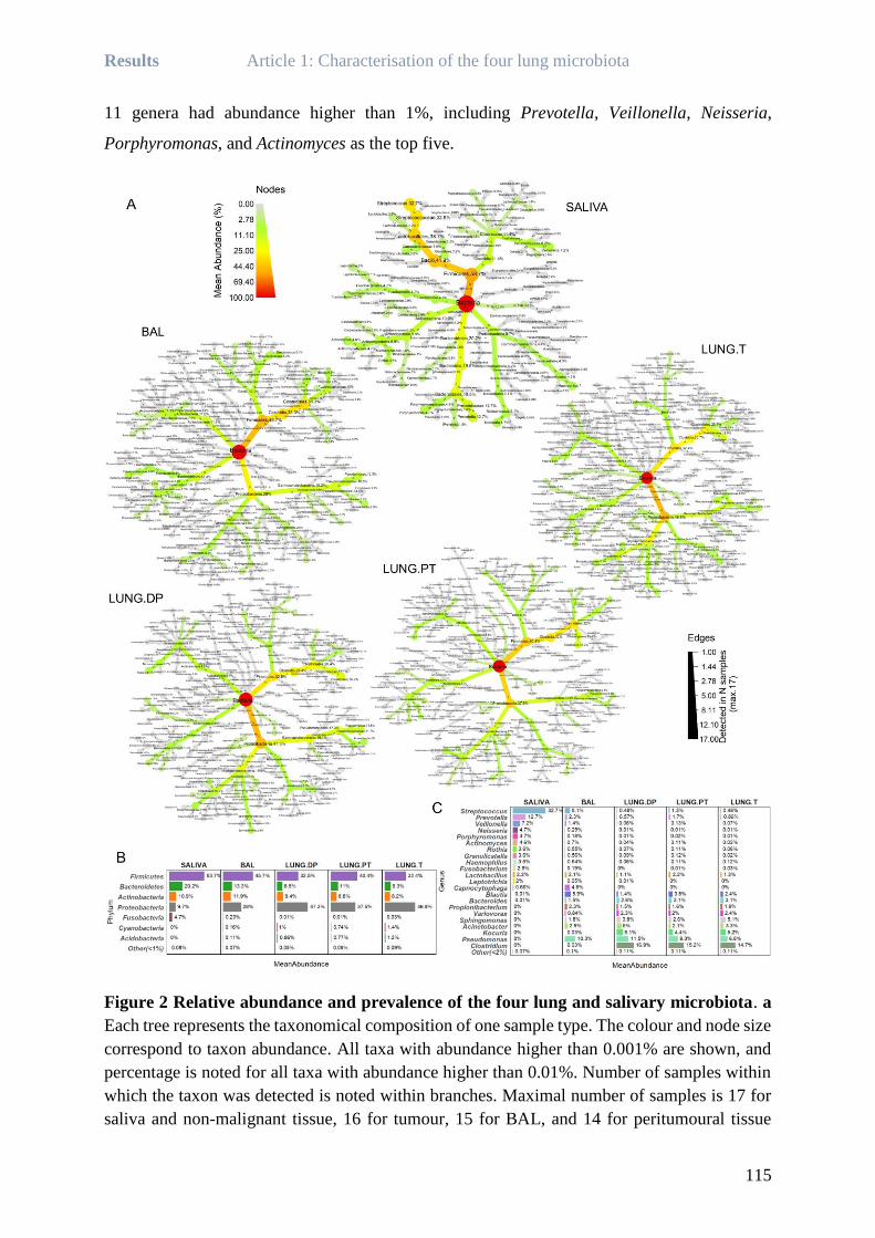
Results Article 1: Characterisation of the four lung microbiota
115
11 genera had abundance higher than 1%, including Prevotella, Veillonella, Neisseria,
Porphyromonas, and Actinomyces as the top five.
Figure 2 Relative abundance and prevalence of the four lung and salivary microbiota. a
Each tree represents the taxonomical composition of one sample type. The colour and node size
correspond to taxon abundance. All taxa with abundance higher than 0.001% are shown, and
percentage is noted for all taxa with abundance higher than 0.01%. Number of samples within
which the taxon was detected is noted within branches. Maximal number of samples is 17 for
saliva and non-malignant tissue, 16 for tumour, 15 for BAL, and 14 for peritumoural tissue

Results Article 1: Characterisation of the four lung microbiota
116
(Table 1). Synthetic presentation of the most abundant taxa was provided on b phylum and c
genus level. BAL - bronchoalveolar lavage fluid, LUNG.DP - non-malignant distal piece,
LUNG.PT - peritumoural tissue, LUNG.T - tumour.
In the lung samples, two dominating phyla were Proteobacteria and Firmicutes, but with
varying abundance ratio depending on the sample. So, phylum Firmicutes was the most
abundant in BAL, in peritumoural tissue two phyla were equally abundant, while
Proteobacteria dominated non-malignant tissue and tumour (Fig. 2A,B). Phyla Bacteroidetes
and Actinobacteria were found in lower abundance in all lung samples (~10% each).
Interestingly, while high abundance of phylum Firmicutes in saliva was almost entirely due to
members of the class Bacilli, in lung samples it is due to class Clostridia, introducing one of
fundamental differences between these two microbiota. Moreover, Clostridia was the most
abundant class in all lung samples except the tumour, where it shared the highest abundance
with the class Alphaproteobacteria. Compared to saliva, the whole phylum Proteobacteria was
more developed in lung samples, containing additional large class of Alphaproteobacteria, but
lacking Epsilonproteobacteria (detected in saliva).
On the genus level, there was no extensive prevalence by one genus as seen in saliva, but rather
a group of representatives with different taxonomic origin (Fig. 2A, C). In the three lung tissues,
Pseudomonas, Clostridium, Kocuria, Acinetobacter and Sphingomonas were the five most
abundant genera, but in BAL, those were Pseudomonas, Blautia, Streptococcus,
Capnocytophaga and Acinetobacter (Fig. 2C). Interestingly, two highly abundant genera in
tissues, Clostridium (~15%) and Kocuria (~5%) were found in very low abundance in BAL.
Inverse was seen for Capnocytophaga, that seemed to be a BAL-related genus (~5%), with a
very low presence in saliva and absence in tissue samples. Furtherly, BAL was the only lung
sample that had higher abundance of so-called supraglottic taxa, as Streptococcus, Prevotella
and Veillonella, compared to other tissue samples. The abundance was also slightly higher in
peritumoural tissue, supporting potentially increased similarity between BAL and peritumoural
tissue seen in beta diversity (Fig. 1A).
Whole phyla and classes significantly different between lung
samples and saliva, but also between BAL and tissues
Figure 3A shows difference in taxa abundance between samples. Taxa with significant
difference are coloured, with a colour scale representing log2 fold change in abundance between
compared sample pair.

Results Article 1: Characterisation of the four lung microbiota
117
Figure 3 Differential abundance between lung and salivary microbiota and their core
composition. a Coloured nodes and branches in each tree represent the taxa with significantly different
abundance between two compared microbiota. The colour intensity is proportional to log2-fold change
in abundance in the favour of the sample with the same colour. Taxa names are shown in the common
legend tree below comparisons. Statistical significance was assessed by zero-inflated general linear
model using Wald’s test (DESeq), with p-value threshold of α ≤ 0.05 after BH correction. b Core
microbiota determined as OTUs present in 100% of each sample for one sample type. The colour
represents relative abundance on transformed log4 scale. c Average value of sum of abundances of the
core OTUs and of other OTUs per each subject in each sample types.
The first row (Fig. 3A) shows taxa with significantly different abundance between saliva and
each of the four lung samples. Compared to saliva, all lung samples had significantly higher
abundance in whole classes of Alphaproteobacteria, Deltaproteobacteria, Cytophagia,

Results Article 1: Characterisation of the four lung microbiota
118
Sphingobacteriia, [Saprospirae], and Acidimicrobiia, but also in whole phyla such as
Cyanobacteria, Acidobacteria, Nitrospirae, Verrucomicrobia and Planctomycetes (latter not
seen for BAL). Similarly, phyla Synergistetes, Spirochaetes, Fusobacteria and TM7 (not in
BAL), classes Epsilonproteobacteria and Erysipelotrichi were significantly more abundant in
saliva than in any of lung samples. On the other hand, multiple descending members of several
higher taxa were not strictly more present in only one sample type. This particularly concerned
members in the classes from the principal phyla Proteobacteria, Firmicutes, Bacteroidetes and
Actinobacteria. Significantly more abundant in saliva samples were orders Neisseriales
(Betaproteobacteria), Pasteurellales (Gammaproteobacteria, contains Haemophilus),
Lactobacillales (Bacilli, contains Streptococcus) and families Veillonellaceae (Clostridia),
Flavobacteriaceae (Flavobacteriia), Prevotellaceae, Porphyromonadaceae (both
Bacteroidia), Actinomycetaceae and Corynebacteriaceae (both Actinobacteria). Conversely,
from the same higher taxa, significantly more abundant in lung samples were orders
Burkholderiales (Betaproteobacteria), Pseudomonadales, Legionellales, Xanthomonadales,
Enterobacteriales (both Gammaproteobacteria), Bacillales, Turicibacteraceae (both Bacilli),
and families Ruminococcaceae, Lachnospiraceae (both Clostridia), Bacteroidaceae
(Bacteroidia), Propionibacteriaceae, Dietziaceae and Bogorellaceae (Actinobacteria).
Second row in Figure 3A shows significant difference in abundance between BAL and the three
lung tissues. With a few pair-reserved exceptions, pattern was highly similar between
comparisons. Also, it was visible that most of the differences indicated significantly increased
abundance in BAL compared to tissues. In BAL, significantly higher abundance was seen in
phylum Fusobacteria, classes Clostridia and Bacilli (genera Streptococcus, Veillonella,
Roseburia, Oribacterium, Phascolarctobacterium, Parvimonas, and Megasphera), orders
Pasteurellales (Haemophilus) and Desulfovibrionales, families Actynomicetaceae,
Flavobacteriaceae (Capnocytophaga), and genera Atopobium, Porphyromonas, Neisseria, and
Rothia. Adversely, the three tissue microbiota had only a few taxa with significantly higher
abundance compared to BAL. Those were the whole phylum Acidobacteria, families
Acetobacteraceae and Clostridiaceae (genus Clostridium), and genus Perlucidibaca.
However, there were differences in taxa that could be observed only between certain BAL-
tissue pairs. Interestingly, the highest number of individual differences was seen in comparison
between BAL and tumour microbiota. Here, only genus Coprococcus was significantly more
abundant in BAL, while genus Kocuria, orders Bdellovibrionales, Myxococcales, Rickettsiales,
and class [Saprospirae] were all significantly more abundant in tumour. Considering non-

Results Article 1: Characterisation of the four lung microbiota
119
malignant tissue microbiota, only family Dietziaceae was more abundant, while orders
Bifidobacteriales and Erysipelotrichales were significantly more abundant in BAL.
Interestingly, tumour and non-malignant tissue had important number of similar differences in
comparison to BAL. While BAL had higher abundance of genera Blautia, Granulicatella,
Ruminococcus, Oscillospira, Prevotella, and Mezorhizobium, more abundant in both tumour
and non-malignant tissue were phylum Cyanobacteria and family [Weeksellaceae]. On the
contrary, peritumoural tissue did not share any of theses differences with BAL as did the other
two tissues. In individual differences, genus Kocuria was the only significantly more abundant
in peritumoural tissue, and inversely, only Staphylococcus was more abundant in BAL.
There were, however, no significant differences between three tissues.
Core OTUs in lung samples mostly members of phylum
Proteobacteria
Core microbiota was determined as OTUs detected in 100% of samples in each group (Fig. 3B).
The highest number of core OTUs was observed in saliva, with the total of 36. Two-fold less
was seen in non-malignant tissue (16), peritumoural tissue (14) and tumour (14), and four-fold
less in BAL (9). 75% of core OTUs in saliva belonged to phylum Firmicutes, with as high as
17/20 OTUs from genus Streptococcus, while additional 15% was from the phylum
Actinobacteria (especially genus Actinomyces). In lung samples, 70% of the core OTUs
belonged to the phylum Proteobacteria (1/3 from class Alphaproteobacteria) and other 30% to
Firmicutes. Core OTUs were mostly shared between different lung sample types, especially
between tissues. OTUs corresponding to genus Variovorax and unclassified members of
families Bradyrhyzobiaceae, Burkholderiaceae, and Bacillaceae were detected in all four lung
microbiota, while OTUs for genera Pseudomonas, Clostridium and Propionibacterium were
only common in all lung tissue microbiota. Even though 30% of lung core OTUs belonged to
Firmicutes, only one OTU corresponded to genus Streptococcus and was a part of BAL core
microbiota. This was also the only core OTU shared between saliva and lung samples, i.e. only
BAL, which were otherwise clearly distinct. Within core microbiota (Fig. 3B), certain OTUs
were uniquely associated with species, such as OTUs for Rothia mucilaginosa,
Propionibacterium acnes, Staphylococcus epidermidis, Prevotella melaninogenica,
Variovorax paradoxus, Veillonella parvula (OTU 518743) and Veillonella dispar (other two
OTUs). In average (Fig. 3C), core microbiota represented 62% of relative abundance in saliva,

Results Article 1: Characterisation of the four lung microbiota
120
against only 22% seen in BAL. In the tissues, the core microbiota represented around half of
the total abundance, ranging from 42% to 50% of abundance.
Microbiota in lower lobes with higher abundance of Firmicutes and
the diversity of peritumoural tissue as the most influenced by
location
Next, we examined whether there is a significant difference between lung microbiota associated
to the tumour lobe relative to its location. Two groups have been considered: upper lobes (UL)
vs. middle/lower lobes (LL). Interestingly, only peritumoural tissue microbiota showed
significantly different beta diversity between two locations in both wUF and uwUF (Fig. 4A,
B). Moreover, difference in beta diversity between UL peritumoural tissue and BAL was not
significant, unlike the one in LL (in UL: wUF p = 0.17, uwUF p = 0.073, vs. in LL: wUF p =
0.004, uwUF p = 0.002). To confirm this observation, for comparison we selected exclusively
distances between each two samples originating from the same patient (Fig. 4D, E), i.e. paired
distances. Indeed, in UL peritumoural microbiota was significantly more similar to both saliva
and BAL, manifested as shorter distances compared to ones in LL (Fig. 4D,E: “distance to
SALIVA”, “distance to BAL”). We next looked at the paired distances between the three tissue
samples (Fig. 4D,E: “distance to LUNG.DP”). The paired distance between non-malignant
tissue and the other two tissues, respectively, was inverse depending on the lobe location. In
UL, there was an increased similarity between non-malignant tissue and tumour, and in LL,
between non-malignant and peritumoural tissue. However, paired distance between
peritumoural tissue and tumour remained unchanged (Fig. 4D,E: “distance to LUNG.PT”),
suggesting a potentially balanced change or exchange of the microbiota maintaining the
distance (and the difference) on the same level. This was inverse to observed by beta diversity
including the totality of distances between tissue samples (not only between paired samples),
showing that peritumoural tissue and tumour are compositionally different in the UL,
significant for both wUF and uwUF (Fig. 4C). In the LL however, the difference was not
significant (p = 0.077, R2 = 0.173). So, based on overall microbiota composition, peritumoural
tissue microbiota was significantly more similar to BAL microbiota in UL, but to tumour
microbiota in LL.

Results Article 1: Characterisation of the four lung microbiota
121
Figure 4 Diversity and predominant taxa in lung samples from upper and lower tumour
lobes. Beta diversity found significantly different between peritumoural tissue from upper and lower
lobes based on both a weighted (wUF) and b unweighted (uwUF) UniFrac distances. c Significantly
different beta diversity based on wUF between peritumoural tissue and tumour in the upper lobe. d
Weighted and e unweighted UF distances between samples coming from the same patient (i.e. paired
distances) compared between upper and lower tumour lobes. The facet name represents the referent
sample (e.g. “distance to BAL”) to which were calculated the distances noted on x-axis (e.g. “from
LUNG.T”). Smaller distance indicates increased similarity. Alpha diversity for four lung samples
between upper and lower tumour lobe assessed by f Faith’s phylogenetic diversity, g number of observed
OTUs, and h Shannon diversity. i Most abundant phyla in each of the microbiota samples if the tumour
is found in upper or lower lobes. Significance of difference in beta diversity was assessed with adonis
function (vegan, 999 permutations). Statistical significance in alpha diversity and paired distances was
assessed with Kruskal-Wallis followed by, where appropriate, Man-Whitney U test with BH correction

Results Article 1: Characterisation of the four lung microbiota
122
for multiple comparison. *: p ≤ 0.05, **: p ≤ 0.01. BAL - bronchoalveolar lavage fluid, LUNG.DP -
non-malignant distal piece, LUNG.PT - peritumoural tissue, LUNG.T - tumour.
Both BAL and peritumoural tissue varied in alpha diversity depending on lobe location, unlike
non-malignant tissue and tumour. BAL in LL had significantly lower phylogenetic diversity
and number of observed OTUs (Fig. 4F,G) compared to UL BAL, and a tendency seen for
Shannon diversity (Fig. 4H). Inversely, LL peritumoural tissue had significantly increased
Shannon diversity (Fig. 4H), with tendency in phylogenetic diversity and number of OTUs.
In LL, there was a marked decrease in abundance of Proteobacteria and increase in phylum
Firmicutes in each of the lung samples (Fig. 4I). Therefore, in LL tumour and non-malignant
tissue had equal abundances in Proteobacteria and Firmicutes (~35%), while BAL and
peritumoural tissue were both dominated by Firmicutes (56% and 45%, respectively).
Actinobacteria and Flavobacteriia show inverse abundance
between BAL and extratumoural tissues depending on the lobe
location, while tumour microbiota remains unchanged
Tumour location in UL or LL significantly influenced the microbial abundance in each of the
analysed sample types, but not in the same manner (Fig. 5). As suggested by diversity results,
microbiota of the peritumoural tissue seemed to be the most influenced by the lobe location.
The changes were limited to members of three major phyla: Firmicutes, Actinobacteria and
Bacteroidetes, and candidate phylum TM7. More precisely, in UL peritumoural tissue more
abundant were class Erysipelotrichi and families Gemellaceae, Streptococcaceae,
Ruminococcaceae, Lachnospiraceae and Veillonellaceae (all phylum Firmicutes), phylum
TM7, classes Bacteroidia (phylum Bacteroidetes) and families Actinomycetaceae and
Bifidobacteriaceae (phylum Actinobacteria). This abundance pattern was very similar to
pattern of initial comparison between BAL and each of lung tissue samples (Fig. 2A). This
could add to the results of beta diversity suggesting that UL peritumoural tissue was more
similar to BAL then it was in LL. On the other hand, LL peritumoural tissue was enriched with
three classes from phylum Bacteroidetes (Flavobacteriia, Sphingobacteriia and Cytophagia),
and with families Clostridiaceae (genus Clostridium) and Micrococcaceae (genus Kocuria)
from phylum Firmicutes and Actinobacteria, respectively.

Results Article 1: Characterisation of the four lung microbiota
123
Figure 5 Differential abundance between upper and lower tumour lobes in salivary and
lung microbiota. Each tree represents taxa with significantly different abundance relative to sample’s
origin (for lung) in the upper or lower tumour lobe. For saliva, the comparison shows the significant
difference in salivary microbiota between patients with tumour either in upper or lower lobe. Coloured
nodes and branches represent the taxa with significantly different abundance and the intensity is
proportional to log2-fold change in abundance in the favour of the lobe noted with the same colour.
Statistical significance was assessed by zero-inflated general linear model using Wald’s test (DESeq),

Results Article 1: Characterisation of the four lung microbiota
124
with p-value threshold of α ≤ 0.05 after BH correction. Bar chart shows the relative abundance of taxa
noted in the taxonomical trees. BAL - bronchoalveolar lavage fluid, LUNG.DP - non-malignant distal
piece, LUNG.PT - peritumoural tissue, LUNG.T - tumour.
Compared to number of affected taxa in peritumoural tissue, both BAL and non-malignant
tissue were less influenced by the lobe location, while tumour seemed to be almost entirely
unaffected (Fig. 5). Moreover, phylum Firmicutes and class Bacteroidia, harbouring the most
differences in peritumoural tissue, were not found significantly different in either non-
malignant tissue or BAL. Instead, in UL non-malignant tissue was enriched with the phylum
Fusobacteria and only a few other lower taxa without involvement of the whole clade (order
Gemellales, families Actinomycetaceae and Pseudomonadaceae). Interestingly, in both non-
malignant and peritumoural tissue phylum Actinobacteria (genus Kocuria) and class
Flavobacteriia (genus Chryseobacterium) were significantly more abundant in LL, while in
BAL these same two taxonomic groups were significantly more abundant in UL, opposite from
the two issues. As seen from the relative abundance (Fig. 4I, Fig. 5), rather than moving from
the common starting point, the abundances were inverse between samples (e.g. Actinobacteria
in UL for BAL vs tissues was 19.06 vs ~5%, and in LL was 4% vs ~13%, respectively). Further,
in UL BAL, genus Neisseria and family Sinobacteraceae (both Proteobacteria) were more
abundant along with the mentioned two clades of Actinobacteria and Flavobacteriia. In LL
BAL, more abundant taxa were more dispersed between taxonomic groups and included the
whole phylum Cyanobacteria, several taxa from the phylum Proteobacteria (orders
Rhodospirillales, Myxococcales), and two genera with their orders, Staphylococcocus (phylum
Firmicutes) and Georgenia (phylum Actinobacteria).
Even though importance was given to log2 fold change of abundance, certain taxa held however
high impact on the overall composition of the samples due to their higher relative abundance
(Fig. 5, Additional file: Fig. 1). This was particularly true for three genera, Clostridium, Kocuria
and Pseudomonas. In peritumoural tissue, genus Clostridium (phylum Firmicutes) ranged from
3% in UL vs 38% in LL, in non-malignant tissue genus Pseudomonas (Gammaproteobacteria)
represented 18% in UL vs 2% in LL lobes, and genus Kocuria (phylum Actinobacteria) ranged
from 0.5% and 0.2% in UL to 12% and 15% in LL in peritumoural and non-malignant tissue,
respectively.
Finally, saliva samples, as the extrapulmonary sample with no direct physical connection to the
tumour location as lung microbiota, also showed significantly different abundance profile
relative to tumour lobe location. If tumours were found in LL, saliva was significantly enriched

Results Article 1: Characterisation of the four lung microbiota
125
in class Bacilli and families Enterobacteriaceae, Moraxellaceae (both Gammaproteobacteria)
and Propionibacteriaceae (Actinobacteria). Curiously, no taxa were detected as significantly
more abundant if tumour was found in UL.
Stratification between lobes defines differences between BAL and
tissues and confirms similarity of BAL and peritumoural tissue in
upper lobes
Stratification of samples by lobe location also revealed or defined differences between samples
(Fig. 6). Between saliva and the four lung samples there was little change, since these samples
were initially already very different (Fig. 3). Only in UL, there was no more difference in
phylum Nitrospira, and orders Neisseriales and Haemophilus between BAL and saliva
(difference significant for all other saliva vs lung comparisons in both lobes, including LL
BAL). Also, the individual difference between lobes (Fig. 5, Fig. 4I) resulted in significantly
higher abundance of Actinobacteria in UL BAL, but inversely, significantly lower abundance
of Actinobacteria in LL BAL compared to saliva (Fig. 6). Comparing saliva and the three lung
tissues, only taxa Spirochaetes, Sphingobacteria and TM7 showed changes relative to the lobe
location. Phylum Spirochaetes was significantly more abundant in saliva only when compared
to UL non-malignant and UL peritumoural tissue (otherwise, no significant difference). Class
Sphingobacteria was significantly more abundant in all lung tissues when compared to saliva,
except in UL peritumoural tissue. This was interesting because neither UL nor LL BAL showed
this difference, once again adding to similarity of BAL and peritumoural tissue in UL. Lastly,
TM7 was significantly more abundant in both UL and LL non-malignant tissue compared to
saliva. But in UL, TM7 was also more abundant in tumour, while in LL this was true for
peritumoural tissue (other comparisons no significant difference). Despite these changes and
stratification by location, saliva was still predominantly abundant in Firmicutes, Fusobacteria,
Epsilonproteobacteria, Erysipelotrichi Prevotella and Neisseria clade, while lung samples
dominated in abundance of Proteobacteria, Acidobacteria, Nitrospirae, Verrucomicrobia,
Cyanobacteria, and the rest of Bacteroidetes.

Results Article 1: Characterisation of the four lung microbiota
126
Figure 6 Comparison of abundance between salivary and lung microbiota relative to
tumour lobe location. Two parts of the figure represent comparison in abundance between samples
linked to upper (U) lobes in the upper part of the figure and to lower (L) lobes in the lower part of the
figure. Coloured nodes and branches represent taxa with significantly different abundance between the
two compared samples. The colour intensity is proportional to log2-fold change in abundance in the
favour of the sample with the same colour. Statistical significance was assessed by zero-inflated general
linear model using Wald’s test (DESeq), with p-value threshold of α ≤ 0.05 after BH correction. BAL -
bronchoalveolar lavage fluid, L – lower lobe, LUNG.DP - non-malignant distal piece, LUNG.PT -
peritumoural tissue, LUNG.T – tumour, U – upper lobe.

Results Article 1: Characterisation of the four lung microbiota
127
Unlike the comparison between saliva and lung samples, the aspect of lobe location induced
much more developed and varying differences between BAL and tissues. Following the
individual inverse difference observed in Fig. 5, in UL, Flavobacteriia and Actinobacteria were
significantly more abundant in BAL, while in LL they were more abundant in the tissues. Also,
the two phyla, Nitrospirae and Cyanobacteria (which between UL BAL and saliva showed no
difference) were consequently more abundant in tissues only in UL, while there was no
difference with BAL in LL. Interestingly, differences in phyla TM7 and Acidobacteria came in
pair. The significantly increased abundance of TM7 in BAL was accompanied with increased
abundances of Acidobacteria in the tissues, otherwise no difference was seen in either of them.
In UL, this was seen in tumour, while in LL this was seen for peritumoural and non-malignant
tissue. Observing the trees, it was visible that the highest number of differences was found
between UL tumour and BAL, including large portion of detected taxa within Proteobacteria,
especially Alphaproteobacteria, as more abundant in tumour. Even though other comparisons
included a few members of Proteobacteria, the count was not as important as in UL tumour.
Further, differences between UL BAL and extratumoural tissues were quite similar, except of
the involvement of Firmicutes in peritumoural tissue. While in UL differences were scarce, in
LL the pattern is as the one seen in comparison to other tissues. This is in accordance to Fig. 5,
where significantly higher abundance of Firmicutes was detected in UL peritumoural tissue
compered to LL. This could be, therefore, one of the major factors behind increased similarity
between BAL and peritumoural tissue in UL. Detailed individual differences between BAL and
tissues can be found in Additional file 1: “Additional explanation of differences between lung
samples”.
Finally, several significant differences were noted between tissues (Fig. 6). They are, however,
few numbered. Curiously, in both locations, more difference was found between tumour and
peritumoural tissue, than between tumour and non-malignant tissue (none and one in UL and
LL, respectively). Differences were also found between two extratumoural tissues.
Interestingly, in each location, several detected differences between tissues addressed same
taxa. Therefore, in UL, class Cytophagia, family Exiguobacteraceae and Clostridiaceae were
the least abundant in peritumoural tissue compared to other two tissues, while in LL family
Microbacteriaceae had significantly lowest abundance in tumour. In overall, differences were
dispersed within four most abundant phyla and considered mostly endpoint taxa. Also, three
tissues were more similar in LL than in UL. Detailed individual differences between the tissues

Results Article 1: Characterisation of the four lung microbiota
128
can be found in Additional file 1: “Additional explanation of differences between lung
samples”.
4 Discussion
Following the last advancement on the interaction between gut microbiota, immune system and
the tumour environment [8–11], there has been a growing interest in studying this concept in
other physiological environments involving extraintestinal tumours. However, before exploring
the effect of the gut microbiota, there has been an increasing necessity to investigate the effect
of the local microbiota on the tumour as well [39]. Despite the emerging number of studies on
the lung microbiota in different malignancies [14,40–43], its involvement in lung cancer is in
its promising beginnings [23,27,29,30,44]. However, for the moment there is still no study
considering the ground difference between different lung samples and their microbiota, while
it is suggested that those could harbour microbiota with varying characteristics [18], and
therefore, have diverging interactions with local immunity and tumour.
This study is, to our knowledge, the first to characterise the lung microbiota originating from
four different lung samples (BAL, non-malignant tissue, peritumoural tissue and tumour),
accompanied with the characterisation of salivary microbiota in NSCLC patients. We
hypothesised that samples’ nature, “architecture”, physiological functions and environment,
will influence characteristics of associated lung microbiota. Therefore, BAL should represent
“planktonic” bacterial population found within the bronchial lumen or associated to biofilms or
mucus [45], sampled along due the hydrodynamic force of the instilled liquid. Non-malignant
tissue from the same lobe but taken on the opposite side from the tumour should represent a
sample with a normal lung architecture, with well-defined small alveolar spaces and single-cell
epithelial layer. In majority, it should harbour the “normal” biofilm, mucus and cell-associated
lung microbiota. On the contrary, tumour represents a tissue with disrupted architecture,
varying in form and obstruction degree relative to its type and grade [2]. Tissue modelling could
also involve overproduction of the mucus as seen in certain subtypes of adenocarcinoma [46],
but also different reaction of the immune system [47]. Finally, peritumoural tissue represents a
non-malignant tissue in the direct contact with the tumour, separated as based on different
histological properties. In literature it is addressed as tumour microenvironment and harbours
different roles in stimulation or suppression of tumour metabolism [48]. Therefore, we
presumed that its characteristics would be different both from the distal non-malignant tissue
and the tumour. Except the number of different lung samples, the particularity of this study is

Results Article 1: Characterisation of the four lung microbiota
129
also in the way of obtaining BAL. It was obtained directly on the excised lobe containing
tumour without using the bronchoscope, to minimise contamination risk of upper airways and
increase the precision in characterisation of “true” BAL microbiota in the tumour proximity.
We reported that the four lung samples significantly clustered versus oral microbiota based on
beta diversity, a confirmation of a previous result in healthy subjects that lung microbiota is
distinct from other communities [29]. Salivary microbiota had several lower alpha diversity
metrics compared to lung microbiota, as well as high dominance of genus Streptococcus and
overall phylum Firmicutes. Along with its homogenous core microbiota (again prevalence of
Streptococcus OTUs) that represented almost ¾ of total relative abundance per patient, this
indicated its inter-subject stability and lower complexity compared to the one of the lung
samples. In addition, the significant difference in abundance including the majority of the taxa
detected in samples clearly placed the four lung samples versus oral microbiota. We identified
typically oral taxonomic groups more abundant in saliva in all comparisons, such as
Fusobacteria, Spirochaetes, Synergistetes, Erysipelotrichi, Epsilonproteobacteria, Bacilli and
Neisseriales, most of them in concordance with previous literature [49,50]. On the other hand,
phyla Acidobacteria, Cyanobacteria, Nitrospira, Verrucomicrobia and Planctomycetes were
detected as strictly lung-associated (detected in each lung sample) with no representatives in
saliva.
Next, we showed that BAL microbiota, even though undoubtedly belonging to lung microbiota
cluster, had significantly different features from tissues. First, multidimensional representation
of the beta diversity placed BAL samples on the side of the lung cluster towards salivary
microbiota. Even though presence of more abundant salivary taxa was very limited in both
tissue samples and BAL, Streptococcus, Prevotella and Veillonella (the three most abundant
genera in saliva) were found elevated in BAL compared to tissue samples. It was previously
suggested that these and other typically oral bacteria are found in low abundance in healthy
lungs, due to their constant elimination [51]. Since BAL represents microbiota of the bronchial
lumen, which undergoes constant influx of upper airways particles by respiration and is also
the first “space” influenced by microaspiration [52], those could explain increased presence of
these supraglottic taxa in BAL compared to the lung tissues. The same reasoning could explain
the pattern of significant differences between samples. Indeed, taxonomic groups significantly
more abundant in saliva when compared to lung microbiota highly corresponded to groups
significantly more abundant in BAL when compared to lung tissues. Second, lung samples had
Proteobacteria and Firmicutes as the two most abundant phyla, however with different ratios.

Results Article 1: Characterisation of the four lung microbiota
130
While BAL was dominated by Firmicutes, almost twice as abundant as Proteobacteria, lung
tissues were dominated by Proteobacteria (as previously reported [29]), or at best, had equal
abundance of the two (peritumoural tissue). Here it is however important to note that the high
abundance of phylum Firmicutes in lung samples was due to members of the class Clostridia,
unlike in saliva, where it was due to highly abundant class Bacilli. This emphasises one of the
essential differences between oral and lung microbiota often omitted by selective presentation
of only phylum or genus level. Third feature was that BAL did not differentiate from lung
tissues only by difference in taxa abundance, but also by the presence of BAL-specific bacteria,
such as genus Capnocytophaga, or absence of tissue specific-bacteria, such as genus Kocuria
or Clostridium. The two latter genera were, however, detected in BAL, but with relative
abundance 175-fold and 520-fold lower (~0.03%), respectively, than in the tissues. This
detection is possibly due to their presence in host’s cells during the DNA extraction, since
cellular BAL was used for BAL analysis [45]. Lastly, no differences in alpha diversity metrics
were detected between four lung samples, suggesting two different but equally “rich” lung
microbiota populations. All this supports the hypothesis that BAL indeed represents a unique
lung microbiota in lung cancer and that concerns of diverging results due to different samples
have been justified [18].
Conversely to our hypothesis, lung tissues “as-they-are” did not show significant differences in
abundance or diversity. Although increased diversity of non-malignant tissue versus tumour
has been previously suggested [29], we did not find any difference. This could be due to the
fact that our study group was more balanced in patients with less and more advanced tumour
stages. In referenced study, the majority of subjects had tumours in stages I and II, manifesting
decreased diversity compared to higher stages according to authors.
We also reported that the existence of the core OTUs and that these were mostly shared between
lung tissues and partially with BAL, consisting in the majority of the members of
Proteobacteria. Interestingly, one of the core OTUs was uniquely associated to species
Variovorax paradoxus, ambiguous aerobic organism found in many niches, also in oral
microbiome, and known for its potent biofilm creation [53,54]. It was detected as the core OTU
in all four lung samples, while conversely, it was not found in saliva (where it was originally
detected). This could suggest its potential role in biofilm creation in lower airways and would
be an interest topic for future verification and investigation of potential role in pathogenesis of
lung cancer [55].

Results Article 1: Characterisation of the four lung microbiota
131
Even though still under debate, several studies connected lower lobe tumours with worse
prognostics [5,56,57]. Therefore, we examined the characteristics of the lung, but also salivary
microbiota, if the tumour was found in upper or lower lobes. Indeed, we noted several changes
in alpha and beta diversity but they were focused on changes in BAL and peritumoural tissue
(with lower and higher alpha diversity, respectively, in LL compared to UL). We reported that
in all four lung samples, lower lobes had decreased abundance of phylum Proteobacteria into
favour of Firmicutes. Increased abundance of phylum Firmicutes was previously seen in BAL
of LC patients compared to patients with benign-mass lesions [44], but also in patients with
COPD [18]. In our study, 62% of patients in the LL were diagnosed with Stage III tumours,
against 30% in the UL. Even though this stays a small study, this finding supports that “shift”
from Proteobacteria to Firmicutes might have a role in lung cancer progression.
We showed that location significantly influenced the abundance of two clades, Actinobacteria
and Flavobacteriia, in the inverse manner between BAL and extratumoural tissues. While both
are more abundant in UL BAL, their abundance in UL extratumoural tissues was 2-10 fold
lower, with exact inverse situation in LL. Both genus Chryseobacterium sp. (Flavobacteriia)
and Kocuria rhizophila (Actinobacteria) detected as more abundant in LL tissues have been
previously reported as uncommon human pathogens [58,59]. However, their increased presence
selectively in LL tissues and their important overall abundance (~4% and 12%, respectively)
suggest that there might be an important communication between different lung environments
depending on local conditions or malignancy status, influencing preferential bacterial growth
in one type of considered environment.
Interestingly, we found that salivary microbiota was also susceptible to the tumour lobe
location. All differences considered taxa elevated in case of LL tumours and included families
previously associated with bacterial exacerbations and infections (Moraxellaceae, members of
Bacilli as Streptococcus, Staphylococcus, etc.) [60] or pulmonary complications
(Propionibacteriaceae, Enterococcaceae)[61–63]. However, these taxa were not found
elevated in LL lung microbiota. This, however, does not exclude their implication in malignant
changes or being its consequence, and will be interesting topic for further consideration.
Finally, we found that peritumoural tissue showed higher similarity to BAL in UL both in beta
diversity and in abundance, while in LL it shared characteristics with other tissue samples.
Moreover, we found that alpha diversity of peritumoural tissue increased in LL to the level of
other tissues, while the ones of BAL decreased. Peritumoural tissue is the tissue in the direct
contact and interaction with the tumour. In the history of cancer research, the role of

Results Article 1: Characterisation of the four lung microbiota
132
extracellular matrix (ECM) surrounding tumour has been extensively studied [64]. Tumour
cells are found to be able to directly influence the rearranging of connective units (such as
collagen) and degradation of ECM to create tumour-permissive environment and enabling
metastatic progression (Altinay 2016) [65,66]. In our case, it is possible that the observed
changes in peritumoural tissue composition reflect certain remodelling of ECM, which could
therefore be either more or less permissive for microbial attachment.
Curiously, of all samples, tumour was almost completely uninfluenced by location. Rather than
from the outside, it is possible that intratumoural conditions, such as oxygen availability,
density, necrosis and other factors [67,68] independent of the external conditions are more
likely to be the influencing ones. This is, however, a matter for further research.
The major strongpoint of this study is the analysis of the microbiota from four different lung
samples covering the major physiologically different environments in NSCLC patient that has
not been characterised previously, with the addition of saliva as the sample of oral microbiota.
The representation using taxonomical trees also gives better insight into sample’s composition,
especially important in characterisation of this type of still scarcely defined microbiota.
Moreover, this study reports higher numbers of detected OTUs as well as diversity indexes than
previously suggested for saliva, non-malignant tissue, tumour and BAL [26,29,30]. The reason
could be due to the higher quality of obtained samples (only 6 from 85 samples lower than 1000
reads) and direct sampling of the BAL in the excised lobe. Also, the observed study group and
stratification by tumour lobe location were well balanced in various clinical and demographic
factors, minimising result’s bias.
The major drawback of this study is a low number of subjects, due to strict inclusion criteria
for the overall study. The limiting factor was also the decision to obtain BAL directly from the
excised lobe to improve the precision in characterisation, but disabling the possibility of
sampling the complementary non-tumour lobe or to perform similar in controls due to
difference in technique.
5 Conclusion
To our knowledge, this is the first trial that studied oral and lung microbiota from both BAL
and three different tissue samples in 18 NSCLC patients. We confirmed that oral and lung
microbiota are significantly different both in diversity and in taxonomy. However, we showed
that BAL indeed represents a unique microbiota compared to three different tissue microbiota
Results Article 1: Characterisation of the four lung microbiota
133
(from non-malignant, peritumoural and tumour tissue). We found that location of the tumour in
upper or lower lobes influenced both oral, BAL and extratumoural microbiota with detection
of Firmicutes as dominating phylum, but surprisingly, not the one of the tumour. Moreover,
few differences were found between tissues, suggesting that these are not conditioned by lobe
location and turning attention to other factors for future consideration, such as tumour type,
aggressiveness or metastatic changes. Finally, the most intensive changes in microbiota relative
to location were seen in peritumoural tissue, possibly reflecting changes in tumoural ECM. Our
findings are the first to give essential characteristics and differences within lung microbiota of
NSCLC and their susceptibility to tumour lobe location, proposing several possible
implications of microbiota in pathology of lung cancer and suggesting potential research
directions for better understanding of the lung microbiota-cancer interaction.
6 Declarations
Ethics approval and consent to participate
The study (and its amendment in June 2018) was approved by the Committee for the Protection
of Persons (CPP) Sud-Est VI, Clermont-Ferrand, France, and The French National Agency for
Medicines and Health Products Safety (ANSM) (study ref. 2016-A01640-51). The study is
registered with the Clinical Trials under ID: NCT03068663. Written informed consent was
obtained from all patients before enrolment in the study.
Consent for publication
Not applicable.
Availability of data and material
According to the practice of the sponsoring institution (Centre Jean Perrin), all samples will be
preserved for 15 years. The datasets generated and/or analysed during the current study are
confidential but are available from the corresponding author on reasonable request and after the
data sharing process is brought into compliance with GDPR (Regulation (EU) 2016/679 of the
European Parliament and of the Council of 27 April 2016).
6.3.1 Data sharing
This statement is intended to clarify Centre Jean Perrin’s position on data sharing with regards
to applicable regulatory frameworks.

Results Article 1: Characterisation of the four lung microbiota
134
6.3.1.1 European regulatory framework
De-identified study data are considered as personal data within the meaning of Regulation (EU)
2016/679 of the European Parliament and of the Council of 27 April 2016, known as « GDPR ».
As a result, their transfer places further processors in the scope of GDPR (Art. 2 and 3) and is
notably subject to
precise information of study subjects (Art. 14),
prior identification of further processors and signature of a transfer agreement (Chapter
V),
data protection impact assessment (Art. 35).
6.3.1.2 French regulatory framework
In addition to GDPR, the transfer of this data by Centre Jean Perrin is a processing in itself that
is subject to Chapter IX and XII of the updated Law n°78-17 of 6 January 1978. Within the
framework of this law, the data processing is permitted by Centre Jean Perrin’s conformity to
the MR-001 Code of conduct (Délibération n° 2018-153 du 3 mai 2018 portant homologation
d'une méthodologie de référence relative aux traitements de données à caractère personnel mis
en œuvre dans le cadre des recherches dans le domaine de la santé avec recueil du consentement
de la personne concernée).
In its last update from May 2018, MR-001 was updated to allow transfer to reviewers within
certain conditions (Article 2.4).
These conditions can be summarised as follow:
The access to study data by an independent mandated reviewer can only be granted through
an interface chosen by the data processor (i.e. Centre Jean Perrin, in this context) for the sole
purpose of re-analysis or for any other purpose supported by a strong rational and a project
synopsis describing why access to the this study’s data (study acronym: MICA) is necessary.
The data processor shall ensure:
That the chosen sharing platform cannot allow extraction of the data;
To grant specific personal and differentiated authorizations to access the data;
That the users be reliably identified
That state of the art cryptography and security measures are used;
That an audit trail of accesses is used;

Results Article 1: Characterisation of the four lung microbiota
135
When data is transferred outside of the EU, that applicable transfer contracts are
established;
That study subjects are informed on the data recipients;
That the data is used for the sole purpose of reproducing published results;
That the data is cleaned from any directly identifying data and that the principle of data
minimization is applied (i.e. limiting the transfer to data used in the publication).
6.3.1.3 Study Data sharing
As a result, for this trial (acronym: MICA), Centre Jean Perrin can share individual participant
data that underlie the results reported in the article under the following conditions:
Limitation to the data processing to the purpose of independent review of the
published results;
Prior minimization of the data (i.e. restriction to data strictly adequate, relevant
and limited to the purpose of the independent review of results);
Prior identification of the reviewer and authorization from Centre Jean Perrin for a
personal access;
Following the signature of a transfer agreement with Centre Jean Perrin, in
conformity with the European Commission’s Standard Contractual Clauses;
Access to study data will be assessed, upon written detailed request sent to the project manager
Emilie THIVAT: [email protected], from 6 months until 5 years following
article’s publication.
Competing interests
The authors declare that the research was conducted in the absence of any commercial or
financial relationships that could be construed as a potential conflict of interest.
Funding
This work was financially supported by the Auvergne Region (France), European Regional
Development Fund (FEDER) and company Greentech (Saint-Beauzire, France). The study
sponsor was Jean Perrin Centre, Clermont-Ferrand, France.
Authors' contributions
ABD, EF, JYB, MF and MPV were responsible for the study design. ABD, ED, EF, MF and
RB planned the experiments. RB, ED and MF performed the experiments. MF and ABD aided

Results Article 1: Characterisation of the four lung microbiota
136
in interpreting the results. RB analysed the results with the statistical approach devised from
IM. RB designed the figures and wrote the manuscript. All authors gave their critical feedback
for the final manuscript version.
Acknowledgements
The authors thank all the patients who accepted to participate in this protocol. They gratefully
acknowledge clinical research associates, Margaux Marcoux and Emilie Thivat, for managing
administrative aspects of the study. The authors gratefully acknowledge the help of Laureen
Crouzet (UMR0454 MEDIS, INRA-UCA) in optimisation of the DNA extraction protocol for
lung microbiota, and Floriane Serre (UMR0454 MEDIS, INRA-UCA) for technical help in
DNA isolation.
7 References
1. International Agency for Research on Cancer (IARC). World Cancer Report 2014. Stewart BW, Wild CP,
editors. Lyon: International Agency for Research on Cancer; 2014.
2. Goldstraw P, Chansky K, Crowley J, Rami-Porta R, Asamura H, Eberhardt WEE, et al. The IASLC lung cancer
staging project: Proposals for revision of the TNM stage groupings in the forthcoming (eighth) edition of the TNM
Classification for lung cancer. J Thorac Oncol. 2016;11:39–51.
3. Asamura H, Goya T, Koshiishi Y, Sohara Y, Eguchi K, Mori M, et al. A Japanese Lung Cancer Registry Study:
Prognosis of 13,010 Resected Lung Cancers. J Thorac Oncol. 2008;3:46–52.
4. Inoue M, Sawabata N, Takeda S, Ohta M, Ohno Y, Maeda H. Results of surgical intervention for p-stage IIIA
(N2) non–small cell lung cancer: Acceptable prognosis predicted by complete resection in patients with single N2
disease with primary tumor in the upper lobe. J Thorac Cardiovasc Surg. 2004;127:1100–6.
5. Hayakawa K, Mitsuhashi N, Saito Y, Furuta M, Nakayama Y, Katano S, et al. Impact of tumor extent and
location on treatment outcome in patients with stage III non-small cell lung cancer treated with radiation therapy.
Jpn J Clin Oncol. 1996;26:221–8.
6. Ou S-HHI, Zell JA, Ziogas A, Anton-Culver H. Prognostic factors for survival of stage I nonsmall cell lung
cancer patients: A population-based analysis of 19,702 stage I patients in the California Cancer Registry from 1989
to 2003. Cancer. 2007;110:1532–41.
7. Goldszmid RS, Dzutsev A, Viaud S, Zitvogel L, Restifo NP, Trinchieri G. Microbiota modulation of myeloid
cells in cancer therapy. Cancer Immunol Res. 2015;3:103–9.
8. Sivan A, Corrales L, Hubert N, Williams JB, Aquino-Michaels K, Earley ZM, et al. Commensal Bifidobacterium
promotes antitumor immunity and facilitates anti-PD-L1 efficacy. Science (80- ). 2015;350:1084–9.
9. Routy B, Le Chatelier E, Derosa L, Duong CPM, Alou MT, Daillère R, et al. Gut microbiome influences efficacy
of PD-1-based immunotherapy against epithelial tumors. Science (80- ). 2018;359:91–7.
10. Daillère R, Vétizou M, Waldschmitt N, Yamazaki T, Isnard C, Poirier-Colame V, et al. Enterococcus hirae
and Barnesiella intestinihominis Facilitate Cyclophosphamide-Induced Therapeutic Immunomodulatory Effects.
Cell. 2016;45:931–43.
11. Viaud S, Saccheri F, Mignot G, Yamazaki T, Daillère R, Hannani D, et al. The intestinal microbiota modulates
the anticancer immune effects of cyclophosphamide. Science. 2013;342:971–6.
12. Dickson RP, Erb-Downward JR, Huffnagle GB. Towards an ecology of the lung: New conceptual models of
pulmonary microbiology and pneumonia pathogenesis. Lancet Respir. Med. 2014. p. 238–46.

Results Article 1: Characterisation of the four lung microbiota
137
13. Marsland BJ, Gollwitzer ES. Host–microorganism interactions in lung diseases. Nat Rev Immunol.
2014;14:827–35.
14. Bernasconi E, Pattaroni C, Koutsokera A, Pison C, Kessler R, Benden C, et al. Airway microbiota determines
innate cell inflammatory or tissue remodeling profiles in lung transplantation. Am J Respir Crit Care Med.
American Thoracic Society; 2016;194:1252–63.
15. Blainey PC, Milla CE, Cornfield DN, Quake SR. Quantitative analysis of the human airway microbial ecology
reveals a pervasive signature for cystic fibrosis. Sci Transl Med. 2012;4:153ra130.
16. Dickson RP, Erb-Downward JR, Freeman CM, McCloskey L, Beck JM, Huffnagle GB, et al. Spatial Variation
in the Healthy Human Lung Microbiome and the Adapted Island Model of Lung Biogeography. Ann Am Thorac
Soc. 2015;12:821–30.
17. Dickson RP, Martinez FJ, Huffnagle GB. The Role of the Microbiome in Exacerbations of Chronic Lung
Diseases. Lancet August. 2014;231016:691–702.
18. Sze MA, Dimitriu PA, Hayashi S, Elliott WM, McDonough JE, Gosselink J V., et al. The Lung Tissue
Microbiome in Chronic Obstructive Pulmonary Disease. Am J Respir Crit Care Med. 2012;185:1073–80.
19. Hilty M, Burke C, Pedro H, Cardenas P, Bush A, Bossley C, et al. Disordered Microbial Communities in
Asthmatic Airways. PLoS One. 2010;5:e8578.
20. Jin C, Lagoudas GK, Zhao C, Bullman S, Bhutkar A, Hu B, et al. Commensal Microbiota Promote Lung
Cancer Development via γδ T Cells. Cell. 2019;176:998–1013.
21. Cheng M, Qian L, Shen GGG, Bian G, Xu T, Xu W, et al. Microbiota modulate tumoral immune surveillance
in lung through a γδT17 immune cell-dependent mechanism. Cancer Res. American Association for Cancer
Research Inc.; 2014;74:4030–41.
22. Boursi B, Mamtani R, Haynes K, Yang Y-X. Recurrent antibiotic exposure may promote cancer formation –
Another step in understanding the role of the human microbiota? Eur J Cancer. Elsevier Ltd; 2015;51:2655–64.
23. Segal LN, Clemente JC, Tsay J-CJ, Koralov SB, Keller BC, Wu BG, et al. Enrichment of the lung microbiome
with oral taxa is associated with lung inflammation of a Th17 phenotype. Nat Microbiol. 2016;1:16031.
24. Segal LN, Clemente JC, Tsay J-CJ, Koralov SB, Keller BC, Wu BG, et al. Enrichment of lung microbiome
with supraglottic taxa is associated with increased pulmonary inflammation. Microbiome. 2013;1:19.
25. Tsay JCJ, Wu BG, Badri MH, Clemente JC, Shen N, Meyn P, et al. Airway microbiota is associated with
upregulation of the PI3K pathway in lung cancer. Am J Respir Crit Care Med. 2018;198:1188–98.
26. Wang K, Huang Y, Zhang Z, Liao J, Ding Y, Fang X, et al. A Preliminary Study of Microbiota Diversity in
Saliva and Bronchoalveolar Lavage Fluid from Patients with Primary Bronchogenic Carcinoma. Med Sci Monit.
2019;25:2819–34.
27. Liu HX, Tao LL, Zhang J, Zhu YG, Zheng Y, Liu D, et al. Difference of lower airway microbiome in bilateral
protected specimen brush between lung cancer patients with unilateral lobar masses and control subjects. Int J
Cancer. 2018;142:769–78.
28. Yan X, Yang M, Liu J, Gao R, Hu J, Li J, et al. Discovery and validation of potential bacterial biomarkers for
lung cancer. Am J Cancer Res. 2015;5:3111–22.
29. Yu G, Gail MH, Consonni D, Carugno M, Humphrys M, Pesatori AC, et al. Characterizing human lung tissue
microbiota and its relationship to epidemiological and clinical features. Genome Biol. 2016;17:163.
30. Peters BA, Hayes RB, Goparaju C, Reid C, Pass HI, Ahn J. The microbiome in lung cancer tissue and
recurrence-free survival. Cancer Epidemiol Biomarkers Prev. 2019;28:731–40.
31. Bingula R, Filaire M, Radosevic-Robin N, Berthon J-Y, Bernalier-Donadille A, Vasson M-P, et al.
Characterisation of gut, lung, and upper airways microbiota in patients with non-small cell lung carcinoma.
Medicine (Baltimore). 2018;97:e13676.
32. RStudio Team. RStudio: Integrated Development Environment for R. Boston, MA; 2016.
33. McMurdie PJ, Holmes S. Phyloseq: An R Package for Reproducible Interactive Analysis and Graphics of

Results Article 1: Characterisation of the four lung microbiota
138
Microbiome Census Data. Watson M, editor. PLoS One. Public Library of Science; 2013;8:e61217.
34. Oksanen J, Blanchet FG, Friendly M, Kindt R, Legendre P, Mcglinn D, et al. vegan: Community Ecology
Package. 2019.
35. Lahti L, Shetty S, Blake T, Salojarvi J. microbiome R package. 2017.
36. Wickham H. ggplot2: Elegant Graphics for Data Analysis. Springer-Verlag New York; 2016.
37. Love MI, Huber W, Anders S. Moderated estimation of fold change and dispersion for RNA-seq data with
DESeq2. Genome Biol. 2014;15:550.
38. Foster ZSL, Sharpton TJ, Grünwald NJ. Metacoder: An R package for visualization and manipulation of
community taxonomic diversity data. PLoS Comput Biol. 2017;13.
39. Dickson RP, Cox MJ. Gut microbiota and protection from pneumococcal pneumonia. Gut. 2017;66:384.
40. Sze M a, Hogg JC, Sin DD. Bacterial microbiome of lungs in COPD. Int J Chron Obstruct Pulmon Dis.
2014;9:229–38.
41. Vital M, Harkema JR, Rizzo M, Tiedje J, Brandenberger C. Alterations of the Murine Gut Microbiome with
Age and Allergic Airway Disease. J Immunol Res. Hindawi Publishing Corporation; 2015;2015:1–8.
42. Dickson RP, Erb-Downward JR, Freeman CM, Walker N, Scales BS, Beck JM, et al. Changes in the lung
microbiome following lung transplantation include the emergence of two distinct Pseudomonas species with
distinct clinical associations. PLoS One. Public Library of Science; 2014;9:e97214.
43. Fodor AA, Klem ER, Gilpin DF, Elborn JS, Boucher RC, Tunney MM, et al. The Adult Cystic Fibrosis Airway
Microbiota Is Stable over Time and Infection Type, and Highly Resilient to Antibiotic Treatment of Exacerbations.
PLoS One. 2012;7.
44. Lee SH, Sung JY, Yong D, Chun J, Kim SKSSY, Song JH, et al. Characterization of microbiome in
bronchoalveolar lavage fluid of patients with lung cancer comparing with benign mass like lesions. Lung Cancer.
2016;102:89–95.
45. Dickson RP, Erb-downward JR, Prescott HC, Martinez FJ, Curtis JL, Lama VN, et al. Cell-associated bacteria
in the human lung microbiome. Microbiome. 2014;2:1–10.
46. Snoeckx A, Dendooven A, Carp L, Desbuquoit D, Spinhoven MJ, Lauwers P, et al. Wolf in Sheep’s Clothing:
Primary Lung Cancer Mimicking Benign Entities. Lung Cancer. Elsevier; 2017. p. 109–17.
47. Busch SE, Hanke ML, Kargl J, Metz HE, MacPherson D, Houghton AM. Lung Cancer Subtypes Generate
Unique Immune Responses. J Immunol. American Association of Immunologists; 2016;197:4493–503.
48. Valkenburg KC, De Groot AE, Pienta KJ. Targeting the tumour stroma to improve cancer therapy. Nat Rev
Clin Oncol. 2018;15:366–81.
49. Charlson ES, Bittinger K, Chen J, Diamond JM, Li H, Collman RG, et al. Assessing Bacterial Populations in
the Lung by Replicate Analysis of Samples from the Upper and Lower Respiratory Tracts. PLoS One. 2012;7.
50. Grassl N, Kulak NA, Pichler G, Geyer PE, Jung J, Schubert S, et al. Ultra-deep and quantitative saliva proteome
reveals dynamics of the oral microbiome. Genome Med. Genome Medicine; 2016;8:44.
51. Bassis CM, Erb-downward JR, Dickson RP, Freeman CM, Schmidt TM, Young VB, et al. Analysis of the
Upper Respiratory Tract Microbiotas as the Source of the Lung and Gastric Microbiotas in Healthy Individuals.
MBio. 2015;6:1–10.
52. Gleeson K, Eggli DF, Maxwell SL. Quantitative aspiration during sleep in normal subjects. Chest.
1997;111:1266–72.
53. Anesti V, McDonald IR, Ramaswamy M, Wade WG, Kelly DP, Wood AP. Isolation and molecular detection
of methylotrophic bacteria occurring in the human mouth. Environ Microbiol. 2005;7:1227–38.
54. Jamieson WD, Pehl MJ, Gregory GA, Orwin PM. Coordinated surface activities in Variovorax paradoxus EPS.
BMC Microbiol. 2009;9.
55. Koo H, Allan RN, Howlin RP, Stoodley P, Hall-Stoodley L. Targeting microbial biofilms: current and

Results Article 1: Characterisation of the four lung microbiota
139
prospective therapeutic strategies. Nat Rev Microbiol. NIH Public Access; 2017;15:740–55.
56. Riquet M, Pricopi C, Arame A, Barthes FLP. From anatomy to lung cancer: Questioning lobe-specific
mediastinal lymphadenectomy reliability. J Thorac Dis. 2016;8:2387–90.
57. Kotoulas CS, Foroulis CN, Kostikas K, Konstantinou M, Kalkandi P, Dimadi M, et al. Involvement of
lymphatic metastatic spread in non-small cell lung cancer accordingly to the primary cancer location. Lung Cancer.
Elsevier; 2004;44:183–91.
58. Mukerji R, Kakarala R, Smith SJ, Kusz HG. Chryseobacterium indologenes: An emerging infection in the
USA. BMJ Case Rep. 2016;2016.
59. Purty S, Saranathan R, Prashanth K, Narayanan K, Asir J, Sheela Devi C, et al. The expanding spectrum of
human infections caused by Kocuria species: a case report and literature review. Emerg Microbes Infect. 2013;
60. Pragman AA, Kim HB, Reilly CS, Wendt C, Isaacson RE. The Lung Microbiome in Moderate and Severe
Chronic Obstructive Pulmonary Disease. PLoS One. 2012;7.
61. Vincenzo Savini, Giovanni Gherardi, Daniela Astolfi, Ennio Polilli, Giordano Dicuonzo, Claudio D’Amario,
et al. Insights into Airway Infections by Enterococci: A Review. Recent Pat Antiinfect Drug Discov. 2012;7:36–
44.
62. Werner JL, Escolero SG, Hewlett JT, Mak TN, Williams BP, Eishi Y, et al. Induction of Pulmonary Granuloma
Formation by Propionibacterium acnes Is Regulated by MyD88 and Nox2. Am J Respir Cell Mol Biol. American
Thoracic Society; 2017;56:121–30.
63. Henao-Martínez AF, González-Fontal GR, Castillo-Mancilla JR, Yang I V. Enterobacteriaceae bacteremias
among cancer patients: an observational cohort study. Int J Infect Dis. NIH Public Access; 2013;17:e374-8.
64. Bhat R, Bissell MJ. Of plasticity and specificity: Dialectics of the microenvironment and macroenvironment
and the organ phenotype. Wiley Interdiscip. Rev. Dev. Biol. 2014. p. 147–63.
65. Quail DF, Joyce JA. Microenvironmental regulation of tumor progression and metastasis. Nat Med.
2013;19:1423–37.
66. Bissell MJ, Hines WC. Why don’t we get more cancer? A proposed role of the microenvironment in restraining
cancer progression. Nat. Med. 2011. p. 320–9.
67. Wouters BG, Koritzinsky M, Chiu RK, Theys J, Buijsen J, Lambin P. Modulation of cell death in the tumor
microenvironment. Semin Radiat Oncol. 2003;13:31–41.
68. Fadaka A, Ajiboye B, Ojo O, Adewale O, Olayide I, Emuowhochere R. Biology of glucose metabolization in
cancer cells. J Oncol Sci. 2017;3:45–51.
8 Additional files
Additional file 1.pdf – contains:
Figure 1 Most abundant genera in four lung and salivary microbiota relative to tumour
lobe location.
chapter “Additional explanation of differences between lung samples (Main manuscript:
Fig. 6)” that adds details on significant differences found in Figure 6
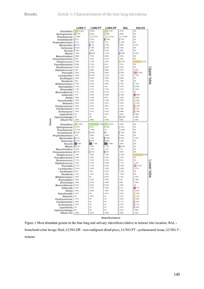
Results Article 1: Characterisation of the four lung microbiota
140

Results Article 1: Characterisation of the four lung microbiota
141
Additional explanation of differences between lung samples (Main manuscript:
Fig. 6)
Except mentioned characteristics, non-malignant tissue and BAL shared similar differences
between locations, unlike other two tissues. Peritumoural tissue and BAL were more different
in lower (LL) than in upper lobes (UL), seen by significantly increased abundance of members
of class Bacteroidia, Coriobacteriia, overall phylum Firmicutes and orders Pasteurellales,
Bifidobacteriales in LL BAL than in LL peritumoural tissue, not seen in UL. On the other hand,
tumour and BAL were more different in UL and represented the richest comparison tree in
tissue vs. BAL category (Main manuscript: Fig. 6). Classes Alphaproteobacteria, Cytophagia,
[Saprospirae] and orders Myxococcales, Bdelovibrionales and Turicibacterales were
significantly more abundant in UL tumour than in BAL, and no difference was seen in LL. On
the contrary, phylum Fusobacteria and class Gammaproteobacteria were both more abundant
in UL BAL than in tumour (no diff. in LL).
Additionally, genera Acinetobacter and Dietzia were more abundant in non-malignant tissue
than in peritumoural tissue, while no differences were detected between non-malignant tissue
and tumour in UL. Conversely, several taxa were different between UL peritumoural tissue and
tumour, including family Lachnospiraceae and genera Actinomyces, Dialister and Prevotella
more abundant and Sediminibacterium, Phenylobacterium and family Myxococcales less
abundant in UL peritumoural tissue. In LL, the differences were very few. Genus
Bifidobacterium was significantly less abundant in LL peritumoural tissue in the three tissues
and family Microbacteriaceae in tumour. In addition, class Erysipelotrichi was significantly
more abundant in LL tumour than in LL peritumoural tissue.

Results Article 2: Lung microbiota and metastatic lymph nodes
142
Article 2: Lung microbiota and metastatic lymph nodes
Research Article
Inverse abundance pattern of tumour and extratumoural lung microbiota
between non-small cell lung cancer patients with or without metastatic
lymph nodes: a cross-sectional clinical study
Rea Bingula1, Edith Filaire1,4, Jérémie Talvas1, Jean-Yves Berthon4, Marie-Paule Vasson1,6,
Annick Bernalier-Donadille5, Nina Radosevic-Robin3, Marc Filaire1,2
Submitted to BMC Microbiome the 25th October 2019
Presence of metastatic lymph nodes (LN) is one of the most important negative prognostic
markers in NSCLC, characterised by significantly shorter overall survival and increased
recidivism. We investigated if this important feature for patient’s outcome is significantly
associated to local lung microbiota composition, based on four different samples (BAL, non-
malignant, peritumoural and tumour tissue). Further, we examined if these two endpoints
(presence or absence of the lymph node metastasis) influence local immunity by investigating
the composition of the tumour immune infiltrate and Th cell phenotypes in BAL. Finally, we
investigated if this immune response is associated to different lung microbiota.
We show that in the presence of metastatic lymph nodes all lung microbiota samples have
increased abundance of the phylum Firmicutes, while diversity metrics show lower values for
the two extratumoural tissue microbiota. We identify 31 genera with inverse patterns in the
abundance between tumour and extratumoural tissues, and between patients with and without
metastatic LN. Moreover, we show that this inversion is closely related to bacterial respiratory
profile. More precisely, identified anaerobic genera are significantly more abundant within the
tumours with metastatic lymph nodes, while aerobic bacteria are more abundant in the
extratumoural tissues. However, exactly inverse is observed for these same genera in patients
without metastatic LN. Therefore, we propose that bacteria “migrate” between the tissues
towards the more favourable growth conditions, since tumour environment drastically changes
with its progression (increased hypoxia, necrosis, lactate production, lower pH). Moreover, we
identify several potential biomarkers in each tissue that might facilitate or improve detection of
the LN metastases without imposing LN biopsy. Finally, we show that BAL microbiota does
not change significantly between these endpoints, but that it is the only one with significant
association with either protumoural or antitumoural immune markers in BAL and in the tumour.

Results Article 2: Lung microbiota and metastatic lymph nodes
143
Research Article
Inverse abundance pattern of tumour and extratumoural lung microbiota
between non-small cell lung cancer patients with or without metastatic lymph
nodes: a cross-sectional clinical study
Rea Bingula1, Edith Filaire1,4, Jérémie Talvas1, Jean-Yves Berthon4, Marie-Paule Vasson1,6,
Annick Bernalier-Donadille5, Nina Radosevic-Robin3, Marc Filaire1,2
Author affiliations:
1University of Clermont-Auvergne, UMR 1019 INRA-UCA, Human Nutrition Unit (UNH), F-
63000 Clermont-Ferrand, France.
2Centre Jean Perrin, Thoracic Surgery Department, 63011 Clermont-Ferrand, France
3University Clermont Auvergne, INSERM U1240, Centre Jean Perrin, Department of
Pathology, 63011 Clermont-Ferrand, France
4Greentech SA, Biopole Clermont-Limagne, 63360 Saint-Beauzire, France
5UMR0454 MEDIS, INRA/UCA, 63122 Saint-Genes-Champanelle, France
6Centre Jean Perrin, CHU Gabriel-Montpied, Clinical Nutrition Unit, F-63000 Clermont-
Ferrand, France
Corresponding author: Marc Filaire; [email protected]

Results Article 2: Lung microbiota and metastatic lymph nodes
144
Abstract
Background. Lung cancer is the leading cause of death by cancer worldwide. In non-small cell
lung cancer (NSCLC) as the most common type, metastatic changes on lymph nodes (LN) are
associated with shorter survival and worse prognosis. Unlike in other cancers, interaction of
lung microbiota with various origin and lung cancer is not well documented. To investigate the
relation between lung microbiota, local immune response and metastatic LN, we conducted an
observational cross-sectional study in 17 NSCLC patients. Lung microbiota was analysed in
bronchoalveolar lavage fluid (BAL) (direct washing of the excised lobe) and three tissues: non-
malignant, peritumoural and tumour tissue. Accompanying immune status was assessed as
tumour infiltrating lymphocytes and T helper and neutrophil profiles in BAL.
Results. We found that phylum Firmicutes replaced Proteobacteria as dominating in tumour
and peritumoural tissue in the presence of metastatic LN. Next, anaerobic genera were
significantly more abundant in tumours with metastatic LN, while aerobic genera were
significantly more abundant in extratumoural tissues. However, the inverse was noted in
patients without metastatic LN. 8 genera from three tissues were identified as promising
prognostic biomarkers of the presence of the metastatic LN (combined AUC of 86.1%, 90.6%
and 92.9% per tissue, respectively). From the four analysed lung communities, only BAL
microbiota had high association with local immune response (both intratumoural and in the
BAL). Moreover, genera positively associated with either anti-tumour or pro-tumour markers
shared the same taxonomic lineage as identified anaerobic (mostly order Clostridiales) and
aerobic tissue genera (mostly class Alphaproteobacteria), respectively.
Conclusions. We evidenced a close connection between metastatic status of LN and tissue lung
microbiota (extratumoural and tumoural) in NSCLC patients, but also the one between BAL
microbiota and local profile of immune cells. Our findings encourage further investigation of
the interaction between lung microbiota from multiple sites and tumour immunity and
environment. Also, the potential clinical application in detection of the metastatic LN through
bacterial pattern in NSCLC patients will require confirmation on larger patient population.
Trial registration. ClinicalTrials.gov ID: NCT03068663. Registered February 27, 2017.
https://clinicaltrials.gov/ct2/show/NCT03068663
Key words:

Results Article 2: Lung microbiota and metastatic lymph nodes
145
Lung microbiota, lung cancer, non-small cell lung cancer, metastatic lymph nodes,
bronchoalveolar lavage, tumour, lung tissue, non-malignant tissue, peritumoural tissue
1 Background
Lung cancer is a leading cause of death by cancer worldwide, the 1st most frequent cancer in
men and the 3rd most frequent in women [1]. It is markedly more frequently diagnosed in older
populations (over 65 years), with non-small cell lung cancer (NSCLC) counting for 85-90% of
all lung cancer cases [2].
The genetic aetiology of NSCLC is already extensively described [3], but the link between lung
cancer and lung microbiota remains scarcely explored [4], despite the growing interest for
cancer-microbiota interaction and the emergence of the gut-lung axis theory [5–7] in the last
decade. The reason partially lies in the long-lasting perception of NSCLC principally, but not
exclusively, as a direct consequence of gene alteration due to various external or internal factors
[2]. Recently, however, a clear connection was established between chronic inflammation and
lung cancer development in the studies of chronic obstructive pulmonary disease (COPD) [8,9].
Moreover, as well as the role of microbiota in these pathologies [10–14], the role of lung
microbiota in lung cancer was suggested as one of the important questions to consider [15].
Several animal models have recently proved that the presence, absence or dysbiosis (e.g. due
to antibiotics) of lung microbiota could influence both lung tumour growth [16,17] and immune
response in the tumour bed [18]. Moreover, a connection was found between enrichment of the
lower airways by oral or supraglottic taxa and increased inflammation and Th17 recruitment in
lung cancer patients that could stimulate pathways involved in tumour progression [19–21].
The studies involving human subjects remain, however, a few numbered, with the invasiveness
of the sampling methods as the major inconvenience for lung microbiota research. So far, most
of the studies on lung microbiota were done on asthmatic or COPD patients by analysis of
sputum or bronchoalveolar lavage fluid (BAL) obtained by bronchoscopy [5,10,14,22], often
used in a routine patient follow-up. Even though these techniques are less invasive than tissue
sampling and therefore more readily performed, they have a higher risk of contamination of
already low biomass samples by upper airways microbiota [23,24]. Also, BAL is likely to
represent lung microbiota found within bronchia that might differ from lung microbiota of the
lung tissue. In current studies on lung cancer microbiota [21,25–30], only two included analysis
of tissue samples. Consequently, microbiota of the tumour and non-malignant tissue in NSCLC

Results Article 2: Lung microbiota and metastatic lymph nodes
146
and its connection to tumour-specific or local immune response have still not been entirely
investigated.
In NSCLC treatment, metastatic changes on lymph nodes (LN) are a major prognostic factor
[31,32], and are defined by TNM classification (i.e. tumour staging) [33]. The presence of
metastatic changes on LN has been related to a significant decrease in overall survival and
response to therapy [33], and is therefore directly implicated in cancer progression and
outcome. However, characterisation of the lung microbiota in the light of metastatic LN
changes has still not been addressed. This knowledge could yield a better understanding of the
tumour microbiological environment and potential involvement in metastatic behaviour of the
tumour.
To broaden insight into both of these matters, we conducted a cross-sectional pilot study in
17 NSCLC patients with primary non-metastatic lung tumours eligible for surgery as a part of
our project [34]. As the first objective, this study determined whether the difference in clinical
tumour stage based on metastatic changes in LN was accompanied by an underlying difference
in the lung microbiota of various origins (BAL, tumour, peritumoural tissue, non-malignant
“healthy” tissue). The second objective of the study was to investigate the connection between
these microbiota and the local immune response (tumour lymphocytic infiltration, immune cell
phenotypes in BAL) in these patients.
2 Results
2.1 Study population
Eighteen patients were prospectively included in the study. Ultimately, lung samples were
obtained from seventeen patients, which were kept for further analysis. Following the post-
resection TNM classification by a pathologist (Table 1), the patients were divided into two
groups for the purpose of this study: Stage I,II patients without metastatic LN, and Stage III
patients with metastatic LN. The groups’ characteristics are presented in Table 2. Body mass
index (BMI) was the only characteristic that differed significantly between the two groups and
corresponded to reported weight loss from Stage III patients. The Chi-square test showed that
both groups were balanced in terms of tumour location, smoking status, tumour type and sex
(data not shown).

Results Article 2: Lung microbiota and metastatic lymph nodes
147
Table 1 Post-surgical TNM classification
pTx pNx Tumour type
Tumour size
(cm)
Stage
group
T1 N0 carcinoid 3.0 I,II
T1 N0 ADK 2.5 I,II
T1 N0 ADK 0.6 I,II
T1 N0 ADK 1.7 I,II
T2 N0 ADK 4.5 I,II
T2 N0 ADK 3.8 I,II
T2 N0 ADK 3.0 I,II
T2 N0 SCC 4.0 I,II
T3 N0 carcinoid 5.0 I,II
T2 N2 ADK 3.0 III
T2 N2 ADK 3.0 III
T2 N2 ADK 2.4 III
T3 N1 ADK 2.0 III
T4 N1 ADK 2.3 III
T4 N1 SCC 8.0 III
T4 N1 SCC 9.5 III
T4 N2 SCC 7.0 III
ADK – adenocarcinoma; SCC – squamous cell carcinoma
Table 2 Characteristics of patients
included in the study
Tumour stage: Stage I, II Stage III p
Total no. of patients 9 8
Stage I/II/III 7/2/0 0/0/8
ADK/SCC/carcinoid 6/1/2 5/3/0
Male/female 6/3 6/2
Smoker/ex-smoker/never-
smoker
2/7/1 0/7/1
Pack-year (smokers and ex-
smokers)
32 ± 23 31 ± 20 0.809
Age (years) 66 ± 10 71 ± 4 0.358
BMI 26.4 ± 3.6 23.6 ± 1.8 0.038
FEV1 (% of expected value) 96 ± 8 100 ± 15 0.606
2.2 Lung microbiota and tumour stages
2.2.1 Diversity differed relative to Stage group, with domination of phylum
Firmicutes in tumour (LUNG.T) and peritumoural tissue (LUNG.PT) of
Stage III patients
Four lung samples characterised throughout the study were: BAL, non-malignant Distal tissue
Piece (LUNG.DP), Peritumoural Tissue (LUNG.PT) and Tumour (LUNG.T).
Tumour and non-malignant tissue had significantly different beta diversity between Stage
groups (Fig. 1a, 1b) based on weighted (wUF) and unweighted UniFrac (uwUF) distance,
respectively. Even though initially no differences were observed between tissue samples (data
not shown), tumour and peritumoural tissue (two closest tissues by sampling location) were
significantly different in Stage I, II (uwUF, Fig. 1c), while tumour and non-malignant tissue
(two most distant tissues by sampling location) were significantly different in Stage III (uwUF,
Fig. 1d).

Results Article 2: Lung microbiota and metastatic lymph nodes
148
Figure 1 Characterisation of the microbiota in lung samples. Beta diversity (presented by
NMDS) in (a) tumour (LUNG.T) between Stage I,II (N = 8) and Stage III (N = 8) based on wUF, (b)
non-malignant tissue (LUNG.DP) between Stage I,II (N = 9) and Stage III (N = 8) based on uwUF, (c)
Stage I,II between tumour (LUNG.T) (N = 8) and peritumoural tissue (LUNG.PT) (N = 7) based on
uwUF, (d) Stage III between LUNG.T (N = 8) and LUNG.DP (N = 8) based on uwUF. Alpha diversity
was analysed between Stage I,II and Stage III for each sample type: (e) phylogenetic diversity, (f)
observed OTUs, (g) Shannon diversity index. Alpha diversity was also analysed between samples within
each Stage group, and the only significant result was in Shannon index for Stage III between LUNG.T
and LUNG.PT (g). (h) Relative abundance relative to the sample type and Stage group in the phylum,
class and genus level. (e-h) Sample numbers were in Stage I,II: LUNG.T (N = 8), LUNG.PT (N = 7),
LUNG.DP (N = 9), BAL (N = 8), and in Stage III: LUNG.T (N = 8), LUNG.PT (N = 7), LUNG.DP (N
= 8), BAL (N = 7). Difference in beta diversity was tested by adonis function (vegan) based on 999
permutations. Difference in alpha diversity between Stage groups was tested by unpaired Wilcoxon test
and between sample types within group by Kruskal-Wallis with BH correction for multiple comparison.

Results Article 2: Lung microbiota and metastatic lymph nodes
149
BAL – bronchoalveolar lavage fluid; LUNG.DP – non-malignant distal piece, LUNG.PT – peritumoural
tissue; LUNG.T – tumour. *: p < 0.05, **: p < 0.01
Inversely, BAL was initially different from each tissue sample (all differences at least p < 0.05
based on both wUF and uwUF) (data not shown). However, in within-group analysis there was
no significant difference between BAL and peritumoural tissue for both groups in wUF
(Stage I,II: p = 0.069, R2 = 0.125; Stage III: p = 0.174, R2 = 0.117) and only in Stage III for
uwUF (Stage I,II: p = 0.027, R2 = 0.117; Stage III: p = 0.082, R2 = 0.135). Interestingly, in
Stage III increased similarity was observed between BAL and tumour (uwUF p = 0.061, R2 =
0.126; Stage I,II: uwUF p = 0.001, R2 = 0.199).
Several alpha diversity metrics (Fig. 1e-g) were significantly lower in Stage III patients both in
non-malignant and peritumoural tissue. Inversely, in Stage III, both in BAL and tumour,
Shannon diversity showed tendency for higher values (Fig. 1g). In addition, in Stage III, tumour
had significantly higher Shannon diversity than peritumoural tissue (Fig. 1g).
Fig. 1h represents the relative abundance of microbiota in phylum, class and genus level,
respective to the sample type and tumour stage group. In Stage I,II, the lung tissue samples
were dominated by two phyla: Proteobacteria (~50%, especially classes Alphaproteobacteria
and Gammaproteobacteria) and Firmicutes (~30%, class Clostridia). Phyla Actinobacteria
(class Actinobacteria) and Bacteroidetes (class Bacteroidia) followed. In stage III, however,
tumour and peritumoural tissue were dominated by phylum Firmicutes (~44%) and
Proteobacteria was the 2nd most abundant (~37%). In non-malignant tissue, there was no
difference in dominating phyla between stages. However, in Stage III abundance of
Proteobacteria was lower in favour of Actinobacteria (~17%).
At the genus level (Fig. 1h), tissue samples shared dominating genera. Clostridium
(Firmicutes), Pseudomonas (Gammaproteobacteria), Kocuria (Actinobacteria), Bacteroides
(Bacteroidetes) Sphingomonas (Alphaproteobacteria), Acinetobacter (Gammaproteobacteria)
and Blautia (Firmicutes) were among the more abundant genera in both groups. Interestingly,
the Clostridium genus, which in Stage I,II was balanced in abundance with either Pseudomonas
or Kocuria depending on the tissue, was ~2-fold more abundant in Stage III and representing
the most abundant genus (~30%).
In Stage I,II (Fig. 1h), BAL was dominated by Firmicutes (~45%, class Clostridia), followed
by almost equal abundances of Proteobacteria (~20%, class Gammaproteobacteria),
Actinobacteria (~16%, class Actinobacteria) and Bacteroidetes (~16%, class Flavobacteriia).

Results Article 2: Lung microbiota and metastatic lymph nodes
150
In Stage III, however, Proteobacteria (~36%, classes Alphaproteobacteria and
Gammaproteobacteria) was more abundant than Bacteroidetes and Actinobacteria. Even
though in Stage I,II Capnocytophaga (~9%) was the most abundant genus, followed by
Pseudomonas, Streptococcus and Blautia (~6% each), Stage III was dominated by
Pseudomonas (~15%), Blautia (~8%) and Streptococcus (~6%). Interestingly, Clostridium,
found as most abundant in lung tissues, was not in the first 50 most abundant genera in BAL in
either of the groups.
2.2.2 Anaerobic and aerobic bacteria showed inverse abundance profiles between
tumour and extratumoural tissue
Figure 2 shows only taxa on the levels of class and genus whose abundance was significantly
different between two Stage groups. Difference in abundance is presented as log2-fold change.
Within each bar there is a significance level and beside the bar is a number representing the
rank of importance of the difference as reported by random forest algorithm. BAL samples were
non-significantly different on selected taxonomic ranks, so they were excluded from graphics.
Figure 2 Differential abundance
of lung tissue microbiota between
Stage groups. (a) Differential
abundance on the class level. (b)
Differential abundance on the genus
level. Asterisk within the bars
represents the adjusted significance
(BH) of differential abundance tested
by Wald’s test of significance of
coefficients in a negative binomial
general linearized model
(microbiomeSeq). Sample numbers
were in Stage I,II: LUNG.T (N = 8),
LUNG.PT (N = 7), LUNG.DP (N = 9),
and in Stage III: LUNG.T (N = 8),
LUNG.PT (N = 7), LUNG.DP (N = 8).
The numbers beside the bars are ranks
of importance of the detected
difference, as detected by random forest
classifier. Following full taxa names
are corresponding order (4 letters) and
phylum (1 capital letter). Orders: Acti –
Actinomycetales, Bact – Bacteroidales,
Burk – Burkholderiales, Caul –
Caulobacterales, Clos – Clostridiales,
Cori – Coriobacteriales, Desu –

Results Article 2: Lung microbiota and metastatic lymph nodes
151
Desulfovibrionales, Erys – Erysipelotrichales, Flav – Flavobacteriales, Lact – Lactobacillales, Ocea –
Oceanospirillales, Rhiz – Rhizobiales, Rhod – Rhodospirillales, Sphi – Sphingobacteriales, Turi –
Turicibacterales. Phyla: A – Actinobacteria, B – Bacteroidetes, F – Firmicutes, P – Proteobacteria
(classes aP – Alphaproteobacteria, bP – Betaproteobacteria, gP – Gammaproteobacteria, dP –
Deltaproteobacteria). [Prevotella] is from the family Paraprevotellaceae, and Prevotella from
Prevotellaceae. [Ruminococcus] is from the family Lachnospiraceae, and Ruminococcus from
Ruminococcaceae. BAL – bronchoalveolar lavage fluid; LUNG.DP – non-malignant distal piece,
LUNG.PT – peritumoural tissue; LUNG.T – tumour. *: p < 0.05, **: p < 0.01, ***: p < 0.001, ****: p
< 0.0001
The eye-catching feature was that the log2 profiles showed strong similarities for peritumoural
and non-malignant tissue, and were inverse to tumour for each shared taxa (Fig. 2a, 2b). This
was observed both on class and genus level. Classes Sphingobacteriia, Flavobacteriia and
Actinobacteria were significantly more abundant in non-malignant and/or peritumoural tissue
of Stage III patients, but less abundant in Stage III tumour (Fig. 2a). Tumour had two unique
differences: classes TM7-3 and Acidobacteria. TM7-3 ranked as the most important difference
(random forest), more abundant in Stage III (0% vs 0.13%), while Acidobacteria was more
abundant in Stage I,II. Likewise, two unique differences could be found in peritumoural tissue:
Nitrospira (1st ranked by random forest) more abundant in Stage III, and Erysipelotrichi
(2nd ranked) more abundant in Stage I,II.
The total of 31 genera were found significantly different between stages (Fig. 2b), with the
highest number in non-malignant tissue. Markedly, an exclusive polarity was seen between the
“lower” and “upper” part of the figure when observing higher taxonomic levels of listed genera
(Fig. 2b – abbreviations next to the genus name). The “upper” part included listed genera from
[Prevotella] to Collinsella), which were more abundant in Stage III tumour and, inversely, in
peritumoural/non-malignant tissue of Stage I,II patients. They belonged to orders Clostridiales
and Turicibacterales (phylum Firmicutes), Bacteroidales (Bacteroidetes), Coriobacteriales
(Actinobacteria), Burkholderiales (Betaproteobacteria), Oceanospiralles
(Gammaproteobacteria) and Desulfovibrionales (Deltaproteobacteria). Conversely, the
“lower” part of the figure included genera from orders Actinomycetales (Actinobacteria),
Rhodospirillales, Rhizobiales and Caulobacterales (Alphaproteobacteria), Sphingobacteriales
and Flavobacteriales (Bacteroidetes) and Erysipelotrichales (Firmicutes).
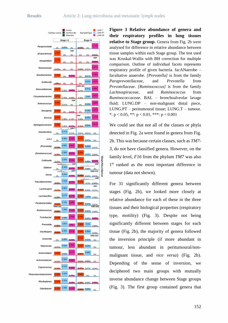
Results Article 2: Lung microbiota and metastatic lymph nodes
152
Figure 3 Relative abundance of genera and
their respiratory profiles in lung tissues
relative to Stage group. Genera from Fig. 2b were
analysed for difference in relative abundance between
tissue samples within each Stage group. The test used
was Kruskal-Wallis with BH correction for multiple
comparison. Outline of individual facets represents
respiratory profile of given bacteria. facANaerobe –
facultative anaerobe. [Prevotella] is from the family
Paraprevotellaceae, and Prevotella from
Prevotellaceae. [Ruminococcus] is from the family
Lachnospiraceae, and Ruminococcus from
Ruminococcaceae. BAL – bronchoalveolar lavage
fluid; LUNG.DP – non-malignant distal piece,
LUNG.PT – peritumoural tissue; LUNG.T – tumour.
*: p < 0.05, **: p < 0.01, ***: p < 0.001
We could see that not all of the classes or phyla
detected in Fig. 2a were found in genera from Fig.
2b. This was because certain classes, such as TM7-
3, do not have classified genera. However, on the
family level, F16 from the phylum TM7 was also
1st ranked as the most important difference in
tumour (data not shown).
For 31 significantly different genera between
stages (Fig. 2b), we looked more closely at
relative abundance for each of these in the three
tissues and their biological properties (respiratory
type, motility) (Fig. 3). Despite not being
significantly different between stages for each
tissue (Fig. 2b), the majority of genera followed
the inversion principle (if more abundant in
tumour, less abundant in peritumoural/non-
malignant tissue, and vice versa) (Fig. 2b).
Depending of the sense of inversion, we
deciphered two main groups with mutually
inverse abundance change between Stage groups
(Fig. 3). The first group contained genera that

Results Article 2: Lung microbiota and metastatic lymph nodes
153
were more abundant in tumour in Stage I,II (low in peritumoural/non-malignant tissue), but in
Stage III were, inversely, more abundant in peritumoural/non-malignant tissue (low in tumour).
This involved mostly genera in the first part of the Figure 3 (from [Eubacterium] to
Sphingobacterium), and included Chryseobacterium and Kocuria with higher part in overall
abundance (Fig. 3). Moreover, these bacteria had in majority aerobic or facultative anaerobic
respiration (red and green outline, Fig. 3). The second group involved genera that in Stage I, II
were more abundant in peritumoural/non-malignant tissue (low in tumour), but in Stage III were
more abundant in tumour. This included mostly genera in the middle of Figure 3 (from
Desulfovibrio to Halomonas) with higher abundant Lactobacillus, Faecalibacterium,
Ruminococcus and Prevotella (Fig. 3). Considering the respiratory metabolism, all bacteria in
this group were obligate anaerobes. Interestingly, in both groups, certain genera in peritumoural
tissue had the same abundance or with very little change in both Stage groups (e.g. Prevotella
or Azospirillum). Also, certain genera did not entirely follow the principle of inverse change in
abundance (Fig. 3, from Anaerostipes until Odoribacter). Nevertheless, the pattern and
respiratory profile of these genera were more similar to the second than to the first group (i.e.
anaerobic). Finally, we also looked at active motility organs in these bacteria, but we found that
only 25% possessed flagella with no difference depending on the group.
2.2.3 Combination of genera as potential biomarkers of metastatic lymph nodes
We used receiver operating characteristic (ROC) analysis to assess genera from Figure 3 as
potential biomarkers in determination of metastatic LN (Fig. 4). In each tissue, several genera
already individually showed high percentage of area under the ROC curve (AUC), especially
Dorea and Coprococcus in peritumoural tissue with 90.9% and 89.8%, respectively. However,
a combination of several genera in each tissue increased this already high percentage. Genera
Parabacteroides, Ruminococcus (family Ruminococcaceae), and Oscillospira in non-
malignant tissue (LUNG.DP), Dorea, Coprococcus and Lachnospira in peritumoural tissue
(LUNG.PT), and Dialister and [Ruminococcus] (family Lachnospiraceae) in tumour
(LUNG.T), represented the combinations with the highest predictive power of 86.1%, 92.9%
and 90.6% AUC per tissue, respectively.

Results Article 2: Lung microbiota and metastatic lymph nodes
154
Figure 4 Potential biomarkers for metastatic lymph nodes in three lung tissues. ROC
analysis was performed on individual genera detected as significantly different between Stage groups
for each tissue in Fig. 2b. Genera with AUC higher than 80% were included for combination analysis.
Only combinations with highest AUC percentage were selected and presented in the Figure for each
lung tissue along with individual curves. BAL – bronchoalveolar lavage fluid; LUNG.DP – non-
malignant distal piece, LUNG.PT – peritumoural tissue; LUNG.T – tumour; AUC – area under the curve
2.3 Tumour-infiltrating lymphocytes and microbiota
2.3.1 Stage groups without difference in tumour-infiltrating lymphocytes (TILs)
Composition of the tumour lymphocytic infiltrate was evaluated by quantity of CD8+
(cytotoxic), FOXP3+ (regulatory, Treg) and PD-1+ T lymphocytes, as well as of CD20+ B
lymphocytes. In addition, expression of an important immune checkpoint marker, PD-L1, was
evaluated both on tumour-infiltrating lymphocytes and tumour cells. No significant difference
was seen between tumour stages in any of the TIL subpopulations (Fig. 5a), or in their ratios,
CD8+/CD20+ and CD8+/FOXP3+ (Additional file 1: Figure 1a). Both groups equally consisted
of high- and low-infiltrated tumours. PD-1 and PD-L1 markers were expressed only in a few
tumours and did not co-occur in the same tumour nor were they associated with one of the Stage
groups (data not shown).
2.3.2 BAL microbiota with the most associations to tumour-infiltrating
lymphocytes (TILs)
We next examined the association of microbiota in the tumour or its surroundings with the
TILs. Eighteen genera significantly correlated with at least one immune marker (Fig. 5b,
Additional file 1: Figure 1b). Correlation profiles were very similar between individual immune
markers but not between samples. Surprisingly, not tumour but BAL microbiota showed the
highest number of significant correlations with TILs (Fig. 5b). Genera Dietzia, Clostridium,
Coprococcus, Ruminococcus, Akkermansia, Oscillospira, Bacteroides, Collinsella, and
[Prevotella] had significant positive and Haemophilus and Mesorhizobium significant negative
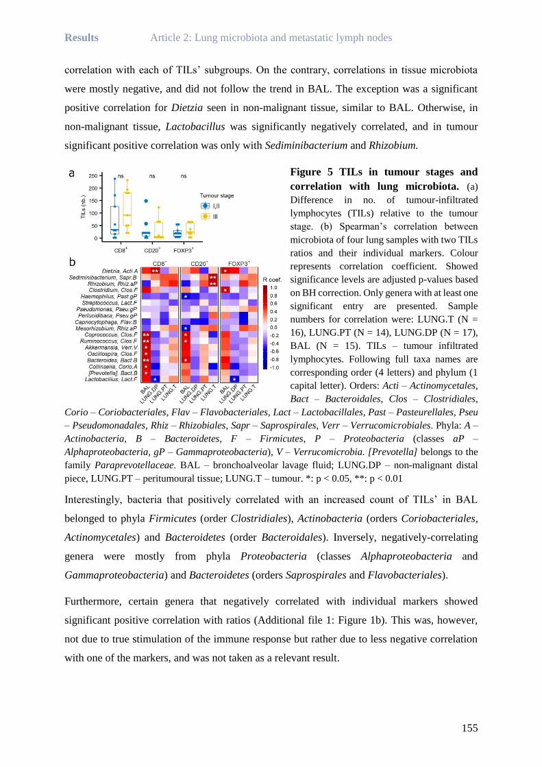
Results Article 2: Lung microbiota and metastatic lymph nodes
155
correlation with each of TILs’ subgroups. On the contrary, correlations in tissue microbiota
were mostly negative, and did not follow the trend in BAL. The exception was a significant
positive correlation for Dietzia seen in non-malignant tissue, similar to BAL. Otherwise, in
non-malignant tissue, Lactobacillus was significantly negatively correlated, and in tumour
significant positive correlation was only with Sediminibacterium and Rhizobium.
Figure 5 TILs in tumour stages and
correlation with lung microbiota. (a)
Difference in no. of tumour-infiltrated
lymphocytes (TILs) relative to the tumour
stage. (b) Spearman’s correlation between
microbiota of four lung samples with two TILs
ratios and their individual markers. Colour
represents correlation coefficient. Showed
significance levels are adjusted p-values based
on BH correction. Only genera with at least one
significant entry are presented. Sample
numbers for correlation were: LUNG.T (N =
16), LUNG.PT (N = 14), LUNG.DP (N = 17),
BAL (N = 15). TILs – tumour infiltrated
lymphocytes. Following full taxa names are
corresponding order (4 letters) and phylum (1
capital letter). Orders: Acti – Actinomycetales,
Bact – Bacteroidales, Clos – Clostridiales,
Corio – Coriobacteriales, Flav – Flavobacteriales, Lact – Lactobacillales, Past – Pasteurellales, Pseu
– Pseudomonadales, Rhiz – Rhizobiales, Sapr – Saprospirales, Verr – Verrucomicrobiales. Phyla: A –
Actinobacteria, B – Bacteroidetes, F – Firmicutes, P – Proteobacteria (classes aP –
Alphaproteobacteria, gP – Gammaproteobacteria), V – Verrucomicrobia. [Prevotella] belongs to the
family Paraprevotellaceae. BAL – bronchoalveolar lavage fluid; LUNG.DP – non-malignant distal
piece, LUNG.PT – peritumoural tissue; LUNG.T – tumour. *: p < 0.05, **: p < 0.01
Interestingly, bacteria that positively correlated with an increased count of TILs’ in BAL
belonged to phyla Firmicutes (order Clostridiales), Actinobacteria (orders Coriobacteriales,
Actinomycetales) and Bacteroidetes (order Bacteroidales). Inversely, negatively-correlating
genera were mostly from phyla Proteobacteria (classes Alphaproteobacteria and
Gammaproteobacteria) and Bacteroidetes (orders Saprospirales and Flavobacteriales).
Furthermore, certain genera that negatively correlated with individual markers showed
significant positive correlation with ratios (Additional file 1: Figure 1b). This was, however,
not due to true stimulation of the immune response but rather due to less negative correlation
with one of the markers, and was not taken as a relevant result.

Results Article 2: Lung microbiota and metastatic lymph nodes
156
2.4 Phenotyping of BAL immune cells and their association to lung
microbiota
2.4.1 Th17 higher in BAL of Stage III patients
Next, we looked at difference in percentage of helper T lymphocytes (Th) and
polymorphonuclear neutrophil (N) subtypes between Stage groups in samples of BAL.
Neutrophil subtypes showed no significant difference between the two Stage groups (Fig. 6a).
In CD4+ Th, the only difference was observed for Th17 cells (Fig. 6b), found significantly
higher in the Stage III group, although overall percentage remained low in both groups.
Consequently, both ratios Th1/Th17 and Treg/Th17 were lower in Stage III (Treg/Th17
significantly) (Fig. 6c). Interestingly, the positive correlation between Treg and Th17 was
present in the Stage III group, with the similar tendency in Stage I,II group (Fig. 6d).
2.4.2 Pro and anti-tumour markers show medium to strong correlation with BAL
microbiota
Similarly, as with the tumour immune infiltrate, we examined the association of immune cells
in BAL and microbiota from lung samples. All four samples were analysed, but only BAL
showed a clear clustering and significant correlations. Figure 6e therefore shows association
between genera in BAL and BAL immune cells with at least one correlation higher than R =
0.4.
We could decipher three groups of associations: group 1, from Oscillospira to Ruminococcus,
group 2, from Haemophilus to Atopobium, and group 3, from Novosphingobium until
WAL_1855D (Fig. 6e). Genera from group 1 positively correlated with Th1, Th2, Th1/Th17,
Th1/Treg, Th1/Th2 and inactive circulating and tethering neutrophils (N). These markers are
most often considered as a part of anti-tumour response [35]. Inversely, they were negatively
correlated with Treg, Th17, activated N and Th17/Treg ratio (significantly for genus Roseburia
and Bacillus), considered as markers of pro-tumour response [35]. Group 3 was the exact
opposite of group 1, while group 2 was found in between. Similar to group 1, group 2 had
positive associations with Th1-associated markers, but had negative associations with Th2 and
circulating N (as in group 3).
Markedly similar to associations observed with TILs, association groups with immune cells in
BAL divided genera by their higher taxonomic levels. So, genera in group 1 belonged
principally to phyla Firmicutes (order Clostridiales) and Bacteroidetes (Bacteroidales), while
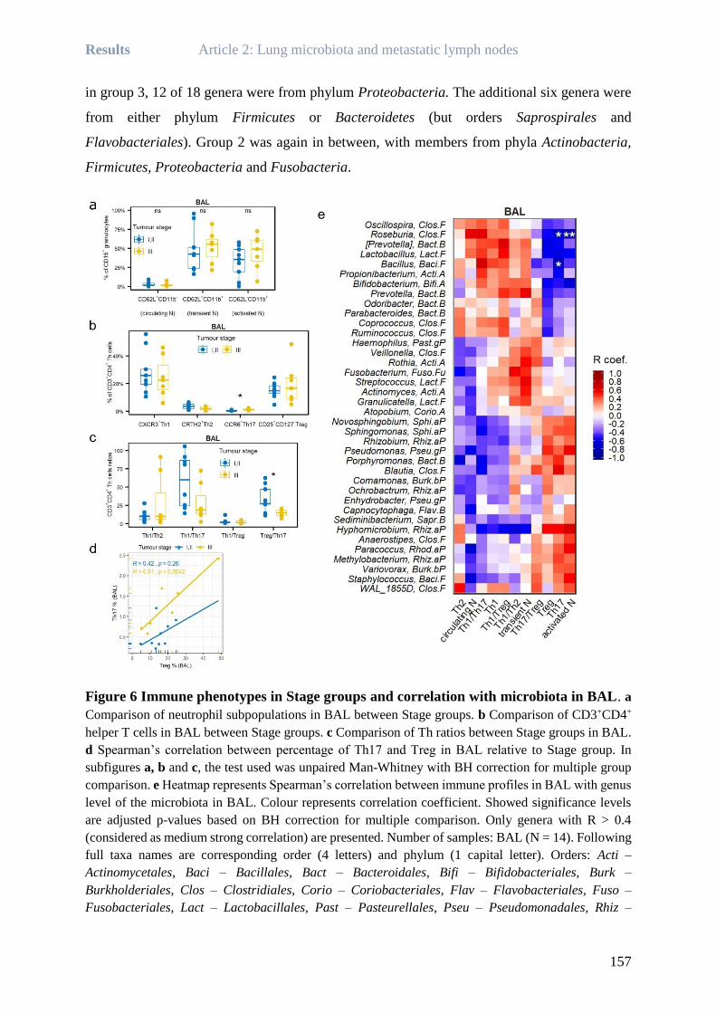
Results Article 2: Lung microbiota and metastatic lymph nodes
157
in group 3, 12 of 18 genera were from phylum Proteobacteria. The additional six genera were
from either phylum Firmicutes or Bacteroidetes (but orders Saprospirales and
Flavobacteriales). Group 2 was again in between, with members from phyla Actinobacteria,
Firmicutes, Proteobacteria and Fusobacteria.
Figure 6 Immune phenotypes in Stage groups and correlation with microbiota in BAL. a
Comparison of neutrophil subpopulations in BAL between Stage groups. b Comparison of CD3+CD4+
helper T cells in BAL between Stage groups. c Comparison of Th ratios between Stage groups in BAL.
d Spearman’s correlation between percentage of Th17 and Treg in BAL relative to Stage group. In
subfigures a, b and c, the test used was unpaired Man-Whitney with BH correction for multiple group
comparison. e Heatmap represents Spearman’s correlation between immune profiles in BAL with genus
level of the microbiota in BAL. Colour represents correlation coefficient. Showed significance levels
are adjusted p-values based on BH correction for multiple comparison. Only genera with R > 0.4
(considered as medium strong correlation) are presented. Number of samples: BAL (N = 14). Following
full taxa names are corresponding order (4 letters) and phylum (1 capital letter). Orders: Acti –
Actinomycetales, Baci – Bacillales, Bact – Bacteroidales, Bifi – Bifidobacteriales, Burk –
Burkholderiales, Clos – Clostridiales, Corio – Coriobacteriales, Flav – Flavobacteriales, Fuso –
Fusobacteriales, Lact – Lactobacillales, Past – Pasteurellales, Pseu – Pseudomonadales, Rhiz –

Results Article 2: Lung microbiota and metastatic lymph nodes
158
Rhizobiales, Rhod – Rhodospirillales, Sapr – Saprospirales, Sphi – Sphingobacteriales. Phyla: A –
Actinobacteria, B – Bacteroidetes, F – Firmicutes, Fu – Fusobacteria, P – Proteobacteria (classes aP
– Alphaproteobacteria, bP – Betaproteobacteria, gP – Gammaproteobacteria). BAL – bronchoalveolar
lavage fluid; LUNG.DP – non-malignant distal piece, LUNG.PT – peritumoural tissue; LUNG.T –
tumour. N – neutrophils. *: p < 0.05, **: p < 0.01, ***: p < 0.001, ****: p < 0.0001
3 Discussion
Even though lung cancer is the leading cause of death from cancer worldwide [1], its connection
to lung microbiota and the interaction of lung microbiota and lung cancer immunology remains
largely unexplored. This is, to our knowledge, the first study in NSCLC analysing lung
microbiota between stages based on metastatic LN. Moreover, lung microbiota was analysed
for the first time in four different samples: BAL as a sample of alveolar “planktonic”
microbiota, and three tissue samples harbouring different physiology: tumour, peritumoural
tissue (tissue in direct contact with tumour and with increasing importance in deciding tumour
progression) [36,37] and non-malignant tissue. In addition, in our study, BAL was not obtained
by bronchoscopy but by direct instillation of physiological serum into the dissected lung,
limiting upper airways contamination and giving a possibility of true sampling of the microbiota
in tumour proximity.
We showed that each Stage group revealed differences between and within samples’
community compositions (Fig. 1). Beta diversity differed significantly between Stage groups
both in tumour and non-malignant tissue. Consequently, peritumoural tissue was
compositionally more similar to non-malignant tissue in Stage I,II, and inversely, to tumour and
BAL in Stage III. We also observed that the Stage III group had lower alpha diversities in
peritumoural and non-malignant tissue compared to Stage I,II, with no change in the other two
samples. Lung tissues were dominated by phylum Proteobacteria in Stage I,II, but in Stage III,
tumour and peritumoural tissue were dominated by Firmicutes. Yu et al. [27] also identified
Proteobacteria as the dominant phylum in non-malignant lung tissue in lung cancer patients.
However, in their study, the alpha diversity of non-malignant tissue was higher in more
advanced stages, which is the inverse of our finding. This could be because they analysed stages
using the base TNM classification [33], which classifies patients into stages depending on the
overall TNM score and not exclusively based on metastatic LN as we performed. In our study,
BAL samples were dominated by phylum Firmicutes without difference relative to Stage group.
Interestingly, the study of Lee et al. [25] found abundance of phylum Firmicutes increased in
BAL of lung cancer patients compared to those with benign mass lesions. In addition, we
observed phylum Firmicutes as dominant in tumour and peritumoural tissue of Stage III
Results Article 2: Lung microbiota and metastatic lymph nodes
159
patients, two samples significantly more similar to BAL in this group. Looking at these
findings, the shift towards phylum Firmicutes might be associated with advancement of lung
cancer.
We identified classes and genera for each lung tissue sample with significant difference in
abundance between two tumour stage groups (Fig. 2). Interestingly, in tumour, the 1st ranked
class by the importance of difference was class TM7-3, more abundant in Stage III. This class
is from the candidate phylum TM7, previously reported as increased both in COPD and lung
cancer [38] and could be implicated in tumour progression. Looking at both analysis on the
class and genus level, we noted the consistent inversion of differential abundance profiles
between tumour vs. peritumoural/non-malignant tissue. In Stage III, genera more abundant in
tumour were less abundant in peritumoural/non-malignant tissue, and it was the inverse in Stage
I,II. Interestingly, members in each of these “inversion groups” mostly belonged to the same
higher taxonomic levels, but on the contrary, these higher levels were not shared between
“inversion groups”.
Next, we divided identified genera into two “abundance change” hypotheses depending on their
abundance in tissues and stages. Interestingly, each hypothesis gathered members that belonged
to one of the major respiratory profiles. The first hypothesis included aerobic bacteria, more
abundant in Stage I, II tumour and scarcely or not present in the other two tissues, and inversely,
in Stage III genera more abundant in peritumoural and non-malignant tissue with lower
abundance in tumour. The second hypothesis involved anaerobic genera, more abundant in
peritumoural and non-malignant tissue in stage I, II. Those in Stage III have increased
abundance in tumour, and decreased abundance in the other two tissues compared to Stage I,II.
We hypothesise that aerobic bacteria from the first group are more abundant in Stage I,II tumour
tissue due to increased protection from the immune system and increased availability of
nutrients [39]. Low-grade tumours are still well oxygenated and could sustain aerobic bacteria.
However, with stage advancing and/or tumour growth, the supply of oxygen and nutrients
becomes insufficient for tumour cells, inducing hypoxia and necrosis within the tumour centre,
accompanied by increased lactate production and acidification [40,41]. The aerobic bacteria
that are not able to live in these conditions “leave” the tumour, i.e. move to peritumoural and
non-malignant tissue. Even though only ¼ of detected genera (Fig. 3) had active motility organs
(flagella), the literature suggests another means of motility, such as “gliding” or attachment on
eukaryotic cells [42,43] that could support this “migration”. In this study, we analysed only a
captured moment and we have no insight into microbial evolution and changes following

Results Article 2: Lung microbiota and metastatic lymph nodes
160
development of the tumour. However, in multiple examples genera were still completely absent
in one group (0%) but present in the other (Fig. 3). Rather than only changing a balance between
microbiota proportions (saying that microbiota already previously existed in the tissues before
the change in abundance), our results of beta diversity on uwUF (based on presence/absence of
OTUs) support the hypothesis that certain bacteria might “migrate” between tissues. Also, in
certain genera, peritumoural tissue has much smaller oscillations between two Stage groups
compared to non-malignant tissue and tumour, which could be due to its position in the middle
of this “migratory” pathway. For the second group, we suggest that anaerobic bacteria could
reside inside the biofilms in the non-malignant and peritumoural tissue, which protects them
from toxic oxygen exposure. The suggestion of biofilm importance in lung microbial
co-location of aerobes and anaerobes, as well as in pathogenesis of lung diseases was previously
suggested [15,44–46]. The majority of anaerobic bacteria from the second hypothesis ferment
glucose to produce lactate [47,48], but also other acids such as butyric, acetic or propionic (e.g.
Faecalibacterium, Odoribacter, Ruminococcus, Bifidobacterium) [49,50]. Since tumour cells
are proposed to influence glucose delivery of the host to the tumour bed [51], this could be the
true chemotactic target of both aerobic and anaerobic bacteria. Once tumour environment
becomes sufficiently hypoxic, anaerobic bacteria could “migrate” from the surrounding biofilm
into the tumour bed. The tumour-targeting anaerobic bacteria were already described in the
context of bacteria-mediated tumour therapy (BMTT). Innate principles of BMTT are based
either on stimulation of CD8+ T cells through bacterial epitopes, resulting in enhanced tumour
surveillance (seen in anaerobic Clostridium spores), or on direct antitumour effect through
nutrient competition with tumour cells (e.g. strains of Clostridium,
Bifidobacterium and Salmonella) [39]. On the other hand, tumour acidification and hypoxia
impair activation and anti-tumour response of immune cells [52], and, as mentioned, anaerobic
bacteria contribute to lactic acid production and acidification. This hypothesis corresponds to
our finding, where anaerobic microbiota was enriched within the tumour of more advanced
stages characterised by metastatic nodes.
From genera in Fig. 3 we identified several potential biomarkers (Fig. 4) that, interestingly, all
belonged to anaerobic bacteria from the second abundance change hypothesis. Moreover, all
except Parabacteroides belonged to order Clostridiales from the phylum Firmicutes. In each
tissue, the AUC percentage of combined genera was high, showing that, potentially, any of the
tissues could serve as an indicator of metastatic LN presence, improve disease follow-up, and

Results Article 2: Lung microbiota and metastatic lymph nodes
161
could limit the necessity of node sampling and analysis. However, selected genera need
confirmation of quantitative analysis (such as qPCR) and larger study population.
Looking at local immune profiles (TILs, BAL immune cells), the only observed difference was
in percentage of Th17 cells, significantly elevated in BAL of the Stage III group. Consequently,
the Treg/Th17 ratio, to which high importance in lung cancer immunity was recently
attributed [53], was lower. Even though certain studies reported Th17 cytokines to affect
effector cytotoxic T cell generation and thus stimulate anti-tumour response [54,55], a
meta-study analysing the results of 6 lung cancer cohorts (total no. of participants = 479) sided
elevated Th17 cytokines with significantly reduced overall and disease free survival,
respectively [56]. In our study, the parallel could be drawn with higher levels found in the
Stage III group characterised by metastatic lymph nodes as a marker of elevated tumour
aggressiveness.
In this study, we exposed first results on relation of different lung microbiota with TILs and
immune cells in BAL. Assessment of the association between four lung microbiota with both
TILs and immune cells in BAL showed surprisingly similar results. In both analyses, only
microbiota from BAL showed clear correlation profiles for multiple markers and correlating
genera mutually corresponded between the two analyses. Genera from phyla Firmicutes
(especially order Clostridiales), Actinobacteria and Bacteroidetes (class Bacteroidia) showed
positive correlation with anti-tumour markers (TILs: CD8+ T lymphocytes, CD20+ B
lymphocytes, FOXP3+ Tregs; in BAL Th1, Th2, Th1/Treg, Th1/Th17, Th1/Th2 ratios and
inactive forms of neutrophils) [35]. Several genera in this group were already described for their
potential in stimulating antitumoural immunity, such as Akkermansia, Bifidobacterium,
Lactobacillus [57,58]. On the contrary, genera from BAL that were positively correlated with
an immunosuppressive profile, particularly with BAL immune cells (Treg, Th17, Th17/Treg,
activated neutrophils) [35] belonged mostly to phyla Proteobacteria (classes
Alphaproteobacteria and Gammaproteobacteria) and Bacteroidetes (classes Flavobacteriia,
Sphingobacteriia, Saprospirae). Conversely to the first group, genera from this group such as
Haemophilus, Streptococcus, Staphylococcus, Pseudomonas were previously associated with
lung pathologies (as COPD) and negative or immunosuppressive impact on the host [10,14].
Interestingly, many BAL genera that positively correlated with anti-tumour markers in BAL
belonged to the same taxonomic lineage as the group of anaerobic genera with increased
abundance in Stage III tumours (therefore decreased in non-malignant and peritumoural tissue),
and vice versa for negatively-correlated genera. Although representing a sample of

Results Article 2: Lung microbiota and metastatic lymph nodes
162
inter-bronchial microbiota, BAL genera were significantly associated both with immune
markers within tumour and in BAL. Surprisingly, however, microbiota of the tissues, including
tumour, showed poor association with immune characteristics from both TILs counts and BAL
immune profiles. Further research will be necessary to determine the exact reason for this, since
the answer is certainly very complex interplay between microbial, immunological and tumour
properties.
The major limitation of this study is the small number of patients due to the strict inclusion
criteria. In this study design, we were not able to perform a longitudinal study following tumour
progression and co-occurring microbiota modification. It would also be interesting to look at
the absolute numbers of both immune cells and specific bacteria to complete these quality based
data. This will be a perspective for future research.
4 Conclusion
To our knowledge, this is the first study in NSCLC patients that analysed lung microbiota from
four different samples (BAL, non-malignant tissue, peritumoural tissue and tumour) and their
relation with local immune response depending on the presence of metastatic lymph nodes. We
showed that BAL microbiota was that most associated with both intra and extra-tumour immune
response, while lung tissues’ microbiota changed relative to presence of metastatic lymph
nodes. We also identified several biomarkers in lung tissues that could help improve patient’s
diagnosis and prognosis. These findings need further confirmation in larger cohorts and
additional research on interaction of various lung microbiota and lung cancer with its potential
use in diagnostics.
5 Materials and methods
5.1 Patient recruitment
Patients pre-considered for inclusion in the study were patients diagnosed with primary NSCLC
eligible for surgical treatment without neoadjuvant platinum-based chemotherapy and
presented before the Thoracic Oncologic Committee of the Centre Jean Perrin,
Clermont-Ferrand, France. Inclusion criteria were: age between 18 and 80 years, body mass
index (BMI) < 29.9, no administration of antibiotics, corticosteroids, and immunosuppressive
drugs or no history of lung infection in at least the past 2 months as well as no previous airways
surgery/cancer treatment. Detailed inclusion/exclusion criteria are available elsewhere [34].
Each patient was attributed a number at inclusion that was used for subsequent analyses.

Results Article 2: Lung microbiota and metastatic lymph nodes
163
5.2 Study design
The study was concentrated around two points: inclusion and surgery. During an outpatient
appointment preceding surgical intervention, eligible patients gave their written informed
consent to participate in the study. The sampling of blood and lung tissues and fluid was done
the day before surgery and during surgery, respectively. Lung sampling was performed on
excised lobe during the operation and, therefore, posed no additional inconvenience for the
patient. Four different lung samples were chosen as each represents a distinct microbiota
relative to the lung physiology of the sample’s origin. A study flowchart as well as detailed
design and power calculation were previously published [34].
5.3 Sampling of lung tissue and BAL
The detailed procedure was previously published elsewhere [34]. Briefly, immediately after
partial or complete pneumonectomy, the lobe was washed by instillation of sterile physiological
serum (2 x 40 mL) directly to the bronchus to obtain BAL fluid (retrieved 8-10 mL in total).
Furthermore, a non-malignant piece distal to the tumour was excised from the same lobe and
designated as LUNG.DP. Tumour and peritumoural tissue were excised together as a pie-slice,
to sample both the surface and centre of the tumour. Carefully, peritumoural tissue was cut off
from the tumour slice based on its histological qualities. The tumour tissue sample was
designated as LUNG.T and the peritumoural tissue sample as LUNG.PT. Tissue samples were
immediately snap-frozen in liquid nitrogen and stored at -80°C until DNA extraction. 3 mL of
BAL were used for immune cell phenotyping, while the rest was directly stored at -80°C until
DNA extraction.
5.4 Tumour immune infiltrate
Immunohistochemistry was performed on deparaffinised tumour tissue using the specific
antibodies to detect subpopulations of immune cells as follows: cytotoxic T-lymphocytes
(anti-CD8, clone SP16, ThermoFisher Scientific, dilution 1:200), regulatory T-lymphocytes
(anti-FOXP3, clone SP97, ThermoFisher Scientific, dilution 1:100), B-lymphocytes (anti-
CD20, clone SP32, Cell Marque, dilution 1:100). The immune response checkpoint axis PD-1–
PD-L1 was assessed by anti-PD-1 (clone NAT105, CellMarque, dilution 1:50) and anti-PD-L1
(clone 28-8, Abcam, dilution 1:50). All staining was performed by a fully automated,
standardised procedure (Benchmark XT, Ventana/Roche). The number of lymphoid cells
expressing each antigen, except PD-L1, was determined on five consecutive high-pass filter
(HPF) (×400) fields (Nikon Eclipse Ci microscope), starting from the invasive front toward the

Results Article 2: Lung microbiota and metastatic lymph nodes
164
tumour centre as previously described [59]. It was used as a parameter reflecting the tumour’s
quantity of a given immune cell subpopulation. The five-field average was used for statistical
analysis. PD-L1 was assessed for both immune and tumour cells and reported as the percentage
of each population expressing the antigen.
5.5 BAL immune cell phenotyping
Leukocytes were obtained after haemolysis from fresh blood and BAL as previously
described [34]. Leukocytes were phenotyped by flow cytometry (LSRII, BD Biosciences) with
the following antibodies: anti-CD3-VioBlue, anti-CD4-APC-Vio770, anti-CD25-APC,
anti-CD127-VioBright FITC, anti-CD183 (CXCR3)-PE-Vio770, anti-CD294 (CRTH2)- PE,
anti-CD196 (CCR6)-PE-Vio615, anti-CD15-FITC, anti-CD62L-PE, anti-CD11b-PE-Vio770
and Viobility 405/520 fixable dye, all purchased from Miltenyi Biotec. Antibodies were used
in dilution 1:10 and viability dye in 1:50.
Granulocyte and lymphocyte population was obtained as a percentage of overall leukocytes
gated on SSC/FSC plot. In the lymphocyte population, CD3+CD4+ helper T lymphocytes
subpopulation was characterised as follows: Th1 as CXCR3+, Th2 as CRTH2+, Th17 as CCR6+
and Treg as CD25+CD127-. In the granulocyte population, neutrophil subpopulations were
characterised as follows: circulating neutrophils as CD15+CD62L+CD11b-, transient
“tethering” form as CD15+CD62L+CD11b+, and activated neutrophils as CD15+CD62L-
CD11b+. Neutrophil subtypes are presented as percentage of CD15+ granulocytes. Both
lymphocyte and neutrophil gating strategy is shown in Additional file 1: Figure 2.
Compensations and controls used the FMO (Fluorescence Minus One) procedure with
corresponding antibody isotypes. The phenotyping of Th lymphocytes was based on previously
published gating strategy but without viability marker [60]. The program used was Kaluza
Analysis 2.1 (Beckman Coulter).
5.6 DNA extraction and negative controls
DNA was extracted as previously described [34]. Lung tissue samples (three samples per
patient) were randomised so that as each extraction group never contained only one sample type
or all samples from the same patient to alleviate the bias of manipulation. All samples were
extracted in two batches.
Since the study involves low biomass samples, background controls were performed throughout
the sampling and extraction procedure. The control sample of physiological serum used for

Results Article 2: Lung microbiota and metastatic lymph nodes
165
BAL was taken from the same vessel and with the same syringe used for lavage afterwards.
During the DNA extraction, miliQ water was used as a negative background control, treated
with all the reagents and passing all the procedures along with the real samples. All controls
were sequenced and analysed along with the samples. All the reagents used in DNA extraction
and sample pre-treatments were either autoclaved, filtered through 20 µm filters or purchased
sterile. All the tools and pipettes were thoroughly washed and disinfected between extractions
of different sample types or between different extraction steps.
5.7 16S ribosomal RNA (16S rRNA) gene sequencing
16S ribosomal RNA gene sequencing was performed by DNAVision, Belgium, using Illumina
MiSeq technology. After PCR amplification of the targeted region V3-V4, libraries were
indexed using the NEXTERA XT Index kit V2. The sequencing was carried out in paired-end
sequencing (2 x 250 bp) by targeting an average of 10,000 reads per sample. Sample sequences
were clustered to OTUs based on 97% sequence similarity. The software used was QIIME
(Quantitative Insights Into Microbial Ecology). The generated “raw” OTU table, taxonomy and
Newick formatted phylogenetic tree provided by DNAVision were used for further
bioinformatics analyses.
5.8 Sequence processing and microbiota analysis
Microbiota statistical analysis and visualisation were performed using RStudio [61] (packages
“phyloseq” [62], “vegan” [63], ”microbiome” [64], “microbiomeSeq” [65], “ggplot2” [66], and
“pROC” [67]).
The “raw” OTU table obtained from DNAVision contained two kingdoms, Bacteria and
Archaea. Only Bacteria were kept for further analysis. Furthermore, the OTU counts of each
BAL sampling control (physiological serum taken at the sampling of BAL) were subtracted
from its corresponding sample. Likewise, the OTU counts of each or multiple extraction
controls were subtracted from samples corresponding to extraction. Mean read no. of 26
negative controls was 86, with average of 21 OTUs detected per control, which gives an average
of 4 reads/detected OTU. None of these OTUs belonged to more represented OTUs in true
samples, and had mostly lower counts than in negative controls. Since samples were extracted
in two batches (with complete randomisation of all sample types), OTUs with 0 count in each
sample of one batch and detected in each sample of the other batch were excluded from further
analysis. Detected OTUs represented marginal count values and left virtually unchanged data.
Only OTUs present in at least 10% of the samples or having more than 50 overall counts were

Results Article 2: Lung microbiota and metastatic lymph nodes
166
kept for further processing (in our case, this was equivalent to keeping the OTUs with minimal
average abundance of 0.001%). Only samples with more than 1,000 reads were included in the
analysis. This excluded 6 samples: in the Stage I,II group 1 BAL, 2 peritumoural tissues and 1
tumour, and in the Stage III group 1 BAL and 1 peritumoural tissue. Average read number of
final sample groups was 17,046 ± 14,879 (mean ± SD) for BAL, 13,352 ± 12,909 for non-
malignant tissue, 13,039 ± 11,394 for peritumoural tissue and 5,846 ± 4.505 for tumour.
For diversity analyses, samples were rarefied at 1,195 reads (min. read no. of samples) with
100 iterations. Alpha diversity was estimated by observed OTU number, Shannon diversity and
Faith’s phylogenetic diversity. Analysis of beta diversity (compositional distance between
samples or groups of samples) was performed on weighted (more importance on difference in
abundance) or unweighted (importance of presence/absence of OTUs) UniFrac distances using
Adonis function (“vegan”) with 999 permutations and presented with 2D non-metric
multidimensional scaling (NMDS).
Relative abundance was calculated from the unrarefied OTU table. Differential abundance
between tumour stages for each sample type was determined with the adapted Deseq procedure
(“microbiomeSeq”) using modelling of abundances as negative binomial distribution (mean
depends on sample specific size factor and concentration of given OTU in a sample). This
procedure starts with the non-rarefied OTU table, followed by log-relative transformation and
Wald’s test to test significance of coefficients in a negative binomial general linearized model
based on sample estimates. Additional output variables of this procedure is a rank of importance
of the detected significant difference, as detected by random forest classifier. Significance
threshold was 0.05 after Benjamini-Hochberg (BH) correction.
Taxa detected as significantly different between tumour stages within respective samples were
next compared between samples (inside the same stage) by non-paired Kruskal-Wallis test with
BH correction.
Receiver operating characteristic (ROC) analysis was done on normalised OTU counts with
general linear model fit. pROC package was used for AUC calculation and plot [67].
5.9 Statistical methods for non-microbial or mixed analysis
All statistical analyses and graphics were done using RStudio [61] (package “ggpubr” [68]).
The tests used were nonparametric (Man-Whitney, Kruskal-Wallis) and 2-sided with the
significance threshold at 0.05 with BH correction for multiple comparison (if not specified

Results Article 2: Lung microbiota and metastatic lymph nodes
167
otherwise). Data are shown as boxplots (aligned or dispersed points) with the central line
representing median, box ends as first and third quartiles, and error bars as minimum and
maximum (higher and lower non-outlier observations) if not specified otherwise. The
Chi-square test was used to examine balance of factors such as smoking, sex, tumour type etc.
between two Stage groups.
Spearman’s correlation was used to determine the association between microbial taxa and
environmental variables. Relative transformation of the OTU table was done prior to analysis
and BH correction was applied both for grouping variables and taxa. The significance threshold
was 0.05.
5.10 Managing missing data – paired/unpaired tests
Due to an insufficient number of reads, 6 lung samples were excluded from the analysis, leaving
62 lung samples overall. Since the excluded samples belonged to different patients and different
tissues (no pattern), keeping a strictly paired analysis approach would drastically impact the
group size. Therefore, the analyses comparing different lung tissue within the same tumour
stage were unpaired.
In only one patient (Stage I,II) immune cells in BAL were not phenotyped. Patient was therefore
excluded only from the paired analysis that included BAL immune profiling and association
with BAL microbiota.
6 Declarations
6.1 Ethics approval and consent to participate
This study (and its amendment in June 2018) was approved by the Ethics Committee (Comité
de protection des personnes (CPP) Sud-Est VI), Clermont-Ferrand, France, and the French
National Agency for Medicines and Health Products Safety (ANSM) (study ref. 2016-A01640-
51). The study was registered with the Clinical Trials (clinicaltrials.gov) under ID:
NCT03068663. Because of the invasiveness of the sampling techniques, the requested control
group was not approved by the CPP. Written informed consent was obtained from all patients
upon inclusion in the study.
6.2 Consent for publication
Not applicable.

Results Article 2: Lung microbiota and metastatic lymph nodes
168
6.3 Availability of data and material
According to the practice of the sponsoring institution (Centre Jean Perrin), all samples will be
preserved for 15 years. The datasets generated and/or analysed during the current study are
confidential but are available from the corresponding author on reasonable request and after the
data sharing process is brought into compliance with GDPR (Regulation (EU) 2016/679 of the
European Parliament and of the Council of 27 April 2016).
6.3.1 Data sharing
This statement is intended to clarify Centre Jean Perrin’s position on data sharing with regards
to applicable regulatory frameworks.
6.3.1.1 European regulatory framework
De-identified study data are considered as personal data within the meaning of Regulation (EU)
2016/679 of the European Parliament and of the Council of 27 April 2016, known as « GDPR ».
As a result, their transfer places further processors in the scope of GDPR (Art. 2 and 3) and is
notably subject to
precise information of study subjects (Art. 14),
prior identification of further processors and signature of a transfer agreement (Chapter
V),
data protection impact assessment (Art. 35).
6.3.1.2 French regulatory framework
In addition to GDPR, the transfer of this data by Centre Jean Perrin is a processing in itself that
is subject to Chapter IX and XII of the updated Law n°78-17 of 6 January 1978. Within the
framework of this law, the data processing is permitted by Centre Jean Perrin’s conformity to
the MR-001 Code of conduct (Délibération n° 2018-153 du 3 mai 2018 portant homologation
d'une méthodologie de référence relative aux traitements de données à caractère personnel mis
en œuvre dans le cadre des recherches dans le domaine de la santé avec recueil du consentement
de la personne concernée).
In its last update from May 2018, MR-001 was updated to allow transfer to reviewers within
certain conditions (Article 2.4).
These conditions can be summarised as follow:

Results Article 2: Lung microbiota and metastatic lymph nodes
169
The access to study data by an independent mandated reviewer can only be granted through
an interface chosen by the data processor (i.e. Centre Jean Perrin, in this context) for the sole
purpose of re-analysis or for any other purpose supported by a strong rational and a project
synopsis describing why access to the this study’s data (study acronym: MICA) is necessary.
The data processor shall ensure:
That the chosen sharing platform cannot allow extraction of the data;
To grant specific personal and differentiated authorizations to access the data;
That the users be reliably identified
That state of the art cryptography and security measures are used;
That an audit trail of accesses is used;
When data is transferred outside of the EU, that applicable transfer contracts are
established;
That study subjects are informed on the data recipients;
That the data is used for the sole purpose of reproducing published results;
That the data is cleaned from any directly identifying data and that the principle of data
minimization is applied (i.e. limiting the transfer to data used in the publication).
6.3.1.3 Study Data sharing
As a result, for this trial (acronym: MICA), Centre Jean Perrin can share individual participant
data that underlie the results reported in the article under the following conditions:
Limitation to the data processing to the purpose of independent review of the
published results;
Prior minimization of the data (i.e. restriction to data strictly adequate, relevant
and limited to the purpose of the independent review of results);
Prior identification of the reviewer and authorization from Centre Jean Perrin for a
personal access;
Following the signature of a transfer agreement with Centre Jean Perrin, in
conformity with the European Commission’s Standard Contractual Clauses;

Results Article 2: Lung microbiota and metastatic lymph nodes
170
Access to study data will be assessed, upon written detailed request sent to the project manager
Emilie THIVAT: [email protected], from 6 months until 5 years following
article’s publication.
6.4 Competing interests
The authors declare that the research was conducted in the absence of any commercial or
financial relationships that could be construed as a potential conflict of interest.
6.5 Funding
This work was financially supported by the Auvergne Region (France), European Regional
Development Fund (ERDF) and the company Greentech (Saint-Beauzire, France). The study
sponsor was Centre Jean Perrin, Clermont-Ferrand, France.
6.6 Authors’ contributions
ABD, EF, JYB, MF and MPV contributed to the study design. ABD, EF, MF, NRR, and RB
planned the experiments. JT, MF, NRR and RB, performed the experiments. Both NRR and
RB analysed immunohistochemical data, and RB analysed remaining data. ABD, MF, NRR
and RB conceived the idea here presented. ABD, JT, MF and NRR helped in interpreting the
results. RB designed the figures and wrote the manuscript. All authors gave their critical
feedback for the final manuscript version.
6.7 Acknowledgements
The authors thank all the patients who agreed to participate in this protocol. They also
acknowledge the Pathology Department (Centre Jean Perrin) for the immunohistochemical
analysis, as well as the clinical research associates, Margaux Marcoux and Emilie Thivat, for
managing the administrative aspects of the study. The authors gratefully acknowledge the help
of Laureen Crouzet and Eve Delmas (UMR0454 MEDIS, INRA-UCA) in optimisation of the
DNA extraction protocol for lung microbiota, and Floriane Serre (UMR0454 MEDIS, INRA-
UCA) for technical help in DNA isolation.
7 References
1. International Agency for Research on Cancer (IARC). World Cancer Report 2014. Stewart BW, Wild CP,
editors. Lyon: International Agency for Research on Cancer; 2014.
2. Planchard D, Popat S, Kerr K, Novello S, Smit EF, Faivre-Finn C, et al. Metastatic non-small cell lung cancer:
ESMO Clinical Practice Guidelines for diagnosis, treatment and follow-up. Ann Oncol Off J Eur Soc Med Oncol.
2019;30:863–70.
3. Aisner DL, Marshall CB. Molecular pathology of non-small cell lung cancer: A practical guide. Am J Clin

Results Article 2: Lung microbiota and metastatic lymph nodes
171
Pathol. 2012;138:332–46.
4. Dickson RP, Cox MJ. Gut microbiota and protection from pneumococcal pneumonia. Gut. 2017;66:384.
5. Bingula R, Filaire M, Radosevic-Robin N, Bey M, Berthon J-Y, Bernalier-Donadille A, et al. Desired
Turbulence? Gut-Lung Axis, Immunity, and Lung Cancer. J Oncol. 2017;2017:1–15.
6. Dumas A, Bernard L, Poquet Y, Lugo-Villarino G, Neyrolles O. The role of the lung microbiota and the gut–
lung axis in respiratory infectious diseases. Cell. Microbiol. 2018. p. 1–9.
7. Schuijt TJ, Lankelma JM, Scicluna BP, de Sousa E Melo F, Roelofs JJTH, de Boer JD, et al. The gut microbiota
plays a protective role in the host defence against pneumococcal pneumonia. Gut. 2016;65:575–83.
8. Houghton AM. Mechanistic links between COPD and lung cancer. Nat Rev Cancer. 2013;13:233–45.
9. Melkamu T, Qian X, Upadhyaya P, O’Sullivan MG, Kassie F. Lipopolysaccharide Enhances Mouse Lung
Tumorigenesis: A Model for Inflammation-Driven Lung Cancer. Vet Pathol. 2013;50:895–902.
10. Sze MA, Dimitriu PA, Suzuki M, Mcdonough JE, Campbell JD, Brothers JF, et al. Host Response to the Lung
Microbiome in Chronic Obstructive Pulmonary Disease. Am J Respir Crit Care Med. 2015;192:438–45.
11. Wilkinson TMA, Patel IS, Wilks M, Donaldson GC, Wedzicha JA. Airway Bacterial Load and FEV 1 Decline
in Patients with Chronic Obstructive Pulmonary Disease. Am J Respir Crit Care Med. 2003;167:1090–5.
12. Shukla SD, Budden KF, Neal R, Hansbro PM. Microbiome effects on immunity, health and disease in the lung.
Clin Transl Immunol. 2017;6:e133.
13. Gomez C, Chanez P. The lung microbiome: The perfect culprit for COPD exacerbations? Eur Respir J.
2016;47:1034–6.
14. Erb-Downward JR, Thompson DL, Han MK, Freeman CM, McCloskey L, Schmidt LA, et al. Analysis of the
Lung Microbiome in the “Healthy” Smoker and in COPD. PLoS One. 2011;6:e16384.
15. Mur LAJ, Huws SA, Cameron SJ, Lewis PD, Lewis KE. Lung cancer: A new frontier for microbiome research
and clinical translation. Ecancermedicalscience. 2018;12:866.
16. Cheng M, Qian L, Shen GGG, Bian G, Xu T, Xu W, et al. Microbiota modulate tumoral immune surveillance
in lung through a γδT17 immune cell-dependent mechanism. Cancer Res. American Association for Cancer
Research Inc.; 2014;74:4030–41.
17. Jin C, Lagoudas GK, Zhao C, Bullman S, Bhutkar A, Hu B, et al. Commensal Microbiota Promote Lung
Cancer Development via γδ T Cells. Cell. 2019;176:998–1013.
18. Viaud S, Daillère R, Boneca IG, Lepage P, Langella P, Chamaillard M, et al. Gut microbiome and anticancer
immune response: Really hot Sh∗t! Cell Death Differ. 2015;22:199–214.
19. Segal LN, Clemente JC, Tsay J-CJ, Koralov SB, Keller BC, Wu BG, et al. Enrichment of the lung microbiome
with oral taxa is associated with lung inflammation of a Th17 phenotype. Nat Microbiol. 2016;1:16031.
20. Segal LN, Clemente JC, Tsay J-CJ, Koralov SB, Keller BC, Wu BG, et al. Enrichment of lung microbiome
with supraglottic taxa is associated with increased pulmonary inflammation. Microbiome. 2013;1:19.
21. Tsay JCJ, Wu BG, Badri MH, Clemente JC, Shen N, Meyn P, et al. Airway microbiota is associated with
upregulation of the PI3K pathway in lung cancer. Am J Respir Crit Care Med. 2018;198:1188–98.
22. Bafadhel M, McKenna S, Terry S, Mistry V, Reid C, Haldar P, et al. Acute exacerbations of chronic obstructive
pulmonary disease: Identification of biologic clusters and their biomarkers. Am J Respir Crit Care Med.
2011;184:662–71.
23. Charlson ES, Bittinger K, Haas AR, Fitzgerald AS, Frank I, Yadav A, et al. Topographical continuity of
bacterial populations in the healthy human respiratory tract. Am J Respir Crit Care Med. 2011;184:957–63.
24. Bassis CM, Erb-downward JR, Dickson RP, Freeman CM, Schmidt TM, Young VB, et al. Analysis of the
Upper Respiratory Tract Microbiotas as the Source of the Lung and Gastric Microbiotas in Healthy Individuals.
MBio. 2015;6:1–10.
25. Lee SH, Sung JY, Yong D, Chun J, Kim SKSSY, Song JH, et al. Characterization of microbiome in
bronchoalveolar lavage fluid of patients with lung cancer comparing with benign mass like lesions. Lung Cancer.
2016;102:89–95.
26. Liu HX, Tao LL, Zhang J, Zhu YG, Zheng Y, Liu D, et al. Difference of lower airway microbiome in bilateral
protected specimen brush between lung cancer patients with unilateral lobar masses and control subjects. Int J
Cancer. 2018;142:769–78.
27. Yu G, Gail MH, Consonni D, Carugno M, Humphrys M, Pesatori AC, et al. Characterizing human lung tissue
microbiota and its relationship to epidemiological and clinical features. Genome Biol. 2016;17:163.
28. Peters BA, Hayes RB, Goparaju C, Reid C, Pass HI, Ahn J. The microbiome in lung cancer tissue and
recurrence-free survival. Cancer Epidemiol Biomarkers Prev. 2019;28:731–40.
29. Wang K, Huang Y, Zhang Z, Liao J, Ding Y, Fang X, et al. A Preliminary Study of Microbiota Diversity in
Saliva and Bronchoalveolar Lavage Fluid from Patients with Primary Bronchogenic Carcinoma. Med Sci Monit.
2019;25:2819–34.
30. Yan X, Yang M, Liu J, Gao R, Hu J, Li J, et al. Discovery and validation of potential bacterial biomarkers for
lung cancer. Am J Cancer Res. 2015;5:3111–22.
31. Liu R, Chen J, Xu S. Examined lymph node count in non-small-cell lung cancer: Will it be a decision making

Results Article 2: Lung microbiota and metastatic lymph nodes
172
approach in treatment of NSCLC? J Thorac Dis. 2017;35:1162–70.
32. Riquet M, Pricopi C, Arame A, Barthes FLP. From anatomy to lung cancer: Questioning lobe-specific
mediastinal lymphadenectomy reliability. J Thorac Dis. 2016;8:2387–90.
33. Goldstraw P, Chansky K, Crowley J, Rami-Porta R, Asamura H, Eberhardt WEE, et al. The IASLC lung cancer
staging project: Proposals for revision of the TNM stage groupings in the forthcoming (eighth) edition of the TNM
Classification for lung cancer. J Thorac Oncol. 2016;11:39–51.
34. Bingula R, Filaire M, Radosevic-Robin N, Berthon J-Y, Bernalier-Donadille A, Vasson M-P, et al.
Characterisation of gut, lung, and upper airways microbiota in patients with non-small cell lung carcinoma.
Medicine (Baltimore). 2018;97:e13676.
35. Chraa D, Naim A, Olive D, Badou A. T lymphocyte subsets in cancer immunity: Friends or foes. J Leukoc
Biol. 2019;105:243–55.
36. Dou TH, Coroller TP, van Griethuysen JJM, Mak RH, Aerts HJWL. Peritumoral radiomics features predict
distant metastasis in locally advanced NSCLC. PLoS One. 2018;13:e0206108.
37. Valkenburg KC, De Groot AE, Pienta KJ. Targeting the tumour stroma to improve cancer therapy. Nat Rev
Clin Oncol. 2018;15:366–81.
38. Mao Q, Jiang F, Yin R, Wang J, Xia W, Dong G, et al. Interplay between the lung microbiome and lung cancer.
Cancer Lett. 2018;415:40–8.
39. Van Dessel N, Swofford CA, Forbes NS. Potent and tumor specific: Arming bacteria with therapeutic proteins.
Ther Deliv. 2015;6:385–99.
40. Wouters BG, Koritzinsky M, Chiu RK, Theys J, Buijsen J, Lambin P. Modulation of cell death in the tumor
microenvironment. Semin Radiat Oncol. 2003;13:31–41.
41. Fadaka A, Ajiboye B, Ojo O, Adewale O, Olayide I, Emuowhochere R. Biology of glucose metabolization in
cancer cells. J Oncol Sci. 2017;3:45–51.
42. Youderian P. Bacterial motility: Secretory secrets of gliding bacteria. Curr Biol. 1998;8:R408–11.
43. Mauriello EMF, Mignot T, Yang Z, Zusman DR. Gliding Motility Revisited: How Do the Myxobacteria Move
without Flagella? Microbiol Mol Biol Rev. 2010;74:229–49.
44. Costerton JW, Stewart PS, Greenberg EP. Bacterial biofilms: A common cause of persistent infections. Science
(80- ). 1999;284:1318–22.
45. Costerton JW, Lewandowski Z, Debeer D, Caldwell D, Korber D. Biofilms, the Customized Microniche. J
Bacteriol. 1994;176:2137–42.
46. Whitchurch CB, Tolker-Nielsen T, Ragas PC, Mattick JS. Extracellular DNA required for bacterial biofilm
formation. Science (80- ). 2002;295:1487.
47. Kageyama A, Benno Y, Nakase T. Phylogenetic and phenotypic evidence for the transfer of Eubacterium
aerofaciens to the genus Collinsella as Collinsella aerofaciens gen. nov., comb. nov. Int J Syst Bacteriol. 1999;49
Pt 2:557–65.
48. Mayo B, Sinderen D, Ventura M. Genome Analysis of Food Grade Lactic Acid-Producing Bacteria: From
Basics to Applications. Curr Genomics. 2008;9:169–83.
49. LeBlanc JG, Chain F, Martín R, Bermúdez-Humarán LG, Courau S, Langella P. Beneficial effects on host
energy metabolism of short-chain fatty acids and vitamins produced by commensal and probiotic bacteria. Microb
Cell Fact. 2017;16:79.
50. Göker M, Gronow S, Zeytun A, Nolan M, Lucas S, Lapidus A, et al. Complete genome sequence of odoribacter
splanchnicus type strain (1651/6 T). Stand Genomic Sci. 2011;4:200–9.
51. Schwartsburd P. Cancer-induced reprogramming of host glucose metabolism: “vicious cycle” supporting
cancer progression. Front Oncol. 2019;9.
52. Damgaci S, Ibrahim-Hashim A, Enriquez-Navas PM, Pilon-Thomas S, Guvenis A, Gillies RJ. Hypoxia and
acidosis: immune suppressors and therapeutic targets. Immunology. 2018;154:354–62.
53. Marshall EA, Ng KW, Kung SHY, Conway EM, Martinez VD, Halvorsen EC, et al. Emerging roles of T helper
17 and regulatory T cells in lung cancer progression and metastasis. Mol Cancer. 2016;15:67.
54. Muranski P, Boni A, Antony PA, Cassard L, Irvine KR, Kaiser A, et al. Tumor-specific Th17-polarized cells
eradicate large established melanoma. Blood. 2008;112:362–73.
55. Martin-Orozco N, Muranski P, Chung Y, Yang XO, Yamazaki T, Lu S, et al. T Helper 17 Cells Promote
Cytotoxic T Cell Activation in Tumor Immunity. Immunity. 2009;31:787–98.
56. Wang XF, Zhu YT, Wang JJ, Zeng DX, Mu CY, Chen Y Bin, et al. The prognostic value of interleukin-17 in
lung cancer: A systematic review with meta-analysis based on Chinese patients. PLoS One. 2017;12:e0185168.
57. Bashiardes S, Tuganbaev T, Federici S, Elinav E. The microbiome in anti-cancer therapy. Semin Immunol.
2017;32:74–81.
58. Naito Y, Uchiyama K, Takagi T. A next-generation beneficial microbe: Akkermansia muciniphila. J Clin
Biochem Nutr. 2018;63:33–5.
59. Nabholtz JM, Abrial C, Mouret-Reynier MA, Dauplat MM, Weber B, Gligorov J, et al. Multicentric
neoadjuvant phase II study of panitumumab combined with an anthracycline/taxane-based chemotherapy in

Results Article 2: Lung microbiota and metastatic lymph nodes
173
operable triple-negative breast cancer: identification of biologically defined signatures predicting treatment
impact. Ann Oncol. Narnia; 2014;25:1570–7.
60. Goncalves-Mendes N, Talvas J, Dualé C, Guttmann A, Corbin V, Marceau G, et al. Impact of Vitamin D
supplementation on influenza vaccine response and immune functions in deficient elderly persons: A randomized
placebo-controlled trial. Front Immunol. 2019;10:65.
61. RStudio Team. RStudio: Integrated Development Environment for R. Boston, MA; 2016.
62. McMurdie PJ, Holmes S. Phyloseq: An R Package for Reproducible Interactive Analysis and Graphics of
Microbiome Census Data. Watson M, editor. PLoS One. Public Library of Science; 2013;8:e61217.
63. Oksanen J, Blanchet FG, Friendly M, Kindt R, Legendre P, Mcglinn D, et al. vegan: Community Ecology
Package. 2019.
64. Lahti L, Shetty S, Blake T, Salojarvi J. microbiome R package. 2017.
65. Ssekagiri A. microbiomeSeq: Microbial community analysis in an environmental context. 2019.
66. Wickham H. ggplot2: Elegant Graphics for Data Analysis. Springer-Verlag New York; 2016.
67. Robin X, Turck N, Hainard A, Tiberti N, Lisacek F, Sanchez JC, et al. pROC: An open-source package for R
and S+ to analyze and compare ROC curves. BMC Bioinformatics. BioMed Central; 2011;12.
68. Kassambara A. ggpubr: “ggplot2” Based Publication Ready Plots. 2018.

Results Article 2: Lung microbiota and metastatic lymph nodes
174
8 Additional files
Additional file 1.pdf – contains two figures:
Figure 1. TILs ratio in tumour stages and correlation with lung microbiota. – Additional
figure showing TILs ratio relative to Stage group (Fig. 1a), and its association to lung
microbiota (Fig. 1b).
Figure 2. Gating strategy for phenotyping of T helper lymphocytes and granulocytic
neutrophils in bronchoalveolar lavage fluid. – additional figure illustrating the gating
strategy on the representative BAL sample.

Results Article 2: Lung microbiota and metastatic lymph nodes
175

176
General discussion, conclusion
and perspectives

General discussion, conclusion and perspectives Discussion
177
1 Discussion
In the history of the research, we could say that the progress of one field was never linear and
different scientific branches never developed simultaneously. One crucial advancement in the
area opens the door into a new and unexplored world, with numerous possibilities and offering
answers to all the problems. The scientific community has already been marked with the
decades of genomics, proteomics, metabolomics, but every next questioned the “divinity” of
the previous. At some point however, there were researchers who were capable to see beyond
the “mainstream” movement, trying to put the things in perspective, beginning to understand
the organism is a puzzle requiring all its pieces in the right place to maintain its homeostasis.
This encouraged the new approach in the research, which then started to use all the developed
tools to fill in the missing pieces and enable seeing the final picture. In this context began the
studies of the microbiota, starting to connect the intestinal homeostasis or dysbiosis with host’s
wellbeing, biological paraphrase of “we are what we eat”. Even though the last decade could
definitely be called the decade of the microbiota, this new “divinity” was well intertwined with
other crucial domains, as immunity, physiology and genetics. The interaction of the gut
microbiota with local mucosa, mesenteric lymph nodes and successively, distal immune
disorders, opened a vast field for research, discussion and potential application of bacteria in
the treatment of the major modern world problem, cancer (Daillère et al. 2016; Sivan et al.
2015; Vétizou et al. 2015; Viaud et al. 2013). Lung cancer, with leading incidence and mortality
worldwide (International Agency for Research on Cancer 2019), is definitely recognised as the
issue with the heaviest impact on the quality of life but also on the social care system. One of
the reasons is its late and mostly accidental discovery in already advanced stages in aged
population, which will too often give into the side effects of adjuvant therapies, surgery or
tumour itself. Therefore, the stimulation of the anticancer effect or improvement of the therapy
by resident microbiota draw the attention of the medical circles. However, as these
advancements are based on animal models and are of relatively recent dates, the studies
including human subjects remain a few, with additional inconvenience – low knowledge of the
effect of the local, in this case, lung microbiota.
Inspired by the idea of the gut-lung axis (discussed in the published review (Bingula et al.
2017)), the purpose of this thesis project was to characterise NSCLC patients through
multidirectional approach, which would provide essential and currently missing knowledge

General discussion, conclusion and perspectives Discussion
178
crucial for better understanding of communication between major factors implicated in the axis
and later development of interventional research. This included characterisation of the
microbiota of the lung, gut and saliva, and its association both with local (intratumoural,
extratumoural, intestinal) and systemic (blood) immune response with regard to microbial
products (as SCFA) and accompanying nutrition status.
Due to multiple possible research axes in this project, we have first decided to concentrate on
the lung region, since it is less explored compared to the gut. We have first focused on the
characterisation of different lung microbiota (tumour, peritumoural and non-malignant tissue,
and BAL), their relation to salivary microbiota and the impact of the location of the tumour
lobe (Article 1). Second, we wanted to address the direct clinical application of our research.
So, we have investigated whether there is an interaction between the presence of metastatic
lymph nodes (as one of the major prognostic factors for NSCLC), different lung microbiota and
local immunity (Article 2). Further research axes will be discussed in Conclusion and
perspectives.
BAL represents unique lung microbiota and is only marginally more similar to
saliva than are the lung tissues
Since its beginnings, the investigation of the lung microbiota has been accompanied by several
inconveniences. One of the major remained the choice of the lung sample that would well
represent the lung communities but that would not be too invasive for the patient. Therefore,
BAL was and still is the most frequent sample type in studies of the lung microbiota, including
lung cancer (Laroumagne et al. 2011; Lee et al. 2016; Liu et al. 2018; Tsay et al. 2018; Wang
et al. 2019). However, it has been previously suggested that lung microbiota found in BAL and
in lung tissues do not share same characteristics (Sze et al. 2012), due to different physiology
of samples.
We have shown for the first time that BAL microbiota harbours distinct microbiota
compared to lung tissue while including up to three different tissue samples (non-
malignant, peritumoural and tumour tissue) (Article 1). BAL represented a unique sample
based on both beta diversity and on differential abundance analysis. Firmicutes was the most
abundant phylum, conversely to Proteobacteria in lung tissue samples. Furthermore, looking
separately characteristics of microbiota in either upper (UL) or medium/lower lobes (LL), we
identified whole clades with inverse abundance between BAL and extratumoural tissue

General discussion, conclusion and perspectives Discussion
179
microbiota, such as Flavobacteriia and Actinobacteria, confirming the initial premise of two
distinct microbial communities.
Since in our study design BAL was obtained directly on the excised lobe for the first time,
we had the opportunity to estimate if BAL from UL was more similar to saliva (as a
representative of upper airways microbiota) than if it was from LL. Following the theory of the
adapted island model of the lung biogeography (Dickson et al. 2015), upper lobes should share
more similar community composition with upper airways, and show increased richness
compared to LL. However, we have not found a significant difference in distance when
comparing saliva with BAL from lower or upper tumour lobe, despite slightly increased
abundance of typically oral taxa in the UL (as Veillonella or Prevotella). This could be either
due to tumour presence influencing the microbiota properties, as previously suggested (Liu et
al. 2018) or because of different sampling approach that importantly lowered the upper airways
contamination. On the contrary, both richness and diversity were significantly lower in the LL
BAL. Since these did not change in tumour and non-malignant tissue, there are several possible
reasons. First that the BAL is more sensitive to distance from the central bronchus than the
tissues, causing lower richness in LL according to the island hypothesis (Dickson et al. 2015).
Second, since upper lobes of the lung are more expanded than the lower lobes during slow
inspiration, more inspired air will enter the lower lobes than the upper lobes (Chang and Yu
1999). This could suggest more dynamic environment in the LL than in the UL, limiting the
establishment of richer community, unlike in the upper airways. This will remain the subject
for further research.
Peritumoural tissue microbiota as a potential indirect marker of modification of the
tumour environment
It has been demonstrated that tumour progression is not only related to tumour itself, but also
to its environment. The remodelling of the ECM under tumour-producing factors to enable
tumour progression has already been reported (Altinay 2016) (Figure 10), but also the
repressive features of the environment on the tumour (Bissell and Hines 2011). However, no
study so far examined whether peritumoural tissue harbours microbiota that differs from the
one in the tumour or non-malignant tissue in the case of NSCLC.

General discussion, conclusion and perspectives Discussion
180
Figure 10 Modification of the extracellular matrix (ECM) promotes cancer progression
(adapted from Altinay 2016). (A) normal structure of the extracellular matrix. (B) Under pathological
conditions, there is an excesive generation of activated fibroblasts that contribute to overproduction of
ECM and deregulation of ECM modelling enzymes. Due to the delicate equilibrium between the local
cells and ECM, these changes stimulate epithelial cellular transformation and hyperplasia, creating a
stiffened matrix. (C) In addition to local cells, immune cells recruited to the tumour site further promote
local inflammation, accompanied by neoangiogenesis facilitating tumour propagation due to the leaky
tumour vasculature. (D) Metastases that survived in the circulation, extravasate at distant site and
express ECM remodelling enzymes to create a local metastatic niche that will furtherly promote survival
and proliferation of cancer cells.
We are the first to include peritumoural tissue in characterisation of the lung microbiota
(Article 1), showing that it is the sole lung tissue sample with significant changes in richness
and in alpha and beta diversity relative to the lobe location. We reported that peritumoural tissue
from UL had increased similarity to BAL (seen in beta diversity and its higher portion of
phylum Firmicutes) while in LL it demonstrated same features as other two tissues. We
proposed that this could actually reflect different type of modifications of the ECM, influencing
both adhesive properties and the tissue’s architecture (Altinay 2016; Bissell and Hines 2011;
Quail and Joyce 2013) and directly modifying local conditions for bacterial growth. In the
pathogenicity of NSCLC, tumours from lower lobes have been associated with decreased
overall survival, but still without clear explanation (Kudo et al. 2012; Riquet et al. 2016). Our
discovery could indicate the indirect detection of subtle differences in the tumour environment
that could be connected either to its increased aggressiveness, or these differences could enable
General discussion, conclusion and perspectives Discussion
181
growth of different types of microbiota that might enable or disable cancer progression (e.g.
occupying adhesive sites and enabling metastatic progression).
Lower lobes and presence of metastatic lymph nodes marked by higher abundance
of the phylum Firmicutes
Several studies have already reported the increase of phylum Firmicutes in the lung microbiota
when comparing control group to the malignant one. This observation has been of especial
interest in COPD (Sze et al. 2012) since studies have shown that COPD could lead to
development of the lung cancer but also that both diseases are based on clinical or subclinical
inflammation creating the mutual “reasoning” ground (Houghton 2013; Melkamu et al. 2013).
We are the first to report that the lung microbiota based on four different sample types
had higher average abundance of the phylum Firmicutes if the tumour was found in the
lower lung lobes (Article 1). Moreover, we noted the increase of Firmicutes in lung tissues if
the patient was diagnosed with metastatic changes in the local lymph nodes (Article 2).
Both of these results are in concordance with the previous findings connecting this shift with
advanced disease state or its presence as mentioned in the previous paragraph. In our study, this
“shift” was mostly due to significantly higher relative abundance of the genus Clostridium.
However, the more precise determination of involved species as well as the absolute quantity
and the implication in tumour immunology remains the subject for the future research.
Metastatic lymph nodes stratify tissue microbiota as tumour versus extratumoural
tissues in an inverse pattern corresponding to bacterial respiratory profiles
Tumour staging is the main prognostic tool in NSCLC and serves in the choice of the
appropriate treatment (Goldstraw et al. 2016). It is based on TNM classification that includes
the information of patient’s tumour size, involvement of the metastatic changes on the
ipsilateral and mediastinal lymph nodes (LN), and occurrence of distal metastasis. The presence
of metastatic changes drastically worsens the prognosis compared to e.g. patients with larger
tumours but without metastatic changes, since their presence has been associated to shorter 5-
year and overall survival (Goldstraw et al. 2016; Planchard et al. 2019).
We are the first to show that the tissue lung microbiota shows specific and inverse pattern
between tumour and extratumoural tissues depending on the lymph node metastatic
status (Article 2). Moreover, we have showed that this pattern was closely related to
bacterial respiratory profiles. As the advantage over previous studies, we had three different

General discussion, conclusion and perspectives Discussion
182
types of the lung tissue, non-malignant, peritumoural and tumour tissue, and therefore the
possibility to better characterise the microbial communities within and in the immediate and
general vicinity of the tumour. 31 genera had significantly different abundance in at least one
of the tissues between patients with and without LN metastasis. Curiously, genera appearing in
non-malignant and peritumoural tissue seemed to have the opposite abundance pattern from the
tumour. It means that if one genus detected in extratumoural tissues was significantly more
abundant in patients with metastatic LN than in the ones without, this same genus showed
exactly the opposite if it was detected in the tumour (Article 2, Figures 2 and 3). Although not
all 31 genera had significantly inverse abundance for each tissue, they all showed the same
tendency to follow this general pattern. Even more striking was the fact that genera with higher
abundance in the tumours from patients with normal LN and in extratumoural tissues from
patients with metastatic LN all had aerobic respiratory profile and belonged mostly to phylum
Proteobacteria. Conversely, the genera with the inverse pattern had anaerobic respiratory
profile and belonged in the majority to the phylum Firmicutes. The inverse abundance distinct
respiratory profiles and less changing abundance in the peritumoural tissue led to the hypothesis
of the “migratory” bacteria. Looking into the tumour physiology, smaller and non-aggressive
tumours are often well oxygenated, with sufficient blood and nutrient supply while providing
increased protection from the immune system due to decreased immunogenicity or
immunosuppression (Van Dessel et al. 2015). Tumour cells can also produce different factors
that centre glucose delivery to tumour cells (Schwartsburd 2019). These conditions could
provide excellent environment for aerobic bacteria. However, with tumour development and
growth, there is an increased rate of lactic acid production due to its anaerobic metabolism,
growing hypoxic conditions and insufficient blood supply by defect neoangiogenic vessels
(Fadaka et al. 2017; Wouters et al. 2003). This environment becomes hostile for aerobic bacteria
that “move out” of the tumour to its surroundings. On the other hand, anaerobic bacteria,
previously suggested to inhabit biofilms within the lungs (Costerton et al. 1994; J. W. Costerton,
Stewart, and Greenberg 1999; Mur et al. 2018; Whitchurch et al. 2002), are attracted by this
changing environment, and “move inside” the tumour. Here, they could feed, among other, on
necrotic tumour cells, while being protected from the immune system by acidic pH inhibiting
immune activity (Damgaci et al. 2018). Even though only 25% of detected genera had active
motility organs, alternative ways of bacterial taxis have been described previously, including
“gliding” or attachment to host’s cells (Mauriello et al. 2010; Youderian 1998). Therefore, it is
possible that there is a dynamic exchange between extra and intratumoural environment

General discussion, conclusion and perspectives Discussion
183
depending on the varying growth conditions and that it could be of great importance for our
understanding of tumour-microbiota interaction.
The metastatic status of the LN nodes is determined by their direct biopsy, either before surgery
by transbronchial echoendoscopy or during surgery. In our study, we have been able to identify
several biomarkers in each lung tissue that could indicate the involvement of the
metastatic LN without the direct necessity of their biopsy. This remains to be confirmed by
the qPCR as the more common technique but could, if validated, immensely facilitate the
correct diagnosis in NSCLC patients.
BAL microbiota not sensitive to presence of metastatic lymph nodes, but the most
associated to immune phenotypes in BAL and tumour-infiltrating lymphocytes
It has been previously shown that the increased abundance of supraglottic taxa in BAL
correlates with increased inflammatory status in the lungs of lung cancer patients seen by the
change in Th17 profile (Segal et al. 2016). However, no study so far evaluated the association
between intratumoural immunity and lung microbiota.
We are the first to show that BAL microbiota is associated both with anti and
proinflammatory immune profile in the BAL and with tumour infiltrating lymphocytes
(TILs) (Article 2). Unlike tissues, BAL microbiota showed no significant difference in the
abundance relative to involvement of the metastatic LN. However, this was the only microbiota
that showed a significant association with local immunity. We found that members of phylum
Proteobacteria positively correlated with protumour markers (Chraa et al. 2019) as activated
neutrophils, Th17, Treg in BAL and with lower infiltration rate of the tumour. On the other
hand, members mostly from the phylum Firmicutes positively correlated with the antitumour
markers as Th1, Th2, inactive forms of neutrophils (Chraa et al. 2019) in BAL and with higher
lymphocyte infiltration rate of the tumour. Several of the detected genera have been previously
reported to have similar effect on immune response. E.g. Akkermansia, Bifidobacterium and
Lactobacillus were detected as genera with antitumour effect (Bashiardes et al. 2017; Naito,
Uchiyama, and Takagi 2018), and Haemophilus, Streptococcus, Staphylococcus, and
Pseudomonas as associated with lung pathologies (as COPD) and negative or
immunosuppressive impact on the host (Erb-Downward et al. 2011; Sze et al. 2015).

General discussion, conclusion and perspectives Conclusion
184
2 Conclusion
The lung cancer is the first cause of death by cancer worldwide and the cancer with the highest
incidence in 2018 along with the breast cancer (International Agency for Research on Cancer
2019). From two major subtypes, NSCLC takes up to 80-85% of overall cases of lung cancer
and hits in majority the older population (Takayuki et al. 2018). While its genetic base is already
well characterised (Aisner and Marshall 2012), the interaction between NSCLC and lung
microbiota counts its first studies. Even though last decade was marked by the growing interest
in the lung microbiota, especially in COPD, asthma and cystic fibrosis (Mao et al. 2018;
O’Dwyer, Dickson, and Moore 2016), lung cancer seemed to stay in the shadow of interest.
However, this started to change following the recent research in animal models, showing that
gut microbiota could influence both tumour growth and host’s response to chemotherapy (Iida
et al. 2013; Routy, Le Chatelier, et al. 2018; Sivan et al. 2015; Vétizou et al. 2015). These
findings evoked the idea of the gut-lung axis, the hypothesis of immune stimulation of the host
by certain intestinal commensal bacteria to increase its antitumour activity in the lung (Bingula
et al. 2017; Budden et al. 2016; Hauptmann and Schaible 2016; Marsland et al. 2015).
Nevertheless, before considering the axis and its distal effect, it was first necessary to
investigate the local effect of the lung microbiota (Dickson and Cox 2017). Here, there have
been multiple challenges, starting with the ethics of the lung sampling in human subjects
focusing the majority of studies to analysis of BAL (Mao et al. 2018). Finally, many issues
remained unanswered, from the basic difference between different lung samples and its
implication in the studies’ conclusions to deeper investigation of tumour-microbiota interaction.
We have shown for the first time that lung microbiota from BAL and lung tissues harbours
different characteristics, and that they all represent true lung samples compared to saliva as a
sample of oral microbiota. Moreover, we have characterised for the first time the microbiota
from four different lung samples, BAL, non-malignant, peritumoural and tumour tissue, and
also placing it in perspective of the tumour lobe location. We have shown that the latter
influenced the most the microbiota from BAL and peritumoural tissue, while tumour was the
least affected. We have also proposed that these changes seen in peritumoural tissue could
reflect the modification of tumour’s vicinity and influence its aggressiveness.
Next, we have shown that tumour and extratumoural tissue have inverse abundance pattern
relative to the involvement of the metastatic lymph nodes. Furthermore, this pattern was closely
General discussion, conclusion and perspectives Conclusion
185
related to bacterial respiratory profiles, identifying aerobic bacteria as the more present in
tumours without metastatic lymph nodes and in extratumoural tissues with metastatic nodes,
and exactly inverse for anaerobic bacteria. We proposed that these two bacterial groups
“switch” places relative to the benefit of the tumour microenvironment, as richness in
nutriments, protection from the immune system and favourable respiratory conditions. Among
these genera we identified potential biomarkers that could add to more accurate and easier
clinical diagnosis of lymph node metastases. Finally, we reported the association between local
extra and intratumoural immune response and BAL microbiota, identifying members of phylum
Proteobacteria as associated with protumoural profile and members of phylum Firmicutes as
associated to antitumoural immune profile. However, this was independent of metastatic lymph
node status.
Our findings are among the first that consider lung microbiota in NSCLC and that are
significantly adding to the overall understanding of the local dynamics between the microbiota,
lung tumour and immunity. Except showing the underlying difference between different lung
microbiota, we also demonstrated the dissimilarity in the association with the immune response.
Our results will, however, need confirmation in the larger number of subjects, especially for the
verification of the potential biomarkers.

General discussion, conclusion and perspectives Perspectives
186
3 Perspectives
Even though we have begun to unveil the mystery of the lung microbiota and its implication in
the lung cancer, many questions still remain unanswered. As our next objective, we will
concentrate to the effect of the metastatic lymph nodes to the rest of the host. This will implicate
the analysis of the immune profiles (as already described in BAL) and cytokines in blood, and
of course, the investigation of characteristics of faecal microbiota and SCFA as its products for
each case. In the scope of the clinical application, we will seek to confirm our discovery of
potential biomarkers by developing the absolute quantification with qPCR, to establish the fast
and precise technique for detection of metastatic lymph nodes from any tissue biopsy in the
tumour lobe.
The immunology of the tumour has become an important factor for consideration when it comes
to the choice of the appropriate therapy or prognostics. This is explained by the fact that each
tumour belongs to the one of the essential cancer-immune phenotypes, harbouring specific
characteristics that influence its interaction with its environment (Chen and Mellman 2017).
Since we have observed in our study (Article 2) very heterogeneous counts of TILs and no
association to tumour microbiota, it is possible that this might be the discriminating factor. Gut
microbiota will also be included in the analysis, since it has been proposed as the initial
“stimulator” of the tumour infiltration by anti-tumour immune cells in animal models (Daillère
et al. 2016; Routy, Le Chatelier, et al. 2018; Sivan et al. 2015; Viaud et al. 2013).
Regarding bacterial products, we have dosed the SCFA in the faeces, but according to the
literature, this represents only 5% of the total produced SCFA in the intestines (den Besten et
al. 2013). Even if this is a good indicator of functionary bacterial groups along with the results
of enumeration from fresh faeces, it will be interesting to dose SCFA in blood. This would
allow us to have insight into the absorbed part, even if it is present in a small amount (Cummings
et al. 1987), that might reach the environment of the lung tumour and play the role in immune
response as previously suggested (Bingula et al. 2017).
The important perspective of this project is also to complete the group of NSCLC patients that
underwent chemotherapy treatment before surgery. This group will enable us to follow the
changes in the gut and salivary microbiota and immune markers during the therapy, with
surgery as the final point with collection of the lung samples. The chemotherapy side effects
due to its non-selectivity, as the disruption of the mucosa and membranes and other fast

General discussion, conclusion and perspectives Perspectives
187
proliferating cells, has been one of the major problems in treated population (Yu 2013).
However, patients do not react in the same manner, but unfortunately, nor does the tumour, as
previously reported (Busch et al. 2016; Catacchio et al. 2018). In animal models, faecal
transplantation from responding patients has already been proved as an effective solution (Sivan
et al. 2015; Vétizou et al. 2015), indicating that gut microbiota is the one gravely influencing
the patient’s susceptibility to therapy. Therefore, the objective will also be to try to identify the
differences between these patients’ subgroups in order to try to influence the non-responders.
Considering the lung tumour and lung microbiota, it will be interesting to correlate the degree
of tumour response to therapy, as tumour retraction, immune infiltration or degree of necrosis,
with both lung and gut microbial factors.

188
References

References
189
Ahrendt, Steven A., John T. Chow, Stephen C. Yang, Li Wu, Mei Jie Zhang, Jin Jen, and David Sidransky. 2000. “Alcohol
Consumption and Cigarette Smoking Increase the Frequency of P53 Mutations in Non-Small Cell Lung Cancer.” Cancer
Research 60(12):3155–59.
Alcindor, T. and N. Beauger. 2011. “Oxaliplatin: A Review in the Era of Molecularly Targeted Therapy.” Current Oncology
(Toronto, Ont.) 18(1):18–25.
Altinay, Serdar. 2016. “Is Extracellular Matrix a Castle Against to Invasion of Cancer Cells?” in Tumor Metastasis. InTech.
American Cancer Society. 2016. “What Is Non-Small Cell Lung Cancer?” Retrieved April 11, 2019
(https://www.cancer.org/cancer/non-small-cell-lung-cancer/about/what-is-non-small-cell-lung-cancer.html).
Arslan, Nur. 2014. “Obesity, Fatty Liver Disease and Intestinal Microbiota.” World Journal of Gastroenterology
20(44):16452–63.
Arthur, Janelle C., Ernesto Perez-Chanona, Marcus Mühlbauer, Sarah Tomkovich, Joshua M. Uronis, Ting-Jia Fan, Barry J.
Campbell, Turki Abujamel, Belgin Dogan, Arlin B. Rogers, Jonathan M. Rhodes, Alain Stintzi, Kenneth W. Simpson,
Jonathan J. Hansen, Temitope O. Keku, Anthony A. Fodor, and Christian Jobin. 2012. “Intestinal Inflammation Targets
Cancer-Inducing Activity of the Microbiota.” Science (New York, N.Y.) 338(6103):120–23.
Arumugam, Manimozhiyan, Jeroen Raes, Eric Pelletier, Denis Le Paslier, Takuji Yamada, Daniel R. Mende, Gabriel R.
Fernandes, Julien Tap, Thomas Bruls, Jean-Michel Batto, Marcelo Bertalan, Natalia Borruel, Francesc Casellas, Leyden
Fernandez, Laurent Gautier, Torben Hansen, Masahira Hattori, Tetsuya Hayashi, Michiel Kleerebezem, Ken Kurokawa,
Marion Leclerc, Florence Levenez, Chaysavanh Manichanh, H. Bjørn Nielsen, Trine Nielsen, Nicolas Pons, and Julie
Poulain. 2011. “Enterotypes of the Human Gut Microbiome.” Nature May 12(4737346):174–80.
Asamura, Hisao, Tomoyuki Goya, Yoshihiko Koshiishi, Yasunori Sohara, Kenji Eguchi, Masaki Mori, Yohichi Nakanishi,
Ryosuke Tsuchiya, Kaoru Shimokata, Hiroshi Inoue, Toshihiro Nukiwa, Etsuo Miyaoka, and Japanese Joint Committee
of Lung Cancer Registry. 2008. “A Japanese Lung Cancer Registry Study: Prognosis of 13,010 Resected Lung Cancers.”
Journal of Thoracic Oncology 3(1):46–52.
Atarashi, Koji, Takeshi Tanoue, Kenshiro Oshima, Wataru Suda, Yuji Nagano, Hiroyoshi Nishikawa, Shinji Fukuda, Takuro
Saito, Seiko Narushima, Koji Hase, Sangwan Kim, Joëlle V Fritz, Paul Wilmes, Satoshi Ueha, Kouji Matsushima,
Hiroshi Ohno, Bernat Olle, Shimon Sakaguchi, Tadatsugu Taniguchi, Hidetoshi Morita, Masahira Hattori, and Kenya
Honda. 2013. “Treg Induction by a Rationally Selected Mixture of Clostridia Strains from the Human Microbiota.”
Nature 500(7461):232–36.
Baban, Chwanrow K., Michelle Cronin, Deirdre O’Hanlon, Gerald C. O’Sullivan, and Mark Tangney. 2010. “Bacteria as
Vectors for Gene Therapy of Cancer.” Bioengineered Bugs 1(6):385–94.
Bäckhed, Fredrik, Claire M. Fraser, Yehuda Ringel, Mary Ellen Sanders, R. Balfour Sartor, Philip M. Sherman, James
Versalovic, Vincent Young, and B. Brett Finlay. 2012. “Defining a Healthy Human Gut Microbiome: Current Concepts,
Future Directions, and Clinical Applications.” Cell Host & Microbe 12(5):611–22.
Balkwill, Frances R. and Alberto Mantovani. 2012. “Cancer-Related Inflammation: Common Themes and Therapeutic
Opportunities.” Seminars in Cancer Biology 22:33–40.
Balzan, Silvio, Claudio De Almeida Quadros, Roberto De Cleva, Bruno Zilberstein, and Ivan Cecconello. 2007. “Bacterial
Translocation: Overview of Mechanisms and Clinical Impact.” Journal of Gastroenterology and Hepatology (Australia)
22(4):464–71.
Banerjee, D., O. A. Khair, and David Honeybourne. 2004. “Impact of Sputum Bacteria on Airway Inflammation and Health
Status in Clinical Stable COPD.” European Respiratory Journal 23(5):685–91.
Barton, Beverly E. 2001. “IL-6-Like Cytokines and Cancer Cachexia: Consequences of Chronic Inflammation.” Immunologic
Research 23(1):41–58.
Bashiardes, Stavros, Timur Tuganbaev, Sara Federici, and Eran Elinav. 2017. “The Microbiome in Anti-Cancer Therapy.”
Seminars in Immunology 32:74–81.
Bassis, Christine M., John R. Erb-downward, Robert P. Dickson, Christine M. Freeman, Thomas M. Schmidt, Vincent B.
Young, James M. Beck, Jeffrey L. Curtis, B. Huffnagle, and Gary B. Huffnagle. 2015. “Analysis of the Upper
Respiratory Tract Microbiotas as the Source of the Lung and Gastric Microbiotas in Healthy Individuals.” MBio 6(2):1–
10.
Beck, James M., Vincent B. Young, and Gary B. Huffnagle. 2012. “The Microbiome of the Lung.” Translational Research
160(4):258–66.
Becker, Yechiel. 2006. “Respiratory Syncytial Virus (RSV) Evades the Human Adaptive Immune System by Skewing the
Th1/Th2 Cytokine Balance toward Increased Levels of Th2 Cytokines and IgE, Markers of Allergy - A Review.” Virus
Genes 33(2):235–52.

References
190
Belizário, José E. and Mauro Napolitano. 2015. “Human Microbiomes and Their Roles in Dysbiosis, Common Diseases, and
Novel Therapeutic Approaches.” Frontiers in Microbiology 6(OCT).
Berger, Kenneth I., Deepak R. Pradhan, Roberta M. Goldring, Beno W. Oppenheimer, William N. Rom, and Leopoldo N.
Segal. 2016. “Distal Airway Dysfunction Identifies Pulmonary Inflammation in Asymptomatic Smokers.” ERJ Open
Research 2(4):00066–02016.
Bernasconi, Eric, Celine Pattaroni, Angela Koutsokera, Christophe Pison, Romain Kessler, Christian Benden, Paola M. Soccal,
Antoine Magnan, John David Aubert, Benjamin J. Marsland, and Laurent P. Nicod. 2016. “Airway Microbiota
Determines Innate Cell Inflammatory or Tissue Remodeling Profiles in Lung Transplantation.” American Journal of
Respiratory and Critical Care Medicine 194(10):1252–63.
den Besten, Gijs, Karen van Eunen, Albert K. Groen, Koen Venema, Dirk-Jan Reijngoud, and Barbara M. Bakker. 2013. “The
Role of Short-Chain Fatty Acids in the Interplay between Diet, Gut Microbiota, and Host Energy Metabolism.” Journal
of Lipid Research 54(9):2325–40.
Bindels, Laure B. and Jean-Paul Thissen. 2016. “Nutrition in Cancer Patients with Cachexia: A Role for the Gut Microbiota?”
Clinical Nutrition Experimental 6:74–82.
Bingula, Rea, Marc Filaire, Nina Radosevic-Robin, Mathieu Bey, Jean-Yves Berthon, Annick Bernalier-Donadille, Marie-
Paule Vasson, and Edith Filaire. 2017. “Desired Turbulence? Gut-Lung Axis, Immunity, and Lung Cancer.” Journal of
Oncology 2017:1–15.
Bissell, Mina J. and William C. Hines. 2011. “Why Don’t We Get More Cancer? A Proposed Role of the Microenvironment
in Restraining Cancer Progression.” Nature Medicine 17(3):320–29.
Blander, J. Magarian, Randy S. Longman, Iliyan D. Iliev, Gregory F. Sonnenberg, and David Artis. 2017. “Regulation of
Inflammation by Microbiota Interactions with the Host.” Nature Immunology 18(8):851–60.
Blumberg, Richard and Fiona Powrie. 2012. “Microbiota, Disease, and Back to Health: A Metastable Journey.” Science
Translational Medicine 4(137).
Bracci, Laura, Federica Moschella, Paola Sestili, Valentina La Sorsa, Mara Valentini, Irene Canini, Sara Baccarini, Sonia
Maccari, Carlo Ramoni, Filippo Belardelli, and Enrico Proietti. 2007. “Cyclophosphamide Enhances the Antitumor
Efficacy of Adoptively Transferred Immune Cells through the Induction of Cytokine Expression, B-Cell and T-Cell
Homeostatic Proliferation, and Specific Tumor Infiltration.” Clinical Cancer Research 13(2 I):644–53.
Brahmer, Julie R. and Drew M. Pardoll. 2013. “Immune Checkpoint Inhibitors: Making Immunotherapy a Reality for the
Treatment of Lung Cancer.” Cancer Immunology Research 1(2):85–91.
Brown, Christopher T., Itai Sharon, Brian C. Thomas, Cindy J. Castelle, Michael J. Morowitz, and Jillian F. Banfield. 2013.
“Genome Resolved Analysis of a Premature Infant Gut Microbial Community Reveals a Varibaculum Cambriense
Genome and a Shift towards Fermentation-Based Metabolism during the Third Week of Life.” Microbiome 1(1):30.
Budden, Kurtis F., Shaan L. Gellatly, David L. A. Wood, Matthew A. Cooper, Mark Morrison, Philip Hugenholtz, and Philip
M. Hansbro. 2016. “Emerging Pathogenic Links between Microbiota and the Gut–Lung Axis.” Nature Reviews
Microbiology 15(1):55–63.
Busch, Stephanie E., Mark L. Hanke, Julia Kargl, Heather E. Metz, David MacPherson, and A. McGarry Houghton. 2016.
“Lung Cancer Subtypes Generate Unique Immune Responses.” Journal of Immunology (Baltimore, Md. : 1950)
197(11):4493–4503.
Cameron, Simon J. S., Keir E. Lewis, Sharon A. Huws, Matthew J. Hegarty, Paul D. Lewis, Justin A. Pachebat, and Luis A. J.
Mur. 2017. “A Pilot Study Using Metagenomic Sequencing of the Sputum Microbiome Suggests Potential Bacterial
Biomarkers for Lung Cancer.” PLoS ONE 12(5).
Cameron, Simon J. S., Keir E. Lewis, Sharon A. Huws, Wanchang Lin, Matthew J. Hegarty, Paul D. Lewis, Luis A. J. Mur,
and Justin A. Pachebat. 2016. “Metagenomic Sequencing of the Chronic Obstructive Pulmonary Disease Upper
Bronchial Tract Microbiome Reveals Functional Changes Associated with Disease Severity.” PLoS ONE 11(2).
Catacchio, Ivana, Anna Scattone, Nicola Silvestris, and Anita Mangia. 2018. “Immune Prophets of Lung Cancer: The
Prognostic and Predictive Landscape of Cellular and Molecular Immune Markers.” Translational Oncology 11(3):825–
35.
Cerdá, Begoña, Margarita Pérez, Jennifer D. Pérez-Santiago, Jose F. Tornero-Aguilera, Rocío González-Soltero, and Mar
Larrosa. 2016. “Gut Microbiota Modification: Another Piece in the Puzzle of the Benefits of Physical Exercise in
Health?” Frontiers in Physiology 7(FEB).
Chang, S. H., S. G. Mirabolfathinejad, H. Katta, A. M. Cumpian, L. Gong, M. S. Caetano, S. J. Moghaddam, and C. Dong.
2014. “T Helper 17 Cells Play a Critical Pathogenic Role in Lung Cancer.” Proceedings of the National Academy of
Sciences 111(15):5664–69.

References
191
Chang, Y. H. and C. P. Yu. 1999. “A Model of Ventilation Distribution in the Human Lung.” Aerosol Science and Technology
30(3):309–19.
Chanudet, E., P. Adam, A. G. Nicholson, A. C. Wotherspoon, R. Ranaldi, G. Goteri, S. A. Pileri, H. Ye, H. K. Müller-
Hermelink, and M. Q. Du. 2007. “Chlamydiae and Mycoplasma Infections in Pulmonary MALT Lymphoma.” British
Journal of Cancer 97(7):949–51.
Charlson, Emily S., Kyle Bittinger, Andrew R. Haas, Ayannah S. Fitzgerald, Ian Frank, Anjana Yadav, Frederic D. Bushman,
and Ronald G. Collman. 2011. “Topographical Continuity of Bacterial Populations in the Healthy Human Respiratory
Tract.” American Journal of Respiratory and Critical Care Medicine 184(8):957–63.
Charlson, Emily S., Jun Chen, Rebecca Custers-Allen, Kyle Bittinger, Hongzhe Li, Rohini Sinha, Jennifer Hwang, Frederic D.
Bushman, and Ronald G. Collman. 2010. “Disordered Microbial Communities in the Upper Respiratory Tract of
Cigarette Smokers.” PLoS ONE 5(12):15216.
Le Chatelier, Emmanuelle, Trine Nielsen, Junjie Qin, Edi Prifti, Falk Hildebrand, Gwen Falony, Mathieu Almeida,
Manimozhiyan Arumugam, Jean-Michel Batto, Sean Kennedy, Pierre Leonard, Junhua Li, Kristoffer Burgdorf, Niels
Grarup, Torben Jørgensen, Ivan Brandslund, Henrik Bjørn Nielsen, Agnieszka S. Juncker, Marcelo Bertalan, Florence
Levenez, Nicolas Pons, Simon Rasmussen, Shinichi Sunagawa, Julien Tap, Sebastian Tims, Erwin G. Zoetendal, Søren
Brunak, Karine Clément, Joël Doré, Michiel Kleerebezem, Karsten Kristiansen, Pierre Renault, Thomas Sicheritz-
Ponten, Willem M. de Vos, Jean-Daniel Zucker, Jeroen Raes, Torben Hansen, Peer Bork, Jun Wang, S. Dusko Ehrlich,
Oluf Pedersen, Eric Guedon, Christine Delorme, Séverine Layec, Ghalia Khaci, Maarten van de Guchte, Gaetana
Vandemeulebrouck, Alexandre Jamet, Rozenn Dervyn, Nicolas Sanchez, Emmanuelle Maguin, Florence Haimet,
Yohanan Winogradski, Antonella Cultrone, Marion Leclerc, Catherine Juste, Hervé Blottière, Eric Pelletier, Denis
LePaslier, François Artiguenave, Thomas Bruls, Jean Weissenbach, Keith Turner, Julian Parkhill, Maria Antolin,
Chaysavanh Manichanh, Francesc Casellas, Natalia Boruel, Encarna Varela, Antonio Torrejon, Francisco Guarner,
Gérard Denariaz, Muriel Derrien, Johan E. T. van Hylckama Vlieg, Patrick Veiga, Raish Oozeer, Jan Knol, Maria
Rescigno, Christian Brechot, Christine M’Rini, Alexandre Mérieux, and Takuji Yamada. 2013. “Richness of Human
Gut Microbiome Correlates with Metabolic Markers.” Nature 500(7464):541–46.
Chen, Daniel S. and Ira Mellman. 2017. “Elements of Cancer Immunity and the Cancer-Immune Set Point.” Nature
541(7637):321–30.
Cheng, Min, Liting Qian, Gan Guodong Gan Shen, Geng Bian, Tingjuan Xu, Weiping Xu, Gan Guodong Gan Shen, Shilian
Hu, Cheng, Min Cheng, Liting Qian, Gan Guodong Gan Shen, Geng Bian, Tingjuan Xu, Weiping Xu, Gan Guodong
Gan Shen, and Shilian Hu. 2014. “Microbiota Modulate Tumoral Immune Surveillance in Lung through a ΓδT17
Immune Cell-Dependent Mechanism.” Cancer Research 74(15):4030–41.
Cho, Josalyn L., Morris F. Ling, David C. Adams, Lucas Faustino, Sabina A. Islam, Roshi Afshar, Jason W. Griffith, Robert
S. Harris, Aylwin Ng, Giorgia Radicioni, Amina A. Ford, Andre K. Han, Ramnik Xavier, William W. Kwok, Richard
Boucher, James J. Moon, Daniel L. Hamilos, Mehmet Kesimer, Melissa J. Suter, Benjamin D. Medoff, and Andrew D.
Luster. 2016. “Allergic Asthma Is Distinguished by Sensitivity of Allergen-Specific CD4+ T Cells and Airway Structural
Cells to Type 2 Inflammation.” Science Translational Medicine 8(359):359–132.
Cho, William CS, Chung K. Kwan, Stephen Yau, Peter PF So, Patricia CM Poon, and Joseph SK Au. 2011. “The Role of
Inflammation in the Pathogenesis of Lung Cancer.” Expert Opinion on Therapeutic Targets 15(9):1127–37.
Chraa, Dounia, Asmaa Naim, Daniel Olive, and Abdallah Badou. 2019. “T Lymphocyte Subsets in Cancer Immunity: Friends
or Foes.” Journal of Leukocyte Biology 105(2):243–55.
Chung, Hachung, Sünje J. Pamp, Jonathan A. Hill, Neeraj K. Surana, Sanna M. Edelman, Erin B. Troy, Nicola C. Reading,
Eduardo J. Villablanca, Sen Wang, Jorge R. Mora, Yoshinori Umesaki, Diane Mathis, Christophe Benoist, David A.
Relman, and Dennis L. Kasper. 2012. “Gut Immune Maturation Depends on Colonization with a Host-Specific
Microbiota.” Cell 149(7):1578–93.
Clemente, Jose C., Luke K. Ursell, Laura Wegener Parfrey, and Rob Knight. 2012. “The Impact of the Gut Microbiota on
Human Health: An Integrative View.” Cell 148(6):1258–70.
Cohen, Louis J., Daria Esterhazy, Seong Hwan Kim, Christophe Lemetre, Rhiannon R. Aguilar, Emma A. Gordon, Amanda J.
Pickard, Justin R. Cross, Ana B. Emiliano, Sun M. Han, John Chu, Xavier Vila-Farres, Jeremy Kaplitt, Aneta Rogoz,
Paula Y. Calle, Craig Hunter, J. Kipchirchir Bitok, and Sean F. Brady. 2017. “Commensal Bacteria Make GPCR Ligands
That Mimic Human Signalling Molecules.” Nature 549(7670):48–53.
Çolak, Yunus, Shoaib Afzal, Peter Lange, and Børge G. Nordestgaard. 2019. “Smoking, Systemic Inflammation, and Airflow
Limitation: A Mendelian Randomization Analysis of 98 085 Individuals From the General Population.” Nicotine &
Tobacco Research 21(8):1036–44.
Corrêa-Oliveira, Renan, José Luís Fachi, Aline Vieira, Fabio Takeo Sato, and Marco Aurélio R. Vinolo. 2016. “Regulation of
Immune Cell Function by Short-Chain Fatty Acids.” Clinical and Translational Immunology 5(4):73.
Costea, Paul I., Georg Zeller, Shinichi Sunagawa, Eric Pelletier, Adriana Alberti, Florence Levenez, Melanie Tramontano,

References
192
Marja Driessen, Rajna Hercog, Ferris-Elias Jung, Jens Roat Kultima, Matthew R. Hayward, Luis Pedro Coelho, Emma
Allen-Vercoe, Laurie Bertrand, Michael Blaut, Jillian R. M. Brown, Thomas Carton, Stéphanie Cools-Portier, Michelle
Daigneault, Muriel Derrien, Anne Druesne, Willem M. de Vos, B. Brett Finlay, Harry J. Flint, Francisco Guarner,
Masahira Hattori, Hans Heilig, Ruth Ann Luna, Johan van Hylckama Vlieg, Jana Junick, Ingeborg Klymiuk, Philippe
Langella, Emmanuelle Le Chatelier, Volker Mai, Chaysavanh Manichanh, Jennifer C. Martin, Clémentine Mery,
Hidetoshi Morita, Paul W. O’Toole, Céline Orvain, Kiran Raosaheb Patil, John Penders, Søren Persson, Nicolas Pons,
Milena Popova, Anne Salonen, Delphine Saulnier, Karen P. Scott, Bhagirath Singh, Kathleen Slezak, Patrick Veiga,
James Versalovic, Liping Zhao, Erwin G. Zoetendal, S. Dusko Ehrlich, Joel Dore, and Peer Bork. 2017. “Towards
Standards for Human Fecal Sample Processing in Metagenomic Studies.” Nature Biotechnology (July 2016).
Costerton, J. W., Z. Lewandowski, D. Debeer, D. Caldwell, and D. Korber. 1994. “Biofilms, the Customized Microniche.”
Journal of Bacteriology 176(8):2137–42.
Costerton, J. W., Philip S. Stewart, and E. P. Greenberg. 1999. “Bacterial Biofilms: A Common Cause of Persistent Infections.”
Science 284(5418):1318–22.
Crome, S. Q., A. Y. Wang, M. K. Levings, and M. Levings. 2009. “Are T Helper 17 Cells Really Pathogenic in Autoimmunity?”
Clin Exp Immunol 159:109–19.
Cukic, Vesna. 2017. “The Association Between Lung Carcinoma and Tuberculosis.” Medical Archives 71(3):212.
Cummings, J. H., E. W. Pomare, H. W. J. Branch, E. Naylor, and G. T. Macfarlane. 1987. “Short Chain Fatty Acids in Human
Large Intestine, Portal, Hepatic and Venous Blood.” Gut 28:1221–27.
Curotto de Lafaille, Maria A., Juan J. Lafaille, and Luis Graça. 2010. “Mechanisms of Tolerance and Allergic Sensitization in
the Airways and the Lungs.” Current Opinion in Immunology 22(5):616–22.
Cvejic, Lydia, Richard Harding, Thomas Churchward, Anthony Turton, Paul Finlay, David Massey, Philip G. Bardin, and Paul
Guy. 2011. “Laryngeal Penetration and Aspiration in Individuals with Stable COPD.” Respirology 16(2):269–75.
Daillère, Romain, Marie Vétizou, Nadine Waldschmitt, Takahiro Yamazaki, Christophe Isnard, Vichnou Poirier-Colame,
Connie P. M. Duong, Caroline Flament, Patricia Lepage, Maria Paula Roberti, Bertrand Routy, Nicolas Jacquelot, Lionel
Apetoh, Sonia Becharef, Sylvie Rusakiewicz, Philippe Langella, Harry Sokol, Guido Kroemer, David Enot, Antoine
Roux, Alexander Eggermont, Eric Tartour, Ludger Johannes, Paul-Louis Woerther, Elisabeth Chachaty, Jean-Charles
Soria, Encouse Golden, Silvia Formenti, Magdalena Plebanski, Mutsa Madondo, Philip Rosenstiel, Didier Raoult,
Vincent Cattoir, Ivo Gomperts Boneca, Mathias Chamaillard, and Laurence Zitvogel. 2016. “Enterococcus Hirae and
Barnesiella Intestinihominis Facilitate Cyclophosphamide-Induced Therapeutic Immunomodulatory Effects.” Cell
45(4):931–43.
Damgaci, Sultan, Arig Ibrahim-Hashim, Pedro M. Enriquez-Navas, Shari Pilon-Thomas, Albert Guvenis, and Robert J. Gillies.
2018. “Hypoxia and Acidosis: Immune Suppressors and Therapeutic Targets.” Immunology 154:354–62.
Dang, Long H., Chetan Bettegowda, David L. Huso, Kenneth W. Kinzler, and Bert Vogelstein. 2002. “Combination
Bacteriolytic Therapy for the Treatment of Experimental Tumors.” Proceedings of the National Academy of Sciences
98(26):15155–60.
Desai, Mahesh S., Anna M. Seekatz, Nicole M. Koropatkin, Thaddeus S. Stappenbeck, Gabriel Nú Ñ Ez, Eric C. Martens,
Nobuhiko Kamada, Christina A. Hickey, Mathis Wolter, Nicholas A. Pudlo, Sho Kitamoto, Nicolas Terrapon, Arnaud
Muller, Vincent B. Young, Bernard Henrissat, and Paul Wilmes. 2016. “A Dietary Fiber-Deprived Gut Microbiota
Degrades the Colonic Mucus Barrier and Enhances Pathogen Susceptibility.” Cell 167:1339–53.
Van Dessel, Nele, Charles A. Swofford, and Neil S. Forbes. 2015. “Potent and Tumor Specific: Arming Bacteria with
Therapeutic Proteins.” Therapeutic Delivery 6(3):385–99.
Dheda, Keertan, Helen Booth, Jim F. Huggett, Margaret A. Johnson, Alimuddin Zumla, and Graham A. W. Rook. 2005. “Lung
Remodeling in Pulmonary Tuberculosis.” The Journal of Infectious Diseases 192(7):1201–9.
Dickson, Robert P. and Michael J. Cox. 2017. “Gut Microbiota and Protection from Pneumococcal Pneumonia.” Gut 66:384.
Dickson, Robert P., John R. Erb-Downward, Christine M. Freeman, Lisa McCloskey, James M. Beck, Gary B. Huffnagle, and
Jeffrey L. Curtis. 2015. “Spatial Variation in the Healthy Human Lung Microbiome and the Adapted Island Model of
Lung Biogeography.” Annals of the American Thoracic Society 12(6):821–30.
Dickson, Robert P., John R. Erb-Downward, and Gary B. Huffnagle. 2014. “Towards an Ecology of the Lung: New Conceptual
Models of Pulmonary Microbiology and Pneumonia Pathogenesis.” The Lancet Respiratory Medicine 2(3):238–46.
Dickson, Robert P., John R. Erb-downward, Fernando J. Martinez, and Gary B. Huffnagle. 2016. “The Microbiome and the
Respiratory Tract.” The Annual Review OfPhysiology (October 2015):1–24.
Dickson, Robert P., Fernando J. Martinez, and Gary B. Huffnagle. 2014. “The Role of the Microbiome in Exacerbations of
Chronic Lung Diseases.” Lancet August 231016(384994414):691–702.
References
193
Didierlaurent, Arnaud, John Goulding, Seema Patel, Robert Snelgrove, Lionel Low, Magali Bebien, Toby Lawrence, Leonie
S. van Rijt, Bart N. Lambrecht, Jean-Claude Sirard, and Tracy Hussell. 2008. “Sustained Desensitization to Bacterial
Toll-like Receptor Ligands after Resolutionof Respiratory Influenza Infection.” The Journal of Experimental Medicine
205(2):323–29.
Dietert, Rodney and Janice Dietert. 2015. “The Microbiome and Sustainable Healthcare.” Healthcare 3(1):100–129.
Dou, Tai H., Thibaud P. Coroller, Joost J. M. van Griethuysen, Raymond H. Mak, and Hugo J. W. L. Aerts. 2018. “Peritumoral
Radiomics Features Predict Distant Metastasis in Locally Advanced NSCLC.” PloS One 13(11):e0206108.
Duan, Min-Chao, Xiao-Ning Zhong, Guang-Nan Liu, and Jin-Ru Wei. 2014. “The Treg/Th17 Paradigm in Lung Cancer.”
Journal of Immunology Research.
Dumitriu, Ingrid E., Donald R. Dunbar, Sarah E. Howie, Tariq Sethi, and Christopher D. Gregory. 2009. “Human Dendritic
Cells Produce TGF-Beta 1 under the Influence of Lung Carcinoma Cells and Prime the Differentiation of
CD4+CD25+Foxp3+ Regulatory T Cells.” The Journal of Immunology 182(5):2795–2807.
Dunn, Gavin P., Allen T. Bruce, Hiroaki Ikeda, Lloyd J. Old, and Robert D. Schreiber. 2002. “Cancer Immunoediting: From
Immunosurveillance to Tumor Escape.” Nature Immunology 3(11):991–98.
Eckburg, P. B. .., E. M. Bik, C. N. Bernstein, E. Purdom, L. Dethlefsen, M. Sargent, S. R. Gill, K. E. Nelson, and D. A. Relman.
2005. “Diversity of the Human Intestinal Microbial Flora.” Science 308:1635–38.
Einarsson, Gisli G., D. M. Comer, L. McIlreavey, J. Parkhill, M. Ennis, M. M. Tunney, and J. S. Elborn. 2016. “Community
Dynamics and the Lower Airway Microbiota in Stable Chronic Obstructive Pulmonary Disease, Smokers and Healthy
Non-Smokers.” Thorax 71(9):795–803.
Ekbom, A. 1991. “Ulcerative Colitis and Colorectal Cancer.” Biomedicine and Pharmacotherapy 45(9):423.
Elinav, Eran, Roni Nowarski, Christoph A. Thaiss, Bo Hu, Chengcheng Jin, and Richard A. Flavell. 2013. “Inflammation-
Induced Cancer: Crosstalk between Tumours, Immune Cells and Microorganisms.” Nature Reviews Cancer 13(11):759–
71.
Erb-Downward, John R., Deborah L. Thompson, Meilan K. Han, Christine M. Freeman, Lisa McCloskey, Lindsay A. Schmidt,
Vincent B. Young, Galen B. Toews, Jeffrey L. Curtis, Baskaran Sundaram, Fernando J. Martinez, and Gary B.
Huffnagle. 2011. “Analysis of the Lung Microbiome in the ‘Healthy’ Smoker and in COPD.” PLoS ONE 6(2):e16384.
Fadaka, Adewale, Basiru Ajiboye, Oluwafemi Ojo, Olusola Adewale, Israel Olayide, and Rosemary Emuowhochere. 2017.
“Biology of Glucose Metabolization in Cancer Cells.” Journal of Oncological Sciences 3:45–51.
Flint, Harry J., Karen P. Scott, Sylvia H. Duncan, Petra Louis, and Evelyne Forano. 2012. “Microbial Degradation of Complex
Carbohydrates in the Gut.” Gut Microbes 3(4):289–306.
Flynn, Jeffrey M., David Niccum, Jordan M. Dunitz, and Ryan C. Hunter. 2016. “Evidence and Role for Bacterial Mucin
Degradation in Cystic Fibrosis Airway Disease” edited by D. J. Wozniak. PLOS Pathogens 12(8):e1005846.
Fodor, Anthony A., Erich R. Klem, Deirdre F. Gilpin, J. Stuart Elborn, Richard C. Boucher, Michael M. Tunney, and Matthew
C. Wolfgang. 2012. “The Adult Cystic Fibrosis Airway Microbiota Is Stable over Time and Infection Type, and Highly
Resilient to Antibiotic Treatment of Exacerbations.” PLoS ONE 7(9).
Francescone, Ralph, Vivianty Hou, and Sergei I. Grivennikov. 2014. “Microbiome, Inflammation, and Cancer.” Cancer
Journal (United States) 20(3):181–89.
Fujimura, K. E., T. Demoor, M. Rauch, a. a. Faruqi, S. Jang, C. C. Johnson, H. a. Boushey, E. Zoratti, D. Ownby, N. W. Lukacs,
and S. V. Lynch. 2014. “House Dust Exposure Mediates Gut Microbiome Lactobacillus Enrichment and Airway Immune
Defense against Allergens and Virus Infection.” Proceedings of the National Academy of Sciences 111(2):805–10.
Fulbright, Laura E., Melissa Ellermann, and Janelle C. Arthur. 2017. “The Microbiome and the Hallmarks of Cancer.” PLOS
Pathogens 13(9):e1006480.
García-Castillo, Valeria, Enrique Sanhueza, Eileen McNerney, Sergio A. Onate, and Apolinaria García. 2016. “Microbiota
Dysbiosis: A New Piece in the Understanding of the Carcinogenesis Puzzle.” Journal of Medical Microbiology
65(12):1347–62.
Garg, Neha, Mingxun Wang, Embriette Hyde, Ricardo R. da Silva, Alexey V. Melnik, Ivan Protsyuk, Amina Bouslimani, Yan
Wei Lim, Richard Wong, Greg Humphrey, Gail Ackermann, Timothy Spivey, Sharon S. Brouha, Nuno Bandeira, Grace
Y. Lin, Forest Rohwer, Douglas J. Conrad, Theodore Alexandrov, Rob Knight, and Pieter C. Dorrestein. 2017. “Three-
Dimensional Microbiome and Metabolome Cartography of a Diseased Human Lung.” Cell Host and Microbe 22(5):705-
716.e4.
Garrett, Wendy S. 2015. “Cancer and the Microbiota.” Science (New York, N.Y.) 348(6230):80–86.

References
194
Gensollen, Thomas, Shankar S. Iyer, Dennis L. Kasper, and Richard S. Blumberg. 2016. “How Colonization by Microbiota in
Early Life Shapes the Immune System.” Science 352(6285):539–44.
Goldstraw, Peter, Kari Chansky, John Crowley, Ramon Rami-Porta, Hisao Asamura, Wilfried E.E. Eberhardt, Andrew G.
Nicholson, Patti Groome, Alan Mitchell, Vanessa Bolejack, David Ball, David G. Beer, Ricardo Beyruti, Frank
Detterbeck, Wilfried Ernst Erich Eberhardt, John Edwards, Françoise Galateau-Sallé, Dorothy Giroux, Fergus Gleeson,
James Huang, Catherine Kennedy, Jhingook Kim, Young Tae Kim, Laura Kingsbury, Haruhiko Kondo, Mark Krasnik,
Kaoru Kubota, Antoon Lerut, Gustavo Lyons, Mirella Marino, Edith M. Marom, J. Van Meerbeeck, Takashi Nakano,
Anna Nowak, Michael Peake, Thomas Rice, Kenneth Rosenzweig, Enrico Ruffini, Valerie Rusch, Nagahiro Saijo, Paul
Van Schil, Jean Paul Sculier, Lynn Shemanski, Kelly Stratton, Kenji Suzuki, Yuji Tachimori, Charles F. Thomas,
William Travis, Ming S. Tsao, Andrew Turrisi, Johan Vansteenkiste, Hirokazu Watanabe, Yi Long Wu, Paul Baas,
Jeremy Erasmus, Seiki Hasegawa, Kouki Inai, Kemp Kernstine, Hedy Kindler, Lee Krug, Kristiaan Nackaerts, Harvey
Pass, David Rice, Conrad Falkson, Pier Luigi Filosso, Giuseppe Giaccone, Kazuya Kondo, Marco Lucchi, Meinoshin
Okumura, Eugene Blackstone, F. Abad Cavaco, E. Ansótegui Barrera, J. Abal Arca, I. Parente Lamelas, A. Arnau Obrer,
R. Guijarro Jorge, G. K. Bascom, A. I. Blanco Orozco, M. González Castro, M. G. Blum, D. Chimondeguy, V.
Cvijanovic, S. Defranchi, B. De Olaiz Navarro, I. Escobar Campuzano, I. Macía Vidueira, E. Fernández Araujo, F.
Andreo García, K. M. Fong, G. Francisco Corral, S. Cerezo González, J. Freixinet Gilart, L. García Arangüena, S. García
Barajas, P. Girard, T. Goksel, M. T. González Budiño, G. González Casaurrán, J. A. Gullón Blanco, J. Hernández
Hernández, H. Hernández Rodríguez, J. Herrero Collantes, M. Iglesias Heras, J. M. Izquierdo Elena, E. Jakobsen, S.
Kostas, P. León Atance, A. Núñez Ares, M. Liao, M. Losanovscky, G. Lyons, R. Magaroles, L. De Esteban Júlvez, M.
Mariñán Gorospe, B. McCaughan, C. Kennedy, R. Melchor Íñiguez, L. Miravet Sorribes, S. Naranjo Gozalo, C. Álvarez
De Arriba, M. Núñez Delgado, J. Padilla Alarcón, J. C. Peñalver Cuesta, J. S. Park, H. Pass, M. J. Pavón Fernández, M.
Rosenberg, V. Rusch, J. S. Sánchez De Cos Escuín, A. Saura Vinuesa, T. E. Strand, M. Serra Mitjans, D. Subotic, S.
Swisher, R. Terra, C. Thomas, K. Tournoy, P. Van Schil, M. Velasquez, Y. L. Wu, and K. Yokoi. 2016. “The IASLC
Lung Cancer Staging Project: Proposals for Revision of the TNM Stage Groupings in the Forthcoming (Eighth) Edition
of the TNM Classification for Lung Cancer.” Journal of Thoracic Oncology 11(1):39–51.
Gollwitzer, Eva S. and Benjamin J. Marsland. 2014. “Microbiota Abnormalities in Inflammatory Airway Diseases - Potential
for Therapy.” Pharmacology & Therapeutics 141(1):32–39.
Gollwitzer, Eva S., Sejal Saglani, Aurélien Trompette, Koshika Yadava, Rebekah Sherburn, Kathy D. McCoy, Laurent P.
Nicod, Clare M. Lloyd, and Benjamin J. Marsland. 2014. “Lung Microbiota Promotes Tolerance to Allergens in
Neonates via PD-L1.” Nature Medicine 20(6):642–47.
Green, Victoria L., Afroditi Alexandropoulou, Mary B. Walker, Andrew A. Walker, Donald M. Sharp, Leslie G. Walker, and
John Greenman. 2010. “Alterations in the Th1/Th2 Balance in Breast Cancer Patients Using Reflexology and Scalp
Massage.” Experimental and Therapeutic Medicine 1(1):97–108.
Greer, Julia B. and Stephen John O’Keefe. 2011. “Microbial Induction of Immunity, Inflammation, and Cancer.” Frontiers in
Physiology JAN(1).
Grivennikov, Sergei I., Kepeng Wang, Daniel Mucida, C. Andrew Stewart, Bernd Schnabl, Dominik Jauch, Koji Taniguchi,
Guann Yi Yu, Christoph H. Österreicher, Kenneth E. Hung, Christian Datz, Ying Feng, Eric R. Fearon, Mohamed
Oukka, Lino Tessarollo, Vincenzo Coppola, Felix Yarovinsky, Hilde Cheroutre, Lars Eckmann, Giorgio Trinchieri, and
Michael Karin. 2012. “Adenoma-Linked Barrier Defects and Microbial Products Drive IL-23/IL-17-Mediated Tumour
Growth.” Nature 491(7423):254–58.
Gur, Chamutal, Yara Ibrahim, Batya Isaacson, Rachel Yamin, Jawad Abed, Moriya Gamliel, Jonatan Enk, Yotam Bar-On,
Noah Stanietsky-Kaynan, Shunit Coppenhagen-Glazer, Noam Shussman, Gideon Almogy, Angelica Cuapio, Erhard
Hofer, Dror Mevorach, Adi Tabib, Rona Ortenberg, Gal Markel, Karmela Miklić, Stipan Jonjic, Caitlin A. Brennan,
Wendy S. Garrett, Gilad Bachrach, and Ofer Mandelboim. 2015. “Binding of the Fap2 Protein of Fusobacterium
Nucleatum to Human Inhibitory Receptor TIGIT Protects Tumors from Immune Cell Attack.” Immunity 42(2):344–55.
Han, Mei Lan K., Yvonne J. Huang, John J. LiPuma, Homer A. Boushey, Richard C. Boucher, William O. Cookson, Jeffrey
L. Curtis, John Erb-Downward, Susan V Lynch, Sanjay Sethi, Galen B. Toews, Vincent B. Young, Matthew C.
Wolfgang, Gary B. Huffnagle, and Fernando J. Martinez. 2012. “Significance of the Microbiome in Obstructive Lung
Disease.” Thorax 67(5):456–63.
Han, Y. W. and X. Wang. 2013. “Mobile Microbiome: Oral Bacteria in Extra-Oral Infections and Inflammation.” Journal of
Dental Research 92(6):485–91.
Hanahan, Douglas and Robert A. Weinberg. 2011. “Hallmarks of Cancer: The Next Generation.” Cell 144(5):646–74.
Haslam, Alyson and Vinay Prasad. 2019. “Estimation of the Percentage of US Patients With Cancer Who Are Eligible for and
Respond to Checkpoint Inhibitor Immunotherapy Drugs.” JAMA Network Open 2(5):e192535.
Hassan, Mohamed, Abdelouahid El-khattouti, and Ritesh Tandon. 2013. “Commensal Microbiota in Cancer Development and
Therapy.” JSM Microbiology 1(1):19–21.
Hauptmann, Matthias and Ulrich E. Schaible. 2016. “Linking Microbiota and Respiratory Disease.” FEBS Letters

References
195
590(21):3721–38.
Herr, Harry W. and Alvaro Morales. 2008. “History of Bacillus Calmette-Guerin and Bladder Cancer: An Immunotherapy
Success Story.” Journal of Urology 179(1):53–56.
Hilty, Markus, Conor Burke, Helder Pedro, Paul Cardenas, Andy Bush, Cara Bossley, Jane Davies, Aaron Ervine, Len Poulter,
Lior Pachter, Miriam F. Moffatt, and William O. C. Cookson. 2010. “Disordered Microbial Communities in Asthmatic
Airways.” PLoS ONE 5(1):e8578.
Hirschmann, Jan V. 2000. “Do Bacteria Cause Exacerbations of COPD?” Chest 118(1):193–203.
Ho Man, Wing, Wouter AA de Steenhuijsen Piters, and Debby Bogaert. 2017. “The Microbiota of the Respiratory Tract:
Gatekeeper to Respiratory Health.” Nature Publishing Group 15.
Hong, Chi Chen, Song Yao, Susan E. McCann, Ree Y. Dolnick, Paul K. Wallace, Zhihong Gong, Lei Quan, Kelvin P. Lee,
Sharon S. Evans, Elizabeth A. Repasky, Stephen B. Edge, and Christine B. Ambrosone. 2013. “Pretreatment Levels of
Circulating Th1 and Th2 Cytokines, and Their Ratios, Are Associated with ER-Negative and Triple Negative Breast
Cancers.” Breast Cancer Research and Treatment 139(2):477–88.
Hooper, Samuel J., St John Crean, Michael J. Fardy, Michael A. O. Lewis, David A. Spratt, William G. Wade, and Melanie J.
Wilson. 2007. “A Molecular Analysis of the Bacteria Present within Oral Squamous Cell Carcinoma.” Journal of
Medical Microbiology 56(12):1651–59.
Hosgood, H. Dean, Amy R. Sapkota, Nathaniel Rothman, Thomas Rohan, Wei Hu, Jun Xu, Roel Vermeulen, Xingzhou He,
James Robert White, Guoping Wu, Emmanuel F. Mongodin, and Qing Lan. 2015. “The Potential Role of Lung
Microbiota in Lung Cancer Attributed to Household Coal Burning Exposures.” Environ Mol Mutagen 55(8):643–51.
Houghton, A. McGarry. 2013. “Mechanistic Links between COPD and Lung Cancer.” Nature Reviews Cancer 13(4):233–45.
Housseau, Franck, Shaoguang Wu, Elizabeth C. Wick, Hongni Fan, Xinqun Wu, Nicolas J. Llosa, Kellie N. Smith, Ada Tam,
Sudipto Ganguly, Jane W. Wanyiri, Thevambiga Iyadorai, Ausama A. Malik, April C. Roslani, Jamunarani S. Vadivelu,
Sara Van Meerbeke, David L. Huso, Drew M. Pardoll, and Cynthia L. Sears. 2016. “Redundant Innate and Adaptive
Sources of IL17 Production Drive Colon Tumorigenesis.” Cancer Research 76(8):2115–24.
Huang, Yvonne J., Snehal Nariya, Jeffrey M. Harris, Susan V Lynch, David F. Choy, Joseph R. Arron, and Homer Boushey.
2015. “The Airway Microbiome in Patients with Severe Asthma: Associations with Disease Features and Severity.”
Journal of Allergy and Clinical Immunology 136(4):874–84.
Huang, Yvonne J., Craig E. Nelson, Eoin L. Brodie, Todd Z. Desantis, Marshall S. Baek, Jane Liu, Tanja Woyke, Martin
Allgaier, Jim Bristow, Jeanine P. Wiener-Kronish, E. Rand Sutherland, Tonya S. King, Nikolina Icitovic, Richard J.
Martin, William J. Calhoun, Mario Castro, Loren C. Denlinger, Emily Dimango, Monica Kraft, Stephen P. Peters,
Stephen I. Wasserman, Michael E. Wechsler, Homer A. Boushey, and Susan V. Lynch. 2011. “Airway Microbiota and
Bronchial Hyperresponsiveness in Patients with Suboptimally Controlled Asthma.” Journal of Allergy and Clinical
Immunology 127(2).
Huber, Samuel, Nicola Gagliani, Lauren A. Zenewicz, Francis J. Huber, Lidia Bosurgi, Bo Hu, Matija Hedl, Wei Zhang,
William O’Connor, Andrew J. Murphy, David M. Valenzuela, George D. Yancopoulos, Carmen J. Booth, Judy H. Cho,
Wenjun Ouyang, Clara Abraham, and Richard A. Flavell. 2012. “IL-22BP Is Regulated by the Inflammasome and
Modulates Tumorigenesis in the Intestine.” Nature 491(7423):259–63.
Iida, Noriho, Amiran Dzutsev, C. Andrew Stewart, Loretta Smith, Nicolas Bouladoux, Rebecca a Weingarten, Daniel a Molina,
Rosalba Salcedo, Timothy Back, Sarah Cramer, Ren-Ming Dai, Hiu Kiu, Marco Cardone, Shruti Naik, Anil K. Patri,
Ena Wang, Francesco M. Marincola, Karen M. Frank, Yasmine Belkaid, Giorgio Trinchieri, and Romina S. Goldszmid.
2013. “Commensal Bacteria Control Cancer Response to Therapy by Modulating the Tumor Microenvironment.”
Science (New York, N.Y.) 342(6161):967–70.
International Agency for Research on Cancer. 2019. “Global Cancer Observatory.” Cancer Today. Retrieved September 9,
2019 (http://gco.iarc.fr/).
International Agency for Research on Cancer. 2014. World Cancer Report 2014. edited by B. W. Stewart and C. P. Wild. Lyon:
International Agency for Research on Cancer.
Ito, Norimasa, Yoshimasa Suzuki, Yuji Taniguchi, and Kiyosuke Ishiguro. 2005. “Prognostic Significance of T Heper 1 and 2
and T Cytotoxic 1 and 2 Cells in Patients with Non-Small Cell Lung Cancer.” Antican 25:2027–31.
Iwasaki, Akiko, Ellen F. Foxman, and Ryan D. Molony. 2017. “Early Local Immune Defences in the Respiratory Tract.” Nature
Reviews Immunology 17(1):7–20.
Jin, Chengcheng, Georgia K. Lagoudas, Chen Zhao, Susan Bullman, Arjun Bhutkar, Bo Hu, Samuel Ameh, Demi Sandel, Xu
Sue Liang, Sarah Mazzilli, Mark T. Whary, Matthew Meyerson, Ronald Germain, Paul C. Blainey, James G. Fox, and
Tyler Jacks. 2019. “Commensal Microbiota Promote Lung Cancer Development via Γδ T Cells.” Cell 176:998–1013.

References
196
Ju, Songwen, Hongxia Qiu, Xiaoyue Zhou, Biqin Zhu, Xin Lv, Xinen Huang, Juan Li, Yu Zhang, Linxiang Liu, Yan Ge, Daniel
E. Johnson, Songguang Ju, and Yongqian Shu. 2009. “CD13 + CD4 + CD25 Hi Regulatory T Cells Exhibit Higher
Suppressive Function and Increase with Tumor Stage in Non-Small Cell Lung Cancer Patients.” Cell Cycle 8(16):2578–
85.
Jungnickel, C., L. H. Schmidt, L. Bittigkoffer, L. Wolf, A. Wolf, F. Ritzmann, A. Kamyschnikow, C. Herr, M. D. Menger, T.
Spieker, R. Wiewrodt, R. Bals, and C. Beisswenger. 2017. “IL-17C Mediates the Recruitment of Tumor-Associated
Neutrophils and Lung Tumor Growth.” Oncogene 36(29):4182–90.
Jungnickel, C., B. Wonnenberg, O. Karabiber, A. Wolf, M. Voss, L. Wolf, A. Honecker, A. Kamyschnikow, C. Herr, R. Bals,
and C. Beisswenger. 2015. “Cigarette Smoke-Induced Disruption of Pulmonary Barrier and Bacterial Translocation
Drive Tumor-Associated Inflammation and Growth.” American Journal of Physiology-Lung Cellular and Molecular
Physiology 309(6):L605–13.
Kaiko, Gerard E., Stacy H. Ryu, Olivia I. Koues, Patrick L. Collins, Lilianna Solnica-Krezel, Edward J. Pearce, Erika L. Pearce,
Eugene M. Oltz, and Thaddeus S. Stappenbeck. 2016. “The Colonic Crypt Protects Stem Cells from Microbiota-Derived
Metabolites.” Cell 167(4):1137.
Kalliomäki, Marko and Erika Isolauri. 2003. “Role of Intestinal Flora in the Development of Allergy.” Current Opinion in
Allergy and Clinical Immunology 3(1):15–20.
Kamada, Nobuhiko, Grace Y. Chen, Naohiro Inohara, and Gabriel Núñez. 2013. “Control of Pathogens and Pathobionts by the
Gut Microbiota.” Nature Immunology 14(7):685–90.
Kau, Andrew L., Philip P. Ahern, Nicholas W. Griffin, Andrew L. Goodman, and Jeffrey I. Gordon. 2011. “Human Nutrition,
the Gut Microbiome and the Immune System.” Nature 474(7351):327–36.
Kearley, Jennifer, Jane E. Barker, Douglas S. Robinson, and Clare M. Lloyd. 2005. “Resolution of Airway Inflammation and
Hyperreactivity after in Vivo Transfer of CD4 + CD25 + Regulatory T Cells Is Interleukin 10 Dependent.” The Journal
of Experimental Medicine 202(11):1539–47.
Kelly, Caleb J., Leon Zheng, Eric L. Campbell, Bejan Saeedi, Carsten C. Scholz, Amanda J. Bayless, Kelly E. Wilson, Louise
E. Glover, Douglas J. Kominsky, Aaron Magnuson, Tiffany L. Weir, Stefan F. Ehrentraut, Christina Pickel, Kristine A.
Kuhn, Jordi M. Lanis, Vu Nguyen, Cormac T. Taylor, and Sean P. Colgan. 2015. “Crosstalk between Microbiota-
Derived Short-Chain Fatty Acids and Intestinal Epithelial HIF Augments Tissue Barrier Function.” Cell Host and
Microbe 17(5):662–71.
Khan, Abdul Arif, Abhinav Shrivastava, and Mohsin Khurshid. 2012. “Normal to Cancer Microbiome Transformation and Its
Implication in Cancer Diagnosis.” Biochimica et Biophysica Acta - Reviews on Cancer 1826(2):331–37.
Kim, Hyeon Ju, Michael Rowe, Ming Ren, J. S. Hong, P. S. Chen, and D. M. Chuang. 2007. “Histone Deacetylase Inhibitors
Exhibit Anti-Inflammatory and Neuroprotective Effects in a Rat Permanent Ischemic Model of Stroke: Multiple
Mechanisms of Action.” Journal of Pharmacology and Experimental Therapeutics 321(3):892–901.
Klaunig, James E., Lisa M. Kamendulis, and Barbara A. Hocevar. 2010. “Oxidative Stress and Oxidative Damage in
Carcinogenesis.” Toxicologic Pathology 38(1):96–109.
Koh, Won Jung, Jun Haeng Lee, Yong Soo Kwon, Kyung Soo Lee, Gee Young Suh, Man Pyo Chung, Hojoong Kim, and O.
Jung Kwon. 2007. “Prevalence of Gastroesophageal Reflux Disease in Patients with Nontuberculous Mycobacterial
Lung Disease.” Chest 131(6):1825–30.
Kostic, Aleksandar D., Eunyoung Chun, Lauren Robertson, Jonathan N. Glickman, Carey Ann Gallini, Monia Michaud,
Thomas E. Clancy, Daniel C. Chung, Paul Lochhead, Georgina L. Hold, Emad M. El-Omar, Dean Brenner, Charles S.
Fuchs, Matthew Meyerson, and Wendy S. Garrett. 2013. “Fusobacterium Nucleatum Potentiates Intestinal
Tumorigenesis and Modulates the Tumor-Immune Microenvironment.” Cell Host and Microbe 14(2):207–15.
Koyama, Kenichi, Hiroshi Kagamu, Satoru Miura, Toru Hiura, Takahiro Miyabayashi, Ryo Itoh, Hideyuki Kuriyama, Hiroshi
Tanaka, Junta Tanaka, Hirohisa Yoshizawa, Koh Nakata, and Fumitake Gejyo. 2008. “Reciprocal CD4 + T-Cell Balance
of Effector CD62L Low CD4 + and CD62L High CD25 + CD4 + Regulatory T Cells in Small Cell Lung Cancer Reflects
Disease Stage.” Clinical Cancer Research 14(21):6770–79.
Krishnamoorthy, Nandini, Anupriya Khare, Timothy B. Oriss, Mahesh Raundhal, Christina Morse, Manohar Yarlagadda, Sally
E. Wenzel, Martin L. Moore, R. Stokes Peebles, Anuradha Ray, and Prabir Ray. 2012. “Early Infection with Respiratory
Syncytial Virus Impairs Regulatory T Cell Function and Increases Susceptibility to Allergic Asthma.” Nature Medicine
18(10):1525–30.
Krohn, Michaela, Miriam Listing, Gracia Tjahjono, Anett Reisshauer, Eva Peters, Burghard F. Klapp, and Martina Rauchfuss.
2011. “Depression, Mood, Stress, and Th1/Th2 Immune Balance in Primary Breast Cancer Patients Undergoing
Classical Massage Therapy.” Supportive Care in Cancer 19(9):1303–11.
Kronenberger, Michael B. and Arlen D. Meyers. 1994. “Dysphagia Following Head and Neck Cancer Surgery.” Dysphagia

References
197
9(4):236–44.
Kryczek, Ilona, Shuang Wei, Wojciech Szeliga, Linhua Vatan, and Weiping Zou. 2009. “Endogenous IL-17 Contributes to
Reduced Tumor Growth and Metastasis.” Blood 114(2):357–59.
Kudo, Yujin, Hisashi Saji, Yoshihisa Shimada, Masaharu Nomura, Jitsuo Usuda, Naohiro Kajiwara, Tatsuo Ohira, and
Norihiko Ikeda. 2012. “Do Tumours Located in the Left Lower Lobe Have Worse Outcomes in Lymph Node-Positive
Non-Small Cell Lung Cancer than Tumours in Other Lobes?” European Journal of Cardio-Thoracic Surgery 42(3):414–
19.
Lagier, J. C., F. Armougom, M. Million, P. Hugon, I. Pagnier, C. Robert, F. Bittar, G. Fournous, G. Gimenez, M. Maraninchi,
J. F. Trape, E. V. Koonin, B. La Scola, and D. Raoult. 2012. “Microbial Culturomics: Paradigm Shift in the Human Gut
Microbiome Study.” Clinical Microbiology and Infection 18(12):1185–93.
Lambrecht, Bart N. and Hamida Hammad. 2012. “The Airway Epithelium in Asthma.” Nature Medicine 18(5):684–92.
Laroumagne, S., A. Salinas-Pineda, C. Hermant, M. Murris, P. A. Gourraud, C. Do, C. Segonds, A. Didier, and J. Mazières.
2011. “Incidence et Caractéristiques Des Colonisations Des Voies Respiratoires Lors Du Diagnostic de Cancer
Bronchique: Étude Rétrospective de 388 Cas.” Revue Des Maladies Respiratoires 28(3):328–35.
Lee, Sang Hoon, Ji Yeon Sung, Dongeun Yong, Jongsik Chun, Se Kyu Sam Song Yee Kim, Joo Han Song, Kyung Soo Chung,
Eun Young Kim, Ji Ye Jung, Young Ae Kang, Young Sam Kim, Se Kyu Sam Song Yee Kim, Joon Chang, Moo Suk
Park, Se Kyu Sam Song Yee Kim, Joon Chang, Moo Suk Park, Young Sam Kim, Se Kyu Sam Song Yee Kim, Joon
Chang, and Moo Suk Park. 2016. “Characterization of Microbiome in Bronchoalveolar Lavage Fluid of Patients with
Lung Cancer Comparing with Benign Mass like Lesions.” Lung Cancer 102:89–95.
Leschner, Sara, Kathrin Westphal, Nicole Dietrich, Nuno Viegas, Jadwiga Jablonska, Marcin Lyszkiewicz, Stefan Lienenklaus,
Werner Falk, Nelson Gekara, Holger Loessner, and Siegfried Weiss. 2009. “Tumor Invasion of Salmonella Enterica
Serovar Typhimurium Is Accompanied by Strong Hemorrhage Promoted by TNF-α.” PLoS ONE 4(8).
Lewkowich, Ian P., Nancy S. Herman, Kathleen W. Schleifer, Matthew P. Dance, Brian L. Chen, Krista M. Dienger, Alyssa
A. Sproles, Jaimin S. Shah, Jörg Köhl, Yasmine Belkaid, and Marsha Wills-Karp. 2005. “CD4 + CD25 + T Cells Protect
against Experimentally Induced Asthma and Alter Pulmonary Dendritic Cell Phenotype and Function.” The Journal of
Experimental Medicine 202(11):1549–61.
Liang, Jing, Yan Li, Xiaolin Liu, Xiaoqun Xu, and Yueran Zhao. 2011. “Relationship between Cytokine Levels and Clinical
Classification of Gastric Cancer.” Asian Pacific Journal of Cancer Prevention : APJCP 12(7):1803–6.
Lieberman, Tami D., Kelly B. Flett, Idan Yelin, Thomas R. Martin, Alexander J. McAdam, Gregory P. Priebe, and Roy
Kishony. 2013. “Genetic Variation of a Bacterial Pathogen within Individuals with Cystic Fibrosis Provides a Record of
Selective Pressures.” Nature Genetics 46(1):82–87.
Lim, Mi Young, Hyo Shin Yoon, Mina Rho, Joohon Sung, Yun-Mi Song, Kayoung Lee, and Gwangpyo Ko. 2016. “Analysis
of the Association between Host Genetics, Smoking, and Sputum Microbiota in Healthy Humans.” Nature Publishing
Group.
Liu, Hai Xia, Li Li Tao, Jing Zhang, Ying Gang Zhu, Yu Zheng, Dong Liu, Min Zhou, Hui Ke, Meng Meng Shi, and Jie Ming
Qu. 2018. “Difference of Lower Airway Microbiome in Bilateral Protected Specimen Brush between Lung Cancer
Patients with Unilateral Lobar Masses and Control Subjects.” International Journal of Cancer 142(4):769–78.
Liu, Renwang, Jun Chen, and Song Xu. 2017. “Examined Lymph Node Count in Non-Small-Cell Lung Cancer: Will It Be a
Decision Making Approach in Treatment of NSCLC?” Journal of Thoracic Disease 35:1162–70.
Lloyd, Clare M., Tracy Delaney, Trang Nguyen, Jane Tian, Carlos Martinez-A, Anthony J. Coyle, and J. C. Gutierrez-Ramos.
2000. “CC Chemokine Receptor (CCR)3/Eotaxin Is Followed by CCR4/Monocyte-Derived Chemokine in Mediating
Pulmonary T Helper Lymphocyte Type 2 Recruitment after Serial Antigen Challenge in Vivo.” Journal of Experimental
Medicine 191(2):265–74.
Lloyd, Clare M. and Catherine M. Hawrylowicz. 2009. “Regulatory T Cells in Asthma.” Immunity 31(3):438–49.
Lloyd, Clare M. and Benjamin J. Marsland. 2017. “Lung Homeostasis: Influence of Age, Microbes, and the Immune System.”
Immunity 46(4):549–61.
Logan, Alan C., Felice N. Jacka, and Susan L. Prescott. 2016. “Immune-Microbiota Interactions: Dysbiosis as a Global Health
Issue.” Current Allergy and Asthma Reports 16(2):1–9.
Louis, Petra, Georgina L. Hold, and Harry J. Flint. 2014. “The Gut Microbiota, Bacterial Metabolites and Colorectal Cancer.”
Nature Reviews Microbiology 12(10):661–72.
Mammen, Manoj J. and Sanjay Sethi. 2017. “Microbiome in Chronic Lung Diseases.” Barcelona Respiratory Network 3(2).
Mao, Qixing, Feng Jiang, Rong Yin, Jie Wang, Wenjie Xia, Gaochao Dong, Weidong Ma, Yao Yang, Lin Xu, and Jianzhong
References
198
Hu. 2018. “Interplay between the Lung Microbiome and Lung Cancer.” Cancer Letters 415:40–48.
Marshall, Erin A., Kevin W. Ng, Sonia H. Y. Kung, Emma M. Conway, Victor D. Martinez, Elizabeth C. Halvorsen, David A.
Rowbotham, Emily A. Vucic, Adam W. Plumb, Daiana D. Becker-Santos, Katey S. S. Enfield, Jennifer Y. Kennett,
Kevin L. Bennewith, William W. Lockwood, Stephen Lam, John C. English, Ninan Abraham, and Wan L. Lam. 2016.
“Emerging Roles of T Helper 17 and Regulatory T Cells in Lung Cancer Progression and Metastasis.” Molecular Cancer
15:67.
Marsland, Benjamin J. and Eva S. Gollwitzer. 2014. “Host–Microorganism Interactions in Lung Diseases.” Nature Reviews
Immunology 14(12):827–35.
Marsland, Benjamin J., Aurélien Trompette, and Eva S. Gollwitzer. 2015. “The Gut-Lung Axis in Respiratory Disease.” Annals
of the American Thoracic Society 12 Suppl 2:S150-6.
Martin-Orozco, Natalia, Pawel Muranski, Yeonseok Chung, Xuexian O. Yang, Tomohide Yamazaki, Sijie Lu, Patrick Hwu,
Nicholas P. Restifo, Willem W. Overwijk, and Chen Dong. 2009. “T Helper 17 Cells Promote Cytotoxic T Cell
Activation in Tumor Immunity.” Immunity 31(5):787–98.
Maslowski, Kendle M., Angelica T. Vieira, Aylwin Ng, Jan Kranich, Frederic Sierro, Di Yu, Heidi C. Schilter, Michael S.
Rolph, Fabienne Mackay, David Artis, Ramnik J. Xavier, Mauro M. Teixeira, Charles R. Mackay, Di Yu, Heidi C.
Schilter, Michael S. Rolph, Fabienne Mackay, David Artis, Ramnik J. Xavier, Mauro M. Teixeira, and Charles R.
Mackay. 2009. “Regulation of Inflammatory Responses by Gut Microbiota and Chemoattractant Receptor GPR43.”
Nature 461(7268):1282–86.
Matson, Vyara, Jessica Fessler, Riyue Bao, Tara Chongsuwat, Yuanyuan Zha, Maria Luisa Alegre, Jason J. Luke, and Thomas
F. Gajewski. 2018. “The Commensal Microbiome Is Associated with Anti-PD-1 Efficacy in Metastatic Melanoma
Patients.” Science 359(6371):104–8.
Mauriello, Emilia M. F., Tâm Mignot, Zhaomin Yang, and David R. Zusman. 2010. “Gliding Motility Revisited: How Do the
Myxobacteria Move without Flagella?” Microbiology and Molecular Biology Reviews 74(2):229–49.
Mazmanian, Sarkis K., Hua Liu Cui, Arthur O. Tzianabos, Dennis L. Kasper, Cui Hua Liu, Arthur O. Tzianabos, and Dennis
L. Kasper. 2005. “An Immunomodulatory Molecule of Symbiotic Bacteria Directs Maturation of the Host Immune
System.” Cell 122(1):107–18.
Mcinnes, Pamela. 2010. “Manual of Procedures for Human Microbiome Project Core Microbiome Sampling Protocol A HMP
Protocol # 07-001.”
Meijer, Kees, Paul de Vos, and Marion G. Priebe. 2010. “Butyrate and Other Short-Chain Fatty Acids as Modulators of
Immunity: What Relevance for Health?” Current Opinion in Clinical Nutrition and Metabolic Care 13(6):715–21.
Melkamu, Tamene, Xuemin Qian, Pramod Upadhyaya, M. G. O’Sullivan, and Fekadu Kassie. 2013. “Lipopolysaccharide
Enhances Mouse Lung Tumorigenesis: A Model for Inflammation-Driven Lung Cancer.” Veterinary Pathology
50(5):895–902.
Mogensen, Trine H. 2009. “Pathogen Recognition and Inflammatory Signaling in Innate Immune Defenses.” Clinical
Microbiology Reviews 22(2):240–73.
Moghaddam, Seyed Javad. 2011. “Nontypeable Haemophilus Influenzae in Chronic Obstructive Pulmonary Disease and Lung
Cancer.” International Journal of Chronic Obstructive Pulmonary Disease 113.
Molina, Julian R., Ping Yang, Stephen D. Cassivi, Steven E. Schild, and Alex A. Adjei. 2008. “Non-Small Cell Lung Cancer:
Epidemiology, Risk Factors, Treatment, and Survivorship.” Mayo Clinic Proceedings 83(5):584–94.
Morita, Hideaki, Ken Arae, Hirotoshi Unno, Kousuke Miyauchi, Sumika Toyama, Aya Nambu, Keisuke Oboki, Tatsukuni
Ohno, Kenichiro Motomura, Akira Matsuda, Sachiko Yamaguchi, Seiko Narushima, Naoki Kajiwara, Motoyasu Iikura,
Hajime Suto, Andrew N. J. McKenzie, Takao Takahashi, Hajime Karasuyama, Ko Okumura, Miyuki Azuma, Kazuyo
Moro, Cezmi A. Akdis, Stephen J. Galli, Shigeo Koyasu, Masato Kubo, Katsuko Sudo, Hirohisa Saito, Kenji Matsumoto,
and Susumu Nakae. 2015. “An Interleukin-33-Mast Cell-Interleukin-2 Axis Suppresses Papain-Induced Allergic
Inflammation By Promoting Regulatory T Cell Numbers.” Immunity 43(1):175–86.
Morris, Alison, James M. Beck, Patrick D. Schloss, Thomas B. Campbell, Kristina Crothers, Jeffrey L. Curtis, Sonia C. Flores,
Andrew P. Fontenot, Elodie Ghedin, Laurence Huang, Kathleen Jablonski, Eric Kleerup, Susan V Lynch, Erica
Sodergren, Homer Twigg, Vincent B. Young, Christine M. Bassis, Arvind Venkataraman, Thomas M. Schmidt, and
George M. Weinstock. 2013. “Comparison of the Respiratory Microbiome in Healthy Nonsmokers and Smokers.”
American Journal of Respiratory and Critical Care Medicine 187(10):1067–75.
Morrissey, David, Gerald O’Sullivan, and Mark Tangney. 2010. “Tumour Targeting with Systemically Administered Bacteria.”
Current Gene Therapy 10(1):3–14.
Morse, Chad A., Stuart F. Quan, Mary Z. Mays, Colleen Green, George Stephen, and Ronnie Fass. 2004. “Is There a
References
199
Relationship between Obstructive Sleep Apnea and Gastroesophageal Reflux Disease?” Clinical Gastroenterology and
Hepatology 2(9):761–68.
Moschen, Alexander R., Romana R. Gerner, Jun Wang, Victoria Klepsch, Timon E. Adolph, Simon J. Reider, Hubert Hackl,
Alexandra Pfister, Johannes Schilling, Patrizia L. Moser, Sarah L. Kempster, Alexander Swidsinski, Dorothea Orth-
Höller, Günter Weiss, John F. Baines, Arthur Kaser, and Herbert Tilg. 2016. “Lipocalin 2 Protects from Inflammation
and Tumorigenesis Associated with Gut Microbiota Alterations.” Cell Host and Microbe 19(4):455–69.
Mur, Luis A. J., Sharon A. Huws, Simon Js Cameron, Paul D. Lewis, and Keir E. Lewis. 2018. “Lung Cancer: A New Frontier
for Microbiome Research and Clinical Translation.” Ecancermedicalscience 12:866.
Muranski, Pawel, Andrea Boni, Paul A. Antony, Lydie Cassard, Kari R. Irvine, Andrew Kaiser, Chrystal M. Paulos, Douglas
C. Palmer, Christopher E. Touloukian, Krzysztof Ptak, Luca Gattinoni, Claudia Wrzesinski, Christian S. Hinrichs, Keith
W. Kerstann, Lionel Feigenbaum, Chi Chao Chan, and Nicholas P. Restifo. 2008. “Tumor-Specific Th17-Polarized Cells
Eradicate Large Established Melanoma.” Blood 112(2):362–73.
Muto, Manabu, Yoshiaki Hitomi, Atsushi Ohtsu, Hiroyuki Shimada, Yukiko Kashiwase, Hiroki Sasaki, Sigeaki Yoshida, and
Hiroyasu Esumi. 2000. “Acetaldehyde Production by Non-Pathogenic Neisseria in Human Oral Microflora: Implications
for Carcinogenesis in Upper Aerodigestive Tract.” International Journal of Cancer 88(3):342–50.
Nagy-Mignotte, H., P. Guillem, A. Vesin, A. C. Toffart, M. Colonna, V. Bonneterre, P. Y. Brichone, C. Brambilla, E.
Brambilla, S. Lantuejoul, J. F. Timsit, and D. Moro-Sibilot. 2011. “Primary Lung Adenocarcinoma: Characteristics by
Smoking Habit and Sex.” European Respiratory Journal 38(6):1412–19.
Naito, Yuji, Kazuhiko Uchiyama, and Tomohisa Takagi. 2018. “A Next-Generation Beneficial Microbe: Akkermansia
Muciniphila.” Journal of Clinical Biochemistry and Nutrition 63(1):33–35.
Nam, Jeong Seok, Masaki Terabe, Mi Jin Kang, Helen Chae, Nga Voong, Yu An Yang, Arian Laurence, Aleksandra
Michalowska, Mizuko Mamura, Scott Lonning, Jay A. Berzofsky, and Lalage M. Wakefield. 2008. “Transforming
Growth Factor β Subverts the Immune System into Directly Promoting Tumor Growth through Interleukin-17.” Cancer
Research 68(10):3915–23.
Netea, Mihai G., Leo A. B. Joosten, Eicke Latz, Kingston H. G. Mills, Gioacchino Natoli, Hendrik G. Stunnenberg, Luke A.
J. O’Neill, and Ramnik J. Xavier. 2016. “Trained Immunity: A Program of Innate Immune Memory in Health and
Disease.” Science 352(6284):427.
Novello, S., F. Barlesi, R. Califano, T. Cufer, S. Ekman, M. Giaj Levra, K. Kerr, S. Popat, M. Reck, S. Senan, G. V Simo, J.
Vansteenkiste, S. Peters, and on behalf of the ESMO Guidelines Committee. 2016. “Metastatic Non-Small-Cell Lung
Cancer: ESMO Clinical Practice Guidelines for Diagnosis, Treatment and Follow-Up.” Annals of Oncology 27:V1–27.
Noverr, Mairi C., Nicole R. Falkowski, Rod A. McDonald, Andrew N. McKenzie, and Gary B. Huffnagle. 2005. “Development
of Allergic Airway Disease in Mice Following Antibiotic Therapy and Fungal Microbiota Increase: Role of Host
Genetics, Antigen, and Interleukin-13.” Infection and Immunity 73(1):30–38.
Noverr, Mairi C. and Gary B. Huffnagle. 2004. “Does the Microbiota Regulate Immune Responses Outside the Gut?” Trends
in Microbiology 12(12):562–68.
Noverr, Mairi C., Rachael M. Noggle, Galen B. Toews, and Gary B. Huffnagle. 2004. “Role of Antibiotics and Fungal
Microbiota in Driving Pulmonary Allergic Responses.” INFECTION AND IMMUNITY 72(9):4996–5003.
Numasaki, Muneo, Jun Ichi Fukushi, Mayumi Ono, Satwant K. Narula, Paul J. Zavodny, Toshio Kudo, Paul D. Robbins,
Hideaki Tahara, and Michael T. Lotze. 2003. “Interleukin-17 Promotes Angiogenesis and Tumor Growth.” Blood
101(7):2620–27.
Numasaki, Muneo, Michael T. Lotze, and Hidetada Sasaki. 2004. “Interleukin-17 Augments Tumor Necrosis Factor-α-Induced
Elaboration of Proangiogenic Factors from Fibroblasts.” Immunology Letters 93(1):39–43.
O’Dwyer, David N., Robert P. Dickson, and Bethany B. Moore. 2016. “The Lung Microbiome, Immunity, and the Pathogenesis
of Chronic Lung Disease.” Journal of Immunology 196(12):4839–47.
Ochoa, Cesar E., Seyedeh Golsar Mirabolfathinejad, Ana Ruiz Venado, Scott E. Evans, Mihai Gagea, Christopher M. Evans,
Burton F. Dickey, and Seyed Javad Moghaddam. 2011. “Interleukin 6, but Not T Helper 2 Cytokines, Promotes Lung
Carcinogenesis.” Cancer Prevention Research 4(1):51–64.
Ohtani, Naoko. 2015. “Microbiome and Cancer.” Seminars in Immunopathology 37(1):65–72.
Osherov, Nir and Ronen Ben-Ami. 2016. “Correction: Modulation of Host Angiogenesis as a Microbial Survival Strategy and
Therapeutic Target.” PLOS Pathogens 12(8):e1005838.
Pallis, Athanasios G. and Konstantinos N. Syrigos. 2013. “Lung Cancer in Never Smokers: Disease Characteristics and Risk
Factors.” Critical Reviews in Oncology/Hematology 88(3):494–503.

References
200
Palucka, A. Karolina and Lisa M. Coussens. 2016. “The Basis of Oncoimmunology.” Cell 164(6):1233–47.
Peck, Ariana and Elizabeth D. Mellins. 2010. “Plasticity of T-Cell Phenotype and Function: The T Helper Type 17 Example.”
Immunology 129(2):147–53.
Perrot, I., D. Blanchard, N. Freymond, S. Isaac, B. Guibert, Y. Pacheco, and S. Lebecque. 2007. “Dendritic Cells Infiltrating
Human Non-Small Cell Lung Cancer Are Blocked at Immature Stage.” The Journal of Immunology 178(5):2763–69.
Peters, Brandilyn A., Richard B. Hayes, Chandra Goparaju, Christopher Reid, Harvey I. Pass, and Jiyoung Ahn. 2019. “The
Microbiome in Lung Cancer Tissue and Recurrence-Free Survival.” Cancer Epidemiology Biomarkers and Prevention
28(4):731–40.
Pevsner-Fischer, Meirav, Timur Tuganbaev, Mariska Meijer, Sheng-Hong Zhang, Zhi-Rong Zeng, Min-Hu Chen, and Eran
Elinav. 2016. “Role of the Microbiome in Non-Gastrointestinal Cancers.” World Journal of Clinical Oncology 7(2):200–
213.
Pinto, Ricardo A., Sonia M. Arredondo, Maria R. Bono, Aldo A. Gaggero, Patricia V Díaz, and S. W. Moses. 2006. “T Helper
1/T Helper 2 Cytokine Imbalance in Respiratory Syncytial Virus Infection Is Associated with Increased Endogenous
Plasma Cortisol.” Pediatrics 117(5):e878-86.
Planchard, D., S. Popat, K. Kerr, S. Novello, E. F. Smit, C. Faivre-Finn, T. S. Mok, M. Reck, P. E. Van Schil, M. D. Hellmann,
and S. Peters. 2019. “Metastatic Non-Small Cell Lung Cancer: ESMO Clinical Practice Guidelines for Diagnosis,
Treatment and Follow-Up.” Annals of Oncology : Official Journal of the European Society for Medical Oncology
30(5):863–70.
Plottel, Claudia S. and Martin J. Blaser. 2011. “Microbiome and Malignancy.” Cell Host and Microbe 10(4):324–35.
Popper, Helmut H. 2016. “Progression and Metastasis of Lung Cancer.” Cancer and Metastasis Reviews 35(1):75–91.
Le Poul, Emmanuel, Cécile Cecile Loison, Sofie Struyf, Jean-Yves Springael, Vincent Lannoy, Marie-eve Decobecq, Stephane
Stéphane Brezillon, Vincent Dupriez, Gilbert Vassart, Jo Van Damme, Marc Parmentier, Michel Detheux, Emmanuel
Le Poul, Sofie Struyf, Jean-Yves Springael, Vincent Lannoy, Marie-eve Decobecq, Vincent Dupriez, Gilbert Vassart,
Jo Van Damme, Marc Parmentier, Michel Detheux, Cécile Cecile Loison, Sofie Struyf, Jean-Yves Springael, Vincent
Lannoy, Marie-eve Decobecq, Stephane Stéphane Brezillon, Vincent Dupriez, Gilbert Vassart, Jo Van Damme, Marc
Parmentier, and Michel Detheux. 2003. “Functional Characterization of Human Receptors for Short Chain Fatty Acids
and Their Role in Polymorphonuclear Cell Activation.” Journal of Biological Chemistry 278(28):25481–89.
Prelaj, Arsela, Rebecca Tay, Roberto Ferrara, Nathalie Chaput, Benjamin Besse, and Raffaele Califano. 2019. “Predictive
Biomarkers of Response for Immune Checkpoint Inhibitors in Non-Small-Cell Lung Cancer.” European Journal of
Cancer (Oxford, England : 1990) 106:144–59.
Proctor, Lita M. 2011. “The Human Microbiome Project in 2011 and Beyond.” Cell Host and Microbe 10(4):287–91.
Quail, Daniela F. and Johanna A. Joyce. 2013. “Microenvironmental Regulation of Tumor Progression and Metastasis.” Nature
Medicine 19(11):1423–37.
Radicioni, Giorgia, Rui Cao, Jerome Carpenter, Amina A. Ford, T. T. Wang, Y. Li, and Mehmet Kesimer. 2016. “The Innate
Immune Properties of Airway Mucosal Surfaces Are Regulated by Dynamic Interactions between Mucins and
Interacting Proteins: The Mucin Interactome.” Mucosal Immunology 9(6):1442–54.
Rajilić-Stojanović, Mirjana. 2013. “Function of the Microbiota.” Best Practice & Research Clinical Gastroenterology 27(1):5–
16.
Ranf, Stefanie. 2016. “Immune Sensing of Lipopolysaccharide in Plants and Animals: Same but Different.” PLOS Pathogens
12(6):e1005596.
Rello, J., V. Ausina, M. Ricart, J. Castella, and G. Prats. 2009. “Impact of Previous Antimicrobial Therapy on the Etiology and
Outcome of Ventilator-Associated Pneumonia. 1993.” Chest 136(5 Suppl):1230–35.
Richmond, Bradley W., Robert M. Brucker, Wei Han, Rui Hong Du, Yongqin Zhang, Dong Sheng Cheng, Linda Gleaves,
Rasul Abdolrasulnia, Dina Polosukhina, Peter E. Clark, Seth R. Bordenstein, Timothy S. Blackwell, and Vasiliy V
Polosukhin. 2016. “Airway Bacteria Drive a Progressive COPD-like Phenotype in Mice with Polymeric
Immunoglobulin Receptor Deficiency.” Nature Communications 7.
Ridlon, Jason M., Dae-Joong Kang, and Phillip B. Hylemon. 2006. “Bile Salt Biotransformations by Human Intestinal
Bacteria.” Journal of Lipid Research 47(2):241–59.
Riquet, Marc, Ciprian Pricopi, Alex Arame, and Françoise Le Pimpec Barthes. 2016. “From Anatomy to Lung Cancer:
Questioning Lobe-Specific Mediastinal Lymphadenectomy Reliability.” Journal of Thoracic Disease 8(9):2387–90.
Roberts, Anita B. and Lalage M. Wakefield. 2003. “The Two Faces of Transforming Growth Factor in Carcinogenesis.”
Proceedings of the National Academy of Sciences 100(15):8621–23.

References
201
Rogan, Mark P., Patrick Geraghty, Catherine M. Greene, Shane J. O’Neill, Clifford C. Taggart, and Noel G. McElvaney. 2006.
“Antimicrobial Proteins and Polypeptides in Pulmonary Innate Defence.” Respiratory Research 7:29.
Routy, Bertrand, Emmanuelle Le Chatelier, Lisa Derosa, Connie P. M. Duong, Maryam Tidjani Alou, Romain Daillère, Aurélie
Fluckiger, Meriem Messaoudene, Conrad Rauber, Maria P. Roberti, Marine Fidelle, Caroline Flament, Vichnou Poirier-
Colame, Paule Opolon, Christophe Klein, Kristina Iribarren, Laura Mondragón, Nicolas Jacquelot, Bo Qu, Gladys
Ferrere, Céline Clémenson, Laura Mezquita, Jordi Remon Masip, Charles Naltet, Solenn Brosseau, Coureche Kaderbhai,
Corentin Richard, Hira Rizvi, Florence Levenez, Nathalie Galleron, Benoit Quinquis, Nicolas Pons, Bernhard Ryffel,
Véronique Minard-Colin, Patrick Gonin, Jean Charles Soria, Eric Deutsch, Yohann Loriot, François Ghiringhelli, Gérard
Zalcman, François Goldwasser, Bernard Escudier, Matthew D. Hellmann, Alexander Eggermont, Didier Raoult,
Laurence Albiges, Guido Kroemer, and Laurence Zitvogel. 2018. “Gut Microbiome Influences Efficacy of PD-1-Based
Immunotherapy against Epithelial Tumors.” Science 359(6371):91–97.
Routy, Bertrand, Vancheswaran Gopalakrishnan, Romain Daillère, Laurence Zitvogel, Jennifer A. Wargo, and Guido Kroemer.
2018. “The Gut Microbiota Influences Anticancer Immunosurveillance and General Health.” Nature Reviews Clinical
Oncology 15(6):382–96.
Rouvier, Eric, M. F. Luciani, M. G. Mattéi, Franqois Denizot, and P. Golstein. 1993. “CTLA-8, Cloned from an Activated T
Cell, Bearing AU-Rich Messenger RNA Instability Sequences, and Homologous to a Herpesvirus Saimiri Gene.”
Journal of Immunology (Baltimore, Md. : 1950) 150(12):5445–56.
Rubinstein, Mara Roxana, Xiaowei Wang, Wendy Liu, Yujun Hao, Guifang Cai, and Yiping W. Han. 2013. “Fusobacterium
Nucleatum Promotes Colorectal Carcinogenesis by Modulating E-Cadherin/β-Catenin Signaling via Its FadA Adhesin.”
Cell Host and Microbe 14(2):195–206.
Russ, Brendan E., Julia E. Prier, Sudha Rao, and Stephen J. Turner. 2013. “T Cell Immunity as a Tool for Studying Epigenetic
Regulation of Cellular Differentiation.” Frontiers in Genetics 4(NOV).
Samuelson, Derrick R., David A. Welsh, and Judd E. Shellito. 2015. “Regulation of Lung Immunity and Host Defense by the
Intestinal Microbiota.” Frontiers in Microbiology 6:1–14.
Sartor, R. Balfour and Sarkis K. Mazmanian. 2012. “Intestinal Microbes in Inflammatory Bowel Diseases.” The American
Journal of Gastroenterology Supplements 1(1):15–21.
Schiavoni, Giovanna, Antonella Sistigu, Mara Valentini, Fabrizio Mattei, Paola Sestili, Francesca Spadaro, Massimo Sanchez,
Silvia Lorenzi, Maria Teresa D’Urso, Filippo Belardelli, Lucia Gabriele, Enrico Proietti, and Laura Bracci. 2011.
“Cyclophosphamide Synergizes with Type I Interferons through Systemic Dendritic Cell Reactivation and Induction of
Immunogenic Tumor Apoptosis.” Cancer Research 71(3):768–78.
Schirmer, Melanie, Sanne P. Smeekens, Hera Vlamakis, Cisca Wijmenga, Mihai G. Netea, Ramnik J. Xavier, Martin Jaeger,
Marije Oosting, Eric A. Franzosa, Rob Ter Horst, Trees Jansen, Liesbeth Jacobs, Marc Jan Bonder, Alexander
Kurilshikov, Jingyuan Fu, Leo A. B. Joosten, Alexandra Zhernakova, and Curtis Huttenhower. 2016. “Linking the
Human Gut Microbiome to Inflammatory Cytokine Production Capacity As Part of the Human Functional Genomics
Project, This Study Investigates the Relationship between the Gut Microbiome and Inter-Individual Variation in
Cytokine Responses in Humans, Providing a Resource of Predicted Microbially Derived Mediators That Influence
Immune Phenotypes in Response to Common Microorganisms. Linking the Human Gut Microbiome to Inflammatory
Cytokine Production Capacity.” Cell 167:1125–36.
Schmidt, Annika, Azzaq Belaaouaj, Rosi Bissinger, Garrit Koller, Laurette Malleret, Ciro D’Orazio, Martino Facchinelli,
Bernhard Schulte-Hubbert, Antonio Molinaro, Otto Holst, Jutta Hammermann, Monika Schniederjans, Keith C. Meyer,
Soeren Damkiaer, Giorgio Piacentini, Baroukh Assael, Kenneth Bruce, Susanne Häußler, John J. LiPuma, Joachim
Seelig, Dieter Worlitzsch, and Gerd Döring. 2014. “Neutrophil Elastase-Mediated Increase in Airway Temperature
during Inflammation.” Journal of Cystic Fibrosis 13(6):623–31.
Schuijt, Tim J., Jacqueline M. Lankelma, Brendon P. Scicluna, Felipe de Sousa E Melo, Joris J. T. H. Roelofs, J. Daan de Boer,
Arjan J. Hoogendijk, Regina de Beer, Alex de Vos, Clara Belzer, Willem M. de Vos, Tom van der Poll, and W. Joost
Wiersinga. 2016. “The Gut Microbiota Plays a Protective Role in the Host Defence against Pneumococcal Pneumonia.”
Gut 65:575–83.
Schwabe, Robert F. and Christian Jobin. 2013. “The Microbiome and Cancer.” Nature Reviews. Cancer 13(11):800–812.
Schwartsburd, Polina. 2019. “Cancer-Induced Reprogramming of Host Glucose Metabolism: ‘Vicious Cycle’ Supporting
Cancer Progression.” Frontiers in Oncology 9(APR).
Sears, Cynthia L. and Wendy S. Garrett. 2014. “Microbes, Microbiota, and Colon Cancer.” Cell Host and Microbe 15(3):317–
28.
Segal, Leopoldo N., Jose C. Clemente, Jun-Chieh J. Tsay, Sergei B. Koralov, Brian C. Keller, Benjamin G. Wu, Yonghua Li,
Nan Shen, Elodie Ghedin, Alison Morris, Phillip Diaz, Laurence Huang, William R. Wikoff, Carles Ubeda, Alejandro
Artacho, William N. Rom, Daniel H. Sterman, Ronald G. Collman, Martin J. Blaser, and Michael D. Weiden. 2016.
“Enrichment of the Lung Microbiome with Oral Taxa Is Associated with Lung Inflammation of a Th17 Phenotype.”

References
202
Nature Microbiology 1(5):16031.
Segal, Leopoldo N., Jose C. Clemente, Jun-Chieh J. Tsay, Sergei B. Koralov, Brian C. Keller, Benjamin G. Wu, Yonghua Li,
Nan Shen, Elodie Ghedin, Alison Morris, Phillip Diaz, Laurence Huang, William R. Wikoff, Carles Ubeda, Alejandro
Artacho, William N. Rom, Daniel H. Sterman, Ronald G. Collman, Martin J. Blaser, Michael D. Weiden, Alexander V
Alekseyenko, Jose C. Clemente, Rohan Kulkarni, Benjamin G. Wu, Zhan Gao, Hao Chen, Kenneth I. Berger, Roberta
M. Goldring, William N. Rom, Martin J. Blaser, and Michael D. Weiden. 2013. “Enrichment of Lung Microbiome with
Supraglottic Taxa Is Associated with Increased Pulmonary Inflammation.” Microbiome 1:19.
Segal, Leopoldo N., William N. Rom, and Michael D. Weiden. 2014. “Lung Microbiome for Clinicians: New Discoveries
about Bugs in Healthy and Diseased Lungs.” Annals of the American Thoracic Society 11(1):108–16.
Segata, Nicola, Susan Haake, Peter Mannon, Katherine P. Lemon, Levi Waldron, Dirk Gevers, Curtis Huttenhower, and
Jacques Izard. 2012. “Composition of the Adult Digestive Tract Bacterial Microbiome Based on Seven Mouth Surfaces,
Tonsils, Throat and Stool Samples.” Genome Biology 13(6):R42.
Sender, Ron, Shai Fuchs, and Ron Milo. 2016. “Are We Really Vastly Outnumbered? Revisiting the Ratio of Bacterial to Host
Cells in Humans.” Cell 164(3):337–40.
Shaker, Reza and Walter J. Hogan. 2000. “Reflex-Mediated Enhancement of Airway Protective Mechanisms.” Pp. 8–14 in
American Journal of Medicine. Vol. 108. Elsevier.
Sheflin, Amy M., Alyssa K. Whitney, and Tiffany L. Weir. 2014. “Cancer-Promoting Effects of Microbial Dysbiosis.” Current
Oncology Reports 16(10):406.
Sivan, Ayelet, Leticia Corrales, Nathaniel Hubert, Jason B. Williams, Keston Aquino-Michaels, Zachary M. Earley, Franco W.
Benyamin, Yuk Man Lei, Bana Jabri, Maria Luisa Alegre, Eugene B. Chang, and Thomas F. Gajewski. 2015.
“Commensal Bifidobacterium Promotes Antitumor Immunity and Facilitates Anti-PD-L1 Efficacy.” Science
350(6264):1084–89.
Slavin, Joanne. 2013. “Fiber and Prebiotics: Mechanisms and Health Benefits.” Nutrients 5(4):1417–35.
Smith, Patrick M., Michael R. Howitt, Nicolai Panikov, Monia Michaud, Carey Ann Gallini, Mohammad Bohlooly-Y, Jonathan
N. Glickman, and Wendy S. Garrett. 2013. “The Microbial Metabolites, Short-Chain Fatty Acids, Regulate Colonic Treg
Cell Homeostasis.” Science 341(6145):569–73.
Stappenbeck, Thadeus S., Lora V Hooper, and Jeffrey I. Gordon. 2002. “Developmental Regulation of Intestinal Angiogenesis
by Indigenous Microbes via Paneth Cells.” Proceedings of the National Academy of Sciences 99(24):15451–55.
Stern, Christian, Nadine Kasnitz, Dino Kocijancic, Stephanie Trittel, Peggy Riese, Carlos A. Guzman, Sara Leschner, and
Siegfried Weiss. 2015. “Induction of CD4+ and CD8+ Anti-Tumor Effector T Cell Responses by Bacteria Mediated
Tumor Therapy.” International Journal of Cancer 137(8):2019–28.
Stewart, Christopher J., Thomas A. Auchtung, Nadim J. Ajami, Kenia Velasquez, Daniel P. Smith, Richard De La Garza,
Ramiro Salas, and Joseph F. Petrosino. 2018. “Effects of Tobacco Smoke and Electronic Cigarette Vapor Exposure on
the Oral and Gut Microbiota in Humans: A Pilot Study.” PeerJ 2018(4):e4693.
Sze, Marc A., Pedro A. Dimitriu, Shizu Hayashi, W. Mark Elliott, John E. McDonough, John V. Gosselink, Joel Cooper, Don
D. Sin, William W. Mohn, and James C. Hogg. 2012. “The Lung Tissue Microbiome in Chronic Obstructive Pulmonary
Disease.” American Journal of Respiratory and Critical Care Medicine 185(10):1073–80.
Sze, Marc A., Pedro A. Dimitriu, Masaru Suzuki, John E. Mcdonough, Josh D. Campbell, John F. Brothers, John R. Erb-
downward, Gary B. Huffnagle, Shizu Hayashi, W. Mark Elliott, Joel Cooper, Don D. Sin, Marc E. Lenburg, Avrum
Spira, William W. Mohn, and James C. Hogg. 2015. “Host Response to the Lung Microbiome in Chronic Obstructive
Pulmonary Disease.” American Journal of Respiratory and Critical Care Medicine 192(4):438–45.
Sze, Marc a, James C. Hogg, and Don D. Sin. 2014. “Bacterial Microbiome of Lungs in COPD.” International Journal of
Chronic Obstructive Pulmonary Disease 9:229–38.
Tabarkiewicz, Jacek, Pawel Rybojad, Andrzej Jablonka, and Jacek Rolinski. 2008. “CD1c+ and CD303+ Dendritic Cells in
Peripheral Blood, Lymph Nodes and Tumor Tissue of Patients with Non-Small Cell Lung Cancer.” Oncology Reports
19(1):237–43.
Takayuki, Nakano, Tanimura Keiko, Uchino Junji, Kaneko Yoshiko, Tamiya Nobuyo, Yamada Tadaaki, and Takayama Koichi.
2018. “Advanced Non-Small-Cell Lung Cancer in Elderly Patients: Patient Features and Therapeutic Management.”
BioMed Research International 2018.
Tartour, Eric, François Fossiez, Isabelle Joyeux, Annie Galinha, Alain Gey, Emmanuel Claret, Xavier Sastre-Garau, Jérôme
Couturier, Veronique Mosseri, Virginie Vives, Jacques Banchereau, Wolf H. Fridman, John Wijdenes, Serge Lebecque,
and Catherine Sautès-Fridman. 1999. “Interleukin 17, a T-Cell-Derived Cytokine, Promotes Tumorigenicity of Human
Cervical Tumors in Nude Mice.” Cancer Research 59(15):3698–3704.

References
203
Témoin, Stéphanie, Alia Chakaki, Ali Askari, Ahmed El-Halaby, Steven Fitzgerald, Randall E. Marcus, Yiping W. Han, and
Nabil F. Bissada. 2012. “Identification of Oral Bacterial DNA in Synovial Fluid of Patients with Arthritis with Native
and Failed Prosthetic Joints.” Journal of Clinical Rheumatology 18(3):117–21.
Teramoto, S., E. Ohga, H. Matsui, T. Ishii, T. Matsuse, and Y. Ouchi. 1999. “Obstructive Sleep Apnea Syndrome May Be a
Significant Cause of Gastroesophageal Reflux Disease in Older People.” Journal of the American Geriatrics Society
47(10):1273–74.
Thaiss, Christoph A., Maayan Levy, Jotham Suez, and Eran Elinav. 2014. “The Interplay between the Innate Immune System
and the Microbiota.” Current Opinion in Immunology 26(1):41–48.
Trompette, Aurélien, Eva S. Gollwitzer, Koshika Yadava, Anke K. Sichelstiel, Norbert Sprenger, Catherine Ngom-Bru, Carine
Blanchard, Tobias Junt, Laurent P. Nicod, Nicola L. Harris, and Benjamin J. Marsland. 2014. “Gut Microbiota
Metabolism of Dietary Fiber Influences Allergic Airway Disease and Hematopoiesis.” Nature Medicine 20(2):159–66.
Tsay, Jun Chieh J., Benjamin G. Wu, Michelle H. Badri, Jose C. Clemente, Nan Shen, Peter Meyn, Yonghua Li, Ting An Yie,
Tenzin Lhakhang, Evan Olsen, Vivek Murthy, Gaetane Michaud, Imran Sulaiman, Aristotelis Tsirigos, Adriana Heguy,
Harvey Pass, Michael D. Weiden, William N. Rom, Daniel H. Sterman, Richard Bonneau, Martin J. Blaser, and
Leopoldo N. Segal. 2018. “Airway Microbiota Is Associated with Upregulation of the PI3K Pathway in Lung Cancer.”
American Journal of Respiratory and Critical Care Medicine 198(9):1188–98.
Valkenburg, Kenneth C., Amber E. De Groot, and Kenneth J. Pienta. 2018. “Targeting the Tumour Stroma to Improve Cancer
Therapy.” Nature Reviews Clinical Oncology 15(6):366–81.
Vaupel, Peter, Arnulf Mayer, and Michael Höckel. 2004. “Tumor Hypoxia and Malignant Progression.” Experimental
Oncology 381(4):335–54.
Venkataraman, Arvind, Christine M. Bassis, James M. Beck, Vincent B. Young, Jeffrey L. Curtis, Gary B. Huffnagle, and
Thomas M. Schmidt. 2015. “Application of a Neutral Community Model to Assess Structuring of the Human Lung
Microbiome.” MBio 6(1).
Vétizou, Marie, Jonathan M. Pitt, Romain Daillère, Patricia Lepage, Nadine Waldschmitt, Caroline Flament, Bertrand Routy,
Maria P. Roberti, Connie P. M. Duong, Vichnou Poirier-colame, David P. Enot, Marion Bérard, Paule Opolon,
Alexander Eggermont, Mathias Chamaillard, and Laurence Zitvogel. 2015. “Anticancer Immunotherapy by CTLA-4
Blockade Relies on the Gut Microbiota.” Science 350(6264):1079–84.
Viaud, Sophie, Fabiana Saccheri, Grégoire Mignot, Takahiro Yamazaki, Romain Daillère, Dalil Hannani, David P. Enot,
Christina Pfirschke, Camilla Engblom, Mikael J. Pittet, Andreas Schlitzer, Florent Ginhoux, Lionel Apetoh, Elisabeth
Chachaty, Paul-Louis Woerther, Gérard Eberl, Marion Bérard, Chantal Ecobichon, Dominique Clermont, Chantal Bizet,
Valérie Gaboriau-Routhiau, Nadine Cerf-Bensussan, Paule Opolon, Nadia Yessaad, Eric Vivier, Bernhard Ryffel,
Charles O. Elson, Joël Doré, Guido Kroemer, Patricia Lepage, Ivo Gomperts Boneca, François Ghiringhelli, and
Laurence Zitvogel. 2013. “The Intestinal Microbiota Modulates the Anticancer Immune Effects of Cyclophosphamide.”
Science (New York, N.Y.) 342(6161):971–76.
Vinolo, Marco A. R., Hosana G. Rodrigues, Elaine Hatanaka, Fábio T. Sato, Sandra C. Sampaio, and Rui Curi. 2011.
“Suppressive Effect of Short-Chain Fatty Acids on Production of Proinflammatory Mediators by Neutrophils.” The
Journal of Nutritional Biochemistry 22(9):849–55.
Vogtmann, Emily and James J. Goedert. 2016. “Epidemiologic Studies of the Human Microbiome and Cancer.” British Journal
of Cancer 114(3):237–42.
Walser, Tonya, Xiaoyan Cui, Jane Yanagawa, J. M. Lee, Eileen Heinrich, Gina Lee, Sherven Sharma, and Steven M. Dubinett.
2008. “Smoking and Lung Cancer: The Role of Inflammation.” Proceedings of the American Thoracic Society 5(8):811–
15.
Wang, Ke, Yufen Huang, Zhenqiang Zhang, Jinling Liao, Yudi Ding, Xiaodong Fang, Lihua Liu, Jing Luo, and Jinliang Kong.
2019. “A Preliminary Study of Microbiota Diversity in Saliva and Bronchoalveolar Lavage Fluid from Patients with
Primary Bronchogenic Carcinoma.” Medical Science Monitor 25:2819–34.
Wang, Lin, Tangsheng Yi, Marcin Kortylewski, Drew M. Pardoll, Defu Zeng, and Hua Yu. 2009. “IL-17 Can Promote Tumor
Growth through an IL-6–Stat3 Signaling Pathway.” The Journal of Experimental Medicine 206(7):1457–64.
Wang, Xiao Fei, Yi Tong Zhu, Jia Jia Wang, Da Xiong Zeng, Chuan Yong Mu, Yan Bin Chen, Wei Lei, Ye Han Zhu, and Jian
An Huang. 2017. “The Prognostic Value of Interleukin-17 in Lung Cancer: A Systematic Review with Meta-Analysis
Based on Chinese Patients.” PLoS ONE 12(9):e0185168.
Wei, Haiming, Rui Sun, Wei Xiao, Jinbo Feng, Chunyan Zhen, Xiaoqun Xu, and Zhigang Tian. 2003. “Traditional Chinese
Medicine Astragalus Reverses Predominance of Th2 Cytokines and Their Up-Stream Transcript Factors in Lung Cancer
Patients.” Oncology Reports 10(5):1507–12.
Whitchurch, Cynthia B., Tim Tolker-Nielsen, Paula C. Ragas, and John S. Mattick. 2002. “Extracellular DNA Required for

References
204
Bacterial Biofilm Formation.” Science 295(5559):1487.
WHO. 2019. “WHO | Burden of COPD.” WHO. Retrieved May 29, 2019 (https://www.who.int/respiratory/copd/burden/en/).
Wikoff, William R., Andrew T. Anfora, Jun Liu, Peter G. Schultz, Scott a Lesley, Eric C. Peters, and Gary Siuzdak. 2009.
“Metabolomics Analysis Reveals Large Effects of Gut Microflora on Mammalian Blood Metabolites.” Proceedings of
the National Academy of Sciences of the United States of America 106(10):3698–3703.
Worlitzsch, Dieter, Robert Tarran, Martina Ulrich, Ute Schwab, Aynur Cekici, Keith C. Meyer, Peter Birrer, Gabriel Bellon,
Jürgen Berger, Tilo Weiss, Konrad Botzenhart, James R. Yankaskas, Scott Randell, Richard C. Boucher, and Gerd
Döring. 2002. “Effects of Reduced Mucus Oxygen Concentration in Airway Pseudomonas Infections of Cystic Fibrosis
Patients.” The Journal of Clinical Investigation 109(3):317–25.
Wouters, Bradly G., Marianne Koritzinsky, Roland K. Chiu, Jan Theys, Jeroen Buijsen, and Philippe Lambin. 2003.
“Modulation of Cell Death in the Tumor Microenvironment.” Seminars in Radiation Oncology 13(1):31–41.
Wroblewski, Lydia E., Richard M. Peek, and Keith T. Wilson. 2010. “Helicobacter Pylori and Gastric Cancer: Factors That
Modulate Disease Risk.” Clinical Microbiology Reviews 23(4):713–39.
Wu, Benjamin G. and Leopoldo N. Segal. 2017. “Lung Microbiota and Its Impact on the Mucosal Immune Phenotype.”
Microbiol Spectr 5(3):161–86.
Wu, Jing, Brandilyn A. Peters, Christine Dominianni, Yilong Zhang, Zhiheng Pei, Liying Yang, Yingfei Ma, Mark P. Purdue,
Eric J. Jacobs, Susan M. Gapstur, Huilin Li, Alexander V Alekseyenko, Richard B. Hayes, and Jiyoung Ahn. 2016.
“Cigarette Smoking and the Oral Microbiome in a Large Study of American Adults.” ISME Journal 10(10):2435–46.
Wu, Shaoguang, Ki Jong Rhee, Emilia Albesiano, Shervin Rabizadeh, Xinqun Wu, Hung Rong Yen, David L. Huso, Frederick
L. Brancati, Elizabeth Wick, Florencia McAllister, Franck Housseau, Drew M. Pardoll, and Cynthia L. Sears. 2009. “A
Human Colonic Commensal Promotes Colon Tumorigenesis via Activation of T Helper Type 17 T Cell Responses.”
Nature Medicine 15(9):1016–22.
Xue, Xinying, Sheng Zhao, Zhenning Zhang, Yamei Wang, Yan Chang, Yali Xu, Hong Jiang, Xiaorong Ma, Jin Qian, Rui
Guo, Kaifei Wang, Xiaoyan Yao, Peiliang Gao, Hui Deng, Mingyue Zhang, Ting Ao, Shan Lang, Lei Pan, and Jianxin
Wang. 2016. “The Relationship of Transforming Growth Factor-β and Lung Cancer.” Int J Clin Exp Med 9(6):9766–80.
Yadava, Koshika, Céline Pattaroni, Anke K. Sichelstiel, Aurélien Trompette, Eva S. Gollwitzer, Olawale Salami, Christophe
von Garnier, Laurent P. Nicod, and Benjamin J. Marsland. 2016. “Microbiota Promotes Chronic Pulmonary
Inflammation by Enhancing IL-17A and Autoantibodies.” American Journal of Respiratory and Critical Care Medicine
193(9):975–87.
Yan, Xinmin, Mingxia Yang, Juan Liu, Ruichen Gao, Jihong Hu, Jiong Li, Lijun Zhang, Yujia Shi, Hongrong Guo, Jinluo
Cheng, Miriam Razi, Shen Pang, Xiaowei Yu, and Shen Hu. 2015. “Discovery and Validation of Potential Bacterial
Biomarkers for Lung Cancer.” American Journal of Cancer Research 5(10):3111–22.
Yannelli, John R., Jo A. Tucker, Giovanna Hidalgo, Sara Perkins, Richard Kryscio, and Edward A. Hirschowitz. 2009.
“Characteristics of PBMC Obtained from Leukapheresis Products and Tumor Biopsies of Patients with Non-Small Cell
Lung Cancer.” Oncology Reports 22(6):1459–71.
Youderian, Philip. 1998. “Bacterial Motility: Secretory Secrets of Gliding Bacteria.” Current Biology 8:R408–11.
Yu, Guoqin, Mitchell H. Gail, Dario Consonni, Michele Carugno, Michael Humphrys, Angela C. Pesatori, Neil E. Caporaso,
James J. Goedert, Jacques Ravel, and Maria Teresa Landi. 2016. “Characterizing Human Lung Tissue Microbiota and
Its Relationship to Epidemiological and Clinical Features.” Genome Biology 17(1):163.
Yu, Jian. 2013. “Intestinal Stem Cell Injury and Protection during Cancer Therapy.” Translational Cancer Research 2(5):384–
96.
Yu, Ta Chung, Fangfang Guo, Yanan Yu, Tiantian Sun, Dan Ma, Jixuan Han, Yun Qian, Ilona Kryczek, Danfeng Sun, Nisha
Nagarsheth, Yingxuan Chen, Haoyan Chen, Jie Hong, Weiping Zou, and Jing Yuan Fang. 2017. “Fusobacterium
Nucleatum Promotes Chemoresistance to Colorectal Cancer by Modulating Autophagy.” Cell 170(3):548-563.e16.
Zappa, Cecilia and Shaker A. Mousa. 2016. “Non-Small Cell Lung Cancer: Current Treatment and Future Advances.”
Translational Lung Cancer Research 5(3):288–300.
Zeng, Melody Y., Daniel Cisalpino, Saranyaraajan Varadarajan, Judith Hellman, H. Shaw Warren, Marilia Cascalho, Naohiro
Inohara, and Gabriel Núñez. 2016. “Gut Microbiota-Induced Immunoglobulin G Controls Systemic Infection by
Symbiotic Bacteria and Pathogens.” Immunity 44(3):647–58.
Zhang, Wei-Quan, Shu-Kang Zhao, Jun-Wen Luo, Xiao-Peng Dong, Ying-Tao Hao, Hui Li, Lei Shan, Yong Zhou, Hu-Bo Shi,
Zai-Yun Zhang, Chuan-Liang Peng, and Xiao-Gang Zhao. 2018. “Alterations of Fecal Bacterial Communities in Patients
with Lung Cancer.” American Journal of Translational Research 10(10):3171–85.

References
205
Zhang, Yan, Shengdong Huang, Dejun Gong, Yanghua Qin, and Qian Shen. 2010. “Programmed Death-1 Upregulation Is
Correlated with Dysfunction of Tumor-Infiltrating CD8+ T Lymphocytes in Human Non-Small Cell Lung Cancer.”
Cellular and Molecular Immunology 7:389–95.
Zhuang, He, Liang Cheng, Yao Wang, Yu-Kun Zhang, Man-Fei Zhao, Gong-Da Liang, Meng-Chun Zhang, Yong-Guo Li,
Jing-Bo Zhao, Yi-Na Gao, Yu-Jie Zhou, and Shu-Lin Liu. 2019. “Dysbiosis of the Gut Microbiome in Lung Cancer.”
Frontiers in Cellular and Infection Microbiology 9.
Zitvogel, Laurence, Maha Ayyoub, Bertrand Routy, and Guido Kroemer. 2016. “Microbiome and Anticancer
Immunosurveillance.” Cell 165(2):276–87.
Zoetendal, E. G., M. Rajilic-Stojanovic, and W. M. de Vos. 2008. “High-Throughput Diversity and Functionality Analysis of
the Gastrointestinal Tract Microbiota.” Gut 57(11):1605–15.
Zong, Aizhen, Hongzhi Cao, and Fengshan Wang. 2012. “Anticancer Polysaccharides from Natural Resources: A Review of
Recent Research.” Carbohydrate Polymers 90(4):1395–1410.

206
Annex

Annex Annex 1
207
Annex 1

Annex Annex 1
208
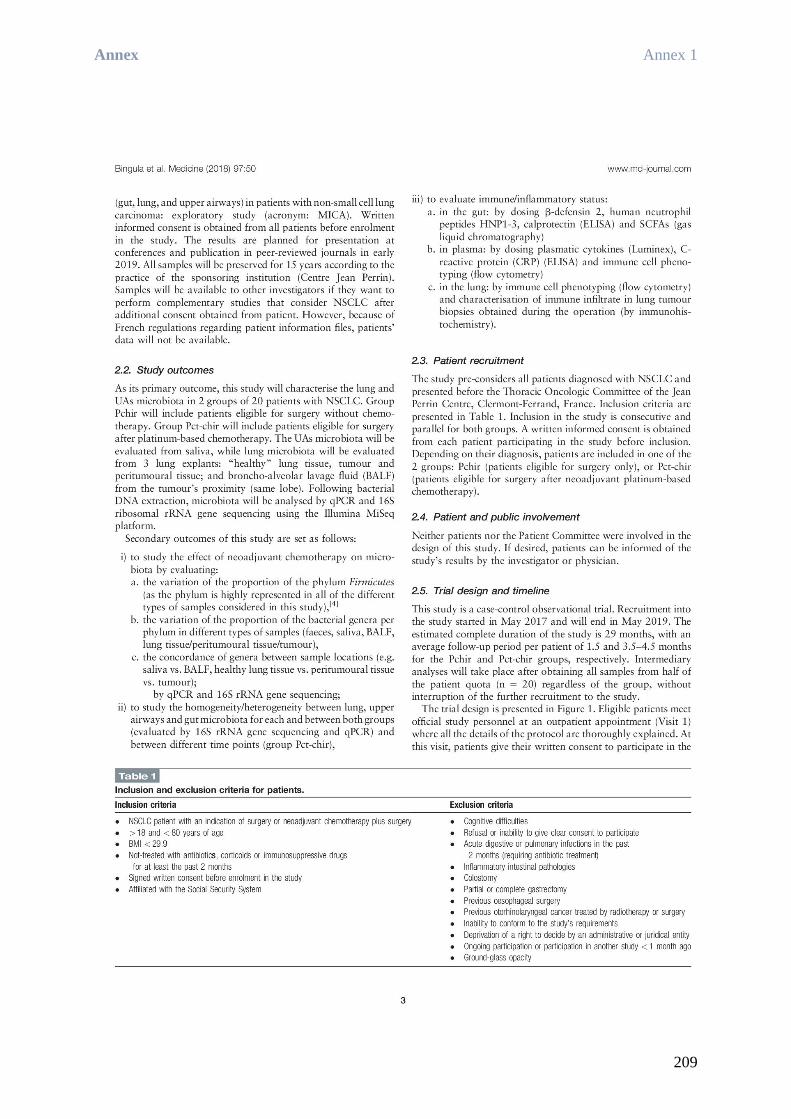
Annex Annex 1
209

Annex Annex 1
210

Annex Annex 1
211
Annex Annex 1
212

Annex Annex 1
213

Annex Annex 1
214
Annex Annex 1
215

Annex Annex 1
216
Annex Annex 1
217

Annex Annex 1
218

Annex Annex 2
219
Annex 2

Annex Annex 2
220
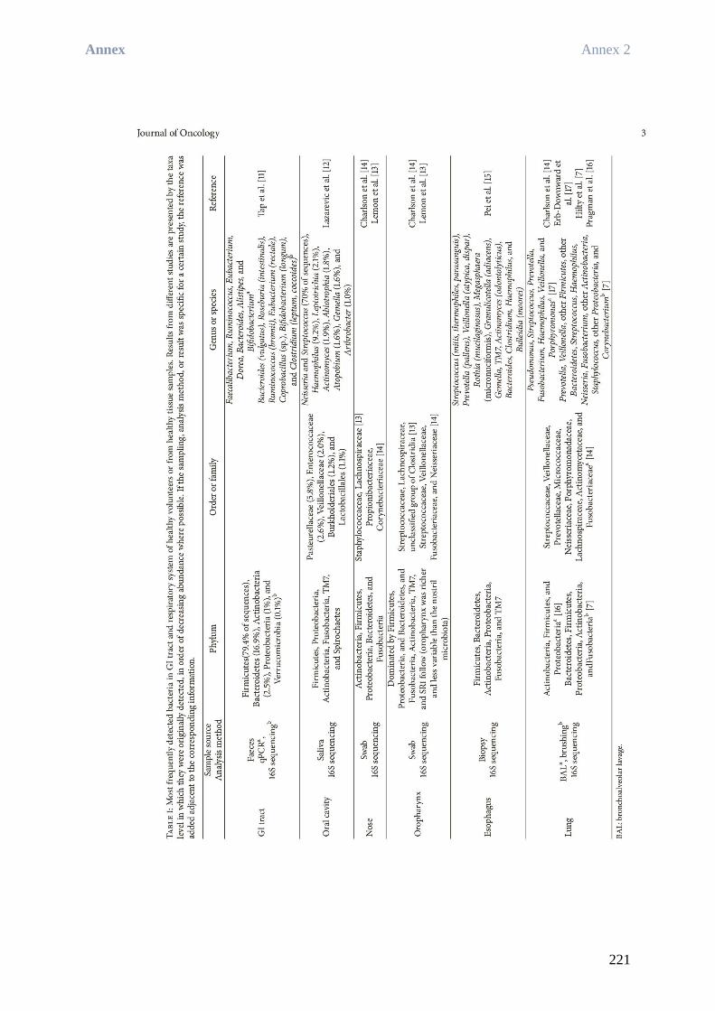
Annex Annex 2
221

Annex Annex 2
222

Annex Annex 2
223

Annex Annex 2
224

Annex Annex 2
225
Annex Annex 2
226

Annex Annex 2
227
Annex Annex 2
228

Annex Annex 2
229

Annex Annex 2
230

Annex Annex 2
231

Annex Annex 2
232

Annex Annex 2
233
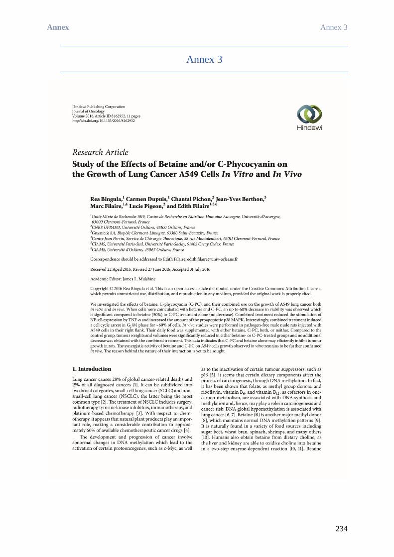
Annex Annex 3
234
Annex 3

Annex Annex 3
235

Annex Annex 3
236

Annex Annex 3
237

Annex Annex 3
238
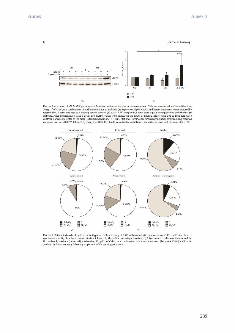
Annex Annex 3
239

Annex Annex 3
240

Annex Annex 3
241

Annex Annex 3
242

Annex Annex 3
243

Annex Annex 3
244

Résumé de thèse en français
Cancer bronchique non à petites cellules, immunité et microbiote :
Base d’étude de l'axe intestin-poumon-cancer du poumon chez
l‘Homme
Soutenue le 20 Décembre 2019 par
Rea BINGULA

Résumé de thèse en français Contexte et problématique
1
1 Contexte et problématique
Selon l'Organisation Mondiale de la Santé (OMS), le cancer du poumon est la première cause
de décès par cancer au monde, avec environ 1 761 000 décès enregistrés et 2 093 876 nouveaux
cas en 2018 (International Agency for Research on Cancer, 2019). La classification se fait en
deux groupes principaux : le cancer bronchique à petites cellules (CBPC) et le cancer
bronchique non à petites cellules (CBNPC). Bien que le CBPC ait un moins bon pronostic et
un taux de létalité très élevé, il ne représente qu’environ 5 % des cas de cancer du poumon.
Le CBNPC est donc plus fréquent et son pronostic dépend des mutations sous-jacentes, du type
histologique et du degré d'agressivité (Molina et al., 2008). Dans le CBNPC, le principal outil
de pronostic est la stadification des tumeurs d'après la classification TNM. Elle permet de
classer les tumeurs en différents stades selon leur taille (T), les changements métastatiques sur
les ganglions lymphatiques (N) et la métastase à distance (M) (Goldstraw et al., 2016). Les
stades les plus avancés, en incluant notamment les changements métastatiques, sont associés à
de moins bons résultats aux traitements et à une survie globale beaucoup plus courte (Planchard
et al., 2019). Jusqu’alors, les mutations dites "d'évasion" étaient considérées comme la
principale voie pour le cancer d’échapper à la réponse immunitaire de l'hôte. En effet, les
cellules cancéreuses accumulent diverses mutations dues à leur taux élevé de renouvellement,
à l'absence de suppresseurs de tumeurs et à des facteurs mutagènes extérieurs (p. ex. fumée,
alcool) jusqu'à ce qu'elles deviennent non identifiables par le système immunitaire de l'hôte
(Dunn et al., 2002).
Cependant, cette approche exclusivement intrinsèque commence à s'assouplir et la recherche
sur le cancer adopte depuis peu une approche plus systémique. Plusieurs facteurs sont
maintenant reconnus comme ayant une influence sur l’immuno-surveillance du cancer, tels que
la nutrition, l'activité physique, le mode de vie (Molina et al., 2008), et plus récemment - le
microbiote.
Le microbiote regroupe un consortium de bactéries, champignons, virus et protozoaires
(Marsland et al., 2015), mais ce terme est le plus souvent utilisé dans la littérature pour désigner
uniquement les bactéries (ce qui est également le cas dans ce manuscrit). Le microbiote du
système gastro-intestinal est le plus étudié, mais il existe aussi des microbiotes résidant sur la
peau et dans les autres cavités corporelles de l’hôte (tractus urogénital, zone oropharyngée,

Résumé de thèse en français Contexte et problématique
2
etc.). Une reconnaissance accrue des fonctions qu’il exerce et son rôle crucial pour l’hôte font
que le microbiote intestinal est souvent considéré comme un "organe oublié".
Avec une biomasse bactérienne de 2 kg, 1 à 10 fois plus de cellules que celles qui constituent
notre corps et 100 fois plus de gènes que le génome humain, cet "organe" assure : l’homéostasie
de l'hôte par la dégradation des nutriments, la synthèse de précurseurs et de vitamines, mais
aussi la stimulation du système immunitaire (Bashiardes et al., 2017; Flint et al., 2012; García-
Castillo et al., 2016; Kamada et al., 2013; Kau et al., 2011; Sender et al., 2016; Zeng et al.,
2016). Le microbiote intestinal a été reconnu comme étant le facteur sous-jacent du
développement du système immunitaire en bas âge (Gensollen et al., 2016; Thaiss et al., 2014),
et sa dysbiose a été reconnue comme étant le facteur sous-jacent de plusieurs troubles
immunitaires, comme l'asthme (Kalliomäki & Isolauri, 2003). On sait maintenant que les
cellules bactériennes commensales stimulent constamment le système immunitaire, en
maintenant un état de " veille " qui permettra de répondre plus efficacement à des antigènes
étrangers, mais aussi en maintenant des mécanismes de " refroidissement " qui empêcheront
une réaction excessive (Curotto de Lafaille et al., 2010; Mazmanian et al., 2005; Noverr &
Huffnagle, 2004).
Cette caractéristique du microbiote commensal a été reconnue dans le traitement du cancer, ce
qui a conduit à l'émergence de la théorie « intestin-lymphe » (Samuelson et al., 2015). Il suggère
que les bactéries intestinales commensales pourraient stimuler l'immunité anti-tumorale non
seulement localement, mais aussi de façon systémique. Ceci est possible grâce à la migration
de bactéries, de leurs métabolites ou de cellules immunitaires stimulées vers l'emplacement
distal via le système lymphatique. Au niveau du site distal, par exemple le lit de la tumeur, ces
facteurs pourraient soit stimuler la réponse antitumorale (comme dans l'intestin), soit des
cellules immunitaires déjà stimulées pourraient exercer directement leur effet antitumoral. La
base de cette théorie repose sur plusieurs travaux menés sur des modèles animaux. Dans ces
études, des animaux élevés dans des conditions exemptes de germes (GF) ou traités avec des
antibiotiques n'ont pas répondu à la chimiothérapie classique avec du cyclophosphamide ou à
la thérapie utilisant des inhibiteurs de point de contrôle immunitaire (Daillère et al., 2016;
Routy, Le Chatelier, et al., 2018; Sivan et al., 2015; Viaud et al., 2013). En revanche,
l’administration de souches bactériennes sélectionnées chez ces animaux a rétabli la réponse au
traitement et a même eu un effet antitumoral si elles étaient administrées sans chimiothérapie
(Sivan et al., 2015).

Résumé de thèse en français Contexte et problématique
3
Ces études ont ouvert un vaste champ d'intérêt sur les oncobiotiques, des bactéries qui
pourraient être utilisées comme médicaments anticancéreux et améliorer la surveillance
immunitaire (Routy, Gopalakrishnan, et al., 2018).
Récemment, un nouvel axe "intestin-poumon" a été évoqué. De multiples observations
suggèrent que la modification d'un régime alimentaire (associée à une modification du
microbiote) ou l'exposition à certaines particules modifient la réponse immunitaire dans les
poumons. La communication inverse est également possible. Sur cette base, la théorie de l'axe
suggère une communication entre ces deux compartiments via la circulation lymphatique et
sanguine. Afin d’aborder ces deux microbiotes très différents, de proposer une connexion basée
sur la recherche à travers le système immunitaire, et d’aborder son application potentielle dans
le cancer du poumon, nous avons écrit un article de synthèse intitulé "Desired Turbulence ?
Gut-Lung Axis, Immunity, and Lung Cancer" (les auteurs dans l’ordre : Rea Bingula,
Marc Filaire, Nina Radosevic-Robin, Mathieu Bey, Jean-Yves Berthon, Annick Bernalier-
Donadille, Marie-Paule Vasson, Edith Filaire), publié dans Journal of Oncology, 17
septembre 2017.
Cet article a porté sur les connaissances actuelles concernant la composition du microbiote
intestinal, rhino-oropharyngé et pulmonaire chez les sujets en bonne santé. Il a fait état de
modifications du microbiote pulmonaire dans le cadre de la Bronchopneumopathie Chronique
Obstructive (BPCO) et dans le cancer du poumon. Il a ensuite fourni une proposition détaillée
d'intéractions entre le système immunitaire de la muqueuse intestinale et le microbiote local, en
tenant compte de l'influence des cellules bactériennes et de leurs métabolites. Cet article a
également porté sur le concept bidirectionnel de l'axe intestin-poumon dans le cadre du cancer,
fondé sur la « théorie de la lymphe » exposée précédemment, et incluant la théorie du cycle
cancer-immunité.
L'intérêt accru pour les caractéristiques du microbiote et de son intéraction avec l'hôte a conduit
à la mise en place du Projet sur le Microbiome Humain (Human Microbiome Project - HMP).
Ce projet vise à obtenir une caractérisation complète du microbiome humain grâce à la
coopération de scientifiques du monde entier. Cependant, le microbiote pulmonaire n’a pas été
initialement inclus dans ce projet (Proctor, 2011), ce qui a encore limité l'intérêt qu’on a pu lui
accorder. Une autre difficulté rencontrée dans ce travail est liée à l'échantillonnage, en raison
du caractère invasif des techniques employées. La plupart des études utilisent comme
échantillon le liquide de lavage broncho-alvéolaire (LBA) obtenu par bronchoscopie, avec un
risque de contamination par les voies respiratoires supérieures (Bassis et al., 2015; Beck et al.,

Résumé de thèse en français Contexte et problématique
4
2012; Charlson et al., 2011). Jusqu'à présent, la plupart des études portaient sur le microbiote
pulmonaire dans le cadre de le BPCO (Banerjee et al., 2004; Einarsson et al., 2016; Erb-
Downward et al., 2011; Moghaddam, 2011; Sze et al., 2015), d'asthme (Gollwitzer & Marsland,
2014; Huang et al., 2011) et de la mucoviscidose (Fodor et al., 2012; Garg et al., 2017). En
effet, dans le cadre de ces pathologies, une bronchoscopie de routine est souvent pratiquée ce
qui simplifie le prélèvement d’échantillons de microbiote pulmonaire.
Ces dernières années ont été marquées par les premières études du microbiote pulmonaire dans
le cas de cancer du poumon. Ces travaux ont mis en évidence une première intéraction entre le
microbiote pulmonaire et des facteurs environnementaux (Yu et al., 2016) et une seconde entre
le microbiote pulmonaire et une inflammation accrue des voies respiratoires inférieures (Segal
et al., 2016).
Même si certaines études évoquaient déjà l'influence "magique" du microbiote intestinal sur la
réponse au cancer du poumon (Schuijt et al., 2016), la communauté scientifique a précisé qu'il
fallait d'abord étudier l'effet du microbiote pulmonaire local avant de pouvoir évoquer le nouvel
axe "intestin-poumon" (Dickson & Cox, 2017).

Résumé de thèse en français Objectifs et hypothèses
5
2 Objectifs et hypothèses
Avant d’élaborer une approche interventionnelle chez l’Homme basée sur des études
antérieures chez l'animal, il est tout d’abord nécessaire d'étudier et de comprendre les facteurs
coexistants dans la réponse de l'hôte au cancer du poumon. Pour ce projet de thèse, nous avons
conçu un essai clinique incluant des patients atteints de CBNPC admissibles à une résection
chirurgicale des tumeurs. Plusieurs objectifs ont été fixés :
i) caractériser le microbiote de l'intestin, des poumons et des voies respiratoires
supérieures chez ces patients
ii) évaluer l'homogénéité/hétérogénéité entre différents microbiotes dans le même
sujet/groupe de patients
iii) évaluer l'impact de la composition du microbiote sur l'état immunitaire et
inflammatoire du patient (évalué dans l'intestin, le sang et les poumons)
Afin d’atteindre ces objectifs, nous avons inclus dans cette étude 17 patients et nous avons
prélevé des échantillons de sang, de salive et de selles. Lors de l’admission et au cours des
interventions chirurgicales, nous avons également prélevé quatre échantillons pulmonaires
(Lavage Broncho Alvéolaire (LBA), tissu non cancéreux, tissus péri-tumoral et tumoral). Les
échantillons salivaires, fécaux et pulmonaires (de 4 types) ont été utilisés pour l'extraction
d'ADN et l'analyse du microbiote par qPCR et séquençage du gène de l'ARN 16S. Afin de
limiter le risque de contamination du LBA par les voies aériennes supérieures, le lavage a été
effectué directement sur le lobe excisé. Des échantillons de selles fraîches ont été utilisés pour
le dénombrement microbiologique des groupes fonctionnels bactériens. Celles-ci ont également
été congelées et utilisées ultérieurement pour doser les Acides Gras à Chaines Courtes (AGCC).
L'état immunitaire a été évalué par la caractérisation de l’infiltration intra-tumorale en
lymphocytes, par le phénotypage des lymphocytes Th et des sous-types de neutrophiles dans le
LBA et le sang, par le dosage des cytokines dans le sang et par le dosage des marqueurs
inflammatoires et antibactériens dans les selles. Chaque patient a également fourni une enquête
nutritionnelle une semaine avant l'intervention chirurgicale afin d’aider à interpréter d'autres
résultats (comme les quantités d'AGCC). D'autres renseignements cliniques et démographiques
ont également été recueillis, comme le lieu de résidence, les antécédents de tabagisme ou la
profession.

Résumé de thèse en français Objectifs et hypothèses
6
Notre première hypothèse a été que le microbiote pulmonaire diffère selon le lieu de
prélèvement, même entre des échantillons proches histologiquement, comme les tissus
péritumoraux et tumoraux. Les échantillons choisis pour l'analyse ont des fonctions
physiologiques et/ou une architecture différente, mais aussi des intéractions différentes avec le
système immunitaire. Le LBA est représentatif de la population microbienne de la lumière
bronchique, donc principalement une population bactérienne aux caractéristiques
planctoniques, ou au contraire, associée aux biofilms ou mucus excrétés par les cellules
alvéolaires. Le tissu non cancéreux représentera quant à lui, un échantillon avec une architecture
pulmonaire normale faite de petites alvéoles et d'épithélium pulmonaire monocouche.
Selon le type histologique la tumeur peut varier, allant d'un tissu très dense comme dans le
carcinome épidermoïde, à un tissu enrichi en mucus, comme dans certains types
d'adénocarcinomes. L'augmentation de la production de mucus pourrait directement servir de
nutriment aux bactéries mucinolytiques (Flynn et al., 2016) et ainsi favoriser la croissance de
certains groupes bactériens. Enfin, l’échantillon de tissu péritumoral, sera considéré comme un
échantillon bien distinct car il possède des caractéristiques histologiques différentes. Même si,
de par son architecture, le tissu péritumoral ressemble à un tissu non cancéreux, sa proximité
avec la tumeur le rend exposé à diverses substances excrétées par les cellules tumorales et à une
invasion possible des cellules tumorales elles-mêmes (Dou et al., 2018).
Un autre aspect important est le rôle du tissu péritumoral, c'est-à-dire du microenvironnement
tumoral (MET), sur la suppression ou la stimulation directe de la tumeur de diverses manières:
par la modification de la composition de la matrice du MET, par la production de facteurs de
croissance, etc. (Valkenburg et al., 2018). Ces deux évènements pourraient directement (avec
des changements dans l'architecture des tissus) ou indirectement (avec une réponse immunitaire
contre les cellules tumorales) influencer le microbiote local.
Nous avons ensuite émis l'hypothèse qu'il y aurait plusieurs facteurs potentiellement
discriminants pour le système immunitaire et le microbiote dans les six échantillons prélevés
(salive, selles et les quatre échantillons pulmonaires).
Le premier facteur est le type histologique de tumeur ; il a déjà été démontré que le type
histologique modifie le microbiote salivaire des patients atteints de cancer du poumon (Yan et
al., 2015), mais également l’alpha diversité dans les tissus tumoraux (Yu et al., 2016). Les
différents types histologiques vont avoir un impact direct sur le microbiote tel qu'expliqué dans
le paragraphe précédent. Ils sont aussi caractérisés par une immunogénicité, une nature

Résumé de thèse en français Objectifs et hypothèses
7
immunosuppressive et un fond génétique différents (Busch et al., 2016), ce qui signifie qu'ils
ne stimuleront pas de la même manière le système immunitaire. Par conséquent, l'afflux et le
profil des cellules immunitaires vers le lit tumoral varient, de même que l'intéraction avec le
microbiote local. La nature de la tumeur et la situation immunitaire locale pourrait se refléter
sur les marqueurs systémiques, et donc sur l'état immunitaire intestinal. Par conséquent, le
microbiote intestinal peut aussi varier selon le type de tumeur. Cependant, dans ce type d'étude,
il n'a pas été possible de déterminer si ce changement est la conséquence d'une maladie, d'un
facteur concomitant ou le résultat d’un changement dans l'équilibre immunitaire.
Le deuxième facteur important est le statut tabagique du patient. La fumée de cigarette présente
plusieurs effets indésirables sur le système respiratoire, comme l'inhalation d'air chaud et la
précipitation de particules potentiellement cancérigènes. Ceci endommage les cellules
épithéliales et altèrent la clairance des voies respiratoires, induisant une inflammation locale et
systémique (Çolak et al., 2019). En résulte une modification des conditions locales de
croissance pour le microbiote et une perturbation de l'homéostasie immunitaire. Les voies
respiratoires supérieures sont affectées par la fumée, et des changements de microbiote liés au
tabagisme ont déjà été rapportés (Wu et al., 2016). Fait intéressant, il n’a pas été observé de
changements dans les voies respiratoires inférieures (Segal et al., 2013). Ainsi, il est possible
que les microbiotes des différents sites pulmonaires ne soient pas sensibles de la même manière
aux effets du tabagisme. Cette hypothèse sera vérifiée dans ce travail. De plus, il a été observé
que le tabagisme modifiait le microbiote intestinal, tant chez les personnes en bonne santé
(Stewart et al., 2018) que chez les patients atteints de cancer du poumon (Zhang et al., 2018;
Zhuang et al., 2019), ce qui montre clairement l'effet du tabagisme au niveau systémique.
Un autre facteur important est l'emplacement de la tumeur. Les poumons sont organisés en cinq
lobes, deux du côté gauche (supérieur et inférieur) et trois du côté droit (supérieur, moyen et
inférieur). Il est intéressant de noter que des taux de survie globale plus faibles et un pronostic
plus défavorable ont été associés à des tumeurs dans les lobes inférieurs, et particulièrement
dans le lobe inférieur gauche (Kudo et al., 2012). Le modèle insulaire adapté à la biogéographie
pulmonaire des sujets sains suggère déjà que le microbiote pulmonaire est différent entre les
lobes (Dickson et al., 2015). Ce modèle décrit la bronche principale comme étant la source des
communautés microbiennes pulmonaires, dont la richesse diminue proportionnellement à
l’éloignement de ce site. Par conséquent, les lobes supérieurs devraient être plus riches en
microorganismes que les lobes inférieurs, mais les lobes controlatéraux devraient avoir des
caractéristiques similaires. Cependant, l'étude qui compare le LBA controlatéral (c'est-à-dire du

Résumé de thèse en français Objectifs et hypothèses
8
lobe gauche et du lobe droit), l'un cancéreux et l'autre non, montre que ce n'est pas le cas pour
le cancer du poumon (Liu et al., 2018). Il est évident que la présence de tumeurs modifie la
composition microbienne et que d'autres études incluant des patients atteints de cancer du
poumon seront nécessaires.
Nous nous sommes ensuite concentrés sur un autre facteur important ayant un rôle pronostique
crucial dans le traitement du CBNPC : la stadification de la tumeur. Plus le stade est élevé, plus
le pronostic global pour le patient est mauvais. Cependant, le stade est déterminé par la
combinaison des trois éléments dans la classification TNM : la taille de la tumeur T, la présence
de métastases au niveau des ganglions lymphatiques médiastinaux et ipsilatéraux N, et la
présence de métastases distantes M (Goldstraw et al., 2016).
Un même stade peut donc parfois représenter des tumeurs véritablement différentes. Par
exemple, une très grosse tumeur sans ganglions lymphatiques métastatiques pourrait appartenir
au même stade qu'une petite tumeur mais avec des ganglions métastatiques. Pour les besoins de
l'étude du microbiote, nous avons décidé d'observer séparément ces éléments et de définir
l'hypothèse que chacun d'entre eux pourrait influencer individuellement nos communautés
microbiennes.
Étant donné que les patients présentant des métastases à distance ne sont pas inclus dans l'étude
en raison de la différence dans la procédure de traitement (non opérable), nous avons considéré
deux autres éléments : la taille de la tumeur et la présence de ganglions lymphatiques
métastatiques. Selon le diamètre, la tumeur peut être plus ou moins oxygénée en raison d'une
vascularisation souvent très fuyante et insuffisante pour les tumeurs de plus grandes tailles,
avec par conséquent un degré de nécrose interne différent (Wouters et al., 2003). Ceci
favoriserait la colonisation de bactéries anaérobies qui pourraient soit défavorablement
diminuer le pH par production de lactate, soit, favorablement augmenter l'immunogénicité
tumorale (Baban et al., 2010; Damgaci et al., 2018; Van Dessel et al., 2015). Des tumeurs plus
grosses pourraient également bloquer les voies bronchiques et ainsi créer des poches micro-
aérobies ou anaérobies. Ceci favoriserait les changements dans la composition microbienne ou
la colonisation par des agents pathogènes.
La présence de ganglions lymphatiques métastatiques est, en revanche, le signe d'une plus
grande agressivité tumorale et d'une fuite vasculaire permettant la dissémination des cellules
tumorales aux ganglions lymphatiques proximaux quelle que soit la taille de la tumeur.
L’existence de ces ganglions lymphatiques métastatiques dénote le caractère invasif de la

Résumé de thèse en français Objectifs et hypothèses
9
tumeur et signale également un microenvironnement "permettant" la propagation des cellules
tumorales. Selon le Dr Bissell (Bissell & Hines, 2011), l'environnement tumoral joue un rôle
énorme dans le développement et le devenir de la tumeur. Dans le cadre de notre étude, certaines
bactéries de l'environnement tumoral pourraient par exemple occuper les sites d’adhésion de
différentes intégrines cellulaires (Garrett, 2015; Khan et al., 2012). L’occupation de ces sites
inhiberait la diapédèse métastatique des cellules tumorales. La présence de ganglions
lymphatiques métastatiques étant un marqueur important de pronostic négatif pour la survie, et
ce quelle que soit la taille de la tumeur (Liu et al., 2017). Elle a été priorisée dans notre analyse.
On sait que les patients atteints de cancer, en particulier lors de stade avancé de la maladie,
signalent souvent une perte de poids sans changements particuliers dans leur alimentation. Ce
phénomène appelé cachexie s'explique, d’une part par la présence de cellules cancéreuses
voraces qui peuvent capter la majeure partie de l'énergie de l'hôte obtenue par digestion, et
d’autre part par différentes cytokines, qui produites par l'hôte en réponse au cancer peuvent
modifier son métabolisme (Barton, 2001). Des changements au niveau du microbiote intestinal
liés à la présence du cancer, ainsi que l'utilisation de probiotiques dans la lutte contre la cachexie
ont été envisagés, mais il reste nécessaire de poursuivre les recherches dans ces domaines
(Bindels & Thissen, 2016). Par conséquent, notre hypothèse concernant l'effet du stade tumoral
sur le microbiote pulmonaire s'est également étendue au microbiote intestinal (en tenant compte
des informations sur la stabilité pondérale du patient et de leur étude nutritionnelle pour une
approche personnalisée).
Enfin, la théorie de l'axe intestin-poumon implique que certains éléments d'origine intestinale,
comme les bactéries ou leurs métabolites, amorceraient les cellules immunitaires situées dans
les ganglions mésentériques. Ces cellules immunitaires seraient ensuite recrutées par des sites
éloignés où elles pourraient agir avec plus de performance (Bingula et al., 2017). Dans notre
cas, cette performance accrue pourrait être caractérisée par une meilleure infiltration des
cellules immunitaires dans les tumeurs pulmonaires. Cependant, il est important de garder à
l'esprit qu'il existe trois types généraux de tumeurs : les tumeurs immuno-inflammées (angl. «
immune-inflamed »), les tumeurs immuno-exclues (« immune-excluded ») et les tumeurs
immuno-désertes (« immune-desert ») (Chen & Mellman, 2017). Sommairement, les tumeurs
immuno-inflammées sont bien infiltrées et immunogènes et les tumeurs immuno-désertes sont
mal infiltrées et peu immunogènes. Les tumeurs immuno-exclues sont quant-à-elles souvent
immunogènes mais avec certains éléments intrinsèques, comme l'expression du PD-1 (Death
Domain Protein-1), qui inhibent l'infiltration et l'activité des cellules immunitaires.

Résumé de thèse en français Objectifs et hypothèses
10
Théoriquement, il serait possible de passer d’un type d’immunité tumorale à l’autre. Par
exemple une tumeur immuno-inflammée qui, après intervention du système immunitaire, ne
conserve que des cellules faiblement immunogènes pourrait devenir immuno-déserte. L’inverse
est également possible : un meilleur amorçage des cellules immunitaires pourrait transformer
une tumeur immuno-déserte en une tumeur immuno-inflammée grâce à une réactivité croisée
entre les cellules tumorales et les épitopes bactériens. Par conséquent, notre hypothèse a reposé
sur le fait que le microbiote intestinal, mais aussi le microbiote pulmonaire, varierait selon le
type immunitaire tumoral.
Les hypothèses avancées seront développées plus en détail au fur et à mesure de l'avancement
des analyses. Les hypothèses exposées comprennent des facteurs ayant une influence à la fois
locale et systémique sur l'hôte, et donc sur son microbiote et son système immunitaire. C'était
la raison sous-jacente pour laquelle il fallait aborder les patients sous différents angles
(microbiote de trois sites, marqueurs immunitaires et inflammatoires locaux et systémiques par
différentes techniques, métabolites bactériens, nutrition et données démographiques). Les
objectifs et les hypothèses que nous allons vérifier seront discutés dans les chapitres "Résultats"
et "Discussion générale".

Résumé de thèse en français Résultats principaux
11
3 Résultats principaux
Study Protocol
Characterisation of Gut, Lung and Upper Airways Microbiota in Patients with Non-
Small Cell Lung Carcinoma: Study Protocol for Case-Control Observational Trial
Rea Bingula, MS, Marc Filaire, Prof, MD, Nina Radosevic-Robin, MD, Jean-Yves Berthon,
PhD, Annick Bernalier-Donadille, PhD, Marie-Paule Vasson, Prof, Emilie Thivat, PhD, Fabrice
Kwiatkowski, MS, Edith Filaire, Prof
Publié dans Medicine le 14 Décembre 2018
La majorité des études explorant le microbiote pulmonaire par séquençage à haut débit utilisent,
pour l'isolement du microbiote, des kits commerciaux qui ne sont pas spécialement adaptés aux
échantillons pulmonaires (exemple : MoBio PowerSoil ou QIAamp Stool Minikit de Qiagen).
Étant donné que les échantillons pulmonaires diffèrent significativement en qualité (tissu très
fibreux) et en niveau de populations bactériennes par rapport aux fèces ou au sol, ces kits ne
sont pas les mieux adaptés et leur rendement final est insuffisant (utilisation de colonnes de
filtration, etc.). C'est pourquoi nous avons décidé de mettre au point un protocole d'"isolement
du microbiote pulmonaire", basé sur le protocole du Human Microbiome Project pour
l'isolement du microbiote intestinal ; protocole dont la performance à précédemment été
comparée à d'autres kits disponibles sur le marché (Costea et al., 2017; Mcinnes, 2010). La
version finale optimisée du protocole, comprenant la conception de l'étude, et le matériel et les
méthodes utilisés ont été publiés dans l’article suivant : "Caractérisation du microbiote de
l'intestin, des poumons et des voies respiratoires supérieures chez les patients atteints de cancer
du poumon non à petites cellules" publié dans Medicine le 14 décembre 2018. Seule la partie
du projet concernant les patients admissibles à la chirurgie sans chimiothérapie sera présentée
dans ce rapport de thèse, et son aperçu synthétique est présenté en Figure 1.

Résumé de thèse en français Résultats principaux
12
Figure 1 Aperçu synthetique d’étude

Résumé de thèse en français Résultats principaux
13
Article 1
Research Article
Characterisation of microbiota in saliva, bronchoalveolar lavage fluid, non-malignant,
peritumoural and tumour tissue in non-small cell lung cancer patients: cross-sectional
clinical trial
Rea Bingula, Edith Filaire, Ioana Molnar, Eve Delmas, Jean-Yves Berthon, Marie-Paule
Vasson, Annick Bernalier-Donadille, Marc Filaire
Soumis dans BMC Respiratory Research le 05 Décembre 2019.
Introduction
Même si des études portant sur le microbiote du poumon cancéreux ont commencé à émerger
il y a plusieurs années, les caractéristiques du microbiote pulmonaire provenant de différents
types d'échantillons ne sont pas encore bien documentées. Le principal problème de la recherche
sur le microbiote pulmonaire provient de différentes méthodes d’échantillonnage qui sont à
l’origine de l’hétérogénéité des échantillons cibles d'une étude à l'autre. Ainsi la majorité des
études actuelles portant sur le microbiote du poumon ont été réalisées sur le LBA, les tissus, la
salive et les expectorations à fréquence variable. Bien qu'il ait déjà été suggéré que le microbiote
pulmonaire provenant du LBA et des tissus pulmonaires abrite des communautés différentes
(Sze et al., 2012), aucune étude n’a encore été réalisée sur ce sujet. De plus, l'analyse du
microbiote dans le cadre de cancer du poumon implique des tissus ayant des rôles, des
intéractions et une immunité distincte, comme les tissus péritumoraux et tumoraux. Par
conséquent, l'objectif de cette étude était d’une part, de fournir une caractérisation détaillée du
microbiote pulmonaire oral et d’autre part, d’obtenir de nouvelles données innovatrices dans ce
domaine. Puisque les tumeurs pulmonaires des lobes inférieurs (LL) ont été associées à une
diminution de la survie, les caractéristiques du microbiote des lobes supérieurs (UL) et
inférieurs ont également été examinées.
Matériels et méthodes
À l'aide de la technologie de séquençage du gène 16S de l'ARNr, nous avons analysé le
microbiote dans la salive, le LBA (obtenu directement sur le lobe excisé), les tissus non
cancéreux, péritumoraux et tumoraux de 18 patients CBNPC admissibles à un traitement
chirurgical. Nous avons fourni : une taxonomie détaillée, la diversité et les principaux groupes
Résumé de thèse en français Résultats principaux
14
bactériens pour chaque microbiote, avec analyse de l'abondance différentielle à tous les niveaux
taxonomiques entre les échantillons et en fonction de la localisation des prélèvements dans les
poumons (modèle linéaire général binomial « zero-inflated » avec correction Benjamini-
Hochberg).
Résultats, doscussion et conclusion
Nous démontrons que le LBA représente un microbiote unique en comparaison avec les autres
échantillons pulmonaires. Ce résultat est basé sur l'analyse de l'abondance différentielle et sur
la Bêta diversité. Simultanément, nous confirmons que les échantillons de LBA forment un
groupe distinct avec les autres échantillons de tissus pulmonaires lorsqu'ils sont placés dans la
perspective du microbiote salivaire. La position du lobe est un facteur important influençant la
composition du microbiote (hypothèse du modèle insulaire adapté à la biogéographie
pulmonaire) et en même temps, les tumeurs du lobe inférieur ont été associées à un pronostic
moins favorable chez les CBNPC. Sur cette base, nous avons fait le choix d’examiner
l'influence de la position du lobe supérieur ou inférieur sur le microbiote analysé. Nous
montrons ainsi que tous les microbiotes analysés, à l'exception du microbiote tumoral, ont une
abondance significativement différente, en particulier dans les phyla Bacteroidetes et
Actinobacteria. De plus, nous montrons que le microbiote du tissu péritumoral est le plus
susceptible au changement selon son emplacement, avec une ressemblance significativement
accrue au microbiote du LBA lorsqu'il se trouve dans les lobes supérieurs. Puisque la tumeur
est connue pour son potentiel à modifier directement son microenvironnement, nous faisons
l’hypothèse que ce changement observé dans le microbiote des tissus péritumoraux reflète en
fait les changements du microenvironnement (sites adhésifs, réarrangement de la matrice
extracellulaire, stimulation immunitaire). Enfin, nous montrons que le phylum des Firmicutes,
précédemment signalé comme étant un phylum abondant dans les tumeurs pulmonaires
évolutives, est également élevé en abondance dans tous les tissus pulmonaires des lobes
inférieurs par rapport aux lobes supérieurs. Il pourrait s'agir d'un potentiel indicateur microbien
d'une agressivité accrue des tumeurs du lobe inférieur, mais il faudra toutefois confirmer cette
hypothèse en incluant notamment davantage les caractéristiques du système immunitaire.
Résumé de thèse en français Résultats principaux
15
Article 2
Research Article
Inverse abundance pattern of tumour and extratumoural lung microbiota between non-
small cell lung cancer patients with or without metastatic lymph nodes: a cross-sectional
clinical study
Rea Bingula, Edith Filaire, Jérémie Talvas, Jean-Yves Berthon, Marie-Paule Vasson, Annick
Bernalier-Donadille, Nina Radosevic-Robin, Marc Filaire
Soumis dans BMC Microbiome le 25 Octobre 2019
Introduction
La présence de ganglions lymphatiques métastatiques (LN) est l'un des marqueurs de pronostic
négatif les plus importants dans le CBNPC (Goldstraw et al., 2016). Il est caractérisé par un
temps de survie beaucoup plus court et par un taux de récidive accru. Nous avons cherché à
savoir si cette caractéristique importante pour le devenir du patient est associée de façon
significative à la composition locale du microbiote pulmonaire. Nous nous sommes basés sur
l'analyse du microbiote pulmonaire de quatre échantillons différents (LBA, tissus non
cancéreux, péritumoraux et tumoraux). De plus, nous avons examiné si ces deux paramètres
(présence ou absence de métastases ganglionnaires) influencent l'immunité locale en étudiant
la composition de l'infiltrat immunitaire tumoral et les phénotypes des lymphocytes Th dans le
LBA. Enfin, nous avons cherché à savoir si cette réponse immunitaire est associée à différents
microbiotes pulmonaires.
Matériels et méthodes
Le microbiote pulmonaire a été analysé en LBA (lavage direct du lobe excisé) et dans trois
tissus (tissus non cancéreux, péritumoraux et tumoraux) par séquençage du gène de l'ARN
ribosomal 16S, suivi par l'analyse biostatistique en RStudio. Le statut immunitaire a été évalué
en dénombrant les lymphocytes infiltrant les tumeurs (marqueurs de l'expression des CD8,
CD20, FOXP3, PD-1 et PD-L1) et en déterminant les profils des lymphocytes T-Auxiliaires et
les neutrophiles dans le LBA. Dans la population lymphocytaire, la sous-population de
lymphocytes T-Auxiliaires CD3+CD4+ a été caractérisée comme suit : Th1 avec marqueur
CXCR3+, Th2 avec marqueur CRTH2+, Th17 avec marqueur CCR6+ et Treg avec marqueurs
CD25+CD127-. Dans la population de granulocytes, les sous-populations de neutrophiles ont

Résumé de thèse en français Résultats principaux
16
été caractérisées comme suit : neutrophiles circulants avec marqueurs CD15+CD62L+CD11b-,
forme transitoire avec marqueurs CD15+CD62L+CD11b+, et neutrophiles activés avec
marqueurs CD15+CD62L-CD11b+. Toutes les analyses statistiques ont été réalisées avec
RStudio.
Résultats, doscussion et conclusion
Nous montrons qu'en présence de ganglions lymphatiques métastatiques, tous les échantillons
de microbiote pulmonaire présentent une abondance accrue du phylum des Firmicutes, tandis
que les mesures de diversité montrent des valeurs plus faibles pour les deux microbiotes des
tissus extratumoraux. Nous avons identifié 31 genres bactériens présentant des schémas
inverses d’abondance entre les tissus tumoraux, extratumoraux, et entre les patients avec et sans
ganglions lymphatiques métastatiques. De plus, nous montrons que cette inversion est
étroitement liée au profil respiratoire bactérien. Plus précisément, les genres anaérobies
identifiés sont significativement plus abondants dans les tumeurs à ganglions lymphatiques
métastatiques, alors que les bactéries aérobies sont plus abondantes dans les tissus
extratumoraux. Cependant, nous observons exactement le phénomène inverse pour ces mêmes
genres bactériens chez les patients ne présentant pas de ganglions lymphatiques métastatiques.
Nous avançons donc l’hypothèse que les bactéries "migrent" entre les tissus vers des conditions
de croissance plus favorables, car l'environnement tumoral change fortement en fonction de sa
progression (hypoxie accrue, nécrose, production de lactate, pH faible). De plus, nous
identifions plusieurs biomarqueurs potentiels dans chaque tissu qui pourraient faciliter ou
améliorer la détection des métastases des ganglions lymphatiques sans imposer de biopsie.
Enfin, nous montrons que le microbiote du LBA ne change pas de façon significative en
fonction du stade tumoral, mais qu'il est le seul à présenter une association significative avec
les marqueurs immunitaires protumoraux ou antitumoraux dans le LBA et dans la tumeur.

Résumé de thèse en français Discussion, conclusion et perspectives
17
4 Discussion, conclusion et perspectives
4.1 Discussion
En raison des multiples axes de recherche possibles dans ce projet, nous avons d'abord décidé
de nous concentrer sur la région pulmonaire, moins explorée que la région intestinale. C'est
pourquoi nous nous sommes focalisés dans un premier temps sur la caractérisation des
différents microbiotes pulmonaires (tumeur, tissu péritumoral et non cancéreux, et LBA), leur
relation avec le microbiote salivaire et l'impact de la localisation du lobe tumoral (Article 1).
Dans un second temps, afin d’envisager une application clinique directe de notre recherche,
nous avons donc cherché à déterminer s'il existe une intéraction entre : la présence de ganglions
lymphatiques métastatiques (l'un des principaux facteurs pronostiques du CBNPC), différents
microbiotes pulmonaires et l'immunité locale (Article 2). D'autres axes de recherche seront
abordés dans « Conclusion et Perspectives ».
Le LBA représente un microbiote pulmonaire unique. Il n’est que sensiblement plus
semblable à la salive que les tissus pulmonaires.
Depuis le début des recherches portant sur l'étude du microbiote pulmonaire, plusieurs
inconvénients sont apparus. L'un des plus importants reste le choix de l'échantillon pulmonaire
qui représenterait correctement les communautés pulmonaires, mais qui ne serait pas trop
invasif pour le patient. Par conséquent, le LBA était et demeure le type d’échantillon le plus
fréquemment utilisé dans les études portant sur le microbiote pulmonaire en général et dans les
cas de cancer du poumon (Laroumagne et al., 2011; Lee et al., 2016; Liu et al., 2018; Tsay et
al., 2018; Wang et al., 2019). Cependant, il a déjà été suggéré que le microbiote pulmonaire
présent dans le LBA et dans les tissus pulmonaires ne partagent pas les mêmes caractéristiques
(Sze et al., 2012), en raison de la physiologie différente des échantillons.
Nous sommes les premiers à avoir montré que le LBA abrite un microbiote distinct de celui du
tissu pulmonaire, en incluant dans nos analyses jusqu'à trois échantillons de tissus différents
(tissu non cancéreux, péritumoral et tumoral) (Article 1). Le LBA représente un échantillon
unique, en se basant à la fois sur la Bêta diversité et sur l'analyse de l'abondance différentielle.
Dans le LBA, les Firmicutes représentent le phylum le plus abondant. En revanche, les
Protéobactéries sont plus abondantes dans les échantillons de tissus pulmonaires. De plus, en
examinant séparément les caractéristiques du microbiote dans les lobes supérieurs (UL) ou

Résumé de thèse en français Discussion, conclusion et perspectives
18
moyens/inférieurs (LL), nous avons identifié des clades entiers avec une abondance inverse
entre le LBA et le microbiote des tissus extratumoraux, tels que les Flavobacteriia et les
Actinobacteria, confirmant l’hypothèse initiale de deux communautés microbiennes distinctes.
Étant donné que dans notre plan d'étude, le LBA a été obtenu pour la première fois directement
sur un lobe excisé, nous avons eu l'opportunité d'estimer si le LBA des lobes supérieurs était
plus semblable à la salive (représentant du microbiote des voies respiratoires supérieures) que
celui des lobes inférieurs. Selon la théorie du modèle insulaire adapté de la biogéographie
pulmonaire (Dickson et al., 2015), les lobes supérieurs devraient avoir une composition en
communautés bactériennes plus semblable à celle des voies respiratoires supérieures. Ils
devraient également présenter une richesse accrue par rapport aux lobes inférieurs. Cependant,
nous n'avons pas constaté de différence significative dans la distance (Bêta diversité) entre la
salive et le LBA du lobe tumoral inférieur ou supérieur, malgré une légère augmentation de
l'abondance des taxons typiquement oraux des lobes supérieurs (tels que les genres Veillonella
ou Prevotella). Une explication de ce résultat pourrait être soit la présence de tumeurs qui
influenceraient les propriétés du microbiote, comme l’ont suggéré précédemment (Liu et al.,
2018), soit à une approche d'échantillonnage différente qui a considérablement réduit la
contamination des voies aériennes supérieures.
Nous avons au contraire observé une richesse et une diversité significativement plus faibles
dans le LBA des lobes inférieurs. Ces deux éléments sont similaires dans les tissus tumoraux et
non cancéreux ; plusieurs raisons sont possibles. Premièrement, le LBA est plus sensible à la
distance de la bronche centrale que les tissus, ce qui entraîne une plus faible richesse dans les
lobes inférieurs selon l’hypothèse du modèle insulaire adapté à la biogéographie pulmonaire
(Dickson et al., 2015). Deuxièmement, comme les lobes supérieurs du poumon sont plus dilatés
que les lobes inférieurs pendant l'inspiration lente, une quantité d’air plus importante pénètre
dans les lobes inférieurs que dans les lobes supérieurs (Chang & Yu, 1999). Nous pouvons
avancer l’hypothèse d’un environnement plus dynamique dans les lobes inférieurs que dans les
lobes supérieurs, ce qui limiterait l'établissement d'une communauté bactérienne plus riche,
contrairement aux voies aériennes supérieures. Ceci fera l'objet de recherches plus
approfondies.

Résumé de thèse en français Discussion, conclusion et perspectives
19
Microbiote des tissus péritumoraux comme potentiel marqueur indirect d’une modification de
l'environnement tumoral
Il a été démontré que la progression de la tumeur n'est pas seulement liée à la tumeur elle-même,
mais aussi à son environnement. Des travaux ont permis d’observer une progression tumorale
par remodelage de la Matrice Extra Cellulaire (MEC) sous l’influence de facteurs tumorogènes
(Altinay, 2016) (Figure 2). D’autres travaux ont également démontrés les caractéristiques
répressives de l'environnement sur la tumeur (Bissell & Hines, 2011). Cependant, aucune étude
n'a jusqu'à présent examiné si les tissus péritumoraux contenaient des microbiotes qui diffèrent
de ceux de la tumeur ou des tissus non cancéreux dans le cas du CBNPC.
Figure 2 La modification de la MEC favorise la progression du cancer (adapté d'Altinay
2016). (A) structure normale de la MEC. (B) Dans des conditions pathologiques, il existe une production excessive de
fibroblastes activés qui contribuent à la surproduction de MEC et à la dérégulation des enzymes responsables de
la modification de la MEC. En raison d’un fragile équilibre entre les cellules locales et la MEC, ces changements
stimulent la transformation cellulaire épithéliale et l'hyperplasie, créant une matrice plus rigide. (C) En plus des
cellules locales, les cellules immunitaires recrutées sur le site de la tumeur favorisent l'inflammation locale,
accompagnée d'une néoangiogénèse facilitant la propagation de la tumeur en raison de la fuite du système
vasculaire. (D) Les métastases qui se sont maintenues dans la circulation, s’extravasent au niveau d’un site distal
et expriment les enzymes de modification de la MEC pour créer une niche métastatique locale qui favorisera
davantage la survie et la prolifération des cellules cancéreuses.
Nous sommes les premiers à inclure le tissu péritumoral dans la caractérisation du microbiote
pulmonaire (Article 1), montrant ainsi qu'il s'agit du seul échantillon de tissu pulmonaire
présentant des changements significatifs de richesse et d’Alpha et Bêta diversité en fonction de
la localisation du lobe. Nous avons signalé que les tissus péritumoraux des lobes supérieurs

Résumé de thèse en français Discussion, conclusion et perspectives
20
présentaient une similarité accrue avec ceux du LBA (observés en Bêta diversité et avec une
présence plus élevée du phylum des Firmicutes). Les tissus péritumoraux des lobes inférieurs
présentent quant-à-eux les mêmes caractéristiques que les deux autres tissus. Nous avons
suggéré que ceci reflète en fait différents types de modifications de la MEC, influençant à la
fois les propriétés adhésives et l'architecture des tissus (Altinay, 2016; Bissell & Hines, 2011;
Quail & Joyce, 2013) et modifiant directement les conditions locales de croissance des
bactéries. Dans la pathogénicité du CBNPC, les tumeurs des lobes inférieurs ont été associées
à une diminution de la survie globale, mais sans explication claire (Kudo et al., 2012; Riquet et
al., 2016). Notre découverte pourrait indiquer que la détection indirecte de différences subtiles
dans l'environnement de la tumeur pourraient être liées soit à son agressivité accrue, soit au
développement de différents types de microbiotes qui pourraient permettre ou non la
progression du cancer (par exemple l'occupation de sites d’adhésion et la progression
métastatique).
Lobes inférieurs et présence de ganglions lymphatiques métastatiques marqués par une plus
grande abondance du phylum des Firmicutes
Plusieurs études ont déjà mis en évidence l'augmentation du phylum des Firmicutes dans le
microbiote pulmonaire en comparant un groupe témoin à un groupe cancéreux. Cette
observation a été d'un intérêt particulier pour le BPCO (Sze et al., 2012) puisque des études ont
montré que cette pathologie pouvait conduire au développement du cancer du poumon mais
aussi que les deux maladies sont basées sur une inflammation clinique ou subclinique créant
ainsi une base mutuelle de « réflexion » (Houghton, 2013; Melkamu et al., 2013).
Nous sommes les premiers à signaler que le microbiote pulmonaire, basé sur l’analyse de quatre
types d'échantillons différents, présente une abondance moyenne plus élevée de
l’embranchement des Firmicutes si la tumeur se situe dans les lobes inférieurs (Article 1). De
plus, nous avons observé une augmentation des Firmicutes dans les tissus pulmonaires si le
patient présentait des changements métastatiques au niveau des ganglions lymphatiques locaux
(Article 2). Ces deux résultats concordent avec ceux décrits dans le paragraphe précédent, qui
établissait un lien entre ce "déplacement" et l'état avancé de la maladie ou de sa présence. Dans
notre étude, ce "déplacement" était principalement dû à une abondance relative
significativement plus élevée du genre Clostridium. Cependant, la détermination plus précise
des espèces impliquées, leur quantité absolue et l'implication de facteurs immuns tumoraux
feront l’objet de futures recherches.

Résumé de thèse en français Discussion, conclusion et perspectives
21
Les ganglions lymphatiques métastatiques séparent le microbiote du tissu tumoral et du tissu
extratumoral selon un schéma inverse correspondant aux profils respiratoires bactériens
La stadification tumorale est le principal outil de pronostic du CBNPC et sert également au
choix du traitement approprié (Goldstraw et al., 2016). Elle est basée sur la classification TNM
qui inclut l'information sur la taille de la tumeur, l'implication des changements métastatiques
sur les ganglions lymphatiques ipsilatéraux et médiastinaux, et l'apparition de métastases
distales. La présence de changements métastatiques aggrave considérablement le pronostic par
rapport aux patients présentant des tumeurs plus importantes mais sans changements
métastatiques, puisque leur présence a été associée à une survie globale raccourcie de 5 ans
(Goldstraw et al., 2016; Planchard et al., 2019).
Nous sommes les premiers à montrer que le microbiote pulmonaire tissulaire présente une
configuration spécifique et inverse entre les tissus tumoraux et extratumoraux en fonction du
statut métastatique des ganglions lymphatiques (Article 2). De plus, nous avons montré que ce
schéma était étroitement lié aux profils respiratoires bactériens. Contrairement aux études
précédentes, nous avons pu bénéficier de trois types différents de tissus pulmonaires (non
cancéreux, péritumoraux et tumoraux) nous permettant ainsi de mieux caractériser les
communautés microbiennes dans et à proximité immédiate de la tumeur. 31 genres bactériens
présentaient une abondance significativement différente dans au moins un des tissus entre les
patients avec et sans métastases des ganglions lymphatiques. Curieusement, si un genre
bactérien détecté dans les tissus extratumoraux était significativement plus abondant chez les
patients présentant des ganglions lymphatiques métastatiques, ce même genre montrait
exactement des proportions inverses s'il était détecté dans la tumeur (Article 2, figures 2 et 3).
Bien que les 31 genres bactériens n'aient pas tous une abondance significativement inverse pour
chaque tissu, ils ont tous montré la même tendance à suivre ce schéma général. Une observation
encore plus frappante est que les genres bactériens présentant une plus grande abondance dans
les tumeurs des patients à ganglions lymphatiques normaux et dans les tissus extratumoraux des
patients à ganglions lymphatiques métastatiques avaient tous un profil respiratoire aérobie et
appartenaient principalement au phylum des Protéobactéries. Inversement, les genres
bactériens présentant un schéma inverse avaient un profil respiratoire anaérobie et appartenaient
en majorité au phylum des Firmicutes. L’observation d'une abondance inverse des profils
respiratoires distincts et l'abondance moins fluctuante dans le tissu péritumoral ont conduit à
l'hypothèse des bactéries "migrantes". En examinant la physiologie de la tumeur, on constate
que les tumeurs plus petites et non agressives sont souvent bien oxygénées, avec un apport
Résumé de thèse en français Discussion, conclusion et perspectives
22
suffisant en sang et en nutriments, tout en offrant une protection accrue contre le système
immunitaire de par leur immunogénicité réduite ou à l’immunosuppression (Van Dessel et al.,
2015). Les cellules tumorales peuvent également produire différents facteurs qui leur
permettent un apport et une administration directe en glucose (Schwartsburd, 2019). Ces
conditions pourraient fournir un excellent environnement pour les bactéries aérobies.
Cependant, au cours du développement et de la croissance des tumeurs, on observe un taux
accru de production d'acide lactique en raison : de son métabolisme anaérobie, de conditions
hypoxiques croissantes et d'un apport sanguin insuffisant par des vaisseaux néoangiogéniques
défectueux (Fadaka et al., 2017; Wouters et al., 2003). Cet environnement devient par
conséquent hostile pour les bactéries aérobies qui se retirent de la tumeur et se déplacent.
D'autre part, les bactéries anaérobies, déjà connues pour être présentes dans des biofilms
pulmonaires (Costerton et al., 1999; Costerton et al., 1994; Mur et al., 2018; Whitchurch et al.,
2002), sont attirées par ce milieu favorable et s’installent ainsi au sein de la tumeur. On pourrait
penser qu’elles trouvent dans cet environnement une source de nutriments grâce aux cellules
tumorales nécrotiques, tout en étant protégées du système immunitaire par un pH acide inhibant
l'activité immunitaire (Damgaci et al., 2018). Même si seulement 25% des genres bactériens
détectés ont des organes de motilité actifs, d'autres méthodes de mouvements bactériens
provoqués par un stimulus (taxies) ont été décrites précédemment, notamment le "glissement"
ou l'attachement aux cellules de l'hôte (Mauriello et al., 2010; Youderian, 1998). Il est donc
possible qu'il y ait un échange dynamique entre l'environnement extra et intratumoral en
fonction des conditions de croissances variables et qu'il soit d'une grande importance pour notre
compréhension de l'intéraction tumeur-microbiote.
Le statut métastatique des ganglions lymphatiques est déterminé par biopsie directe, soit avant
l'intervention chirurgicale par écho-endoscopie transbronchique, soit durant l'intervention.
Dans notre étude, nous avons pu identifier plusieurs biomarqueurs dans chaque tissu
pulmonaire qui pourraient indiquer l'implication des ganglions lymphatiques métastatiques
sans qu’une biopsie soit directement nécessaire. Cela reste à confirmer par qPCR, technique
courante de biologie moléculaire qui pourrait, si elle est validée, faciliter grandement le
diagnostic chez les patients atteints de CBNPC.

Résumé de thèse en français Discussion, conclusion et perspectives
23
Le microbiote du LBA n’est pas sensible à la présence de ganglions lymphatiques métastatiques,
mais il est le plus associé aux phénotypes immunitaires dans le LBA et aux lymphocytes
infiltrant les tumeurs
Il a déjà été démontré que l'abondance accrue des taxons supra-glottiques dans le LBA est en
corrélation avec l'augmentation de l'état inflammatoire dans les poumons des patients atteints
de cancer du poumon. Ceci est observé par le changement du profil Th17 (Segal et al., 2016).
Cependant, aucune étude n'a jusqu'à présent évalué l'association entre l'immunité intratumorale
et le microbiote pulmonaire.
Nous sommes les premiers à montrer que le microbiote du LBA est associé à la fois au profil
immunitaire anti et pro-inflammatoire du LBA et aux lymphocytes infiltrant les tumeurs (TILs)
(Article 2). Contrairement aux tissus, le LBA n'a montré aucune différence significative dans
l'abondance quant à l'implication des ganglions lymphatiques métastatiques. Cependant, c'est
le seul microbiote qui a montré une association significative avec l'immunité locale. Nous avons
constaté que les taxons de l'embranchement des Protéobactéries étaient positivement corrélés
avec les marqueurs protumoraux (Chraa et al., 2019) tels que les neutrophiles activés, les Th17,
les Treg dans le LBA et avec une infiltration moindre de la tumeur. D'autre part, la plupart des
taxons de l'embranchement des Firmicutes étaient positivement corrélés à des marqueurs
antitumoraux tels que Th1, Th2, les formes inactives de neutrophiles (Chraa et al., 2019) dans
le LBA et avec une infiltration plus élevée de la tumeur. Plusieurs des genres détectés ont déjà
été signalés comme ayant une action similaire sur la réponse immunitaire. Par exemple,
Akkermansia, Bifidobacterium et Lactobacillus ont été détectés comme des genres bactériens
ayant un effet antitumoral (Bashiardes et al., 2017; Naito et al., 2018). Les genres Haemophilus,
Streptococcus, Staphylococcus et Pseudomonas sont quant-à-eux associés à des pathologies
pulmonaires (BPCO) ayant un impact négatif ou immunosuppressif sur l'hôte (Erb-Downward
et al., 2011; Sze et al., 2015).
4.2 Conclusion
Le cancer du poumon est la première cause de décès par cancer dans le monde et il s’agit
également du cancer ayant la plus forte incidence en 2018 avec le cancer du sein (International
Agency for Research on Cancer, 2019). Avec deux sous-types principaux, le CBNPC représente
jusqu'à 80 à 85 % de l'ensemble des cas de cancer du poumon et touche en majorité une
population âgée (Takayuki et al., 2018). Bien que sa base génétique soit déjà bien caractérisée

Résumé de thèse en français Discussion, conclusion et perspectives
24
(Aisner & Marshall, 2012), l'intéraction entre le CBNPC et le microbiote pulmonaire ne font
l’objet que d’études très récentes. Même si la dernière décennie a été marquée par un intérêt
croissant pour le microbiote pulmonaire, en particulier le BPCO, l'asthme et la fibrose cystique
(Mao et al., 2018; O’Dwyer et al., 2016), le cancer du poumon reste encore peu exploré.
Cependant, la situation évolue grâce à de récentes recherches portant sur des modèles animaux,
et montrant que le microbiote intestinal peut à la fois influencer la croissance tumorale et la
réponse de l'hôte à la chimiothérapie (Iida et al., 2013; Routy, Le Chatelier, et al., 2018; Sivan
et al., 2015; Vétizou et al., 2015). Ces résultats évoquent l'idée de l'axe intestin-poumon,
l'hypothèse d'une stimulation immunitaire de l'hôte par certaines bactéries intestinales pour
augmenter l’activité antitumorale dans les poumons (Bingula et al., 2017; Budden et al., 2016;
Hauptmann & Schaible, 2016; Marsland et al., 2015). Néanmoins, avant de considérer l'axe et
son effet distal, il a d'abord fallu étudier l'effet local du microbiote pulmonaire (Dickson & Cox,
2017). Les défis ont été multiples, à commencer par l'éthique de l'échantillonnage pulmonaire
chez les sujets humains. La majorité des études ont porté sur l'analyse des LBA (Mao et al.,
2018). Enfin, de nombreuses questions sont restées sans réponses, comme l’implication que
peut engendrer la différence fondamentale entre les différents échantillons pulmonaires dans
les conclusions des études menées, ou l'étude approfondie de l'intéraction tumeur-microbiote.
Nous avons montré pour la première fois que le microbiote pulmonaire provenant du LBA et
des tissus pulmonaires présente des caractéristiques différentes, et qu'ils représentent tous de
véritables échantillons pulmonaires en comparaison à l’échantillon de microbiote oral qu’est la
salive. De plus, nous avons caractérisé pour la première fois le microbiote de quatre échantillons
pulmonaires différents (LBA, tissus non cancéreux, péritumoraux et tumoraux) et nous avons
également placé nos observations avec la perspective de la localisation du lobe tumoral. Nous
avons montré que c'est ce dernier paramètre qui a le plus influencé le microbiote des LBA et
des tissus péritumoraux, tandis que la tumeur était la moins concernée. Nous avons également
avancé l’hypothèse que les changements observés dans les tissus péritumoraux pourraient
refléter la modification de la proximité de la tumeur et pourrait influencer son agressivité.
Nous avons ensuite montré que l'abondance des tissus tumoraux et extratumoraux est
inversement proportionnelle à celle des ganglions lymphatiques métastatiques. De plus, ce
schéma était étroitement lié aux profils respiratoires bactériens, identifiant les bactéries aérobies
comme étant les plus présentes dans les tumeurs sans ganglions lymphatiques métastatiques et
dans les tissus extratumoraux avec ganglions métastatiques. Le schéma inverse a été observé
pour les bactéries anaérobies. Nous avons avancé l’hypothèse que ces deux groupes bactériens

Résumé de thèse en français Discussion, conclusion et perspectives
25
se délocalisent selon le microenvironnement tumoral, soit la richesse en nutriments, la
protection contre le système immunitaire et des conditions respiratoires favorables. Parmi ces
genres bactériens, nous avons identifié des biomarqueurs potentiels qui pourraient contribuer à
un diagnostic clinique plus précis et plus facile des métastases ganglionnaires. Enfin, nous
avons signalé l'association entre la réponse immunitaire extra et intratumorale locale et le
microbiote du LBA, identifiant le phylum des Protéobactéries comme étant associés au profil
protumoral et le phylum des Firmicutes comme étant associés à un profil antitumoral. Ces
observations étaient indépendantes du statut métastatique des ganglions lymphatiques.
Nos résultats sont parmi les premiers à tenir compte du microbiote pulmonaire dans le CBNPC
et ils contribuent de façon significative à la compréhension globale de la dynamique locale entre
le microbiote, la tumeur pulmonaire et l'immunité. En plus de montrer la différence sous-jacente
entre les différents microbiotes pulmonaires, nous avons également démontré la dissimilarité
de l'association avec la réponse immunitaire. Nos résultats devront toutefois être confirmés avec
un plus grand nombre de sujets, en particulier pour la vérification des biomarqueurs potentiels.
4.3 Perspectives
Bien que nous ayons initié les recherches sur le microbiote pulmonaire et son implication dans
le cancer du poumon, de nombreuses questions demeurent sans réponses. Un prochain objectif
sera de nous focaliser sur l'effet des ganglions lymphatiques métastatiques sur l’ensemble de
l'hôte. Ceci impliquera l'analyse des profils immunitaires (comme déjà décrit dans le LBA), des
cytokines dans le sang, et bien sûr, de l'étude des caractéristiques du microbiote fécal et de la
production d’AGCC. Dans le cadre de l'application clinique, nous chercherons à confirmer
notre découverte de biomarqueurs potentiels en développant la quantification absolue par
qPCR, afin de disposer d’une technique rapide et précise pour la détection des ganglions
lymphatiques métastatiques à partir d’une biopsie de tissu dans le lobe tumoral.
L'immunologie de la tumeur est devenue un facteur important à prendre en considération dans
le choix du traitement approprié ou dans l’établissement du pronostic. Ceci s'explique par le
fait que chaque tumeur appartient à l'un des phénotypes essentiels de l'immunité cancéreuse,
présentant des caractéristiques spécifiques qui influencent son intéraction avec son
environnement (Chen & Mellman, 2017). Comme nous l’avons observé dans notre étude
(Article 2), il est possible que des numérations très hétérogènes de lymphocytes infiltrant les
tumeurs ainsi qu’aucune association avec le microbiote tumoral soit un facteur discriminant. Le

Résumé de thèse en français Discussion, conclusion et perspectives
26
microbiote intestinal sera bien entendu inclus dans l'analyse, puisqu'il a été décrit comme
"stimulateur" initial de l'infiltration tumorale par les cellules immunitaires antitumorales dans
des modèles animaux (Daillère et al., 2016; Routy, Le Chatelier, et al., 2018; Sivan et al., 2015;
Viaud et al., 2013).
En ce qui concerne les métabolites bactériens, nous avons dosé les AGCC dans les fèces, mais
selon la littérature, cela ne représente que 5% du total des AGCC produits au niveau intestinal
(den Besten et al., 2013). Même s'il s'agit d'un bon indicateur des groupes fonctionnels
bactériens et des résultats de dénombrement des selles fraîches, il sera intéressant de doser les
AGCC dans le sang. Cela nous permettrait d'avoir un aperçu de la partie absorbée, même si elle
est présente en petite quantité (Cummings et al., 1987), qui pourrait atteindre l'environnement
de la tumeur pulmonaire et jouer le rôle dans la réponse immunitaire comme suggéré
précédemment (Bingula et al., 2017).
Une perspective importante de ce projet est également de compléter le groupe de patients
CBNPC qui ont subi un traitement de chimiothérapie avant la chirurgie. Ce groupe nous
permettra de suivre l'évolution du microbiote intestinal et salivaire et les marqueurs
immunitaires au cours de la thérapie, la chirurgie étant le point final de la collecte des
échantillons pulmonaires. Les effets secondaires de la chimiothérapie dus à sa non-sélectivité,
comme la perturbation de la muqueuse, des membranes et d'autres cellules à prolifération
rapide, ont été l'un des problèmes majeurs de la population traitée (Yu, 2013). Cependant, les
patients ne réagissent pas de la même manière, mais malheureusement, la tumeur non plus,
comme cela a déjà été démontré (Busch et al., 2016; Catacchio et al., 2018). Chez les modèles
animaux, la transplantation fécale de patients ayant répondu au traitement s'est déjà avérée être
une solution efficace (Sivan et al., 2015; Vétizou et al., 2015), indiquant que le microbiote
intestinal est celui qui influence le plus la sensibilité du patient au traitement. Par conséquent,
l'objectif sera également d'essayer d'identifier les différences entre les sous-groupes bactériens
présents chez ces patients afin d'essayer d'influencer les non-répondants. Compte tenu de la
tumeur pulmonaire et du microbiote pulmonaire, il sera intéressant de corréler le degré de
réponse tumorale au traitement, comme la rétraction tumorale, l'infiltration immunitaire ou le
degré de nécrose, avec les facteurs microbiens pulmonaires et digestifs.

Résumé de thèse en français Références
27
5 Références
Aisner, D.L. & Marshall, C.B. (2012) Molecular pathology of non-small cell lung cancer: A practical guide. American Journal
of Clinical Pathology, 138 (3): 332–346.
Altinay, S. (2016) Is Extracellular Matrix a Castle Against to Invasion of Cancer Cells? In: Tumor Metastasis. InTech.
Baban, C.K., Cronin, M., O’Hanlon, D., O’Sullivan, G.C. & Tangney, M. (2010) Bacteria as vectors for gene therapy of cancer.
Bioengineered bugs, 1 (6): 385–394.
Banerjee, D., Khair, O.A. & Honeybourne, D. (2004) Impact of sputum bacteria on airway inflammation and health status in
clinical stable COPD. European Respiratory Journal, 23 (5): 685–691.
Barton, B.E. (2001) IL-6-Like Cytokines and Cancer Cachexia: Consequences of Chronic Inflammation. Immunologic
Research, 23 (1): 41–58.
Bashiardes, S., Tuganbaev, T., Federici, S. & Elinav, E. (2017) The microbiome in anti-cancer therapy. Seminars in
Immunology, 32: 74–81.
Bassis, C.M., Erb-downward, J.R., Dickson, R.P., Freeman, C.M., Schmidt, T.M., Young, V.B., Beck, J.M., Curtis, J.L.,
Huffnagle, B. & Huffnagle, G.B. (2015) Analysis of the Upper Respiratory Tract Microbiotas as the Source of the Lung and
Gastric Microbiotas in Healthy Individuals. mBio, 6 (2): 1–10.
Beck, J.M., Young, V.B. & Huffnagle, G.B. (2012) The microbiome of the lung. Translational Research, 160 (4): 258–266.
den Besten, G., van Eunen, K., Groen, A.K., Venema, K., Reijngoud, D.-J. & Bakker, B.M. (2013) The role of short-chain
fatty acids in the interplay between diet, gut microbiota, and host energy metabolism. Journal of lipid research, 54 (9): 2325–
40.
Bindels, L.B. & Thissen, J.-P. (2016) Nutrition in cancer patients with cachexia: A role for the gut microbiota? Clinical
Nutrition Experimental, 6: 74–82.
Bingula, R., Filaire, M., Radosevic-Robin, N., Bey, M., Berthon, J.-Y., Bernalier-Donadille, A., Vasson, M.-P. & Filaire, E.
(2017) Desired Turbulence? Gut-Lung Axis, Immunity, and Lung Cancer. Journal of Oncology, 2017: 1–15.
Bissell, M.J. & Hines, W.C. (2011) Why don’t we get more cancer? A proposed role of the microenvironment in restraining
cancer progression. Nature Medicine, 17 (3): 320–329.
Budden, K.F., Gellatly, S.L., Wood, D.L.A., Cooper, M.A., Morrison, M., Hugenholtz, P. & Hansbro, P.M. (2016) Emerging
pathogenic links between microbiota and the gut–lung axis. Nature Reviews Microbiology, 15 (1): 55–63.
Busch, S.E., Hanke, M.L., Kargl, J., Metz, H.E., MacPherson, D. & Houghton, A.M. (2016) Lung Cancer Subtypes Generate
Unique Immune Responses. The Journal of Immunology, 197 (11): 4493–4503.
Catacchio, I., Scattone, A., Silvestris, N. & Mangia, A. (2018) Immune Prophets of Lung Cancer: The Prognostic and Predictive
Landscape of Cellular and Molecular Immune Markers. Translational Oncology, 11 (3): 825–835.
Chang, Y.H. & Yu, C.P. (1999) A Model of Ventilation Distribution in the Human Lung. Aerosol Science and Technology, 30
(3): 309–319.
Charlson, E.S., Bittinger, K., Haas, A.R., Fitzgerald, A.S., Frank, I., Yadav, A., Bushman, F.D. & Collman, R.G. (2011)
Topographical continuity of bacterial populations in the healthy human respiratory tract. American Journal of Respiratory and
Critical Care Medicine, 184 (8): 957–963.
Chen, D.S. & Mellman, I. (2017) Elements of cancer immunity and the cancer-immune set point. Nature, 541 (7637): 321–
330.
Chraa, D., Naim, A., Olive, D. & Badou, A. (2019) T lymphocyte subsets in cancer immunity: Friends or foes. Journal of
Leukocyte Biology, 105 (2): 243–255.
Çolak, Y., Afzal, S., Lange, P. & Nordestgaard, B.G. (2019) Smoking, Systemic Inflammation, and Airflow Limitation: A
Mendelian Randomization Analysis of 98 085 Individuals From the General Population. Nicotine & Tobacco Research, 21 (8):
1036–1044.
Costea, P.I., Zeller, G., Sunagawa, S., Pelletier, E., Alberti, A., Levenez, F., Tramontano, M., Driessen, M., Hercog, R., Jung,
F.-E., Kultima, J.R., Hayward, M.R., Coelho, L.P., Allen-Vercoe, E., Bertrand, L., Blaut, M., Brown, J.R.M., Carton, T., Cools-
Portier, S., Daigneault, M., Derrien, M., Druesne, A., de Vos, W.M., Finlay, B.B., Flint, H.J., Guarner, F., Hattori, M., Heilig,
H., Luna, R.A., van Hylckama Vlieg, J., Junick, J., Klymiuk, I., Langella, P., Le Chatelier, E., Mai, V., Manichanh, C., Martin,
J.C., Mery, C., Morita, H., O’Toole, P.W., Orvain, C., Patil, K.R., Penders, J., Persson, S., Pons, N., Popova, M., Salonen, A.,
Saulnier, D., Scott, K.P., Singh, B., Slezak, K., Veiga, P., Versalovic, J., Zhao, L., Zoetendal, E.G., Ehrlich, S.D., Dore, J. &
Bork, P. (2017) Towards standards for human fecal sample processing in metagenomic studies. Nature Biotechnology, (July
2016).
Costerton, J.W., Lewandowski, Z., Debeer, D., Caldwell, D. & Korber, D. (1994) Biofilms, the Customized Microniche.
Journal of Bacteriology, 176 (8): 2137–2142.
Costerton, J.W., Stewart, P.S. & Greenberg, E.P. (1999) Bacterial biofilms: A common cause of persistent infections. Science,
284 (5418): 1318–1322.
Cummings, J.H., Pomare, E.W., Branch, W.J., Naylor, C.P. & Macfarlane, G.T. (1987) Short chain fatty acids in human large
intestine, portal, hepatic and venous blood. Gut, 28 (10): 1221–1227.

Résumé de thèse en français Références
28
Curotto de Lafaille, M.A., Lafaille, J.J. & Graça, L. (2010) Mechanisms of tolerance and allergic sensitization in the airways
and the lungs. Current Opinion in Immunology, 22 (5): 616–622.
Daillère, R., Vétizou, M., Waldschmitt, N., Yamazaki, T., Isnard, C., Poirier-Colame, V., Duong, C.P.M., Flament, C., Lepage,
P., Roberti, M.P., Routy, B., Jacquelot, N., Apetoh, L., Becharef, S., Rusakiewicz, S., Langella, P., Sokol, H., Kroemer, G.,
Enot, D., Roux, A., Eggermont, A., Tartour, E., Johannes, L., Woerther, P.L., Chachaty, E., Soria, J.C., Golden, E., Formenti,
S., Plebanski, M., Madondo, M., Rosenstiel, P., Raoult, D., Cattoir, V., Boneca, I.G., Chamaillard, M. & Zitvogel, L. (2016)
Enterococcus hirae and Barnesiella intestinihominis Facilitate Cyclophosphamide-Induced Therapeutic Immunomodulatory
Effects. Immunity, 45 (4): 931–943.
Damgaci, S., Ibrahim-Hashim, A., Enriquez-Navas, P.M., Pilon-Thomas, S., Guvenis, A. & Gillies, R.J. (2018) Hypoxia and
acidosis: immune suppressors and therapeutic targets. Immunology, 154: 354–362.
Van Dessel, N., Swofford, C.A. & Forbes, N.S. (2015) Potent and tumor specific: Arming bacteria with therapeutic proteins.
Therapeutic Delivery, 6 (3): 385–399.
Dickson, R.P. & Cox, M.J. (2017) Gut microbiota and protection from pneumococcal pneumonia. Gut, 66: 384.
Dickson, R.P., Erb-Downward, J.R., Freeman, C.M., McCloskey, L., Beck, J.M., Huffnagle, G.B. & Curtis, J.L. (2015) Spatial
Variation in the Healthy Human Lung Microbiome and the Adapted Island Model of Lung Biogeography. Annals of the
American Thoracic Society, 12 (6): 821–30.
Dou, T.H., Coroller, T.P., van Griethuysen, J.J.M., Mak, R.H. & Aerts, H.J.W.L. (2018) Peritumoral radiomics features predict
distant metastasis in locally advanced NSCLC. PloS one, 13 (11): e0206108.
Dunn, G.P., Bruce, A.T., Ikeda, H., Old, L.J. & Schreiber, R.D. (2002) Cancer immunoediting: From immunosurveillance to
tumor escape. Nature Immunology, 3 (11): 991–998.
Einarsson, G.G., Comer, D.M., McIlreavey, L., Parkhill, J., Ennis, M., Tunney, M.M. & Elborn, J.S. (2016) Community
dynamics and the lower airway microbiota in stable chronic obstructive pulmonary disease, smokers and healthy non-smokers.
Thorax, 71 (9): 795–803.
Erb-Downward, J.R., Thompson, D.L., Han, M.K., Freeman, C.M., McCloskey, L., Schmidt, L.A., Young, V.B., Toews, G.B.,
Curtis, J.L., Sundaram, B., Martinez, F.J. & Huffnagle, G.B. (2011) Analysis of the Lung Microbiome in the “Healthy” Smoker
and in COPD. PLoS ONE, 6 (2): e16384.
Fadaka, A., Ajiboye, B., Ojo, O., Adewale, O., Olayide, I. & Emuowhochere, R. (2017) Biology of glucose metabolization in
cancer cells. Journal of Oncological Sciences, 3: 45–51.
Flint, H.J., Scott, K.P., Duncan, S.H., Louis, P. & Forano, E. (2012) Microbial degradation of complex carbohydrates in the
gut. Gut Microbes, 3 (4): 289–306.
Flynn, J.M., Niccum, D., Dunitz, J.M. & Hunter, R.C. (2016) Evidence and Role for Bacterial Mucin Degradation in Cystic
Fibrosis Airway Disease D. J. Wozniak ed. PLOS Pathogens, 12 (8): e1005846.
Fodor, A.A., Klem, E.R., Gilpin, D.F., Elborn, J.S., Boucher, R.C., Tunney, M.M. & Wolfgang, M.C. (2012) The Adult Cystic
Fibrosis Airway Microbiota Is Stable over Time and Infection Type, and Highly Resilient to Antibiotic Treatment of
Exacerbations. PLoS ONE, 7 (9).
García-Castillo, V., Sanhueza, E., McNerney, E., Onate, S.A. & García, A. (2016) Microbiota dysbiosis: a new piece in the
understanding of the carcinogenesis puzzle. Journal of Medical Microbiology, 65 (12): 1347–1362.
Garg, N., Wang, M., Hyde, E., da Silva, R.R., Melnik, A. V., Protsyuk, I., Bouslimani, A., Lim, Y.W., Wong, R., Humphrey,
G., Ackermann, G., Spivey, T., Brouha, S.S., Bandeira, N., Lin, G.Y., Rohwer, F., Conrad, D.J., Alexandrov, T., Knight, R. &
Dorrestein, P.C. (2017) Three-Dimensional Microbiome and Metabolome Cartography of a Diseased Human Lung. Cell Host
and Microbe, 22 (5): 705-716.e4.
Garrett, W.S. (2015) Cancer and the microbiota. Science (New York, N.Y.), 348 (6230): 80–86.
Gensollen, T., Iyer, S.S., Kasper, D.L. & Blumberg, R.S. (2016) How colonization by microbiota in early life shapes the
immune system. Science, 352 (6285): 539–544.
Goldstraw, P., Chansky, K., Crowley, J., Rami-Porta, R., Asamura, H., Eberhardt, Wilfried E.E., Nicholson, A.G., Groome, P.,
Mitchell, A., Bolejack, V., Ball, D., Beer, D.G., Beyruti, R., Detterbeck, F., Eberhardt, Wilfried Ernst Erich, Edwards, J.,
Galateau-Sallé, F., Giroux, D., Gleeson, F., Huang, J., Kennedy, Catherine, Kim, J., Kim, Y.T., Kingsbury, L., Kondo, H.,
Krasnik, M., Kubota, K., Lerut, A., Lyons, Gustavo, Marino, M., Marom, E.M., Van Meerbeeck, J., Nakano, T., Nowak, A.,
Peake, M., Rice, T., Rosenzweig, K., Ruffini, E., Rusch, Valerie, Saijo, N., Van Schil, Paul, Sculier, J.P., Shemanski, L.,
Stratton, K., Suzuki, K., Tachimori, Y., Thomas, C.F., Travis, W., Tsao, M.S., Turrisi, A., Vansteenkiste, J., Watanabe, H.,
Wu, Yi Long, Baas, P., Erasmus, J., Hasegawa, S., Inai, K., Kernstine, K., Kindler, H., Krug, L., Nackaerts, K., Pass, Harvey,
Rice, D., Falkson, C., Filosso, P.L., Giaccone, G., Kondo, K., Lucchi, M., Okumura, M., Blackstone, E., Abad Cavaco, F.,
Ansótegui Barrera, E., Abal Arca, J., Parente Lamelas, I., Arnau Obrer, A., Guijarro Jorge, R., Bascom, G.K., Blanco Orozco,
A.I., González Castro, M., Blum, M.G., Chimondeguy, D., Cvijanovic, V., Defranchi, S., De Olaiz Navarro, B., Escobar
Campuzano, I., Macía Vidueira, I., Fernández Araujo, E., Andreo García, F., Fong, K.M., Francisco Corral, G., Cerezo
González, S., Freixinet Gilart, J., García Arangüena, L., García Barajas, S., Girard, P., Goksel, T., González Budiño, M.T.,
González Casaurrán, G., Gullón Blanco, J.A., Hernández Hernández, J., Hernández Rodríguez, H., Herrero Collantes, J.,
Iglesias Heras, M., Izquierdo Elena, J.M., Jakobsen, E., Kostas, S., León Atance, P., Núñez Ares, A., Liao, M., Losanovscky,
M., Lyons, G., Magaroles, R., De Esteban Júlvez, L., Mariñán Gorospe, M., McCaughan, B., Kennedy, C., Melchor Íñiguez,
R., Miravet Sorribes, L., Naranjo Gozalo, S., Álvarez De Arriba, C., Núñez Delgado, M., Padilla Alarcón, J., Peñalver Cuesta,
J.C., Park, J.S., Pass, H., Pavón Fernández, M.J., Rosenberg, M., Rusch, V., Sánchez De Cos Escuín, J.S., Saura Vinuesa, A.,
Strand, T.E., Serra Mitjans, M., Subotic, D., Swisher, S., Terra, R., Thomas, C., Tournoy, K., Van Schil, P., Velasquez, M.,
Wu, Y. L. & Yokoi, K. (2016) The IASLC lung cancer staging project: Proposals for revision of the TNM stage groupings in
the forthcoming (eighth) edition of the TNM Classification for lung cancer. Journal of Thoracic Oncology, 11 (1): 39–51.

Résumé de thèse en français Références
29
Gollwitzer, E.S. & Marsland, B.J. (2014) Microbiota abnormalities in inflammatory airway diseases - Potential for therapy.
Pharmacology & therapeutics, 141 (1): 32–9.
Hauptmann, M. & Schaible, U.E. (2016) Linking microbiota and respiratory disease. FEBS Letters, 590 (21): 3721–3738.
Houghton, A.M. (2013) Mechanistic links between COPD and lung cancer. Nature Reviews Cancer, 13 (4): 233–245.
Huang, Y.J., Nelson, C.E., Brodie, E.L., Desantis, T.Z., Baek, M.S., Liu, J., Woyke, T., Allgaier, M., Bristow, J., Wiener-
Kronish, J.P., Sutherland, E.R., King, T.S., Icitovic, N., Martin, R.J., Calhoun, W.J., Castro, M., Denlinger, L.C., Dimango, E.,
Kraft, M., Peters, S.P., Wasserman, S.I., Wechsler, M.E., Boushey, H.A. & Lynch, S. V. (2011) Airway microbiota and
bronchial hyperresponsiveness in patients with suboptimally controlled asthma. Journal of Allergy and Clinical Immunology,
127 (2).
Iida, N., Dzutsev, A., Stewart, C.A., Smith, L., Bouladoux, N., Weingarten, R. a, Molina, D. a, Salcedo, R., Back, T., Cramer,
S., Dai, R.-M., Kiu, H., Cardone, M., Naik, S., Patri, A.K., Wang, E., Marincola, F.M., Frank, K.M., Belkaid, Y., Trinchieri,
G. & Goldszmid, R.S. (2013) Commensal bacteria control cancer response to therapy by modulating the tumor
microenvironment. Science (New York, N.Y.), 342 (6161): 967–70.
International Agency for Research on Cancer (2019) Global Cancer Observatory [Internet]. http://gco.iarc.fr/, accessed 9
September 2019.
Kalliomäki, M. & Isolauri, E. (2003) Role of intestinal flora in the development of allergy. Current opinion in allergy and
clinical immunology, 3 (1): 15–20.
Kamada, N., Chen, G.Y., Inohara, N. & Núñez, G. (2013) Control of pathogens and pathobionts by the gut microbiota. Nature
Immunology, 14 (7): 685–690.
Kau, A.L., Ahern, P.P., Griffin, N.W., Goodman, A.L. & Gordon, J.I. (2011) Human nutrition, the gut microbiome and the
immune system. Nature, 474 (7351): 327–36.
Khan, A.A., Shrivastava, A. & Khurshid, M. (2012) Normal to cancer microbiome transformation and its implication in cancer
diagnosis. Biochimica et Biophysica Acta - Reviews on Cancer, 1826 (2): 331–337.
Kudo, Y., Saji, H., Shimada, Y., Nomura, M., Usuda, J., Kajiwara, N., Ohira, T. & Ikeda, N. (2012) Do tumours located in the
left lower lobe have worse outcomes in lymph node-positive non-small cell lung cancer than tumours in other lobes? European
Journal of Cardio-thoracic Surgery, 42 (3): 414–419.
Laroumagne, S., Salinas-Pineda, A., Hermant, C., Murris, M., Gourraud, P.A., Do, C., Segonds, C., Didier, A. & Mazières, J.
(2011) Incidence et caractéristiques des colonisations des voies respiratoires lors du diagnostic de cancer bronchique: Étude
rétrospective de 388 cas. Revue des Maladies Respiratoires, 28 (3): 328–335.
Lee, S.H., Sung, J.Y., Yong, D., Chun, J., Kim, S.K.S.S.Y., Song, J.H., Chung, K.S., Kim, E.Y., Jung, J.Y., Kang, Y.A., Kim,
Y.S., Kim, S.K.S.S.Y., Chang, J., Park, M.S., Kim, S.K.S.S.Y., Chang, J., Park, M.S., Kim, Y.S., Kim, S.K.S.S.Y., Chang, J.
& Park, M.S. (2016) Characterization of microbiome in bronchoalveolar lavage fluid of patients with lung cancer comparing
with benign mass like lesions. Lung Cancer, 102: 89–95.
Liu, H.X., Tao, L.L., Zhang, J., Zhu, Y.G., Zheng, Y., Liu, D., Zhou, M., Ke, H., Shi, M.M. & Qu, J.M. (2018) Difference of
lower airway microbiome in bilateral protected specimen brush between lung cancer patients with unilateral lobar masses and
control subjects. International Journal of Cancer, 142 (4): 769–778.
Liu, R., Chen, J. & Xu, S. (2017) Examined lymph node count in non-small-cell lung cancer: Will it be a decision making
approach in treatment of NSCLC? Journal of Thoracic Disease, 35: 1162–1170.
Mao, Q., Jiang, F., Yin, R., Wang, J., Xia, W., Dong, G., Ma, W., Yang, Y., Xu, L. & Hu, J. (2018) Interplay between the lung
microbiome and lung cancer. Cancer Letters, 415: 40–48.
Marsland, B.J., Trompette, A. & Gollwitzer, E.S. (2015) The Gut-Lung Axis in Respiratory Disease. Annals of the American
Thoracic Society, 12 Suppl 2: S150-6.
Mauriello, E.M.F., Mignot, T., Yang, Z. & Zusman, D.R. (2010) Gliding Motility Revisited: How Do the Myxobacteria Move
without Flagella? Microbiology and Molecular Biology Reviews, 74 (2): 229–249.
Mazmanian, S.K., Cui, H.L., Tzianabos, A.O., Kasper, D.L., Liu, C.H., Tzianabos, A.O. & Kasper, D.L. (2005) An
immunomodulatory molecule of symbiotic bacteria directs maturation of the host immune system. Cell, 122 (1): 107–118.
Mcinnes, P. (2010) Manual of Procedures for Human Microbiome Project Core Microbiome Sampling Protocol A HMP
Protocol # 07-001.
Melkamu, T., Qian, X., Upadhyaya, P., O’Sullivan, M.G. & Kassie, F. (2013) Lipopolysaccharide Enhances Mouse Lung
Tumorigenesis: A Model for Inflammation-Driven Lung Cancer. Veterinary Pathology, 50 (5): 895–902.
Moghaddam, S.J. (2011) Nontypeable Haemophilus influenzae in chronic obstructive pulmonary disease and lung cancer.
International Journal of Chronic Obstructive Pulmonary Disease: 113.
Molina, J.R., Yang, P., Cassivi, S.D., Schild, S.E. & Adjei, A.A. (2008) Non-small cell lung cancer: epidemiology, risk factors,
treatment, and survivorship. Mayo Clinic proceedings, 83 (5): 584–94.
Mur, L.A.J., Huws, S.A., Cameron, S.J., Lewis, P.D. & Lewis, K.E. (2018) Lung cancer: A new frontier for microbiome
research and clinical translation. ecancermedicalscience, 12: 866.
Naito, Y., Uchiyama, K. & Takagi, T. (2018) A next-generation beneficial microbe: Akkermansia muciniphila. Journal of
Clinical Biochemistry and Nutrition, 63 (1): 33–35.
Noverr, M.C. & Huffnagle, G.B. (2004) Does the microbiota regulate immune responses outside the gut? Trends in
Microbiology, 12 (12): 562–568.
O’Dwyer, D.N., Dickson, R.P. & Moore, B.B. (2016) The Lung Microbiome, Immunity, and the Pathogenesis of Chronic Lung
Disease. Journal of Immunology, 196 (12): 4839–47.

Résumé de thèse en français Références
30
Planchard, D., Popat, S., Kerr, K., Novello, S., Smit, E.F., Faivre-Finn, C., Mok, T.S., Reck, M., Van Schil, P.E., Hellmann,
M.D. & Peters, S. (2019) Metastatic non-small cell lung cancer: ESMO Clinical Practice Guidelines for diagnosis, treatment
and follow-up. Annals of oncology : official journal of the European Society for Medical Oncology, 30 (5): 863–870.
Proctor, L.M. (2011) The human microbiome project in 2011 and beyond. Cell Host and Microbe, 10 (4): 287–291.
Quail, D.F. & Joyce, J.A. (2013) Microenvironmental regulation of tumor progression and metastasis. Nature medicine, 19
(11): 1423–37.
Riquet, M., Pricopi, C., Arame, A. & Barthes, F.L.P. (2016) From anatomy to lung cancer: Questioning lobe-specific
mediastinal lymphadenectomy reliability. Journal of Thoracic Disease, 8 (9): 2387–2390.
Routy, B., Le Chatelier, E., Derosa, L., Duong, C.P.M., Alou, M.T., Daillère, R., Fluckiger, A., Messaoudene, M., Rauber, C.,
Roberti, M.P., Fidelle, M., Flament, C., Poirier-Colame, V., Opolon, P., Klein, C., Iribarren, K., Mondragón, L., Jacquelot, N.,
Qu, B., Ferrere, G., Clémenson, C., Mezquita, L., Masip, J.R., Naltet, C., Brosseau, S., Kaderbhai, C., Richard, C., Rizvi, H.,
Levenez, F., Galleron, N., Quinquis, B., Pons, N., Ryffel, B., Minard-Colin, V., Gonin, P., Soria, J.C., Deutsch, E., Loriot, Y.,
Ghiringhelli, F., Zalcman, G., Goldwasser, F., Escudier, B., Hellmann, M.D., Eggermont, A., Raoult, D., Albiges, L., Kroemer,
G. & Zitvogel, L. (2018) Gut microbiome influences efficacy of PD-1-based immunotherapy against epithelial tumors. Science,
359 (6371): 91–97.
Routy, B., Gopalakrishnan, V., Daillère, R., Zitvogel, L., Wargo, J.A. & Kroemer, G. (2018) The gut microbiota influences
anticancer immunosurveillance and general health. Nature Reviews Clinical Oncology, 15 (6): 382–396.
Samuelson, D.R., Welsh, D.A. & Shellito, J.E. (2015) Regulation of lung immunity and host defense by the intestinal
microbiota. Frontiers in Microbiology, 6: 1–14.
Schuijt, T.J., Lankelma, J.M., Scicluna, B.P., de Sousa E Melo, F., Roelofs, J.J.T.H., de Boer, J.D., Hoogendijk, A.J., de Beer,
R., de Vos, A., Belzer, C., de Vos, W.M., van der Poll, T. & Wiersinga, W.J. (2016) The gut microbiota plays a protective role
in the host defence against pneumococcal pneumonia. Gut, 65: 575–583.
Schwartsburd, P. (2019) Cancer-induced reprogramming of host glucose metabolism: “vicious cycle” supporting cancer
progression. Frontiers in Oncology, 9 (APR).
Segal, L.N., Clemente, J.C., Tsay, J.-C.J., Koralov, S.B., Keller, B.C., Wu, B.G., Li, Y., Shen, N., Ghedin, E., Morris, A., Diaz,
P., Huang, L., Wikoff, W.R., Ubeda, C., Artacho, A., Rom, W.N., Sterman, D.H., Collman, R.G., Blaser, M.J. & Weiden, M.D.
(2016) Enrichment of the lung microbiome with oral taxa is associated with lung inflammation of a Th17 phenotype. Nature
Microbiology, 1 (5): 16031.
Segal, L.N., Clemente, J.C., Tsay, J.-C.J., Koralov, S.B., Keller, B.C., Wu, B.G., Li, Y., Shen, N., Ghedin, E., Morris, A., Diaz,
P., Huang, L., Wikoff, W.R., Ubeda, C., Artacho, A., Rom, W.N., Sterman, D.H., Collman, R.G., Blaser, M.J., Weiden, M.D.,
Alekseyenko, A. V, Clemente, J.C., Kulkarni, R., Wu, B.G., Gao, Z., Chen, H., Berger, K.I., Goldring, R.M., Rom, W.N.,
Blaser, M.J. & Weiden, M.D. (2013) Enrichment of lung microbiome with supraglottic taxa is associated with increased
pulmonary inflammation. Microbiome, 1: 19.
Sender, R., Fuchs, S. & Milo, R. (2016) Are We Really Vastly Outnumbered? Revisiting the Ratio of Bacterial to Host Cells
in Humans. Cell, 164 (3): 337–340.
Sivan, A., Corrales, L., Hubert, N., Williams, J.B., Aquino-Michaels, K., Earley, Z.M., Benyamin, F.W., Lei, Y.M., Jabri, B.,
Alegre, M.L., Chang, E.B. & Gajewski, T.F. (2015) Commensal Bifidobacterium promotes antitumor immunity and facilitates
anti-PD-L1 efficacy. Science, 350 (6264): 1084–1089.
Stewart, C.J., Auchtung, T.A., Ajami, N.J., Velasquez, K., Smith, D.P., De La Garza, R., Salas, R. & Petrosino, J.F. (2018)
Effects of tobacco smoke and electronic cigarette vapor exposure on the oral and gut microbiota in humans: A pilot study.
PeerJ, 2018 (4): e4693.
Sze, M.A., Dimitriu, P.A., Hayashi, S., Elliott, W.M., McDonough, J.E., Gosselink, J. V., Cooper, J., Sin, D.D., Mohn, W.W.
& Hogg, J.C. (2012) The Lung Tissue Microbiome in Chronic Obstructive Pulmonary Disease. American Journal of
Respiratory and Critical Care Medicine, 185 (10): 1073–1080.
Sze, M.A., Dimitriu, P.A., Suzuki, M., Mcdonough, J.E., Campbell, J.D., Brothers, J.F., Erb-downward, J.R., Huffnagle, G.B.,
Hayashi, S., Elliott, W.M., Cooper, J., Sin, D.D., Lenburg, M.E., Spira, A., Mohn, W.W. & Hogg, J.C. (2015) Host Response
to the Lung Microbiome in Chronic Obstructive Pulmonary Disease. American Journal of Respiratory and Critical Care
Medicine, 192 (4): 438–445.
Takayuki, N., Keiko, T., Junji, U., Yoshiko, K., Nobuyo, T., Tadaaki, Y. & Koichi, T. (2018) Advanced Non-Small-Cell Lung
Cancer in Elderly Patients: Patient Features and Therapeutic Management. BioMed Research International, 2018.
Thaiss, C.A., Levy, M., Suez, J. & Elinav, E. (2014) The interplay between the innate immune system and the microbiota.
Current Opinion in Immunology, 26 (1): 41–48.
Tsay, J.C.J., Wu, B.G., Badri, M.H., Clemente, J.C., Shen, N., Meyn, P., Li, Y., Yie, T.A., Lhakhang, T., Olsen, E., Murthy,
V., Michaud, G., Sulaiman, I., Tsirigos, A., Heguy, A., Pass, H., Weiden, M.D., Rom, W.N., Sterman, D.H., Bonneau, R.,
Blaser, M.J. & Segal, L.N. (2018) Airway microbiota is associated with upregulation of the PI3K pathway in lung cancer.
American Journal of Respiratory and Critical Care Medicine, 198 (9): 1188–1198.
Valkenburg, K.C., De Groot, A.E. & Pienta, K.J. (2018) Targeting the tumour stroma to improve cancer therapy. Nature
Reviews Clinical Oncology, 15 (6): 366–381.
Vétizou, M., Pitt, J.M., Daillère, R., Lepage, P., Waldschmitt, N., Flament, C., Routy, B., Roberti, M.P., Duong, C.P.M.,
Poirier-colame, V., Enot, D.P., Bérard, M., Opolon, P., Eggermont, A., Chamaillard, M. & Zitvogel, L. (2015) Anticancer
immunotherapy by CTLA-4 blockade relies on the gut microbiota. Science, 350 (6264): 1079–1084.
Viaud, S., Saccheri, F., Mignot, G., Yamazaki, T., Daillère, R., Hannani, D., Enot, D.P., Pfirschke, C., Engblom, C., Pittet,
M.J., Schlitzer, A., Ginhoux, F., Apetoh, L., Chachaty, E., Woerther, P.-L., Eberl, G., Bérard, M., Ecobichon, C., Clermont,
Résumé de thèse en français Références
31
D., Bizet, C., Gaboriau-Routhiau, V., Cerf-Bensussan, N., Opolon, P., Yessaad, N., Vivier, E., Ryffel, B., Elson, C.O., Doré,
J., Kroemer, G., Lepage, P., Boneca, I.G., Ghiringhelli, F. & Zitvogel, L. (2013) The intestinal microbiota modulates the
anticancer immune effects of cyclophosphamide. Science (New York, N.Y.), 342 (6161): 971–6.
Wang, K., Huang, Y., Zhang, Z., Liao, J., Ding, Y., Fang, X., Liu, L., Luo, J. & Kong, J. (2019) A Preliminary Study of
Microbiota Diversity in Saliva and Bronchoalveolar Lavage Fluid from Patients with Primary Bronchogenic Carcinoma.
Medical Science Monitor, 25: 2819–2834.
Whitchurch, C.B., Tolker-Nielsen, T., Ragas, P.C. & Mattick, J.S. (2002) Extracellular DNA required for bacterial biofilm
formation. Science, 295 (5559): 1487.
Wouters, B.G., Koritzinsky, M., Chiu, R.K., Theys, J., Buijsen, J. & Lambin, P. (2003) Modulation of cell death in the tumor
microenvironment. Seminars in Radiation Oncology, 13 (1): 31–41.
Wu, J., Peters, B.A., Dominianni, C., Zhang, Y., Pei, Z., Yang, L., Ma, Y., Purdue, M.P., Jacobs, E.J., Gapstur, S.M., Li, H.,
Alekseyenko, A. V, Hayes, R.B. & Ahn, J. (2016) Cigarette smoking and the oral microbiome in a large study of American
adults. ISME Journal, 10 (10): 2435–2446.
Yan, X., Yang, M., Liu, J., Gao, R., Hu, J., Li, J., Zhang, L., Shi, Y., Guo, H., Cheng, J., Razi, M., Pang, S., Yu, X. & Hu, S.
(2015) Discovery and validation of potential bacterial biomarkers for lung cancer. American Journal of Cancer Research, 5
(10): 3111–3122.
Youderian, P. (1998) Bacterial motility: Secretory secrets of gliding bacteria. Current Biology, 8: R408–R411.
Yu, G., Gail, M.H., Consonni, D., Carugno, M., Humphrys, M., Pesatori, A.C., Caporaso, N.E., Goedert, J.J., Ravel, J. & Landi,
M.T. (2016) Characterizing human lung tissue microbiota and its relationship to epidemiological and clinical features. Genome
Biology, 17 (1): 163.
Yu, J. (2013) Intestinal stem cell injury and protection during cancer therapy. Translational cancer research, 2 (5): 384–396.
Zeng, M.Y., Cisalpino, D., Varadarajan, S., Hellman, J., Warren, H.S., Cascalho, M., Inohara, N. & Núñez, G. (2016) Gut
Microbiota-Induced Immunoglobulin G Controls Systemic Infection by Symbiotic Bacteria and Pathogens. Immunity, 44 (3):
647–658.
Zhang, W.-Q., Zhao, S.-K., Luo, J.-W., Dong, X.-P., Hao, Y.-T., Li, H., Shan, L., Zhou, Y., Shi, H.-B., Zhang, Z.-Y., Peng,
C.-L. & Zhao, X.-G. (2018) Alterations of fecal bacterial communities in patients with lung cancer. American journal of
translational research, 10 (10): 3171–3185.
Zhuang, H., Cheng, L., Wang, Y., Zhang, Y.-K., Zhao, M.-F., Liang, G.-D., Zhang, M.-C., Li, Y.-G., Zhao, J.-B., Gao, Y.-N.,
Zhou, Y.-J. & Liu, S.-L. (2019) Dysbiosis of the Gut Microbiome in Lung Cancer. Frontiers in Cellular and Infection
Microbiology, 9.